Guideline Therapeutics and COVID-19 - WHO | World Health ...

Broad-Spectrum Antiviral TherapeuticsTodd H. Rider*, Christina E. Zook, Tara L. Boettcher, Scott T. Wick, Jennifer S. Pancoast, Benjamin D.
Zusman
Lincoln Laboratory, Massachusetts Institute of Technology, Lexington, Massachusetts, United States of America
Abstract
Currently there are relatively few antiviral therapeutics, and most which do exist are highly pathogen-specific or have otherdisadvantages. We have developed a new broad-spectrum antiviral approach, dubbed Double-stranded RNA (dsRNA)Activated Caspase Oligomerizer (DRACO) that selectively induces apoptosis in cells containing viral dsRNA, rapidly killinginfected cells without harming uninfected cells. We have created DRACOs and shown that they are nontoxic in 11mammalian cell types and effective against 15 different viruses, including dengue flavivirus, Amapari and Tacaribearenaviruses, Guama bunyavirus, and H1N1 influenza. We have also demonstrated that DRACOs can rescue mice challengedwith H1N1 influenza. DRACOs have the potential to be effective therapeutics or prophylactics for numerous clinical andpriority viruses, due to the broad-spectrum sensitivity of the dsRNA detection domain, the potent activity of the apoptosisinduction domain, and the novel direct linkage between the two which viruses have never encountered.
Citation: Rider TH, Zook CE, Boettcher TL, Wick ST, Pancoast JS, et al. (2011) Broad-Spectrum Antiviral Therapeutics. PLoS ONE 6(7): e22572. doi:10.1371/journal.pone.0022572
Editor: Suryaprakash Sambhara, Center for Disease Control and Prevention, United States of America
Received May 20, 2011; Accepted June 24, 2011; Published July 27, 2011
Copyright: � 2011 Rider et al. This is an open-access article distributed under the terms of the Creative Commons Attribution License, which permitsunrestricted use, distribution, and reproduction in any medium, provided the original author and source are credited.
Funding: This work is funded by grant AI057159 (http://www.niaid.nih.gov/Pages/default.aspx) from the National Institute of Allergy and Infectious Diseases andthe New England Regional Center of Excellence for Biodefense and Emerging Infectious Diseases, with previous funding from the Defense Advanced ResearchProjects Agency, Defense Threat Reduction Agency, and Director of Defense Research & Engineering. The funders had no role in study design, data collection andanalysis, decision to publish, or preparation of the manuscript. Opinions, interpretations, conclusions, and recommendations are those of the authors and are notnecessarily endorsed by the United States government.
Competing Interests: THR is the inventor on patents and patent applications covering DRACOs: Rider TH (issued October 24, 2006) Anti-pathogen treatments.U.S. Patent 7,125,839; Rider TH (issued July 28, 2009) Anti-pathogen treatments. U.S. Patent 7,566,694; Rider TH (filed June 18, 2009) Anti-Pathogen Treatments.U.S. Patent Application 20100098680; Rider TH (filed February 7, 2003) Anti-Pathogen Treatments. European Patent Application 03716001.7; Rider TH (filedFebruary 7, 2003) Anti-Pathogen Treatments. Canadian Patent Application 2,475,247; Rider TH (filed February 7, 2003) Anti-Pathogen Treatments. PatentCooperation Treaty Serial No. US03/03978; Rider TH (filed February 7, 2003) Anti-Pathogen Treatments. Japanese Patent Application 2003565429; Rider TH (filedNovember 19, 2009) Anti-Pathogen Treatments. Japanese Patent Application 2009262426. This does not alter the authors’ adherence to all the PLoS ONE policieson sharing data and materials.
* E-mail: [email protected]
Introduction
A serious threat is posed by viral pathogens, including clinical
viruses (HIV, hepatitis viruses, etc.), natural emerging viruses
(avian and swine influenza strains, SARS, etc.), and viruses
relevant to potential bioterrorism (Ebola, smallpox, etc.). Unfor-
tunately, there are relatively few prophylactics or therapeutics for
these viruses, and most which do exist can be divided into three
broad categories [1–3]: (1) Specific inhibitors of a virus-associated
target (e.g., HIV protease inhibitors, RNAi) generally must be
developed for each virus or viral strain, are prone to resistance if a
virus mutates the drug target, are not immediately available for
emerging or engineered viral threats, and can have unforeseen
adverse effects. (2) Vaccines also require a new vaccine to be
developed for each virus or viral strain, must be administered
before or in some cases soon after exposure to be effective, are not
immediately available for emerging or engineered viral threats,
can have unforeseen adverse effects, and are difficult to produce
for certain pathogens (e.g., HIV). (3) Interferons and other pro- or
anti-inflammatories are less virus-specific, but still are only useful
against certain viruses, and they can have serious adverse effects
through their interactions with the immune and endocrine
systems.
To overcome these shortcomings of existing approaches, we
have developed and demonstrated a novel antiviral approach that
is effective against a very broad spectrum of viruses, nontoxic in
vitro and in vivo, and potentially suitable for either prophylactic
or therapeutic administration. Our approach, which we call a
Double-stranded RNA (dsRNA) Activated Caspase Oligomerizer
(DRACO), is designed to selectively and rapidly kill virus-infected
cells while not harming uninfected cells.
Our DRACO approach combines two natural cellular process-
es. The first process involves dsRNA detection in the interferon
pathway. Most viruses have double- or single-stranded RNA
(ssRNA) genomes and produce long dsRNA helices during trans-
cription and replication; the remainder of viruses have DNA genomes
and typically produce long dsRNA via symmetrical transcription
[4–5]. In contrast, uninfected mammalian cells generally do not
produce long dsRNA (greater than ,21–23 base pairs) [4–5].
Natural cellular defenses exploit this difference in order to detect
and to attempt to counter viral infections [6–7]. For example,
protein kinase R (PKR) contains an N-terminal domain with two
dsRNA binding motifs (dsRBM 1 and 2) and a C-terminal kinase
domain [8–9]. Binding of multiple PKR proteins to dsRNA with a
length of at least 30–50 base pairs [5] activates the PKRs via trans-
autophosphorylation; activated PKR then phosphorylates eIF-2a,
thereby inhibiting translation of viral (and cellular) proteins. Other
examples of proteins that detect viral dsRNA include 29,59-
oligoadenylate (2–5A) synthetases [10], RNase L (activated via
dimerization by 2–5A produced by 2–5A synthetases in response to
PLoS ONE | www.plosone.org 1 July 2011 | Volume 6 | Issue 7 | e22572

dsRNA [11]), TLR 3 [12], interferon-inducible ADAR1 [13], and
RIG-I and Mda-5 [6–7].
The second natural process used by our approach is one of the
last steps in the apoptosis pathway [14], in which complexes
containing intracellular apoptosis signaling molecules, such as
apoptotic protease activating factor 1 (Apaf-1) [15–16] or FLICE-
activated death domain (FADD) [17–18], simultaneously bind
multiple procaspases. The procaspases transactivate via cleavage,
activate additional caspases in the cascade, and cleave a variety of
cellular proteins [14], thereby killing the cell.
Many viruses attempt to counter these defenses. A wide variety
of viruses target dsRNA-induced signaling proteins, including IPS-
1, interferon response factors (IRFs), interferons and interferon
receptors, JAK/STAT proteins, and eIF-2a [19–20]. Some viral
products attempt to sequester dsRNA (e.g., poxvirus E3L [21]) or
to directly interfere with cellular dsRNA binding domains (e.g.,
HIV TAR RNA [19–20]). Virtually all viruses that inhibit apopto-
sis do so by targeting early steps in the pathway, for example by
inhibiting p53, mimicking anti-apoptotic Bcl-2, or interfering with
death receptor signaling [22–23]. Among the few viral proteins
that directly inhibit one or more caspases are African swine fever
virus A224L (which inhibits caspase 3) [24], poxvirus CrmA
(which inhibits caspases 1, 8, and 10 but not others) [25], and
baculovirus p35 (which inhibits several caspases but is relatively
ineffective against caspase 9) [25].
Because PKR activation and caspase activation function in
similar ways and involve proteins that have separate domains with
well-defined functions, these two processes can be combined to
circumvent most viral blockades [26–27]. In its simplest form, a
DRACO is a chimeric protein with one domain that binds to viral
dsRNA and a second domain (e.g., a procaspase-binding domain
or a procaspase) that induces apoptosis when two or more
DRACOs crosslink on the same dsRNA. If viral dsRNA is present
inside a cell, DRACOs will bind to the dsRNA and induce
apoptosis of that cell. If viral dsRNA is not present inside the cell,
DRACOs will not crosslink and apoptosis will not occur.
For delivery into cells in vitro or in vivo, DRACOs can be fused
with proven protein transduction tags, including a sequence from
the HIV TAT protein [28], the related protein transduction
domain 4 (PTD) [29], and polyarginine (ARG) [30]. These tags
have been shown to carry large cargo molecules into both the
cytoplasm and the nucleus of all cell types in vitro and in vivo, even
across the blood-brain barrier.
Results and Discussion
We produced DRACOs with different dsRNA detection do-
mains, apoptosis induction domains, and transduction tags (Figure
1). The dsRNA detection domains included PKR1–181, PKR1–181
with dsRBM 1 (NTE3L), dsRBM 2 (CTE3L), or dsRBM 1 and 2
(26E3L) replaced by the dsRNA binding motif from poxvirus
E3L, and RNaseL1–335 (which binds to 2–5A produced by
endogenous cellular 2–5A synthetases in response to viral dsRNA).
The apoptosis induction domains included FADD1–90 Death
Effector Domain (DED, which binds to procaspase 8), Apaf-11–97
caspase recruitment domain (CARD, which binds to procaspase
9), and murine Apaf-11–97 (mApaf) CARD. Except for mApaf, all
domains refer to the human sequence. Isolated dsRNA detection
domains and apoptosis induction domains were produced as
negative controls. Mutant DRACOs with deleterious K64E [9]
and homologous K154E mutations in the PKR domain were also
produced as negative controls. Proteins were produced with TAT,
PTD, or ARG tags on the N terminus, C terminus, or both
termini. Proteins were expressed in BL21(DE3)pLysS Rosetta E.
coli. An empty expression vector was transformed into the E. coli
and the same purification protocol was followed, resulting in
control extract without DRACOs.
DRACO rapidly entered cells, persisted within cells for days,
and mediated apoptosis in cells transfected with dsRNA. PKR-
Apaf DRACO with PTD or TAT tags entered cells efficiently,
whereas DRACO without a transduction tag did not (Figure 2A).
DRACO entered cells within 10 minutes, reached a maximum
after approximately 1.5 hours (Figures 2B, S1), and persisted
inside cells for at least 8 days (Figure 2C). L929 cells transfected
with both DRACO and poly(I):poly(C) dsRNA exhibited greatly
increased apoptosis within 24 hours, whereas cells that received
only DRACO did not (Figure 3). Pan-caspase and caspase-9
inhibitors eliminated DRACO-mediated apoptosis in the presence
of dsRNA.
We measured the viability of normal human lung fibroblast
(NHLF) cells that had been treated with PKR-Apaf DRACOs
or negative controls and then challenged with 130 plaque forming
units (pfu) per well rhinovirus 1B (Figures 4, S2, S3). Untreated cell
populations succumbed to virus within days, indicating that any
innate cellular responses were ineffective against the virus or
blocked by the virus. DRACOs with PTD, TAT, and ARG tags
prevented significant cytopathic effects (CPE) in virus-challenged
cell populations by rapidly killing any initially infected cells,
thereby terminating the infection in its earliest stages. DRACOs
had no apparent toxicity in unchallenged cells. Isolated PKR1–181
and Apaf-11–97 domains were nontoxic but not antiviral, even
when added simultaneously (but not covalently linked). DRACO
with deleterious amino acid changes also had little efficacy.
Likewise, an amount of purified bacterial extract (without
DRACOs) approximately 10-fold greater than the average volume
of DRACOs added to cells was nontoxic and not efficacious,
demonstrating that any remaining bacterial contaminants such as
lipopolysaccharide did not affect the cells or produce antiviral
activity. Thus the antiviral efficacy appears to require intact
functional DRACOs. Tests using DRACOs with protein trans-
duction tags on the N terminus, C terminus, or both termini
indicated that N-terminal tags generally worked the best (data not
shown). DRACOs with transduction tags penetrated cells and
were antiviral when administered by themselves (Figures 2, S2A),
but efficacy was enhanced by co-administration with Roche
FuGene 6 to maximize uptake (Figure S2B), so FuGene was used
in experiments unless otherwise noted. Cell viability measured 7
days post infection (dpi) showed little difference if DRACO-
containing medium was removed 3 dpi after untreated cells had
widespread CPE; there was no relapse of viral CPE in treated cells
after DRACOs were withdrawn (Figure 4B).
DRACOs were added approximately 24 hours before virus
unless otherwise noted, but other dosing times were tested (Figure
4C). One dose of PTD-PKR-Apaf DRACO was efficacious
against rhinovirus 1B in NHLF cells when added up to 6 days
before infection, supporting the western data (Figure 2C) that
DRACO persisted inside cells for at least 8 days. Up to 3 days after
infection, one DRACO dose could still rescue a significant per-
centage of the cell population. After 3 days, virtually all of the cells
had already been killed or at least infected by the virus.
Additional DRACO designs exhibited efficacy against rhinovi-
rus (Figure 5A). Other effective dsRNA detection domains in-
cluded NTE3L, CTE3L, 26E3L, and RNaseL1–335. Other effec-
tive apoptotic domains included FADD1–90, mApaf11–97, and
procaspases [26–27]. Although the initial performance of these
alternate DRACOs was generally inferior to that of PKR-Apaf
human DRACO in these experiments, better performance might
be achieved with further optimization. These results demonstrate
Broad-Spectrum Antiviral Therapeutics
PLoS ONE | www.plosone.org 2 July 2011 | Volume 6 | Issue 7 | e22572

that the alternate DRACO designs are nontoxic and effica-
cious against virus, and they support the DRACO mechanism of
action.
In addition to improving survival of the cell population,
DRACOs reduced viral titers from virus-challenged cells (Figures
5B, S4). One dose of PKR-Apaf DRACO administered to NHLF
cells 24 hours before 300 pfu/well rhinovirus 1B eliminated any
measurable viral titer in cell supernatant samples collected 4 dpi.
The median effective concentration for DRACOs with PTD,
TAT, and ARG tags against a variety of viruses was 2–3 nM, as
illustrated for PTD-PKR-Apaf DRACO against rhinovirus 1B,
murine encephalomyelitis, and murine adenovirus (Figures 5C).
DRACOs were effective against a broad spectrum of other
viruses in a variety of cell types (Tables 1–2). DRACOs were
effective against rhinoviruses 2 and 30 in NHLF cells (data not
shown) and rhinovirus 14 in HeLa cells (Figure S4). DRACOs
Figure 1. A variety of DRACOs and controls were produced. (A) DRACOs with different dsRNA detection and apoptosis induction domainswere designed and produced. All domains were human except murine Apaf-1 (mApaf-1), and some dsRNA detection domains used PKR1–181 withvaccinia E3L dsRNA binding motif replacing PKR dsRBM 1 (NTE3L), dsRBM 2 (CTE3L), or both (26E3L). His denotes His6 purification tag and Txddenotes PTD, TAT, or ARG transduction tag. DRACOs with transduction tags on the N-, C-, or both termini were produced. (B) This protein gel showsexamples of DRACOs and negative controls that were produced. 1 mg was loaded per lane. Final yields were approximately 30 mg purified proteinper liter of culture.doi:10.1371/journal.pone.0022572.g001
Broad-Spectrum Antiviral Therapeutics
PLoS ONE | www.plosone.org 3 July 2011 | Volume 6 | Issue 7 | e22572

Figure 2. DRACOs penetrated cells and persisted for days. (A) DRACOs with PTD or TAT tags entered H1-HeLa cells more readily than DRACOwithout a transduction tag. 400 nM PKR-Apaf DRACO was added to medium for 1 hour, and then cells were trypsinized and washed to remove anyDRACO on the cell surface. Cells were lysed and analyzed for DRACO by westerns using anti-His6 antibodies. Lysate from approximately 105 cells wasloaded in each lane. A known amount of purified PKR-Apaf DRACO was used as a standard as indicated. (B) DRACOs entered HeLa cells within10 minutes and reached a maximum after 1.5 hours. 400 nM TAT-PKR-Apaf DRACO was added to medium for the specified time, and then cells wereanalyzed as in (A). (C) DRACOs persisted within HeLa cells for at least 8 days. 500 nM PTD-PKR-Apaf DRACO was added to cell medium for 1 hour, andthen cells were put into DRACO-free medium. After the specified number of days, cells were analyzed as in (A).doi:10.1371/journal.pone.0022572.g002
Figure 3. DRACOs mediated apoptosis in cells containing dsRNA. L929 cells transfected with both DRACO and poly(I):poly(C) dsRNAexhibited apoptosis within 24 hours, whereas cells that received only DRACO did not. Caspase inhibitors eliminated DRACO-mediated apoptosis inthe presence of dsRNA.doi:10.1371/journal.pone.0022572.g003
Broad-Spectrum Antiviral Therapeutics
PLoS ONE | www.plosone.org 4 July 2011 | Volume 6 | Issue 7 | e22572

Figure 4. DRACOs were effective against rhinovirus 1B in NHLF cells. (A) 100 nM DRACO was effective against 130 pfu/well rhinovirus,whereas 100 nM negative controls were not (12 dpi). (B) Cell viability measured 7 dpi showed little difference if 100 nM DRACO-containing mediumwas removed 3 dpi when untreated cells had widespread CPE from 130 pfu/well rhinovirus 1B; there was no relapse of viral CPE in treated cells afterDRACOs were withdrawn. (C) 1 dose of 25 nM PTD-PKR-Apaf DRACO was effective against rhinovirus 1B in NHLF cells when it was added from 6 daysbefore infection to 3 days after infection. (Complete viral CPE in untreated cell populations required 3–4 days in our experiments, and for theseexperiments a significant fraction of cells were still uninfected 3 dpi.) Cell viability was measured 14 dpi.doi:10.1371/journal.pone.0022572.g004
Broad-Spectrum Antiviral Therapeutics
PLoS ONE | www.plosone.org 5 July 2011 | Volume 6 | Issue 7 | e22572

Figure 5. DRACOs were effective against rhinovirus 1B and other viruses. (A) Multiple 100 nM DRACOs were effective against 130 pfu/wellrhinovirus (4 dpi). Even better performance of these alternate DRACOs might be achieved with further optimization. (B) PKR-Apaf DRACOs reducedthe viral titer in supernatant from NHLF cells challenged with 300 pfu/well rhinovirus 1B to undetectable levels. PKR and Apaf-1 domains notcovalently linked increased viral titers somewhat, possibly by interfering with the antiviral activity of endogenous wild-type PKR and Apaf-1. Cellswere treated with 100 nM DRACO or controls. Supernatants were collected 4 dpi and their viral titers determined by serial dilution onto fresh 96-wellNHLF plates. (C) The EC50 for PTD-PKR-Apaf DRACO was 2–3 nM against 130 pfu/well rhinovirus 1B in NHLF cells (measured 3 dpi), and 50 pfu/wellmurine encephalomyelitis (3 dpi) and 50 pfu/well murine adenovirus (11 dpi) in L929 cells.doi:10.1371/journal.pone.0022572.g005
Broad-Spectrum Antiviral Therapeutics
PLoS ONE | www.plosone.org 6 July 2011 | Volume 6 | Issue 7 | e22572
were effective against murine adenovirus in L929 cells if added
before or up to at least 72 hours after virus (Figures 6, S5),
demonstrating efficacy against a DNA virus (Figures 6A, S5), in
murine cells (using human apoptotic DRACO domains to recruit
endogenous murine procaspases), when treatment is delayed until
significantly after infection (Figure 6B), and with a variety of
DRACO designs (Figure 6C). DRACOs were effective against
murine encephalomyelitis in L929 cells regardless of whether the
DRACO-containing medium was removed 3 dpi (Figure 7A),
whether DRACOs were added before or after infection (Figure
7B), and which DRACOs were used (Figures 7C, S6). DRACOs
were effective in Vero E6 cells against Amapari and Tacaribe,
arenaviruses that are closely related to lymphocytic choriomenin-
gitis virus (LCMV), Lassa, and Junin viruses (Figures 8A, S7, S8).
Likewise, DRACOs were effective against Guama strain Be An
277 (Figures 8B, S9); comparable results were obtained for Guama
strain Be Ar 12590 (data not shown). Guama virus is a significant
human pathogen and is closely related to other bunyaviruses such
as Rift Valley fever, hantavirus, and Crimean-Congo virus.
DRACOs were similarly effective against dengue type 2 (New
Guinea C) hemorrhagic fever virus, a major human pathogen that
is very closely related to other flaviviruses such as West Nile virus,
Yellow fever virus, and Omsk virus (Figures 8C, S10, S11).
DRACOs were also effective against H1N1 influenza A/PR/8/34
in normal human hepatocytes (Figure S12 left), reovirus 3 in
BALB/3T3 murine cells (Figure S12 center), and adenovirus 5 in
AD293 cells (Figure S12 right).
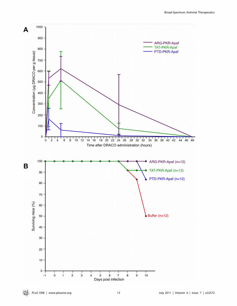
DRACOs appeared promising in proof-of-concept trials with
adult BALB/c mice. Intraperitoneal (i.p.) PKR-Apaf DRACO
penetrated the liver, kidney, and lungs and persisted at least
24–48 hours (Figure 9A). Live mice and harvested mouse organs
showed no apparent toxicity. PTD-PKR-Apaf and TAT-PKR-
Apaf DRACOs administered i.p. from day -1 through day 3
greatly reduced the morbidity in mice challenged intranasally (i.n.)
Table 1. We have demonstrated DRACO efficacy against a broad spectrum of viruses.
Virus Family Genome Envelope Replicates in Species Receptor
Rhinovirus 1B Picornavirus +ssRNA No Cytoplasm Human LDL receptor
Rhinovirus 2 Picornavirus +ssRNA No Cytoplasm Human LDL receptor
Rhinovirus 14 Picornavirus +ssRNA No Cytoplasm Human ICAM-1
Rhinovirus 30 Picornavirus +ssRNA No Cytoplasm Human LDL receptor
Theiler’s encephalomyelitis Picornavirus +ssRNA No Cytoplasm Mouse Sialic acid
Dengue type 2 Flavivirus +ssRNA Yes Cytoplasm Human DC-SIGN, etc.
Influenza H1N1 A/PR/8/34 Orthomyxovirus 2ssRNA Yes Nucleus Human Sialic acid
Influenza H1N1 A/WS/33 Orthomyxovirus 2ssRNA Yes Nucleus Human Sialic acid
Tacaribe Arenavirus 2ssRNA Yes Cytoplasm Bat Transferrin receptor 1
Amapari Arenavirus 2ssRNA Yes Cytoplasm Rodent Transferrin receptor 1
Guama Be An 277 Bunyavirus 2ssRNA Yes Cytoplasm Rodent Unidentified
Guama Be Ar 12590 Bunyavirus 2ssRNA Yes Cytoplasm Rodent Unidentified
Reovirus 3 Reovirus dsRNA No Cytoplasm Human Sialic acid
Adenovirus 5 Adenovirus dsDNA No Nucleus Human CAR
Murine adenovirus Adenovirus dsDNA No Nucleus Mouse CAR
These include viruses with DNA, dsRNA, positive-sense ssRNA, and negative-sense ssRNA genomes; enveloped and non-enveloped viruses; viruses that replicate in thecytoplasm and viruses that replicate in the nucleus; human, bat, and rodent viruses; and viruses that use a variety of cellular receptors.doi:10.1371/journal.pone.0022572.t001
Table 2. We have demonstrated that DRACO is effective and nontoxic in a wide variety of cell types.
Cells Species Tissue Immortalized Viruses
Lung fibroblasts Human Lung No Rhino 1B, 2, 30; Flu 33, 34
Hepatocytes Human Liver No Rhino 1B, 2, 30; Flu 33, 34
Airway epithelial Human Trachea No Flu A/PR/8/34
Osteoblasts Human Bone No Rhino 1B, 2, 30; Flu 33, 34
Aortic muscle Human Heart No Rhino 1B, 2, 14, 30; Flu 33, 34
AD293 Human Kidney Yes Adeno 5
H1-HeLa Human Cervix Yes Rhino 14
Vero E6 Monkey Kidney Yes Amapari, Tacaribe, Guama, Dengue
L929 Mouse Fibroblast Yes Enceph, MAdeno, Reo 3
BALB/3T3 Mouse Fibroblast Yes Reo 3
NIH/3T3 Mouse Fibroblast Yes Encephalomyelitis
These include cells representing a variety of tissues; human, mouse, and monkey cells, and both primary and immortalized cells.doi:10.1371/journal.pone.0022572.t002
Broad-Spectrum Antiviral Therapeutics
PLoS ONE | www.plosone.org 7 July 2011 | Volume 6 | Issue 7 | e22572

Figure 6. DRACOs were effective against murine adenovirus in L929 cells. (A) 100 nM DRACOs were effective against 50 pfu/well murineadenovirus, whereas all negative controls were not (16 dpi). (B) 100 nM PTD-PKR-Apaf DRACO was effective if added before or up to at least 72 hoursafter adenovirus (16 dpi). (C) Multiple 100 nM DRACOs were effective against 50 pfu/well murine adenovirus (11 dpi). Even better performance ofthese alternate DRACOs might be achieved with further optimization.doi:10.1371/journal.pone.0022572.g006
Broad-Spectrum Antiviral Therapeutics
PLoS ONE | www.plosone.org 8 July 2011 | Volume 6 | Issue 7 | e22572

Figure 7. DRACOs were effective against murine encephalomyelitis in L929 cells. (A) 100 nM DRACOs were effective against 50 pfu/wellencephalomyelitis. Cell viability measured 6 dpi showed little difference if DRACO-containing medium was removed 3 dpi when untreated cells hadwidespread CPE; there was no relapse of viral CPE in treated cells after DRACOs were withdrawn. (B) 100 nM PTD-PKR-Apaf DRACO was effective ifadded before, simultaneously with, or up to at least 6 hours after encephalomyelitis. (C) Multiple 100 nM DRACOs were effective against 50 pfu/wellmurine encephalomyelitis (4 dpi). Even better performance of these alternate DRACOs might be achieved with further optimization.doi:10.1371/journal.pone.0022572.g007
Broad-Spectrum Antiviral Therapeutics
PLoS ONE | www.plosone.org 9 July 2011 | Volume 6 | Issue 7 | e22572

Figure 8. DRACOs were effective against arenaviruses, bunyaviruses, and flaviviruses. 200 nM DRACOs with PTD, TAT, and ARG proteintransduction tags were effective in Vero E6 cells against (A) 30 pfu/well Amapari (assayed 15 dpi), (B) 30 pfu/well Guama strain Be An 277 (assayed5 dpi), and (C) 10 pfu dengue type 2 (assayed 20 dpi).doi:10.1371/journal.pone.0022572.g008
Broad-Spectrum Antiviral Therapeutics
PLoS ONE | www.plosone.org 10 July 2011 | Volume 6 | Issue 7 | e22572

Broad-Spectrum Antiviral Therapeutics
PLoS ONE | www.plosone.org 11 July 2011 | Volume 6 | Issue 7 | e22572

with 1.3 LD50 influenza H1N1 A/PR/8/34 and reduced the day-
2 lung viral titers by over an order of magnitude (Figure 9B).
Similarly, PTD-RNaseL-Apaf, TAT-RNaseL-Apaf, and ARG-
RNaseL-Apaf DRACOs administered i.p. from day -1 through
day 3 prevented morbidity in mice challenged i.n. with 0.3 LD50
influenza and reduced the day-2 viral titers by an order of
magnitude or more (Figure 9C). PKR-Apaf DRACO adminis-
tered i.n. to mice penetrated the lungs and persisted over 24 hours
(Figure 10A). PTD-PKR-Apaf, TAT-PKR-Apaf, and ARG-PKR-
Apaf DRACOs administered i.n. on day 0 reduced the morbidity
in mice challenged i.n. with 1 LD50 influenza (Figure 10B).
Based on these encouraging initial animal trials, future work
should be done to test and optimize antiviral efficacy, pharma-
cokinetics, and absence of toxicity in vitro and in vivo. Future experi-
ments can further characterize and optimize dsRNA binding,
apoptosis induction, cellular transduction, and other DRACO
properties. More extensive trials are also needed to determine how
long after infection DRACOs can be used successfully, or if
DRACOs are useful against chronic viral infections without pro-
ducing unacceptable levels of cell death in vivo.
DRACOs should be effective against numerous clinical and
NIAID priority viruses, due to the broad-spectrum sensitivity of the
dsRNA detection domain, the potent activity of the apoptosis
induction domain, and the novel direct linkage between the two
which viruses have never encountered. We have demonstrated that
DRACOs are effective against viruses with DNA, dsRNA, positive-
sense ssRNA, and negative-sense ssRNA genomes; enveloped and
non-enveloped viruses; viruses that replicate in the cytoplasm and
viruses that replicate in the nucleus; human, bat, and rodent viruses;
and viruses that use a variety of cellular receptors (Table 1).
Materials and Methods
Ethics statement for mouse trialsThis study was carried out in strict accordance with the recom-
mendations in the Guide for the Care and Use of Laboratory
Animals of the National Institutes of Health. The protocol was
approved by the Committee on Animal Care of MIT (Assurance
Number: A-3125-01). Guidelines to minimize suffering were fol-
lowed, and avertin anesthetic was used for intranasal procedures.
CloningE.F. Meurs provided PKR cDNA and Y. Shi donated human
Apaf-1 cDNA. RNaseL1–335 sequence was cloned from HeLa
cells. PKR-E3L, FADD1–90 (with L75A and L76A to prevent
spontaneous self-association [18]) and murine Apaf-11–97 sequenc-
es came from BioBasic. Genes for DRACOs and controls were
constructed using PCR and restriction cloning. TAT (YGRKK-
RRQRRR), PTD-4 (YARAAARQARA), and ARG (R9) tags were
incorporated at N- and/or C-termini. Genes were inserted into
pET100/D-TOPO (Invitrogen).
Protein productionEach vector was transformed into Rosetta BL21(DE3)pLysS E.
coli (EMD Biosciences), bacteria were plated on Luria broth (LB)
agar with 100 mg ml21 ampicillin and 34 mg ml21 chloramphen-
icol, and plates were incubated overnight at 37uC. One colony was
inoculated into ampicillin-chloramphenicol LB and grown over-
night (37uC, 225 r.p.m.), then diluted 1:30 into ampicillin-
chloramphenicol LB and incubated (30uC, 225 r.p.m.) until
OD600 reached 1.0. 0.5 mM isopropyl b-D-1-thiogalactopyrano-
side was added and flasks were incubated overnight. E. coli were
recovered by centrifugation (5,000 r.p.m., 30 min., 4uC) and lysed
by sonication, and His6-tagged proteins were purified using Ni-
NTA agarose (Invitrogen) following the manufacturer’s protocols.
Proteins were eluted into 1.56PBS with 300 mM imidazole and
10% (vol/vol) glycerol, concentrated with Amicon-15 (10 kDa
cutoff, 3,000 g) to .5 mg/ml, and filter-sterilized. Protein con-
centrations were measured relative to BSA standards by Bradford
assay (BioRad) and Gel Doc densitometry.
CellsL929 (CCL-1), NIH/3T3 (CRL-1658), BALB/3T3 (CCL-163),
H1-HeLa (CRL-1958), MDCK (CCL-34), and Vero E6 (CRL-
1586) (ATCC) and AD293 (Stratagene) were cultured in complete
DMEM (Gibco). Normal human lung fibroblasts, small airway
epithelial cells, osteoblasts, hepatocytes, and aortic smooth muscle
cells (Lonza) were cultured in cell-specific media (Lonza).
VirusesDengue type 2 (New Guinea C, VR-1584), Amapari (VR-477),
Tacaribe (VR-1272), Guama (Be An 277, VR-407; Be Ar 12590,
VR-420), murine adenovirus (VR-550), Theiler’s murine enceph-
alomyelitis (VR-57), reovirus 3 (VR-824), influenza H1N1 A/PR/
8/34 (ATCC VR-1469), influenza H1N1 A/WS/33 (ATCC VR-
1520), rhinovirus 1B (VR-481), rhinovirus 2 (VR-482), rhinovirus
14 (VR-284), and rhinovirus 30 (VR-505) were obtained from
ATCC. Adenovirus 5 was obtained from Stratagene. Influenza A/
PR/8/34 for animal trials was donated by P. Palese.
Cell assaysContact-inhibited cells were grown to 50–80% confluence and
non-contact inhibited cells to 20–50% confluence in 96-well plates
with 100 ml/well medium. DRACOs or controls were added to
columns of wells, 8 wells/column. Except in Figs. 2 and S2, 0.4–
1% (vol/vol) Roche FuGene 6 was co-administered with
DRACOs and controls to optimize cellular uptake. Wells received
virus approximately 24 hours after DRACO unless otherwise
noted. On selected days, cell viability in each plate was measured
using CellTiter 96 (Promega). Assay schedules, viral doses, and
other parameters were optimized for different cell/virus systems.
Micrographs were taken in 24-well plates under similar conditions.
DRACO cell penetration assaysCells in 24-well plates were incubated with DRACOs for
varying lengths of time, then trypsinized, washed thoroughly in
PBS, and lysed. Lysate from approximately 105 cells was loaded in
each lane. DRACOs were detected via westerns using mouse anti-
His6 (Invitrogen) and goat anti-mouse IgG HRP (Jackson).
Apoptosis assays70% confluent 96-well L929 plates were treated with 10 mM Z-
VAD-FMK pan-caspase inhibitor or 20 mM Z-LEHD-FMK
Figure 9. DRACOs appeared promising when administered via intraperitoneal (i.p.) injection in proof-of-concept trials with adultBALB/c mice. (A) 2.5 mg PTD-PKR-Apaf DRACO administered i.p. penetrated the liver, kidney, and lungs and persisted for at least 48 hours.Averages of 3 mice per data point are plotted, and error bars show s.e.m. (B) PTD-PKR-Apaf and TAT-PKR-Apaf DRACOs administered i.p. from day -1through day 3 greatly reduced the morbidity and day-2 lung viral titers in mice challenged intranasally (i.n.) with 1.3 LD50 influenza H1N1 A/PR/8/34.(C) PTD-RNaseL-Apaf, TAT-RNaseL-Apaf, and ARG-RNaseL-Apaf DRACOs administered i.p. from day -1 through day 3 greatly reduced the morbidityand day-2 lung viral titers in mice challenged i.n. with 0.3 LD50 influenza H1N1 A/PR/8/34.doi:10.1371/journal.pone.0022572.g009
Broad-Spectrum Antiviral Therapeutics
PLoS ONE | www.plosone.org 12 July 2011 | Volume 6 | Issue 7 | e22572
Broad-Spectrum Antiviral Therapeutics
PLoS ONE | www.plosone.org 13 July 2011 | Volume 6 | Issue 7 | e22572

caspase-9 inhibitor (R&D Systems), then 75 mM camptothecin
(Calbiochem) or 100 nM DRACOs with or without 25 ng/well
poly(I):poly(C) dsRNA (Sigma) transfected using FuGene (Roche)
following manufacturers’ protocols. After 24 hours, apoptosis was
determined using Caspase-Glo 3/7 (Promega).
Viral titersTiters were determined by serial dilutions onto 96-well NHLF
(for rhinovirus 1B) or H1-HeLa (for rhinovirus 14) plates, with 8
wells per 10-fold dilution and with the number of wells exhibiting
CPE measured 5 dpi. Reed-Muench titers were calculated from
the results (1 TCID50<0.7 pfu). Error bars indicate s.e.m. from 3
trials.
Statistical analysisCellTiter 96 cell viabilities were normalized to 100% for
untreated uninfected and 0% for untreated virus-killed cells.
Graphs indicate averages (n = 8) with s.e.m. Experiments were
repeated at least 3 times with similar results.
Mouse trials7-week-old female BALB/c mice (Charles River) received
DRACO i.n. (,0.5 mg in 50 ml) or i.p. (0.8–2.5 mg in 200 ml).
Mice were challenged i.n. with 0.3–1.3 LD50 influenza H1N1 A/
PR/8/34. Mice received DRACO i.p. once daily on days -1 and
1–3 and twice on day 0, or just one i.n. DRACO dose
simultaneously with virus. Lungs were harvested on day 2 and
viral titers determined by serial dilutions onto 96-well MDCK
plates. For pharmacokinetics, organs were harvested at designated
times, then sonicated into 1 ml PBS with 1% Triton X-100. 1 mg
organ solution was mixed with 26Laemmeli buffer, boiled 5 min.,
and run on a 10–20% SDS PAGE gel with a standard curve of
purified DRACO, followed by western blots with anti-Apaf
(Millipore) and HEP-labeled anti-rabbit IgG (Jackson Immunor-
esearch). Blots were developed with Pierce luminescent reagent
and exposed to film. DRACO bands were quantitated by Gel Doc
densitometry vs. the standards.
Supporting Information
Figure S1 DRACOs entered normal human lung fibro-blasts. NHLF cells were incubated overnight with 500 nM PTD-
PKR-Apaf DRACO labeled with Lumio (Invitrogen), washed with
Hank’s balanced salt solution, and photographed with a fluo-
rescent microscope to compare (A) untreated and (B) DRACO-
treated cells. DRACOs appeared to be distributed throughout
each cell in both the cytoplasm and the nucleus.
(TIF)
Figure S2 FuGene co-administration with DRACOsimproved cellular uptake and antiviral efficacy. (A)
100 nM DRACOs with PTD, TAT, and ARG protein transduc-
tion tags were effective against rhinovirus 1B in NHLF cells
without FuGene co-administration. Cell viability was measured 3
days after infection with 130 pfu/well. (B) Co-administration of
FuGene with DRACOs lowered the EC50 of DRACOs, as shown
here for PTD-PKR Apaf DRACO against 130 pfu/well rhinovi-
rus 1B in NHLFs.
(TIF)
Figure S3 200 nM PTD-PKR-Apaf DRACO was effectiveagainst rhinovirus 1B in NHLF cells. Representative
photographs were taken 20 days after challenge with 300 pfu/
well. Scale bar = 50 mm.
(TIF)
Figure S4 DRACOs decreased the viral titer of rhinovi-rus 14 in H1-HeLa cells. One 120 nM dose of PTD-PKR-
Apaf DRACO administered to cells 24 hours before or simulta-
neously with 10 pfu/well rhinovirus 14 eliminated any measurable
titer 3 dpi. One DRACO dose administered 24 or 30 hours after
infection halved the 3-dpi viral titer.
(TIF)
Figure S5 200 nM PTD-PKR-Apaf DRACO was effectiveagainst murine adenovirus in L929 cells. Representative
photographs were taken 15 days after challenge with 30 pfu/well.
Scale bar = 25 mm.
(TIF)
Figure S6 200 nM PTD-PKR-Apaf DRACO was effectiveagainst murine encephalomyelitis in L929 cells. Repre-
sentative photographs were taken 21 days after challenge with
50 pfu/well. Scale bar = 25 mm.
(TIF)
Figure S7 100 nM PTD-PKR-Apaf DRACO was effectiveagainst Amapari arenavirus in Vero E6 cells. Represen-
tative photographs were taken 11 days after challenge with
300 pfu/well. Scale bar = 100 mm.
(TIF)
Figure S8 100 nM PTD-PKR-Apaf DRACO was effectiveagainst Tacaribe arenavirus in Vero E6 cells. Photographs
were taken 8 days after challenge with 140 pfu/well. Scale
bar = 100 mm.
(TIF)
Figure S9 200 nM PTD-PKR-Apaf DRACO was effectiveagainst Guama Be An 277 bunyavirus in Vero E6 cells.Photographs were taken 4 days after challenge with 30 pfu/well.
Scale bar = 100 mm.
(TIF)
Figure S10 200 nM PKR-Apaf DRACO was effectiveagainst dengue flavivirus in Vero E6 cells. Cell viability
was measured 18 days after challenge with 16 pfu/well.
(TIF)
Figure S11 100 nM PTD-PKR-Apaf DRACO was effec-tive against dengue flavivirus in Vero E6 cells. Photo-
graphs were taken 7 days after challenge with 160 pfu/well. Scale
bar = 100 mm.
(TIF)
Figure S12 DRACOs were effective against a broadspectrum of other viruses in a variety of cell types. Left
four photos: 100 nM PKR-Apaf DRACO was effective against
H1N1 influenza A/PR/8/34 in normal human hepatocytes.
Untreated cells challenged with 105 pfu/well died within 3 days,
whereas treated challenged cells were cultured for 72 days with no
sign of viral CPE. Center four photos: 100 nM PTD-PKR-Apaf
DRACO was effective against reovirus 3 in BALB/3T3 murine
Figure 10. DRACOs appeared promising when administered via intranasal (i.n.) injection in proof-of-concept trials with adult BALB/c mice. (A) 0.5 mg PKR-Apaf DRACO administered i.n. to adult BALB/c mice penetrated the lungs and persisted over 24 hours. Averages of 3 miceper data point are plotted, and error bars show s.e.m. (B) PTD-PKR-Apaf, TAT-PKR-Apaf, and ARG-PKR-Apaf DRACOs administered i.n. on day 0reduced the morbidity in mice challenged i.n. with 1 LD50 influenza H1N1 A/PR/8/34.doi:10.1371/journal.pone.0022572.g010
Broad-Spectrum Antiviral Therapeutics
PLoS ONE | www.plosone.org 14 July 2011 | Volume 6 | Issue 7 | e22572

cells. Photographs were taken 11 days after challenge with 30 pfu/
well reovirus 3. Right four photos: 200 nM PTD-26E3L-Apaf
DRACO was effective against adenovirus 5 in human embryonic
kidney AD293 cells. Fluorescent microscope photographs were
taken 4 days after challenge with 25 pfu/well adenovirus 5
expressing enhanced green fluorescent protein (EGFP). Scale
bars = 50 mm.
(TIF)
Acknowledgments
We thank E.F. Meurs (Institut Pasteur) for PKR cDNA, Y. Shi (Princeton
University) for human Apaf-1 cDNA, P. Palese (Mount Sinai School of
Medicine) for A/PR/8/34 influenza, and MIT Division of Comparative
Medicine for mouse facilities and advice. We are grateful to G. Johnson for
assistance in the lab, B. Lemus for producing the EGFP adenovirus, and E.
Schwoebel, C. Cabrera, G. Beltz, and S. Chiang for helpful discussions.
Author Contributions
Conceived and designed the experiments: THR. Performed the experi-
ments: THR. Wrote the paper: THR. Designed DRACOs: THR.
Conducted cell assays: CZ TB. Lead for animal trials: TB. Lead for
protein production and purification: SW. Assisted with experiments: JP BZ.
References
1. Krausslich HG, Bartenschlager R, eds. (2009) Handbook of Experimental
Pharmacology 189: Antiviral Strategies. Berlin: Springer.2. Demberg T, Robert-Guroff M (2009) Mucosal immunity and protection against
HIV/SIV infection: strategies and challenges for vaccine design. Int RevImmunol 28: 20–48.
3. Boomker JM, de Leij LF, The TH, Harmsen MC (2005) Viral chemokine-
modulatory proteins: tools and targets. Cytokine Growth Factor Rev 16:91–103.
4. Knipe DM, Howley PM, eds. (2006) Fields Virology, 5th ed. Philadelphia:Lippincott Williams & Wilkins.
5. Samuel CE (2004) Knockdown by RNAi—proceed with caution. Nat
Biotechnol 22: 280–282.6. Sadler AJ, Williams BRG (2008) Interferon-inducible antiviral effectors. Nat Rev
Immunol 8: 559–568.7. Yoneyama M, Fujita T (2010) Recognition of viral nucleic acids in innate
immunity. Rev Med Virol 20: 4–22.8. Sadler AJ, Williams BR (2007) Structure and function of the protein kinase R.
Curr Top Microbiol Immunol 316: 253–292.
9. Wu S, Kaufman RJ (1996) Double-stranded (ds) RNA binding and notdimerization correlates with the activation of the dsRNA-dependent protein
kinase (PKR). J Biol Chem 271: 1756–1763.10. Hovanessian AG, Justesen J (2007) The human 29-59oligoadenylate synthetase
family: unique interferon-inducible enzymes catalyzing 29-59 instead of 39-59
phosphodiester bond formation. Biochimie 89: 779–788.11. Bisbal C, Silverman RH (2007) Diverse functions of RNase L and implications in
pathology. Biochimie 89: 789–798.12. Kawai T, Akira S (2010) The role of pattern-recognition receptors in innate
immunity: update on Toll-like receptors. Nat Immunol 11: 373–84.
13. Placido D, Brown BA, Lowenhaupt K, Rich A, Athanasiadis A (2007) A left-handed RNA double helix bound by the Za domain of the RNA-editing enzyme
ADAR1. Structure 15: 395–404.14. Pop C, Salvesen GS (2009) Human caspases: activation, specificity, and
regulation. J Biol Chem 284: 21777–21781.15. Qin H, Srinivasula SM, Wu G, Fernandes-Alnemri T, Alnemri ES, et al. (1999)
Structural basis of procaspase-9 recruitment by the apoptotic protease-activating
factor 1. Nature 399: 549–557.
16. Bao Q, Shi Y (2007) Apoptosome: a platform for the activation of initiator
caspases. Cell Death Differ 14: 56–65.17. Eberstadt M, Huang B, Chen Z, Meadows RP, Ng SC, et al. (1998) NMR
structure and mutagenesis of the FADD (Mort1) death-effector domain. Nature392: 941–945.
18. Muppidi JR, Lobito AA, Ramaswamy M, Yang JK, Wang L, et al. (2006)
Homotypic FADD interactions through a conserved RXDLL motif are requiredfor death receptor-induced apoptosis. Cell Death Differ 13: 1641–50.
19. Randall RE, Goodbourn S (2008) Interferons and viruses: an interplay betweeninduction, signalling, antiviral responses and virus countermeasures. J Gen Virol
89: 1–47.
20. Weber F, Haller O (2007) Viral suppression of the interferon system. Biochimie89: 836–842.
21. Langland JO, Kash JC, Carter V, Thomas MJ, Katze MG, et al. (2006)Suppression of proinflammatory signal transduction and gene expression by the
dual nucleic acid binding domains of the vaccinia virus E3L proteins. J Virol 80:10083–10095.
22. Galluzzi L, Brenner C, Morselli E, Touat Z, Kroemer G (2008) Viral control of
mitochondrial apoptosis. PLoS Pathogens 4: e1000018.23. Postigo A, Ferrer PE (2009) Viral inhibitors reveal overlapping themes in
regulation of cell death and innate immunity. Microbes Infect 11: 1071–1078.24. Nogal ML, Gonzalez de Buitrago G, Rodrıguez C, Cubelos B, Carrascosa AL,
et al. (2001) African swine fever virus IAP homologue inhibits caspase activation
and promotes cell survival in mammalian cells. J Virol 75: 2535–43.25. Callus BA, Vaux DL (2007) Caspase inhibitors: viral, cellular and chemical. Cell
Death Differ 14: 73–78.26. Rider TH (2006) Anti-pathogen treatments. U.S. Patent 7,125,839.
27. Rider TH (2009) Anti-pathogen treatments. U.S. Patent 7,566,694.
28. Gump JM, Dowdy SF (2007) TAT transduction: the molecular mechanism andtherapeutic prospects. Trends Mol Med 13: 443–448.
29. Ho A, Schwarze SR, Mermelstein SJ, Waksman G, Dowdy SF (2001) Syntheticprotein transduction domains: enhanced transduction potential in vitro and in
vivo. Cancer Res 61: 474–477.30. Goun EA, Pillow TH, Jones LR, Rothbard JB, Wender PA (2006) Molecular
transporters: synthesis of oligoguanidinium transporters and their application to
drug delivery and real-time imaging. ChemBioChem 7: 1497–1515.
Broad-Spectrum Antiviral Therapeutics
PLoS ONE | www.plosone.org 15 July 2011 | Volume 6 | Issue 7 | e22572
Copyright © 2022 FDOKUMEN