The nucleoside antiviral prodrug remdesivir in treating COVID ...
15
REVIEW Open Access The nucleoside antiviral prodrug remdesivir in treating COVID-19 and beyond with interspecies significance Daisy Yan 1 , One Hyuk Ra 2 and Bingfang Yan 3* Abstract Infectious pandemics result in hundreds and millions of deaths, notable examples of the Spanish Flu, the Black Death and smallpox. The current pandemic, caused by SARS-CoV-2 (severe acute respiratory syndrome coronavirus 2), is unprecedented even in the historical term of pandemics. The unprecedentedness is featured by multiple surges, rapid identification of therapeutic options and accelerated development of vaccines. Remdesivir, originally developed for Ebola viral disease, is the first treatment of COVID-19 (Coronavirus disease 2019) approved by the United States Food and Drug Administration. As demonstrated by in vitro and preclinical studies, this therapeutic agent is highly potent with a broad spectrum activity against viruses from as many as seven families even cross species. However, randomized controlled trials have failed to confirm the efficacy and safety. Remdesivir improves some clinical signs but not critical parameters such as mortality. This antiviral agent is an ester/phosphorylation prodrug and excessive hydrolysis which increases cellular toxicity. Remdesivir is given intravenously, leading to concentration spikes and likely increasing the potential of hydrolysis-based toxicity. This review has proposed a conceptual framework for improving its efficacy and minimizing toxicity not only for the COVID-19 pandemic but also for future ones caused by remdesivir-sensitive viruses. Keywords: Animal model, Carboxylesterases, COVID-19, Coronavirus, Drug-drug interactions, interspecies difference, Pandemic, Remdesivir, SARS-CoV-2 Introduction Good health is one of the most desirable, and probably the most manageable asset for human being. For most people, good health can be achieved through life-style monitoring, enhanced physical exercises, and under- standing of the family history (Abu-Zeinah and DeSan- cho 2020; Feng et al. 2020; Ford et al. 2020; Nieman and Pence 2020; Sepandi et al. 2020). Infectious pandemics, on the other hand, directly challenge this assumption. Infectious pandemics are defined as outbreaks of infec- tious diseases over large areas among all populations, although health status may somewhat determine the se- verity (McGrath et al. 2020; Morens and Fauci 2020; Ransing et al. 2020). The history of infectious pandemics in terms of transmission is tied inextricably to human- kind development or activities in a broad sense including society-driven industrialization, increased community engagement and intensified congregations for population-based activities (Coccia 2020; Habersaat et al. 2020; Levin 2020; Moreno et al. 2021). The last contrib- uting factor is relevant, particularly to the current COVID-19 pandemic. Nevertheless, infectious pan- demics, even in today’s world, would decimate human populations or cause mankind disruptions tremendously. © The Author(s). 2021 Open Access This article is licensed under a Creative Commons Attribution 4.0 International License, which permits use, sharing, adaptation, distribution and reproduction in any medium or format, as long as you give appropriate credit to the original author(s) and the source, provide a link to the Creative Commons licence, and indicate if changes were made. The images or other third party material in this article are included in the article's Creative Commons licence, unless indicated otherwise in a credit line to the material. If material is not included in the article's Creative Commons licence and your intended use is not permitted by statutory regulation or exceeds the permitted use, you will need to obtain permission directly from the copyright holder. To view a copy of this licence, visit http://creativecommons.org/licenses/by/4.0/. The Creative Commons Public Domain Dedication waiver (http://creativecommons.org/publicdomain/zero/1.0/) applies to the data made available in this article, unless otherwise stated in a credit line to the data. * Correspondence: [email protected] 3 Division of Pharmaceutical Sciences, James L. Winkle College of Pharmacy, University of Cincinnati, Cincinnati, OH 45229, USA Full list of author information is available at the end of the article Yan et al. Animal Diseases (2021) 1:15 https://doi.org/10.1186/s44149-021-00017-5
-
Upload
khangminh22 -
Category
Documents
-
view
0 -
download
0
Transcript of The nucleoside antiviral prodrug remdesivir in treating COVID ...

REVIEW Open Access
The nucleoside antiviral prodrug remdesivirin treating COVID-19 and beyond withinterspecies significanceDaisy Yan1, One Hyuk Ra2 and Bingfang Yan3*
Abstract
Infectious pandemics result in hundreds and millions of deaths, notable examples of the Spanish Flu, the BlackDeath and smallpox. The current pandemic, caused by SARS-CoV-2 (severe acute respiratory syndrome coronavirus2), is unprecedented even in the historical term of pandemics. The unprecedentedness is featured by multiplesurges, rapid identification of therapeutic options and accelerated development of vaccines. Remdesivir, originallydeveloped for Ebola viral disease, is the first treatment of COVID-19 (Coronavirus disease 2019) approved by theUnited States Food and Drug Administration. As demonstrated by in vitro and preclinical studies, this therapeuticagent is highly potent with a broad spectrum activity against viruses from as many as seven families even crossspecies. However, randomized controlled trials have failed to confirm the efficacy and safety. Remdesivir improvessome clinical signs but not critical parameters such as mortality. This antiviral agent is an ester/phosphorylationprodrug and excessive hydrolysis which increases cellular toxicity. Remdesivir is given intravenously, leading toconcentration spikes and likely increasing the potential of hydrolysis-based toxicity. This review has proposed aconceptual framework for improving its efficacy and minimizing toxicity not only for the COVID-19 pandemic butalso for future ones caused by remdesivir-sensitive viruses.
Keywords: Animal model, Carboxylesterases, COVID-19, Coronavirus, Drug-drug interactions, interspecies difference,Pandemic, Remdesivir, SARS-CoV-2
IntroductionGood health is one of the most desirable, and probablythe most manageable asset for human being. For mostpeople, good health can be achieved through life-stylemonitoring, enhanced physical exercises, and under-standing of the family history (Abu-Zeinah and DeSan-cho 2020; Feng et al. 2020; Ford et al. 2020; Nieman andPence 2020; Sepandi et al. 2020). Infectious pandemics,on the other hand, directly challenge this assumption.Infectious pandemics are defined as outbreaks of infec-tious diseases over large areas among all populations,
although health status may somewhat determine the se-verity (McGrath et al. 2020; Morens and Fauci 2020;Ransing et al. 2020). The history of infectious pandemicsin terms of transmission is tied inextricably to human-kind development or activities in a broad sense includingsociety-driven industrialization, increased communityengagement and intensified congregations forpopulation-based activities (Coccia 2020; Habersaat et al.2020; Levin 2020; Moreno et al. 2021). The last contrib-uting factor is relevant, particularly to the currentCOVID-19 pandemic. Nevertheless, infectious pan-demics, even in today’s world, would decimate humanpopulations or cause mankind disruptions tremendously.
© The Author(s). 2021 Open Access This article is licensed under a Creative Commons Attribution 4.0 International License,which permits use, sharing, adaptation, distribution and reproduction in any medium or format, as long as you giveappropriate credit to the original author(s) and the source, provide a link to the Creative Commons licence, and indicate ifchanges were made. The images or other third party material in this article are included in the article's Creative Commonslicence, unless indicated otherwise in a credit line to the material. If material is not included in the article's Creative Commonslicence and your intended use is not permitted by statutory regulation or exceeds the permitted use, you will need to obtainpermission directly from the copyright holder. To view a copy of this licence, visit http://creativecommons.org/licenses/by/4.0/.The Creative Commons Public Domain Dedication waiver (http://creativecommons.org/publicdomain/zero/1.0/) applies to thedata made available in this article, unless otherwise stated in a credit line to the data.
* Correspondence: [email protected] of Pharmaceutical Sciences, James L. Winkle College of Pharmacy,University of Cincinnati, Cincinnati, OH 45229, USAFull list of author information is available at the end of the article
Yan et al. Animal Diseases (2021) 1:15 https://doi.org/10.1186/s44149-021-00017-5

Throughout the human history, there have been quitea few infectious pandemics (Glatter and Finkelman 2021;Jester et al. 2018; Johnson and Mueller 2002; Linden-baum et al. 1967; Pollitzer 1954; Siddique et al. 1995).The brutal killers are exemplified by the Spanish Flu, theBlack Death, smallpox and Asiatic cholera. The smallpoxis estimated to have killed between 300 and 500 million(Blower and Bernoulli 2004; Krylova and Earn 2020;Thèves et al. 2014). From 1346 to 1353, the Black Death(also called the bubonic plague) caused by the plaguebacterium Y. pestis (probably other variants) killed 75–200 million people throughout the Asia, Europe, and Af-rica continents (Glatter and Finkelman 2021). Some in-fectious pandemics last for decades. For example, thecurrent HIV/AIDS pandemic which started four decadesago, has killed 38 million people worldwide (de Cocket al. 2021; GBD 2017; 22, 23). Among all infectiouspathogens, influenza virus is recognized to cause themost pandemics in the last century or so. The 1918 FluPandemic killed 20-50 million people, the 1956–58Asian Flu killed 2 million and the 1968 Flu Pandemickilled 1–4 million people (Jester et al. 2018; Jester et al.2020; Johnson and Mueller 2002; Morse et al. 2012; Sal-zberger et al. 2018).Since the turn of the twenty-first century, the pan-
demic pathogens have shifted from Influenza viruses tocoronaviruses in the sense of pandemic frequency. In-deed, the 2009 H1N1 influenza pandemic, commonly re-ferred to as Swine Flu, is the only Flu pandemic in thetwenty-first century (Staniland and Smith 2013). The Flupandemic of 2009 was initially seen in Mexico and killedapproximately 300,000 people worldwide (Staniland andSmith 2013). In contrast, there have three pandemicscaused by coronaviruses during the first 21 years of thiscentury. The SARS-CoV pandemic of 2002 (severe acuterespiratory syndrome-associated coronavirus) has a con-firmed number of over 8000 cases with estimated 813mortalities (Anderson et al. 2004; Hui and Zumla 2019).The MERS-CoV pandemic of 2012 (middle east respira-tory syndrome coronavirus) has much lower number ofconfirmed cases (~ 2500) but with a similar number ofmortalities (858), representing a mortality rate of > 35%(Azhar et al. 2019; Chafekar and Fielding 2018).The SARS-CoV-2 pandemic of 2019 (severe acute re-
spiratory syndrome-associated coronavirus-2) was re-ported initially in December of 2019 (Hu et al. 2021). Ayear and half later, the confirmed cases have reached thenumber of 203 million with a total mortality of 4.3 mil-lion worldwide (Johns Hopkins University CoronavirusResource Center, 2021). Clearly the number of the fatal-ity has been the greatest since the 1918 Flu Pandemic(Jester et al. 2018; Jester et al. 2020; Johnson and Mueller2002; Morse et al. 2012; Salzberger et al. 2018;), a cen-tury health alert so to speak. The recent infectious
pandemics, with an exception of the Flu pandemic of2009, are all associated with coronaviruses. However, themagnitudes in terms of confirmed cases and mortalitydiffer markedly. The SARS-CoV-2 pandemic of 2019represents the overwhelming numbers of confirmedcases and mortality (Johns Hopkins University Corona-virus Resource Center, 2021); the MERS-CoV pandemicof 2012 represents the least number of confirmed casesbut the highest mortality (Azhar et al. 2019; Chafekarand Fielding 2018); and the SARS-CoV pandemic of2002 is in the middle (Anderson et al. 2004; Hui andZumla 2019).While vaccines are an important part of preventative
measures to stop the spread, some coronaviruses haveshown rapid adaptability and differentiation. As differentstrains are identified, the pathogen’s ability to mutatecould outstrip our ability to create targeted vaccines. Forinstance, the new SARS-CoV-2 delta variant has shownvaccine breakthrough in Pfizer, Moderna, and Covaxinvaccines (https://pubmed.ncbi.nlm.nih.gov/34268529/).Therefore, it is necessary to have a treatment-based ap-proach with anti-viral agents in addition to apreventative-based approach. The urgency to tackle thecurrent pandemic in therapeutics has been focusedlargely on repurposing drugs for SARS-CoV-2. Remdesi-vir is originally developed for Ebola viral disease and hasbeen shown to exert a broad-spectrum of viruses includ-ing coronaviruses (Eastman et al. 2020). Therefore,remdesivir is an ideal candidate to be repurposed forCOVID-19.
Overview of remdesivirCOVID-19 has become the biggest global health crisis inthe modern history (Bassetto et al. 2021; Johns HopkinsUniversity Coronavirus Resource Center, 2021; Tabish2020). This crisis is amplified by lack of specific thera-peutics and high levels of transmission (Alshaeri andNatto 2020; Bassetto et al. 2021; Inglesby 2020; Linkaet al. 2020; Liu et al. 2020; Song et al. 2020). SARS-CoV-2, the pathogen of COVID-19, has a basic reproductionnumber (R0 value) of 1.8–3.6 (Linka et al. 2020; Liuet al. 2020; Song et al. 2020). In certain regions, the R0value goes as high as 5.0, pointing to extremely fasttransmission (Linka et al. 2020; Liu et al. 2020; Songet al. 2020). Although SARS-CoV-2 belongs to the familyof coronavirus like members of SARS-CoV and MERS-CoV, SARS-CoV-2 structurally differ markedly fromother coronaviruses (Liya et al. 2020; Rabaan et al. 2020;Satarker and Nampoothiri 2020). As a result, existinganti-coronaviral agents are not effective (Shamsi et al.2021). The urgency to tackle this pandemic in the areaof therapeutics has been focused largely on repurposingdrugs for SARS-CoV-2 (Chenoweth et al. 2020; Donget al. 2020; Gao et al. 2020; Gordon et al. 2020; Gurwitz
Yan et al. Animal Diseases (2021) 1:15 Page 2 of 15

2020; Ko et al. 2020; Lai et al. 2020; Martinez 2021;Shanmugaraj et al. 2020; Ton et al. 2020; Warren et al.2016; Yao et al. 2020). Indeed, several antiviral agentstargeting other viruses demonstrate reasonable efficacysuch as the anti-Ebola agent remdesivir, the anti-HIVcombination of lopinavir/ritonavir and the anti-parasiticdrug the anti-parasitic drug avermectin (Bixler et al.2017; Gilead Sciences 2020; NIH clinical trialNCT04280705 of remdesivir to treat COVID-19 begins2020; Hoenen et al. 2019; Lo et al. 2017; Goldman et al.2020; Grein et al. 2020; Siegel et al. 2017). Remdesivirappears to be the most promising (Beigel et al. 2020;Goldman et al. 2020; Grein et al. 2020; Spinner et al.2020; Wang et al. 2020), and represents the first treat-ment for COVID-19 approved by the United State Foodand Drug Administration (FDA News 2020). Remdesivir
was initially granted for emergency use authorizationand later for full approval.
Chemical and structural features of remdesivirRemdesivir structurally belongs to the large class of nu-cleoside/nucleotide drugs (Cavaliere et al. 2017; Liver-Tox 2020; Meier 2017; Mirza 2019). Drugs in this classusually have anti-viral, anti-cancer and immunosuppres-sive activities (Borbone et al. 2021; Damaraju et al. 2003;Khungar and Han 2010; Krecmerova 2017; Stucker andAckermann 2011). These drugs generally have a hetero-cyclic ring linked to the phosphorus atom at the center(Fig. 1, the connecting atoms marked with a red arrow).Interestingly, H-P linker (heterocyclic ring-phosphorus)varies among these therapeutics (Fig. 1). The anti-HIVdrugs tenofovir disoproxil and tenofovir alafenamide,
Fig. 1 Chemical structure of remdesivir, sofosbuvir, tenofovir disoproxil and tenofovir alafenamide Those are nucleoside/nucleotide drugsfeatured by the heterocyclic ring linked to the phosphorus atom at the center. The connecting atoms are pointed by a red arrow
Yan et al. Animal Diseases (2021) 1:15 Page 3 of 15

essential medicines listed by the World HealthOrganization (WHO), have a linker of a propane (Fig. 1),whereas remdesivir and the paradigm shift anti-hepatitisC viral agent sofosbuvir have a linker of oxolane (San-tander-Ballestín et al. 2021; Tao et al. 2020). Even be-tween sofosbuvir and remdesivir, the linker varies withstrong implications of pharmacological activities (Fig. 1).Remdesivir but not sofosbuvir has a cyano structure at-tached to oxolane. The cyano structure is implicated inanticancer activity (Jordheim et al. 2013; Labbé et al.2020; Liu et al. 2021; Ruchelman et al. 2011; Tretyakovaet al. 2019). Finally, remdesivir, like others, is an esterand the ester linkage increases lipophilicity critical forcell permeability. This is particularly of significance asremdesivir has a relatively poor water-solubility (Euro-pean Medicine Agency 2020).
Mechanism of actionRemdesivir undergoes hydrolysis initially followed byphosphorylation steps to form nucleoside triphosphate(Fig. 2) (Ottoni et al. 2020). It is the phosphorylated me-tabolite that delivers potent antiviral activity throughdistinct but related mechanisms (Fig. 2): (A) interferingwi th the ac t ion of v i ra l RNA-dependent RNApolymerase (RdRp); (B) evading exoribonuclease-proofreading; and (C) causing delayed/cyano-group me-diated chain termination of viral genome (Chen et al.2020; Malin et al. 2020; Ottoni et al. 2020; Singh et al.2020; Tchesnokov et al. 2019; Yin et al. 2020). We haveshown that human carboxylesterase-1 (CES1), a highlyefficient enzyme, was involved in the hydrolytic activa-tion of remdesivir (Shen et al. 2021a, b). However, theprecise identity of enzyme(s) for phosphorylation remainto be determined. As for the three mechanisms of ac-tion, it is clear that mechanisms A and C share the
ultimate outcomes: delayed viral replication and in favorof antiviral activity. The mechanism of action B, on theother hand, can be considered as both desirable andnon-desirable actions. Proofreading of genetic replica-tions stabilizes the genome of virus but lack of strongproofreading capacity leads greater-than-expected in-stability of mutations. The emerged variants of SARS-CoV-2, with increased transmission capacity and evengreater clinical severities, have argued that cautions mustbe exercised in this regard (Dicken et al. 2021; Martinet al. 2021).
Broad spectrum of antiviral activityAs mentioned above, remdesivir was originally devel-oped against Ebola viral infection (Bixler et al. 2017;Hoenen et al. 2019; Warren et al. 2016). Interestingly,this antiviral agent has since been shown to exert abroad spectrum of activity against as many as seven viralfamilies (Jean et al. 2020; Pruijssers et al. 2020). Theseviruses, as specified in their genome, range from positiveto negative, and to ambisense RNA viruses (Table 1).Critically, remdesivir has exhibited high potency towardsthese viruses with an exception of Hantaviridae. Mem-bers of the family Filoviridae are highly sensitive towardsremdesivir with an EC50 value of as low as 3 nM (con-centrations with half-maximal inhibition) (Table 1). Not-able members in this family are Ebola virus andMarburg virus, which cause severe diseases known asviral hemorrhagic fevers (Reynolds and Marzi 2017; Shif-flett and Marzi 2019). Majority of viruses from the fam-ily of Coronaviridae, which SARS-CoV-2 belongs to, arehighly sensitive to remdesivir as well (Table 1) (Jeanet al. 2020; Malin et al., 2020; Pruijssers et al. 2020).With human lung cells and primary human airway epi-thelial cultures, remdesivir inhibits SARS-CoV-2
Fig. 2 Therapeutic activation of remdesivir. This antiviral agent undergoes hydrolysis followed by several phosphorylation steps to form theantiviral metabolite nucleoside triphosphate. In human, the hydrolysis is achieved by CES1 but enzyme(s) for phosphorylation remains tobe determined
Yan et al. Animal Diseases (2021) 1:15 Page 4 of 15

replication with an EC50 value of 0.01 μM (Pruijsserset al. 2020). However, Coronaviridae members exhibitlarge strain differences from an EC50 of 0.02 to 4.90 μM(Table 1), representing an ~ 500-fold difference.
Efficacy and safetyCOVID-19 has become the biggest global health crisis inthe modern history, and its acceleration in a relativelyshort period presented unprecedented urgency (JohnsHopkins University Coronavirus Resource Center, 2021;Hu et al. 2021). The urgency has led to a strategy of re-purposing of existing drugs as a viable and probably themost efficient approach to deal with COVID-19. Indeed,reasonable efficacy and safety profiles have been re-ported in relevance to this strategy not only for remdesi-vir but also for others (Bixler et al. 2017; Gilead Sciences2020; NIH clinical trial of remdesivir to treat COVID-19begins 2020; Hoenen et al. 2019; Lo et al. 2017; Gold-man et al. 2020; Grein et al. 2020; Siegel et al. 2017).Among all of the repurposing medicines, remdesivir hasbeen extensively studied. On the other hand, SARS-CoV-2 behaves differently from others such as SARS-CoV and MERS-CoV, two highly related viruses thathave caused pandemics (Liya et al. 2020; Rabaan et al.2020; Satarker and Nampoothiri 2020; Shamsi et al.2021). Nevertheless, below is a brief discussion ofremdesivir regarding efficacy, safety and potential mech-anisms for safety concerns.
Efficacy of remdesivirThe efficacy of remdesivir has been studied by severalresearch identities: single research laboratories ormultiple-institutional or even global efforts. The resultsare informative but not conclusive as many variables areinvolved in the study design and/or the primary out-comes of a study to pursue. Table 2 listed several remde-sivir clinical trials and their efficacy outcomes. Whilethere are some studies that support the use of remdesi-vir, the majority of studies conclude that there were nostatistically significant clinical benefits. The Grein studyfound that 68% patients hospitalized for severe Covid-19showed clinical improvement (Grein et al. 2020) andsimilarly, the Beigel study reported that remdesivir wassuperior to placebo in shortening the time to recoveryand lowering respiratory tract infection (Beigel et al.2020). Conversely, multiple studies have not found sig-nificant clinical improvement (Goldman et al. 2020;Wang et al. 2020), or difference in clinical status inmoderate COVID-19 patients treated with remdesivircompared to regular standard of care (Spinner et al.2020). The Solidarity study concludes that remdesivirhas little or no effect on hospitalized patients withCOVID-19, as indicated by overall mortality, initiationof ventilation, and duration of hospital stay (WHO Soli-darity Trial Consortium 2021). It should be noted thatthe Solidarity study, not shown in Table 2, represented aglobal effort with out categorical details (WHO Solidar-ity Trial Consortium 2021).
Safety of remdesivirThe clinical studies, as discussed above, have pictured anencouraging but serious concerns regarding the use ofremdesivir for COVID-19 (Table 3) (Beigel et al. 2020;Goldman et al. 2020; Grein et al. 2020; Spinner et al.2020; Wang et al. 2020). There are many contributingfactors to the conflicting observations including studydesign, patient populations, existing conditions, severityof COVID-19, use of other medications, and intrinsicadverse effects of remdesivir (discussed below). Indeed,the discontinued rate was as high as 11.6% (Table 3, The
Table 1 Broad-spectrum antiviral activity
Family Genome Strains tested EC50 (μM)a
Arenaviridae Ambisense RNA 3 0.47–4.50
Coronaviridae Positive RNA 20 0.02–4.90
Flaviviridae Positive RNA 5 0.06–4.20
Filoviridae Negative RNA 14 0.003–0.14
Hantaviridae Negative RNA 1 7.00
Paramyxoviridae Negative RNA 8 0.02–0.79
Pneumoviridae Negative RNA 3 0.02–0.05aEC50: Concentrations with half-maximal inhibition
Table 2 Efficacy of remdesivir in human clinical trials
Characteristic aBeigel bWang cGrein dGoldman eSpinner
Randomized controlled trial Yes1 Yes1 No Yes2 Yes2
Median time to recovery (Remdesivir) 10 days 21 days 11 day (10-day treatment)
Median time to recovery (Control) 15 days 23 days 10 day (5-day treatment)
Days to recovery (Remdesivir/control) 0.67 0.91 1.10
Clinical improvement (10-day) 68% 65%
Clinical improvement (5-day) 70%a Beigel et al. 2020; b Wang et al. 2020; c Grein et al. 2020; d Goldman et al. 2020; e Spinner et al. 20201Randomized, double-blind, placebo-control clinical trials; 2randomized, open-label clinical trials
Yan et al. Animal Diseases (2021) 1:15 Page 5 of 15
Wang study). In consistent with the high discontinuedrates, the rates of serious adverse events were high aswell (Beigel et al. 2020; Goldman et al. 2020; Grein et al.2020; Spinner et al. 2020; Wang et al. 2020). Interest-ingly, remdesivir, when used for a longer duration suchas 10 versus 5 day-treatment, caused greater number ofadverse events (the Goldman study) or deaths (the Spin-ner study (Table 3). The serious adverse events rangefrom cardiovascular events, to pulmonary disorders, andto hepatic concerns (Beigel et al. 2020; Goldman et al.2020; Grein et al. 2020; Spinner et al. 2020; Wang et al.2020). In terms of mortality, the results are not quite
conclusive. Some studies have reported similar or com-parable rates of death between remdesivir and controlgroups (Table 3) (Spinner et al. 2020; Wang et al. 2020).
Mechanistic links to the safety concernsThe safety concerns of remdesivir are likely resultedfrom several important mechanisms. First, COVID-19 isa disease with multiple phases, typically from the initialinfectious phase, to viral replication phase and to patho-logical phase. The phase-symptoms are clinically definedas mild, pulmonary and inflammatory stage (Soy et al.2020). As a result, remdesivir likely delivers clinical
Table 3 Safety of remdesivir in human clinical trials
Characteristic aBeigel bWang cGrein dGoldman eSpinner
Discontinued rate (Remdesivir) 10-day 9.8% 11.6% 7.5% 10.2% 4.1%
Discontinued rate (Remdesivir) 5-day 4.5% 2.1%
Discontinued rate (Control) 10-day 13.5% 5.1%
Discontinued rate (Remdesivir/control) 0.73 2.27 2.27f
Serious adverse events (Remdesivir-10 day) 24.6% 18.1% 22.6% 34.5% 5.2%
Serious adverse events (Remdesivir-5 day) 21.0% 4.7%
Serious adverse events (Control) 31.6% 25.6% 9.0%
Adverse events (Remdesivir/control) 0.78 0.69 1.64
Death (Remdesivir-10 day) 10.9% 14.2% 13.2% 10.7% 1.6%
Death (Remdesivir-5 day) 8.0% 1.0%
Death (Control) 14.8% 12.8% 2.0%a Beigel et al. 2020; b Wang et al. 2020; c Grein et al. 2020; d Goldman et al. 2020; e Spinner et al. 2020f Comparison between 10-day and 5-day group
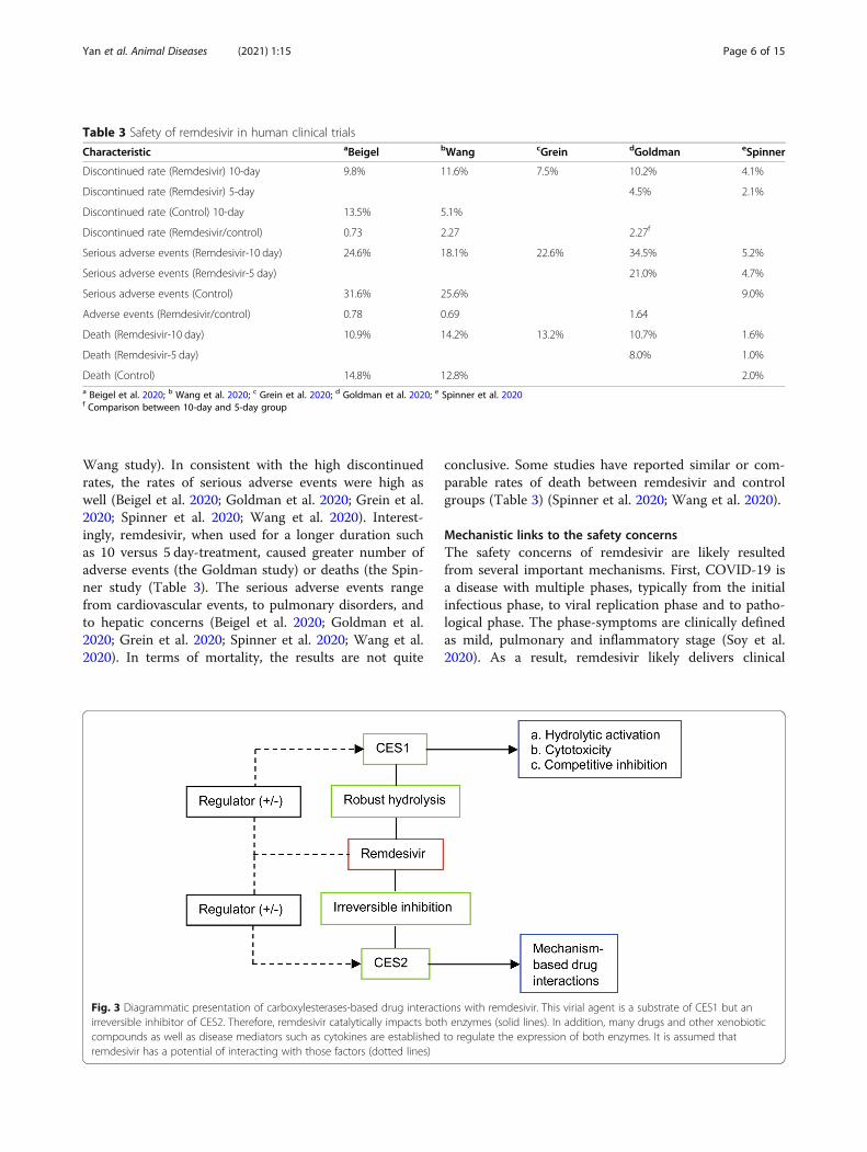
Fig. 3 Diagrammatic presentation of carboxylesterases-based drug interactions with remdesivir. This virial agent is a substrate of CES1 but anirreversible inhibitor of CES2. Therefore, remdesivir catalytically impacts both enzymes (solid lines). In addition, many drugs and other xenobioticcompounds as well as disease mediators such as cytokines are established to regulate the expression of both enzymes. It is assumed thatremdesivir has a potential of interacting with those factors (dotted lines)
Yan et al. Animal Diseases (2021) 1:15 Page 6 of 15

benefits depending on a stage of the disease. Second,COVID-19 impacts functions of multiple organs. Whilethe respiratory system is considered to be the primaryroute for infectious transmission, there are other systemssuch as the digestive system identified to play such asrole (Gavriatopoulou et al. 2020). Nevertheless, the rela-tively pathological impact among these organs, onceagain, may vary depending on a disease stage. And third,COVID-19 patients usually receive diverse types oftherapeutic approaches such as oxygen therapy, anti-inflammatory therapy, and of course antiviral therapy(Beigel et al. 2020; Goldman et al. 2020; Grein et al.2020; Spinner et al. 2020; Wang et al. 2020).The implications of drug-drug interactions and hepatic
toxicity in the safety concerns point to an intimate in-volvement of remdesivir metabolism. Indeed, COVID-19patients, probably in all cases, receive more than one oreven more drugs (Beigel et al. 2020; Goldman et al.2020; Grein et al. 2020; Spinner et al. 2020; Wang et al.2020). Remdesivir is an ester prodrug and it is thereforeassumed to have hydrolysis-based interactions. Even forhydrolytic interactions, the underlying mechanisms canbe distinct with two notable actions: regulated expres-sion of remdesivir hydrolase(s) (dotted lines) and modu-lated catalysis toward remdesivir (solid lines) (Fig. 3).The modulated catalysis toward remdesivir hydrolysis isconsidered to be intrinsic as hydrolysis is required forthe therapeutic activity of remdesivir. It has been con-firmed that remdesivir was hydrolytically activated byCES1 (Shen et al. 2021a). However, excessive hydrolysiscauses severe cytotoxicity dominantly through inhibitedproliferation and enhanced apoptosis (Shen et al. 2021a).In addition, remdesivir has been shown to irreversiblyinhibit carboxylesterase-2 (CES2) (Shen et al. 2021b).This carboxylesterase is a major hydrolase with distinctsubstrate specificity, regulated expression and tissue dis-tribution (Chen et al. 2012; Shen et al. 2019; Shen andYan 2017; Shi et al. 2006; Shi et al. 2008; Tang et al.2006; Xiao et al. 2012; Yang et al. 2007; Yang et al. 2009;Zhu et al. 2000). Conceivably, irreversible inhibition ofthis hydrolase is a contributing factor to drug-drug in-teractions with potential pharmacological and toxico-logical significance.
COVID-19 animal modelsAnimal models are critical for pathological understand-ing and therapeutic confirmation. During the past yearand half, concerted efforts have been made in developinganimal models for COVID-19 (Cleary et al. 2020; Lud-wig and Zarbock 2020; Muñoz-Fontela et al. 2020; Veen-huis and Zeiss 2021; Zeiss et al. 2021). These effortshave been focused on several critical aspects: transmissi-bility, disease process, therapeutic efficacy, immune re-sponse, and species-differential mechanisms. Studies on
the transmissibility have been focused on ACE2 receptor(angiotensin-converting enzyme-2) (Bao et al. 2020;Shang et al. 2020; Sun et al. 2020), a dual functional pro-tein as a receptor and an enzyme critical for physio-logical function (i.e., blood pressure) and infectioustransmissibility (i.e., SARS-CoV-2). These studies havefirmly established the critical role of ACE2 in the trans-missibility. Animals sharing with humans the higherACE2 sequence identity, generally have higher rates oftransmissibility (Chan et al. 2020). The most commonlyused mammalian research model mouse, sharing rela-tively a low identity with human in terms of ACE2 se-quence, does not confer efficient transmission of SARS-CoV-2 (Muñoz-Fontela et al. 2020; Zeiss et al. 2021).Nevertheless, efforts have successfully created variousmouse COVID-19 models by genetic approaches(Muñoz-Fontela et al. 2020; Zeiss et al. 2021), such asreplacement of the mouse ace2 with the correspondinghuman ACE2. It should be emphasized that mutationsof the receptor binding domain of SARS-CoV-2 is recog-nized to be critical for increased transmissibility andeven increased morbidity and mortality (Greaney et al.2021; Jackson et al. 2021; Leung et al. 2020; Guruprasad2021).
Disease modeling of COVID-19While transmissibility is the determinant factor for thepassage of infectious diseases, pathological changes, inline with the process, is one of the most, probably themost important factor for modeling. Table 4 showsmajor COVID-19 animal models with specifics of viralreplication, clinical signs and immune responses(Muñoz-Fontela et al. 2020; Zeiss et al. 2021; Veenhuisand Zeiss 2021). These categories or manifestations arecommonly seen among COVID-19 patients (Zeiss et al.2021). However, not all information on these categorieshas been collected among these animal models. Never-theless, Syrian hamsters model well to humans (Table4). Pigs, chickens and ducks are not susceptible toCOVID-19 and not viable animal candidates (Bao et al.2020; Imai et al. 2020; Lakdawala and Menachery 2020).Dogs have a low susceptibility to SARS-CoV-2. Infec-tious viral RNA was not detected in pharyngeal swabs ofinoculated dogs, and four of the six dogs failed to sero-convert (Shi et al. 2020). Coronaviruses are endemicamong bats, and there is a bat SARS-like CoV strain thatshares a common ancestor with SARS-CoV-2, divergingapproximately 40–70 years ago (Boni et al. 2020). Batsinoculated with SARS-CoV-2 displayed high viral loadsand live virus could be obtained from oral swabs, tracheaand nasal epithelium. However, bats do not display anyclinical signs of infection, giving credence to their knownviral tolerance. Compared to ferrets, their antibody re-sponse is less robust (Schlottau et al. 2020).
Yan et al. Animal Diseases (2021) 1:15 Page 7 of 15

Efficacy of remdesivir in animal modelsTherapeutic or efficacy confirmation is another majorstep, probably the most critical step in terms of man-aging a disease (Johansen et al. 2020; Sheahan et al.2020; Yu et al. 2020). Modeling of therapeutic confirm-ation, compared with disease modeling itself, is compli-cated by the interplay between host and therapeuticagent, remdesivir in this case. Nevertheless, there areseveral studies in the literature about efficacy of remde-sivir against SARS-CoV-2 (Martinez et al. 2021; Pruijs-sers et al. 2020; Williamson et al. 2020; Ye et al. 2021;Yuan et al. 2021). These studies are informative but theinformation is incomplete and/or inconsistent in termsof study design, dosage regimens, and/or the definedoutcomes. For example, the dosage regimens were differ-ent and so were the dosing routes in some cases. Table 5summarized the results from these studies on the thera-peutic confirmation.Pruijssers et al. investigated the efficacy of remdesivir
in a mouse model (Ces1c knockout) (Pruijssers et al.2020). The animals were inoculated with a chimericvirus (SARS1/SARS2-RdRp). This chimeric virus en-codes the RNA-dependent RNA polymerase of SARS-CoV-2. Remdesivir treatment was initiated at 1-day postinoculation (1 dpi) at 25 mg/kg through subcutaneousinjection and continued every 12 h until the end of thestudy at 5 dpi. The viral burden was decreased by atleast 99% in the remdesivir group. Clinical signs such as
lung hemorrhage and pulmonary function were drastic-ally improved. Williamson et al. investigated the efficacyof remdesivir in a rhesus macaque model (Williamsonet al. 2020). The treatment was initiated at 12 h afterSARS-CoV-2 inoculation and continued once dailythrough 6 dpi. One group intravenously received a load-ing dose of 10 mg/kg remdesivir, followed by a dailymaintenance dose of 5 mg/kg, and the other group re-ceived vehicle control. They reported that macaquestreated with remdesivir did not show signs of respiratorydisease with overwhelming reductions of viral burden.At necropsy, remdesivir-treated animals had lower lungviral loads and reduced lung damage. Thus, treatmentwith remdesivir initiated early during infection had aclinical benefit in rhesus macaques infected with SARS-CoV-2.Ye et al. investigated the efficacy of remdesivir in ham-
sters through intraperitoneal injection (Ye et al. 2021).The treatment with remdesivir (15 mg/kg) was per-formed at 2 dpi and 3 dpi post-inoculation. The viralburden was monitored at 4 and 14 dpi as well as bodyweight daily. Remdesivir reduced the viral burden inmultiple respiratory tissues (e.g., nasal) by at least 80%.The body weight in remdesivir but not the vehicle groupcontinued to increase. Promisingly, these parameterswere improved further at 14 dpi. Yuan et al. investigatedthe efficacy of clofazimine (a leprosy medicine) in ham-sters against SARS-C0V-2 infection (Yuan et al. 2021).
Table 4 Major SARS-CoV-2 animal models and reported manifestations
Manifestations Adapted micea Cat Ferret Hamster Non-human primates
Viral shedding √ √ √ √ √
Fever/nasal discharge/labored breathing √ √ √
Pneumonia √ √ √ √
Gastrointestinal/renal signs √
Cardiovascular/neurological signs √ √
Sex-difference in clinical signs √
Aging–related severity/susceptibility √ √ √
Elevated systemic inflammation √ √ √
Innate immunity √ √ √ √
T cell response √ √ √
B cell response √ √ √ √aVarious types of genetically modified mice with differential manifestations
Table 5 Efficacy of remdesivir in SARS-CoV-2 animal models
Author Model L/M dose Route Vial burden Clinical improvement
Pruijssers et al. 2020 Mousea 25 mg/kgb sc > 99 ↓ ↑↑↑
Williamson et al. 2020 R. macaques 10/5 mg/kg/d iv 100 x ↓ ↑↑↑↑
Ye et al. 2021 Hamster 15 mg/kg ip > 80% ↓ ↑↑↑↑
Yuan et al. 2021 Hamster 15 mg/kg ip ~ 20% ↓ ↑↑a Ces1c knockout; b Twice a day; R rhesus, sc subcutaneous injection, iv intravenous injection, ip intraperitoneal injection
Yan et al. Animal Diseases (2021) 1:15 Page 8 of 15

Remdesivir was included as a positive control. The treat-ment with remdesivir (15 mg/kg) was performed at 1, 2and 3 dpi through intraperitoneal injection. The viralburden was monitored at 4 dpi and body weight daily.Remdesivir reduced the viral burden by ~ 20% in thelung tissue. It should be noted that viral titers were de-termined by plaque-forming assay in the Yuan study(2021), whereas the Ye study used RT-PCR assay (Yeet al. 2021). The body weight of the remdesivir groupwas higher than that of the control group at 3 and 4 dpi(Yuan et al. 2021).
Interspecies significanceThe efficacy studies about remdesivir in animal modelsare informative but may not recapitulate clinical settingscompletely (Martinez et al. 2021; Pruijssers et al. 2020;Williamson et al. 2020; Ye et al. 2021; Yuan et al. 2021).It is encouraging that all of the studies have demon-strated benefits from the use of remdesivir, however,such a conclusion cannot be convincingly drawn fromhuman clinical studies (Beigel et al. 2020; Goldman et al.2020; Grein et al. 2020; Spinner et al. 2020; Wang et al.2020). There are nonetheless several contributing factorson the study design. First, remdesivir treatment in theanimal models was initiated 12 or 24 h after SARS-CoV-2 exposure. It is not clear whether this represents thesituation in human clinical trials. Second, COVID-19 pa-tients are generally treated with a loading dose of 200mg with 9-day maintenance dose of 100 mg. It is not
clear how closely the exposure of remdesivir in the ani-mal models was in line with the human exposure. Andthird, there is a relatively large range in the dosage regi-mens among these models, and it was difficulty to drawa dosing-dependent efficacy. For example, the Pruijssersstudy used a daily dose of 50 mg (Martinez et al. 2021;Pruijssers et al. 2020; Williamson et al. 2020; Ye et al.2021; Yuan et al. 2021), representing 3–10 times of themaintenance daily dose in the other studies. It was com-plicated even more that none of these studies fully moni-tored the metabolic fate of remdesivir (Martinez et al.2021; Pruijssers et al. 2020; Williamson et al. 2020; Yeet al. 2021; Yuan et al. 2021).Remdesivir is an ester/phosphorylation prodrug and
hydrolysis of the ester represents the first step towardthe therapeutic activation (Ottoni et al. 2020). Next weexamined whether the commonly used COVID-19 ani-mal models have comparable expression of carboxyles-terases, a highly efficient class of hydrolases. weperformed a Western analysis with liver microsomes andserum from 9 different species with the antibody againstrat Ces1d. This antibody was raised against bacteriallyexpressed Ces1d. No glycosylation has been shown tocross-react with any carboxylesterase (Xiao et al. 2012;Yan et al. 1995). As showed in Fig. 4, this antibody rec-ognized a single band in the liver microsomes frommonkey, hamster, rabbit, cat and human but multiplebands in others such as mouse, guinea pig, and dog. Theintensity of the band varied from one to another species.
Fig. 4 Immunoblots of liver microsomes and serum from various species with anti-rat Ces1d. Microsomes (10 μg) or serum (0.5 μL) from maturemales were resolved by 7.5% SDS-PAGE and transferred electrophoretically to nitrocellulose membranes. The blots were blocked by milk anddetected by the antibody against recombinant rat Ces1d through E. coli expression system. This antibody has been shown to have a broad-crossreactivity activity among carboxylesterases
Yan et al. Animal Diseases (2021) 1:15 Page 9 of 15
For example, the intensity varied by at least 3-fold be-tween monkey and hamster. In addition, both mice andrats expressed high levels of serum carboxylesterase (Fig.4). These findings conclude that cautions must be exer-cised regarding ester drugs in terms of their pharmaco-dynamics and pharmacokinetic determinants amongvarious species.
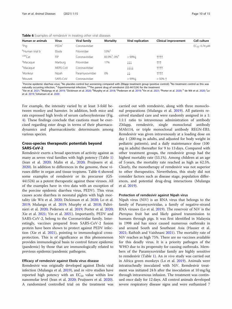
Cross-species therapeutic potentials beyondSARS-CoV-2Remdesivir exerts a broad spectrum of activity against asmany as seven viral families with high potency (Table 1)(Jean et al. 2020; Malin et al., 2020; Pruijssers et al.2020). In addition to differences in the genome, these vi-ruses differ in organ and tissue tropisms. Table 6 showedsome examples of remdesivir or its precursor (GS-441524) as a potent therapeutic against these viruses. Allof the examples have in vivo data with an exception ofthe porcine epidemic diarrhea virus, PEDV). This viruscauses acute diarrhea in neonatal piglets with high mor-tality (de Wit et al. 2020; Dickinson et al. 2020; Lo et al.2019; Mulangu et al. 2019; Murphy et al. 2018; Paltri-nieri et al. 2020; Pedersen et al. 2019; Porter et al. 2020;Xie et al. 2021; Yin et al. 2021). Importantly, PEDV andSARS-CoV-2, belong to the Coronaviridae family. Inter-estingly, vaccines prepared from SARS-CoV-2 fusionprotein have been shown to protect against PEDV infec-tion (Xie et al. 2021), pointing to immunological cross-protection. This is of significance as this phenomenonprovides immunological basis to control future epidemic(pandemic) by those that are immunologically related toprevious epidemic/pandemic pathogens.
Efficacy of remdesivir against Ebola virus diseaseRemdesivir was originally developed against Ebola viralinfection (Mulangu et al. 2019), and in vitro studies havereported high potency with an EC50 value within lownanomolar level (Jean et al. 2020; Pruijssers et al. 2020).A randomized controlled trial on the treatment was
carried out with remdesivir, along with three monoclo-nal preparations (Mulangu et al. 2019). All patients re-ceived standard care and were randomly assigned in a 1:1:1:1 ratio to intravenous administration of antibodyZMapp, remdesivir, single monoclonal antibodyMAb114, or triple monoclonal antibody REGN-EB3.Remdesivir was given intravenously at a loading dose onday 1 (200 mg in adults, and adjusted for body weight inpediatric patients), and a daily maintenance dose (100mg in adults) thereafter for 9 to 13 days. Compared withother treatment groups, the remdesivir group had thehighest mortality rate (53.1%). Among children at an ageof 5 years, the mortality rate reached as high as 62.5%.Clearly, the monotherapy of remdesivir was not superiorto other therapeutics. Nevertheless, this study did notconsider factors such as disease stage, population differ-ences, and potential drug-drug interactions (Mulanguet al. 2019).
Protection of remdesivir against Nipah virusNipah virus (NiV) is an RNA virus that belongs to thefamily of Paramyxoviridae, a family of negative-strandRNA viruses (Lo et al. 2019). The reservoir of NiV is thePteropus fruit bat and likely gained transmission tohumans through pigs. It was first identified in Malaysiain 1998 and has since caused numerous outbreaks inand around South and Southeast Asia (Hauser et al.2021; Rathish and Vaishnani 2021). The mortality rate ofNiV reaches as high 75%. There are no vaccines availablefor this deadly virus. It is a priority pathogen of theWHO due to its propensity for causing outbreaks. Mem-bers of the Paramyxoviridae family are highly sensitiveto remdesivir (Table 1). An in vivo study was carried outin Africa green monkeys (Lo et al. 2019). Animals wereintratracheally inoculated with NiV. Remdesivir treat-ment was initiated 24 h after the inoculation at 10 mg/kgthrough intravenous infusion. The treatment was contin-ued once daily for 12 days. All control animals developedsevere respiratory disease signs and were euthanized 7
Table 6 Examples of remdesivir in treating other viral diseases
Human or animals Virus Viral family Mortality Viral replication Clinical improvement Cell cultureaPig PEDA1 Coronaviridae EC50: 0.74 μMbHuman trial b Ebola Filoviridae 53%2
cdefCat FIP Coronaviridae 83.9%3, 0%4 > 99%↓ ↑↑↑↑gMacaque Marburg Filoviridae 17% ↓↓↓ ↑↑↑hMacaque MERS-CoV Coronaviridae ↓↓↓↓ ↑↑↑↑iMonkeyi Nipah Paramyxoviridae 0% ↓↓ ↑↑↑↑jMousek SARS-CoV Coronaviridae > 99%↓ > 50% ↑1Porcine epidemic diarrhea virus; 2No placebo control but worsening compared with ZMapp treatment group (positive control); 3No treatment control as this wasnaturally occurring infection; 4 Experimmental infection; 3,4The parent drug of remdesivir (GS-441524) for the treatmentaXie et al. 2021; bMulangu et al. 2019; cDickinson et al. 2020; dMurphy et al. 2018; ePedersen et al. 2019; fYin et al. 2021; gPorter et al. 2020; h de Wit et al. 2020; iLoet al. 2019; jJohansen et al. 2020
Yan et al. Animal Diseases (2021) 1:15 Page 10 of 15

or 8 dpi due to the disease severity (humane endpoints)(Lo et al. 2019). In contrast, none of remdesivir-treatedanimals developed severe symptoms. This study con-cluded that remdesivir represented a promising antiviraltreatment for NiV infection.
GS-441524, the parent drug of remdesivir for natural orexperimental feline infectious peritonitisFeline infectious peritonitis (FIP), a deadly disease fordomestic cats, is caused by FIP virus (FIPV), probably byFIPV-related viruses as well (Dickinson et al. 2020; Mur-phy et al. 2018; Paltrinieri et al. 2020; Pedersen et al.2019; Yin et al. 2021). FIPV and SARS-CoV-2 share sev-eral major traits: (A) they belong to the Coronaviridaefamily and (B) both FIPV and SARS-CoV-2 have hightransmissibility although the former is more deadly. Onthe other hand, they differ in organ tropism: FIPV tar-gets predominantly the gastrointestinal tract, whereasSARS-CoV-2 targets predominantly the pulmonary sys-tem (Dickinson et al. 2020; Gavriatopoulou et al. 2020;Murphy et al. 2018; Pedersen et al. 2019; Yin et al.2021). Nevertheless, several investigators tested GS-441524, the parent drug of remdesivir, for the efficacyagainst natural and experimental FIPV infection (Dickin-son et al. 2020; Murphy et al. 2018; Pedersen et al. 2019;Yin et al. 2021). Dickinson et al. treated four naturallyoccurring FIP cases with neurological manifestationsand demonstrated clear clinical improvement (Dickinsonet al. 2020). Yin et al. reported that FIP-suspected catshad a mortality rate of 67%, however, an overwhelmingmajority of cats treated with GS-441524 survived (Yinet al. 2021). Similar efficacy was reported by Pedersen(Pedersen et al. 2019). Finally, Murphy et al. reported inexperimentally FIPV infected cats that GS-441524caused a rapid and efficient reversal of clinical signs andreturned to normality among all cats (Murphy et al.2018).
Conclusions/further perspectivesThe COVID-19 pandemic is unprecedented even in thehistorical term and the unprecedentedness is featured bymultiple surges, rapid identification of therapeutic op-tions and accelerated development of vaccines. Thetherapeutic options have been focused largely on repur-posing existing medicines. Remdesivir, originally devel-oped for Ebola viral disease, is the first treatment ofCOVID-19 approved by the United States FDA. In vitroand animal studies have shown that this antiviral agenthad broad-spectrum activities with high potency. How-ever, human clinical trials for COVID-19 or Ebola havefailed to confirm the favorable properties on both effi-cacy and safety from preclinical studies. One explanationis that animal models have not faithfully recapitulatedthe pathological and pharmacological processes in
human. Another explanation is that COVID-19 patientsin the trials have received multiple therapeutics with in-creased risk for drug-drug interactions. These interac-tions likely have profound-species differences. Finally,remdesivir requires hydrolysis and phosphorylation toexert antiviral activity and excessive hydrolysis increasescytotoxicity. Several options should be considered: (1)remdesivir is given through intravenous injection, aroute that quickly builds high concentrations, and otheradministration routes should be considered to preventconcentration spikes that cause safety concerns; (2) for-mulations of remdesivir are so developed to minimizethe toxicological potentials; and (3) the chemical struc-ture of remdesivir, particular the ester linkage, should bemodified to reduce the risk. Once again, remdesivir hasbeen shown to be broad-spectrum and high potency.Optimization of administration routes, delivery formula-tions and chemical structure (e.g., the ester linkage) willsignify not only for the current COVID-19 pandemicbut also for future ones caused by remdesivir-sensitiveviruses.
AbbreviationsACE2: Angiotensin-converting enzyme 2; CES1: Carboxylesterase-1;Ces1d: Carboxyleseerase-1d; CES2: Carboxylesterase-2; COVID-19: Coronavirusdisease 2019; EC50: Half maximal effective concentration; FIPV: Felineinfectious peritonitis; MERS-CoV: Middle East respiratory syndromecoronavirus; NiV: Nipah virus; PEDV: Porcine epidemic diarrhea virus; SARS-CoV: Severe acute respiratory syndrome coronavirus; SARS-CoV-2: Severeacute respiratory syndrome coronavirus 2
AcknowledgementsNot applicable.
Authors’ contributionsAll three contributed to draft the manuscript and Yan, B finalized themanuscript. The author(s) read and approved the final manuscript.
Authors’ informationYan, D has received the MD degree in 2021 and has been matched forresidency in dermatology at Boston University, School of Medicine; Ra, OHreceived the MD degree in 2020 and is currently doing residency in theDepartment of Anesthesiology, Brigham and Women’s Hospital, HarvardUniversity; and Yan, B is a professor of Pharmaceutical Science and AssociateDean for Research at the University of Cincinnati James L. Winkle College ofPharmacy.
FundingThis work was supported by National Institutes of Health GrantsR01EB018748, R21AI153031 and University of Cincinnati Cancer Center (Yan,B).
Availability of data and materialsThis is a review article. The data supporting their findings can be found inthe literature as described below.
Declarations
Ethics approval and consent to participateNot applicable.
Consent for publicationNot applicable.
Yan et al. Animal Diseases (2021) 1:15 Page 11 of 15
Competing interestsAuthor Bingfang Yan was not involved in the journal’s review or decisionsrelated to this manuscript.
Author details1Sidney Kimmel Medical College, Thomas Jefferson University, 1025 WalnutSt, Philadelphia, PA 19107, USA. 2Department of Anesthesiology, Brighamand Women’s Hospital, 75 Francis St, Boston, MA 02115, USA. 3Division ofPharmaceutical Sciences, James L. Winkle College of Pharmacy, University ofCincinnati, Cincinnati, OH 45229, USA.
Received: 7 June 2021 Accepted: 25 July 2021
ReferencesAbu-Zeinah, G., and M.T. DeSancho. 2020. Understanding sideroblastic anemia:
An overview of genetics, epidemiology, pathophysiology and currenttherapeutic options. Journal of Blood Medicine 11: 305–318.
Alshaeri, H.K., and Z.S. Natto. 2020. A contemporary look at COVID-19medications: Available and potentially effective drugs. European Review forMedical and Pharmacological Sciences 24: 9188–9195.
Anderson, R.M., C. Fraserm, A.C. Ghanim, C.A. Donnelly, S. Riley, N.M. Ferguson, G.M. Leung, T.H. Lam, and A.J. Hedley. 2004. Epidemiology, transmissiondynamics and control of SARS: The 2002-2003 epidemic. PhilosophicalTransactions of the Royal Society of London. Series B, Biological Sciences 359:1091–1105.
Azhar, E.I., D.S.C. Hui, Z.A. Memish, C. Drosten, and A. Zumla. 2019. The MiddleEast respiratory syndrome (MERS). Infectious Disease Clinics of North America33: 891–905.
Bao, L., W. Deng, B. Huang, H. Gao, J. Liu, L. Ren, Q. Wei, P. Yu, Y. Xu, F. Qi, et al.2020. The pathogenicity of SARS-CoV-2 in hACE2 transgenic mice. Nature.583: 830–833.
Bassetto, F., P. Marchica, G.P. Azzena, T. Brambullo, F. Facchin, G. Masciopinto, L.Pandis, and V. Vindigni. 2021. Brief history in the time of SARS-CoV-2pandemic in Italy. A close look on a plastic surgery unit and plastic surgeonsefforts during the COVID-19 outbreak. Annali Italiani di Chirurgia. 10:S0003469X21034886.
Beigel, J.H., K.M. Tomashek, L.E. Dodd, A.K. Mehta, B.S. Zingman, A.C. Kalil, E.Hohmann, H.Y. Chu, A. Luetkemeyer, S. Kline, et al. 2020. ACTT-1 study groupmembers. Remdesivir for the treatment of Covid-19 - final report. The NewEngland Journal of Medicine 383: 1813–1826.
Bixler, S.L., A.J. Duplantier, and S. Bavari. 2017. Discovering drugs for thetreatment of Ebola virus. Current Treatment Options in Infectious Diseases 9:299–317.
Blower, S., and D. Bernoulli. 2004. An attempt at a new analysis of the mortalitycaused by smallpox and of the advantages of inoculation to prevent it.Reviews in Medical Virology 14: 275–288.
Boni, M.F., P. Lemey, X. Jiang, T.T. Lam, B.W. Perry, T.A. Castoe, A. Rambaut, and D.L. Robertson. 2020. Evolutionary origins of the SARS-CoV-2 sarbecoviruslineage responsible for the COVID-19 pandemic. Nature Microbiology 5: 1408–1417.
Borbone, N., G. Piccialli, G.N. Roviello, and G. Oliviero. 2021. Nucleoside analogsand nucleoside precursors as drugs in the fight against SARS-CoV-2 andother coronaviruses. Molecules. 26: 986.
Cavaliere, A., K.C. Probst, A.D. Westwell, and M. Slusarczyk. 2017. Fluorinatednucleosides as an important class of anticancer and antiviral agents. FutureMedicinal Chemistry 9: 1809–1833.
Chafekar, A., and B.C. Fielding. 2018. MERS-CoV: Understanding the latest humancoronavirus threat. Viruses. 10:93.
Chan, J.F., A.J. Zhang, S. Yuan, V.K. Poon, C.C. Chan, A.C. Lee, W.M. Chan, Z. Fan, H.W. Tsoi, and L. Wen. 2020. Simulation of the clinical and pathologicalmanifestations of coronavirus disease 2019 (COVID-19) in a Golden SyrianHamster model: Implications for disease pathogenesis and transmissibility.Clinical Infectious Diseases 71: 2428–2446.
Chen, P.L., N.Y. Lee, C.T. Cia, W.C. Ko, and P.R. Hsueh. 2020. A review of treatmentof coronavirus disease 2019 (COVID-19): Therapeutic repurposing and unmetclinical needs. Frontiers in Pharmacology 11: 584956.
Chen, Y.T., D. Shi, D. Yang, and B. Yan. 2012. Antioxidant sulforaphane andsensitizer trinitrobenzene sulfonate induce carboxylesterase-1 through anovel element transactivated by nuclear factor-E2 related factor-2.Biochemical Pharmacology 84: 864–871.
Chenoweth, A.M., B.D. Wines, J.C. Anania, and P. Mark-Hogarth. 2020. Harnessingthe immune system via FcγR function in immune therapy: A pathway tonext-gen mAbs. Immunology and Cell Biology 98: 287–304.
Cleary, S.J., S.C. Pitchford, R.T. Amison, R. Carrington, C.L. Robaina-Cabrera, M.Magnen, M.R. Looney, E. Gray, and C.P. Page. 2020. Animal models ofmechanisms of SARS-CoV-2 infection and COVID-19 pathology. British Journalof Pharmacology 177: 4851–4865.
Coccia, M. 2020. Factors determining the diffusion of COVID-19 and suggestedstrategy to prevent future accelerated viral infectivity similar to COVID. SciTotal Environ. 729: 138474.
Damaraju, V.L., S. Damaraju, J.D. Young, S.A. Baldwin, J. Mackey, M.B. Sawyer, andC.E. Cass. 2003. Nucleoside anticancer drugs: The role of nucleosidetransporters in resistance to cancer chemotherapy. Oncogene. 22: 7524–7536.
de Cock, K.M., H.W. Jaffe, and J.W. Curran. 2021. Reflections on 40 years of AIDS.Emerging Infectious Diseases 27: 1553–1560.
de Wit, E., F. Feldmann, J. Cronin, R. Jordan, A. Okumura, T. Thomas, D. Scott, T.Cihlar, and H. Feldmann. 2020. Prophylactic and therapeutic remdesivir (GS-5734) treatment in the rhesus macaque model of MERS-CoV infection.Proceedings of the National Academy of Sciences of the United States ofAmerica 117: 6771–6776.
Dicken, S.J., Murray, M.J., Thorne, L.G., Reuschl, A.K., Forrest, C., Ganeshalingham,M., Muir, L., Kalemera, M.D., Palor, M., McCoy, L.E et al., 2021. Characterisationof B.1.1.7 and pangolin coronavirus spike provides insights on theevolutionary trajectory of SARS-CoV-2. bioRxiv. 03.22.436468.
Dickinson, P.J., M. Bannasch, S.M. Thomasy, V.D. Murthy, K.M. Vernau, M. Liepnieks,E. Montgomery, K.E. Knickelbein, B. Murphy, and N.C. Pedersen. 2020. Antiviraltreatment using the adenosine nucleoside analogue GS-441524 in cats withclinically diagnosed neurological feline infectious peritonitis. Journal ofVeterinary Internal Medicine 34: 1587–1593.
Dong, L., S. Hu, and J. Gao. 2020. Discovering drugs to treat coronavirus disease2019 (COVID-19). Drug Discoveries & Therapeutics 14: 58–60.
Eastman, R.T., J.S. Roth, K.R. Brimacombe, A. Simeonov, M. Shen, S. Patnaik, and M.D. Hall. 2020. Remdesivir: A review of its discovery and development leadingto emergency use authorization for treatment of COVID-19. ACS CentralScience 6: 672–683.
European Medicine Agency: Summary on Compassionate Use, April 3, 2020.FDA News Release FDA Approves First Treatment for COVID-19, October 22,
2020.Feng, F., S. Tuchman, J.W. Denninger, G.L. Fricchione, and A. Yeung. 2020. Qigong
for the prevention, treatment, and rehabilitation of COVID-19 infection inolder adults. The American Journal of Geriatric Psychiatry 28: 812–819.
Ford, A.C., A.D. Sperber, M. Corsetti, and M. Camilleri. 2020. Irritable bowelsyndrome. Lancet. 396: 1675–1688.
Gao, J., Z. Tian, and X. Yang. 2020. Breakthrough: Chloroquine phosphate hasshown apparent efficacy in treatment of COVID-19 associated pneumonia inclinical studies. Bioscience Trends 14: 72–73.
Gavriatopoulou, M., E. Korompoki, D. Fotiou, I. Ntanasis-Stathopoulos, T.Psaltopoulou, E. Kastritis, E. Terpos, and M.A. Dimopoulos. 2020. Organ-specific manifestations of COVID-19 infection. Clinical and ExperimentalMedicine 20: 493–506.
GBD. 2017. HIV collaborators., 2017. Global, regional, and national incidence,prevalence, and mortality of HIV, 1980-2017, and forecasts to 2030, for 195countries and territories: A systematic analysis for the global burden ofdiseases, injuries, and risk factors study 2017. Lancet HIV. 6: e831–ee59.
Gilead Sciences Initiates Two Phase 3 Studies of Investigational AntiviralRemdesivir for the Treatment of COVID-19, U.S. FDA Grants InvestigationalNew Drug Authorization to Study Remdesivir for the Treatment of COVID-19,2020.
Glatter, K.A., and P. Finkelman. 2021. History of the plague: An ancient pandemicfor the age of COVID-19. The American Journal of Medicine 134: 176–181.
Goldman, J.D., D.C.B. Lye, D.S. Hui, K.M. Marks, R. Bruno, R. Montejano, C.D.Spinner, M. Galli, M.Y. Ahn, R.G. Nahass, et al. 2020. GS-US-540-5773investigators. Remdesivir for 5 or 10 days in patients with severe Covid-19.The New England Journal of Medicine 383: 1827–1837.
Gordon, C.J., E.P. Tchesnokov, J.Y. Feng, D.P. Porter, and M. Götte. 2020. Theantiviral compound remdesivir potently inhibits RNA-dependent RNApolymerase from Middle East respiratory syndrome coronavirus. The Journalof Biological Chemistry 295: 4773–4779.
Greaney, A.J., T.N. Starr, P. Gilchuk, S.J. Zost, E. Binshtein, A.N. Loes, S.K. Hilton, J.Huddleston, R. Eguia, and K.H.D. Crawford. 2021. Complete mapping of
Yan et al. Animal Diseases (2021) 1:15 Page 12 of 15

mutations to the SARS-CoV-2 spike receptor-binding domain that escapeantibody recognition. Cell Host & Microbe 29: 44–57.
Grein, J., N. Ohmagari, D. Shin, G. Diaz, E. Asperges, A. Castagna, T. Feldt, G.Green, M.L. Green, F.X. Lescure, et al. 2020. Compassionate use of Remdesivirfor patients with severe Covid-19. The New England Journal of Medicine 382:2327–2336.
Guruprasad, L. 2021. Human SARS CoV-2 spike protein mutations. Proteins. 89:569–576.
Gurwitz, D. 2020. Angiotensin receptor blockers as tentative SARS-CoV-2therapeutics. Drug Development Research 81: 537–540.
Habersaat, K.B., C. Betsch, M. Danchin, C.R. Sunstein, R. Böhm, A. Falk, N.T. Brewer,S.B. Omer, M. Scherzer, S. Sah, et al. 2020. Ten considerations for effectivelymanaging the COVID-19 transition. Nature Human Behaviour 4: 677–687.
Hauser, N., A.C. Gushiken, S. Narayanan, S. Kottilil, and J.V. Chua. 2021. Evolutionof Nipah virus infection: Past, present, and future considerations. TropicalMedicine and Infectious Disease 6: 24.
Hoenen, T., A. Groseth, and H. Feldmann. 2019. Therapeutic strategies to targetthe Ebola virus life cycle. Nature Reviews. Microbiology 17: 593–606.
Hu, B., H. Guo, P. Zhou, and Z.L. Shi. 2021. Characteristics of SARS-CoV-2 andCOVID-19. Nature Reviews. Microbiology 19: 141–154.
Hui, D.S.C., and A. Zumla. 2019. Severe acute respiratory syndrome: Historical,epidemiologic, and clinical features. Infectious Disease Clinics of North America33: 869–889.
Imai, M., K. Iwatsuki-Horimoto, M. Hatta, S. Loeber, P.J. Halfmann, N. Nakajima, T.Watanabe, M. Ujie, K. Takahashi, M. Ito, et al. 2020. Syrian hamsters as a smallanimal model for SARS-CoV-2 infection and countermeasure development.Proceedings of the National Academy of Sciences of the United States ofAmerica 117: 16587–16595.
Inglesby, T.V. 2020. Public health measures and the reproduction number ofSARS-CoV-2. JAMA. 323: 2186–2187.
Jackson, C.B., L. Zhang, M. Farzan, and H. Choe. 2021. Functional importance ofthe D614G mutation in the SARS-CoV-2 spike protein. Biochemical andBiophysical Research Communications 538: 108–115.
Jean, S.S., P.I. Lee, and P.R. Hsueh. 2020. Treatment options for COVID-19: Thereality and challenges. Journal of Microbiology, Immunology, and Infection 53:436–443.
Jester, B., T. Uyeki, and D. Jernigan. 2018. Readiness for responding to a severepandemic 100 years after 1918. American Journal of Epidemiology 187: 2596–2602.
Jester, B.J., T.M. Uyeki, and D.B. Jernigan. 2020. Fifty years of influenza a(H3N2)following the pandemic of 1968. American Journal of Public Health 110: 669–676.
Johansen, M.D., A. Irving, X. Montagutelli, M.D. Tate, I. Rudloff, M.F. Nold, N.G.Hansbro, R.Y. Kim, C. Donovan, G. Liu, et al. 2020. Animal and translationalmodels of SARS-CoV-2 infection and COVID-19. Mucosal Immunology 13:877–891.
Johns Hopkins University Coronavirus Resource Center, 2021. https://coronavirus.jhu.edu/.
Johnson, N.P., and J. Mueller. 2002. Updating the accounts: Global mortality ofthe 1918-1920 “Spanish” influenza pandemic. Bulletin of the History ofMedicine 76: 105–115.
Jordheim, L.P., D. Durantel, F. Zoulim, and C. Dumontet. 2013. Advances in thedevelopment of nucleoside and nucleotide analogues for cancer and viraldiseases. Nature Reviews. Drug Discovery 12: 447–464.
Khungar, V., and S.H. Han. 2010. A systematic review of side effects of nucleosideand nucleotide drugs used for treatment of chronic hepatitis B. CurrentHepatitis Reports 9: 75–90.
Ko, W.C., J.M. Rolain, N.Y. Lee, P.L. Chen, C.T. Huang, P.I. Lee, and P.R. Hsueh. 2020.Arguments in favour of remdesivir for treating SARS-CoV-2 infections.International Journal of Antimicrobial Agents 55: 105933.
Krecmerova, M. 2017. Amino acid Ester prodrugs of nucleoside and nucleotideantivirals. Mini Reviews in Medicinal Chemistry 17: 818–833.
Krylova, O., and D.J.D. Earn. 2020. Patterns of smallpox mortality in London,England, over three centuries. PLoS Biology 18: e3000506.
Labbé, M.O., L. Collins, C.A. Lefebvre, W. Maharsy, J. Beauregard, S. Dostie, M.Prévost, M. Nemer, and Y. Guindon. 2020. Identification of a C3’-nitrilenucleoside analogue inhibitor of pancreatic cancer cell line growth.Bioorganic & Medicinal Chemistry Letters 30: 126983.
Lai, C.C., T.P. Shih, W.C. Ko, H.J. Tang, and P.R. Hsueh. 2020. Severe acuterespiratory syndrome coronavirus 2 (SARS-CoV-2) and coronavirus disease-
2019 (COVID-19): The epidemic and the challenges. International Journal ofAntimicrobial Agents 55: 105924.
Lakdawala, S.S., and V.D. Menachery. 2020. The search for a COVID-19 animalmodel. Science. 368: 942–943.
Leung, K., M.H. Shum, G.M. Leung, T.T. Lam, and J.T. Wu. 2020. Earlytransmissibility assessment of the N501Y mutant strains of SARS-CoV-2 in theUnited Kingdom, October to November 2020. Euro Surveillance 26: 2002106.
Levin, J. 2020. The Faith Community and the SARS-CoV-2 outbreak: Part of theproblem or part of the solution? Journal of Religion and Health 59: 2215–2228.
Lindenbaum, J., W.B. Greenough, and M.R. Islam. 1967. Antibiotic therapy ofcholera. Bulletin of the World Health Organization 36: 871–883.
Linka, K., M. Peirlinck, and E. Kuhl. 2020. The reproduction number of COVID-19and its correlation with public health interventions. Computational Mechanics28: 1–16.
Liu, X., H. Kantarjian, and W. Plunkett. 2021. Sapacitabine for cancer. ExpertOpinion on Investigational Drugs 21: 541–555.
Liu, Y., A.A. Gayle, A. Wilder-Smith, and J. Rocklöv. 2020. The reproductive numberof COVID-19 is higher compared to SARS coronavirus. Journal of TravelMedicine 27: taaa021.
LiverTox. 2020. Clinical and research information on drug-induced liver injury.Bethesda: National Institute of Diabetes and Digestive and Kidney Diseases;2012–. Nucleoside Analogues.
Liya, G., W. Yuguang, L. Jian, Y. Huaiping, H. Xue, H. Jianwei, M. Jiaju, L. Youran, M.Chen, and J. Yiqing. 2020. Studies on viral pneumonia related to novelcoronavirus SARS-CoV-2, SARS-CoV, and MERS-CoV: A literature review. APMIS.128: 423–432.
Lo, M.K., F. Feldmann, J.M. Gary, R. Jordan, R. Bannister, J. Cronin, N.R. Patel, J.D.Klena, S.T. Nichol, T. Cihlar, et al. 2019. Remdesivir (GS-5734) protects Africangreen monkeys from Nipah virus challenge. Science Translational Medicine 11:eaau9242.
Lo, M.K., R. Jordan, A. Arvey, J. Sudhamsu, P. Shrivastava-Ranjan, A.L. Hotard, M.Flint, L.K. McMullan, D. Siegel, M.O. Clarke, et al. 2017. GS-5734 and its parentnucleoside analog inhibit filo-, Pneumo-, and paramyxoviruses. ScientificReports 7: 43395.
Ludwig, S., and A. Zarbock. 2020. Coronaviruses and SARS-CoV-2: A briefoverview. Anesthesia and Analgesia 131: 93–96.
Malin, J.J., I. Suárez, V. Priesner, G. Fätkenheuer, and J. Rybniker. 2020. Remdesiviragainst COVID-19 and other viral diseases. Clinical Microbiology Reviews 34:e00162–e00120.
Martin, C., S. McDonald, S. Bale, M. Luteijn, and R. Sarkar. 2021. Construction of ademand and capacity model for intensive care and hospital ward beds, andmortality from COVID-19. BMC Medical Informatics and Decision Making 21:138.
Martinez, D.R., Schäfer, A., Leist, S.R., Li, D., Gully, K., Yount, B., Feng, J.Y., Bunyan, E.,Porter, D.P., Cihlar, T. et al., 2021. Prevention and therapy of SARS-CoV-2 andthe B.1.351 variant in mice. Cell Rep. 36:109450.
Martinez, M.A. 2021. Compounds with therapeutic potential against novelrespiratory 2019 coronavirus. Antimicrobial Agents and Chemotherapy 64:e00399–e00320.
McGrath, B.A., M.J. Brenner, S.J. Warrillow, V. Pandian, A. Arora, T.S. Cameron, J.M.Añon, G. Hernández-Martínez, R.D. Truog, S.D. Block, et al. 2020.Tracheostomy in the COVID-19 era: Global and multidisciplinary guidance.The Lancet Respiratory Medicine 8: 717–725.
Meier, C. 2017. Nucleoside diphosphate and triphosphate prodrugs - anunsolvable task? Antiviral Chemistry & Chemotherapy 25: 69–82.
Mirza, A.Z. 2019. Advancement in the development of heterocyclic nucleosidesfor the treatment of cancer - a review. Nucleosides, Nucleotides & Nucleic Acids38: 836–857.
Moreno, G.K., Braun KM, Pray IW, Segaloff HE, Lim A, Poulson K, Meiman J,Borcher J, Westergaard RP, Moll MK et.al., 2021. SARS-CoV-2 transmission inintercollegiate athletics not fully mitigated with daily antigen testing.medRxiv. 03.03.21252838.
Morens, D.M., and A.S. Fauci. 2020. Emerging pandemic diseases: How we got toCOVID-19. Cell. 182: 1077–1092.
Morse, S.S., J.A. Mazet, M. Woolhouse, C.R. Parrish, D. Carroll, W.B. Karesh, C.Zambrana-Torrelio, W.I. Lipkin, and P. Daszak. 2012. Prediction and preventionof the next pandemic zoonosis. Lancet. 380: 1956–1965.
Mulangu, S., L.E. Dodd, R.T. Davey Jr., O. Tshiani-Mbaya, M. Proschan, D. Mukadi,M. Lusakibanza-Manzo, D. Nzolo, A. Tshomba-Oloma, et al. 2019. PALM
Yan et al. Animal Diseases (2021) 1:15 Page 13 of 15

consortium study team. A randomized, controlled trial of Ebola virus diseasetherapeutics. The New England Journal of Medicine 381: 2293–2303.
Muñoz-Fontela, C., W.E. Dowling, S.G.P. Funnell, P.S. Gsell, A.X. Riveros-Balta, R.A.Albrecht, H. Andersen, R.S. Baric, and M.W. Carroll. 2020. Animal models forCOVID-19. Nature. 586: 509–515.
Murphy, B.G., M. Perron, E. Murakami, K. Bauer, Y. Park, C. Eckstrand, M. Liepnieks,and N.C. Pedersen. 2018. The nucleoside analog GS-441524 strongly inhibitsfeline infectious peritonitis (FIP) virus in tissue culture and experimental catinfection studies. Veterinary Microbiology 219: 226–233.
Nieman, D.C., and B.D. Pence. 2020. Exercise immunology: Future directions.Journal of Sport and Health Science 9: 432–445.
NIH clinical trial NCT04280705 of remdesivir to treat COVID-19 begins at https://www.nih.gov/news-events/news-releases/nih-clinical-trial-remdesivir-treat-covid-19-begins. 2020.
Ottoni, M.P., J.D. Ricciardone, A. Nadimpalli, S. Singh, A.M. Katsomya, L.M. Pokoso,and R. Petrucci. 2020. Ebola-negative neonates born to Ebola-infectedmothers after monoclonal antibody therapy: A case series. The Lancet Child &Adolescent Health 4: 884–888.
Paltrinieri, S., A. Giordano, A. Stranieri, and S. Lauzi. 2020. Feline infectiousperitonitis (FIP) and coronavirus disease 19 (COVID-19): Are they similar?Transboundary and Emerging Diseases. 68:1786–1799.
Pedersen, N.C., M. Perron, M. Bannasch, E. Montgomery, E. Murakami, M.Liepnieks, and H. Liu. 2019. Efficacy and safety of the nucleoside analog GS-441524 for treatment of cats with naturally occurring feline infectiousperitonitis. Journal of Feline Medicine and Surgery 21: 271–281.
Pollitzer, R. 1954. Cholera studies: 1. History of the disease. Bulletin of the WorldHealth Organization 10: 421–461.
Porter, D.P., J.M. Weidner, L. Gomba, R. Bannister, C. Blair, R. Jordan, J. Wells, K.Wetzel, N. Garza, S. Van Tongeren, et al. 2020. Remdesivir (GS-5734) isefficacious in Cynomolgus macaques infected with Marburg virus. TheJournal of Infectious Diseases 222: 1894–1901.
Pruijssers, A.J., A.S. George, A. Schäfer, S.R. Leist, L.E. Gralinksi, K.H. Dinnon 3rd., B.L.Yount, M.L. Agostini, L.J. Stevens, J.D. Chappell, et al. 2020. Remdesivir inhibitsSARS-CoV-2 in human lung cells and chimeric SARS-CoV expressing theSARS-CoV-2 RNA polymerase in mice. Cell Reports 32: 107940.
Rabaan, A.A., S.H. Al-Ahmed, S. Haque, R. Sah, R. Tiwari, Y.S. Malik, K. Dhama, M.I.Yatoo, D.K. Bonilla-Aldana, and A.J. Rodriguez-Morales. 2020. SARS-CoV-2,SARS-CoV, and MERS-COV: A comparative overview. Le Infezioni in Medicina28: 174–184.
Ransing, R., R. Ramalho, R. de Filippis, M.I. Ojeahere, R. Karaliuniene, L. Orsolini, M.Pinto da Costa, I. Ullah, P. Grandinetti, D. Gashi-Bytyçi, et al. 2020. Infectiousdisease outbreak related stigma and discrimination during the COVID-19pandemic: Drivers, facilitators, manifestations, and outcomes across theworld. Brain, Behavior, and Immunity 89: 555–558.
Rathish, B., and K. Vaishnani. 2021. Nipah virus. In StatPearls. Treasure Island:StatPearls Publishing.
Reynolds, P., and A. Marzi. 2017. Ebola and Marburg virus vaccines. Virus Genes 53:501–515.
Ruchelman, A.L., H.W. Man, R. Chen, W. Liu, L. Lu, D. Cedzik, L. Zhang, J. Leisten,A. Collette, R.K. Narla, et al. 2011. 1,1-Diarylalkenes as anticancer agents: Dualinhibitors of tubulin polymerization and phosphodiesterase 4. Bioorganic &Medicinal Chemistry 19: 6356–6374.
Salzberger, B., A. Mohr, and F. Hitzenbichler. 2018. Die influenza 1918 [thepandemic influenza 1918]. Deutsche Medizinische Wochenschrift 143: 1858–1863.
Santander-Ballestín, S., D. Gómez-Martín, S. Lorente-Pérez, and M.J. Luesma-Bartolomé. 2021. Hepatitis C: A pharmacological therapeutic update. Journalof Clinical Medicine 10: 1568.
Satarker, S., and M. Nampoothiri. 2020. Structural proteins in severe acuterespiratory syndrome coronavirus-2. Archives of Medical Research 51: 482–491.
Schlottau, K., M. Rissmann, A. Graaf, J. Schön, J. Sehl, C. Wylezich, D. Höper, T.C.Mettenleiter, A. Balkema-Buschmann, T. Harder, et al. 2020. SARS-CoV-2 infruit bats, ferrets, pigs, and chickens: An experimental transmission study. TheLancet Microbe. 1: e218–ee25.
Sepandi, M., M. Taghdir, Y. Alimohamadi, S. Afrashteh, and H. Hosamirudsari.2020. Factors associated with mortality in COVID-19 patients: A systematicreview and meta-analysis. Iranian Journal of Public Health 49: 1211–1221.
Shamsi, A., T. Mohammad, S. Anwar, S. Amani, M.S. Khan, F.M. Husain, M.T.Rehman, A. Islam, and M.I. Hassan. 2021. Potential drug targets of SARS-CoV-2: From genomics to therapeutics. International Journal of BiologicalMacromolecules 177: 1–9.
Shang, J., G. Ye, K. Shi, Y. Wan, C. Luo, H. Aihara, Q. Geng, A. Auerbach, and F. Li.2020. Structural basis of receptor recognition by SARS-CoV-2. Nature. 581:221–224.
Shanmugaraj, B., K. Siriwattananon, K. Wangkanont, and W. Phoolcharoen. 2020.Perspectives on monoclonal antibody therapy as potential therapeuticintervention for coronavirus disease-19 (COVID-19). Asian Pacific Journal ofAllergy and Immunology 38: 10–18.
Sheahan, T.P., A.C. Sims, S. Zhou, R.L. Graham, A.J. Pruijssers, M.L. Agostini, S.R.Leist, A. Schäfer, K.H. Dinnon 3rd, L.J. Stevens, et al. 2020. An orallybioavailable broad-spectrum antiviral inhibits SARS-CoV-2 in human airwayepithelial cell cultures and multiple coronaviruses in mice. ScienceTranslational Medicine 12: eabb5883.
Shen, Y., W. Eades, and B. Yan. 2021a. The COVID-19 medicine remdesivir isactivated by carboxylesterase-1 and excessive hydrolysis increasescytotoxicity. Hepatology Communications. https://doi.org/10.1002/hep4.1736.
Shen, Y., W. Eades, and B. Yan. 2021b. Remdesivir potently inhibitscarboxylesterase-2 through covalent modifications: Signifying strong drug-drug interactions. Fundamental & Clinical Pharmacology 35: 432–434.
Shen, Y., Z. Shi, and B. Yan. 2019. Carboxylesterases: Pharmacological inhibition,regulated expression and transcriptional involvement of nuclear receptorsand other transcription factors. Nuclear Receptor Research 6: 101435.
Shen, Y., and B. Yan. 2017. Covalent inhibition of carboxylesterase-2 by sofosbuvirand its effect on the hydrolytic activation of tenofovir disoproxil. Journal ofHepatology 66: 660–661.
Shi, D., J. Yang, D. Yang, E.L. LeCluyse, C. Black, L. You, F. Akhlaghi, and B. Yan.2006. Anti-influenza prodrug oseltamivir is activated by carboxylesterasehuman carboxylesterase 1, and the activation is inhibited by antiplateletagent clopidogrel. The Journal of Pharmacology and Experimental Therapeutics319: 1477–1484.
Shi, D., J. Yang, D. Yang, and B. Yan. 2008. Dexamethasone suppresses theexpression of multiple rat carboxylesterases through transcriptionalrepression: Evidence for an involvement of the glucocorticoid receptor.Toxicology. 254: 97–105.
Shi, J., Z. Wen, G. Zhong, H. Yang, C. Wang, B. Huang, R. Liu, X. He, L. Shuai, Z.Sun, et al. 2020. Susceptibility of ferrets, cats, dogs, and other domesticatedanimals to SARS-coronavirus 2. Science. 368: 1016–1020.
Shifflett, K., and A. Marzi. 2019. Marburg virus pathogenesis - differences andsimilarities in humans and animal models. Virology Journal 16: 165.
Siddique, A.K., A. Salam, M.S. Islam, K. Akram, R.N. Majumdar, K. Zaman, N.Fronczak, and S. Laston. 1995. Why treatment centres failed to preventcholera deaths among Rwandan refugees in Goma, Zaire. The Lancet 345:359–361.
Siegel, D., H.C. Hui, E. Doerffler, M.O. Clarke, K. Chun, L. Zhang, S. Neville, E. Carra,W. Lew, B. Ross, et al. 2017. Discovery and synthesis of a Phosphoramidateprodrug of a Pyrrolo[2,1-f] [triazin-4-amino] adenine C-nucleoside (GS-5734)for the treatment of Ebola and emerging viruses. Journal of MedicinalChemistry 60: 1648–1661.
Singh, A.K., A. Singh, R. Singh, and A. Misra. 2020. Remdesivir in COVID-19: Acritical review of pharmacology, pre-clinical and clinical studies. Diabetes andMetabolic Syndrome: Clinical Research and Reviews 14: 641–648.
Song, Y., M. Zhang, L. Yin, K. Wang, Y. Zhou, M. Zhou, and Y. Lu. 2020. COVID-19treatment: Close to a cure? A rapid review of pharmacotherapies for thenovel coronavirus (SARS-CoV-2). International Journal of Antimicrobial Agents56: 106080.
Soy, M., G. Keser, P. Atagündüz, F. Tabak, I. Atagündüz, and S. Kayhan. 2020.Cytokine storm in COVID-19: Pathogenesis and overview of anti-inflammatory agents used in treatment. Clinical Rheumatology 39: 2085–2094.
Spinner, C.D., R.L. Gottlieb, G.J. Criner, J.R. Arribas-López, A.M. Cattelan, A. Soriano-Viladomiu, O. Ogbuagu, P. Malhotra, K.M. Mullane, A. Castagna, et al. 2020.GS-US-540-5774 investigators. Effect of Remdesivir vs standard care onclinical status at 11 days in patients with moderate COVID-19: A randomizedclinical trial. JAMA. 324: 1048–1057.
Staniland, K., and G. Smith. 2013. Flu frames. Sociology of Health & Illnes 35: 309–324.
Stucker, F., and D. Ackermann. 2011. Immunsuppressiva - Wirkungen,Nebenwirkungen und Interaktionen [immunosuppressive drugs - how theywork, their side effects and interactions]. Therapeutische Umschau 68: 679–686.
Sun, S.H., Q. Chen, H.J. Gu, G. Yang, Y.X. Wang, X.Y. Huang, S.S. Liu, N.N. Zhang, X.F. Li, and R. Xiong. 2020. A mouse model of SARS-CoV-2 infection andpathogenesis. Cell Host & Microbe 28: 124–133.
Yan et al. Animal Diseases (2021) 1:15 Page 14 of 15
Tabish, S.A. 2020. COVID-19 pandemic: Emerging perspectives and future trends.Journal of Public Health Research 9: 1786.
Tang, M., M. Mukundan, J. Yang, N. Charpentier, E.L. LeCluyse, C. Black, D. Yang, D.Shi, and B. Yan. 2006. Antiplatelet agents aspirin and clopidogrel arehydrolyzed by distinct carboxylesterases, and clopidogrel is transesterificatedin the presence of ethyl alcohol. The Journal of Pharmacology andExperimental Therapeutics 319: 1467–1476.
Tao, X., Y. Lu, Y. Zhou, L. Zhang, and Y. Chen. 2020. Efficacy and safety of theregimens containing tenofovir alafenamide versus tenofovir disoproxilfumarate in fixed-dose single-tablet regimens for initial treatment of HIV-1infection: A meta-analysis of randomized controlled trials. InternationalJournal of Infectious Diseases 93: 108–117.
Tchesnokov, E.P., J.Y. Feng, D.P. Porter, and M. Götte. 2019. Mechanism ofinhibition of Ebola virus RNA-dependent RNA polymerase by Remdesivir.Viruses. 11: 326.
Thèves, C., P. Biagini, and E. Crubézy. 2014. The rediscovery of smallpox. ClinicalMicrobiology and Infection 20: 210–218.
Ton, A.T., F. Gentile, M. Hsing, F. Ban, and A. Cherkasov. 2020. Rapid identificationof potential inhibitors of SARS-CoV-2 main protease by deep docking of 1.3billion compounds. Molecular Informatics 39: e2000028.
Tretyakova, E.V., E.V. Salimova, L.V. Parfenova, M.M. Yunusbaeva, L.U. Dzhemileva,V.A. D'yakonov, and U.M. Dzhemilev. 2019. Synthesis of newdihydroquinopimaric acid analogs with nitrile groups as apoptosis-inducinganticancer agents. Anti-Cancer Agents in Medicinal Chemistry 19: 1172–1183.
Veenhuis, R.T., and C.J. Zeiss. 2021. Animal models of COVID-19 II. Comparativeimmunology. ILAR Journal: ilab010. https://doi.org/10.1093/ilar/ilab010.
Wang, Y., D. Zhang, G. Du, R. Du, J. Zhao, Y. Jin, S. Fu, L. Gao, Z. Cheng, Q. Lu,et al. 2020. Remdesivir in adults with severe COVID-19: A randomised,double-blind, placebo-controlled, multicentre trial. The Lancet. 395: 1569–1578.
Warren, T.K., R. Jordan, M.K. Lo, A.S. Ray, R.L. Mackman, V. Soloveva, D. Siegel, M.Perron, R. Bannister, H.C. Hui, et al. 2016. Therapeutic efficacy of the smallmolecule GS-5734 against Ebola virus in rhesus monkeys. Nature. 531: 381–385.
WHO Solidarity Trial Consortium. 2021. Repurposed antiviral drugs for Covid-19 -interim WHO solidarity trial results. The New England Journal of Medicine 384:497–411.
Williamson, B.N., F. Feldmann, B. Schwarz, K. Meade-White, D.P. Porter, J. Schulz,N. van Doremalen, I. Leighton, C.K. Yinda, L. Pérez-Pérez, et al. 2020. Clinicalbenefit of remdesivir in rhesus macaques infected with SARS-CoV-2. Nature.585: 273–276.
Xiao, D., Y.T. Chen, D. Yang, and B. Yan. 2012. Age-related inducibility ofcarboxylesterases by the antiepileptic agent phenobarbital and implicationsin drug metabolism and lipid accumulation. Biochemical Pharmacology 84:232–239.
Xie, Y., X. Guo, T. Hu, D. Wei, X. Ma, J. Wu, B. Huang, and J. Shen. 2021. Significantinhibition of porcine epidemic diarrhea virus in vitro by Remdesivir, itsparent nucleoside and β-D-N4-hydroxycytidine. Virologica Sinica: 1–9.
Yan, B., D. Yang, P. Bullock, and A. Parkinson. 1995. Rat serum carboxylesterase.Cloning, expression, regulation, and evidence of secretion from liver. TheJournal of Biological Chemistry 270: 19128–19134.
Yang, D., R.E. Pearce, X. Wang, R. Gaedigk, Y.J. Wan, and B. Yan. 2009. Humancarboxylesterases HCE1 and HCE2: Ontogenic expression, inter-individualvariability and differential hydrolysis of oseltamivir, aspirin, deltamethrin andpermethrin. Biochemical Pharmacology 77: 238–247.
Yang, J., D. Shi, D. Yang, X. Song, and B. Yan. 2007. Interleukin-6 alters the cellularresponsiveness to clopidogrel, irinotecan, and oseltamivir by suppressing theexpression of carboxylesterases HCE1 and HCE2. Molecular Pharmacology 72:686–694.
Yao, X., F. Ye, M. Zhang, C. Cui, B. Huang, P. Niu, X. Liu, L. Zhao, E. Dong, C. Song,et al. 2020. In vitro antiviral activity and projection of optimized dosingdesign of hydroxychloroquine for the treatment of severe acute respiratorysyndrome coronavirus 2 (SARS-CoV-2). Clinical Infectious Diseases 71: 732–739.
Ye, Z.W., S. Yuan, J.F. Chan, A.J. Zhang, C.Y. Yu, C.P. Ong, D. Yang, C.C. Chan, K.Tang, J. Cao, et al. 2021. Beneficial effect of combinationalmethylprednisolone and remdesivir in hamster model of SARS-CoV-2infection. Emerging Microbes & Infections. 10: 291–304.
Yin, W., C. Mao, X. Luan, D.D. Shen, Q. Shen, H. Su, X. Wang, F. Zhou, W. Zhao, M.Gao, et al. 2020. Structural basis for inhibition of the RNA-dependent RNApolymerase from SARS-CoV-2 by remdesivir. Science. 368: 1499–1504.
Yin, Y., T. Li, C. Wang, X. Liu, H. Ouyang, W. Ji, J. Liu, X. Liao, J. Li, and C. Hu. 2021.A retrospective study of clinical and laboratory features and treatment oncats highly suspected of feline infectious peritonitis in Wuhan, China.Scientific Reports 11: 5208.
Yu, J., L.H. Tostanoski, L. Peter, N.B. Mercado, K. McMahan, S.H. Mahrokhian, J.P.Nkolola, J. Liu, Z. Li, A. Chandrashekar, et al. 2020. DNA vaccine protectionagainst SARS-CoV-2 in rhesus macaques. Science. 369: 806–811.
Yuan, S., X. Yin, X. Meng, J.F. Chan, Z.W. Ye, L. Riva, L. Pache, C.C. Chan, P.M. Lai, C.C. Chan, et al. 2021. Clofazimine broadly inhibits coronaviruses includingSARS-CoV-2. Nature. 593: 418–423.
Zeiss, C.J., S. Compton, and R.T. Veenhuis. 2021. Animal models of COVID-19. I.Comparative virology and disease pathogenesis. ILAR J: ilab007. https://doi.org/10.1093/ilar/ilab007. Epub ahead of print.
Zhu, W., L. Song, H. Zhang, L. Matoney, E. LeCluyse, and B. Yan. 2000.Dexamethasone differentially regulates expression of carboxylesterase genesin humans and rats. Drug Metabolism and Disposition 28: 186–191.
Publisher’s NoteSpringer Nature remains neutral with regard to jurisdictional claims inpublished maps and institutional affiliations.
Yan et al. Animal Diseases (2021) 1:15 Page 15 of 15