design of peptide-based prodrug chemistry and ... - Indiana University
136
DESIGN OF PEPTIDE-BASED PRODRUG CHEMISTRY AND ITS APPLICATION TO GLUCAGON-LIKE PEPTIDE I Arnab De Submitted to the faculty of the University Graduate School in partial fulfillment of the requirements for the degree Master of Sciences in the Department of Chemistry , Indiana University August’ 2007
-
Upload
khangminh22 -
Category
Documents
-
view
1 -
download
0
Transcript of design of peptide-based prodrug chemistry and ... - Indiana University
DESIGN OF PEPTIDE-BASED PRODRUG CHEMISTRY AND ITS APPLICATION
TO
GLUCAGON-LIKE PEPTIDE I
Arnab De
Submitted to the faculty of the University Graduate School
in partial fulfillment of the requirements for the degree
Master of Sciences in the Department of Chemistry ,
Indiana University August’ 2007

ii

iii
© 2007 (Arnab De)
ALL RIGHTS RESERVED

iv
DEDICATION
This thesis is dedicated to my parents, my family and my mentor, Professor
Richard DiMarchi.
I would like to take this chance to sincerely thank my parents for my education,
for helping me to imbibe the values that helped to get past difficult times. I also want to
thank them for their wisdom especially in the times I thought I did not need it.
I want to convey my deepest regards to my mentor for his unwavering support at
bitter and disappointing times. His unbridled excitement, enthusiasm and encouragement
in my progress helped to introduce me to the world of research. I shall be forever grateful
to him for all my scientific achievements and otherwise for he was my first teacher. It is
in this light that I shall remember him all my life.
Thank you so much, Ma, Baba and Professor!

v
ACKNOWLEDGMENTS
I express my gratitude to everybody in the Department of Chemistry, Indiana
University, Bloomington who helped my growth as a scientist. I am indebted to my
mentor, Professor Dimarchi for all his valuable contributions towards my education on all
fronts. He has always guided me both as a researcher and a teacher. I thank my
committee members, Professor Tolbert, Professor Widlanski and Professor Clemmer for
their viewpoints and vital feedback. I thank everyone in the DiMarchi laboratory for all
their help without which I could not complete this thesis. I profusely thank Mr. Jay Levy
and Mr. David Smiley for their help in the chemical aspect of my work. I was fortunate to
be assisted by Dr. Vasily Gelfanov in the bio-assays and Dr. Todd Parody with his
suggestions during my work.
Lastly, I want to thank my dearest ‘Ri’ for pitching in with her patient and
invaluable feedback. It was enjoyable discussing some of the science with her. I am going
to remember my MS thesis with fond memories and anticipation.
vi
Arnab De
DESIGN OF PEPTIDE-BASED PRODRUG CHEMISTRY AND ITS APPLICATION
TO GLUCAGON-LIKE PEPTIDE I
ABSTRACT – Peptide-based drugs are highly effective medicines with relatively short
duration of action and of variable therapeutic index. Glucagon-like peptide 1 is a
hormone that offers promise in the treatment of Type II diabetes. However, the biggest
problem in the therapeutic use of GLP-1 is its extremely short half-life in plasma (~2
min). A prodrug of GLP-1 should extend and improve the pharmacodynamics of this
peptide hormone. We have designed prodrugs that slowly convert to the parent drug at
physiological conditions of 37C and pH 7.2 driven by their inherent chemical instability
without the need of any enzymatic cleavage. We observed that amide prodrugs could not
convert to the active peptides under physiological conditions. Consequently, we decided
to synthesize peptide drugs which had a hydroxy-terminal extension instead of a N-
terminal amine. Ester prodrugs were prepared using these hydroxy-peptides as the
scaffold. We explored the diketopiperazine and diketomorpholine (DKP and DMP)
strategy for the chemical flexibility that it offers to develop prodrugs with variable time
actions. The esters proved to be more labile than the corresponding amides and the
dynamic range in rate of cleavage ranged from an hour to almost half a week. We found
that the rate of cleavage depends on the structure and stereochemistry of the dipeptide
pro-moiety and also on the strength of the nucleophile. The careful selection of
appropriate functionality that balances chemical, biological and immunological features
under physiological conditions has also been reported.

vii
Contents
A) Introduction - Peptide Hormones in the Treatment of Diabetes
I. Diabetes
II. Insulin
III. Glucagon Incretin Peptide 1 (GLP-1)
IV. Prodrug
B) Hypothesis & Prodrug Design (Application of diketopiperazines to prolongation of
GLP-1 pharmacodynamics)
C) Experimental procedure
I. Peptide synthesis (Boc method)
II. Peptide synthesis (Fmoc method)
III. Depsi-peptide synthesis (Amino ester formation)
IV. Depsi-peptide synthesis (Hydroxyl ester formation)
V. HF cleavage of the peptidyl-resin
VI. Analysis by mass spectrometry
VII. Analysis by High Pressure Liquid Chromatography (HPLC)
VIII. Preparative purification using HPLC
XI. Bioassay: Luciferase-based assay for cAMP detection
D) Experimental Results and Discussion
viii
I. GLP-Oxyntomodulin
II. Adding dipeptides to the N terminus of GLP-1
III. Adding dipeptides to the N terminus of F7,GLP(8-36)-CEX
IV. Depsi-peptides and Esters
V. Adding dipeptides to the OH terminus of HO-F7,GLP(8-36)-CEX
VI. Bioassay of selected longer-acting prodrug candidates
VII. Ester prodrugs at internal peptide sites
a) Hypothesis & Design
b) Synthesis and analysis
E) Conclusion
ix
Figure Legends
I. Figure 1: Primary structure of Insulin
II. Figure 2: Pharmacodynamics of common insulin analogues
III. Figure 3A: Primary structure of GLP-1(7-36) amide and action of DPP IV
IV. Figure 3B: Primary structure of GLP-1(7-37) acid and action of DPP IV
V. Figure 3C: 2D-NMR structure of GLP-1
VI. Figure 4: Pictorial explanation of GLP-1 therapy
VII. Figure 5A: General prodrug chemistry hypothesis
VIII. Figure 5B: Hypothesis (N-terminal amide and ester prodrugs)
IX. Figure 6: Cleavage of amide and ester prodrugs
X. Figure 7: Pictorial explanation of luciferase reporter gene assay
XI. Figure 8A: Peptide synthesis (Boc method)
XII. Figure 8B: Peptide synthesis (Fmoc method)
XIII. Figure 9: Depsi-peptide synthesis (Amino ester formation)
XIV. Figure 10: α-Hydroxyl- N terminal peptide extension
XV. Figure 11A: Mass spectra of purified GLP-oxyntomodulin chimeric peptide
XVI. Figure 11B: Bioassay results of GLP-oxyntomodulin chimeric peptide
XVII. Figure 12A: Schematic synthesis of Class I prodrugs
XVIII. Figure 12B: Cleavage of an amide bond to form DKP and H7,GLP(8-37)
XIX. Figure 12C: Cleavage of an amide bond to form DKP and F7,GLP(8-37)
XX. Figure 13: Mass spectra of G5P6H7,GLP(8-37)
XXI. Figure 14A: Dipeptide extended F7,GLP(8-36)-CEX
XXII. Figure 14B: Schematic synthesis of Class II prodrugs

x
XXIII. Figure 15: HPLC analysis of G5G6F7,GLP(8-36)-CEX
XXIV. Figure 16A: Schematic synthesis of HO-F7,GLP(8-36)-CEX
XXV. Figure 16B: Bioassay results of HO-F7,GLP(8-36)-CEX
XXVI. Figure 17A: Schematic synthesis of Class III prodrugs
XXVII. Figure 17B: Schematic synthesis of Class IV prodrugs
XXVIII. Figure 17C: Cleavage of Class III and Class IV prodrugs
XXIX. Figure 18A: HO-F5X6-O-F7,GLP(8-36)-CEX
XXX. Figure 18B: F5V6-O-F7,GLP(8-36)-CEX
XXXI. Figure 19: Formation of HO-F7,GLP(8-36)-CEX (MALDI data)
XXXII. Figure 20: HO-G5X6-O-F7,GLP(8-36)CEX
XXXIII. Figure 21: Dipeptide extended HO-F7,GLP(8-36)-CEX
XXXIV. Figure 22: Transition State diagram
XXXV. Figure 23: Structures of the peptides represented in Figure 24 and Table 8
XXXVI. Figure 24A: Bioassay results of select longer-acting GLP-1 prodrugs
XXXVII. Figure 24B: Histogram showing the potency of various prodrugs
XXXVIII. Figure 25: Bioassay results that show the reversal of prodrugs to drugs
XXXIX. Figure 26A: (H7F),(E9Q),GLP(8-36)-CEX
XL. Figure 26B: (H7F),(E9Q),(T11S),GLP(8-36)-CEX
XLI. Figure 27: Bioassay of internal serine/ threonine ester drug candidates
XLII. Figure 28: Formation of (H7F),(E9Q),(T11S),GLP(8-36)-CEX
XLIII. Figure 29: Representative kinetic profile for appearance of a drug
XLIV. Figure 30: Ester prodrugs resistant to cleavage
XLV. Figure 31A: Relative potency of G5V6-O-F7,GLP(8-36)CEX

xi
XLVI. Figure 31B: Relative potency of HO-F5F6-O-F7,GLP(8-36)-CEX
XLVII. Figure 32: Prodrugs of varying half lives

xii
Index of Tables
I. Table I: Pharmacodynamics of common insulin analogues
II. Table II: Attempted Cleavage of H7,GLP(8-37) prodrugs to form native GLP-1
III. Table III: Attempted Cleavage of Dipeptide extended F7,GLP(8-37)
IV. Table IV: Attempted Cleavage of dipeptides attached to the N terminus of
F7,GLP(8-36)CEX
V. Table V: Attempted Cleavage of dipeptides at the N terminus of G7,GLP(8-
36)CEX
VI. Table VI: Cleavage of dipeptide esters attached to the OH terminus of HO-
F7,GLP(8-36)-CEX
VII. Table VII: Cleavage of esters to yield HO-G5X6-O-F7,GLP(8-36)CEX
VIII. Table VIII: Bioassay of select longer-acting ester prodrugs
IX. Table IX: Bioassay results validating the conversion of prodrugs to drugs
X. Table X: Bioassay results of internal serine/ threonine drug candidates
xiii
Appendix
I. Appendix I: Schematic synthesis of longer-acting prodrugs
II. Appendix II: Structure of peptides in Table 2
III. Appendix III: Structure of peptides in Table 3
IV. Appendix IV: Structure of peptides in Table 4
V. Appendix V: Structure of peptides in Table 5
VI. Appendix VI: Structure of peptides in Table 6
VII. Appendix VII: Structure of peptides in Table 7
VIII. Appendix VIII: Acylation of HO-His7,GLP(8-37)
IX. Appendix IX: Synthesis of (H7F),(E9Q),(T11S),GLP(8-36)-CEX
X. Appendix X: Synthesis of F7Q9-S11–(Gly-Gly)-GLP(8-36)-CEX
XI. Appendix XI: A note on nomenclature

1
Design of Peptide-based Prodrug Chemistry and its Application to
Glucagon-Like Peptide I
A) Introduction
I) Diabetes:
Diabetes is an ancient disease that continues to affect a diverse population in
modern times. The first recorded cases of diabetes date to ancient times in Egypt and
India (where it was called Madhumeha in ancient Ayurvedic medicine).1 The term
"diabetes" was first coined by a Greek physician named Aretaeus of Capasdocia.1
The prevalence of diabetes has grown steadily over the last thirty years, largely as
a result of poor diet and a rapid rise in the prevalence of obesity. Diabetes is a disease
associated with sizable morbidity and excessive mortality. It imposes an immense
financial burden on those afflicted with the disease and general societal health care costs.
It is broadly accepted that there currently is a worldwide epidemic of Type 2 diabetes
(often referred to as adult-onset diabetes). Moreover, numerous clinical studies have
shown that most cases could be prevented or managed by lifestyle modifications and
proper medication.2 In certain demographic populations, like the Pima Indians of North
America, as much as 30% of the adult population has been diagnosed with Type 2
diabetes. In the United States alone, 18 million people (6.3%) currently suffer from
diabetes which in turn is a leading cause of blindness and heart attacks, as well as kidney
and vascular disease 3,4.
The primary physiological cause of diabetes is the defective utilization of glucose
by insulin-responsive cells in the body. Consequently, blood glucose levels increase
despite elevations in insulin concentrations and hyperglycemia eventually emerges. The

2
glucose accumulates in the blood instead of being absorbed and metabolized. Therefore,
the cells do not generate enough energy to perform their normal activities while the
persistently high blood glucose concentration is damaging to numerous tissues, especially
the eyes, kidney and nerves.
There are three different forms of diabetes that are distinguished by their
etiological onset and progression. The physiological effects vary in severity and cause,
but all induce similar types of damage to physical health.
1) Type 1 diabetes: Type 1 diabetes is often referred to as juvenile diabetes
because the majority of cases strike before adulthood. This is a chronic disease of
childhood and approximately 150,000 people under the age of 18 and more than a million
people in total are afflicted by this disease in the United States alone. Its prevalence is
rising at a rate of 3% a year.5 Over time, Type 1 diabetes can lead to serious medical
complications such as cardiovascular diseases, diabetic retinopathy and diabetic
neuropathy.6 Type 1 diabetes is most often the result of a humoral based auto-immune
response against β cells of the islets of Langerhans which are located within the pancreas.
These cells are responsible for producing the insulin required for normal metabolic
homeostasis. Such patients typically lose about 80-90% of their β cells7 and the
remaining population is insufficient to meet the body’s normal insulin requirements. This
leads to hyperglycemia or what is also known as “insulin-dependent diabetes”. People
diagnosed with Type 1 diabetes need to be treated with daily insulin injections.
Type 1 diabetes also presents us with a challenging paradox. If the subjects could
be identified before hyperglycemia occurs, the initiation of the autoimmune process

3
might be prevented, thus halting the development of diabetes. Unfortunately, a definitive
diagnostic method to determine who will eventually develop Type 1 diabetes does not
exist and attempts to develop such methods would require large numbers of test subjects.
Without such a diagnostic, the development of an efficacious therapy is difficult since
finding the subjects is very hard and the risk-benefit ratio of an experimental medicine in
a subject prior to disease onset is unknown.6
2) Type 2 diabetes (non insulin-dependent): This is the more common form of
diabetes. A healthy person’s body secretes enough insulin to maintain a steady blood
glucose level. In Type 2 diabetes, the body does not produce enough insulin in a relative
and often an absolute sense. Resistance to insulin action is a physiological hallmark
feature of this disease. This resistance to insulin is often caused by obesity8. Although
this is not a universal phenomenon, underweight patients are often found to have had
impaired insulin secretion while the obese exhibit “insulin resistance”.9
Approximately 90-95% patients that suffer from diabetes reportedly have Type 2
diabetes10 which normally occurs after 40 years of age. Hence it is known as “adult-onset
diabetes”. Due to changes in dietary habits and lack of exercise, it is no longer
uncommon for Type 2 diabetes to occur in younger people, even adolescents. This shows
a strong correlation with the alarming rise in the prevalence of obesity and a sedentary
lifestyle.
3) Gestational diabetes: This occurs in pregnant women, hence the name. If the
blood glucose level is high in a woman during pregnancy but not at other times, then she
is said to have gestational diabetes. The cause is currently unknown but seems similar to

4
Type II diabetes where pregnancy imposes a level of insulin resistance. Approximately
4% of pregnant women are purported to have this disease, and around 135,000 new cases
are reported every year.11 Further observations show that approximately 50% of
gestational diabetes reappears as Type II diabetes within two years of child bearing.11
II) Insulin:
In two landmark papers12, 13 in 1922, Fredrick Banting, Charles Best, James Collip
and John Macleod reported the extraction of insulin from the pancreas of a dog. This
extract was subsequently shown to lower the blood glucose level in surgically induced
diabetic animal models. The clear demonstration that diabetes is caused by the
deficiency of insulin and could be reversed pharmacologically makes these two of the
more important scientific papers of the 20th century.
Insulin is a rather small protein, with a molecular weight of slightly less than
6,000 Daltons. The primary amino acid sequencing was accomplished by Sanger in
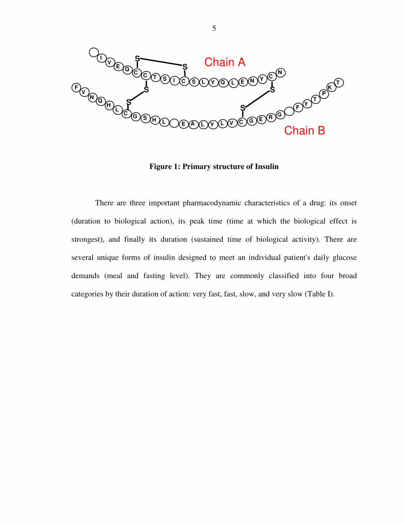
1959.14 It is composed of two peptide chains, designated as the A chain and B chain. The
A chain has 21 amino acids while the B chain has 30.
The chains are linked by two disulfide bonds (residues A7 to B7, and A20 to
B19). In addition, there is a third intramolecular disulfide bond in the A chain (residues
A6 to A11) (Fig 1). The complete conservation of these three disulfide bonds throughout
the mammalian phylum underscores the critical importance of this bonding pattern. This
also increases the structural complexity of the molecule and makes it more difficult to
synthesize as compared to a single peptide chain.
5
Figure 1: Primary structure of Insulin
There are three important pharmacodynamic characteristics of a drug: its onset
(duration to biological action), its peak time (time at which the biological effect is
strongest), and finally its duration (sustained time of biological activity). There are
several unique forms of insulin designed to meet an individual patient's daily glucose
demands (meal and fasting level). They are commonly classified into four broad
categories by their duration of action: very fast, fast, slow, and very slow (Table I).
S
S
S S
S S
K
H
I
P T
C C
F V
N Q
H L
C G S
A E R
Y V L L C G E G
F
Y
Y Y T
N
V E
Q T S I S L Q L E C N
C
L
N Chain A
Chain B

6
*Since patients respond to insulin differently, the peak action and duration are given as ranges.
Table 1: Pharmacodynamics of common insulin analogs
The time action curves of some common insulin analogs are shown in Figure 2.
Ultralente is a very slow acting insulin, hence it is usually used with Humalog® (very
fast acting) or native regular insulin (fast acting)15 to more accurately mimic the normal
daily physiological variation in insulin activity.
Figure 2: Pharmacodynamics of common insulin analogs.
Numerous combinations of these insulin analogs facilitate regulation of blood
glucose in virtually all forms of diabetes. However, they all have several severe
limitations. The most relevant is the finding that insulin overdose is the single most
Onset Peak Action* Duration*
Humalog® 0.25 hr 0.5 – 1.5 hrs 3-5 hrs (very fast)
Regular 0.5 hr 2-4 hrs 6-8 hrs (fast)
NPH, Lente 1-3 hrs 6-12 hrs 18-24 hrs (slow)
Ultralente 4-8 hrs 12-18 hrs 24-28hrs (very slow)

7
potent cause of life-threatening hypoglycaemia16. This has been confirmed by many
clinical trials, most notably by the Diabetes Control and Complications Trial Research
Group17. Studies performed in representative populations clearly demonstrate that weight
gain is another problem associated with insulin therapy18. Such weight gain can
paradoxically increase insulin resistance and thus the amount of insulin needed. Obesity
also increases cardiovascular risks19.
While insulin is a miraculous substance it is a challenging medicine. Its complex
structure, especially its intra and intermolecular disulfide bonds makes it difficult to
synthesize in an inexpensive manner and the molecule hard to formulate. Furthermore,
the natural tendency of insulin to form insoluble multimeric complexes at high
concentrations seriously complicates its commercial production. Additionally, the narrow
therapeutic index and thermal instability of insulin over extended periods of time makes
refrigeration a necessity. Lack of adequate refrigeration is a major issue in many parts of
the world.
What is most needed is a medicine that is capable of normalizing blood glucose
without the risk of hypoglycemia. In this context, it is helpful to note that Glucagon-like
peptide 1 (GLP-1) therapy has been shown to increase native insulin synthesis and
secretion without inducing hypoglycemia20.
III) Glucagon like peptide 1 (GLP 1):
Glucagon-like peptide 1 is a hormone that offers the promise of revolutionizing
the treatment of Type II diabetes. In a landmark paper published in 1985,21 two
endogenous peptides were reported which had a high sequence homology to glucagon
8
and, like insulin, displayed high conservation across a range of different species. These
two peptides were Glucagon-like peptide-1 (GLP-1) and Glucagon-like peptide-2 (GLP-
2) which were first identified in the course of cloning the gene for proglucagon. Upon
testing, it was found that GLP-1 stimulated insulin release while GLP-2 did not21. Early
clinical experiences suggested that GLP-1 has an attractive pharmacologic profile22.
Throughout this thesis, GLP-1(7-37)-acid has been denoted simply as GLP with changes
added to this nomenclature to signify related peptides.
GLP is secreted from the gut in response to a meal23. It is an incretin hormone that
has the potential to offer an ideal treatment for Type 2 diabetes24. GLP enhances the
secretion of insulin25 only when the blood glucose level is high, eliminating the risk of
hypoglycemia. It inhibits glucagon secretion therein maintaining an optimal ratio of
insulin and glucagon26. GLP has further been shown to reduce food intake via its effects
on gut motility (it inhibits the motility of the upper gut), leading to weight loss and
decreased obesity26. Thus, GLP by virtue of its multiple biological actions has emerged
as a valuable tool in the treatment of Type 2 diabetes, and related metabolic syndromes.
There is another more physiological factor to rationalize GLP therapy. Type 2
Diabetes is characterized by insufficient insulin secretion and declining β-cell function.
This β-cell defect partly results from the progressive loss in β-cells function, as noted
previously. GLP also has an apparent mitogenic effect on the β-cells of the pancreas27,
and thus stimulates the in vivo biosynthesis of insulin (thus addressing the main defect of
progressive decay in β-cell capacity) 26. Additionally, recent research demonstrates that
almost two thirds of the insulin secreted in response to a meal is because of the action of
insulinotropic actions of the incretin hormones, like GLP28. Indeed patients with Type 2
9
Diabetes have been long known to exhibit a variable loss in the incretin action of GLP29.
In this way, GLP therapy addresses the physiological replacement for the loss in incretin
action associated with Type 2 Diabetes.
GLP stimulates the secretion of insulin by interacting with the GLP G-Protein
Coupled Receptor (GPCR) expressed on the surface of β-cells of the pancreas. This
receptor is coupled positively to the adenylyl cyclase system30. After ligand activation,
the adenylyl cyclase is stimulated leading to an increase in cAMP concentration within
the β-cells31. This in turn activates protein kinase A that leads to an avalanche of
additional biochemical events32. GLP mediated receptor action via adenylate cyclase is
the basis of our in-vitro bio-assay, and has proven useful in design of GLP analogs with
therapeutic promise (see Experimental Design of Bioassays). It is to be noted that this
insulinotropic effect of GLP is strictly glucose dependent33.
Glucagon-like peptide 1 physiologically exists in two forms of comparable
biopotency34. The first is the more abundant34 GLP(7-36) amide where the C terminus is
an arginine amide (Figure 3A). The second form is the GLP(7-37) acid where the C
terminus is a glycine (Figure 3B). The secondary structure of GLP-1 is shown in Figure
3C.

10
Figure 3A: Primary structure of GLP-1(7-36) amide and action of DPP IV
Figure 3B: Primary structure of GLP-1(7-37) acid and action of DPP IV.
amide
11
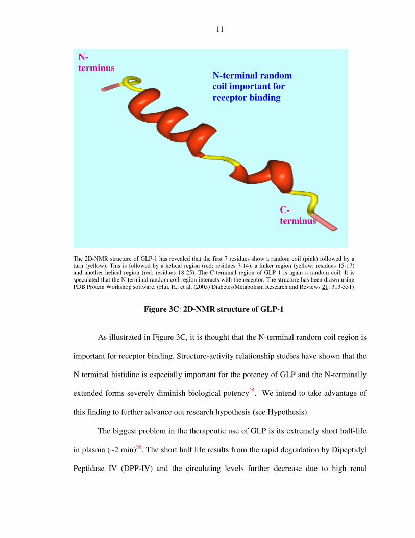
The 2D-NMR structure of GLP-1 has revealed that the first 7 residues show a random coil (pink) followed by a turn (yellow). This is followed by a helical region (red; residues 7-14), a linker region (yellow; residues 15-17) and another helical region (red; residues 18-25). The C-terminal region of GLP-1 is again a random coil. It is speculated that the N-terminal random coil region interacts with the receptor. The structure has been drawn using PDB Protein Workshop software. (Hui, H., et al. (2005) Diabetes/Metabolism Research and Reviews 21: 313-331)
Figure 3C: 2D-NMR structure of GLP-1
As illustrated in Figure 3C, it is thought that the N-terminal random coil region is
important for receptor binding. Structure-activity relationship studies have shown that the
N terminal histidine is especially important for the potency of GLP and the N-terminally
extended forms severely diminish biological potency35. We intend to take advantage of
this finding to further advance out research hypothesis (see Hypothesis).
The biggest problem in the therapeutic use of GLP is its extremely short half-life
in plasma (~2 min)36. The short half life results from the rapid degradation by Dipeptidyl
Peptidase IV (DPP-IV) and the circulating levels further decrease due to high renal
N-terminus
C-terminus
N-terminal random coil important for receptor binding
12
clearance37. As shown in Figure 3A and 3B, DPP-IV cleaves GLP between the alanine
residue at position 8 and the glutamic acid residue at position 938. This cleavage not only
inactivates the peptide but it also yields the residual GLP(9-36) amide; an antagonist at
the GLP receptor39. Hence, to obtain reasonable glycemic control, the GLP should be
administered continuously for a prolonged period36.
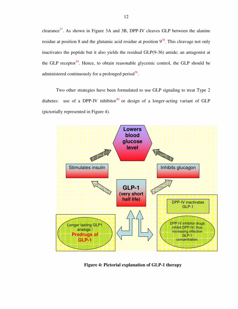
Two other strategies have been formulated to use GLP signaling to treat Type 2
diabetes: use of a DPP-IV inhibitor40 or design of a longer-acting variant of GLP
(pictorially represented in Figure 4).
Figure 4: Pictorial explanation of GLP-1 therapy
Longer lasting GLP1 analogs /
Prodrugs of GLP-1
DPP-IV inactivates GLP-1
GLP-1 (very short
half life)
DPP-IV inhibitor drugs inhibit DPP-IV; thus increasing effective
GLP-1 concentration.
Stimulates insulin Inhibits glucagon
Lowers blood
glucose level

13
The most obvious difference between these two therapies seems to be their effect
on body weight. While native GLP and its analogs promote loss of weight, the DPP-IV
inhibitors act by preventing gain of weight41. Additionally, several other issues regarding
the feasibility of using DPP-IV inhibitors need to be addressed before effective
therapeutic use42. Even after treatment with DPP-IV inhibitors it is not clear if there
would be enough GLP to attain peak efficacy in the body. This is because the amount of
endogenous GLP secreted is severely reduced in diabetes. The fact that DPP-IV non-
specifically acts on peptides other than GLP adds to its efficacy but is also a matter of
concern. The larger issue pertains to the other useful functions of DPP-IV in different
tissues, such that adverse effects of DPP-IV inhibitors are theoretically possible42.
Our work concentrated on designing a prodrug for GLP i.e., a longer-acting
variant of GLP. A few longer-acting analogs of GLP have been identified, two of which
will be discussed below. The first peptide is Exendin-4 which is an agonist for GLP
receptor43 and is found in the saliva of the Gila monster44 (Heloderma species native to
several American States). Exendin-4 was discovered by John Eng, an endocrinologist at
the Bronx Veteran’s Affairs Medical Center in New York. It is being marketed by
Amylin Pharmaceuticals. Exendin-4 functions in a manner similar to that of native GLP
with which it shares 53% amino acid homology. Several important functional residues
like the N-terminal histidine are conserved. It is much more resistant to cleavage by DPP-
IV than GLP as a result of the glycine at the second residue instead of alanine. Thus, it is
found that Exendin-4 has all the biological functions of GLP, but with a longer half life
(60-90 minutes)45. While the t1/2 is much greater as compared to native GLP (t1/2 ~2min),
an even longer-acting drug is highly desirable. Finally, since Exendin-4 is of non-human

14
origin, it has been reported to induce an immune response that compromises long-term
clinical efficacy46.
Another longer-acting GLP analog is called Liraglutide. In this peptide analog,
there is an arginine in the 34th position instead of a lysine. Additionally, a glutamic acid
residue is added to the lysine residue at position 2647 and the α-amine of this glutamate is
acylated with a C16 fatty acid. Upon entering the bloodstream, the fatty acid non-
covalently binds to serum albumin (a protein facilitating plasma transport)48. The serum
albumin distributes the drug throughout the body and also protects against cleavage47. As
a result Liraglutide’s t1/2 is increased to about 10-14 hrs and the drug can be injected once
a day. This drug is being developed by Novo Nordisk and has now entered Phase 3 trials.
The N-terminal histidine of GLP (7th position as shown in Figure 3B) is important
for pancreatic receptor activation of GLP and exendin-4. Previous amino acid
substitutions for this histidine with lysine and alanine have resulted in a large loss of
bioactivity35. Hence it has been concluded that the imidazole ring of the histidine is
necessary for the full potency of the molecule. GLP lowers blood glucose in diabetic
patients, and might restore β cell sensitivity to exogenous insulin secretagogues. Studies
so far have yielded evidence that GLP therapy is safe and effective for Type 2 diabetics49.
We want to develop a longer-acting GLP prodrug, possibly with weekly or even monthly
duration of in vivo biological action.

15
IV) Prodrug:
A prodrug is the precursor of a drug. According to The International Union of
Pure and Applied Chemistry (IUPAC), the term prodrug is defined as “any compound
that undergoes biotransformation before exhibiting its pharmacological effects. Prodrugs
can thus be viewed as drugs containing specialized non-toxic protective groups used in a
transient manner to alter or to eliminate certain limiting properties in the parent
molecule”50. The term “prodrug” was first introduced by Albert in 1950 to signify
pharmacologically inactive chemical derivatives that undergo conversion endogenously
to become an active pharmacological agent to increase their usefulness or decrease their
toxicity51. In this regard, prodrugs can be used for various purposes. Most prodrugs are
designed to facilitate high cellular absorption following administration52 (i.e.
valaciclovir). Prodrugs must be soluble in an aqueous media to be absorbed properly.
Insolubility of a drug can also cause significant pain at the site of injection. An example
is clindamicyn where injections are painful, but its phosphate ester prodrug improves
solubility and alleviates the pain53. The prodrug should be non-toxic, stable in storage and
must be resistant to degradation in different body fluids until that point when it reaches
its site of action. Finally after reaching the specific site of action, there should be a
quantitative release of the drug53.
The prodrug approach is commonly utilized with small molecules (less than 500
Daltons)54 to enhance oral delivery. The main obstacle to using peptides as potential
drugs is their short half life in the systemic circulation because of proteolytic hydrolysis
and the rapid clearance by blood, liver and kidney55. A second limitation is their limited

16
oral absorption55. Due to these problems of short half life and poor oral bioavailability,
alternative routes of delivery are being explored56.
The prodrug that we envision has certain unique characteristics. Through
structural refinement, we intend to appropriately extend the biological half life and
broaden the therapeutic index of GLP. Our prodrug concept is not focused on oral
bioavailability as with conventional small drug approaches, but upon extended biological
half life. Consequently, many of the stringent necessities of conventional prodrugs are not
relevant. Most prodrugs as described above are designed to facilitate transport across
biological membranes. Our prodrug is used to delay the time of action by inhibiting
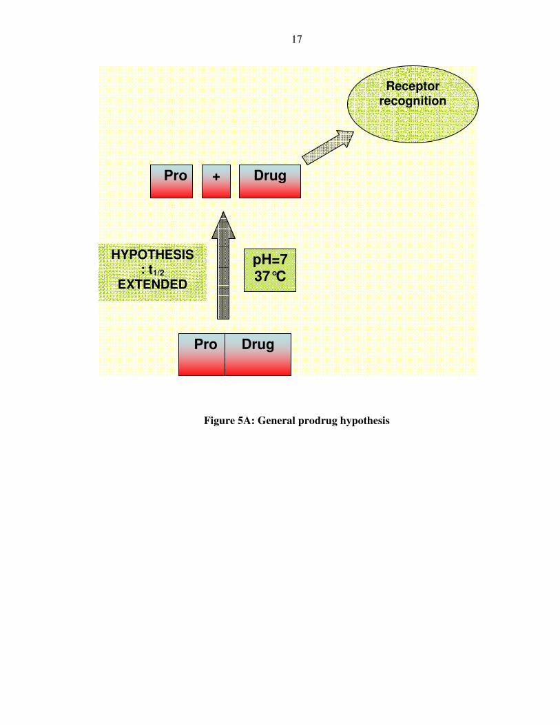
recognition by the corresponding receptor. Receptor recognition is the primary means of
degradation and thus termination of biological activity of our drug. We seek to convert
our prodrugs to active peptides by controlling the chemical conversion to structures that
can be recognized by the receptor. The speed of this chemical conversion will determine
the time of onset and duration of in vivo biological action (Figure 5).
The final element of this work is the application of selective pegylation57 to delay
non-productive, premature, in-vivo clearance. Peptides are easily cleared because of their
relatively small molecular size when compared to plasma proteins. Increasing the
molecular weight of a peptide above 40kDa exceeds the renal threshold and significantly
extends duration in the plasma. The judicious choice in the site of attachment of a
polyethylene glycol polymer that is ten fold larger than insulin or GLP is a sizable
challenge.
17
Figure 5A: General prodrug hypothesis
Pro Drug
Pro Drug
HYPOTHESIS : t1/2
EXTENDED
+
pH=7 37°C
Receptor recognition
18
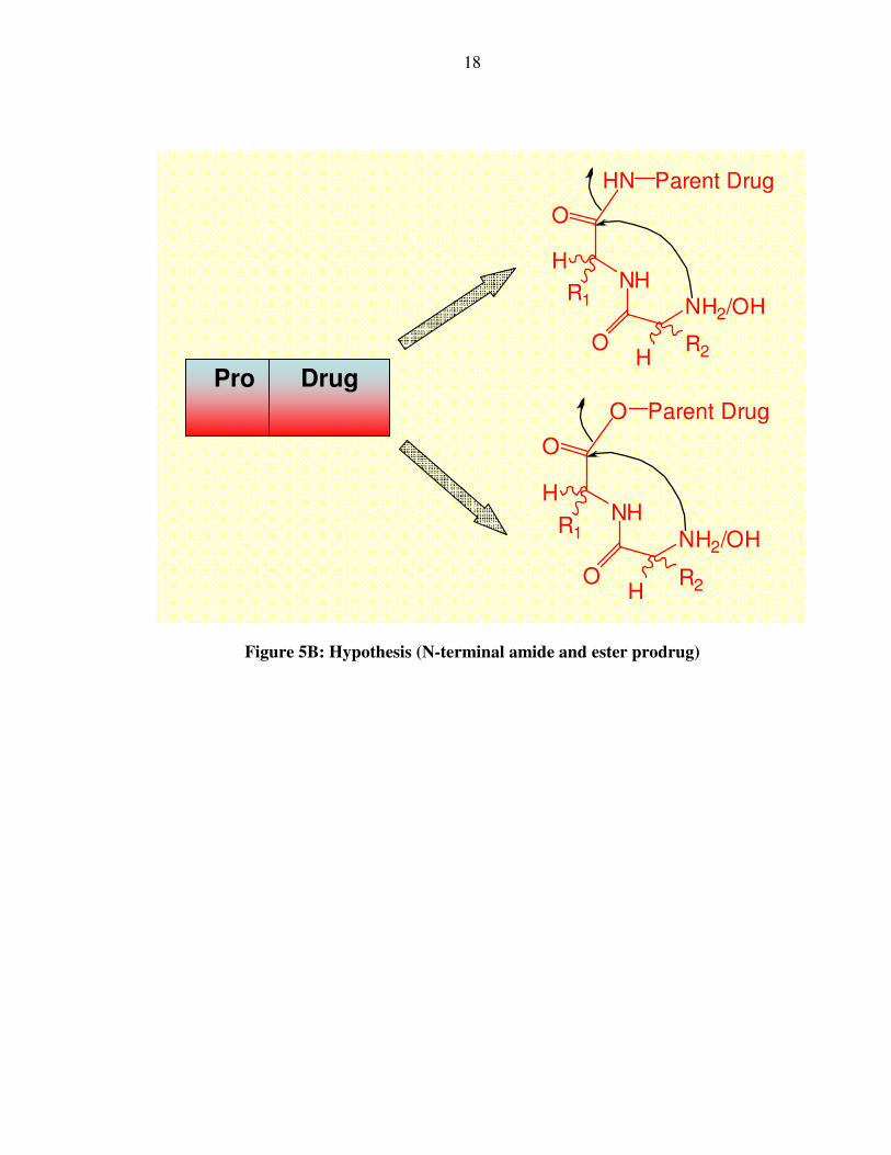
Figure 5B: Hypothesis (N-terminal amide and ester prodrug)
O
NH
O
NH2/OH
HN Parent Drug
H
R1
HR2
Pro Drug
O
NH
O
NH2/OH
O Parent Drug
H
R1
HR2

19
B) Hypothesis:
We believe that by making a prodrug at the N-terminal end of GLP, it should be
possible to extend and improve the pharmacodynamics of this peptide hormone (Figure
5A and 5B).
The ideal prodrug should be soluble in water at a pH of 7.2 and 37°C, and it
should be stable in the powder form for long term storage. It should be immunologically
silent and biologically inactive when injected in the body; and be quantitatively converted
to the active drug within a defined period of time. Our greatest interest lies in prodrugs
with a t1/2 of between 10-100 hrs (weekly, or even monthly duration) under physiological
conditions (pH of 7.2 and 37°C).
During the course of this research, we identified four GLP analogs that are
physically and chemically stable, and whose conversion from prodrug to the active drug
form under normal physiological conditions ranges within the optimal range. One of
these peptides converts to GLP with a half life of 64 hrs. The in vivo extension in
duration of action of this magnitude would constitute the longest acting peptide prodrug
ever designed. These analogs possess a minimal alteration to the native amino acid
sequence, and this should minimize potential adverse immunogenic affects. The
bioactivities of these synthetic peptides have been determined using in vitro cellular
assays.
At the beginning of this research, many strategies were considered for
constructing our prodrugs. It was contemplated that the protecting groups of the prodrug
could be cleaved by enzymes as reported for nucleotide prodrugs58. However, we decided
to design a prodrug that would slowly convert to the parent drug at physiological

20
conditions of 37оC and pH 7.2 driven by inherent chemical instability. The pH and
temperature were relied upon for this conversion, as they are virtually invariant
physiologically. We are seeking a prodrug that converts quantitatively to the drug under
physiological conditions without the aid of any enzyme. The establishment of prodrug
chemistry at the N-terminal end of GLP should be translatable for use with other peptides
where this specific site is vital to bioactivity.
We decided to synthesize a chemical derivative of GLP that would convert to
GLP spontaneously as stated above. A few possible choices were contemplated before
deciding that our primary target in prodrug chemistry would be diketopiperazine
formation. The N-terminal histidine is important for the potency of our drug, and a
reversibly modified histidine side chain was considered, as has been reported for
Thyrotropin-Releasing Hormone (TRH)59. However, we dispensed with this approach
since this prodrug chemistry is specific to the imidazole ring. This means that the prodrug
chemistry requires the presence of a histidine, and possibly also at an N-terminal site.
Thiol esters could also be synthesized by thioesterification60. However, a thiol
ester is relatively unstable in physiology and thus would likely cleave too fast, thereby
resulting in rapid clearance of the prodrug in its active form. Additionally, the use of
thiol-based chemistry is fraught with other difficulties, such as instability of disulfide
bonds. We decided to forgo this course of action.
Our work therefore focused on making amide and ester prodrugs at the N-
terminus that upon cleavage of the suitable amide or ester bond generates the desired
drug. Esters are normally more labile than amides; however they are easily hydrolyzed by
the ubiquitous serum esterases53. Hence, amide bond based prodrugs were a much more

21
attractive design, although the risk of peptidase degradation may potentially complicate
in vivo application.
In considering amide prodrugs, it is reported that histidyl-proline amide cyclizes
to a cyclo (His-Pro)61 at a pH of 7 and 37оC with a t1/2 of 140 minutes, with the release of
NH3. The imidazole ring is purported to be playing a catalytic role at this pH. Hence,
other dipeptides that did not have a histidine cleaved more slowly, and tripeptides did not
cleave at all61. Thus, it seemed from this work that there might be a basic difference in
the rate of diketopiperazine formation from the cleavage of a secondary amide (as in a
tripeptide) as compared to a primary one (as in a dipeptide amide).
In another paper62, the intramolecular aminolysis of Phe-Pro-p-nitroanilide (Phe-
Pro-pNA) to Phe-Pro-diketopiperazine (Phe-Pro-DKP) was studied as a function of pH.
The pH-rate plot showed that the rate of the formation of the DKP was dependent on the
degree of ionization of the N-terminal amino group, with the unprotonated free amine
being more reactive than the protonated form. In their experiment, the authors used an
activated, strongly electron withdrawing p-nitroanilide dipeptide instead of a natural
tripeptide. This was because these p-nitroanilide dipeptides dissociate by DKP formation
more rapidly than amino acid amides (i.e., natural dipeptides), thereby greatly facilitating
their use in kinetic studies. The calculated t1/2 of conversion of a Gly-Pro-p-nitroanilide
(Gly-Pro-pNA) dipeptide to Gly-Pro-diketopiperazine (Gly-Pro-DKP) under
physiological conditions was about 120 hours. This is consistent with the previous
assertion that there is a significant difference in the dissociation rate between a primary
(around 140 minutes as in previous example61) and an activated secondary amide. It is

22
also important to note that in a natural tripeptide, the half life of the DKP formation
would be further extended since there is no electronic assistance from the pNA.
In both these papers61,62, it seems that the presence of proline in the C terminus of
the dipeptide extension accentuates the formation rate of the DKP. This is likely due to
contribution of the cis-proline conformer in the facilitation of the dipeptide's adoption of
an optimal steric conformation for formation of DKP. The observations with
modifications of an N-terminal residue upon the rate of DKP formation at pH 7.0 have
been more varied. Reports suggest that it might depend on the pKa of the residue61, on its
bulk63, or on the conformational stability of the resulting DKPs62.
We also envisioned modification of a hydroxyl group at what otherwise would be
the N terminus to prepare a depsi-peptide and thus make an ester prodrug. An ester can
cleave hydrolytically64,65 or via the formation of five66,67 or six membered rings (like
DKP). The ester prodrugs of Floxuridine (FUdR)65 convert by general ester hydrolysis
i.e., a nucleophilic attack by water on the ester carbonyl. For the most part, the bulk of the
pro-moiety influenced the hydrolysis of the FUdR prodrug (the Val ester prodrugs
dissociated the slowest). In another paper, the authors studied the cyclization of the
dipeptide esters in paracetamol to form a diketopiperazine68. They observed that they
could obtain differential time action depending on the dipeptide structure. They also
considered the possibility that in paracetamol, the drug release might have been via the
general mechanism of ester hydrolysis and not the formation of a DKP ring. However,
they eliminated it as in that case the nature of the dipeptide would have a lesser effect on
reactivity. The proof of labile esters cleaving very fast under physiological conditions is

23
exemplified by the fast cleavage of the dipeptide esters in paracetamol where all the
prodrugs had a t1/2 of less than 20 minutes68.
In another investigational report, this time with cyclosporine-A prodrugs69,70 it has
also been seen that through modulating the chemical nature of dipeptide esters it was
possible to get conversion rates at physiological conditions ranging from minutes to
several hours, but not longer. In these papers68,69, it seems that the presence of a
minimally bulky glycine residue in the C terminus of the dipeptide extension accentuates
the rate of formation of the DKP. This might be because of less bulk and the preferred
conformational effect of glycine. However in the cyclosporine prodrug69, it seems
unexpected that the presence of a proline in the C terminus of the DKP actually
attenuates the rate of the conversion.
In another investigation71, the C terminal amides of glycine were rapidly
hydrolyzed at 25°C and a pH of 7 when the N-terminus was N-hydroxyethylated. The t1/2
of bis-N-2-hydroxyethylglycinamide is three hours. In this case, the C terminal amide
bond is activated by H bonding with the N-hydroxyethyl group. However, there was no
practical way that one could modify the structure of the N-hydroxyethyl group as the
precise Vander Waal radii were required to activate the amide group.
Consequently, we explored the intramolecular cyclization reaction of dipeptide
esters and amides to form diketopiperazine as an example of a chemoreversible prodrug
(Figure 6). This chemistry is reasonably straightforward and allows at least four points
(stereochemistry of R1 and R2, nature of the nucleophile and leaving group as shown in
Figure 6A and 6B) where structure can be stereochemically controlled to refine the rate

24
of formation with release of the active peptide. Lastly, they can also be prepared from
readily available alpha-amino acids using established chemistry72.
As shown below (Figure 6A and 6B), prodrugs of varying half lives were
designed by modifying R1 and R2. In the reaction below, there is an N-terminal histidine
residue (native peptide), and an amide bond is being broken (Figure 6A). In the second
equation, the ester bond of phenyllactic acid (hydroxyl phenylalanine) is being broken
(Figure 6B). Though it might be beneficial to use the histidine in the 7th position of GLP,
alternatives to the native N-terminal histidine were utilized for synthetic and analytical
ease. Once the chemistry of a longer-acting prodrug is established, it is plausible to return
to the histidine or for that matter any other suitably potent amino acid at the N-terminus.

25
A)
+ H2N-GLP
HN
O
NH
O
NH2
GLP
pH=7.2
R2 H
R1
HNH
HN
O
O
R1
R2
37oC
B)
pH=7.2
NH
HN
O
O
R1
R2
37oC
GLP(8-37)OO
NH
O
R1
H
R2H
CH
NH2
CH2Ph
+ HO-Phe-GLP(8-37)
Figure 6: Cleavage of amide (A) and ester prodrugs (B)
Thus, we propose to make a prodrug that will slowly convert (t1/2 between 10-100
hrs) to the parent drug at physiological conditions of 37оC and pH 7.2 so that they could
be administered at a weekly or monthly frequency. As far as possible, a native sequence
shall be used in the prodrug so as to minimize the chances of an immunological response.
We rely on the pH and the temperature for this intramolecular conversion, as they are
virtually invariant. It is of essence to note that this reaction should be concentration
independent.
Four such peptides were identified with protracted half lives, with minimal
potency as compared to the drug in the luciferase-based bioassay. These prodrugs

26
regained their potencies after incubation in PBS buffer at a pH of 7.2 and temperature of
37°C.
Prodrugs of this thesis can be broadly classified into 4 different types of prodrugs:
1. An amine nucleophile cleaving an amide bond (Class 1): This will dissociate with the
formation of the corresponding 2,5-diketopiperazine.
CH
NH
O
NH
O
NH2
R2
H
R1
X
GLP
H
2. A hydroxyl nucleophile cleaving an amide bond (Class 2): This will dissociate with the
formation of the corresponding 2,5-diketomorpholine.
CH
NH
O
NH
O
OH
R2
H
R1
X
GLP
H
27
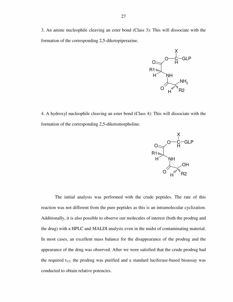
3. An amine nucleophile cleaving an ester bond (Class 3): This will dissociate with the
formation of the corresponding 2,5-diketopiperazine.
H
O
NH
O
NH2
R2
H
R1
O CH
X
GLP
4. A hydroxyl nucleophile cleaving an ester bond (Class 4): This will dissociate with the
formation of the corresponding 2,5-diketomorpholine.
H
O
NH
O
OH
R2
H
R1
O CH
X
GLP
The initial analysis was performed with the crude peptides. The rate of this
reaction was not different from the pure peptides as this is an intramolecular cyclization.
Additionally, it is also possible to observe our molecules of interest (both the prodrug and
the drug) with a HPLC and MALDI analysis even in the midst of contaminating material.
In most cases, an excellent mass balance for the disappearance of the prodrug and the
appearance of the drug was observed. After we were satisfied that the crude prodrug had
the required t1/2, the prodrug was purified and a standard luciferase-based bioassay was
conducted to obtain relative potencies.

28
The experimental design of our bioassay was based on the general principles of
“reporter gene technology”73 (Figure 7). In our case, the luciferase-based reporter gene
assay for cAMP detection was used. The changes in the intracellular cAMP
concentrations74 caused by the GLP receptor-mediated interactions are detected by the
changes in the expression level of the luciferase gene. The transcription of this gene is
regulated by the cAMP response-element binding protein (CREB) binding to cAMP
response element (CRE).
This is an artificially created test system where the luciferase gene is downstream
to the CRE which resembles nature’s response system all the way up to the point of gene
expression where the luciferase gene is expressed. This modification is necessary as the
concentration of activated luciferase is easier to measure than that of cAMP.

29
Figure 7: Pictorial Explanation of luciferase-based reporter gene assay
ATP
cAMP
GLP-1 analog
G protein receptor
LUCIFERASE BINDING ASSAY
Luciferase gene
Receptor mediated changes
30
C) Experimental Procedure: Synthesis of GLP analogs
In this project, numerous GLP analogs were synthesized. The standard procedure
is described briefly here, and the details are discussed later.
PAM resin (PAM resin is OCH2-phenylacetamidomethyl–copolystyrene-1%
divinylbenzene), (100-180 mesh, 1% DVB cross-linked polystyrene; loading of 0.7-
1.0mmol/g), Boc-protected and Fmoc protected amino acids were purchased from
Midwest Biotech. Other reagents such as the α-hydroxy-acids (phenyllactic acid and
glycolic acid) were purchased from Aldrich. The solid phase peptide syntheses using
Boc-protected amino acids were performed on an Applied Biosystem 430A Peptide
Synthesizer75. Fmoc protected amino acid synthesis was performed using the Applied
Biosystems Model 433 Peptide Synthesizer. The manual synthesis of depsi-peptides was
performed in sintered reaction vessels using analogous procedures75,76.
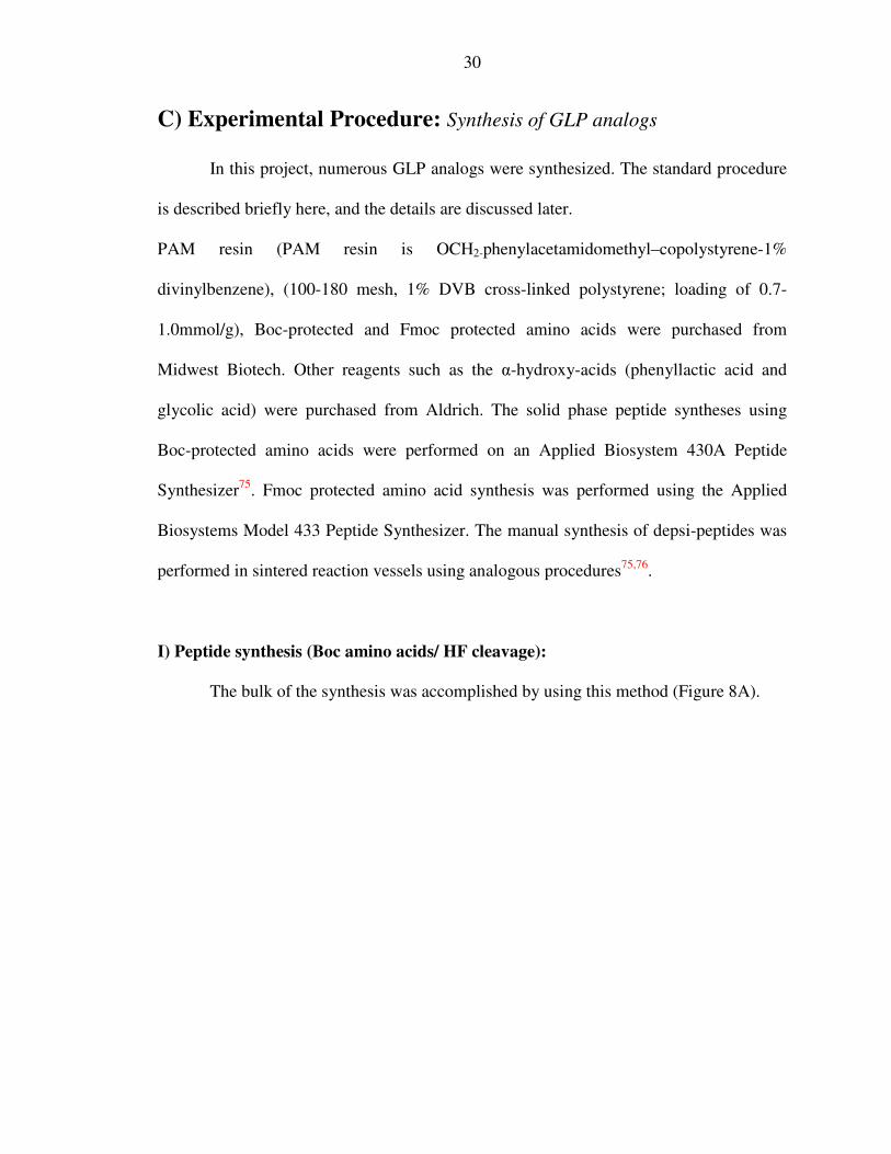
I) Peptide synthesis (Boc amino acids/ HF cleavage):
The bulk of the synthesis was accomplished by using this method (Figure 8A).

31
H
OX
BocNH OH
Y
H
H2N O
O
X
BocNH
H
HN
Y
H
O
OO
+H2C PAM
H2C PAM
1.9 mmol DEPBTinDMF+ 1ml DIEA
TFA
X
NH2
H
HN
Y
H
O
OOH2C PAM
(0.2mmol)(2 .0mmol)
Figure 8A: Peptide synthesis (Boc method)
Synthesis of these analogs was performed on the Applied Biosystem Model 430A
Peptide Synthesizer. Synthetic peptides were constructed by sequential addition of amino
acids, and activated esters of each amino acid were generated by the addition of 1.9mmol
(3.8 ml of a 0.5M solution) of 3-(Diethoxy-phosphoryloxy)-3H-benzo[d][1,2,3] triazin-4-
one (DEPBT) in DMF to a cartridge containing 2mmol of Boc protected amino acid. The
amino acids were dissolved by bubbling Nitrogen gas through the cartridge. 1 ml of N,N-
Diisopropylethylamine was added to the cartridge to effect ester formation. This solution
was transferred to the reaction vessel containing the 0.2 mmol of the C-terminal residue
attached to the PAM resin, vortexed several times, and allowed to couple to the resin for

32
10 minutes. After washing to remove the unreacted reagents, the N-terminal Boc
protecting group was removed by treatment with trifluoroacetic acid (TFA) for 5 minutes.
The resin was washed with DMF and the cycle was repeated for the desired number of
steps until the chain was assembled. The reaction vessel at the end of the synthesis
(typically 30 amino acids) contained approximately 1.2-1.5g of protected peptidyl-PAM
resin. The resin was washed numerous times with dimethylformamide (DMF), treated
with trifluoroacetic acid to remove the last tBoc protecting group and finally washed
several additional times with DMF, dichloromethane (DCM) and dried.
The peptidyl-resin was treated with anhydrous HF (procedure explained later in
this section), and this typically yielded approximately 350 mg (~50% yield) of a crude
deprotected-peptide.
II) Peptide synthesis (Fmoc amino acids/ HF cleavage):
This synthesis scheme was performed manually with a few amino acids at
selective sites. In this work, the Fmoc amino acids were used only to synthesize the
internal serine prodrugs, as a part of a wider synthetic strategy. Here, it is to be noted that
although FMOC chemistry has been used in the synthesis, the peptides have always been
built on PAM resin that required treatment with HF to cleave the peptide from the solid
support. The yield of these peptides is approximately as stated earlier for Boc/PAM
synthesis.
33
H
OX
FmocNH OH
Y
H
H2N O
O
X
FmocNH
H
HN
Y
H
O
OO
+H2C PAM
H2C PAM
1.9mmol DEPBTinDMF+ 1ml DIEA
20%piperidine
X
NH2
H
HN
Y
H
O
OOH2C PAM
(0.2mmol)(2 .0mmol)
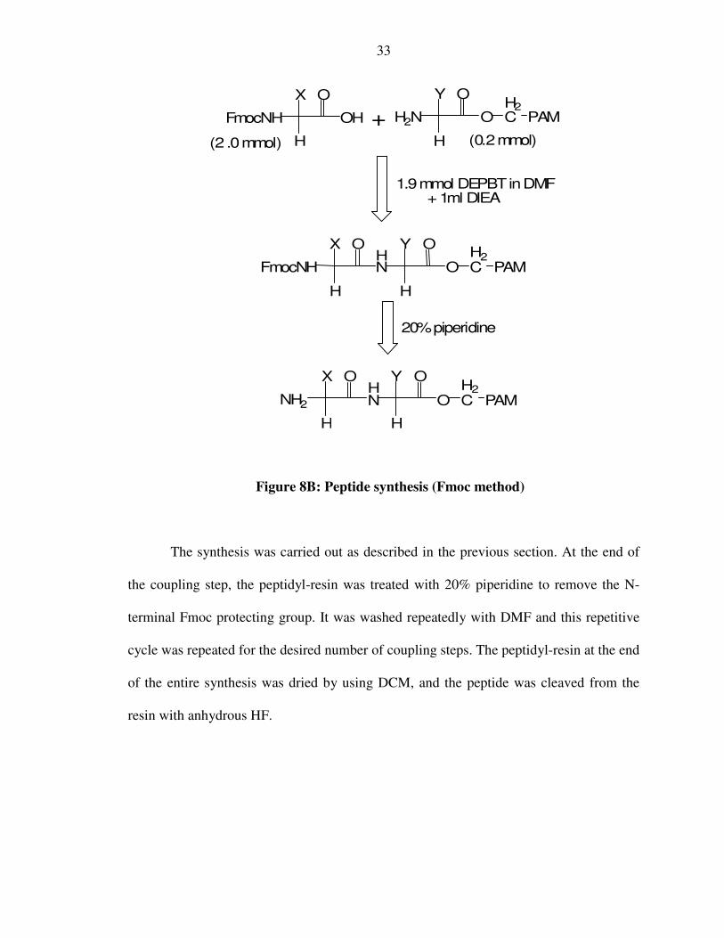
Figure 8B: Peptide synthesis (Fmoc method)
The synthesis was carried out as described in the previous section. At the end of
the coupling step, the peptidyl-resin was treated with 20% piperidine to remove the N-
terminal Fmoc protecting group. It was washed repeatedly with DMF and this repetitive
cycle was repeated for the desired number of coupling steps. The peptidyl-resin at the end
of the entire synthesis was dried by using DCM, and the peptide was cleaved from the
resin with anhydrous HF.

34
III) Depsi-peptide synthesis (Amino ester formation):
These syntheses76 were performed manually (Figure 9). In this case, the peptidyl-
resin had an α-hydroxyl-N terminal extension instead of a N-terminal amine and the
acylation was done at the α hydroxyl group. This reaction takes a longer time than that of
the amide bond formation, as the hydroxyl group is a weaker nucleophile as compared to
the amine. The reaction time was typically 12 hours.
(2 .0mmol) H
OX
BocNH OH
Y
H
HO O
O
X
BocNH
H
O
Y
H
O
OO
+H2C PAM
H2C PAM
1.0mmol DICinDCM(0oC)
+ 0.9mmol DMAP
(12 hrsat roomtemp)
TFA
X
NH2
H
O
Y
H
O
OOH2C PAM
(0.2mmol)
Figure 9: Depsi-peptide synthesis (Amino ester formation)
Initially, the activated esters of each amino acid were generated by the addition of
1mmol (0.155 ml of Diisopropylcarbodiimide (DIC) to a cartridge containing a solution

35
of 2mmol of Boc protected amino acid residue in 2 ml DCM. This was cooled to 10˚C for
10 minutes. 0.9 mmol (244 mg) of dimethylaminopyridine (DMAP) was added to the
cartridge to accelerate ester formation. This mixture was transferred to the reaction vessel
containing the peptidyl-resin upon which the peptide was synthesized. The reaction
vessel was stirred for 12 hours.
The peptidyl-resin was dried using DCM and the synthesis of the desired peptide
was continued. The peptidyl-resin at the end of the entire synthesis was dried by using
DCM, and finally treated with anhydrous HF to generate the desired peptide.
IV) N-terminal hydroxyl peptide synthesis (α hydroxyl- N terminal extension)74:
In this reaction, the free amine of the peptidyl-resin reacts with an α hydroxyl acid
to form an α hydroxyl- N terminal extension (Figure 10).
H
OX
OH OH
Y
H
H2N O
O
X
OH
H
HN
Y
H
O
OO
+H2C PAM
H2C PAM
0.9mmol DEPBTinDMF+ 0.5ml DIEA
(6 hrsat roomtemp)
(0.2mmol)
(1 .0mmol)
Figure 10: α hydroxyl- N terminal extension

36
In this regard, only two such α hydroxyl acids were used namely, glycolic acid
(OH-glycine) and phenyllactic acid (OH-phenylalanine). These syntheses were also
performed manually. The peptides were constructed by addition of the α hydroxyl acid,
and activated esters of the α hydroxyl acid were generated by the addition of 0.9mmol of
DEPBT (270 mg) to a cartridge containing a solution of 1mmol of Boc protected residue
in 2 ml DMF. 0.5 ml of DIEA (N, N-Diisopropylethylamine) was added to the cartridge
to accelerate ester formation. This mixture was transferred to the reaction vessel
containing the peptidyl-resin upon which the peptide was synthesized. The reaction time
was 6 hours.
The peptidyl-resin was dried using DCM and the synthesis of the desired peptide
was continued. The peptidyl-resin at the end of the entire synthesis was dried by using
DCM, and cleaved by anhydrous HF to generate the free peptide.
V) HF treatment of the peptidyl-resin:
The peptidyl-resin (30mg to 200mg) was placed in the hydrogen fluoride (HF)
reaction vessel for cleavage. 500µL of p-cresol was added to the vessel as a carbonium
ion scavenger. The vessel was attached to the HF system and submerged in the
methanol/dry ice mixture. The vessel was evacuated with a vacuum pump and 10ml of
HF was distilled to the reaction vessel. This reaction mixture of the peptidyl-resin and the
HF was stirred for one hour at 0°C, after which a vacuum was established and the HF
was quickly evacuated (10-15 min). The vessel was removed carefully and filled with
approximately 35 ml of ether to precipitate the peptide and to extract the p-cresol and
small molecule organic protecting groups resulting from HF treatment. This mixture was

37
filtered utilizing a teflon filter and repeated twice to remove all excess cresol. This filtrate
was discarded. The precipitated peptide dissolves in approximately 20ml of 10% acetic
acid (aq). This filtrate, which contained the desired peptide, was collected and
lyophilized.
VI) Analysis using mass spectrometry:
The mass spectra were obtained using a Sciex API-III electrospray quadrapole mass
spectrometer with a standard ESI ion source. Ionization conditions that were used are as
follows: ESI in the positive-ion mode; ion spray voltage, 3.9 kV; orifice potential, 60 V.
The nebulizing and curtain gas used was nitrogen flow rate of .9L/min. Mass spectra was
recorded from 600-1800 Thompsons at 0.5 Th per step and 2msec dwell time. The sample
(about 1mg/mL) was dissolved in 50% aqueous acetonitrile with 1% acetic acid and
introduced by an external syringe pump at the rate of 5 µL/min.
When the peptides were analyzed in PBS solution by ESI MS, they were first
desalted using a ZipTip solid phase extraction tip containing 0.6 µL C4 resin, according
to instructions provided by the manufacturer.
(http://www.millipore.com/catalogue.nsf/docs/C5737)
VII) High Pressure Liquid Chromatography (HPLC) analysis:
Preliminary analyses were performed with these crude peptides to get an
approximation of their relative conversion rates in Phosphate Buffered Saline (PBS)
buffer (pH, 7.2) using high performance liquid chromatography (HPLC) and MALDI
analysis. The crude peptide samples were dissolved in the PBS buffer at a concentration

38
of 1mg/ml. 1ml of the resulting solution was stored in a 1.5ml HPLC vial which was
then sealed and incubated at 37°C. Aliquots of 100µl were drawn out at various time
intervals, cooled to room temperature and analyzed by HPLC.
The HPLC analyses were performed using a Beckman System Gold
Chromatography system using a UV detector at 214 nm. HPLC analyses were performed
on a 150mm x 4.6mm C18 Vydac column. The flow rate was 1ml/min. Solvent A
contained 0.1% TFA in distilled water, and solvent B contained 0.1% TFA in 90%
CH3CN. A linear gradient was employed (40% to 70%B in 15 minutes). The data was
collected and analyzed using Peak Simple Chromatography software.
The initial rates of hydrolysis were used to measure the rate constant for the
dissociation of the respective prodrugs. The concentrations of the prodrug and the drug
were estimated from their peak areas respectively. The first order dissociation rate
constants of the prodrugs were determined by plotting the logarithm of the concentration
of the prodrug at various time intervals. The slope of this plot gives the rate constant ‘k’.
The half lives of the degradation of the various prodrugs were then calculated by using
the formula t1/2 = .693/k.
VIII) Preparative purification using HPLC:
After we were satisfied that the prodrug had an appropriate t1/2, the prodrug was
purified. The purification was performed using HPLC analysis on a silica based 1 x 25cm
Vydac C18 (5µ particle size, 300A° pore size) column. The instruments used were:
Waters Associates model 600 pump, Injector model 717, and UV detector model 486. A
wavelength of 214 nm was used for all samples. Solvent A contained 10% CH3CN /0.1%

39
TFA in distilled water, and solvent B contained 0.1% TFA in CH3CN. A linear gradient
was employed (0 to 100%B in 2 hours). The flow rate is 1.2ml/min and the fraction size
was 6ml. From ~350 mgs of crude peptide, 80 mgs of the pure peptide (~23% yield) was
typically obtained.
IX) Bioassay Experimental Design: Luciferase-based reporter gene assay for cAMP
detection
The ability of each GLP analog or prodrug to induce cAMP was measured77 in a
firefly luciferase-based reporter assay (Figure 7). The cAMP production that is induced is
directly proportional to the GLP binding to its receptor. HEK293 cells co-transfected
with the GLP receptor and luciferase gene linked to cAMP responsive element were
employed for bioassay.
The cells were serum-deprived by culturing 16 hours in Dulbecco Minimum
Essential Medium (Invitrogen, Carlsbad, CA) supplemented with 0.25% Bovine Growth
Serum (HyClone, Logan, UT) and then incubated with serial dilutions of either GLP
analogs or prodrugs for 5 hours at 37oC, 5% CO2 in 96 well poly-D-Lysine-coated
“Biocoat” plates (BD Biosciences, San Jose, CA). At the end of the incubation, 100 µL of
LucLite luminescence substrate reagent (Perkin Elmer, Wellesley, MA) were added to
each well. The plate was shaken briefly, incubated 10 min in the dark and light output
was measured on MicroBeta-1450 liquid scintillation counter (Perkin-Elmer, Wellesley,
MA). The effective 50% concentrations (EC50) were calculated by using Origin software
(OriginLab, Northampton, MA).
40
D) Experimental Results and Discussion
I) GLP-Oxyntomodulin
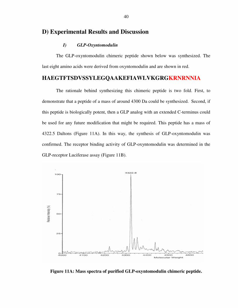
The GLP-oxyntomodulin chimeric peptide shown below was synthesized. The
last eight amino acids were derived from oxyntomodulin and are shown in red.
HAEGTFTSDVSSYLEGQAAKEFIAWLVKGRGKRNRNNIA
The rationale behind synthesizing this chimeric peptide is two fold. First, to
demonstrate that a peptide of a mass of around 4300 Da could be synthesized. Second, if
this peptide is biologically potent, then a GLP analog with an extended C-terminus could
be used for any future modification that might be required. This peptide has a mass of
4322.5 Daltons (Figure 11A). In this way, the synthesis of GLP-oxyntomodulin was
confirmed. The receptor binding activity of GLP-oxyntomodulin was determined in the
GLP-receptor Luciferase assay (Figure 11B).
Figure 11A: Mass spectra of purified GLP-oxyntomodulin chimeric peptide.

41
Figure 11B: Bioassay results of GLP-oxyntomodulin chimeric peptide
The GLP-oxyntomodulin was found to be at least as potent as the native GLP
peptide in the luciferase assay. The GLP-oxyntomodulin also has a higher apparent
maximal efficacy as compared to GLP (Figure 11B). This observation warrants additional
study as the less potent oxyntomodulin also demonstrated super-efficacy. This signifies
that the first portion of the project is successful and we focused on the central element of
this study, specifically the N-terminal prodrug design.
II) Adding dipeptides to the N terminus
Dipeptides were covalently attached to the N-terminus of GLP (sequence in
Figure 3B) to study differential tendencies for intramolecular cyclization and cleavage
through diketopiperazine formation. This is represented schematically in Figure 12A. All
class I prodrugs were synthesized by analogous procedures.

42
The biologically inactive dipeptide-extended GLP was converted to the active
GLP upon cleavage of the amide bond along with the DKP (Figure 12B and Table 2). In
Figure 12C (Table 3 and 4), the same conversion is shown with the phenylalanine in the
1st position of GLP. Prodrugs of varying half lives were envisioned by chemically
modifying the R1 and R2 positions. This validates our reason for probing the DKP
formation strategy.
O
NH2
H
HN
R1
O
NHH
HN
OH R2
NH2
R1
GLP1 OH2C PAM
HF (1hr)
H2N
GLP1 OH2C PAM
H
OR1
BocNH OH
1) 1.9 mmol DEPBTinDMF+1ml DIEA/
2) TFA
H
OR2
BocNH OH
1) 1.9 mmol DEPBTin DMF+1ml DIEA/
2) TFA
GLP1 OH2C PAM
O
NHH
HN
OH R2
NH2
R1
GLP1
(2.0 mmol)
(2 .0mmol)
(0 .2 mmol)
Figure 12A: Schematic synthesis of Class I prodrugs

43
pH=7.2
NH
HN
O
O
R1
R237oC
+ H2N-GLPO
NH
H
HNHN
H
N
OH R2
GLP(8-37)
NH2
R1
6
5
Figure 12B: Cleavage of amide bond to form DKP + H7,GLP(8-37)
pH=7.2
NH
HN
O
O
R1
R237oC
+ H2N-F7GLP(8-37)O
NH
H
HN
OH R2
NH2
HCH2Ph
R1
GLP(8-37)6
5
Figure 12C: Cleavage of amide bond to form DKP + F7,GLP(8-37)
The results of the cleavage in the prodrugs are shown in Table 2. The first peptide
synthesized was named G5P6H7,GLP(8-37) where the R1 is the side chain for proline and
R2 refers to the side chain for glycine (peptide 1 in Table 2). All peptides mentioned
hereafter will have the same systematic nomenclature and the stereochemistry was
assumed to be the l-isomer unless otherwise stated. The peptide was prepared
synthetically by solid phase synthesis as described earlier. The synthesis was confirmed
by MALDI-MS analysis (3509.5 Da) as shown in Figure 13.

44
Figure 13: Mass spectra of G5P6H7,GLP(8-37)
To explore the possible formation of DKP, and simultaneous regeneration of the
H7,GLP(8-37), the G5P6H7,GLP(8-37) was incubated in PBS buffer at 37˚C for
approximately a week. Additionally, the peptide was heated at 100˚C to accelerate the
amide bond cleavage. Analysis by reverse phase HPLC showed no apparent cleavage of
the amide bond in this first set (Table 2). The structures of the peptides in Table 2 are
illustrated in Appendix II.
To investigate the propensity of various prodrugs to undergo DPK formation, all
subsequent peptides were similarly synthesized and analyzed by MS and HPLC
following treatment at 100˚C and 37˚C as described above.

45
Serial No Peptide (Xaa1Yaa2-
GLP1)
Rate of cleavage at
100˚C
Rate of cleavage at
37˚C
1 GP No cleavage No cleavage
2 PP No cleavage No cleavage
3 γ Glu No cleavage No cleavage
4 E No cleavage No cleavage
5 P No cleavage No cleavage
6 H No cleavage No cleavage
7 PH No cleavage No cleavage
In 4, 5 and 6, there is just a single amino acid added to the N terminus of GLP-1. In 7, the dipeptide modification was made to sandwich the carbonyl bond of interest between two histidines. In all these cases there is histidine in the 1stposition (histidyl leaving group) of GLP-1.
Table 2: Attempted cleavage of H7,GLP(8-37) prodrugs to form native GLP-1
The dipeptide extension of peptides 1 and 2 in Table 2 were synthesized to
facilitate DPK formation by sterically assisting in the cleavage of the amide bond. It was
thought that the cis-orientation of proline would contribute in the facilitation of the
dipeptide's adoption of an optimal steric conformation for the formation of DKP.
However, the amide bond is quite robust and did not cleave. In peptides 3, 4, 5 and 6, an
acid-base catalyzed general hydrolysis of the amide bond was attempted. Since, the
leaving group will be the histidine at the N-terminus, it was purported that the imidazole
ring might in some way assist in the cleavage by general acid-base catalysis. In 7, the
carbonyl bond of interest is sandwiched between two histidines (amino acid Y and the

46
histidyl leaving group). This design was directed at a proton assisted cleavage of the
amide bond via diketopiperazine formation. But even this prodrug did not cleave.
At this point, it was speculated that perhaps the imidazole nucleus was playing an
attenuating role in the cleavage of the amide bond. To test this possibility, a different
leaving group was studied. F7,GLP(8-37) was synthesized and purified using the standard
procedure described above. It was determined to be a full agonist with 10% the potency
of native GLP. Dipeptides were added to this GLP analog to study the same type of
reaction as described above (Table 3; reaction shown in Figure 12C). The structures of
the peptides in Table 3 are drawn out in Appendix III.
Serial No Peptide (Xaa1Yaa2-
F7a-GLP8-37)
Rate of cleavage at
100˚C
Rate of cleavage at
37˚C
1 GP No cleavage No cleavage
2 GSarb No cleavage No cleavage
‘a’: In all these cases there is phenylalanine in the 1st position (phenylalanyl leaving group) of GLP. ‘b’: Sar represents sarcosine
Table 3: Attempted cleavage of dipeptide extended F7,GLP(8-37)
The dipeptide extension of peptides 1 and 2 (Table 3) were designed to sterically
assist in the cleavage of the amide bond. In peptide 2, sarcosine was used69, as it has been
previously reported to enhance the rate of cleavage. However, the amide bond remained
resistant to cleavage.
A minor difficulty was encountered at this point. It seemed that the F7,GLP(8-37)
analogs were not very soluble in PBS at 37˚C. Hence, we focused on a modified GLP

47
analog; GLP(7-36)-CEX amide where the C terminus is a serine amide. The CEX
sequence is the C-terminal nine amino acids of exendin-4. The last nine amino acids were
derived from exendin-4 and are shown in red.
H7AEGTFTSDVSSYLEGQAAKEFIAWLVKGRPSSGAPPPS-
amide
This peptide been observed in our laboratory to be ten times more potent in vitro
than the native GLP sequence, and its analogs are appreciably soluble in PBS.
Throughout this thesis, GLP(7-36)-CEX amide has been denoted simply as GLP(7-36)-
CEX with changes added to this nomenclature to signify related peptides.
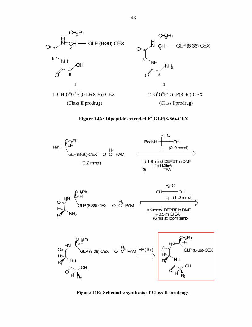
III) Adding dipeptides to the N terminus of F7,GLP(8-36)-CEX
G5G6F7,GLP(8-36)-CEX (class 1) and OH-G5G6F7,GLP(8-36)-CEX (class 2) were
synthesized to determine if either of the nucleophiles (amine or hydroxyl) could cleave
the amide bond by 2,5-diketopiperazine or 2,5-diketomorpholine formation respectively
and thus regenerate the F7,GLP(8-36)-CEX. The two compounds are shown below
(Figure 14A). The schematic synthesis of the class 2 prodrug is represented in Figure
14B. All subsequent Class II prodrugs were synthesized by analogous procedures.
48
GLP(8-36) CEXHN
O
NH
O
CH
OH
CH2Ph
6
5
7
GLP(8-36) CEXHN
O
NH
O
CH
NH2
CH2Ph
6
5
7
1 2
1: OH-G5G6F7,GLP(8-36)-CEX 2: G5G6F7,GLP(8-36)-CEX
(Class II prodrug) (Class I prodrug)
Figure 14A: Dipeptide extended F7,GLP(8-36)-CEX
O
NH2
H
HN
R1
O
NH
H
HN
OH R2
OH
R1
HF(1hr)
H
OR1
BocNH OH
1) 1.9mmol DEPBTinDMF+1ml DIEA/
2) TFA
H
OR2
OH OH
O
NH
H
HN
OH R2
OH
R1
H2NH
CH2Ph
GLP(8-36)-CEX OH2C PAM
GLP(8-36)-CEX
0.9mmol DEPBTinDMF+0.5ml DIEA
(6hrsat roomtemp)
CH2PhH
CH2PhH
CH2PhH
GLP(8-36)-CEX OH2C PAM
GLP(8-36)-CEX OH2C PAM
(2.0mmol)
(1 .0mmol)
(0 .2mmol)
Figure 14B: Schematic synthesis of Class II prodrugs

49
The analyses of the two peptides after incubation in PBS (Figure 14A and Table
4) showed that neither the amine, nor the hydroxyl nucleophile could cleave the amide
bond. The structures of the peptides in Table 4 are also illustrated in Appendix IV (along
with the relevant stereochemistry).
Serial No Peptide (Xaa1Yaa2-
F7a-GLP-CEX)
Rate of cleavage at
100˚C
Rate of cleavage at
37˚C
1 GG (class 1) No cleavage No cleavage
2 HO-GG (class 2) No cleavage No cleavage
‘a’: In all these cases there is phenylalanine in the 1st position (phenylalanyl leaving group) of GLP.
Table 4: Attempted cleavage of dipeptides attached to the N terminus of F7,GLP(8-
36)-CEX
Representative HPLC analyses of G5G6F7,GLP(8-36)-CEX dissolved in PBS
before and after treatment at 100˚C for two hours is shown in Figure 15. This analysis
was done with the crude peptide. The amide bond did not cleave and the DKP was not
generated. This was reflected by the absence of a shift of the HPLC peak in the elution
profile of the prodrug to that of the dipeptide-shortened peptide drug F7,GLP(8-36)-CEX
(not shown here) even when heated at 100˚C.

50
1) G5G6F7,GLP(8-36)-CEX
in PBS at 37˚C (0 min)
2) G5G6F7,GLP(8-36)-CEX
in PBS at 100˚C (120 min)
Figure 15: HPLC analysis of G5G6F7,GLP(8-36)-CEX
In Table 5, the analyses of two peptides with a glycine at the N-terminal position
are shown. These peptides were tested to check the effect of the less bulky glycyl leaving
group, as opposed to the previously studied phenylalanine in Table 4. The structures of
the peptides in Table 4 are displayed in Appendix V.
Serial No Peptide
(Xaa1Yaa2-G7a-
GLP-CEX)
Rate of cleavage at
100˚C
Rate of cleavage at
37˚C
3 GG (class 1) No cleavage No cleavage
4 HO-GG (class 2) No cleavage No cleavage
‘a’: In all these cases there is glycine in the 1st position (glycyl leaving group) of GLP.
Table 5: Attempted cleavage of dipeptides added to the N terminus of
G7,GLP(8-36)CEX

51
Based on the results shown in Table 5, it was concluded that the amide bond is
very difficult to cleave under physiological conditions either by 2,5-diketopiperazine
(DKP) or 2,5-diketomorpholine (DMP) formation. This is independent of the
nucleophiles and leaving groups tested, even under elevated temperature. The focus of
this study then moved to esters which were anticipated to be easier to cleave as compared
to the amides.
IV) Depsi-peptides and Esters
Depsi-peptides were synthesized through addition of dipeptides to the hydroxyl
group at the N terminus of another peptide via an ester linkage (Figure 9). The coupling
procedures are described in the experimental section and they proved highly effective. As
before, dipeptides of differential tendency for intramolecular cyclization
(diketopiperazine formation) and release of the parent drug (N-terminal hydroxyl peptide)
were studied. While adding the dipeptide, both Class 3 (an amine nucleophile cleaving an
ester bond) and Class 4 (a hydroxyl nucleophile cleaving an ester bond – Figure 10)
compounds were prepared and studied.
The initial work began with imidazole-lactic acid (OH-His) as the terminal amino
acid and subsequent leaving group. The HO-His7,GLP(8-37) was synthesized. It was a
full agonist with 25% the potency of native GLP-1. There were several attempts to add a
dipeptide to the α hydroxyl group of HO-His7,GLP(8-37) and test for the cleavage of the
new ester bond. However, there was a problem in selectively acylating the α-hydroxyl
group of this peptide. As “protected” imidazole lactic acid was not commercially
available, we decided initially to work with the compound where the imidazole group

52
was unprotected. As a result, the “unprotected imidazole” was inadvertently acylated
along with the hydroxyl group. A synthetic scheme for the preparation of HO-
His7,GLP(8-37), its subsequent acylation and the formation of multiple side-products is
depicted in Appendix VIII.
Consequently, Phenyllactic acid (OH-phenylalanine) was chosen in this initial test
as it was readily available, and the return to a protected HO-His would be conducted if
the work with HO-Phe proved successful.
V) Adding dipeptides to the OH terminus of HO-F7,GLP(8-36)-CEX
The HO-F7,GLP(8-36)-CEX peptide was synthesized from the GLP(8-36)-CEX
PAM resin (the synthetic scheme is represented in Figure 16A) and served as the parent
drug for these experiments. This peptide has an EC50 value of 0.008nM (gold color in
Figure 16B) while native GLP has an EC50 value of 0.011nM. Therefore, the HO-
F7,GLP(8-36)-CEX was found to be at least as potent as the native GLP-1. This is a most
important and a somewhat unexpected observation that the histidine can be substituted by
phenyllactic acid. This warranted additional validation. Subsequent experiments yielded
identical results. The possibility exists that this result is a function of the CEX extension
in the HO-F7,GLP(8-36)-CEX. In this regard, it has been reported that the C-terminal
region of exendin-4 increases its affinity to the GLP receptor78.
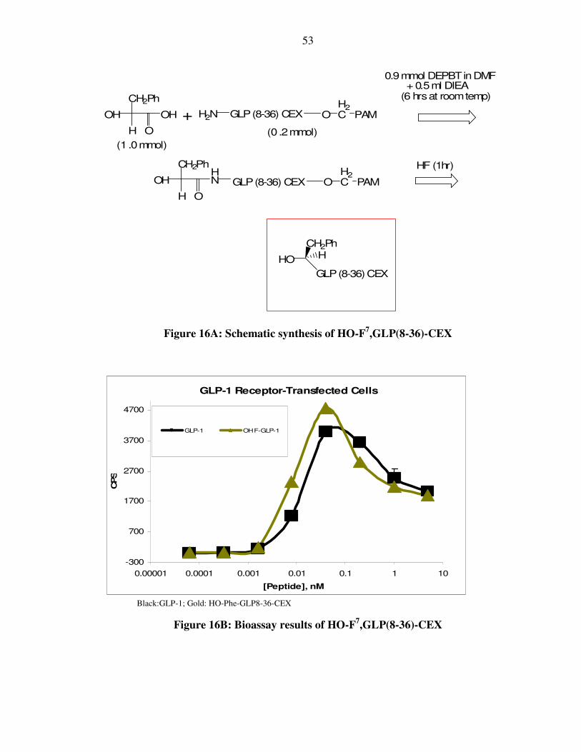
53
H
OH OH H2N
CH2Ph
OH
H
HN
O
+
0.9mmol DEPBTinDMF+ 0.5ml DIEA
(6 hrsat roomtemp)
O
CH2Ph
GLP(8-36) CEX OH2C PAM
GLP(8-36) CEX OH2C PAM
HO HCH2Ph
GLP(8-36)CEX
HF (1hr)
(1 .0mmol)
(0 .2mmol)
Figure 16A: Schematic synthesis of HO-F7,GLP(8-36)-CEX
GLP-1 Receptor-Transfected Cells
-300
700
1700
2700
3700
4700
0.00001 0.0001 0.001 0.01 0.1 1 10
[Peptide], nM
CPS
GLP-1 OH F-GLP-1
Black:GLP-1; Gold: HO-Phe-GLP8-36-CEX
Figure 16B: Bioassay results of HO-F7,GLP(8-36)-CEX

54
The α hydroxyl group of HO-F7,GLP(8-36)-CEX was acylated and both Class 3
(an amine nucleophile cleaving an ester bond; Figure 17A) and Class 4 (a hydroxyl
nucleophile cleaving an ester bond; Figure 17B) compounds were prepared and studied.
Along with the 2,5-diketopiperazine (amine nucleophile) or 2,5-diketomorpholine
(hydroxyl nucleophile), the biologically active HO-F7,GLP(8-36)-CEX is released as
shown in Figure 17C.
CH2Ph
OH
H
HN
O
H
OR1
BocNH OH
1) 1.0mmol DICin DCM(0oC)
+ 0.9mmol DMAP
(12hrsat roomtemp)/
2) TFA
O
NH2
H
O HCH2Ph
R1
H O
R2
BocNH OH
1) 1.9mmol DEPBTinDMF+ 1ml DIEA/
2) TFA
O
NH
H
O
OH R2
NH2
HCH2Ph
R1
GLP(8-36) CEX OH2C PAM
O
NH
H
O
OH R2
NH2
HCH2Ph
R1
GLP(8-36) CEX
GLP(8-36) CEX OH2C PAM
GLP(8-36) CEX OH2C PAM
HF(1hr)
(0 .2mmol)
(2 .0mmol)
(2 .0mmol)
Figure 17A: Schematic synthesis of Class III prodrugs

55
CH2Ph
OH
H
HN
O
H
OR1
BocNH OH
1) 1.0mmol DICinDCM(0oC)
+0.9mmol DMAP
(12hrsat roomtemp)/
2) TFA
O
NH2
H
O HCH2Ph
R1
H O
R2
HO OH
O
NH
H
O
OH R2
OH
HCH2Ph
R1
GLP(8-36)CEX OH2C PAM
O
NH
H
O
OH R2
OH
HCH2Ph
R1
GLP(8-36) CEX
GLP(8-36)CEX OH2C PAM
GLP(8-36)CEX OH2C PAM
HF(1hr)
0.9 mmol DEPBTinDMF+ 0.5ml DIEA
(6hrs at roomtemp)
(0 .2mmol)
(2 .0mmol)
(1 .0mmol)
Figure 17B: Schematic synthesis of Class IV prodrugs
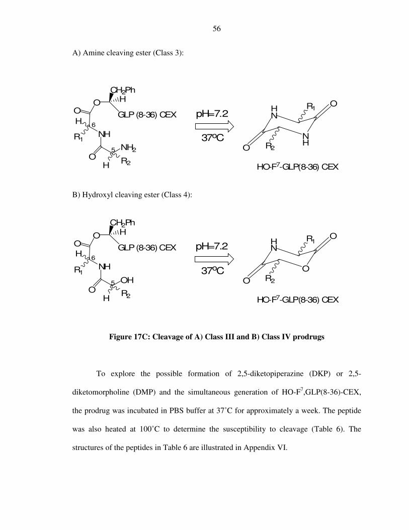
56
A) Amine cleaving ester (Class 3):
pH=7.2
NH
HN
O
O
R1
R2
37oC
HO-F7-GLP(8-36) CEX
O
NH
O
ONH2
HCH2Ph
GLP(8-36) CEX
HR2
H
R1
6
5
B) Hydroxyl cleaving ester (Class 4):
pH=7.2
O
HN
O
O
R1
R2
37oC
HO-F7-GLP(8-36) CEX
O
NH
O
OOH
HCH2Ph
GLP(8-36)CEX
HR2
H
R1
6
5
Figure 17C: Cleavage of A) Class III and B) Class IV prodrugs
To explore the possible formation of 2,5-diketopiperazine (DKP) or 2,5-
diketomorpholine (DMP) and the simultaneous generation of HO-F7,GLP(8-36)-CEX,
the prodrug was incubated in PBS buffer at 37˚C for approximately a week. The peptide
was also heated at 100˚C to determine the susceptibility to cleavage (Table 6). The
structures of the peptides in Table 6 are illustrated in Appendix VI.

57
Serial No Peptide (Yaa1Xaa2-O-Phe7-GLP-8-36-
CEX)
t1/2*
of cleavage at 37˚C
1 Gly-Gly (class 3) 0.87 hrs
2 HO-Gly-Gly (class 4) 1.13 hrs
3 HO-Phe-Val (class 4) No cleavage
4 HO-Phe-dVal (class 4) 50.58 hrs
5 HO-Phe-Ala (class 4) 2.83 hrs
6 HO-Phe-dAla (class 4) 2.41 hrs
7 HO-Phe-Gly (class 4) 2.46 hrs
8 HO-Phe-Phe (class 4) 33.31 hrs
9 HO-Phe-dPhe (class 4) 7.65 hrs
10 Phe-Val (class 3) 64.0 hrs
* t1/2 is the time required for 50% release of the HO-Phe-GLP8-36-CEX at 37°C in PBS (pH7.2). It is calculated by a standard first order reaction plot. All these esters, including #3 cleaved at 100°C. In all these cases, there is HO-Phe in the 1st position (hydroxy phenylalanyl leaving group) of GLP.
Table 6: Cleavage of dipeptide extended HO-F7,GLP(8 36)-CEX
The first two peptides shown in Table 6 (Peptides 1 and 2) were analyzed to
determine if either of the nucleophiles (amine or hydroxyl) could cleave the ester bond by
DKP or DMP formation respectively. The results validated our expectation that the esters
are more susceptible to cleavage than the amides. The relatively slower rate of cleavage
of peptides 3, 4, 8, 9 and 10 seem to suggest that the bulk of the dipeptide extensions
(Phe or Val) greatly affect the rate of dissociation of the prodrug. While there is virtually
no difference between a methyl and a hydrogen side chain (5, 6 and 7), the presence of an
isopropyl group (β branching) greatly attenuates the rate of ester cleavage as evidenced in
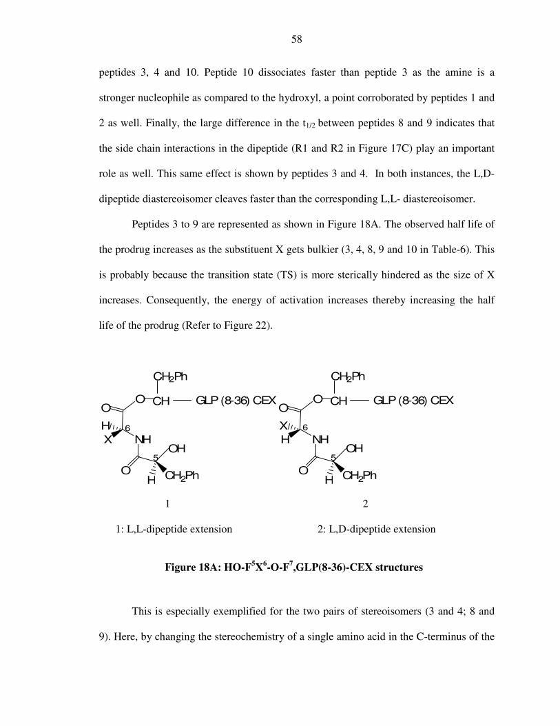
58
peptides 3, 4 and 10. Peptide 10 dissociates faster than peptide 3 as the amine is a
stronger nucleophile as compared to the hydroxyl, a point corroborated by peptides 1 and
2 as well. Finally, the large difference in the t1/2 between peptides 8 and 9 indicates that
the side chain interactions in the dipeptide (R1 and R2 in Figure 17C) play an important
role as well. This same effect is shown by peptides 3 and 4. In both instances, the L,D-
dipeptide diastereoisomer cleaves faster than the corresponding L,L- diastereoisomer.
Peptides 3 to 9 are represented as shown in Figure 18A. The observed half life of
the prodrug increases as the substituent X gets bulkier (3, 4, 8, 9 and 10 in Table-6). This
is probably because the transition state (TS) is more sterically hindered as the size of X
increases. Consequently, the energy of activation increases thereby increasing the half
life of the prodrug (Refer to Figure 22).
GLP(8-36) CEXOO
NH
O
H
X
CH2PhH
CH
OH
CH2Ph
6
5
GLP(8-36) CEXOO
NH
O
X
H
CH2PhH
CH
OH
CH2Ph
6
5
1 2
1: L,L-dipeptide extension 2: L,D-dipeptide extension
Figure 18A: HO-F5X6-O-F7,GLP(8-36)-CEX structures
This is especially exemplified for the two pairs of stereoisomers (3 and 4; 8 and
9). Here, by changing the stereochemistry of a single amino acid in the C-terminus of the

59
dipeptide extension, a huge difference in rate was observed. The steric hindrance of the
corresponding transition states is far greater in a L,L-dipeptide diastereoisomer (Analog 1
in Figure 18A) as compared to a L,D- diastereoisomer (Analog 2 in Figure 18A). Hence,
3 has a greater t1/2 as compared to 4, and 9 has a t1/2 greater than 10.
Compound 10 is F5V6-O-F7,GLP(8-36)-CEX (Figure 18B). This has a t1/2 of 64
hrs and represents the longest duration peptidic prodrug to cleavage under physiological
conditions, reported to date. The transition state in this case is sizably hindered because
of the steric interaction between the phenylalanyl and the valyl side chain.
GLP(8-36) CEXOO
NH
O
H
CH2PhH
CH
NH2
CH2Ph
6
5
Figure 18B: F5V6-O-F7,GLP(8-36)-CEX (t1/2=64 hrs)
The experimental data supporting the cleavage of compound 10 (Figure 18B)
(4264 Da) to form the parent drug HO-F7,GLP(8-36)-CEX (4019 Da) is shown in Figure
19. Since the reaction studied is an intramolecular cyclization, it is assumed that the
presence of other impurities in the sample does not affect the rate of internal dissociation.
In addition, both our molecules of interest (the prodrug and the drug) have been followed
with a MALDI analysis even in the midst of contaminating material.

60
Phe-Val-O-F7-GLP8-36 CEX (4264Da) in PBS at 37˚C at 0 min. 2) Phe-Val-O-F7-GLP8-36 CEX (4264Da) cleaving to the parent HO-F7-GLP 8-36 CEX (4019 Da) after incubation in PBS at 37˚C for 64 hours.
Figure 19: Cleavage of F5V6-O-F7,GLP(8-36)-CEX to form
HO-F7,GLP(8-36)-CEX
A second set of analogous depsipeptides were also studied to further examine the
effects of the dipeptide extension structure upon the rate of cleavage (Table 7). Their
4264 Da
4019.122 Da
4263.745 Da
1
2
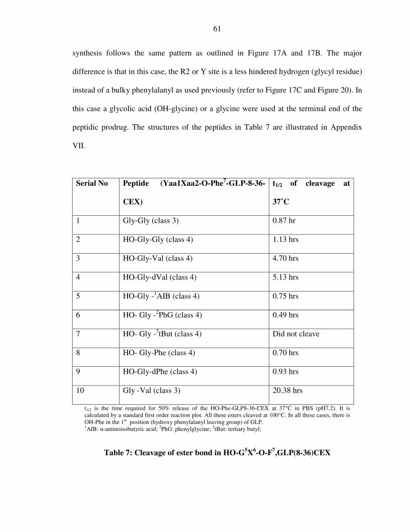
61
synthesis follows the same pattern as outlined in Figure 17A and 17B. The major
difference is that in this case, the R2 or Y site is a less hindered hydrogen (glycyl residue)
instead of a bulky phenylalanyl as used previously (refer to Figure 17C and Figure 20). In
this case a glycolic acid (OH-glycine) or a glycine were used at the terminal end of the
peptidic prodrug. The structures of the peptides in Table 7 are illustrated in Appendix
VII.
Serial No Peptide (Yaa1Xaa2-O-Phe7-GLP-8-36-
CEX)
t1/2 of cleavage at
37˚C
1 Gly-Gly (class 3) 0.87 hr
2 HO-Gly-Gly (class 4) 1.13 hrs
3 HO-Gly-Val (class 4) 4.70 hrs
4 HO-Gly-dVal (class 4) 5.13 hrs
5 HO-Gly -1AIB (class 4) 0.75 hrs
6 HO- Gly -2PhG (class 4) 0.49 hrs
7 HO- Gly -3tBut (class 4) Did not cleave
8 HO- Gly-Phe (class 4) 0.70 hrs
9 HO-Gly-dPhe (class 4) 0.93 hrs
10 Gly -Val (class 3) 20.38 hrs
t1/2 is the time required for 50% release of the HO-Phe-GLP8-36-CEX at 37°C in PBS (pH7.2). It is calculated by a standard first order reaction plot. All these esters cleaved at 100°C. In all these cases, there is OH-Phe in the 1st position (hydroxy phenylalanyl leaving group) of GLP. 1AIB: α-aminoisobutyric acid; 2PhG: phenylglycine; 3tBut: tertiary butyl;
Table 7: Cleavage of ester bond in HO-G5X6-O-F7,GLP(8-36)CEX
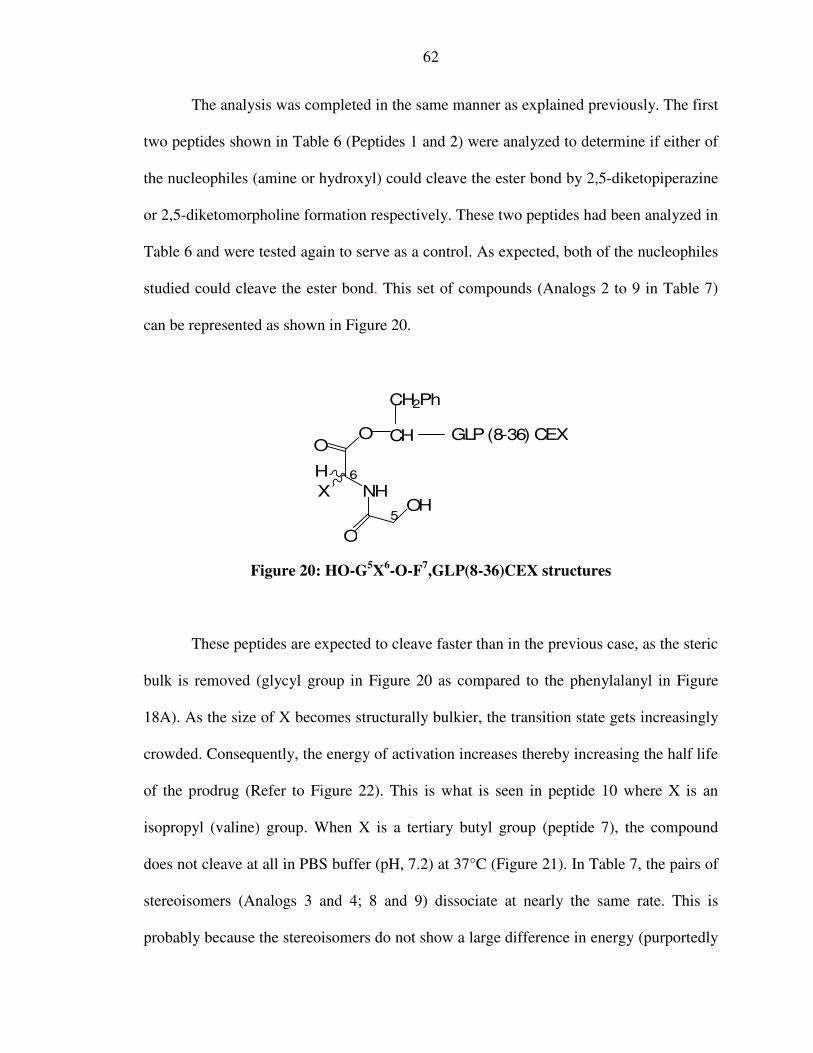
62
The analysis was completed in the same manner as explained previously. The first
two peptides shown in Table 6 (Peptides 1 and 2) were analyzed to determine if either of
the nucleophiles (amine or hydroxyl) could cleave the ester bond by 2,5-diketopiperazine
or 2,5-diketomorpholine formation respectively. These two peptides had been analyzed in
Table 6 and were tested again to serve as a control. As expected, both of the nucleophiles
studied could cleave the ester bond. This set of compounds (Analogs 2 to 9 in Table 7)
can be represented as shown in Figure 20.
GLP(8-36) CEXOO
NH
O
OHX
H
CH
CH2Ph
6
5
Figure 20: HO-G5X6-O-F7,GLP(8-36)CEX structures
These peptides are expected to cleave faster than in the previous case, as the steric
bulk is removed (glycyl group in Figure 20 as compared to the phenylalanyl in Figure
18A). As the size of X becomes structurally bulkier, the transition state gets increasingly
crowded. Consequently, the energy of activation increases thereby increasing the half life
of the prodrug (Refer to Figure 22). This is what is seen in peptide 10 where X is an
isopropyl (valine) group. When X is a tertiary butyl group (peptide 7), the compound
does not cleave at all in PBS buffer (pH, 7.2) at 37°C (Figure 21). In Table 7, the pairs of
stereoisomers (Analogs 3 and 4; 8 and 9) dissociate at nearly the same rate. This is
probably because the stereoisomers do not show a large difference in energy (purportedly

63
because of the non-chiral nature of glycine). Hence, in this case, both the glycyl,L-
dipeptide extension and the glycyl,D-dipeptide extension have a comparable energetic
transition state.
GLP(8-36) CEXOO
NH
O
NH2
HH
H
CH
CH2Ph
6
5
GLP(8-36) CEXOO
NH
O
OH
HH
H
CH
CH2Ph
6
5
1 2
1: G5V6-O-F7,GLP(8-36)CEX 2: HO-G5tBuG6-O-F7,GLP(8-36)CEX
(t1/2=18 hrs) (did not cleave at all)
Figure 21: Dipeptide extended HO-F7,GLP(8-36)-CEX
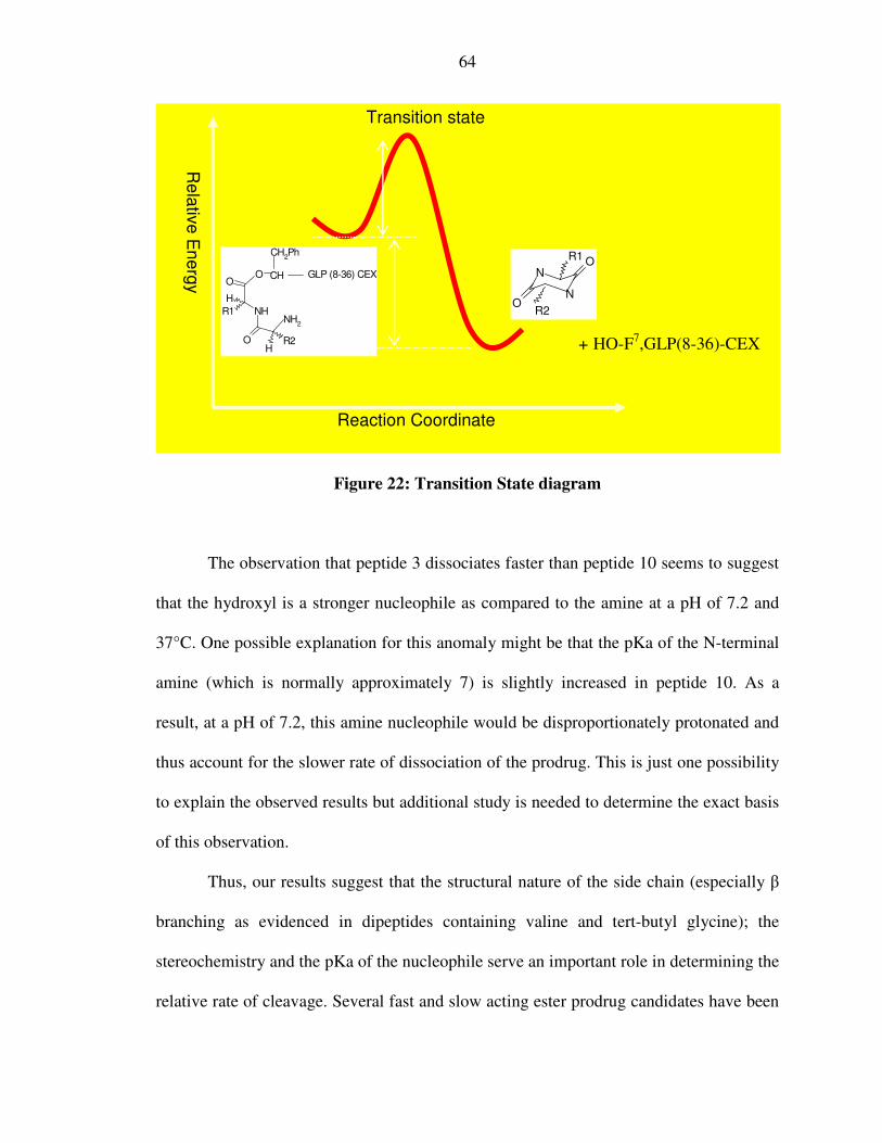
64
Figure 22: Transition State diagram
The observation that peptide 3 dissociates faster than peptide 10 seems to suggest
that the hydroxyl is a stronger nucleophile as compared to the amine at a pH of 7.2 and
37°C. One possible explanation for this anomaly might be that the pKa of the N-terminal
amine (which is normally approximately 7) is slightly increased in peptide 10. As a
result, at a pH of 7.2, this amine nucleophile would be disproportionately protonated and
thus account for the slower rate of dissociation of the prodrug. This is just one possibility
to explain the observed results but additional study is needed to determine the exact basis
of this observation.
Thus, our results suggest that the structural nature of the side chain (especially β
branching as evidenced in dipeptides containing valine and tert-butyl glycine); the
stereochemistry and the pKa of the nucleophile serve an important role in determining the
relative rate of cleavage. Several fast and slow acting ester prodrug candidates have been
NO
ON
R1
R2
Re
lativ
e E
ne
rgy
Reaction Coordinate
Transition state
OO
NH
O
H
R1
HR2
CH
NH2
CH2Ph
GLP (8-36) CEX
+ HO-F7,GLP(8-36)-CEX

65
identified which were tested for their potency. As described earlier in our hypothesis, a
prodrug should have minimal potency as compared to the drug, and its potency should
increase consistent with the speed of cleavage of the dipeptide extension.
VI) Bioassays of selected longer-acting prodrug candidates
Four of the longer duration prodrugs were chosen for further analysis in the
biopotency tests. Luciferase-based bio-assays were performed after purifying all these
peptides by HPLC and confirming their masses by MALDI-MS analysis. Percent potency
was calculated for purposes of comparing the mean EC50 of the parent with that of the
respective prodrug. The results are tabulated in Table 8 and then pictorially represented in
Figure 24A and B. The peptides tested in Table 8 are represented below in Figure 23. The
syntheses of these prodrugs are schematically shown in Appendix I.

66
H
OH
CH2Ph
GLP (8-36) CEX
OO
NH
O
H
CH2PhH
CH
OH
CH2Ph
GLP (8-36) CEX
OO
NH
O
H
CH2PhH
CH
NH2
CH2Ph
GLP (8-36) CEX
OO
NH
O
OHCH
2Ph
CH2Ph
H
H
CH
CH2Ph
GLP (8-36) CEX
OO
NH
O
NH2
HH
H
CH
CH2Ph
GLP (8-36) CEX
Figure 23: Structures of the peptides represented in Figure 24 and Table 8
HO-F7,GLP(8-36)-CEX (red)
HO-F5dV6-O-F7,GLP(8-36)-CEX (t1/2 = 50.5 hr) (navy blue)
F5V6-O-F7,GLP(8-36)-CEX (t1/2
= 64 hrs) (sky blue)
HO-F5F6-O-F7,GLP(8-36)-CEX CEX (t1/2 = 33.3 hr) (pink)
G5V6-O-F7,GLP(8-36)CEX (t1/2
= 20.3 hr) (yellow)

67
Serial
No
(Color)
Peptide (t1/2):
(Yaa1Xaa2-O-Phe-GLP-8-
36-CEX)
EC50 ± Std.
deviation (nM)
% potency with
respect to parent
1 (red) HO-F7-GLP8-36 CEX (parent
drug candidate)
0.028 ± 0.006 100% (parent drug)
2 (navy
blue)
HO-Phe-dVal (t1/2 = 50.5 hrs) 0.40 ± 0.06 7%
3 (sky
blue)
Phe-Val (t1/2 = 64.0 hrs) 2.070 ± 0.41 1.3%
4 (pink) HO-Phe- Phe (t1/2 = 33.3 hrs) 0.771 ± 0.249 3.6%
5
(yellow)
Gly-Val (t1/2 = 20.3 hrs) 0.230 ± 0.10 12%
The color code refers to the respective peptides represented in the Figure 22. Percent potency calculations were done comparing the mean EC50 of the parent with that of the prodrug.
Table 8: Bioassay of select longer-acting ester prodrugs
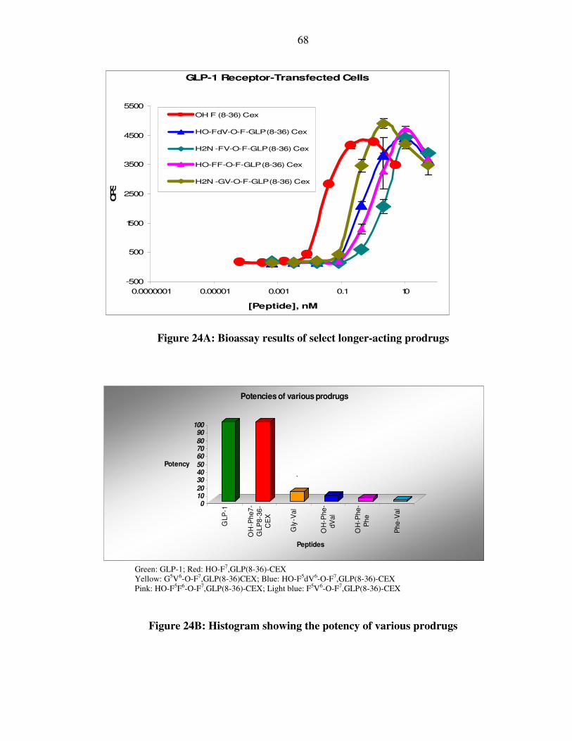
68
GLP-1 Receptor-Transfected Cells
-500
500
1500
2500
3500
4500
5500
0.0000001 0.00001 0.001 0.1 10
[Peptide], nM
CPS
OH F (8-36) Cex
HO-FdV-O-F-GLP(8-36) Cex
H2N -FV-O-F-GLP(8-36) Cex
HO-FF-O-F-GLP(8-36) Cex
H2N -GV-O-F-GLP(8-36) Cex
Figure 24A: Bioassay results of select longer-acting prodrugs
0
10
20
30
40
50
60
70
80
90
100
Potency
GL
P-1
OH
-Ph
e7
-
GL
P8
-36
-
CE
X
Gly
-Va
l
OH
-Ph
e-
dV
al
OH
-Ph
e-
Ph
e
Ph
e-V
al
Peptides
Potencies of various prodrugs
`
Green: GLP-1; Red: HO-F7,GLP(8-36)-CEX Yellow: G5V6-O-F7,GLP(8-36)CEX; Blue: HO-F5dV6-O-F7,GLP(8-36)-CEX Pink: HO-F5F6-O-F7,GLP(8-36)-CEX; Light blue: F5V6-O-F7,GLP(8-36)-CEX
Figure 24B: Histogram showing the potency of various prodrugs
69
These observations clearly show that addition of an ester linked dipeptide
sequence to the terminal-hydroxyl group of the parent drug drastically reduces the
potency of the drug. Two peptides were further analyzed to demonstrate that the prodrugs
revert towards the potency of the parent drug following incubation for 24 hours, in PBS
(pH, 7.2). The peptides chosen were analogs 4 and 5, based upon their physiologically
relevant t1/2 values being closest to 24 hours (Table 9).
These experiments were performed using HPLC purified samples with the
luciferase-based assay. Percent potency calculations were done comparing the mean EC50
of the parent with that of the prodrug. The results are tabulated in Table 9 and then
pictorially represented in Figure 25.

70
Serial No
/ (Color)
Peptide (t1/2):
(Yaa1Xaa2-O-Phe-GLP-8-
36-CEX)
EC50 ± Std.
deviation (nM)
% potency with
respect to parent
Black HO-F7-GLP8-36-CEX
(parent drug candidate)
0.023 ± 0.010 100% (parent drug)
5(168 hrs)
(red)*
Gly-Val (t1/2 = 20.3 hrs)
incubated for 168 hrs.
0.024 ± 0.012 95.8 %
4(0 hrs) (navy
blue)
HO-Phe- Phe (t1/2 = 33.3 hrs)
before incubation.
0.612 ± 0.270 3.7 %
4(24 hrs)
(sky blue)
HO-Phe- Phe (t1/2 = 33.3 hrs)
incubated for 24 hrs.
0.134 ± 0.050 17.2 %
5(0 hrs)
(pink)
Gly-Val (t1/2 = 20.3 hrs)
before incubation.
0.211 ± 0.072 10.9 %
5(24 hrs)
(gold)
Gly-Val (t1/2 = 20.3 hrs)
incubated for 24 hrs.
0.05 ± 0.020 46 %
*- Peptide 5 after incubation in PBS for a week (potency almost completely restored). () – Time inside parenthesis refers to the time of incubation of the respective peptide The color codes refer to the colors with which the respective peptides are represented in the Figure 23.
Table 9: Bioassay results that show the conversion of prodrugs to drugs
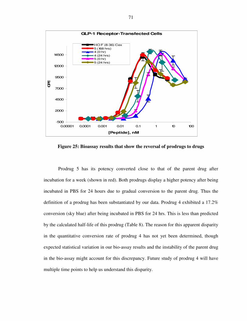
71
GLP-1 Receptor-Transfected Cells
-500
2000
4500
7000
9500
12000
14500
0.00001 0.0001 0.001 0.01 0.1 1 10 100
[Peptide], nM
CPS
HO F (8-36) Cex
5 (168 hrs)
4 (0 hr)
4 (24 hrs)
5 (0 hr)
5 (24 hrs)
Figure 25: Bioassay results that show the reversal of prodrugs to drugs
Prodrug 5 has its potency converted close to that of the parent drug after
incubation for a week (shown in red). Both prodrugs display a higher potency after being
incubated in PBS for 24 hours due to gradual conversion to the parent drug. Thus the
definition of a prodrug has been substantiated by our data. Prodrug 4 exhibited a 17.2%
conversion (sky blue) after being incubated in PBS for 24 hrs. This is less than predicted
by the calculated half-life of this prodrug (Table 8). The reason for this apparent disparity
in the quantitative conversion rate of prodrug 4 has not yet been determined, though
expected statistical variation in our bio-assay results and the instability of the parent drug
in the bio-assay might account for this discrepancy. Future study of prodrug 4 will have
multiple time points to help us understand this disparity.
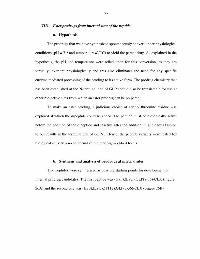
72
VII) Ester prodrugs from internal sites of the peptide
a. Hypothesis
The prodrugs that we have synthesized spontaneously convert under physiological
conditions (pH = 7.2 and temperature=37˚C) to yield the parent drug. As explained in the
hypothesis, the pH and temperature were relied upon for this conversion, as they are
virtually invariant physiologically and this also eliminates the need for any specific
enzyme mediated processing of the prodrug to its active form. The prodrug chemistry that
has been established at the N-terminal end of GLP should also be translatable for use at
other bio-active sites from which an ester prodrug can be prepared.
To make an ester prodrug, a judicious choice of serine/ threonine residue was
explored at which the dipeptide could be added. The peptide must be biologically active
before the addition of the dipeptide and inactive after the addition, in analogous fashion
to our results at the terminal end of GLP-1. Hence, the peptide variants were tested for
biological activity prior to pursuit of the prodrug modified forms.
b. Synthesis and analysis of prodrugs at internal sites
Two peptides were synthesized as possible starting points for development of
internal prodrug candidates. The first peptide was (H7F),(E9Q),GLP(8-36)-CEX (Figure
26A) and the second one was (H7F),(E9Q),(T11S),GLP(8-36)-CEX (Figure 26B).

73
Figure 26A: (H7F),(E9Q),GLP(8-36)-CEX
*- The ester prodrug was synthesized at the circled hydroxyl group for both peptides.
Figure 26B: (H7F),(E9Q),(T11S),GLP(8-36)-CEX
The synthesis of an ester prodrug was attempted at the hydroxyl group of the 5th
amino acid (threonine or serine side chain marked in Figure 26A and 26B respectively).
For these molecules, a Boc benzyl-type synthesis on a PAM resin was conducted until the
point of Phe at the 12th position. The rest of the synthesis was completed using Fmoc-
based chemistry. This offered the opportunity to selectively remove the protecting group
of the serine side chain, leaving all the other protecting groups intact. This allowed for
the selective acylation on the free hydroxyl group of the serine at the 11th position.
NH
FTSDVSSYLEGQAAKEFIAWLVKGR-CEX
HFAQG
O
HH
OH
OH
ONH
FTSDVSSYLEGQAAKEFIAWLVKGR-CEX
HFAQG
CH3
H

74
More specifically a Boc-benzyl strategy could not be used throughout the
synthesis as the hydroxyl protecting benzyl group of serine would require HF treatment
for removal. Consequently, an ester prodrug could not be synthesized on the peptidic
resin as the HF treatment would simultaneous remove the peptide from the resin. This is
why, we added the Ser at the 11th position (Fmoc-O-t butyl-L-seine) and the subsequent
Gly, Gln, Ala and Phe as Fmoc-protected amino acids. The t-butyl protecting group of
the serine was selectively removed by treatment with TFA for 2 hours. The Boc-protected
dipeptidic pro-moiety was added to this hydroxyl group to finish the synthesis of the ester
prodrug. The final Boc-protecting group was removed by treatment with TFA and the N-
terminal Fmoc group was removed by treatment with 20% piperidine in DMF. This was
found to be a useful strategy to build the dipeptide esters from the hydroxyl side chain of
serine and threonine residues. The glutamic acid residue at the 3rd position was also
replaced with glutamine to prevent any unwanted coupling on the side chain carboxyl
group. It was assumed that this would not lead to a loss of activity as previous reports
have suggested that the 3rd position is not critical for GLP potency35.
The luciferase bio-assay was conducted after purification of these peptides by
HPLC, and confirmation of their masses by MALDI as described previously. Percent
potency was calculated and the mean EC50 of the peptide (drug candidate) was compared
with that of native GLP-1. The results are tabulated in Table-10 and then shown in Figure
27.
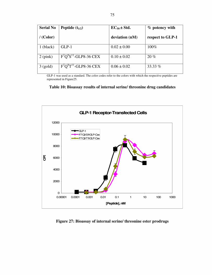
75
Serial No
/ (Color)
Peptide (t1/2) EC50 ± Std.
deviation (nM)
% potency with
respect to GLP-1
1 (black) GLP-1 0.02 ± 0.00 100%
2 (pink) F7Q9S11-GLP8-36 CEX 0.10 ± 0.02 20 %
3 (gold) F7Q9T11-GLP8-36 CEX 0.06 ± 0.02 33.33 %
GLP-1 was used as a standard. The color codes refer to the colors with which the respective peptides are represented in Figure25.
Table 10: Bioassay results of internal serine/ threonine drug candidates
Figure 27: Bioassay of internal serine/ threonine ester prodrugs
GLP-1 Receptor-Transfected Cells
0
2000
4000
6000
8000
10000
12000
0.00001 0.0001 0.001 0.01 0.1 1 10 100 1000
[Peptide], nM
CPS
GLP-1
F7 Q9 S11 GLP-Cex
F7 Q9 T11 GLP-Cex
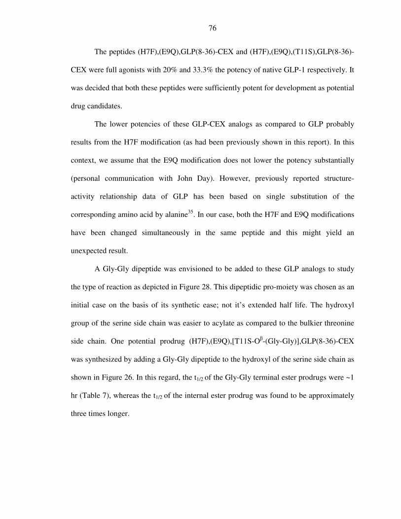
76
The peptides (H7F),(E9Q),GLP(8-36)-CEX and (H7F),(E9Q),(T11S),GLP(8-36)-
CEX were full agonists with 20% and 33.3% the potency of native GLP-1 respectively. It
was decided that both these peptides were sufficiently potent for development as potential
drug candidates.
The lower potencies of these GLP-CEX analogs as compared to GLP probably
results from the H7F modification (as had been previously shown in this report). In this
context, we assume that the E9Q modification does not lower the potency substantially
(personal communication with John Day). However, previously reported structure-
activity relationship data of GLP has been based on single substitution of the
corresponding amino acid by alanine35. In our case, both the H7F and E9Q modifications
have been changed simultaneously in the same peptide and this might yield an
unexpected result.
A Gly-Gly dipeptide was envisioned to be added to these GLP analogs to study
the type of reaction as depicted in Figure 28. This dipeptidic pro-moiety was chosen as an
initial case on the basis of its synthetic ease; not it’s extended half life. The hydroxyl
group of the serine side chain was easier to acylate as compared to the bulkier threonine
side chain. One potential prodrug (H7F),(E9Q),[T11S-Oβ-(Gly-Gly)],GLP(8-36)-CEX
was synthesized by adding a Gly-Gly dipeptide to the hydroxyl of the serine side chain as
shown in Figure 26. In this regard, the t1/2 of the Gly-Gly terminal ester prodrugs were ~1
hr (Table 7), whereas the t1/2 of the internal ester prodrug was found to be approximately
three times longer.

77
OHN
FTSDVSSYLEGQAAKEFIAWLVKGR-CEX
HF7AQ9G
OO
N
NH2O
H
H
H
H
H
H
11
HO
HN
FTSDVSSYLEGQAAKEFIAWLVKGR-CEX
HO
H
H
F7AQ9G
11
NH
NH O
O
Figure 28: Conversion of serine ester prodrug (H7F),(E9Q),[T11S-Oβ-(Gly-
Gly)],GLP(8-36)-CEX to drug (H7F),(E9Q),(T11S),GLP(8-36)-CEX
Unfortunately, it was not possible to acylate the hydroxyl of the threonine side
chain with a Gly-Gly dipeptide. This analogous addition of the dipeptide to Thr relative
to Ser failed probably because of the steric restraints imposed by the methyl group at C-3
of the threonine side chain as compared to the hydrogen at C-3 of the serine side chain.
It was observed that the (H7F),(E9Q),[T11S-Oβ-(Gly-Gly)],GLP(8-36)-CEX
prodrug dissociated with a t1/2 = 2.7 hrs to yield the parent drug
t1/2 = 3 hrs
+

78
(H7F),(E9Q),(T11S),GLP(8-36)-CEX along with the 2,5-diketopiperazine (DKP). We
were unable to determine the absolute potency of this prodrug because its half life is
lower than the incubation period of the prodrug in the cell culture based luciferase assay
(5 hrs). The half life of the prodrug (2.7 hrs) indicates that approximately 70% of the
prodrug would have converted to the active entity in 5 hours; hence the assay could not
be accurately performed on this prodrug. However, we have nevertheless established the
conversion of the internal serine prodrug to the active entity.
Since the conditions for the conversion of the prodrugs to the drug (pH = 7.2 and
temperature=37˚C) are virtually invariant physiologically, it was found that the prodrug
chemistry that was established at the O-terminal end of GLP could be used to cleave from
an internal site as well, specifically Ser but conceptually Thr. Thus our initial hypothesis
is validated.

79
E) Conclusion:
Clinical studies have revealed that GLP-1 therapy is an effective treatment for
Type II diabetes. In addition, it might be intrinsically safer than insulin therapy because
of its glucose dependent action, thus eliminating the chances of hypoglycemia49. So far
no cases of hypoglycemia have been reported using treatment with a GLP-related drug.
An ideal prodrug should be stable to storage but must convert to the active drug
under a specific set of conditions. The desired set of conditions for the activation of the
prodrug will depend on its purpose and site of action. Detailed stability studies are
important for the rigorous characterization of promising prodrugs. However, a rapid
screening helps in the identification of such prodrug candidates. Such screening provides
insights to the structure, stereochemistry and the site of attachment of the pro-moiety to
the drug and how these affect the conversion of the inactive prodrug to the active drug. In
the work presented here, the conditions for the conversion are a pH of 7.2 and
temperature of 37°C. These conditions were used for the cleavage of the prodrugs as they
are physiologically invariant and can thus be translated into other peptidic drugs as well.
The relative potency of prodrug candidates to the parent drug were determined through a
receptor-based cell culture assay.
Prodrug strategies have often been implemented to improve the efficacy and
safety of important drugs in different diseases79. In this report, the prodrug strategy has
been used to increase the time action of the GLP analog by extending the half life of the
prodrug. Although a number of different pro-moieties could be used, the ‘dipeptide pro-
moiety or the α hydroxyl dipeptide analog’ which dissociated by the formation of
80
diketopiperazine (DKP) or diketomorpholine (DMP) along with the release of the active
GLP analog over a wide time range was chosen.
Our strategy has a number of advantages. Firstly, a number of structurally diverse
amino acids (aliphatic, aromatic, acidic, basic, neutral) and α hydroxyl amino acids are
commercially available. Secondly, given the natural nature of amino acids there is likely
to be fewer safety and immunogenic concerns regarding their use as pro-moieties. In this
context, it should also be noted that the native amino acid sequence was maintained as far
as possible, and this should minimize potential adverse immunogenic responses.
However the magnitude and nature of such adverse reactions will need to be explored in
vivo. Thirdly, the amino acid properties and peptide synthetic chemistries are well
established. Finally, and most importantly the chemistry of diketopiperazine formation
allowed at least four points where the structure could be stereochemically controlled to
refine the formation rate of the active GLP analog. Prodrugs of varying half lives were
synthesized by chemically modifying R1 and R2 in the context of different nucleophiles
(Figure 6). This generated the novel chemoreversible prodrugs of GLP, which converted
to the active entity via the intramolecular cyclization and subsequent release of 2,5-
diketopiperazine (DKP) or 2,5-diketomorpholine (DMP). The large variance of half lives
amongst the various prodrugs ranging from a t1/2 = 0.5 - 64.0 hrs underscores the great
potential of this chemoreversible prodrug system in accurately tailoring the release of the
parent drug under physiological conditions. The prodrug F5V6-O-F7,GLP(8-36)-CEX
with a t1/2 = 64 hrs is the longest acting chemically defined peptidic prodrug reported to
date.

81
Four different varieties of prodrugs (Class 1, 2, 3 and 4) of GLP or biologically
active GLP analogs were synthesized. Both the amide and the ester prodrugs were
synthesized in good yield. In the beginning, the amide prodrugs were synthesized by
adding a dipeptide to the amine group at the N-terminus. The cleavage of the amide bond
of interest was attempted under physiological conditions with both an amine and a
hydroxyl nucleophile (Table 4 and 5). Different leaving groups were employed, with a
histidine (Table 2), phenylalanine (Table 3 and 4) and a glycine (Table 5) at the N-
terminus of GLP analogs. For solubility purposes, we decided to work with a C-terminal
modified GLP analog, GLP-CEX.
We observed that the amide bond could not be dissociated by 2,5-
diketopiperazine (DKP) or 2,5-diketomorpholine (DMP) formation under physiological
conditions. In fact, the amide bond could not be cleaved even when heated at 100°C at a
pH=7.2. Consequently, an amide prodrug could not be synthesized using this approach.
As a result, our attention focused on synthesizing ester prodrugs.
Two distinct classes of dipeptide esters were tested to study the cleavage of the
ester bond. Class 3 variants employed an N-terminal amine nucleophile while Class 4
variants contained a hydroxyl nucleophile to cleave the ester bond (see Figure 10).
Selectively acylating the α-hydroxyl group of HO-H7,GLP(8-36)-CEX proved to be very
difficult in the absence of a “protected” imidazole peptide. Hence, we decided to work
with phenyllactic acid and subsequently synthesized the HO-F7,GLP(8-36)-CEX which
was found to be at least as potent as the native GLP in repetitive analysis. The ester
prodrugs were synthesized using this parent drug as the scaffold.
82
The esters proved to be much more labile than the corresponding amides and most
of the ester prodrugs dissociated to give the active drug under physiological conditions
along with the assumed formation of 2,5-diketopiperazine (DKP) or 2,5-
diketomorpholine (DMP). The degradation half lives of the respective prodrugs were
calculated based on the first order kinetics (t1/2 = .693/k) where ‘k’ is the first order rate
constant for the degradation of the prodrug. The disappearance of the prodrug and the
appearance of the drug were both simultaneously analyzed and calculated using HPLC.
Generally, there was an excellent mass balance between the loss of the prodrug and the
appearance of the drug. However in our results, only the estimated half life of the
disappearance of the prodrug has been reported.
A sample kinetic profile reflecting the disappearance of the prodrug
(H7F),(E9Q),[T11S-Oβ-(Gly-Gly)],GLP(8-36)-CEX and the appearance of the parent
drug (H7F),(E9Q),(T11S),GLP(8-36)-CEX (Figure 28) under physiological conditions is
shown in Figure 29. The slope of the first order plot (blue diamonds) represents the
dissociation constant of the prodrug from which the half life was calculated to be 2.8 hrs.
The experiment was performed over several half lives and the recovery of the parent
compound was more than 90%.

83
Analysis of (F7Q9-S11–(Gly-Gly)-GLP8-36-CEX)
1
1.5
2
2.5
3
3.5
4
4.5
5
0 2 4 6 8 10 12 14
Time (hrs)
Ln (conc)
Blue diamonds represent the disappearance of the prodrug. Pink squares represent the appearance of the drug.
The logarithm of the concentrations of the prodrug (blue) and the drug (pink) were plotted on the y axis as a function of time on the x axis.
Figure 29: Typical kinetic profile showing the disappearance of a prodrug and the appearance of a drug
Of all the ester prodrugs that were studied, only two did not cleave and they are
represented in Figure 30.

84
O
NH
H
HN
OH CH2Ph
OH
HCH2Ph
GLP(8-36)CEX6
5
O
NH
H
HN
OH H
OH
HCH2Ph
GLP(8-36)CEX
6
5
1: HO-F5V6-O-F7,GLP(8-36)-CEX 2. HO-G5tBut6-O-F7,GLP(8-36)CEX
Figure 30: Ester prodrugs resistant to cleavage
The most likely reason for this is the increased size of the R1 and R2 in
compounds 1 and 2 (Figure 30). The steric bulk of the isopropyl and tertiary butyl group
crowds the transition state increasing the activation barrier for the intramolecular
cyclization. As a result, the cyclization does not occur. When the hydroxyl nucleophile in
compound 1 was replaced with the amine, the corresponding F5V6-O-F7,GLP(8-36)-CEX
(Figure 18B) cleaved with a long half life of 64.0 hours. The presence of cleavage
relative to the hydroxyl-peptide is because the amine is a stronger nucleophile, as
compared to the hydroxyl. Thus it is seen that the chemical stability of the ester bond is
influenced by the structure of the amino acid promoieties. The bulk of the side chain of
the corresponding amino acids and the strength of the nucleophile play a role in the
cleavage of the ester prodrugs. Additionally, stereochemistry of the two amino acids
further influences the rate of cleavage. These results are consistent with related
observations in the field of DKP-like prodrugs61,63.
Our primary goal was to synthesize longer-acting prodrug candidates of GLP with
a daily or even a weekly dosage. This is especially important as the longest acting DPP-

85
IV resistant GLP analog (Liraglutide) that is presently in Phase III trials has a biological
half life of 10-14 hrs47,48. Four prodrug candidates with varying half lives of 20.3hrs,
33.3hrs, 50.5 hrs and 64.0 hrs were successfully synthesized (Figure 23 and Appendix I).
Each of these prodrug candidates should display extended duration of in vivo action and
possibly constitute ‘once a week’ formulations, since this prolongation would be in
addition to the natural time action of the parent drug.
The bio-assay we used is highly sensitive and reproducible. All the prodrugs have
minimal inherent receptor potency relative to the parent drug (Table 8). The potency of
G5V6-O-F7,GLP(8-36)-CEX is nearly restored quantitatively (Figure 31A), while that of
HO-F5F6-O-F7,GLP(8-36)-CEX is only partial after incubation in PBS at 37˚C for 24
hours (Figure 31B). The reason for latter observation is not immediately clear; and
warrants further study.
0
10
20
30
40
50
60
70
80
90
100
Potency
Gly-Val (0
hrs)
Gly-Val (24
hrs)
Gly-Val (1
week)
OH-Phe7-
GLP8-36-
CEX
GLP-1
Peptides
Potency of Gly-Val-O-Phe7-GLP8-36-CEX
Green: GLP-1; Black: HO-F7,GLP(8-36)-CEX
Red: G5V6-O-F7,GLP(8-36)CEX after incubation in PBS for 1 week; Yellow: G5V6-O-F7,GLP(8-36)CEX after incubation in PBS for 24 hours;
Pink: G5V6-O-F7,GLP(8-36)CEX in PBS at 0 hours;
Figure 31A: Relative potency of G5V6-O-F7,GLP(8-36)CEX

86
0
10
20
30
40
50
60
70
80
90
100
Potency
OH-Phe-Phe
(0 hrs)
OH-Phe-Phe
(24 hrs)
OH-Phe7-
GLP8-36-CEX
GLP-1
Peptides
Potency of OH-Phe-Phe-O-Phe7-GLP8-36-CEX
Green: GLP-1; Black: HO-F7,GLP(8-36)-CEX
Light blue: HO-F5F6-O-F7,GLP(8-36)-CEX after incubation in PBS for 24 hours; Dark blue: HO-F5F6-O-F7,GLP(8-36)-CEX in PBS at 0 hours;
Figure 31B: Relative potency of HO-F5F6-O-F7,GLP(8-36)-CEX
The biological potency of the internal serine ester prodrug, (H7F),(E9Q),[T11S-
Oβ-(Gly-Gly)],GLP(8-36)-CEX could not be determined as its half life (2.8 hrs) is much
shorter than the incubation period (5hrs) in the luciferase assay. However, its chemical
conversion to the active (H7F),(E9Q),(T11S),GLP(8-36)-CEX (Figure 28 and 29) has
been documented validating that the diketopiperazine chemistry is feasible from an
internal ester site of the peptide. The internal ester prodrug (H7F),(E9Q),[T11S-Oβ-(Gly-
Gly)],GLP(8-36)-CEX cleaves with a half life that is longer than the terminal prodrug
G5G6-O-F7,GLP(8-36)-CEX. The reason for this might be that the β-ester bond of the
internal prodrug is more resistant to cleavage than the terminal α-ester bond.
In the overall context of things, we would have much preferred to have been able
to cleave both the amide and ester bonds. This would provide the opportunity to utilize
this prodrug chemistry at amine as well as hydroxyl containing drug candidates. One

87
central disadvantage of ester prodrugs is the ubiquitous presence of esterases in vivo; and
hence the effective half life of the ester prodrug might be much faster in blood then
reported here.
In conclusion, our prodrugs were extremely stable when stored as a powder at
4°C. They are soluble in physiological buffers, and exhibit differential conversion rates to
the active drug under physiological conditions. This latter point is the central importance
of my work. The results of this study clearly indicate that the cis-conformation of the
dipeptide favors cleavage. Furthermore, that the overall structure (especially β branching
as evidenced by the slow rates in pro-moieties containing valine), stereochemistry of the
side chains, and the strength of the nucleophile play a profound role in affecting the rates
of prodrug cleavage. It was also importantly shown that rate of conversion can be fine-
tuned by selecting the structure of the pro-moieties. This is of utmost importance in the
design of a properly functioning prodrug, as represented in Figure 32. The dynamic range
in rate of cleavage ranged from an hour to almost half a week. This provides appreciable
opportunity for choosing from a wide spectrum of potential prodrugs, each varying in
duration of action. These prodrugs with their tailored rate of conversions can provide a
versatile lead for further optimization of their pharmacological profiles.
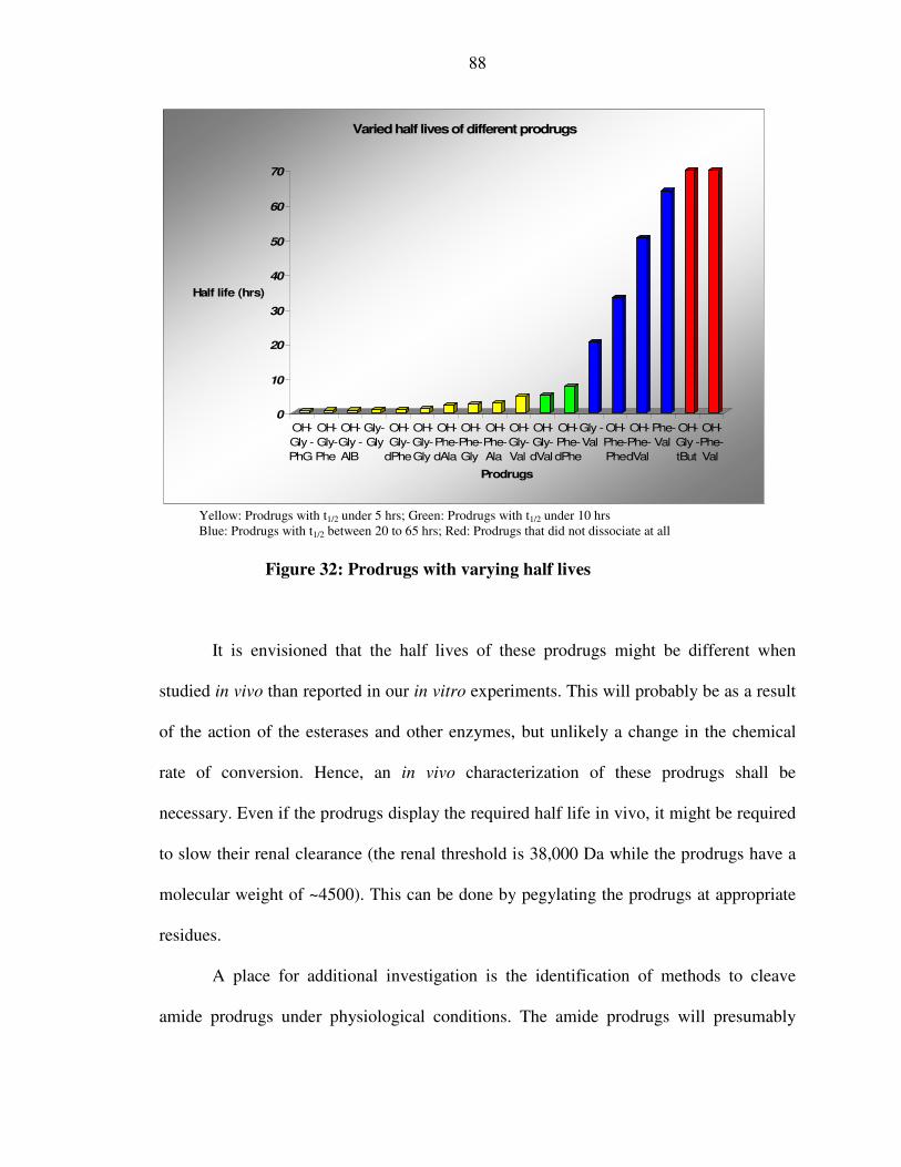
88
0
10
20
30
40
50
60
70
Half life (hrs)
OH-
Gly -
PhG
OH-
Gly-
Phe
OH-
Gly -
AIB
Gly-
Gly
OH-
Gly-
dPhe
OH-
Gly-
Gly
OH-
Phe-
dAla
OH-
Phe-
Gly
OH-
Phe-
Ala
OH-
Gly-
Val
OH-
Gly-
dVal
OH-
Phe-
dPhe
Gly -
Val
OH-
Phe-
Phe
OH-
Phe-
dVal
Phe-
Val
OH-
Gly -
tBut
OH-
Phe-
Val
Prodrugs
Varied half lives of different prodrugs
Yellow: Prodrugs with t1/2 under 5 hrs; Green: Prodrugs with t1/2 under 10 hrs Blue: Prodrugs with t1/2 between 20 to 65 hrs; Red: Prodrugs that did not dissociate at all
Figure 32: Prodrugs with varying half lives
It is envisioned that the half lives of these prodrugs might be different when
studied in vivo than reported in our in vitro experiments. This will probably be as a result
of the action of the esterases and other enzymes, but unlikely a change in the chemical
rate of conversion. Hence, an in vivo characterization of these prodrugs shall be
necessary. Even if the prodrugs display the required half life in vivo, it might be required
to slow their renal clearance (the renal threshold is 38,000 Da while the prodrugs have a
molecular weight of ~4500). This can be done by pegylating the prodrugs at appropriate
residues.
A place for additional investigation is the identification of methods to cleave
amide prodrugs under physiological conditions. The amide prodrugs will presumably
89
have an enhanced half life relative to that of the ester prodrugs. However, the increased
flexibility of using amine-based drugs and their prodrugs to escape what might be
selective degradation by esterases warrants further investigation.
We have observed a number of prodrugs that could be potentially administered on
a ‘once a week’ basis. Even longer acting prodrug candidates might be desired. Those
will be prodrugs that can be administered on a ‘once a month’, or even in a ‘once a year’
formulation. However, it is worth noting that there are many other mechanisms beyond
‘diketopiperazine formation’ by which the prodrugs can be destroyed in vivo. Since
peptides intrinsically decompose in vivo under physiological conditions, there is a limit
set to the “time of action” of a specific peptide-based drug. However, our ability to use
inverted stereochemistry of the dipeptide with no change in rate of chemical cleavage is a
huge advantage in minimizing enzymatic destruction of the prodrug element.
The studies of the structure of additional pro-moieties and how they affect the rate
of conversion will provide additional diversity in prodrug chemistry for future use. This
might then lead to a more effective structure-activity correlation study which can be
subsequently used to synthesize prodrugs of optimal stability and pharmacokinetic
profiles.

90
Bibliography
1) Majumdar, S. K. (2001) Bulletin of the Indian Institute of the History of Medicine
Hyderabad 31(1): 57-70
2) Pendergrass, M. (2007) Nature Endocrinology and Metabolism 3(1): 1
3) http://www.diabetes.org
4) Cryer, P. E. (2007) Nature Endocrinology and Metabolism 3(1): 4-5
5) Onkamo, P., et al. (2000) Diabetologia 43(10): 1334–1336
6) Kishiyama, C. M., Chase, H., and Barker, J. M. (2006) Reviews in Endocrine and
Metabolic Disorders 7(3): 215-224
7) The Diabetes Control and Complications Trial Research Group (1998) Annals of
Internal Medicine 128(7): 517-523.
8) Cavaghan, M.K. et al. (2000) The Journal of Clinical Investigation 106: 329–333
9) Sadikot, S.M., Jaslok Hospital and Research Centre, Mumbai
http://diabetesindia.com/
10) United Kingdom Prospective Diabetes Study (1995) Diabetes 44: 1249–1258
11) http://www.diabetes.org/gestational-diabetes.jsp
12) Banting, F. G., et al. (1922) Canadian Medical Association Journal 12: 141-146.
13) Banting, F. G. and Best, C. H. J. (1922) Journal of Laboratory and Clinical Medicine
7: 464-472.
14) Sanger, F. (1959) Science 129(3359): 1340-1344.
15) Lepore, M., et al. (2000). Diabetes 49(12): 2142-2148.
16) De León, D.D. and Stanley, C.A, (2007) Nature Clinical Practice Endocrinology &
Metabolism 3: 57 – 68

91
17) The Diabetes Control and Complications Trial Research Group. (1993) The New
England Journal of Medicine 329(14): 977-986
18) Nicholas G. A., and Gomez-Caminero, A. (2007) Diabetes, Obesity and Metabolism
9(1): 96-102
19) Johnston, L. W., and Weinstock, R. S. (2006) Mayo Clinic Proceedings 81(12): 1615-
1620
20) Nauck, M.A., et al. (2002) The Journal of Clinical Endocrinology & Metabolism
87(3): 1239-1246
21) Schmidt W. E., Siegel, E. G., and Creutzfeldt, W. (1985) Diabetologia 28(9): 704-
707
22) Green B. D. et al., (2003) Biological Chemistry 384: 1543-1551.
23) Holst, J. J. (1999) Trends in Endocrinology and Metabolism 10: 229 –234
24) McCormack, J.G. (2006) Biochemical Society Transactions 34: 238–242
25) Orskov, C., et al. (1996) Wourld Journal of Gastroenterology 31: 665– 670
26) Daniel, J., et al., (2006) The Lancet 368(9548): 1696-1705
27) Giorgino, F., et al. (2006) Diabetes Research and Clinical Practice 74(2): S152-S155
28) Butler, A. E., et al., (2003) Diabetes 52: 102–110
29) Nauck, M., et al., (1986) Diabetologia 29: 46 –52
30) Drucker, D. J., et al., (1987) Proceedings of the National Academy of Science 84:
3434–3438
31) Holz, G. G., et al. (1999) Journal of Biological Chemistry 274: 14147–14156
32) Gromada, J., et al., (1998) Pflugers Archiv. European Journal of Physiology 435:
583–594
92
33) Weir, G. C. et al., (1989) Diabetes 38: 338–342
34) Rabenhoj, L., et al., (1994) Diabetes 43(4): 535-539
35) Adelhorst, K., et al., (1994) Journal of Biological Chemistry 269(9): 6275-6278
36) Larsen, J., et al., (2001) Diabetes Care 24: 1416–1421
37) Ørskov, C., Wettergren, A., and Holst, J. J. (1993) Diabetes 42: 658–661
38) Deacon, C. F. et al., (1995) Journal of Clinical Endocrinology and Metabolism 80:
952–957
39) Knudsen, L.B., and Pridal, L. (1996) European Journal of Pharmacology 318: 429–
435
40) Holst, J. J., and Deacon, C. F. (1998) Diabetes 47(11): 1663-1670.
41) Åhren, B., et al., (2004) Journal of Clinical Endocrinology and Metabolism 89: 2078–
2084
42) Stuart A. R., Gulve, E. A., and Minghan, W. (2004) Chemical Reviews 104(3): 1255
-1282
43) Thorens, B., et al., (1993) Diabetes 42(11): 1678–1682
44) Eng, J. Et al., (1992) Journal of Biological Chemistry 267(11): 7402-7405
45) Kolterman, O. G., et al., (2005) American Journal of Health System Pharmacy 62:
173–181
46) Kendall, D. M., et al., (2005) Diabetes Care 28: 1083–1091
47) Knudsen, L. B., et al., (2000) Journal of Medicinal Chemistry 43: 1664–1669
48) Bjerre, Knudsen et al., (2005) Diabetes 52(1): 321–322
49) Deacon, C.F. (2004) Diabetes 53: 2181-2189
50) IUPAC Pure & Applied Chemistry (1998) 70(5): 1129–1143
93
51) Albert, A., (1958) Nature 182: 421–423
52) Jiunn, H. L., and Lu, A.Y. H. (1997) Pharmacology Review 49: 403–449
53) Stañczak, A., and Ferra, A. (2006) Pharmacological Reports 58: 599-613
54) Beaumont, K., et al., (2003) Current Drug Metabolism 4: 461-485
55) Humphrey, M. J., and Ringrose, P. S. (1986) Drug Metabolism Reviews 17: 283-310
56) Saffran, M., et al., (1988) Journal of Pharmaceutical Sciences 77: 33-38
57) Lee, S. H. et al., (2005): Bioconjugate Chemistry 16: 377-382
58) Poijarvi-Virta, P. (2006) Current Medicinal Chemistry 13(28): 3441-3465
59) Bundgard, H., and Moss, J. (1990) Pharmaceuical Research 7(9): 885-892
60) Oliyai, R., and Valentino J. S. (1993) Annual Review of Pharmacology and
Toxicology 33: 521-544
61) Bundgard, H., and Moss, J. (1990) Journal of Pharmacy and Pharmacology 42: 7-12
62) Goolcharran, C. and Borchardt, R. T. (1998) Journal of Pharmaceutical Sciences
87(3): 283-288
63) Borchardt, R. T., and Cohen, L. A. (1972) Journal of the American Chemical Society
94: 9166-9174
64) Larsen, S. W., Anderson, M., Larsen, C. (2004) European Journal of Pharmaceutical
Sciences 22(5): 399-408
65) Balvinder, S., et al., (2003) Pharmaceutical Research 20(9): 1381-1388
66) Hamada, Y., et al., (2004) Bioorganic & Medicinal Chemistry 12: 159–170
67) Hamada, Y., et al., (2002) Bioorganic & Medicinal Chemistry 10: 4155–4167
68) Santos, C., et al., (2005) Bioorganic & Medicinal Chemistry Letters 15:1595–1598
69) Hamel, A. R. et al., (2004) Journal of Peptide Research 63: 147-154
94
70) Hamel, A.R., Hubler, F., and Mutter, M., (2005) Journal of Peptide Research 65: 364-
374
71) Suggs, J. W., and Pires, R. M. (1997): Tetrahedron Letters 38: 2227-2230
72) Fischer, P. M. (2003) Journal of Peptide Science 9(1): 9-35
73) Louise, H. N. (1999) Biochemical Pharmacology 58: 749–757
74) Williams, C. (2004) Nature Reviews, Drug Discovery 3: 125-135
75) Schnolzer, M., et al., (1992) Int J Pept Protein Res 40(3-4):180-193
76) “Personal Communications” – with Prof. Phil Dawson (Scripps Research Institute,
San Diego, California)
77) Samantha, E. G., Peter, J. B., and Louise, H. N. (1997) Journal of Biomolecular
Screening 2(4): 235-240
78) Donnelly, D., (2003) Journal of Biological Chemistry 278: 10195-10200
79) Denny, W. A. (2001) Prodrug strategies in cancer therapy. European Journal of
Medicinal Chemistry 36: 577–595
80) Menger, F. M., Ladika, M. (1988) Journal of American Chemical Society 110(20):
6794-6796
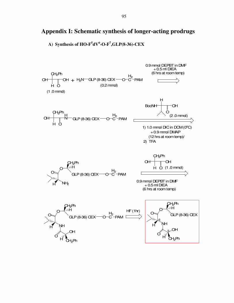
95
Appendix I: Schematic synthesis of longer-acting prodrugs
A) Synthesis of HO-F5dV6-O-F7,GLP(8-36)-CEX
H
OH OH H2N
CH2Ph
OH
H
HN
O
+
0.9mmol DEPBTinDMF+0.5ml DIEA
(6hrsat roomtemp)
O
CH2Ph
GLP(8-36) CEX
O
H
BocNH OH
1) 1.0 mmol DICin DCM(0oC)
+0.9mmol DMAP
(12hrsat roomtemp)/
2) TFA
O
NH2
O HCH2Ph
H
H O
CH2Ph
OH OH
O
NH
O
OH CH2Ph
OH
HCH2Ph
H
OH2C PAM
GLP(8-36) CEX OH2C PAM
O
NH
O
OH CH2Ph
OH
HCH2Ph
H
GLP(8-36) CEX
GLP(8-36) CEX OH2C PAM
GLP(8-36) CEX OH2C PAM
HF(1hr)
0.9mmol DEPBTinDMF+ 0.5ml DIEA
(6 hrs at roomtemp)
(0.2mmol)
5
6
(1 .0mmol)
(1 .0mmol)
(2 .0mmol)
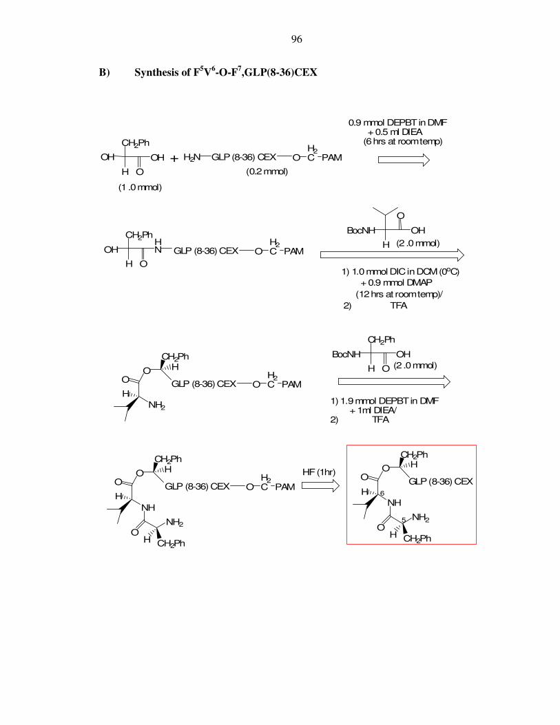
96
B) Synthesis of F5V6-O-F7,GLP(8-36)CEX
H
OH OH H2N
CH2Ph
OH
H
HN
O
+
0.9 mmol DEPBTinDMF+0.5 ml DIEA
(6hrs at roomtemp)
O
CH2Ph
GLP(8-36) CEX
H
O
BocNH OH
1) 1.0mmol DICinDCM(0oC)
+0.9 mmol DMAP
(12hrs at roomtemp)/
2) TFA
O
NH2
H
O HCH2Ph
H O
CH2Ph
BocNH OH
1)1.9mmol DEPBTin DMF+ 1ml DIEA/
2) TFA
O
NH
H
O
OH CH2Ph
NH2
HCH2Ph
OH2C PAM
GLP (8-36)CEX OH2C PAM
O
NH
H
O
OH CH2Ph
NH2
HCH2Ph
GLP(8-36)CEX
GLP (8-36)CEX OH2C PAM
GLP (8-36)CEX OH2C PAM
HF(1hr)
(0.2mmol)
6
5
(1 .0mmol)
(2 .0mmol)
(2 .0mmol)
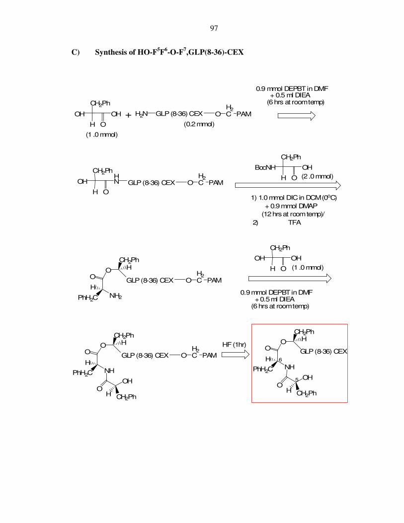
97
C) Synthesis of HO-F5F6-O-F7,GLP(8-36)-CEX
H
OH OH H2N
CH2Ph
OH
H
HN
O
+
0.9 mmol DEPBTinDMF+ 0.5 ml DIEA
(6hrs at roomtemp)
O
CH2Ph
GLP(8-36) CEX
H O
CH2Ph
BocNH OH
1) 1.0mmol DICin DCM(0oC)
+0.9mmol DMAP
(12hrsat roomtemp)/
2) TFA
O
NH2
H
O HCH2Ph
PhH2C
H O
CH2Ph
OH OH
O
NH
H
O
OH CH2Ph
OH
HCH2Ph
PhH2C
OH2C PAM
GLP(8-36) CEX OH2C PAM
O
NH
H
O
OH CH2Ph
OH
HCH2Ph
PhH2C
GLP(8-36) CEX
GLP(8-36) CEX OH2C PAM
GLP(8-36) CEX OH2C PAM
HF(1hr)
0.9mmol DEPBTinDMF+0.5ml DIEA
(6hrsat roomtemp)
(0.2mmol)
6
5
(1 .0mmol)
(1 .0mmol)
(2 .0mmol)

98
D) Synthesis of G5V6-O-F7,GLP(8-36)CEX
CH2Ph
OH
H
HN
O
H
O
BocNH OH
1) 1.0 mmol DICinDCM(0oC)
+ 0.9mmol DMAP
(12hrsat roomtemp)/
2) TFA
O
NH2
H
O HCH2Ph
H O
H
BocNH OH
1) 1.9mmol DEPBTin DMF+1ml DIEA/
2) TFA
O
NH
H
O
OH H
NH2
HCH2Ph
GLP(8-36) CEX OH2C PAM
O
NH
H
O
OH H
NH2
HCH2Ph
GLP(8-36) CEX
GLP(8-36) CEX OH2C PAM
GLP(8-36) CEX OH2C PAM
HF(1hr)
6
5
H
OH OH H2N+
0.9mmol DEPBTinDMF+0.5ml DIEA
(6hrsat roomtemp)
O
CH2Ph
GLP(8-36) CEX OH2C PAM
(0.2mmol)
(1 .0mmol)
(2 .0mmol)
(2 .0mmol)

99
Appendix II: Structure of peptides in Table 2
Serial No Structure
1.
O
N
O
NH2
NH
GLP
HH
H
2.
O
N
O
NH
NH
GLP
H
H
3.
ONH HN
H
N
OO
H
NH2
GLP (8-36)

100
Serial No Structure
4.
O
NH2
H
NH HN
H
N
O
O GLP (8-36)
5.
O
NH
H
NH HN
H
N
GLP (8-36)
6.
O
NH2
H
NH HN
H
N
NH
N
GLP (8-36)
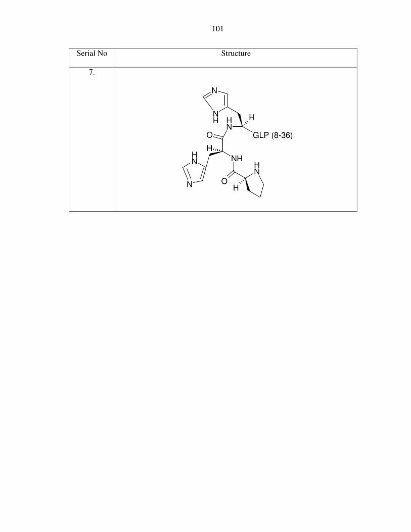
101
Serial No Structure
7.
O
NH
H
NH HN
H
N
NH
N OH
NH
GLP (8-36)
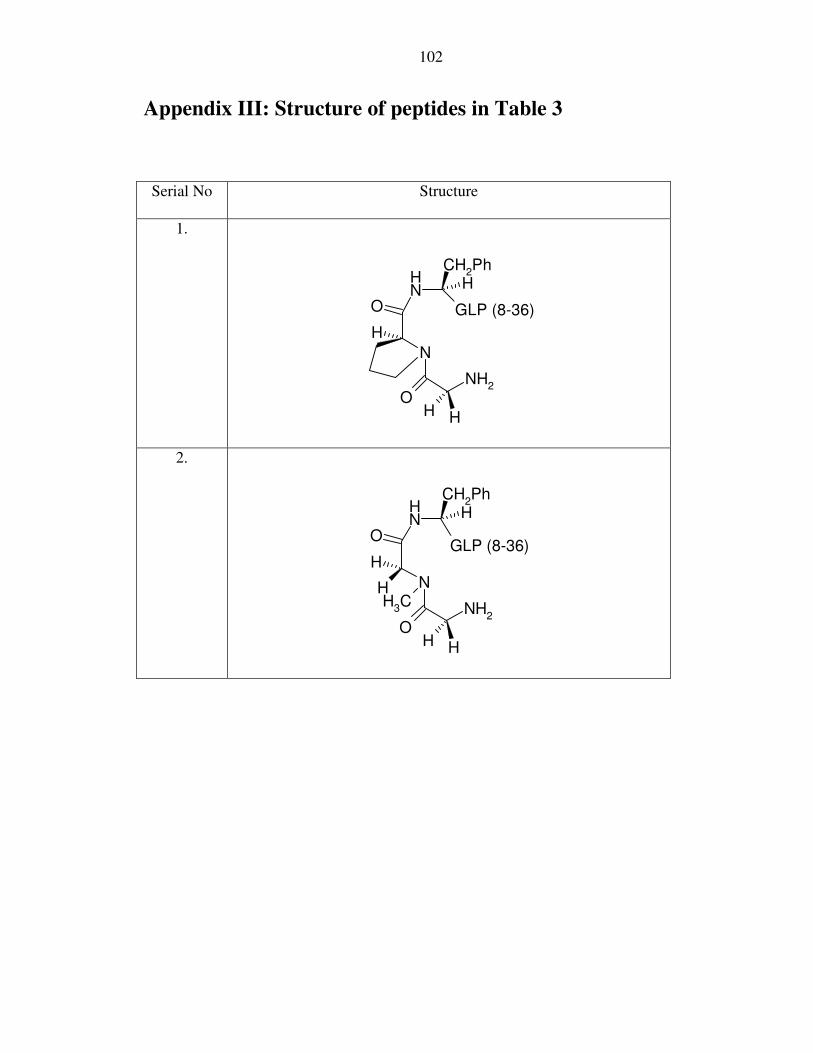
102
Appendix III: Structure of peptides in Table 3
Serial No Structure
1.
O
N
H
NH
OH H
NH2
HCH
2Ph
GLP (8-36)
2.
O
N
H
NH
OH H
NH2
HCH
2Ph
HCH
3
GLP (8-36)

103
Appendix IV: Structure of peptides in Table 4
Serial No Structure
1.
O
NH
H
HN
OH H
NH2
HCH2Ph
H
GLP(8-36) CEX
2.
O
NH
H
HN
OH H
OH
HCH2Ph
H
GLP(8-36) CEX

104
Appendix V: Structure of peptides in Table 5
Serial No Structure
1.
O
NH
H
HN
OH H
NH2
HH
H
GLP(8-36) CEX
2.
O
NH
H
HN
OH H
OH
HH
H
GLP(8-36) CEX
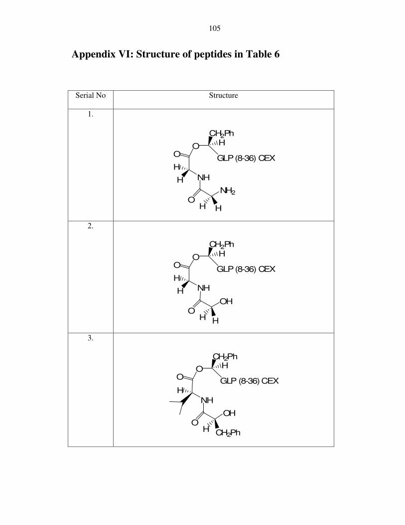
105
Appendix VI: Structure of peptides in Table 6
Serial No Structure
1.
O
NH
H
O
OH
NH2
HCH2Ph
H
GLP(8-36) CEX
H
2.
O
NH
H
O
OH H
OH
HCH2Ph
H
GLP(8-36) CEX
3.
O
NH
H
O
OH CH2Ph
OH
HCH2Ph
GLP (8-36)CEX
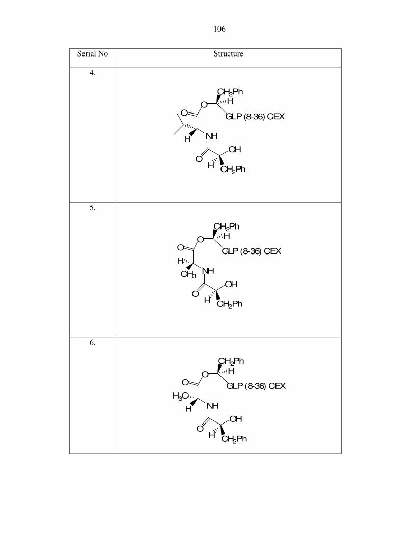
106
Serial No Structure
4.
O
NH
O
OH CH2Ph
OH
HCH2Ph
H
GLP(8-36) CEX
5.
O
NH
H
O
OH CH2Ph
OH
HCH2Ph
CH3
GLP(8-36) CEX
6.
O
NH
O
OH CH2Ph
OH
HCH2Ph
H
H3CGLP(8-36) CEX
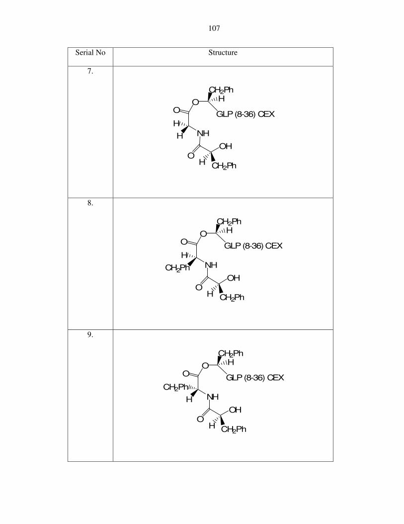
107
Serial No Structure
7.
O
NH
H
O
OH CH2Ph
OH
HCH2Ph
H
GLP(8-36) CEX
8.
O
NH
H
O
OH CH2Ph
OH
HCH2Ph
CH2Ph
GLP(8-36)CEX
9.
O
NH
O
OH CH2Ph
OH
HCH2Ph
H
CH2PhGLP(8-36) CEX
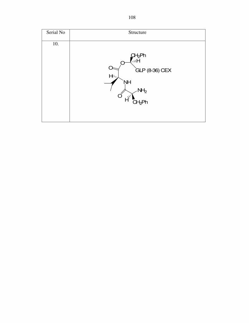
108
Serial No Structure
10.
O
NH
H
O
OH CH2Ph
NH2
HCH2Ph
GLP (8-36)CEX

109
Appendix VII: Structure of peptides in Table 7
Serial No Structure
1.
O
NH
H
O
OH H
NH2
HCH2Ph
H
GLP(8-36) CEX
2.
O
NH
H
O
OH H
OH
HCH2Ph
H
GLP(8-36) CEX
3.
O
NH
H
O
OH H
OH
HCH2Ph
GLP(8-36) CEX

110
Serial No Structure
4.
O
NH
O
OH H
OH
HCH2Ph
H
GLP(8-36)CEX
5.
O
NH
H3C
O
OH H
OH
HCH2Ph
CH3
GLP(8-36) CEX
6.
O
NH
H
O
OH H
OH
HCH2Ph
GLP(8-36)CEX
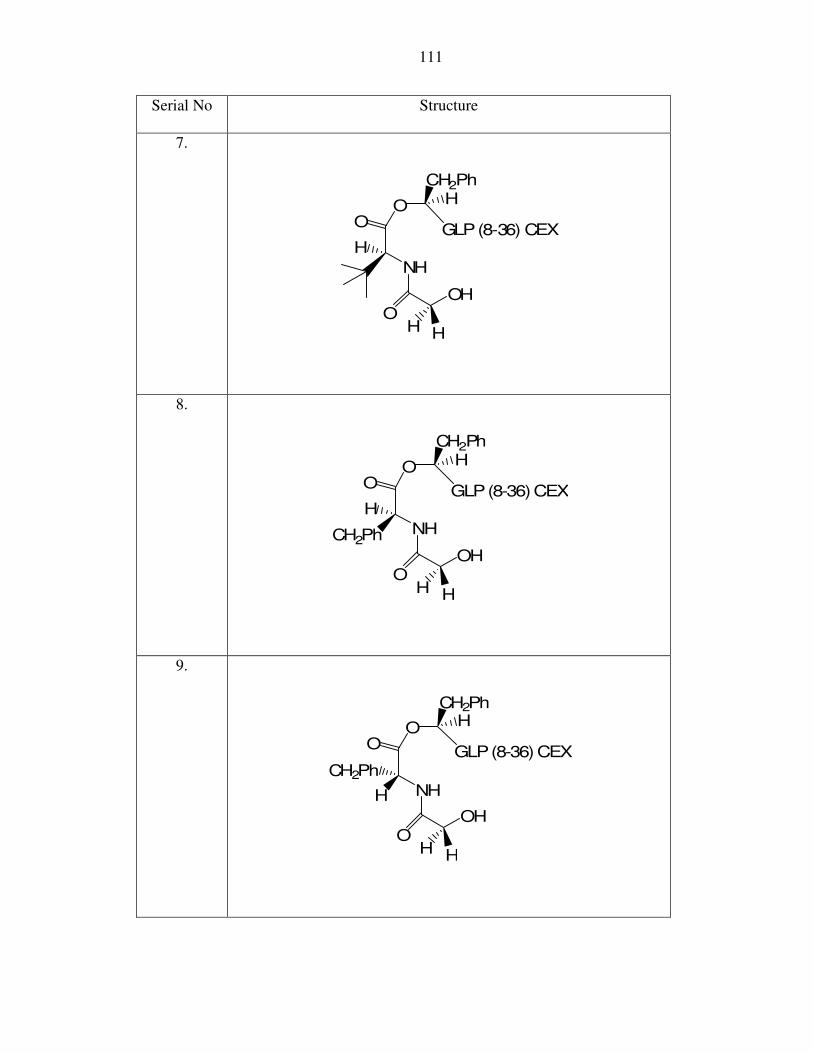
111
Serial No Structure
7.
O
NH
H
O
OH H
OH
HCH2Ph
GLP(8-36) CEX
8.
O
NH
H
O
OH H
OH
HCH2Ph
CH2Ph
GLP(8-36)CEX
9.
O
NH
CH2Ph
O
OH H
OH
HCH2Ph
H
GLP(8-36) CEX

112
Serial No Structure
10.
O
NH
H
O
OH H
NH2
HCH2Ph
GLP(8-36) CEX
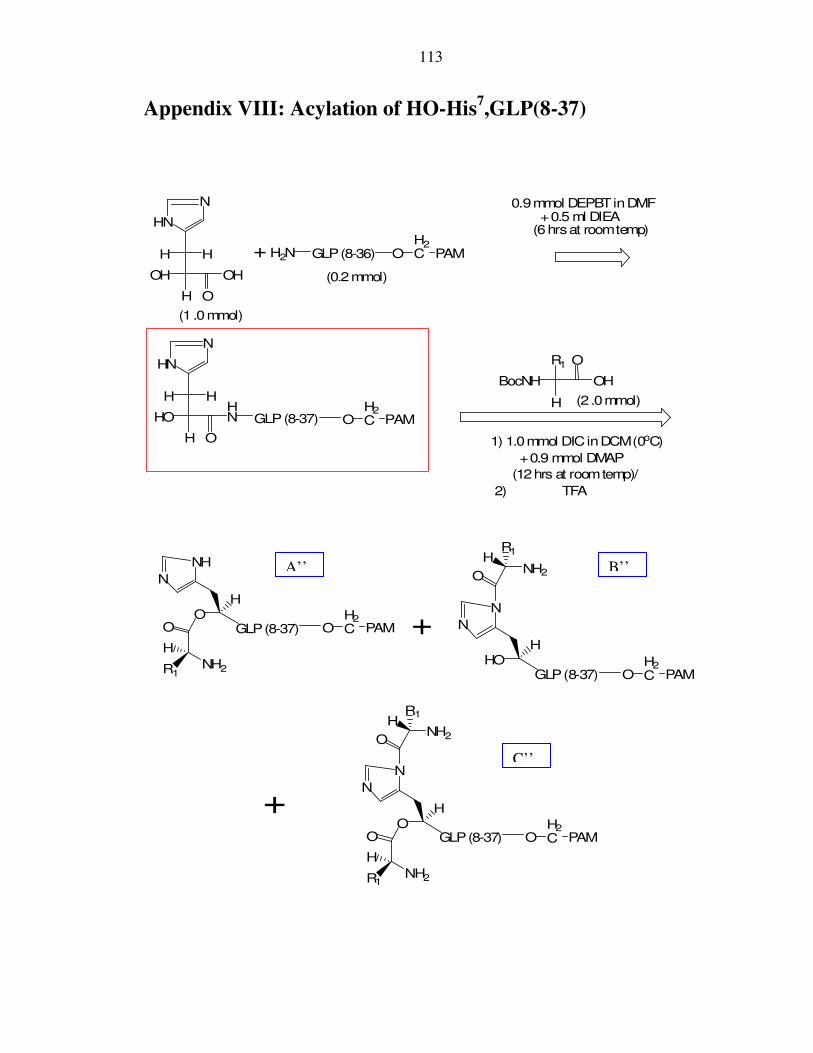
113
Appendix VIII: Acylation of HO-His7,GLP(8-37)
H
OH OH
H2N
HN
+
0.9mmol DEPBTinDMF+0.5 ml DIEA
(6hrsat roomtemp)
O
GLP(8-36)
H
OR1
BocNH OH
1) 1.0mmol DICinDCM(0oC)
+0.9 mmol DMAP
(12hrs at roomtemp)/
2) TFA
OH2C PAM
GLP(8-37) OH2C PAM
H H
HN
N
H
HO
O
H H
HN
N
O
NH2
H
OH
GLP(8-37)
R1
N
N
O
HR1
NH2
O
NH2
H
OH
GLP(8-37)
R1
N
NH
HOH
GLP(8-37)
N
N
O
HR1
NH2
OH2C PAM
OH2C PAM
OH2C PAM
+
+
(0.2mmol)
(1 .0mmol)
(2 .0mmol)
A’’ B’’
C’’
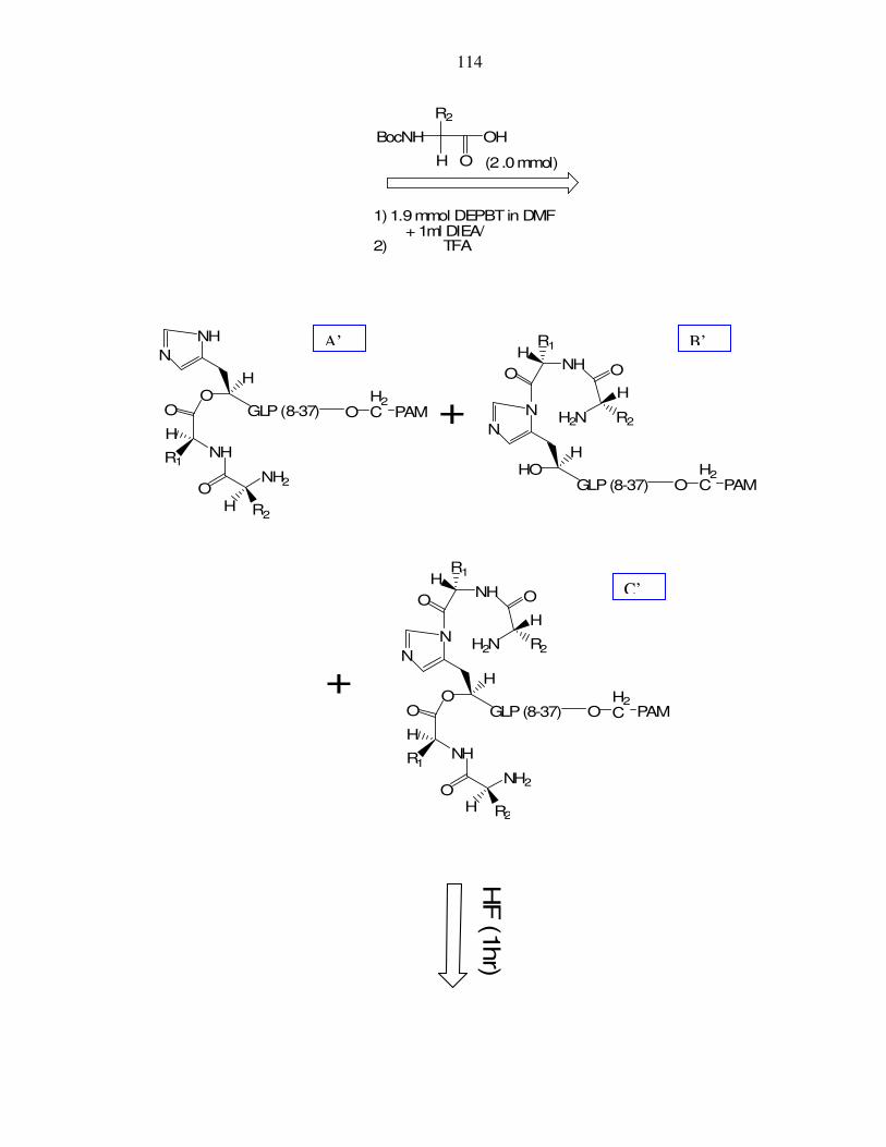
114
H O
R2
BocNH OH
1)1.9mmol DEPBTin DMF+ 1ml DIEA/
2) TFA
O
NH
H
OH
GLP(8-37)
R1
N
N
O
HR1
NH
OH R2
NH2
O
H
R2H2N
O
NH
H
OH
GLP(8-37)
R1
N
NH
OH R2
NH2
OH2C PAM
OH2C PAM
HOH
GLP(8-37)
N
N
O
HR1
NH O
H
R2H2N
OH2C PAM
+
+
(2 .0mmol)
HF
(1hr)
A’ B’
C’

115
O
NH
H
OH
GLP(8-37)
R1
N
N
O
HR1
NH
OH R2
NH2
O
H
R2H2N
O
NH
H
O
H
GLP(8-37)
R1
N
NH
OH R2
NH2
HO
H
GLP(8-37)
N
N
O
HR1
NH O
H
R2H2N+
+
C
A B

116
Appendix IX: Synthesis of (H7F),(E9Q),(T11S),GLP(8-36)-
CEX
FTSDVSSYLEGQAAKEFIAWLVKGR-CEX OH2C PAM
H2NFTSDVSSYLEGQAAKEFIAWLVKGR-CEX
H
OH2C PAM
H2N
H O
CH2O-t-butyl
FmocNH OH
i) 1.9mmol DEPBTin DMF+ 1ml DIEA/
ii) 20%piperidine inDMF
O
1)
2) 2 .0mmol Fmoc-Gly i)1.9mmol DEPBTinDMF+ 1ml DIEA/
ii) 20%piperidine inDMF
3) 2 .0mmol Fmoc-Gln i)1.9mmol DEPBTinDMF+ 1ml DIEA/
ii) 20%piperidine inDMF
4) 2 .0mmol Fmoc-Ala i) 1.9mmol DEPBTinDMF+ 1ml DIEA/
ii) 20%piperidine inDMF
5) 2 .0mmol Fmoc-Phe i)1.9mmol DEPBTinDMF+ 1ml DIEA/
(0.2mmol)
H
H
t-butyl-O
OH2C PAM
butyl-t-O
HN
FTSDVSSYLEGQAAKEFIAWLVKGR-CEX
HFAQG
O
H
H
FmocHN
(2 .0mmol)

117
HO
HN
FTSDVSSYLEGQAAKEFIAWLVKGR-CEX
HFAQG
O
H
H
1) TFAfor 2hrs
2) 20%piperidine
OH2C PAM
HFcleavage
HN
FTSDVSSYLEGQAAKEFIAWLVKGR-CEX
HF7AQ9G
HO H
H
O
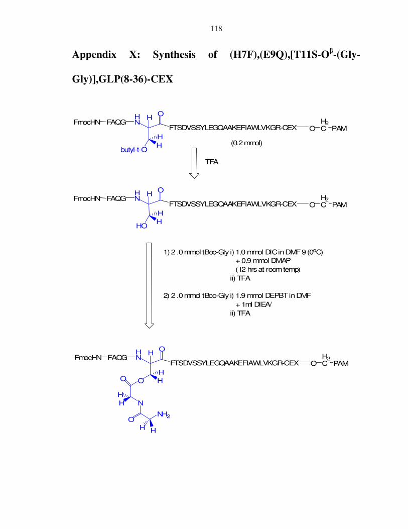
118
Appendix X: Synthesis of (H7F),(E9Q),[T11S-Oβ-(Gly-
Gly)],GLP(8-36)-CEX
1)2 .0mmol tBoc-Gly i)1.0 mmol DICinDMF9(0oC)
+ 0.9 mmol DMAP
(12 hrsat roomtemp)
ii) TFA
2) 2 .0mmol tBoc-Gly i) 1.9 mmol DEPBTinDMF
+ 1ml DIEA/
ii) TFA
OH2C PAM
butyl-t-O
HN
FTSDVSSYLEGQAAKEFIAWLVKGR-CEX
HFAQG
O
H
H
FmocHN
TFA
OH2C PAM
HO
HN
FTSDVSSYLEGQAAKEFIAWLVKGR-CEX
HFAQG
O
H
H
FmocHN
HN
FTSDVSSYLEGQAAKEFIAWLVKGR-CEX
HFAQG
OO
N
NH2O
H
H
H
H
H
H
O
FmocHNO
H2C PAM
(0.2mmol)

119
20% piperidine
HN
FTSDVSSYLEGQAAKEFIAWLVKGR-CEX
HFAQG
OO
N
NH2O
H
H
H
H
H
H
O
H2NO
H2C PAM
HF cleavage
HN
FTSDVSSYLEGQAAKEFIAWLVKGR-CEX
HF7AQ9G
OO
N
NH2O
H
H
H
H
H
H
O
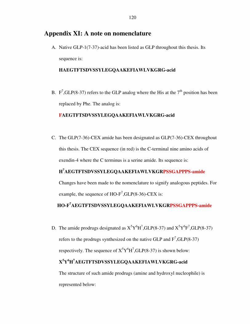
120
Appendix XI: A note on nomenclature
A. Native GLP-1(7-37)-acid has been listed as GLP throughout this thesis. Its
sequence is:
HAEGTFTSDVSSYLEGQAAKEFIAWLVKGRG-acid
B. F7,GLP(8-37) refers to the GLP analog where the His at the 7th position has been
replaced by Phe. The analog is:
FAEGTFTSDVSSYLEGQAAKEFIAWLVKGRG-acid
C. The GLP(7-36)-CEX amide has been designated as GLP(7-36)-CEX throughout
this thesis. The CEX sequence (in red) is the C-terminal nine amino acids of
exendin-4 where the C terminus is a serine amide. Its sequence is:
H7AEGTFTSDVSSYLEGQAAKEFIAWLVKGRPSSGAPPPS-amide
Changes have been made to the nomenclature to signify analogous peptides. For
example, the sequence of HO-F7,GLP(8-36)-CEX is:
HO-F7AEGTFTSDVSSYLEGQAAKEFIAWLVKGRPSSGAPPPS-amide
D. The amide prodrugs designated as X5Y6H7,GLP(8-37) and X5Y6F7,GLP(8-37)
refers to the prodrugs synthesized on the native GLP and F7,GLP(8-37)
respectively. The sequence of X5Y6H7,GLP(8-37) is shown below:
X5Y6H7AEGTFTSDVSSYLEGQAAKEFIAWLVKGRG-acid
The structure of such amide prodrugs (amine and hydroxyl nucleophile) is
represented below:

121
O
NH
O
NH2/OH
HN H7AEGTFTSDVSSYLEGQAAKEFIAWLVKGRG-acid
H
R6
HR5
6
5
E. The ester prodrugs are designated as X5Y6-O-F7,GLP(8-36)-CEX with an amine
nucleophile:
X5Y6F7AEGTFTSDVSSYLEGQAAKEFIAWLVKGRPSSGAPPPS-amide
and HO-X5Y6-O-F7,GLP(8-36)-CEX with a hydroxyl nucleophile respectively.
HO-X5Y6-O-F7AEGTFTSDVSSYLEGQAAKEFIAWLVKGRPSSGAPPPS-amide
The structure of such ester prodrugs (amine and hydroxyl nucleophile) is
represented below:
6
5
O
NH
O
NH2/OH
O F7AEGTFTSDVSSYLEGQAAKEFIAWLVKGRPSSGAPPPS-amide
H
R6
HR5
F. The preliminary peptides for the synthesis of the internal ester prodrugs are
(H7F),(E9Q),GLP(8-36)-CEX and (H7F),(E9Q),(T11S),GLP(8-36)-CEX. The

122
mutations from the native sequence are represented in parenthesis and shown in
red. The sequence of (H7F),(E9Q),GLP(8-36)-CEX is :
F7AQ9GTFTSDVSSYLEGQAAKEFIAWLVKGRPSSGAPPPS-amide
and the sequence of (H7F),(E9Q),(T11S),GLP(8-36)-CEX is:
F7AQ9GS11FTSDVSSYLEGQAAKEFIAWLVKGRPSSGAPPPS-amide
The structure of (H7F),(E9Q),(T11S),GLP(8-36)-CEX is given by:
OHN
FTSDVSSYLEGQAAKEFIAWLVKGRPSSGAPPPS-amide
HF7AQ9G
HO H
H
G. The internal ester prodrug designated as (H7F),(E9Q),[T11S-Oβ-(Gly-
Gly)],GLP(8-36)-CEX was synthesized from the serine at the 11th position of
(H7F),(E9Q),(T11S),GLP(8-36)-CEX. The (Gly-Gly) dipeptide pro-moiety
(shown in red) was added to the S11. The sequence of (H7F),(E9Q),[T11S-Oβ-
(Gly-Gly)],GLP(8-36)-CEX is given by:
F7AQ9GS11FTSDVSSYLEGQAAKEFIAWLVKGRPSSGAPPPS-amide
GG
The structure of (H7F),(E9Q),[T11S-Oβ-(Gly-Gly)],GLP(8-36)-CEX is
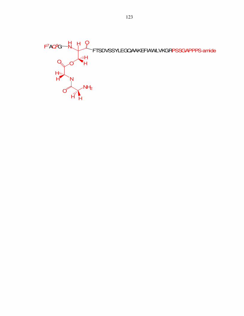
123
OHN
FTSDVSSYLEGQAAKEFIAWLVKGRPSSGAPPPS-amide
HF7AQ9G
OO
N
NH2O
H
H
H
H
H
H