A Novel Tumor-Activated Prodrug Strategy Targeting Ferrous ...
10
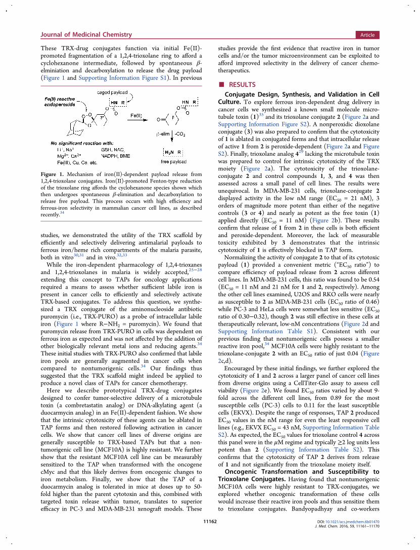
A Novel Tumor-Activated Prodrug Strategy Targeting Ferrous Iron Is Effective in Multiple Preclinical Cancer Models Benjamin Spangler, †,‡ Shaun D. Fontaine, ‡,∇ Yihui Shi, § Lidia Sambucetti, § Aras N. Mattis, ∥ Byron Hann, ⊥ James A. Wells, ‡,# and Adam R. Renslo* ,‡ † Graduate Program in Chemistry and Chemical Biology, University of CaliforniaSan Francisco, San Francisco, California 94158, United States ‡ Department of Pharmaceutical Chemistry, University of CaliforniaSan Francisco, San Francisco, California 94158, United States § SRI International, Menlo Park, California 94025-3493, United States ∥ Department of Pathology, University of CaliforniaSan Francisco, San Francisco, California 94158, United States ⊥ Preclinical Therapeutic Core, University of CaliforniaSan Francisco, San Francisco, California 94158, United States # Department of Cellular and Molecular Pharmacology, University of CaliforniaSan Francisco, San Francisco, California 94158, United States * S Supporting Information ABSTRACT: Here we describe a new approach for tumor targeting in which augmented concentrations of Fe(II) in cancer cells and/or the tumor microenvironment triggers drug release from an Fe(II)-reactive prodrug conjugate. The 1,2,4- trioxolane scaffold developed to enable this approach can in principle be applied to a broad range of cancer therapeutics and is illustrated here with Fe(II)-targeted forms of a microtubule toxin and a duocarmycin-class DNA-alkylating agent. We show that the intrinsic reactivity/toxicity of the duocarmycin analog is masked in the conjugated form and this greatly reduced toxicity in mice. This in turn permitted elevated dosing levels, leading to higher systemic exposure and a significantly improved response in tumor xenograft models. Overall our results suggest that Fe(II)-dependent drug delivery via trioxolane conjugates could have significant utility in expanding the therapeutic index of a range of clinical and preclinical stage cancer chemotherapeutics. ■ INTRODUCTION The recent development of molecularly targeted cancer therapeutics has been accompanied by renewed interest in technologies for the tumor/cell-selective delivery of potent but intrinsically nonselective cytotoxic agents. These technologies include antibody−drug conjugates (ADCs) that recognize cell- surface antigens and tumor-activated prodrugs (TAPs) that exploit nutrient transporters 1−3 or differences in hypoxia associated with the tumor microenvironment. 4,5 Accumulating evidence suggests that an increase in reactive, “labile” intracellular iron is another metabolic signature of cancer, as recently reviewed. 6 Tumor targeting strategies designed to exploit changes in iron homeostasis remain largely unexplored however, despite clinical precedent for iron-dependent pharmacology in antimalarial therapy with artemisinins. 7,8 Redox cycling of iron in enzyme cofactors is essential for cellular processes ranging from de novo nucleotide synthesis to the maintenance of genomic stability, cell cycle regulation, and mitochondrial respiration. 6 However, when unbound and unregulated, redox active iron promotes the disproportionation of hydrogen peroxide (Fenton reaction) to produce hydroxyl and hydroperoxyl radicals, reactive oxygen species (ROS) that confer cellular damage and can lead to apoptosis or ferroptosis. 9−11 Iron homeostasis is therefore highly regulated to ensure sufficient labile iron is available to support essential enzyme function while limiting exposure to unbound, redox active iron species. 12−14 Rapidly proliferating cells have increased requirements for DNA synthesis, repair, and mitochondrial respiration and therefore have increased demands for iron cofactor biosyn- thesis. Accordingly, iron acquisition and export pathways are altered in many cancers so as to increase the labile iron pool. 6,15−18 Furthermore, iron has been shown to contribute to tumor initiation and growth 15,19 and epidemiological evidence has established links between tumor iron metabolism and clinical outcomes in breast cancer patients. 17,20 Given that labile Fe(II) promotes Fenton chemistry, we sought to develop a tumor-targeting strategy in which Fenton reaction of a peroxidic prodrug was coupled to release of drug payloads. Recognizing that antimalarial agents such as arterolane 21−24 exhibit finely tuned iron(II) reactivity, 25−28 we subsequently developed 29,30 an arterolane-inspired small molecule platform (denoted TRX herein) for Fe(II)-dependent drug delivery. Received: October 7, 2016 Published: November 21, 2016 Article pubs.acs.org/jmc © 2016 American Chemical Society 11161 DOI: 10.1021/acs.jmedchem.6b01470 J. Med. Chem. 2016, 59, 11161−11170 This is an open access article published under an ACS AuthorChoice License, which permits copying and redistribution of the article or any adaptations for non-commercial purposes.
-
Upload
khangminh22 -
Category
Documents
-
view
0 -
download
0
Transcript of A Novel Tumor-Activated Prodrug Strategy Targeting Ferrous ...
A Novel Tumor-Activated Prodrug Strategy Targeting Ferrous Iron IsEffective in Multiple Preclinical Cancer ModelsBenjamin Spangler,†,‡ Shaun D. Fontaine,‡,∇ Yihui Shi,§ Lidia Sambucetti,§ Aras N. Mattis,∥ Byron Hann,⊥
James A. Wells,‡,# and Adam R. Renslo*,‡
†Graduate Program in Chemistry and Chemical Biology, University of CaliforniaSan Francisco, San Francisco, California 94158,United States‡Department of Pharmaceutical Chemistry, University of CaliforniaSan Francisco, San Francisco, California 94158, United States§SRI International, Menlo Park, California 94025-3493, United States∥Department of Pathology, University of CaliforniaSan Francisco, San Francisco, California 94158, United States⊥Preclinical Therapeutic Core, University of CaliforniaSan Francisco, San Francisco, California 94158, United States#Department of Cellular and Molecular Pharmacology, University of CaliforniaSan Francisco, San Francisco, California 94158,United States
*S Supporting Information
ABSTRACT: Here we describe a new approach for tumortargeting in which augmented concentrations of Fe(II) incancer cells and/or the tumor microenvironment triggers drugrelease from an Fe(II)-reactive prodrug conjugate. The 1,2,4-trioxolane scaffold developed to enable this approach can inprinciple be applied to a broad range of cancer therapeuticsand is illustrated here with Fe(II)-targeted forms of amicrotubule toxin and a duocarmycin-class DNA-alkylatingagent. We show that the intrinsic reactivity/toxicity of the duocarmycin analog is masked in the conjugated form and this greatlyreduced toxicity in mice. This in turn permitted elevated dosing levels, leading to higher systemic exposure and a significantlyimproved response in tumor xenograft models. Overall our results suggest that Fe(II)-dependent drug delivery via trioxolaneconjugates could have significant utility in expanding the therapeutic index of a range of clinical and preclinical stage cancerchemotherapeutics.
■ INTRODUCTIONThe recent development of molecularly targeted cancertherapeutics has been accompanied by renewed interest intechnologies for the tumor/cell-selective delivery of potent butintrinsically nonselective cytotoxic agents. These technologiesinclude antibody−drug conjugates (ADCs) that recognize cell-surface antigens and tumor-activated prodrugs (TAPs) thatexploit nutrient transporters1−3 or differences in hypoxiaassociated with the tumor microenvironment.4,5 Accumulatingevidence suggests that an increase in reactive, “labile”intracellular iron is another metabolic signature of cancer, asrecently reviewed.6 Tumor targeting strategies designed toexploit changes in iron homeostasis remain largely unexploredhowever, despite clinical precedent for iron-dependentpharmacology in antimalarial therapy with artemisinins.7,8
Redox cycling of iron in enzyme cofactors is essential forcellular processes ranging from de novo nucleotide synthesis tothe maintenance of genomic stability, cell cycle regulation, andmitochondrial respiration.6 However, when unbound andunregulated, redox active iron promotes the disproportionationof hydrogen peroxide (Fenton reaction) to produce hydroxyland hydroperoxyl radicals, reactive oxygen species (ROS) thatconfer cellular damage and can lead to apoptosis or
ferroptosis.9−11 Iron homeostasis is therefore highly regulatedto ensure sufficient labile iron is available to support essentialenzyme function while limiting exposure to unbound, redoxactive iron species.12−14
Rapidly proliferating cells have increased requirements forDNA synthesis, repair, and mitochondrial respiration andtherefore have increased demands for iron cofactor biosyn-thesis. Accordingly, iron acquisition and export pathways arealtered in many cancers so as to increase the labile ironpool.6,15−18 Furthermore, iron has been shown to contribute totumor initiation and growth15,19 and epidemiological evidencehas established links between tumor iron metabolism andclinical outcomes in breast cancer patients.17,20 Given that labileFe(II) promotes Fenton chemistry, we sought to develop atumor-targeting strategy in which Fenton reaction of aperoxidic prodrug was coupled to release of drug payloads.Recognizing that antimalarial agents such as arterolane21−24
exhibit finely tuned iron(II) reactivity,25−28 we subsequentlydeveloped29,30 an arterolane-inspired small molecule platform(denoted TRX herein) for Fe(II)-dependent drug delivery.
Received: October 7, 2016Published: November 21, 2016
Article
pubs.acs.org/jmc
© 2016 American Chemical Society 11161 DOI: 10.1021/acs.jmedchem.6b01470J. Med. Chem. 2016, 59, 11161−11170
This is an open access article published under an ACS AuthorChoice License, which permitscopying and redistribution of the article or any adaptations for non-commercial purposes.
These TRX-drug conjugates function via initial Fe(II)-promoted fragmentation of a 1,2,4-trioxolane ring to afford acyclohexanone intermediate, followed by spontaneous β-elminiation and decarboxylation to release the drug payload(Figure 1 and Supporting Information Figure S1). In previous
studies, we demonstrated the utility of the TRX scaffold byefficiently and selectively delivering antimalarial payloads toferrous iron/heme rich compartments of the malaria parasite,both in vitro30,31 and in vivo.32,33
While the iron-dependent pharmacology of 1,2,4-trioxanesand 1,2,4-trioxolanes in malaria is widely accepted,25−28
extending this concept to TAPs for oncology applicationsrequired a means to assess whether sufficient labile iron ispresent in cancer cells to efficiently and selectively activateTRX-based conjugates. To address this question, we synthe-sized a TRX conjugate of the aminonucleoside antibioticpuromycin (i.e., TRX-PURO) as a probe of intracellular labileiron (Figure 1 where R−NH2 = puromycin). We found thatpuromycin release from TRX-PURO in cells was dependent onferrous iron as expected and was not affected by the addition ofother biologically relevant metal ions and reducing agents.34
These initial studies with TRX-PURO also confirmed that labileiron pools are generally augmented in cancer cells whencompared to nontumorigenic cells.34 Our findings thussuggested that the TRX scaffold might indeed be applied toproduce a novel class of TAPs for cancer chemotherapy.Here we describe prototypical TRX-drug conjugates
designed to confer tumor-selective delivery of a microtubuletoxin (a combretastatin analog) or DNA-alkylating agent (aduocarmycin analog) in an Fe(II)-dependent fashion. We showthat the intrinsic cytotoxicity of these agents can be ablated inTAP forms and then restored following activation in cancercells. We show that cancer cell lines of diverse origins aregenerally susceptible to TRX-based TAPs but that a non-tumorigenic cell line (MCF10A) is highly resistant. We furthershow that the resistant MCF10A cell line can be measurablysensitized to the TAP when transformed with the oncogenecMyc and that this likely derives from oncogenic changes toiron metabolism. Finally, we show that the TAP of aduocarmycin analog is tolerated in mice at doses up to 50-fold higher than the parent cytotoxin and this, combined withtargeted toxin release within tumor, translates to superiorefficacy in PC-3 and MDA-MB-231 xenograft models. These
studies provide the first evidence that reactive iron in tumorcells and/or the tumor microenvironment can be exploited toafford improved selectivity in the delivery of cancer chemo-therapeutics.
■ RESULTSConjugate Design, Synthesis, and Validation in Cell
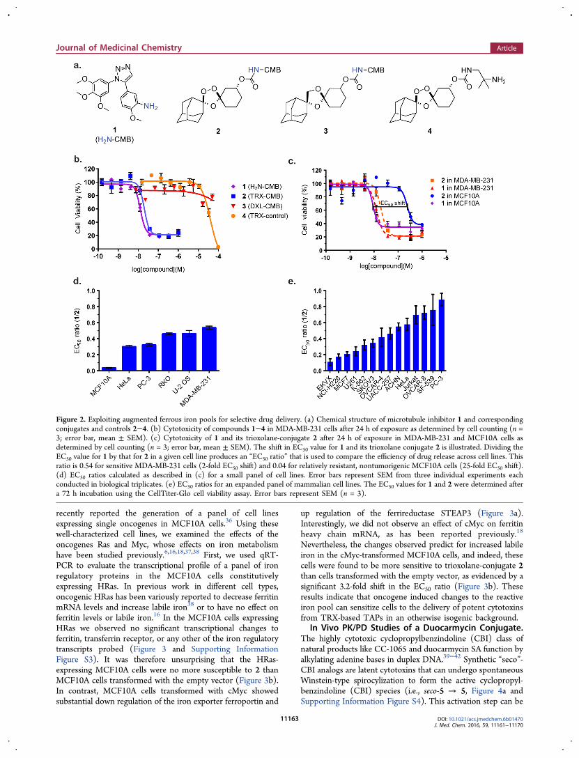
Culture. To explore ferrous iron-dependent drug delivery incancer cells we synthesized a known small molecule micro-tubule toxin (1)35 and its trioxolane conjugate 2 (Figure 2a andSupporting Information Figure S2). A nonperoxidic dioxolaneconjugate (3) was also prepared to confirm that the cytotoxicityof 1 is ablated in conjugated forms and that intracellular releaseof active 1 from 2 is peroxide-dependent (Figure 2a and FigureS2). Finally, trioxolane analog 429 lacking the microtubule toxinwas prepared to control for intrinsic cytotoxicity of the TRXmoiety (Figure 2a). The cytotoxicity of the trioxolane-conjugate 2 and control compounds 1, 3, and 4 was thenassessed across a small panel of cell lines. The results wereunequivocal. In MDA-MB-231 cells, trioxolane-conjugate 2displayed activity in the low nM range (EC50 = 21 nM), 3orders of magnitude more potent than either of the negativecontrols (3 or 4) and nearly as potent as the free toxin (1)applied directly (EC50 = 11 nM) (Figure 2b). These resultsconfirm that release of 1 from 2 in these cells is both efficientand peroxide-dependent. Moreover, the lack of measurabletoxicity exhibited by 3 demonstrates that the intrinsiccytotoxicity of 1 is effectively blocked in TAP form.Normalizing the activity of conjugate 2 to that of its cytotoxic
payload (1) provided a convenient metric (“EC50 ratio”) tocompare efficiency of payload release from 2 across differentcell lines. In MDA-MB-231 cells, this ratio was found to be 0.54(EC50 = 11 nM and 21 nM for 1 and 2, respectively). Amongthe other cell lines examined, U2OS and RKO cells were nearlyas susceptible to 2 as MDA-MB-231 cells (EC50 ratio of 0.46)while PC-3 and HeLa cells were somewhat less sensitive (EC50ratio of 0.30−0.32), though 2 was still effective in these cells attherapeutically relevant, low-nM concentrations (Figure 2d andSupporting Information Table S1). Consistent with ourprevious finding that nontumorigenic cells possess a smallerreactive iron pool,34 MCF10A cells were highly resistant to thetrioxolane-conjugate 2 with an EC50 ratio of just 0.04 (Figure2c,d).Encouraged by these initial findings, we further explored the
cytotoxicity of 1 and 2 across a larger panel of cancer cell linesfrom diverse origins using a CellTiter-Glo assay to assess cellviability (Figure 2e). We found EC50 ratios varied by about 9-fold across the different cell lines, from 0.89 for the mostsusceptible cells (PC-3) cells to 0.11 for the least susceptiblecells (EKVX). Despite the range of responses, TAP 2 producedEC50 values in the nM range for even the least responsive celllines (e.g., EKVX EC50 = 43 nM, Supporting Information TableS2). As expected, the EC50 values for trioxolane control 4 acrossthis panel were in the μM regime and typically ≥2 log units lesspotent than 2 (Supporting Information Table S2). Thisconfirms that the cytotoxicity of TAP 2 derives from releaseof 1 and not significantly from the trioxolane moiety itself.
Oncogenic Transformation and Susceptibility toTrioxolane Conjugates. Having found that nontumorigenicMCF10A cells were highly resistant to TRX-conjugates, weexplored whether oncogenic transformation of these cellswould increase their reactive iron pools and thus sensitize themto trioxolane conjugates. Bandyopadhyay and co-workers
Figure 1. Mechanism of iron(II)-dependent payload release from1,2,4-trioxolane conjugates. Iron(II)-promoted Fenton-type reductionof the trioxolane ring affords the cyclohexanone species shown whichthen undergoes spontaneous β-elimination and decarboxylation torelease free payload. This process occurs with high efficiency andferrous-iron selectivity in mammalian cancer cell lines, as describedrecently.34
Journal of Medicinal Chemistry Article
DOI: 10.1021/acs.jmedchem.6b01470J. Med. Chem. 2016, 59, 11161−11170
11162
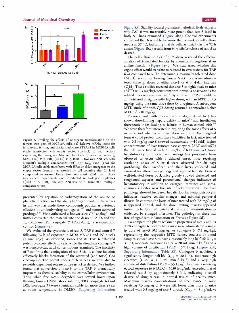
recently reported the generation of a panel of cell linesexpressing single oncogenes in MCF10A cells.36 Using thesewell-characterized cell lines, we examined the effects of theoncogenes Ras and Myc, whose effects on iron metabolismhave been studied previously.6,16,18,37,38 First, we used qRT-PCR to evaluate the transcriptional profile of a panel of ironregulatory proteins in the MCF10A cells constitutivelyexpressing HRas. In previous work in different cell types,oncogenic HRas has been variously reported to decrease ferritinmRNA levels and increase labile iron38 or to have no effect onferritin levels or labile iron.16 In the MCF10A cells expressingHRas we observed no significant transcriptional changes toferritin, transferrin receptor, or any other of the iron regulatorytranscripts probed (Figure 3 and Supporting InformationFigure S3). It was therefore unsurprising that the HRas-expressing MCF10A cells were no more susceptible to 2 thanMCF10A cells transformed with the empty vector (Figure 3b).In contrast, MCF10A cells transformed with cMyc showedsubstantial down regulation of the iron exporter ferroportin and
up regulation of the ferrireductase STEAP3 (Figure 3a).Interestingly, we did not observe an effect of cMyc on ferritinheavy chain mRNA, as has been reported previously.18
Nevertheless, the changes observed predict for increased labileiron in the cMyc-transformed MCF10A cells, and indeed, thesecells were found to be more sensitive to trioxolane-conjugate 2than cells transformed with the empty vector, as evidenced by asignificant 3.2-fold shift in the EC50 ratio (Figure 3b). Theseresults indicate that oncogene induced changes to the reactiveiron pool can sensitize cells to the delivery of potent cytotoxinsfrom TRX-based TAPs in an otherwise isogenic background.
In Vivo PK/PD Studies of a Duocarmycin Conjugate.The highly cytotoxic cyclopropylbenzindoline (CBI) class ofnatural products like CC-1065 and duocarmycin SA function byalkylating adenine bases in duplex DNA.39−42 Synthetic “seco”-CBI analogs are latent cytotoxins that can undergo spontaneousWinstein-type spirocylization to form the active cyclopropyl-benzindoline (CBI) species (i.e., seco-5 → 5, Figure 4a andSupporting Information Figure S4). This activation step can be
Figure 2. Exploiting augmented ferrous iron pools for selective drug delivery. (a) Chemical structure of microtubule inhibitor 1 and correspondingconjugates and controls 2−4. (b) Cytotoxicity of compounds 1−4 in MDA-MB-231 cells after 24 h of exposure as determined by cell counting (n =3; error bar, mean ± SEM). (c) Cytotoxicity of 1 and its trioxolane-conjugate 2 after 24 h of exposure in MDA-MB-231 and MCF10A cells asdetermined by cell counting (n = 3; error bar, mean ± SEM). The shift in EC50 value for 1 and its trioxolane conjugate 2 is illustrated. Dividing theEC50 value for 1 by that for 2 in a given cell line produces an “EC50 ratio” that is used to compare the efficiency of drug release across cell lines. Thisratio is 0.54 for sensitive MDA-MB-231 cells (2-fold EC50 shift) and 0.04 for relatively resistant, nontumorigenic MCF10A cells (25-fold EC50 shift).(d) EC50 ratios calculated as described in (c) for a small panel of cell lines. Error bars represent SEM from three individual experiments eachconducted in biological triplicates. (e) EC50 ratios for an expanded panel of mammalian cell lines. The EC50 values for 1 and 2 were determined aftera 72 h incubation using the CellTiter-Glo cell viability assay. Error bars represent SEM (n = 3).
Journal of Medicinal Chemistry Article
DOI: 10.1021/acs.jmedchem.6b01470J. Med. Chem. 2016, 59, 11161−11170
11163
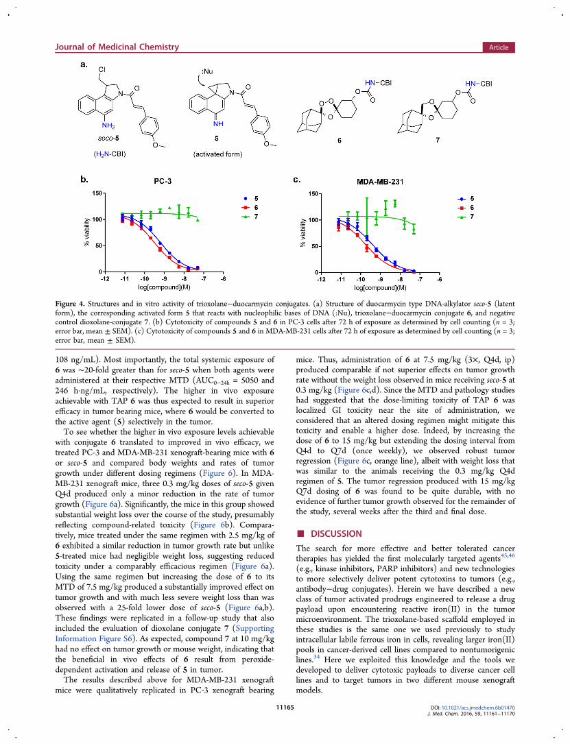
prevented by acylation or carbamoylation of the aniline orphenolic function, and the ability to “cage” seco-CBI derivativesin this way has made these compounds popular as cytotoxiceffectors in antibody−drug conjugates2,42 and tumor-activatedprodrugs.3,41 We synthesized a known seco-CBI analog40 andfurther converted the material into the desired TAP 6 and the1,3-dioxolane-CBI conjugate 7 (DXL-CBI), a nonperoxidiccontrol (Figure 4a).We evaluated the cytotoxicity of seco-5, TAP 6, and control 7
following 72 h of exposure in MDA-MB-231 and PC-3 cells(Figure 4b,c). As expected, seco-5 and its TAP 6 exhibitedpotent cytotoxic effects in cells, while the dioxolane conjugate 7was noncytotoxic at all concentrations examined. The inactivityof 7 confirms that conjugation of seco-5 via its aniline functioneffectively blocks formation of the activated (and toxic) CBIelectrophile. The potent effects of 6 in cells are thus due toperoxide-dependent release of free seco-5 as desired. We furtherfound that conversion of seco-5 to the TAP 6 dramaticallyimproves its chemical stability in the extracellular environment.Thus, while free seco-5 degraded over several hours uponthawing from a DMSO stock solution, TRX conjugate 6 (andDXL conjugate 7) were chemically stable for more than a yearat room temperature in DMSO (Supporting Information
Figure S5). Stability toward premature hydrolysis likely explainswhy TAP 6 was measurably more potent than seco-5 itself inboth cell lines examined (Figure 4b,c). Control experimentsconfirmed that 6 is stable for more than a week in cell culturemedia at 37 °C, indicating that its cellular toxicity in the 72 hassays (Figure 4b,c) results from intracellular release of seco-5 asdesired.The cell culture studies of 5−7 above revealed the effective
ablation of 5-mediated toxicity by chemical conjugation at ananiline function (Figure 4a−c). We next asked whether thiscaging effect would translate to reduced in vivo toxicity for TAP6 as compared to 5. To determine a maximally tolerated dose(MTD), nontumor bearing female NSG mice were adminis-tered three ip doses of either seco-5 or 6 at 4-day intervals(Q4d). These studies revealed that seco-5 is highly toxic to mice(MTD ≈ 0.3 mg/kg), consistent with previous observations forrelated duocarmycin analogs.41 By contrast, TAP 6 could beadministered at significantly higher doses, with an MTD of 7.5mg/kg, using the same three dose Q4d regimen. A subsequentMTD study of 6 with Q7d dosing returned a somewhat higherMTD of ∼10 mg/kg.Previous work with duocarmycin analogs related to 5 has
shown dose-limiting hepatotoxicity in mice43 and insufficienttherapeutic index leading to failures in human clinical trials.44
We were therefore interested in exploring the toxic effects of 5in mice and whether administration in the TRX-conjugatedform 6 could protect from these toxicities. In fact, mice treatedwith 0.3 mg/kg seco-5 showed substantially (∼10-fold) higherconcentrations of liver transaminase enzymes (ALT and AST)than did mice treated with 7.5 mg/kg of 6 (Figure 5c). Sincehepatotoxicity of duocarmycin analogs has previously beenobserved to occur with a delayed onset, mice receivingescalating doses of 5 or 6 were observed for 50 dayspostdosing, then sacrificed and their livers collected andassessed for altered morphology and signs of toxicity. Even atwell-tolerated doses of 5, mice grossly showed darkened androughened capsular and parenchymal changes indicative ofhepatotoxicity in addition to enlarged intestines and seros-anguinous ascites near the site of administration. The liverpathology showed increased hepatic lobular lymphohistiocyticinfiltrates, reactive cellular changes, and minimal periportalfibrosis. In contrast, the livers of mice treated with 7.5 mg/kg of6 appeared normal, and the dose limiting toxicity appearedinstead to be localized toxicity at the site of administration asevidenced by enlarged intestines. The pathology in these wasfree of significant inflammation or fibrosis (Figure 5d).To compare the pharmacokinetic properties of seco-5 and its
TRX conjugate 6, healthy NSG mice were administered a singleip dose of seco-5 (0.3 mg/kg) or conjugate 6 (7.5 mg/kg),representing the respective MTD values. Analysis of bloodsamples showed seco-5 to have a reasonably long half-life (t1/2 =3.8 h), moderate clearance (CL/F = 20 mL min−1 kg−1) and ahigh volume of distribution (Vz/F = 6.7 L/kg) (Figure 5a,b,Supporting Information Table S3). Conjugate 6 exhibited asignificantly longer half-life (t1/2 = 20.4 h), moderate-highclearance (CL/F = 31.3 mL min−1 kg−1) and a very highvolume of distribution (Vz/F = 55 L/kg). In animals receiving6, total exposure to 6 (AUC = 5050 h·ng/mL) exceeded that ofreleased seco-5 by approximately 8-fold, indicating a smalldegree of drug release in normal tissues of healthy mice.Maximum plasma concentrations of free seco-5 in micereceiving 7.5 mg/kg of 6 were still lower than those in micetreated with 0.3 mg/kg of seco-5 directly (Cmax = 48 ng/mL vs
Figure 3. Profiling the effects of oncogenic transformation on theferrous iron pool of MCF10A cells. (a) Relative mRNA levels forferroportin, ferritin, and the ferrireductase STEAP3 in MCF10A cellsstably transfected with empty vector (control) or with vectorsexpressing the oncogenes Myc or Hras (n = 3; error bar, mean ±SEM; (∗∗) P ≤ 0.01, (∗∗∗∗) P ≤ 0.0001, two-way ANOVA withDunnett’s multiple comparisons test). (b) EC50 ratio (1/2) forMCF10A cells stably transfected with HRas or cMyc oncogenes or theempty vector (control) as assessed by cell counting after 24 h ofcompound exposure. Error bars represent SEM from threeindependent experiments each conducted in biological triplicates((∗∗) P ≤ 0.01, one-way ANOVA with Dunnett’s multiplecomparisons test).
Journal of Medicinal Chemistry Article
DOI: 10.1021/acs.jmedchem.6b01470J. Med. Chem. 2016, 59, 11161−11170
11164
108 ng/mL). Most importantly, the total systemic exposure of6 was ∼20-fold greater than for seco-5 when both agents wereadministered at their respective MTD (AUC0−24h = 5050 and246 h·ng/mL, respectively). The higher in vivo exposureachievable with TAP 6 was thus expected to result in superiorefficacy in tumor bearing mice, where 6 would be converted tothe active agent (5) selectively in the tumor.To see whether the higher in vivo exposure levels achievable
with conjugate 6 translated to improved in vivo efficacy, wetreated PC-3 and MDA-MB-231 xenograft-bearing mice with 6or seco-5 and compared body weights and rates of tumorgrowth under different dosing regimens (Figure 6). In MDA-MB-231 xenograft mice, three 0.3 mg/kg doses of seco-5 givenQ4d produced only a minor reduction in the rate of tumorgrowth (Figure 6a). Significantly, the mice in this group showedsubstantial weight loss over the course of the study, presumablyreflecting compound-related toxicity (Figure 6b). Compara-tively, mice treated under the same regimen with 2.5 mg/kg of6 exhibited a similar reduction in tumor growth rate but unlike5-treated mice had negligible weight loss, suggesting reducedtoxicity under a comparably efficacious regimen (Figure 6a).Using the same regimen but increasing the dose of 6 to itsMTD of 7.5 mg/kg produced a substantially improved effect ontumor growth and with much less severe weight loss than wasobserved with a 25-fold lower dose of seco-5 (Figure 6a,b).These findings were replicated in a follow-up study that alsoincluded the evaluation of dioxolane conjugate 7 (SupportingInformation Figure S6). As expected, compound 7 at 10 mg/kghad no effect on tumor growth or mouse weight, indicating thatthe beneficial in vivo effects of 6 result from peroxide-dependent activation and release of 5 in tumor.The results described above for MDA-MB-231 xenograft
mice were qualitatively replicated in PC-3 xenograft bearing
mice. Thus, administration of 6 at 7.5 mg/kg (3×, Q4d, ip)produced comparable if not superior effects on tumor growthrate without the weight loss observed in mice receiving seco-5 at0.3 mg/kg (Figure 6c,d). Since the MTD and pathology studieshad suggested that the dose-limiting toxicity of TAP 6 waslocalized GI toxicity near the site of administration, weconsidered that an altered dosing regimen might mitigate thistoxicity and enable a higher dose. Indeed, by increasing thedose of 6 to 15 mg/kg but extending the dosing interval fromQ4d to Q7d (once weekly), we observed robust tumorregression (Figure 6c, orange line), albeit with weight loss thatwas similar to the animals receiving the 0.3 mg/kg Q4dregimen of 5. The tumor regression produced with 15 mg/kgQ7d dosing of 6 was found to be quite durable, with noevidence of further tumor growth observed for the remainder ofthe study, several weeks after the third and final dose.
■ DISCUSSION
The search for more effective and better tolerated cancertherapies has yielded the first molecularly targeted agents45,46
(e.g., kinase inhibitors, PARP inhibitors) and new technologiesto more selectively deliver potent cytotoxins to tumors (e.g.,antibody−drug conjugates). Herein we have described a newclass of tumor activated prodrugs engineered to release a drugpayload upon encountering reactive iron(II) in the tumormicroenvironment. The trioxolane-based scaffold employed inthese studies is the same one we used previously to studyintracellular labile ferrous iron in cells, revealing larger iron(II)pools in cancer-derived cell lines compared to nontumorigeniclines.34 Here we exploited this knowledge and the tools wedeveloped to deliver cytotoxic payloads to diverse cancer celllines and to target tumors in two different mouse xenograftmodels.
Figure 4. Structures and in vitro activity of trioxolane−duocarmycin conjugates. (a) Structure of duocarmycin type DNA-alkylator seco-5 (latentform), the corresponding activated form 5 that reacts with nucleophilic bases of DNA (:Nu), trioxolane−duocarmycin conjugate 6, and negativecontrol dioxolane-conjugate 7. (b) Cytotoxicity of compounds 5 and 6 in PC-3 cells after 72 h of exposure as determined by cell counting (n = 3;error bar, mean ± SEM). (c) Cytotoxicity of compounds 5 and 6 in MDA-MB-231 cells after 72 h of exposure as determined by cell counting (n = 3;error bar, mean ± SEM).
Journal of Medicinal Chemistry Article
DOI: 10.1021/acs.jmedchem.6b01470J. Med. Chem. 2016, 59, 11161−11170
11165
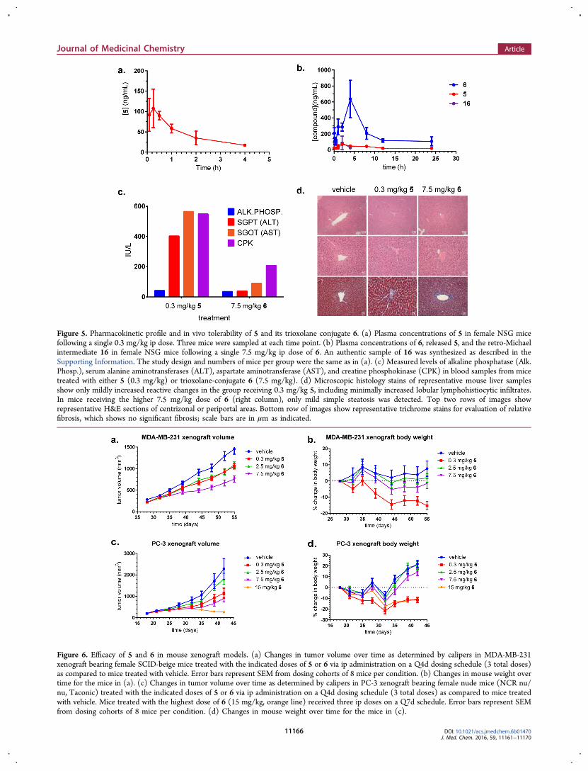
Figure 5. Pharmacokinetic profile and in vivo tolerability of 5 and its trioxolane conjugate 6. (a) Plasma concentrations of 5 in female NSG micefollowing a single 0.3 mg/kg ip dose. Three mice were sampled at each time point. (b) Plasma concentrations of 6, released 5, and the retro-Michaelintermediate 16 in female NSG mice following a single 7.5 mg/kg ip dose of 6. An authentic sample of 16 was synthesized as described in theSupporting Information. The study design and numbers of mice per group were the same as in (a). (c) Measured levels of alkaline phosphatase (Alk.Phosp.), serum alanine aminotransferases (ALT), aspartate aminotransferase (AST), and creatine phosphokinase (CPK) in blood samples from micetreated with either 5 (0.3 mg/kg) or trioxolane-conjugate 6 (7.5 mg/kg). (d) Microscopic histology stains of representative mouse liver samplesshow only mildly increased reactive changes in the group receiving 0.3 mg/kg 5, including minimally increased lobular lymphohistiocytic infiltrates.In mice receiving the higher 7.5 mg/kg dose of 6 (right column), only mild simple steatosis was detected. Top two rows of images showrepresentative H&E sections of centrizonal or periportal areas. Bottom row of images show representative trichrome stains for evaluation of relativefibrosis, which shows no significant fibrosis; scale bars are in μm as indicated.
Figure 6. Efficacy of 5 and 6 in mouse xenograft models. (a) Changes in tumor volume over time as determined by calipers in MDA-MB-231xenograft bearing female SCID-beige mice treated with the indicated doses of 5 or 6 via ip administration on a Q4d dosing schedule (3 total doses)as compared to mice treated with vehicle. Error bars represent SEM from dosing cohorts of 8 mice per condition. (b) Changes in mouse weight overtime for the mice in (a). (c) Changes in tumor volume over time as determined by calipers in PC-3 xenograft bearing female nude mice (NCR nu/nu, Taconic) treated with the indicated doses of 5 or 6 via ip administration on a Q4d dosing schedule (3 total doses) as compared to mice treatedwith vehicle. Mice treated with the highest dose of 6 (15 mg/kg, orange line) received three ip doses on a Q7d schedule. Error bars represent SEMfrom dosing cohorts of 8 mice per condition. (d) Changes in mouse weight over time for the mice in (c).
Journal of Medicinal Chemistry Article
DOI: 10.1021/acs.jmedchem.6b01470J. Med. Chem. 2016, 59, 11161−11170
11166
The trioxolane TAP scaffold used here was engineered toconfer “traceless” release of drug payloads, thereby enabling abroad scope of potential applications encompassing molecularlytargeted and generally cytotoxic agents possessing suitableamine or alcohol functionality for conjugation. When thisstrategy is employed, the site of drug conjugation is selected sothat the intrinsic activity/toxicity of tethered drug is ablated,thereby preventing or minimizing exposure of active drug innontargeted tissues. To exemplify this approach, we preparedamine-linked TAP conjugates 2 and 6 from the microtubuleinhibitor 1 and duocarmycin analog seco-5, respectively. Weconfirmed that the activity/toxicity of these agents was ablatedin the TAP form by preparing dioxolane-drug conjugates 3 and7, which proved inactive at the highest concentrations tested(Figure 2b and Figure 4b,c). The lack of measurable activity for3 and 7 thus confirms that drug release from 2 and 6 isperoxide dependent. Consistent with the expected mechanismof iron(II)-dependent drug release, the relative sensitivity ofcells to 2 (Figure 2d) largely mirrored the response of theTRX-PURO probe to intracellular labile iron in the same celllines.34
We found that sensitivity to TRX-CMB conjugate 2 variedby about 9-fold across a panel of cancer cell lines from diverseorigins (Figure 2e). Further interrogation of these data mayprovide insight into the specific alterations of iron metabolismthat predict for increased tumor susceptibility toward TRX-drug conjugates. In our preliminary study of oncogenic changesand related effects on iron metabolism, we observed that Myc-driven changes in MCF10A cells produced increased sensitivityto trioxolane conjugate 2. Consistent with these observations,several of the most 2-susceptible cell lines examined here, suchas RKO and MDA-MB-231, are known to overexpress Myc.36,47
Thus, iron(II)-dependent drug delivery could find utility intargeting Myc-driven tumors indirectly via the alteration of ironmetabolism induced by this prevalent oncogene. These findingsare particularly relevant given how intractable Myc driventumors have been toward other targeted therapies.48
To examine the utility of trioxolane-mediated drug deliveryin vivo, we prepared trioxolane TAP 6 in which a potentduocarmycin-class cytotoxin (seco-5) is stabilized chemicallyand inactivated biologically (while in prodrug form).Significantly, conjugate 6 was tolerated in mice at doses upto 50-fold higher than seco-5 and produced superior efficacy intwo different xenograft models when administered at or near itsMTD. A pharmacokinetic study in healthy mice revealed thatadministration of seco-5 in the form 6 not only limited exposureto free drug (to ∼15% of the total dose) but also significantlyimproved distribution to tissues and the total duration of drugexposure. These properties of 6 enabled safe administration at arelatively high dose of 15 mg/kg once weekly, and this dosingregimen produced a particularly robust and durable tumorregression in a PC-3 xenograft model. Overall, our studiesindicate that trioxolane-based TAPs release their drug payloadin proportion to the concentration of labile iron(II)encountered in different cell/tissue types. While intracellulariron(II) pools are likely implicated in TAP activation, it ispossible that other aspects of tumor biology in vivo (e.g.,hypoxia, macrophage infiltration, tumor necrosis) contribute tothe presence of excess reactive iron(II) in the tumormicroenvironment.In conclusion, trioxolane-mediated iron(II)-dependent drug
delivery is a new approach for cell/tissue selective drugtargeting that leverages elevated reactive iron(II) concen-
trations in tumor cells and in the tumor microenvironment.Here we described two prototypical trioxolane-drug conjugatesbearing cytotoxins with distinct mechanisms of cellular toxicity.We confirmed that the intrinsic toxicity of these agents couldbe ablated in conjugated forms and yet fully realized followingcell- or tumor-selective release at their intended site of action.These results should encourage further study of this concept toidentify drugs and tumor types that best leverage this new drugdelivery approach.
■ EXPERIMENTAL SECTIONThe known cytotoxic agents 135 and seco-540 were prepared aspreviously described. These compounds were coupled to knowntrioxolane29 and dioxolane30 intermediates via activated nitrophenylcarbonate or isocyanate intermediates as we have describedpreviously29−31 and as further detailed in the Supporting Information.
All compounds tested in cells or animals were judged to be of >95%purity as determined using a Waters Micromass ZQTM, equippedwith Waters 2795 separation module, Waters 2996 photodiode arraydetector (254 nm), and Waters 2424 ELS detector. Separations werecarried out with an XTerra MS C18, 5 μm, 4.6 mm × 50 mm column,at ambient temperature (unregulated) using a mobile phase of water−methanol containing a constant 0.10% formic acid. Representative LCchromatograms are provided in the Supporting Information.
Mammalian cell lines were maintained in an atmosphere of 5% CO2in RPMI 1640 media purchased from HyClone supplemented with10% FBS (Gibco), Pen/Strep (1× final concentration, Gemini Bio-Products), and nonessential amino acids (UCSF Cell Culture Facility).Unless otherwise noted, cell lines were obtained from ATCC andverified by STR profiling. Graphing and analysis of data were doneusing GraphPad Prism 6 software and Microsoft Excel 2010. Figureswere prepared with Adobe Design Standard CS6 software.
Statistics. Error bars in all figures represent SEM unless otherwiseindicated. When three or more mean values were compared, one- ortwo-way ANOVA tests were applied as required with Dunnett’smultiple comparisons tests used to determine significance. Statisticalsignificance is indicated as follows: ∗ = P ≤ 0.05, ∗∗ = P ≤ 0.01, ∗∗∗ =P ≤ 0.001, ∗∗∗∗ = P ≤ 0.0001.
Toxicity by Nuclei Counting. Cells were plated in 96-wellGreiner black μClear tissue culture plates at 3000−6000 cells per wellin RPMI 1640 cell culture media (or the appropriate growth mediumas specified) and incubated at 37 °C in 5% CO2 incubators for at least16 h prior to exposure to compounds. Cells were then treated intriplicate with escalating concentrations of compounds performed inmedium containing 0.1% DMSO (100 μL of media, per well). 24−72h after treatment (as specified), medium was removed and cells werewashed with 100 μL of PBS and then fixed in 4% paraformaldehyde for10 min at rt and stained with Hoechst nuclear stain at a finalconcentration of 10 μg/mL in PBS for 10 min at rt. After fixing, thecells were stored in 100 μL of PBS for imaging. Wells were imagedwith an IN Cell 2000 automated cell imager at 10× magnification with9 images per well (complete coverage) in bright field and DAPIchannel fluorescence, and images were analyzed for nuclei count by INCell developer software. EC50 values were calculated in GraphPadPrism from normalized dose−response curves.
Toxicity by CellTiter-Glo. Cells were harvested, resuspended, andplated with a Wellmate liquid handler (Thermo Scientific) into 384-well plates and cultured for 24 h before dosing. Master compoundplates were made with a Janus (PerkinElmer), then further diluted toachieve uniform final concentrations of DMSO of 0.1% in media for alltreatment conditions. Compound treatments in media were added tothe cell plates with a Matrix Platemate (Thermo Scientific). Cellviabilities were determined 72 h after treatment by Cell-Titer Gloassay (Promega) on the Envision multilabel plate reader (PerkinElm-er). Relative luminescent units (RLU) were plotted againstcorresponding drug concentrations and fitted with a standard four-parameter sigmoidal curve with GraphPad Prism 6. Data were furtherfit for EC50 shift parameters in GraphPad Prism 6 to determine EC50
Journal of Medicinal Chemistry Article
DOI: 10.1021/acs.jmedchem.6b01470J. Med. Chem. 2016, 59, 11161−11170
11167
ratios for 1/2. Data are reported as the EC50 ratio, and error barsrepresent SEM (n = 3). Data for cell lines in which EC50 ratio fits wereambiguous, R2 values were less than 0.9, or response to the free drugwas less than 40% were not reported.Quantitative PCR. Cells were seeded at 300 000 cells per well in 6-
well plates and grown to confluence and then collected with trypsin(0.05%), washed with PBS, and snap-frozen in liquid N2 and thenstored at −78 °C. Cell pellets were processed for mRNA isolationusing Qiagen RNeasy Mini Kit with QIAshredder lysate homogenizersand on column DNA digest. Isolated mRNA was analyzed forconcentration and purity on a ThermoScientific NanoDrop 2000cspectrophotometer, and 1000 ng of mRNA from each sample wastranslated to cDNA using Invitrogen SuperScript III First-StrandSynthesis System. The resulting cDNA was used for qRT-PCR analysis(10 ng/reaction) with SsoAdvanced Universal SYBR Green Supermixin a Roche LightCycler 480. GAPDH was used as an endogenouscontrol, and relative mRNA levels were calculated from a standardcurve of pooled samples with LightCycler 480 software used forsecond derivative maximum analysis and standard curve fitting.Samples were prepared and run in biological triplicates, and errorbars represent SEM. A list of the gene specific primers used is providedin the Supporting Information.Maximum Tolerable Dose Studies. To evaluate the tolerability
of the experimental agents, groups of three NSG (NOD SCIDgamma) mice were treated with three ip injections of test article (Q4dor Q7d in separate studies) in a formulation comprising 50:40:10 PEG400/20% 2-hydroxypropylcylcodextrine in water/DMSO. Individualgroups (n = 3) received doses that increased in 2- or 3-fold steps untila given dose caused one or more mice in the group to reach protocollimits for tolerance (>20% weight loss) at any point post dosing.Pharmacokinetics Studies. To evaluate the in vivo pharmaco-
kinetic properties of the experimental agents, female NSG mice weretreated with a single ip injection of test compounds formulated in50:40:10 PEG 400/20% 2-hydroxypropylcylcodextrine in water/DMSO. Blood samples were collected 5 min, 15 min, 30 min, 1 h,2 h, 4 h, 8 h, 12 h, and 24 h after dosing (3 mice were sampled pertime point; each group of mice was sampled ≤3 times over 24 h) andanalyzed for plasma concentrations of each compound via MS/MSanalysis conducted by Integrated Analytical Solutions, Inc. (Berkeley,CA). The resulting data were analyzed with WinNonlin software tocalculate standard PK parameters.Efficacy Studies. To evaluate the in vivo properties of the
experimental agents, we used the heterotopic indirect tumor xenograftmodel in nude mice (NCR nu/nu, Taconic) and SCID-beige mice.Early passage PC-3 cells were harvested, and a cell suspension (1:1serum free DMEM/Matrigel) was injected subcutaneously (sc) intothe right flank of anesthetized donor nude mice (106 cells/mouse in0.1 mL). For MDA-MB-231 xenografts, cells were injected into themammary fat pad of anesthetized female SCID-beige mice (106 cells/mouse in 0.1 mL of PBS). When the mean tumor volume was 250−400 mm3, tumor-bearing mice were treated with the indicated doses ofcompounds formulated in 50:40:10 PEG 400/20% 2-hydroxypropyl-cylcodextrine in water/DMSO via ip administration with the indicatedfrequency. Tumor volume (by caliper) and mouse weight weremonitored twice weekly.
■ ASSOCIATED CONTENT
*S Supporting InformationThe Supporting Information is available free of charge on theACS Publications website at DOI: 10.1021/acs.jmed-chem.6b01470.
Supplemental tables, figures and schemes; syntheticprocedures and compound characterization for newcompounds; animal welfare statement; and a list ofgene-specific primers used for qRT-PCR analysis (PDF)Molecular formula strings and some data (CSV)
■ AUTHOR INFORMATIONCorresponding Author*Phone: 415-514-9698. Fax: 415-514-4507. E-mail: [email protected] R. Renslo: 0000-0002-1240-2846Present Address∇S.D.F.: Prolynx LLC, San Francisco, CA, 94158.Author ContributionsB.S., J.A.W., and A.R.R. designed compounds and conceivedexperiments. B.S. and S.D.F. synthesized compounds. B.S.,S.D.F, Y.S., and B.H. acquired data. All authors analyzed thedata. A.R.R. supervised the project. B.S. and A.R.R wrote themanuscript. All authors reviewed, edited, and commented onthe manuscript.NotesThe authors declare no competing financial interest.
■ ACKNOWLEDGMENTSA.R.R. acknowledges funding from the U.S. National Institutesof Health (R01 Grant AI105106) and from the NIH NationalCenter for Advancing Translational Science (UCSF-CTSIGrant UL1 TR000004). B.S. acknowledges funding from theNIH Research Training Grant in Chemistry and ChemicalBiology (Grant T32 GM064337). The authors thank Prof. S.Bandyopadhyay (UCSF) for providing the MCF10A cell linesused in these studies, Steven Chen for technical support inautomated cell imaging and image analysis with InCellDeveloper software, Dr. Tetsuya Matsuguchi for his assistancewith qRT-PCR set up and analysis, Dr. Alan Wolfe for hisassistance in calculating pharmacokinetic parameters, and thePreclinical Therapeutic Core at UCSF where in vivo experi-ments were performed.
■ ABBREVIATIONS USEDTAP, tumor-activated prodrug; ADC, antibody−drug con-jugate; TRX, a 1,2,4-trioxolane scaffold for payload delivery;DXL, 1,3-dioxolane scaffold used as nonperoxidic controls;PURO, puromycin; qRT-PCR, quantitative real-time polymer-ase chain reaction; CBI, cyclopropylbenzindoline; MTD,maximally tolerated dose; Q4d, dosed every 4 days; Q7d,dosed every 7 days; ALT, alanine transaminase; AST, aspartatetransaminase; CPK, creatine phosphokinase; H&E, hematox-ylin and eosin stained; CL, clearance; F, fraction of drugachieving systemic exposure following an ip dose; Vz, volume ofdistribution; AUC, area under the curve
■ REFERENCES(1) Lorusso, P. M.; Edelman, M. J.; Bever, S. L.; Forman, K. M.; Pilat,M.; Quinn, M. F.; Li, J.; Heath, E. I.; Malburg, L. M.; Klein, P. J.;Leamon, C. P.; Messmann, R. A.; Sausville, E. A. Phase I study offolate conjugate EC145 (vintafolide) in patients with refractory solidtumors. J. Clin. Oncol. 2012, 30, 4011−4016.(2) Beck, A.; Reichert, J. M. Antibody-drug conjugates: present andfuture. MAbs. 2014, 6, 15−17.(3) Kratz, F.; Muller, I. A.; Ryppa, C.; Warnecke, A. Prodrugstrategies in anticancer chemotherapy. ChemMedChem 2008, 3, 20−53.(4) Brown, J. M.; Giaccia, A. J. The unique physiology of solidtumors: opportunities (and problems) for cancer therapy. Cancer Res.1998, 58, 1408−1416.(5) Brown, J. M.; Wilson, W. R. Exploiting tumour hypoxia in cancertreatment. Nat. Rev. Cancer 2004, 4, 437−447.
Journal of Medicinal Chemistry Article
DOI: 10.1021/acs.jmedchem.6b01470J. Med. Chem. 2016, 59, 11161−11170
11168

(6) Torti, S. V.; Torti, F. M. Iron and cancer: more ore to be mined.Nat. Rev. Cancer 2013, 13, 342−355.(7) Chaturvedi, D.; Goswami, A.; Saikia, P. P.; Barua, N. C.; Rao, P.G. Artemisinin and its derivatives: a novel class of anti-malarial andanti-cancer agents. Chem. Soc. Rev. 2010, 39, 435−454.(8) O’Neill, P. M.; Posner, G. H. A medicinal chemistry perspectiveon artemisinin and related endoperoxides. J. Med. Chem. 2004, 47,2945−2964.(9) Mercer, A. E.; Copple, I. M.; Maggs, J. L.; O’Neill, P. M.; Park, B.K. The role of heme and the mitochondrion in the chemical andmolecular mechanisms of mammalian cell death induced by theartemisinin antimalarials. J. Biol. Chem. 2011, 286, 987−996.(10) Mercer, A. E.; Maggs, J. L.; Sun, X.-M.; Cohen, G. M.;Chadwick, J.; O’Neill, P. M.; Park, B. K. Evidence for the involvementof carbon-centered radicals in the induction of apoptotic cell death byartemisinin compounds. J. Biol. Chem. 2007, 282, 9372−9382.(11) Dixon, S. J.; Stockwell, B. R. The role of iron and reactiveoxygen species in cell death. Nat. Chem. Biol. 2014, 10, 9−17.(12) Pantopoulos, K. Iron metabolism and the IRE/IRP regulatorysystem: an update. Ann. N. Y. Acad. Sci. 2004, 1012, 1−13.(13) Wang, J.; Pantopoulos, K. Regulation of cellular ironmetabolism. Biochem. J. 2011, 434, 365−381.(14) Richardson, D. R.; Ponka, P. The molecular mechanisms of themetabolism and transport of iron in normal and neoplastic cells.Biochim. Biophys. Acta, Rev. Biomembr. 1997, 1331, 1−40.(15) Boult, J.; Roberts, K.; Brookes, M. J.; Hughes, S.; Bury, J. P.;Cross, S. S.; Anderson, G. J.; Spychal, R.; Iqbal, T.; Tselepis, C.Overexpression of cellular iron import proteins is associated withmalignant progression of esophageal adenocarcinoma. Clin. Cancer Res.2008, 14, 379−387.(16) Kakhlon, O.; Gruenbaum, Y.; Cabantchik, Z. Ferritin expressionmodulates cell cycle dynamics and cell responsiveness to H-ras-induced growth via expansion of the labile iron pool. Biochem. J. 2002,363, 431−436.(17) Pinnix, Z. K.; Miller, L. D.; Wang, W.; D’Agostino, R.; Kute, T.;Willingham, M. C.; Hatcher, H.; Tesfay, L.; Sui, G.; Di, X.; Torti, S. V.;Torti, F. M. Ferroportin and iron regulation in breast cancerprogression and prognosis. Sci. Transl. Med. 2010, 2, 43ra56.(18) Wu, K.-J.; Polack, A.; Dalla-Favera, R. Coordinated regulation ofiron-controlling genes, H-ferritin and IRP2, by c-MYC. Science 1999,283, 676−679.(19) Toyokuni, S. Role of iron in carcinogenesis: cancer as aferrotoxic disease. Cancer Sci. 2009, 100, 9−16.(20) Miller, L. D.; Coffman, L. G.; Chou, J. W.; Black, M. A.; Bergh,J.; D’Agostino, R.; Torti, S. V.; Torti, F. M. An iron regulatory genesignature predicts outcome in breast cancer. Cancer Res. 2011, 71,6728−6737.(21) Valecha, N.; Looareesuwan, S.; Martensson, A.; Abdulla, S. M.;Krudsood, S.; Tangpukdee, N.; Mohanty, S.; Mishra, S. K.; Tyagi, P.K.; Sharma, S. K.; Moehrle, J.; Gautam, A.; Roy, A.; Paliwal, J. K.;Kothari, M.; Saha, N.; Dash, A. P.; Bjorkman, A. Arterolane, a newsynthetic trioxolane for treatment of uncomplicated Plasmodiumfalciparum malaria: a phase II, multicenter, randomized, dose-findingclinical trial. Clin. Infect. Dis. 2010, 51, 684−691.(22) Borstnik, K.; Paik, I.; Shapiro, T. A.; Posner, G. H. Antimalarialchemotherapeutic peroxides: artemisinin, yingzhaosu A and relatedcompounds. Int. J. Parasitol. 2002, 32, 1661−1667.(23) Vennerstrom, J. L.; Arbe-Barnes, S.; Brun, R.; Charman, S. A.;Chiu, F. C.; Chollet, J.; Dong, Y.; Dorn, A.; Hunziker, D.; Matile, H.;McIntosh, K.; Padmanilayam, M.; Santo Tomas, J.; Scheurer, C.;Scorneaux, B.; Tang, Y.; Urwyler, H.; Wittlin, S.; Charman, W. N.Identification of an antimalarial synthetic trioxolane drug developmentcandidate. Nature 2004, 430, 900−904.(24) Valecha, N.; Krudsood, S.; Tangpukdee, N.; Mohanty, S.;Sharma, S. K.; Tyagi, P. K.; Anvikar, A.; Mohanty, R.; Rao, B. S.; Jha,A. C.; Shahi, B.; Singh, J. P. N.; Roy, A.; Kaur, P.; Kothari, M.; Mehta,S.; Gautam, A.; Paliwal, J. K.; Arora, S.; Saha, N. Arterolane maleateplus piperaquine phosphate for treatment of uncomplicated
plasmodium falciparum malaria: A comparative, multicenter, random-ized clinical trial. Clin. Infect. Dis. 2012, 55, 663−671.(25) Creek, D. J.; Charman, W. N.; Chiu, F. C. K.; Prankerd, R. J.;Dong, Y.; Vennerstrom, J. L.; Charman, S. A. Relationship betweenantimalarial activity and heme alkylation for spiro- and dispiro-1,2,4-trioxolane antimalarials. Antimicrob. Agents Chemother. 2008, 52,1291−1296.(26) Creek, D.; Charman, W.; Chiu, F. C. K.; Prankerd, R. J.;Mccullough, K. J.; Dong, Y.; Vennerstrom, J. L.; Charman, S. A. Iron-mediated degradation kinetics of substituted dispiro-1, 2, 4-trioxolaneantimalarials. J. Pharm. Sci. 2007, 96, 2945−56.(27) Tang, Y.; Dong, Y.; Wang, X.; Sriraghavan, K.; Wood, J. K.;Vennerstrom, J. L. Dispiro-1,2,4-trioxane analogues of a prototypedispiro-1,2,4-trioxolane: mechanistic comparators for artemisinin inthe context of reaction pathways with iron(II). J. Org. Chem. 2005, 70,5103−5110.(28) Wang, X.; Creek, D. J.; Schiaffo, C. E.; Dong, Y.; Chollet, J.;Scheurer, C.; Wittlin, S.; Charman, S. A.; Dussault, P. H.; Wood, J. K.;Vennerstrom, J. L. Spiroadamantyl 1,2,4-trioxolane, 1,2,4-trioxane, and1,2,4-trioxepane pairs: relationship between peroxide bond iron(II)reactivity, heme alkylation efficiency, and antimalarial activity. Bioorg.Med. Chem. Lett. 2009, 19, 4542−4545.(29) Fontaine, S. D.; Dipasquale, A. G.; Renslo, A. R. Efficient andStereocontrolled Synthesis of 1,2,4-trioxolanes useful for ferrous iron-dependent drug delivery. Org. Lett. 2014, 16, 5776−5779.(30) Fontaine, S. D.; Spangler, B.; Gut, J.; Lauterwasser, E. M. W.;Rosenthal, P. J.; Renslo, A. R. Drug delivery to the malaria parasiteusing an arterolane-like scaffold. ChemMedChem 2015, 10, 47−51.(31) Mahajan, S. S.; Deu, E.; Lauterwasser, E. M. W.; Leyva, M. J.;Ellman, J. A.; Bogyo, M.; Renslo, A. R. A fragmenting hybrid approachfor targeted delivery of multiple therapeutic agents to the malariaparasite. ChemMedChem 2011, 6, 415−419.(32) Deu, E.; Chen, I. T.; Lauterwasser, E. M. W.; Valderramos, J.; Li,H.; Edgington, L. E.; Renslo, A. R.; Bogyo, M. Ferrous iron-dependentdrug delivery enables controlled and selective release of therapeuticagents in vivo. Proc. Natl. Acad. Sci. U. S. A. 2013, 110, 18244−18249.(33) Lauterwasser, E. M. W.; Fontaine, S. D.; Li, H.; Gut, J.; Katneni,K.; Charman, S. A.; Rosenthal, P. J.; Bogyo, M.; Renslo, A. R.Trioxolane-mediated delivery of mefloquine limits brain exposure in amouse model of malaria. ACS Med. Chem. Lett. 2015, 6, 1145−1149.(34) Spangler, B.; Morgan, C. W.; Fontaine, S. D.; Vander Wal, M.N.; Chang, C. J.; Wells, J. A.; Renslo, A. R. A reactivity-based probe ofthe intracellular labile ferrous iron pool. Nat. Chem. Biol. 2016, 12,680−685.(35) Odlo, K.; Hentzen, J.; dit Chabert, J. F.; Ducki, S.; Gani, O. A. B.S. M.; Sylte, I.; Skrede, M.; Florenes, V. A.; Hansen, T. V. 1,5-Disubstituted 1,2,3-triazoles as cis-restricted analogues of combretas-tatin A-4: Synthesis, molecular modeling and evaluation as cytotoxicagents and inhibitors of tubulin. Bioorg. Med. Chem. 2008, 16, 4829−4838.(36) Martins, M. M.; Zhou, A. Y.; Corella, A.; Horiuchi, D.; Yau, C.;Rakshandehroo, T.; Gordan, J. D.; Levin, R. S.; Johnson, J.; Jascur, J.;Shales, M.; Sorrentino, A.; Cheah, J.; Clemons, P. A.; Shamji, A. F.;Schreiber, S. L.; Krogan, N. J.; Shokat, K. M.; McCormick, F.; Goga,A.; Bandyopadhyay, S. Linking tumor mutations to drug responses viaa quantitative chemical-genetic interaction map. Cancer Discovery2015, 5, 154−167.(37) Radulescu, S.; Brookes, M. J.; Salgueiro, P.; Ridgway, R. A.;McGhee, E.; Anderson, K.; Ford, S. J.; Stones, D. H.; Iqbal, T. H.;Tselepis, C.; Sansom, O. J. Luminal iron levels govern intestinaltumorigenesis after Apc loss in vivo. Cell Rep. 2012, 2, 270−282.(38) Yang, W. S.; Stockwell, B. R. Synthetic lethal screening identifiescompounds activating iron-dependent, nonapoptotic cell death inoncogenic-RAS-harboring cancer cells. Chem. Biol. 2008, 15, 234−245.(39) Boger, D. L.; Johnson, D. S. CC-1065 and the duocarmycins:unraveling the keys to a new class of naturally derived DNA alkylatingagents. Proc. Natl. Acad. Sci. U. S. A. 1995, 92, 3642−3649.
Journal of Medicinal Chemistry Article
DOI: 10.1021/acs.jmedchem.6b01470J. Med. Chem. 2016, 59, 11161−11170
11169
(40) Yang, S.; Denny, W. A. A new short synthesis of 3-substituted 5-amino-1-(chloromethyl)-1,2-dihydro-3H-benzo[e]indoles (amino-CBIs). J. Org. Chem. 2002, 67, 8958−8961.(41) Atwell, G. J.; Milbank, J. J.; Wilson, W. R.; Hogg, A.; Denny, W.A. 5-Amino-1-(chloromethyl)-1,2-dihydro-3H-benz[e]indoles: rela-tionships between structure and cytotoxicity for analogues bearingdifferent DNA minor groove binding subunits. J. Med. Chem. 1999, 42,3400−3411.(42) Jeffrey, S. C.; Torgov, M. Y.; Andreyka, J. B.; Boddington, L.;Cerveny, C. G.; Denny, W. A.; Gordon, K. A.; Gustin, D.; Haugen, J.;Kline, T.; Nguyen, M. T.; Senter, P. D. Design, synthesis, and in vitroevaluation of dipeptide-based antibody minor groove binderconjugates. J. Med. Chem. 2005, 48, 1344−1358.(43) McGovren, J. P.; Clarke, G. L.; Pratt, E. A.; DeKoning, T. F.Preliminary toxicity studies with the DNA-binding antibiotic, CC-1065. J. Antibiot. 1984, 37, 63−70.(44) Markovic, S. N.; Suman, V. J.; Vukov, A. M.; Fitch, T. R.;Hillman, D. W.; Adjei, A. A.; Alberts, S. R.; Kaur, J. S.; Braich, T. A.;Leitch, J. M.; Creagan, E. T. Phase II trial of KW2189 in patients withadvanced malignant melanoma. Am. J. Clin. Oncol. 2002, 25, 308−312.(45) Workman, P. Genomics and the second golden era of cancerdrug development. Mol. BioSyst. 2005, 1, 17−26.(46) Lord, C. J.; Ashworth, A. The DNA damage response andcancer therapy. Nature 2012, 481, 287−294.(47) Taylor, C. W.; Kim, Y. S.; Childress-Fields, K. E.; Yeoman, L. C.Sensitivity of nuclear c-Myc levels and induction to differentiation-inducing agents in human colon tumor cell lines. Cancer Lett. 1992, 62,95−105.(48) Horiuchi, D.; Anderton, B.; Goga, A. Taking on ChallengingTargets: Making Myc druggable. Am. Soc. Clin. Oncol. Educ. B 2014,34, e497−502.
Journal of Medicinal Chemistry Article
DOI: 10.1021/acs.jmedchem.6b01470J. Med. Chem. 2016, 59, 11161−11170
11170