Self-Assembly Behavior of Colistin and Its Prodrug Colistin Methanesulfonate: Implications for...
16
Self-assembly behaviour of colistin and its prodrug colistin methanesulfonate: implications for solution stability and solubilization Stephanie J. Wallace 1 , Jian Li 1 , Roger L. Nation 1 , Richard J. Prankerd 2 , Tony Velkov 3,4 , and Ben J. Boyd 2,* 1 Facility for Anti-Infective Drug Development and Innovation, Monash Institute of Pharmaceutical Sciences, Parkville, Melbourne, Victoria 3052, Australia 2 Drug Delivery, Disposition and Dynamics, Monash Institute of Pharmaceutical Sciences, Parkville, Melbourne, Victoria 3052, Australia 3 Medicinal Chemistry and Drug Action, Monash Institute of Pharmaceutical Sciences, Parkville, Melbourne, Victoria 3052, Australia 4 School of Medicine, Deakin University, Pigdons Rd, Geelong 3127 Victoria, Australia Abstract Colistin is an amphiphilic antibiotic that has re-emerged into clinical use due to the increasing prevalence of difficult-to-treat Gram-negative infections. The existence of self-assembling colloids in solutions of colistin and its derivative prodrug, colistin methanesulfonate (CMS) was investigated. Colistin and CMS reduced the air-water interfacial tension, and dynamic light scattering (DLS) studies showed the existence of 2.07 ± 0.3 nm aggregates above 1.5 mM for colistin, and of 1.98 ± 0.36 nm aggregates for CMS above 3.5 mM (mean ± SD). Above the respective critical micelle concentrations (CMC) the solubility of azithromycin, a hydrophobic antibiotic, increased approximately linearly with increasing surfactant concentration (5:1 mol ratio colistin:azithromycin), suggestive of hydrophobic domains within the micellar cores. Rapid conversion of CMS to colistin occurred below the CMC (60 % over 48 hr), while conversion above the CMC was less than 1 %. The formation of colistin and CMS micelles demonstrated in this study is the proposed mechanism for solubilization of azithromycin and the concentration-dependent stability of CMS. Keywords Micelle; dynamic light scattering; amphiphilic peptide antibiotic; azithromycin Introduction Self assembly of pharmacologically active compounds in solution is an interesting, important and frequently understated phenomenon. Amphiphilic structure is often a prerequisite for pharmacological activity, and can be key to the interaction between drug molecules and biological membranes. 1 Colistin (also known as polymyxin E, Figure 1A) is an old antibiotic that exhibits membrane permeabilizing and bactericidal activity against Gram-negative * Corresponding Author: Mailing address: Drug Delivery, Disposition and Dynamics, Monash Institute of Pharmaceutical Sciences, Monash University, 381 Royal Parade, Parkville, Victoria 3052, Australia. Phone: +61 3 9903 9112. Fax: +61 3 9903 9560. [email protected]. NIH Public Access Author Manuscript J Phys Chem B. Author manuscript; available in PMC 2011 April 15. Published in final edited form as: J Phys Chem B. 2010 April 15; 114(14): 4836–4840. doi:10.1021/jp100458x. NIH-PA Author Manuscript NIH-PA Author Manuscript NIH-PA Author Manuscript
Transcript of Self-Assembly Behavior of Colistin and Its Prodrug Colistin Methanesulfonate: Implications for...

Self-assembly behaviour of colistin and its prodrug colistinmethanesulfonate: implications for solution stability andsolubilization
Stephanie J. Wallace1, Jian Li1, Roger L. Nation1, Richard J. Prankerd2, Tony Velkov3,4, andBen J. Boyd2,*1 Facility for Anti-Infective Drug Development and Innovation, Monash Institute of PharmaceuticalSciences, Parkville, Melbourne, Victoria 3052, Australia2 Drug Delivery, Disposition and Dynamics, Monash Institute of Pharmaceutical Sciences, Parkville,Melbourne, Victoria 3052, Australia3 Medicinal Chemistry and Drug Action, Monash Institute of Pharmaceutical Sciences, Parkville,Melbourne, Victoria 3052, Australia4 School of Medicine, Deakin University, Pigdons Rd, Geelong 3127 Victoria, Australia
AbstractColistin is an amphiphilic antibiotic that has re-emerged into clinical use due to the increasingprevalence of difficult-to-treat Gram-negative infections. The existence of self-assembling colloidsin solutions of colistin and its derivative prodrug, colistin methanesulfonate (CMS) was investigated.Colistin and CMS reduced the air-water interfacial tension, and dynamic light scattering (DLS)studies showed the existence of 2.07 ± 0.3 nm aggregates above 1.5 mM for colistin, and of 1.98 ±0.36 nm aggregates for CMS above 3.5 mM (mean ± SD). Above the respective critical micelleconcentrations (CMC) the solubility of azithromycin, a hydrophobic antibiotic, increasedapproximately linearly with increasing surfactant concentration (5:1 mol ratio colistin:azithromycin),suggestive of hydrophobic domains within the micellar cores. Rapid conversion of CMS to colistinoccurred below the CMC (60 % over 48 hr), while conversion above the CMC was less than 1 %.The formation of colistin and CMS micelles demonstrated in this study is the proposed mechanismfor solubilization of azithromycin and the concentration-dependent stability of CMS.
KeywordsMicelle; dynamic light scattering; amphiphilic peptide antibiotic; azithromycin
IntroductionSelf assembly of pharmacologically active compounds in solution is an interesting, importantand frequently understated phenomenon. Amphiphilic structure is often a prerequisite forpharmacological activity, and can be key to the interaction between drug molecules andbiological membranes.1 Colistin (also known as polymyxin E, Figure 1A) is an old antibioticthat exhibits membrane permeabilizing and bactericidal activity against Gram-negative
*Corresponding Author: Mailing address: Drug Delivery, Disposition and Dynamics, Monash Institute of Pharmaceutical Sciences,Monash University, 381 Royal Parade, Parkville, Victoria 3052, Australia. Phone: +61 3 9903 9112. Fax: +61 3 9903 [email protected].
NIH Public AccessAuthor ManuscriptJ Phys Chem B. Author manuscript; available in PMC 2011 April 15.
Published in final edited form as:J Phys Chem B. 2010 April 15; 114(14): 4836–4840. doi:10.1021/jp100458x.
NIH
-PA Author Manuscript
NIH
-PA Author Manuscript
NIH
-PA Author Manuscript

bacteria such as Pseudomonas aeruginosa, Acinetobacter baumannii and Klebsiellapneumoniae.2 The escalating incidence of antibiotic resistance3,4 and the lack of newantibacterial compounds under development5 has resulted in a renewed interest and resurgencein the clinical use of colistin. Colistin became available for clinical use in 1959 and has neverbeen subjected to modern drug development scrutiny, including a full assessment of itspharmacological and physicochemical properties.
Colistin is a complex, multi-component antibiotic mixture. The main constituents accountingfor approximately 85% of this mixture are colistin A and colistin B (Figure 1a).6 The structureof colistin A contains an octanoic acyl hydrophobic moiety, while colistin B contains aheptanoic acyl residue. The headgroup of colistin A and B is a pentacationic decapeptide,possessing five diaminobutyric acid (Dab) residues that are positively charged at physiologicalpH. The hydrophobic fatty acyl tail and hydrophilic cationic headgroup renders colistinamphiphilic.
In the clinical setting, colistin is administered in the form of a less toxic and non-active prodrug,7 the sulfomethyl derivative, colistin methanesulfonate (CMS) (Figure 1b). CMS is apolyanionic compound resulting from the sulfomethylation of the five Dab residues of colistin.All five sulfomethyl groups of the inactive prodrug CMS must be cleaved from the peptidestructure to form the active parent compound colistin.7 Conversion of CMS to colistin has beenshown to be rapid at low, clinically relevant concentrations in plasma and urine in vivo8 andin buffer solutions in vitro (<100 μg/mL at 37 °C).9 In contrast, more concentrated solutionsof CMS (>70 mg/mL), such as those relevant to commercial pharmaceutical formulations, havebeen shown to be stable for extended periods with respect to formation of colistin.10 The selfassembly of CMS monomers into association colloids at a critical concentration offers apotential explanation for the concentration-dependent stability of CMS.10 The formation ofmicelles or colloidal aggregates has been reported to influence the stability of other antibiotics,including the penicillins.11–16 Other authors have suggested the existence of colloidal speciesin solutions of polymyxin B (sulfate, Figure 1c)17 and colistin (sulfate),18 however no directevidence has been published to confirm this phenomenon, or to indicate the concentration atwhich self assembly occurs.18,19 The self assembly of CMS is therefore of interest from theperspective of the stability of pharmaceutical formulations of CMS. Combination antibiotictreatments are becoming an important approach for minimizing development of bacterialresistance; hence the potential for colistin and CMS to provide micellar solubilization of poorlywater-soluble antibiotics, such as macrolides,20 is also of interest.
The primary aim of this investigation was to characterize the self-assembly behaviour of bothcolistin and CMS in solution. The potential solubilizing capacity of colistin and CMS solutionswas investigated using a poorly water-soluble macrolide antibiotic, azithromycin (Figure 1e).In addition, we investigated whether micellization could be a possible mechanism for theobserved concentration-dependent stability of CMS.
Materials and MethodsMaterials
Triton X-100®, polymyxin B nonapeptide hydrochloride (PMBN), sodium chloride, potassiumchloride, sodium phosphate, sodium hydroxide, potassium dihydrogen phosphate and 9-fluorenylmethyloxycarbonyl chloride (FMOC-Cl) were obtained from Sigma (St Louis, MO).Colistin sulfate was from Zhejiang Shenghua Biok Biology Co. Ltd (Huzhou, China), CMSsodium from Alpharma (Copenhagen, Denmark), azithromycin dihydrate EP from Kopran PtyLtd (Mumbai, India). Water was purified using a Milli-Q® water purification system (MilliporeCorp., Bedford, MA). All analytical reagents were of HPLC grade and all chemicals were usedas received. All CMS and colistin solutions were freshly prepared.
Wallace et al. Page 2
J Phys Chem B. Author manuscript; available in PMC 2011 April 15.
NIH
-PA Author Manuscript
NIH
-PA Author Manuscript
NIH
-PA Author Manuscript
Characterization of colistin and CMS self assemblyDynamic light scattering—Colloidal aggregation in aqueous solutions of colistin and CMSwas investigated by dynamic light scattering (DLS) (Malvern Zetasizer Nano S, MalvernInstruments, Worcestershire, UK).21,22 This DLS instrument uses a 4 mW He-Ne laser (λ =632.8 nm) with detection at 173° and a thermostatted sample chamber set to 25 °C. Theviscosity and refractive index of water at 25 °C, 0.8937 cP and 1.333 23, respectively, wereused for all measurements. In the absence of self-assembling colloidal aggregates, the intensityof back-scattered light is comparable to that of the solvent. In the presence of aggregates, theintensity of back-scattered light increases with increasing concentration of aggregates.
Solutions were prepared in Milli-Q water by dilution from stock solutions and were filteredthrough a 0.02 μm Anotop® filter (Whatman, Maidstone, UK) to remove dust prior to DLSmeasurements in a low-volume polystyrene cuvette. The back-scattered light from the solvent(S173solvent) was subtracted from that of each sample (S173) and was plotted as a function ofconcentration. Each sample was prepared in triplicate and was measured three times to ensurereproducibility. Triton X-100® served as a ‘positive control’ and PMBN (Figure 1d), which isnot amphiphilic, was used as a ‘negative control’.
Size information was obtained from the correlation function by two means on the ZetasizerNano DS using the software package DTS Nano v5.10. For z-average diameters, a singleexponential was fitted to the correlation function to yield a cumulant analysis. To obtain thedistribution of particle sizes, a multiple exponential was fitted to the correlation function.
Surface tension—Surface tension measurements were carried out on solutions of colistinand CMS using a NIMA DST 9005 automatic tensiometer (Nima Technology Ltd, UK) fittedwith a platinum Du Nouy ring (ring diameter 20.6 mm, wire diameter 500 μm). Solutions wereallowed to equilibrate to 25 °C before the ring was immersed 5 mm below the surface of thesolution. The tensiometer was automatically retracted from the solution at a rate of 5 mm/min.Between measurements, the ring was rinsed with Milli-Q® water and ethanol, and furthercleaned by flaming. Solutions were prepared in triplicate.
Solubilization experiments—The solubility of azithromycin (aqueous solubility < 200μg/mL)24 in CMS and colistin solutions was investigated, and compared to that in water andpH 7.4 phosphate buffered saline (PBS) (137 mM NaCl, 2.7 mM KCl, 100 mM Na2HPO4, 2mM KH2PO4). Excess azithromycin was added to dissolution medium in 50-mL polypropylenetubes and shaken in a water bath at 25 °C; each solution was conducted in three replicates.Samples were collected to determine the time at which no further dissolution of azithromycinoccurred, deemed to be the equilibrium solubility. Azithromycin suspension samples werefiltered through a 0.2 μm regenerated cellulose Minisart® filter (Sartorius, Göttingen,Germany) to remove any undissolved azithromycin (preliminary studies showed negligibleadsorption to the membrane). Filtered samples (200 μL) were stored at −20 °C and diluted 1:1with methanol (MeOH) prior to high-performance liquid chromatographic (HPLC) analysis.The HPLC system (Shimadzu, Kyoto, Japan) comprised of a SIL-10A controller, twoLC-10AD pumps, a SIL-10AD auto-injector, a CTO-2A column oven, a DCH-14A degasserand a SPD-10A UV detector connected to a multi-instrument data acquisition and dataprocessing system (Class-VP, Shimadzu, Kyoto, Japan). Azithromycin was separated andquantified on a Waters C8 Symmetry® column and a mobile phase of MeOH:0.02 MKH2PO4 (80:20 v/v). The KH2PO4 solution was adjusted to pH 7 with 10 % NaOH beforemixing with MeOH. Sample was eluted at a flow rate of 1.0 mL/min with UV detection at 210nm. The retention time of azithromycin was approximately 5.2 min. Good linearity wasachieved (> 0.998) over the range 0.1 – 2 mg/mL. Levels of accuracy and reproducibility wereless than 4.5 % and 6 %, respectively. The lower limit of quantification was 0.05 mg/mL.
Wallace et al. Page 3
J Phys Chem B. Author manuscript; available in PMC 2011 April 15.
NIH
-PA Author Manuscript
NIH
-PA Author Manuscript
NIH
-PA Author Manuscript

Stability of CMS in solution—The degradation of CMS in solution over time was assessedby measuring the extent of colistin formation. CMS solutions were prepared at 0.052, 0.52, 5.2and 52 mM in 0.9% (154 mM) NaCl. A 50-mL aliquot of each solution was dispensed into 50-mL polypropylene tubes and stored in the dark at 4°C or 25°C for 120 hr (n = 3 for eachconcentration at each temperature). Samples (1 mL) were collected at 0, 2, 4, 8, 12, 24, 48 and120 hr and stored at −20 °C pending analysis for colistin concentration by a validated HPLCassay25. Briefly, samples (150 μL) were reacted with FMOC-Cl to produce fluorescentderivatives of colistin, which were subsequently analyzed using reversed-phase HPLC. Theamount of colistin formed was then used to calculate (on a molar basis) the amount of CMSremaining in solution. Acceptable linearity was achieved (> 0.994) over the range 1 to 60 μg/mL. Inter-day accuracy was within 4.5 % and reproducibility was within than 8 %. The lowerlimit of quantification was 1 μg/mL.
ResultsFigure 2 shows the intensity of light scattered by solutions of CMS, colistin and PMBN, as afunction of concentration. Inflections in the scattering patterns are observed for colistin andCMS at approximately 1.5 mM and 3.5 mM, respectively, where the scattering pattern increasesabove that of the solvent. Conventionally, this point is identified as the CMC.15 In contrast,solutions of PMBN showed negligible scattering above that of water (Figure 2), and no reliablesize data could be obtained from DLS measurements up to 20 mM. Colistin and CMS micelleshad z-average diameters of approximately 2.07± 0.30 and 1.98 ± 0.36 nm (mean ± SD), atconcentrations above the CMC, respectively, while for Triton X-100 the z-average was 8.17± 0.59 nm (Figure 3). Z-average data (Figure 4) indicated no change in micelle size withincreasing concentration for both CMS and colistin micelles.
The surface tension measurements on solutions of colistin and CMS clearly show that bothcolistin and CMS are surface active, reducing the surface tension to approximately 35–40 mN/m at 10 mM (Figure 5), however, a definitive CMC could not be deduced from these data.
The data presented in Figure 6 shows the capacity of colistin and CMS solutions to solubilizeazithromycin. The solubility of azithromycin increased approximately linearly with colistin orCMS concentrations above their respective CMCs.
Figure 7 shows the stability of CMS presented as the percentage of CMS remaining, at variousinitial concentrations of CMS, over 120 hr. Rapid conversion of CMS to colistin occurred inthe CMS solution with an initial concentration of 0.052 mM. Less rapid conversion to colistinoccurred in the CMS solution with an initial concentration of 0.52 mM, while, degradation ofCMS at 5.2 and 52 mM (well above the CMC) was negligible.
DiscussionThere have been a limited number of reports suggesting the self assembly of colistin in aqueoussolution. While studying the interaction of colistin with phospholipids using two probes thatfluoresce in a hydrophobic environment (e.g. within the interior of a micelle), Mestres et al.found that both probes exhibited moderate fluorescence intensity when incubated in colistinsolutions in the absence of phospholipids.18 It was concluded that this fluorescence indicatedthe existence of micelles or aggregates. However, neither the fluorescence data nor the colistinconcentrations at which these experiments were carried out were reported. In another study,the interactions between polymyxin B (Figure 1c) and liposomes were investigated. Theauthors indicated that they had identified a CMC for polymyxin B, however these data werenot reported.19
Wallace et al. Page 4
J Phys Chem B. Author manuscript; available in PMC 2011 April 15.
NIH
-PA Author Manuscript
NIH
-PA Author Manuscript
NIH
-PA Author Manuscript
In the present study, DLS measurements have directly established the existence of aggregatesin solutions of colistin and CMS, but not those of the non-amphiphilic PMBN. The slightdeviation from linear scattering intensity vs. concentration for both colistin and CMS (Figure2) is typical of charged compounds and has been observed in light scattering studies of othercharged, micelle-forming drug molecules.26–28 The inflection points in the curves indicated aCMC for colistin at 1.5 mM and for CMS at 3.5 mM. The value for the CMC of colistin(polymyxin E) was substantially lower than that suggested by Lawrence et al. for polymyxinB, although it is not clear how the CMC was measured in that case.19
CMS micelles are apparently slightly smaller than colistin micelles (Figure 3). Althoughcolistin and CMS have the same distribution of lengths for the hydrophobic tails (Figure 1aand 1b), CMS is a larger molecule because of the bulkier sulfomethylated head group.However, it is not unreasonable to expect colistin sulfate to form slightly larger micelles thanCMS sodium by virtue of the divalent sulfate counter ion, reducing head group electrostaticrepulsion.15 Other drug molecules have also been observed to form micelles of a similar size,29 for example gramicidin, a membrane-permeabilizing peptide comprising 15 amino acids1,30.
The size of colistin and CMS micelles remained the same with increasing concentration (Figure5). Based on this observation, it is likely that colistin and CMS micelles follow a ‘closedassociation’ model, having a discrete number of monomers per micelle, rather than associatingfollowing the ‘open’ or ‘step-wise’ growth model15. Polymyxin B has been shown to exhibitflexibility in the hydrophobic region31, which, is a structural prerequisite for the closedassociation model.32
Poorly-water soluble drugs are solubilized by the hydrophobic interior of micelles and theamount of drug solubilized generally increases linearly with increasing surfactantconcentration above the CMC.33 Such a relationship was observed for azithromycin solubilizedin both colistin and CMS solutions. This strongly indicates the association of the fatty acidtails of colistin and CMS, which provides a hydrophobic solubilizing environment forazithromycin. An ion-pair interaction between ionized azithromycin (pKa = 8.74) and anionicCMS within micelles was considered as a possible explanation for the solubilization ofazithromycin; however, this kind of interaction cannot explain the solubilization ofazithromycin in colistin solutions.
Strong evidence has been presented for the self assembly of colistin and CMS in aqueousenvironments. The marked change in the observed rates of CMS conversion to colistin between0.52 and 5.2 mM (Figure 7) is entirely consistent with the CMC of 3.5 mM for CMS, asdetermined by light scattering (Figure 2). Based upon this observation, the micellisation ofCMS would appear to afford some protection of the labile sulphomethyl groups fromdegradation. Assuming that micellisation is driven by attractions between the flexible fattyacid tails of CMS, the anionic, susceptible sulfomethyl groups would be orientated towardsthe exterior surface of the micelle. The activity of water at micelle surfaces has been shown tobe different from that the bulk;34 high concentrations of counter ions immobilized in the Sternlayer can cause the depletion and reduced mobility of water at the micellar interface.35
Therefore, the hydrolysis of susceptible sulfomethyl groups arranged within a micelle can beslower than that of the monomer dispersed in the bulk.
Colistin is a mixture of two major molecules with identical head groups but fatty acyl tails ofdifferent lengths, likely to impart slightly different surface properties, making elucidation of aprecise CMC difficult. In addition, the characterization of CMS is complicated by thepossibility that rapid conversion to colistin occurs at low concentration, changing the surfaceactive behaviour of the solutions with time, and that the distribution of sulfomethyl groups in
Wallace et al. Page 5
J Phys Chem B. Author manuscript; available in PMC 2011 April 15.
NIH
-PA Author Manuscript
NIH
-PA Author Manuscript
NIH
-PA Author Manuscript

CMS is unknown and likely of a polydisperse nature, i.e. the conversion may not proceed in astepwise fashion and will be a distribution. The identification of the CMC from surface tensionmeasurements in this system is complicated by the aforementioned polydisperse nature ofcolistin and CMS structures, and potentially small concentrations of other surface activeimpurities. Nevertheless, the interfacial activity of the colistin amphiphiles is also evident inthe surface tension measurements shown in Figure 6. The reduction in interfacial tension causedby colistin after the CMC to approximately 40 dynes/cm is comparable to observed for othercationic octyl-chain surfactants.36
Organization of drug molecules into self-assembling colloids has been shown to affect ratesof drug degradation,11,37,38 and has been proposed as the mechanism for the enhanced stabilityof CMS at high concentrations.10 The formation of colistin in pharmaceutical formulations ofCMS is of concern because colistin is much more toxic than CMS (LD50 217.7mg/kg and 5.43mg/kg in mice, respectively).39 Recently, the death of a cystic fibrosis patient following theinhalation of a solution of CMS was purportedly due to the formation of colistin in the CMSformulation prior to use,40 however, the direct link to stability in that case has beenquestioned10. Nevertheless, the instability of CMS at low concentrations has potential to impacton the clinical use of CMS. While CMS for parenteral use is presented in freeze dried form, itis reconstituted and diluted substantially in intravenous fluids prior to administration, whichhas been shown to accelerate the conversion of CMS to colistin.10
In conclusion, the self association of the polymyxin antibiotic peptides, colistin and CMS, hasbeen demonstrated in aqueous solution using DLS and solubilization techniques. The formationof aggregates of CMS in solution is proposed to be the mechanism for the greater stability ofCMS observed at high concentrations.
AcknowledgmentsThe authors wish to acknowledge the valuable advice of Professor David Attwood from the University of Manchester,UK. JL is an Australian National Health and Medical Research Council R. Douglas Wright Research Fellow. Thework described was supported by Award Number R01AI079330 and Award Number R01AI070896 from the NationalInstitute of Allergy and Infectious Diseases. The content is solely the responsibility of the authors and does notnecessarily represent the official views of the National Institute of Allergy and Infectious Diseases or the NationalInstitutes of Health.
References1. Schreier S, Malheiros SV, de Paula E. Biochim Biophys Acta 2000;1508:210. [PubMed: 11090827]2. Li J, Nation RL, Turnidge JD, Milne RW, Coulthard K, Rayner CR, Paterson DL. Lancet Infect Dis
2006;6:589. [PubMed: 16931410]3. Gaynes R, Edwards JR. Clin Infect Dis 2005;41:848. [PubMed: 16107985]4. Gould IM. Int J Antimicrob Agents 2009;34(Suppl 3):S2. [PubMed: 19596110]5. Spellberg B, Powers JH, Brass EP, Miller LG, Edwards JE Jr. Clin Infect Dis 2004;38:1279. [PubMed:
15127341]6. Leroy P, Decolin D, Nicolas S, Archimbault P, Nicolas A. J Pharm Biomed Anal 1989;7:1837.
[PubMed: 2490572]7. Bergen PJ, Li J, Rayner CR, Nation RL. Antimicrob Agents Chemother 2006;50:1953. [PubMed:
16723551]8. Li J, Coulthard K, Milne R, Nation RL, Conway S, Peckham D, Etherington C, Turnidge J. J Antimicrob
Chemother 2003;52:987. [PubMed: 14585859]9. Li J, Milne RW, Nation RL, Turnidge JD, Coulthard K. Antimicrob Agents Chemother 2003;47:1364.
[PubMed: 12654671]10. Wallace SJ, Li J, Rayner CR, Coulthard K, Nation RL. Antimicrob Agents Chemother 2008;52:3047.
[PubMed: 18606838]
Wallace et al. Page 6
J Phys Chem B. Author manuscript; available in PMC 2011 April 15.
NIH
-PA Author Manuscript
NIH
-PA Author Manuscript
NIH
-PA Author Manuscript
11. Ong JT, Kostenbauder HB. J Pharm Sci 1975;64:1378. [PubMed: 239209]12. Kupka T, Dziegielewski JO, Pasterna G. J Pharm Biomed Anal 1993;11:103. [PubMed: 8504181]13. Attwood D, Agarwal SP. J Pharm Pharmacol 1984;36:563. [PubMed: 6148407]14. Carey MC, Montet JC, Small DM. Biochemistry 1975;14:4896. [PubMed: 1182127]15. Attwood, D.; Florence, AT. Surfactant Systems: Their chemistry, pharmacy and biology. Chapman
and Hall; Bristol: 1983.16. Carey MC, Small DM. J Lipid Res 1971;12:604. [PubMed: 5098396]17. McAllister SM, Alpar HO, Brown MR. J Antimicrob Chemother 1999;43:203. [PubMed: 11252325]18. Mestres C, Alsina MA, Busquets MA, Muranyi I, Reig F. Int J Pharm 1998;160:99.19. Lawrence SM, Alpar HO, McAllister SM, Brown MR. J Drug Target 1993;1:303. [PubMed: 8069572]20. Vaara M. Antimicrob Agents Chemother 1993;37:354. [PubMed: 8383945]21. Anilkumar P, Jayakannan M. J Phys Chem B. 200922. Swanson-Vethamuthu M, Feitosa E, Brown W. Langmuir 1998;14:1590.23. Gales AC, Reis AO, Jones RN. J Clin Microbiol 2001;39:183. [PubMed: 11136768]24. British Pharmacopoeia. Vol. 1. London Stationary Office; UK: 2010.25. Li J, Milne RW, Nation RL, Turnidge JD, Coulthard K, Johnson DW. J Chromatogr B Biomed Sci
Appl 2001;761:167. [PubMed: 11587346]26. Attwood D, Gibson J. J Pharm Pharmacol 1978;30:176. [PubMed: 24692]27. Attwood D, Tolley JA. J Pharm Pharmacol 1980;32:533. [PubMed: 6106687]28. Attwood D, Natarajan R. J Pharm Pharmacol 1981;33:136. [PubMed: 6116752]29. Attwood, D. Private communication. University of Manchester; UK: 2009.30. Woolley GA, Wallace BA. J Membr Biol 1992;129:109. [PubMed: 1279177]31. Meredith JJ, Dufour A, Bruch MD. J Phys Chem B 2009;113:544. [PubMed: 19099436]32. Attwood D. Adv Colloid Interface Sci 1995;55:271.33. He Y, Yalkowsky SH. Int J Pharm 2006;314:15. [PubMed: 16580158]34. Dutkiewicz E, Jakubowska A. Chemphyschem 2002;3:221. [PubMed: 12503130]35. Munoz M, Rodriguez A, Del Mar Graciani M, Moya ML. Int J Chem Kinetics 2002;34:445.36. Acosta EJ, Mesbah A, Tsui T. J Surfact Deterg 2006;9:367.37. Gaboriau F, Cheron M, Leroy L, Bolard J. Biophys Chem 1997;66:1. [PubMed: 17029866]38. Lamy-Freund MT, Ferreira VF, Faljoni-Alario A, Schreier S. J Pharm Sci 1993;82:162. [PubMed:
8383201]39. Wright WW, Welch H. Antibiotics Annu 1960;1959/1960:61.40. McCoy KS. N Engl J Med 2007;357:2310. [PubMed: 18046039]
Wallace et al. Page 7
J Phys Chem B. Author manuscript; available in PMC 2011 April 15.
NIH
-PA Author Manuscript
NIH
-PA Author Manuscript
NIH
-PA Author Manuscript

Wallace et al. Page 8
J Phys Chem B. Author manuscript; available in PMC 2011 April 15.
NIH
-PA Author Manuscript
NIH
-PA Author Manuscript
NIH
-PA Author Manuscript
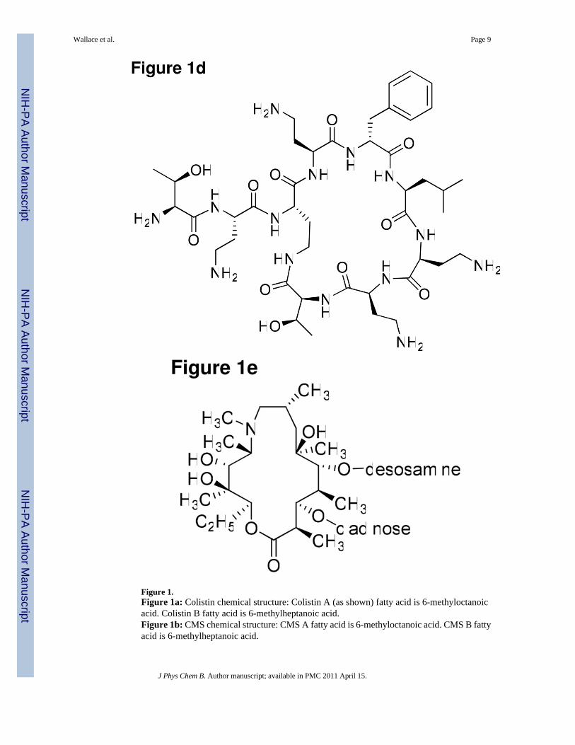
Figure 1.Figure 1a: Colistin chemical structure: Colistin A (as shown) fatty acid is 6-methyloctanoicacid. Colistin B fatty acid is 6-methylheptanoic acid.Figure 1b: CMS chemical structure: CMS A fatty acid is 6-methyloctanoic acid. CMS B fattyacid is 6-methylheptanoic acid.
Wallace et al. Page 9
J Phys Chem B. Author manuscript; available in PMC 2011 April 15.
NIH
-PA Author Manuscript
NIH
-PA Author Manuscript
NIH
-PA Author Manuscript
Figure 1c: Polymyxin B chemical structure: Polymyxin B1 fatty acid is 6-methyloctanoic acid.Polymyxin B2 fatty acid is 6-methylheptanoic acid.Figure 1d: Polymyxin B nonapeptide chemical structure.Figure 1e: Azithromycin chemical structure.
Wallace et al. Page 10
J Phys Chem B. Author manuscript; available in PMC 2011 April 15.
NIH
-PA Author Manuscript
NIH
-PA Author Manuscript
NIH
-PA Author Manuscript

Figure 2.Dynamic light scattering of solutions of colistin, CMS and polymyxin B nonapeptide versusconcentration of the respective species. The dynamic light scattering is expressed as theintensity of light scattered at 173° (S173) for solutions of the species minus the intensity oflight scattered by the solvent (S173solvent). Arrows indicate points of inflection (CMC).
Wallace et al. Page 11
J Phys Chem B. Author manuscript; available in PMC 2011 April 15.
NIH
-PA Author Manuscript
NIH
-PA Author Manuscript
NIH
-PA Author Manuscript

Figure 3.Intensity size distribution profiles for colistin, CMS and Triton X-100 micelles at 25 °C.
Wallace et al. Page 12
J Phys Chem B. Author manuscript; available in PMC 2011 April 15.
NIH
-PA Author Manuscript
NIH
-PA Author Manuscript
NIH
-PA Author Manuscript
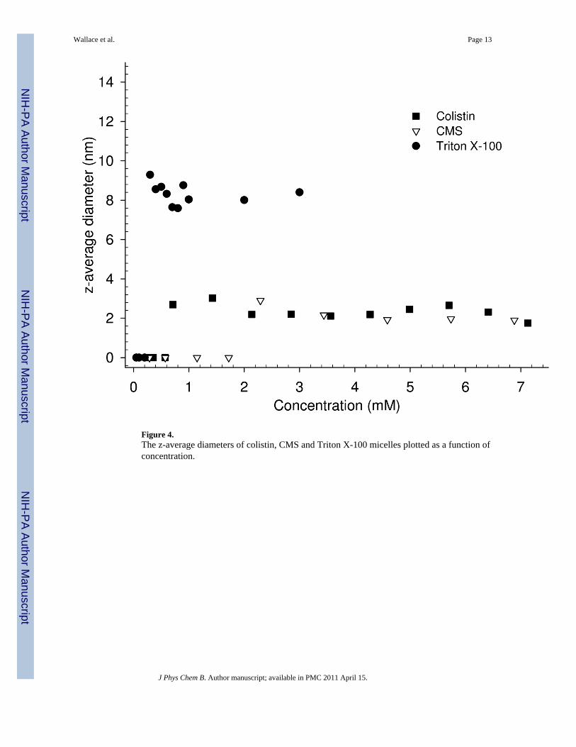
Figure 4.The z-average diameters of colistin, CMS and Triton X-100 micelles plotted as a function ofconcentration.
Wallace et al. Page 13
J Phys Chem B. Author manuscript; available in PMC 2011 April 15.
NIH
-PA Author Manuscript
NIH
-PA Author Manuscript
NIH
-PA Author Manuscript

Figure 5.Surface tension of aqueous solutions of colistin and CMS.
Wallace et al. Page 14
J Phys Chem B. Author manuscript; available in PMC 2011 April 15.
NIH
-PA Author Manuscript
NIH
-PA Author Manuscript
NIH
-PA Author Manuscript
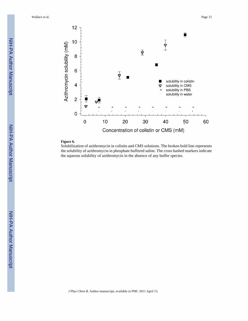
Figure 6.Solubilization of azithromycin in colistin and CMS solutions. The broken bold line representsthe solubility of azithromycin in phosphate buffered saline. The cross hashed markers indicatethe aqueous solubility of azithromycin in the absence of any buffer species.
Wallace et al. Page 15
J Phys Chem B. Author manuscript; available in PMC 2011 April 15.
NIH
-PA Author Manuscript
NIH
-PA Author Manuscript
NIH
-PA Author Manuscript
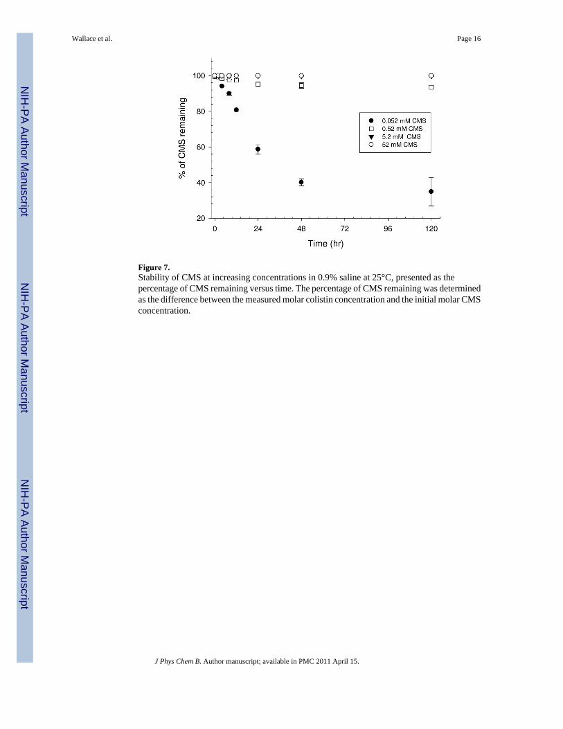
Figure 7.Stability of CMS at increasing concentrations in 0.9% saline at 25°C, presented as thepercentage of CMS remaining versus time. The percentage of CMS remaining was determinedas the difference between the measured molar colistin concentration and the initial molar CMSconcentration.
Wallace et al. Page 16
J Phys Chem B. Author manuscript; available in PMC 2011 April 15.
NIH
-PA Author Manuscript
NIH
-PA Author Manuscript
NIH
-PA Author Manuscript