Adenomatous Polyposis Coli (APC) Is Required for Normal Development of Skin and Thymus
13
Adenomatous Polyposis Coli (APC) Is Required for Normal Development of Skin and Thymus Mari Kuraguchi 1,2 , Xiu-Ping Wang 2 , Roderick T. Bronson 3 , Rebecca Rothenberg 1 , Nana Yaw Ohene-Baah 1 , Jennifer J. Lund 2 , Melanie Kucherlapati 1,2 , Richard L. Maas 2 , Raju Kucherlapati 1* 1 Harvard-Partners Center for Genetics and Genomics, Harvard Medical School, Boston, Massachusetts, United States of America, 2 Division of Genetics, Department of Medicine, Brigham and Women’s Hospital, Harvard Medical School, Boston, Massachusetts, United States of America, 3 Rodent Histopathology Core, Dana-Farber Harvard Cancer Center, Boston, Massachusetts, United States of America The tumor suppressor gene Apc (adenomatous polyposis coli) is a member of the Wnt signaling pathway that is involved in development and tumorigenesis. Heterozygous knockout mice for Apc have a tumor predisposition phenotype and homozygosity leads to embryonic lethality. To understand the role of Apc in development we generated a floxed allele. These mice were mated with a strain carrying Cre recombinase under the control of the human Keratin 14 (K14) promoter, which is active in basal cells of epidermis and other stratified epithelia. Mice homozygous for the floxed allele that also carry the K14-cre transgene were viable but had stunted growth and died before weaning. Histological and immunochemical examinations revealed that K14-cre–mediated Apc loss resulted in aberrant growth in many ectodermally derived squamous epithelia, including hair follicles, teeth, and oral and corneal epithelia. In addition, squamous metaplasia was observed in various epithelial-derived tissues, including the thymus. The aberrant growth of hair follicles and other appendages as well as the thymic abnormalities in K14-cre; Apc CKO/CKO mice suggest the Apc gene is crucial in embryonic cells to specify epithelial cell fates in organs that require epithelial– mesenchymal interactions for their development. Citation: Kuraguchi M, Wang XP, Bronson RT, Rothenberg R, Ohene-Baah NY, et al. (2006) Adenomatous polyposis coli (APC) is required for normal development of skin and thymus. PLoS Genet 2(9): e146. DOI: 10.1371/journal.pgen.0020146 Introduction Adenomatous polyposis coli (APC) is a member of the Wnt signaling pathway and one of its known functions is to regulate the levels of b-catenin. Alterations in b-catenin regulation are very common in human tumors [1]. Loss of APC is associated with stabilization of the cytosolic b-catenin that ultimately results in its migration to the nucleus and activating a cascade of events leading to tumorigenesis. APC also interacts with a multitude of other cellular proteins, including axin-2 (AXIN2), plakoglobin (JUP), Asef (ARHGEF4), kinesin superfamily–associated protein 3 (KIFAP3), EB1 (MAPRE1), microtubules, and the human homolog of Droso- phila discs large (DLG1). These interactions suggest that APC can potentially regulate many cellular functions, including intercellular adhesion, cytoskeletal organization, regulation of plakoglobin levels, regulation of the cell cycle and apoptosis, orientation of asymmetric stem cell division, and control of cell polarization [2,3]. APC is a tumor suppressor gene. Somatic mutations in APC are frequently found in many sporadic tumors of the colon and rectum. Autosomal dominant germline mutations in APC cause familial adenomatous polypois (FAP) and its variant, Gardner syndrome. FAP patients are characterized by hundreds of adenomatous colorectal polyps, with an almost inevitable progression to colorectal cancer in the third and fourth decades of life [4,5]. In addition to colorectal neoplams, these individuals can develop extracolonic symp- toms, among which are upper gastrointestinal tract polyps, congenital hypertrophy of the retinal pigment epithelium, desmoid tumors, disorders of the maxillary and skeletal bones, and dental abnormalities [6], suggesting the impor- tance of APC gene functions in these organ systems. Although the role of APC in the initiation of human colorectal cancer is well established, its role in other tissue and developmental processes are not well understood. Given the importance of regulation of Wnt signaling in embryonic pattern formation and morphogenesis of many organs, mechanistic understanding of APC in development and in extracolonic tissues becomes critical to better assess potential adverse events in humans. One approach to understand the role of Apc in development is to develop mice with an inactivating Apc mutation. Several genetically modified mouse strains for Apc have been described [7–10]. Most of these models, in the heterozygous state, show a gastro- intestinal and other tumor predisposition phenotype [7–10]. Mouse embryos that are homozygous for the genetic modification die during embryogenesis, and some of the models do not survive beyond gastrulation [8,11]. An Editor: Gregory S. Barsh, Stanford University School of Medicine, United States of America Received March 29, 2006; Accepted July 28, 2006; Published September 15, 2006 A previous version of this article appeared as an Early Online Release on July 28, 2006 (DOI: 10.1371/journal.pgen.0020146.eor). DOI: 10.1371/journal.pgen.0020146 Copyright: Ó 2006 Kuraguchi et al. This is an open-access article distributed under the terms of the Creative Commons Attribution License, which permits unrestricted use, distribution, and reproduction in any medium, provided the original author and source are credited. Abbreviations: APC, adenomatous polyposis coli; CKO, conditional knockout; E, embryonic day; ES, embryonic stem; FAP, familial adenomatous polyposis; FRT, FLP recognition target; ISH, in situ hybridization; K14, keratin 14; ORS, outer root sheath; P, postnatal day; TEC, thymic epithelial cell * To whom correspondence should be addressed. E-mail: rkucherlapati@partners. org PLoS Genetics | www.plosgenetics.org September 2006 | Volume 2 | Issue 9 | e146 1362
-
Upload
independent -
Category
Documents
-
view
0 -
download
0
Transcript of Adenomatous Polyposis Coli (APC) Is Required for Normal Development of Skin and Thymus

Adenomatous Polyposis Coli (APC) Is Requiredfor Normal Development of Skin and ThymusMari Kuraguchi
1,2, Xiu-Ping Wang
2, Roderick T. Bronson
3, Rebecca Rothenberg
1, Nana Yaw Ohene-Baah
1,
Jennifer J. Lund2
, Melanie Kucherlapati1,2
, Richard L. Maas2
, Raju Kucherlapati1*
1 Harvard-Partners Center for Genetics and Genomics, Harvard Medical School, Boston, Massachusetts, United States of America, 2 Division of Genetics, Department of
Medicine, Brigham and Women’s Hospital, Harvard Medical School, Boston, Massachusetts, United States of America, 3 Rodent Histopathology Core, Dana-Farber Harvard
Cancer Center, Boston, Massachusetts, United States of America
The tumor suppressor gene Apc (adenomatous polyposis coli) is a member of the Wnt signaling pathway that isinvolved in development and tumorigenesis. Heterozygous knockout mice for Apc have a tumor predispositionphenotype and homozygosity leads to embryonic lethality. To understand the role of Apc in development wegenerated a floxed allele. These mice were mated with a strain carrying Cre recombinase under the control of thehuman Keratin 14 (K14) promoter, which is active in basal cells of epidermis and other stratified epithelia. Micehomozygous for the floxed allele that also carry the K14-cre transgene were viable but had stunted growth and diedbefore weaning. Histological and immunochemical examinations revealed that K14-cre–mediated Apc loss resulted inaberrant growth in many ectodermally derived squamous epithelia, including hair follicles, teeth, and oral and cornealepithelia. In addition, squamous metaplasia was observed in various epithelial-derived tissues, including the thymus.The aberrant growth of hair follicles and other appendages as well as the thymic abnormalities in K14-cre; ApcCKO/CKO
mice suggest the Apc gene is crucial in embryonic cells to specify epithelial cell fates in organs that require epithelial–mesenchymal interactions for their development.
Citation: Kuraguchi M, Wang XP, Bronson RT, Rothenberg R, Ohene-Baah NY, et al. (2006) Adenomatous polyposis coli (APC) is required for normal development of skin andthymus. PLoS Genet 2(9): e146. DOI: 10.1371/journal.pgen.0020146
Introduction
Adenomatous polyposis coli (APC) is a member of the Wntsignaling pathway and one of its known functions is toregulate the levels of b-catenin. Alterations in b-cateninregulation are very common in human tumors [1]. Loss ofAPC is associated with stabilization of the cytosolic b-cateninthat ultimately results in its migration to the nucleus andactivating a cascade of events leading to tumorigenesis. APCalso interacts with a multitude of other cellular proteins,including axin-2 (AXIN2), plakoglobin (JUP), Asef (ARHGEF4),kinesin superfamily–associated protein 3 (KIFAP3), EB1(MAPRE1), microtubules, and the human homolog of Droso-phila discs large (DLG1). These interactions suggest that APCcan potentially regulate many cellular functions, includingintercellular adhesion, cytoskeletal organization, regulationof plakoglobin levels, regulation of the cell cycle andapoptosis, orientation of asymmetric stem cell division, andcontrol of cell polarization [2,3].
APC is a tumor suppressor gene. Somatic mutations in APCare frequently found in many sporadic tumors of the colonand rectum. Autosomal dominant germline mutations in APCcause familial adenomatous polypois (FAP) and its variant,Gardner syndrome. FAP patients are characterized byhundreds of adenomatous colorectal polyps, with an almostinevitable progression to colorectal cancer in the third andfourth decades of life [4,5]. In addition to colorectalneoplams, these individuals can develop extracolonic symp-toms, among which are upper gastrointestinal tract polyps,congenital hypertrophy of the retinal pigment epithelium,desmoid tumors, disorders of the maxillary and skeletalbones, and dental abnormalities [6], suggesting the impor-tance of APC gene functions in these organ systems.
Although the role of APC in the initiation of humancolorectal cancer is well established, its role in other tissueand developmental processes are not well understood. Giventhe importance of regulation of Wnt signaling in embryonicpattern formation and morphogenesis of many organs,mechanistic understanding of APC in development and inextracolonic tissues becomes critical to better assess potentialadverse events in humans. One approach to understand therole of Apc in development is to develop mice with aninactivating Apc mutation. Several genetically modifiedmouse strains for Apc have been described [7–10]. Most ofthese models, in the heterozygous state, show a gastro-intestinal and other tumor predisposition phenotype [7–10].Mouse embryos that are homozygous for the geneticmodification die during embryogenesis, and some of themodels do not survive beyond gastrulation [8,11]. An
Editor: Gregory S. Barsh, Stanford University School of Medicine, United States ofAmerica
Received March 29, 2006; Accepted July 28, 2006; Published September 15, 2006
A previous version of this article appeared as an Early Online Release on July 28,2006 (DOI: 10.1371/journal.pgen.0020146.eor).
DOI: 10.1371/journal.pgen.0020146
Copyright: � 2006 Kuraguchi et al. This is an open-access article distributed underthe terms of the Creative Commons Attribution License, which permits unrestricteduse, distribution, and reproduction in any medium, provided the original authorand source are credited.
Abbreviations: APC, adenomatous polyposis coli; CKO, conditional knockout; E,embryonic day; ES, embryonic stem; FAP, familial adenomatous polyposis; FRT, FLPrecognition target; ISH, in situ hybridization; K14, keratin 14; ORS, outer rootsheath; P, postnatal day; TEC, thymic epithelial cell
* To whom correspondence should be addressed. E-mail: [email protected]
PLoS Genetics | www.plosgenetics.org September 2006 | Volume 2 | Issue 9 | e1461362

alternate approach to understand the role of Apc indevelopment and/or in specific tissues is to generate a mousestrain that carries a conditionally modified allele and mate itwith a mouse strain that facilitates the modification of theconditional allele in specific cell lineages.
To assess the role of Apc in different stages of lifesystematically, we generated mice containing a conditionalknockout (CKO) mutant allele of Apc (ApcCKO). These micewere mated with a strain carrying Cre recombinase under thecontrol of the human Keratin 14 (K14) promoter, which isactive in basal cells of epidermis and other stratified epithelia.We report here that K14 promoter-driven loss of Apc resultedin aberrant development of several organs that requireinductive epithelial–mesenchymal interactions, includinghair follicle, teeth, and thymus, and resulted in neonataldeath in mice. We found that Apc plays a crucial role indeterminations of cell fates during the embryonic develop-ment, possibly via temporal and tissue-specific regulation ofb-catenin levels in the skin, its appendages, and in the thymus.
Results
Generation of the ApcCKO and ApcD580 MiceTo investigate the role of Apc in development of skin and
its appendages, we used the Cre/loxP technology to introducea conditional mutation of the Apc gene in mice. Weconstructed embryonic stem (ES) cells and mice carrying anApc allele harboring both a pair of loxP sites flanking Apcexon 14 and a pair of FLP recognition target (FRT) sitesflanking PGK-neomycin selection cassette by recombineering[12,13] (Figure 1A, ApcCKON allele, N for neomycin cassette). APGK-neomycin cassette was inserted in the same transcrip-tional orientation as Apc in intron 14 of the endogenous gene.The loxP and FRT sites were used to aid unidirectionalrecombination [12,13]. Two mouse lines containing the samemodification were generated from two independent ESclones to ensure that these two lines behave in the sameway. These ApcCKON/þmice were crossed with FLPe-deleter togenerate ApcCKO/þ mice that were heterozygous for the final
Apc conditional (ApcCKO) allele that removed the PGK-neomycin cassette and contains only the loxP sites in theintrons flanking exon 14. To assess the effect of deleting exon14 in mice, both lines of ApcCKO/þmice were crossed with theCre-deleter to generate the germline knockout line of Apc,designated ApcD580/þ. The mutant allele (ApcD580) lacks exon 14(Figure 1A). The transcript from loss of exon 14 results in ashift in the normal reading frame, resulting in a prematurechain termination codon which, if utilized, would result in atruncated polypeptide that is 605 aa in length, of which thefirst 580 aa correspond to the normal Apc protein.Southern blot analysis of tail DNA from F1 offspring of
both ApcCKON and ApcD580 lines confirmed the germlinetransmission of modified Apc allele (Figure 1B). Mice thatare heterozygous for ApcD580 mutation are viable but have asignificantly reduced lifespan (Figure 1C). These resultssuggested that deletion of exon 14 indeed results in eitherloss or abnormal function of the Apc gene product. ApcD580/þ
mice have median survival of 5 mo of age (Figure 1C), withprogressive signs of rectal bleeding and anemia. Similar to theresults reported with an independently generated ApcD580/þ
conditional mouse strain [14], ApcD580/þ mice had more than100 (120 6 37, n ¼ 11) intestinal tumors at the time of theirdeath (Figure S1). Inactivation of wild-type Apc is animportant prerequisite for tumor development. We analyzed30 intestinal tumors from ApcD580/þ mice by in vitro tran-scription and translation assay, but none of them showedtruncated Apc products (unpublished data), indicating thatthe most likely mechanism of wild-type Apc inactivation is byallelic loss. The mutant allele had to be maintained andtransmitted through male mice, as ApcD580/þ females werefrequently not healthy enough to successfully nurse their ownpups because of their tumor burden.ApcCKO/þ mice were intercrossed to generate ApcCKO/CKO
offspring. Approximately one-quarter of the offspring (17 of81) were homozygous for the ApcCKO allele. These mice as wellas heterozygous mice for ApcCKO allele are normal, showing nodifferences in their survival to the wild-type littermates(Figure 1C). We tested whether our ApcCKO allele cancompliment the wild-type allele by crossing the ApcCKO/CKO
female with ApcD580/þ male mouse. The resultant ApcCKO/D580
offspring were viable and born in the Mendelian ratio,suggesting that the presence of loxP sites in introns flankingexon 14 have no adverse effect on the function of the Apcgene.
K14-Driven Loss of Apc Results in Severe GrowthRetardation and Early LethalityTo introduce the mutation of Apc into cells expressing K14,
we crossed WW6 ES cell–derived [15] ApcCKO/þmice with K14-cre recombinase mice in FVB background [16]. The K14-cre;ApcCKO/þ mice were normal in appearance and were fertile.K14-cre; ApcCKO/þmales were crossed to ApcCKO/CKO females toavoid the potential deleter effect in oocytes of K14-cre–positive females [17]. The mice were intercrossed thereafterfor maintenance; hence, the mice used for analysis were in amixed background of FVB, 129/S, and C57BL/6 in similarproportions, with minimal contribution of SJ.The K14 promoter is a commonly used epidermal cell
promoter because of its expression by the mitotically activecells of the epidermis and its appendages in mature skin [18],but most notably it is active in embryonic ectoderm as early
PLoS Genetics | www.plosgenetics.org September 2006 | Volume 2 | Issue 9 | e1461363
Role of APC in Skin and Its Appendages
Synopsis
Patients with familial adenomatous polyposis (FAP) and its variant,Gardner’s syndrome, will develop hundreds of colorectal polyps. It isa heritable disease that is linked to a genetic mutation in the tumorsuppressor gene APC (adenomatous polyposis coli). These individ-uals also develop extracolonic symptoms, among which arecongenital hypertrophy of the retinal pigment epithelium, desmoidtumors, epidermoid cysts, disorders of the maxillary and skeletalbones, and dental abnormalities, suggesting the importance of APCfunctions in these organs. To understand the role of Apc indevelopment and in organs other than intestine, we generated Apcmutant mice that can conditionally delete the gene when exposedto Cre recombinase. These mice were mated with K14 (Keratin 14)–cre mice that express Cre recombinase in skin and its appendages.The authors found that the mutant mice that have lost Apc in K14-cre transgene–expressing tissues were viable, but had stuntedgrowth and died before weaning. These mutant mice showeddevelopmental abnormalities not only in skin but also in manyepithelial-derived tissues, including teeth and thymus. This workdemonstrates the importance of Apc in development of manyorgans, and provides new insights into diagnosis and managementof patients with APC mutations.

as the single layered ectodermal stage of embryonic day (E)9.5 [19]. A restricted expression of K14 is also found in thymicepithelial cells (TECs) in the medulla of normal thymus [20].
We genotyped a total of 458 pups (8–10 d old) from 67litters resulting from crosses between K14-cre; ApcCKO/þ andApcCKO/CKO mice. The mutant mice of the genotype K14-cre;ApcCKO/CKO (hereafter, KA mutant) were born, but theobserved frequency of KA mutants was much less thanexpected (78 of 458 [17.0%]; p , 0.0005 Chi-square analysis,Table 1). To assess the basis for the neonatal lethality of KAmutants, we monitored three litters from birth to weaning bymeasuring the body weight of each pup every day. A total of25 pups were born from three litters, of which 7 (28%) wereconfirmed to be K14-cre; ApcCKO/CKO by genotyping, indicatingthat KA mutants were born in the expected Mendelian ratio.
The KA mutant pups were nursed normally, and there wasmilk in their stomachs during the first 2 or 3 d after birth, butthey failed to thrive (Figure 2). By postnatal day (P) 8–10, atthe time of genotyping, many KA mutant pups wereconsiderably smaller than their littermates (Figure 2B–2F)and some have died at or prior to this age. None of KAmutants survived to weaning age.The ability of whole embryos to exclude blue dye was used
to examine the epidermal barrier, normally acquired begin-ning at E16 and complete by E18.5 [21]. Analyses of E17.5–E18.5 KA mutants showed that they were able to exclude bluedye, indicating that the epidermal barrier was intact (FigureS2). At these embryonic ages, there were no differences in sizebetween the mutants and their littermates, but the mutantsshowed a patch of ‘‘birthmark’’ or dark pigmentation on their
Figure 1. Generation of the Conditional Apc Allele
(A) Schematic diagram of exons 14 and 15 of the mouse Apc gene, the targeting vector, and the resulting conditional allele with 2 LoxP sitessandwiching the exon 14. The PGK-neomycin cassette was inserted within intron 14 by recombineering technique. This cassette is sandwiched by 2 FRTsites that could be removed by crossing to FLPe-expressing mice. Positions of PCR primers used for genotyping PCR (F2, R2, R4) and RT-PCR (F546 andR721) are indicated. Positions of probe used for Southern blot analysis with NdeI sites are also shown. Upon Cre-mediated recombination, exon 14 isremoved and leads to truncated Apc protein, of which the first 580 aa correspond to the normal.(B) Southern blot analysis of NdeI-digested genomic tail DNA isolated from F1 mice of various Apc mouse lines (ApcCKON, ApcD580), hybridized to a 600-bp probe. Tail genomic DNA from ApcCKON F1 mice derived from a modified ES clone showed a 12-kb band for the ApcCKON allele and a 10-kb band forthe wild-type allele, whereas genomic DNA from the ApcD580 mouse was heterozygous for the ApcD580 allele (9.2-kb band).(C) Kaplan-Meier survival plot of ApcCKO/þmice (thin solid line, n¼ 39), ApcCKO/CKO mice (thin dotted line, n¼ 57), ApcD580/þmice (solid line, n¼ 51), andwild-type littermates (broken line, n¼ 21). Heterozygosity of the ApcD580 allele led to a significantly shortened survival (p , 0.0001), whereas those ofheterozygous and homozygous ApcCKO mice had no significant difference to that of wild-type littermates.DOI: 10.1371/journal.pgen.0020146.g001
PLoS Genetics | www.plosgenetics.org September 2006 | Volume 2 | Issue 9 | e1461364
Role of APC in Skin and Its Appendages

foreheads and a dark median line that ran caudally from headto tail. Their external ears or pinnae were shriveled inappearance and pigmented compared to those of littermates.
External characteristics of KA mutants that were evident atE18.5 persisted after birth and became more prominent asthey grew (Figure 2A–2F). Growth of pelage hair was generallydelayed in the mutants. At around P8, the KA mutants werehairless and had wrinkled skin while their phenotypicallynormal littermates had a smooth thin coat of hair (Figure 2B).At this age, two lower incisors start to erupt in normallittermates and these were absent in the KA mutants (Figure2C and 2D). Animals also tended to be smaller and aroundP10–P12 displayed abnormally short and misshapen vibrissaeand short, shaggy pelage hairs (Figure 2C and 2D). Develop-ment of thick ridges in their skin, particularly around theears, eyelids, forehead, nose, and paws, became noticeable(Figure 2E). These regions looked scaly, and these animalshardly kept their eyes open. In contrast to the normallittermates that consistently increased their body weight withage, surviving KA mutants started to lose weight from P10onwards; by P16–P17 they were all lethargic, and none ofthem survived to weaning (Figure 2E and 2F). At the time ofautopsy all the mutants were toothless, without incisors ormolars, and their stomachs were consistently small and hadno solid food, unlike their age-matched littermates, suggest-ing that the observed weight loss could be the result of failureto ingest solid food (Figure 2F). Interestingly, changes in bodyweights and timing of hair growth varied considerably amongmutant pups even if they were from the same litter, whereasthose of phenotypically normal littermates tended to besimilar. This difference was also reflected in the variation intiming of death in mutants: some mutant pups were bornalive but died within a day or two, some survived close to theweaning age. This variability of the mutant phenotypessuggests possible variation in the timing and efficiency ofcre-mediated Apc deletion. It is possible that the geneticbackground has a role to play in this variability.
Gross examination of internal organs also showed that themutants’ thymi were consistently inconspicuous and werevery small for their age, whereas those of their littermateswere very prominent in size (Figure 2G). This difference wasevident as early as P3. Quite frequently mutant thymi in P12–P17 mutant mice also contained black deposits within thetissue (unpublished data). Mutant mice were also examinedfor any skeletal abnormalities by preparing skeletal speci-mens of P16–P17 mice stained with Alizarin red. No differ-ences between the normal and KA mutant mice in the
mandibular bone can be detected, but the mutant micelacked or had underdeveloped set of maxillary incisors andmolars (Figure 2H). We detected no other major skeletalabnormalities.
Genotype- and Tissue-Specific Expression of theTruncated Apc TranscriptsTo assess the molecular effects of the K14-cre–mediated
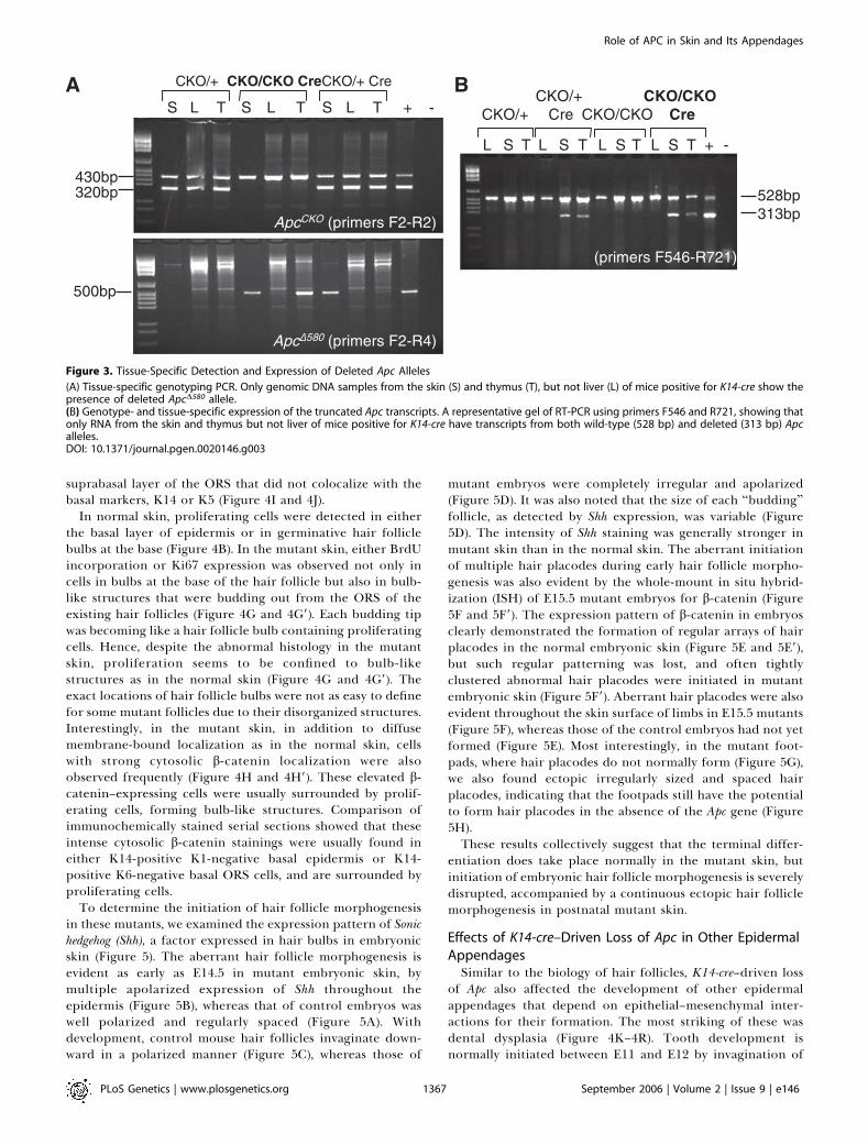
recombination, we screened for the presence of deleted Apc(ApcD580) alleles. Genomic DNA was extracted from liver,thymus, and skin from all 4 possible genotypes: K14-cre;ApcCKO/CKO, K14-cre; ApcCKO/þ, ApcCKO/CKO, and ApcCKO/þ. Gen-otyping on genomic DNA from these tissues showed that theApcD580 allele (500-bp product) was detected only from theskin and thymus of the K14-cre–positive mice. The presence ofmutant Apc allele in the thymus of K14-cre; ApcCKO/þmice wasconsistently much less than the DNA from the skin of thesame animal or other tissues from the KA mutants. Inaddition, this product was not detected at all in either theliver of K14-cre–positive or in any of the K14-cre–negativemouse tissues samples, establishing that Cre-mediated re-combination has taken place in the tissue-specific manner inthe mice that inherited K14-cre (Figure 3A).Apc transcripts were also analyzed by RT-PCR with primers
spanning exon 14 (Figure 1A) using total RNA isolated fromthe corresponding tissue samples. We detected the expectedRT-PCR product (313 bp) from the truncated Apc (ApcD580)allele only in the tissues where Cre recombinase is known tobe expressed in the K14-cre–positive mice. However, thisproduct was not detected in either the K14-cre–negativemouse tissues samples or the liver of K14-cre–positive mice,and only the product from the wild-type allele (528 bp) wasdetected from these RNA samples, further confirming thatCre-mediated recombination has taken place in the tissue-and genotype-specific manner (Figure 3B).
K14-cre–Driven Apc Loss Induced Aberrant Hair Folliclesthroughout the EpidermisTo understand the basis for delayed and abnormal hair
development in the KA mutants, we conducted a histologicaland immunohistochemical examination (Figure 4). The hairfollicle is an epidermal appendage that consists of an upperpermanent portion, and a lower cycling portion thatproduces the hair [22,23]. The outer root sheath (ORS) iscontiguous with and biochemically similar to the basal layerof the epidermis. The inner layers of the hair follicle includethree concentric layers of inner root sheath and threeconcentric layers of hair-producing cells. At the base of thehair follicle is the germinative hair follicle bulb, whichcontains rapidly proliferating ‘‘matrix’’ cells that differ-entiate to populate all of the layers of the inner root sheathand the hair shaft itself [22]. During the anagen phase of thehair cycle (until P15), hair follicles of phenotypically normalmice grew deeply into the subcutaneous fat and wereuniformly spaced and aligned in parallel arrays at a specificangle relative to the skin surface (Figure 4A). In contrast, KAmutant follicles were irregularly spaced and often seen asdisoriented and clamped invaginations at P3 that becameeven more remarkable at P12 when the mutant mice werecovered by fur coat (Figure 4F). Bulbs were often bent inaddition to being irregularly angled to one another and theirsizes and locations were often variable. Clusters of multiple
Table 1. Genotype Distribution of Progeny from the Matings
Genotype Number of Pups (%) ApcD580 a Phenotype
ApcCKO/þ — 144 (31.5) � Normal
ApcCKO/þ K14-cre 116 (25.3) þ Normal
ApcCKO/CKO — 120 (26.2) � Normal
ApcCKO/CKO K14-cre 78 (17.0) þ Skin abnormality,
die in , 3 wk
Total 458 (100)
aThe detection of ApcD580 allele in tail DNA by genotyping.p , 0.0005 by Chi-square analysis. Red text indicates mutant.DOI: 10.1371/journal.pgen.0020146.t001
PLoS Genetics | www.plosgenetics.org September 2006 | Volume 2 | Issue 9 | e1461365
Role of APC in Skin and Its Appendages

invaginations or dysplastic follicular structures were fre-quently observed throughout the epidermis, whereas otherregions showed gaps with no follicles. Serial sectioningindicated that some of the hair follicles in the P12 mutantskin were not properly formed or shorter than normal. Takentogether, these features could account for the apparentlydelayed, followed by outgrowth, of the short and shaggy-looking fur coat of these mutant mice.
Apc is a regulator of b-catenin that is important for Wntsignaling. We examined the patterns of expression of b-catenin in the affected tissues. In the normal skin, b-catenin, amember of the adherens junction complex, was found in theORS of hair follicles and basal layer of epidermis, where K14expression is also observed (Figure 4C and 4D), whereas the
expression of K1, involucrin, and loricrin (markers forspinous and granular layers of epidermis) was only observedin the nonbasal epidermis (unpublished data). The patterns ofexpression of K14, K1, involucrin, and loricrin, in skin frommutant and normal littermate mice at P3–P17, showed nosignificant differences in the terminal differentiation (Figure4A–4D, 4F–4I). Similarly, the pattern of expression of K6,which is normally only expressed in the suprabasal or innerlayer of the ORS of the hair follicle but not in the epidermis(Figure 4E), did not change. Due to the abnormal anddisorganized structure of hair follicles themselves, K6 local-ization highlighted the histological abnormality (Figure 4J).Yet as in the normal skin, K6 was principally seen only in the
Figure 2. Postnatal Mortality and Stunted Growth in K14-cre; ApcCKO/CKO Mutant Mice
Animals whose genotype is either heterozygous or homozygous for the wild-type Apc allele are referred to as normal (N); those whose genotype are K14-cre; ApcCKO/CKO and show the presence of K14-cre–recombined mutant Apc allele are called mutant (M).(A) Two P3 mutant mice, M1 and M2, and their normal littermates, showing size variation among mutants.(B) P8 mutant mouse (right) and a normal littermate. Note sparseness of hair coat and abnormal ears.(C–D) Vibrissae of whisker pads are short and oddly angled in a P12 mutant mouse (C), relative to control (D). Note the lack of incisors in the mutant.(E) A P17 mutant mouse (right) with its littermate. Its bare forehead, dorsal median line, and abnormal ears are evident.(F) Growth curve of mutants and normal littermates. Mutants exhibit stunted growth, which became more prominent as they aged, and weighsignificantly less than littermates from P8 (p , 0.05).(G) Comparison of mutant and normal thymus from P3 mice. The mutant thymus (left) is dramatically smaller for its age compared to the normallittermate (right). The scale bar equals 1 mm.(H) Skeletal preparations of normal (left) and mutant (right), showing differences in development of both incisor (I) and molar (M) teeth.DOI: 10.1371/journal.pgen.0020146.g002
PLoS Genetics | www.plosgenetics.org September 2006 | Volume 2 | Issue 9 | e1461366
Role of APC in Skin and Its Appendages
suprabasal layer of the ORS that did not colocalize with thebasal markers, K14 or K5 (Figure 4I and 4J).
In normal skin, proliferating cells were detected in eitherthe basal layer of epidermis or in germinative hair folliclebulbs at the base (Figure 4B). In the mutant skin, either BrdUincorporation or Ki67 expression was observed not only incells in bulbs at the base of the hair follicle but also in bulb-like structures that were budding out from the ORS of theexisting hair follicles (Figure 4G and 4G9). Each budding tipwas becoming like a hair follicle bulb containing proliferatingcells. Hence, despite the abnormal histology in the mutantskin, proliferation seems to be confined to bulb-likestructures as in the normal skin (Figure 4G and 4G9). Theexact locations of hair follicle bulbs were not as easy to definefor some mutant follicles due to their disorganized structures.Interestingly, in the mutant skin, in addition to diffusemembrane-bound localization as in the normal skin, cellswith strong cytosolic b-catenin localization were alsoobserved frequently (Figure 4H and 4H9). These elevated b-catenin–expressing cells were usually surrounded by prolif-erating cells, forming bulb-like structures. Comparison ofimmunochemically stained serial sections showed that theseintense cytosolic b-catenin stainings were usually found ineither K14-positive K1-negative basal epidermis or K14-positive K6-negative basal ORS cells, and are surrounded byproliferating cells.
To determine the initiation of hair follicle morphogenesisin these mutants, we examined the expression pattern of Sonichedgehog (Shh), a factor expressed in hair bulbs in embryonicskin (Figure 5). The aberrant hair follicle morphogenesis isevident as early as E14.5 in mutant embryonic skin, bymultiple apolarized expression of Shh throughout theepidermis (Figure 5B), whereas that of control embryos waswell polarized and regularly spaced (Figure 5A). Withdevelopment, control mouse hair follicles invaginate down-ward in a polarized manner (Figure 5C), whereas those of
mutant embryos were completely irregular and apolarized(Figure 5D). It was also noted that the size of each ‘‘budding’’follicle, as detected by Shh expression, was variable (Figure5D). The intensity of Shh staining was generally stronger inmutant skin than in the normal skin. The aberrant initiationof multiple hair placodes during early hair follicle morpho-genesis was also evident by the whole-mount in situ hybrid-ization (ISH) of E15.5 mutant embryos for b-catenin (Figure5F and 5F9). The expression pattern of b-catenin in embryosclearly demonstrated the formation of regular arrays of hairplacodes in the normal embryonic skin (Figure 5E and 5E9),but such regular patterning was lost, and often tightlyclustered abnormal hair placodes were initiated in mutantembryonic skin (Figure 5F9). Aberrant hair placodes were alsoevident throughout the skin surface of limbs in E15.5 mutants(Figure 5F), whereas those of the control embryos had not yetformed (Figure 5E). Most interestingly, in the mutant foot-pads, where hair placodes do not normally form (Figure 5G),we also found ectopic irregularly sized and spaced hairplacodes, indicating that the footpads still have the potentialto form hair placodes in the absence of the Apc gene (Figure5H).These results collectively suggest that the terminal differ-
entiation does take place normally in the mutant skin, butinitiation of embryonic hair follicle morphogenesis is severelydisrupted, accompanied by a continuous ectopic hair folliclemorphogenesis in postnatal mutant skin.
Effects of K14-cre–Driven Loss of Apc in Other EpidermalAppendagesSimilar to the biology of hair follicles, K14-cre–driven loss
of Apc also affected the development of other epidermalappendages that depend on epithelial–mesenchymal inter-actions for their formation. The most striking of these wasdental dysplasia (Figure 4K–4R). Tooth development isnormally initiated between E11 and E12 by invagination of
Figure 3. Tissue-Specific Detection and Expression of Deleted Apc Alleles
(A) Tissue-specific genotyping PCR. Only genomic DNA samples from the skin (S) and thymus (T), but not liver (L) of mice positive for K14-cre show thepresence of deleted ApcD580 allele.(B) Genotype- and tissue-specific expression of the truncated Apc transcripts. A representative gel of RT-PCR using primers F546 and R721, showing thatonly RNA from the skin and thymus but not liver of mice positive for K14-cre have transcripts from both wild-type (528 bp) and deleted (313 bp) Apcalleles.DOI: 10.1371/journal.pgen.0020146.g003
PLoS Genetics | www.plosgenetics.org September 2006 | Volume 2 | Issue 9 | e1461367
Role of APC in Skin and Its Appendages

ectodermally derived oral epithelium into the underlyingcranial neural crest–derived mesenchyme, generating a toothgerm. Despite the grossly toothless phenotype of KA mutants,histological analysis of their oral cavities revealed theformation of multiple tooth buds at each location. Theseaberrant teeth obviously failed to grow out during the dietarytransition from milk to solid food. Analogous to theexpression patterns of K14 and b-catenin in the normal skin,diffuse membrane-bound expression of b-catenin was de-tected in K14-expressing oral epithelium and ameloblasts ofnormal mice. In mutants, some of the K14-expressing cellsalso showed strong cytosolic/nuclear b-catenin staining, asobserved in the mutant skin (Figure 4Q, 4Q9, and 4R).Initiation of ectopic tooth buds in the mutant mice wasevident at E15.5 by extra dots of Shh expression adjacent tothe primary teeth (data not shown).
Loss of Apc also leads to hyperplasia in squamous epitheliaof cornea, oral, salivary, and Hardarian glands (unpublisheddata). Squamous metaplasia to hair follicle–like structures,ectopic hair follicle morphogenesis was also observed in theseepithelia.
K14-cre–Driven Apc Loss Results in Hypoplastic/Athymic
MiceThymus is an organ that is also known to have K14
expression [16]. It represents the primary lymphoid organ forthymocyte development and selection. Distinct population ofTECs of cortex and medulla mediates both of these criticalfunctions. Cortical and medullary TEC subsets are charac-terized by differential expression of four keratin species: K8,K18, K5, and K14.The normal thymus is a lobulated lymphoid organ, each
lobule clearly showing the two distinct TEC compartments,an outer cortex and an inner medulla (Figure 6). There wereno major differences in the histology of thymus between theages P3 to P17 in phenotypically normal littermates. Asshown in the H&E staining of thymus, the cortex was formedof dense lymphoid tissue that lacks nodules (Figure 6A). Sincethe stroma of the medulla is less heavily infiltrated withlymphocytes than the cortex, the medulla stained more lightlythan the cortex. In normal mice, the thymus retains its sizeuntil the young adult age and regresses thereafter by atrophy.In the normal young mice we examined (P3–P17), it is evident
Figure 4. Histological and Immunochemical Examination of P12 Skin and Teeth
(A–E) P12 normal skin. (F–J) P12 mutant skin. (K–N) P12 normal oral cavity. (O–R) P12 mutant oral cavity. Stained with H&E for histology (A, F, K–L, O–P),Ki67 (B, G), b-catenin (C, H, M, Q), K14 (D, I, N, R), and K6 (E, J). Aberrant follicular morphogenesis, characterized by formation of irregularly spaced,nonpolarized hair follicles, in mutant skin is evident. Despite the abnormal histology, proliferation seems to be confined to hair bulb-like structures(arrows in [G], inset [G9] at higher magnification), but in mutant skin (arrows in [H], inset [H9] at higher magnification) and oral cavity (arrows in insets[Q9] at higher magnification) elevated cytosolic localization of b-catenin is detected in some cells. Scale bars: 50 lm for (A–F), (H–J); 250 lm for (K) and(O); 100 lm for (G), (L–N), (P–R); 20 lm for (Q9).DOI: 10.1371/journal.pgen.0020146.g004
PLoS Genetics | www.plosgenetics.org September 2006 | Volume 2 | Issue 9 | e1461368
Role of APC in Skin and Its Appendages

that thymocytes were mitotically active in the cortex asdetermined by BrdU immunostaining (Figure 6B). Immuno-histochemistry of normal thymus from P3 to P17 miceshowed a similar staining pattern for K14 in that itsexpression was restricted to a small population of TECs inthe inner medullary region and in the keratinocytes inHassall’s corpuscles (Figure 6D). Diffuse cytoplasmic stainingfor b-catenin was also detected in the medullary epithelialcells (Figure 6C). In contrast to K14 expression, diffusestaining for K8 was observed in epithelial cells both in themedulla and cortex (Figure S3). K1 staining was not detectedin young mice at P3 but in older mice it was detected indifferentiated keratinocytes in some of Hassall’s corpuscles(Figure S3).
The histological abnormalities of thymus were evident asearly as P3 in KA mutants (Figure 6E and 6H). The thymuswas made of two lobules as in the normal mice but the mutantthymus was significantly smaller in size than that of the age-matched controls (Figure 2G). Interestingly, variations in thephenotypic severity of the mutant pups at P3 were prom-inently reflected in the extent of histological abnormalities ofthymus. A P3 KA mutant pup that showed milder phenotypewith a comparable body weight to its normal littermates(Figure 2A, M2) showed milder thymus abnormalities (Figure6E) compared to its more severe mutant littermate (Figure2A, M1; Figure 6H). The milder P3 mutant thymus was alreadymuch smaller in size compared to those of normal littermates
(data not shown) but two epithelial compartments of thymuswere histologically still distinguishable, with colonization ofthymocytes evident in the cortex. However, there were smallpopulations of lightly stained cells by H&E extending fromthe edge of the outer cortex towards inner medulla (Figure6E), and these cells showed intense nuclear b-catenin stainingwhereas the rest of the medullary cells showed diffuse b-catenin staining pattern similar to that of the control (Figure6F). Localization of K14 was limited to a few cells in themedulla and some overlapped with K8 localization (Figure6G).In the other P3 mutant thymus the distinct thymic
epithelial compartments have been lost completely, and onlya few lymphocytes were remaining at the edges and some inthe middle (Figure 6H). Proliferative activities were no longerobserved in thymocytes as prominently as in the normalthymus, but the epithelial cells seemed to be formingconcentric structures (Figure 6I). Unlike in the normal ormild mutant thymi, the severe P3 mutant thymus showedextensive K14 expression that overlapped with K8 expression(Figure 6K). These cells were more like basal cells of the skinthan TECs and were adjacent to the most immature lookingcells that were showing strong nuclear and cytoplasmic b-catenin staining (Figure 6J). The nuclear staining of b-cateninwas not observed in the normal age-matched thymus (Figure6C). Most notably, the K14 and b-catenin staining patternswere mutually exclusive (Figure 6J and 6K).
Figure 5. Expression of Shh and b-catenin Transcripts in Normal (ApcCKO/CKO) and Mutant (K14-cre; ApcCKO/CKO) Embryonic Skin
(A–D) Section ISH with Shh probe in E14.5 normal (A), E14.5 mutant (B), E16.5 normal (C), and E16.5 mutant (D) skin. Broken lines indicate the interfacebetween epithelium and mesenchyme. Scale bars: 50 lm. Whole mount in situ detection of b-catenin in E15.5 normal (E, G), mutant (F, H) embryos.Aberrant initiation of multiple hair placodes is evident at E14.5. Loss of K14-driven Apc loss caused aberrant pattern formation (F9) and formed ectopichair placodes in normally hairless foot pads (H, arrows) which are absent in normal (G).DOI: 10.1371/journal.pgen.0020146.g005
PLoS Genetics | www.plosgenetics.org September 2006 | Volume 2 | Issue 9 | e1461369
Role of APC in Skin and Its Appendages

At P10–P13, the mutant thymus consisted of numerousenlarged Hassall’s corpuscle–like structures, made of arrays ofK14- and K8-expressing keratinizing epithelial cells sur-rounding large keratin deposits (Figure 6L and 6O). Therewere numerous neutrophils and macrophages infiltrating thethymus in response to these keratins; hence, these structurescould be called pyogranuloma. Varying degrees of differ-entiation-specific markers depending on the age of mice, inthis case K1 and involucrin that are normally present only inHassall’s corpuscles, were also detected in mutant thymi(Figure S3). In these mice no thymocytes were detectable.BrdU incorporation was only observed in very few keratiniz-ing epithelial cells, looking somewhat similar to the pattern ofmature skin (Figure 6M). The diffuse expression of b-cateninwas also present in these epithelial cells, and at this age fewercells were positive for nuclear b-catenin staining (Figure 6N).As in the younger mutant mice, however, nuclear localizationof b-catenin was only observed in K14-negative cells thatlooked like undifferentiated basal cells. In older P17 mice, thehistopathology and keratin expression pattern of the mutantthymus was similar to that of P13 except for the fact that b-catenin expression became increasingly diffuse and appearedto colocalize with K8/K14 expression (data not shown). Thiscoincided with fewer immature cells in the older mutantthymus.
Collectively, these results suggest that loss of Apc and
consequent stabilization of b-catenin in K14-expressing TECslead to their aberrant proliferation and differentiation tokeratinocytes, causing massive squamous metaplasia, ratherthan to form either medullary or cortical TECs. Loss ofproper TEC compartments consequently resulted in loss ofthymocytes for maturation and the mice to be ‘‘athymic.’’
Discussion
Apc is implicated in the Wnt signaling pathway that isinvolved both in development and tumorigenesis. Humangermline mutations in APC cause FAP [4,5], which ischaracterized by hundreds of adenomatous colorectal polyps,with an almost inevitable progression to colorectal cancer inthe third and fourth decades of life. The phenotypicalfeatures of FAP and its variant, Gardner’s syndrome, can bevery variable. As well as colorectal polyps, these individualscan develop extracolonic symptoms, among which are uppergastrointestinal tract polyps, congenital hypertrophy of theretinal pigment epithelium, desmoid tumors, disorders of themaxillary and skeletal bones, and dental abnormalities [6].While the heterozygous knockout mice for Apc developadenomatous polyps predominantly in small intestine, thehomozygous embryos die before gastrulation. To gain moreinsights into the effects of Apc loss in tissues other thangastrointestinal tract during life of animals and to circumventthe embryonic lethality associated with Apc nullizygosity, we
Figure 6. Histological and Immunochemical Examination of Thymus
(A–D) P3 normal thymus. (E–G) Mild P3 mutant thymus. (H–K) Severe P3 mutant thymus. (L–O) P13 mutant thymus. Stained with H&E for histology (A, E,H, L), BrdU (B, I, M), b-catenin (C, F, J, N), and K14 (D, G, K, O). (B) Actively dividing thymocytes are visible at the superficial edge of cortex of normal P3thymus. Note the progression of histological abnormalities in the mutant thymus from mild P3, severe P3 to P13 (A, E, H, L). Scale bars, 20 lm.DOI: 10.1371/journal.pgen.0020146.g006
PLoS Genetics | www.plosgenetics.org September 2006 | Volume 2 | Issue 9 | e1461370
Role of APC in Skin and Its Appendages

created a mouse strain carrying a conditional allele of Apc(ApcCKO) in which exon 14 of the Apc is flanked by loxPsequences. The homozygous mice for the conditional alleleare viable and indistinguishable to the normal mice, allowingus to study the roles of Apc in a tissue- and temporal-specificmanner.
To assess the phenotypic consequences of inactivation ofthe Apc gene in cells that express K14, we created mice thatare homozygous for the ApcCKO allele and contain a K14-cretransgene. These mice failed to thrive and died beforeweaning. They also exhibited hair, tooth, and thymusphenotypes.
Apc and Hair Follicle MorphogenesisCurrent models of hair follicle development suggest that
the establishment of a regular array of placodes in the surfaceepithelium in response to the first dermal message is achievedthrough the competing activities of molecules that promoteor repress placode fate [24]. There is accumulating evidencethat activation of the Wnt signaling pathway in the dermismay be involved in establishing the first dermal signal.Experimental activation of epithelial b-catenin signaling (byexpression of N-terminal–truncated, constitutively stabilizedforms of b-catenin or ectopic expression of Lef1) inducesectopic follicles in both mouse and chick skin [25–27].Conversely, down-regulation of b-catenin signaling (throughLef1 knock-out, ectopic expression of Wnt inhibitor Dkk1 orconditional deletion of b-catenin in epidermis) results in lossof vibrissae and some pelage follicles in mice [28–30]. Ourfinding that K14-driven Apc loss in embryonic ectodermresulted in irregularly spaced and often tightly clusteredabnormal hair placode initiation and follicle morphogenesis(Figures 4F, 5B, 5D, and 5F) as well as in the development ofmultiple tooth buds (Figure 4O and 4P), is in line with theeffects seen in activation of epithelial b-catenin signalingduring placode formation, indicating the role of Apc inspecification of embryonic ectodermal stem cells to producea hair follicle. Given the role of Apc in down-regulation of b-catenin, loss of Apc would inevitably lead to alteredexpression of b-catenin in ectodermal cells during the hairplacode formation, giving rise to aberrant follicular growththroughout the embryonic epidermis, including the footpads,where normally hairless (Figure 5B, 5D, 5F, and 5H). Ofinterest are the phenotypic similarities and differencesbetween our KA mutant mice and the previously describedK14-DN87bcat transgenic mice expressing stable b-cateninunder the control of K14 promoter [25] and K14-Lef1transgenic mice [27]. Surprisingly, our mutant mice sharedmore similarities with the K14-Lef1 mice than the K14-DN87bcat transgenic mice. For example, the KA mutant micedisplayed the disoriented and short, curly whiskers seen inK14-Lef1but not in K14-DN87bcat transgenic mice. Inaddition, KA mutant mice displayed a disorganized hair coatbeginning with the emergence of neonatal hairs, similar tothat observed in K14-Lef1 mice, whereas in K14-DN87bcatmice, embryonic hair follicle morphogenesis was unaffectedand other skin changes only emerged postnatally. The reasonfor this difference could be attributed to one or all of threepossible contributing factors. (1) Although the K14 promoteritself is known to initiate the expression at E9.5 and elevateddramatically by E13.5–E14.5 [19,31], many factors couldinfluence the expression of promoter/transgene construct in
transgenic mice. One of the most important considerationshas to do with the integration site of the transgene in themouse genome. Depending on the location of integration, thetranscriptional activity of even the same transgene could varyconsiderably. It is possible that the apparent lack of DN87bcatfunction in embryogenesis could be due to the delayed orweak expression of the K14-DN87bcat transgene compared tothe intrinsic promoter. In contrast, Cre-mediated recombi-nation is seen in E13.5 skin of K14-cre transgenic mice used inthis study [16] and K14-cre–mediated truncation of the Apcgene is likely to have occurred by then, in time for the secondwave of hair follicle morphgenesis. (2) Another important factis that in the aforementioned transgenic model the over-expression of transgene is confined to the basal cells of theepidermis and the ORS of hair follicles, where the K14promoter is active. The expression of transgene is hencetransient and stops once K14-expressing cells terminallydifferentiate. In contrast, in our model, K14-cre–mediateddeletion of Apc would result in Apc mutation not only in K14promoter active cells, but remain so in all cell layers thatderived from the K14 promoter active progenitors. Thesederivatives include the suprabasal epidermis and the entireepithelium of the hair follicle. Stabilization of b-catenin in abroader population of cells could account for some of thephenotypic differences. However, we did not detect anyelevated b-catenin expression in either K1- or K6-positivedifferentiated cells in postnatal mutant skin. This indicatesthat despite the absence of Apc and potential stabilization ofb-catenin in derivatives of K14-expressing progenitors,elevated b-catenin expression, and subsequent cell fatedetermination may only take place in basal cells. (3) Wecannot exclude the possibility that given the multifunctionalproperties of Apc, disruption of its functions other thandown-regulation of b-catenin may also have contributed tothe observed overt phenotype.While some features of our mutant mice were similar to
these transgenic mice, other phenotypic aspects were largelydistinct. In addition to aberrant hair follicle morphogenesis,K14-driven loss of Apc caused formation of multiple toothbuds that, like hair follicles, were known to develop throughinductive interactions between the epithelium and mesen-chyme. This observation was similar to the ectopic tooth budsfound in animals misexpressing Lef1 [27], but more severeand was also present at birth, indicating the effect of Apc lossduring the initiation of embryonic tooth development, whichwas evident by aberrant Shh expression in E15.5 embryonicoral epithelium (unpublished data). It should be noted thatalthough multiple tooth buds were histologically visible(Figure 4O and 4P), these teeth never broke out and the KAmutant mice appeared toothless. This unusual severe toothdefect is unique to these mutant mice. In addition, neither ofthe two transgenic mice was postnatally lethal as in the KAmutant mice. We did not find any obvious histopathologicalabnormalities in the internal organs of KA mice that couldcontribute to the lethality. However, the fact that all themutants had lower weight (Figure 2F) with hardly anyevidence of solid food in their stomach indicates that themutants might have died of starvation. Dermal fat wasreduced in the mutant skin, possibly as a consequence ofpoor nutrition caused by the absence of teeth. Since theweight loss in KA mutants started from P8–P10 while pupswere still nursed by their mothers, starvation due to lack of
PLoS Genetics | www.plosgenetics.org September 2006 | Volume 2 | Issue 9 | e1461371
Role of APC in Skin and Its Appendages

teeth cannot be the sole cause of death, but is likely to be acontributing factor. Absence of teeth and mammary glandshave been observed in mice deficient in Lef1 and ectopicallyexpressing Dkk1 [29,30] but their absence was due to theblock in development before the bud stage. Hence, neitherloss nor excess of tooth bud formation allows properdevelopment of teeth for mice to have a healthy diet andnormal life. Mechanistic studies to understand how increasedlevels of b-catenin leads to altered skin and tooth phenotypesare under way.
Apc and Thymus OrganogenesisIn this study, we observed that K14-driven loss of Apc
resulted in a small thymus with severe squamous metaplasialeading to the formation of numerous pyogranuloma and lossof proper meshwork structure for thymocyte maturation,rendering the mice athymic. Previous studies have shown thatin normal adult thymus K14 expression is found togetherwith K5 in the stellate medullary TECs, but not in associationwith K5þ TECs in the cortex or at the cortico-medullaryjunction. In addition, it has been demonstrated that K14þ
TECs do not coexpress K8; hence, there are two distinctmedullary subsets, namely a K8�K14þ stellate subset and aK8þK14� globular subset [32]. In agreement with the previousresults, in P3 normal thymus K14 expression was restricted tostellate medullary TECs (Figure 6D), whereas K8 expressionwas found throughout the TECs (unpublished data). We couldnot clarify whether these two keratins were coexpressed inthe same TECs without double-staining. There were individ-ual differences among mutants and these were prominentlyreflected in the histological abnormalities of the thymus atP3, but as the older surviving mutants all showed the samehistopathologies of the thymus, the mutant thymi eventuallyseem to result in the same fate. It is unclear when K14-creinduction actually takes place in the mutant thymus, but asthe population of cells showing a strong nuclear b-cateninstaining as well as the cells expressing K14 were small andthymic epithelial compartments still existed in a mild P3mutant thymus (Figure 6E and 6F), it seems that initialdifferentiation to medullary and cortical TECs and thymocytecolonization have already taken place prior to the majoreffects of K14-driven Apc loss. However, cells with nuclear b-catenin and K8þK14þ double-positive epithelial cells in-creased subsequently, associated with active proliferation inthe latter group of cells and loss of TEC compartments. Withage, b-catenin expression pattern became more diffuse andfewer epithelial cells showed nuclear localization butK8þK14þ cells remained, forming concentric structures ofepithelial cells filled with hard keratin deposits and infiltratedwith vast number of neutrophils and macrophages. Only afew epithelial cells were dividing and hardly any thymocyteswere present by this time. These results suggest that with thedeletion of Apc in TECs, stabililization and nuclear local-ization of b-catenin took place and subsequently these cellsdifferentiated into keratinocytes that expressed both K14 andK8, similar to the basal cells of the skin, instead of TECsubsets. The expansion and differentiation of these kerati-nocytes lead to loss of proper thymic epithelial compart-ments and in turn produced and deposited the hard keratinsthat consequently caused vast amount of neutrophils andmacrophages to infiltrate the thymus. These aberrantepithelial structures eventually have overtaken the whole of
the thymus and driven out the colonized thymocytes. K14-driven loss of Apc and subsequent constitutive expression ofb-catenin in TECs have therefore misdirected them to wrongepithelial cell fate, not allowing proper differentiation toeither cortical or medullary TECs, which is essential fornormal thymocyte development. This is not only evidentfrom the lack of dividing thymocytes in the mutant thymus byP13, but also by the differential expression pattern ofkeratins, which were more skin-like than TEC-like. Theimportance of Apc function in thymic development has beendemonstrated by thymocyte-specific loss of Apc by crossing adifferent strain of Apc conditional mice and LckCre trans-genic mice [33]. Here, by K14-driven TEC-specific loss of Apc,we have demonstrated its importance in thymus developmentnot only in thymocytes but also in TECs.It is of interest that dental abnormalities, such as super-
numerary and impacted teeth similar to those observed inour mutant mice, are frequently seen in patients withGardner’s syndrome, carriers of APC germline mutation [6].The importance of odonto-stomatological examinationsshould hence be pointed out as a means of reaching apresumptive diagnosis, whose confirmation is vital to thepatient. Further characterization of the mechanism of suchdevelopmental defects using our mouse model shouldprovide important insights into Apc function in multipleorgan systems and to give better insights into potentialadverse events in human subjects.In conclusion, we have shown that loss of Apc in K14-
expressing embryonic cells causes aberrant morphogenesis invarious skin appendages, including hair follicles and teeth,and abnormal thymus organogensis. Our results providegenetic evidence that expression of Apc is essential forregulation of proper cell fates in these organs that requireepithelial–mesenchymal interactions.
Materials and Methods
Construction of targeting vectors. To target the Apc locus, weobtained BAC clone RP23-233F17 that contains all the sequence ofApc except for the 59 UTR and exon 1. Using a recombineering-basedmethod for generating conditional mutations [12,13], subcloning ofBAC and further modifications were conducted. The genomicsequence encompassing Apc exons 11 to 15 was first subcloned intopBR322 vector, and then a loxP site was introduced into intron 13followed by loxP-FRT-PGKneor-FRT selection cassette into intron14 ofthe Apc gene (Figure 1A).
Generation of ApcCKO ES cells and mice. The linearized targetingvectors were electroporated into 129/S-derived ES cells, WW6 cells[15]. All candidate G418-resistant ES clones were screened by long-range gene-specific PCR using Expand Long Template PCR System(Roche, Indianapolis, Indiana, United States), followed by sequencingto validate the correct insertion of the single loxP site and theselection cassette. Two ES clones with correct ApcCKON/þmodificationwere injected into C57BL/6J blastocysts, after which chimeric micewith high levels of ES cell contribution were backcrossed to C57BL/6Jfemales to produce heterozygous F1 offspring. The genomic DNAsamples obtained from the tail of F1 offspring were subsequentlyanalyzed by Southern blot analysis to further confirm the correcthomologous recombination. By crossing the heterozygous mice toFLPe deleter mice [34], PGKneor cassette was deleted in the germlineby FRT-mediated recombination to generate mice with the finalApcCKO allele. ApcCKO heterozygous mice were crossed together togenerate homozygous mice, and the homozygous offspring wereinterbred for maintenance.
Generation of ApcD580 mice. ApcCKO heterozygote mice were crossedwith Cre deleter mice, EIIA-cre transgenic mice that express Cre inearly embryo [35], to knockout the Apc allele in the germline,consequently creating a knockout strain (Figure 1A). The resultantApcD580/þmice were maintained by backcrossing to C57BL/6J females.
PLoS Genetics | www.plosgenetics.org September 2006 | Volume 2 | Issue 9 | e1461372
Role of APC in Skin and Its Appendages

Generation of K14-cre; ApcCKO/CKO mice. The mice analyzed in thisstudy were generated by crossing ApcCKO heterozygote mice of the F1generation (C57BL/6J 3 129/S background) with K14-cre transgenicmice (FVB background). K14-cre; ApcCKO/þ male mice thus generatedwere then crossed with ApcCKO/CKO females to generate homozygousand heterozygous ApcCKO offspring either with or without K14-cre.The mice were intercrossed thereafter for maintenance.
Genotyping of mice. Genomic DNA from tips of mouse tails obtainedat;10 d of age was genotyped in a single PCR reaction with the followingprimers: Apc-Int13F2 (GAGAAACCCTGTCTCGAAAAAA) and Apc-Int13R2 (AGTGCTGTTTCTATGAGTCAAC), resulting in 320-bp and430-bp products from the wild-type and conditional ApcCKO allele,respectively. For the detection of Cre-mediated deleted Apc allele,ApcD580, the primer Apc-Int14R4 (TTGGCAGACTGTGTATATAAGC)in combination with Apc-Int13F2 resulted in 500-bp product.Primers Cre-F1 (TCCAATTTACTGACCGTACACC) and Cre-R1(CCGACGATGAAGCATGTTTAG) were used for detection of cretransgene in the germline, resulting in 300-bp products. Amplifica-tion was performed in a 25-ll volume containing 15 mM Tris-HCl(pH 8.3), 50 mM KCl, 2.5 mM MgCl2, 0.2 mM dNTP, 0.2lM of eachprimer, and 1.25 U of AmpliTaq Gold (Applied Biosystems, FosterCity, California, United States). The reactions were heated for 10 minat 94 8C to heat-activate the enzyme followed by 30 PCR cycles at 948C for 30 s, 58 8C for 30 s, and 72 8C for 45 s, followed by the finalextension for 5 min at 72 8C.
Skin permeability assay. Unfixed and freshly isolated E18.5embryos were rinsed in PBS and then submerged in X-gal reactionmix at pH 4.5 (100 mM NaPO4, 1.3 mM MgCl2, 3 mM K3Fe(CN)6, 3mM K4Fe(CN)6, and 1 mg/ml X-gal) at room temperature overnight[36]. At this pH in the absence of epidermal barrier, the solutionpenetrates epidermis and an endogenous b-galactosidase–like activitycatalyzes production of a blue precipitate. After staining, embryoswere washed twice in PBS, fixed overnight at 4 8C in 4%paraformaldehyde, and were photographed using a 35 mm Nikondigital camera.
Skeletal preparations. The skinned and eviscerated bodies of theoldest surviving KA mutants (P16–P17) and their age-matchedlittermates were placed into 1% potassium hydroxide (KOH) for 5d. The bodies were transferred into a fresh solution of 1% KOHcontaining a few drops of 0.5% alizarin red S (Sigma, St. Louis,Missouri, United States) and left for another 5 d. The stained bodieswere stored in glycerin and viewed under a dissection microscope.
Analysis for tissue-specific recombination. Genotype/tissue-specificrecombination of the conditional allele was examined by both PCRand RT-PCR. DNA samples extracted from various tissues collected atthe time of autopsy were examined by genotyping PCR as described.RNA extracted from various tissue homogenates in Trizol reagentwere examined for expression of wild-type and truncated Apc allelesby SuperScript One-Step RT-PCR with Platinum Taq (Invitrogen,Carlsbad, California, United States), following manufacturer’s proto-col. Approximately 200 ng of total RNA from either skin, liver, orthymus from each genotype was reverse-transcribed using primersApc-F546 (TGAGGAATTTGTCTTGGCGAG) and Apc-R721(GCACTTCCCATGGCAATCATT), resulting in 528-bp and 313-bpproducts from the wild-type/ApcCKO alleles and ApcD580 allele,respectively.
BrdU labeling. Mice were injected intraperitoneally with approx-imately 50 lg/g body weight of BrdU (Sigma) dissolved in PBS 2 hbefore their death. Tissue samples were fixed in Bouin’s andprocessed as described below.
Histological and immunochemical analysis. Mutant mice and age-matching littermates were humanely killed at various ages by CO2inhalation. Mice were skinned and pieces of skin were either snap-frozen in liquid nitrogen or immediately homogenized in Trizolreagent and stored at�80 8C until molecular analysis, or fixed flat ona piece of paper towel in Bouin’s solution for histological andimmunohistochemical examinations. The mice were then dissectedfor gross examination, various tissues were similarly collected forfuture molecular analyses, and then the whole body was fixed inBouin’s solution. The samples were then submitted to RodentHistopathological Core for processing and histopathological exami-nations.
For immunohistochemistry, 5-lm paraffin-embedded tissue sec-tions were deparafinized in xylene, followed by alcohol rehydration.After quenching endogenous peroxidases in 3% H2O2 in methanol,the slides were rinsed in distilled water, and an antigen retrieval step
was carried out in a microwave oven for a total of 10 min inpreheated citrate buffer (pH 6.0). The slides were then incubated withprimary antibodies at room temperature overnight. Antibodies usedwere b-catenin (BD Transduction Lab, San Diego, California, UnitedStates), keratins 1, 5, 6, 14, involucrin, loricrin (Covance, Berkeley,California, United States), keratin 8 (Abcam, Cambridge, UnitedKingdom), Ki67 (Vector Laboratories, Burlingame, California, UnitedStates) and BrdU (Roche). Biotinylated secondary antibodies (donkeyanti-rabbit and goat anti-mouse IgG, 1:250; Jackson ImmunoRe-search, West Grove, Pennsylvania, United States), followed by theVectastain Elite ABC kit (Vector Laboratories) were used fordetection. The slides were stained with DAB and counterstainedwith Mayer’s hematoxylin.
In situ hybridization. Section ISH using rat Shh probe [37] andwhole-mount ISH using b-catenin probe [38] were performed aspreviously described [39,40]. Riboprobes labeled with DIG weredetected with BM purple AP substrate precipitation solution (Roche).
Supporting Information
Figure S1. Multiple Intestinal Neoplasia in ApcD580 Mice
(A) A representative appearance of small intestine of 4-month-oldmouse. Scale bar, 1.5 mm.(B) Histological analysis of longitudinal section of small intestineshown in (A) by H&E staining. Scale bar, 100 lm.(C) The typical histopathological appearance of an adenoma shown in(B). Scale bar, 20lm.
Found at DOI: 10.1371/journal.pgen.0020146.sg001 (621 KB PPT).
Figure S2. Skin Permeability Assay and Neonatal Mice
(A) Skin permeability assay of E18.5 embryos. Both normal (N) andKA mutant (M) embryos can exclude the blue dye at this age, showingno defects in skin barrier function. Note slight pigmentations inpinnnae and ‘‘birthmark’’ in the forehead of mutant embryo (B),which are present at P1 (C), and become prominent as they grow.
Found at DOI: 10.1371/journal.pgen.0020146.sg002 (749 KB PPT).
Figure S3. Immunochemical Examination of P13 Normal and MutantThymi
(A–E) P13 normal thymus. (F–K) P13 mutant thymus. Stained with K8(A), (D), (F), (I), K1 (B), (E), (G), (J), and involucrin (C), (H), (K). Scalebars, 100 lm for (A–C), (F–H), (J–K) and 20 lm for (D–E), (I). Note thelack of involucrin staining in normal thymus but varying degree ofpositive cells in mutant thymus.
Found at DOI: 10.1371/journal.pgen.0020146.sg003 (6.4 MB PPT).
Accession Numbers
The GenBank ((http://www.ncbi.nlm.nih.gov/Genbank) accession num-bers for the genes and gene products discussed in this paper are Apc(NM_007462), b-catenin (NM_007614), AXIN 2 (NM_004655),plakoglobin (NM_002230), Asef (NM_015320), kinesin superfam-ily–associated protein 3 (NM_014970), EB1 (NM_012325), humanhomolog of Drosophila Discs large (U13897), Lef1 (NM_010703), Dkk1(NM_010051), K1 (NM_008473), K5 (NM_027011), K6(NM_008476), K8 (NM_031170), K14 (NM_016958), involucrin(NM_008412), loricrin (NM_008508), and Shh (NM_009170).
Acknowledgments
Author contributions.M. Kuraguchi and R. Kucherlapati conceivedand designed the experiments. M. Kuraguchi, X. Wang, R. Bronson, R.Rothenberg, N. Ohene-Baah, J. Lund, and M. Kucherlapati performedthe experiments. M. Kuraguchi analyzed the data. R. Maas con-tributed reagents/materials/analysis tools. M. Kuraguchi wrote thepaper.
Funding. This work was supported by grants from the NationalCancer Institute (CA-084301), the National Institute of Environ-mental Health Sciences (ES-11040), and the National Institute ofDental and Craniofacial Research (DE-11697, DE-16140).
Competing interests. The authors have declared that no competinginterests exist.
PLoS Genetics | www.plosgenetics.org September 2006 | Volume 2 | Issue 9 | e1461373
Role of APC in Skin and Its Appendages

References1. Moon RT, Kohn AD, De Ferrari GV, Kaykas A (2004) WNT and beta-
catenin signalling: Diseases and therapies. Nat Rev Genet 5: 691–701.2. Nathke IS (2004) The adenomatous polyposis coli protein: the Achilles heel
of the gut epithelium. Annu Rev Cell Dev Biol 20: 337–366.3. Fodde R (2003) The multiple functions of tumour suppressors: It’s all in
APC. Nat Cell Biol 5: 190–192.4. Kinzler KW, Vogelstein B (1996) Lessons from hereditary colorectal cancer.
Cell 87: 159–170.5. Groden J, Thliveris A, Samowitz W, Carlson M, Gelbert L, et al. (1991)
Identification and characterization of the familial adenomatous polyposiscoli gene. Cell 66: 589–600.
6. Galiatsatos P, Foulkes WD (2006) Familial adenomatous polyposis. Am JGastroenterol 101: 385–398.
7. Smits R, Kartheuser A, Jagmohan-Changur S, Leblanc V, Breukel C, et al.(1997) Loss of Apc and the entire chromosome 18 but absence of mutationsat the Ras and Tp53 genes in intestinal tumors from Apc1638N, a mousemodel for Apc-driven carcinogenesis. Carcinogenesis 18: 321–327.
8. Oshima M, Oshima H, Kitagawa K, Kobayashi M, Itakura C, et al. (1995)Loss of Apc heterozygosity and abnormal tissue building in nascentintestinal polyps in mice carrying a truncated Apc gene. Proc Natl Acad SciU S A 92: 4482–4486.
9. Moser AR, Luongo C, Gould KA, McNeley MK, Shoemaker AR, et al. (1995)ApcMin: A mouse model for intestinal and mammary tumorigenesis. Eur JCancer 31A: 1061–1064.
10. Fodde R, Edelmann W, Yang K, van Leeuwen C, Carlson C, et al. (1994) Atargeted chain-termination mutation in the mouse Apc gene results inmultiple intestinal tumors. Proc Natl Acad Sci U S A 91: 8969–8973.
11. Ishikawa TO, Tamai Y, Li Q, Oshima M, Taketo MM (2003) Requirement fortumor suppressor Apc in the morphogenesis of anterior and ventral mouseembryo. Dev Biol 253: 230–246.
12. Lee EC, Yu D, Martinez de Velasco J, Tessarollo L, Swing DA, et al. (2001) Ahighly efficient Escherichia coli–based chromosome engineering systemadapted for recombinogenic targeting and subcloning of BAC DNA.Genomics 73: 56–65.
13. Liu P, Jenkins NA, Copeland NG (2003) A highly efficient recombineering-based method for generating conditional knockout mutations. Genome Res13: 476–484.
14. Colnot S, Niwa-Kawakita M, Hamard G, Godard C, Le Plenier S, et al. (2004)Colorectal cancers in a new mouse model of familial adenomatouspolyposis: Influence of genetic and environmental modifiers. Lab Invest84: 1619–1630.
15. Ioffe E, Liu Y, Bhaumik M, Poirier F, Factor SM, et al. (1995) WW6: Anembryonic stem cell line with an inert genetic marker that can be traced inchimeras. Proc Natl Acad Sci U S A 92: 7357–7361.
16. Jonkers J, Meuwissen R, van der Gulden H, Peterse H, van der Valk M, et al.(2001) Synergistic tumor suppressor activity of BRCA2 and p53 in aconditional mouse model for breast cancer. Nat Genet 29: 418–425.
17. Hafner M, Wenk J, Nenci A, Pasparakis M, Scharffetter-Kochanek K, et al.(2004) Keratin 14 Cre transgenic mice authenticate keratin 14 as an oocyte-expressed protein. Genesis 38: 176–181.
18. Fuchs E, Green H (1980) Changes in keratin gene expression duringterminal differentiation of the keratinocyte. Cell 19: 1033–1042.
19. Byrne C, Tainsky M, Fuchs E (1994) Programming gene expression indeveloping epidermis. Development 120: 2369–2383.
20. Fuchs E (1996) The cytoskeleton and disease: Genetic disorders ofintermediate filaments. Annu Rev Genet 30: 197–231.
21. Hardman MJ, Sisi P, Banbury DN, Byrne C (1998) Patterned acquisition ofskin barrier function during development. Development 125: 1541–1552.
22. Alonso L, Fuchs E (2003) Stem cells of the skin epithelium. Proc Natl AcadSci U S A 100 (Suppl 1): 11830–11835.
23. Alonso L, Fuchs E (2003) Stem cells in the skin: Waste not, Wnt not. GenesDev 17: 1189–1200.
24. Millar SE (2002) Molecular mechanisms regulating hair follicle develop-ment. J Invest Dermatol 118: 216–225.
25. Gat U, DasGupta R, Degenstein L, Fuchs E (1998) De novo hair folliclemorphogenesis and hair tumors in mice expressing a truncated beta-catenin in skin. Cell 95: 605–614.
26. Noramly S, Freeman A, Morgan BA (1999) beta-catenin signaling caninitiate feather bud development. Development 126: 3509–3521.
27. Zhou P, Byrne C, Jacobs J, Fuchs E (1995) Lymphoid enhancer factor 1directs hair follicle patterning and epithelial cell fate. Genes Dev 9: 700–713.
28. Huelsken J, Vogel R, Erdmann B, Cotsarelis G, Birchmeier W (2001) beta-Catenin controls hair follicle morphogenesis and stem cell differentiationin the skin. Cell 105: 533–545.
29. Andl T, Reddy ST, Gaddapara T, Millar SE (2002) WNT signals are requiredfor the initiation of hair follicle development. Dev Cell 2: 643–653.
30. van Genderen C, Okamura RM, Farinas I, Quo RG, Parslow TG, et al. (1994)Development of several organs that require inductive epithelial-mesen-chymal interactions is impaired in LEF-1–deficient mice. Genes Dev 8:2691–2703.
31. Wang X, Zinkel S, Polonsky K, Fuchs E (1997) Transgenic studies with akeratin promoter-driven growth hormone transgene: prospects for genetherapy. Proc Natl Acad Sci U S A 94: 219–226.
32. Klug DB, Carter C, Crouch E, Roop D, Conti CJ, et al. (1998)Interdependence of cortical thymic epithelial cell differentiation and T-lineage commitment. Proc Natl Acad Sci U S A 95: 11822–11827.
33. Gounari F, Chang R, Cowan J, Guo Z, Dose M, et al. (2005) Loss ofadenomatous polyposis coli gene function disrupts thymic development.Nat Immunol 6: 800–809.
34. Rodriguez CI, Buchholz F, Galloway J, Sequerra R, Kasper J, et al. (2000)High-efficiency deleter mice show that FLPe is an alternative to Cre-loxP.Nat Genet 25: 139–140.
35. Lakso M, Pichel JG, Gorman JR, Sauer B, Okamoto Y, et al. (1996) Efficientin vivo manipulation of mouse genomic sequences at the zygote stage. ProcNatl Acad Sci U S A 93: 5860–5865.
36. Turksen K, Troy TC (2002) Permeability barrier dysfunction in transgenicmice overexpressing claudin 6. Development 129: 1775–1784.
37. Wang XP, Aberg T, James MJ, Levanon D, Groner Y, et al. (2005) Runx2(Cbfa1) inhibits Shh signaling in the lower but not upper molars of mouseembryos and prevents the budding of putative successional teeth. J DentRes 84: 138–143.
38. Laurikkala J, Pispa J, Jung HS, Nieminen P, Mikkola M, et al. (2002)Regulation of hair follicle development by the TNF signal ectodysplasinand its receptor Edar. Development 129: 2541–2553.
39. Murtaugh LC, Chyung JH, Lassar AB (1999) Sonic hedgehog promotessomitic chondrogenesis by altering the cellular response to BMP signaling.Genes Dev 13: 225–237.
40. Riddle RD, Johnson RL, Laufer E, Tabin C (1993) Sonic hedgehog mediatesthe polarizing activity of the ZPA. Cell 75: 1401–1416.
PLoS Genetics | www.plosgenetics.org September 2006 | Volume 2 | Issue 9 | e1461374
Role of APC in Skin and Its Appendages