Leptin induces an Apc genotype-associated colon epithelial cell chemokine production pattern...
10
Leptin induces an Apc genotype-associated colon epithelial cell chemokine production pattern associated with macrophage chemotaxis and activation Jenifer I.Fenton 1,2, , Stephen D.Hursting 1,3,4 , Susan N.Perkins 1,3 and Norman G.Hord 2 1 Cancer Prevention Fellowship Program, Division of Cancer Prevention, National Cancer Institute, Bethesda, MD, USA, 2 Department of Food Science and Human Nutrition, Michigan State University, East Lansing, MI, USA, 3 Laboratory of Biosystems and Cancer, National Cancer Institute, Bethesda, MD, USA and 4 Division of Nutritional Sciences, University of Texas, Austin, TX, USA To whom correspondence should be addressed at: Jenifer I Fenton, 210 G.M. Trout Bldg, Michigan State University, East Lansing, MI 48824 Email: [email protected] Leptin is an adipocyte-derived cytokine associated with obesity and inflammation recently shown to influence colon epithelial cell fate and colon inflammation. Thus, the purpose of this study is to investigate the influences of leptin exposure on the production of proinflammatory signals by a model of normal [YAMC (Apc +/+ )] and preneoplastic [IMCE (Apc Min/+ )] colon epithelial cells. Here, we characterize the production of specific CC and CXC chemokines by IMCE and YAMC cells using an antibody-based cytokine array. Further, since epithelial cells are hypothesized to be accessory to the inflammatory response, we assessed the ability of supernants from leptin-exposed colon epithelial cells to activate macro- phage chemotaxis and nitric oxide production. Both YAMC and IMCE cells produced the following chemo- kines from the CC family; MCP-1, MIP-3a, TCA-3, CTACK and RANTES. These cell lines also produced the following CXC chemokines; MIP-2, CXCL18, KC and LIX. Conditioned media from leptin-treated YAMC and IMCE cells induced nitric oxide production by macro- phages (P < 0.05). However, only conditioned media from leptin-treated IMCE cells induced macrophage chemo- taxis (P < 0.05). These data imply that preneoplastic but not normal cells may selectively attract immune cells that promote their survival and transformation. Taken together with our previous data, we conclude that leptin promotes the proliferation of a model of preneoplastic cells (IMCE) and induces the production of chemokines which may activate macrophages and promote macro- phage cell chemotaxis. These data provide a rational basis for leptin-induced cross-talk between preneoplastic epi- thelial cells and immune cells that may influence the promotional phase of carcinogenesis. Introduction Obesity is directly associated with an increased risk of cancer at several organ sites, including colon, breast (in post- menopausal women), endometrium, esophagus and kidney (1,2). However, the underlying pathophysiological mecha- nisms regarding cancer risk is ambiguous. Intra-abdominal fat is immunologically active and excess amounts are associated with the proinflammatory milieu promoted by obesity that may underlie many disease associations (3). This insulin-resistant, proinflammatory state is also characterized by elevated blood insulin, insulin-like growth factors and leptin. The elevated levels of these biological mediators may be related to the development of certain cancers, including colon cancer (4). Therefore, adipose tissue secretes hormones and cytokines that may play a role in the systemic inflammatory state that is linked to obesity and subsequent cancer risk. One such adipocyte-derived hormone leptin, which plays a crucial role in regulating energy balance, is elevated in obese individuals (5). In addition, hyperleptine- mia is associated with inflammatory bowel disease (IBD) and colon cancer risk (6,7). Leptin is a cytokine and possesses immunomodulatory activities such as modulation of T cell phenotypes such as phagocytic function (8–10). A wide variety of immune, normal epithelial and epithelial-derived tumor cell types including colon epithelial cells express the leptin receptor which mediates leptin’s biological effects (11). It is hypothesized that leptin may act locally within the gastrointestinal tract to influence intestinal functions, such as nutrient absorption (12). However, it is unclear whether elevated systemic leptin levels may have pathophysiological implications for colon epithelial cells and the development of colon cancer, or if it may indirectly mediate the process via a wide variety of other cell types located in the gut. To date, leptin’s effect on intestinal inflammation has primarily been studied in the context of immune cells. Siegmund et al. identified leptin as a critical mediator of intestinal inflamma- tion using a mouse model lacking the leptin receptor (Ob-R) (13). More recently, leptin was shown to affect gut immune response, partly by acting on the long isoform of its receptor expressed on T lymphocytes (14). A growing body of evidence has associated the activation of components of the innate and adaptive immune response with epithelial cell transformation and the promotion of epithelial carcinogenesis (15–17). Various models describing the process leading to colon cancer begin with the initiat- ing event such as inherited or somatic mutations in the adenomatous polyposis coli (Apc) tumor suppressor gene. The normal cellular functions of APC, including prolifera- tion, migration, differentiation and apoptosis are disrupted by these truncating mutations (18). APC participates in these processes via regulation of Wnt signaling in association with axin and glycogen synthase 3b to target the adhesion protein and transcription factor b-catenin for ubiquitin-mediated proteosomal degradation. Excess cytosolic b-catenin result- ing from dysregulated degradation results in nuclear translo- cation, dimerization with Tcf/LEF (T cell factor/lymphoid enhancer factor) and transcriptional activation of target genes Abbreviations: IBD, inflammatory bowel disease; IFN-g , interferon-g ; PTX, pertussis toxin. Carcinogenesis vol.28 no.2 pp.455–464, 2007 doi:10.1093/carcin/bgl137 Advance Access publication August 4, 2006 # The Author 2006. Published by Oxford University Press. All rights reserved. For Permissions, please email: [email protected] 455 by guest on July 6, 2015 http://carcin.oxfordjournals.org/ Downloaded from
-
Upload
independent -
Category
Documents
-
view
0 -
download
0
Transcript of Leptin induces an Apc genotype-associated colon epithelial cell chemokine production pattern...
Leptin induces an Apc genotype-associated colon epithelial cellchemokine production pattern associated with macrophage chemotaxisand activation
Jenifer I.Fenton1,2,�, Stephen D.Hursting1,3,4,Susan N.Perkins1,3 and Norman G.Hord2
1Cancer Prevention Fellowship Program, Division of Cancer Prevention,National Cancer Institute, Bethesda, MD, USA, 2Department of FoodScience and Human Nutrition, Michigan State University, East Lansing,MI, USA, 3Laboratory of Biosystems and Cancer, National Cancer Institute,Bethesda, MD, USA and 4Division of Nutritional Sciences, University ofTexas, Austin, TX, USA
�To whom correspondence should be addressed at: Jenifer I Fenton, 210G.M. Trout Bldg, Michigan State University, East Lansing, MI 48824Email: [email protected]
Leptin is an adipocyte-derived cytokine associated withobesity and inflammation recently shown to influencecolon epithelial cell fate and colon inflammation. Thus, thepurpose of this study is to investigate the influences ofleptin exposure on the production of proinflammatorysignals by a model of normal [YAMC (Apc+/+)] andpreneoplastic [IMCE (ApcMin/+)] colon epithelial cells.Here, we characterize the production of specific CC andCXC chemokines by IMCE and YAMC cells using anantibody-based cytokine array. Further, since epithelialcells are hypothesized to be accessory to the inflammatoryresponse, we assessed the ability of supernants fromleptin-exposed colon epithelial cells to activate macro-phage chemotaxis and nitric oxide production. BothYAMC and IMCE cells produced the following chemo-kines from the CC family; MCP-1, MIP-3a, TCA-3,CTACK and RANTES. These cell lines also produced thefollowing CXC chemokines; MIP-2, CXCL18, KC andLIX. Conditioned media from leptin-treated YAMC andIMCE cells induced nitric oxide production by macro-phages (P < 0.05). However, only conditioned media fromleptin-treated IMCE cells induced macrophage chemo-taxis (P < 0.05). These data imply that preneoplastic butnot normal cells may selectively attract immune cells thatpromote their survival and transformation. Takentogether with our previous data, we conclude that leptinpromotes the proliferation of a model of preneoplasticcells (IMCE) and induces the production of chemokineswhich may activate macrophages and promote macro-phage cell chemotaxis. These data provide a rational basisfor leptin-induced cross-talk between preneoplastic epi-thelial cells and immune cells that may influence thepromotional phase of carcinogenesis.
Introduction
Obesity is directly associated with an increased risk of cancerat several organ sites, including colon, breast (in post-menopausal women), endometrium, esophagus and kidney
(1,2). However, the underlying pathophysiological mecha-nisms regarding cancer risk is ambiguous. Intra-abdominalfat is immunologically active and excess amounts areassociated with the proinflammatory milieu promoted byobesity that may underlie many disease associations (3). Thisinsulin-resistant, proinflammatory state is also characterizedby elevated blood insulin, insulin-like growth factors andleptin. The elevated levels of these biological mediators maybe related to the development of certain cancers, includingcolon cancer (4). Therefore, adipose tissue secretes hormonesand cytokines that may play a role in the systemicinflammatory state that is linked to obesity and subsequentcancer risk. One such adipocyte-derived hormone leptin,which plays a crucial role in regulating energy balance, iselevated in obese individuals (5). In addition, hyperleptine-mia is associated with inflammatory bowel disease (IBD) andcolon cancer risk (6,7). Leptin is a cytokine and possessesimmunomodulatory activities such as modulation of T cellphenotypes such as phagocytic function (8–10). A widevariety of immune, normal epithelial and epithelial-derivedtumor cell types including colon epithelial cells expressthe leptin receptor which mediates leptin’s biologicaleffects (11).
It is hypothesized that leptin may act locally within thegastrointestinal tract to influence intestinal functions, such asnutrient absorption (12). However, it is unclear whetherelevated systemic leptin levels may have pathophysiologicalimplications for colon epithelial cells and the development ofcolon cancer, or if it may indirectly mediate the process via awide variety of other cell types located in the gut. To date,leptin’s effect on intestinal inflammation has primarily beenstudied in the context of immune cells. Siegmund et al.identified leptin as a critical mediator of intestinal inflamma-tion using a mouse model lacking the leptin receptor (Ob-R)(13). More recently, leptin was shown to affect gut immuneresponse, partly by acting on the long isoform of its receptorexpressed on T lymphocytes (14).
A growing body of evidence has associated the activationof components of the innate and adaptive immune responsewith epithelial cell transformation and the promotion ofepithelial carcinogenesis (15–17). Various models describingthe process leading to colon cancer begin with the initiat-ing event such as inherited or somatic mutations in theadenomatous polyposis coli (Apc) tumor suppressor gene.The normal cellular functions of APC, including prolifera-tion, migration, differentiation and apoptosis are disrupted bythese truncating mutations (18). APC participates in theseprocesses via regulation of Wnt signaling in association withaxin and glycogen synthase 3b to target the adhesion proteinand transcription factor b-catenin for ubiquitin-mediatedproteosomal degradation. Excess cytosolic b-catenin result-ing from dysregulated degradation results in nuclear translo-cation, dimerization with Tcf/LEF (T cell factor/lymphoidenhancer factor) and transcriptional activation of target genes
Abbreviations: IBD, inflammatory bowel disease; IFN-g , interferon-g ; PTX,pertussis toxin.
Carcinogenesis vol.28 no.2 pp.455–464, 2007doi:10.1093/carcin/bgl137Advance Access publication August 4, 2006
# The Author 2006. Published by Oxford University Press. All rights reserved. For Permissions, please email: [email protected] 455
by guest on July 6, 2015http://carcin.oxfordjournals.org/
Dow
nloaded from
such as cyclin D1 which enhances survival and proliferationof initiated cells (19). If these initiated precancerous cells(Apc+/�) survive, they proliferate during the promotionalphase of carcinogenesis. During this phase further geneticdamage can be incurred such as mutations in other genes, forexample, p53 or deleted in colon cancer (DCC) (20). Inactive IBD there is an increase in the mucosal macrophagepopulation which secrete cytokines and reactive oxygenspecies (21). These non-genetic stimuli can encourage thesurvival and proliferation of initiated cells in the case ofinflammation-associated colorectal cancer, like cases associ-ated with colitis or IBD (16).
Activation of epithelial cells by bacterial pathogens orapposing immune cells to produce high concentrations ofchemokines, cytokines and growth factors can cause them tobecome ‘accessory’ cells in the inflammatory process.Epithelial cell production of immunomodulatory mediatorscan modulate neoplastic phenotypes such as angiogenesis andcell growth and survival (22). In vitro models, animal studiesand clinical evidence lends support to the hypothesis thatcancer development largely depends on the ability ofsurvival-advantaged mutant cells (such as ApcMin/+ colonicepithelial cells) to hijack and exploit the normal physiolo-gical processes of the host (23). It is now recognized thateach stage of cancer development is exquisitely susceptibleto regulation by innate and adaptive immune cells (16).Whereas fully activated antitumor immunity by adaptiveimmune cells might result in eradication of malignant cells,chronic activation of various types of innate immune cells inor around premalignant tissues may actually promote tumordevelopment (20).
Cross-talk between immune and epithelial is a characteristicof tumor promotion even at the earliest stages of carcino-genesis (24). Macrophages are important innate immune cellsthat play an important role in tumor development. Evidencesupporting a role for macrophages in tumor progression can beinferred from studies examining the link between tumor-associated macrophage (TAM) levels and prognosis. Uponhistologic analysis, high TAM numbers are an independentprognostic factor in many forms of cancer (25). Theseobservations correlate with animal studies using macrophage-depleted mice to investigate the role of macrophages in tumorprogression in vivo (26,27).
Our previous data shows that leptin preferentially pro-motes the survival and proliferation of a preneoplastic colonepithelial cell line [IMCE (ApcMin/+)], and that leptin is not agrowth factor for a normal colon epithelial cell line [YAMC(Apc+/+)] (28). Since leptin is an adipocyte-derived cytokineand influences colon epithelial cell fate, it is important toaddress the interaction between leptin exposure and inflam-mation promoting factors by colon epithelial cells. Thepurpose of this study is to investigate the role of leptinexposure on the production of proinflammatory signals bycolon epithelial cells and subsequent associated migrationand activation of macrophages.
Using cells with contrasting adenomatous polyposis coli(Apc) genotypes, we hypothesized that leptin treatment caninfluence the production of proinflammatory signals by colonepithelial cells differing in Apc genotype. We utilized aunique model system of conditionally immortalized non-tumorigenic colon epithelial cell lines to dissect these earlyevents (29). These cell lines [YAMC (Apc+/+) cells andIMCE (ApcMin/+) cells] have proven to be excellent models of
normal and preneoplastic cells. As such, the phenotypicchanges in the IMCE (ApcMin/+) cells, including growthfactor-induced migration, cell–cell communication andiNOS/COX-2 expression, are consistent with known earlyphenotypes in human colorectal cancer (30–32). Indeed,unstimulated IMCE cells showed an enhanced total cytosolicb-catenin, and a markedly greater amount of nuclearbeta-catenin/LEF-1 DNA binding complex in response tonitric oxide than YAMC cells (31,33). These preneoplasticphenotypes are consistent with known early phenotypes inhuman colorectal cancer. In ApcMin/+ mice, a heterozygousmutation in Apc has been demonstrated to be sufficient toreduce microtubule polymerization and Cx43 expression inintestinal epithelial cell lines (34). These data suggest that theprogressive loss of wild-type Apc in colorectal carcinogenesisis associated with the acquisition of growth promoting andapoptosis inhibitory capabilities that promote neoplasia.
We employed these unique model cells to demonstrate thatleptin induced an Apc-genotype-dependent specific differen-tial production of specific CC and CXC chemokine familymembers. Further, we show that supernants from leptin-treated IMCE (ApcMin/+) cells induced macrophage chemo-taxis and nitric oxide production. Given the ‘gatekeeper’status of the Apc gene in colon carcinogenesis, it is highlyrelevant that normal and preneoplastic cells produce differentlevels of potentially immunomodulatory chemokines.
Materials and methods
Chemicals
All chemicals were purchased from Sigma (St Louis, MO, USA) unlessotherwise noted. Growth medium, insulin/transferrin/selenium, murineinterferon (IFN)-g and type IV collagen were purchased from LifeTechnologies (Rockville, MD, USA). Neonatal calf serum was purchasedfrom Gemini Bio-Products (Woodland, CA, USA). Recombinant murineleptin was purchased from R&D Systems (Minneapolis, MN, USA).
YAMC and IMCE cells and cell culture conditions
The YAMC (Apc+/+) cells were developed from the transgenic SV40 largeT antigen mouse (35). The IMCE (ApcMin/+) cells were derived from an F1hybrid between the SV 40 large T antigen transgenic mouse and the ApcMin/+
mouse (30). Both of these cell lines are non-tumorigenic in nude mice, do notgrow in soft agar and survive in culture only on extracellular matrix proteinssuch as collagen I (30,35). Both YAMC (Apc+/+) and IMCE (ApcMin/+) cellsexpress the heat-labile SV40 large T antigen under the control of an IFN-g-inducible promoter. At 33�C the temperature-sensitive SV40 large T antigenis active and drives cell proliferation. At 39�C the temperature-sensitivemutation yields an inactive protein, and cells behave as non-proliferating,differentiated colon epithelial cells (36). Cells were cultured as describedpreviously (32). Briefly, cells were cultured at 33�C on collagen I untilreaching �70% confluence. Once 70% confluent, cells were transferred to39�C in serum-free and IFN-g-free medium for 24 h before each experiment.This period allows for cessation of SV40 large T antigen-driven cellproliferation, depletion of residual growth factors (serum) and a briefstabilization period. Moving the cells to 39�C at 70% confluence preventscontact inhibition of proliferation by allowing enough room for a low levelof proliferation while still permitting gap junction formation. These cell linesbehave like normal cells in that they are contact inhibited and undergoapoptosis if they achieve maximal confluence. Therefore, conditions areoptimized for cells to proliferate slowly for 24 h at 39�C and then undergocell death over 5–8 days, similar to the life cycle of a normal colonepithelial cell.
Proliferation assay
IMCE (ApcMin/+) cells were grown in 96-well plates as described above.Briefly, �1500 cells/well were seeded in 96-well plates coated with collagenI (BD Biosciences; San Jose, CA, USA) as described above. Cells were leftat 33�C overnight to adhere and reach 70% confluence. Plates were thenmoved to 39�C for 24 h in serum-free and IFN-g-free medium to allow forcessation of SV40 large T antigen-driven cell proliferation and to achieve
456
J.I.Fenton et al.
by guest on July 6, 2015http://carcin.oxfordjournals.org/
Dow
nloaded from
stability. After the 24 h stabilization period at 39�C the cells were treated(8 wells per treatment) with leptin (1 or 50 ng/ml) or co-treatmentcombinations with pertussis toxin (PTX) (Biomol; Plymouth Meeting, PA,USA), a drug that uncouples G proteins from receptors by ADP ribosylatinga cysteine near the carboxy terminus of the A subunit (37). Since chemokinereceptors are G-protein receptors, PTX thus serves as a global inhibitor ofchemokine receptors. Briefly, cells were pretreated with 100 ng/ml PTX andthen co-treatments were carried out (38).
Cell proliferation was measured after 48 h of treatment as describedpreviously (28). Briefly, cell proliferation was measured using a commer-cially available compound, calcein AM (Molecular Probes, Eugene, OR,USA), that is colorless, non-fluorescent and cell membrane permeable. Thecompound fluoresces when cleaved by non-specific esterases in activelyproliferating cells. After 48 h of treatment, the cells were treated with 100 mlof 1 mM calcein AM in PBS for 30 min, and fluorescence was read at anexcitation wavelength of 485 nm and emission wavelength of 530 nm in aCytofluor� fluorescent plate reader (Millipore, Bedford, MA, USA). Wepreviously confirmed that this technique measures cell proliferation via flowcytometric analysis in a previous article (28).
Leptin treatment of colon epithelial cells
After the 24 h stabilization period at 39�C, the IMCE (ApcMin/+) and YAMC(Apc+/+) cells were treated with leptin (1 or 50 ng/ml). Conditioned mediumwas collected 48 h after treatment and stored at �80�C. Total protein wasmeasured using the BCA assay (Pierce Biotechnology, Rockford, IL, USA)in the conditioned media samples to serve as an adjustment factor ifnecessary. However, total protein was not different between the conditionedmedia samples (data not shown). The dose of leptin and collection periodwas based on our previously published data with leptin and these cell lines(28). Leptin was measured by commercial ELISA (R&D Systems,Minneapolis, MN, USA) in the conditioned media after the 48 h treatmentperiod to verify that leptin was no longer present in the media to rule out adirect effect of leptin on macrophage activation and migration.
Cytokine antibody array
After incubation for 48 h at 39�C, the conditioned medium was harvestedfrom the IMCE (ApcMin/+) and YAMC (Apc+/+) cells treated with leptin (1 or50 ng/ml) and centrifuged at 1500· g for 10 min to remove cell debris andstored at �80�C until cytokine analysis. Total protein was not significantlydifferent across the conditioned media samples (data not shown). Therefore,conditioned medium (undiluted) was probed for cytokine profile using theRayBio� Mouse Cytokine Antibody Array 3.1 kit according to themanufacturer’s instructions (RayBiotech�; Norcross, GA, USA). Briefly,membranes were blocked with a blocking buffer, and then 2 ml of mediumfrom each culture of treated cells was individually added and incubated at4�C overnight. Membranes were washed; 1 ml of primary biotin-conjugatedantibody was added and incubated at room temperature for 2 h. Themembranes were then incubated with 2 ml of horseradish peroxidase-conjugated streptavidin at room temperature for 30 min and cytokinepresence was detected by chemiluminescence. Films of array dots werescanned with a densitometer and converted to densitometric units usingQuantity One� software (Bio-Rad Laboratories, Hercules, CA, USA) as perthe manufacturer’s instruction. Data were generated according to recom-mendations from RayBiotech. Briefly, autoradiography films were digitizedand circles were measured using QuantityOne� software. Data wereimported into an Excel spreadsheet, normalized against a control acrossmembranes and final values were calculated using the RayBio� MurineCytokine 3.1 Analysis Tool.
Macrophage chemotaxis
RAW 264.7 murine macrophage cells (kindly provided by Dr J Pestka,Michigan State University) were cultured in Dulbecco’s Modified Eagle’sMedium supplemented with 10% (v/v) heat-inactivated fetal bovine serum,100 Unit/ml penicillin and 100 mg/ml streptomycin (Invitrogen, Carlsbad,CA, USA) in a 5% CO2-humidified incubator at 37�C. Macrophage cellnumber was assessed by trypan blue dye exclusion using a hematocytometer.Cells were then collected and prepared as per manufacturer instructions forthe QCM� chemotaxis (8 mM) cell migration assay (Chemicon, Temecula,CA, USA). Briefly, 30 000 RAW cells were seeded in the upper chamber ofthe provided 96-well plates. The lower chambers were filled with conditionedmedium from the IMCE (ApcMin/+) or YAMC (Apc+/+) control or leptin-treated cells. The plates were incubated overnight to allow for RAW 264.7cell migration through the pores and into the lower chamber or to the outsidebottom of the chamber. Any cells attached to the outside of the chamberwere detached using the provided detachment buffer and collected accordingto manufacturer instructions. Any present cells were detected using acompound that fluoresces when exposed to non-specific enzymes in live cells
(provided with the kit). The plate was read at an excitation wavelength of485 nm and emission wavelength of 530 nm using a Cytofluor fluorescentplate reader (Millipore, Bedford, MA, USA) and data were analyzed.
Nitric oxide assay—macrophage activation
Macrophages were cultured in 24 well plates as described, medium wasremoved and macrophages were treated with previously collected condi-tioned medium from control and leptin-treated IMCE (ApcMin/+) and YAMC(Apc+/+). Macrophages were treated for 24 h with conditioned medium fromIMCE (ApcMin/+) and YAMC (Apc+/+) and NO was measured in the medium.Macrophage co-treatment experiments with the IMCE (ApcMin/+) and YAMC(Apc+/+) conditioned medium and neutralizing antibodies were carried outusing anti-murine MCP-1, MIP-3a or MIP-2 antibodies (Santa CruzBiotechnology Inc., Santa Cruz, CA, USA) or isotype control at concentra-tions of 1, 0.1 or 0.01 mg/ml for 24 h.
That medium, exposed to macrophages for 24 h, was collected andassayed for nitric oxide as described previously. Briefly, nitrite, a stable endproduct of nitric oxide metabolism, was measured in conditioned mediumusing the Greiss reaction and sodium nitrite as a standard. Aliquots of 75 mlof conditioned medium were added to an equal volume of a freshly preparedmixture of equal proportions of 1% sulfanilamide and 0.1% naph-thylethylenediamine dihydrochloride. The absorbance of the solution wasread at 540 nm after 10 min incubation and NO concentrations weredetermined using a standard curve of sodium nitrite.
Statistical analysis
Data were assessed statistically by comparing supernants isolated fromcontrol cells with supernants from leptin-treated cells. Differences inproliferation, cytokine production, NO production and cell migration werecompared using ANOVA in combination with Tukey’s multiple comparisonstest. Nitric oxide values are reported as mean ± SEM mM of NO. The Prism�
software package (Graph Pad, San Diego, CA, USA) was utilized for thisanalysis.
Results
Leptin analysis of conditioned media
Leptin was measured by commercial ELISA in the condi-tioned media after the 48 h treatment period to verify thatleptin was no longer present in the media. The purpose ofthis experiment was to rule out a direct effect of any residual(added) leptin on macrophage activation and migration (datanot shown).
Cell proliferation
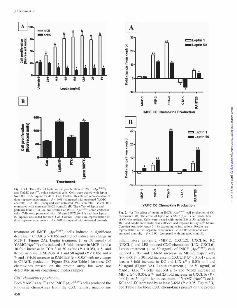
YAMC (Apc+/+) and IMCE (ApcMin/+) cells were treated withconcentrations of leptin ranging from 0.01 to 50 ng/ml. Theseconcentrations were chosen to represent a similar physiolo-gical range of low to high circulating concentrations ofleptin. As observed previously, in YAMC (Apc+/+) cellsleptin significantly decreased cell proliferation. An oppositeeffect was observed in IMCE (ApcMin/+) cells as seen inFigure 1A (28). Using PTX as a global inhibitor of G-proteinchemokine receptors, we co-treated with leptin and PTX todetermine whether blocking chemokines could explain theproliferative response induced by leptin in IMCE cells. PTXalone had no effect on cell survival (Figure 1B).
CC chemokine production
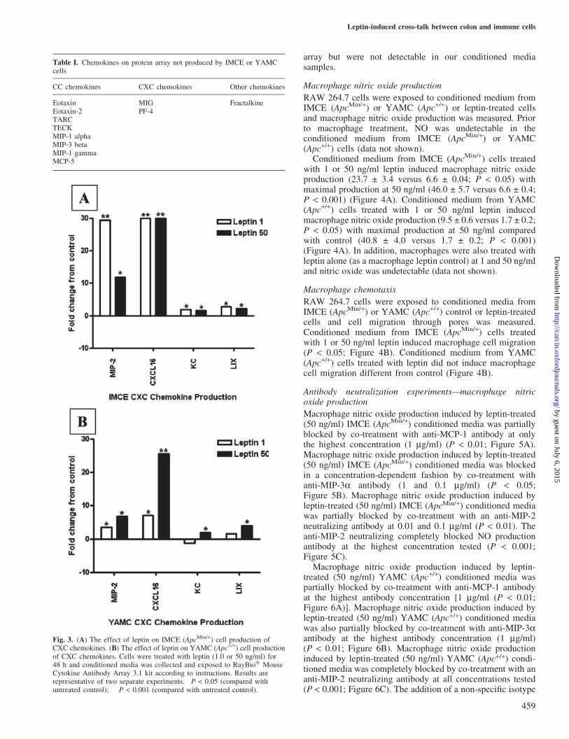
Both YAMC (Apc+/+) and IMCE (ApcMin/+) cells produced thefollowing chemokines from the CC family; monocyte chemo-attractant protein-1 (MCP-1;CCL2),macrophage inflammatoryprotein-3a (MIP-3a; CCL20), T cell activation-3 (TCA-3;CCL1), cutaneous T cell-attracting chemokine (CTACK;CCL27) and CC-chemokine-5 (RANTES; CCL5) (Figure 2).Leptin treatment (1 or 50 ng/ml) of IMCE (ApcMin/+) cellsinduced at least a 3-fold increase in RANTES (P < 0.05) and a>10-fold increase in MIP-3a and TCA-3 (P < 0.05) with thehighest production at 1 ng/ml leptin (P < 0.001). Leptin
457
Leptin-induced cross-talk between colon and immune cells
by guest on July 6, 2015http://carcin.oxfordjournals.org/
Dow
nloaded from
treatment of IMCE (ApcMin/+) cells induced a significantdecrease in CTAK (P < 0.05) and did not induce any change inMCP-1 (Figure 2A). Leptin treatment (1 or 50 ng/ml) ofYAMC (Apc+/+) cells induced a 3-fold increase inMCP-1 and a30-fold increase in TCA-3 at 50 ng/ml (P < 0.05), a 5- and8-fold increase in MIP-3a at 1 and 50 ng/ml (P < 0.05) and a7- and 18-fold increase in RANTES (P < 0.05) with no changein CTACK production (Figure 2B). See Table I for those CCchemokines present on the protein array but were notdetectable in our conditioned media samples.
CXC chemokines production
Both YAMC (Apc+/+) and IMCE (ApcMin/+) cells produced thefollowing chemokines from the CXC family; macrophage
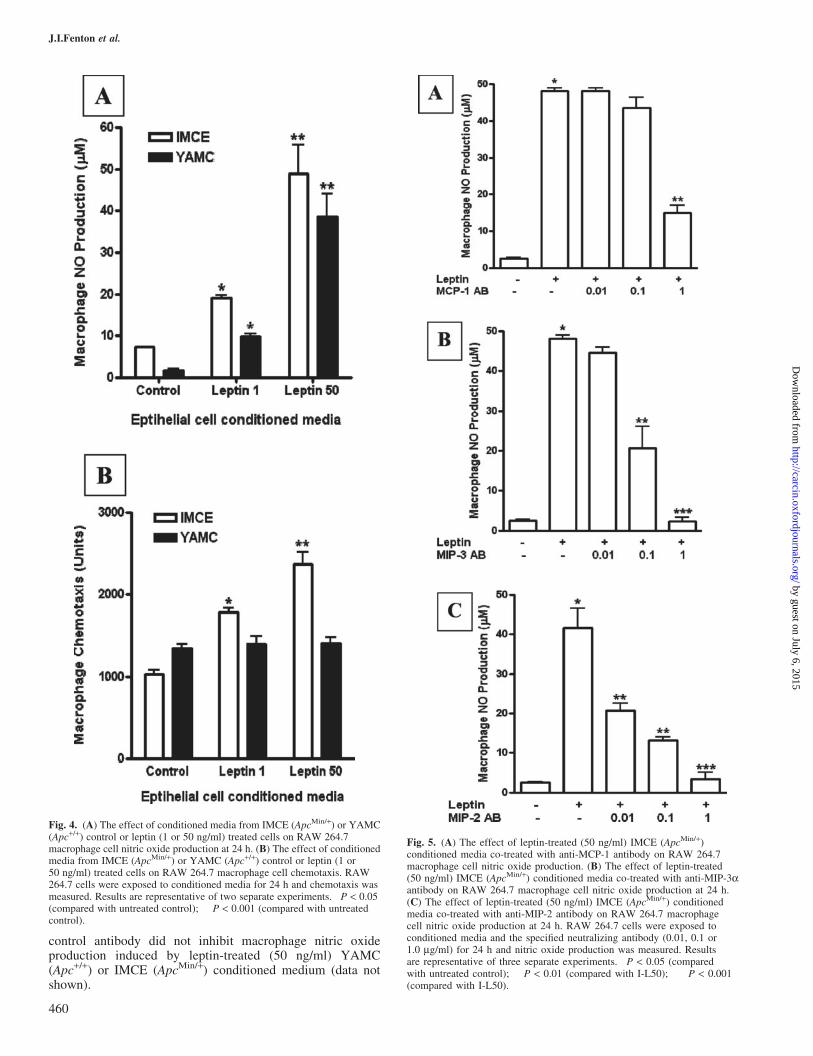
inflammatory protein-2 (MIP-2; CXCL2), CXCL16, KC(CXCL1) and LPS induced CXC chemokine (LIX; CXCL6).Leptin treatment (1 or 50 ng/ml) of IMCE (ApcMin/+) cellsinduced a 30- and 10-fold increase in MIP-2, respectively(P < 0.001), a 30-fold increase in CXCL18 (P < 0.001) and atleast a 3-fold increase in KC and LIX (P < 0.05) at 1 and50 ng/ml (Figure 3A). Leptin treatment (1 or 50 ng/ml) ofYAMC (Apc+/+) cells induced a 5- and 7-fold increase inMIP-2 (P < 0.05), a 7- and 25-fold increase in CXCL16 (P <0.001). At 50 ng/ml leptin treatment of YAMC (Apc+/+) cells,KC and LIX increased by at least 3-fold (P < 0.05; Figure 3B).See Table I for those CXC chemokines present on the protein
Fig. 2. (A) The effect of leptin on IMCE (ApcMin/+) cell production of CCchemokines. (B) The effect of leptin on YAMC (Apc+/+) cell productionof CC chemokines. Cells were treated with leptin (1.0 or 50 ng/ml) for48 h and conditioned media was collected and exposed to RayBio� MouseCytokine Antibody Array 3.1 kit according to instructions. Results arerepresentative of two separate experiments. �P < 0.05 (compared withuntreated control); ��P < 0.001 (compared with untreated control).
Fig. 1. (A) The effect of leptin on the proliferation of IMCE (ApcMin/+)and YAMC (Apc+/+) colon epithelial cells. Cells were treated with leptinfrom 0.01 to 50 ng/ml for 48 h. Con, Control. Results are representative ofthree separate experiments. �P < 0.01 (compared with untreated YAMCcontrol); �P < 0.001 (compared with untreated IMCE control); ��P < 0.0001(compared with untreated IMCE control). (B) The effect of leptin andpertussis toxin (PTX) on proliferation of IMCE (ApcMin/+) colon epithelialcells. Cells were pretreated with 100 ng/ml PTX for 1 h and then leptin(50 ng/ml) was added for 48 h. Con, Control. Results are representative ofthree separate experiments. �P < 0.01 (compared with untreated control).
458
J.I.Fenton et al.
by guest on July 6, 2015http://carcin.oxfordjournals.org/
Dow
nloaded from
array but were not detectable in our conditioned mediasamples.
Macrophage nitric oxide production
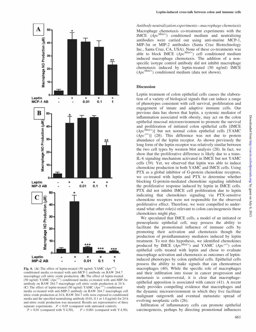
RAW 264.7 cells were exposed to conditioned medium fromIMCE (ApcMin/+) or YAMC (Apc+/+) or leptin-treated cellsand macrophage nitric oxide production was measured. Priorto macrophage treatment, NO was undetectable in theconditioned medium from IMCE (ApcMin/+) or YAMC(Apc+/+) cells (data not shown).
Conditioned medium from IMCE (ApcMin/+) cells treatedwith 1 or 50 ng/ml leptin induced macrophage nitric oxideproduction (23.7 ± 3.4 versus 6.6 ± 0.04; P < 0.05) withmaximal production at 50 ng/ml (46.0 ± 5.7 versus 6.6 ± 0.4;P < 0.001) (Figure 4A). Conditioned medium from YAMC(Apc+/+) cells treated with 1 or 50 ng/ml leptin inducedmacrophage nitric oxide production (9.5 ± 0.6 versus 1.7 ± 0.2;P < 0.05) with maximal production at 50 ng/ml comparedwith control (40.8 ± 4.0 versus 1.7 ± 0.2; P < 0.001)(Figure 4A). In addition, macrophages were also treated withleptin alone (as a macrophage leptin control) at 1 and 50 ng/mland nitric oxide was undetectable (data not shown).
Macrophage chemotaxis
RAW 264.7 cells were exposed to conditioned media fromIMCE (ApcMin/+) or YAMC (Apc+/+) control or leptin-treatedcells and cell migration through pores was measured.Conditioned medium from IMCE (ApcMin/+) cells treatedwith 1 or 50 ng/ml leptin induced macrophage cell migration(P < 0.05; Figure 4B). Conditioned medium from YAMC(Apc+/+) cells treated with leptin did not induce macrophagecell migration different from control (Figure 4B).
Antibody neutralization experiments—macrophage nitricoxide production
Macrophage nitric oxide production induced by leptin-treated(50 ng/ml) IMCE (ApcMin/+) conditioned media was partiallyblocked by co-treatment with anti-MCP-1 antibody at onlythe highest concentration (1 mg/ml) (P < 0.01; Figure 5A).Macrophage nitric oxide production induced by leptin-treated(50 ng/ml) IMCE (ApcMin/+) conditioned media was blockedin a concentration-dependent fashion by co-treatment withanti-MIP-3a antibody (1 and 0.1 mg/ml) (P < 0.05;Figure 5B). Macrophage nitric oxide production induced byleptin-treated (50 ng/ml) IMCE (ApcMin/+) conditioned mediawas partially blocked by co-treatment with an anti-MIP-2neutralizing antibody at 0.01 and 0.1 mg/ml (P < 0.01). Theanti-MIP-2 neutralizing completely blocked NO productionantibody at the highest concentration tested (P < 0.001;Figure 5C).
Macrophage nitric oxide production induced by leptin-treated (50 ng/ml) YAMC (Apc+/+) conditioned media waspartially blocked by co-treatment with anti-MCP-1 antibodyat the highest antibody concentration [1 mg/ml (P < 0.01;Figure 6A)]. Macrophage nitric oxide production induced byleptin-treated (50 ng/ml) YAMC (Apc+/+) conditioned mediawas also partially blocked by co-treatment with anti-MIP-3aantibody at the highest antibody concentration (1 mg/ml)(P < 0.01; Figure 6B). Macrophage nitric oxide productioninduced by leptin-treated (50 ng/ml) YAMC (Apc+/+) condi-tioned media was completely blocked by co-treatment with ananti-MIP-2 neutralizing antibody at all concentrations tested(P < 0.001; Figure 6C). The addition of a non-specific isotype
Fig. 3. (A) The effect of leptin on IMCE (ApcMin/+) cell production ofCXC chemokines. (B) The effect of leptin on YAMC (Apc+/+) cell productionof CXC chemokines. Cells were treated with leptin (1.0 or 50 ng/ml) for48 h and conditioned media was collected and exposed to RayBio� MouseCytokine Antibody Array 3.1 kit according to instructions. Results arerepresentative of two separate experiments. �P < 0.05 (compared withuntreated control); ��P < 0.001 (compared with untreated control).
Table I. Chemokines on protein array not produced by IMCE or YAMCcells
CC chemokines CXC chemokines Other chemokines
Eotaxin MIG FractalkineEotaxin-2 PF-4TARCTECKMIP-1 alphaMIP-3 betaMIP-1 gammaMCP-5
459
Leptin-induced cross-talk between colon and immune cells
by guest on July 6, 2015http://carcin.oxfordjournals.org/
Dow
nloaded from
control antibody did not inhibit macrophage nitric oxideproduction induced by leptin-treated (50 ng/ml) YAMC(Apc+/+) or IMCE (ApcMin/+) conditioned medium (data notshown).
Fig. 5. (A) The effect of leptin-treated (50 ng/ml) IMCE (ApcMin/+)conditioned media co-treated with anti-MCP-1 antibody on RAW 264.7macrophage cell nitric oxide production. (B) The effect of leptin-treated(50 ng/ml) IMCE (ApcMin/+) conditioned media co-treated with anti-MIP-3aantibody on RAW 264.7 macrophage cell nitric oxide production at 24 h.(C) The effect of leptin-treated (50 ng/ml) IMCE (ApcMin/+) conditionedmedia co-treated with anti-MIP-2 antibody on RAW 264.7 macrophagecell nitric oxide production at 24 h. RAW 264.7 cells were exposed toconditioned media and the specified neutralizing antibody (0.01, 0.1 or1.0 mg/ml) for 24 h and nitric oxide production was measured. Resultsare representative of three separate experiments. �P < 0.05 (comparedwith untreated control); ��P < 0.01 (compared with I-L50); ���P < 0.001(compared with I-L50).
Fig. 4. (A) The effect of conditioned media from IMCE (ApcMin/+) or YAMC(Apc+/+) control or leptin (1 or 50 ng/ml) treated cells on RAW 264.7macrophage cell nitric oxide production at 24 h. (B) The effect of conditionedmedia from IMCE (ApcMin/+) or YAMC (Apc+/+) control or leptin (1 or50 ng/ml) treated cells on RAW 264.7 macrophage cell chemotaxis. RAW264.7 cells were exposed to conditioned media for 24 h and chemotaxis wasmeasured. Results are representative of two separate experiments. �P < 0.05(compared with untreated control); ��P < 0.001 (compared with untreatedcontrol).
460
J.I.Fenton et al.
by guest on July 6, 2015http://carcin.oxfordjournals.org/
Dow
nloaded from
Antibody neutralization experiments—macrophage chemotaxis
Macrophage chemotaxis co-treatment experiments with theIMCE (ApcMin/+) conditioned medium and neutralizingantibodies were carried out using anti-murine MCP-1,MIP-3a or MIP-2 antibodies (Santa Cruz BiotechnologyInc., Santa Cruz, CA, USA). None of these co-treatments wasable to block IMCE (ApcMin/+) cell conditioned mediuminduced macrophage chemotaxis. The addition of a non-specific isotype control antibody did not inhibit macrophagechemotaxis induced by leptin-treated (50 ng/ml) IMCE(ApcMin/+) conditioned medium (data not shown).
Discussion
Leptin treatment of colon epithelial cells causes the elabora-tion of a variety of biological signals that can induce a rangeof phenotypes consistent with cell survival, proliferation andengagement of innate and adaptive immune cells. Ourprevious data has shown that leptin, a systemic mediator ofinflammation associated with obesity, may act on the colonepithelial mucosal microenvironment to promote the survivaland proliferation of initiated colon epithelial cells [IMCE(ApcMin/+)] but not normal colon epithelial cells [YAMC(Apc+/+)] (28). This difference was not due to proteinabundance of the leptin receptor. As shown previously thelong form of the leptin receptor was relatively similar betweenthe two cell types by western blot analysis (28). In fact, weshow that the proliferative difference is likely due to a trans-IL-6 signaling mechanism activated in IMCE but not YAMCcells (39). Yet, we observed that leptin was able to inducechemokine production in both YAMC and IMCE cells. UsingPTX as a global inhibitor of G-protein chemokine receptors,we co-treated with leptin and PTX to determine whetherblocking G-protein-mediated chemokine signaling inhibitedthe proliferative response induced by leptin in IMCE cells.PTX did not inhibit IMCE cell proliferation due to leptinindicating that chemokines signaling via PTX-sensitivechemokine receptors were not responsible for the observedproliferative effect. Therefore, we were compelled to under-stand what other role(s) relevant to colon carcinogenesis thesechemokines might play.
We speculated that IMCE cells, a model of an initiated orpreneoplastic epithelial cell, may possess the ability tofacilitate the promotional influence of immune cells bypromoting their activation and chemotaxis though theproduction of proinflammatory mediators induced by leptintreatment. To test this hypothesis, we identified chemokinesproduced by IMCE (ApcMin/+) and YAMC (Apc+/+) colonepithelial cells treated with leptin and chose to evaluatemacrophage activation and chemotaxis as outcomes of leptin-induced phenotypes by colon epithelial cells. Epithelial cellspossess the ability to make signals that can chemoattractmacrophages (40). While the specific role of macrophagesand their infiltration into tissue in cancer progression andmetastasis is controversial, it is clear that macrophage-epithelial apposition is associated with cancer (41). A recentstudy provides compelling evidence that macrophages andthe dynamic microenvironment in which they live facilitatemalignant outgrowth and eventual metastatic spread ofevolving neoplastic cells (26).
Infiltration of inflammatory cells can promote epithelialcarcinogenesis, perhaps by directing promotional influences
Fig. 6. (A) The effect of leptin-treated (50 ng/ml) YAMC (Apc+/+)conditioned media co-treated with anti-MCP-1 antibody on RAW 264.7macrophage cell nitric oxide production. (B) The effect of leptin-treated(50 ng/ml) YAMC (Apc+/+) conditioned media co-treated with anti-MIP-3aantibody on RAW 264.7 macrophage cell nitric oxide production at 24 h.(C) The effect of leptin-treated (50 ng/ml) YAMC (Apc+/+) conditionedmedia co-treated with anti-MIP-2 antibody on RAW 264.7 macrophage cellnitric oxide production at 24 h. RAW 264.7 cells were exposed to conditionedmedia and the specified neutralizing antibody (0.01, 0.1 or 1.0 mg/ml) for 24 hand nitric oxide production was measured. Results are representative of threeseparate experiments. �P < 0.05 (compared with untreated control);��P < 0.01 (compared with Y-L50), ���P < 0.001 (compared with Y-L50).
461
Leptin-induced cross-talk between colon and immune cells
by guest on July 6, 2015http://carcin.oxfordjournals.org/
Dow
nloaded from
on epithelial cells (e.g. activation of oncogenes, etc.), byactivation of specific signaling pathways in initiated cell, orby altering epithelial–stromal interactions (42). We utilizedcell lines which represent the earliest event in coloncarcinogenesis; one cell line is normal and the otherpossesses a truncated copy of Apc, a protein involved intargeting b-catenin for proteosomal degradation and consid-ered key susceptibility protein in colon cancer. Using thesecell lines, we demonstrate the potential for epithelial cells toshape the innate immune response to leptin exposuredepending on their Apc genotype. The data presented herecharacterizes the CC and CXC chemokine production bycolon epithelial cells contrasting in Apc genotype induced byleptin treatment (1 and 50 ng/ml). We describe thedifferential leptin-induced production of five CC chemokinesand four CXC chemokines in colon epithelial cells contrast-ing in Apc genotype using a novel protein array method.Based on these results, it was logical to speculate on thespecific phenotypes of immune cells activated by the patternof chemokines produced.
We surveyed two phenotypes of macrophage function:macrophage chemotaxis and macrophage nitric oxideproduction. We show that conditioned medium from leptin-treated IMCE (ApcMin/+) cells induces macrophage chemo-taxis and nitric oxide production at 1 and 50 ng/ml leptintreatment. Both cell types, when exposed to leptin, releaseproducts that induce NO production by macrophages.However, the IMCE (ApcMin/+) cells appear to secrete factorsin response to leptin at a lower dose than YAMC (Apc+/+)cells. Further, IMCE (ApcMin/+) cells produced leptin-inducedfactors able to induce macrophage chemotaxis. This obser-vation was consistent with our hypothesis that, in response toa systemic signal, preneoplastic cells are capable ofelaborating signals that results in the activation of immunecells. While normal cells [YAMC (Apc+/+)] were able toinduce macrophage NO production in response to leptin, thisresponse is hypothesized to act only on local residentmacrophages as our data indicate that they do not make achemoattractant signals in response to leptin. In contrast,preneoplastic [IMCE (ApcMin/+)] cells display the ability toboth activate and chemoattract macrophages in response toleptin, which occurs at lower concentrations of leptin thannormal cells. We cannot rule out that part of the observedeffect could be due to increasing cell numbers over thecourse of the leptin treatment. However, one would expectthat total protein levels would increase in the media. As totalprotein concentrations were similar across all conditionedmedia samples (data not shown) we do not believe theelevated chemokine/cytokine effect is due to changes in cellnumber. We believe the effect to be specific. Given the factthat leptin-induced differences in chemokine secretionbetween YAMC and IMCE cells can vary up to 20-fold, itis unlikely that differences in cell number of 2- to 3-foldaccount for the differences in chemokine secretion.
Among the candidate macrophage activation and chemoat-tractant signals elaborated by leptin treatment, we identifiedMIP-2 and to a lesser extent MIP-3a and MCP-1 as signalsinvolved in the elaborated macrophage phenotypes. MIP-2 isan excellent candidate for involvement in intestinalepithelial-immune cross-talk. MIP-2 is expressed in intestinalepithelial cells after stimulation with LPS or proinflammatorycytokines and is not expressed in epithelial cells isolatedfrom healthy intestine in vivo (43). MIP-2 expression is
increased in IBD and dextran sulfate sodium-inducedinflammation (30,31). In addition, MIP-2 secretion byepithelial cells increases neutrophil and lymphocyte recruit-ment in the mouse intestine (44). To a lesser extent MIP-3aand MCP-1 are associated with intestinal inflammation.MIP-3 is induced in response to inflammation (45) and isinvolved in maturation of intestinal macrophages (46). Inintestinal epithelial cells, MCP-1 production was enhanced inresponse to an intracellular pathogen (47). However, weacknowledge that other measured and unmeasured epithelialfactors may also play a role in macrophage activation. This isevidenced by the lack of concordance in the concentrationdependence of MIP-2 change across leptin concentration(Figure 1) and macrophage NO production induced byconditioned medium from leptin-treated colon epithelialcells. Research is ongoing to elucidate the role of leptin-induced colon epithelial cell production of MIP-2. It ispossible that expression of the MIP-2 receptor CXCR-2 orother chemokine receptors may explain the observations.However, the chemokine network has built-in redundancies;chemokines are known to bind to more than one receptorand most receptors can respond to more than one chemokine.As such, our data identify only a few of the potentialimmunomodulatory factors elaborated by IMCE cells; thesedata do not rule out the existence of other factors secreted inresponse to leptin that may be responsible for these measuredeffects. In addition, we are also examining the response ofother innate immune cells to see if they respond similarly.
Basal production of protective factors by intestinal epithe-lial cells, including chemokines, cytokines and heat shockproteins, plays a critical role in intestinal homeostasis andrepair (48). In contrast, the inducible production of chemo-kines and cytokines by endocrine factors and hormones, or inresponse to tissue damage or bacterial pathogens, are criticalto innate and adaptive immune responses (49). Importantly,inducible chemokines are becoming recognized as importantcomponents in the promotional phase of carcinogenesis (42).Most tumors engage in a complex chemokine network whichcan influence immune responses to the tumor, direct thecellular composition of leukocyte infiltrate and also play a rolein angiogenesis (50). It appears from our data that this ‘switch’towards a more active immunomodulatory epithelial cellphenotype during cancer promotion may be occurring muchearlier than currently suggested. Characterization of thechemokine receptor profile on these two cell lines may gleanimportant insights into the ability of these cell lines todifferentially respond to signals coming from apposingimmune cells.
These data highlight the importance of examining theeffects of immunomodulatory factors on early events in cancerprogression using a continuum model system. Acquiredmutations in Apc may provide an opportunity for the specificpromotional effects by adipoctye-derived factors such asleptin. This susceptibility would then only impact thoseindividuals ‘at risk’ for colorectal cancer. However, currenttechnologies cannot determine risk this early in colon cancercarcinogenesis. We argue that our model provides an excellentopportunity to examine the effects of these adipocyte-derivedfactors in a reductionist system. We identified a noveldifferential effect of leptin on models of normal andpreneoplastic colon epithelial cells to elaborate immune-epithelial cell cross-talk in the colon. Taken together with ourprevious data, leptin promotes the proliferation of initiated
462
J.I.Fenton et al.
by guest on July 6, 2015http://carcin.oxfordjournals.org/
Dow
nloaded from
IMCE (ApcMin/+) cells via a trans-IL-6 mechanism, inducesthe production of chemokines which may activate macro-phages and promote immune cell chemotaxis. We underscorethe importance of how mutations in Apc can lead to significantdifferences in the response to systemic signals (e.g. leptin) andthe elaboration of paracrine signals (e.g. chemokines) fromcolon epithelial cells to mucosal immune cells whilemaintaining a non-tumorigenic phenotype. These data providea rational basis for concluding that initiated epithelial cellsparticipate in cross-talk with immune cells that may influencethe promotional phase of carcinogenesis. These data, takentogether with our other published data, provide furtherbiological plausibility for a role for systemic mediators ofinflammation associated with obesity involved in the promo-tion of preneoplastic colon epithelial lesions via a leptin-mediated mechanism.
Acknowledgements
The authors gratefully acknowledge the National Cancer Institute CancerPrevention Fellowship Program for support while carrying out this project.The authors gratefully acknowledge Dr. Venu Gangur, Michigan, StateUniversity, for consulation regarding research design and interpretation ofdata relating to chemokines.
Conflict of Interest Statement: None declared.
References
1.Bianchini,F., Kaaks,R. and Vainio,H. (2002) Overweight, obesity, andcancer risk. Lancet Oncol., 3, 565–574.
2.Calle,E.E. and Kaaks,R. (2004) Overweight, obesity and cancer:epidemiological evidence and proposed mechanisms. Nat. Rev. Cancer,4, 579–591.
3. John,B.J., Irukulla,S., Abulafi,A.M., Kumar,D. and Mendall,M.A. (2006)Systematic review: adipose tissue, obesity and gastrointestinal diseases.Aliment. Pharmacol. Ther., 23, 1511–1523.
4.Bray,G.A. and Bellanger,T. (2006) Epidemiology, trends, and morbiditiesof obesity and the metabolic syndrome. Endocrine, 29, 109–117.
5.Vendrell,J., Broch,M., Vilarrasa,N., Molina,A., Gomez,J.M., Gutierrez,C.,Simon,I., Soler,J. and Richart,C. (2004) Resistin, adiponectin, ghrelin,leptin, and proinflammatory cytokines: relationships in obesity. Obes. Res.,12, 962–971.
6.Stattin,P., Lukanova,A., Biessy,C., Soderberg,S., Palmqvist,R., Kaaks,R.,Olsson,T. and Jellum,E. (2004) Obesity and colon cancer: does leptinprovide a link? Int. J. Cancer, 109, 149–152.
7.Stattin,P., Palmqvist,R., Soderberg,S., Biessy,C., Ardnor,B., Hallmans,G.,Kaaks,R. and Olsson,T. (2003) Plasma leptin and colorectal cancer risk: aprospective study in Northern Sweden. Oncol. Rep., 10, 2015–2021.
8.Zhang,T., Nanney,L.B., Peeler,M.O., Williams,C.S., Lamps,L.,Heppner,K.J., DuBois,R.N. and Beauchamp,R.D. (1997) Decreasedtransforming growth factor beta type II receptor expression in intestinaladenomas from Min/+ mice is associated with increased cyclin D1 andcyclin-dependent kinase 4 expression. Cancer Res., 57, 1638–1643.
9.Zhang,F., Basinski,M.B., Beals,J.M. et al. (1997) Crystal structure of theobese protein leptin-E100. Nature, 387, 206–209.
10.Otero,M., Lago,R., Lago,F., Casanueva,F.F., Dieguez,C., Gomez-Reino,J.J. and Gualillo,O. (2005) Leptin, from fat to inflammation: oldquestions and new insights. FEBS Lett., 579, 295–301.
11.Sweeney,G. (2002) Leptin signalling. Cell Signal, 14, 655–663.12.Alavi,K., Schwartz,M.Z., Prasad,R., O’Connor,D. and Funanage,V.
(2002) Leptin: a new growth factor for the small intestine. J. Pediatr.Surg., 37, 327–330.
13.Siegmund,B., Lehr,H.A. and Fantuzzi,G. (2002) Leptin: a pivotalmediator of intestinal inflammation in mice. Gastroenterology, 122,2011–2025.
14.Siegmund,B., Sennello,J.A., Jones-Carson,J., Gamboni-Robertson,F.,Lehr,H.A., Batra,A., Fedke,I., Zeitz,M. and Fantuzzi,G. (2004) Leptinreceptor expression on T lymphocytes modulates chronic intestinalinflammation in mice. Gut, 53, 965–972.
15.Clevers,H. (2004) At the crossroads of inflammation and cancer. Cell,118, 671–674.
16.Balkwill,F. and Coussens,L.M. (2004) Cancer: an inflammatory link.Nature, 431, 405–406.
17.Brower,V. (2005) Feeding the flame: new research adds to role ofinflammation in cancer development. J. Natl Cancer Inst., 97, 251–253.
18.Mahmoud,N.N., Bilinski,R.T., Churchill,M.R., Edelmann,W.,Kucherlapati,R. and Bertagnolli,M.M. (1999) Genotype–phenotypecorrelation in murine Apc mutation: differences in enterocyte migrationand response to sulindac. Cancer Res., 59, 353–359.
19.Haramis,A.P., Hurlstone,A., van der Velden,Y., Begthel,H., van denBorn,M., Offerhaus,G.J. and Clevers,H.C. (2006) Adenomatous polyposiscoli-deficient zebrafish are susceptible to digestive tract neoplasia. EMBORep., 7, 444–449.
20.Fearon,E.R. and Vogelstein,B. (1990) A genetic model for colorectaltumorigenesis. Cell, 61, 759–767.
21.Mahida,Y.R. (2000) The key role of macrophages in the immunopatho-genesis of inflammatory bowel disease. Inflamm. Bowel Dis., 6, 21–33.
22.Kagnoff,M.F. and Eckmann,L. (1997) Epithelial cells as sensors formicrobial infection. J. Clin. Invest., 100, 6–10.
23.de Visser,K.E., Eichten,A. and Coussens,L.M. (2006) Paradoxical roles ofthe immune system during cancer development.Nat. Rev. Cancer, 6, 24–37.
24.Houghton,A.N., Uchi,H. and Wolchok,J.D. (2005) The role of the immunesystem in early epithelial carcinogenesis: B-ware the double-edged sword.Cancer Cell, 7, 403–405.
25.Bingle,L., Brown,N.J. and Lewis,C.E. (2002) The role of tumour-associated macrophages in tumour progression: implications for newanticancer therapies. J. Pathol., 196, 254–265.
26.Lin,E.Y., Nguyen,A.V., Russell,R.G. and Pollard,J.W. (2001) Colony-stimulating factor 1 promotes progression of mammary tumors tomalignancy. J. Exp. Med., 193, 727–740.
27.Aharinejad,S., Paulus,P., Sioud,M., Hofmann,M., Zins,K., Schafer,R.,Stanley,E.R. and Abraham,D. (2004) Colony-stimulating factor-1blockade by antisense oligonucleotides and small interfering RNAssuppresses growth of human mammary tumor xenografts in mice. CancerRes., 64, 5378–5384.
28.Fenton,J.I., Hord,N.G., Lavigne,J.A., Perkins,S.N. and Hursting,S.D.(2005) Leptin, insulin-like growth factor-1, and insulin-like growthfactor-2 are mitogens in ApcMin/+ but not Apc+/+ colonic epithelial celllines. Cancer Epidemiol. Biomarkers Prev., 14, 1646–1652.
29.Fenton,J.I. and Hord,N.G. (2006) Stage matters: choosing relevant modelsystems to address hypotheses in diet and cancer chemopreventionresearch. Carcinogenesis, 27, 893–902.
30.Whitehead,R.H. and Joseph,J.L. (1994) Derivation of conditionallyimmortalized cell lines containing the Min mutation from the normalcolonic mucosa and other tissues of an ‘‘Immortomouse’’/Min hybrid.Epithelial Cell Biol., 3, 119–125.
31.Mei,J.M., Hord,N.G., Winterstein,D.F., Donald,S.P. and Phang,J.M.(2000) Expression of prostaglandin endoperoxide H synthase-2 inducedby nitric oxide in conditionally immortalized murine colonic epithelialcells. FASEB J., 14, 1188–1201.
32.Fenton,J.I., Wolff,M.S., Orth,M.W. and Hord,N.G. (2002) Membrane-type matrix metalloproteinases mediate curcumin-induced cell migrationin non-tumorigenic colon epithelial cells differing in Apc genotype.Carcinogenesis, 23, 1065–1070.
33.Mei,J.M., Hord,N.G., Winterstein,D.F., Donald,S.P. and Phang,J.M.(1999) Differential expression of prostaglandin endoperoxide Hsynthase-2 and formation of activated beta-catenin-LEF-1 transcriptioncomplex in mouse colonic epithelial cells contrasting in Apc.Carcinogenesis, 20, 737–740.
34.Husoy,T., Knutsen,H.K., Cruciani,V., Olstorn,H.B., Mikalsen,S.O.,Loberg,E.M. and Alexander,J. (2005) Connexin43 is overexpressed inApc(Min/+)-mice adenomas and colocalises with COX-2 inmyofibroblasts. Int. J. Cancer, 116, 351–358.
35.Whitehead,R.H., VanEeden,P.E., Noble,M.D., Ataliotis,P. and Jat,P.S.(1993) Establishment of conditionally immortalized epithelial cell linesfrom both colon and small intestine of adult H-2Kb-tsA58 transgenicmice. Proc. Natl Acad. Sci. USA, 90, 587–591.
36.D’Abaco,G.M., Whitehead,R.H. and Burgess,A.W. (1996) Synergybetween Apc min and an activated ras mutation is sufficient to inducecolon carcinomas. Mol. Cell Biol., 16, 884–891.
37.Moss,J. and Vaughan,M. (1988) ADP-ribosylation of guanyl nucleotide-binding regulatory proteins by bacterial toxins. Adv. Enzymol. Relat.Areas Mol. Biol., 61, 303–379.
38.Yang,C.C., Ogawa,H., Dwinell,M.B., McCole,D.F., Eckmann,L. andKagnoff,M.F. (2005) Chemokine receptor CCR6 transduces signals thatactivate p130Cas and alter cAMP-stimulated ion transport in humanintestinal epithelial cells. Am. J. Physiol. Cell Physiol., 288, C321– C328.
463
Leptin-induced cross-talk between colon and immune cells
by guest on July 6, 2015http://carcin.oxfordjournals.org/
Dow
nloaded from
39.Fenton,J.I., Hursting,S.D., Perkins,S.N. and Hord,N.G. (2006)Interleukin-6 production induced by leptin treatment promotes cellproliferation in an Apc (Min/+) colon epithelial cell line.Carcinogenesis, 27, 1507–1515.
40.Wilson,J. and Balkwill,F. (2002) The role of cytokines in the epithelialcancer microenvironment. Semin. Cancer Biol., 12, 113–120.
41.Sickert,D., Aust,D.E., Langer,S., Haupt,I., Baretton,G.B. and Dieter,P.(2005) Characterization of macrophage subpopulations in colon cancerusing tissue microarrays. Histopathology, 46, 515–521.
42.Coussens,L.M. and Werb,Z. (2001) Inflammatory cells and cancer: thinkdifferent! J. Exp. Med., 193, F23–F26.
43.Ohno,Y., Lee,J., Fusunyan,R.D., MacDermott,R.P. and Sanderson,I.R.(1997) Macrophage inflammatory protein-2: chromosomal regulation inrat small intestinal epithelial cells. Proc. Natl Acad. Sci. USA, 94,10279–10284.
44.Ohtsuka,Y., Lee,J., Stamm,D.S. and Sanderson,I.R. (2001) MIP-2 secretedby epithelial cells increases neutrophil and lymphocyte recruitment in themouse intestine. Gut, 49, 526–533.
45.Hausmann,M., Bataille,F., Spoettl,T., Schreiter,K., Falk,W.,Schoelmerich,J., Herfarth,H. and Rogler,G. (2005) Physiological role of
macrophage inflammatory protein-3 alpha induction during maturation ofintestinal macrophages. J. Immunol., 175, 1389–1398.
46.Lee,H.J., Choi,S.C., Choi,E.Y. et al. (2005) Iron chelator induces MIP-alpha/CCL20 in human intestinal epithelial cells: implication fortriggering mucosal adaptive immunity. Exp. Mol. Med., 37, 297–310.
47.Mennechet,F.J., Kasper,L.H., Rachinel,N., Li,W., Vandewalle,A. andBuzoni-Gatel,D. (2002) Lamina propria CD4+ T lymphocytes synergizewith murine intestinal epithelial cells to enhance proinflammatoryresponse against an intracellular pathogen. J. Immunol., 168,2988–2996.
48.Rakoff-Nahoum,S., Paglino,J., Eslami-Varzaneh,F., Edberg,S. andMedzhitov,R. (2004) Recognition of commensal microflora by toll-likereceptors is required for intestinal homeostasis. Cell, 118, 229–241.
49.Cario,E. (2005) Bacterial interactions with cells of the intestinal mucosa:Toll-like receptors and NOD2. Gut, 54, 1182–1193.
50.Kulbe,H., Levinson,N.R., Balkwill,F. and Wilson,J.L. (2004) Thechemokine network in cancer—much more than directing cellmovement. Int. J. Dev. Biol., 48, 489–496.
Received April 10, 2006; revised June 7, 2006; accepted July 19, 2006
464
J.I.Fenton et al.
by guest on July 6, 2015http://carcin.oxfordjournals.org/
Dow
nloaded from