The role of APC in WNT pathway activation in serrated ...
10
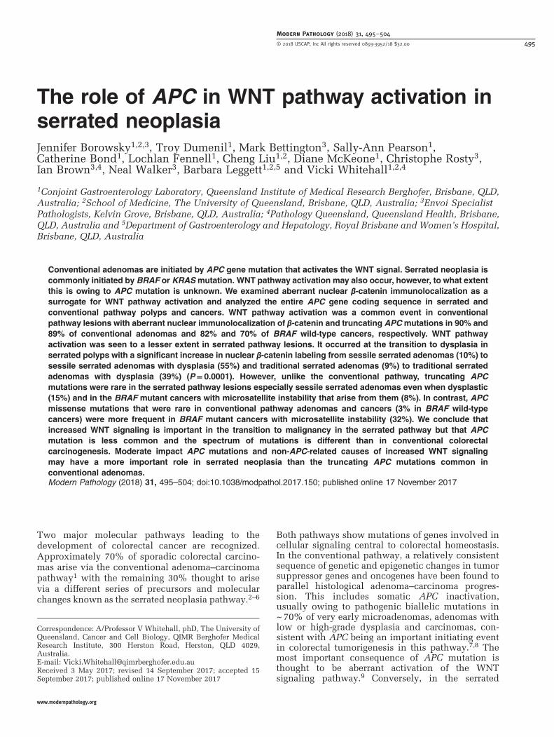
The role of APC in WNT pathway activation in serrated neoplasia Jennifer Borowsky 1,2,3 , Troy Dumenil 1 , Mark Bettington 3 , Sally-Ann Pearson 1 , Catherine Bond 1 , Lochlan Fennell 1 , Cheng Liu 1,2 , Diane McKeone 1 , Christophe Rosty 3 , Ian Brown 3,4 , Neal Walker 3 , Barbara Leggett 1,2,5 and Vicki Whitehall 1,2,4 1 Conjoint Gastroenterology Laboratory, Queensland Institute of Medical Research Berghofer, Brisbane, QLD, Australia; 2 School of Medicine, The University of Queensland, Brisbane, QLD, Australia; 3 Envoi Specialist Pathologists, Kelvin Grove, Brisbane, QLD, Australia; 4 Pathology Queensland, Queensland Health, Brisbane, QLD, Australia and 5 Department of Gastroenterology and Hepatology, Royal Brisbane and Women’s Hospital, Brisbane, QLD, Australia Conventional adenomas are initiated by APC gene mutation that activates the WNT signal. Serrated neoplasia is commonly initiated by BRAF or KRAS mutation. WNT pathway activation may also occur, however, to what extent this is owing to APC mutation is unknown. We examined aberrant nuclear β-catenin immunolocalization as a surrogate for WNT pathway activation and analyzed the entire APC gene coding sequence in serrated and conventional pathway polyps and cancers. WNT pathway activation was a common event in conventional pathway lesions with aberrant nuclear immunolocalization of β-catenin and truncating APC mutations in 90% and 89% of conventional adenomas and 82% and 70% of BRAF wild-type cancers, respectively. WNT pathway activation was seen to a lesser extent in serrated pathway lesions. It occurred at the transition to dysplasia in serrated polyps with a significant increase in nuclear β-catenin labeling from sessile serrated adenomas (10%) to sessile serrated adenomas with dysplasia (55%) and traditional serrated adenomas (9%) to traditional serrated adenomas with dysplasia (39%) (P = 0.0001). However, unlike the conventional pathway, truncating APC mutations were rare in the serrated pathway lesions especially sessile serrated adenomas even when dysplastic (15%) and in the BRAF mutant cancers with microsatellite instability that arise from them (8%). In contrast, APC missense mutations that were rare in conventional pathway adenomas and cancers (3% in BRAF wild-type cancers) were more frequent in BRAF mutant cancers with microsatellite instability (32%). We conclude that increased WNT signaling is important in the transition to malignancy in the serrated pathway but that APC mutation is less common and the spectrum of mutations is different than in conventional colorectal carcinogenesis. Moderate impact APC mutations and non-APC-related causes of increased WNT signaling may have a more important role in serrated neoplasia than the truncating APC mutations common in conventional adenomas. Modern Pathology (2018) 31, 495–504; doi:10.1038/modpathol.2017.150; published online 17 November 2017 Two major molecular pathways leading to the development of colorectal cancer are recognized. Approximately 70% of sporadic colorectal carcino- mas arise via the conventional adenoma–carcinoma pathway 1 with the remaining 30% thought to arise via a different series of precursors and molecular changes known as the serrated neoplasia pathway. 2–6 Both pathways show mutations of genes involved in cellular signaling central to colorectal homeostasis. In the conventional pathway, a relatively consistent sequence of genetic and epigenetic changes in tumor suppressor genes and oncogenes have been found to parallel histological adenoma–carcinoma progres- sion. This includes somatic APC inactivation, usually owing to pathogenic biallelic mutations in ~ 70% of very early microadenomas, adenomas with low or high-grade dysplasia and carcinomas, con- sistent with APC being an important initiating event in colorectal tumorigenesis in this pathway. 7,8 The most important consequence of APC mutation is thought to be aberrant activation of the WNT signaling pathway. 9 Conversely, in the serrated Correspondence: A/Professor V Whitehall, phD, The University of Queensland, Cancer and Cell Biology, QIMR Berghofer Medical Research Institute, 300 Herston Road, Herston, QLD 4029, Australia. E-mail: [email protected] Received 3 May 2017; revised 14 September 2017; accepted 15 September 2017; published online 17 November 2017 Modern Pathology (2018) 31, 495 – 504 © 2018 USCAP, Inc All rights reserved 0893-3952/18 $32.00 495 www.modernpathology.org
-
Upload
khangminh22 -
Category
Documents
-
view
4 -
download
0
Transcript of The role of APC in WNT pathway activation in serrated ...
The role of APC in WNT pathway activation inserrated neoplasiaJennifer Borowsky1,2,3, Troy Dumenil1, Mark Bettington3, Sally-Ann Pearson1,Catherine Bond1, Lochlan Fennell1, Cheng Liu1,2, Diane McKeone1, Christophe Rosty3,Ian Brown3,4, Neal Walker3, Barbara Leggett1,2,5 and Vicki Whitehall1,2,4
1Conjoint Gastroenterology Laboratory, Queensland Institute of Medical Research Berghofer, Brisbane, QLD,Australia; 2School of Medicine, The University of Queensland, Brisbane, QLD, Australia; 3Envoi SpecialistPathologists, Kelvin Grove, Brisbane, QLD, Australia; 4Pathology Queensland, Queensland Health, Brisbane,QLD, Australia and 5Department of Gastroenterology and Hepatology, Royal Brisbane and Women’s Hospital,Brisbane, QLD, Australia
Conventional adenomas are initiated by APC gene mutation that activates the WNT signal. Serrated neoplasia iscommonly initiated by BRAF or KRASmutation. WNT pathway activation may also occur, however, to what extentthis is owing to APC mutation is unknown. We examined aberrant nuclear β-catenin immunolocalization as asurrogate for WNT pathway activation and analyzed the entire APC gene coding sequence in serrated andconventional pathway polyps and cancers. WNT pathway activation was a common event in conventionalpathway lesions with aberrant nuclear immunolocalization of β-catenin and truncating APC mutations in 90% and89% of conventional adenomas and 82% and 70% of BRAF wild-type cancers, respectively. WNT pathwayactivation was seen to a lesser extent in serrated pathway lesions. It occurred at the transition to dysplasia inserrated polyps with a significant increase in nuclear β-catenin labeling from sessile serrated adenomas (10%) tosessile serrated adenomas with dysplasia (55%) and traditional serrated adenomas (9%) to traditional serratedadenomas with dysplasia (39%) (P= 0.0001). However, unlike the conventional pathway, truncating APCmutations were rare in the serrated pathway lesions especially sessile serrated adenomas even when dysplastic(15%) and in the BRAF mutant cancers with microsatellite instability that arise from them (8%). In contrast, APCmissense mutations that were rare in conventional pathway adenomas and cancers (3% in BRAF wild-typecancers) were more frequent in BRAF mutant cancers with microsatellite instability (32%). We conclude thatincreased WNT signaling is important in the transition to malignancy in the serrated pathway but that APCmutation is less common and the spectrum of mutations is different than in conventional colorectalcarcinogenesis. Moderate impact APC mutations and non-APC-related causes of increased WNT signalingmay have a more important role in serrated neoplasia than the truncating APC mutations common inconventional adenomas.Modern Pathology (2018) 31, 495–504; doi:10.1038/modpathol.2017.150; published online 17 November 2017
Two major molecular pathways leading to thedevelopment of colorectal cancer are recognized.Approximately 70% of sporadic colorectal carcino-mas arise via the conventional adenoma–carcinomapathway1 with the remaining 30% thought to arisevia a different series of precursors and molecularchanges known as the serrated neoplasia pathway.2–6
Both pathways show mutations of genes involved incellular signaling central to colorectal homeostasis.In the conventional pathway, a relatively consistentsequence of genetic and epigenetic changes in tumorsuppressor genes and oncogenes have been found toparallel histological adenoma–carcinoma progres-sion. This includes somatic APC inactivation,usually owing to pathogenic biallelic mutations in~70% of very early microadenomas, adenomas withlow or high-grade dysplasia and carcinomas, con-sistent with APC being an important initiating eventin colorectal tumorigenesis in this pathway.7,8 Themost important consequence of APC mutation isthought to be aberrant activation of the WNTsignaling pathway.9 Conversely, in the serrated
Correspondence: A/Professor V Whitehall, phD, The University ofQueensland, Cancer and Cell Biology, QIMR Berghofer MedicalResearch Institute, 300 Herston Road, Herston, QLD 4029,Australia.E-mail: [email protected] 3 May 2017; revised 14 September 2017; accepted 15September 2017; published online 17 November 2017
Modern Pathology (2018) 31, 495–504
© 2018 USCAP, Inc All rights reserved 0893-3952/18 $32.00 495
www.modernpathology.org

neoplasia pathway mutations of BRAF or KRAS arethought to initiate development of serrated polypsincluding sessile serrated adenomas and traditionalserrated adenomas via activation of the MAP kinasepathway.3 The role of aberrant WNT signaling in theserrated pathway is less clear than for the conven-tional adenoma–carcinoma pathway.
Nuclear translocation of the normally membranousβ-catenin protein indicates canonical WNT pathwayactivation. Most conventional adenomas shownuclear β-catenin accumulation, which is a down-stream consequence of pathogenic APC mutation.10,11Whereas some studies have shown increased nuclearβ-catenin staining by immunohistochemistry inserrated precursor polyps with neoplasticprogression,12–14 reports are inconsistent.15 Truncat-ing APC mutations appear to be uncommon in lesionsof the serrated pathway, but again the literature showsmarked variation of reported rates in serrated path-way lesions including precursor polyps16–23 and theBRAFmutant cancers that arise from them.24–26 Manystudies show limited mutational analysis focusingonly on the mutational cluster region at codons 1281–1556 of the APC gene. Although this is thought tocontain ~50–60% of truncating APC mutations,27,28 ithas the potential to miss up to 40% of mutations.Inconsistent and confusing nomenclature of serratedpathway lesions29,30 are likely contributors for thewidely variable rates of nuclear β-catenin and APCmutations found in literature. Fortunately, a standar-dized diagnostic nomenclature of different serratedpolyps was formulated in 2010.31
To address these defects in our knowledge andunderstanding of the role of APC mutations andWNT pathway activation in the serrated neoplasiapathway, we have undertaken a comprehensivemutational analysis of the entire coding region ofthe APC gene in combination with immunohisto-chemical assessment of β-catenin cellular location ina large cohort of colorectal cancers stratified byBRAF mutation and a broad spectrum of currentlydefined serrated pathway lesions. We find thatalthough truncating APC mutations are rare, mis-sense APC mutations are relatively common in theserrated pathway cancers. We suggest the possibilitythat missense APC mutations may be contributors tothe ‘just-right’ level of WNT/β-catenin signaling32required for colorectal tumorigenesis via the serratedneoplasia pathway. We also confirm that truncatingAPC mutation does not account for most of theincreased WNT signaling in the serrated pathway.
Materials and methods
Samples
The WNT pathway was examined in a range ofcolorectal polyps and cancers by immunohistochem-ical assessment of β-catenin cellular localization andAPC mutational analysis. The BRAFV600E mutation
was detected by allele-specific PCR as previouslydescribed.33 Lynch syndrome cancers were effec-tively excluded as BRAF mutant cancers do not arisein the context of Lynch Syndrome and all the BRAFwild-type cancers showed retention of mismatchrepair proteins.
This included immunohistochemical assessmentof serrated pathway lesions comprising 102 BRAFmutant cancers (stratified by microsatellite instabil-ity status including 62 cancers with microsatelliteinstability and 40 microsatellite stable cancers), 162traditional serrated adenomas, 38 traditional serratedadenomas with dysplasia, 20 sessile serrated adeno-mas, and 137 sessile serrated adenomas withdysplasia. β-catenin was scored separately in thedysplastic and non-dysplastic parts of the sessileserrated adenomas with dysplasia to give 157 resultsfor β-catenin in non-dysplastic sessile serratedadenomas. As a control group 354 BRAF wild-typecancers and 96 conventional adenomas (including46 tubular adenomas and 50 tubulovillous adeno-mas) were assessed. BRAF mutant carcinomas andBRAF wild-type carcinomas were included in theserrated pathway group and the conventional path-way group respectively based on evidence of theoccurrence of BRAFmutations in these pathways.3,34Histological assessment of serrated precursor polypsfollowed the guidelines of the World HealthOrganization.31 Our traditional serrated adenomagroup consists of those traditional serrated adenomaswithout overt morphological atypia or mitotic activ-ity (sometimes also referred to as ordinary traditionalserrated adenomas), and our traditional serratedadenoma with dysplasia group refers to those, whichhave developed overt cytological dysplasia and/ormitotic activity (also sometimes referred to asadvanced traditional serrated adenomas).
The APC-coding region was sequenced in a subsetof 189 of these cases including 80 BRAF mutantcancers (50 microsatellite unstable and 30 micro-satellite stable), 20 sessile serrated adenoma, 20sessile serrated adenoma with dysplasia, 14 tradi-tional serrated adenoma, 6 traditional serratedadenoma with dysplasia, 30 BRAF wild-type color-ectal cancer, and 19 conventional adenomas (10tubulovillous adenomas and 9 tubular adenomas).
Traditional serrated adenomas, traditional serratedadenomas with dysplasia and sessile serrated ade-nomas with dysplasia were collected in a consecu-tive fashion by a single pathologist and author (NW)between June 2007 and June 2013 and have beenincluded in prior studies.12,35 The sessile serratedadenomas, tubular adenomas, and tubulovillousadenomas were also sourced from archival speci-mens at Envoi Specialist Pathologists over the sametime period but were not consecutive. The 456carcinomas were collected in a consecutive fashionby all pathologists at Envoi Specialist Pathologistsbetween January 2011 and June 2012. Cases withinsufficient material for complete immunohisto-chemical and molecular analysis were excluded,
APC mutation in serrated neoplasia
496 J Borowsky et al
Modern Pathology (2018) 31, 495–504

including 23 rectal cancers with pre-operative radio-therapy and without pre-operative biopsy. The studywas approved by the ethics committee of QIMRBerghofer Medical Research Institute (P1298).
Immunohistochemical Analysis
For all cases, aberrant nuclear immunolocalization ofβ-catenin was used as a surrogate for WNT pathwayactivation, compared with the normal membranousstaining pattern. Cytoplasmic β-catenin stainingfound in 98% of cases did not further contribute toanalysis. Immunohistochemistry was performed ontissue sections cut from the formalin-fixed, paraffin-embedded blocks. Sections were cut at 4 μm andthen dewaxed and rehydrated. Antigen retrieval forβ-catenin was performed by incubation in low pHantigen retrieval solution (pH 6.0, Biocare Medical,Concord, CA, USA) at 112 °C for 7min. Sectionswere then stained following the manufacturer’sinstructions (1:600, Cell Marque, Rocklin, CA,USA). Slides were counterstained with Mayer’shaematoxylin. Cases were assessed for both intensityand the proportion of cells staining positive in thenucleus by JB as previously described.12 Intensitywas scored as 0–3 (0 =no staining, 1 =weak staining,2 =moderate staining, 3 = strong staining) and extent0–4 (0 =no staining, 1 = 1–10% of cells, 2 = 11–50%of cells, 3 = 51–90% of cells, 4≥ 90% of cells). Whenintensity was variable, an average of the intensitywas used. Positive β-catenin required nuclear stain-ing with a score of ≥ 2 in the intensity and/or extentcategory. A semiquantitative nuclear score wascalculated by multiplying the nuclear intensity andnuclear extent giving three categories of low (1–4),moderate (5–8), and high (9–12) and used to test forpossible association between β-catenin expressionand the type of APC mutation.
APC Mutational Analysis
Targeted amplicon sequencing was conducted usinga custom enrichment panel including 242 ampliconsspanning across the entire coding region of APC(GeneRead DNAseq Targeted Panel, Qiagen). DNAwas extracted from the formalin-fixed, paraffin-embedded blocks using the Chelex-100 extractionmethod (Bio-Rad Laboratories, Hercules, CA, USA).In brief, three 10 μm sections were cut from the FFPEblocks and heated to 90 °C in 200 μl of 0.5% Tween-20 in 1 × TE and then digested with 80mg ofproteinase K at 55 °C for 3 h. After digestion, 200 μlof 5% Chelex-100 was added to the samples and theywere heated to 99°, centrifuged and then cooled onice, and then the paraffin removed. Then, 200 μl ofchloroform was added, the samples centrifuged for15min, and the final product from the surface phaseremoved by manual pipette. DNA concentration wasestablished by spectrophotometry (NanoDrop 2000,Thermo Scientific, Fremont, CA, USA). In cases
where there was contamination of the formalin-fixed, paraffin-embedded block by non-lesionaltissue, manual microdissection was performed usinga sterile scalpel blade with a marked haematoxylinand eosin-stained section as a guide. Library pre-paration was completed using the TruSeq Nano DNAHT Sample Prep Kit (Illumina) as per the manufac-turers’ recommendations. Libraries were subse-quently sequenced on MiSeq.
For data processing reads were aligned to hg19using BWA-MEM (version 0.7.10-r879). Variantswere called using the QIAGEN low variance CLCBiomedical workbench variant caller. We annotatedall genotyped variants with SnpEff 4.2 (build 2015-12-05)36 classifying variants based on their putativeeffect on the protein product. Truncating mutationscategorized as high impact include nonsense, frame-shift, and splice site mutations. The significance ofmoderate impact missense mutations were assessedby pathogenicity prediction tools (CADD, MetaSMV,PolyPhen2 HDIV and HVAR, Mutation Assessor,LRT, MetaLR, SIFT, FATHMM, and Mutation Tastersoftwares). The websites were simultaneously con-sulted using dedicated Alamut InteractiveBiosoftware.37 In addition, variant allele frequencywithin the sample was considered since a somaticmutation within one allele would not be expected tobe present in 440% of the reads since all sampleshad at least 20% non-neoplastic cell contamination.Further, the literature was searched for informationregarding identified missense variants. Silent andmodifier variants were considered to be low impactand were excluded from downstream analysis.
Statistical Analysis
Categorical variables were compared by Fisher’sexact test. A P-value of ≤ 0.05 was consideredsignificant. GraphPad Prism version 6.02 was usedfor statistical analyses.
Results
Immunohistochemical assessment of cellular β-cate-nin location showed nuclear β-catenin staining inmost conventional pathway lesions including 86/96(90%) of conventional adenomas and 290/354 (82%)of BRAF wild-type cancers (Table 1). Lower rates ofnuclear β-catenin staining were seen in serratedpolyps including sessile serrated adenomas 15/157(10%) and traditional serrated adenomas 15/162(9%). A significant increase in nuclear β-cateninstaining was seen at the transition to dysplasia inboth serrated polyps: sessile serrated adenomas withdysplasia 75/137 (55%) and traditional serratedadenomas with dysplasia 15/38 (39%) (P=0.0001)(Table 1 and Figures 1a-d). This was similar to theprevalence of nuclear β-catenin staining in BRAFmutant colorectal cancer (45/102, 44%) and was
Modern Pathology (2018) 31, 495–504
APC mutation in serrated neoplasia
J Borowsky et al 497

approximately half of that seen in BRAF wild-typecolorectal cancers (290/354, 82%).
The coding region of APC was sequenced in 189samples. Rates of β-catenin staining were similar
between the large immunostained cohort and smal-ler mutation cohorts for all morphological subgroupsexcept for traditional serrated adenomas, whichshowed staining in 15/162 (9%) of the overall cohort
Table 1 WNT activation measured by nuclear β-catenin staining and APC mutational analysis in subset of cases
Conventional pathway Nuclear β-catenin APC mutational analysis
Overall cohort Subset APC truncating APC missense APC combined
Tubular adenoma, tubulovillous adenoma 86/96 (90%) 19/19 (100%) 17/19 (89%) 1/19 (5%) 17/19 (89%)BRAF wild-type colorectal cancer 290/354 (82%) 27/30 (90%) 21/30 (70%) 1/30 (3%) 21/30 (70%)
Serrated pathway:Sessile serrated adenoma 15/157 (10%) 0/20 0/20 1/20 (5%) 1/20 (5%)Sessile serrated adenoma with dysplasia 75/137 (55%) 12/20 (60%) 3/20 (15%) 1/20 (5%) 4/20 (20%)Traditional serrated adenoma 15/162 (9%) 4/14 (29%) 5/14 (36%) 1/14 (7%) 5/14 (36%)Traditional serrated adenoma with dysplasia 15/38 (39%) 2/6 (33%) 2/6 (33%) 1/6 (17%) 2/6 (33%)BRAF mutant microsatellite unstable cancer 30/62 (48%) 26/50 (52%) 4/50 (8%) 16/50 (32%) 20/50 (40%)BRAF mutant microsatellite stable cancer 15/40 (38%) 15/30 (50%) 5/30 (17%) 3/30 (10%) 8/30 (27%)BRAF mutant (all) 45/102 (44%) 41/80 (51%) 9/80 (11%) 19/80 (24%) 28/80 (35%)
Table 1 showing nuclear β-catenin staining in the overall cohort and a subset (189 cases) that underwent APC mutational analysis. APC mutationsare represented in three columns. APC truncating represents cases showing at least one truncating mutation (nonsense, frameshift or splice site).APCmissense represents cases showing missense mutation(s). APC combined represents all cases with any (truncating or missense) APC mutation.
Figure 1 Sessile serrated adenoma (a) and traditional serrated adenoma (c) showing transition to dysplasia, with aberrant nuclear β-catenin in the dysplastic component only (b, d) respectively). Invasive carcinoma (e) with high power view of nuclear β-catenin positivity(f).
Modern Pathology (2018) 31, 495–504
APC mutation in serrated neoplasia
498 J Borowsky et al

compared with 4/14 (29%) of cases selected formutational analysis.
Samples contained an average of 646074.93 reads.A total of 99.96% of bases were covered at × 100depth. A total of 129 variants were detected. Sixty-one were annotated as high impact variants consist-ing of truncating mutations including 40 nonsense,17 frameshift and 4 splice site mutations. Twenty-nine were annotated as moderate impact and con-sisted of missense mutations. Of the missensevariants, eight were excluded based on a high variantallele frequency of 40–60%, suggesting a germlinepolymorphism rather than somatic mutation and twovariants (V1822D and G2502S) had previously beenshown to be non-deleterious38 (See SupplementaryTable 1). Alamut in silico prediction providedinformation pertaining to possible pathogenicity ofmissense variants and is presented inSupplementary table 1, however these predictionswere not included in further analyses due to thedifficulty in assessing pathogenicity of sporadicmutations in programs primarily designed for ana-lyzing germline variants. The remainder of variantswas low impact and modifier variants and wereexcluded from further analysis. Some samples hadtwo or more truncating APC mutations. Thisincluded 11 conventional adenomas, 6 BRAF wild-type colorectal cancer, 1 sessile serrated adenomaswith dysplasia, 2 traditional serrated adenomas, 1traditional serrated adenoma with dysplasia, and 3BRAF mutant microsatellite stable colorectal cancer.In addition, cases showing a truncating and amissense mutation included one case each from thegroups conventional adenoma, BRAF wild-typecolorectal cancer and traditional serrated adenoma.One BRAF mutant microsatellite unstable colorectalcancer showed two missense mutations.
As expected, adenomas and cancers of the con-ventional pathway showed high rates of truncatingAPC mutations in conventional adenomas 17/19(89%) and BRAF wild-type cancers 21/30 (70%)and this correlated with the high rates of nuclearβ-catenin staining. More intense or diffuse β-cateninstaining reflected by moderate or high nuclearscores5–12 was more frequent in cases with truncat-ing APC mutations 24/57 (42%) than those with
missense mutations 1/21 (5%) (P=0.0020) (Table 2).Conversely, truncating APC mutations were rare insessile serrated adenomas (none) and sessile serratedadenomas with dysplasia 3/20 (15%), as well as inBRAF mutant cancers 9/80 (11%) (Table 1 andSupplementary Table 2). Traditional serrated adeno-mas 5/14 (36%), and traditional serrated adenomaswith dysplasia 2/6 (33%) had intermediate ratesalthough the small numbers limit interpretation inthis rare polyp type. Four cases with one or moretruncating APC mutation did not show nuclearβ-catenin staining. This included two BRAF wild-type colorectal cancers, one BRAF mutant cancerwith microsatellite unstable, and one traditionalserrated adenoma. Tissue section preparation wasgood for these cases and lesional tissue showedmembranous and areas of cytoplasmic staining,indicative of satisfactory immunohistochemicaltechnique. No explanatory histological features wereobserved, and three of the four cases had truncatingmutations within the mutational cluster region of theAPC gene. Furthermore, no specific histologicalfeatures were identified within the subsets of polyptypes and colorectal cancers, to correlate with orpredict APC mutation status.
The rate of truncating mutations in the sessileserrated adenomas with dysplasia (15%) and BRAFmutant cancers (11%) was significantly lower thanimmunohistochemical evidence of WNT signalingactivation (P=0.0079 and P=0.0001, respectively))suggesting there must be other causes of WNTactivation. In BRAF mutant microsatellite unstablecancers, there was a significantly higher rate ofmissense APC mutation compared to BRAF mutantmicrosatellite stable cancers (P=0.0299) and BRAFwild-type cancers (P=0.0019) (Table 1). It is possiblethat at least some of these mutations may becontributing to increased WNT signaling in thesecancers.
A common pattern of positivity was a score of 1 fornuclear extent and score of 2 for nuclear intensity(Supplementary Table 1). An extent score of 1(1–10% of cells) would be consistent with stainingin a small number of cells. In total, 42/105 (40%)cases showed this pattern (nuclear intensity 2,nuclear extent 1). Of those 42 cases, most were
Table 2 WNT activation measured by nuclear β-catenin score (nuclear intensity and extent multiplied) in the subset of cases with APCmutational analysis (n=189)
β-catenin nuclear scorea
0 (no expression) 1–4 5–8 9–12
Truncating APC mutation(s) 4/57 (7%) 29/57 (51%) 12/57 (21%) 12/57 (21%)Missense APC mutation(s) 11/21 (54%) 9/21 (43%) 1/21 (5%) 0/21No APC mutation 63/111 (57%) 34/111 (31%) 8/111 (7%) 6/111 (5%)
Abbreviation: APC, adenomatous polyposis coli gene.Table 2 showing number of cases in each category of nuclear score (0 =no expression, 1–4 low, 5–8 moderate, and 9–12 high) as per cases with atleast one truncating mutation (57 cases), cases with missense mutation(s) only (21 cases) and cases with neither APC mutation type (111 cases).aImmunohistochemical scoring and calculation of Nuclear Score as per Materials and methods.
Modern Pathology (2018) 31, 495–504
APC mutation in serrated neoplasia
J Borowsky et al 499

identified in cancers 33/42 (79%), particularly thosewith BRAF mutation 29/42 (69%), both microsatel-lite stable 13/30 (43%) and microsatellite unstable16/50 (32%) cancers. A pattern of strong nuclearβ-catenin staining at the invasive front of colorectalcancers with no nuclear staining in the center of thetumor toward the luminal surface was commonlyobserved.
Evidence of combined truncating and missensemutation was identified in a tubulovillous adenoma(X1486FS and D1484E), a BRAF wild-type cancer
(Q1090X and P2158R), a traditional serrated ade-noma (S811X and N30Y) and as a ‘third hit’ in atraditional serrated adenoma with dysplasia(X1307FS, splice site mutation, and N30Y).
Of the truncating mutations identified, 62% in theconventional pathway, and 33–37% in the serratedpathway occurred outside of the mutational clusterregion. In total, 88% of missense mutations identi-fied occurred outside of the mutational cluster region(see Figure 2). Evidence of retention of 1–2 20 amino-acid repeats was identified in 82% of conventional
Figure 2 APC mutations in colorectal carcinomas (a) and colorectal polyps (b). Selected regions of the APC gene are shown includingOD=oligomerization domain, Armadillo repeats, 15AA repeats, MCR=mutational cluster region, 20AA repeats, EB1-binding site andhDLG region. In colorectal carcinomas in (a) red circles represent BRAF wild-type colorectal cancer, blue circles BRAF mutantmicrosatellite unstable colorectal cancer, and green circles BRAF mutant microsatellite stable colorectal cancer. In colorectal polyps in (b)red circles represent conventional adenomas (tubular adenoma, tubulovillous adenoma), blue circles sessile serrated adenoma and sessileserrated adenoma with dysplasia, and green circles traditional serrated adenomas and traditional serrated adenomas with dysplasia.
Modern Pathology (2018) 31, 495–504
APC mutation in serrated neoplasia
500 J Borowsky et al

adenomas and 80% of BRAF wild-type cancers (seeSupplementary Table 2 and Figure 2).
Discussion
Mutations in APC occur in familial adenomatouspolyposis,39 and occur frequently in sporadic color-ectal cancers.40–42 Our findings are consistent withthe initiating event of truncating APC mutations andsubsequent increased WNT signaling in the conven-tional pathway. Most of our conventional adenomasand BRAF wild-type cancers showed nuclearβ-catenin localization (90 and 82%, respectively)and truncating APC mutation (89 and 70%, respec-tively). The lower rate of APC mutation in BRAFwild-type carcinomas is not unexpected as thisgroup will also contain some serrated pathwaycancers with KRAS mutation or BRAF mutationsother than V600E.
The evolution of the serrated neoplasia pathwaythrough which 30% of colon cancers progress is lesswell understood.3,4,43 Mutation of BRAF, virtuallynever observed in conventional adenomas, isthought to be an early event in the initiation ofsessile serrated adenomas. Progression to sessileserrated adenomas with dysplasia and subsequentBRAF mutant colorectal cancers is associated withDNA CpG island methylation, which can silenceimportant tumor suppressor genes.44–49 CpG islandmethylation of the promoter region of MLH-1 is acommon event in sessile serrated adenomas withdysplasia and this causes mismatch repair deficiencyleading to microsatellite unstable, which profoundlyinfluences subsequent tumorigenesis. Mismatchrepair deficiency greatly increases the rate of muta-tion and some of these mutations are functionallyimportant. Traditional serrated adenomas are rare,and the least understood of the precursor polyps.These develop via sessile serrated adenomas withBRAF mutation, or alternatively by initiating KRASmutation. Traditional serrated adenomas progress tomicrosatellite stable cancers, which can definitely betraced to the serrated pathway when they have aBRAF mutation.50
The role of aberrant WNT signaling in the serratedpathway however is less clear. The present study isconsistent with previous evidence of WNT pathwayactivation at the transition to dysplasia in bothsessile serrated adenomas and traditional serratedadenomas with an increase in nuclear β-catenin from10% in sessile serrated adenomas to 55% in sessileserrated adenomas with dysplasia, and 9% intraditional serrated adenomas to 39% in traditionalserrated adenomas with dysplasia. No furtherincrease of WNT activation is seen in BRAF mutantcancers (44%), indicating that altered WNT signalingfacilitates progression of serrated polyps. Althoughhigh rates of truncating APC mutation correlatedwith WNT pathway activation in conventional path-way lesions, this correlation was not evident in
serrated pathway lesions. In total, 55% of sessileserrated adenomas with dysplasia showed immuno-histochemical evidence of WNT pathway activation,but only 15% had truncating APC mutations.Similarly, 44% of BRAF mutant cancers showedevidence of WNT pathway activation, but only 11%had truncating APC mutations, suggesting othermechanisms of activating WNT are common inserrated neoplasia.
A number of alternative mechanisms of WNTactivation have been identified and these generallydo not result in such marked upregulation of WNTsignaling as do truncating APC mutations. Levels ofβ-catenin in the cytoplasm are regulated via WNTreceptors (FZD, LRP5/6),51 RNF43,52 secreted pro-teins (SFRPS, WIF, DKK, WISE/SOST, Norrin,R-spondins)53–57 and by a destruction complexincluding APC, AXIN, GSK-3beta, CK1alpha, whichallows phosphorylation, ubiquitination, and protea-somal degradation of β-catenin in the absence ofWNT ligand. Concentrations of β-catenin are nor-mally kept low in the cytoplasm to prevent translo-cation to the nucleus. When WNT signaling is activetranslocation of β-catenin into the nucleus recruitsTCF/LEF family cofactors activating target genetranscription.58 Mutations and methylation eventshave been identified involving several of WNTpathway components other than APC. Mutations inthe gene encoding β-catenin (CTNNB1) are found in5% of colorectal cancers; however, these predomi-nantly occur in Lynch syndrome cancers.59 Inacti-vating mutations in the transmembrane ligaseantagonists RNF43 and its functional homologZNRF3 have been reported in colorectal cancersparticularly BRAF mutant microsatellite unstablecancers52,60–64 and traditional serrated adenomas.18Rearrangements involving R-spondin genes RSPO2and RSPO3 have been reported in 10% of colorectalcancers,65,66 and PTPRK-RSPO3 fusions werereported in traditional serrated adenomas.18 Methy-lation induced silencing of upstream WNT suppres-sors such as the SFRPs, and mutated in colorectalcancer gene (MCC) are reported to be common insessile serrated adenomas.67–69 Our results suggestthat truncating APC mutations are very uncommonin the serrated pathway and it is likely that WNTsignaling is predominantly altered by the mechan-isms described above. It is possible that thesealternate mechanisms activate the WNT signal to alesser extent and these synergize better with theexisting BRAF mutation and are thus selected inthese polyps.
There was a markedly higher incidence of mis-sense APC mutations in serrated pathway cancers(24%), particularly, BRAF mutant cancers withmicrosatellite unstable (32%) compared with BRAFwild-type cancers (3%). Contrary to their prior lackof association with truncating mutations or loss ofheterozygosity we showed missense variants con-current with truncating mutations in four of ourcases. This raises the possibility that they may not be
Modern Pathology (2018) 31, 495–504
APC mutation in serrated neoplasia
J Borowsky et al 501

non-pathogenic bystanders.70 These mutations mayarise as a consequence of microsatellite instabilitybut may be selected for because they moderatelyincrease WNT signaling, thus providing a growthadvantage to the mutated cells.
A particular pattern of β-catenin staining wasobserved frequently in BRAF mutant cancers(69%). These cases showed moderate nuclear stain-ing in a small percentage of cells at the invasivefront, but lack of nuclear localization in the center ofthe tumor, or toward the luminal surface. Thisphenomenon has been previously described71–73 incolorectal cancer and discussion has pertained to itssignificance in relation to molecular changes relatedto epithelial–mesenchymal transition. Furthermore,one study showed an association between thispattern of β-catenin staining and increased frequencyof liver metastasis.74 This pattern of stainingoccurred predominantly in BRAF mutant cancersand may contribute to the epithelial–mesenchymaltransition in this tumor subgroup.75
There were some limitation in this study related tolack of availability of non-neoplastic tissue forvalidation of excluded germline missense variantsbased on variant allele frequency (see methodology),small polyp numbers for the traditional serratedadenoma category, and lack of loss of heterozygosityanalysis at the APC locus.
In summary, we have examined WNT activationimmunohistochemically and APC mutation by sequen-cing the entire coding region in a comprehensive cohortof serrated polyp precursors and cancers. Our studyconfirms the timing WNT pathway activation at thetransition to dysplasia in serrated precursor polyps.Interestingly, although truncating APC mutations wereinfrequent in serrated pathway lesions, we foundmissense APC mutations to be overrepresented in thisgroup, particularly BRAF mutant cancers with micro-satellite instability. Missense APC mutations requirefurther investigation as potential activators or co-activators of WNT signaling in the serrated neoplasiapathway.
Acknowledgments
This study was funded by a National Health andMedical Research Council grant (NHMRC: 1050455)and by Pathology Queensland. Vicki Whitehall issupported by a Gastroenterological Society of Aus-tralia Senior Research Fellowship.
Disclosure/conflict of interest
The authors declare no conflict of interest.
References
1 Fearon ER, Vogelstein B. A genetic model for colorectaltumorigenesis. Cell 1990;61:759–767.
2 Snover DC. Update on the serrated pathway to color-ectal carcinoma. Hum Pathol 2011;42:1–10.
3 Leggett B, Whitehall V. Role of the serrated pathway incolorectal cancer pathogenesis. Gastroenterology2010;138:2088–2100.
4 Bettington M, Walker N, Clouston A, et al. The serratedpathway to colorectal carcinoma: current concepts andchallenges. Histopathology 2013;62:367–386.
5 Rex DK, Ahnen DJ, Baron JA, et al. Serrated lesions ofthe colorectum: review and recommendations from anexpert panel. Am J Gastroenterol 2012;107:1315–1329.
6 O'Brien MJ, Zhao Q, Yang S. Colorectal serratedpathway cancers and precursors. Histopathology2015;66:49–65.
7 Vogelstein B, Fearon ER, Hamilton SR, et al. Geneticalterations during colorectal-tumour development. NEngl J Med 1988;319:525–532.
8 Jen J, Powell SM, Papadopoulos N, et al. Moleculardeterminants of dysplasia in colorectal lesions. CancerRes 1994;54:5523–5526.
9 Nathke IS. The adenomatous polyposis coli protein: theAchilles heel of the gut epithelium. Annu Rev Cell DevBiol 2004;20:337–366.
10 Morin PJ, Sparks AB, Korinek V, et al. Activation ofbeta-catenin-Tcf signaling in colon cancer by mutationsin beta-catenin or APC. Science 1997;275:1787–1790.
11 Joo M, Shahsafaei A, Odze RD. Paneth cell differentia-tion in colonic epithelial neoplasms: evidence for therole of the Apc/beta-catenin/Tcf pathway. Hum Pathol2009;40:872–880.
12 Bettington ML, Walker NI, Rosty C, et al. A clinico-pathological and molecular analysis of 200 traditionalserrated adenomas. Mod Pathol 2015;28:414–427.
13 Yachida S, Mudali S, Martin SA, et al. Beta-cateninnuclear labeling is a common feature of sessile serratedadenomas and correlates with early neoplastic progres-sion after BRAF activation. Am J Surg Pathol 2009;33:1823–1832.
14 Fujita K, Yamamoto H, Matsumoto T, et al. Sessileserrated adenoma with early neoplastic progression: aclinicopathologic and molecular study. Am J SurgPathol 2011;35:295–304.
15 Fu X, Li L, Peng Y. Wnt signalling pathway in theserrated neoplastic pathway of the colorectum: possibleroles and epigenetic regulatory mechanisms. J ClinPathol 2012;65:675–679.
16 Boparai KS, Dekker E, Van Eeden S et al. Hyperplasticpolyps and sessile serrated adenomas as a phenotypicexpression of MYH-associated polyposis.Gastroenter-ology 2008;135:2014–2018.
17 Boparai KS, Dekker E, Polak MM, et al. A serratedcolorectal cancer pathway predominates over theclassic WNT pathway in patients with hyperplasticpolyposis syndrome. Am J Pathol 2011;178:2700–2707.
18 Sekine S, Yamashita S, Tanabe T, et al. FrequentPTPRK-RSPO3 fusions and RNF43 mutations in color-ectal traditional serrated adenoma. J Pathol 2016;239:133–138.
19 Qiu Y, Fu X, Zhang W, et al. Prevalence and molecularcharacterisation of the sessile serrated adenoma in asubset of the Chinese population. J Clin Pathol 2014;67:491–498.
20 Hiyama T, Yokozaki H, Shimamoto F, et al. Frequentp53 gene mutations in serrated adenomas of thecolorectum. J Pathol 1998;186:131–139.
21 Fu X, Li J, Li K, et al. Hypermethylation of APCpromoter 1A is associated with moderate activation of
Modern Pathology (2018) 31, 495–504
APC mutation in serrated neoplasia
502 J Borowsky et al

Wnt signalling pathway in a subset of colorectalserrated adenomas. Histopathology 2009;55:554–563.
22 Shi Y, Li J, Wu SY, et al. BRAF mutation is associatedwith the unique morphology of traditional serratedadenoma of the colorectum. Int J Surg Pathol 2013;21:442–448.
23 Uchida H, Ando H, Maruyama K, et al. Geneticalterations of mixed hyperplastic adenomatous polypsin the colon and rectum. Jpn J Cancer Res 1998;89:299–306.
24 Suehiro Y, Wong CW, Chirieac LR, et al. Epigenetic-genetic interactions in the APC/WNT, RAS/RAF, andP53 pathways in colorectal carcinoma. Clin Cancer Res2008;14:2560–2569.
25 TCGA. Comprehensive molecular characterization ofhuman colon and rectal cancer. Nature 2012;487:330–337.
26 Scholtka B, Schneider M, Melcher R, et al. A genemarker panel covering the Wnt and the Ras-Raf-MEK-MAPK signalling pathways allows to detect genemutations in 80% of early (UICC I) colon cancer stagesin humans. Cancer Epidemiol 2009;33:123–129.
27 Rowan AJ, Lamlum H, Ilyas M, et al. APC mtuations insporadic colorectal tumors: a mutational "hotspot' andinterdependence of the "two hits". Proc Natl Acad SciUSA 2000;97:3352–3357.
28 Cheadle JP, Krawczak M, Thomas MW, et al. Differentcombinations of biallelic APC mutation confer differentgrowth advantages in colorectal tumours. Cancer Res2002;62:363–366.
29 Torlakovic EE, Gomez JD, Driman DK, et al. Sessileserrated adenoma (SSA) vs. traditional serratedadenoma (TSA). Am J Surg Pathol 2008;32:21–29.
30 Aust DE, Baretton GB. Serrated polyps of the colon andrectum (hyperplastic polyps, sessile serrated adeno-mas, traditional serrated adenomas, and mixed polyps)-proposal for diagnostic criteria. Virchows Arch2010;457:291–297.
31 Bosman FT. WHO, IARC. WHO classification oftumours of the digestive system. IARC Press: Lyon,France, 2010.
32 Groves C, Lamlum H, Crabtree M, et al. Mutationcluster region, association between germline andsomatic mutations and genotype-phenotype correlationin upper gastrointestinal familial adenomatous poly-posis. Am J Pathol 2002;160:2055–2061.
33 Young J, Barker MA, Simms LA, et al. Evidence forBRAF mutation and variable levels of microsatelliteinstability in a syndrome of familial colorectal cancer.Clin Gastroenterol Hepatol 2005;3:254–263.
34 Pino M, Chung DC. The chromosomal instability pathwayin colon cancer. Gastroenterology 2010;138:2059–2072.
35 Bettington M, Walker N, Rosty C, et al. Clinicopatho-logical and molecular features of sessile serratedadenomas with dysplasia or carcinoma. Gut 2017;66:97–106.
36 Cingolani P, Platts A, Wang le L, et al. A program forannotating and predicting the effects of single nucleo-tide polymorphisms, SnpEff: SNPs in the genome ofDrosophila melanogaster strain w1118; iso-2; iso-3. Fly(Austin) 2012;6:80–92.
37 Alamut Visual version 2.6 (Interactive Biosoftware,Rouen, France).
38 Tranah GJ, Giovannucci E, Ma J, et al. APC Asp1822Valand Gly2502Ser polymorphisms and risk of colorectalcancer and adenoma. Cancer Epidemiol BiomarkersPrev 2005;14:863–870.
39 Groden J, Thliveris A, Samowitz W, et al. Identificationand characterization of the familial adenomatouspolyposis coli gene. Cell 1991;66:589–600.
40 Miyoshi Y, Nagase H, Ando H, et al. Somatic mutationsin the APC gene in colorectal tumors: mutation clusterregion in the APC gene. Hum Mol Genet 1992;1:229–233.
41 Powell S, Zilz N, Beazer-Barclay Y, et al. APCmutations occur early during colorectal tumorigenesis.Nature 1992;359:235–237.
42 Samowitz WS, Slattery ML, Sweeney C, et al. APCmutations and other genetic and epigenetic changes incolon cancer. Mol Cancer Res 2007;5:165–170.
43 Jass JR. Classification of colorectal cancer based oncorrelation of clinical, morphological and molecularfeatures. Histopathology 2007;50:113–130.
44 Yang S, Farraye FA, Mack C, et al. BRAF and KRASmutations in hyperplastic polyps and serrated adeno-mas of the colorectum: relationship to histology andCpG island methylation status. Am J Surg Pathol2004;28:1452–1459.
45 Kambara T, Simms LA, Whitehall VL, et al. BRAFmutation is associated with DNA methylation inserrated polyps and cancers of the colorectum. Gut2004;53:1137–1144.
46 Rosenberg DW, Yang S, Pleau DC, et al. Mutations inBRAF and KRAS differentially distinguish serratedversus non-serrated hyperplastic aberrant crypt fociin humans. Cancer Res 2007;67:3551–3554.
47 Sheridan TB, Fenton H, Lewin MR, et al. Sessileserrated adenomas with low- and high-grade dysplasiaand early carcinomas: an immunohistochemical studyof serrated lesions "caught in the act". Am J Clin Pathol2006;126:564–571.
48 O'Brien MJ, Yang S, Clebanoff JL, et al. Hyperplastic(serrated) polyps of the colorectum: relationship of CpGisland methylator phenotype and K-ras mutation tolocation and histologic subtype. Am J Surg Pathol2004;28:423–434.
49 Spring KJ, Zhao ZZ, Karamatic R, et al. High prevalenceof sessile serrated adenomas with BRAF mutations: aprospective study of patients undergoing colonoscopy.Gastroenterology 2006;131:1400–1407.
50 Bettington M, Walker N, Rosty C, et al. Serratedtubulovillous adenoma of the large intestine. Histo-pathology 2016;68:578–587.
51 He X, Semenov M, Tamai K, et al. LDL receptor-relatedproteins 5 and 6 in Wnt/beta-catenin signaling:arrows point the way. Development 2004;131:1663–1677.
52 Koo BK, Spit M, Jordens I, et al. Tumour suppressorRNF43 is a stem-cell E3 ligase that induces endocytosisof Wnt receptors. Nature 2012;488:665–669.
53 Bovolenta P, Esteve P, Ruiz JM, et al. Beyond Wntinhibition: new functions of secreted Frizzled-relatedproteins in development and disease. J Cell Sci2008;121:737–746.
54 Semenov M, Tamai K, He X. SOST is a ligand for LRP5/LRP6 and a Wnt signaling inhibitor. J Biol Chem2005;280:26770–26775.
55 Xu Q, Wang Y, Dabdoub A, et al. Vascular developmentin teh retina and inner ear: control by Norrin andFrizzled-4, a high-affinity ligand-receptor pair. Cell2004;116:883–895.
56 de Lau W, Barker N, Low TY, et al. Lgr5 homologuesassociate with Wnt receptors and mediate R-spondinsignalling. Nature 2011;476:293–297.
Modern Pathology (2018) 31, 495–504
APC mutation in serrated neoplasia
J Borowsky et al 503

57 Semenov MV, Tamai K, Brott BK, et al. Head inducerDickkopf-1 is a ligand for Wnt coreceptor LRP6. CurrBiol 2001;11:951–961.
58 Barker N, Clevers H. Mining the Wnt pathway forcancer therapeutics. Nat Rev Drug Discov 2006;5:997–1014.
59 Johnson V, Volikos E, Halford SE, et al. Exon 3 beta-catenin mutations are specifically associated withcolorectal carcinomas in hereditary non-polyposiscolorectal cancer syndrome. Gut 2005;54:264–267.
60 Giannakis M, Hodis E, Jasmine Mu X, et al. RNF43 isfrequently mutated in colorectal and endometrialcancers. Nat Genet 2014;46:1264–1266.
61 Bond CE, McKeone DM, Kalimutho M, et al. RNF43and ZNRF3 are commonly altered in serrated pathwaycolorectal tumorigenesis. Oncotarget 2016;7:70589–70600.
62 Hao HX, Xie Y, Zhang Y, et al. ZNRF3 promotes Wntreceptor turnover in an R-spondin-sensitive manner.Nature 2012;485:195–200.
63 de Lau W, Peng WC, Gros P, et al. The R-spondin/Lgr5/Rnf43 module: regulator of Wnt signal strength. GenesDev 2014;28:305–316.
64 Yan HH, Lai JC, Ho SL et al. RNF43 germline and somaticmutation in serrated neoplasia pathway and its associa-tion with BRAF mutation. Gut 2016;66:1645–1656.
65 Seshagiri S, Stawiski EW, Durinck S, et al. RecurrentR-spondin fusions in colon cancer. Nature 2012;488:660–664.
66 Shinmura K, Kahyo T, Kato H, et al. RSPO fusiontranscripts in colorectal cancer in Japanese population.Mol Biol Rep 2014;41:5375–5384.
67 Dhir M, Yachida S, Van Neste L et al. Sessile serratedadenomas and classical adenomas: an epigenetic
perspective on premalignant neopalstic lesions ofthe gastrointestinal tract. Int J Cancer 2010;129: 1889.1898.
68 Suzuki H, Watkins DN, Jair KW, et al. Epigeneticinactivation of SFRP genes allows constitutive WNTsignaling in colorectal cancer. Nat Genet 2004;36:417–422.
69 Kohonen-Corish MR, Sigglekow ND, Susanto J, et al.Promoter methylation of the mutated in colorectalcancer gene is a frequent early event incolorectal cancer. Oncogene 2007;26:4435–4441.
70 Christie M, Jorissen RN, Mouradov D, et al. DifferentAPC genotypes in proximal and distal sporadic color-ectal cancers suggest distinct WNT/beta-catenin signal-ling thresholds for tumourigenesis. Oncogene 2013;32:4675–4682.
71 Gavert N, Ben-Shmuel A, Lemmon V, et al. Nuclearfactor-kappaB signaling and ezrin are essential for L1-mediated metastasis of colon cancer cells. J Cell Sci2010;123:2135–2143.
72 Brabletz T, Jung A, Dag S, et al. β-catenin regulatesthe expression of the matrix metalloproteinase-7 inhuman colorectal cancer. Am J Pathol 1999;155:1033–1038.
73 Brabletz T, Jung A, Reu S, et al. Variable beta-cateninexpression in colorectal cancers indicates tumorprogression driven by the tumor environment. ProcNatl Acad Sci USA 2001;98:10356–10361.
74 Suzuki H, Masuda N, Shimura T, et al. Nuclear beta-catenin expression at the invasive front and in thevessels predicts liver metastasis in colorectal carci-noma. Anticancer Res 2008;28:1821–1830.
75 Barras D. BRAF mutation in colorectal cancer:an update. Biomark Cancer 2015;7:9–12.
Supplementary Information accompanies the paper on Modern Pathology website (http://www.nature.com/modpathol)
Modern Pathology (2018) 31, 495–504
APC mutation in serrated neoplasia
504 J Borowsky et al