The WNT/ROR Pathway in Cancer: From Signaling to ... - MDPI
32
Cells 2021, 10, 142. https://doi.org/10.3390/cells10010142 www.mdpi.com/journal/cells Review The WNT/ROR Pathway in Cancer: From Signaling to Therapeutic Intervention Kerstin Menck 1,2 , Saskia Heinrichs 1,2 , Cornelia Baden 1,2 and Annalen Bleckmann 1,2,3, * 1 Department of Medicine A, Hematology, Oncology, and Pneumology, University Hospital Münster, 48149 Münster, Germany; [email protected] (K.M.); [email protected] (S.H.); [email protected] (C.B.) 2 West German Cancer Center, University Hospital Münster, 48149 Münster, Germany 3 Department of Hematology/Medical Oncology, University Medical Center Göttingen, 37099 Göttingen, Germany * Correspondence: [email protected]; Tel.: +49‐0251‐8352712 Abstract: The WNT pathway is one of the major signaling cascades frequently deregulated in human cancer. While research had initially focused on signal transduction centered on β‐catenin as a key effector activating a pro‐tumorigenic transcriptional response, nowadays it is known that WNT ligands can also induce a multitude of β‐catenin‐independent cellular pathways. Traditionally, these comprise WNT/planar cell polarity (PCP) and WNT/Ca 2+ signaling. In addition, signaling via the receptor tyrosine kinase‐like orphan receptors (RORs) has gained increasing attention in cancer research due to their overexpression in a multitude of tumor entities. Active WNT/ROR signaling has been linked to processes driving tumor development and progression, such as cell proliferation, survival, invasion, or therapy resistance. In adult tissue, the RORs are largely absent, which has spiked the interest in them for targeted cancer therapy. Promising results in preclinical and initial clinical studies are beginning to unravel the great potential of such treatment approaches. In this review, we summarize seminal findings on the structure and expression of the RORs in cancer, their downstream signaling, and its output in regard to tumor cell function. Furthermore, we present the current clinical anti‐ROR treatment strategies and discuss the state‐of‐the‐art, as well as the challenges of the different approaches. Keywords: ROR1; ROR2; cancer; WNT signaling; non‐canonical; targeted therapy; immunotherapy; CAR T cells 1. Introduction Since the discovery of the INT1 proto‐oncogene, also known as WNT1, and its identification as a key mediator of tumorigenesis, WNT signaling has evolved as one of the main pathways implicated in cancer development and progression. Nineteen different WNT glycoproteins have been identified in humans, which can potentially interact with ten members of the Frizzled (FZD) receptor family, as well as with different associated co‐receptors [1]. As WNT ligands can bind to a variety of receptors, the receptor repertoire of the receiving cell, as well as the available co‐receptors and downstream signaling factors, determine which downstream signaling cascade will be activated [2]. Classically, WNT signals can either trigger a β‐catenin dependent, canonical or a β‐catenin‐ independent, non‐canonical signaling response. The WNT/β‐catenin pathway is the best characterized WNT signaling cascade to date. It can be activated by the binding of canonical WNT ligands (e.g., WNT3A) to a FZD receptor and the co‐receptor low‐density lipoprotein receptor‐related protein 5/6 (LRP5/6). This leads to the phosphorylation of LRP5/6 and to the association of both receptors, thereby activating Disheveled (DSH). DSH inhibits the destruction complex, consisting of adenomatous polyposis coli protein Citation: Menck, K.; Heinrichs, S.; Baden, C.; Bleckmann, A. The WNT/ROR Pathway in Cancer: From Signaling to Therapeutic Intervention. Cells 2021, 10, 142. https://doi.org/10.3390/cells10010142 Received: 16 December 2020 Accepted: 11 January 2021 Published: 12 January 2021 Publisher’s Note: MDPI stays neutral with regard to jurisdictional claims in published maps and institutional affiliations. Copyright: © 2021 by the authors. Licensee MDPI, Basel, Switzerland. This article is an open access article distributed under the terms and conditions of the Creative Commons Attribution (CC BY) license (http://creativecommons.org/licenses /by/4.0/).
-
Upload
khangminh22 -
Category
Documents
-
view
2 -
download
0
Transcript of The WNT/ROR Pathway in Cancer: From Signaling to ... - MDPI
Cells 2021, 10, 142. https://doi.org/10.3390/cells10010142 www.mdpi.com/journal/cells
Review
The WNT/ROR Pathway in Cancer: From Signaling to
Therapeutic Intervention
Kerstin Menck 1,2, Saskia Heinrichs 1,2, Cornelia Baden 1,2 and Annalen Bleckmann 1,2,3,*
1 Department of Medicine A, Hematology, Oncology, and Pneumology, University Hospital Münster,
48149 Münster, Germany; [email protected] (K.M.); [email protected] (S.H.);
[email protected] (C.B.) 2 West German Cancer Center, University Hospital Münster, 48149 Münster, Germany 3 Department of Hematology/Medical Oncology, University Medical Center Göttingen, 37099 Göttingen,
Germany
* Correspondence: [email protected]; Tel.: +49‐0251‐8352712
Abstract: The WNT pathway is one of the major signaling cascades frequently deregulated in
human cancer. While research had initially focused on signal transduction centered on β‐catenin as
a key effector activating a pro‐tumorigenic transcriptional response, nowadays it is known that
WNT ligands can also induce a multitude of β‐catenin‐independent cellular pathways.
Traditionally, these comprise WNT/planar cell polarity (PCP) and WNT/Ca2+ signaling. In addition,
signaling via the receptor tyrosine kinase‐like orphan receptors (RORs) has gained increasing
attention in cancer research due to their overexpression in a multitude of tumor entities. Active
WNT/ROR signaling has been linked to processes driving tumor development and progression,
such as cell proliferation, survival, invasion, or therapy resistance. In adult tissue, the RORs are
largely absent, which has spiked the interest in them for targeted cancer therapy. Promising results
in preclinical and initial clinical studies are beginning to unravel the great potential of such
treatment approaches. In this review, we summarize seminal findings on the structure and
expression of the RORs in cancer, their downstream signaling, and its output in regard to tumor cell
function. Furthermore, we present the current clinical anti‐ROR treatment strategies and discuss the
state‐of‐the‐art, as well as the challenges of the different approaches.
Keywords: ROR1; ROR2; cancer; WNT signaling; non‐canonical; targeted therapy; immunotherapy;
CAR T cells
1. Introduction
Since the discovery of the INT1 proto‐oncogene, also known as WNT1, and its
identification as a key mediator of tumorigenesis, WNT signaling has evolved as one of
the main pathways implicated in cancer development and progression. Nineteen different
WNT glycoproteins have been identified in humans, which can potentially interact with
ten members of the Frizzled (FZD) receptor family, as well as with different associated
co‐receptors [1]. As WNT ligands can bind to a variety of receptors, the receptor repertoire
of the receiving cell, as well as the available co‐receptors and downstream signaling
factors, determine which downstream signaling cascade will be activated [2]. Classically,
WNT signals can either trigger a β‐catenin dependent, canonical or a β‐catenin‐
independent, non‐canonical signaling response. The WNT/β‐catenin pathway is the best
characterized WNT signaling cascade to date. It can be activated by the binding of
canonical WNT ligands (e.g., WNT3A) to a FZD receptor and the co‐receptor low‐density
lipoprotein receptor‐related protein 5/6 (LRP5/6). This leads to the phosphorylation of
LRP5/6 and to the association of both receptors, thereby activating Disheveled (DSH).
DSH inhibits the destruction complex, consisting of adenomatous polyposis coli protein
Citation: Menck, K.; Heinrichs, S.;
Baden, C.; Bleckmann, A. The
WNT/ROR Pathway in Cancer:
From Signaling to Therapeutic
Intervention. Cells 2021, 10, 142.
https://doi.org/10.3390/cells10010142
Received: 16 December 2020
Accepted: 11 January 2021
Published: 12 January 2021
Publisher’s Note: MDPI stays
neutral with regard to jurisdictional
claims in published maps and
institutional affiliations.
Copyright: © 2021 by the authors.
Licensee MDPI, Basel, Switzerland.
This article is an open access article
distributed under the terms and
conditions of the Creative Commons
Attribution (CC BY) license
(http://creativecommons.org/licenses
/by/4.0/).
Cells 2021, 10, 142 2 of 32
(APC), AXIN, Casein kinase 1 (CK‐1), and glycogen synthase kinase‐3 (GSK‐3), which
usually constitutively targets β‐catenin for degradation. This inhibition enables the
translocation of β‐catenin into the nucleus where it activates the transcription of target
genes predominantly involved in the regulation of cell fate, proliferation, and
differentiation [3].
In addition, WNT ligands can trigger a large number of additional β‐catenin‐
independent signaling pathways, some of which have been insufficiently analyzed.
Classically, these non‐canonical WNT pathways comprise the WNT/planar cell polarity
(PCP) and the WNT/Ca2+ pathway. WNT/PCP is triggered by non‐canonical WNT ligands
(e.g., WNT5A) and activates the small GTPases RHOA and RAC, which are associated
with the regulation of cell polarity, motility, and migration [4]. WNT/Ca2+, in turn, leads
to the phospholipase C (PLC)‐mediated release of Ca2+ into the cell and activates Ca2+‐
dependent effector molecules, such as protein kinase C, calcineurin, or calmodulin‐
dependent kinase II, which induce cellular rearrangements, as well as a transcriptional
response via nuclear factor of activated associated with T cells (NFAT) [5]. Although
certain WNT ligands tend to predominantly activate either canonical or non‐canonical
signaling, they are not exclusive for one or the other, and often can induce different
signaling responses depending on the cellular context.
In recent years, it has become apparent that at least one other subpathway exists in
addition to the two established non‐canonical WNT subpathways, namely WNT/ROR
signaling via the two receptor tyrosine kinase‐like orphan receptors (RORs), ROR1 and
ROR2. Both belong to the receptor tyrosine kinase (RTK) family, and although they had
initially been described as orphan receptors, WNT proteins have meanwhile been
identified as their long‐missing ligands. Studies in mice have revealed that both RORs
play a major role in development, as animals lacking ROR2 show severe developmental
disorders, e.g., dwarfism, short limbs, and tails, facial abnormalities, septal defects as well
as respiratory dysfunction resulting in neonatal lethality [6,7]. Although ROR1 knockout
mice do not exhibit similar severe morphological disorders, they die within 24 h after
birth, presumably due to respiratory dysfunction [8]. In humans, mutations in the ROR2
gene have also been described to cause two genetic disorders associated with severe
skeletal defects: brachydactyly type B (BDB) and Robinow syndrome [9–11], which points
to a central role for ROR2 in human embryogenesis as well. Mutations in ROR1 have not
been linked to any human disease yet. Still, both ROR1 and ROR2 are overexpressed in
many cancer entities and studies have linked overactive WNT/ROR signaling to key
tumorigenic processes such as cell survival, proliferation, and invasiveness. In this review,
we will summarize the current knowledge about WNT/ROR signaling in cancer, starting
with the relevance of both RORs in the various tumor entities and their influence on tumor
cell function. We will give a comprehensive overview about the downstream signaling
elicited by ROR1 and ROR2, and revisit the current treatment strategies and first
promising clinical data for targeting the WNT/ROR pathway in the context of cancer.
2. The ROR Family
The ROR family contains two members: ROR1 and ROR2, which are highly
conserved from metazoans to humans. Both proteins share an overall amino acid identity
of 58% and were already successfully cloned in 1992 from the human neuroblastoma cell
line SH‐SY5Y [12]. The RORs are single‐pass transmembrane receptors that harbor an
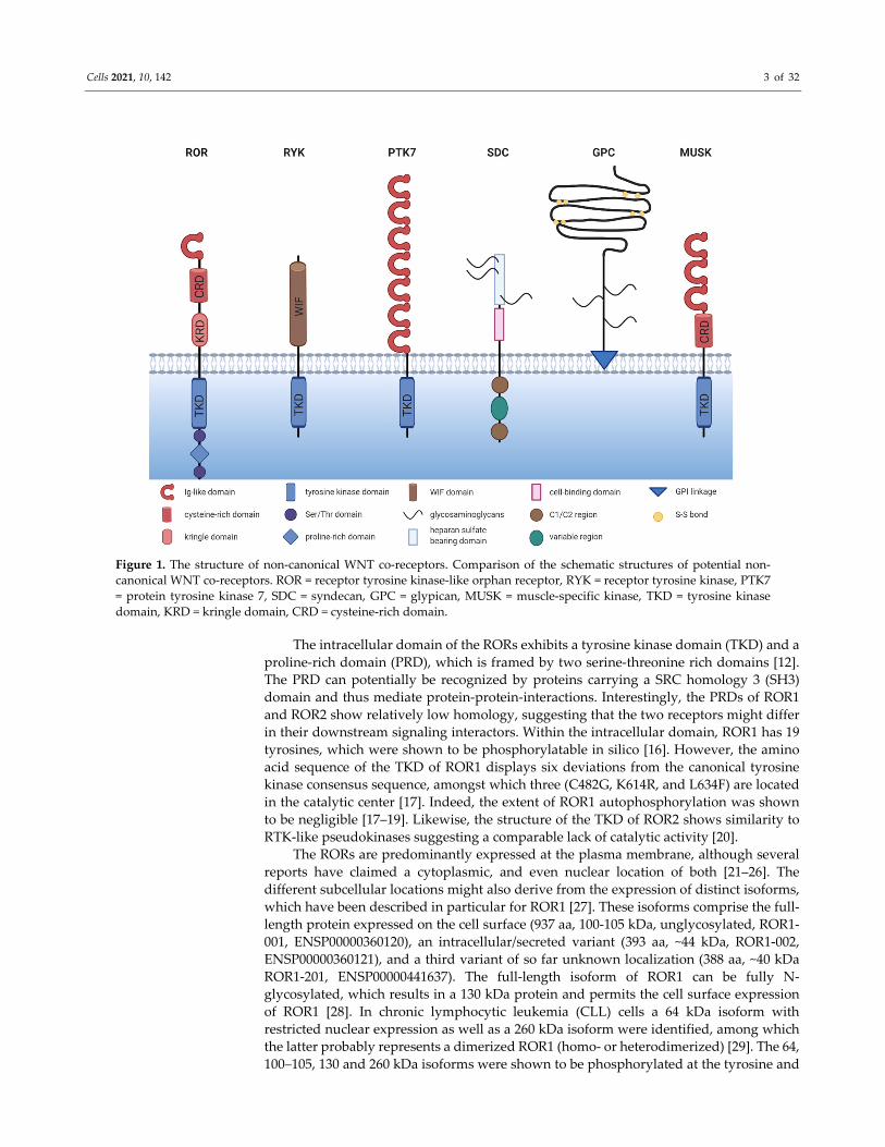
immunoglobulin (Ig)‐like domain, a cysteine‐rich domain (CRD) and a kringle domain
(KRD) in their extracellular part [12] (Figure 1). The CRD of ROR1 and ROR2 is similar to
that of the FZD receptors and has been identified as the essential domain for the binding
of WNT ligands [13,14]. RORs are the only RTK family members possessing a KRD, which
was shown to be essential for ROR1/ROR2 hetero‐oligomerization [15].
Cells 2021, 10, 142 3 of 32
Figure 1. The structure of non‐canonical WNT co‐receptors. Comparison of the schematic structures of potential non‐
canonical WNT co‐receptors. ROR = receptor tyrosine kinase‐like orphan receptor, RYK = receptor tyrosine kinase, PTK7
= protein tyrosine kinase 7, SDC = syndecan, GPC = glypican, MUSK = muscle‐specific kinase, TKD = tyrosine kinase
domain, KRD = kringle domain, CRD = cysteine‐rich domain.
The intracellular domain of the RORs exhibits a tyrosine kinase domain (TKD) and a
proline‐rich domain (PRD), which is framed by two serine‐threonine rich domains [12].
The PRD can potentially be recognized by proteins carrying a SRC homology 3 (SH3)
domain and thus mediate protein‐protein‐interactions. Interestingly, the PRDs of ROR1
and ROR2 show relatively low homology, suggesting that the two receptors might differ
in their downstream signaling interactors. Within the intracellular domain, ROR1 has 19
tyrosines, which were shown to be phosphorylatable in silico [16]. However, the amino
acid sequence of the TKD of ROR1 displays six deviations from the canonical tyrosine
kinase consensus sequence, amongst which three (C482G, K614R, and L634F) are located
in the catalytic center [17]. Indeed, the extent of ROR1 autophosphorylation was shown
to be negligible [17–19]. Likewise, the structure of the TKD of ROR2 shows similarity to
RTK‐like pseudokinases suggesting a comparable lack of catalytic activity [20].
The RORs are predominantly expressed at the plasma membrane, although several
reports have claimed a cytoplasmic, and even nuclear location of both [21–26]. The
different subcellular locations might also derive from the expression of distinct isoforms,
which have been described in particular for ROR1 [27]. These isoforms comprise the full‐
length protein expressed on the cell surface (937 aa, 100‐105 kDa, unglycosylated, ROR1‐
001, ENSP00000360120), an intracellular/secreted variant (393 aa, ~44 kDa, ROR1‐002,
ENSP00000360121), and a third variant of so far unknown localization (388 aa, ~40 kDa
ROR1‐201, ENSP00000441637). The full‐length isoform of ROR1 can be fully N‐
glycosylated, which results in a 130 kDa protein and permits the cell surface expression
of ROR1 [28]. In chronic lymphocytic leukemia (CLL) cells a 64 kDa isoform with
restricted nuclear expression as well as a 260 kDa isoform were identified, among which
the latter probably represents a dimerized ROR1 (homo‐ or heterodimerized) [29]. The 64,
100–105, 130 and 260 kDa isoforms were shown to be phosphorylated at the tyrosine and
Cells 2021, 10, 142 4 of 32
serine residues and were differentially expressed in CLL patients, depending on their
progression status [29]. Possible isoforms for ROR2 have not been described yet.
3. Expression of ROR1/2 in Cancer
In order to assess the targetability of ROR1 and ROR2 as novel therapeutic approach,
it is important to determine their pattern and level of expression in healthy tissue, in which
ROR‐targeted therapies might cause undesired off‐target effects, and thus toxicity.
Furthermore, before choosing which cancer entities might benefit from ROR targeting, it
should be noted that the RORs are not uniformly expressed in all cancer tissues, and that
their function might differ in the different entities. Therefore, we discuss the current
knowledge on the expression of ROR1 and ROR2 in cancer in the following chapter and
summarize the findings in Table 1.
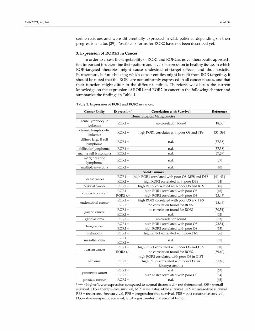
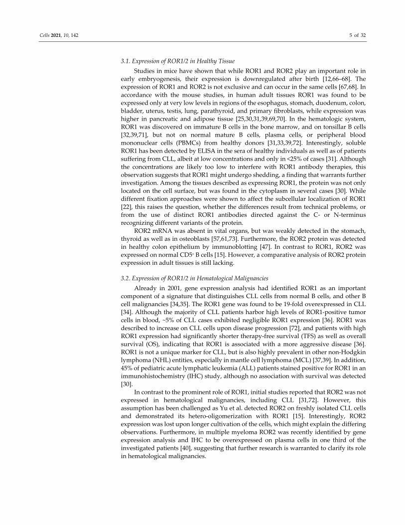
Table 1. Expression of ROR1 and ROR2 in cancer.
Cancer Entity Expression 1 Correlation with Survival Reference
Hematological Malignancies
acute lymphocytic
leukemia ROR1 + no correlation found [18,30]
chronic lymphocytic
leukemia ROR1 + high ROR1 correlates with poor OS and TFS [31–36]
diffuse large B cell
lymphoma ROR1 + n.d. [37,38]
follicular lymphoma ROR1 + n.d. [37,38]
mantle cell lymphoma ROR1 + n.d. [37,39]
marginal zone
lymphoma ROR1 + n.d. [37]
multiple myeloma ROR2 + n.d. [40]
Solid Tumors
breast cancer ROR1 + high ROR1 correlated with poor OS, MFS and DFS [41–43]
ROR2 + high ROR2 correlated with poor DFS [44]
cervical cancer ROR2 + high ROR2 correlated with poor OS and RFS [45]
colorectal cancer ROR1 + high ROR1 correlated with poor OS [46]
ROR2 +/− high ROR2 correlated with poor OS [23,47]
endometrial cancer ROR1 + high ROR1 correlated with poor OS and PFS
[48,49] ROR2 + no correlation found for ROR2
gastric cancer ROR1 + no correlation found for ROR1 [50,51]
ROR2 − n.d. [52]
glioblastoma ROR2 + no correlation found [53]
lung cancer ROR1 + high ROR1 correlated with poor OS [22,54]
ROR2 + high ROR2 correlated with poor OS [55]
melanoma ROR1 + high ROR1 correlated with poor PRS [56]
mesothelioma ROR1 +
ROR2 + n.d. [57]
ovarian cancer ROR1 + high ROR1 correlated with poor OS and DFS [58]
ROR2 +/− no correlation found for ROR2 [59,60]
sarcoma ROR2 +
high ROR2 correlated with poor OS in GIST
high ROR2 correlated with poor DSS in
leiomyosarcoma
[61,62]
pancreatic cancer ROR1 + n.d. [63]
ROR2 + high ROR2 correlated with poor OS [64]
prostate cancer ROR2 − n.d. [65] 1 +/− = higher/lower expression compared to normal tissue; n.d. = not determined, OS = overall
survival, TFS = therapy‐free survival, MFS = metastasis‐free survival, DFS = disease‐free survival,
RFS = recurrence‐free survival, PFS = progression‐free survival, PRS = post recurrence survival,
DSS = disease‐specific survival, GIST = gastrointestinal stromal tumor.
Cells 2021, 10, 142 5 of 32
3.1. Expression of ROR1/2 in Healthy Tissue
Studies in mice have shown that while ROR1 and ROR2 play an important role in
early embryogenesis, their expression is downregulated after birth [12,66–68]. The
expression of ROR1 and ROR2 is not exclusive and can occur in the same cells [67,68]. In
accordance with the mouse studies, in human adult tissues ROR1 was found to be
expressed only at very low levels in regions of the esophagus, stomach, duodenum, colon,
bladder, uterus, testis, lung, parathyroid, and primary fibroblasts, while expression was
higher in pancreatic and adipose tissue [25,30,31,39,69,70]. In the hematologic system,
ROR1 was discovered on immature B cells in the bone marrow, and on tonsillar B cells
[32,39,71], but not on normal mature B cells, plasma cells, or peripheral blood
mononuclear cells (PBMCs) from healthy donors [31,33,39,72]. Interestingly, soluble
ROR1 has been detected by ELISA in the sera of healthy individuals as well as of patients
suffering from CLL, albeit at low concentrations and only in <25% of cases [31]. Although
the concentrations are likely too low to interfere with ROR1 antibody therapies, this
observation suggests that ROR1 might undergo shedding, a finding that warrants further
investigation. Among the tissues described as expressing ROR1, the protein was not only
located on the cell surface, but was found in the cytoplasm in several cases [30]. While
different fixation approaches were shown to affect the subcellular localization of ROR1
[22], this raises the question, whether the differences result from technical problems, or
from the use of distinct ROR1 antibodies directed against the C‐ or N‐terminus
recognizing different variants of the protein.
ROR2 mRNA was absent in vital organs, but was weakly detected in the stomach,
thyroid as well as in osteoblasts [57,61,73]. Furthermore, the ROR2 protein was detected
in healthy colon epithelium by immunoblotting [47]. In contrast to ROR1, ROR2 was
expressed on normal CD5+ B cells [15]. However, a comparative analysis of ROR2 protein
expression in adult tissues is still lacking.
3.2. Expression of ROR1/2 in Hematological Malignancies
Already in 2001, gene expression analysis had identified ROR1 as an important
component of a signature that distinguishes CLL cells from normal B cells, and other B
cell malignancies [34,35]. The ROR1 gene was found to be 19‐fold overexpressed in CLL
[34]. Although the majority of CLL patients harbor high levels of ROR1‐positive tumor
cells in blood, ~5% of CLL cases exhibited negligible ROR1 expression [36]. ROR1 was
described to increase on CLL cells upon disease progression [72], and patients with high
ROR1 expression had significantly shorter therapy‐free survival (TFS) as well as overall
survival (OS), indicating that ROR1 is associated with a more aggressive disease [36].
ROR1 is not a unique marker for CLL, but is also highly prevalent in other non‐Hodgkin
lymphoma (NHL) entities, especially in mantle cell lymphoma (MCL) [37,39]. In addition,
45% of pediatric acute lymphatic leukemia (ALL) patients stained positive for ROR1 in an
immunohistochemistry (IHC) study, although no association with survival was detected
[30].
In contrast to the prominent role of ROR1, initial studies reported that ROR2 was not
expressed in hematological malignancies, including CLL [31,72]. However, this
assumption has been challenged as Yu et al. detected ROR2 on freshly isolated CLL cells
and demonstrated its hetero‐oligomerization with ROR1 [15]. Interestingly, ROR2
expression was lost upon longer cultivation of the cells, which might explain the differing
observations. Furthermore, in multiple myeloma ROR2 was recently identified by gene
expression analysis and IHC to be overexpressed on plasma cells in one third of the
investigated patients [40], suggesting that further research is warranted to clarify its role
in hematological malignancies.
Cells 2021, 10, 142 6 of 32
3.3. Expression of ROR1 in Solid Tumors
Overexpression of ROR1 has not only been reported in hematological malignancies,
but has also gained increasing attention in solid tumors. IHC analyses have demonstrated
a wide‐spread expression of ROR1 across many tumor entities with significantly higher
expression levels in cancerous than in adjacent normal tissue (Table 1), including a
particularly strong expression in melanoma, colon, pancreatic, lung, and breast cancer
[22,41,56]. Chang et al. reported that high ROR1 expression was associated with a lower
pathological tumor (pT) stage and the absence of perineural invasion in gastric cancer
patients who underwent gastrectomy and did not receive neoadjuvant chemotherapy
[50]. However, so far this remains the only analysis that suggested that ROR1 might be
associated with limited disease, while the majority of studies indicated that it is rather
associated with aggressiveness and poor survival. ROR1 expression was positively
associated with clinical stage and lymph node metastasis in colorectal cancer, for which it
served as an independent prognostic marker [46]. In ovarian cancer, high ROR1
expression was likewise associated with tumor grade as well as lymph node metastasis
[58]. In Ewing sarcoma, ROR1 expression was markedly higher in metastases than in
localized disease [70], and in pancreatic cancer, ROR1 was identified on circulating tumor
cells (CTCs) as an essential factor for their invasive phenotype [63].
In breast cancer, ROR1 levels seem to vary between the different molecular subtypes,
as gene expression data have implied particularly high levels in poorly differentiated and
triple‐negative breast cancers, the most aggressive breast cancer subtype, in which very
high expression (top 10%) was associated with shorter OS [41]. Similar results were
obtained from an IHC study in which 57% of triple‐negative breast cancers stained highly
positive for ROR1, whereas signals were detected in only 12% of the estrogen receptor
(ER) and progesterone receptor (PR)‐positive patients, and no expression at all was found
in Her2/Neu‐positive breast cancers [25]. Interestingly, breast cancer brain metastases
have likewise been shown to highly express ROR1 [74], hinting at its role in metastatic
spread.
ROR1 has also been extensively studied in lung cancer. Since it was identified as a
transcriptional target of homeobox protein Nkx‐2.1 [75], a major lineage‐survival
oncogene in lung cancer, it is not surprising that many lung tumors harbor ROR1.
Comparable to breast cancer, ROR1 was expressed in certain subtypes and was
particularly high in lung adenocarcinoma (40–65% positive), while squamous carcinomas
stained positive at much lower frequencies [22,25,76]. In lung adenocarcinoma, ROR1
functions as an independent prognostic predictor for OS [57]. Both ROR1 and ROR2 were
found to be highly expressed in a lung cancer cell line derived from CTCs from a patient
with relapsed small cell lung cancer [77]. This fits to the observation in two patients with
ROR1‐negative primary tumors, in whom the metastases gained ROR1 [25]. While these
observations suggest that ROR1 might be involved in metastasis, the same study also
showed that 40% of ROR1‐positive primary tumors lost their ROR1 expression in the
matched metastatic lesions [25], thus questioning this concept. Taken together, this raises
the question whether ROR1 solely functions as a bystander molecule upregulated during
tumor progression, or whether the functional consequence of WNT/ROR signaling is
highly context‐specific as discussed later in this article.
3.4. Expression of ROR2 in Solid Tumors—It Is not Always That Simple
Compared with ROR1, the expression profile of ROR2 in cancer is much more
heterogeneous (Table 1). Although most studies describe ROR2 as a positive prognostic
factor in a variety of solid tumor entities, a limited number of reports observed a loss of
ROR2 in cancerous compared to normal tissue and an association of high ROR2
expression with a favorable outcome. One problem that might have caused some of the
contradictory results is that several commercially available antibodies have been reported
Cells 2021, 10, 142 7 of 32
to display non‐specific binding [77], thus bringing into question the reliability of the IHC
results obtained with these antibodies.
A particularly high percentage of ROR2‐positive tumors was found in breast cancer
(87%), glioblastoma (>90%), and neuroblastoma (80%) [24,53,78]. Interestingly, ROR2 was
expressed in only 20 of 48 of primary melanoma cases, while all of the investigated visceral
or subcutaneous metastases (48/48) stained positive [79]. Similar observations have been
reported for lymph node and brain metastases in breast cancer [74,80]. Indeed, ROR2
expression was found to correlate with tumor stage and/or lymph node metastasis in lung
[55], cervical [45], and breast cancer [24]. Taken together, these studies point to a specific
role for ROR2 in invasive growth and metastasis of the named tumor entities.
In contrast, downregulation of ROR2 in solid tumor cells, compared with adjacent
normal tissue, has been described for gastric [52] and prostate carcinoma [65].
Contradictory findings for ROR2 have been reported for endometrial, colorectal, and
ovarian cancer. Initially, the study by Lara et al. demonstrated promoter
hypermethylation and consequently repressed expression of ROR2 in 5/8 colorectal cancer
cell lines and 3/6 patients [47]. In contrast, another study using quantitative real‐time PCR
detected a significantly higher expression of ROR2 in colorectal cancer compared with
adjacent normal tissue [23]. In ovarian cancer, a comparative analysis has revealed that
ROR1 was largely expressed in cancer cells, while the number of patients with ROR2‐
positive tumors was significantly lower [81]. While ROR2 was found upregulated in a
large ovarian carcinoma cohort compared with normal ovarian tissue [59], another report
investigating aggressive high‐grade serous ovarian carcinoma has claimed the opposite
[60]. Whether these controversial findings reflect context‐specific functions of ROR2, or
whether they arise from non‐specific antibody binding in IHC analyses remains to be
clarified.
3.5. ROR1/2 in the Tumor Stroma
While research has traditionally focused on the expression and function of the two
RORs in cancer cells, several recent reports support the notion that they also play a
relevant role in the cancer‐associated microenvironment. In ovarian cancer, stroma cell
expression of ROR1 was lower than in the tumor cells, while the opposite was true for
ROR2 [81]. Interestingly, the expression levels of ROR2 increased even further in
metastatic tissue compared with matched primary tumor samples [81]. In pancreatic
cancer stromal ROR2 correlated with regional lymph node invasion and represented an
independent prognostic factor [64]. While the significance of stromal ROR2 remains
elusive so far, novel data have shown that ROR2 is upregulated in reactive astrocytes at
brain injury sites [82]. Concordantly, both ROR1 and ROR2 were induced in skeletal
muscles by pro‐inflammatory cytokines (e.g., TNF‐α, IL‐1β) released after injury [83].
These findings suggest that the upregulation of the RORs might occur in stromal cells due
to the inflammatory conditions often observed in the tumor microenvironment.
4. The Function of ROR1/2 in Cancer
WNT signaling is known to regulate many key cellular processes, and aberrant
regulation of this pathway has been found to contribute to the development and
progression of many human cancers. Likewise, the deregulated expression of the WNT
co‐receptors ROR1 and ROR2 has been associated with several cellular features that
promote malignancy, namely cell proliferation, survival, migration/invasion, and
stemness, as it will be discussed in the following chapters.
While most studies identified ROR1 as an oncogene, the role of ROR2 in cancer is less
clear and has been controversially discussed. Studies in osteosarcoma, melanoma, CLL,
breast, and renal cancer have claimed that ROR2 acts as an oncogene in these entities. In
contrast, in prostate, colorectal and endometrial cancer some studies attribute a tumor
suppressive function to ROR2. Whether the output of ROR2 signaling differs especially in
colorectal and endometrial cancer, two entities substantially driven by the β‐catenin‐

Cells 2021, 10, 142 8 of 32
dependent, canonical WNT pathway, or whether different organ microenvironments with
varying repertoires of FZDs and alternative WNT co‐receptors are responsible for the
diverging effects is not yet fully understood.
4.1. ROR1/2 in Cell Proliferation and Survival
More than a decade ago both ROR1 and ROR2 were identified as pro‐survival kinases
by an RNAi screen in HeLa cells [84]. This finding spiked the interest of cancer biologists
early on to not only evaluate the potential of these novel receptors as cancer biomarkers,
but also to consider their functional involvement in tumor development and progression.
Meanwhile it became evident that ROR1 plays a major role in cancer cell survival by
promoting proliferation, while at the same time counteracting apoptosis. This has not only
been observed in a multitude of in vitro studies on cancer cell lines
[15,22,29,41,48,51,56,57,75,76], but was also confirmed in xenograft studies in vivo [41,75].
In a mouse model for human CLL, the B cell‐restricted expression of ROR1 accelerated
leukemogenesis through an interaction of ROR1 with the T cell leukemia 1 (TCL1)
oncogene, which activated pro‐survival signaling via the protein kinase AKT and resulted
in leukemia cell proliferation and resistance to apoptosis [85]. In line with this,
transcriptomic analyses of ROR1‐expressing patient‐derived CLL cells revealed a gene
expression signature associated with protein kinase activation, AKT‐related signaling
factors and proliferation, while ROR1‐negative CLL cells were characterized by
subnetworks associated with apoptosis and consequential RNA processing and decay
[36]. Interestingly, recent observations indicate that the pro‐survival function of ROR1 not
only strongly supports leukemogenesis, but is also linked with the development of
leukemia. In mice, the knockout of miRNA miR‐15/16, leading to the overexpression of
the pro‐survival factor BCL2 as well as ROR1, induced the development of B cell
lymphoma in 23% of the animals, while 77% developed an aggressive acute myeloid
leukemia (AML) [86]. This finding indicates a great need for further investigation of
ROR1, not only in lymphoma, but also in AML, and suggests that combined targeting of
ROR1 (e.g., cirmtuzumab) and BCL2 (e.g., venetoclax) might be worth considering. Taken
together, these observations support the notion that the pro‐survival function of ROR1 is
also valid in vivo.
Mechanistic studies have shown that the effect of ROR1 on proliferation and
apoptosis required phosphorylation of its PRD [16], thus suggesting an activation of
ROR1‐dependent downstream signaling. Concordantly, additional stimulation of ROR1
with WNT5A supported its effect on cell survival in CLL cells [33,36]. Blockade of ROR1
in lung cancer cells was found to suppress expression of CDK4 and CCNE1, two
important cell cycle regulators, as well as Bcl‐XL and Bcl‐2, two critical anti‐apoptotic
factors, while it increased the expression of several pro‐apoptotic factors including Bak,
caspase‐3, and caspase‐7 [87]. However, whether these factors are direct target genes of
ROR1 and which signaling pathways are implemented in their upstream regulation
remains elusive so far. Moreover, two novel mechanisms that might contribute to the pro‐
survival function of ROR1 have recently been recognized: on the one hand, ROR1
inhibition was described to increase p53 activity [88], on the other hand, an involvement
of ROR1 in autophagy was suggested [87]. Further research is required to understand the
role of ROR1 in these processes in general as well as in the context of cancer.
In ovarian cancer ROR1 was described to have a synergistic effect with ROR2 on cell
proliferation, since only a double knockdown led to a significant reduction in tumor cell
proliferation [59]. Similar to ROR1, ROR2 supported proliferation and tumor growth,
while inhibiting apoptosis both in vitro as well as in vivo in mouse models of
osteosarcoma, breast and renal cancer [44,89–91]. Likewise, the expression of several cell
cycle‐related genes including CDK1/2/4, CCNE1, CCND1/2, PCNA, and MKI67 has been
described to be regulated by ROR2 in these cancer entities [80,90,92]. In contrast, in a study
on ovarian cancer the induction of ROR2 expression in the tumor cells led to endoplasmic
reticulum stress and decreased cell viability [60].
Cells 2021, 10, 142 9 of 32
A study in NIH3T3 fibroblasts has demonstrated that ROR2 was dynamically
regulated during the cell cycle, probably because it is under the transcriptional control of
the cell cycle regulator E2F1 [93]. ROR2 knockdown led to an accumulation of
osteosarcoma cells in the G0/G1 phase [92] suggesting that ROR2 itself is essential for cell
cycle progression. Downregulation of ROR2 not only decreased the expression of various
E2F1 target genes, but also induced Forkhead box protein O1 (FoxO) target genes [93],
thus favoring apoptosis induction.
Recent studies have implied that ROR1 and ROR2 might not only promote cell
survival through the regulation of pro‐mitotic factors, but also by controlling cellular
structures implicated in pro‐survival signaling, such as filopodia [28,94]. In gastric cancer
cells, ROR2 was described as stimulating the number and length of specific signaling
filopodia, so‐called cytonemes, which transport WNT ligands to neighboring cells, and
thus activate canonical WNT signaling responses stimulating cell proliferation [95]. ROR1,
in contrast, was found to interact with Caveolin‐1, Cavin‐1, and Cavin‐3 and was not only
required for correct caveolae formation, but also for caveolae‐dependent endocytosis and
induction of signaling responses [96,97]. As several pro‐survival RTKs (e.g., epidermal
growth factor receptor (EGFR), hepatocyte growth factor receptor (MET), Insulin‐like
growth factor 1 receptor (IGF1R)) depend on these functions, inhibiting ROR1 in lung
cancer was proposed as a novel shortcut to bypass resistance to EGFR tyrosine kinase
inhibitors [96]. Indeed, targeting ROR1 helped to reinstall sensitivity to erlotinib treatment
in resistant lung cancer cell lines [98], thus further underlining its potential as a
therapeutic target.
4.2. ROR1/2 in Therapy Resistance and Cancer Stem Cells
Next to the established function of the RORs in cell proliferation, another detrimental
consequence of active WNT/ROR signaling in cancer has currently emerged, namely the
establishment of resistant tumor cell clones. Several studies have reported the
upregulation of ROR1 or ROR2 in chemoresistant cancer cell lines [88,99] as well as in
patient‐derived tissues after chemo‐ or targeted therapy [100–102]. In melanoma,
O’Connell et al. initially observed opposing roles for ROR1 and ROR2: melanoma cell lines
resistant to inhibition of the serine/threonine‐protein kinase B‐raf (BRAF) were
characterized by upregulated ROR2, but downregulated ROR1 [103]. Another study on
uveal melanoma cell lines described upregulation of both RORs after mitogen‐activated
protein kinase kinase (MEK) inhibition and implicated them in the induction of a pro‐
survival AKT signaling response [104]. In ALL cell line models, and primary cells, the
knockdown of ROR1 enhanced sensitivity to several small molecule inhibitors in current
clinical use [105]. Addressing the question of how ROR1/2 can mediate therapy resistance,
three different modes are currently being discussed:
Firstly, WNT/ROR1 signaling was identified as a rescue pathway that can induce
nuclear factor kappa‐light‐chain‐enhancer of activated B cells (NF‐κB), phosphoinositide
3‐kinase (PI3K)/protein kinase B (AKT), and MEK/extracellular‐signal‐regulated kinase
(ERK) activation independent of B cell receptor (BCR)/Bruton tyrosine kinase (BTK) in
CLL and MCL [18,106,107]. This activation was mediated via the formation of a complex
between ROR1 and CD19 [107] and represented a novel resistance mechanism for
treatment strategies centered around BTK/BCR inhibition. Indeed, synergistic effects have
been shown for co‐targeting both ROR1 and the BCR or BCL‐2 family, underlining the
large potential for ROR1‐targeted therapies in overcoming MCL and CLL drug resistance
[106,108].
Secondly, ROR1 was shown to upregulate the expression of ATP‐dependent
translocase ABCB1, a multi‐drug efflux pump, and thus facilitate drug export from cancer
cells [88]. Indeed, targeting ROR1 in ovarian cancer increased the efficacy of second
mitochondria‐derived activator of caspase (SMAC) mimetics and taxanes [109]. In line,
combining paclitaxel treatment with ROR1 blockade significantly enhanced tumor
growth inhibition in a patient‐derived xenograft (PDX) model for breast cancer [102].
Cells 2021, 10, 142 10 of 32
Thirdly, ROR1 has been associated with treatment‐resistant cancer stem cells as
reviewed in [110]. This conclusion was based on observations in ovarian cancer stem cells,
in which ROR1 was highly expressed and regulated the expression of the self‐renewal
marker polycomb complex protein BMI‐1 [111]. Meanwhile, studies on breast cancer
primary cells have likewise identified a population of cells carrying the cancer stem cell
markers ALDH1+/CD44+/CD24low that displayed particularly high ROR1 expression and
showed enhanced growth of tumor spheres in vitro as well as tumorigenicity in vivo
[100,102]. These ROR1+ cells were characterized by enhanced stemness frequency and
expressed markers typically associated with stem cells such as e.g., POU5F1, NANOG, or
SOX2 [100]. Concordantly, in a phase 1 trial on cirmtuzumab, a monoclonal antibody
directed against ROR1, gene expression signatures associated with stemness were
markedly lower in post‐treatment samples of CLL patients [112]. Taken together, these
observations suggest that ROR1 is indeed expressed on cancer stem cells and might thus
be involved in therapy resistance and relapse. Whether ROR2 fulfills the same functions
as ROR1 in the development of drug resistance or whether it acts via distinct mechanisms
remains to be elucidated.
4.3. ROR1/2 in EMT and Metastasis
The clinical observations that ROR1 and ROR2 are associated with disease
progression and metastasis in many cancer patients have inspired researchers to
investigate the role of both co‐receptors in the underlying cellular processes. In CLL,
stimulation of ROR1‐expressing cancer cells with WNT5A stimulated their migratory
potential and CXCL12/CCL19‐directed chemotaxis, while this effect was not observed in
ROR1‐negative cells [33,36]. Similar observations have been made in breast cancer in
which ROR1 knockdown decreased CXCR4 expression resulting in decreased chemotaxis
of the cells towards a CXCL12 gradient [42]. Moreover, in several in vitro solid tumor
models ROR1 knockdown reduced cellular migration/invasion. These include Ewing
sarcoma [70], glioblastoma spheres [113], mesothelioma [57], breast cancer [42,114],
ovarian cancer [59] or melanoma [56]. Another report on melanoma observed that ROR1
was associated with a poorly invasive phenotype, and its knockdown increased invasion
in vitro as well as metastasis formation in vivo. However, these diverging results might
be caused by the simultaneous upregulation of compensatory ROR2/WNT5A [103]. In line
with cell motility, ROR1 was found to regulate cell adhesion [56].
First reports about a pro‐invasive function of ROR2 stem from studies on HeLa cells
showing that WNT5A‐ROR2 mediated polarized cell migration in this cell line [115]. This
has since been confirmed in several solid tumor entities in which knockdown of ROR2
resulted in decreased migration and/or invasion, e.g., in mesothelioma [57], melanoma
[79], renal cancer [89], breast cancer [114], ovarian cancer [59,99,116,117], prostate cancer
[118], leiomyosarcoma, gastrointestinal stroma tumors [62], and osteosarcoma [61]. Taken
together, the data imply that both ROR1 and ROR2 are associated with highly malignant
cancer cell phenotypes characterized by high motility and aggressiveness.
The pro‐migratory and ‐invasive functions of ROR1 and ROR2 might not only result
from the activation of a typical non‐canonical WNT signaling response, which will be
discussed in the next chapter, but have been associated with the induction of an epithelial‐
to‐mesenchymal‐transition (EMT) phenotype. EMT is a reversible cellular program in
which cancer cells lose their adhesive, epithelial characteristics and gain a more motile,
mesenchymal phenotype, which drives tumor spreading. ROR1‐high compared to ROR1‐
low breast cancer tumors initially showed high expression of genes typically associated
with mesenchymal cells [42]. In renal cell carcinoma patients expression of ROR2 was
found to be tightly associated with the EMT regulator TWIST and the matrix‐degrading
enzyme MMP2 [89]. Further in vitro studies have indeed confirmed higher expression of
typical epithelial factors (e.g., CK19, CDH1) and lower expression of mesenchymal factors
(e.g., SNAI2, ZEB1, VIM) after ROR1 or ROR2 knockdown in cancer cells. This is
indicative of a role for both co‐receptors in fostering EMT [42,56,80,111,113,117]. One
Cells 2021, 10, 142 11 of 32
explanation why the RORs are so closely associated with the EMT program is that their
expression has been revealed to be under the control of key EMT‐inducing transcription
factors. For instance, TWIST was demonstrated to activate the transcription of the ROR1
gene [119], while the expression of ROR2 was found to be regulated by SNAI1 [120].
Another report claimed that ROR2 itself was responsible for regulating SNAI1 expression
[117], raising the chicken and egg question—which comes first?
The set of EMT genes regulated by ROR2, in particular, comprises the matrix
metalloproteinases MMP2 and MMP9 [80,89,120]. As these enzymes are critical for
degrading the extracellular matrix, their enhanced expression in ROR‐positive cells might
explain their large potential for invasive growth. Another mechanism by which especially
ROR2 promotes invasiveness is the induction of invadopodia formation [94,121], i.e.,
special plasma membrane protrusions that can serve as specific sites for the secretion of
matrix‐degrading enzymes. Recently, ROR2 was found to upregulate the expression of
intraflagellar transport 20 (IFT20), a protein originally involved in the microtubule‐based
transport in the cilium. IFT20 regulates Golgi structure and function which are required
for correct cell polarization and subsequent invasive behavior [122]. WNT5A‐ROR was
also described as inducing the degradation of the kinesin KIF26b which was identified as
a novel cytoskeletal effector of WNT/ROR signaling and regulated cell migration.
Although the mechanism of WNT/ROR/KIF26b signaling is not fully understood yet, it
was proposed that it fine‐tunes de‐adhesion and/or retraction at the trailing edge of
migrating cells [123]. Thus, it seems that the RORs are substantially involved in regulating
processes associated with cytoskeleton dynamics that finally result in increased motility
and invasiveness.
Evidence suggests that these findings are also valid in vivo, as ROR1 knockdown
significantly reduced the capacity of breast cancer cells intravenously injected into
immunodeficient mice to invade the lung 24 to 72 h after injection [42], pointing to a role
for ROR1 in extravasation and adhesion due to its control of the EMT phenotype. Similar
observations have been made for ROR2: multiple myeloma cells with stable ROR2
knockdown were mostly unable to home into the bone marrow due to a defective
adhesion to the bone marrow microenvironment [40]. Consequently, cancer cells with
reduced expression of ROR1 or ROR2 were characterized by a significantly impaired
potential in forming metastases in mouse models of melanoma or breast cancer [42,79].
However, some reports claim that in some cancer subtypes, the RORs are rather
associated with less motile and less invasive tumor cells. For instance, when discussing
ROR1 and EMT, the special case of hepatocellular carcinoma should be mentioned, since
in that tumor entity ROR1 was associated more with an epithelial rather than a
mesenchymal phenotype indicating that it might play an opposing role here [124]. For
ROR2, especially in colorectal and prostate cancer conflicting data have been obtained.
For the latter, ROR2 knockdown was initially found to reduce cell invasion [118].
However, a recent study reported a reduction in tumor motility and invasiveness upon
ROR2 overexpression [65], again raising the question whether the regulation of ROR
expression levels is critical for determining their functional impact. Moreover, WNT5A
secreted by the osteoblastic niche was observed to induce dormancy in prostate cancer
cells via ROR2 by repressing canonical WNT signaling and thus inhibiting bone
metastasis [125]. Whether this interesting finding is specific for prostate cancer, or for bone
metastasis, or if it also occurs in other tumor entities remains unknown. In the second
example of colorectal carcinoma, hypermethylation, and thus silencing, of the ROR2
promoter was identified as an early event in carcinogenesis. Decreased ROR2 levels were
associated with increased proliferation and migration, but with greatly reduced
invasiveness [47,126]. While the early downregulation of ROR2 might represent a way to
circumvent its inhibitory influence on canonical WNT signaling, the observed differential
regulation of migration and invasion could indicate that ROR2 is indeed responsible for
dynamically fine‐tuning these cellular processes. In general, ROR2 seems to have tumor
suppressive effects particularly in tumors driven by canonical WNT signaling (e.g.,
Cells 2021, 10, 142 12 of 32
colorectal, prostate, or endometrial cancer), whereas it acts rather as an oncogene in
tumors with predominantly active non‐canonical WNT signaling (e.g., melanoma, breast
cancer). This suggests that the different signaling contexts have a significant impact on the
functional output of WNT/ROR signaling.
5. WNT/ROR Signaling at a Glance
The vast majority of studies have associated both ROR1 and ROR2 with the activation
of β‐catenin‐independent WNT signaling responses triggered by binding of the non‐
canonical WNT ligand WNT5A. In the following chapter, we will discuss the current
knowledge about WNT/ROR signaling, which is summarized in Figures 2 and 3. When
modulating the expression of one of the ROR co‐receptors in order to study downstream
signaling events, it should be carefully considered that this could trigger upregulation of
the other ROR co‐receptor which might be able to compensate for the loss. For instance,
in melanoma ROR1 knockdown was followed by increased expression of ROR2 and
WNT5A, while the knockdown of ROR2 stimulated ROR1 expression [103], confirming a
reciprocal regulation. Concordantly, in breast cancer cells ROR2 overexpression
augmented ROR1 levels [114].
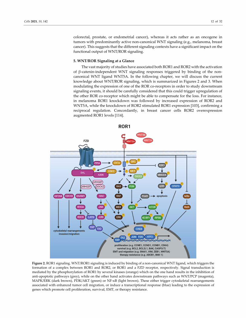
Figure 2. ROR1 signaling. WNT/ROR1 signaling is induced by binding of a non‐canonical WNT ligand, which triggers the
formation of a complex between ROR1 and ROR2, or ROR1 and a FZD receptor, respectively. Signal transduction is
mediated by the phosphorylation of ROR1 by several kinases (orange) which on the one hand results in the inhibition of
anti‐apoptotic pathways (grey), while on the other hand activates downstream pathways such as WNT/PCP (magenta),
MAPK/ERK (dark brown), PI3K/AKT (green) or NF‐κB (light brown). These either trigger cytoskeletal rearrangements
associated with enhanced tumor cell migration, or induce a transcriptional response (blue) leading to the expression of
genes which promote cell proliferation, survival, EMT, or therapy resistance.
Cells 2021, 10, 142 13 of 32
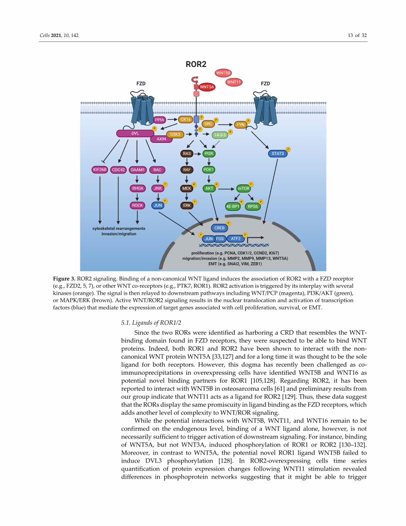
Figure 3. ROR2 signaling. Binding of a non‐canonical WNT ligand induces the association of ROR2 with a FZD receptor
(e.g., FZD2, 5, 7), or other WNT co‐receptors (e.g., PTK7, ROR1). ROR2 activation is triggered by its interplay with several
kinases (orange). The signal is then relayed to downstream pathways including WNT/PCP (magenta), PI3K/AKT (green),
or MAPK/ERK (brown). Active WNT/ROR2 signaling results in the nuclear translocation and activation of transcription
factors (blue) that mediate the expression of target genes associated with cell proliferation, survival, or EMT.
5.1. Ligands of ROR1/2
Since the two RORs were identified as harboring a CRD that resembles the WNT‐
binding domain found in FZD receptors, they were suspected to be able to bind WNT
proteins. Indeed, both ROR1 and ROR2 have been shown to interact with the non‐
canonical WNT protein WNT5A [33,127] and for a long time it was thought to be the sole
ligand for both receptors. However, this dogma has recently been challenged as co‐
immunoprecipitations in overexpressing cells have identified WNT5B and WNT16 as
potential novel binding partners for ROR1 [105,128]. Regarding ROR2, it has been
reported to interact with WNT5B in osteosarcoma cells [61] and preliminary results from
our group indicate that WNT11 acts as a ligand for ROR2 [129]. Thus, these data suggest
that the RORs display the same promiscuity in ligand binding as the FZD receptors, which
adds another level of complexity to WNT/ROR signaling.
While the potential interactions with WNT5B, WNT11, and WNT16 remain to be
confirmed on the endogenous level, binding of a WNT ligand alone, however, is not
necessarily sufficient to trigger activation of downstream signaling. For instance, binding
of WNT5A, but not WNT3A, induced phosphorylation of ROR1 or ROR2 [130–132].
Moreover, in contrast to WNT5A, the potential novel ROR1 ligand WNT5B failed to
induce DVL3 phosphorylation [128]. In ROR2‐overexpressing cells time series
quantification of protein expression changes following WNT11 stimulation revealed
differences in phosphoprotein networks suggesting that it might be able to trigger
Cells 2021, 10, 142 14 of 32
activation of ERK1, AKT, as well as GSK3B, known downstream effectors of ROR2
signaling [133].
Interestingly, the expression of ROR1/2 and their established ligand WNT5A is
tightly associated and regulated in a positive feedback loop. ROR1 was found to
upregulate WNT5A, which in turn triggered ROR1 expression via STAT3 [56,134].
Similarly, WNT5A can upregulate ROR2 levels and vice versa the downregulation of
ROR2 decreased WNT5A expression [79,120,135]. A recent study has reported
chemotactic migration of Ewing sarcoma cells towards WNT5A, which was impaired by
ROR1 silencing [70]. Such WNT gradients might enable the secreting cells to specifically
attract target cells by inducing dynamic cell movements through the regulation of non‐
canonical WNT/ROR signaling.
5.2. Getting the RORs Started: Receptor Activation
Ligand stimulation can either induce ROR1/ROR2 homo‐ or heterodimerization
[15,136,137], or their association with FZD receptors [127,138,139]. Moreover, in HEK293
cells, ROR2, but not ROR1, was found to form a complex with PTK7 and WNT5A, which
activated JNK and AP‐1 [140]. Thus, it is not yet fully understood whether the RORs are
indeed “true” co‐receptors that are dependent on an interaction with FZDs to activate
signaling, or whether they can fulfill this function alone, or via the interaction with other
co‐receptors (e.g., PTK7).
While the kinase function of the RORs has been a point of discussion [17,19,141],
several other kinases have been implicated in their phosphorylation, and thus activation.
Examples include GSK3, the receptor tyrosine kinase MET or the proto‐oncogene SRC
[16,17,75,130,142]. Furthermore, the WNT5A‐induced association of ROR2 with FZD led
to the activation of ROR2‐bound CK1ε by the FZD‐bound protein phosphatase 2A (PP2A)
[143]. In turn, CK1ε mediated phosphorylation of ROR2 as well as FZD‐associated DVL,
which was required for downstream signaling [143,144]. A similar mechanism could
apply for ROR1 as the phosphorylation of DVL3 was blocked upon inhibition of ROR1
and CK1 in CLL cells [145].
5.3. Induction of Non‐Canonical WNT Signaling
DVL proteins are essential signaling hubs in the WNT pathway that become
phosphorylated upon a WNT stimulus and transmit signals to downstream effectors
(reviewed in [146]). This is also true for WNT/ROR signaling [56,70,91,114,121]. Activated
DVL proteins can relay the signals to distinct signaling branches centered around the
small GTPases RHO, RAC and CDC42 which become activated upon WNT/ROR
stimulation [15,56,57,102,137]. The regulation was even observed in vivo as a gene set
enrichment analysis showed that pre‐treatment CLL samples displayed a higher
expression of genes associated with activation of RAC1 or RHOA than matched samples
after treatment with the ROR1‐blocking antibody cirmtuzumab [112].
WNT/ROR‐induced activation of RHOA was found to require the formin homology
protein DAAM1 which interacted with DVL and recruited RHOA [147]. Next to DAAM1,
several guanine nucleotide exchange factors (GEFs) are involved in the ROR‐dependent
activation of the small GTPases (e.g., ARHGEF, DOCK2) [15,108,148–150]. GEFs catalyze
the exchange of free cytosolic GTP for bound GDP and are thus critical regulators of
GTPase function. Concordantly, as RHO, RAC, and CDC42 control cytoskeletal
reorganization, their activation explains the drastic changes in cell motility observed upon
WNT/ROR signaling that result in enhanced proliferation, migration, as well as tumor
engraftment in vitro and in vivo [108,148,150].
WNT/ROR signaling was furthermore shown to induce cell migration via the
activation of the RAC downstream targets JNK, JUN, and ATF2 [114,115,121,127,138,151].
Although non‐canonical WNT signaling has so far mainly been described to occur
primarily via signaling cascades at the protein level, overexpression of ROR2 in MCF‐7
breast cancer cells resulted in large changes at the transcriptomic level with more than
Cells 2021, 10, 142 15 of 32
2860 differentially expressed genes (e.g., FAT1, VIL1, HNF4G, WIPF1). These potential
novel ROR2 target genes were centered around the processes metabolism, cell remodeling
and migration [152,153]. A recent study furthermore identified PLOD2, HADH, LCOR,
and REEP1 as novel target genes of the ROR2/DVL2/ATF2 pathway, which were involved
in the proliferation of colorectal cancer cells [154]. As so far no explicit target gene sets
were known for non‐canonical WNT signaling, these discoveries will facilitate further
research on the signaling mediators and upstream regulators of the pathway.
5.4. Crosstalk with Canonical WNT Signaling
Although the WNT pathway has classically been divided into canonical and non‐
canonical signaling, it has nowadays become increasingly clear that this black‐and‐white
separation is not entirely possible. There are many crosslinks at several levels, including
the ROR co‐receptors, in particular ROR2. While most reports did not find any indications
of an interaction of ROR1 with canonical WNT ligands or with the induction of a β‐
catenin‐dependent signaling in cancer cells [33,56,105], many reports described an
inhibitory function of ROR2 on β‐catenin‐dependent WNT signaling [2,24,125,131,155].
Indeed, gene expression data from the TCGA database pointed to a negative correlation
of ROR2 expression with an active canonical WNT signature in breast cancer patients [91],
suggesting that the inhibitory effect of ROR2 also occurs in vivo. Surprisingly, other
studies observed that WNT3A‐induced activation of canonical WNT signaling involved
ROR2 [156,157]. Taken together, it seems that ROR2 regulates the net balance of WNT
signaling depending on the receptor context and the signaling factors available
downstream.
5.5. Crosstalk with Other Major Cancer Signaling Pathways
The RORs seem to be crossroads that can link WNT signals to other major signaling
pathways implicated in the regulation of key cellular processes. Among them, in
particular the PI3K/AKT/mammalian target of rapamycin (mTOR) pathway seems to be
closely associated with WNT/ROR signaling. Activation of ROR1 and ROR2 was shown
to result in the phosphorylation of PI3K, AKT, cAMP response element‐binding protein
(CREB), and mTOR as well as its downstream targets S6 and 4EBP1 in numerous tumor
entities [18,22,40,41,44,56,70,151,158,159]. In vivo, metastases derived from ROR1
knockdown breast cancer cells injected into immunodeficient mice showed diminished
expression of phosphorylated AKT and CREB [42]. An association between ROR1,
phosphorylated CREB and AKT was also observed by IHC in human gastric cancer
patients [50]. In CLL patients, blockade of ROR1 by cirmtuzumab reduced mTOR‐induced
genes [160].
While the link between ROR1/2 and PI3K activation is not yet completely
understood, ROR1 has been shown to interact with the intracellular adaptor protein 14‐3‐
3ζ [161], which binds the p85 regulatory subunit of the PI3K complex and thus supports
its translocation to the cell membrane [162]. Concordantly, ROR2 can phosphorylate 14‐3‐
3β [136], which has been suggested as also activating PI3K/AKT signaling [163]. Thus, the
interaction of both ROR receptors with proteins of the 14‐3‐3 family might be one
mechanism by which they are able to trigger activation of the PI3K/AKT/mTOR pathway.
Alternatively, in lung cancer cells ROR1 was shown to be important for sustaining the
EGFR‐ERBB3‐mediated activation of PI3K [75], revealing another potential regulatory
crosslink. Furthermore, the RORs harbor a conserved SH2 binding site in their TKD,
which could mediate direct binding to PI3K.
Looking at the downstream signaling events following PI3K activation, AKT is
thought to be responsible for controlling expression of EMT‐related genes [164], which
might explain the function of the RORs in this process. Another consequence of ROR1‐
mediated AKT activation is the phosphorylation, and thus inactivation, of the pro‐
apoptotic transcription factor FoxO [165]. Indeed, ROR1 can not only regulate FoxO
activity [56,75], but was additionally shown to inhibit ASK1/p38 signaling [166]. Taken
Cells 2021, 10, 142 16 of 32
together, its crosstalk with these two pathways is probably responsible for the pro‐
survival effects of ROR1 observed in many cancer cells.
Next to PI3K/AKT/mTOR, there seems to be significant crosstalk of WNT/ROR1
signaling with the Hippo‐YAP/TAZ pathway. Recently, the elevated levels of stress
hormones observed during breast cancer progression were shown to induce
glucocorticoid receptor signaling in distant tissues, which resulted in the induction of
ROR1. ROR1, in turn, supported tumor colonization and metastasis formation at these
sites [109]. Similar observations have been made in ovarian cancer, in which the synthetic
glucocorticoid dexamethasone induced ROR1 expression, which correlated with elevated
RHOA, YAP/TAZ, and BMI‐1 levels in a panel of ovarian cancer cell lines as well as in the
primary patient‐derived cells [167]. As both YAP/TAZ and BMI‐1 regulate the
differentiation and self‐renewing capacity of cancer stem cells, these observations could
provide the molecular basis underlying the ROR1‐associated stemness phenotype. The
link between ROR1 and the Hippo pathway has been comprehensively reviewed in [110].
Other pathways activated by WNT/ROR signaling comprise MAPK/ERK [18,88,168],
STAT3 [105,169], or NF‐κB [170]. However, which specific ROR functions are mediated
by the crosstalk with these pathways is not yet fully understood.
5.6. ROR1/2 in the Nucleus—More Than Hearsay?
Coupling of the ROR1 and ROR2 cytoplasmic domain to fluorescent fusion proteins
suggested that both harbor a potential nuclear localization signal in their juxtamembrane
domain and have a potential for nuclear translocation [21]. When ROR1 was forced to be
expressed in the nucleus, it acted as a transcriptional modulator inducing expression of
EZR, RDX, SOS, and CALD1. All four genes are associated with cytoskeletal
rearrangements, and indeed, nuclear ROR1 triggered the formation of a large number of
stress fibers and promoted cell migration [171]. For nuclear ROR2, no target genes have
been identified, but a recent report confirmed the translocation of endogenous ROR2 into
the nucleus in pancreatic ductal epithelial cells with adipocyte‐derived conditioned
medium containing high levels of WNT5A/B [26]. However, it remains unclear what
specific role the RORs play in the nucleus and with which interaction partners they
mediate their effects. Furthermore, it has to be clarified why nuclear ROR1 and/or ROR2
staining was largely absent in the IHC studies published so far. Potentially, the nuclear
isoform of the RORs could undergo cleavage and thus escape detection by antibodies
which are mostly directed against the extracellular domain. Alternatively, the detection
of nuclear ROR2 could be caused by nonspecific binding of the antibody [77]. Further
studies on endogenous ROR1/2 will have to be performed to find answers to these
questions.
6. Targeted Therapy
The unique role of the ROR receptors as described above, being expressed primarily
on cancer tissue, makes them ideal targets for therapeutic intervention. As they are not
exclusively expressed on malignant tissue, worries have emerged about high toxicity of
substances directed against the RORs; however, different clinical trials have so far shown
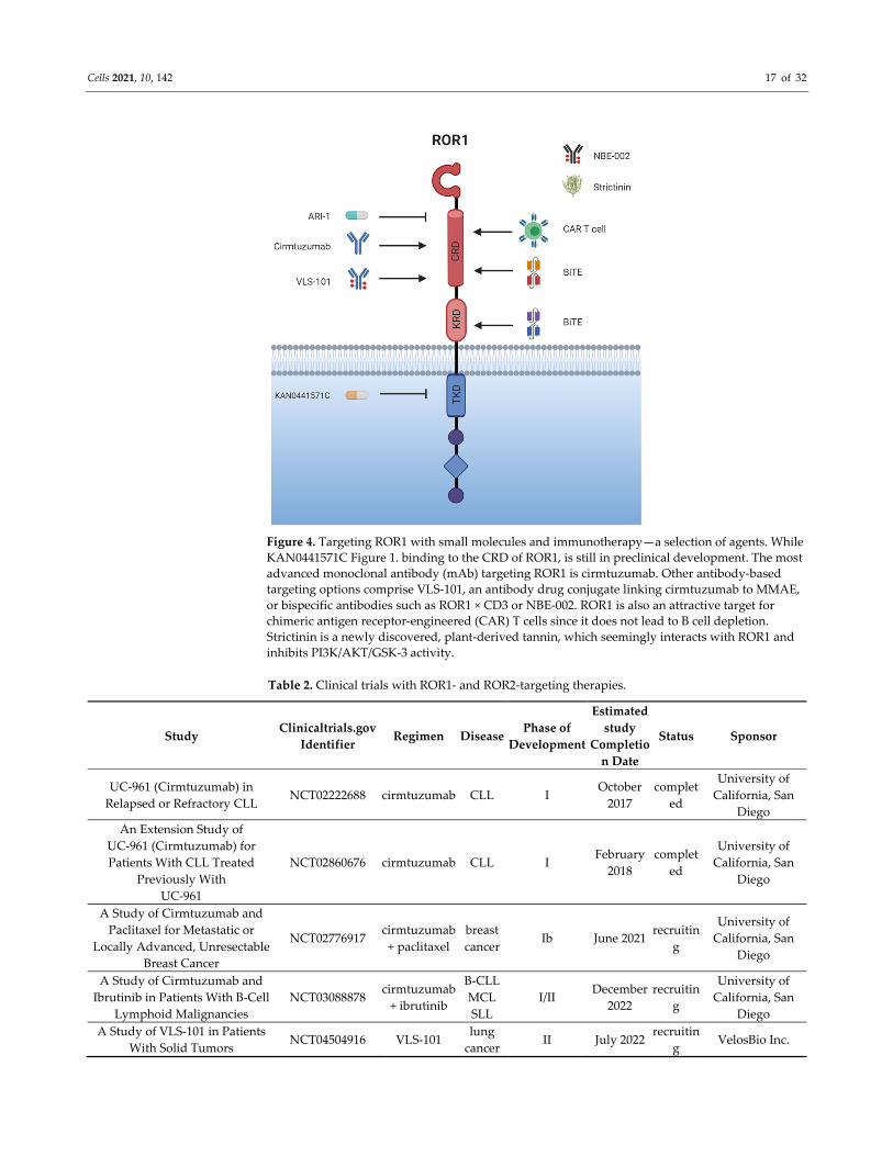
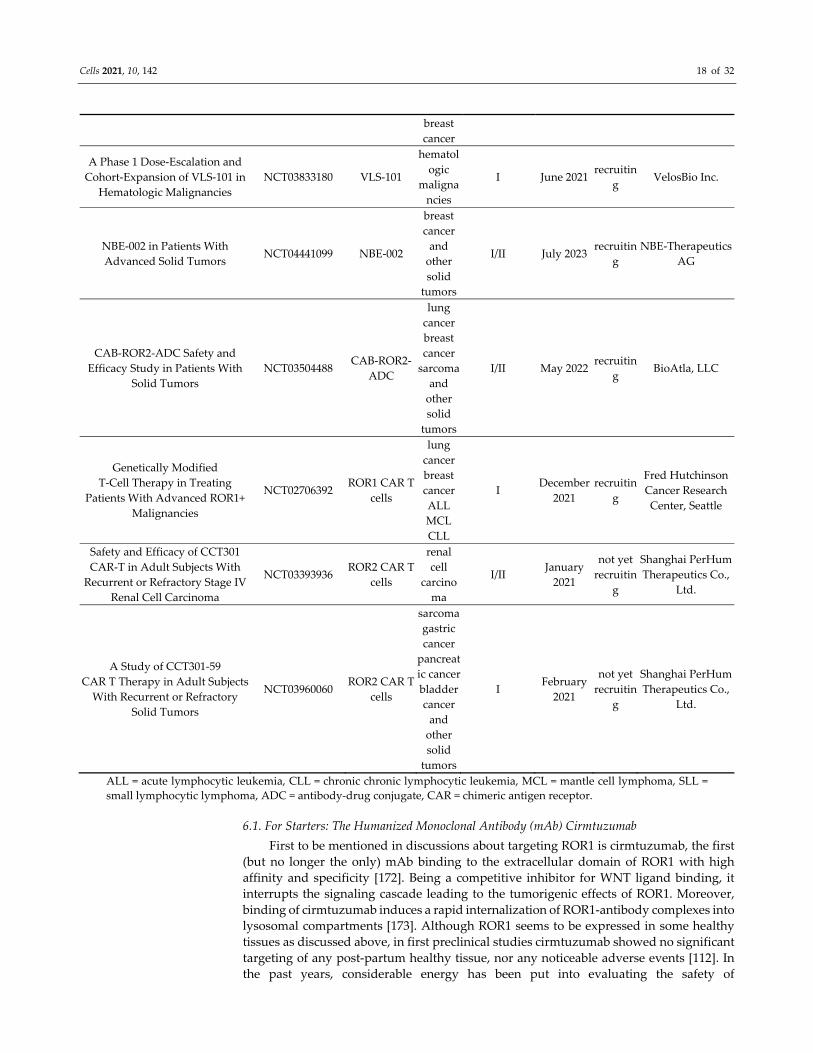
good tolerability and safety. An overview of the conducted clinical trials is given in Table 2.
While different approaches are currently being developed for targeting ROR1, research
on ROR2 with regard to its use in cancer therapy is still in its infancy. In the following
chapter, we will present the available clinical data and discuss the advances and
challenges of the distinct anti‐ROR treatment strategies, which are summarized in Figure
4.
Cells 2021, 10, 142 17 of 32
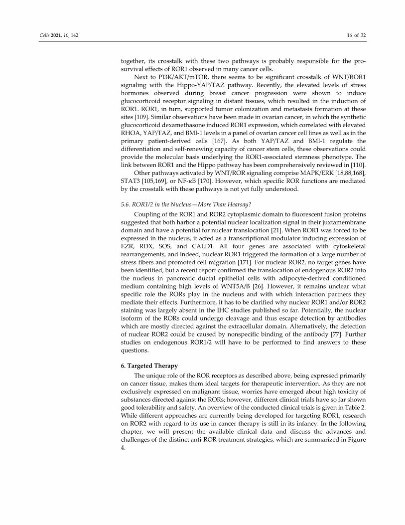
Figure 4. Targeting ROR1 with small molecules and immunotherapy—a selection of agents. While
KAN0441571C Figure 1. binding to the CRD of ROR1, is still in preclinical development. The most
advanced monoclonal antibody (mAb) targeting ROR1 is cirmtuzumab. Other antibody‐based
targeting options comprise VLS‐101, an antibody drug conjugate linking cirmtuzumab to MMAE,
or bispecific antibodies such as ROR1 × CD3 or NBE‐002. ROR1 is also an attractive target for
chimeric antigen receptor‐engineered (CAR) T cells since it does not lead to B cell depletion.
Strictinin is a newly discovered, plant‐derived tannin, which seemingly interacts with ROR1 and
inhibits PI3K/AKT/GSK‐3 activity.
Table 2. Clinical trials with ROR1‐ and ROR2‐targeting therapies.
Study Clinicaltrials.gov
Identifier Regimen Disease
Phase of
Development
Estimated
study
Completio
n Date
Status Sponsor
UC‐961 (Cirmtuzumab) in
Relapsed or Refractory CLL NCT02222688 cirmtuzumab CLL I
October
2017
complet
ed
University of
California, San
Diego
An Extension Study of
UC‐961 (Cirmtuzumab) for
Patients With CLL Treated
Previously With
UC‐961
NCT02860676 cirmtuzumab CLL I February
2018
complet
ed
University of
California, San
Diego
A Study of Cirmtuzumab and
Paclitaxel for Metastatic or
Locally Advanced, Unresectable
Breast Cancer
NCT02776917 cirmtuzumab
+ paclitaxel
breast
cancer Ib June 2021
recruitin
g
University of
California, San
Diego
A Study of Cirmtuzumab and
Ibrutinib in Patients With B‐Cell
Lymphoid Malignancies
NCT03088878 cirmtuzumab
+ ibrutinib
B‐CLL
MCL
SLL
I/II December
2022
recruitin
g
University of
California, San
Diego
A Study of VLS‐101 in Patients
With Solid Tumors NCT04504916 VLS‐101
lung
cancer II July 2022
recruitin
g VelosBio Inc.
Cells 2021, 10, 142 18 of 32
breast
cancer
A Phase 1 Dose‐Escalation and
Cohort‐Expansion of VLS‐101 in
Hematologic Malignancies
NCT03833180 VLS‐101
hematol
ogic
maligna
ncies
I June 2021 recruitin
g VelosBio Inc.
NBE‐002 in Patients With
Advanced Solid Tumors NCT04441099 NBE‐002
breast
cancer
and
other
solid
tumors
I/II July 2023 recruitin
g
NBE‐Therapeutics
AG
CAB‐ROR2‐ADC Safety and
Efficacy Study in Patients With
Solid Tumors
NCT03504488 CAB‐ROR2‐
ADC
lung
cancer
breast
cancer
sarcoma
and
other
solid
tumors
I/II May 2022 recruitin
g BioAtla, LLC
Genetically Modified
T‐Cell Therapy in Treating
Patients With Advanced ROR1+
Malignancies
NCT02706392 ROR1 CAR T
cells
lung
cancer
breast
cancer
ALL
MCL
CLL
I December
2021
recruitin
g
Fred Hutchinson
Cancer Research
Center, Seattle
Safety and Efficacy of CCT301
CAR‐T in Adult Subjects With
Recurrent or Refractory Stage IV
Renal Cell Carcinoma
NCT03393936 ROR2 CAR T
cells
renal
cell
carcino
ma
I/II January
2021
not yet
recruitin
g
Shanghai PerHum
Therapeutics Co.,
Ltd.
A Study of CCT301‐59
CAR T Therapy in Adult Subjects
With Recurrent or Refractory
Solid Tumors
NCT03960060 ROR2 CAR T
cells
sarcoma
gastric
cancer
pancreat
ic cancer
bladder
cancer
and
other
solid
tumors
I February
2021
not yet
recruitin
g
Shanghai PerHum
Therapeutics Co.,
Ltd.
ALL = acute lymphocytic leukemia, CLL = chronic chronic lymphocytic leukemia, MCL = mantle cell lymphoma, SLL =
small lymphocytic lymphoma, ADC = antibody‐drug conjugate, CAR = chimeric antigen receptor.
6.1. For Starters: The Humanized Monoclonal Antibody (mAb) Cirmtuzumab
First to be mentioned in discussions about targeting ROR1 is cirmtuzumab, the first
(but no longer the only) mAb binding to the extracellular domain of ROR1 with high
affinity and specificity [172]. Being a competitive inhibitor for WNT ligand binding, it
interrupts the signaling cascade leading to the tumorigenic effects of ROR1. Moreover,
binding of cirmtuzumab induces a rapid internalization of ROR1‐antibody complexes into
lysosomal compartments [173]. Although ROR1 seems to be expressed in some healthy
tissues as discussed above, in first preclinical studies cirmtuzumab showed no significant
targeting of any post‐partum healthy tissue, nor any noticeable adverse events [112]. In
the past years, considerable energy has been put into evaluating the safety of
Cells 2021, 10, 142 19 of 32
cirmtuzumab in clinical trials (phase I and II) [112] in hematological B cell malignancies,
where it had been identified as a tumor marker, then extending its use to ROR1‐expressing
solid tumors, such as breast cancer.
The first phase I trial (NCT02222688) tested cirmtuzumab in patients with relapsed
or refractory CLL. It was shown to be well‐tolerated, no dose‐limiting toxicity was
described, and only a narrow range of adverse events was published [112].
Pharmacokinetics studies showed a half‐life of 32.4 days with a downregulation of ROR1
signaling‐associated effects detectable for about six months, before an increase occurred.
While no complete remission (CR) or partial remission (PR) was reached, 17 out of 26
patients met a stable disease situation. An affiliated extension study (NCT02860676) was
performed, which tested cirmtuzumab in CLL patients for six to twelve months. The
results have not yet been published. Following this lead, there were several trials with
cirmtuzumab in combination with the BTK inhibitor ibrutinib in vitro and in mice,
knowing that these two agents target different, but interconnected pathways associated
with constitutive BCR signaling important for leukemia cell proliferation. In these
preclinical experiments, the two agents showed a profitable synergy [108]. In January
2018, a phase Ib/II Study (NCT03088878) was implemented, investigating the safety and
effectiveness of a combined treatment with cirmtuzumab and ibrutinib in patients with B
cell lymphoid malignancies. Interim results again showed a good tolerability, no dose‐
limiting toxicity, and an acceptable effectiveness [174].
These results encouraged exploration in the field of solid tumors, which are similarly
characterized by an overexpression of ROR1. One of the malignancies with a desperate
need for new and targeted therapies is breast cancer. High ROR1 levels in breast cancer
are associated with chemoresistance, metastasis, and poorer outcomes [41,102]. A
preclinical study has demonstrated that cirmtuzumab infusions every two weeks
inhibited re‐engraftment of breast cancer cells and the formation of pulmonary metastases
in a mouse PDX model. Evaluating the combination of chemotherapy with paclitaxel and
immunotherapy with cirmtuzumab showed the great benefit in combining the two [102]
as discussed in Section 4.2. A phase Ib trial of cirmtuzumab in combination with paclitaxel
was therefore initiated for the treatment of patients with metastatic, or locally advanced,
unresectable breast cancer (NCT02776917). Although the completion date is anticipated
in 2021, first preliminary findings seem promising [175].
6.2. New Approaches—Going beyond Cirmtuzumab?
In addition to cirmtuzumab, a second generation mAb targeting ROR1 has been
created: 5F1‐B10, which induced apoptosis in melanoma and bladder cancer cells by
inhibiting pro‐survival ROR1 signaling [29,176]. Apart from its potential in cancer
therapy, it has been furthermore suggested as a diagnostic tool for detecting bladder
cancer cells by flow cytometry in urine samples of patients. However, as cirmtuzumab is
a well‐developed, already (getting) established agent that has gained a foothold, catching
up with it will be surely a challenge to overcome.
An alternative approach in cancer immunotherapy is therapeutic cancer vaccination.
In a preclinical study by Wu et al. a vaccine based on ROR1‐expressing ovarian cancer
stem cells induced high immunogenicity and prophylactic effectiveness against ovarian
cancer [177]. Although ROR1 could indeed serve as a promising tumor antigen with high
immunogenicity, therapeutic cancer vaccines still face the challenges of overcoming
cancer immunosuppression and eliciting an adequate immune response; major obstacles
that have thus far resulted in disappointing results of the vaccination approach in patients.
6.3. More Is More: Conjugates and Bispecific Antibodies
Besides mAbs, there are many more therapeutic options currently being developed
based on the activation of the humoral immune response to counteract tumor growth,
namely conjugates and bispecific antibodies. Bispecific antibodies (biAbs) are molecules
combining a constant region Fc‐ and an antigen‐binding Fab‐fragment derived from two
Cells 2021, 10, 142 20 of 32
different mAbs (first generation biAb), or second generation biABs that combine two
single chain variable fragments (scFv) from different antibodies. While one of the scFv
connects to a target cell, the other one binds a T cell, which is then activated. Such
bispecific T cell engagers (BiTEs) represent a subclass of biAbs, defined by a CD3‐specific
arm and a tumor cell‐specific arm [178]. Associated with this intended mechanism of
action are a range of severe adverse events, such as the cytokine‐release‐syndrome
(known also from the application of chimeric antigen receptor‐engineered (CAR) T cells).
Qi et al. designed a bispecific ROR1 × CD3 scFv‐Fc format binding to a membrane‐
proximal epitope of the KRD [179]. Using in vitro and in vivo approaches, they showed it
to be more active than biAbs binding to a membrane‐distal epitope of ROR1 since it
facilitated the formation of the cytotoxic immunological synapse and receptor clustering
[180]. Interestingly, BiTEs showed superior cytotoxicity against various tumor cell lines
when composed of scFvs directed against the CRD as opposed to against the Ig‐like
domain [181]. Although no T‐cell activation was observed in ROR1‐negative tissue, the
anti‐tumor activity of the BiTEs did not depend on the actual level of ROR1 expression,
demonstrating a low effector to target ratio. Nonetheless, lower drug doses were required
compared with conventional mAbs, which argues for a higher potency of biAbs in
inducing an immune reaction. Compared with CAR T cell therapy, which will be
discussed below, continuous drug infusion is necessary for antibody treatment because of
the drug clearance. The upside of this is that its action can be rapidly terminated at any
time in the case of an adverse event by simply stopping its administration. This is in
contrast to the uncontrollable expansion of CAR T cells in vivo [181,182].
Since it had already been in clinical testing, it seemed advantageous to use
cirmtuzumab to create an antibody‐drug conjugate (ADC) by linking it to the anti‐
neoplastic agent monomethyl auristatin E (MMAE). Based on cirmtuzumab’s high
binding specificity towards ROR1‐positive cells and its rapid internalization into target
cells, this approach promises to safely deliver and release MMAE into the tumor cell
[173,183]. Two such ADCs were developed in parallel, VLS‐101 and cirmtuzumab—ADC‐
7 [173,184]. Cirmtuzumab—ADC‐7 was observed to cause a CR of ROR1‐positive CLL
and MCL cells in animals. Moreover, the combination of venetoclax with cirmtuzumab—
ADC‐7 was tested and showed a valuable synergistic effect in targeting ROR1‐positive
leukemia and lymphoma cell lines. Concordantly, in a PDX model for Richter’s syndrome,
a rare complication of CLL resulting in transformation towards an even more aggressive
form of lymphoma, VLS‐101 demonstrated a high specificity towards ROR1 by reaching
tumor cells in peripheral blood, bone marrow, and spleen, leading to significant disease
regression. CR was even observed in lymphoma cells without universal ROR1 expression,
an effect that was attributed to the high toxicity of MMAE. No further tumor growth was
documented even several months after the end of treatment, which led to longer patient
survival [184]. Currently, VLS‐101 is being tested in a phase I clinical trial (NCT03833180).
An alternative ROR1‐based ADCs is NBE‐002, a conjugate of a mAb targeting ROR1
and the anthracycline PNU‐159682 (a bioactive agent of nemorubicin). First preliminary
results indicated that the conjugate showed cytotoxicity in various solid tumor entities,
with the strongest effect observed in triple‐negative breast cancer cells [185]. A tumor
rechallenge in formerly NBE‐002‐treated, tumor‐free animals resulted in an inhibition of
tumor growth, although NBE‐002 was not applied again, implying that an immunological
memory was being triggered. A combination with checkpoint inhibitors also showed
promising results in a preclinical setting [185]. First clinical results are to be announced in
2021. For CLL, MCL, and pre‐B‐ALL cells, the anti‐ROR1 antibody drug conjugate
huXBR1‐402‐PNU is currently being evaluated pre‐clinically in vitro and in vivo [186].
Considering that ADCs such as, e.g., brentuximab vedotin or ado‐trastuzumab emtansine
are nowadays successfully used in the clinic for treating specific cancer entities, the
strategy of also using anti‐ROR1 ADCs to target ROR1‐positive tumors seems especially
promising.
Cells 2021, 10, 142 21 of 32
6.4. CAR‐T Cells—Promising Future or More about Adverse Events?
Another way of inducing activation of the humoral immune system in antineoplastic
therapy is with CAR T cells. These are autologous T cells engineered to carry receptors
specific for tumor antigens, e.g., CD19 or ROR1, thus directing the T cell towards the
tumor cell. Following binding, the engineered T cell gets activated, focusing its cytotoxic
activity on the malignant cell. Since CAR T cells are able to proliferate, the antineoplastic
effect is deemed to be persistent for a longer period of time, consequently as are the
adverse events. Up to date, off‐target cytotoxicity represents one of the main challenges
in CAR T cell therapy, among other reasons because it requires a suitable antigen with a
high selectivity towards malignant cells [187]. The success of CAR T cell therapy does not
only depend on the design of the CAR T cell itself though, but also on the tumor burden,
the former lymphodepletion, and the type of malignancy (solid or hematologic) to be
targeted. Until now, CAR T cell therapy in solid tumors is not as effective as in
hematological malignancies. A number of reasons contribute to this circumstance, one of
them being a lower specificity towards the target cell. This could be enhanced by
engineering a CAR T cell that recognizes two antigens on one target cell, thus fostering T
cell activation. Other reasons are the mechanical and biochemical obstacles the CAR T
cells have to overcome in order to reach their target tumor cell within the tissue. It has
been shown that CAR T cells in solid tumors run down quicker, expressing signs of
exhaustion earlier than the equivalents in hematological malignancies due to the
immunosuppressive microenvironment that the tumor creates [188,189].
All in all, ROR1 CAR T cells achieved promising preclinical results. The effectiveness
of ROR1 CAR T cell therapy was reported in three‐dimensional (3D) cell culture models
of lung and breast cancer, however, it was also documented that the CAR T cells showed
signs of exhaustion already after a short period of time. Whether this disadvantage can be
overcome by a second boost infusion remains to be tested [190]. ROR1 CAR T cells also
showed effectiveness in high risk sarcoma xenograft models (e.g., Ewing sarcoma,
osteosarcoma, rhabdomyosarcoma) [191]. As already mentioned, one concern when
applying ROR1‐directed CAR T cells is potential off‐target activity, and thus toxicity.
Although ROR1 is generally not expressed in healthy tissue, it is detected at higher levels
in adipocytes and pancreatic cells. Indeed, incubation of ROR1 CAR T cells with primary
adipocytes in vitro resulted in T cell activation and the release of Interleukin‐2, Interferon‐
γ, and granulocyte macrophage colony‐stimulating factor, pointing to potential cross‐
reactivity [25,39]. However, no toxicity was reported after infusion of CAR T cells in
macaques, who have a similar ROR1 distribution and expression pattern in normal tissues
as humans [25], even after the infusion of very high doses of functional ROR1 CAR T cells
[69]. Thus, the potential cross‐reactivity seems neglectable in vivo. These efforts and the
promising preclinical data led to the implementation of a phase I study with ROR1 CAR
T cells (NCT02706392), which does not only include patients with hematological
malignancies, but also solid tumors. The projected primary completion date is December
2021.
6.5. Small Molecule—Big Impact?
A different approach in targeted therapy are small molecule inhibitors. Recently, the
first anti‐ROR1 inhibitor was designed which prevented phosphorylation of the TKD and
thus blocked ROR1‐induced signaling [192]. In vitro, treatment with KAN0439834
induced apoptosis of ROR1‐positive CLL cells. The EC50 was reported to be over 60‐fold
higher for ROR1‐positive than ‐negative cells, and neither normal T nor B cells were
targeted by the compound. Compared with the anti‐tumor effects of other inhibitors
commonly applied in the treatment of CLL, KAN0439834 reached similar levels of
apoptosis induction as venetoclax (Bcl‐2 inhibitor) in patient‐derived CLL cells, and was
more effective than ibrutinib (BTK inhibitor) and idelalisib (PI3K inhibitor). In a direct
comparison with venetoclax, KAN0439834 showed a higher specificity for CLL cells and
Cells 2021, 10, 142 22 of 32
displayed less cytotoxicity for healthy PBMCs [192]. Given the overexpression of ROR1 in
many solid tumors and the urgent need for targeted strategies for these entities, as well,
KAN0439834 has already been tested for application in pancreatic cancer [193]. In this
preclinical study on pancreatic cancer cell lines, it proved equally effective in inducing
tumor cell apoptosis, at even lower EC50 values than with erlotinib (EGFR inhibitor) or
ibrutinib. Interestingly, KAN0439834 was reported to induce significantly higher levels of
apoptosis than a ROR1‐targeting mAb. However, whether this difference holds true in
direct comparison with the established ROR1 mAb, cirmtuzumab, remains to be clarified.
On a molecular level, KAN0439834 inhibited the phosphorylation of
PI3K/AKT/mTOR/CREB and SRC, both in CLL as well as in pancreatic cancer cells
[192,193], and thus blocked major pro‐survival pathways induced by ROR1. As these
pathways can also be activated by EGFR, which is frequently overexpressed in pancreatic
cancer, the combination of KAN0439834 with erlotinib or ibrutinib (which can also
function as an EGFR inhibitor [194]) showed clear additive effects and highlighted the
benefit of potential combinatory treatment strategies [193].
Based on these promising results, an improved second‐generation small molecule
inhibitor (KAN0441571C) was developed. It has so far only been tested in diffuse large B
cell lymphoma cell lines. KAN0441571C showed a higher cytotoxicity than KAN0439834,
impressing with a longer halftime in mice than its predecessor (11 h instead of 1.2 h),
although there was no improvement in terms of specificity towards ROR1. Inhibition of
downstream ROR1 pro‐survival signaling was observed as with KAN0439834 and
resulted in dose‐dependent cytotoxicity. In cell viability assays KAN0441571C was
superior to ibrutinib, but showed similar cytotoxicity as venetoclax [38]. However, the
combination of KAN0441571C with the latter resulted in almost complete tumor cell
killing, again underlining the benefit of combined treatment. Picking up on these
developments, another small molecule inhibitor was designed (ARI‐1), which directly
targets the extracellular WNT‐binding CRD of ROR1 [195]. Treatment with ARI‐1 blocked
downstream ROR1 signaling via PI3K/AKT/mTOR, thus inhibiting proliferation and
migration of ROR1‐positive non‐small cell lung cancer cells in vitro and in vivo. ARI‐1
was reported to show a high specificity for ROR1 and was effective also in cells that
harbored resistance against EGFR‐targeting substances. This fits to the report discussed
earlier that, in lung cancer cells, targeting ROR1 can help to circumvent resistance due to
its role in sustaining signaling of several pro‐survival RTKs [96]. The observation that
ARI‐1 did not cause severe toxicity towards the heart, liver, spleen, lung, or kidney in a
mouse xenograft model is promising for its possible application in human patients [195].
Since the conducted studies have so far been limited to preclinical evaluation, clinical data
will be required to determine the potential and the limitations of the developed small
molecule inhibitors with regard to the development of resistance mechanisms, which is a
common drawback in this therapy approach. Skipping to triple‐negative breast cancer, a
South African plant has come into the focus of ROR1 targeting strategies: Myrothamnus
flabellifolius contains strictinin, a polyphenol which appears to be a competitive ligand for
ROR1 binding sites and can therefore inhibit ROR1 signaling in the tumor cells. However,
in silico experiments show that a high expression of ROR1 is needed for a therapeutic
effect [196].
6.6. ROR2, the Wallflower?—To Be Continued
When it comes to ROR2, the degree of its translation into the clinic does not yet match
that of ROR1. However, based on the functional data presented in the previous chapters,
this seems surprising since evidence has accumulated for several solid tumors that clearly
highlight ROR2 as an oncogene in these entities (e.g., breast cancer, lung cancer, and
sarcoma). The current ROR2‐centered research activities comprise mostly adoptive
immunotherapy approaches, as the kinase activity of ROR2 is still a matter of debate.
What is known, though, is that the TKD of ROR2 is relatively unique amongst the RTK
superfamily [197], and could be exploited in the development of small molecule
Cells 2021, 10, 142 23 of 32
inhibitors. Ongoing research on targeting ROR2 by adoptive immunotherapy includes
mAbs, CAR T cells, and ADCs. Peng et al. designed the rabbit mAb XBR2‐401, which
binds to a membrane‐proximal epitope in the KRD of ROR2 [198]. Fortunately, the
extracellular domains of human and rabbit ROR2 are very similar which promises
valuable preclinical results. Subsequently, they developed the mAb into a CAR T cell
format, which demonstrated a high specificity towards ROR2. Moreover, XBR2‐401 was
used as a base for designing a biAb (ROR2 x CD3), showing specificity for ROR2 in vitro
[199]. Further investigation concerning ROR2‐targeting ADCs are also in progress [200].
One potential candidate might be BA3021, a CAB‐ROR2‐ADC, which reversibly interacts
with ROR2 in conditions reflecting the tumor microenvironment, but less so in normal
tissue [201]. It is currently being tested in a clinical phase I/II study in patients with
advanced solid tumors (NCT03504488). When looking at the clinical trials registered to
date, there are two additional studies investigating the ROR2‐specific CAR T cells in solid
malignancies expressing ROR2 (NCT03960060) or, more specifically, ROR2‐positive renal
carcinomas (NCT03393936). However, neither are yet recruiting; therefore, the clinical
success of these treatment strategies remains to be seen.
7. Conclusions
Starting with the identification of the RORs as orphan receptors, it is now clear that
both ROR1 and ROR2 are essential members of the WNT pathway that can bind WNT
ligands and activate downstream β‐catenin‐independent signaling. While ongoing
research has begun to reveal the pro‐tumorigenic functions of ROR1 and ROR2 in cancer,
the mechanisms underlying their regulation and their context‐dependent functionality in
the distinct tumor entities, which has caused conflicting observations, are still largely
unknown. Nonetheless, both are attractive targets for targeted therapy in selected tumor
types, especially in combination with established drugs (e.g., erlotinib, venetoclax,
ibrutinib) or even with anti‐CD19 CAR T cell therapy. Given the current lack of
possibilities to clinically target non‐canonical WNT signaling despite its increasingly
acknowledged relevance in human cancer, the ongoing development of anti‐ROR therapy
strategies is surely exciting.
Author Contributions: conceptualization: K.M. and A.B.; writing—original draft preparation: K.M.,
S.H., and C.B.; writing—review and editing: K.M., S.H., C.B., and A.B.; supervision: K.M. and A.B.
All authors have read and agreed to the published version of the manuscript.
Funding: This work was funded by the Deutsche Forschungsgemeinschaft (DFG, German Research
Foundation—project 424252458) and the German Ministry of Education and Research (BMBF)
e:Med project MyPathSem (031L0024).
Institutional Review Board Statement: Not applicable.
Informed Consent Statement: Not applicable.
Data Availability Statement: No new data were created or analyzed in this study. Data sharing is
not applicable to this article.
Acknowledgments: The illustrations were created with BioRender.com.
Conflicts of Interest: The authors declare no conflict of interest.
References
1. Nusse, R.; Clevers, H. Wnt/β‐Catenin Signaling, Disease, and Emerging Therapeutic Modalities. Cell 2017, 169, 985–999,
doi:10.1016/j.cell.2017.05.016.
2. Mikels, A.J.; Nusse, R. Purified Wnt5a Protein Activates or Inhibits Beta‐Catenin‐TCF Signaling Depending on Receptor
Context. PLoS Biol. 2006, 4, e115, doi:10.1371/journal.pbio.0040115.
3. Anthony, C.C.; Robbins, D.J.; Ahmed, Y.; Lee, E. Nuclear Regulation of Wnt/β‐Catenin Signaling: It’s a Complex Situation. Genes
2020, 11, 886, doi:10.3390/genes11080886.
Cells 2021, 10, 142 24 of 32
4. Yang, Y.; Mlodzik, M. Wnt‐Frizzled/Planar Cell Polarity Signaling: Cellular Orientation by Facing the Wind (Wnt). Annu. Rev.
Cell Dev. Biol. 2015, 31, 623–646, doi:10.1146/annurev‐cellbio‐100814‐125315.
5. Haseeb, M.; Pirzada, R.H.; Ain, Q.U.; Choi, S. Wnt Signaling in the Regulation of Immune Cell and Cancer Therapeutics. Cells
2019, 8, 1380, doi:10.3390/cells8111380.
6. DeChiara, T.M.; Kimble, R.B.; Poueymirou, W.T.; Rojas, J.; Masiakowski, P.; Valenzuela, D.M.; Yancopoulos, G.D. Ror2,
Encoding a Receptor‐like Tyrosine Kinase, Is Required for Cartilage and Growth Plate Development. Nat. Genet. 2000, 24, 271–
274, doi:10.1038/73488.
7. Takeuchi, S.; Takeda, K.; Oishi, I.; Nomi, M.; Ikeya, M.; Itoh, K.; Tamura, S.; Ueda, T.; Hatta, T.; Otani, H.; et al. Mouse Ror2
Receptor Tyrosine Kinase Is Required for the Heart Development and Limb Formation. Genes Cells 2000, 5, 71–78,
doi:10.1046/j.1365‐2443.2000.00300.x.
8. Nomi, M.; Oishi, I.; Kani, S.; Suzuki, H.; Matsuda, T.; Yoda, A.; Kitamura, M.; Itoh, K.; Takeuchi, S.; Takeda, K.; et al. Loss of
MRor1 Enhances the Heart and Skeletal Abnormalities in MRor2‐Deficient Mice: Redundant and Pleiotropic Functions of
MRor1 and MRor2 Receptor Tyrosine Kinases. Mol. Cell. Biol. 2001, 21, 8329–8335, doi:10.1128/MCB.21.24.8329‐8335.2001.
9. Oldridge, M.; Fortuna, A.M.; Maringa, M.; Propping, P.; Mansour, S.; Pollitt, C.; DeChiara, T.M.; Kimble, R.B.; Valenzuela, D.M.;
Yancopoulos, G.D.; et al. Dominant Mutations in ROR2, Encoding an Orphan Receptor Tyrosine Kinase, Cause Brachydactyly
Type B. Nat. Genet. 2000, 24, 275–278, doi:10.1038/73495.
10. Afzal, A.R.; Rajab, A.; Fenske, C.D.; Oldridge, M.; Elanko, N.; Ternes‐Pereira, E.; Tüysüz, B.; Murday, V.A.; Patton, M.A.; Wilkie,
A.O.; et al. Recessive Robinow Syndrome, Allelic to Dominant Brachydactyly Type B, Is Caused by Mutation of ROR2. Nat.
Genet. 2000, 25, 419–422, doi:10.1038/78107.
11. Van Bokhoven, H.; Celli, J.; Kayserili, H.; van Beusekom, E.; Balci, S.; Brussel, W.; Skovby, F.; Kerr, B.; Percin, E.F.; Akarsu, N.;
et al. Mutation of the Gene Encoding the ROR2 Tyrosine Kinase Causes Autosomal Recessive Robinow Syndrome. Nat. Genet.
2000, 25, 423–426, doi:10.1038/78113.
12. Masiakowski, P.; Carroll, R.D. A Novel Family of Cell Surface Receptors with Tyrosine Kinase‐like Domain. J. Biol. Chem. 1992,
267, 26181–26190.
13. Masiakowski, P.; Yancopoulos, G.D. The Wnt Receptor CRD Domain Is Also Found in MuSK and Related Orphan Receptor
Tyrosine Kinases. Curr. Biol. 1998, 8, R407, doi:10.1016/S0960‐9822(98)70263‐5.
14. Xu, Y.K.; Nusse, R. The Frizzled CRD Domain Is Conserved in Diverse Proteins Including Several Receptor Tyrosine Kinases.
Curr. Biol. 1998, 8, R405–R406, doi:10.1016/S0960‐9822(98)70262‐3.
15. Yu, J.; Chen, L.; Cui, B.; Widhopf, G.F.; Shen, Z.; Wu, R.; Zhang, L.; Zhang, S.; Briggs, S.P.; Kipps, T.J. Wnt5a Induces ROR1/ROR2
Heterooligomerization to Enhance Leukemia Chemotaxis and Proliferation. J. Clin. Investig. 2016, 126, 585–598,
doi:10.1172/JCI83535.
16. Gentile, A.; Lazzari, L.; Benvenuti, S.; Trusolino, L.; Comoglio, P.M. The ROR1 Pseudokinase Diversifies Signaling Outputs in
MET‐Addicted Cancer Cells. Int. J. Cancer 2014, 135, 2305–2316, doi:10.1002/ijc.28879.
17. Gentile, A.; Lazzari, L.; Benvenuti, S.; Trusolino, L.; Comoglio, P.M. Ror1 Is a Pseudokinase that Is Crucial for Met‐Driven
Tumorigenesis. Cancer Res. 2011, 71, 3132–3141, doi:10.1158/0008‐5472.CAN‐10‐2662.
18. Bicocca, V.T.; Chang, B.H.; Masouleh, B.K.; Muschen, M.; Loriaux, M.M.; Druker, B.J.; Tyner, J.W. Crosstalk between ROR1 and
the Pre‐B‐Cell Receptor Promotes Survival of t(1;19) Acute Lymphoblastic Leukemia. Cancer Cell 2012, 22, 656–667,
doi:10.1016/j.ccr.2012.08.027.
19. Bainbridge, T.W.; DeAlmeida, V.I.; Izrael‐Tomasevic, A.; Chalouni, C.; Pan, B.; Goldsmith, J.; Schoen, A.P.; Quiñones, G.A.;
Kelly, R.; Lill, J.R.; et al. Evolutionary Divergence in the Catalytic Activity of the CAM‐1, ROR1 and ROR2 Kinase Domains.
PLoS ONE 2014, 9, e102695, doi:10.1371/journal.pone.0102695.
20. Forrester, W.C. The Ror Receptor Tyrosine Kinase Family. Cell. Mol. Life Sci. CMLS 2002, 59, 83–96, doi:10.1007/s00018‐002‐8407‐
9.
21. Tseng, H.‐C.; Lyu, P.‐C.; Lin, W. Nuclear Localization of Orphan Receptor Protein Kinase (Ror1) Is Mediated through the
Juxtamembrane Domain. BMC Cell Biol. 2010, 11, 48, doi:10.1186/1471‐2121‐11‐48.
22. Zhang, S.; Chen, L.; Wang‐Rodriguez, J.; Zhang, L.; Cui, B.; Frankel, W.; Wu, R.; Kipps, T.J. The Onco‐Embryonic Antigen ROR1
Is Expressed by a Variety of Human Cancers. Am. J. Pathol. 2012, 181, 1903–1910, doi:10.1016/j.ajpath.2012.08.024.
23. Mei, H.; Lian, S.; Zhang, S.; Wang, W.; Mao, Q.; Wang, H. High Expression of ROR2 in Cancer Cell Correlates with Unfavorable
Prognosis in Colorectal Cancer. Biochem. Biophys. Res. Commun. 2014, 453, 703–709, doi:10.1016/j.bbrc.2014.09.141.
24. Henry, C.; Quadir, A.; Hawkins, N.J.; Jary, E.; Llamosas, E.; Kumar, D.; Daniels, B.; Ward, R.L.; Ford, C.E. Expression of the
Novel Wnt Receptor ROR2 Is Increased in Breast Cancer and May Regulate Both β‐Catenin Dependent and Independent Wnt
Signalling. J. Cancer Res. Clin. Oncol. 2015, 141, 243–254, doi:10.1007/s00432‐014‐1824‐y.
25. Balakrishnan, A.; Goodpaster, T.; Randolph‐Habecker, J.; Hoffstrom, B.G.; Jalikis, F.G.; Koch, L.K.; Berger, C.; Kosasih, P.L.;
Rajan, A.; Sommermeyer, D.; et al. Analysis of ROR1 Protein Expression in Human Cancer and Normal Tissues. Clin. Cancer
Res. 2017, 23, 3061–3071, doi:10.1158/1078‐0432.CCR‐16‐2083.
26. Carbone, C.; Piro, G.; Gaianigo, N.; Ligorio, F.; Santoro, R.; Merz, V.; Simionato, F.; Zecchetto, C.; Falco, G.; Conti, G.; et al.
Adipocytes Sustain Pancreatic Cancer Progression through a Non‐Canonical WNT Paracrine Network Inducing ROR2 Nuclear
Shuttling. Int. J. Obes. 2018, 42, 334–343, doi:10.1038/ijo.2017.285.
27. Reddy, U.R.; Phatak, S.; Pleasure, D. Human Neural Tissues Express a Truncated Ror1 Receptor Tyrosine Kinase, Lacking Both
Extracellular and Transmembrane Domains. Oncogene 1996, 13, 1555–1559.
Cells 2021, 10, 142 25 of 32
28. Kaucká, M.; Krejčí, P.; Plevová, K.; Pavlová, S.; Procházková, J.; Janovská, P.; Valnohová, J.; Kozubík, A.; Pospíšilová, S.; Bryja,
V. Post‐Translational Modifications Regulate Signalling by Ror1. Acta Physiol. Oxf. Engl. 2011, 203, 351–362, doi:10.1111/j.1748‐
1716.2011.02306.x.
29. Hojjat‐Farsangi, M.; Ghaemimanesh, F.; Daneshmanesh, A.H.; Bayat, A.‐A.; Mahmoudian, J.; Jeddi‐Tehrani, M.; Rabbani, H.;
Mellstedt, H. Inhibition of the Receptor Tyrosine Kinase ROR1 by Anti‐ROR1 Monoclonal Antibodies and SiRNA Induced
Apoptosis of Melanoma Cells. PLoS ONE 2013, 8, e61167, doi:10.1371/journal.pone.0061167.
30. Dave, H.; Anver, M.R.; Butcher, D.O.; Brown, P.; Khan, J.; Wayne, A.S.; Baskar, S.; Rader, C. Restricted Cell Surface Expression
of Receptor Tyrosine Kinase ROR1 in Pediatric B‐Lineage Acute Lymphoblastic Leukemia Suggests Targetability with
Therapeutic Monoclonal Antibodies. PLoS ONE 2012, 7, e52655, doi:10.1371/journal.pone.0052655.
31. Baskar, S.; Kwong, K.Y.; Hofer, T.; Levy, J.M.; Kennedy, M.G.; Lee, E.; Staudt, L.M.; Wilson, W.H.; Wiestner, A.; Rader, C.
Unique Cell Surface Expression of Receptor Tyrosine Kinase ROR1 in Human B‐Cell Chronic Lymphocytic Leukemia. Clin.
Cancer Res. 2008, 14, 396–404, doi:10.1158/1078‐0432.CCR‐07‐1823.
32. DaneshManesh, A.H.; Mikaelsson, E.; Jeddi‐Tehrani, M.; Bayat, A.A.; Ghods, R.; Ostadkarampour, M.; Akhondi, M.;
Lagercrantz, S.; Larsson, C.; Österborg, A.; et al. Ror1, a Cell Surface Receptor Tyrosine Kinase Is Expressed in Chronic
Lymphocytic Leukemia and May Serve as a Putative Target for Therapy. Int. J. Cancer 2008, 123, 1190–1195, doi:10.1002/ijc.23587.
33. Fukuda, T.; Chen, L.; Endo, T.; Tang, L.; Lu, D.; Castro, J.E.; Widhopf, G.F.; Rassenti, L.Z.; Cantwell, M.J.; Prussak, C.E.; et al.
Antisera Induced by Infusions of Autologous Ad‐CD154‐Leukemia B Cells Identify ROR1 as an Oncofetal Antigen and Receptor
for Wnt5a. Proc. Natl. Acad. Sci. USA 2008, 105, 3047–3052, doi:10.1073/pnas.0712148105.
34. Klein, U.; Tu, Y.; Stolovitzky, G.A.; Mattioli, M.; Cattoretti, G.; Husson, H.; Freedman, A.; Inghirami, G.; Cro, L.; Baldini, L.; et
al. Gene Expression Profiling of B Cell Chronic Lymphocytic Leukemia Reveals a Homogeneous Phenotype Related to Memory
B Cells. J. Exp. Med. 2001, 194, 1625–1638, doi:10.1084/jem.194.11.1625.
35. Rosenwald, A.; Alizadeh, A.A.; Widhopf, G.; Simon, R.; Davis, R.E.; Yu, X.; Yang, L.; Pickeral, O.K.; Rassenti, L.Z.; Powell, J.; et
al. Relation of Gene Expression Phenotype to Immunoglobulin Mutation Genotype in B Cell Chronic Lymphocytic Leukemia.
J. Exp. Med. 2001, 194, 1639–1648, doi:10.1084/jem.194.11.1639.
36. Cui, B.; Ghia, E.M.; Chen, L.; Rassenti, L.Z.; DeBoever, C.; Widhopf, G.F.; Yu, J.; Neuberg, D.S.; Wierda, W.G.; Rai, K.R.; et al.
High‐Level ROR1 Associates with Accelerated Disease Progression in Chronic Lymphocytic Leukemia. Blood 2016, 128, 2931–
2940, doi:10.1182/blood‐2016‐04‐712562.
37. Barna, G.; Mihalik, R.; Timár, B.; Tömböl, J.; Csende, Z.; Sebestyén, A.; Bödör, C.; Csernus, B.; Reiniger, L.; Peták, I.; et al. ROR1
Expression Is Not a Unique Marker of CLL. Hematol. Oncol. 2011, 29, 17–21, doi:10.1002/hon.948.
38. Ghaderi, A.; Daneshmanesh, A.H.; Moshfegh, A.; Kokhaei, P.; Vågberg, J.; Schultz, J.; Olin, T.; Harrysson, S.; Smedby, K.E.;
Drakos, E.; et al. ROR1 Is Expressed in Diffuse Large B‐Cell Lymphoma (DLBCL) and a Small Molecule Inhibitor of ROR1
(KAN0441571C) Induced Apoptosis of Lymphoma Cells. Biomedicines 2020, 8, 170, doi:10.3390/biomedicines8060170.
39. Hudecek, M.; Schmitt, T.M.; Baskar, S.; Lupo‐Stanghellini, M.T.; Nishida, T.; Yamamoto, T.N.; Bleakley, M.; Turtle, C.J.; Chang,
W.‐C.; Greisman, H.A.; et al. The B‐Cell Tumor–Associated Antigen ROR1 Can Be Targeted with T Cells Modified to Express a
ROR1‐Specific Chimeric Antigen Receptor. Blood 2010, 116, 4532–4541, doi:10.1182/blood‐2010‐05‐283309.
40. Frenquelli, M.; Caridi, N.; Antonini, E.; Storti, F.; Viganò, V.; Gaviraghi, M.; Occhionorelli, M.; Bianchessi, S.; Bongiovanni, L.;
Spinelli, A.; et al. The WNT Receptor ROR2 Drives the Interaction of Multiple Myeloma Cells with the Microenvironment
through AKT Activation. Leukemia 2020, 34, 257–270, doi:10.1038/s41375‐019‐0486‐9.
41. Zhang, S.; Chen, L.; Cui, B.; Chuang, H.‐Y.; Yu, J.; Wang‐Rodriguez, J.; Tang, L.; Chen, G.; Basak, G.W.; Kipps, T.J. ROR1 Is
Expressed in Human Breast Cancer and Associated with Enhanced Tumor‐Cell Growth. PLoS ONE 2012, 7, e31127,
doi:10.1371/journal.pone.0031127.
42. Cui, B.; Zhang, S.; Chen, L.; Yu, J.; Widhopf, G.F.; Fecteau, J.‐F.; Rassenti, L.Z.; Kipps, T.J. Targeting ROR1 Inhibits Epithelial–
Mesenchymal Transition and Metastasis. Cancer Res. 2013, 73, 3649–3660, doi:10.1158/0008‐5472.CAN‐12‐3832.
43. Chien, H.‐P.; Ueng, S.‐H.; Chen, S.‐C.; Chang, Y.‐S.; Lin, Y.‐C.; Lo, Y.‐F.; Chang, H.‐K.; Chuang, W.‐Y.; Huang, Y.‐T.; Cheung,
Y.‐C.; et al. Expression of ROR1 Has Prognostic Significance in Triple Negative Breast Cancer. Virchows Arch. 2016, 468, 589–
595, doi:10.1007/s00428‐016‐1911‐3.
44. Guo, M.; Ma, G.; Zhang, X.; Tang, W.; Shi, J.; Wang, Q.; Cheng, Y.; Zhang, B.; Xu, J. ROR2 Knockdown Suppresses Breast Cancer
Growth through PI3K/ATK Signaling. Aging 2020, 12, 13115–13127, doi:10.18632/aging.103400.
45. Sun, B.; Ye, X.; Lin, L.; Shen, M.; Jiang, T. Up‐Regulation of ROR2 Is Associated with Unfavorable Prognosis and Tumor
Progression in Cervical Cancer. Int. J. Clin. Exp. Pathol. 2015, 8, 856–861.
46. Zhou, J.‐K.; Zheng, Y.‐Z.; Liu, X.‐S.; Gou, Q.; Ma, R.; Guo, C.‐L.; Croce, C.M.; Liu, L.; Peng, Y. ROR1 Expression as a Biomarker
for Predicting Prognosis in Patients with Colorectal Cancer. Oncotarget 2017, 8, 32864–32872, doi:10.18632/oncotarget.15860.
47. Lara, E.; Calvanese, V.; Huidobro, C.; Fernández, A.F.; Moncada‐Pazos, Á.; Obaya, Á.J.; Aguilera, O.; González‐Sancho, J.M.;
Sánchez, L.; Astudillo, A.; et al. Epigenetic Repression of ROR2 Has a Wnt‐Mediated, pro‐Tumourigenic Role in Colon Cancer.
Mol. Cancer 2010, 9, 170, doi:10.1186/1476‐4598‐9‐170.
48. Henry, C.E.; Llamosas, E.; Daniels, B.; Coopes, A.; Tang, K.; Ford, C.E. ROR1 and ROR2 Play Distinct and Opposing Roles in
Endometrial Cancer. Gynecol. Oncol. 2018, 148, 576–584, doi:10.1016/j.ygyno.2018.01.025.
49. Liu, D.; Gunther, K.; Enriquez, L.A.; Daniels, B.; O’Mara, T.A.; Tang, K.; Spurdle, A.B.; Ford, C.E. ROR1 Is Upregulated in
Endometrial Cancer and Represents a Novel Therapeutic Target. Sci. Rep. 2020, 10, 1–13, doi:10.1038/s41598‐020‐70924‐z.
Cells 2021, 10, 142 26 of 32
50. Chang, H.; Jung, W.Y.; Kang, Y.; Lee, H.; Kim, A.; Kim, B. Expression of ROR1, PAkt, and PCREB in Gastric Adenocarcinoma.
Ann. Diagn. Pathol. 2015, 19, 330–334, doi:10.1016/j.anndiagpath.2015.06.010.
51. Tao, J.; Zhi, X.; Zhang, X.; Fu, M.; Huang, H.; Fan, Y.; Guan, W.; Zou, C. MiR‐27b‐3p Suppresses Cell Proliferation through
Targeting Receptor Tyrosine Kinase like Orphan Receptor 1 in Gastric Cancer. J. Exp. Clin. Cancer Res. CR 2015, 34, 1–13,
doi:10.1186/s13046‐015‐0253‐3.
52. Yan, L.; Du, Q.; Yao, J.; Liu, R. ROR2 Inhibits the Proliferation of Gastric Carcinoma Cells via Activation of Non‐Canonical Wnt
Signaling. Exp. Ther. Med. 2016, 12, 4128–4134, doi:10.3892/etm.2016.3883.
53. Kim, Y.; Hong, M.; Do, I.‐G.; Ha, S.Y.; Lee, D.; Suh, Y.‐L. Wnt5a, Ryk and Ror2 Expression in Glioblastoma Subgroups. Pathol.
Res. Pract. 2015, 211, 963–972, doi:10.1016/j.prp.2015.10.001.
54. Zheng, Y.‐Z.; Ma, R.; Zhou, J.‐K.; Guo, C.‐L.; Wang, Y.‐S.; Li, Z.‐G.; Liu, L.‐X.; Peng, Y. ROR1 Is a Novel Prognostic Biomarker
in Patients with Lung Adenocarcinoma. Sci. Rep. 2016, 6, 36447, doi:10.1038/srep36447.
55. Lu, C.; Wang, X.; Zhu, H.; Feng, J.; Ni, S.; Huang, J. Over‐Expression of ROR2 and Wnt5a Cooperatively Correlates with
Unfavorable Prognosis in Patients with Non‐Small Cell Lung Cancer. Oncotarget 2015, 6, 24912–24921.
56. Fernández, N.B.; Lorenzo, D.; Picco, M.E.; Barbero, G.; Dergan‐Dylon, L.S.; Marks, M.P.; García‐Rivello, H.; Gimenez, L.;
Labovsky, V.; Grumolato, L.; et al. ROR1 Contributes to Melanoma Cell Growth and Migration by Regulating N‐Cadherin
Expression via the PI3K/Akt Pathway. Mol. Carcinog. 2016, 55, 1772–1785, doi:10.1002/mc.22426.
57. Saji, T.; Nishita, M.; Ogawa, H.; Doi, T.; Sakai, Y.; Maniwa, Y.; Minami, Y. Critical Role of the Ror‐Family of Receptor Tyrosine
Kinases in Invasion and Proliferation of Malignant Pleural Mesothelioma Cells. Genes Cells 2018, 23, 606–613,
doi:10.1111/gtc.12599.
58. Zhang, H.; Qiu, J.; Ye, C.; Yang, D.; Gao, L.; Su, Y.; Tang, X.; Xu, N.; Zhang, D.; Xiong, L.; et al. ROR1 Expression Correlated
with Poor Clinical Outcome in Human Ovarian Cancer. Sci. Rep. 2014, 4, 5811, doi:10.1038/srep05811.
59. Henry, C.; Llamosas, E.; Knipprath‐Mészáros, A.; Schoetzau, A.; Obermann, E.; Fuenfschilling, M.; Caduff, R.; Fink, D.; Hacker,
N.; Ward, R.; et al. Targeting the ROR1 and ROR2 Receptors in Epithelial Ovarian Cancer Inhibits Cell Migration and Invasion.
Oncotarget 2015, 6, 40310–40326.
60. Li, R.; Liu, T.; Shi, J.; Luan, W.; Wei, X.; Yu, J.; Mao, H.; Liu, P. ROR2 Induces Cell Apoptosis via Activating IRE1α/JNK/CHOP
Pathway in High‐Grade Serous Ovarian Carcinoma In Vitro and In Vivo. J. Transl. Med. 2019, 17, 1–17, doi:10.1186/s12967‐019‐
02178‐x.
61. Morioka, K.; Tanikawa, C.; Ochi, K.; Daigo, Y.; Katagiri, T.; Kawano, H.; Kawaguchi, H.; Myoui, A.; Yoshikawa, H.; Naka, N.;
et al. Orphan Receptor Tyrosine Kinase ROR2 as a Potential Therapeutic Target for Osteosarcoma. Cancer Sci. 2009, 100, 1227–
1233, doi:10.1111/j.1349‐7006.2009.01165.x.
62. Edris, B.; Espinosa, I.; Mikels, A.; Lee, C.‐H.; Steigen, S.E.; Zhu, S.; Montgomery, K.D.; Lazar, A.J.F.; Lev, D.; Fletcher, J.A.; et al.
ROR2 Is a Novel Prognostic Biomarker and a Potential Therapeutic Target in Leiomyosarcoma and Gastrointestinal Stromal
Tumour. J. Pathol. 2012, 227, 223–233, doi:10.1002/path.3986.
63. Xu, G.‐L.; Shen, J.; Xu, Y.‐H.; Wang, W.‐S.; Ni, C.‐F. ROR1 Is Highly Expressed in Circulating Tumor Cells and Promotes
Invasion of Pancreatic Cancer. Mol. Med. Rep. 2018, 18, 5087–5094, doi:10.3892/mmr.2018.9500.
64. Huang, J.; Fan, X.; Wang, X.; Lu, Y.; Zhu, H.; Wang, W.; Zhang, S.; Wang, Z. High ROR2 Expression in Tumor Cells and Stroma
Is Correlated with Poor Prognosis in Pancreatic Ductal Adenocarcinoma. Sci. Rep. 2015, 5, 12991, doi:10.1038/srep12991.
65. Tseng, J.‐C.; Huang, S.‐H.; Lin, C.‐Y.; Wang, B.‐J.; Huang, S.‐F.; Shen, Y.‐Y.; Chuu, C.‐P. ROR2 Suppresses Metastasis of Prostate
Cancer via Regulation of MiR‐199a‐5p–PIAS3–AKT2 Signaling Axis. Cell Death Dis. 2020, 11, 1–11, doi:10.1038/s41419‐020‐2587‐
9.
66. Oishi, I.; Takeuchi, S.; Hashimoto, R.; Nagabukuro, A.; Ueda, T.; Liu, Z.‐J.; Hatta, T.; Akira, S.; Matsuda, Y.; Yamamura, H.; et
al. Spatio‐Temporally Regulated Expression of Receptor Tyrosine Kinases, MRor1, MRor2, during Mouse Development:
Implications in Development and Function of the Nervous System. Genes Cells 1999, 4, 41–56, doi:10.1046/j.1365‐
2443.1999.00234.x.
67. Al‐Shawi, R.; Ashton, S.V.; Underwood, C.; Simons, J.P. Expression of the Ror1 and Ror2 Receptor Tyrosine Kinase Genes
during Mouse Development. Dev. Genes Evol. 2001, 211, 161–171, doi:10.1007/s004270100140.
68. Matsuda, T.; Nomi, M.; Ikeya, M.; Kani, S.; Oishi, I.; Terashima, T.; Takada, S.; Minami, Y. Expression of the Receptor Tyrosine
Kinase Genes, Ror1 and Ror2, during Mouse Development. Mech. Dev. 2001, 105, 153–156, doi:10.1016/s0925‐4773(01)00383‐5.
69. Berger, C.; Sommermeyer, D.; Hudecek, M.; Berger, M.; Balakrishnan, A.; Paszkiewicz, P.J.; Kosasih, P.L.; Rader, C.; Riddell,
S.R. Safety of Targeting ROR1 in Primates with Chimeric Antigen Receptor–Modified T Cells. Cancer Immunol. Res. 2015, 3, 206–
216, doi:10.1158/2326‐6066.CIR‐14‐0163.
70. Potratz, J.; Tillmanns, A.; Berning, P.; Korsching, E.; Schaefer, C.; Lechtape, B.; Schleithoff, C.; Unland, R.; Schäfer, K.‐L.; Müller‐
Tidow, C.; et al. Receptor Tyrosine Kinase Gene Expression Profiles of Ewing Sarcomas Reveal ROR1 as a Potential Therapeutic
Target in Metastatic Disease. Mol. Oncol. 2016, 10, 677–692, doi:10.1016/j.molonc.2015.12.009.
71. Broome, H.E.; Rassenti, L.Z.; Wang, H.‐Y.; Meyer, L.M.; Kipps, T.J. ROR1 Is Expressed on Hematogones (Non‐Neoplastic
Human B‐Lymphocyte Precursors) and a Minority of Precursor‐B Acute Lymphoblastic Leukemia. Leuk. Res. 2011, 35, 1390–
1394, doi:10.1016/j.leukres.2011.06.021.
72. Daneshmanesh, A.H.; Porwit, A.; Hojjat‐Farsangi, M.; Jeddi‐Tehrani, M.; Tamm, K.P.; Grandér, D.; Lehmann, S.; Norin, S.;
Shokri, F.; Rabbani, H.; et al. Orphan Receptor Tyrosine Kinases ROR1 and ROR2 in Hematological Malignancies. Leuk.
Lymphoma 2013, 54, 843–850, doi:10.3109/10428194.2012.731599.

Cells 2021, 10, 142 27 of 32
73. Billiard, J.; Way, D.S.; Seestaller‐Wehr, L.M.; Moran, R.A.; Mangine, A.; Bodine, P.V.N. The Orphan Receptor Tyrosine Kinase
Ror2 Modulates Canonical Wnt Signaling in Osteoblastic Cells. Mol. Endocrinol. 2005, 19, 90–101, doi:10.1210/me.2004‐0153.
74. Klemm, F.; Bleckmann, A.; Siam, L.; Chuang, H.N.; Rietkötter, E.; Behme, D.; Schulz, M.; Schaffrinski, M.; Schindler, S.; Trümper,
L.; et al. β‐Catenin‐Independent WNT Signaling in Basal‐like Breast Cancer and Brain Metastasis. Carcinogenesis 2011, 32, 434–
442, doi:10.1093/carcin/bgq269.
75. Yamaguchi, T.; Yanagisawa, K.; Sugiyama, R.; Hosono, Y.; Shimada, Y.; Arima, C.; Kato, S.; Tomida, S.; Suzuki, M.; Osada, H.;
et al. NKX2‐1/TITF1/TTF‐1‐Induced ROR1 Is Required to Sustain EGFR Survival Signaling in Lung Adenocarcinoma. Cancer
Cell 2012, 21, 348–361, doi:10.1016/j.ccr.2012.02.008.
76. Liu, Y.; Yang, H.; Chen, T.; Luo, Y.; Xu, Z.; Li, Y.; Yang, J. Silencing of Receptor Tyrosine Kinase ROR1 Inhibits Tumor‐Cell
Proliferation via PI3K/AKT/MTOR Signaling Pathway in Lung Adenocarcinoma. PLoS ONE 2015, 10, e0127092,
doi:10.1371/journal.pone.0127092.
77. Ma, S.S.Q.; Henry, C.E.; Llamosas, E.; Higgins, R.; Daniels, B.; Hesson, L.B.; Hawkins, N.J.; Ward, R.L.; Ford, C.E. Validation of
Specificity of Antibodies for Immunohistochemistry: The Case of ROR2. Virchows Arch. Int. J. Pathol. 2017, 470, 99–108,
doi:10.1007/s00428‐016‐2019‐5.
78. Dave, H.; Butcher, D.; Anver, M.; Bollard, C.M. ROR1 and ROR2‐Novel Targets for Neuroblastoma. Pediatric Hematol. Oncol.
2019, 36, 352–364, doi:10.1080/08880018.2019.1646365.
79. O’Connell, M.P.; Fiori, J.L.; Xu, M.; Carter, A.D.; Frank, B.P.; Camilli, T.C.; French, A.D.; Dissanayake, S.K.; Indig, F.E.; Bernier,
M.; et al. The Orphan Tyrosine Kinase Receptor, ROR2, Mediates Wnt5A Signaling in Metastatic Melanoma. Oncogene 2010, 29,
34–44, doi:10.1038/onc.2009.305.
80. Xu, J.; Shi, J.; Tang, W.; Jiang, P.; Guo, M.; Zhang, B.; Ma, G. ROR2 Promotes the Epithelial‐Mesenchymal Transition by
Regulating MAPK/P38 Signaling Pathway in Breast Cancer. J. Cell. Biochem. 2020, 121, 4142–4153, doi:10.1002/jcb.29666.
81. Henry, C.; Emmanuel, C.; Lambie, N.; Loo, C.; Kan, B.; Kennedy, C.; de Fazio, A.; Hacker, N.; Ford, C. Distinct Patterns of
Stromal and Tumor Expression of ROR1 and ROR2 in Histological Subtypes of Epithelial Ovarian Cancer. Transl. Oncol. 2017,
10, 346–356, doi:10.1016/j.tranon.2017.01.014.
82. Endo, M.; Ubulkasim, G.; Kobayashi, C.; Onishi, R.; Aiba, A.; Minami, Y. Critical Role of Ror2 Receptor Tyrosine Kinase in
Regulating Cell Cycle Progression of Reactive Astrocytes Following Brain Injury. Glia 2017, 65, 182–197, doi:10.1002/glia.23086.
83. Kamizaki, K.; Doi, R.; Hayashi, M.; Saji, T.; Kanagawa, M.; Toda, T.; Fukada, S.; Ho, H.‐Y.H.; Greenberg, M.E.; Endo, M.; et al.
The Ror1 Receptor Tyrosine Kinase Plays a Critical Role in Regulating Satellite Cell Proliferation during Regeneration of Injured
Muscle. J. Biol. Chem. 2017, 292, 15939–15951, doi:10.1074/jbc.M117.785709.
84. MacKeigan, J.P.; Murphy, L.O.; Blenis, J. Sensitized RNAi Screen of Human Kinases and Phosphatases Identifies New
Regulators of Apoptosis and Chemoresistance. Nat. Cell Biol. 2005, 7, 591–600, doi:10.1038/ncb1258.
85. Widhopf, G.F.; Cui, B.; Ghia, E.M.; Chen, L.; Messer, K.; Shen, Z.; Briggs, S.P.; Croce, C.M.; Kipps, T.J. ROR1 Can Interact with
TCL1 and Enhance Leukemogenesis in Eμ‐TCL1 Transgenic Mice. Proc. Natl. Acad. Sci. USA 2014, 111, 793–798,
doi:10.1073/pnas.1308374111.
86. Balatti, V.; Croce, C.M. MicroRNA Dysregulation and Multi‐Targeted Therapy for Cancer Treatment. Adv. Biol. Regul. 2020, 75,
100669, doi:10.1016/j.jbior.2019.100669.
87. Zhou, Q.; Zhou, S.; Wang, H.; Li, Y.; Xiao, X.; Yang, J. Stable Silencing of ROR1 Regulates Cell Cycle, Apoptosis, and Autophagy
in a Lung Adenocarcinoma Cell Line. Int. J. Clin. Exp. Pathol. 2020, 13, 1108–1120.
88. Fultang, N.; Illendula, A.; Lin, J.; Pandey, M.K.; Klase, Z.; Peethambaran, B. ROR1 Regulates Chemoresistance in Breast Cancer
via Modulation of Drug Efflux Pump ABCB1. Sci. Rep. 2020, 10, 1–12, doi:10.1038/s41598‐020‐58864‐0.
89. Wright, T.M.; Brannon, A.R.; Gordan, J.D.; Mikels, A.J.; Mitchell, C.; Chen, S.; Espinosa, I.; van de Rijn, M.; Pruthi, R.; Wallen,
E.; et al. Ror2, a Developmentally Regulated Kinase, Promotes Tumor Growth Potential in Renal Cell Carcinoma. Oncogene 2009,
28, 2513–2523, doi:10.1038/onc.2009.116.
90. Yang, C.‐M.; Ji, S.; Li, Y.; Fu, L.‐Y.; Jiang, T.; Meng, F.‐D. Ror2, a Developmentally Regulated Kinase, Is Associated With Tumor
Growth, Apoptosis, Migration, and Invasion in Renal Cell Carcinoma. Oncol. Res. 2017, 25, 195–205,
doi:10.3727/096504016X14732772150424.
91. Roarty, K.; Pfefferle, A.D.; Creighton, C.J.; Perou, C.M.; Rosen, J.M. Ror2‐Mediated Alternative Wnt Signaling Regulates Cell
Fate and Adhesion during Mammary Tumor Progression. Oncogene 2017, 36, 5958–5968, doi:10.1038/onc.2017.206.
92. Huang, J.; Shi, Y.; Li, H.; Tan, D.; Yang, M.; Wu, X. Knockdown of Receptor Tyrosine Kinase‐like Orphan Receptor 2 Inhibits
Cell Proliferation and Colony Formation in Osteosarcoma Cells by Inducing Arrest in Cell Cycle Progression. Oncol. Lett. 2015,
10, 3705–3711, doi:10.3892/ol.2015.3797.
93. Endo, M.; Tanaka, Y.; Otsuka, M.; Minami, Y. E2F1‐Ror2 Signaling Mediates Coordinated Transcriptional Regulation to
Promote G1/S Phase Transition in BFGF‐Stimulated NIH/3T3 Fibroblasts. FASEB J. Off. Publ. Fed. Am. Soc. Exp. Biol. 2020, 34,
3413–3428, doi:10.1096/fj.201902849R.
94. Nishita, M.; Yoo, S.K.; Nomachi, A.; Kani, S.; Sougawa, N.; Ohta, Y.; Takada, S.; Kikuchi, A.; Minami, Y. Filopodia Formation
Mediated by Receptor Tyrosine Kinase Ror2 Is Required for Wnt5a‐Induced Cell Migration. J. Cell Biol. 2006, 175, 555–562,
doi:10.1083/jcb.200607127.
95. Mattes, B.; Dang, Y.; Greicius, G.; Kaufmann, L.T.; Prunsche, B.; Rosenbauer, J.; Stegmaier, J.; Mikut, R.; Özbek, S.; Nienhaus,
G.U.; et al. Wnt/PCP Controls Spreading of Wnt/β‐Catenin Signals by Cytonemes in Vertebrates. eLife 2018, 7, e36953,
doi:10.7554/eLife.36953.
Cells 2021, 10, 142 28 of 32
96. Yamaguchi, T.; Lu, C.; Ida, L.; Yanagisawa, K.; Usukura, J.; Cheng, J.; Hotta, N.; Shimada, Y.; Isomura, H.; Suzuki, M.; et al.
ROR1 Sustains Caveolae and Survival Signalling as a Scaffold of Cavin‐1 and Caveolin‐1. Nat. Commun. 2016, 7, 10060,
doi:10.1038/ncomms10060.
97. Yamaguchi, T.; Hayashi, M.; Ida, L.; Yamamoto, M.; Lu, C.; Kajino, T.; Cheng, J.; Nakatochi, M.; Isomura, H.; Yamazaki, M.; et
al. ROR1‐CAVIN3 Interaction Required for Caveolae‐Dependent Endocytosis and pro‐Survival Signaling in Lung
Adenocarcinoma. Oncogene 2019, 38, 5142–5157, doi:10.1038/s41388‐019‐0785‐7.
98. Wang, H.‐L.; Liu, Y.‐C.; Long, M.‐P.; Zheng, C.; Yang, J.‐H. Blocking ROR1 Enhances the Roles of Erlotinib in Lung
Adenocarcinoma Cell Lines. Oncol. Lett. 2019, 18, 2977–2984, doi:10.3892/ol.2019.10643.
99. Henry, C.E.; Llamosas, E.; Djordjevic, A.; Hacker, N.F.; Ford, C.E. Migration and Invasion Is Inhibited by Silencing ROR1 and
ROR2 in Chemoresistant Ovarian Cancer. Oncogenesis 2016, 5, e226, doi:10.1038/oncsis.2016.32.
100. Islam, S.S.; Uddin, M.; Noman, A.S.M.; Akter, H.; Dity, N.J.; Basiruzzman, M.; Uddin, F.; Ahsan, J.; Annoor, S.; Alaiya, A.A.; et
al. Antibody‐Drug Conjugate T‐DM1 Treatment for HER2+ Breast Cancer Induces ROR1 and Confers Resistance through
Activation of Hippo Transcriptional Coactivator YAP1. EBioMedicine 2019, 43, 211–224, doi:10.1016/j.ebiom.2019.04.061.
101. Veskimäe, K.; Scaravilli, M.; Niininen, W.; Karvonen, H.; Jaatinen, S.; Nykter, M.; Visakorpi, T.; Mäenpää, J.; Ungureanu, D.;
Staff, S. Expression Analysis of Platinum Sensitive and Resistant Epithelial Ovarian Cancer Patient Samples Reveals New
Candidates for Targeted Therapies. Transl. Oncol. 2018, 11, 1160–1170, doi:10.1016/j.tranon.2018.07.010.
102. Zhang, S.; Zhang, H.; Ghia, E.M.; Huang, J.; Wu, L.; Zhang, J.; Lam, S.; Lei, Y.; He, J.; Cui, B.; et al. Inhibition of Chemotherapy
Resistant Breast Cancer Stem Cells by a ROR1 Specific Antibody. Proc. Natl. Acad. Sci. USA 2019, 116, 1370–1377,
doi:10.1073/pnas.1816262116.
103. O’Connell, M.P.; Marchbank, K.; Webster, M.R.; Valiga, A.A.; Kaur, A.; Vultur, A.; Li, L.; Herlyn, M.; Villanueva, J.; Liu, Q.; et
al. Hypoxia Induces Phenotypic Plasticity and Therapy Resistance in Melanoma via the Tyrosine Kinase Receptors ROR1 and
ROR2. Cancer Discov. 2013, 3, 1378–1393, doi:10.1158/2159‐8290.CD‐13‐0005.
104. Faião‐Flores, F.; Emmons, M.F.; Durante, M.A.; Kinose, F.; Saha, B.; Fang, B.; Koomen, J.M.; Chellappan, S.; Maria‐Engler, S.S.;
Rix, U.; et al. HDAC Inhibition Enhances the in Vivo Efficacy of MEK Inhibitor Therapy in Uveal Melanoma. Clin. Cancer Res.
Off. J. Am. Assoc. Cancer Res. 2019, 25, 5686–5701, doi:10.1158/1078‐0432.CCR‐18‐3382.
105. Karvonen, H.; Perttilä, R.; Niininen, W.; Hautanen, V.; Barker, H.; Murumägi, A.; Heckman, C.A.; Ungureanu, D. Wnt5a and
ROR1 Activate Non‐Canonical Wnt Signaling via RhoA in TCF3‐PBX1 Acute Lymphoblastic Leukemia and Highlight New
Treatment Strategies via Bcl‐2 Co‐Targeting. Oncogene 2019, 38, 3288–3300, doi:10.1038/s41388‐018‐0670‐9.
106. Karvonen, H.; Chiron, D.; Niininen, W.; Ek, S.; Jerkeman, M.; Moradi, E.; Nykter, M.; Heckman, C.A.; Kallioniemi, O.;
Murumägi, A.; et al. Crosstalk between ROR1 and BCR Pathways Defines Novel Treatment Strategies in Mantle Cell
Lymphoma. Blood Adv. 2017, 1, 2257–2268, doi:10.1182/bloodadvances.2017010215.
107. Zhang, Q.; Wang, H.Y.; Liu, X.; Nunez‐Cruz, S.; Jillab, M.; Melnikov, O.; Nath, K.; Glickson, J.; Wasik, M.A. Cutting Edge:
ROR1/CD19 Receptor Complex Promotes Growth of Mantle Cell Lymphoma Cells Independently of the B Cell Receptor–BTK
Signaling Pathway. J. Immunol. 2019, 203, 2043–2048, doi:10.4049/jimmunol.1801327.
108. Yu, J.; Chen, L.; Cui, B.; Wu, C.; Choi, M.Y.; Chen, Y.; Zhang, L.; Rassenti, L.Z.; Widhopf II, G.F.; Kipps, T.J. Cirmtuzumab
Inhibits Wnt5a‐Induced Rac1 Activation in Chronic Lymphocytic Leukemia Treated with Ibrutinib. Leukemia 2017, 31, 1333–
1339, doi:10.1038/leu.2016.368.
109. Obradović, M.M.S.; Hamelin, B.; Manevski, N.; Couto, J.P.; Sethi, A.; Coissieux, M.‐M.; Münst, S.; Okamoto, R.; Kohler, H.;
Schmidt, A.; et al. Glucocorticoids Promote Breast Cancer Metastasis. Nature 2019, 567, 540–544, doi:10.1038/s41586‐019‐1019‐4.
110. Karvonen, H.; Barker, H.; Kaleva, L.; Niininen, W.; Ungureanu, D. Molecular Mechanisms Associated with ROR1‐Mediated
Drug Resistance: Crosstalk with Hippo‐YAP/TAZ and BMI‐1 Pathways. Cells 2019, 8, 812, doi:10.3390/cells8080812.
111. Zhang, S.; Cui, B.; Lai, H.; Liu, G.; Ghia, E.M.; Widhopf, G.F.; Zhang, Z.; Wu, C.C.N.; Chen, L.; Wu, R.; et al. Ovarian Cancer
Stem Cells Express ROR1, Which Can Be Targeted for Anti–Cancer‐Stem‐Cell Therapy. Proc. Natl. Acad. Sci. USA 2014, 111,
17266–17271, doi:10.1073/pnas.1419599111.
112. Choi, M.Y.; Widhopf, G.F.; Ghia, E.M.; Kidwell, R.L.; Hasan, M.K.; Yu, J.; Rassenti, L.Z.; Chen, L.; Chen, Y.; Pittman, E.; et al.
Phase I Trial: Cirmtuzumab Inhibits ROR1 Signaling and Stemness Signatures in Patients with Chronic Lymphocytic Leukemia.
Cell Stem Cell 2018, 22, 951‐959.e3, doi:10.1016/j.stem.2018.05.018.
113. Jung, E.‐H.; Lee, H.‐N.; Han, G.‐Y.; Kim, M.‐J.; Kim, C.‐W. Targeting ROR1 Inhibits the Self‐Renewal and Invasive Ability of
Glioblastoma Stem Cells. Cell Biochem. Funct. 2016, 34, 149–157, doi:10.1002/cbf.3172.
114. Bleckmann, A.; Conradi, L.‐C.; Menck, K.; Schmick, N.A.; Schubert, A.; Rietkötter, E.; Arackal, J.; Middel, P.; Schambony, A.;
Liersch, T.; et al. β‐Catenin‐Independent WNT Signaling and Ki67 in Contrast to the Estrogen Receptor Status Are Prognostic
and Associated with Poor Prognosis in Breast Cancer Liver Metastases. Clin. Exp. Metastasis 2016, 33, 309–323,
doi:10.1007/s10585‐016‐9780‐3.
115. Nomachi, A.; Nishita, M.; Inaba, D.; Enomoto, M.; Hamasaki, M.; Minami, Y. Receptor Tyrosine Kinase Ror2 Mediates Wnt5a‐
Induced Polarized Cell Migration by Activating c‐Jun N‐Terminal Kinase via Actin‐Binding Protein Filamin A. J. Biol. Chem.
2008, 283, 27973–27981, doi:10.1074/jbc.M802325200.
116. Arabzadeh, S.; Hossein, G.; Salehi‐Dulabi, Z.; Zarnani, A.H. WNT5A–ROR2 Is Induced by Inflammatory Mediators and Is
Involved in the Migration of Human Ovarian Cancer Cell Line SKOV‐3. Cell. Mol. Biol. Lett. 2016, 21, 1–14, doi:10.1186/s11658‐
016‐0003‐3.
Cells 2021, 10, 142 29 of 32
117. Xu, Y.; Ma, Y.‐H.; Pang, Y.‐X.; Zhao, Z.; Lu, J.‐J.; Mao, H.‐L.; Liu, P.‐S. Ectopic Repression of Receptor Tyrosine Kinase–like
Orphan Receptor 2 Inhibits Malignant Transformation of Ovarian Cancer Cells by Reversing Epithelial–Mesenchymal
Transition. Tumor Biol. 2017, 39, 1010428317701627, doi:10.1177/1010428317701627.
118. Yamamoto, H.; Oue, N.; Sato, A.; Hasegawa, Y.; Yamamoto, H.; Matsubara, A.; Yasui, W.; Kikuchi, A. Wnt5a Signaling Is
Involved in the Aggressiveness of Prostate Cancer and Expression of Metalloproteinase. Oncogene 2010, 29, 2036–2046,
doi:10.1038/onc.2009.496.
119. Cao, J.; Wang, X.; Dai, T.; Wu, Y.; Zhang, M.; Cao, R.; Zhang, R.; Wang, G.; Jiang, R.; Zhou, B.P.; et al. Twist Promotes Tumor
Metastasis in Basal‐like Breast Cancer by Transcriptionally Upregulating ROR1. Theranostics 2018, 8, 2739–2751,
doi:10.7150/thno.21477.
120. Ren, D.; Minami, Y.; Nishita, M. Critical Role of Wnt5a–Ror2 Signaling in Motility and Invasiveness of Carcinoma Cells
Following Snail‐Mediated Epithelial–Mesenchymal Transition. Genes Cells 2011, 16, 304–315, doi:10.1111/j.1365‐
2443.2011.01487.x.
121. Enomoto, M.; Hayakawa, S.; Itsukushima, S.; Ren, D.Y.; Matsuo, M.; Tamada, K.; Oneyama, C.; Okada, M.; Takumi, T.; Nishita,
M.; et al. Autonomous Regulation of Osteosarcoma Cell Invasiveness by Wnt5a/Ror2 Signaling. Oncogene 2009, 28, 3197–3208,
doi:10.1038/onc.2009.175.
122. Nishita, M.; Park, S.‐Y.; Nishio, T.; Kamizaki, K.; Wang, Z.; Tamada, K.; Takumi, T.; Hashimoto, R.; Otani, H.; Pazour, G.J.; et
al. Ror2 Signaling Regulates Golgi Structure and Transport through IFT20 for Tumor Invasiveness. Sci. Rep. 2017, 7, 1,
doi:10.1038/s41598‐016‐0028‐x.
123. Susman, M.W.; Karuna, E.P.; Kunz, R.C.; Gujral, T.S.; Cantú, A.V.; Choi, S.S.; Jong, B.Y.; Okada, K.; Scales, M.K.; Hum, J.; et al.
Kinesin Superfamily Protein Kif26b Links Wnt5a‐Ror Signaling to the Control of Cell and Tissue Behaviors in Vertebrates. eLife
2017, 6, e26509, doi:10.7554/eLife.26509.
124. Cetin, M.; Odabas, G.; Douglas, L.R.; Duriez, P.J.; Balcik‐Ercin, P.; Yalim‐Camci, I.; Sayan, A.E.; Yagci, T. ROR1 Expression and
Its Functional Significance in Hepatocellular Carcinoma Cells. Cells 2019, 8, 210, doi:10.3390/cells8030210.
125. Ren, D.; Dai, Y.; Yang, Q.; Zhang, X.; Guo, W.; Ye, L.; Huang, S.; Chen, X.; Lai, Y.; Du, H.; et al. Wnt5a Induces and Maintains
Prostate Cancer Cells Dormancy in Bone. J. Exp. Med. 2019, 216, 428–449, doi:10.1084/jem.20180661.
126. Ma, S.S.Q.; Srivastava, S.; Llamosas, E.; Hawkins, N.J.; Hesson, L.B.; Ward, R.L.; Ford, C.E. ROR2 Is Epigenetically Inactivated
in the Early Stages of Colorectal Neoplasia and Is Associated with Proliferation and Migration. BMC Cancer 2016, 16, 508,
doi:10.1186/s12885‐016‐2576‐7.
127. Oishi, I.; Suzuki, H.; Onishi, N.; Takada, R.; Kani, S.; Ohkawara, B.; Koshida, I.; Suzuki, K.; Yamada, G.; Schwabe, G.C.; et al.
The Receptor Tyrosine Kinase Ror2 Is Involved in Non‐Canonical Wnt5a/JNK Signalling Pathway. Genes Cells Devoted Mol. Cell.
Mech. 2003, 8, 645–654, doi:10.1046/j.1365‐2443.2003.00662.x.
128. Janovska, P.; Poppova, L.; Plevova, K.; Plesingerova, H.; Behal, M.; Kaucka, M.; Ovesna, P.; Hlozkova, M.; Borsky, M.;
Stehlikova, O.; et al. Autocrine Signaling by Wnt‐5a Deregulates Chemotaxis of Leukemic Cells and Predicts Clinical Outcome
in Chronic Lymphocytic Leukemia. Clin. Cancer Res. Off. J. Am. Assoc. Cancer Res. 2016, 22, 459–469, doi:10.1158/1078‐0432.CCR‐
15‐0154.
129. Menck, K.; Heinrichs, S.; Wlochowitz, D.; Sitte, M.; Noeding, H.; Janshoff, A.; Treiber, H.; Ruhwedel, T.; Schatlo, B.; Brelie, C.
von der; et al. WNT11 Is a Novel Ligand for ROR2 in Human Breast Cancer. bioRxiv 2020, doi:10.1101/2020.12.18.423402.
130. Akbarzadeh, S.; Wheldon, L.M.; Sweet, S.M.M.; Talma, S.; Mardakheh, F.K.; Heath, J.K. The Deleted in Brachydactyly B Domain
of ROR2 Is Required for Receptor Activation by Recruitment of Src. PLoS ONE 2008, 3, e1873, doi:10.1371/journal.pone.0001873.
131. Mikels, A.; Minami, Y.; Nusse, R. Ror2 Receptor Requires Tyrosine Kinase Activity to Mediate Wnt5A Signaling. J. Biol. Chem.
2009, 284, 30167–30176, doi:10.1074/jbc.M109.041715.
132. Grumolato, L.; Liu, G.; Mong, P.; Mudbhary, R.; Biswas, R.; Arroyave, R.; Vijayakumar, S.; Economides, A.N.; Aaronson, S.A.
Canonical and Noncanonical Wnts Use a Common Mechanism to Activate Completely Unrelated Coreceptors. Genes Dev. 2010,
24, 2517–2530, doi:10.1101/gad.1957710.
133. Sitte, M.; Menck, K.; Wachter, A.; Reinz, E.; Korf, U.; Wiemann, S.; Bleckmann, A.; Beissbarth, T. Reconstruction of Different
Modes of WNT Dependent Protein Networks from Time Series Protein Quantification. Stud. Health Technol. Inform. 2019, 267,
175–180, doi:10.3233/SHTI190823.
134. Li, P.; Harris, D.; Liu, Z.; Liu, J.; Keating, M.; Estrov, Z. Stat3 Activates the Receptor Tyrosine Kinase Like Orphan Receptor‐1
Gene in Chronic Lymphocytic Leukemia Cells. PLoS ONE 2010, 5, e11859, doi:10.1371/journal.pone.0011859.
135. Yuan, Y.; Niu, C.C.; Deng, G.; Li, Z.Q.; Pan, J.; Zhao, C.; Yang, Z.L.; Si, W.K. The Wnt5a/Ror2 Noncanonical Signaling Pathway
Inhibits Canonical Wnt Signaling in K562 Cells. Int. J. Mol. Med. 2011, 27, 63–69, doi:10.3892/ijmm.2010.560.
136. Liu, Y.; Ross, J.F.; Bodine, P.V.N.; Billiard, J. Homodimerization of Ror2 Tyrosine Kinase Receptor Induces 14‐3‐3β
Phosphorylation and Promotes Osteoblast Differentiation and Bone Formation. Mol. Endocrinol. 2007, 21, 3050–3061,
doi:10.1210/me.2007‐0323.
137. Wu, X.; Yan, T.; Hao, L.; Zhu, Y. Wnt5a Induces ROR1 and ROR2 to Activate RhoA in Esophageal Squamous Cell Carcinoma
Cells. Cancer Manag. Res. 2019, 11, 2803–2815, doi:10.2147/CMAR.S190999.
138. Nishita, M.; Itsukushima, S.; Nomachi, A.; Endo, M.; Wang, Z.; Inaba, D.; Qiao, S.; Takada, S.; Kikuchi, A.; Minami, Y.
Ror2/Frizzled Complex Mediates Wnt5a‐Induced AP‐1 Activation by Regulating Dishevelled Polymerization. Mol. Cell. Biol.
2010, 30, 3610–3619, doi:10.1128/MCB.00177‐10.
Cells 2021, 10, 142 30 of 32
139. Lee, J.G.; Heur, M. Interleukin‐1β‐Induced Wnt5a Enhances Human Corneal Endothelial Cell Migration through Regulation of
Cdc42 and RhoA. Mol. Cell. Biol. 2014, 34, 3535–3545, doi:10.1128/MCB.01572‐13.
140. Martinez, S.; Scerbo, P.; Giordano, M.; Daulat, A.M.; Lhoumeau, A.‐C.; Thomé, V.; Kodjabachian, L.; Borg, J.‐P. The PTK7 and
ROR2 Protein Receptors Interact in the Vertebrate WNT/Planar Cell Polarity (PCP) Pathway. J. Biol. Chem. 2015, 290, 30562–
30572, doi:10.1074/jbc.M115.697615.
141. Sheetz, J.B.; Mathea, S.; Karvonen, H.; Malhotra, K.; Chatterjee, D.; Niininen, W.; Perttilä, R.; Preuss, F.; Suresh, K.; Stayrook,
S.E.; et al. Structural Insights into Pseudokinase Domains of Receptor Tyrosine Kinases. Mol. Cell 2020, 79, 390–405.e7,
doi:10.1016/j.molcel.2020.06.018.
142. Yamamoto, H.; Yoo, S.K.; Nishita, M.; Kikuchi, A.; Minami, Y. Wnt5a Modulates Glycogen Synthase Kinase 3 to Induce
Phosphorylation of Receptor Tyrosine Kinase Ror2. Genes Cells Devoted Mol. Cell. Mech. 2007, 12, 1215–1223, doi:10.1111/j.1365‐
2443.2007.01128.x.
143. Curto, J.; Del Valle‐Pérez, B.; Villarroel, A.; Fuertes, G.; Vinyoles, M.; Peña, R.; García de Herreros, A.; Duñach, M. CK1ε and
P120‐Catenin Control Ror2 Function in Noncanonical Wnt Signaling. Mol. Oncol. 2018, 12, 611–629, doi:10.1002/1878‐0261.12184.
144. Kani, S.; Oishi, I.; Yamamoto, H.; Yoda, A.; Suzuki, H.; Nomachi, A.; Iozumi, K.; Nishita, M.; Kikuchi, A.; Takumi, T.; et al. The
Receptor Tyrosine Kinase Ror2 Associates with and Is Activated by Casein Kinase Iϵ. J. Biol. Chem. 2004, 279, 50102–50109, doi:10.1074/jbc.M409039200.
145. Kaucká, M.; Plevová, K.; Pavlová, Š.; Janovská, P.; Mishra, A.; Verner, J.; Procházková, J.; Krejčí, P.; Kotašková, J.; Ovesná, P.; et
al. The Planar Cell Polarity Pathway Drives Pathogenesis of Chronic Lymphocytic Leukemia by the Regulation of B‐
Lymphocyte Migration. Cancer Res. 2013, 73, 1491–1501, doi:10.1158/0008‐5472.CAN‐12‐1752.
146. Sharma, M.; Castro‐Piedras, I.; Simmons, G.E.; Pruitt, K. Dishevelled: A Masterful Conductor of Complex Wnt Signals. Cell.
Signal. 2018, 47, 52–64, doi:10.1016/j.cellsig.2018.03.004.
147. Zhu, Y.; Tian, Y.; Du, J.; Hu, Z.; Yang, L.; Liu, J.; Gu, L. Dvl2‐Dependent Activation of Daam1 and RhoA Regulates Wnt5a‐
Induced Breast Cancer Cell Migration. PLoS ONE 2012, 7, e37823, doi:10.1371/journal.pone.0037823.
148. Hasan, M.K.; Yu, J.; Chen, L.; Cui, B.; Widhopf II, G.F.; Rassenti, L.; Shen, Z.; Briggs, S.P.; Kipps, T.J. Wnt5a Induces ROR1 to
Complex with HS1 to Enhance Migration of Chronic Lymphocytic Leukemia Cells. Leukemia 2017, 31, 2615–2622,
doi:10.1038/leu.2017.133.
149. Hasan, M.K.; Yu, J.; Widhopf, G.F.; Rassenti, L.Z.; Chen, L.; Shen, Z.; Briggs, S.P.; Neuberg, D.S.; Kipps, T.J. Wnt5a Induces
ROR1 to Recruit DOCK2 to Activate Rac1/2 in Chronic Lymphocytic Leukemia. Blood 2018, 132, 170–178, doi:10.1182/blood‐
2017‐12‐819383.
150. Hasan, M.K.; Widhopf, G.F.; Zhang, S.; Lam, S.M.; Shen, Z.; Briggs, S.P.; Parker, B.A.; Kipps, T.J. Wnt5a Induces ROR1 to Recruit
Cortactin to Promote Breast‐Cancer Migration and Metastasis. NPJ Breast Cancer 2019, 5, 1–11, doi:10.1038/s41523‐019‐0131‐9.
151. Witte, F.; Bernatik, O.; Kirchner, K.; Masek, J.; Mahl, A.; Krejci, P.; Mundlos, S.; Schambony, A.; Bryja, V.; Stricker, S. Negative
Regulation of Wnt Signaling Mediated by CK1‐Phosphorylated Dishevelled via Ror2. FASEB J. 2010, 24, 2417–2426,
doi:10.1096/fj.09‐150615.
152. Bayerlová, M.; Klemm, F.; Kramer, F.; Pukrop, T.; Beißbarth, T.; Bleckmann, A. Newly Constructed Network Models of Different
WNT Signaling Cascades Applied to Breast Cancer Expression Data. PLoS ONE 2015, 10, e0144014,
doi:10.1371/journal.pone.0144014.
153. Bayerlová, M.; Menck, K.; Klemm, F.; Wolff, A.; Pukrop, T.; Binder, C.; Beißbarth, T.; Bleckmann, A. Ror2 Signaling and Its
Relevance in Breast Cancer Progression. Front. Oncol. 2017, 7, 135, doi:10.3389/fonc.2017.00135.
154. Voloshanenko, O.; Schwartz, U.; Kranz, D.; Rauscher, B.; Linnebacher, M.; Augustin, I.; Boutros, M. β‐Catenin‐Independent
Regulation of Wnt Target Genes by RoR2 and ATF2/ATF4 in Colon Cancer Cells. Sci. Rep. 2018, 8, 1–14, doi:10.1038/s41598‐018‐
20641‐5.
155. Flores‐Hernández, E.; Velázquez, D.M.; Castañeda‐Patlán, M.C.; Fuentes‐García, G.; Fonseca‐Camarillo, G.; Yamamoto‐
Furusho, J.K.; Romero‐Avila, M.T.; García‐Sáinz, J.A.; Robles‐Flores, M. Canonical and Non‐Canonical Wnt Signaling Are
Simultaneously Activated by Wnts in Colon Cancer Cells. Cell. Signal. 2020, 72, 109636, doi:10.1016/j.cellsig.2020.109636.
156. Li, C.; Chen, H.; Hu, L.; Xing, Y.; Sasaki, T.; Villosis, M.F.; Li, J.; Nishita, M.; Minami, Y.; Minoo, P. Ror2 Modulates the Canonical
Wnt Signaling in Lung Epithelial Cells through Cooperation with Fzd2. BMC Mol. Biol. 2008, 9, 11, doi:10.1186/1471‐2199‐9‐11.
157. Rasmussen, N.R.; Wright, T.M.; Brooks, S.A.; Hacker, K.E.; Debebe, Z.; Sendor, A.B.; Walker, M.P.; Major, M.B.; Green, J.; Wahl,
G.M.; et al. Receptor Tyrosine Kinase‐like Orphan Receptor 2 (Ror2) Expression Creates a Poised State of Wnt Signaling in Renal
Cancer. J. Biol. Chem. 2013, 288, 26301–26310, doi:10.1074/jbc.M113.466086.
158. Daneshmanesh, A.H.; Hojjat‐Farsangi, M.; Moshfegh, A.; Khan, A.S.; Mikaelsson, E.; Österborg, A.; Mellstedt, H. The
PI3K/AKT/MTOR Pathway Is Involved in Direct Apoptosis of CLL Cells Induced by ROR1 Monoclonal Antibodies. Br. J.
Haematol. 2015, 169, 455–458, doi:10.1111/bjh.13228.
159. Dai, B.; Yan, T.; Zhang, A. ROR2 Receptor Promotes the Migration of Osteosarcoma Cells in Response to Wnt5a. Cancer Cell Int.
2017, 17, 1–9, doi:10.1186/s12935‐017‐0482‐y.
160. Sanchez‐Lopez, E.; Ghia, E.M.; Antonucci, L.; Sharma, N.; Rassenti, L.Z.; Xu, J.; Sun, B.; Kipps, T.J.; Karin, M. NF‐ΚB‐P62‐NRF2
Survival Signaling Is Associated with High ROR1 Expression in Chronic Lymphocytic Leukemia. Cell Death Differ. 2020, 27,
2206–2216, doi:10.1038/s41418‐020‐0496‐1.
Cells 2021, 10, 142 31 of 32
161. Yu, J.; Chen, L.; Chen, Y.; Hasan, M.K.; Ghia, E.M.; Zhang, L.; Wu, R.; Rassenti, L.Z.; Widhopf, G.F.; Shen, Z.; et al. Wnt5a
Induces ROR1 to Associate with 14‐3‐3ζ for Enhanced Chemotaxis and Proliferation of Chronic Lymphocytic Leukemia Cells.
Leukemia 2017, 31, 2608–2614, doi:10.1038/leu.2017.132.
162. Neal, C.L.; Xu, J.; Li, P.; Mori, S.; Yang, J.; Neal, N.N.; Zhou, X.; Wyszomierski, S.L.; Yu, D. Overexpression of 14‐3‐3ζ in Cancer
Cells Activates PI3K via Binding the P85 Regulatory Subunit. Oncogene 2012, 31, 897–906, doi:10.1038/onc.2011.284.
163. Tang, Y.; Lv, P.; Sun, Z.; Han, L.; Zhou, W. 14‐3‐3β Promotes Migration and Invasion of Human Hepatocellular Carcinoma Cells
by Modulating Expression of MMP2 and MMP9 through PI3K/Akt/NF‐ΚB Pathway. PLoS ONE 2016, 11, e0146070,
doi:10.1371/journal.pone.0146070.
164. Karimi Roshan, M.; Soltani, A.; Soleimani, A.; Rezaie Kahkhaie, K.; Afshari, A.R.; Soukhtanloo, M. Role of AKT and MTOR
Signaling Pathways in the Induction of Epithelial‐Mesenchymal Transition (EMT) Process. Biochimie 2019, 165, 229–234,
doi:10.1016/j.biochi.2019.08.003.
165. Farhan, M.; Wang, H.; Gaur, U.; Little, P.J.; Xu, J.; Zheng, W. FOXO Signaling Pathways as Therapeutic Targets in Cancer. Int.
J. Biol. Sci. 2017, 13, 815–827, doi:10.7150/ijbs.20052.
166. Ida, L.; Yamaguchi, T.; Yanagisawa, K.; Kajino, T.; Shimada, Y.; Suzuki, M.; Takahashi, T. Receptor Tyrosine Kinase‐like Orphan
Receptor 1, a Target of NKX2‐1/TTF‐1 Lineage‐survival Oncogene, Inhibits Apoptosis Signal‐regulating Kinase 1‐mediated Pro‐
apoptotic Signaling in Lung Adenocarcinoma. Cancer Sci. 2016, 107, 155–161, doi:10.1111/cas.12858.
167. Karvonen, H.; Arjama, M.; Kaleva, L.; Niininen, W.; Barker, H.; Koivisto‐Korander, R.; Tapper, J.; Pakarinen, P.; Lassus, H.;
Loukovaara, M.; et al. Glucocorticoids Induce Differentiation and Chemoresistance in Ovarian Cancer by Promoting ROR1‐
Mediated Stemness. Cell Death Dis. 2020, 11, 790, doi:10.1038/s41419‐020‐03009‐4.
168. Hasan, M.K.; Ghia, E.M.; Rassenti, L.Z.; Widhopf, G.F.; Kipps, T.J. Wnt5a Enhances Proliferation of Chronic Lymphocytic
Leukemia and ERK1/2 Phosphorylation via a ROR1/DOCK2‐Dependent Mechanism. Leukemia 2020, 1–10, doi:10.1038/s41375‐
020‐01055‐7.
169. Villarroel, A.; Del Valle‐Pérez, B.; Fuertes, G.; Curto, J.; Ontiveros, N.; Garcia de Herreros, A.; Duñach, M. Src and Fyn Define a
New Signaling Cascade Activated by Canonical and Non‐Canonical Wnt Ligands and Required for Gene Transcription and
Cell Invasion. Cell. Mol. Life Sci. 2020, 77, 919–935, doi:10.1007/s00018‐019‐03221‐2.
170. Chen, Y.; Chen, L.; Yu, J.; Ghia, E.M.; Choi, M.Y.; Zhang, L.; Zhang, S.; Sanchez‐Lopez, E.; Widhopf, G.F.; Messer, K.; et al.
Cirmtuzumab Blocks Wnt5a/ROR1 Stimulation of NF‐ΚB to Repress Autocrine STAT3 Activation in Chronic Lymphocytic
Leukemia. Blood 2019, 134, 1084–1094, doi:10.1182/blood.2019001366.
171. Tseng, H.‐C.; Kao, H.‐W.; Ho, M.‐R.; Chen, Y.‐R.; Lin, T.‐W.; Lyu, P.‐C.; Lin, W.‐C. Cytoskeleton Network and Cellular Migration
Modulated by Nuclear‐Localized Receptor Tyrosine Kinase ROR1. Anticancer Res. 2011, 31, 4239–4249.
172. Choi, M.Y.; Widhopf, G.F.; Wu, C.C.N.; Cui, B.; Lao, F.; Sadarangani, A.; Cavagnaro, J.; Prussak, C.; Carson, D.A.; Jamieson, C.;
et al. Pre‐Clinical Specificity and Safety of UC‐961, a First‐In‐Class Monoclonal Antibody Targeting ROR1. Clin. Lymphoma
Myeloma Leuk. 2015, 15, S167–S169, doi:10.1016/j.clml.2015.02.010.
173. Mian, Y.A.; Widhopf II, G.F.; Vo, T.‐T.; Jessen, K.; Rassenti, L.Z.; Kipps, T.J. Development of Cirmtuzumab Antibody‐Drug
Conjugates (ADCs) Targeting Receptor Tyrosine Kinase‐like Orphan Receptor 1 (ROR1). Blood 2018, 132, 1862–1862,
doi:10.1182/blood‐2018‐99‐119447.
174. Choi, M.Y.; Wierda, W.G.; Lee, H.J.; Tzachanis, D.; Ianopulos, X.; Jezior, D.; Breitmeyer, J.B.; Jamieson, C.H.; Kipps, T.J. Phase
1/2 Trial of Cirmtuzumab and Ibrutinib: Planned Analysis of Phase 1 CLL Cohorts. J. Clin. Oncol. 2019, 37, 7527–7527,
doi:10.1200/JCO.2019.37.15_suppl.7527.
175. Shatsky, R.A.; Schwab, R.B.; Helsten, T.L.; Pittman, E.I.; Chen, R.; Breitmeyer, J.B.; Jamieson, C.H.; Kipps, T.J.; Parker, B.A.
Abstract P3‐10‐18: Phase 1b Trial of Cirmtuzumab and Paclitaxel for Locally Advanced, Unresectable and Metastatic Breast
Cancer. Cancer Res. 2020, 80, Abstract nr P3‐10–18, doi:10.1158/1538‐7445.SABCS19‐P3‐10‐18.
176. Bayat, A.‐A.; Sadeghi, N.; Fatemi, R.; Nowroozi, M.R.; Ohadian Moghadam, S.; Borzuee, M.; Radmanesh, A.; Khodadoost, M.;
Sarrafzadeh, A.R.; Zarei, O.; et al. Monoclonal Antibody Against ROR1 Induces Apoptosis in Human Bladder Carcinoma Cells.
Avicenna J. Med. Biotechnol. 2020, 12, 165–171.
177. Wu, D.; Yu, X.; Wang, J.; Hui, X.; Zhang, Y.; Cai, Y.; Ren, M.; Guo, M.; Zhao, F.; Dou, J. Ovarian Cancer Stem Cells with High
ROR1 Expression Serve as a New Prophylactic Vaccine for Ovarian Cancer. J. Immunol. Res. 2019, 2019, 9394615,
doi:10.1155/2019/9394615.
178. Suryadevara, C.M.; Gedeon, P.C.; Sanchez‐Perez, L.; Verla, T.; Alvarez‐Breckenridge, C.; Choi, B.D.; Fecci, P.E.; Sampson, J.H.
Are BiTEs the “Missing Link” in Cancer Therapy? OncoImmunology 2015, 4, e1008339, doi:10.1080/2162402X.2015.1008339.
179. Qi, J.; Li, X.; Peng, H.; Cook, E.M.; Dadashian, E.L.; Wiestner, A.; Park, H.; Rader, C. Potent and Selective Antitumor Activity of
a T Cell‐Engaging Bispecific Antibody Targeting a Membrane‐Proximal Epitope of ROR1. Proc. Natl. Acad. Sci. USA 2018, 115,
E5467–E5476, doi:10.1073/pnas.1719905115.
180. Li, J.; Stagg, N.J.; Johnston, J.; Harris, M.J.; Menzies, S.A.; DiCara, D.; Clark, V.; Hristopoulos, M.; Cook, R.; Slaga, D.; et al.
Membrane‐Proximal Epitope Facilitates Efficient T Cell Synapse Formation by Anti‐FcRH5/CD3 and Is a Requirement for
Myeloma Cell Killing. Cancer Cell 2017, 31, 383–395, doi:10.1016/j.ccell.2017.02.001.
181. Gohil, S.H.; Paredes‐Moscosso, S.R.; Harrasser, M.; Vezzalini, M.; Scarpa, A.; Morris, E.; Davidoff, A.M.; Sorio, C.; Nathwani,
A.C.; Peruta, M.D. An ROR1 Bi‐Specific T‐Cell Engager Provides Effective Targeting and Cytotoxicity against a Range of Solid
Tumors. OncoImmunology 2017, 6, e1326437, doi:10.1080/2162402X.2017.1326437.
Cells 2021, 10, 142 32 of 32
182. Kroschinsky, F.; Stölzel, F.; von Bonin, S.; Beutel, G.; Kochanek, M.; Kiehl, M.; Schellongowski, P.; on behalf of the Intensive
Care in Hematological and Oncological Patients (iCHOP) Collaborative Group New Drugs, New Toxicities: Severe Side Effects
of Modern Targeted and Immunotherapy of Cancer and Their Management. Crit. Care 2017, 21, 89, doi:10.1186/s13054‐017‐1678‐
1.
183. Cui, B.; Widhopf, G.F., II; Prussak, C.E.; Wu, C.C.N.; Sadarangani, A.; Zhang, S.; Lao, F.; Jamieson, C.H.; Carson, D.A.; Kipps,
T.J. Cirmtuzumab Vedotin (UC‐961ADC3), An Anti‐ROR1‐Monomethyl Auristatin E Antibody‐Drug Conjugate, Is a Potential
Treatment For ROR1‐Positive Leukemia and Solid Tumors. Blood 2013, 122, 1637–1637, doi:10.1182/blood.V122.21.1637.1637.
184. Vaisitti, T.; Jessen, K.; Vo, T.‐T.; Ko, M.; Arruga, F.; Vitale, N.; Braggio, E.; Di Napoli, A.; Chadburn, A.; Allan, J.N.; et al. Vls‐101
Is a Novel Therapeutic Antibody‐Drug Conjugate (ADC) Targeting Receptor Tyrosine Kinase‐like Orphan Receptor 1 (ROR1)
in Richter’s Syndrome (RS). Blood 2019, 134, 2856–2856, doi:10.1182/blood‐2019‐126827.
185. Beerli, R.R.; Waldmeier, L.; Gébleux, R.; Pretto, F.; Grawunder, U. Abstract LB‐197: NBE‐002, an Anthracycline‐Based Immune‐
Stimulatory Antibody Drug Conjugate (IADC) Targeting ROR1 for the Treatment of Triple‐Negative Breast Cancer. Cancer Res.
2019, 79, LB‐197, doi:10.1158/1538‐7445.AM2019‐LB‐197.
186. Hu, E.; Do, P.; Mani, R.; Frissora, F.; Pearson, R.; Lozanski, G.; Peng, H.; Waldmeier, L.; Beerli, R.; Rader, C.; et al. Abstract 1541:
Evaluation of ROR1 Targeted Antibody Drug Conjugate in ROR1 Positive Leukemia. Cancer Res. 2019, 79, 1541–1541,
doi:10.1158/1538‐7445.AM2019‐1541.
187. June, C.H.; O’Connor, R.S.; Kawalekar, O.U.; Ghassemi, S.; Milone, M.C. CAR T Cell Immunotherapy for Human Cancer. Science
2018, 359, 1361–1365, doi:10.1126/science.aar6711.
188. Heyman, B.; Yang, Y. Chimeric Antigen Receptor T Cell Therapy for Solid Tumors: Current Status, Obstacles and Future
Strategies. Cancers 2019, 11, 191, doi:10.3390/cancers11020191.
189. Srivastava, S.; Salter, A.I.; Liggitt, D.; Yechan‐Gunja, S.; Sarvothama, M.; Cooper, K.; Smythe, K.S.; Dudakov, J.A.; Pierce, R.H.;
Rader, C.; et al. Logic‐Gated ROR1 Chimeric Antigen Receptor Expression Rescues T Cell‐Mediated Toxicity to Normal Tissues
and Enables Selective Tumor Targeting. Cancer Cell 2019, 35, 489–503.e8, doi:10.1016/j.ccell.2019.02.003.
190. Wallstabe, L.; Göttlich, C.; Nelke, L.C.; Kühnemundt, J.; Schwarz, T.; Nerreter, T.; Einsele, H.; Walles, H.; Dandekar, G.; Nietzer,
S.L.; et al. ROR1‐CAR T Cells Are Effective against Lung and Breast Cancer in Advanced Microphysiologic 3D Tumor Models.
JCI Insight 2019, 4, 18, doi:10.1172/jci.insight.126345.
191. Huang, X.; Park, H.; Greene, J.; Pao, J.; Mulvey, E.; Zhou, S.X.; Albert, C.M.; Moy, F.; Sachdev, D.; Yee, D.; et al. IGF1R‐ and
ROR1‐Specific CAR T Cells as a Potential Therapy for High Risk Sarcomas. PLoS ONE 2015, 10, e0133152,
doi:10.1371/journal.pone.0133152.
192. Hojjat‐Farsangi, M.; Daneshmanesh, A.H.; Khan, A.S.; Shetye, J.; Mozaffari, F.; Kharaziha, P.; Rathje, L.‐S.; Kokhaei, P.; Hansson,
L.; Vågberg, J.; et al. First‐in‐Class Oral Small Molecule Inhibitor of the Tyrosine Kinase ROR1 (KAN0439834) Induced
Significant Apoptosis of Chronic Lymphocytic Leukemia Cells. Leukemia 2018, 32, 2291–2295, doi:10.1038/s41375‐018‐0113‐1.
193. Daneshmanesh, A.H.; Hojjat‐Farsangi, M.; Ghaderi, A.; Moshfegh, A.; Hansson, L.; Schultz, J.; Vågberg, J.; Byström, S.; Olsson,
E.; Olin, T.; et al. A Receptor Tyrosine Kinase ROR1 Inhibitor (KAN0439834) Induced Significant Apoptosis of Pancreatic Cells
Which Was Enhanced by Erlotinib and Ibrutinib. PLoS ONE 2018, 13, e0198038, doi:10.1371/journal.pone.0198038.
194. Gao, W.; Wang, M.; Wang, L.; Lu, H.; Wu, S.; Dai, B.; Ou, Z.; Zhang, L.; Heymach, J.V.; Gold, K.A.; et al. Selective Antitumor
Activity of Ibrutinib in EGFR‐Mutant Non–Small Cell Lung Cancer Cells. JNCI J. Natl. Cancer Inst. 2014, 106,
doi:10.1093/jnci/dju204.
195. Liu, X.; Pu, W.; He, H.; Fan, X.; Zheng, Y.; Zhou, J.‐K.; Ma, R.; He, J.; Zheng, Y.; Wu, K.; et al. Novel ROR1 Inhibitor ARI‐1
Suppresses the Development of Non‐Small Cell Lung Cancer. Cancer Lett. 2019, 458, 76–85, doi:10.1016/j.canlet.2019.05.016.
196. Fultang, N.; Illendula, A.; Chen, B.; Wu, C.; Jonnalagadda, S.; Baird, N.; Klase, Z.; Peethambaran, B. Strictinin, a Novel ROR1‐
Inhibitor, Represses Triple Negative Breast Cancer Survival and Migration via Modulation of PI3K/AKT/GSK3ß Activity. PLoS
ONE 2019, 14, e217789, doi:10.1371/journal.pone.0217789.
197. Ford, C.E.; Qian Ma, S.S.; Quadir, A.; Ward, R.L. The Dual Role of the Novel Wnt Receptor Tyrosine Kinase, ROR2, in Human
Carcinogenesis. Int. J. Cancer 2013, 133, 779–787, doi:10.1002/ijc.27984.
198. Peng, H.; Nerreter, T.; Chang, J.; Qi, J.; Li, X.; Karunadharma, P.; Martinez, G.J.; Fallahi, M.; Soden, J.; Freeth, J.; et al. Mining
Naïve Rabbit Antibody Repertoires by Phage Display for Monoclonal Antibodies of Therapeutic Utility. J. Mol. Biol. 2017, 429,
2954–2973, doi:10.1016/j.jmb.2017.08.003.
199. Goydel, R.S.; Weber, J.; Peng, H.; Qi, J.; Soden, J.; Freeth, J.; Park, H.; Rader, C. Affinity Maturation, Humanization, and Co‐
Crystallization of a Rabbit Anti‐Human ROR2 Monoclonal Antibody for Therapeutic Applications. J. Biol. Chem. 2020, 295, 5995–
6006, doi:10.1074/jbc.RA120.012791.
200. Hellmann, I.; Waldmeier, L.; Bannwarth‐Escher, M.‐C.; Maslova, K.; Wolter, F.I.; Grawunder, U.; Beerli, R.R. Novel Antibody
Drug Conjugates Targeting Tumor‐Associated Receptor Tyrosine Kinase ROR2 by Functional Screening of Fully Human
Antibody Libraries Using Transpo‐MAb Display on Progenitor B Cells. Front. Immunol. 2018, 9, 2490,
doi:10.3389/fimmu.2018.02490.
201. Sharp, L.L.; Chang, C.; Frey, G.; Wang, J.; Liu, H.; Xing, C.; Yalcin, S.; Walls, M.; Ben, Y.; Boyle, W.J.; et al. Abstract 833: Anti‐
Tumor Efficacy of BA3021, a Novel Conditionally Active Biologic (CAB) Anti‐ROR2 ADC. Cancer Res. 2018, 78, 833–833,
doi:10.1158/1538‐7445.AM2018‐833.