Elevated Verbal IQ and Idiopathic Precocious Sexual Maturation

Developmental Biology 331 (2009) 58–67
Contents lists available at ScienceDirect
Developmental Biology
j ourna l homepage: www.e lsev ie r.com/deve lopmenta lb io logy
LGR5 deficiency deregulates Wnt signaling and leads to precocious Paneth celldifferentiation in the fetal intestine
Marie Isabelle Garcia a, Mariangela Ghiani a, Anne Lefort a, Frédérick Libert a,Sandra Strollo a, Gilbert Vassart a,b,⁎a Institut de Recherche Interdisciplinaire en Biologie Humaine et Moléculaire (IRIBHM), Faculty of Medicine, Université Libre de Bruxelles, 1070 Brussels, Belgiumb Department of Medical Genetics, Erasme Hospital, Faculty of Medicine, Université Libre de Bruxelles, 1070 Brussels, Belgium
⁎ Corresponding author. IRIBHM, Faculty of Medicine808 route de Lennik, 1070 Brussels, Belgium. Fax: +32
E-mail address: [email protected] (G. Vassart).
0012-1606/$ – see front matter © 2009 Elsevier Inc. Aldoi:10.1016/j.ydbio.2009.04.020
a b s t r a c t
a r t i c l e i n f oArticle history:Received for publication 27 January 2009Revised 16 April 2009Accepted 20 April 2009Available online 24 April 2009
Keywords:GPCRLGR5GPR49Fetal intestinePaneth cellsStem cellsWnt signalingOrphan receptor
The orphan Leucine-rich repeat G protein-coupled receptor 5 (LGR5/GPR49), a target of Wnt signaling, is amarker of adult intestinal stem cells (SC). However, neither its function in the adults, nor duringdevelopment of the intestine have been addressed yet. In this report, we investigated the role of LGR5 duringileal development by using LGR5 null/LacZ–NeoR knock-in mice. X-gal staining experiments showed that,after villus morphogenesis, Lgr5 expression becomes restricted to dividing cells clustered in the intervillusregion and is more pronounced in the distal small intestine. At day E18.5, LGR5 deficiency leads to prematurePaneth cell differentiation in the small intestine without detectable effects on differentiation of other celllineages, nor on epithelial cell proliferation or migration. Quantitative RT-PCR experiments showed thatexpression from the LGR5 promoter was upregulated in LGR5-null mice, pointing to the existence of anautoregulatory negative feedback loop in intact animals. This deregulation was associated with over-expression of Wnt target genes in the intervillus epithelium. Transcriptional profiling of mutant mice ileumsrevealed that LGR5 function is associated with expression of SC and SC niche markers. Together, our dataidentify LGR5 as a negative regulator of the Wnt pathway in the developing intestine.
© 2009 Elsevier Inc. All rights reserved.
Introduction
Continuous high-rate renewal of the epithelium in the adult smallintestine constitutes a useful model system to study the molecularmechanisms governing stem cell maintenance, cell lineage commit-ment and tissue homeostasis. It is well established that intestinal stemcells (SC) located at the bottom of the crypts of Lieberkühn are able toself-renew and, following asymmetrical division, give rise to transit-amplifying cells that differentiate into all intestinal epithelial celllineages along the crypt–villus unit (Crosnier et al., 2006). Absorptiveenterocytes and the secretory goblet and enteroendocrine cells moveupwards, to be shed at the tip of the villi after a life cycle of about4 days in mice. Conversely, the Paneth cells, involved in the secretionof cryptdins/defensins, migrate to (or stay at) the bottom of thecrypts, and display a longer life cycle of 20 days (Bry et al., 1994).Extensive studies have demonstrated a major role for the Wnt, Notch,hedgehog, and TGF-β/BMP signaling pathways in intestinal epithe-lium homeostasis (Crosnier et al., 2006; Scoville et al., 2008).However, how these pathways coordinate cell proliferation anddifferentiation temporally and spatially still needs to be clarified.
, Université Libre de Bruxelles,2 555 46 55.
l rights reserved.
The canonical Wnt pathway has been shown to control prolifera-tion in the adult intestine. Its inadequate stimulation secondary toactivating mutations in the β-catenin gene, or loss-of functionmutations in the negative regulatory components (APC, Axin1 orAxin2) leads to colon cancer in humans (Lustig et al., 2002; Pinto andClevers, 2005; Yan et al., 2001). During mouse small intestinedevelopment, Wnt molecules have complex effects on proliferationand differentiation. Intestinal epithelium morphogenesis startsaround embryonic day E14 when the endoderm-derived pseudo-stratified epithelium undergoes a strong reshaping, involving themesoderm-derived mesenchyme, to form villi and the intervillusregions containing the proliferating progenitors. Crypts, containingstem and Paneth cells, will form later within the first two postnatalweeks from the invagination of the intervillus regions (Crosnier et al.,2006). Wnt signaling appears to play a critical role during thesemorphogenetic events as disruption of Tcf-4 expression, a transcrip-tion factor downstream in this signaling cascade, blocks mouseprogenitor proliferation in the intervillus region (Korinek et al., 1998).Controversial data have been reported recently by Kim et al. (2007)arguing that Wnt signaling takes place in the villi during lateembryonic intestinal development. Besides, activation of Wnt alsodrives the differentiation program of Paneth cells (van Es et al., 2005;Andreu et al., 2008). How these apparently contradictory phenomenaare controlled in physiology remains to be elucidated.

59M.I. Garcia et al. / Developmental Biology 331 (2009) 58–67
Among the average 80 target genes of the Wnt signaling pathway,the orphan Leucine-rich repeat G protein-coupled Receptor 5 (LGR5/GPR49) is specifically expressed at the crypt bottom of the adultintestine by the so-called crypt base columnar cells (Barker et al.,2007). Since these cells, interspersed between Paneth cells, have beenidentified as SC by lineage tracking experiments, LGR5 appears as areliable marker of adult intestinal SC (Barker et al., 2007; Cheng andLeblond, 1974). However, unequivocal identification of intestinal SC isnot definitively settled as similar lineage tracking experimentsperformed with the Bmi1 gene point to cells in position +4 in thecrypts (Sangiorgi and Capecchi, 2008), giving also support to thehistorical identification of label-retaining cells as SC (Marshman et al.,2002). Interestingly, the association of LGR5 expressionwith SC is alsoobserved in other organs along the gastrointestinal tract (stomach andcolon) and in the skin (Barker et al., 2007; Blanpain et al., 2004, Morriset al., 2004). Together with its paralogs LGR4 and LGR6, LGR5 isstructurally related to members of the glycoprotein hormone receptorfamily (Hsu et al., 1998). However, its physiological function as well asthe signaling pathway(s) involved upon its activation, is still poorlyunderstood due to lack of identified natural ligand. In vivo, LGR5deficiency results in complete neonatal lethality as a consequence of atongue developmental defect interfering with suckling (Morita et al.,2004). Nevertheless, the potential effect of LGR5 invalidation on SCfunction, in particular in the intestine, has not been addressed yet.
In the present study, we report the first phenotypic characteriza-tion of LGR5 gene knock-out mice in relation with intestinaldevelopment. We show that LGR5 deficiency induces prematuredifferentiation of Paneth cell with concomitant upregulation of Wntsignaling. Our data identify LGR5 as a negative regulator of Wntpathway in progenitor cells of the developing intestinal epithelium.
Materials and methods
Animal experiments
Animals were housed in a temperature (21±1 °C) and humidity(55±10%)-controlled roomwith a 12 h light:12 h dark cycle. Food andwater were available ad libitum. Animal procedures were conductedin accordance with the guidelines of the European CommunitiesDirective 86/609/EEC regulating animal research and were approvedby the Local Ethical Committee. LGR5/GPR49-LacZ (LGR5−/−) mutantmice were previously generated in the C57BL/6 background (Moritaet al., 2004). Studies were performed on embryos generated bycrossing heterozygous male and female LGR5+/− mice. The day thevaginal plug was observed was considered as embryonic day 0.5(E0.5). DNA isolated from embryonic tissue or tail biopsies was usedfor genotyping animals as described (Mendive et al., 2006; Moritaet al., 2004).
BrdU labeling, histology, X-gal staining, immunofluorescence andimmunohistochemistry
For BrdU labeling of embryos, pregnant mice were injected intra-peritoneally with 1 mg/30 g body weight of CldU or IdU (Sigma) inPBS 90 min or 24 h before sacrifice, respectively. Embryonic smallintestine dissected from pregnant mice were immediately fixed with4% paraformaldehyde overnight at +4 °C and then, sedimentedthrough subsequent 20 and 30% sucrose solutions before OCTcompound embedding. Seven μm sections, collected on SuperFrostPlus slides were stained with Hematoxylin/Eosin, with alcian blue(pH 2.5)/nuclear fast red or the Lendrum's procedure (Klinipath) toevidence Goblet and Paneth cells, respectively. Paneth cells' countingwas carried out blind with respect to genotype. X-gal staining wasperformed as follows: fresh tissues were fixed on ice for 40 min withthe following solution (0.5% paraformaldehyde, 2.5% glutaraldehydein 0.1 M phosphate buffer (pH 7.35), washed three times with 0.1 M
phosphate buffer (pH 7.35), stained overnight at 37 °C with a LacZ-buffer (3.1 mM potassium ferricyanide; 3.1 mM potassium ferrocya-nide; 1 mM MgCl2; 0.4 mg/ml X-gal in 0.1 M phosphate buffer).Reaction was stopped by washing in 0.1 M phosphate buffer. Postfixing in PAF 4% and OCT embedding were performed as describedabove. Sections were counterstained with nuclear fast red. Forimmunofluorescence or immunohistochemistry, antigen retrievalwas performed by microwaving sections in 10 mM sodium citratebuffer, pH 6.0 before sample permeabilization with 0.1% Triton X-100in PBS and incubation in blocking buffer (1% BSA, 5% horse serum, 0.1%Triton X-100 in PBS). Sections were incubated with primaryantibodies: goat anti-Villin (Santa Cruz biotechnology), rabbit anti-Chromogranin A (Immunostar), rat anti-BrdU (Abcam), or mouseanti-BrdU (BD) and further proceeded as previously described(Mendive et al., 2006). TUNEL assays were performed according tothe manufacturer's instructions (Roche).
In situ hybridization
Embryonic small intestines were processed in RNAse-freeconditions as described above. Twenty μm cryo-sections, collectedon SuperFrost Plus slides were hybridized with RNA digoxigenin-labeled probes according to standard procedures. The Axin2containing plasmid was kindly provided by F. Costantini (ColumbiaUniversity, USA) and used as described (Jho et al., 2002). Thecryptdin probes were generated by PCR using the following primers:antisense forward, 5′-ggatccattaaccctcactaaagggaaaagacacttgt-cctcctctctgccc-3′ and antisense reverse, 5′-cggcgggggcagcagta-3;sense forward, 5′-aagagactaaaactgaggagcagc-3′ and sense reverse,5′-ggatccattaaccctcactaaagggaatcagcggcgggggcagcagta-3.
Gene expression analysis
Microarray analysis was performed on four independent pairs ofKO/WT littermate E18.5–E19 embryos. Ileal total RNA was extractedusing the Mirvana kit (Ambion, Texas, USA). Double-stranded cDNAwas synthesized from 1 μg of total RNA, followed by production ofantisense RNA containing the modified nucleotide 5-(3-aminoallyl)-UTP using the Amino Allyl MessageAmp™ II aRNA Amplification kit(Ambion, Texas, USA). After labeling with Cy3 or Cy5 (GE HealthcareBio-Sciences, New Jersey, USA), KO/WT sample pairs were hybridizedonto MEEBO slides (Stanford Functional Genomics Facility, CA, USA).The oligonucleotides' set consists of 38784 70-mer probes designedusing a transcriptome-based annotation of exonic structure forgenomic loci. Hybridizations were replicated with dye swap.
Microarray data analysis
Slides were scanned using a Molecular Devices 4000B laserscanner and expression levels were quantified using GenePix Pro 6.1image analysis software (Axon Instruments, CA, USA). Image acquisi-tions were performed with automatic photomultiplier gains (PMT)adjustment. Artefact-associated spots were eliminated by both visualand software-guided flags, as well as spots with a signal/backgroundfluorescence ratio less than 2. The fluorescence values were importedinto Acuity 4.0 software package (Molecular Devices, Union City, CA,USA). A non-linear locally weighted scatter plot (Lowess) normal-izationmethod applied to each individual block (print-tip option) wascarried out using Acuity 4.0 software package (Molecular Devices,Union City, CA, USA). Data obtained from mean of the normalizedlog2-ratio calculated for each duplicate were used for the subsequentsteps. A first selection was done on the basis of the SignificanceAnalysis of Microarrays (SAM)method (Tusher et al., 2001). “SAMoneclass” (with one hundred permutations of the repeated measure-ments) was used to estimate the percentage of genes identified bychance, resulting in a list of genes with a false discovery rate (q-value)

60 M.I. Garcia et al. / Developmental Biology 331 (2009) 58–67
b8%. From this list, a second selection was done by using a cut-off ofabsolute normalized fold change N1.3 in at least 4/8 of the hybridizedarrays (Supplemental data Table 1). The complete microarray datasetwas deposited in Gene Expression Omnibus (accession number:GSE13337).
Quantitative real-time PCR (qRT-PCR)
Primer sequences are listed in Supplemental data Table 2.Expression levels were normalized to that of the Gapdh and orYwhaz genes, whose expression remains constant among genotypes.qRT-PCR results were obtained using qBase v1.3.4 free software. Eachtest sample was run in duplicate.
Statistical analysis
Graphical analysis was performed with Graph Pad Prism. Thesignificance of differences between genotypes was determined by t-test analysis. Significance was taken as pb0.05 and tendency for adifference was indicated by 0.05bpb0.1.
Results
Spatial and temporal pattern of LGR5 expression in the intestine
We investigated the spatio-temporal expression of LGR5 duringnormal development of the mouse intestine. qPCR experimentsperformed at day E18.5 showed differential regional expression ofthe receptor, its transcript being 2.5 fold higher in the ileum than inthe duodenum or the colon (Fig.1A). To identify LGR5-expressing cellsin each region, we performed X-gal staining, taking advantage of theLGR5 null/LacZ knock-in mouse strain, in which the LacZ/MC1 neocassette replaces exon 18 encoding transmembrane and C-terminaldomains of the receptor (Morita et al., 2004). As shown in Figs. 1B andC, a non-specific blue signal was evidenced on the surface of villi in theileum and duodenum of embryos at day E18.5 irrespective of thegenotype. This is characteristic of the situation around birth, as it is notobserved at earlier times (see Fig. 1D) nor in adults. Nevertheless,specific punctiform staining was detected in the ileum of hetero-zygous (HE) and homozygous (KO) tissues, exclusively in cellsclustered in the intervillus region (Fig. 1B), which is known to containthe highly proliferating progenitor cells (Crosnier et al., 2006 andFig. 2). In the duodenum, Lgr5-expressing cells were similarly foundin the intervillus zone, whereas in the colon, a few positive cells weredetected in the crypts (Fig. 1C, left and right panels, respectively).This expression pattern was confirmed by in situ hybridizationexperiments (data not shown).
When compared with the situation at day E18.5, a higherproportion of LGR5-expressing cells was found in the ileal epitheliumof animals at day E15.5 (compare Figs. 1B and D). Such differencelikely reflects both expansion of the villus compartment during lategestation (see Figs. 1B and D), and progression towards the adultsituation where LGR5-expressing cells are restricted to a limitednumber of stem cells within the crypts (Barker et al., 2007).
Effect of LGR5 deficiency on progenitor cell proliferation and migration
We next investigated if LGR5 deficiency would affect the positionand number of LacZ-expressing cells. As observed in Fig. 1B, a similarnumber of clustered LacZ-expressing cells was localized in theintervillus region of KO and HE ileums [9.6±0.9 versus (vs.) 8.2±0.3 LacZ-positive cells/intervillus segment, respectively, (p=0.15)].To determine if LGR5 loss alters progenitor cell proliferation, pregnantfemale micewere injected with BrdU 90min before being sacrificed atday E18.5. BrdU incorporation was detected by immunohistochem-istry on X-gal stained ileal sections. As shown in Fig. 2A, an average of
50% LacZ-expressing cells of the intervillus region exhibited nuclearlabeling for BrdU in both HE and KO tissues, indicating that, as in adulttissues (Barker et al., 2007), LGR5-expressing cells correspond toproliferating progenitors in the embryonic intestine. X-gal stainingwas disproportionately strong in KO animals as compared to HE (seebelow). As illustrated in Fig. 2B, overall intervillus cell proliferationwas not significantly affected by loss of LGR5 expression in null mice(5.5±0.4 vs. 5.6±0.2 CldU positive cells/intervillus in WT and KOileums, respectively; pN0.5). In addition, the pattern of migration ofBrdU-labeled cells, examined on ileal sections of E18.5 mice pulse-labeled 24 h before tissue collection, showed that the overall cellmigration rate remained unchanged in KO mice (Fig. 2B). Apoptosiswas explored in KO andWT ileums at E18.5 by the TUNEL assay. It wasvirtually non-existent in both genotypes (data not shown).
LGR5 deficiency does not impair differentiation of the cell lineagesnormally present at E18.5
Next, we analyzed cell lineage differentiation in WT and KO ilealtissues at embryonic day E18.5. Hematoxylin/Eosin staining of ileal KOsections showed a grossly normal morphology, with typical villusstructure (Fig. 3A) lined by a villin-positive epithelium (Fig. 3C),absence of crypts at this early stage, and presence of muscle layerssurrounding the mesenchyme. Alcian blue and immunofluorescencestaining with the pan-endocrine marker Chromogranin A showed thatgoblet and enteroendocrine cell differentiation had similarly occurredin WT and KO animals (Figs. 3B and D, respectively). Therefore,although minor effect on cell differentiation cannot be excluded (seeDiscussion), loss of LGR5 does not seem to substantially impairdifferentiation of the three epithelial lineages normally present at thisprenatal developmental stage.
LGR5 deficiency induces premature Paneth cell differentiation
The fourth cell lineage, Paneth cells, normally appears within thetwo first weeks of life in mice and expresses cryptdins as markers (Bryet al., 1994, Inoue et al., 2008). In situ hybridization experiments usingprobes specific for these markers showed positive cells in theintervillus region of the epithelium of KO tissues whereasWT sectionsdisplayed little if any positivity (Fig. 4A). The Lendrum's stainingprocedure, which evidences mature Paneth cells, further confirmedthat these cells were not yet differentiated in WT E18.5 intestine,while cells with typical cytoplasmic fuchsia–red secretion granuleswere found in the intervillus region of KOmice (Fig. 4B). Occasionally,positive cells were also found in the KO villi (data not shown).Quantification of Paneth cell differentiation showed a strong induc-tion in KO vs. WT mice (pb0.001) and a trend for precocious Panethcell differentiation in HE vs. WT mice (p=0.053) (Fig. 4C, left panel).In addition, Lendrum's staining performed on X-gal-stained sectionsof the E18.5 KOmice showed that Paneth cells differentiate from LacZ-expressing cells (Fig. 4C, right panel). qRT-PCR experiments (Fig. 4D)and transcript profiling (see below and supplemental data Table 1)showed that expression of a wide panel of Paneth cell markers wasupregulated in KO mice, including P-lysozyme, cryptdins and Mmp7(Muller et al., 2005; Weeks et al., 2006).
To determine when Paneth cell differentiation initiates in LGR5-deficient mice, cryptdin 5 expression, which is specifically associatedwith the Paneth differentiation program (Inoue et al., 2008), wasmeasured at different time points during ileal development. Asshown in Fig. 4E, expression of cryptdin 5 was strongly induced in KOmice at E17.5 vs. E16.5 (p=0.01), whereas it remained at low levelsin LGR5-expressing ileums at any time point (at E17.5, HE vs. KO,pb0.001). Time course expression analysis with primers specific forcryptdin 1, themore precocious and abundantmember of the cryptdinfamily, similarly showed a significant upregulation of its expression atE17.5 in KO vs. HE tissues (pb0.0001, Fig. 1 in Supplemental Data).
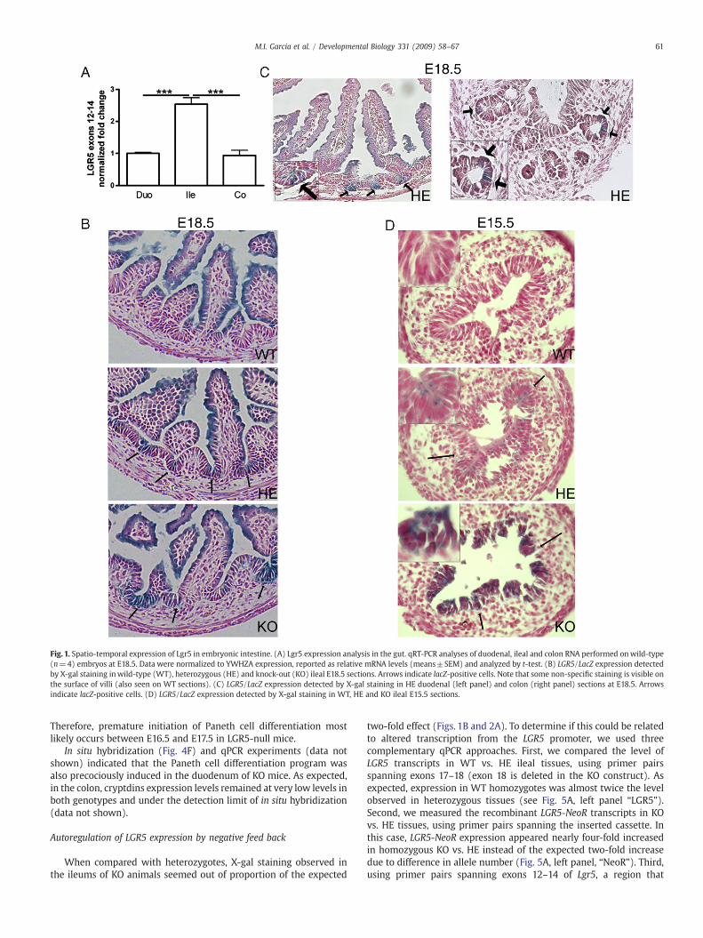
Fig. 1. Spatio-temporal expression of Lgr5 in embryonic intestine. (A) Lgr5 expression analysis in the gut. qRT-PCR analyses of duodenal, ileal and colon RNA performed onwild-type(n=4) embryos at E18.5. Data were normalized to YWHZA expression, reported as relative mRNA levels (means±SEM) and analyzed by t-test. (B) LGR5/LacZ expression detectedby X-gal staining inwild-type (WT), heterozygous (HE) and knock-out (KO) ileal E18.5 sections. Arrows indicate lacZ-positive cells. Note that some non-specific staining is visible onthe surface of villi (also seen on WT sections). (C) LGR5/LacZ expression detected by X-gal staining in HE duodenal (left panel) and colon (right panel) sections at E18.5. Arrowsindicate lacZ-positive cells. (D) LGR5/LacZ expression detected by X-gal staining in WT, HE and KO ileal E15.5 sections.
61M.I. Garcia et al. / Developmental Biology 331 (2009) 58–67
Therefore, premature initiation of Paneth cell differentiation mostlikely occurs between E16.5 and E17.5 in LGR5-null mice.
In situ hybridization (Fig. 4F) and qPCR experiments (data notshown) indicated that the Paneth cell differentiation program wasalso precociously induced in the duodenum of KO mice. As expected,in the colon, cryptdins expression levels remained at very low levels inboth genotypes and under the detection limit of in situ hybridization(data not shown).
Autoregulation of LGR5 expression by negative feed back
When compared with heterozygotes, X-gal staining observed inthe ileums of KO animals seemed out of proportion of the expected
two-fold effect (Figs. 1B and 2A). To determine if this could be relatedto altered transcription from the LGR5 promoter, we used threecomplementary qPCR approaches. First, we compared the level ofLGR5 transcripts in WT vs. HE ileal tissues, using primer pairsspanning exons 17–18 (exon 18 is deleted in the KO construct). Asexpected, expression in WT homozygotes was almost twice the levelobserved in heterozygous tissues (see Fig. 5A, left panel “LGR5”).Second, we measured the recombinant LGR5-NeoR transcripts in KOvs. HE tissues, using primer pairs spanning the inserted cassette. Inthis case, LGR5-NeoR expression appeared nearly four-fold increasedin homozygous KO vs. HE instead of the expected two-fold increasedue to difference in allele number (Fig. 5A, left panel, “NeoR”). Third,using primer pairs spanning exons 12–14 of Lgr5, a region that

Fig. 2. Cell proliferation and migration in E18.5 ileums. (A) Immunohistochemicaldetection of BrdU performed on X-gal stained ileal E18.5 HE or KO sections. Arrowheadsevidence LacZ-expressing cells positive for nuclear BrdU staining. (B) Immunohisto-chemical detection of CldU and IdU performed on ileal E18.5 sections 90-min and 24-hafter injection, respectively. Bar, 100 μm.
Fig. 3. LGR5 deficiency does not impair differentiation of cell lineages normally presentin E18.5 intestines. (A) Hematoxylin/Eosin staining of ileal cross sections of WT and KOmice. (B) Alcian blue–Nuclear fast red staining of ileal cross sections ofWTand KOmice.Arrowheads evidence blue-labeled Goblet cells. (C) and (D) Immunofluorescence co-staining for the enterocyte marker villin and nuclear DAPI and for the enteroendocrinemarker Chromogranin A and nuclear DAPI, respectively. Arrowheads show enteroendo-crine cells.
62 M.I. Garcia et al. / Developmental Biology 331 (2009) 58–67
encodes the extracellular domain of the protein, and is present in thesame copy number in WT and KO animals, we confirmed that the lossof functional LGR5 gene product correlates with a close to twofoldupregulation of transcription from the LGR5 promoter (Fig. 5A, rightpanel; KO vs. WT/HE, pb0.0001). As shown in Fig. 5B, suchderegulation of transcription from the LGR5 promoter was alreadydetected at day E15.5 (KO vs. HE, p=0.045; KO vs. WT, p=0.023).Together, these data indicate that LGR5 exerts a negative control on itsown expression.
Overactivation of Wnt signaling in LGR5 null mice
Since LGR5 is a Wnt target and its deregulation appears at a timepoint at which Wnt signaling is induced in the intestine (Lustig et al.,2002; Wong et al., 2002), we questioned whether the negativefeedback control of Lgr5 expression could occur through this pathway.To test this hypothesis, we measured the effect of LGR5 geneinvalidation in the ileum on the expression of additional Wnt targetgenes (Batlle et al., 2002; Jho et al., 2002; Jubb et al., 2006; van derFlier et al., 2009; Zeilstra et al., 2008). At E18.5, consistent upregulatedexpression of the Wnt regulator Axin2/conductin, the transcriptionalfactor Ascl2 and the CD44 glycoprotein was detected in KO vs. HE/WTileums (pb0.01). In contrast, expression of the Tyr kinase EphB3receptor was similar in all genotypes (Fig. 6A). In agreement with thetime course of Wnt expression during intestinal development,expression of Axin2 was induced at E15.5 in the 3 genotypes (Fig. 6B,pb0.05). However, whereas it decreased from E16.5 in WT and HEmice, Axin2 expression remained significantly increased in KO tissues(KO vs. WT/HE at E16.5, p=0.02; KO vs. HE at E17.5, pb0.001). Thesedata favor the hypothesis that upregulation of LGR5 gene expression inLGR5-KOmice takes place in the context of an inappropriate activationof the Wnt pathway. In situ hybridization using Axin2 as a probeshowed that Wnt overactivation exclusively occurs in epithelial cellslocated in the intervillus region of the ileum (Fig. 6C). As shown in Fig.6D, upregulation of Axin2, Ascl2 and CD44 was also detected in the
proximal intestine and, to a lesser extent, in the large intestine of KOtissues. The milder effect of LGR5 loss in the colon might be explainedby a delayed spatio-temporal induction of intestinal differentiation.Again, EphB3 gene expression remained at similar levels in theduodenum, ileum and colon of KO and WT mice.
Transcriptional profiling of ileal tissue from LGR5-deficient mice
To further explore the role of LGR5 in intestinal development andhomeostasis, we compared in vivo global gene expression at E18.5 inWT and KO mouse ileums. Four individual KO/WT littermate pairscoming from 3 independent matings were analyzed by SAM. 219differentially expressed genes were identified (80 unique up- and 139downregulated transcripts; complete list provided in the Supple-mental data as Table 1). In agreement with precocious Paneth celldifferentiation and overactivation of the Wnt pathway, one third ofthe up-regulated genes were genes expressed by Paneth cells, and a

Fig. 5. Autoregulation of the LGR5 locus. (A) Analysis of the expression level of the LGR5 gene at E18.5. Left panel, LGR5 exons 17-18/NeoR expression. Right panel, LGR5 exons 12–14expression. qRT-PCR analyses of ileal RNA were performed on WT (n=13), HE (n=14) and KO (n=12) embryos coming from 4 different litters. (B) LGR5 exons 12–14 expressionmeasured at earlier times of development. For each time point, qRT-PCR analysis of ileal RNA was performed on embryos coming from the same litters. At E14.5, the number ofindividuals analyzed was n=3, 4 and 6 for WT, HE and KO, respectively. See legend of Fig. 4E for the other time points. Data were normalized to YWHZA expression in the samesamples, represented as normalized fold change compared to HE E14.5 expression (means±SEM) and analyzed by t-test.
Fig. 4. LGR5 deficiency leads to precocious Paneth cells differentiation in E18.5 intestines. (A) In situ hybridization of WT and KO ileal sections showing cryptdin 4 expression.Arrowheads show cryptdin-expressing cells. (B) Lendrum' staining on ilealWTand KO sections. Inset of an intervillus KO section showing characteristic Paneth cell secretion vesiclesstained bright fuchsia (Arrows). Arrowheads showGoblet cells stained yellow. (C) Left panel: Paneth cell quantification on E18.5 ileums performed on aminimum of 9 tissue sectionsper animal (n=2 for each genotype). Data (means±SEM) were analyzed by t-test. Right panel: Lendrum's staining performed on an X-gal-stained KO section et E18.5. The arrowindicates a LacZ-positive Paneth cell. (D) qRT-PCR analysis of the Panethmarkers P-lysozyme and Mmp7 expression on ileums of WT (n=12) and KO (n=13) mice. (E) Time courseof expression of the Paneth cell marker cryptdin 5. For each time point, qRT-PCR analysis of ileal RNAwas performed on embryos coming from the same litter. At E15.5, the number ofindividuals analyzed was n=2, 6 and 3 for WT, HE and KO, respectively. At E16.5, n=2, 2 and 3 for WT, HE and KO, respectively. At E17.5, n=1, 7 and 3 for WT, HE and KO,respectively. Data were normalized to YWHZA expression, reported as relative mRNA levels (means±SEM) and analyzed by t-test. (F) In situ hybridization of WT and KO duodenalsections showing cryptdin 4 expression. Arrowheads show cryptdin-expressing cells.
63M.I. Garcia et al. / Developmental Biology 331 (2009) 58–67

Fig. 6.Wnt overactivation in Lgr5-null mice. (A) qRT-PCR analysis of Wnt target genes expression in the ileum ofWT, HE and KOmice (n=13,14 and 13, respectively). Analysis was performed by t-test. (B) Axin2 expression measured at earliertimes of development (see Fig. 5B legend). (C) In situ hybridization of WT and KO ileal sections showing Axin2 expression. For each picture, a magnification is shown below. (D) qRT-PCR analysis of Wnt target genes expression in theduodenum, ileum and colon of WT and KO (n=4 for each genotype) embryos at E18.5. Data were normalized to YWHZA expression, reported as relative mRNA levels (means±SEM) and analyzed by t-test.
64M.I.G
arciaet
al./Developm
entalBiology331
(2009)58
–67

Table 1Partial list of transcripts identified bymicroarray analysis to be upregulated in the ileumof LGR5 KO mice.
Function Gene Gene name Q valuea
(%)Mean foldchange(qPCR)
Cell adhesion/ECMFhdc1 FH2 domain containing 1 4.43 1.56MPZL2/Eva1 Myelin protein zero-like 2/
epithelial V-like antigen 13.32 1.31
Pcdh8/Papc Protocadherin 8 0.71 1.58Transcription factorsAscl2/
Mach2Achaete-scute complexhomolog-like 2 (Drosophila)
0 1.52(1.82)
Ccdc59 Coiled-coil domain containing 59 0 1.51Myb Myeloblastosis oncogene 0 1.33Prox2 Prospero-related homeobox gene 4.43 1.63Rbp2 Retinol binding protein 2 0.29 1.69Rnf43 Ring finger protein 43 0 1.32
Cell cycle/GrowthOip5/Lint-25 Opa interacting protein
5/LAP2α-binding protein0 1.29
Tm4sf4 Transmembrane 4 superfamilymember 4
0 1.33
Tubgcp4 Tubulin, gamma complex associatedprotein 4
4.43 1.29
MetabolismAbhd1 Abhydrolase domain containing 1 0 1.40Acpp Acid phosphatase, prostate 2.30 1.32Ahcy/CuBP/
SahhS-adenosylhomocysteine hydrolase 0.50 1.65
Fbp2 Fructose bisphosphatase 1 1.27 1.61Gst3 Glutathione S-transferase, alpha 3 2.30 1.59Ppa1 Pyrophosphatase (inorganic) 1 0 1.32Siat7a ST6 (alpha-N-acetyl-neuraminyl-2,3-beta-
galactosyl-1,3)-N-acetylgalactosaminidealpha-2,6-sialyltransferase 1
0 1.97
Molecule TransportAqp4 Aquaporin 4 0 1.35Kcnq1 Potassium voltage-gated channel,
subfamily Q, member 10.29 1.55
Slc6a7 Solute carrier family6 (neurotransmitter transporter, L-proline),member 7
0 1.38
ImmunityCcl19 Chemokine (C–C motif) ligand 19 0.50 1.36Defcr-rs1 Defensin related sequence cryptdin peptide⁎ 3.32 2.21
(3.95)Lzp P-lysozyme structural⁎ 0 2.49
(5.76)Mmp7 Matrix metallopeptidase 7⁎ 1.92 1.49
(4.75)Mt1 metallothionein 1⁎ 0.71 1.38Saa3 Serum amyloid A3 0.50 1.68SerpinA10 Ser (or cys) peptidase inhibitor, clade A
(α-1 antiproteinase, antitrypsin) member 10⁎0 1.49
Cell signalingAdora1 Adenosine A1 receptor 0.29 1.55Axin2/Axil Axin2/Conductin 0.29 1.29
(1.62)Fgf15 Fibroblast growth factor 15 0.29 4.29Rgs1 Regulator of G-protein signaling 1 1.92 1.89
a Defines the false discovery rate, q=1 corresponds to 1% risk that the regulatedgene is a false positive one.⁎ Genes expressed by Paneth cells (Muller et al., 2005; Weeks et al., 2006; Molmenti
et al., 1993; Szczurek et al., 2001).
Table 2Partial list of transcripts identified by microarray analysis to be downregulated in theileum of LGR5 KO mice.
Function Gene Gene name Q valuea
(%)Mean foldchange(qPCR)
Cell adhesion/ECMCol3a1 Collagen, type III, alpha 1 0 0.73Dcn Decorin 0 0.66Eln Elastin 0 0.58Firt2 Fibronectin leucine rich
transmembrane protein 20 0.75
Fln2 Fibulin 2 0 0.60Gja4/Cnx37 gap junction membrane channel
protein alpha 4/Connexin 370 0.72
Hapln1 Hyaluronan and proteoglycan linkprotein 1
0 0.77
Itm2A Integral membrane protein 2A⁎ 0 0.61 (0.69)Klk5 Kallikrein 1-related peptidase b5 1.92 0.68Mfap5/MAGP2 Microfibrillar associated protein 5/
microfibril-associated glycoproteins-20 0.49
Nov/CCN3 Nephroblastoma overexpressed gene/CCN3⁎
0 0.59 (0.55)
Postn/PN/Osf2 Periostin/osteoblast specific factor 0 0.62Sulf1 Sulfatase 1 0 0.77Tnx Tenascin XB 1.27 0.79
Transcription factorsEbf2 Early B-cell factor 2 0 0.72Gig1/Zfp704 Glucocorticoid induced gene 1/Zinc
finger protein 704⁎0.37 0.69
Odd1 Odd-skipped related 1 (Drosophila) 0.37 0.68Peg3/Pw1/Zfp102 Paternally expressed 3⁎ 0.37 0.73Wt1 Wilms tumor homolog 0 0.63
Cell cycle/GrowthAkap12 A kinase (PRKA) anchor protein
(gravin) 122.30 0.78
Ambra1 Autophagy/beclin 1 regulator 1 0 0.78Ccdc80/Urb/Sgg1 Coiled-coil domain containing 80/
Steroid sensitive gene 10 0.65
Gas1 Growth arrest specific 1⁎ 0.719 0.79Rbpp9/Bog Retinoblastoma binding protein 9 0 0.73Ubc Ubiquitin C 2.30 0.81
MetabolismAcad10 Acyl-Coenzyme A dehydrogenase
family, member 100 0.62
Dhrs6/Bdh2 Dehydrogenase/reductase (SDRfamily) member 6/3-hydroxybutyratedehydrogenase, type 2
1.27 0.73
Molecule TransportClca3/Gob5 Chloride channel calcium activated 3 0.50 0.73Kcne1 Potassium voltage-gated channel, Isk-
related family, member 1-like2.75 0.78
Slc6a14 Solute carrier family 6(neurotransmitter transporter),member 14
0 0.72
Cell signalingCxcl12 Chemokine (C–X–C motif) ligand 12 0 0.77Dkk2 Dickkopf homolog 2 (Xenopus laevis) 1.27 0.76)Dlk1 Delta-like 1 homolog (Drosophila) 0 0.67 (0.71)Hey3 Hairy/enhancer-of-split related with
YRPW motif-like0.71 0.78 (0.83)
Igfbp5 Insulin-like growth factor bindingprotein 5⁎
0 0.59 (0.75)
Nrk/Nesk Nik related kinase 0 0.61Ptn Pleiotrophin⁎ 0 0.69Retnlb/Xcp3/Fizz2 Resistin like beta 1.92 0.64Ror1 Receptor tyrosine kinase-like orphan
receptor 10 0.76
Sfrp2 Secreted frizzled-related protein 2 1.92 0.69Sst Somatostatin 2.30 0.70 (0.64)
a Defines the false discovery rate, q=1 corresponds to 1% risk that the regulatedgene is a false positive one.⁎ Genes reported to be expressed by stem cells (Blanpain et al., 2004; Morris et al.,
2004; Gupta et al., 2007).
65M.I. Garcia et al. / Developmental Biology 331 (2009) 58–67
number of known targets of Wnt signaling were upregulated (Table 1,Figs. 4 and 6A). Among the genes downregulated in null mice, anumber code for molecules associated with “stemness” (such as thecell adhesion molecule Itm2A, the growth factor binding proteinIgfbp5, the transcription factors Gig1 and Peg3, the cell cycle regulatorGas1 or the signaling molecule Pleiotrophin) (Table 2). Additionaldownregulated genes code for components of the extracellular matrixinvolved in cell signaling (such as Decorin and Elastin) as well as forendothelial cell markers (such as Cnx37), which could altogether
contribute to the establishment of the progenitor cell niche (Cabello-Verrugio and Brandan, 2007; Karnik et al., 2003; Simon andMcWhorter, 2002). As reported in Fig. 7, qRT-PCR experiments
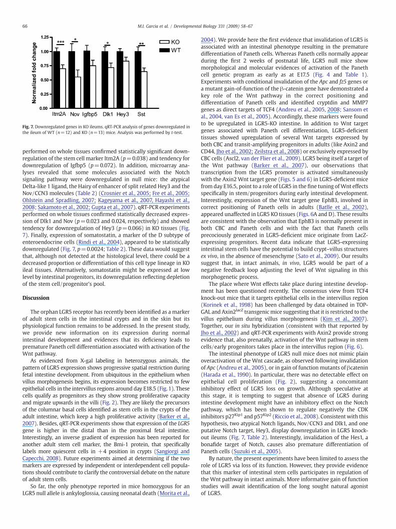
Fig. 7. Downregulated genes in KO ileums. qRT-PCR analysis of genes downregulated inthe ileum of WT (n=12) and KO (n=13) mice. Analysis was performed by t-test.
66 M.I. Garcia et al. / Developmental Biology 331 (2009) 58–67
performed on whole tissues confirmed statistically significant down-regulation of the stem cell marker Itm2A (p=0.038) and tendency fordownregulation of Igfbp5 (p=0.072). In addition, microarray ana-lyses revealed that some molecules associated with the Notchsignaling pathway were downregulated in null mice: the atypicalDelta-like 1 ligand, the Hairy of enhancer of split related Hey3 and theNov/CCN3 molecules (Table 2) (Crosnier et al., 2005; Fre et al., 2005;Ohlstein and Spradling, 2007; Kageyama et al., 2007, Hayashi et al.,2008; Sakamoto et al., 2002; Gupta et al., 2007). qRT-PCR experimentsperformed on whole tissues confirmed statistically decreased expres-sion of Dlk1 and Nov (p=0.023 and 0.024, respectively) and showedtendency for downregulation of Hey3 (p=0.066) in KO tissues (Fig.7). Finally, expression of somatostatin, a marker of the D subtype ofenteroendocrine cells (Rindi et al., 2004), appeared to be statisticallydownregulated (Fig. 7, p=0.0024; Table 2). These data would suggestthat, although not detected at the histological level, there could be adecreased proportion or differentiation of this cell type lineage in KOileal tissues. Alternatively, somatostatin might be expressed at lowlevel by intestinal progenitors, its downregulation reflecting depletionof the stem cell/progenitor's pool.
Discussion
The orphan LGR5 receptor has recently been identified as a markerof adult stem cells in the intestinal crypts and in the skin but itsphysiological function remains to be addressed. In the present study,we provide new information on its expression during normalintestinal development and evidences that its deficiency leads topremature Paneth cell differentiation associated with activation of theWnt pathway.
As evidenced from X-gal labeling in heterozygous animals, thepattern of LGR5 expression shows progressive spatial restriction duringfetal intestine development. From ubiquitous in the epithelium whenvillus morphogenesis begins, its expression becomes restricted to fewepithelial cells in the intervillus regions around day E18.5 (Fig. 1). Thesecells qualify as progenitors as they show strong proliferative capacityand migrate upwards in the villi (Fig. 2). They are likely the precursorsof the columnar basal cells identified as stem cells in the crypts of theadult intestine, which keep a high proliferative activity (Barker et al.,2007). Besides, qRT-PCR experiments show that expression of the LGR5gene is higher in the distal than in the proximal fetal intestine.Interestingly, an inverse gradient of expression has been reported foranother adult stem cell marker, the Bmi-1 protein, that specificallylabels more quiescent cells in +4 position in crypts (Sangiorgi andCapecchi, 2008). Future experiments aimed at determining if the twomarkers are expressed by independent or interdependent cell popula-tions should contribute to clarify the controversial debate on the natureof adult stem cells.
So far, the only phenotype reported in mice homozygous for anLGR5 null allele is ankyloglossia, causing neonatal death (Morita et al.,
2004). We provide here the first evidence that invalidation of LGR5 isassociated with an intestinal phenotype resulting in the prematuredifferentiation of Paneth cells. Whereas Paneth cells normally appearduring the first 2 weeks of postnatal life, LGR5 null mice showmorphological and molecular evidences of activation of the Panethcell genetic program as early as at E17.5 (Fig. 4 and Table 1).Experiments with conditional invalidation of the Apc and fz5 genes ora mutant gain-of-function of the β-catenin gene have demonstrated akey role of the Wnt pathway in the correct positioning anddifferentiation of Paneth cells and identified cryptdin and MMP7genes as direct targets of TCF4 (Andreu et al., 2005, 2008; Sansom etal., 2004, van Es et al., 2005). Accordingly, these markers were foundto be upregulated in LGR5-KO intestine. In addition to Wnt targetgenes associated with Paneth cell differentiation, LGR5-deficienttissues showed upregulation of several Wnt targets expressed byboth CBC and transit-amplifying progenitors in adults (like Axin2 andCD44, Jho et al., 2002; Zeilstra et al., 2008) or exclusively expressed byCBC cells (Ascl2, van der Flier et al., 2009). LGR5 being itself a target ofthe Wnt pathway (Barker et al., 2007), our observations thattranscription from the LGR5 promoter is activated simultaneouslywith the Axin2Wnt target gene (Figs. 5 and 6) in LGR5-deficient micefrom day E16.5, point to a role of LGR5 in the fine tuning ofWnt effectsspecifically in stem/progenitors during early intestinal development.Interestingly, expression of the Wnt target gene EphB3, involved incorrect positioning of Paneth cells in adults (Batlle et al., 2002),appeared unaffected in LGR5 KO tissues (Figs. 6A and D). These resultsare consistent with the observation that EphB3 is normally present inboth CBC and Paneth cells and with the fact that Paneth cellsprecociously generated in LGR5-deficient mice originate from LacZ-expressing progenitors. Recent data indicate that LGR5-expressingintestinal stem cells have the potential to build crypt–villus structuresex vivo, in the absence of mesenchyme (Sato et al., 2009). Our resultssuggest that, in intact animals, in vivo, LGR5 would be part of anegative feedback loop adjusting the level of Wnt signaling in thismorphogenetic process.
The place where Wnt effects take place during intestine develop-ment has been questioned recently. The consensus view from TCF4knock-out mice that it targets epithelial cells in the intervillus region(Korinek et al., 1998) has been challenged by data obtained in TOP-GAL and Axin2lacZ transgenicmice suggesting that it is restricted to thevillus epithelium during villus morphogenesis (Kim et al., 2007).Together, our in situ hybridization (consistent with that reported byJho et al., 2002) and qRT-PCR experiments with Axin2 provide strongevidence that, also prenatally, activation of the Wnt pathway in stemcells/early progenitors takes place in the intervillus region (Fig. 6).
The intestinal phenotype of LGR5 null mice does not mimic plainoveractivation of the Wnt cascade, as observed following invalidationof Apc (Andreu et al., 2005), or in gain of function mutants of βcatenin(Harada et al., 1990). In particular, there was no detectable effect onepithelial cell proliferation (Fig. 2), suggesting a concomitantinhibitory effect of LGR5 loss on growth. Although speculative atthis stage, it is tempting to suggest that absence of LGR5 duringintestine development might have an inhibitory effect on the Notchpathway, which has been shown to regulate negatively the CDKinhibitors p27Kip1 and p57Kip2 (Riccio et al., 2008). Consistent with thishypothesis, two atypical Notch ligands, Nov/CCN3 and Dlk1, and oneputative Notch target, Hey3, display downregulation in LGR5 knock-out ileums (Fig. 7, Table 2). Interestingly, invalidation of the Hes1, abonafide target of Notch, causes also premature differentiation ofPaneth cells (Suzuki et al., 2005).
By nature, the present experiments have been limited to assess therole of LGR5 via loss of its function. However, they provide evidencethat this marker of intestinal stem cells participates in regulation ofthe Wnt pathway in intact animals. More informative gain of functionstudies will await identification of the long sought natural agonistof LGR5.

67M.I. Garcia et al. / Developmental Biology 331 (2009) 58–67
Acknowledgments
We thank the members of the Pierre Vanderhaeghen laboratory(IRIBHM) and Jean Marie Vanderwinden (Neurophysiology lab, ULB)for helpful advices. We are grateful to H Clevers (Utrecht, Nether-lands) for having provided the LGR5/GPR49-LacZ mouse line.Supported by the Interuniversity Attraction Poles Programme-BelgianState-Belgian Science Policy (6/14), the European Union grant LSHB-CT-2003-503337, the Fonds de la Recherche Scientifique Médicaleof Belgium, the Walloon Region (program “Cibles”) and the FondsErasme.
Appendix A. Supplementary data
Supplementary data associated with this article can be found, inthe online version, at doi:10.1016/j.ydbio.2009.04.020.
References
Andreu, P., Colnot, S., Godard, C., Gad, S., Chafey, P., Niwa-Kawakita, M., Laurent-Puig, P.,Kahn, A., Robine, S., Perret, C., Romagnolo, B., 2005. Crypt-restricted proliferationand commitment to the Paneth cell lineage following Apc loss in the mouseintestine. Development 132, 1443–1451.
Andreu, P., Peignon, G., Slomianny, C., Taketo, M.T., Colnot, S., Robine, S., Lamarque, D.,Laurent-Puig, P., Perret, C., Romagnolo, B., 2008. A genetic study of the Wnt/β-catenin signalling in Paneth cell differentiation. Dev. Biol. 324, 288–296.
Barker, N., van Es, J.H., Kuipers, J., Kujala, P., van den Born, M., Cozijnsen, M., Haegebarth,A., Korving, J., Begthel, H., Peters, P.J., Clevers, H., 2007. Identification of stem cells insmall intestine and colon by marker gene LGR5. Nature 449, 1003–1007.
Batlle, E., Henderson, J.T., Beghtel, H., van den Born, M.M., Sancho, E., Huls, G., Meeldijk,J., Robertson, J., van de Wetering, M., Pawson, T., Clevers, H., 2002. Beta-catenin andTCF mediate cell positioning in the intestinal epithelium by controlling theexpression of EphB/ephrinB. Cell 111, 251–263.
Blanpain, C., Lowry, W.E., Geoghegan, A., Polak, L., Fuchs, E., 2004. Self-renewal,multipotency, and the existence of two cell populations within an epithelial stemcell niche. Cell 118, 635–648.
Bry, L., Falk, P.G., Huttner, K., Ouellette, A., Midtvedt, T., Gordon, J.I., 1994. Paneth celldifferentiation in the developing intestine of normal and transgenic mice. Proc.Natl. Acad. Sci. U. S. A. 91, 10335–10339.
Cabello-Verrugio, C., Brandan, E., 2007. A novel modulatory mechanism of transforminggrowth factor-beta signaling through decorin and Lrp-1. J. Biol. Chem. 282,18842–18850.
Cheng, H., Leblond, C.P., 1974. Origin, differentiation and renewal of the four mainepithelial cell types in the mouse small intestine. I. Columnar cell. Am. J. Anat. 141,461–479.
Crosnier, C., Vargesson, N., Gschmeissner, S., Ariza-McNaughton, L., Morrison, A., Lewis,J., 2005. Delta-Notch signalling controls commitment to a secretory fate in thezebrafish intestine. Development 132, 1093–1104.
Crosnier, C., Stamataki, D., Lewis, J., 2006. Organizing cell renewal in the intestine: stemcells, signals and combinatorial control. Nat. Rev. Genet. 7, 349–359.
Fre, S., Huyghe, M., Mourikis, P., Robine, S., Louvard, D., Artavanis-Tsakonas, S., 2005.Notch signals control the fate of immature progenitor cells in the intestine. Nature435, 964–968.
Gupta, R., Hong, D., Iborra, F., Sarno, S., Enver, T., 2007. NOV/CCN3 functions as aregulator of human hematopoietic stem or progenitor cells. Science 316, 590–593.
Harada, N., Tamai, Y., Ishikawa, T., Sauer, B., Takaku, K., Oshima, M., Taketo, M.M., 1990.Intestinal polyposis in mice with a dominant stable mutation of the beta-cateningene. EMBO J. 18, 5931–5942.
Hayashi, T., Kobubo, H., Hartman, B.H., Ray, C.A., Reh, T.A., Bermingham-McDonogh, O.,2008. Hesr1 and Hesr2 may act as early effectors of Notch signaling in thedeveloping cochlea. Dev. Biol. 316, 87–99.
Hsu, S.Y., Liang, S.G., Hsueh, A.J., 1998. Characterization of two LGR genes homo-logous to gonadotropin and thyrotropin receptors with extracellular leucine-richrepeats and a G protein-coupled, seven-transmembrane region. Mol. Endocrinol.12, 1830–1845.
Inoue, R., Tsuruta, T., Nojima, I., Nakayama, K., Tsukahara, T., Yajima, T., 2008. Postnatalchanges in the expression of genes for cryptdins 1–6 and the role of luminalbacteria in cryptdin gene expression inmouse small intestine. FEMS Immunol. Med.Microbiol. 52, 407–416.
Jho, E.H., Zhang, T., Domon, C., Joo, C.K., Freund, J.N., Costantini, F., 2002. Wnt/beta-catenin/Tcf signaling induces the transcription of Axin-2, a negative regulator ofthe signaling pathway. Mol. Cell Biol. 22, 1172–1183.
Jubb, A.M., Chalasani, S., Frantz, G.D., Smits, R., Grabsch, H.I., Kavi, V., Maughan, N.J.,Hillan, K.J., Quirke, P., Koeppen, P.H., 2006. Achaete-scute like 2 (ascl2) is a target ofWnt signalling and is upregulated in intestinal neoplasia. Oncogene 25, 3445–3457.
Kageyama, R., Ohtsuka, T., Kobayashi, T., 2007. The Hes gene family: repressors andoscillators that orchestrate embryogenesis. Development 134, 1243–1251.
Karnik, S.K., Brooke, B.S., Bayes-Genis, A., Sorensen, L., Wythe, J.D., Schwartz, R.S.,Keating, M.T., Li, D.Y., 2003. A critical role for elastin signaling in vascularmorphogenesis and disease. Development 130, 411–423.
Kim, B.M., Mao, J., Taketo, M.M., Shivdasani, R.A., 2007. Phases of canonical Wntsignaling during the development of mouse intestinal epithelium. Gastroenterol-ogy 133, 529–538.
Korinek, V., Barker, N., Moerer, P., van Donselaar, E., Huls, G., Peters, P.J., Clevers, H., 1998.Depletion of epithelial stem-cell compartments in the small intestine of micelacking Tcf-4. Nat. Genet. 19, 379–383.
Lustig, B., Jerchow, B., Sachs, M., Weiler, S., Pietsch, T., Karsten, U., van de Wetering, M.,Clevers, H., Schlag, P.M., Birchmeier, W., Berhens, J., 2002. Negative feedback loop ofWnt signaling through upregulation of conductin/axin2 in colorectal and livertumors. Mol. Cell Biol. 22, 1184–1193.
Marshman, E., Booth, C., Potten, C.S., 2002. The intestinal epithelial stem cell. Bioessays24, 91–98.
Mendive, F., Laurent, P., Van Schoore, G., Skarnes, W., Pochet, R., Vassart, G., 2006.Defective postnatal development of the male reproductive tract in LGR4 knockoutmice. Dev. Biol. 290, 421–434.
Molmenti, E.P., Perlmutter, D.H., Rubin, D.C., 1993. Cell-specific expression of alpha1-antitrypsin in human intestinal epithelium. J. Clin. Invest. 92, 2022–2034.
Morita, H., Mazerbourg, S., Bouley, D.M., Luo, C.-W., Kawamura, K., Kuwabara, Y.,Baribault, H., Tian, H., Hsueh, A.J.W., 2004. Neonatal lethality of LGR5 null mice isassociated with ankyloglossia and gastrointestinal distension. Mol. Cell Biol. 24,9736–9743.
Morris, R.J., Liu, Y., Marles, L., Yang, Z., Trempus, C., Li, S., Lin, J.S., Sawicki, J.A., Cotsarelis,G., 2004. Capturing and profiling adult hair follicle stem cells. Nat. Biotechnol. 22,411–417.
Muller, C.A., Autenrieth, I.B., Peschel, A., 2005. Innate defenses of the intestinalepithelial barrier. Cell Mol. Life Sci. 62, 1297–1307.
Ohlstein, B., Spradling, A., 2007. Multipotent drosophilia intestinal stem cells specifydaughter cell fates by differential notch signaling. Science 315, 988–992.
Pinto, D., Clevers, H., 2005. Wnt, stem cells and cancer in the intestine. Biol. Cell 97,185–196.
Riccio, O., van Gijn, M.E., Bezdek, A.C., Pellegrinet, L., van Es, J.H., Zimber-Strobl, U.,Strobl, L.J., Honjo, T., Clevers, H., Radtke, F., 2008. Loss of intestinal crypt progenitorcells owing to inactivation of both Notch1 and Notch2 is accompanied byderepression of CDK inhibitors p27Kip1 and p57Kip2. EMBO Rep. 9, 377–383.
Rindi, G., Leiter, A.B., Kopin, A.S., Bordi, C., Solcia, E., 2004. The “normal” endocrine cell ofthe gut changing concepts and new evidences. Ann. N. Y. Acad. Sci. 1014, 1–12.
Sakamoto, K., Yamaguchi, S., Ando, R., Miyawaki, A., Kabasawa, Y., Takagi, M., Li, C.L.,Perbal, B., Katsube, K., 2002. The nephroblastoma overexpressed gene (NOV/ccn3)protein associates with Notch1 extracellular domain and inhibits myoblastdifferentiation via Notch signaling pathway. J. Biol. Chem. 277, 29399–29405.
Sangiorgi, E., Capecchi, M.R., 2008. Bmi1 is expressed in vivo in intestinal stem cells. Nat.Genet. 40, 915–920.
Sansom, O.J., Reed, K.R., Hayes, A.J., Ireland, H., Brinkmann, H., Newton, I.P., Batlle, E.,Simon-Assmann, P., Clevers, H., Nathke, I.S., Clarke, A.R., Winton, D.J., 2004. Loss ofApc in vivo immediately perturbs Wnt signaling, differentiation, and migration.Genes Dev. 18, 1385–1390.
Sato, T., Vries, R.G., Snippert, H.J., van deWetering,M., Barker, N., Stange, D.E., van Es, J.H.,Abo, A., Kujala, P., Peters, P.J., Clevers, H., 2009. Single Lgr5 stem cells build crypt-villus structures in vitro without a mesenchymal niche. Nature, advance onlinepublication 29 March 2009, doi:10.1038/nature07935.
Scoville, D.H., Sato, T., He, X.C., Li, L., 2008. Current view: intestinal stem cells andsignaling. Gastroenterology 134, 849–864.
Simon, A.M., McWhorter, R., 2002. Vascular abnormalities in mice lacking theendothelial gap junction proteins connexin37 and connexin40. Dev. Biol. 15,206–220.
Suzuki, K., Fukui, H., Kayahara, T., Sawada, M., Seno, H., Hiai, H., Kageyama, R., Okano, H.,Chiba, T., 2005. Hes1-deficient mice show precocious differentiation of Paneth cellsin the small intestine. Biochem. Biophys. Res. Comm. 328, 348–352.
Szczurek, E.I., Bjornsson, C.S., Taylor, C.G., 2001. Dietary zinc deficiency and repletionmodulate metallothionein immunolocalization and concentration in small intes-tine and liver of rats. J. Nutr. 131, 2132–2138.
Tusher, V.G., Tibshirani, R., Chu, G., 2001. Significance analysis of microarrays applied tothe ionizing radiation response. Proc. Natl. Acad. Sci. U. S. A. 98, 5116–5121.
van der Flier, L.G., van Gijn, M.E., Hatzis, P., Kujala, P., Haegebarth, A., Stange, D.E.,Begthel, H., van den Born, M., Guryev, V., Oving, I., van Es, J.H., Barker, N., Peters, P.J.,van de Wetering, M., Clevers, H., 2009. Transcription factor Achaete scute-like 2controls intestinal stem cell fate. Cell 136, 903–912.
van Es, J.H., Jay, P., Gregorieff, A., van Gijn, M.E., Jonkheer, S., Hatzis, P., Thiele, A., van denBorn, M., Begthel, H., Brabletz, T., Taketo, M.M., Clevers, H., 2005. Wnt signallinginduces maturation of Paneth cells in intestinal crypts. Nat. Cell. Biol. 7, 381–386.
Weeks, C.S., Tanabe, H., Cummings, J.E., Crampton, S.P., Sheynis, T., Jelinek, R.,Vanderlick, T.K., Cocco, M.J., Ouellette, A.J., 2006. Matrix metalloproteinase-7activation of mouse Paneth cell pro-alpha-defensins: SER43 down arrow ILE44proteolysis enables membrane-disruptive activity. J. Biol. Chem. 281, 28932–28942.
Wong, M.H., Huelsken, J., Birchmeier, W., Gordon, J.I., 2002. Selection of multipotentstem cells during morphogenesis of small intestinal crypts of Lieberkühn isperturbed by stimulation of Lef-1/beta-catenin signalling. J. Biol. Chem. 277,15843–15850.
Yan, D., Wiesmann, M., Rohan, M., Chan, V., Jefferson, A.B., Guo, L., Sakamoto, D.,Caothien, R.H., Fuller, J.H., Reinhard, C., Garcia, P.D., Randazzo, F.M., Escobedo, J.,Fantl, W.J., Williams, L.T., 2001. Elevated expression of axin2 and hnkd mRNAprovides evidence that Wnt/beta-catenin signaling is activated in human colontumors. Proc. Natl. Acad. Sci. U. S. A. 98, 14973–14978.
Zeilstra, J., Joosten, S.P., Dokter, M., Verwiel, E., Spaargaren, M., Pals, S.T., 2008. Deletionof the WNT target and cancer stem cell marker CD44 in Apc(Min/+) miceattenuates intestinal tumorigenesis. Cancer Res. 68, 3655–3661.
Copyright © 2022 FDOKUMEN