Open versus laparoscopic liver resection for colorectal liver ...
Upload
independentCategory
view
0download
0
10.1128/MCB.22.4.1184-1193.2002.
2002, 22(4):1184. DOI:Mol. Cell. Biol. BehrensClevers, Peter M. Schlag, Walter Birchmeier and JürgenTorsten Pietsch, Uwe Karsten, Marc van de Wetering, Hans Barbara Lustig, Boris Jerchow, Martin Sachs, Sigrid Weiler, Colorectal and Liver Tumors
inthrough Upregulation of Conductin/Axin2 Negative Feedback Loop of Wnt Signaling
http://mcb.asm.org/content/22/4/1184Updated information and services can be found at:
These include:
REFERENCEShttp://mcb.asm.org/content/22/4/1184#ref-list-1at:
This article cites 50 articles, 24 of which can be accessed free
CONTENT ALERTS more»articles cite this article),
Receive: RSS Feeds, eTOCs, free email alerts (when new
http://journals.asm.org/site/misc/reprints.xhtmlInformation about commercial reprint orders: http://journals.asm.org/site/subscriptions/To subscribe to to another ASM Journal go to:
on June 12, 2013 by guesthttp://m
cb.asm.org/
Dow
nloaded from

MOLECULAR AND CELLULAR BIOLOGY, Feb. 2002, p. 1184–1193 Vol. 22, No. 40270-7306/02/$04.00�0 DOI: 10.1128/MCB.22.4.1184–1193.2002Copyright © 2002, American Society for Microbiology. All Rights Reserved.
Negative Feedback Loop of Wnt Signaling through Upregulation ofConductin/Axin2 in Colorectal and Liver Tumors
Barbara Lustig,1,2 Boris Jerchow,1 Martin Sachs,3 Sigrid Weiler,3 Torsten Pietsch,4 Uwe Karsten,1Marc van de Wetering,5 Hans Clevers,5 Peter M. Schlag,2 Walter Birchmeier,1
and Jürgen Behrens3*Max Delbrueck Center for Molecular Medicine, D-13092 Berlin,1 Department of Surgery and Surgical Oncology, Robert-Roessle-
Klinik, D-13122 Berlin,2 Nikolaus-Fiebiger-Zentrum, University of Erlangen, D-91054 Erlangen,3 and Department ofNeuropathology, University of Bonn Medical Center, D-53105 Bonn,4 Germany, and Department of
Immunology, University Hospital Utrecht, NL-3584 CX Utrecht, The Netherlands5
Received 7 August 2001/Returned for modification 12 September 2001/Accepted 16 November 2001
Activation of Wnt signaling through �-catenin/TCF complexes is a key event in the development of varioustumors, in particular colorectal and liver tumors. Wnt signaling is controlled by the negative regulatorconductin/axin2/axil, which induces degradation of �-catenin by functional interaction with the tumor sup-pressor APC and the serine/threonine kinase GSK3�. Here we show that conductin is upregulated in humantumors that are induced by �-catenin/Wnt signaling, i.e., high levels of conductin protein and mRNA werefound in colorectal and liver tumors but not in the corresponding normal tissues. In various other tumor types,conductin levels did not differ between tumor and normal tissue. Upregulation of conductin was also observedin the APC-deficient intestinal tumors of Min mice. Inhibition of Wnt signaling by a dominant-negative mutantof TCF downregulated conductin but not the related protein, axin, in DLD1 colorectal tumor cells. Conversely,activation of Wnt signaling by Wnt-1 or dishevelled increased conductin levels in MDA MB 231 and Neuro2Acells, respectively. In time course experiments, stabilization of �-catenin preceded the upregulation of con-ductin by Wnt-1. These results demonstrate that conductin is a target of the Wnt signaling pathway. Upregu-lation of conductin may constitute a negative feedback loop that controls Wnt signaling activity.
The Wnt signaling pathway is involved in a variety ofdevelopmental processes and in tumor formation, in partic-ular of colorectal and liver tumors (2, 8, 33). A hallmark ofWnt signaling is the stabilization of cytoplasmic �-catenin fol-lowed by its association with TCF transcription factors, whichleads to the transcription of Wnt target genes (5, 18, 30, 46).The cytoplasmic component conductin (also named axin2 oraxil) functions as a negative regulator of Wnt signaling byinducing degradation of �-catenin (3, 29, 40, 49). Biochemi-cally, conductin acts as a scaffold for the assembly of a multi-protein complex which includes the tumor suppressor APCand the serine/threonine kinase GSK3�. In this complex,�-catenin is phosphorylated by GSK3�, which leads to its ubiq-uitination and degradation in proteasomes (1, 21, 49). Con-ductin induces downregulation of �-catenin when transientlyoverexpressed in colon carcinoma cells and inhibits Wnt-in-duced as well as endogenous axis formation in early Xenopusembryos (3, 14, 51).
Conductin is related to axin, with which it shares 45% aminoacid identity (3, 51). Conductin and axin have similar biochem-ical and cell biological properties but may differ in their in vivofunctions. While axin is homogenously distributed in themouse embryo (51), conductin is more selectively expressed inspecific tissues (3; B. Jerchow and W. Birchmeier, unpublisheddata). Axin has been identified as the product of the fused gene
locus in the mouse. The fused mutations lead to defects inembryonal body axis formation (47, 51).
During embryonal development, Wnt signaling stabilizes�-catenin by blocking the activity of the conductin/axin-based �-catenin degradation complex. This pathway in-volves the activation of several intermediary components byWnts, including the cytoplasmic protein dishevelled, whichinteracts with conductin/axin (22, 27, 28, 36). Due to truncat-ing mutations of APC or point mutations in the phosphoryla-tion sites of �-catenin, various tumor types show constitutivestabilization of �-catenin and permanent activation of TCF/�-catenin-driven gene transcription (6, 31, 33). Specifically, theAPC gene is mutated in the inherited disease familial adeno-matous polyposis (FAP), which leads to formation of multiplecolorectal adenomas and carcinomas and in addition accountsfor about 80% of sporadic colorectal carcinomas (33). �-Cate-nin is mutated in about 5% of colorectal carcinomas (31, 38)and in up to 50% of hepatoblastomas and hepatocellular car-cinomas (23, 25, 44), as well as in a variety of other tumors (2).Alterations of axin and conductin/axin2 have also been de-scribed previously for hepatocellular carcinomas and for a frac-tion of unstable microsatellite colorectal tumors, respectively(29, 39).
Several lines of evidence suggest an essential role for�-catenin/Wnt signaling in tumorigenesis. Accumulation of�-catenin in the cytoplasm and nucleus was demonstratedpreviously on tissue sections of colorectal and liver tumors,although regional differences within a given tumor exist (16,20, 24, 25). Furthermore, several target genes of �-catenin/TCF complexes with a possible function in tumorigenesis, such
* Corresponding author. Mailing address: Nikolaus-Fiebiger-Zen-trum, University of Erlangen, Glueckstr. 6, D-91054 Erlangen, Ger-many. Phone: 49-9131-8529109. Fax: 49-9131-8529111. E-mail: [email protected].
1184
on June 12, 2013 by guesthttp://m
cb.asm.org/
Dow
nloaded from
as c-myc, cyclin D1, and matrilysin, have been described else-where (9, 15, 45). Moreover, transgenic expression of stabilizedversions of �-catenin in mice induces tumor formation (13),and Min (multiple intestinal neoplasia) mice, which carry agerm line mutation of APC, develop spontaneous tumors inthe small intestine that show stabilization of �-catenin (32, 43).
In the study reported in this paper, we have determined theexpression pattern of conductin in a variety of human tumorsand normal tissue. We found that conductin is upregulated incolorectal adenomas and carcinomas and in liver tumors, incomparison to the corresponding normal tissues. Biochemicalmanipulation of Wnt signaling in cell lines and genetic analysisof Min mice indicate that conductin itself is a target of Wntsignaling. Our data suggest that conductin is part of a negativefeedback loop which serves to restrain Wnt signaling activity bylimiting the amounts of �-catenin. As upregulation of conduc-tin appears to be an early event in �-catenin/Wnt-inducedtumorigenesis, conductin might be a useful marker for earlytumor diagnosis.
MATERIALS AND METHODS
Generation of the anticonductin monoclonal antibody C/G7. Female NMRImice were immunized intraperitoneally with 75 �g of a recombinant fragment ofmouse conductin (amino acids 2 to 396) (3) in phosphate-buffered saline (PBS)with Freund’s adjuvant. Eight weeks later, the mice were boosted intraperitone-ally with 75 �g of the conductin fragment in PBS without adjuvant. Spleen cellsfrom one mouse were harvested 4 days after the boost and fused with X63-Ag8.653 myeloma cells by employing standard techniques, except that azaserinewas used in the selection medium. Growth medium was RPMI 1640 supple-mented initially with 15 and then 10% fetal calf serum (FCS; Biochrom, Berlin,Germany) and finally with 0.5% hybridoma serum (Pandar, Heidelberg, Germa-ny). Initial hybridoma growth and cloning were supported by peritoneal feedercells from NMRI mice. Hybridoma supernatants were tested in an enzyme-linked immunosorbent assay with recombinant conductin used as a coating at 5�g/ml. Bovine thyroglobulin served as a negative control, and affinity-purifiedrabbit polyclonal antiserum against conductin (3) was used as a positive control.One hybridoma clone specific for conductin was selected (C/G7; immunoglobu-lin G1 � chain), recloned, and frozen. Immunofluorescence tests and Westernblotting with conductin- or axin-transfected Neuro2A cells showed that C/G7 isspecific for conductin and does not cross-react with axin. Large-scale productionof C/G7 hybridoma supernatant was carried out by growing hybridoma in dialysistubes in a special gyration apparatus (Kretzschmar, Berlin, Germany).
Cell culture, Western blotting, and transfections. Cells were cultured in Dul-becco modified Eagle medium (DMEM) containing 10% FCS and streptomycin-penicillin. The colon, lung, breast, bladder, pancreas, and prostate carcinomaand the hepatoblastoma cell lines used in Fig. 1 have been described previously(7, 10, 25). The melanoma cell lines SK-MEL3 and RPMI 7951 were obtainedfrom the American Type Culture Collection, and Juso, Parl, IGR-3, and Strömerwere obtained from J. P. Johnson, University of Munich.
For Western blot analysis of conductin in human tumor cell lines, 3 � 106 cellswere cultured overnight on 10-cm-diameter dishes and extracted with 500 �l oflysis buffer (L-CAM assay buffer containing 1% Triton X-100 and 1 mM phe-nylmethylsulfonyl fluoride [4]). For analysis of human tumors, normal and tumorsamples were snap-frozen and stored at �80°C. Cryostat sections (10 �m) wereextracted in lysis buffer on ice for 15 min, sonicated for 10 s, and centrifuged for15 min at 16,000 � g. Equal amounts of extracted protein as determined by theBradford protein assay (Bio-Rad) were separated by sodium dodecyl sulfate(SDS)–8% polyacrylamide gel electrophoresis and blotted on Immobilon mem-branes (Amersham). Proteins were detected using mouse C/G7 antibody againstconductin (concentrated hybridoma supernatant at 1:100 dilution), rabbit anti-axin (1:200 dilution; gift from K. Willert, Stanford University), mouse anti-cytokeratin 19 (1:100; Santa Cruz), rat antitubulin (1:1,000; Serotec, Kidlington,United Kingdom), and mouse anti-�-catenin (1:400; Transduction Laboratories)antibodies. After incubation with peroxidase-coupled secondary antibodies, blotswere developed using enhanced chemiluminescence reagent (NEN) and exposedto X-ray films (Kodak).
The T-REx system (Invitrogen, Carlsbad, Calif.) was used according to themanufacturer’s instructions to generate DLD1 colorectal tumor cells expressing
tetracycline-inducible dominant-negative TCF. In short, 107 cells were trans-fected by electroporation with 20 �g of FspI-linearized pcDNA6TR. Cells weregrown in RPMI supplemented with 10% FCS, antibiotics, and blasticidin(10 �g/ml). After 3 weeks of selection, blasticidin-resistant colonies were ex-panded and transfected with pcDNA4TO-luciferase. One clone (TR7-DLD1)that showed the strongest induction was chosen and subsequently transfectedwith 20 �g of PvuI-linearized �NTCF1 or �NTCF4 expression plasmids codingfor TCF proteins that lack the �-catenin binding domain (26). After selection onZeocin (500 �g/ml), resistant colonies were tested for dominant-negative TCFinduction by immunohistochemical staining after addition of doxycycline. Theclones with the strongest induction (named �NTCF1-DLD1 and �NTCF4-DLD1)were used in the experiments. A luciferase reporter assay (Top/Fop assay) (26,30) was used to confirm that dominant-negative TCF induction resulted inabrogation of �-catenin/TCF-driven transcription. For Western blots, 5 � 105
TR7-DLD1, �NTCF1-DLD1, or �NTCF4-DLD1 cells were plated overnight on10-cm-diameter dishes, induced with 1 �g of doxycycline (Sigma-Aldrich FineChemicals, St. Louis, Mo.) per ml for 12 h, and extracted in 300 �l of lysis buffer.
To obtain conditioned media, confluent Rat2/Wnt-1 and Rat2/MV7 cells werecultured overnight in serum-free DMEM (12). For stimulation of MDA MB 231breast carcinoma cells, 3 � 105 cells were cultured overnight in six-well dishes,serum starved in DMEM for 16 h, and incubated with conditioned media. At theindicated time points, cells were lysed for 20 min on ice either in 100 �l ofhypotonic buffer (20 mM Tris-Cl [pH 7.5], 1 mM EDTA, 1 mM phenylmethyl-sulfonyl fluoride) to obtain the cytosolic fractions for analysis of �-catenin (12)or in L-CAM assay buffer containing Triton X-100 for analysis of conductin. Inthe time course experiment shown in Fig. 5D, both conductin and �-catenin weredetected in the cytosolic extracts.
For transient transfections, 106 Neuro2A cells were cultured overnight on10-cm-diameter dishes and transfected with a mixture of 20 �g of a mousedishevelled-2 expression plasmid (Dvl-2 [28]), 2.5 �g of pCMVCD20, and 2.5 �gof pSGCD20. An expression plasmid for vav-2 instead of Dvl-2 was used as anegative control. Cells were trypsinized after 24 h and incubated with anti-CD20antibody (Dianova), and the transfected cells were captured with magnetic beads(Miltenyi) coated with anti-mouse immunoglobulin G. Eluted cells were ex-tracted in lysis buffer and processed for Western blots as described above.
Northern blots and cancer profiling arrays. TR7-DLD1 or �NTCF1-DLD1(4.5 � 106) cells were plated on three 150-mm-diameter plates in DMEM–10%FCS. For induction of �NTCF1, the cells were treated the next day for 14 h with1 �g of doxycycline/ml. MDA MB 231 cells were treated with Wnt-1-conditionedmedium or control medium for 4 h. Total RNA was isolated with Trizol reagent(Invitrogen), and poly(A)� RNA was isolated with the Oligotex mRNA Midi kit(Qiagen Inc., Valencia, Calif.) according to the manufacturer’s protocol. Twomicrograms of poly(A)� RNA per lane was electrophoresed on formaldehydeagarose gels (37) followed by blotting on Hybond-XL membranes (AmershamPharmacia Biotech Inc., Piscataway, N.J.) by capillary transfer. Hybridization ofthe blots was carried out overnight at 65°C with 32P-labeled cDNA probesderived from human conductin (Image clone IMAGp998I141906) and axin (Im-age clone IMAGp998B12254) or rat glyceraldehyde-3-phosphate dehydrogenasein 5� SSPE (1� SSPE is 0.18 M NaCl, 10 mM NaH2PO4, and 1 mM EDTA [pH7.7])–5� Denhardt’s solution–0.5% SDS containing 100 �g of sheared anddenatured salmon testis DNA/ml. The filters were washed at 65°C for 20 mineach with 2� SSC (1� SSC is 0.15 M NaCl plus 0.015 M sodium citrate)–0.1%SDS, 0.5� SSC–0.5% SDS, and 0.1� SSC–0.1% SDS, followed by exposure toX-ray films.
The cancer profiling array membrane (PT3578–1; Clontech Laboratories, Inc.,Palo Alto, Calif.) was hybridized with the cDNA probes described above accord-ing to the manufacturer’s protocol. In brief, the membrane was prehybridized for30 min with 10 �l of ExpressHyb solution (Clontech) at 65°C. The labeled cDNAprobes were mixed with 30 �g of Cot-1 DNA (Roche Molecular Biochemicals,Indianapolis, Ind.), 150 �g of sheared salmon testis DNA, and 50 �l of 20� SSCin a total volume of 200 �l. The mixture was heated at 95°C for 5 min, incubatedat 65°C for 30 min, and mixed with 5 ml of fresh ExpressHyb solution. The filterwas hybridized overnight at 65°C and washed four times at 65°C for 20 min with2� SSC–0.5% SDS, once with 0.2� SSC–0.5% SDS, and finally for 5 min atroom temperature with 2� SSC. For consecutive hybridizations, filters werestripped in boiling 0.5% SDS solution. For normalization, the filter was finallyhybridized with a human ubiquitin probe (Clontech). The filter was exposed toX-ray films for documentation. For quantification, the filter was scanned on anFLA-3000 image reader (Fuji Photo Film Co. Ltd.) and the spots were analyzedwith AIDA 2.3.1 software (Raytest USA Inc., Wilmington, Del.).
Mouse experiments. C57BL/6 male Min (Apc�/Min) mice were obtained com-mercially (The Jackson Laboratory, Bar Harbor, Maine). Conductin�/lacZ micewere generated from heterozygous embryonic stem cells derived from the E14
VOL. 22, 2002 NEGATIVE FEEDBACK LOOP OF Wnt SIGNALING VIA CONDUCTIN 1185
on June 12, 2013 by guesthttp://m
cb.asm.org/
Dow
nloaded from

embryonic stem cell line that were injected into C57BL/6 blastocysts (describedelsewhere [Jerchow and Birchmeier, unpublished]). Heterozygous Min mice andconductin�/lacZ mice were crossed to obtain double heterozygous mice. Forintestinal analysis, mice were sacrificed at 6 to 8 months. The intestines werecollected, flushed with PBS, opened longitudinally, and mounted on Whatmanpaper. Either tumors were dissected for cryosectioning and subsequent �-galac-tosidase staining, or whole-mount staining of the intestine was performed. �-Ga-lactosidase staining was carried out as described elsewhere (17). Embedding inparaffin was performed according to the manufacturer’s protocol (Paraplast;Sherwood Medical). Stained tissue was cut at 10 �m and counterstained withnuclear fast red. In situ hybridizations were performed as described elsewhere onparaffin sections of mouse intestine using antisense probes specific for conductinand TCF4 (19).
RESULTS
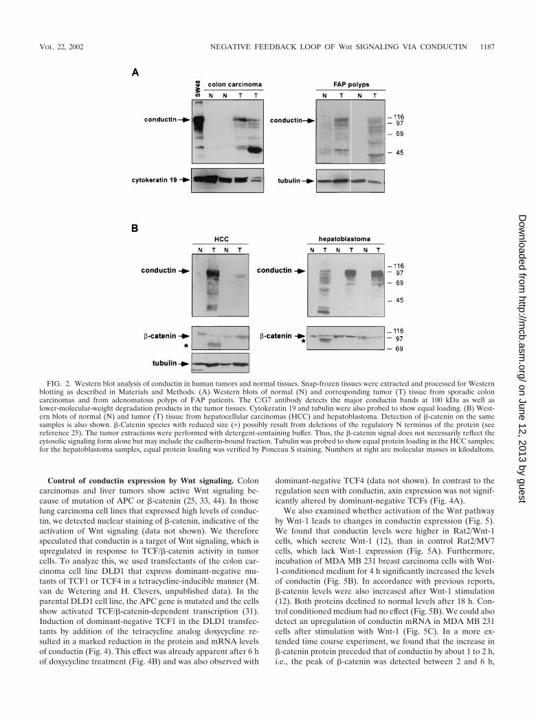
Upregulation of conductin in human tumors. We have stud-ied the expression of conductin in a variety of tumor cell linesfrom different tissues by Western blotting with a novel mono-clonal antibody, C/G7. High amounts of conductin were readilydetected in the majority of colon carcinoma cells, in all hepa-toblastoma cells, and in some lung carcinoma cells. In contrast,most breast, bladder, pancreas, and prostate carcinoma andmelanoma cells expressed low levels of conductin, which couldbe detected only after overexposure of the blots (Fig. 1). Toanalyze conductin expression in human tumors, we performedWestern blotting on colon carcinoma tissue samples and oncorresponding normal colon mucosa from the same patients.Conductin was present in high amounts in the tumors but wasbarely detectable in the normal tissue (Fig. 2A). We also an-alyzed adenomas from FAP patients and again found upregu-lation of conductin in the tumors compared to the normal
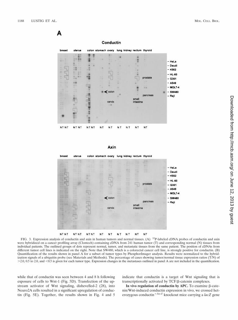
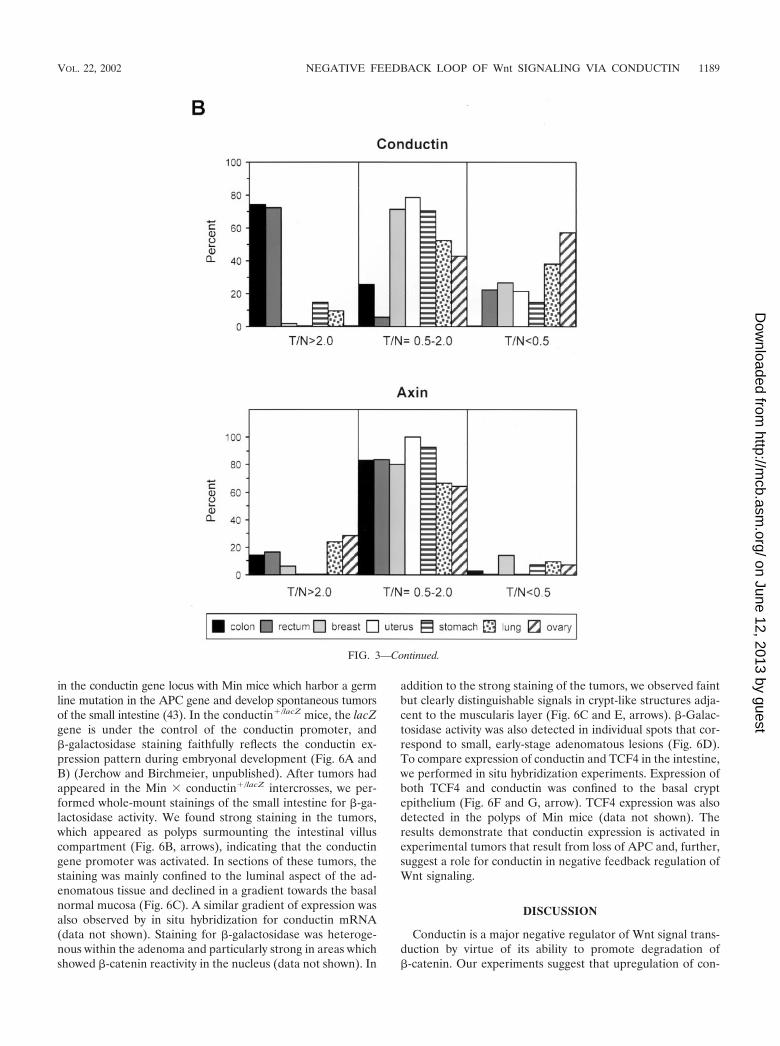
mucosa (Fig. 2A). Upregulation of conductin was also seen insporadic colon adenomas (data not shown). Similarly, conduc-tin levels were markedly increased in liver tumors (hepatocel-lular carcinomas and hepatoblastomas) compared to those innormal liver tissue (Fig. 2B). Thus, the conductin protein ap-pears to be expressed in large amounts in tumors that showactivated �-catenin/Wnt signaling. These results prompted usto analyze conductin mRNA levels in a larger number of dif-ferent normal and tumor tissues. We made use of a cancerprofiling array (Clontech), which contains normalized cDNAfrom 241 tumor and corresponding normal tissues from indi-vidual patients. After hybridization of this array with a humanconductin cDNA probe, we found the strongest signals in sam-ples of uterus, colon, and rectum (Fig. 3A). Again, a significantupregulation of conductin compared to normal tissue was ob-served in colon and rectal tumors. Quantification of the resultsshowed a more than twofold increase in conductin mRNAlevels in about 70% of the examined colon and rectal carcino-mas (range, 2.2- to 14.4-fold [Fig. 3B]). Conductin mRNAexpression was also elevated, albeit to a lesser extend, in mostof the remaining colorectal tumor samples. However, in themajority of the other tumor types, no significant differencesbetween normal and tumor tissue were observed, with theexception of 60 and 40% of uterine and lung carcinomas,respectively, which showed a more than twofold decrease inconductin levels. In contrast to the differential expression pat-tern of conductin, axin was equally expressed in the varioustissue samples, and no differences between normal and tumortissues were seen (Fig. 3).
FIG. 1. Western blot analysis of conductin in human tumor cell lines. Western blotting was performed using the C/G7 antibody on proteinextracts from the indicated tumor cell lines. Equal amounts of protein were loaded per lane. Conductin was detected as a double band around 100kDa in most cell lines. In some cell lines, either the upper or the lower band is seen. The same blots were also probed with antitubulin antibodiesto demonstrate similar protein loading. Note that the Western blots of conductin in the lower panels (breast, bladder, pancreas, prostate, andmelanoma) were exposed five times longer than those in the upper panels (colon, hepatoblastoma, and lung). Numbers at right are molecularmasses in kilodaltons.
1186 LUSTIG ET AL. MOL. CELL. BIOL.
on June 12, 2013 by guesthttp://m
cb.asm.org/
Dow
nloaded from
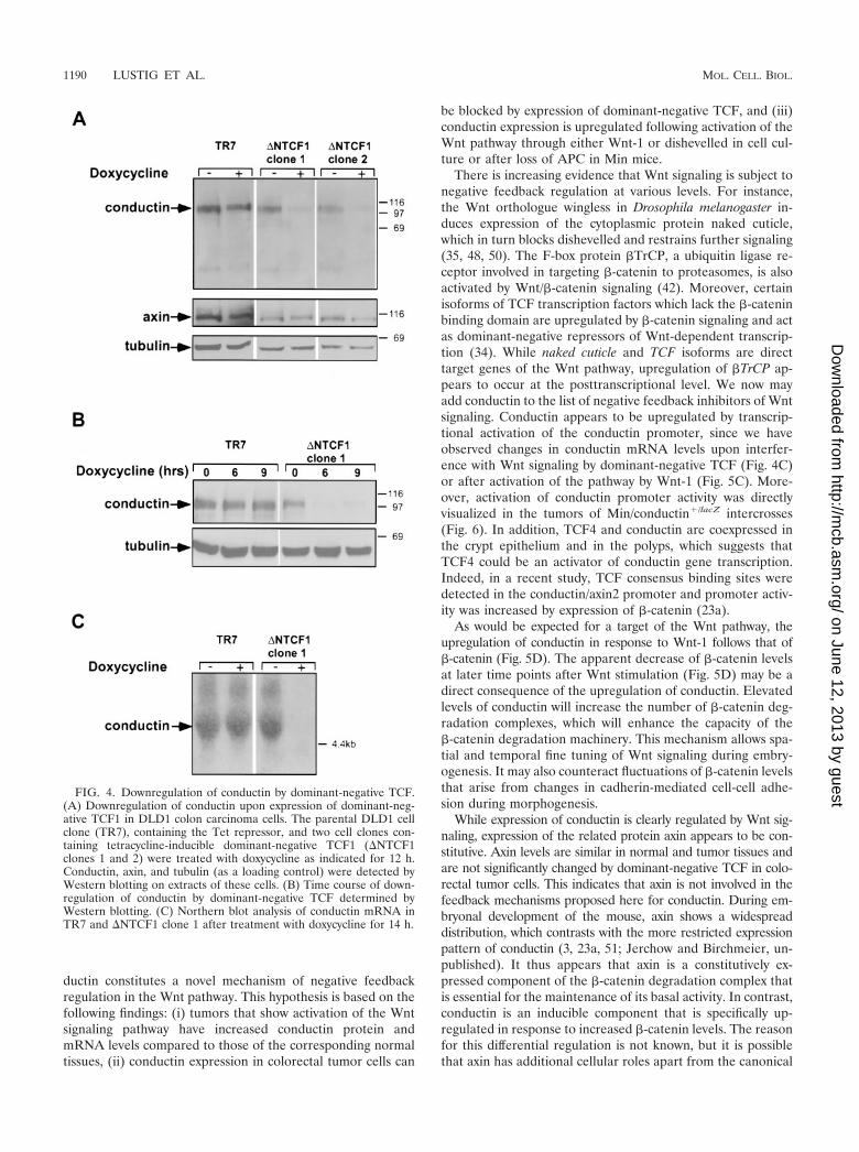
Control of conductin expression by Wnt signaling. Coloncarcinomas and liver tumors show active Wnt signaling be-cause of mutation of APC or �-catenin (25, 33, 44). In thoselung carcinoma cell lines that expressed high levels of conduc-tin, we detected nuclear staining of �-catenin, indicative of theactivation of Wnt signaling (data not shown). We thereforespeculated that conductin is a target of Wnt signaling, which isupregulated in response to TCF/�-catenin activity in tumorcells. To analyze this, we used transfectants of the colon car-cinoma cell line DLD1 that express dominant-negative mu-tants of TCF1 or TCF4 in a tetracycline-inducible manner (M.van de Wetering and H. Clevers, unpublished data). In theparental DLD1 cell line, the APC gene is mutated and the cellsshow activated TCF/�-catenin-dependent transcription (31).Induction of dominant-negative TCF1 in the DLD1 transfec-tants by addition of the tetracycline analog doxycycline re-sulted in a marked reduction in the protein and mRNA levelsof conductin (Fig. 4). This effect was already apparent after 6 hof doxycycline treatment (Fig. 4B) and was also observed with
dominant-negative TCF4 (data not shown). In contrast to theregulation seen with conductin, axin expression was not signif-icantly altered by dominant-negative TCFs (Fig. 4A).
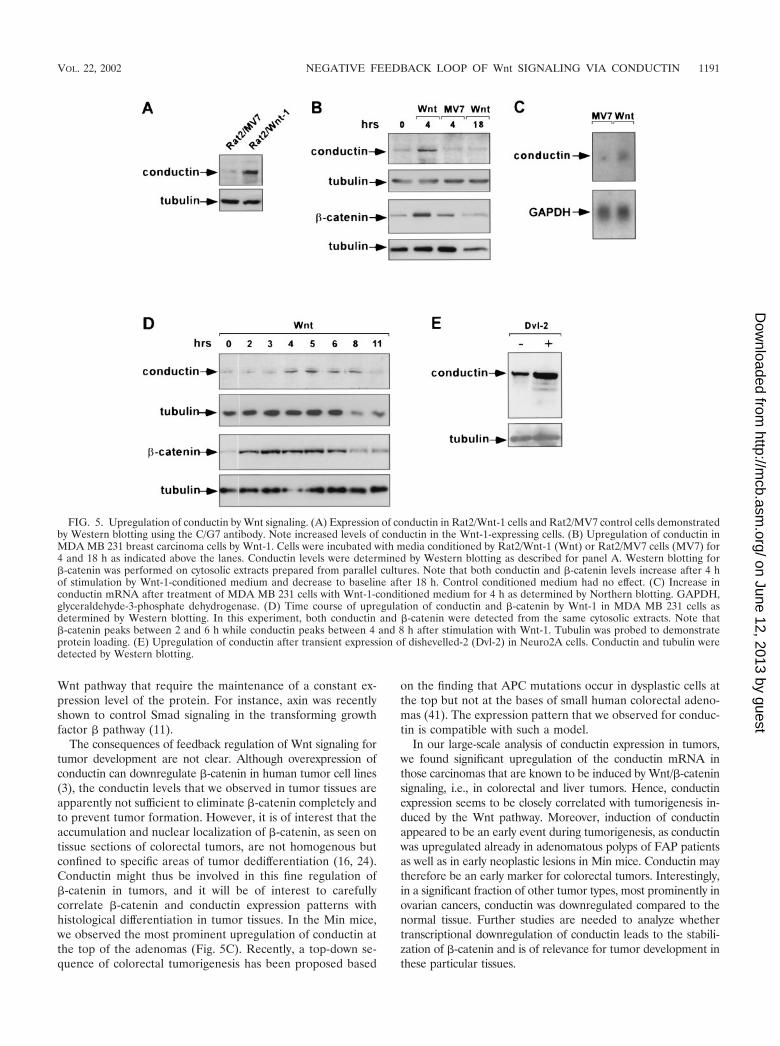
We also examined whether activation of the Wnt pathwayby Wnt-1 leads to changes in conductin expression (Fig. 5).We found that conductin levels were higher in Rat2/Wnt-1cells, which secrete Wnt-1 (12), than in control Rat2/MV7cells, which lack Wnt-1 expression (Fig. 5A). Furthermore,incubation of MDA MB 231 breast carcinoma cells with Wnt-1-conditioned medium for 4 h significantly increased the levelsof conductin (Fig. 5B). In accordance with previous reports,�-catenin levels were also increased after Wnt-1 stimulation(12). Both proteins declined to normal levels after 18 h. Con-trol conditioned medium had no effect (Fig. 5B). We could alsodetect an upregulation of conductin mRNA in MDA MB 231cells after stimulation with Wnt-1 (Fig. 5C). In a more ex-tended time course experiment, we found that the increase in�-catenin protein preceded that of conductin by about 1 to 2 h,i.e., the peak of �-catenin was detected between 2 and 6 h,
FIG. 2. Western blot analysis of conductin in human tumors and normal tissues. Snap-frozen tissues were extracted and processed for Westernblotting as described in Materials and Methods. (A) Western blots of normal (N) and corresponding tumor (T) tissue from sporadic coloncarcinomas and from adenomatous polyps of FAP patients. The C/G7 antibody detects the major conductin bands at 100 kDa as well aslower-molecular-weight degradation products in the tumor tissues. Cytokeratin 19 and tubulin were also probed to show equal loading. (B) West-ern blots of normal (N) and tumor (T) tissue from hepatocellular carcinomas (HCC) and hepatoblastoma. Detection of �-catenin on the samesamples is also shown. �-Catenin species with reduced size (�) possibly result from deletions of the regulatory N terminus of the protein (seereference 25). The tumor extractions were performed with detergent-containing buffer. Thus, the �-catenin signal does not necessarily reflect thecytosolic signaling form alone but may include the cadherin-bound fraction. Tubulin was probed to show equal protein loading in the HCC samples;for the hepatoblastoma samples, equal protein loading was verified by Ponceau S staining. Numbers at right are molecular masses in kilodaltons.
VOL. 22, 2002 NEGATIVE FEEDBACK LOOP OF Wnt SIGNALING VIA CONDUCTIN 1187
on June 12, 2013 by guesthttp://m
cb.asm.org/
Dow
nloaded from
while that of conductin was seen between 4 and 8 h followingexposure of cells to Wnt-1 (Fig. 5D). Transfection of the up-stream activator of Wnt signaling, dishevelled-2 (28), intoNeuro2A cells resulted in a significant upregulation of conduc-tin (Fig. 5E). Together, the results shown in Fig. 4 and 5
indicate that conductin is a target of Wnt signaling that istranscriptionally activated by TCF/�-catenin complexes.
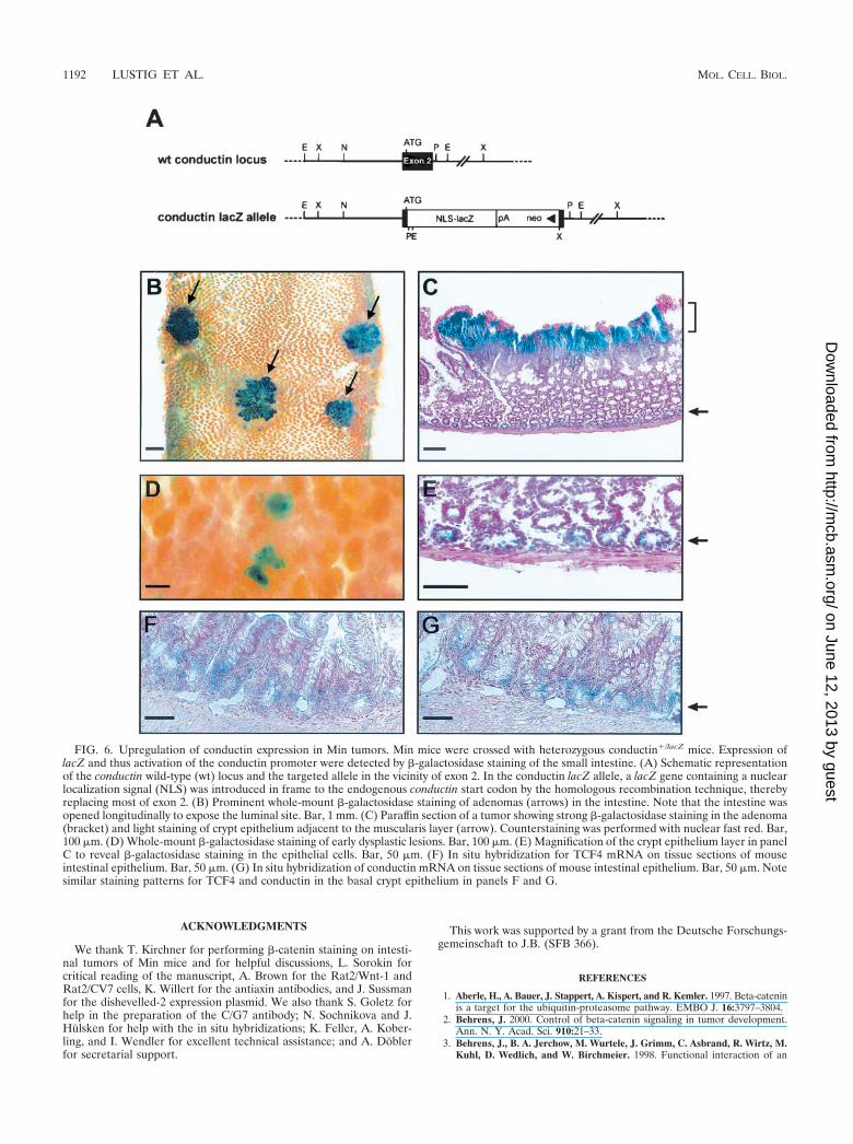
In vivo regulation of conductin by APC. To examine �-cate-nin/Wnt-induced conductin expression in vivo, we crossed het-erozygous conductin�/lacZ knockout mice carrying a lacZ gene
FIG. 3. Expression analysis of conductin and axin in human tumors and normal tissues. (A) 32P-labeled cDNA probes of conductin and axinwere hybridized on a cancer profiling array (Clontech) containing cDNA from 241 human tumor (T) and corresponding normal (N) tissues fromindividual patients. The outlined groups of dots represent normal, tumor, and metastatic tissues from the same patient. The position of cDNAs fromdifferent tumor cell lines is indicated on the right. Note that SW480, which is a colorectal cancer cell line, is strongly positive for conductin. (B)Quantification of the results shown in panel A for a subset of tumor types by PhosphorImager analysis. Results were normalized to the hybrid-ization signals of a ubiquitin probe (see Materials and Methods). The percentage of cases showing tumor/normal tissue expression ratios (T/N) of�2.0, 0.5 to 2.0, and 0.5 is given for each tumor type. Expression changes in the metastases outlined in panel A are not included in the quantification.
1188 LUSTIG ET AL. MOL. CELL. BIOL.
on June 12, 2013 by guesthttp://m
cb.asm.org/
Dow
nloaded from
in the conductin gene locus with Min mice which harbor a germline mutation in the APC gene and develop spontaneous tumorsof the small intestine (43). In the conductin�/lacZ mice, the lacZgene is under the control of the conductin promoter, and�-galactosidase staining faithfully reflects the conductin ex-pression pattern during embryonal development (Fig. 6A andB) (Jerchow and Birchmeier, unpublished). After tumors hadappeared in the Min � conductin�/lacZ intercrosses, we per-formed whole-mount stainings of the small intestine for �-ga-lactosidase activity. We found strong staining in the tumors,which appeared as polyps surmounting the intestinal villuscompartment (Fig. 6B, arrows), indicating that the conductingene promoter was activated. In sections of these tumors, thestaining was mainly confined to the luminal aspect of the ad-enomatous tissue and declined in a gradient towards the basalnormal mucosa (Fig. 6C). A similar gradient of expression wasalso observed by in situ hybridization for conductin mRNA(data not shown). Staining for �-galactosidase was heteroge-nous within the adenoma and particularly strong in areas whichshowed �-catenin reactivity in the nucleus (data not shown). In
addition to the strong staining of the tumors, we observed faintbut clearly distinguishable signals in crypt-like structures adja-cent to the muscularis layer (Fig. 6C and E, arrows). �-Galac-tosidase activity was also detected in individual spots that cor-respond to small, early-stage adenomatous lesions (Fig. 6D).To compare expression of conductin and TCF4 in the intestine,we performed in situ hybridization experiments. Expression ofboth TCF4 and conductin was confined to the basal cryptepithelium (Fig. 6F and G, arrow). TCF4 expression was alsodetected in the polyps of Min mice (data not shown). Theresults demonstrate that conductin expression is activated inexperimental tumors that result from loss of APC and, further,suggest a role for conductin in negative feedback regulation ofWnt signaling.
DISCUSSION
Conductin is a major negative regulator of Wnt signal trans-duction by virtue of its ability to promote degradation of�-catenin. Our experiments suggest that upregulation of con-
FIG. 3—Continued.
VOL. 22, 2002 NEGATIVE FEEDBACK LOOP OF Wnt SIGNALING VIA CONDUCTIN 1189
on June 12, 2013 by guesthttp://m
cb.asm.org/
Dow
nloaded from
ductin constitutes a novel mechanism of negative feedbackregulation in the Wnt pathway. This hypothesis is based on thefollowing findings: (i) tumors that show activation of the Wntsignaling pathway have increased conductin protein andmRNA levels compared to those of the corresponding normaltissues, (ii) conductin expression in colorectal tumor cells can
be blocked by expression of dominant-negative TCF, and (iii)conductin expression is upregulated following activation of theWnt pathway through either Wnt-1 or dishevelled in cell cul-ture or after loss of APC in Min mice.
There is increasing evidence that Wnt signaling is subject tonegative feedback regulation at various levels. For instance,the Wnt orthologue wingless in Drosophila melanogaster in-duces expression of the cytoplasmic protein naked cuticle,which in turn blocks dishevelled and restrains further signaling(35, 48, 50). The F-box protein �TrCP, a ubiquitin ligase re-ceptor involved in targeting �-catenin to proteasomes, is alsoactivated by Wnt/�-catenin signaling (42). Moreover, certainisoforms of TCF transcription factors which lack the �-cateninbinding domain are upregulated by �-catenin signaling and actas dominant-negative repressors of Wnt-dependent transcrip-tion (34). While naked cuticle and TCF isoforms are directtarget genes of the Wnt pathway, upregulation of �TrCP ap-pears to occur at the posttranscriptional level. We now mayadd conductin to the list of negative feedback inhibitors of Wntsignaling. Conductin appears to be upregulated by transcrip-tional activation of the conductin promoter, since we haveobserved changes in conductin mRNA levels upon interfer-ence with Wnt signaling by dominant-negative TCF (Fig. 4C)or after activation of the pathway by Wnt-1 (Fig. 5C). More-over, activation of conductin promoter activity was directlyvisualized in the tumors of Min/conductin�/lacZ intercrosses(Fig. 6). In addition, TCF4 and conductin are coexpressed inthe crypt epithelium and in the polyps, which suggests thatTCF4 could be an activator of conductin gene transcription.Indeed, in a recent study, TCF consensus binding sites weredetected in the conductin/axin2 promoter and promoter activ-ity was increased by expression of �-catenin (23a).
As would be expected for a target of the Wnt pathway, theupregulation of conductin in response to Wnt-1 follows that of�-catenin (Fig. 5D). The apparent decrease of �-catenin levelsat later time points after Wnt stimulation (Fig. 5D) may be adirect consequence of the upregulation of conductin. Elevatedlevels of conductin will increase the number of �-catenin deg-radation complexes, which will enhance the capacity of the�-catenin degradation machinery. This mechanism allows spa-tial and temporal fine tuning of Wnt signaling during embry-ogenesis. It may also counteract fluctuations of �-catenin levelsthat arise from changes in cadherin-mediated cell-cell adhe-sion during morphogenesis.
While expression of conductin is clearly regulated by Wnt sig-naling, expression of the related protein axin appears to be con-stitutive. Axin levels are similar in normal and tumor tissues andare not significantly changed by dominant-negative TCF in colo-rectal tumor cells. This indicates that axin is not involved in thefeedback mechanisms proposed here for conductin. During em-bryonal development of the mouse, axin shows a widespreaddistribution, which contrasts with the more restricted expressionpattern of conductin (3, 23a, 51; Jerchow and Birchmeier, un-published). It thus appears that axin is a constitutively ex-pressed component of the �-catenin degradation complex thatis essential for the maintenance of its basal activity. In contrast,conductin is an inducible component that is specifically up-regulated in response to increased �-catenin levels. The reasonfor this differential regulation is not known, but it is possiblethat axin has additional cellular roles apart from the canonical
FIG. 4. Downregulation of conductin by dominant-negative TCF.(A) Downregulation of conductin upon expression of dominant-neg-ative TCF1 in DLD1 colon carcinoma cells. The parental DLD1 cellclone (TR7), containing the Tet repressor, and two cell clones con-taining tetracycline-inducible dominant-negative TCF1 (�NTCF1clones 1 and 2) were treated with doxycycline as indicated for 12 h.Conductin, axin, and tubulin (as a loading control) were detected byWestern blotting on extracts of these cells. (B) Time course of down-regulation of conductin by dominant-negative TCF determined byWestern blotting. (C) Northern blot analysis of conductin mRNA inTR7 and �NTCF1 clone 1 after treatment with doxycycline for 14 h.
1190 LUSTIG ET AL. MOL. CELL. BIOL.
on June 12, 2013 by guesthttp://m
cb.asm.org/
Dow
nloaded from
Wnt pathway that require the maintenance of a constant ex-pression level of the protein. For instance, axin was recentlyshown to control Smad signaling in the transforming growthfactor � pathway (11).
The consequences of feedback regulation of Wnt signaling fortumor development are not clear. Although overexpression ofconductin can downregulate �-catenin in human tumor cell lines(3), the conductin levels that we observed in tumor tissues areapparently not sufficient to eliminate �-catenin completely andto prevent tumor formation. However, it is of interest that theaccumulation and nuclear localization of �-catenin, as seen ontissue sections of colorectal tumors, are not homogenous butconfined to specific areas of tumor dedifferentiation (16, 24).Conductin might thus be involved in this fine regulation of�-catenin in tumors, and it will be of interest to carefullycorrelate �-catenin and conductin expression patterns withhistological differentiation in tumor tissues. In the Min mice,we observed the most prominent upregulation of conductin atthe top of the adenomas (Fig. 5C). Recently, a top-down se-quence of colorectal tumorigenesis has been proposed based
on the finding that APC mutations occur in dysplastic cells atthe top but not at the bases of small human colorectal adeno-mas (41). The expression pattern that we observed for conduc-tin is compatible with such a model.
In our large-scale analysis of conductin expression in tumors,we found significant upregulation of the conductin mRNA inthose carcinomas that are known to be induced by Wnt/�-cateninsignaling, i.e., in colorectal and liver tumors. Hence, conductinexpression seems to be closely correlated with tumorigenesis in-duced by the Wnt pathway. Moreover, induction of conductinappeared to be an early event during tumorigenesis, as conductinwas upregulated already in adenomatous polyps of FAP patientsas well as in early neoplastic lesions in Min mice. Conductin maytherefore be an early marker for colorectal tumors. Interestingly,in a significant fraction of other tumor types, most prominently inovarian cancers, conductin was downregulated compared to thenormal tissue. Further studies are needed to analyze whethertranscriptional downregulation of conductin leads to the stabili-zation of �-catenin and is of relevance for tumor development inthese particular tissues.
FIG. 5. Upregulation of conductin by Wnt signaling. (A) Expression of conductin in Rat2/Wnt-1 cells and Rat2/MV7 control cells demonstratedby Western blotting using the C/G7 antibody. Note increased levels of conductin in the Wnt-1-expressing cells. (B) Upregulation of conductin inMDA MB 231 breast carcinoma cells by Wnt-1. Cells were incubated with media conditioned by Rat2/Wnt-1 (Wnt) or Rat2/MV7 cells (MV7) for4 and 18 h as indicated above the lanes. Conductin levels were determined by Western blotting as described for panel A. Western blotting for�-catenin was performed on cytosolic extracts prepared from parallel cultures. Note that both conductin and �-catenin levels increase after 4 hof stimulation by Wnt-1-conditioned medium and decrease to baseline after 18 h. Control conditioned medium had no effect. (C) Increase inconductin mRNA after treatment of MDA MB 231 cells with Wnt-1-conditioned medium for 4 h as determined by Northern blotting. GAPDH,glyceraldehyde-3-phosphate dehydrogenase. (D) Time course of upregulation of conductin and �-catenin by Wnt-1 in MDA MB 231 cells asdetermined by Western blotting. In this experiment, both conductin and �-catenin were detected from the same cytosolic extracts. Note that�-catenin peaks between 2 and 6 h while conductin peaks between 4 and 8 h after stimulation with Wnt-1. Tubulin was probed to demonstrateprotein loading. (E) Upregulation of conductin after transient expression of dishevelled-2 (Dvl-2) in Neuro2A cells. Conductin and tubulin weredetected by Western blotting.
VOL. 22, 2002 NEGATIVE FEEDBACK LOOP OF Wnt SIGNALING VIA CONDUCTIN 1191
on June 12, 2013 by guesthttp://m
cb.asm.org/
Dow
nloaded from
ACKNOWLEDGMENTS
We thank T. Kirchner for performing �-catenin staining on intesti-nal tumors of Min mice and for helpful discussions, L. Sorokin forcritical reading of the manuscript, A. Brown for the Rat2/Wnt-1 andRat2/CV7 cells, K. Willert for the antiaxin antibodies, and J. Sussmanfor the dishevelled-2 expression plasmid. We also thank S. Goletz forhelp in the preparation of the C/G7 antibody; N. Sochnikova and J.Hülsken for help with the in situ hybridizations; K. Feller, A. Kober-ling, and I. Wendler for excellent technical assistance; and A. Döblerfor secretarial support.
This work was supported by a grant from the Deutsche Forschungs-gemeinschaft to J.B. (SFB 366).
REFERENCES
1. Aberle, H., A. Bauer, J. Stappert, A. Kispert, and R. Kemler. 1997. Beta-cateninis a target for the ubiquitin-proteasome pathway. EMBO J. 16:3797–3804.
2. Behrens, J. 2000. Control of beta-catenin signaling in tumor development.Ann. N. Y. Acad. Sci. 910:21–33.
3. Behrens, J., B. A. Jerchow, M. Wurtele, J. Grimm, C. Asbrand, R. Wirtz, M.Kuhl, D. Wedlich, and W. Birchmeier. 1998. Functional interaction of an
FIG. 6. Upregulation of conductin expression in Min tumors. Min mice were crossed with heterozygous conductin�/lacZ mice. Expression oflacZ and thus activation of the conductin promoter were detected by �-galactosidase staining of the small intestine. (A) Schematic representationof the conductin wild-type (wt) locus and the targeted allele in the vicinity of exon 2. In the conductin lacZ allele, a lacZ gene containing a nuclearlocalization signal (NLS) was introduced in frame to the endogenous conductin start codon by the homologous recombination technique, therebyreplacing most of exon 2. (B) Prominent whole-mount �-galactosidase staining of adenomas (arrows) in the intestine. Note that the intestine wasopened longitudinally to expose the luminal site. Bar, 1 mm. (C) Paraffin section of a tumor showing strong �-galactosidase staining in the adenoma(bracket) and light staining of crypt epithelium adjacent to the muscularis layer (arrow). Counterstaining was performed with nuclear fast red. Bar,100 �m. (D) Whole-mount �-galactosidase staining of early dysplastic lesions. Bar, 100 �m. (E) Magnification of the crypt epithelium layer in panelC to reveal �-galactosidase staining in the epithelial cells. Bar, 50 �m. (F) In situ hybridization for TCF4 mRNA on tissue sections of mouseintestinal epithelium. Bar, 50 �m. (G) In situ hybridization of conductin mRNA on tissue sections of mouse intestinal epithelium. Bar, 50 �m. Notesimilar staining patterns for TCF4 and conductin in the basal crypt epithelium in panels F and G.
1192 LUSTIG ET AL. MOL. CELL. BIOL.
on June 12, 2013 by guesthttp://m
cb.asm.org/
Dow
nloaded from
axin homolog, conductin, with beta-catenin, APC, and GSK3beta. Science280:596–599.
4. Behrens, J., M. M. Mareel, F. M. Van Roy, and W. Birchmeier. 1989.Dissecting tumor cell invasion: epithelial cells acquire invasive properties afterthe loss of uvomorulin-mediated cell-cell adhesion. J. Cell Biol. 108:2435–2447.
5. Behrens, J., J. P. von Kries, M. Kuhl, L. Bruhn, D. Wedlich, R. Grosschedl,and W. Birchmeier. 1996. Functional interaction of beta-catenin with thetranscription factor LEF-1. Nature 382:638–642.
6. Bienz, M., and H. Clevers. 2000. Linking colorectal cancer to Wnt signaling.Cell 103:311–320.
7. Brinkmann, V., H. Foroutan, M. Sachs, K. M. Weidner, and W. Birchmeier.1995. Hepatocyte growth factor/scatter factor induces a variety of tissue-spe-cific morphogenic programs in epithelial cells. J. Cell Biol. 131:1573–1586.
8. Cadigan, K. M., and R. Nusse. 1997. Wnt signaling: a common theme inanimal development. Genes Dev. 11:3286–3305.
9. Crawford, H. C., B. M. Fingleton, L. A. Rudolph-Owen, K. J. Goss, B.Rubinfeld, P. Polakis, and L. M. Matrisian. 1999. The metalloproteinasematrilysin is a target of beta-catenin transactivation in intestinal tumors.Oncogene 18:2883–2891.
10. Frixen, U. H., J. Behrens, M. Sachs, G. Eberle, B. Voss, A. Warda, D.Lochner, and W. Birchmeier. 1991. E-cadherin-mediated cell-cell adhesionprevents invasiveness of human carcinoma cells. J. Cell Biol. 113:173–185.
11. Furuhashi, M., K. Yagi, H. Yamamoto, Y. Furukawa, S. Shimada, Y. Naka-mura, A. Kikuchi, K. Miyazono, and M. Kato. 2001. Axin facilitates Smad3activation in the transforming growth factor beta signaling pathway. Mol.Cell. Biol. 21:5132–5141.
12. Giarre, M., M. V. Semenov, and A. M. Brown. 1998. Wnt signaling stabilizesthe dual-function protein beta-catenin in diverse cell types. Ann. N. Y. Acad.Sci. 857:43–55.
13. Harada, N., Y. Tamai, T. Ishikawa, B. Sauer, K. Takaku, M. Oshima, andM. M. Taketo. 1999. Intestinal polyposis in mice with a dominant stablemutation of the beta-catenin gene. EMBO J. 18:5931–5942.
14. Hart, M. J., R. de los Santos, I. N. Albert, B. Rubinfeld, and P. Polakis. 1998.Downregulation of beta-catenin by human Axin and its association with theAPC tumor suppressor, beta-catenin and GSK3 beta. Curr. Biol. 8:573–581.
15. He, T. C., A. B. Sparks, C. Rago, H. Hermeking, L. Zawel, L. T. da Costa,P. J. Morin, B. Vogelstein, and K. W. Kinzler. 1998. Identification of c-MYCas a target of the APC pathway. Science 281:1509–1512.
16. Herter, P., C. Kuhnen, K. M. Muller, A. Wittinghofer, and O. Muller. 1999.Intracellular distribution of beta-catenin in colorectal adenomas, carcinomasand Peutz-Jeghers polyps. J. Cancer Res. Clin. Oncol. 125:297–304.
17. Hogan, B., R. Beddington, F. Costantini, and E. Lacey. 1994. Manipulatingthe mouse embryo. Cold Spring Harbor Laboratory Press, Plainview, N.Y.
18. Huber, O., R. Korn, J. McLaughlin, M. Ohsugi, B. G. Herrmann, and R.Kemler. 1996. Nuclear localization of beta-catenin by interaction with tran-scription factor LEF-1. Mech. Dev. 59:3–10.
19. Huelsken, J., R. Vogel, V. Brinkmann, B. Erdmann, C. Birchmeier, and W.Birchmeier. 2000. Requirement for beta-catenin in anterior-posterior axisformation in mice. J. Cell Biol. 148:567–578.
20. Hugh, T. J., S. A. Dillon, G. O’Dowd, B. Getty, M. Pignatelli, G. J. Poston,and A. R. Kinsella. 1999. Beta-catenin expression in primary and metastaticcolorectal carcinoma. Int. J. Cancer 82:504–511.
21. Ikeda, S., S. Kishida, H. Yamamoto, H. Murai, S. Koyama, and A. Kikuchi.1998. Axin, a negative regulator of the Wnt signaling pathway, forms acomplex with GSK-3beta and beta-catenin and promotes GSK-3beta-depen-dent phosphorylation of beta-catenin. EMBO J. 17:1371–1384.
22. Itoh, K., A. Antipova, M. J. Ratcliffe, and S. Sokol. 2000. Interaction ofDishevelled and Xenopus Axin-related protein is required for Wnt signaltransduction. Mol. Cell. Biol. 20:2228–2238.
23. Jeng, Y. M., M. Z. Wu, T. L. Mao, M. H. Chang, and H. C. Hsu. 2000.Somatic mutations of beta-catenin play a crucial role in the tumorigenesis ofsporadic hepatoblastoma. Cancer Lett. 152:45–51.
23a.Jho, E.-h., T. Zhang, C. Domon, C.-K. Joo, J.-N. Freund, and F. Costantini.2002. Wnt/�-catenin/Tcf signaling induces the transcription of Axin2, a neg-ative regulator of the signaling pathway. Mol. Cell. Biol. 22:1172–1183.
24. Kirchner, T., and T. Brabletz. 2000. Patterning and nuclear beta-cateninexpression in the colonic adenoma-carcinoma sequence. Analogies with em-bryonic gastrulation. Am. J. Pathol. 157:1113–1121.
25. Koch, A., D. Denkhaus, S. Albrecht, I. Leuschner, D. von Schweinitz, and T.Pietsch. 1999. Childhood hepatoblastomas frequently carry a mutated deg-radation targeting box of the beta-catenin gene. Cancer Res. 59:269–273.
26. Korinek, V., N. Barker, P. J. Morin, D. van Wichen, R. de Weger, K. W.Kinzler, B. Vogelstein, and H. Clevers. 1997. Constitutive transcriptionalactivation by a beta-catenin-Tcf complex in APC-/- colon carcinoma. Science275:1784–1787.
27. Li, L., H. Yuan, C. D. Weaver, J. Mao, G. H. Farr III, D. J. Sussman, J.Jonkers, D. Kimelman, and D. Wu. 1999. Axin and Frat1 interact with dvland GSK, bridging Dvl to GSK in Wnt-mediated regulation of LEF-1.EMBO J. 18:4233–4240.
28. Li, L., H. Yuan, W. Xie, J. Mao, A. M. Caruso, A. McMahon, D. J. Sussman, and
D. Wu. 1999. Dishevelled proteins lead to two signaling pathways. Regulation ofLEF-1 and c-Jun N-terminal kinase in mammalian cells. J. Biol. Chem. 274:129–134.
29. Liu, W., X. Dong, M. Mai, R. S. Seelan, K. Taniguchi, K. K. Krishnadath,K. C. Halling, J. M. Cunningham, L. A. Boardman, C. Qian, E. Christensen,S. S. Schmidt, P. C. Roche, D. I. Smith, and S. N. Thibodeau. 2000. Muta-tions in AXIN2 cause colorectal cancer with defective mismatch repair byactivating beta-catenin/TCF signalling. Nat. Genet. 26:146–147.
30. Molenaar, M., M. van de Wetering, M. Oosterwegel, J. Peterson-Maduro, S.Godsave, V. Korinek, J. Roose, O. Destree, and H. Clevers. 1996. XTcf-3transcription factor mediates beta-catenin-induced axis formation in Xeno-pus embryos. Cell 86:391–399.
31. Morin, P. J., A. B. Sparks, V. Korinek, N. Barker, H. Clevers, B. Vogelstein,and K. W. Kinzler. 1997. Activation of beta-catenin-Tcf signaling in coloncancer by mutations in beta-catenin or APC. Science 275:1787–1790.
32. Paulsen, J. E., I. L. Steffensen, E. M. Loberg, T. Husoy, E. Namork, and J.Alexander. 2001. Qualitative and quantitative relationship between dysplas-tic aberrant crypt foci and tumorigenesis in the Min/� mouse colon. CancerRes. 61:5010–5015.
33. Polakis, P. 2000. Wnt signaling and cancer. Genes Dev. 14:1837–1851.34. Roose, J., G. Huls, M. van Beest, P. Moerer, K. van der Horn, R. Gold-
schmeding, T. Logtenberg, and H. Clevers. 1999. Synergy between tumor sup-pressor APC and the beta-catenin-Tcf4 target Tcf1. Science 285:1923–1926.
35. Rousset, R., J. A. Mack, K. A. Wharton, Jr., J. D. Axelrod, K. M. Cadigan,M. P. Fish, R. Nusse, and M. P. Scott. 2001. Naked cuticle targets dishevelledto antagonize Wnt signal transduction. Genes Dev. 15:658–671.
36. Salic, A., E. Lee, L. Mayer, and M. W. Kirschner. 2000. Control of beta-catenin stability: reconstitution of the cytoplasmic steps of the wnt pathwayin Xenopus egg extracts. Mol. Cell 5:523–532.
37. Sambrook, J., and D. W. Russell. 2001. Molecular cloning: a laboratorymanual, 3rd ed. Cold Spring Harbor Laboratory Press, Plainview, N.Y.
38. Samowitz, W. S., M. D. Powers, L. N. Spirio, F. Nollet, F. van Roy, and M. L.Slattery. 1999. Beta-catenin mutations are more frequent in small colorectaladenomas than in larger adenomas and invasive carcinomas. Cancer Res.59:1442–1444.
39. Satoh, S., Y. Daigo, Y. Furukawa, T. Kato, N. Miwa, T. Nishiwaki, T. Kawa-soe, H. Ishiguro, M. Fujita, T. Tokino, Y. Sasaki, S. Imaoka, M. Murata, T.Shimano, Y. Yamaoka, and Y. Nakamura. 2000. AXIN1 mutations in hep-atocellular carcinomas, and growth suppression in cancer cells by virus-mediated transfer of AXIN1. Nat. Genet. 24:245–250.
40. Seidensticker, M. J., and J. Behrens. 2000. Biochemical interactions in thewnt pathway. Biochim. Biophys. Acta 1495:168–182.
41. Shih, I. M., T. L. Wang, G. Traverso, K. Romans, S. R. Hamilton, S. Ben-Sasson, K. W. Kinzler, and B. Vogelstein. 2001. Top-down morphogenesis ofcolorectal tumors. Proc. Natl. Acad. Sci. USA 98:2640–2645.
42. Spiegelman, V. S., T. J. Slaga, M. Pagano, T. Minamoto, Z. Ronai, and S. Y.Fuchs. 2000. Wnt/beta-catenin signaling induces the expression and activityof betaTrCP ubiquitin ligase receptor. Mol. Cell 5:877–882.
43. Su, L. K., K. W. Kinzler, B. Vogelstein, A. C. Preisinger, A. R. Moser, C.Luongo, K. A. Gould, and W. F. Dove. 1992. Multiple intestinal neoplasiacaused by a mutation in the murine homolog of the APC gene. Science256:668–670.
44. Terris, B., P. Pineau, L. Bregeaud, D. Valla, J. Belghiti, P. Tiollais, C.Degott, and A. Dejean. 1999. Close correlation between beta-catenin genealterations and nuclear accumulation of the protein in human hepatocellularcarcinomas. Oncogene 18:6583–6588.
45. Tetsu, O., and F. McCormick. 1999. Beta-catenin regulates expression ofcyclin D1 in colon carcinoma cells. Nature 398:422–426.
46. van de Wetering, M., R. Cavallo, D. Dooijes, M. van Beest, J. van Es, J.Loureiro, A. Ypma, D. Hursh, T. Jones, A. Bejsovec, M. Peifer, M. Mortin,and H. Clevers. 1997. Armadillo coactivates transcription driven by theproduct of the Drosophila segment polarity gene dTCF. Cell 88:789–799.
47. Vasicek, T. J., L. Zeng, X. J. Guan, T. Zhang, F. Costantini, and S. M.Tilghman. 1997. Two dominant mutations in the mouse fused gene are theresult of transposon insertions. Genetics 147:777–786.
48. Wharton, K. A., Jr., G. Zimmermann, R. Rousset, and M. P. Scott. 2001.Vertebrate proteins related to Drosophila Naked Cuticle bind Dishevelledand antagonize Wnt signaling. Dev. Biol. 234:93–106.
49. Yamamoto, H., S. Kishida, T. Uochi, S. Ikeda, S. Koyama, M. Asashima, andA. Kikuchi. 1998. Axil, a member of the Axin family, interacts with bothglycogen synthase kinase 3� and �-catenin and inhibits axis formation ofXenopus embryos. Mol. Cell. Biol. 18:2867–2875.
50. Yan, D., J. B. Wallingford, T. Q. Sun, A. M. Nelson, C. Sakanaka, C. Rein-hard, R. M. Harland, W. J. Fantl, and L. T. Williams. 2001. Cell autonomousregulation of multiple Dishevelled-dependent pathways by mammalian Nkd.Proc. Natl. Acad. Sci. USA 98:3802–3807.
51. Zeng, L., F. Fagotto, T. Zhang, W. Hsu, T. J. Vasicek, W. L. Perry III, J. J.Lee, S. M. Tilghman, B. M. Gumbiner, and F. Costantini. 1997. The mouseFused locus encodes Axin, an inhibitor of the Wnt signaling pathway thatregulates embryonic axis formation. Cell 90:181–192.
VOL. 22, 2002 NEGATIVE FEEDBACK LOOP OF Wnt SIGNALING VIA CONDUCTIN 1193
on June 12, 2013 by guesthttp://m
cb.asm.org/
Dow
nloaded from
Copyright © 2022 FDOKUMEN