Rapid neurogenesis through transcriptional activation in human stem cells
ARTICLE
Received 24 Jun 2013 | Accepted 6 Nov 2013 | Published 4 Dec 2013
APC/C-Cdh1 coordinates neurogenesisand cortical size during developmentMaria Delgado-Esteban1,2, Irene Garcıa-Higuera2,3, Carolina Maestre1, Sergio Moreno2 & Angeles Almeida1,2
The morphology of the adult brain is the result of a delicate balance between neural pro-
genitor proliferation and the initiation of neurogenesis in the embryonic period. Here we
assessed whether the anaphase-promoting complex/cyclosome (APC/C) cofactor, Cdh1—
which regulates mitosis exit and G1-phase length in dividing cells—regulates neurogenesis
in vivo. We use an embryo-restricted Cdh1 knockout mouse model and show that functional
APC/C-Cdh1 ubiquitin ligase activity is required for both terminal differentiation of cortical
neurons in vitro and neurogenesis in vivo. Further, genetic ablation of Cdh1 impairs the ability
of APC/C to promote neurogenesis by delaying the exit of the progenitor cells from the cell
cycle. This causes replicative stress and p53-mediated apoptotic death resulting
in decreased number of cortical neurons and cortex size. These results demonstrate that
APC/C-Cdh1 coordinates cortical neurogenesis and size, thus posing Cdh1 in the molecular
pathogenesis of congenital neurodevelopmental disorders, such as microcephaly.
DOI: 10.1038/ncomms3879
1 Instituto de Investigacion Biomedica de Salamanca (IBSAL), Hospital Universitario de Salamanca, Fundacion IECSCYL, 37007 Salamanca, Spain. 2 Institutode Biologıa Funcional y Genomica (IBFG), CSIC/Universidad de Salamanca, IBSAL, 37007 Salamanca, Spain. 3 Instituto de Biologıa Molecular y Celular delCancer (IBMCC), CSIC/Universidad de Salamanca, 37007 Salamanca, Spain. Correspondence and requests for materials should be addressed to A.A.(email: [email protected]).
NATURE COMMUNICATIONS | 4:2879 | DOI: 10.1038/ncomms3879 | www.nature.com/naturecommunications 1
& 2013 Macmillan Publishers Limited. All rights reserved.

The morphology and size of the adult brain strictly dependon the number of neurons that are generated from theirprogenitor cells during the embryonic period1. To ensure
that the adequate source of future neurons is timely produced, anintense proliferative activity takes place at the ventricular (VZ)and subventricular (SVZ) zones lining the cerebral ventricles2,3.In the very early stages of cerebral cortex development, theprogenitor cells must divide symmetrically; however, in laterstages of development, these neural progenitor cells undergoasymmetric divisions, thus allowing self-renewal and productionof either neurons or intermediate progenitor cells4,5. The lattercells further undergo symmetric, differentiating divisions, toincrease the number of neurons, a process that persists until theproliferative cell pool is exhausted4–6. Thus, the structure of thebrain is the result of a delicate and timely controlled balancebetween the symmetric divisions to maintain the progenitor cellpool, and the asymmetric divisions to generate a newlydifferentiated neuron3,4,6.
Neurogenesis is a complex process that relies on an as yetunknown molecular switch that tightly coordinates the cell cycleexit with the start of the differentiation process7–10. Recentobservations suggest that the cell cycle length is a key factor thatdetermines the balance between the maintenance of progenitorcells and neuronal differentiation10. In fact, it has been shownthat neurogenesis in the cerebral cortex is stimulated bylengthening the G1 phase1,11,12 and delayed by shorteningit13,14; however, the molecular mechanism that coordinates thisprocess during embryogenesis is unknown.
The anaphase-promoting complex/cyclosome (APC/C) is amultisubunit E3 ubiquitin ligase that, by targeting securin, mitoticcyclins and other substrates for proteasomal degradation15,regulates the progression from metaphase to anaphase, exitfrom mitosis and quiescence maintenance16. For full functionalactivity, APC/C requires to be bound to either Cdc20 or Cdh1cofactors17–19. In early mitosis, cyclin–cyclin-dependent kinase(Cdk) complexes phosphorylate several subunits of the APC/C,which bind to Cdc20 leading to the initiation of anaphase20. Incontrast, during S phase, G2 phase and early mitosis, cyclin–Cdkcomplexes phosphorylate Cdh1, inhibiting its binding to APC/C21,22. During late mitosis, cyclin–Cdk complexes are inactivated,and phosphatases are activated, thus dephosphorylating Cdh1that binds to, and activates, APC/C23,24. Accordingly, while APC/C-Cdc20 dictates mitotic exit, APC/C-Cdh1 maintains cells inG1 phase15,16,23,24.
Given its critical role in determining the G1-phase length, wesought to investigate whether APC/C-Cdh1 activity would beresponsible for the switch from progenitor cells cycling toneurogenesis in the cerebral cortex. By using Sox2-Cre condi-tional Cdh1-knockout mice25, here we show that functional APC/C-Cdh1 activity is necessary to couple the progenitor cell cycleexit with neuronal differentiation. Further, we show that Cdh1loss, which shortens G1 phase and extends S phase, causesreplicative stress leading to p53-mediated death of neuralprogenitor cells and reduction of the cortical size. Thesefindings demonstrate that Cdh1 is required for proper corticalneurogenesis, which has important implications in the control ofmammalian brain size and the pathogenesis of congenitalneurodevelopmental disorders, such as microcephaly (MCPH).
ResultsAPC/C-Cdh1 supports neuronal differentiation and survival.To study the possible role of APC/C-Cdh1 in neuronal differ-entiation, we first established an in vitro model of progressivedifferentiation of embryonic day 14.5 (E14.5) cells of the mousebrain cortex. As shown in Fig. 1a, cells profusely expressed Cdh1
throughout the 9-day incubation period, showing a progressiveincrease in its electrophoretic mobility. Map2—a neuronal-specific marker—was absent up to day 2, and then graduallyaccumulated with an inverse pattern to that of the F-box protein,S-phase kinase-associated protein 2 (Skp2), a substrate of APC/C-Cdh1 (refs 26,27). In turn, the Cdk inhibitor p27, abundantlyand progressively accumulated as from day 2 in culture(Fig. 1a). Flow cytometric analyses of these cells revealed aprogressive decline in bromodeoxyuridine (BrdU) incorporation(Supplementary Fig. S1A) and a decrease in cell cycle activity, asjudged by the increased G0/G1 phase and the decrease in S andG2/M phases observed during the first 2 days in culture(Supplementary Fig. S1B). Together, these data indicate thatmouse cortical cells in culture exit the cell cycle leading to aneuronal phenotype.
We next assessed whether APC/C-Cdh1 activity mediated theneuronal differentiation process. As shown in Fig. 1b, the activityof APC/C-Cdh1 increased from day 1 to 2, as judged by theability of APC/C-immunoprecipitated extracts obtained from thebrain cortex-cultured cells to ubiquitylate its cognate target, cyclinB1. In contrast, the activity of cyclin B1-target, Cdk1, rapidlydisappeared along the differentiation process, as determined bythe ability of nuclear extracts to phosphorylate histone H1(Fig. 1c). In Cdh1-immunoprecipitated cell extracts subjected toanti-phosphoserine immunoblotting, we observed Cdh1 sponta-neous dephosphorylation as from day 1 in culture (Fig. 1d), aphenomenon that was abolished by incubation of the cells withthe phosphatase inhibitor, okadaic acid (Fig. 1d). These data arecompatible with the notion that dephosphorylated Cdh1 isrequired to support functional APC/C-Cdh1 during in vitroneuronal differentiation. Interestingly, treatment of mousecortical cells in culture with okadaic acid abolished the time-dependent increase in neurite extension (Fig. 1e) and triggeredapoptotic cell death (Fig. 1f). These results suggest—although donot directly prove—that functional APC/C-Cdh1 is required topromote neuronal differentiation. To further validate thishypothesis, cortical cells at day 0 in culture (4 h after plating)were transfected with either the APC/C inhibitor hEmi1, or withthe phospho-mimetic (Cdh1-P) or non-phosphorylatable (Cdh1-A) forms of Cdh1. The APC/C inhibitor hEmi1 significantlyincreased the percentage of cells in S phase measured by BrdUincorporation (Supplementary Fig. S1C) and abolished neuriteextension (Supplementary Fig. S1D). Interestingly, both BrdUincorporation and inhibition of neurite extension were delayed byCdh1-P and accelerated by Cdh1-A (Fig. 1g,h). Moreover,apoptotic death was increased by both hEmi (SupplementaryFig. S1E) and Cdh1-P, but not by Cdh1-A (Fig. 1i). Thus,active—dephosphorylated—form of Cdh1 leading to functionalAPC/C-Cdh1 is able to support neuronal differentiation andsurvival in vitro.
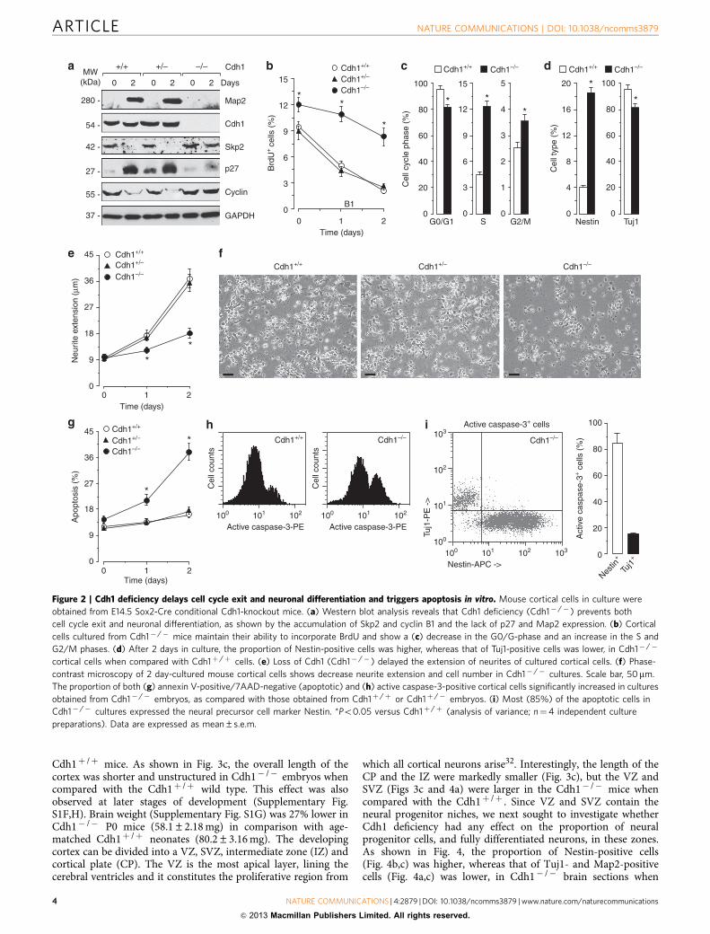
Complete inactivation of Cdh1 leads to embryonic lethality atmid-gestation (E10.5) caused by specific aberrations in theplacenta owing to the essential role of this protein inendoreduplication25,28. These defects were rescued by using aSox2-Cre transgenic mouse strain that specifically expresses theCre recombinase in most cells in the developing embryo but notin the placenta29. To confirm the essential role of Cdh1 inneuronal differentiation and survival, we next characterized thedifferentiation process of cortical cells in culture obtained fromE14.5 mice with a Sox2-Cre-mediated Cdh1 deficiency25. Asshown in Fig. 2a, Cdh1 was present at 0–2 days in culture incortical cells obtained from wild type (Cdh1þ /þ ) and Cdh1heterozygous (Cdh1þ /� ) mice, but it was absent in cellsobtained from Cdh1� /� mice. Interestingly, an increase in theelectrophoretic mobility of Cdh1 was observed from day 1 to 2 inculture both in Cdh1þ /þ and Cdh1þ /� cells, similar to results
ARTICLE NATURE COMMUNICATIONS | DOI: 10.1038/ncomms3879
2 NATURE COMMUNICATIONS | 4:2879 | DOI: 10.1038/ncomms3879 | www.nature.com/naturecommunications
& 2013 Macmillan Publishers Limited. All rights reserved.

shown in Fig. 1a. Map2, which appeared at day 2 in culture inCdh1þ /þ and Cdh1þ /� cells, was not present in Cdh1� /�
cells. Skp2 and cyclin B1 proteins disappeared at day 2 in cultureboth in Cdh1þ /þ and Cdh1þ /� cells, but not in Cdh1� /�
cells. Moreover, p27 accumulated from day 1 to 2 in culture bothin Cdh1þ /þ and Cdh1þ /� cells, but not in Cdh1� /� cells(Fig. 2a). Accordingly, cortical cells cultured from Cdh1� /�
mice maintained their ability to incorporate BrdU up to day 2(Fig. 2b), when they showed a decrease in the G0/G1 phase andan increase in the S and G2/M phases (Fig. 2c). This indicateshortening of the G1 phase and enlargement of the S phase inCdh1-depleted cultured cortical cells, as previously described forhuman cell lines and mouse embryonic fibroblasts deficient inCdh1 (refs 25,30,31). It should be noticed that, on day 2 inculture, the proportion of Nestin-positive cells was higher,whereas that of Tuj1-positive cells was lower, in Cdh1� /�
cortical cells when compared with Cdh1þ /þ cells (Fig. 2d). Inaddition, loss of Cdh1 significantly delayed the extension ofneurites (Fig. 2e,f). The proportion of annexin V-positive/7-Aminoactinomycin D (7-AAD)-negative (apoptotic; Fig. 2g) and
active caspase-3-positive (Fig. 2h) cortical cells significantlyincreased in Cdh1� /� cultures, but not in Cdh1þ /þ orCdh1þ /� cultures. Moreover, most of the active caspase-3-positive cells in Cdh1� /� cultures expressed the neuralprecursor cell marker Nestin, whereas only 15% expressed theneuronal marker Tuj1 (Fig. 2i). These results confirm that Cdh1is essential for mouse cortical progenitor cells to acquire a fullydifferentiated phenotype and to survive in culture.
Loss of Cdh1 triggers replicative stress and apoptotic death. Toassess whether Cdh1 was involved in cell cycle exit and neuronaldifferentiation in vivo, we next analysed the brain cortex inCdh1� /� E14.5 embryos. Lack of Cdh1 increased mitotic cyclinB1 expression in the cerebral cortex (Fig. 3a). Cdh1 deficiency didnot significantly alter body weight (Fig. 3a) but it decreased (20%)the brain weight (Fig. 3b) in comparison with Cdh1þ /þ . How-ever, the brain-to-body-weight ratio (Supplementary Fig. S2A)was very similar in heterozygous Cdh1þ /� (bearing both the Cretransgene and the floxed allele) when compared with control
Cdh1Cdh1-ACdh1-P
Control
0
9
18
27
36
45
210
#
#
Cdh1Cdh1-ACdh1-P
Control
0
10
20
30
40
50
#
#
210
#N
eurit
e ex
tens
ion
inG
FP
+ c
ells
(%
)
Cdh1Cdh1-ACdh1-P
Control
210
#
#
#
0
3
6
9
12
15
0
10
20
30
40
50
Neu
rite
exte
nsio
n (μ
m)
Control
210Time (days)
**Cdk1
0 Days1 2 4 6
H1
10 2
IP : α Cdh1WB :days
Cdh1
PSer
10 2
Untreated
210Time (days)
*
*
ControlOkadaic acid
0
9
18
27
36
45
34 -
31 -
54 -
54 -
Apo
ptos
is (
%)
Brd
U+
GF
P+ c
ells
(%
)
Apo
ptot
ic G
FP
+ c
ells
(%
)
Time after transfection(days)
Time after transfection(days)
Time after transfection(days)
Okadaic acidMW
(kDa)
MW(kDa)
Cdk1 activityOkadaic acid
Map2
Cdh1
Skp2
p27
GAPDH
0 Days1 2 4 6 9
37 -
27 -
42 -
54 -
280 -0 20 60 min
10 2 Days
0 20 60 0 20 60
6045301500
20
40
60
80
Time (min)
0 day1 day2 days
55 -
MW(kDa)
MW(kDa)
AP
C/C
act
ivity
(a.
u.)
Ub-
Cyc
lin B
1
Figure 1 | Neuronal differentiation and survival is associated to Cdh1 dephosphorylation and APC/C-Cdh1 activation. Mouse cortical cells in
culture differentiate to a neuronal phenotype, as showed by western blotting (a). (b) The activity of APC/C-Cdh1 increased during the first 2 days in
culture, (c) whereas the activity of cyclin B1-target, Cdk1, rapidly disappeared along the differentiation process. (d) Cdh1 is dephosphorylated as from
day 1 in culture, as evidenced by anti-phosphoserine immunoblotting of Cdh1 immunoprecipitates. This effect was prevented by incubation of the cells with
the PP2A inhibitor, okadaic acid. Treatment of mouse cortical cells in culture with okadaic acid (e) abolishes the time-dependent increase in neurite
extension and (f) triggers apoptotic cell death. At 4 h after plating, cortical cells were transfected with pIRES2-EGFP mammalian expression vector
co-expressing the enhanced GFP and either the wild type (Cdh1), phospho-mimetic (Cdh1-P) or non-phosphorylatable (Cdh1-A) forms of Cdh1. Control
cells were transfected with empty vector (pIRES2-EGFP). Analysis was performed in transfected (identified by GFPþ fluorescence) cells (day 0: 4–6 h
after transfection). Both (g) BrdU incorporation and (h) inhibition of neurite extension are delayed by Cdh1-P but accelerated by Cdh1-A. (I) Cdh1-P, but
not by Cdh1-A, induces apoptotic cell death. *Po0.05 versus control. #Po0.05 versus Cdh1 (analysis of variance; n¼4 independent culture preparations).
Data are expressed as mean±s.e.m.
NATURE COMMUNICATIONS | DOI: 10.1038/ncomms3879 ARTICLE
NATURE COMMUNICATIONS | 4:2879 | DOI: 10.1038/ncomms3879 | www.nature.com/naturecommunications 3
& 2013 Macmillan Publishers Limited. All rights reserved.
Cdh1þ /þ mice. As shown in Fig. 3c, the overall length of thecortex was shorter and unstructured in Cdh1� /� embryos whencompared with the Cdh1þ /þ wild type. This effect was alsoobserved at later stages of development (Supplementary Fig.S1F,H). Brain weight (Supplementary Fig. S1G) was 27% lower inCdh1� /� P0 mice (58.1±2.18 mg) in comparison with age-matched Cdh1þ /þ neonates (80.2±3.16 mg). The developingcortex can be divided into a VZ, SVZ, intermediate zone (IZ) andcortical plate (CP). The VZ is the most apical layer, lining thecerebral ventricles and it constitutes the proliferative region from
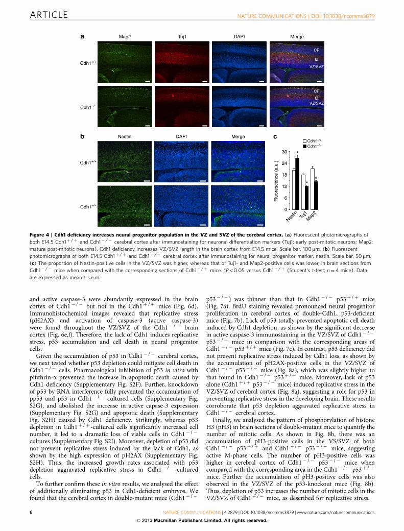
which all cortical neurons arise32. Interestingly, the length of theCP and the IZ were markedly smaller (Fig. 3c), but the VZ andSVZ (Figs 3c and 4a) were larger in the Cdh1� /� mice whencompared with the Cdh1þ /þ . Since VZ and SVZ contain theneural progenitor niches, we next sought to investigate whetherCdh1 deficiency had any effect on the proportion of neuralprogenitor cells, and fully differentiated neurons, in these zones.As shown in Fig. 4, the proportion of Nestin-positive cells(Fig. 4b,c) was higher, whereas that of Tuj1- and Map2-positivecells (Fig. 4a,c) was lower, in Cdh1� /� brain sections when
Map2
Cdh1
Skp2
+/–+/+ –/–
20 20 20 Days
GAPDH
Cdh1
p27
55 -
27 -
42 -
54 -
280 -
37 -
*
0
20
40
60
80
100
Tuj1
*
0
4
8
16
20
Cel
l typ
e (%
)
12
Nestin
Cdh1+/+ Cdh1–/–
Nestin-APC ->
Tuj1
-PE
->
Cdh1+/+ Cdh1–/–
*
0
20
40
60
80
100
Cel
l cyc
le p
hase
(%
)
S
*
0
3
6
9
12
15
0
1
2
3
4
5
*
0
9
18
27
36
45
Neu
rite
exte
nsio
n (μ
m)
210Time (days)
Cdh1+/+
Cdh1+/+Cdh1+/–Cdh1+/–
Cdh1–/–Cdh1–/–
**
210Time (days)
0
3
6
9
12
15
*
Cdh1+/+
Cdh1+/–
Cdh1–/–
**
CyclinB1
Cel
l cou
nts
Cdh1–/– Cdh1–/–
0
20
40
60
80
100
G2/MG0/G1
Brd
U+
cells
(%
)
MW(kDa)
Apo
ptos
is (
%)
Active caspase-3-PE
210Time (days)
0
9
18
27
36
45 Cdh1+/+
Cdh1+/–
Cdh1–/–*
* Cel
l cou
nts
Cdh1+/+
Active caspase-3-PE100 101 102 100 101 102
100
100
101
101 102 103
102
103
Act
ive
casp
ase-
3+ c
ells
(%
)
Active caspase-3+ cells
Tuj1+
Nestin
+
Figure 2 | Cdh1 deficiency delays cell cycle exit and neuronal differentiation and triggers apoptosis in vitro. Mouse cortical cells in culture were
obtained from E14.5 Sox2-Cre conditional Cdh1-knockout mice. (a) Western blot analysis reveals that Cdh1 deficiency (Cdh1� /� ) prevents both
cell cycle exit and neuronal differentiation, as shown by the accumulation of Skp2 and cyclin B1 and the lack of p27 and Map2 expression. (b) Cortical
cells cultured from Cdh1� /� mice maintain their ability to incorporate BrdU and show a (c) decrease in the G0/G-phase and an increase in the S and
G2/M phases. (d) After 2 days in culture, the proportion of Nestin-positive cells was higher, whereas that of Tuj1-positive cells was lower, in Cdh1� /�
cortical cells when compared with Cdh1þ /þ cells. (e) Loss of Cdh1 (Cdh1� /� ) delayed the extension of neurites of cultured cortical cells. (f) Phase-
contrast microscopy of 2 day-cultured mouse cortical cells shows decrease neurite extension and cell number in Cdh1� /� cultures. Scale bar, 50mm.
The proportion of both (g) annexin V-positive/7AAD-negative (apoptotic) and (h) active caspase-3-positive cortical cells significantly increased in cultures
obtained from Cdh1� /� embryos, as compared with those obtained from Cdh1þ /þ or Cdh1þ /� embryos. (i) Most (85%) of the apoptotic cells in
Cdh1� /� cultures expressed the neural precursor cell marker Nestin. *Po0.05 versus Cdh1þ /þ (analysis of variance; n¼4 independent culture
preparations). Data are expressed as mean±s.e.m.
ARTICLE NATURE COMMUNICATIONS | DOI: 10.1038/ncomms3879
4 NATURE COMMUNICATIONS | 4:2879 | DOI: 10.1038/ncomms3879 | www.nature.com/naturecommunications
& 2013 Macmillan Publishers Limited. All rights reserved.

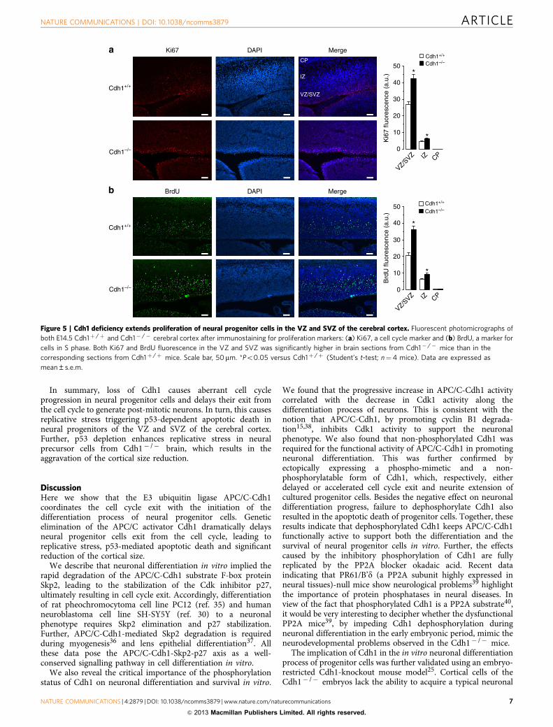
compared with sections from Cdh1þ /þ mice, suggestingincreased proliferation of neural progenitor cells and decreasedneurogenesis in Cdh1-deficient mice. To test this hypothesisfurther, we analysed the proportion of proliferating cells in thecerebral cortex. As shown in Fig. 5, both Ki67 (Fig. 5a) and BrdU(Fig. 5b) fluorescence was dramatically higher in the VZ/SVZ inCdh1� /� mice when compared with Cdh1þ /þ mice, indicatingincreased DNA replication in the mutant brains. It should benoted that the proportion of Tuj1-, Nestin- (Supplementary Fig.S2B) and BrdU- (Supplementary Fig. S2C) positive cells in brainsections from Cdh1þ /� mice were similar to that found inCdh1þ /þ mice, ruling out the possible effect of Cre expressionon neural progenitor differentiation and proliferation. Together,these results indicate that the lack of Cdh1 extends S-phase lengthof neural progenitor cells of the VZ/SVZ of the cerebral cortex atthe expense of an overall reduction in newly differentiatedneurons.
Our data therefore reveal that the loss of APC/C-Cdh1 activityprevents neural progenitor cells from exiting the cell cycle in vivo,thus confirming the conclusions drawn from the brain corticalcultured cells in vitro (Fig. 2b,c). Considering that Cdh1deficiency causes apoptotic death of cultured neural progenitorcells (Fig. 2f–i), we next studied whether this effect occurredin vivo. As depicted in Fig. 6a, Cdh1 loss induced apoptotic celldeath as shown by the dramatic increase in the proportion ofactive caspase-3-positive cells in the VZ/SVZ. To further confirmthe progenitor cell nature of the dying cells in the Cdh1� /�
brain, we next looked for Tbr2—an intermediate progenitor cellmarker—, Pax6—a neuroepithelial and radial glia cell marker—and active caspase-3 immunostaining in serial sections from the
same brain block. As shown in Fig. 6b, active caspase-3 wasexpressed in both Pax6þ (VZ) and Tbr2þ (basal VZ and SVZ)in the brain cortex. Moreover, neural precursors (Sox2þ /Nestinþ cells; Fig. 6c), but not newly committed neurons(Tuj1þ cells; Supplementary Fig. S2D), were found to undergoapoptotic cell death. Further, apoptosis was also detected byterminal deoxynucleotidyl transferase dUTP nick end-labellingassay in both Pax6þ and Tbr2þ progenitor cells (SupplementaryFig. S2E). Together, these data confirm that Cdh1 is essential forthe survival of mouse cortical progenitor cells.
Next, the underlying molecular mechanism involved in Cdh1loss-induced apoptosis of neural precursor cells was studied. It isknown that Cdh1 depletion results in shorter G1 and prematureentry into the S phase, that is, when the replicative machinery isnot yet ready25,31,33. This triggers a prolonged, unscheduledS-phase progression that results in replicative stress and DNAdamage responses25,31,33, leading to activation of the p53 pathwayand/or cell death31,33. The increase in DNA replication (Figs 2b,cand 5b) and the induction of apoptosis (Figs 2g,h and 6a–c) inCdh1-null neural precursor cells prompted us to study whetherthe lack of Cdh1 activated the DNA damage response pathway.As observed in Supplementary Fig. S2F, Cdh1� /� cortical cellsaccumulated replicative stress as shown by the high expression ofphosphorylated H2AX, a replicative stress marker, after 2 days inculture. Moreover, in good agreement with the fact thatreplicative stress is coordinated by the ataxia telangiectasia andRad3-related protein (Atr)-checkpoint kinase1 (Chk1) pathway34,loss of Cdh1 increased the phosphorylation of the ATR targetChk1 (pChk1), as well as both total (p53) and phosphorylatedforms (pp53) of p53, in cultured cells. Further, pH2AX, pp53, p53
Cdh1
Cyclin B1
GAPDH37 -
55 -
54 -
CP
IZ
SVZ
VZ
*
0
100
200
300
400
500
Cer
ebra
l cor
tex
leng
th (
μm)
*
0
8
16
24
32
40
0
60
120
180
240
300
*
0
4
8
12
16
20
Bra
in/b
ody
wei
ght (
%)
Bod
y w
eigh
t (m
g)
Cdh1+/
+
Cdh1–/
–
Cdh1+/
+
Cdh1–/
–
Cdh1+/
+
Cdh1–/
–
Cdh1+/
+
Cdh1–/
–
Bra
in w
eigh
t (m
g)
Cdh1+/+ Cdh1–/–Cdh1+/+
Cdh1+/+
Cdh1–/–
Cdh1–/–MW(kDa)
Cdh1–/–
Cdh1–/–
Cdh1+/+
Cdh1+/+
Figure 3 | Cdh1 deficiency reduces brain size and cerebral cortex length in vivo. (a) Cdh1 loss did not modify body size but (b) significantly decreased
brain weight in E14.5 mouse embryos. Scale bar, 100mm. (c) The overall length of the cortex was shorter and unstructured in Cdh1� /� mice when
compared with that of Cdh1þ /þ mice. The length of the intermediate zone (IZ) was smaller, but the ventricular (VZ) and subventricular (SVZ) zones
were larger in the Cdh1� /� mice when compared with the Cdh1þ /þ controls. Scale bar, 100 mm. *Po0.05 versus Cdh1þ /þ (Student’s t-test; n¼ 6
mice). Data are expressed as mean±s.e.m.
NATURE COMMUNICATIONS | DOI: 10.1038/ncomms3879 ARTICLE
NATURE COMMUNICATIONS | 4:2879 | DOI: 10.1038/ncomms3879 | www.nature.com/naturecommunications 5
& 2013 Macmillan Publishers Limited. All rights reserved.
and active caspase-3 were abundantly expressed in the braincortex of Cdh1� /� but not in the Cdh1þ /þ mice (Fig. 6d).Immunohistochemical images revealed that replicative stress(pH2AX) and activation of caspase-3 (active caspase-3)were found throughout the VZ/SVZ of the Cdh1� /� braincortex (Fig. 6e,f). Therefore, the lack of Cdh1 induces replicativestress, p53 accumulation and cell death in neural progenitorcells.
Given the accumulation of p53 in Cdh1� /� cerebral cortex,we next tested whether p53 depletion could mitigate cell death inCdh1� /� cells. Pharmacological inhibition of p53 in vitro withpifithrin-a prevented the increase in apoptotic death caused byCdh1 deficiency (Supplementary Fig. S2F). Further, knockdownof p53 by RNA interference fully prevented the accumulation ofpp53 and p53 in Cdh1� /� -cultured cells (Supplementary Fig.S2G), and abolished the increase in active capase-3 expression(Supplementary Fig. S2G) and apoptotic death (SupplementaryFig. S2H) caused by Cdh1 deficiency. Strikingly, whereas p53depletion in Cdh1þ /þ -cultured cells significantly increased cellnumber, it led to a dramatic loss of viable cells in Cdh1� /�
cultures (Supplementary Fig. S2I). Moreover, depletion of p53 didnot prevent replicative stress induced by the lack of Cdh1, asshown by the high expression of pH2AX (Supplementary Fig.S2H). Thus, the increased growth rates associated with p53depletion aggravated replicative stress in Cdh1� /� -culturedcells.
To further confirm these in vitro results, we analysed the effectof additionally eliminating p53 in Cdh1-deficient embryos. Wefound that the cerebral cortex in double-mutant mice (Cdh1� /�
p53� /� ) was thinner than that in Cdh1� /� p53þ /þ mice(Fig. 7a). BrdU staining revealed pronounced neural progenitorproliferation in cerebral cortex of double-Cdh1, p53-deficientmice (Fig. 7b). Lack of p53 totally prevented apoptotic cell deathinduced by Cdh1 depletion, as shown by the significant decreasein active caspase-3 immunostaining in the VZ/SVZ of Cdh1� /�
p53� /� mice in comparison with the corresponding areas ofCdh1� /� p53þ /þ mice (Fig. 7c). In contrast, p53 deficiency didnot prevent replicative stress induced by Cdh1 loss, as shown bythe accumulation of pH2AX-positive cells in the VZ/SVZ ofCdh1� /� p53� /� mice (Fig. 8a), which was slightly higher tothat found in Cdh1� /� p53þ /þ mice. Moreover, lack of p53alone (Cdh1þ /þ p53� /� mice) induced replicative stress in theVZ/SVZ of cerebral cortex (Fig. 8a), suggesting a role for p53 inpreventing replicative stress in the developing brain. These resultscorroborate that p53 depletion aggravated replicative stress inCdh1� /� cerebral cortex.
Finally, we analysed the pattern of phosphorylation of histoneH3 (pH3) in brain sections of double-mutant mice to quantify thenumber of mitotic cells. As shown in Fig. 8b, there was anaccumulation of pH3-positive cells in the VS/SVZ of bothCdh1� /� p53þ /þ and Cdh1� /� p53� /� mice, suggestingactive M-phase cells. The number of pH3-positive cells washigher in cerebral cortex of Cdh1� /� p53� /� mice whencompared with the corresponding area in the Cdh1� /� p53þ /þ
mice. Further the accumulation of pH3-positive cells was alsoobserved in the VZ/SVZ of the p53-knockout mice (Fig. 8b).Thus, depletion of p53 increases the number of mitotic cells in theVZ/SVZ of Cdh1� /� mice, as described for replicative stress.
Nestin DAPI Merge
Map2
Cdh1+/+
Cdh1+/+
Cdh1–/–
Cdh1+/+
Cdh1–/–
Cdh1–/–
DAPI Merge
IZ
CP
VZ/SVZIZ
CP
*
*
0
6
12
18
24
30
Flu
ores
cenc
e (a
.u.)
*
Tuj1
VZ/SVZ
Nestin
Tuj1
Map
2
Figure 4 | Cdh1 deficiency increases neural progenitor population in the VZ and SVZ of the cerebral cortex. (a) Fluorescent photomicrographs of
both E14.5 Cdh1þ /þ and Cdh1� /� cerebral cortex after immunostaining for neuronal differentiation markers (Tuj1: early post-mitotic neurons; Map2:
mature post-mitotic neurons). Cdh1 deficiency increases VZ/SVZ length in the brain cortex from E14.5 mice. Scale bar, 100mm. (b) Fluorescent
photomicrographs of both E14.5 Cdh1þ /þ and Cdh1� /� cerebral cortex after immunostaining for neural progenitor marker, nestin. Scale bar, 50mm.
(c) The proportion of Nestin-positive cells in the VZ/SVZ was higher, whereas that of Tuj1- and Map2-positive cells was lower, in brain sections from
Cdh1� /� mice when compared with the corresponding sections of Cdh1þ /þ mice. *Po0.05 versus Cdh1þ /þ (Student’s t-test; n¼4 mice). Data
are expressed as mean±s.e.m.
ARTICLE NATURE COMMUNICATIONS | DOI: 10.1038/ncomms3879
6 NATURE COMMUNICATIONS | 4:2879 | DOI: 10.1038/ncomms3879 | www.nature.com/naturecommunications
& 2013 Macmillan Publishers Limited. All rights reserved.
In summary, loss of Cdh1 causes aberrant cell cycleprogression in neural progenitor cells and delays their exit fromthe cell cycle to generate post-mitotic neurons. In turn, this causesreplicative stress triggering p53-dependent apoptotic death inneural progenitors of the VZ and SVZ of the cerebral cortex.Further, p53 depletion enhances replicative stress in neuralprecursor cells from Cdh1� /� brain, which results in theaggravation of the cortical size reduction.
DiscussionHere we show that the E3 ubiquitin ligase APC/C-Cdh1coordinates the cell cycle exit with the initiation of thedifferentiation process of neural progenitor cells. Geneticelimination of the APC/C activator Cdh1 dramatically delaysneural progenitor cells exit from the cell cycle, leading toreplicative stress, p53-mediated apoptotic death and significantreduction of the cortical size.
We describe that neuronal differentiation in vitro implied therapid degradation of the APC/C-Cdh1 substrate F-box proteinSkp2, leading to the stabilization of the Cdk inhibitor p27,ultimately resulting in cell cycle exit. Accordingly, differentiationof rat pheochromocytoma cell line PC12 (ref. 35) and humanneuroblastoma cell line SH-SY5Y (ref. 30) to a neuronalphenotype requires Skp2 elimination and p27 stabilization.Further, APC/C-Cdh1-mediated Skp2 degradation is requiredduring myogenesis36 and lens epithelial differentiation37. Allthese data pose the APC/C-Cdh1-Skp2-p27 axis as a well-conserved signalling pathway in cell differentiation in vitro.
We also reveal the critical importance of the phosphorylationstatus of Cdh1 on neuronal differentiation and survival in vitro.
We found that the progressive increase in APC/C-Cdh1 activitycorrelated with the decrease in Cdk1 activity along thedifferentiation process of neurons. This is consistent with thenotion that APC/C-Cdh1, by promoting cyclin B1 degrada-tion15,38, inhibits Cdk1 activity to support the neuronalphenotype. We also found that non-phosphorylated Cdh1 wasrequired for the functional activity of APC/C-Cdh1 in promotingneuronal differentiation. This was further confirmed byectopically expressing a phospho-mimetic and a non-phosphorylatable form of Cdh1, which, respectively, eitherdelayed or accelerated cell cycle exit and neurite extension ofcultured progenitor cells. Besides the negative effect on neuronaldifferentiation progress, failure to dephosphorylate Cdh1 alsoresulted in the apoptotic death of progenitor cells. Together, theseresults indicate that dephosphorylated Cdh1 keeps APC/C-Cdh1functionally active to support both the differentiation and thesurvival of neural progenitor cells in vitro. Further, the effectscaused by the inhibitory phosphorylation of Cdh1 are fullyreplicated by the PP2A blocker okadaic acid. Recent dataindicating that PR61/B’d (a PP2A subunit highly expressed inneural tissues)-null mice show neurological problems39 highlightthe importance of protein phosphatases in neural diseases. Inview of the fact that phosphorylated Cdh1 is a PP2A substrate40,it would be very interesting to decipher whether the dysfunctionalPP2A mice39, by impeding Cdh1 dephosphorylation duringneuronal differentiation in the early embryonic period, mimic theneurodevelopmental problems observed in the Cdh1� /� mice.
The implication of Cdh1 in the in vitro neuronal differentiationprocess of progenitor cells was further validated using an embryo-restricted Cdh1-knockout mouse model25. Cortical cells of theCdh1� /� embryos lack the ability to acquire a typical neuronal
Ki67
*
*
0
10
20
30
40
50
*
0
10
20
30
40
50
*
Cdh1+/+
Cdh1–/–
Cdh1+/+
Cdh1–/–
Cdh1+/+
Cdh1–/–
Cdh1+/+
Cdh1–/–
DAPI Merge
BrdU DAPI Merge
IZ
CP
VZ/SVZ
Ki6
7 flu
ores
cenc
e (a
.u.)
Brd
U fl
uore
scen
ce (
a.u.
)
VZ/SVZ IZ CP
VZ/SVZ IZ CP
Figure 5 | Cdh1 deficiency extends proliferation of neural progenitor cells in the VZ and SVZ of the cerebral cortex. Fluorescent photomicrographs of
both E14.5 Cdh1þ /þ and Cdh1� /� cerebral cortex after immunostaining for proliferation markers: (a) Ki67, a cell cycle marker and (b) BrdU, a marker for
cells in S phase. Both Ki67 and BrdU fluorescence in the VZ and SVZ was significantly higher in brain sections from Cdh1� /� mice than in the
corresponding sections from Cdh1þ /þ mice. Scale bar, 50mm. *Po0.05 versus Cdh1þ /þ (Student’s t-test; n¼4 mice). Data are expressed as
mean±s.e.m.
NATURE COMMUNICATIONS | DOI: 10.1038/ncomms3879 ARTICLE
NATURE COMMUNICATIONS | 4:2879 | DOI: 10.1038/ncomms3879 | www.nature.com/naturecommunications 7
& 2013 Macmillan Publishers Limited. All rights reserved.

phenotype in culture, as assessed by Map2 protein abundance,BrdU incorporation, cell cycle protein expression and neuriteextension. In accordance with previous results25,30,31, we alsofound that Cdh1 loss delays cell cycle exit, shortens G1 phase andprolongs S and G2/M phases. Moreover, we show that cerebralcortex of E14.5 Cdh1� /� mice showed a reduction of newlydifferentiated neurons at the expense of increased DNAreplication of progenitor cells. This is in good agreement withprevious results showing fewer cortical neurons and reducedcortical size as a consequence of experimental prolongation of cellcycle of neural precursors10,41. In our Cdh1� /� mice, the
lengths of the VZ/SVZ are increased, whereas those of theIZ and CP layers of the cortex are reduced, leading to reducedcerebral cortical size, which is a phenotype compatible withMCPH42.
It is interesting to note that genetic defects in DNA damageresponse lead to human MCPH syndromes, such as ATR Seckelsyndrome, caused by mutations in the gene encoding the ATRprotein43, and primary MCPH caused by mutations in MCPH1(ref. 44), indicating that MCPH may be caused by alterations ofneuronal progenitors in response to DNA damage45,46. Wepreviously reported25 that Cdh1� /� cells show premature entry
Pax6
VZ
pH2AX
0
4
8
12
16
20
*
*
pH2AX
pp53
p53
GAPDH
ActiveCaspase-3
Cdh1+/+
Cdh1+/+
Cdh1+/+Cdh1–/–
Cdh1–/–
Cdh1–/–
MW(kDa)
37 -
17 -
53 -
53 -
15 -
0
4
8
12
16
Tbr2
SVZCdh1+/+
Cdh1–/–
Cdh1–/– Cdh1–/–
*
*
Cdh1+/+
Cdh1–/–
Cdh1+/+
Cdh1–/–
0
4
8
12
16
20
Act
ive
casp
ase-
3+ c
ells
(num
ber
per
field
)VZ/S
VZ IZ CP
VZ/SVZ IZ CP
SVZVZ
Active caspase-3
Active caspase-3 + Sox2
Active caspase-3 + Nestin
Act
ive
casp
ase-
3+ c
ells
(num
ber
per
field
)
Active caspase-3
Active caspase-3 Active caspase-3pHA2X pHA2X
pH2A
X+ c
ells
(num
ber
per
field
)
Figure 6 | Loss of Cdh1 triggers replicative stress and apoptotic cell death in the VZ and SVZ of the cerebral cortex. (a) Fluorescent photomicrographs
of both E14.5 Cdh1þ /þ and Cdh1� /� cerebral cortex immunostained with the apoptotic marker active caspase 3 reveal that Cdh1 deficiency triggers
apoptotic cell death in the VZ/SVZ of the brain cortex. Scale bar, 50mm. (b) Active caspase-3 expression was located in both Pax6þ (VZ) and Tbr2þ
(basal VZ and SVZ) zones. Scale bar, 50mm. (c) Double labelling revealed that neural precursor cells (Sox2þ/Nestinþ cells) underwent apoptotic
cell death. Scale bar, 20mm. (d) Western blotting shows that the brain cortex of Cdh1-deficient mice expressed pH2AX and both total (p53) and
phosphorylated forms (pp53) of p53, which were absent in Cdh1þ /þ mice. (e) Immunofluorescent images reveal that replicative stress (pH2AX staining)
was found throughout the VZ/SVZ of the Cdh1� /� brain cortex. Scale bar, 50mm. (f) Replicative stress and activation of capase-3 in the VZ/SVZ of the
Cdh1� /� mice were further confirmed by pH2AX and active caspase-3 immunostaining in histological brain sections. Scale bar, 20 mm. Field: 0.25 mm2.
*Po0.05 versus Cdh1þ /þ (Student’s t-test; n¼4 mice). Data are expressed as mean±s.e.m.
ARTICLE NATURE COMMUNICATIONS | DOI: 10.1038/ncomms3879
8 NATURE COMMUNICATIONS | 4:2879 | DOI: 10.1038/ncomms3879 | www.nature.com/naturecommunications
& 2013 Macmillan Publishers Limited. All rights reserved.

into S phase and replicative defects. Conversely, results fromother laboratories showed that Cdh1 depletion initiates aberrantDNA replication, triggering a p53-mediated response to DNAdamage in cultured human cells33,31. Here we furtherdemonstrate that loss of Cdh1 in vivo triggers cyclin B1accumulation and replicative stress in the cerebral cortex. Inthis sense, high levels of cyclin B1-CDK1 activity prevent theassembly of origins of replication and the efficient timing of Sphase15, which could lead to DNA replicative stress. In turn, thiswould activate a p53-mediated apoptotic death-signallingpathway in neural progenitor cells in the VZ/SVZ resulting inreduced neurogenesis and MCPH in Cdh1-deficient mice.Interestingly, knockdown of p53 in vitro, and genetic deletionof p53 in vivo, abolished apoptotic cell death. On the contrary,p53 loss increases neural progenitors loss in vitro and aggravatesMCPH caused by Cdh1 deficiency in vivo. Further, depletionof p53 enhances Cdh1� /� -mediated neural progenitorproliferation, as revealed by BrdU and pH3 staining, whichtriggers the increase in the amount of replicative stress in thecerebral cortex. Taken together, these findings indicate that p53mediates the apoptotic death in the brain but aggravatesreplication stress and MCPH phenotypes of the Cdh1� /�
mice. In this context, mutations that promote cell proliferationfurther potentiate replicative stress that, if sufficiently high,compromises cell viability47,48, as we observed in the Cdh1� /�
mice. Recently, Murga et al.47 described that p53 loss increasesreplicative stress leading to aggravation of neurodevelopmentaldamage and MCPH phenotypes in a murine model of the human
ATR Seckel syndrome characterized by a severe deficiency inATR. In this context, the increased amount of replicative stressgenerated by p53 loss in the ATR-deficient mice also increases theamount of cell eliminated by apoptosis during embryogenesis47.Similarly, loss of Chk1 leads to p53-independent cell death inzebrafish49. We thus speculate that a similar pathway might beoccurring in the Cdh1� /� p53� /� -mutant mice. Alternatively,the accumulated pool of neural precursor cells in the VZ/SVZ ofCdh1� /� cerebral cortex could be suggestive of a migrationdefect. Recently, Qin and Zhang50 have described thatdownregulation of the APC/C-Cdh1 target KLF4 (ref. 51) isessential for normal neurogenesis. Similarly, neural progenitorcells with high expression of KLF4 fail to migrate and developinto neurons50. Future studies would be needed to fullyunderstand the role of Cdh1 on neuronal migration duringbrain development.
In conclusion, here we describe that loss of Cdh1 reduces thelength of the G1 phase and increases S-phase duration inneural precursor cells, which generates replicative stress,triggering apoptotic death of neural precursor cells.Consequently, neurogenesis is severely affected leading to theshortening of the cerebral cortex and relative brain size,compatible with microencephaly (Supplementary Fig. S3). Inaddition, we reveal that p53 wholly account for the apoptotic celldeath of neural progenitors. These results demonstrate thatAPC/C-Cdh1 coordinates cortical neurogenesis and size, thusposing Cdh1 in the molecular pathogenesis of congenitalneurodevelopmental disorders, such as MCPH.
BrdU
0
10
20
30
40
50
* *0
4
8
12
16
20
Cdh1–/– p53+/+
Cdh1–/– p53+/+
Cdh1–/– p53+/+
Cdh1–/– p53–/–
Cdh1–/– p53–/–
Cdh1–/– p53+/+
Cdh1–/– p53–/–
Cdh1–/– p53–/–
Cdh1–/– p53+/+
Cdh1–/– p53–/–
Active caspase-3
Act
ive
casp
ase-
3+ c
ells
(num
ber
per
field
)
Brd
U fl
uore
scen
ce (
a.u.
)
VZ/SVZ IZ CP
VZ/SVZ IZ
Figure 7 | Expression of p53 mediates apoptotic cell death of neural progenitor cells in the Cdh1� /� cerebral cortex. (a) Histological staining of brain
sections from E14.5 Cdh1� /� p53þ /þ and Cdh1� /� p53� /� mice. Scale bar, 200mm. (b) Fluorescent photomicrographs of E14.5 Cdh1� /� p53þ /þ
and Cdh1� /� p53� /� cerebral cortex after immunostaining for a marker of S-phase cells, BrdU. Scale bar, 50mm. (c) Additional depletion of p53
prevents apoptosis in Cdh1-deficient embryonic brain, as revealed by active caspase 3 immunostaining. Scale bar, 50mm (left panel), 7mm (right panel).
Field: 0.25 mm2. *Po0.05 versus Cdh1� /� p53þ /þ (Student’s t-test; n¼4 mice). Data are expressed as mean±s.e.m.
NATURE COMMUNICATIONS | DOI: 10.1038/ncomms3879 ARTICLE
NATURE COMMUNICATIONS | 4:2879 | DOI: 10.1038/ncomms3879 | www.nature.com/naturecommunications 9
& 2013 Macmillan Publishers Limited. All rights reserved.

MethodsSox2-Cre-mediated Cdh1 conditional knockout mice. The Cdh1lox/lox-targetedmouse model was generated by flanking exons 2 and 3 of the murine Fzr1 locuswith loxP sequences25. To obtain Cdh1-deficient mice (Cdh1� /D; Sox2-Creþ /T,referred as Cdh1� /� ), a transgenic strain expressing Cre in embryonic but not inextra-embryonic tissues (Sox2-Cre) was used29. We also utilized Cdh1þ /D; Sox2-Creþ /T (referred as Cdh1� /þ ) and Cdh1þ /lox; Sox2-Creþ /þ (referred asCdh1þ /þ ) mouse embryos. To generate embryos deficient for p53 and Cdh1, weset up crosses between 2–3-month-old Cdh1lox/lox; Sox2-Creþ /þ ; p53þ /� femalemice and 2–5 month Cdh1þ /� ; Sox2-Creþ /T; p53� /� male mice.
For BrdU incorporation in 14.5-day-old embryos, timed pregnant females wereadministered BrdU at 50 mg kg� 1 of body weight by intraperitoneal injection. Allanimals were maintained in a mixed 129/Sv (25%)�CD1 (25%)�C57BL/6J (50%)background. All animals used in this work were obtained from the AnimalExperimentation Unit of the University of Salamanca, in accordance with Spanishlegislation (RD 1201/2005) under licence from the Spanish Ministry of Science andInnovation. Mice were housed under standard conditions in the pathogen-freeanimal facility of the University of Salamanca (Salamanca, Spain) following theanimal care standards of the institution. Animal experiments were approved by theBioethics Committee of the University of Salamanca.
Cell culture treatments and transfections. Primary cultures of corticalneurons were prepared from E14.5 mouse embryo cortices. Cells were seeded at2.0� 105 cells cm� 2 in DMEM (Sigma, Madrid, Spain) supplemented with 10%(v/v) FCS (Roche Diagnostics, Heidelberg, Germany) and incubated at 37 �Cin a humidified 5% CO2-containing atmosphere. At 4 h after plating, the mediumwas replaced with Neurobasal medium (Invitrogen, Madrid, Spain) supplementedwith 2% B27 (Invitrogen) and glutamine 4 mM (Invitrogen). When indicated,medium contained the phosphatase inhibitor okadaic acid (10 nM)52 or exposedto the specific inhibitor of p53-mediated transcriptional activation, pifithrin-a(10 nM)53.
Cells were transfected 4 h after plating with plasmid DNA or short interferingRNA (siRNA) using Lipofectamine2000 (Invitrogen). hEmi1 was used to inhibitthe APC/C complex by co-transfecting cells with pCS2-hEmi1 (generously donatedby Dr Peter Jackson, Stanford University School of Medicine, Stanford, CA) pluspGFP (Clontech Laboratories Inc., Palo Alto, CA) to identify transfected (greenfluorescent protein; GFPþ cells) cells, and control transfections were carried outwith a truncated form of hEmi1 carrying a deletion of the zinc-binding region thatcauses loss of function (pCS2-Myc5-hEmi1DZBR, generously donated by Dr PeterJackson). Human full-length Cdh1 complementary DNA was a generous gift of DrJ Pines (Gurdon Institute, University of Cambridge, UK) and was used to obtain
the Ser-40, Thr-121 and Ser-163 triple alanine (Cdh1-A) or aspartate (Cdh1-P)Cdh1 mutants using the QuikChange XL site directed mutagenesis kit (Stratagene,La Jolla, CA, USA). Residues were mutated either to Ala (Cdh1-A; to blockphosphorylation) or to Asp (Cdh1-D; to mimic a phosphorylated status)54.Cells were transfected with the pIRES2-EGFP mammalian expression vector(Invitrogen) co-expressing the enhanced GFP and either the full-length wild typeor mutant forms of Cdh1 (ref. 54). Depletion of p53 was performed by siRNA(50-CCACUUGAUGGAGAGUAUUUU-30)55; siRNA against luciferase was usedfor control transfections38. For plasmids, the transfection efficiencies in neuronswere, approximately, 7%, as judged by the proportion of GFPþ neurons versustotal number of neurons (as evidenced by flow cytometry analysis); for siRNA, thetransfection efficiencies were 490%, as judged by the level of protein knockdownshown in the Supplementary Fig. S2F.
BrdU incorporation into DNA and cell cycle phase percentage were determinedby flow cytometry. This was achieved after 3 h of incubation with 10 mg ml� 1
BrdU using the APC BrdU Flow Kit (BD Biosciences), following themanufacturer’s instructions30.
When cell phenotype was studied, cells were fixed and permeabilized using theFix&Perm kit from Caltag (Invitrogen) and were stained with Nestin-APC andTuj1-PE (BD Biosciences), according to the protocol included in the technical datasheet. The percentage of cells expressing Nestin and Tuj1 were quantified by flowcytometry.
Western blotting. Cells were lysed in buffer containing 1% SDS, 2 mM EDTA,150 mM NaCl, 12,5 mM Na2HPO4 and 1% Triton X-100, supplemented withphosphatase inhibitors (1 mM Na3VO4 and 50 mM NaF) and protease inhibitors(100 mM phenylmethylsulfonyl fluoride, 50 mg ml� 1 anti-papain, 50 mg ml� 1
pepstatin, 50mg ml� 1 amastatin, 50 mg ml� 1 leupeptin, 50 mg ml� 1 bestatin and50 mg ml� 1 soybean trypsin inhibitor) and boiled for 5 min. Protein concentrationswere determined with the BCA Protein Assay kit (Thermo Fisher Scientific).Extracts were centrifuged at 13,000 g for 5 min at 4 �C. Aliquots of lysates (20–50 mgprotein) were subjected to SDS–polyacrylamide gel electrophoresis and blottedwith anti-Map2 (1:200)(ab32454, Abcam), anti-phosphoserine (1:1,000) (ab9332,Abcam), anti-Cdh1 (1/20) (AR38, a generous gift from Dr J Gannon, Clare HallLaboratories, Cancer Research UK), anti-Skp2 (1:200) (32-3,400, Zymed), anti-p27(1:1,000) (554069, BD Biosciences), anti-Cdk1 (1:250) (610037, BD Biosciences),anti-cyclin B1 (1:100) (554176, BD Biosciences), anti-p53 (1:100) (554157, BDBiosciences), anti-pp53 (1:500) (Ser15; 9286, Cell Signaling), anti-pH2AX (1:250)(Ser139, 05-6360, Upstate), anti-active caspase 3 (1:500) (Asp175, 9661 CellSignaling), anti-pChk1 (1:500) (Ser317, 2344 Cell Signaling) or anti-GAPDH(1:40,000) (glyceraldehyde-3-phosphate dehydrogenase; Sigma) overnight at
0
4
8
12
16
20
*
*
0
5
10
15
20
25pH3
pH2AX
pH3
Cdh1+/+ p53–/–Cdh1–/– p53–/–Cdh1–/– p53+/+Cdh1–/– p53+/+
Cdh1–/– p53–/–
Cdh1+/+ p53–/–
Cdh1–/– p53+/+
Cdh1–/– p53–/–
Cdh1+/+ p53–/–
Cdh1+/+ p53–/–Cdh1–/– p53–/–Cdh1–/– p53+/+
pH2A
X+ c
ells
(num
ber
per
field
)pH
3+ c
ells
(num
ber
per
field
)
VZ/SVZ IZ
VZ/SVZ IZ
Figure 8 | Replicative stress induced by Cdh1 loss in the VZ and SVZ of the cerebral cortex is p53 independent. (a) Fluorescent photomicrographs
of cerebral cortex from E14.5 Cdh1� /� p53þ /þ and Cdh1� /� p53� /� mice after immunostaining with pH2AX reveal replicative stress in the
VZ/SVZ. Scale bar, 20mm. (b) Fluorescent photomicrographs of cerebral cortex show that the number of pH3-positive cells in the VZ/SVZ of E14.5
Cdh1� /� p53þ /þ is further increased by codepletion of p53 (Cdh1� /� p53� /� ). Fluorescent photomicrographs reveal (a) replicative stress and
(b) increased pH3-positive cells in the VZ/SVZ of Cdh1þ /þ p53� /� mice. Scale bar, 20mm. Field: 0.25 mm2. *Po0.05 versus Cdh1� /� p53þ /þ
(analysis of variance; n¼4 mice). Data are expressed as mean±s.e.m.
ARTICLE NATURE COMMUNICATIONS | DOI: 10.1038/ncomms3879
10 NATURE COMMUNICATIONS | 4:2879 | DOI: 10.1038/ncomms3879 | www.nature.com/naturecommunications
& 2013 Macmillan Publishers Limited. All rights reserved.

4 �C. Signal detection was performed with an enhanced chemiluminescencedetection kit (Thermo Fisher Scientific). Full blots and gel scans are included inSupplementary Fig. S4.
Cdk1 kinase assay. Cells were lysed in ice-cold buffer containing 50 mM Tris (pH7.5), 150 mM NaCl, 10 mM EDTA, 2 mM EGTA, 1% NP-40, supplemented withthe phosphatase and protease inhibitors cited above. After clearing debris bycentrifugation, extracts (200 mg protein) were incubated with anti-Cdk1 (2 mg) for4 h at 4 �C, followed by the addition of 30 ml of protein A-agarose (GE HealthcareLife Sciences) for 2 h at 4 �C. Immunoprecipitates were washed four times in lysisbuffer and resuspended in kinase buffer (20 mM Tris pH 7.6, 20 mM MgCl2, 2mMMnCl2, 1 mM EDTA, 1 mM EGTA and 0.1 mM dithiothreitol) containing 20 mMATP, 10 mCi of [g-32P]ATP and histone H1 (50 mg ml� 1; Sigma). Samples weresubjected to SDS–polyacrylamide gel (12%) electrophoresis and transferred pro-teins were visualized by autoradiography or blotted with anti-Cdk1 (1:250)(610037, BD Biosciences).
APC/C ubiquitin ligase activity. Active APC/C was immunoprecipitated fromcell nuclear extracts using monoclonal anti-APC3 antibody (10mg) (610455, BDBiosciences) and immobilized on Dynabeads Protein A (Invitrogen). For ubiqui-tination assays, immunoprecipitates were incubated at 37 �C in 10ml of buffer(0.1 M KCl, 2.5 mM MgCl2, 2 mM ATP, 7.5 mg ubiquitin, 0.3 mM dithiothreitol,135 mM MG132, 1 mM ubiquitin aldehyde, 2.5 mM His-UbcH10 and 2.5 mMUbcH5a in 20 mM Tris–HCl, pH 7.5) containing 2.5 ml of APC/C beads and 1 ml of[35S]cyclin B1. Reactions were stopped at the indicated time points with SDSsample buffer, mixtures resolved by SDS–polyacrylamide gel electrophoresis andvisualized by phosphorimaging54.
Flow cytometric detection of apoptotic cell death. Neurons were carefullydetached from the plates using 1 mM EDTA (tetrasodium salt) in PBS (pH 7.4) andwere stained with annexin V-APC and 7-AAD in binding buffer (100 mM HEPES,140 mM NaCl, 2.5 mM CaCl2) to determine quantitatively the percentage ofapoptotic neurons by flow cytometry. Cells were stained with annexin V-APC and7-AAD in binding buffer (100 mM HEPES, 140 mM NaCl, 2.5 mM CaCl2),according to the manufacturer’s instructions, and 3� 105 cells were analysed, infour replicates per condition, on a FACScalibur flow cytometer (15 mW argon ionlaser tuned at 488 nm; CellQuest software, BD Biosciences). Transfected (identifiedby GFPþ fluorescence) cell population were analysed, and the annexin V-APC-stained cells that were 7AAD negative were considered to be apoptotic.
Flow cytometric detection of active caspase-3. Active caspase-3 was detectedusing the ApoActive3 Kit (Bachem, San Carlos, CA, USA), following the manu-facturer’s instructions. After detaching cells with 1 mM EDTA (tetrasodium salt)and centrifugation, cell pellets were fixed during 20 min, resuspended in PBSþ 2%BSA and incubated for 1 h with 1� rabbit anti-caspase 3. Cells were then incu-bated with 1:500 anti-rabbit Cy3 (Jackson ImmunoResearch) for 1 h. Between eachstep, cells were washed with either PBS (until labelling of samples) or PBSþ 1%BSA and resuspended in PBSþ 1% BSA before analysis by flow cytometry (tunedat 488 nm; CellQuest software, BD Biosciences)53. When cell phenotype wasstudied, cells were fixed and permeabilized using the Fix&Perm kit from Caltag(Invitrogen) and were stained with active caspase-3-fluorescein isothiocyanate(1:10) (560901), Nestin-APC (1:10) (560393) and Tuj1-PE (1:10) (560381) (BDBiosciences), according to the protocol included in the technical data sheet. Thepercentage of active caspase-3þ cells expressing Nestin and Tuj1 were quantifiedby flow cytometry.
Immunohistochemistry. Timed murine embryos were harvested from pregnantdams according to animal care standards of the University of Salamanca. Theembryo heads were fixed with 4% (w/v) paraformaldehyde, 0.2% (w/v) picric acidin 0.1 M phosphate buffer pH 7.4 (ref. 56) for 10 days at 4 �C. Brains were removedfrom the skull, post-fixed for an additional day and sagittally dissected out in twoparts. Brain blocks were rinsed successively for 10 min, 30 min and 2 h with 0.1 MPBS (pH 7.4), and immersed in sucrose/PBS with concentrations of 20 and 30%sequentially until they sank. After cryoprotection, tissues were embedded inOptimal cutting temperature compound (OCT) (catalogue #4583; Thermo FisherScientific) and frozen with liquid nitrogen. Finally, brains were sectioned in thesagittal plane on a cryostat at 20 mm and mounted onto Superfrost Plus slides(Thermo Fisher Scientific). Sagittal sections containing the entire cortex wereselected, rinsed twice in 0.1 M PBS and single- or double-labelled with thecorresponding antibodies diluted in blocking solution (0.2% Triton X-100 and 5%normal goat serum in 0.1 M PBS) for 72 h at 4 �C, followed by secondary antibodylabelling for 2 h at room temperature and 40 ,6-diamidino-2-phenylindole staining(0.5mg ml� 1 40 ,6-diamidino-2-phenylindole in PBS) for 10 min. After each step,sections were carefully rinsed three times for 10 min each in PBS. Sections weremounted with Fluoromount (Sigma) aqueous mounting medium and examined ona microscope (Nikon Inverted 13 microscope Eclipse Ti-E) equipped with apre-centred fibre illuminator (Nikon Intensilight C-HGFI) and Black and Whitecharge-coupled device digital camera (Hamamatsu ORCAER), or on a confocal
microscope (TCS SP2; Leica, Mannheim, Germany). For Nestin, Ki67 and activecaspase-3 staining, an antigen retrieval procedure was required57. For BrdUstaining, sections were pre-treated with DNAse (10mg per slide) at 37 �C for 1 hand washed three times in 0.1 M phosphate buffer for 5 min.
The primary antibodies used were the following: rabbit monoclonal anti-Ki67(1/200) (RM-9106, NeoMarkers), mouse monoclonal anti-BrdU (1:500)(RPN2012, Amersham), rabbit polyclonal anti-neuron-specific beta III Tubulin-neuronal marker (1:500) (Tuj 1, ab18207, Abcam), rabbit polyclonal anti-Map2-neuronal marker (1:500) (Map2, ab11267 Abcam), mouse monoclonal anti-nestin(1:500) (556309, BD Pharmingen), rabbit polyclonal anti-Pax6 (1:250) (PRB-278P,Covance Research Products Inc), rabbit polyclonal anti-Tbr2 (1:250) (ab23345,Abcam), mouse monoclonal anti-Sox2 (1:50) (97959, Abcam) and rabbitpolyclonal anti-active caspase-3 (1:50) (9661S, Asp175, Cell Signaling). Secondaryantibodies were Alexa 568-conjugated goat anti-rabbit (1:500), Alexa 488-conjugated goat anti-rabbit (1:500) and Alexa 594-conjugated goat anti-mouse(1:500) (Molecular Probes, Invitrogen), Cy2-conjugated affiniPure goat anti-mouse(1:500) and Cy3-conjugated affiniPure goat anti-rabbit (1:500) (Jackson Immu-noResearch). Occasionally, embryos were fixed in 10%-buffered formalin (Sigma)and embedded in paraffin wax. Sections of 3 or 5 mm thickness were stained withhaematoxylin and eosin for histological examination or processed for immuno-histochemical analysis using specific antibodies against active caspase-3 (1:50)(9661S, Asp175, Cell Signaling), pH2AX (1:250) (clonJBW301, Ser139, 16-193,Millipore) and pH3 (1:100) (06-570, Cell Signalling).
Terminal deoxynucleotidyl transferase dUTP nick end-labelling assay wasperformed following the manufacturer’s protocol (3333566, Roche Diagnostics).
Immunohistochemistry digital images (RGB images) were used for fluorescencequantification, using the NIH image-processing package ImageJ (ImageJ 1.43n;Java 1.6.0_26[64-bit]). Mean fluorescence values were measured in the whole-image area or in the VZ/SVZ, IZ or CP area defined in Fig. 4. The pixel intensitiesranged from 0 (black) to 255 (white). Fluorescence background was measured andsubtracted. Values are mean±s.e.m. from 20 measurements from 4 different brainsections and are expressed as arbitrary units (a.u.) of fluorescence.
Statistical analysis. Measurements from individual cultures were always per-formed in quadruplicate. The results are expressed as mean±s.e.m. values for fourdifferent culture preparations. For the in vivo experiments, we used four to sixanimals per condition. Statistical analysis of the results was performed by one-wayanalysis of variance, followed by the least significant difference multiple range testor by the Student’s t-test for comparisons between two groups of values. In allcases, Po0.05 was considered significant.
References1. Miyata, T., Kawaguchi, D., Kawaguchi, A. & Gotoh, Y. Mechanisms that
regulate the number of neurons during mouse neocortical development. Curr.Opin. Neurobiol. 20, 22–28 (2010).
2. Tang, B. L. Molecular genetic determinants of human brain size. Biochem.Biophys. Res. Commun. 345, 911–916 (2006).
3. Bystron, I., Blakemore, C. & Rakic, P. Development of the human cerebralcortex: Boulder Committee revisited. Nat. Rev. Neurosci. 9, 110–122 (2008).
4. Miyata, T. et al. Asymmetric production of surface-dividing and non-surface-dividing cortical progenitor cells. Development 131, 3133–3145 (2004).
5. Gotz, M. & Huttner, W. B. The cell biology of neurogenesis. Nat. Rev. Mol. CellBiol. 6, 777–788 (2005).
6. Noctor, S. C., Martinez-Cerdeno, V., Ivic, L. & Kriegstein, A. R. Corticalneurons arise in symmetric and asymmetric division zones and migratethrough specific phases. Nat. Neurosci. 7, 136–144 (2004).
7. Cremisi, F., Philpott, A. & Ohnuma, S. Cell cycle and cell fate interactions inneural development. Curr. Opin. Neurobiol. 13, 26–33 (2003).
8. Nguyen, L., Besson, A., Roberts, J. M. & Guillemot, F. Coupling cell cycle exit,neuronal differentiation and migration in cortical neurogenesis. Cell Cycle 5,2314–2318 (2006).
9. Dehay, C. & Kennedy, H. Cell-cycle control and cortical development. Nat. Rev.Neurosci. 8, 438–450 (2007).
10. Hindley, C. & Philpott, A. Co-ordination of cell cycle and differentiation in thedeveloping nervous system. Biochem J. 444, 375–382 (2012).
11. Lange, C. & Calegari, F. Cdks and cyclins link G1 length and differentiation ofembryonic, neural and hematopoietic stem cells. Cell Cycle 9, 1893–1900(2010).
12. Lian, G. et al. Filamin A regulates neural progenitor proliferation and corticalsize through Wee1-dependent Cdk1 phosphorylation. J. Neurosci. 32,7672–7684 (2012).
13. Lange, C., Huttner, W. B. & Calegari, F. Cdk4/cyclinD1 overexpression inneural stem cells shortens G1, delays neurogenesis, and promotes thegeneration and expansion of basal progenitors. Cell Stem Cell 5, 320–331(2009).
14. Pilaz, L. J. et al. Forced G1-phase reduction alters mode of division, neuronnumber, and laminar phenotype in the cerebral cortex. Proc. Natl Acad. Sci.USA 106, 21924–21929 (2009).
NATURE COMMUNICATIONS | DOI: 10.1038/ncomms3879 ARTICLE
NATURE COMMUNICATIONS | 4:2879 | DOI: 10.1038/ncomms3879 | www.nature.com/naturecommunications 11
& 2013 Macmillan Publishers Limited. All rights reserved.

15. Pines, J. Cubism and the cell cycle: the many faces of the APC/C. Nat. Rev. Mol.Cell Biol. 12, 427–438 (2011).
16. Thornton, B. R. & Toczyski, D. P. Precise destruction: an emerging picture ofthe APC. Genes Dev. 20, 3069–3078 (2006).
17. Schwab, M., Lutum, A. S. & Seufert, W. Yeast Hct1 is a regulator of Clb2 cyclinproteolysis. Cell 90, 683–693 (1997).
18. Sigrist, S. J. & Lehner, C. F. Drosophila fizzy-related down-regulates mitoticcyclins and is required for cell proliferation arrest and entry into endocycles.Cell 90, 671–681 (1997).
19. Visintin, R., Prinz, S. & Amon, A. CDC20 and CDH1: a family of substrate-specific activators of APC-dependent proteolysis. Science 278, 460–463 (1997).
20. Kraft, C. et al. Mitotic regulation of the human anaphase-promoting complexby phosphorylation. EMBO J. 22, 6598–6609 (2003).
21. Zachariae, W., Schwab, M., Nasmyth, K. & Seufert, W. Control of cyclinubiquitination by CDK-regulated binding of Hct1 to the anaphase promotingcomplex. Science 282, 1721–1724 (1998).
22. Kramer, E. R., Scheuringer, N., Podtelejnikov, A. V., Mann, M. & Peters, J. M.Mitotic regulation of the APC activator proteins CDC20 and CDH1. Mol. Biol.Cell 11, 1555–1569 (2000).
23. Eguren, M., Manchado, E. & Malumbres, M. Non-mitotic functions of theanaphase-promoting complex. Semin. Cell Dev. Biol. 22, 572–578 (2011).
24. Almeida, A. Regulation of APC/C-Cdh1 and its function in neuronal survival.Mol. Neurobiol. 46, 547–554 (2012).
25. Garcia-Higuera, I. et al. Genomic stability and tumour suppression by the APC/C cofactor Cdh1. Nat. Cell Biol. 10, 802–811 (2008).
26. Bashir, T., Dorrello, N. V., Amador, V., Guardavaccaro, D. & Pagano, M.Control of the SCF(Skp2-Cks1) ubiquitin ligase by the APC/C(Cdh1) ubiquitinligase. Nature 428, 190–193 (2004).
27. Wei, W. et al. Degradation of the SCF component Skp2 in cell-cycle phase G1by the anaphase-promoting complex. Nature 428, 194–198 (2004).
28. Li, M. et al. The adaptor protein of the anaphase promoting complex Cdh1 isessential in maintaining replicative lifespan and in learning and memory. Nat.Cell Biol. 10, 1083–1089 (2008).
29. Hayashi, S., Lewis, P., Pevny, L. & McMahon, A. P. Efficient gene modulation inmouse epiblast using a Sox2Cre transgenic mouse strain. Mech. Dev. 119(Suppl1): S97–S101 (2002).
30. Cuende, J., Moreno, S., Bolanos, J. P. & Almeida, A. Retinoic aciddownregulates Rae1 leading to APC(Cdh1) activation and neuroblastoma SH-SY5Y differentiation. Oncogene 27, 3339–3344 (2008).
31. Sigl, R. et al. Loss of the mammalian APC/C activator FZR1 shortens G1 andlengthens S phase but has little effect on exit from mitosis. J. Cell Sci. 122,4208–4217 (2009).
32. Noctor, S. C., Flint, A. C., Weissman, T. A., Dammerman, R. S. & Kriegstein, A.R. Neurons derived from radial glial cells establish radial units in neocortex.Nature 409, 714–720 (2001).
33. Engelbert, D., Schnerch, D., Baumgarten, A. & Wasch, R. The ubiquitin ligaseAPC(Cdh1) is required to maintain genome integrity in primary human cells.Oncogene 27, 907–917 (2008).
34. Lopez-Contreras, A. J. & Fernandez-Capetillo, O. The ATR barrier toreplication-born DNA damage. DNA Repair 9, 1249–1255 (2010).
35. Harmey, D., Smith, A., Simanski, S., Moussa, C. Z. & Ayad, N. G. The anaphasepromoting complex induces substrate degradation during neuronaldifferentiation. J. Biol. Chem. 284, 4317–4323 (2009).
36. Li, W., Wu, G. & Wan, Y. The dual effects of Cdh1/APC in myogenesis. FASEBJ. 21, 3606–3617 (2007).
37. Wu, G. et al. The anaphase-promoting complex coordinates initiation of lensdifferentiation. Mol. Biol. Cell 18, 1018–1029 (2007).
38. Almeida, A., Bolanos, J. P. & Moreno, S. Cdh1/Hct1-APC is essential for thesurvival of postmitotic neurons. J. Neurosci. 25, 8115–8121 (2005).
39. Louis, J. V. et al. Mice lacking phosphatase PP2A subunit PR61/B’delta(Ppp2r5d) develop spatially restricted tauopathy by deregulation of CDK5 andGSK3beta. Proc. Natl Acad. Sci. USA 108, 6957–6962 (2011).
40. Yang, F. et al. Identification of a novel mitotic phosphorylation motif associatedwith protein localization to the mitotic apparatus. J. Cell Sci. 120, 4060–4070(2007).
41. Ohnuma, S. & Harris, W. A. Neurogenesis and the cell cycle. Neuron 40,199–208 (2003).
42. Barkovich, A. J., Millen, K. J. & Dobyns, W. B. A developmental classification ofmalformations of the brainstem. Ann. Neurol. 62, 625–639 (2007).
43. O’Driscoll, M., Ruiz-Perez, V. L., Woods, C. G., Jeggo, P. A. & Goodship, J. A. Asplicing mutation affecting expression of ataxia-telangiectasia and Rad3-relatedprotein (ATR) results in Seckel syndrome. Nat. Genet. 33, 497–501 (2003).
44. Jackson, A. P. et al. Identification of microcephalin, a protein implicated indetermining the size of the human brain. Am. J. Hum. Genet. 71, 136–142(2002).
45. O’Driscoll, M. & Jeggo, P. A. The role of the DNA damage response pathwaysin brain development and microcephaly: insight from human disorders.DNA Repair 7, 1039–1050 (2008).
46. Reynolds, J. J. & Stewart, G. S. A single strand that links multipleneuropathologies in human disease. Brain 136, 14–27 (2013).
47. Murga, M. et al. A mouse model of ATR-Seckel shows embryonic replicativestress and accelerated aging. Nat. Genet. 41, 891–898 (2009).
48. Ruzankina, Y. et al. Tissue regenerative delays and synthetic lethality in adultmice after combined deletion of Atr and Trp53. Nat. Genet. 41, 1144–1149(2009).
49. Sidi, S. et al. Chk1 suppresses a caspase-2 apoptotic response to DNA damagethat bypasses p53, Bcl-2, and caspase-3. Cell 133, 864–877 (2008).
50. Qin, S. & Zhang, C. L. Role of Kruppel-like factor 4 in neurogenesis andradial neuronal migration in the developing cerebral cortex. Mol. Cell Biol. 32,4297–4305 (2012).
51. Hu, D. & Wan, Y. Regulation of Kruppel-like factor 4 by the anaphasepromoting complex pathway is involved in TGF-beta signaling. J. Biol. Chem.286, 6890–6901 (2011).
52. Chang, H. Y., Jennings, P. C., Stewart, J., Verrills, N. M. & Jones, K. T. Essentialrole of protein phosphatase 2A in metaphase II arrest and activation of mouseeggs shown by okadaic acid, dominant negative protein phosphatase 2A, andFTY720. J. Biol. Chem. 286, 14705–14712 (2011).
53. Gomez-Sanchez, J. C. et al. The human Tp53 Arg72Pro polymorphism explainsdifferent functional prognosis in stroke. J. Exp. Med. 208, 429–437 (2011).
54. Maestre, C., Delgado-Esteban, M., Gomez-Sanchez, J. C., Bolanos, J. P. &Almeida, A. Cdk5 phosphorylates Cdh1 and modulates cyclin B1 stability inexcitotoxicity. EMBO J. 27, 2736–2745 (2008).
55. Hasegawa, K. & Yoshikawa, K. Necdin regulates p53 acetylation via Sirtuin1to modulate DNA damage response in cortical neurons. J. Neurosci. 28,8772–8784 (2008).
56. Somogyi, P. & Takagi, H. A note on the use of picric acid-paraformaldehyde-glutaraldehyde fixative for correlated light and electron microscopicimmunocytochemistry. Neuroscience 7, 1779–1783 (1982).
57. Tang, X., Falls, D. L., Li, X., Lane, T. & Luskin, M. B. Antigen-retrievalprocedure for bromodeoxyuridine immunolabeling with concurrent labelingof nuclear DNA and antigens damaged by HCl pretreatment. J. Neurosci. 27,5837–5844 (2007).
AcknowledgementsWe are very grateful to Professor Juan P Bolanos (University of Salamanca, Spain) for hishelpful discussions and revision of this manuscript. We also thank the technical assis-tance of Monica Resch, Monica Carabias and Silvia Gonzalez. This work was funded byInstituto de Salud Carlos III (PS09/0366, PI12/0685; RD06/0026/1008), FEDER (Eur-opean regional development fund), Ministerio de Economıa y Competitividad(BFU2011-28274, CSD2007-00015) and Junta de Castilla y Leon (CSI240A12-1). I.G.-H.is supported by Fundacion Cientıfica de la Asociacion Espanola contra el Cancer(AECC). M.D.-E. is supported by Instituto de Salud Carlos III (RD12/0014/0007).
Author contributionsA.A. designed and supervised research. M.D.-E. performed most of experiments. I.G.-H.and S.M. generated the two mouse models used in this study and contributed in thedesign of some experiments. I.G.-H. performed the genotyping of all embryos and helpedwith the histological analysis. C.M. carried out the analysis of APC/C activity and Cdh1phosphorylation. M.D.-E. and A.A. analysed the experimental data. A.A. wrote themanuscript.
Additional informationSupplementary Information accompanies this paper at http://www.nature.com/naturecommunications
Competing financial interests: The authors declare no competing financial interests.
Reprints and permission information is available online at http://npg.nature.com/reprintsandpermissions/
How to cite this article: Delgado-Esteban, M. et al. APC/C-Cdh1 coordinates neuro-genesis and cortical size during development. Nat. Commun. 4:2879 doi: 10.1038/ncomms3879 (2013).
ARTICLE NATURE COMMUNICATIONS | DOI: 10.1038/ncomms3879
12 NATURE COMMUNICATIONS | 4:2879 | DOI: 10.1038/ncomms3879 | www.nature.com/naturecommunications
& 2013 Macmillan Publishers Limited. All rights reserved.
Copyright © 2022 FDOKUMEN