Deferred Tax related to Assets and Liabilities arising from a ...
Upload
independentCategory
view
4download
0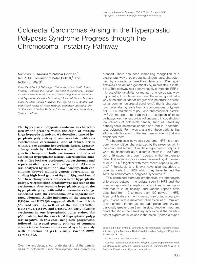
Colorectal Carcinomas Arising in the HyperplasticPolyposis Syndrome Progress through theChromosomal Instability Pathway
Nicholas J. Hawkins,* Patricia Gorman,†
Ian P. M. Tomlinson,‡ Peter Bullpitt,§ andRobyn L. Ward¶
From the School of Pathology,* University of New South Wales,
Sydney, Australia; the Human Cytogenetics Laboratory,† Imperial
Cancer Research Fund, London, United Kingdom; the Molecular
and Population Genetics Laboratory,‡ Imperial Cancer Research
Fund, London, United Kingdom; the Department of Anatomical
Pathology,§ Prince of Wales Hospital, Randwick, Australia; and
St. Vincent’s School of Medicine,¶ University of New South Wales,
Sydney, Australia
The hyperplastic polyposis syndrome is character-ized by the presence within the colon of multiplelarge hyperplastic polyps. We describe a case of hy-perplastic polyposis syndrome associated with twosynchronous carcinomas , one of which ariseswithin a pre-existing hyperplastic lesion. Compar-ative genomic hybridization was used to determinegenetic changes in both carcinomas and severalassociated hyperplastic lesions. Microsatellite anal-ysis at five loci was performed on carcinomas andrepresentative hyperplastic polyps , and p53 statuswas analyzed by immunohistochemistry. Both car-cinomas showed multiple genetic aberrations , in-cluding high level gains of 8q and 13q, and loss of5q. These changes were not seen in the hyperplasticpolyps. Microsatellite instability was not seen in thecarcinomas, four separate hyperplastic polyps , thehyperplastic polyp with mild adenomatous changeassociated with the carcinoma, or a separate ser-rated adenoma. Allelic imbalance in the cancers atD5S346 and D17S938 suggested allelic loss of bothp53 and APC , as well as at the loci D13S263 ,D13S174, D13S159, and D18S49. An early invasivecarcinoma in one hyperplastic polyp stained forp53 protein, but the associated hyperplastic polypwas negative. In this case , neoplastic progressionfollowed the typical genetic pathway of commoncolorectal carcinoma and occurred synchronouslywith mutation of p53. (Am J Pathol 2000,157:385–392)
Over the last decade, our understanding of the geneticbasis of colorectal tumor development has greatly in-
creased. There has been increasing recognition of adistinct pathway of colorectal carcinogenesis, character-ized by sporadic or hereditary defects in DNA repairenzymes and defined genetically by microsatellite insta-bility. This pathway has been variously termed the RER1,microsatellite instability, or mutator phenotype pathway.Importantly, it has thrown into relief the more typical path-way of colorectal cancer progression (referred to hereaf-ter as common colorectal carcinoma), that is character-ized, inter alia, by early loss of adenomatous polyposiscoli (APC), mutations of p53, and chromosomal instabil-ity.1 An important first step in the description of thesepathways was the recognition of unusual clinicopatholog-ical variants of colorectal cancer, such as hereditarynonpolyposis colorectal cancer and familial adenoma-tous polyposis. For it was analysis of these variants thatallowed identification of the key genetic events that un-derpinned them.
The hyperplastic polyposis syndrome (HPS) is an un-common condition, characterized by the presence withinthe colon and rectum of multiple hyperplastic polyps. Itwas first described as a discrete entity in 1980,2 andsome 49 cases have been reported in the literature todate. This includes those cases reviewed by Jorgensenet al in 1996,3 together with more recent reports by oth-ers.4–9 Torlakovic and Snover have also described apotential variant of HPS, which they have termed theserrated adenomatous polyposis syndrome.10
This combined literature emphasizes the phenotypicdifferences between the polyps seen in HPS and thecommon sporadic hyperplastic polyp. Clearly, an impor-tant feature is multiplicity, and various reports havedescribed from 12 to more than 100 polyps in HPS.A second feature is the increased size of individual pol-yps; lesions with a maximum dimension of 10 mm arequite common. In contrast, sporadic polyps are only oc-casionally greater than 5 mm in size.2 Another importantcharacteristic of the hereditary syndrome is the distribu-tion of hyperplastic lesions in the colon. Sporadic hyper-
Supported in part by the St Vincent’s Clinic Foundation Travelling Fellow-ship and by the Macquarie Bank–Royal Australian College of PhysiciansFellowship (R.L.W.).
Accepted for publication April 27, 2000.
Address reprint requests to Prof. Robyn L. Ward, Department of Med-ical Oncology, St. Vincent’s Hospital, Victoria St., Darlinghurst, NSW 2010,Australia. E-mail: [email protected].
American Journal of Pathology, Vol. 157, No. 2, August 2000
Copyright © American Society for Investigative Pathology
385

plastic polyps are seen mainly in the rectum and sigmoidcolon, whereas lesions in HPS are distributed in a rela-tively even fashion throughout the length of the largebowel. Finally, a particular feature of the polypoid lesionsin the HPS is their tendency to show atypical cytologicalfeatures, while maintaining the serrated architecture ofthe hyperplastic polyp. In the context of sporadic lesions,this combination is variously referred to as a mixed hy-perplastic polyp or serrated adenoma.11 Although seenonly rarely in sporadic lesions, it has been reported to agreater though variable extent in HPS,8,10 often as foci ofadenomatous dysplasia.4,12–14
It has been suggested that the atypia seen in sporadicserrated adenomas reflects neoplastic progression in anotherwise hyperplastic or metaplastic lesion and thatsuch lesions may represent an alternative pathway tocolorectal carcinogenesis.15 Not surprisingly, the findingof multiple serrated adenomas in the setting of HPS hasled to speculation that individuals with this condition alsoshare a significant increase in cancer risk that is analo-gous to the increased risk seen in adenomatous polypo-sis.4,8 This contention is supported by the fact that 18 of the49 cases (35%) of hyperplastic polyposis or serrated ad-enomatous polyposis reported in the literature have beenassociated with synchronous colorectal carcinomas.
However, ascertainment bias will certainly favor theidentification of HPS in the presence of synchronouscarcinoma. The possibility remains that carcinoma in theHPS may not arise from serrated adenomas or hyperplas-tic polyps, but rather from unrecognized adenomas of theusual type, or indeed de novo. One carcinoma reported inan individual with HPS was shown to have arisen in anotherwise typical tubular adenoma.16 Furthermore, al-though cases of adenocarcinoma have been reported asactually arising within sporadic hyperplastic polyps andserrated adenomas,17,18 to date this phenomenon hasnot been reported in individuals with the HPS syndrome.
If carcinomas do arise within a morphologically distinc-tive precursor lesion, such as those of HPS, then thisraises the intriguing possibility that the lesions may reflecthitherto unrecognized genetic alterations within colonicepithelial cells. Furthermore, these alterations may beimportant in the development of other, more commonforms of colorectal carcinoma. Jass has pursued thisissue and has highlighted a number of the biochemicaland genetic differences that he considers to be typical ofwhat he has termed the “mild mutator” pathway,19 includ-ing low-level microsatellite instability. Yet to date, infor-mation on genetic changes in HPS and the carcinomasthat arise within it remain limited, largely because of therelative rarity of the syndrome.
In this report, we describe a case of the hyperplasticpolyposis syndrome associated with two synchronouscarcinomas of different stages, one of which shows clearhistological evidence of origin within a pre-existing hy-perplastic lesion. We also present a cytogenetic andmolecular analysis of the carcinomas and representativepremalignant lesions from this individual, in an effort toshed light on the genetic changes that lead to the devel-opment of carcinoma in this syndrome.
Case Report, Materials and Methods
Report of Case
A 73-year-old man presented with acute bowel obstruc-tion of 2 days’ duration. He had previously been well andhad no past or family history of colorectal carcinoma orother bowel diseases. Investigation revealed a constrict-ing lesion of the descending colon, and he underwent anemergency subtotal colectomy. His postoperative coursewas uneventful.
The obstructing lesion was a large, infiltrative andpoorly differentiated adenocarcinoma (45 3 80 mm) thatencircled most of the bowel wall and extended 15 mmthrough the bowel wall, with extensive infiltration of theserosa (T4). Of the 13 pericolic and regional lymph nodesidentified within the descending colon, nine were involvedby tumor. No hepatic or other distant metastases werereported at the time of operation, and the tumor was stagedas Dukes stage C (TNM stage III: T4, N1, M0, RO).
A second, smaller adenocarcinoma was identifiedwithin the transverse colon, 200 mm proximal to the firsttumor. It was an ulcerating lesion (10 3 8 mm) andinvaded the bowel wall to a depth of 3 mm. It had anexophytic margin of pale tissue, up to 3 mm in thickness.Histological examination (Figure 1, A and B) revealed amoderately differentiated adenocarcinoma invading thesubmucosa, without evidence of adjacent involved lymphnodes (Dukes stage A; TNM stage I: T1, N0, M0, R0). Therim of pale tissue surrounding the tumor had the appear-ance of hyperplastic epithelium and focally showed fea-tures suggestive of adenomatous transformation (Figure1, B and C).
Approximately 75 separate mucosal polyps were dis-tributed randomly but evenly throughout the descending,transverse, and ascending colon, as well as the cecum.They varied in size from 2 to 10 mm, and eight lesionswere greater than 8 mm in diameter. One lesion was seento arise separately from, but in continuity with, the smallertumor, and several lesions were within 10 mm of themargins of the larger tumor. The larger lesions were pale,with a soft texture and coarsely wrinkled surface, and didnot appear macroscopically to be typical of tubular ade-nomas. On microscopic examination, nearly all of thelarger lesions and all representative smaller ones showeda typical hyperplastic architecture (Figure 2A). However,one of the discrete polyps examined (4 3 3 mm) showedthe typical features of a serrated adenoma (Figure 2B).A typical hyperplastic polyp was also noted within theappendix.
Tissues and DNA
Before processing, representative tissue samples wereremoved from the larger carcinoma (T1) and four repre-sentative hyperplastic polyps (HP1–4), as well as fromnormal mucosa (N) and an involved lymph node adjacentto the large tumor (LN). DNA was extracted from thesetissues by standard methods. In the case of the smallercarcinoma (T2), its associated hyperplastic polyp (HP-T),and the separate serrated lesion (SA), relevant neoplastic
386 Hawkins et alAJP August 2000, Vol. 157, No. 2

tissues were microdissected from 12 toludine blue-stained tissue sections (10 mm) cut from paraffin-embed-ded blocks. After extraction of paraffin by xylene andethanol, DNA was extracted with a Qiamp Tissue kit(Qiagen, Germany), according to the instructions of themanufacturer.
Comparative Genomic Hybridization
Comparative genomic hybridization (CGH) was per-formed using directly labeled fluorochrome-conjugatedDNA. Samples were analyzed on at least three occasionsand were also cohybridized against normal mucosal DNA(N), as well as unrelated normal male DNA. Briefly, tissuesamples (T1, T2, LN, HP1) and normal DNA were labeledby nick translation,20 but DNA from other lesions was ofinsufficient quality for CGH analysis. Fluoroscein isothio-cyanate-labeled DNA from each lesion and Texas red-labeled reference DNA (500 ng) were precipitated in the
presence of 50 mg Cot-I DNA (Life Technologies, Gaith-ersburg, MD), sodium acetate, and ethanol. The probeswere dissolved in 10 ml of hybridization buffer (50% for-mamide, 10% dextran sulfate, 1% Tween 20, 23 stan-dard saline citrate, pH 7.0), denatured by heating to 75°Cfor 5 minutes, and then allowed to preanneal at 37°C for30 minutes. The mixture was then applied to denaturedmetaphase slides of male human lymphocytes (Vysis,Downers Grove, IL), and the slides were incubated at37°C for 72 hours. After washing in 50% formamide/23standard saline citrate, ethanol dehydration, and air dry-ing, they were counterstained with 4,6-diamidino-2-phe-nylindole. Images were captured with a cooled charge-coupled device camera attached to a Zeiss Axioskopmicroscope. Images from five to nine metaphases wereanalyzed with the Quantitative Image Processing System(QUIPS; Vysis). A chromosome region was considered tobe gained if the mean hybridization ratio between the testand normal samples was .1.2:1, and ratios of ,0.8:1
Figure 1. Histological and immunohistochemical appearance of the small carcinoma and its associated hyperplastic lesion. A: A scanning power view shows thepolypoid nature of the smaller carcinoma (T2), with a central infiltrative lesion and surrounding hyperplastic epithelium. B: The same lesion is shown at highermagnification, in which the close proximity of carcinoma and hyperplastic-type epithelium can be seen. C: The mildly adenomatous morphology of much of thelesion (HP-T) surrounding the small carcinoma highlights the dense eosinophilic cytoplasm and mild nuclear and architectural atypia seen focally in this lesion.Hematoxylin and eosin. Bars represent 2 mm (A), 400 mm (B), and 50 mm (C). D: p53 immunostaining shows strong positivity for the carcinoma (brown staining),in contrast to the surrounding hyperplastic lesion and normal mucosa. A high-power view of the interface between carcinoma and hyperplastic lesion is shown(bar 5 150 mm), and a scanning-power view of the entire lesion is shown in the inset (bar 5 2 mm). Hematoxylin counterstain.
Carcinoma in Hyperplastic Polyposis 387AJP August 2000, Vol. 157, No. 2

represented loss. High-level gain was defined as a ratioof .1.5:1 or as discrete spots of green signal. Negativecontrol hybridizations were included in all experiments.
Polymerase Chain Reaction Analysis forMicrosatellite Instability and Allelic Imbalance
Polymerase chain reaction amplification of microsatelliteloci, using primers for D5S346, D2S123, Bat 26, Bat 25,and D17S938, was used to determine the microsatellitestatus21 of all lesions (T1, T2, LN, HP-T, SA, HP1–4) ascompared with normal mucosal DNA from the same in-dividual. In addition, DNA from a subset of samples (T1,T2, HP1) was amplified using the primers D13S263,D13S174, D13S159, and D18S49 to further evaluatesome of the abnormalities detected by CGH. In all reac-tions, DNA was amplified in a 10-ml reaction containing 3ml of DNA, 200 mmol/L dNTPs, 1.5 mmol/L MgCl2, 0.8mmol/L of each primer, 0.5 U Taq polymerase in a bufferof 50 mmol/L KCl, 10 mmol/L Tris-HCl, and 0.1% TritonX-100 (pH 9.0). The reactions were incubated at 95°C for5 minutes, followed by 35 cycles of 95°C, 57°C, and 72°Cfor 1 minute each. Products were run on an ABI 377sequencer and analyzed with Genescan and Genotypersoftware (Applied Biosystems, Foster City, CA). Allelicimbalance was recorded if the area under either allelepeak was reduced in the tumor sample to less than 50%of its normal value with respect to the other allele.
Immunohistochemical Analysis of p53
Detection of accumulated p53 protein within tissues wasperformed on 4-mm paraffin-embedded sections, usingan anti-human p53 monoclonal antibody (DO-7; Dako).22
Antigen was detected after microwave antigen retrieval in
0.1 mol/L citrate buffer, and bound primary antibody wasdetected using horseradish peroxidase-labeled sheepanti-mouse antibody. Color was developed with diamino-benzidine substrate (Sigma), and sections were counter-stained with hematoxylin.
Results
The pathological findings in this case fit well with theaccepted definitions of HPS in terms of multiplicity, dis-tribution, and size of the hyperplastic lesions.3 Althoughmost of the polyps in this case were hyperplastic innature, one showed serrated adenomatous change, typ-ified by a serrated architecture, with cells showing abun-dant eosinophilic cytoplasm, goblet cell depletion, andoval vesicular nuclei with prominent nucleoli and nuclearstratification (Figure 2B). No areas of moderate or severedysplasia were seen in this lesion, and no adenomatouspolyps of the usual type were seen within the colectomyspecimen.
It was evident that the small carcinoma had arisenwithin a hyperplastic lesion, which surrounded it on allsides (Figure 1, A and B). There was also evidence ofmild adenomatous change within this hyperplastic lesion(Figure 1, B and C), although this change was focal, as isoften the case in such lesions.19 While hyperplastic epi-thelium was continuous with some sections of the largercarcinoma, it is impossible to exclude the possibility ofcollision of this large and invasive tumor with a separateadjacent hyperplastic lesion. Neither carcinoma showedthe phenotypic features of “serrated adenocarcinoma”reported by Jass in the setting of HPS.8
Immunohistochemical analysis of p53 showed strongaccumulation of the protein within nuclei of the smallertumor (Figure 1D). This was not seen in any of the cells of
Figure 2. Microscopic features of a typical hyperplastic polyp (A) and the single serrated adenoma (B) seen in the resected colon. For each micrograph, the barrepresents 200 mm, and the inset (bottom right of each micrograph) shows a fourfold magnification of the same lesion. Hematoxylin and eosin.
388 Hawkins et alAJP August 2000, Vol. 157, No. 2

the surrounding hyperplastic lesion, nor was it present inthe serrated adenoma or the other four hyperplastic pol-yps examined. Interestingly, the larger tumor was alsonegative for nuclear p53 accumulation (not shown).
Comparative Genomic Hybridization Analysis
CGH was used to analyze molecular genetic abnormali-ties in DNA from both carcinomas, as well as from one ofthe hyperplastic polyps (HP1). No molecular genetic ab-normalities were detected in the hyperplastic polyp. Incontrast, the large carcinoma (T1) showed 11 chromo-somal aberrations (five gains, six losses), and the smallcarcinoma (T2) showed 16 changes (nine gains, sevenlosses; Figures 3 and 4).
Although these changes were clearly not identical,several chromosomes were affected in both cancers,including 4, 5, 8, and 13, as shown schematically inFigure 4. The loss of 4q32-ter, 5q, and 18q12–21 seen inthis study, as well as the high-level gains in 13q and 20q,have been reported frequently in sporadic colorectal carci-noma. Interestingly, the small carcinoma showed two re-gions of amplification at 8p11.2 and 8q23–24, and the largecarcinoma showed a novel region of amplification at 10q21,as well as high-level amplification of 18q11.2–21.1.
Microsatellite Analysis
There was no evidence of microsatellite instability at anyof the five loci examined, in either of the carcinomas, inthe involved lymph node draining the larger tumor (LN), inthe serrated adenoma (SA), or in any of five hyperplasticpolyps examined (HP1–4, HP-T).
The D5S346 locus is found near the APC gene locus onchromosome 5, and the D17S938 locus is in close prox-imity to the p53 gene. Loss of heterozygosity (LOH) at theD5S346 locus was observed in the large carcinoma aswell as an involved draining lymph node, a result that was
consistent with the loss of 5q observed in the primarytumor by CGH. However, LOH was not found at D5S346in the small cancer, despite the fact that CGH had alsodemonstrated 5q loss in this tumor. As such, this mayrepresent a homozygous deletion at this marker. TheD17S938 locus showed LOH in both large and smallcarcinomas. In the case of the small cancer this loss waslikely to be the result of an interstitial deletion, becauseCGH did not detect any abnormalities of chromosome 17.In contrast, CGH showed loss of chromosome 17 in thelarge tumor. Loss of heterozygosity at either the D5S346
Figure 3. Representative chromosomes and CGH profiles in the large carcinoma (A) and small carcinoma (B). Chromosomal gains are indicated by green, andlosses are shown in red.
Figure 4. Summary of chromosomal changes observed in the large and smallcarcinomas. Gains are displayed to the right of the chromosomal ideogram,and losses are shown on the left. Changes in the small carcinoma are shownas solid lines and changes in the large carcinoma as dotted lines. Double linesmark regions affected by high-level gain or loss. Chromosomes that werenormal in both lesions are not shown.
Carcinoma in Hyperplastic Polyposis 389AJP August 2000, Vol. 157, No. 2

or D17S938 locus was not seen in any of the five hyper-plastic polyps, including the lesion that gave rise to thecancer, or in the serrated adenoma.
Polymerase chain reaction using microsatellite mark-ers was also used to further define the abnormalitiesdetected on chromosomes 13q and 18q. In the smallercarcinoma, allelic imbalance was observed at theD18S49 locus (18q12.1–12.2), consistent with the loss of18q found by CGH. However, the region of high level gainon 18q noted in the larger cancer was not reflected inallelic imbalance at this locus, possibly because D18S49was not encompassed within the region of gain, or be-cause the gain was “symmetrical.” Allelic imbalance wasalso observed in both the large and small carcinomas,using markers D13S263 (13q14.1–21.1), D13S159(13q22–31), and D13S174 (13q32–33), which is consis-tent with the 13q changes seen by CGH. In the case ofthe larger carcinoma, the pattern of change was likely toreflect the presence of multiple copies of chromosome13, whereas the changes in the smaller tumor suggestedmore discrete regions of gain.
Discussion
The occurrence of two synchronous colonic carcinomasin the setting of hyperplastic polyposis, one of them at anearly stage of invasion, is a rare event. It provides anopportunity to examine the critical biology of the disease,at the point of transformation from in situ dysplasia toinvasive carcinoma.
We believe that the small carcinoma was very likely tohave arisen within a pre-existing hyperplastic polyp thatshowed focal areas of adenomatous transformation. Al-though the possibility of a “collision tumor” cannot betotally excluded, the symmetry of the carcinoma, arisingcentrally within and completely surrounded by the hyper-plastic lesion, makes this possibility remote. Furthermore,although hyperplastic patterns of reactivity are not un-common in the transition from normal epithelium to tumor,the polypoid nature of the entire lesion and the presenceof considerable hyperplastic tissue with a serrated architec-ture make this explanation also unlikely. Finally, the pres-ence of areas within the surrounding lesion that showedevidence of early adenomatous transformation is an impor-tant indicator that this lesion was a consequence of neo-plastic progression in a large hyperplastic polyp, of thetype often seen in the hyperplastic polyposis syndrome.
It is interesting in this context that much of the putativeprecursor lesion maintained the morphology of a hyper-plastic polyp. For althoughe tumor progression may haveoccurred within dysplastic foci, it seems that these focidid not have a significant growth advantage over themore usual hyperplastic elements and thus failed to dom-inate the lesion. This observation is in keeping with thecommon observation of mixed hyperplastic polyps, inwhich foci of serrated adenoma coexist with hyperplasticepithelium, yet fail to demonstrate an overwhelmingclonal growth advantage within the lesion. An alternativeexplanation could be that there is an extremely rapidprogression from serrated adenoma to invasive carci-
noma, yet the finding of serrated adenomas without se-vere dysplasia is common in the hyperplastic polyposissyndrome and indeed was observed in this case.
The closer analysis of tissues in this case, through thecombined approaches of comparative genomic hybrid-ization, microsatellite analysis, and p53 immunohisto-chemistry, has allowed us to better understand the natureof the neoplastic progression in these lesions. Further-more, this combined approach has allowed validation ofdata gained separately through each modality. Threepoints can be made on the basis of these results: thatcarcinoma in HPS develops through the pathway of chro-mosomal instability, that mutation of p53 is an early eventin this process, and that microsatellite instability was notimportant in this case.
Carcinoma Progresses by Normal Pathways
In the carcinomas analyzed in this study, both the patternand number of molecular cytogenetic abnormalities mir-rored those described in a number of recent CGH anal-yses of the common type of CRC.23–25 For instance, in astudy of 45 sporadic colorectal cancers, De Angelisfound that aneuploid tumors had a median of 9.0 chro-mosomal abnormalities, with high-level gains seen fre-quently in 8q, 13q, and 20.24 Both cancers in this studyclearly exhibited more than nine changes; high-levelgains of chromosome 8 and 13 were seen in both lesions,and high-level gain of chromosome 20q was also presentin the smaller carcinoma. This latter finding in particularhas been well described.23,25 The significance of theseamplifications in tumor progression remains unknown.Interestingly, the finding of allelic imbalance at 13q inboth tumors may serve as a useful marker in the morerapid evaluation of the presence of amplification and inthe closer exploration of the biological significance of thisphenomenon in colorectal carcinoma.
Mutation of p53 Is an Early Event in MalignantProgression
It is clear that the smaller carcinoma developed a muta-tion in the p53 gene that was not present in the putativeprecursor lesion. This was demonstrated directly by im-munohistochemistry and was consistent with the findingof LOH at the D17S398 locus. It is likely that this was aregion of interstitial loss and hence would not be de-tected by CGH. Nevertheless, the finding of widespreadchromosomal instability in the tumor, with a large numberof discrete chromosomal abnormalities detected byCGH, further supports the hypothesis that malignant pro-gression occurs as a result of p53 inactivation and sec-ondary chromosomal instability.
Althoughe the larger cancer showed widespread chro-mosomal instability, LOH at D17S398, and extensive lossof chromosome 17 by CGH, it was interesting that theimmunohistochemistry for p53 was negative. The mostlikely explanation is that the remaining p53 allele had aframeshift, chain-terminating or nonsense mutation thatcaused failure of protein expression, an event that under-
390 Hawkins et alAJP August 2000, Vol. 157, No. 2

lies the imperfect correlation between immunohistochem-ical results and mutations in p53.26
On the other hand, we could find no evidence of p53mutation in any precursor lesions by immunohistochem-istry and no LOH at D17S398 in five hyperplastic polypsand one serrated adenoma. Although p53 mutations arerare in hyperplastic polyps,27,28 there is considerablevariation in the literature regarding the incidence of p53positivity in serrated adenomas, with reports varying from5% to 65%.27,29 To some extent, this may simply reflectvariation in the degree of dysplasia within the lesions ofeach series and, by default, the diagnostic criteria ap-plied to the selection of cases. Extrapolation of data fromthis study suggests that lesions with high-grade dyspla-sia (carcinoma in situ) are likely to contain p53 mutations,whereas serrated adenomas with only low-grade dyspla-sia will show no abnormalities.
In the common form of colorectal neoplasia, mutationof p53 is known to be a critical event in the progressionfrom adenoma to invasive carcinoma.30 However, thereasons for the development of mutations at this point inthe adenoma-carcinoma sequence are not clear. It hasbeen suggested that the formation of an adenoma, withits disordered crypt architecture, allows the exposure ofproliferating cells to fecal carcinogens.3 This leads insome cases to p53 mutations and subsequent neoplasticprogression via impaired apoptosis and chromosomalinstability. It is plausible that a similar mechanism may beat work in the hyperplastic polyp/serrated adenoma le-sions typical of the hyperplastic polyposis syndrome.
The Nature of the Underlying Defect RemainsUncertain
This study has failed to elucidate the exact nature of themolecular lesions that underpin the HP syndrome. Thelack of chromosomal abnormalities detectable by CGH inthe precursor lesions is in sharp contrast to reports ofchromosomal aberrations in adenomas25,31 and sug-gests that the CGH approach is unlikely to throw furtherlight on this issue.
We found no evidence of microsatellite instability ineither the carcinomas or any of the precursor lesionsexamined, including hyperplastic polyps and a serratedadenoma. In fact, no instability was found at any locus,despite the use of both fresh tissue and carefully micro-dissected materials. This is in contrast to the findings ofJass, who showed low-level microsatellite instability inserrated adenomas in the setting of HPS and proposedthat it may be an important event in the development ofsubsequent carcinomas.15
With regard to this discrepancy, we consider the pos-sibility of gross contamination of tumor DNA by normaltissue to be an unlikely explanation for the absence ofmicrosatellite instability. In fact, the relative purity of thetumor DNA is evidenced by our ability to demonstrateLOH at several alleles in both tumor samples and thecorrelation between these findings, CGH, and immuno-histochemistry. The use of different microsatellite markersand, in particular, the use of tetranucleotide repeat mark-
ers and the MYCL markers favored by Jass may contrib-ute to differences reported here.
The true biological significance of the microsatellitelow-instability phenotype remains uncertain, and the def-inition of this phenomenon remains both arbitrary andsubject to considerable operational flexibility. Clearly it isnot a universal event in HPS or important in tumor pro-gression in this case. However, it is possible that there isvariability in the process of carcinogenesis within thehyperplastic polyposis syndrome, and indeed the syn-drome may represent a phenotype with more than onegenetic basis.
In summary, this case provides morphological andgenetic evidence that carcinoma developing in the set-ting of hyperplastic polyposis can arise directly from ahyperplastic lesion. In this case, microsatellite instabilitywas not important in either the development of the pri-mary lesion or subsequent tumor progression. Rather,progression of the lesions involved loss of p53 and thedevelopment of chromosomal instability, a pathway ofneoplastic progression also seen in the common form ofcolorectal carcinoma.
References
1. Fujiwara T, Stolker JM, Watanabe T, Rashid A, Longo P, Eshleman JR,Booker S, Lynch HT, Jass JR, Green JS, Kim H, Jen J, Vogelstein B,Hamilton SR: Accumulated clonal genetic alterations in familial andsporadic colorectal carcinomas with widespread instability in micro-satellite sequences. Am J Pathol 1998, 153:1063–1078
2. Williams GT, Arthur JF, Bussey HJ, Morson BC: Metaplastic polypsand polyposis of the colorectum. Histopathology 1980, 4:155–170
3. Jorgensen H, Mogensen AM, Svendsen LB: Hyperplastic polyposis ofthe large bowel. Three cases and a review of the literature. Scand JGastroenterol 1996, 31:825–830
4. Orii S, Nakamura S, Sugai T, Habano W, Akasaka I, Nakasima F,Kazama H, Hasimoto Y, Takahasi H, Sugawara M, Sato S: Hyperplas-tic (metaplastic) polyposis of the colorectum associated with adeno-mas and an adenocarcinoma. J Clin Gastroenterol 1997, 25:369–372
5. McCann BG: A case of metaplastic polyposis of the colon associatedwith focal adenomatous change and metachronous adenocarcino-mas. Histopathology 1988, 13:700–702
6. Lieverse RJ, Kibbelaar RE, Griffioen G, Lamers CB: Colonic adeno-carcinoma in a patient with multiple hyperplastic polyps. NetherlandsJ Med 1995, 46:185–188
7. Keljo DJ, Weinberg AG, Winick N, Tomlinson G: Rectal cancer in an11 year old girl with hyperplastic polyposis. J Pediatr GastroenterolNutr 1999, 28:327–332
8. Jeevaratnam P, Cottier DS, Browett PJ, Van De Water NS, Pokos V,Jass JR: Familial giant hyperplastic polyposis predisposing to colo-rectal cancer: a new hereditary bowel cancer syndrome. J Pathol1996, 179:20–25
9. Place RJ, Simmang CL: Hyperplastic adenomatous polyposis syn-drome. J Am Coll Surg 1999, 188:503–507
10. Torlakovic E, Snover DC: Serrated adenomatous polyposis in hu-mans. Gastroenterology 1996, 110:748–755
11. Longacre TA, Fenoglio-Preiser CM: Mixed hyperplastic adenomatouspolyps/serrated adenomas. A distinct form of colorectal neoplasia.Am J Surg Pathol 1990, 14:524–537
12. Cooper HS, Patchefsky AS, Marks G: Adenomatous and carcinoma-tous changes within hyperplastic colonic epithelium. Dis Colon Rec-tum 1979, 22:152–156
13. Spjut H, Estrada RG: The significance of epithelial polyps of the largebowel. Pathol Annu 1977, 1:147–170
14. Shepherd NA: Inverted hyperplastic polyposis of the colon. J ClinPathol 1993, 46:56–60
15. Iino H, Jass JR, Simms LA, Young J, Leggett B, Ajioka Y, Watanabe
Carcinoma in Hyperplastic Polyposis 391AJP August 2000, Vol. 157, No. 2

H: DNA microsatellite instability in hyperplastic polyps, serrated ad-enomas, and mixed polyps: a mild mutator pathway for colorectalcancer? J Clin Pathol 1999, 52:5–9
16. Warner AS, Glick ME, Fogt F: Multiple large hyperplastic polyps of thecolon coincident with adenocarcinoma. Am J Gastroenterol 1994,89:123–125
17. Franzin G, Novelli P: Adenocarcinoma occurring in a hyperplastic(metaplastic) polyp of the colon. Endoscopy 1982, 14:28–30
18. Urbanski SJ, Kossakowska AE, Marcon N, Bruce WR: Mixed hyper-plastic adenomatous polyps—an underdiagnosed entity. Report of acase of adenocarcinoma arising within a mixed hyperplastic adeno-matous polyp. Am J Surg Pathol 1984, 8:551–556
19. Jass JR: Serrated adenoma and colorectal cancer. J Pathol 1999,187:499–502
20. Kallioniemi A, Kallioniemi OP, Piper J, Tanner M, Stokke T, Chen L,Smith HS, Pinkel D, Gray JW, Waldman FM: Detection and mappingof amplified DNA sequences in breast cancer by comparativegenomic hybridization. Proc Natl Acad Sci USA 1994, 91:2156–2160
21. Boland CR, Thibodeau SN, Hamilton SR, Sidransky D, Eshleman JR,Burt RW, Meltzer SJ, Rodriguez-Bigas MA, Fodde R, Ranzani GN,Srivastava S: A National Cancer Institute workshop on microsatelliteinstability for cancer detection and familial predisposition: develop-ment of international criteria for the determination of microsatelliteinstability in colorectal cancer. Cancer Res 1998, 58:5248–5257
22. Ward RL, Todd AV, Santiago F TOC, Hawkins NJ: Activation of theK-ras oncogene in colorectal neoplasms is associated with de-creased apoptosis. Cancer 1997, 79:1106–1113
23. Korn WM, Yasutake T, Kuo WL, Warren RS, Collins C, Tomita M, GrayJ, Waldman FM: Chromosome arm 20q gains and other genomicalterations in colorectal cancer metastatic to liver, as analyzed by
comparative genomic hybridization and fluorescence in situ hybrid-ization. Genes Chromosom Cancer 1999, 25:82–90
24. De Angelis PM, Clausen OP, Schjolberg A, Stokke T: Chromosomalgains and losses in primary colorectal carcinomas detected by CGHand their associations with tumour DNA ploidy, genotypes and phe-notypes. Br J Cancer 1999, 80:526–535
25. Ried T, Knutzen R, Steinbeck R, Blegen H, Schrock E, Heselmeyer K,du Manoir S, Auer G: Comparative genomic hybridization reveals aspecific pattern of chromosomal gains and losses during the genesisof colorectal tumors. Genes Chromosom Cancer 1996, 15:234–245
26. Lane DP, Hall PA: MDM2—arbiter of p53’s destruction. Trends Bio-chem Sci 1997, 22:372–374
27. Kang M, Mitomi H, Sada M, Tokumitsu Y, Takahashi Y, Igarashi M,Katsumata T, Okayasu I: Ki-67, p53, and Bcl-2 expression of serratedadenomas of the colon. Am J Surg Pathol 1997, 21:417–423
28. Rashid A, Zahurak M, Goodman SN, Hamilton SR: Genetic epidemi-ology of mutated K-ras proto-oncogene, altered suppressor genes,and microsatellite instability in colorectal adenomas. Gut 1999, 44:826–833
29. Hiyama T, Yokozaki H, Shimamoto F, Haruma K, Yasui W, KajiyamaG, Tahara E: Frequent p53 gene mutations in serrated adenomas ofthe colorectum. J Pathol 1998, 186:131–139
30. Lengauer C, Kinzler KW, Vogelstein B: Genetic instability in colorectalcancers. Nature 1997, 386:623–627
31. Meijer GA, Hermsen MA, Baak JP, van Diest PJ, Meuwissen SG,Belien JA, Hoovers JM, Joenje H, Snijders PJ, Walboomers JM:Progression from colorectal adenoma to carcinoma is associated withnon-random chromosomal gains as detected by comparativegenomic hybridisation. J Clin Pathol 1998, 51:901–909
392 Hawkins et alAJP August 2000, Vol. 157, No. 2
Copyright © 2022 FDOKUMEN