Blastic NK-Cell Lymphomas (Agranular CD4+CD56+ Hematodermic Neoplasms): A Review

doi:10.1182/blood-2005-02-0496Prepublished online July 19, 2005;2005 106: 3498-3506
Magda De Smedt, Inge Hoebeke, Katia Reynvoet, Georges Leclercq and Jean Plum microenvironmenttoward B-, NK-, monocytic/dendritic-, or T-cell lineage in thymus Different thresholds of Notch signaling bias human precursor cells
http://bloodjournal.hematologylibrary.org/content/106/10/3498.full.htmlUpdated information and services can be found at:
(1930 articles)Signal Transduction � (973 articles)Phagocytes �
(5022 articles)Immunobiology �Articles on similar topics can be found in the following Blood collections
http://bloodjournal.hematologylibrary.org/site/misc/rights.xhtml#repub_requestsInformation about reproducing this article in parts or in its entirety may be found online at:
http://bloodjournal.hematologylibrary.org/site/misc/rights.xhtml#reprintsInformation about ordering reprints may be found online at:
http://bloodjournal.hematologylibrary.org/site/subscriptions/index.xhtmlInformation about subscriptions and ASH membership may be found online at:
Copyright 2011 by The American Society of Hematology; all rights reserved.Washington DC 20036.by the American Society of Hematology, 2021 L St, NW, Suite 900, Blood (print ISSN 0006-4971, online ISSN 1528-0020), is published weekly
For personal use only. by guest on June 9, 2013. bloodjournal.hematologylibrary.orgFrom

IMMUNOBIOLOGY
Different thresholds of Notch signaling bias human precursor cells toward B-,NK-, monocytic/dendritic-, or T-cell lineage in thymus microenvironmentMagda De Smedt, Inge Hoebeke, Katia Reynvoet, Georges Leclercq, and Jean Plum
Notch receptors are involved in lineagedecisions in multiple developmental sce-narios, including hematopoiesis. Here, wetreated hybrid human–mouse fetal thy-mus organ culture with the �-secretaseinhibitor 7 (N-[N-(3,5-difluorophenyl)-L-alanyl]-S-phenyl-glycine t-butyl ester)(DAPT) to establish the role of Notchsignaling in human hematopoietic lin-eage decisions. The effect of inhibition ofNotch signaling was studied starting fromcord blood CD34� or thymic CD34�CD1�,CD34�CD1�, or CD4ISP progenitors.Treatment of cord blood CD34� cells with
low DAPT concentrations results in aber-rant CD4ISP and CD4/CD8 double-positive (DP) thymocytes, which are nega-tive for intracellular T-cell receptor �
(TCR�). On culture with intermediate andhigh DAPT concentrations, thymicCD34�CD1� cells still generate aberrantintracellular TCR�� DP cells that haveundergone DJ but not VDJ recombina-tion. Inhibition of Notch signaling shiftsdifferentiation into non–T cells in a thy-mic microenvironment, depending on thestarting progenitor cells: thymic CD34�CD1�
cells do not generate non–T cells, thymic
CD34�CD1� cells generate NK cells andmonocytic/dendritic cells, and cord bloodCD34�Lin� cells generate B, NK, and mono-cytic/dendritic cells in the presence of DAPT.Our data indicate that Notch signaling iscrucial to direct human progenitor cells intothe T-cell lineage, whereas it has a negativeimpact on B, NK, and monocytic/dendriticcell generation in a dose-dependent fash-ion. (Blood. 2005;106:3498-3506)
© 2005 by The American Society of Hematology
Introduction
T cells develop from pluripotent hematopoietic stem cells (HSCs)through a series of differentiation steps. In humans, differentiatingthymocytes can be divided into 4 main subsets based on theirexpression of CD4 and CD8 coreceptors. The most immaturethymocytes are the CD34� CD4�CD8� double-negative (DN)cells. As a first step toward the expression of a functional T-cellreceptor (TCR), CD4�CD3� immature single-positive (CD4ISP)cells start to rearrange the TCRB gene. Subsequently, the TCR�chain becomes assembled into the pre-TCR complex with theinvariant pre-T� chain. Pre–TCR signaling confers survival andallows development to proceed through a CD4�CD8�TCRlow
double-positive (DP) subset of thymocytes. Finally, positive andnegative selection results in the maturation of major histocompat-ibility complex (MHC)–restricted CD4 or CD8 single-positive(SP) T cells.1,2
Multiple signal transduction pathways are involved in directingcell fate decisions during T-cell development. These includeregulation by Notch proteins, a family of highly conservedtransmembrane receptors.3 Notch is a key player in T-cell develop-ment, and its role has been recently reviewed.4-6 There is goodevidence that in mice, reduced signaling through Notch1 causes anearly block in T-cell development and results in the expansion ofimmature B cells.7-9 However, the role of Notch1 in later stages ofT-cell development is contentious. Although earlier data pointedtoward a role for Notch1 in the lineage decision between �� and ��
T cells and between CD4 and CD8 SP thymocytes,10-12 analysis of 2different conditional Notch1 knockout mouse strains has failed toconfirm these findings.13,14 This strongly argues against an impor-tant role for Notch1 in later stages of thymocyte development,though the possibility remains that other family members compen-sate for the lack of Notch1.
The role of Notch proteins in lymphocyte development isalmost exclusively based on experiments in mice. However, it iswidely accepted that differences between the thymuses of mice andhumans exist,15 including the expression of cell-surface markerssuch as CD25 on early progenitors and the subdivision of the DNstage by CD44 and CD25.16 The role of Notch in human T-celldifferentiation has only been addressed by overexpression of theactive form of Notch1 in CD34� progenitors to evaluate itsinfluence on T-cell differentiation in hybrid human–mouse fetalthymus organ culture (FTOC).17,18 More recently, it was shown thathuman T-cell differentiation starting from CD34� stem cells can besupported by OP-9 stromal cells engineered to express the Notchligand Delta-like-1.19,20 In light of these findings, it is important toevaluate the necessity of Notch in human hematopoietic lineagedecisions and thymocyte development. It has been observed thatpresenilin-dependent �-secretase, which serves to cleave amyloidprecursor proteins in neuronal cells, also catalyzes the release ofthe intracellular domain of Notch proteins.21,22 Several compoundsthat inhibit �-secretase and, consequently, Notch cleavage are
From the Department of Clinical Chemistry, Microbiology and Immunology,Ghent University, University Hospital Ghent, Belgium.
Submitted February 2, 2005; accepted July 13, 2005. Prepublished online asBlood First Edition Paper, July 19, 2005; DOI 10.1182/blood-2005-02-0496.
Supported by grants from Concerted Research Action of the University ofGhent (01G00905) and Fund for Scientific Research-Flanders (G.0331.03).
M.D.S. and I.H. contributed equally to this manuscript.
The online version of this article contains a data supplement.
Reprints: Jean Plum, Department of Clinical Chemistry, Microbiology andImmunology, Ghent University, University Hospital Ghent, 4BlokA, DePintelaan 185, B-9000 Ghent, Belgium; e-mail: [email protected].
The publication costs of this article were defrayed in part by page chargepayment. Therefore, and solely to indicate this fact, this article is herebymarked ‘‘advertisement’’ in accordance with 18 U.S.C. section 1734.
© 2005 by The American Society of Hematology
3498 BLOOD, 15 NOVEMBER 2005 � VOLUME 106, NUMBER 10
For personal use only. by guest on June 9, 2013. bloodjournal.hematologylibrary.orgFrom
available and can be used to study the role of Notch signaling.23,24
In this paper, we show that the inhibition of Notch signaling inhybrid human-mouse FTOC impairs the development of human Tcells and biases, in a dose-dependent way, development towardB-cell, NK-cell, and monocytic/dendritic-cell differentiation. Thus,our findings show for the first time CD34� precursor cells as animportant site of Notch action in the human thymus.
Materials and methods
Cells and sorting
Pediatric thymuses and cord blood (CB) samples were obtained and wereused according to the guidelines of the Medical Ethical Commission ofGhent University Hospital (Belgium). CD34� CB cells were purified bypositive selection with CD34 magnetically activated cell sorter (MACS)beads (Miltenyi Biotec, Bergisch Gladbach, Germany) and stained withCD34–antigen-presenting cells (APCs), CD3–fluorescein isothiocyanate(FITC), CD19-FITC, and CD56–phycoerythrin (PE) (all monoclonalantibodies [mAbs] from BD Immunocytometry Systems [BDIS], MountainView, CA) to sort CD34�Lin� cells by flow cytometry (FACSVantage;BDIS). CD34� thymocytes were purified by positive selection with CD34MACS beads, stained with CD34-APC and CD1-PE, and sorted forCD1�CD34� and CD1�CD34� progenitors. CD4�ISP thymocytes wereenriched by negative depletion of CD8�CD3� thymocytes usingDynalBeads (Dynal, Hamburg, Germany) and were labeled with CD4-PEand CD3-FITC, CD8-FITC, HLA-DR-FITC, and CD34-APC to sortCD4� CD34�CD3�CD8�HLA-DR� cells. Purity of the cells wasalways at least 98%.
FTOC and flow cytometry
The mixed human-mouse FTOC was performed as described previ-ously.25,26 FTOCs were cultured with the �-secretase inhibitor 7 (N-[N-(3,5-difluorophenyl)-L-alanyl]-S-phenyl-glycine t-butyl ester] (DAPT; PeptidesInternational Inc, Louisville, KY) at various concentrations, as indicated, orwith the solvent dimethyl sulfoxide (DMSO). Half the medium waschanged weekly. After different time points of FTOC, harvested thymocyteswere incubated with anti–mouse FcR���/III (clone 2.4.G2)27 and humanimmunoglobulin G (IgG) (FcBlock; Miltenyi) to avoid nonspecific staining.Cells were stained with anti–mouse CD45-CyChrome (BD PharMingen,San Diego, CA) in combination with one or more of the followinganti–human mAbs: CD8�-PE, TCR-��-PE, CD79a-PE, CD56-APC(Coulter, Miami, FL), CD34-APC, TCR-��-FITC, CD3-APC or -FITC,CD4-APC or -PE, CD14-PE, CD19-FITC or PE, CD20-FITC, CD7-FITC,CD10-PE, CD56-PE, CD69-PE, HLA-DR-APC, or the appropriate controlmAb (IgG1 and IgG2a-FITC, APC, or PE) (BDIS). Human viable cells,gated by exclusion of propidium iodide and mouse CD45� cells, wereexamined for the expression of the antigens on a FACScalibur usingCellQuest Pro software (BDIS). For intracellular staining, cells were fixedand permeabilized using Fix and Perm (Imtec, San Francisco, CA)according to the manufacturer’s instructions and were stained with anti–TCR�1-PE (Ancell, Bayport, MN), CD3-APC (BDIS), or CD79a-PE(Coulter). For DNA analysis, after staining with mAbs for surface antigens,cells underwent gentle fixation with 0.25% paraformaldehyde for 1 hour at4°C. After washing, the cells were permeabilized with 0.05% Tween 20 for15 minutes at 37°C, as described.28 Cells were treated with RNase (Sigma,Bornem, Belgium) and stained with 7-amino-actinomycin D (7-AAD;Becton and Dickinson) for 30 minutes at 25°C. Doublets were excluded bydiscriminating red fluorescence channel area � width pulses, and DNAcontent was measured. Apoptotic cells were detected using the annexinV–PE labeling kit from BD PharMingen.
Gene expression analysis by real-time RT-PCR
Total RNA from thymocytes was isolated as described.29 cDNA synthesiswas performed by oligo(dT) priming, and real-time reverse transcription–
polymerase chain reaction (RT-PCR) was performed as described.29 Hypo-xanthine guanine phosphoribosyl transferase (HPRT) mRNA was used fornormalization. Human-specific primers were selected using Primer Expresssoftware (Applied Biosystems, Foster City, CA) and are shown inSupplemental Table S1, which is available at the Blood website (see theSupplemental Materials link at the top of the online article). PCR wasperformed with an annealing temperature of 60°C. Comparative quantifica-tion of the target gene expression in the samples was performed based oncycle threshold (Ct) normalized to HPRT using the Ct method.30
PCR analysis of TCRB gene locus recombination
DNA was extracted with the QIAmp DNA minikit (Qiagen, Hilden,Germany), and 50 ng was used for amplification by PCR. PCR conditionswere performed as described31 under the following conditions: 5 minutes at94°C, 37 cycles of 60 seconds at 94°C, 60 seconds at 63°C (DJ�
amplification) or 58°C (VDJ� amplification), followed by 7 minutes at72°C. Primers for DJ� corresponded to bases 44 to 63 (TBF1) and 3025 to3006 (TBR1), as previously reported.32,33 For VDJ� amplification, amixture of 5 primers specific for V�2-5-8-13 families was used with TBR1.Primers are listed in Table S1 and are shown in Figure 4.
Southern blot analysis
PCR products were run on a 2% agarose gel in 1� Tris-acetate EDTA(ethylenediaminetetraacetic acid) buffer. Specificity of the amplified frag-ments was validated by their predicted size and Southern blot analysis. Gelswere blotted in alkaline buffer (0.4 N NaOH) onto Hybond N� membranes(Amersham, Little Chalfont, United Kingdom). Membranes were hybrid-ized with a biotinylated TBR3 probe for 16 hours at 55°C, washed, revealedwith streptavidin–horseradish peroxidase and the enhanced chemilumines-cence (ECL) advanced detection system (Amersham), and visualized usingx-ray film (Eastman Kodak, Rochester, NY).
Results
Notch signaling can be blocked by the �-secretase inhibitorDAPT in hybrid human-mouse FTOC
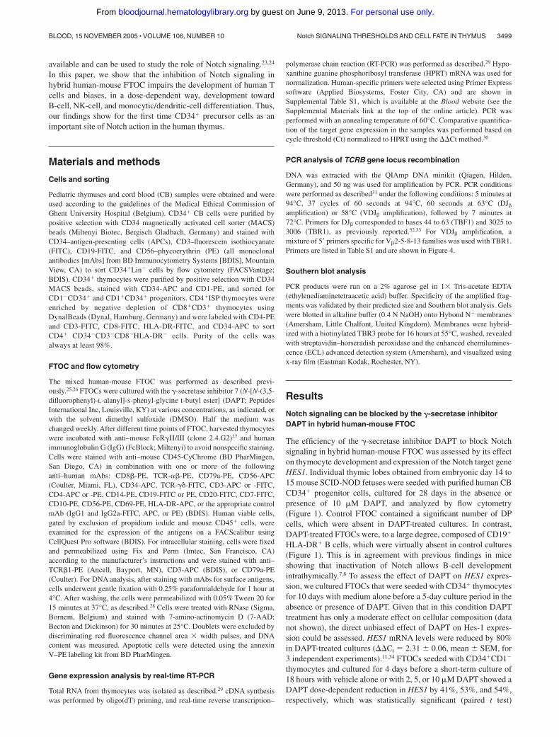
The efficiency of the �-secretase inhibitor DAPT to block Notchsignaling in hybrid human-mouse FTOC was assessed by its effecton thymocyte development and expression of the Notch target geneHES1. Individual thymic lobes obtained from embryonic day 14 to15 mouse SCID-NOD fetuses were seeded with purified human CBCD34� progenitor cells, cultured for 28 days in the absence orpresence of 10 �M DAPT, and analyzed by flow cytometry(Figure 1). Control FTOC contained a significant number of DPcells, which were absent in DAPT-treated cultures. In contrast,DAPT-treated FTOCs were, to a large degree, composed of CD19�
HLA-DR� B cells, which were virtually absent in control cultures(Figure 1). This is in agreement with previous findings in miceshowing that inactivation of Notch allows B-cell developmentintrathymically.7,8 To assess the effect of DAPT on HES1 expres-sion, we cultured FTOCs that were seeded with CD34� thymocytesfor 10 days with medium alone before a 5-day culture period in theabsence or presence of DAPT. Given that in this condition DAPTtreatment has only a moderate effect on cellular composition (datanot shown), the direct unbiased effect of DAPT on Hes-1 expres-sion could be assessed. HES1 mRNA levels were reduced by 80%in DAPT-treated cultures (Ct � 2.31 0.06, mean SEM, for3 independent experiments).11,34 FTOCs seeded with CD34�CD1�
thymocytes and cultured for 4 days before a short-term culture of18 hours with vehicle alone or with 2, 5, or 10 �M DAPT showed aDAPT dose-dependent reduction in HES1 by 41%, 53%, and 54%,respectively, which was statistically significant (paired t test)
Notch SIGNALING THRESHOLDS AND CELL FATE IN THYMUS 3499BLOOD, 15 NOVEMBER 2005 � VOLUME 106, NUMBER 10
For personal use only. by guest on June 9, 2013. bloodjournal.hematologylibrary.orgFrom
between control and DAPT-treated cultures (P � .01) and between2 �M and 5 �M DAPT-treated cultures (P � .02). Our resultsclearly demonstrate that the �-secretase inhibitor DAPT is capableof blocking Notch signaling in hybrid human-mouse FTOC.
Inhibition of Notch signaling in FTOC of CB CD34� progenitorcells arrests T-cell development and shifts differentiation to B,NK, and monocytic-dendritic cells in a dose-dependent way
Because DAPT did not interfere with the entry of the precursorcells into the thymic lobes (data not shown), we performed theexperiments by adding DAPT from the start, including the hangingdrop. Half the medium was changed weekly to maintain theconcentration of active product. Inhibition of Notch with 2 and10 �M DAPT did not result in a significant change in theabsolute human cell numbers generated in FTOC after 28 days.Interestingly, there was a significant decrease in the total cellnumber when the FTOC was treated with an intermediate doseof 5 �M DAPT (Table 1). This observation must be interpreted
in view of the change in the cellular composition and thecell-specific generation potential.
The frequency and number of CD34� cells, which represent themost immature cells, were significantly reduced by more than 50%in cultures with 2 and 5 �M of DAPT (Table 1 and Table S2),compatible with the view that Notch supports the maintenance ofCD34� progenitor cells.35 At a dose of 10 �M, the frequency andtotal number of CD34� cells tended to be lower, but were no longersignificantly decreased (Table 1 and Table S2). It is possible that theefficient generation of B cells in this condition, which we willpresent further, is accompanied by an increase in CD34� Bcell–committed progenitors.
In mice, the inhibition of Notch signaling results in theaccumulation of B cells.7 FTOC seeded with CB CD34�Lin�
precursor cells showed a significant dose-dependent increase infrequency and in absolute numbers of B cells after the inhibition ofNotch signaling by DAPT (Figure 2A), varying from 12- to460-fold, depending on the dose of DAPT (Table 1). After 28 daysof culture with the highest concentration of DAPT, B cellsrepresented the most predominant population, encompassing morethan 50% of all human cells (Figure 1), which were positive forCD10 and CD20 and partly positive for intracellular CD79a(Figure S1A).
In rat FTOC, inhibition of Notch by DAPT results in a dramaticincrease in the number of NK cells.34 Here, DAPT-treated FTOCshowed a significant increase in the frequency and number of cellspositively staining for CD56, which is expressed on all human NKcells (Figure 2A-B; Table 1), from less than 3% in controls toapproximately 10% with the highest concentration of DAPT,corresponding to a 2.5-fold increase in the absolute number ofCD56� cells (Figure 2A; Table 1). At a lower concentration of theinhibitor (5 �M), the frequency of the number of CD56� cellsincreased to more than 17%, corresponding to more than a 3.5-foldincrease in absolute number of CD56� cells (Figure 2A; Table 1).Most of the CD56� cells were negative for CD3, CD4 (Figure 2A),and CD8� surface expression (data not shown) and positive forCD7 (Figure S1B). This corresponds to the phenotype of matureperipheral NK cells and argues that the CD56� cells in DAPT-treated cultures represent true NK cells.36 On activation, NK cellsare known to up-regulate triggering receptors such as CD69 ontheir surfaces, endowing them with new recognition capabilities.37
Figure 1. DAPT inhibits Notch signaling in hybrid human–mouse fetal thymusorgan culture. Representative flow cytometric analyses of human cells fromFTOC seeded with human CD34� CB progenitor cells and cultured for 28 days inthe absence or presence of 10 �M DAPT. Quadrants were set according toisotype controls.
Table 1. Influence of �-secretase inhibition by DAPT on human cellularity and cell number of human subpopulations after 28 days of FTOCseeded with human CD34�Lin� cord blood progenitor cells
Cell type Control, no. cells
DAPT treatment
2 �M, no. cells 5 �M, no. cells 10 �M, no. cells
Human cells 55 645 8 676 41 591 6 869 24 541 3 191* 32 089 1 755
CD34� 5 618 1 469 1 792 736* 1 411 110* 1 930 188
CD7� 50 646 8 137 27 448 9 725* 8 531 2 563* 4 533 1 060*
CD4ISP 24 828 3 974 18 487 4 598 915 596* 0 0*
DP 8 562 3 238 2 549 951 345 173* 0 0*
IC CD3� 53 119 8 319 35 414 6 671 5 333 1 856* 518 326*
IC TCR-� 14 103 3 490 344 210* 25 25* 0 0*
CD3� 10 952 5 146 1 474 737* 21 21* 0 0*
TCR-�� 8 449 2 740 291 212* 0 0* 0 0*
TCR-�� 2 479 313 294 114* 0 0* 0 0*
CD4�HLA-DR� 1 614 266 1 391 496 2 640 502 5 266 790*
CD14� 231 75 241 60 533 107* 1 352 356*
CD56� 1 247 656 2 052 379 4 324 1 134* 3 079 861
CD19� 38 45 454 171* 6 516 2 219* 17 357 3 052*
Results represent mean SEM of 4 independent experiments.*Significant difference (P � .05; paired Student t test) compared with the vehicle-treated control cultures.
3500 De SMEDT et al BLOOD, 15 NOVEMBER 2005 � VOLUME 106, NUMBER 10
For personal use only. by guest on June 9, 2013. bloodjournal.hematologylibrary.orgFrom

Most CD56� cells in DAPT-treated fetal thymuses were negativefor CD69 surface expression, suggestive of a nonactivated pheno-type (Figure S1B). Collectively, this suggests that in the absence ofNotch signaling, NK-cell development from CD34� progenitors isfavored. At the highest concentration (10 �M) of DAPT, CD34�
progenitor cells are preferentially biased toward B cells, whereas inthe presence of intermediate concentrations, the CD34� cells areless efficiently skewed toward B-cell differentiation but are able togenerate NK cells. At low concentrations of DAPT, the generationof NK cells is still favored compared with untreated cultures, butnot to the same extent as with intermediate DAPT concentrations.
FTOC seeded with human CB CD34� progenitors and culturedin high (10 �M) DAPT concentrations resulted in the emergence ofa population with characteristics of monocytic and dendritic cells.These cells coexpressed CD4 and HLA-DR and had relatively highside scatter, and some of them were CD14� (Figure 2B and data notshown). The absolute numbers of CD4�HLA-DR� cells andCD14� cells were significantly increased in FTOC treated withhigh and intermediate concentrations of DAPT and resulted in amore than 3-fold increase (Table 1).
In control FTOCs, CB CD34� progenitors undergo T-cellmaturation in the mouse thymic microenvironment. This is shownby the progress of CD34� cells into CD4ISP cells, which are
immature thymocytes that start to rearrange TCRB. After 28 days ofculture, more than 40% of the human cells achieved this maturationstage (Figure 2A-B). Approximately 15% of the cells alreadymatured to a further stage, with coexpression of CD4�CD8 andsurface expression of CD3 (Figure 2A-B). The frequency of cellsthat stain for intracellular TCR� exceeds that of DP thymocytes, aspart of CD4ISP, and CD4�CD8��� cells express intracellularTCR�.38 FTOC with human CB CD34� progenitors displayed acomplete block of T-cell differentiation at high DAPT concentra-tions. Although a low number of CD4�CD3� cells appeared(Figure 2B), those cells were not CD4ISP immature T-cell precur-sors; rather, they belonged to the monocytic/dendritic cell lineageaccording to the coexpression of HLA-DR, the absence of intracel-lular CD3 (Figure 2A-B), and the different scatter properties (datanot shown). At intermediate DAPT concentrations, CD34� CBprogenitor cells were able to progress toward T cells, but thenumber of CD4ISP and DP cells was extremely low. At low DAPTconcentrations, the number of CD4ISP and DP cells tended to belower than for the control, but there was no significant difference.However, some striking differences were observed in the 2 �MDAPT-treated cultures. One was the nearly complete absence ofintracellular TCR-�, and the other was a significant reduction inCD7 expression (Table 1).
Inhibition of Notch signaling in CD34�CD1� thymic progenitorcells does not lead to the generation of B lymphocytes, allowsNK- and monocytic/dendritic-cell maturation, and results indifferentiation of abnormal T lymphocytes in FTOC
To have a better view of the effect of Notch inhibition on T-cellmaturation, we explored the influence of DAPT on FTOC seededwith CD34� progenitors purified from human thymocytes. Thoseprogenitor cells have already entered the thymus and receivedNotch signaling39 and are more efficient generating large numbersof human T cells with faster kinetics in FTOC.
In contrast to FTOCs that were seeded with CB CD34� cells,those seeded with CD34�CD1� thymic precursors were unable togenerate B cells, even at the highest DAPT concentration (data notshown). This is compatible with the view that CD34� progenitorcells in the thymus have received Notch triggering and possiblyother signals that have already irreversibly induced the CD34�
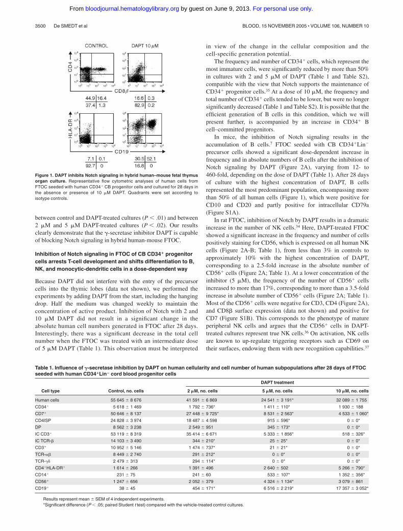
precursor cell to a stage wherein the capacity to develop into B cellsis lost, but they retained their capacity to develop into NK cells.The absolute number of NK cells was the highest with intermediateconcentrations of the inhibitor (Tables 2, 3). Surprisingly, and incontrast to the CB CD34� cultures, there was a significantdose-dependent increase in the frequency and number of DP cellsgenerated in DAPT-treated FTOC. However, those DP cells wereabnormal because they lacked surface CD3 and intracellularTCR-� expression (Figure 3A; Tables 2, 3). Furthermore, theinhibition of Notch triggering in CD34� thymic progenitorsleads to a reduction of CD7 in a concentration-dependentmanner (Figure 3B).
FTOC with increasing DAPT concentrations exhibited a progres-sive decrease in the number of mature CD3� DP cells, togetherwith an elevation in the number of CD4ISP cells (Tables 2, 3).Importantly, CD7 was markedly down-regulated in those cells(Figure 3B). CD7 is found primarily on T, NK, and pre–B cells. Thefact that CD7 expression was unchanged on NK cells arguesagainst unspecific loss of expression or detectability.
Figure 2. Influence of DAPT inhibition of Notch signaling on differentiation ofCD34� CB cells in hybrid human-mouse FTOC. Murine fetal thymic lobes wereseeded with human CD34� CB cells and cultured in FTOC for 28 days in the absenceof presence of 2, 5, or 10 �M DAPT. (A) Frequencies of subpopulations of humanhematopoietic cells at the end of the culture period. Bars represent mean SEM of 4independent experiments. Asterisks indicate statistically significant differences(P � .05) compared with vehicle-treated control. (B) Representative dot plots andhistogram of flow cytometric analysis of control cultures and cultures treated with10 �M DAPT. Bold line and thin line in the histogram represent results from theDAPT-treated and control culture, respectively.
Notch SIGNALING THRESHOLDS AND CELL FATE IN THYMUS 3501BLOOD, 15 NOVEMBER 2005 � VOLUME 106, NUMBER 10
For personal use only. by guest on June 9, 2013. bloodjournal.hematologylibrary.orgFrom
Inhibition of Notch signaling in differentiating CD34�CD1� thymicprogenitor cells in FTOC affects VDJ�, but not DJ�, rearrangement
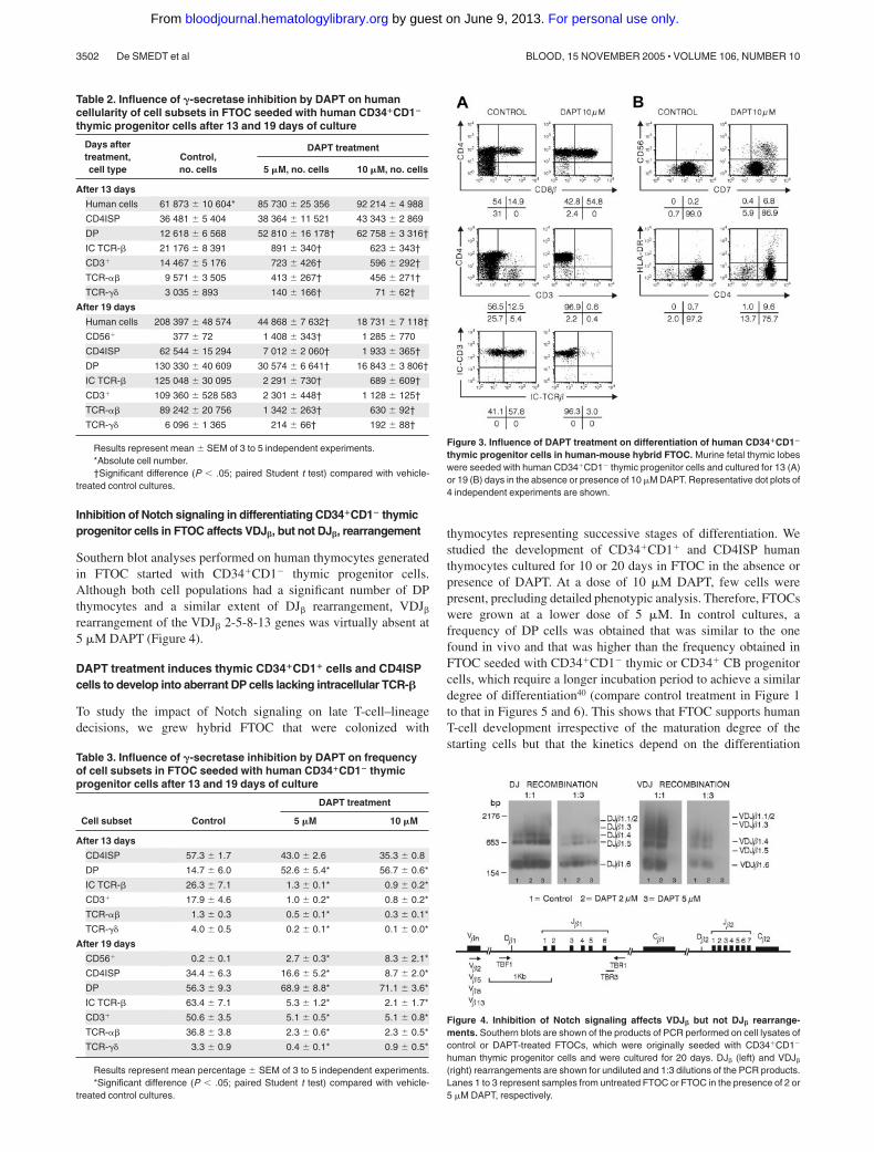
Southern blot analyses performed on human thymocytes generatedin FTOC started with CD34�CD1� thymic progenitor cells.Although both cell populations had a significant number of DPthymocytes and a similar extent of DJ� rearrangement, VDJ�
rearrangement of the VDJ� 2-5-8-13 genes was virtually absent at5 �M DAPT (Figure 4).
DAPT treatment induces thymic CD34�CD1� cells and CD4ISPcells to develop into aberrant DP cells lacking intracellular TCR-�
To study the impact of Notch signaling on late T-cell–lineagedecisions, we grew hybrid FTOC that were colonized with
thymocytes representing successive stages of differentiation. Westudied the development of CD34�CD1� and CD4ISP humanthymocytes cultured for 10 or 20 days in FTOC in the absence orpresence of DAPT. At a dose of 10 �M DAPT, few cells werepresent, precluding detailed phenotypic analysis. Therefore, FTOCswere grown at a lower dose of 5 �M. In control cultures, afrequency of DP cells was obtained that was similar to the onefound in vivo and that was higher than the frequency obtained inFTOC seeded with CD34�CD1� thymic or CD34� CB progenitorcells, which require a longer incubation period to achieve a similardegree of differentiation40 (compare control treatment in Figure 1to that in Figures 5 and 6). This shows that FTOC supports humanT-cell development irrespective of the maturation degree of thestarting cells but that the kinetics depend on the differentiation
Table 2. Influence of �-secretase inhibition by DAPT on humancellularity of cell subsets in FTOC seeded with human CD34�CD1�
thymic progenitor cells after 13 and 19 days of culture
Days aftertreatment,cell type
Control,no. cells
DAPT treatment
5 �M, no. cells 10 �M, no. cells
After 13 days
Human cells 61 873 10 604* 85 730 25 356 92 214 4 988
CD4ISP 36 481 5 404 38 364 11 521 43 343 2 869
DP 12 618 6 568 52 810 16 178† 62 758 3 316†
IC TCR-� 21 176 8 391 891 340† 623 343†
CD3� 14 467 5 176 723 426† 596 292†
TCR-�� 9 571 3 505 413 267† 456 271†
TCR-�� 3 035 893 140 166† 71 62†
After 19 days
Human cells 208 397 48 574 44 868 7 632† 18 731 7 118†
CD56� 377 72 1 408 343† 1 285 770
CD4ISP 62 544 15 294 7 012 2 060† 1 933 365†
DP 130 330 40 609 30 574 6 641† 16 843 3 806†
IC TCR-� 125 048 30 095 2 291 730† 689 609†
CD3� 109 360 528 583 2 301 448† 1 128 125†
TCR-�� 89 242 20 756 1 342 263† 630 92†
TCR-�� 6 096 1 365 214 66† 192 88†
Results represent mean SEM of 3 to 5 independent experiments.*Absolute cell number.†Significant difference (P � .05; paired Student t test) compared with vehicle-
treated control cultures.
Table 3. Influence of �-secretase inhibition by DAPT on frequencyof cell subsets in FTOC seeded with human CD34�CD1� thymicprogenitor cells after 13 and 19 days of culture
Cell subset Control
DAPT treatment
5 �M 10 �M
After 13 days
CD4ISP 57.3 1.7 43.0 2.6 35.3 0.8
DP 14.7 6.0 52.6 5.4* 56.7 0.6*
IC TCR-� 26.3 7.1 1.3 0.1* 0.9 0.2*
CD3� 17.9 4.6 1.0 0.2* 0.8 0.2*
TCR-�� 1.3 0.3 0.5 0.1* 0.3 0.1*
TCR-�� 4.0 0.5 0.2 0.1* 0.1 0.0*
After 19 days
CD56� 0.2 0.1 2.7 0.3* 8.3 2.1*
CD4ISP 34.4 6.3 16.6 5.2* 8.7 2.0*
DP 56.3 9.3 68.9 8.8* 71.1 3.6*
IC TCR-� 63.4 7.1 5.3 1.2* 2.1 1.7*
CD3� 50.6 3.5 5.1 0.5* 5.1 0.8*
TCR-�� 36.8 3.8 2.3 0.6* 2.3 0.5*
TCR-�� 3.3 0.9 0.4 0.1* 0.9 0.5*
Results represent mean percentage SEM of 3 to 5 independent experiments.*Significant difference (P � .05; paired Student t test) compared with vehicle-
treated control cultures.
Figure 3. Influence of DAPT treatment on differentiation of human CD34�CD1�
thymic progenitor cells in human-mouse hybrid FTOC. Murine fetal thymic lobeswere seeded with human CD34�CD1� thymic progenitor cells and cultured for 13 (A)or 19 (B) days in the absence or presence of 10 �M DAPT. Representative dot plots of4 independent experiments are shown.
Figure 4. Inhibition of Notch signaling affects VDJ� but not DJ� rearrange-ments. Southern blots are shown of the products of PCR performed on cell lysates ofcontrol or DAPT-treated FTOCs, which were originally seeded with CD34�CD1�
human thymic progenitor cells and were cultured for 20 days. DJ� (left) and VDJ�
(right) rearrangements are shown for undiluted and 1:3 dilutions of the PCR products.Lanes 1 to 3 represent samples from untreated FTOC or FTOC in the presence of 2 or5 �M DAPT, respectively.
3502 De SMEDT et al BLOOD, 15 NOVEMBER 2005 � VOLUME 106, NUMBER 10
For personal use only. by guest on June 9, 2013. bloodjournal.hematologylibrary.orgFrom
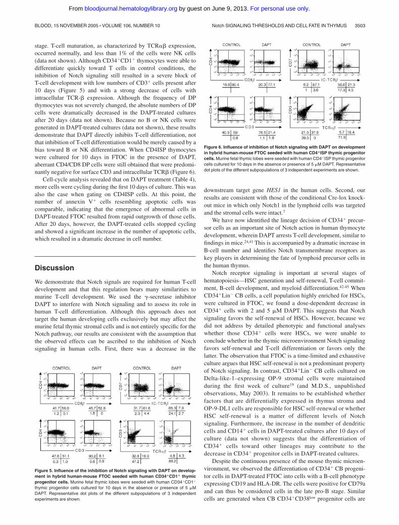
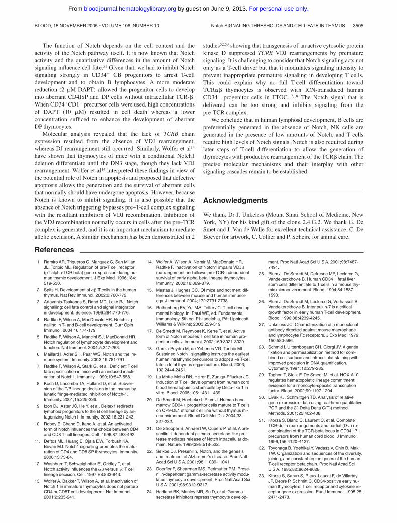
stage. T-cell maturation, as characterized by TCR�� expression,occurred normally, and less than 1% of the cells were NK cells(data not shown). Although CD34�CD1� thymocytes were able todifferentiate quickly toward T cells in control conditions, theinhibition of Notch signaling still resulted in a severe block ofT-cell development with low numbers of CD3� cells present after10 days (Figure 5) and with a strong decrease of cells withintracellular TCR-� expression. Although the frequency of DPthymocytes was not severely changed, the absolute numbers of DPcells were dramatically decreased in the DAPT-treated culturesafter 20 days (data not shown). Because no B or NK cells weregenerated in DAPT-treated cultures (data not shown), these resultsdemonstrate that DAPT directly inhibits T-cell differentiation, notthat inhibition of T-cell differentiation would be merely caused by abias toward B or NK differentiation. When CD4ISP thymocyteswere cultured for 10 days in FTOC in the presence of DAPT,aberrant CD4/CD8 DP cells were still obtained that were predomi-nantly negative for surface CD3 and intracellular TCR� (Figure 6).
Cell-cycle analysis revealed that on DAPT treatment (Table 4),more cells were cycling during the first 10 days of culture. This wasalso the case when gating on CD4ISP cells. At this point, thenumber of annexin V� cells resembling apoptotic cells wascomparable, indicating that the emergence of abnormal cells inDAPT-treated FTOC resulted from rapid outgrowth of those cells.After 20 days, however, the DAPT-treated cells stopped cyclingand showed a significant increase in the number of apoptotic cells,which resulted in a dramatic decrease in cell number.
Discussion
We demonstrate that Notch signals are required for human T-celldevelopment and that this regulation bears many similarities tomurine T-cell development. We used the �-secretase inhibitorDAPT to interfere with Notch signaling and to assess its role inhuman T-cell differentiation. Although this approach does nottarget the human developing cells exclusively but may affect themurine fetal thymic stromal cells and is not entirely specific for theNotch pathway, our results are consistent with the assumption thatthe observed effects can be ascribed to the inhibition of Notchsignaling in human cells. First, there was a decrease in the
downstream target gene HES1 in the human cells. Second, ourresults are consistent with those of the conditional Cre-lox knock-out mice in which only Notch1 in the lymphoid cells was targetedand the stromal cells were intact.7
We have now identified the lineage decision of CD34� precur-sor cells as an important site of Notch action in human thymocytedevelopment, wherein DAPT arrests T-cell development, similar tofindings in mice.24,41 This is accompanied by a dramatic increase inB-cell number and identifies Notch transmembrane receptors askey players in determining the fate of lymphoid precursor cells inthe human thymus.
Notch receptor signaling is important at several stages ofhematopoiesis—HSC generation and self-renewal, T-cell commit-ment, B-cell development, and myeloid differentiation.42-45 WhenCD34�Lin� CB cells, a cell population highly enriched for HSCs,were cultured in FTOC, we found a dose-dependent decrease inCD34� cells with 2 and 5 �M DAPT. This suggests that Notchsignaling favors the self-renewal of HSCs. However, because wedid not address by detailed phenotypic and functional analyseswhether those CD34� cells were HSCs, we were unable toconclude whether in the thymic microenvironment Notch signalingfavors self-renewal and T-cell differentiation or favors only thelatter. The observation that FTOC is a time-limited and exhaustiveculture argues that HSC self-renewal is not a predominant propertyof Notch signaling. In contrast, CD34�Lin� CB cells cultured onDelta-like-1–expressing OP-9 stromal cells were maintainedduring the first week of culture19 (and M.D.S., unpublishedobservations, May 2003). It remains to be established whetherfactors that are differentially expressed in thymus stroma andOP-9-DL1 cells are responsible for HSC self-renewal or whetherHSC self-renewal is a matter of different levels of Notchsignaling. Furthermore, the increase in the number of dendriticcells and CD14� cells in DAPT-treated cultures after 10 days ofculture (data not shown) suggests that the differentiation ofCD34� cells toward other lineages may contribute to thedecrease in CD34� progenitor cells in DAPT-treated cultures.
Despite the continuous presence of the mouse thymic microen-vironment, we observed the differentiation of CD34� CB progeni-tor cells in DAPT-treated FTOC into cells with a B-cell phenotypeexpressing CD19 and HLA-DR. The cells were positive for CD79aand can thus be considered cells in the late pro-B stage. Similarcells are generated when CB CD34�CD38low progenitor cells are
Figure 5. Influence of the inhibition of Notch signaling with DAPT on develop-ment in hybrid human-mouse FTOC seeded with human CD34�CD1� thymicprogenitor cells. Murine fetal thymic lobes were seeded with human CD34�CD1�
thymic progenitor cells cultured for 10 days in the absence or presence of 5 �MDAPT. Representative dot plots of the different subpopulations of 3 independentexperiments are shown.
Figure 6. Influence of inhibition of Notch signaling with DAPT on developmentin hybrid human-mouse FTOC seeded with human CD4�ISP thymic progenitorcells. Murine fetal thymic lobes were seeded with human CD4�ISP thymic progenitorcells cultured for 10 days in the absence or presence of 5 �M DAPT. Representativedot plots of the different subpopulations of 3 independent experiments are shown.
Notch SIGNALING THRESHOLDS AND CELL FATE IN THYMUS 3503BLOOD, 15 NOVEMBER 2005 � VOLUME 106, NUMBER 10
For personal use only. by guest on June 9, 2013. bloodjournal.hematologylibrary.orgFrom

cocultured with the murine stromal cell line MS-5.46 Takentogether, we conclude that Notch signaling represses the B-cell fatein early precursor cells in the thymus.
Importantly, CD34�CD1� thymic progenitor cells failed todevelop into B lymphocytes under the same conditions. These datasuggest that the B-cell–lineage potential of hematopoietic progeni-tor cells is lost immediately after entry into thymus. One study47
has shown that murine thymic CD117�CD44�CD25� (DN1) cellsgive rise to B lymphocytes when cultured on control OP-9 stromalcells. They observed less than 1% B-cell progenitors in the DN1thymic cell population. They also observed that low levels of Notchsignaling inhibit B-cell potential and that high levels induce Tlymphopoiesis. Porritt et al48 have found that among DN1 prothy-mocytes, a subset can be characterized with B-lineage potential.This could indicate that cells with B-lineage potential have losttheir B-cell potential before seeding the thymus by low levels ofNotch signaling and that sustained Notch signaling in the thymusleads to T-lineage commitment. It is possible that FTOC in thepresence of DAPT is not sensitive enough to detect a low precursorfrequency of B cells or that human thymic CD34�CD1� cells havea lower frequency of B-cell precursors than murine DN1 cells.More detailed analysis of thymic CD34� cells and culture on OP-9stromal cells is required to determine the B-cell–lineage potentialof these cells.
Our data indicate that Notch signaling also reduces NK celldifferentiation in a thymic microenvironment. A direct effect ofaltered Notch signaling is compatible with the observation thatincreasing DAPT concentrations progressively inhibited T-cellmaturation in FTOC and, at the same time, led to increased NK cellnumbers. These data strongly suggest that the inhibition of Notch
signaling is responsible for directing lymphoid progenitors into NKcells. These findings are also consistent with those from experi-ments in mice47 and in rats.34
It is clear that CD7 expression was influenced by the inhibitionof Notch signaling. When CD34�Lin� CB cells were cultured inFTOC, both the frequency and the absolute numbers of CD7� cellswere significantly lower in DAPT-treated cultures in a dose-dependent way at the end of the culture. Given that after shorterincubation (10 days) the CD34� cells did not express CD7 in thepresence of DAPT (data not shown), we concluded that CD7expression was inhibited from the start. This is compatible with theview that the progression of multipotent CD34� progenitors towardT-/NK-cell progenitors is dependent on Notch signaling. However,CD7 expression was differentially regulated on T and NK cellsbecause CD7 was down-regulated in developing T cells, whereas itwas not affected in NK cells. This is best illustrated when thymicCD34�CD1� progenitors were cultured in FTOC at low DAPTconcentrations. In these conditions, differentiation toward DPthymocytes occurred and was paradoxically enhanced, but CD7expression level was decreased on T cells and unchanged on NKcells. This precludes the hypothesis that CD7 is directly influencedby Notch signaling and suggests that this occurs in the context ofother factors that are present according to the cell type. Eventhough CD7 is expressed in early T-cell ontogeny, the roles thatCD7 plays in T-cell development remain unclear. It has been shownin mice that the number of thymocytes and the induced thymocyteapoptosis is normal in CD7-deficient mice, which indicates thatCD7 is not involved in the early stages of T-cell development.49 Itwas also demonstrated that NK-cell development and function arenot impaired in CD7 disrupted mice.50
Table 4. Influence of �-secretase inhibition by DAPT on human cellularity, proliferative status, and annexin V� cells in cell subsets in FTOCseeded with CD34�CD1� thymic progenitor cells after different time points of culture
Cell type, treatment Day 4 Day 7 Day 10 Day 20 Day 26
Cell no.
Control 16 391 2 911 49 107 315 61 303 9 024 502 068 13 762 828 750
2 �M DAPT 11 389 713 56 108 4 024 108 889 45 035 285 806 43 782 148 353
5 �M DAPT 13 016 2 674 40 626 5 763 123 874 5 633 94 166 75 717 38 500
S/G2/M cells in total cell population, %
Control 17 5.9 10 1.4 12 4.2 18 2.4 6.2
2 �M DAPT 22 7.4 18 4.2 20 2.8 4 1.2 1.3
5 �M DAPT 25 1.3 27 0.8 15 0.4 1 0.1 0.5
Annexin V� cells in total cell population, %
Control 45 12.7 42 5.7 55 3.5 38 6.4 32
2 �M DAPT 49 14.4 37 1.3 38 2.0 32 8.4 36
5 �M DAPT 52 16.5 49 5.4 44 9.1 64 9.3 65
S/G2/M cells in CD4� subset, %
Control ND 8 9 2.3 23 2.1 6.2
2 �M DAPT ND 20 18 3.1 8 2.6 1.3
5 �M DAPT ND 27 15 0.6 2 0.7 0.5
Annexin V� cells in CD4� subset, %
Control ND 40 1.8 55 3.6 51 10.5 44
2 �M DAPT ND 36 4.0 38 4.6 41 12.6 48
5 �M DAPT ND 48 0.1 49 9.2 66 11.9 73
S/G2/M cells in CD4�CD8� subset, %
Control ND ND ND 12 1.7 3.6
2 �M DAPT ND ND ND 1 0.3 0.5
5 �M DAPT ND ND ND 1 0.3 0.2
Annexin V� cells in CD4�CD8� subset, %
Control ND ND ND 42 12.6 41
2 �M DAPT ND ND ND 43 11.0 51
5 �M DAPT ND ND ND 69 15.8 74
Results represent mean SEM of 2 independent experiments, except for figures without SEM shown, when only 1 experiment was performed.ND indicates not done.
3504 De SMEDT et al BLOOD, 15 NOVEMBER 2005 � VOLUME 106, NUMBER 10
For personal use only. by guest on June 9, 2013. bloodjournal.hematologylibrary.orgFrom
The function of Notch depends on the cell context and theactivity of the Notch pathway itself. It is now known that Notchactivity and the quantitative differences in the amount of Notchsignaling influence cell fate.51 Given that, we had to inhibit Notchsignaling strongly in CD34� CB progenitors to arrest T-celldevelopment and to obtain B lymphocytes. A more moderatereduction (2 �M DAPT) allowed the progenitor cells to developinto aberrant CD4ISP and DP cells without intracellular TCR-�.When CD34�CD1� precursor cells were used, high concentrationsof DAPT (10 �M) resulted in cell death whereas a lowerconcentration sufficed to enhance the development of aberrantDP thymocytes.
Molecular analysis revealed that the lack of TCRB chainexpression resulted from the absence of VDJ rearrangement,whereas DJ rearrangement still occurred. Similarly, Wolfer et al14
have shown that thymocytes of mice with a conditional Notch1deletion differentiate until the DN3 stage, though they lack VDJrearrangement. Wolfer et al14 interpreted these findings in view ofthe potential role of Notch in apoptosis and proposed that defectiveapoptosis allows the generation and the survival of aberrant cellsthat normally should have undergone apoptosis. However, becauseNotch is known to inhibit signaling, it is also possible that theabsence of Notch triggering bypasses pre–T-cell complex signalingwith the resultant inhibition of VDJ recombination. Inhibition ofthe VDJ recombination normally occurs in cells after the pre–TCRcomplex is generated, and it is an important mechanism to mediateallelic exclusion. A similar mechanism has been demonstrated in 2
studies52,53 showing that transgenesis of an active cytosolic proteinkinase D suppressed TCRB VDJ rearrangements by prematuresignaling. It is challenging to consider that Notch signaling acts notonly as a T-cell driver but that it modulates signaling intensity toprevent inappropriate premature signaling in developing T cells.This could explain why no full T-cell differentiation towardTCR�� thymocytes is observed with ICN-transduced humanCD34� progenitor cells in FTOC.17,18 The Notch signal that isdelivered can be too strong and inhibits signaling from thepre-TCR complex.
We conclude that in human lymphoid development, B cells arepreferentially generated in the absence of Notch, NK cells aregenerated in the presence of low amounts of Notch, and T cellsrequire high levels of Notch signals. Notch is also required duringlater steps of T-cell differentiation to allow the generation ofthymocytes with productive rearrangement of the TCR� chain. Theprecise molecular mechanisms and their interplay with othersignaling cascades remain to be established.
Acknowledgments
We thank Dr J. Unkeless (Mount Sinai School of Medicine, NewYork, NY) for his kind gift of the clone 2.4.G.2. We thank G. DeSmet and I. Van de Walle for excellent technical assistance, C. DeBoever for artwork, C. Collier and P. Scheire for animal care.
References
1. Ramiro AR, Trigueros C, Marquez C, San MillanJL, Toribio ML. Regulation of pre-T cell receptor(pT alpha-TCR beta) gene expression during hu-man thymic development. J Exp Med. 1996;184:519-530.
2. Spits H. Development of �� T cells in the humanthymus. Nat Rev Immunol. 2002;2:760-772.
3. Artavanis-Tsakonas S, Rand MD, Lake RJ. Notchsignalling: cell fate control and signal integrationin development. Science. 1999;284:770-776.
4. Radtke F, Wilson A, MacDonald HR. Notch sig-nalling in T- and B-cell development. Curr OpinImmunol. 2004;16:174-179.
5. Radtke F, Wilson A, Mancini SJ, MacDonald HR.Notch regulation of lymphocyte development andfunction. Nat Immunol. 2004;5:247-253.
6. Maillard I, Adler SH, Pear WS. Notch and the im-mune system. Immunity. 2003;19:781-791.
7. Radtke F, Wilson A, Stark G, et al. Deficient T cellfate specification in mice with an induced inacti-vation of Notch1. Immunity. 1999;10:547-558.
8. Koch U, Lacombe TA, Holland D, et al. Subver-sion of the T/B lineage decision in the thymus bylunatic fringe-mediated inhibition of Notch-1.Immunity. 2001;15:225-236.
9. Izon DJ, Aster JC, He Y, et al. Deltex1 redirectslymphoid progenitors to the B cell lineage by an-tagonizing Notch1. Immunity. 2002;16:231-243.
10. Robey E, Chang D, Itano A, et al. An activatedform of Notch influences the choice between CD4and CD8 T cell lineages. Cell. 1996;87:483-492.
11. Deftos ML, Huang E, Ojala EW, Forbush KA,Bevan MJ. Notch1 signalling promotes the matu-ration of CD4 and CD8 SP thymocytes. Immunity.2000;13:73-84.
12. Washburn T, Schweighoffer E, Gridley T, et al.Notch activity influences the �� versus �� T celllineage decision. Cell. 1997;88:833-843.
13. Wolfer A, Bakker T, Wilson A, et al. Inactivation ofNotch 1 in immature thymocytes does not perturbCD4 or CD8T cell development. Nat Immunol.2001;2:235-241.
14. Wolfer A, Wilson A, Nemir M, MacDonald HR,Radtke F. Inactivation of Notch1 impairs VDJ�rearrangement and allows pre-TCR-independentsurvival of early alpha beta lineage thymocytes.Immunity. 2002;16:869-879.
15. Mestas J, Hughes CC. Of mice and not men: dif-ferences between mouse and human immunol-ogy. J Immunol. 2004;172:2731-2738.
16. Rothenberg EV, Yui MA, Telfer JC. T-cell develop-mental biology. In: Paul WE, ed. FundamentalImmunology. 5th ed. Philadelphia, PA: LippincottWilliams & Wilkins; 2003:259-319.
17. De Smedt M, Reynvoet K, Kerre T, et al. Activeform of Notch imposes T cell fate in human pro-genitor cells. J Immunol. 2002;169:3021-3029.
18. Garcia-Peydro M, de Yebenes VG, Toribio ML.Sustained Notch1 signalling instructs the earliesthuman intrathymic precursors to adopt a �� T-cellfate in fetal thymus organ culture. Blood. 2003;102:2444-2451.
19. La Motte-Mohs RN, Herer E, Zuniga-Pflucker JC.Induction of T cell development from human cordblood hematopoietic stem cells by Delta-like 1 invitro. Blood. 2005;105:1431-1439.
20. De Smedt M, Hoebeke I, Plum J. Human bonemarrow CD34� progenitor cells mature to T cellson OP9-DL1 stromal cell line without thymus mi-croenvironment. Blood Cell Mol Dis. 2004;33:227-232.
21. De Strooper B, Annaert W, Cupers P, et al. A pre-senilin-1-dependent gamma-secretase-like pro-tease mediates release of Notch intracellular do-main. Nature. 1999;398:518-522.
22. Selkoe DJ. Presenilin, Notch, and the genesisand treatment of Alzheimer’s disease. Proc NatlAcad Sci U S A. 2001;98:11039-11041.
23. Doerfler P, Shearman MS, Perlmutter RM. Prese-nilin-dependent gamma-secretase activity modu-lates thymocyte development. Proc Natl Acad SciU S A. 2001;98:9312-9317.
24. Hadland BK, Manley NR, Su D, et al. Gamma-secretase inhibitors repress thymocyte develop-
ment. Proc Natl Acad Sci U S A. 2001;98:7487-7491.
25. Plum J, De Smedt M, Defresne MP, Leclercq G,Vandekerckhove B. Human CD34� fetal liverstem cells differentiate to T cells in a mouse thy-mic microenvironment. Blood. 1994;84:1587-1593.
26. Plum J, De Smedt M, Leclercq G, Verhasselt B,Vandekerckhove B. Interleukin-7 is a criticalgrowth factor in early human T-cell development.Blood. 1996;88:4239-4245.
27. Unkeless JC. Characterization of a monoclonalantibody directed against mouse macrophageand lymphocyte Fc receptors. J Exp Med. 1979;150:580-596.
28. Schmid I, Uittenbogaart CH, Giorgi JV. A gentlefixation and permeabilization method for com-bined cell surface and intracellular staining withimproved precision in DNA quantification.Cytometry. 1991;12:279-285.
29. Taghon T, Stolz F, De Smedt M, et al. HOX-A10regulates hematopoietic lineage commitment:evidence for a monocyte-specific transcriptionfactor. Blood. 2002;99:1197-1204.
30. Livak KJ, Schmittgen TD. Analysis of relativegene expression data using real-time quantitativePCR and the 2(-Delta Delta C(T)) method.Methods. 2001;25:402-408.
31. Ktorza S, Blanc C, Laurent C, et al. CompleteTCR-delta rearrangements and partial (D-J) re-combination of the TCR-beta locus in CD34�7�precursors from human cord blood. J Immunol.1996;156:4120-4127.
32. Toyonaga B, Yoshikai Y, Vadasz V, Chin B, MakTW. Organization and sequences of the diversity,joining, and constant region genes of the humanT-cell receptor beta chain. Proc Natl Acad SciU S A. 1985;82:8624-8628.
33. Ktorza S, Sarun S, Rieux-Laucat F, de VillartayJP, Debre P, Schmitt C. CD34-positive early hu-man thymocytes: T cell receptor and cytokine re-ceptor gene expression. Eur J Immunol. 1995;25:2471-2478.
Notch SIGNALING THRESHOLDS AND CELL FATE IN THYMUS 3505BLOOD, 15 NOVEMBER 2005 � VOLUME 106, NUMBER 10
For personal use only. by guest on June 9, 2013. bloodjournal.hematologylibrary.orgFrom
34. Van Den Brandt J, Voss K, Schott M, Hunig T,Wolfe MS, Reichardt HM. Inhibition of Notch sig-nalling biases rat thymocyte development to-wards the NK cell lineage. Eur J Immunol. 2004;34:1405-1413.
35. Ohishi K, Varnum-Finney B, Bernstein ID. Delta-1enhances marrow and thymus repopulating abil-ity of human CD34(�)CD38(�) cord blood cells.J Clin Invest. 2002;110:1165-1174.
36. Robertson M, Ritz J. Biology and clinical rel-evance of human natural killer cells. Blood. 1990;76:2421-2438.
37. Lopez-Cabrera M, Santis A, Fernandez-Ruiz E, etal. Molecular cloning, expression, and chromo-somal localization of the human earliest lympho-cyte activation antigen AIM/CD69, a new memberof the C-type animal lectin superfamily of signal-transmitting receptors. J Exp Med. 1993;178:537-547.
38. Blom B, Verschuren MCM, Heemskerk MHM, etal. TCR gene rearrangements and expression ofthe pre-T cell receptor complex during humanT-cell differentiation. Blood. 1999;93:3033-3043.
39. Harman BC, Jenkinson EJ, Anderson G. Entryinto the thymic microenvironment triggers Notchactivation in the earliest migrant T cell progeni-tors. J Immunol. 2003;170:1299-1303.
40. Weekx SF, Snoeck HW, Offner F, et al. Genera-
tion of T cells from adult human hematopoieticstem cells and progenitors in a fetal thymic organculture system: stimulation by tumor necrosis fac-tor-alpha. Blood. 2000;95:2806-2812.
41. Dovey HF, John V, Anderson JP, et al. Functionalgamma-secretase inhibitors reduce beta-amyloidpeptide levels in brain. J Neurochem. 2001;76:173-181.
42. Ohishi K, Varnum-Finney B, Bernstein ID. Thenotch pathway: modulation of cell fate decisionsin hematopoiesis. Int J Hematol. 2002;75:449-459.
43. Milner LA, Bigas A. Notch as a mediator of cellfate determination in hematopoiesis: evidenceand speculation. Blood. 1999;93:2431-2448.
44. Izon DJ, Punt JA, Pear WS. Deciphering the roleof Notch signalling in lymphopoiesis. Curr OpinImmunol. 2002;14:192-199.
45. Maillard I, He Y, Pear WS. From the yolk sac tothe spleen: new roles for Notch in regulating he-matopoiesis. Immunity. 2003;18:587-589.
46. Berardi AC, Meffre E, Pflumio F, et al. IndividualCD34�CD38lowCD19�CD10� progenitor cellsfrom human cord blood generate B lymphocytesand granulocytes. Blood. 1997;89:3554-3564.
47. Schmitt TM, Ciofani M, Petrie HT, Zuniga-Pflucker JC. Maintenance of T cell specificationand differentiation requires recurrent Notch re-
ceptor-ligand interactions. J Exp Med. 2004;200:469-479.
48. Porritt HE, Rumfelt LL, Tabrizifard S, Schmitt TM,Zuniga-Pflucker JC, Petrie HT. Heterogeneityamong DN1 prothymocytes reveals multiple pro-genitors with different capacities to generate Tcell and non-T cell lineages. Immunity. 2004;20:735-745.
49. Heinly CS, Sempowski GD, Lee DM, et al. Com-parison of thymocyte development and cytokineproduction in CD7-deficient, CD28-deficient andCD7/CD28 double-deficient mice. Int Immunol.2001;13:157-166.
50. Bonilla F, Kokron C, Swinton P, Geha R. Targetedgene disruption of murine CD7. Int Immunol.1997;9:1875-1883.
51. Dallas MH, Varnum-Finney B, Delaney C, Kato K,Bernstein ID. Density of the Notch ligand Delta1determines generation of B and T cell precursorsfrom hematopoietic stem cells. J Exp Med. 2005;201:1361-1366.
52. Marklund U, Lightfoot K, Cantrell D. Intracellularlocation and cell context-dependent function ofprotein kinase D. Immunity. 2003;19:491-501.
53. Hinton HJ, Alessi DR, Cantrell DA. The serinekinase phosphoinositide-dependent kinase 1(PDK1) regulates T cell development. Nat Immu-nol. 2004;5:539-545.
3506 De SMEDT et al BLOOD, 15 NOVEMBER 2005 � VOLUME 106, NUMBER 10
For personal use only. by guest on June 9, 2013. bloodjournal.hematologylibrary.orgFrom
Copyright © 2022 FDOKUMEN