Viral and Cellular Requirements for the Nuclear Entry of Retroviral Preintegration Nucleoprotein...
29
Viruses 2013, 5, 2483-2511; doi:10.3390/v5102483 viruses ISSN 1999-4915 www.mdpi.com/journal/viruses Review Viral and Cellular Requirements for the Nuclear Entry of Retroviral Preintegration Nucleoprotein Complexes Kenneth A. Matreyek and Alan Engelman * Department of Cancer Immunology and AIDS, Dana-Farber Cancer Institute, and Department of Medicine, Harvard Medical School, Boston, Massachusetts, USA; E-Mail: [email protected] * Author to whom correspondence should be addressed; E-Mail: [email protected]; Tel.: +1-617-632-4361; Fax: +1-617-632-4338. Received: 19 July 2013; in revised form: 26 September 2013 / Accepted: 3 October 2013 / Published: 7 October 2013 Abstract: Retroviruses integrate their reverse transcribed genomes into host cell chromosomes as an obligate step in virus replication. The nuclear envelope separates the chromosomes from the cell cytoplasm during interphase, and different retroviral groups deal with this physical barrier in different ways. Gammaretroviruses are dependent on the passage of target cells through mitosis, where they are believed to access chromosomes when the nuclear envelope dissolves for cell division. Contrastingly, lentiviruses such as HIV-1 infect non-dividing cells, and are believed to enter the nucleus by passing through the nuclear pore complex. While numerous virally encoded elements have been proposed to be involved in HIV-1 nuclear import, recent evidence has highlighted the importance of HIV-1 capsid. Furthermore, capsid was found to be responsible for the viral requirement of various nuclear transport proteins, including transportin 3 and nucleoporins NUP153 and NUP358, during infection. In this review, we describe our current understanding of retroviral nuclear import, with emphasis on recent developments on the role of the HIV-1 capsid protein. Keywords: HIV; lentivirus; nuclear import; nuclear pore complex; capsid; TNPO3; CPSF6; NUP153; NUP358. OPEN ACCESS
Transcript of Viral and Cellular Requirements for the Nuclear Entry of Retroviral Preintegration Nucleoprotein...

Viruses 2013, 5, 2483-2511; doi:10.3390/v5102483
virusesISSN 1999-4915
www.mdpi.com/journal/viruses
Review
Viral and Cellular Requirements for the Nuclear Entry of
Retroviral Preintegration Nucleoprotein Complexes
Kenneth A. Matreyek and Alan Engelman
*
Department of Cancer Immunology and AIDS, Dana-Farber Cancer Institute, and Department of
Medicine, Harvard Medical School, Boston, Massachusetts, USA; E-Mail: [email protected]
* Author to whom correspondence should be addressed; E-Mail: [email protected];
Tel.: +1-617-632-4361; Fax: +1-617-632-4338.
Received: 19 July 2013; in revised form: 26 September 2013 / Accepted: 3 October 2013 /
Published: 7 October 2013
Abstract: Retroviruses integrate their reverse transcribed genomes into host cell
chromosomes as an obligate step in virus replication. The nuclear envelope separates the
chromosomes from the cell cytoplasm during interphase, and different retroviral groups
deal with this physical barrier in different ways. Gammaretroviruses are dependent on the
passage of target cells through mitosis, where they are believed to access chromosomes
when the nuclear envelope dissolves for cell division. Contrastingly, lentiviruses such as
HIV-1 infect non-dividing cells, and are believed to enter the nucleus by passing through
the nuclear pore complex. While numerous virally encoded elements have been proposed
to be involved in HIV-1 nuclear import, recent evidence has highlighted the importance of
HIV-1 capsid. Furthermore, capsid was found to be responsible for the viral requirement of
various nuclear transport proteins, including transportin 3 and nucleoporins NUP153 and
NUP358, during infection. In this review, we describe our current understanding of
retroviral nuclear import, with emphasis on recent developments on the role of the HIV-1
capsid protein.
Keywords: HIV; lentivirus; nuclear import; nuclear pore complex; capsid; TNPO3;
CPSF6; NUP153; NUP358.
OPEN ACCESS

Viruses 2013, 5
2484
1. Introduction
While some viruses go through the intracellular portions of their life cycle exclusively in the
cytoplasm of the host cell, others have adapted to utilize the unique features offered by the nucleus.
This includes viruses of the family Retroviridae, which differ from other animal viruses by requiring
integration into host chromosomes as an obligate step of their life cycles. Aside from the periods of
time when animal cells are actively dividing, cell chromosomes and the associated nucleoplasm are
separated from the cytoplasm by two sets of adjoining membranes, together referred to as the nuclear
envelope. The family Retroviridae is comprised of seven viral genera, including α through ε, lenti-,
and spuma-. The γ-retrovirus Moloney murine leukemia virus (MLV) and lentivirus HIV-1 are historic
model systems for the study of retroviral nuclear import due to their contrasting phenotypes under
certain conditions of viral infection. MLV is thought to require the dissolution of the nuclear envelope
to gain access to host chromosomes, as infection by MLV is highly dependent on the cycling state
of the cells that are being infected [1]. Contrastingly, HIV-1 productively infects post-mitotic cell
types [2], and accordingly possesses a mechanism to bypass the intact nuclear envelope to gain access
to the sites of integration.
HIV-1 nucleoprotein complexes are believed to enter the nucleoplasm by passing through nuclear
pore complexes (NPCs), which form stable channels through the nuclear envelope during interphase
and gate-keep the trafficking of molecules between the cell nucleus and cytoplasm (Figure 1) [3]. Each
NPC is composed of ~30 different protein constituents called nucleoporins (NUPs) [4–6], which are
found in various multiples of eight to yield the 120 MDa tubular structures found in animal cells [7].
Transport through the NPC is highly selective; molecules less than ~9 nm in diameter are able to
passively diffuse through the channel, while those up to ~39 nm must be actively transported by
interacting with specific carrier proteins [8]. Protein cargos are most often imported by members of the
karyopherin (KPN) β superfamily [9]. These proteins pass through the pore by interacting with
phenylalanine-glycine (FG) motifs that are found in highly flexible domains present in about one-third
of the NUPs and line the inner channel of the NPC structure. Though the precise biophysical
mechanism of nuclear transport is not completely understood, it is clear that transport is largely
dictated by the properties exerted by these FG-containing domains [10].
Directionality of nuclear translocation is governed by the gradient of the small Ras-related nuclear
(Ran) GTPase protein, which is established through the asymmetric distribution of two Ran co-factors
on opposite sides of the nuclear envelope (Figure 1). Regulator of chromosome condensation (RCC) 1,
which is also referred to as Ran guanine nucleotide exchange factor (RanGEF), achieves nuclear
compartmentalization through its association with nucleosomes [11], while Ran GTPase activating
protein (RanGAP) is found associated with NUP358 filaments emanating from the cytoplasmic face of
the NPC [12]. The classical mechanism of protein nuclear import is governed by two features: Ran
binds KPN β proteins when it is complexed with guanosine triphosphate (GTP) rather than guanosine
diphosphate (GDP), and protein cargos undergoing nuclear import preferentially bind Ran-free KPN
β proteins. Due to the Ran gradient, KPN β proteins bind their import cargos within the cytoplasm, and
ferry their cargos through the nuclear pore by interacting with the FG barrier [13]. Once in the nucleus,
abundantly present RanGTP binds the KPN β protein, freeing the protein cargo within the nucleus.
RanGTP-bound KPN β proteins are able to transit back into the cytoplasm, where Ran binding protein

Viruses 2013, 5
2485
(RanBP) 1 and RanGAP stimulate the conversion of RanGTP to RanGDP [14] and dissociate Ran
from the KPN β protein, completing the import cycle. Protein nuclear export cargos differ by
preferentially binding RanGTP-bound carrier proteins, such as exportin (XPO) 1, to form ternary
complexes that are released into the cytoplasm when Ran is freed upon binding with
RanBP1/2 [15,16].
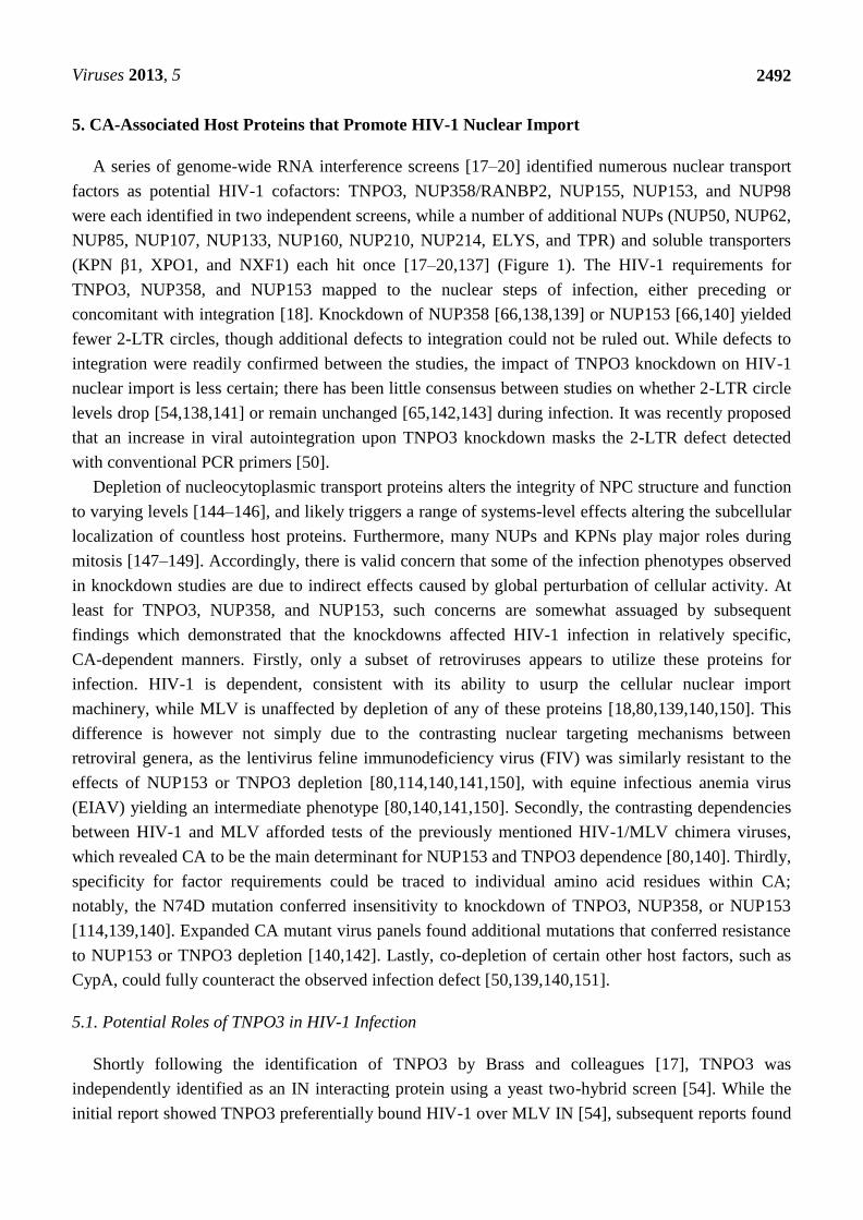
Figure 1. Schematic of the nuclear pore complex (NPC) and classical nuclear import
pathway. (top) General NPC substructures and locations of nucleoporins (NUPs) that
scored as potential HIV-1 co-factors in genome-wide RNA interference screens [17–20].
Asterisks denote NUPs that scored in more than one screen. (bottom) The Ran-based
nuclear import cycle. Import protein cargo binds to a karyopherin (KPN) β protein,
oftentimes bridged by a member of the KPN α protein family (KPN β1, which is also
referred to as importin β1, and KPN α2 or importin α1, depicted, are canonical members of
each protein family). KPN β1 ferries the complex through the NPC channel. The
engagement of KPN β1 by Ran-GTP concentrated at the nuclear basket releases the KPN
α-cargo complex into the nucleus. KPN β1 becomes free to bind additional import cargo
after Ran dissociates from it upon RanBP1 binding and Ran-GTP hydrolysis, stimulated by
RanGAP concentrated at the cytoplasmic filaments.
2. Measurements of the HIV-1 Nucleoprotein Substrate for Nuclear Import
Mature HIV-1 virions harbor a relatively full complement of viral proteins, including gag- (matrix,
MA; capsid, CA; nucleocapsid, NC; p6) and pol- (protease, PR; reverse transcriptase, RT; and
integrase, IN) encoded proteins, as well as a handful of accessory proteins (Vif, Vpr, and Nef). CA is
composed of two independently folded protein domains, the N-terminal domain (NTD) and C-terminal
domain (CTD), which are separated by a flexible linker [21]. During particle maturation,
approximately one-half of the complement of CA protein condenses into a conical shell that is

Viruses 2013, 5
2486
predominantly comprised of hexameric CA rings; twelve pentameric rings afford shape declinations
necessary to enclose retroviral CA shells [22–25]. The biological significance of the remaining CA
proteins that are not incorporated into the core is currently unknown. The mature core shell encases the
viral components that are necessary to complete the early steps of retroviral infection, which includes
the two copies of the viral RNA in complex with NC, RT, and IN. Shortly after viral-cell fusion, the
virus reverse transcribes its genome in the context of a subviral complex that is commonly referred to
as the reverse transcription complex (RTC) (Figure 2) [26]. DNA synthesis likely triggers CA shell
disassembly, as prevention of reverse transcription can delay the steps of core uncoating [27,28]. As
the CA core begins to disassemble, some viral proteins diffuse away from the now permeable CA
shell [29]. The combination of CA core disassembly and additional host protein recruitment increases
the size of the RTC to an estimated ~100–250 nm in diameter [30–32]. The number of complete, or
near-complete reverse transcribed genomes in a population of infected cells can be readily measured
by quantitative PCR, most commonly with a primer pair that generates an amplicon spanning from the
upstream long terminal repeat (LTR) to a sequence past the primer binding site, such as a sequence in
the upstream region of the gag gene; the viral DNAs detected by such reactions are commonly referred
to as late reverse transcription (LRT) products because they depend on the second template switch of
reverse transcription for their formation [33,34]. Once reverse transcription is completed,
IN hydrolyzes the extremities of the linear viral DNA adjacent to conserved cytosine-adenine
dinucleotides located within the viral LTRs to generate reactive CAOH-3’ ends [35,36]; the resulting
3’-hydroxyl groups are subsequently used by IN to cut target DNA to effect viral DNA joining [37].
By convention, the integration-competent nucleoprotein complex formed by IN 3’ processing activity
is referred to as the preintegration complex (PIC). HIV-1 PIC formation is believed to largely occur
within the cytoplasm [38], as the virus traffics to the nuclear periphery along microtubules [32].
The majority of virus particles are noninfectious [39], and it is accordingly challenging to observe
HIV-1 PIC nuclear transport directly with certainty. Nuclear transport is routinely inferred through
comparison of steady-state levels of readily detectable markers of the bulk infection. Arguably, the
most direct method is quantitative assessment of LRT DNA in cytoplasmic versus nuclear fractions.
Importantly, this requires careful validation of the fidelity of cellular fractionation, using control
protein or nucleic acid markers appropriate to represent the separate compartments [40,41]. While
many fractionation procedures likely separate soluble cytoplasmic components adequately, protocols
likely differ in their abilities to distinguish nucleoplasmic viral DNA from PICs associated with the
NPC, or strongly associated with the cytoskeleton. Before successfully integrating into a host
chromosome, a subset of PICs [42] are instead diverted to form non-productive circular DNA products
through the action of host cell-mediated DNA repair pathways: 1-LTR circles can be produced by
homologous recombination [43] or from aberrant reverse transcription [44,45], while 2-LTR circles are
formed through non-homologous end joining of the viral DNA [46]. PCR primers that amplify
products that span the LTR-LTR circle junction provide a convenient, albeit indirect measurement for
assessing the competence of the virus to reach the nuclear interior. Due to the generation of reactive
CAOH ends by IN 3’ processing activity, a fraction of PICs aberrantly integrate their LTR ends back
into an internal region of the viral DNA in a process referred to as autointegration [47–50].
Importantly, autointegrants formed by the insertion of one LTR end in the vicinity of the second viral
DNA end can score as positive in assays quantitating 2-LTR circles, confounding the use of 2-LTR

Viruses 2013, 5
2487
circle measures as readouts for nuclear viral DNA [50]. Specific aspects of PCR design, which take
into account the DNA sequence at the LTR-LTR circle junction, can accordingly help to mitigate this
complication [50]. Absolute and relative levels of integration can be measured through PCR reactions
specific for integrated proviral DNA [34,51], while the distribution of integration sites along
chromosomes are assessed by identifying sequences of viral-cellular DNA junctions within the
infected cell population [52,53]. While continuous advancement of various microscopy-based
approaches provides an important additional avenue to assess HIV-1 nuclear import and integration
[31,54,55], high-throughput, live-cell approaches capable of kinetically witnessing individual nuclear
import events are not yet available.
Figure 2. PCR-based methods for detection of post-entry to viral DNA integration steps of
HIV-1 infection. (A) Generalized replication intermediates and byproducts leading up to
integration. (B) Order of viral trafficking and RT and IN enzymatic steps. (C) Summary
of viral DNA species that serve as markers for the various infection intermediates
and byproducts.
Various biochemical approaches have yielded insight into the viral proteins that remain as part of
the viral nucleoprotein substrate for nuclear import. The RTC/PIC observed in the cytoplasm exceeds
the diameter of the pore [31,32], so only a fraction of the viral and cellular proteins that associate with
the PIC in the cytoplasm likely enter the nucleus [31]. The double-stranded reverse-transcribed viral
DNA and a tetramer of IN protein form the heart of the PIC, as they comprise the intasome
nucleoprotein complex that drives integration [56,57]. Keeping in mind that only one functional PIC is
formed per infectious event, the identities of other PIC-associated viral proteins have been difficult to
identify precisely. MA, RT, and Vpr were repeatedly found to be components of viral nucleoprotein

Viruses 2013, 5
2488
complexes isolated from nuclear fractions [40,58–61]. A handful of studies found NC and PR as
well [59,60,62], while CA was noticeably absent from many of these same studies [40,58–62]. In fact,
CA was either observed to be absent [26,61], or found in only scant amounts [38] within viral
complexes extracted from whole-cell or cytoplasmic extracts, prompting initial belief that the HIV-1
core uncoats completely prior to PIC formation. Subsequent microscopy studies have more readily
observed CA in association with cytoplasmic nucleoprotein complexes [31,32], though the duration of
this association remains largely unknown. One study found CA-staining ultrastructures associated with
NPCs and the nuclear envelope [63], with the authors consequently suggesting that the majority of
uncoating occurs at the nuclear periphery upon completion of reverse transcription. Although the
extent of CA core integrity at the nuclear periphery is controversial, PIC-associated IN is at least
partially exposed to the cytoplasm: cytoplasmically-localized fusion proteins containing green
fluorescent protein (GFP) and the IN-binding domain of lens epithelium-derived growth factor
(LEDGF)/p75 potently inhibited HIV-1 infection after reverse transcription [64]. While some studies
have detected CA in the nuclear fraction following HIV-1 infection [65,66], it is not entirely clear how
much of this signal represents intranuclear CA rather than CA protein associated with the nuclear
envelope. It would therefore be instructive to determine how much of this signal co-fractionates with
nuclear PICs.
3. Viral and Cellular Elements Implicated in HIV-1 PIC Nuclear Import
Many of the viral elements found in association with the PIC have been proposed to be important
for HIV-1 nuclear import. Nuclear localization signals (NLSs) present in MA [67,68] and IN [60,69],
as well as various non-canonical karyophilic signals in Vpr [61,70–74], have each been proposed to
recruit cellular nuclear transport proteins. Basic-type NLSs within IN have been proposed to recruit
KPN α adaptor proteins importin α1 (Rch1) [60] and importin α3 (KPNA4) [75], which would
presumably require additional binding to KPN β1 for function (Figure 1). IN can also directly interact
with KPN β proteins importin 7 [76,77] and transportin 3 (TNPO3, TRN-SR2, or importin 12) [54,78],
though the relevance of these interactions have been brought into question [79–81]. IN and Vpr are
additionally proposed to bind NUPs directly to facilitate nuclear import without the need for adaptor
KPN carrier proteins, which include interactions between IN and NUP153 [82], and Vpr with
Pom121 [70] or hCG1 [83].
The reverse transcribed genome is suggested to be an important determinant of HIV-1 PIC nuclear
import, primarily through a triple stranded DNA flap element generated through the action of the
central polypurine tract (cPPT) and central termination signal (CTS) [84,85]. While this element is not
absolutely required for either nuclear import or infection [86–88], numerous groups have confirmed
that the DNA flap exerts a positive effect during infection [89–91]. DNA plus-strand extension from
the cPPT primer likely decreases the overall duration of reverse transcription within the cell [90,92],
which may indirectly influence viral nuclear import during instances of limiting nucleotide
concentrations, for example. Such a kinetic advantage to reverse transcription conferred by the cPPT is
consistent with its ability to reduce the time-frame in which the viral single-stranded DNA is sensitive
to the inhibitory activity of APOBEC3 cytosine deaminase restriction factors [93–95], and may
similarly protect the RTC/PIC from other host defense proteins that could derail its trafficking [96].

Viruses 2013, 5
2489
While HIV-1 MA, IN, and Vpr NLSs can confer nuclear localization when fused to otherwise
cytoplasmic proteins, some studies have refuted the importance of these signals in PIC nuclear import
during infection [87,90,97–99]. Various features of HIV-1 biology can help to explain some of these
discrepancies. Firstly, MA and IN in particular are not known to function as free proteins during the
early phase of HIV-1 infection. Thus, studying MA or IN as recombinant proteins expressed in human
cells may not uncover behaviors relevant to PIC biology. Secondly, many of the viral constituents of
the PIC are multifunctional proteins, whereby mutations may result in multiple coincident defects to
infection, obfuscating the targeted assessment of contributions to a particular phenotype. Lastly, there
is little evidence to support that HIV-1 enters the nucleus during mitosis when its passage through the
NPC is blocked [100]. Thus, the historical perspective that HIV-1 nuclear import mutants would
specifically be blocked for infection of non-dividing target cell types would since appear to be
largely misguided.
4. CA Functionally Determines Requirements for Nuclear Trafficking
The field has more recently moved to view CA as the major viral protein that mediates HIV-1
nuclear import. Masahiro Yamashita and Michael Emerman demonstrated that infection by an HIV-1
chimeric virus that carried the MLV CA protein was cell cycle dependent [101], mimicking the
property of parental MLV. The defect to infection upon cell cycle arrest was at the step of nuclear
import, as a decrease in the formation of 2-LTR circle DNAs relative to wild-type (WT) HIV-1 was
observed [101]. They subsequently determined that certain point mutations in HIV-1 CA, including
T54A/N57A and Q63A/Q67A (Figure 3), also imparted cell cycle dependence to HIV-1 [41]. Notably,
the infection defect exhibited by the T54A/N57A mutant virus upon cell cycle arrest occurred after
nuclear entry but before integration [41]. While this mutant was sensitive to cell cycle arrest in all cell
lines tested, the growth arrest phenotypes of A92E and G94D CA mutant viruses, which are
hypersensitive to the levels of CA-interacting host protein cyclophilin A (CypA) in certain cell lines
(HeLa and H9 cells), were restricted to these same cells [102,103]. Cell cycle arrest also inhibited
these viruses after nuclear import, as both LRT and 2-LTR circle levels remained unchanged. While
the infection defects experienced by these CA mutant viruses upon cell cycle arrest occur following
HIV-1 nuclear entry, the Q63A/Q67A mutant virus appeared to be defective for nuclear import, as it
exhibited decreased levels of 2-LTR circle formation as compared to WT virus in dividing cells [104].
Q63A/Q67A CA cores recovered from whole virions following detergent treatment were less stable
than WT cores in vitro [105], but the mutant viral RTCs and PICs retained a greater complement of
CA protein than did the WT virus during infection [27,41,104]. This apparent delay in Q63A/Q67A
core uncoating appears related to the nuclear import and integration defects experienced by this
mutant virus.
In addition to CypA, other CA-interacting host factors have been observed to affect HIV-1 nuclear
import and integration. The rhesus Trim5α restriction factor normally restricts HIV-1 infection by
targeting the viral core for disassembly and degradation prior to the completion of reverse transcription
[106,107]. Inhibition of cellular proteasome activity with the small molecule MG132 rescued reverse
transcription while having no effect on the ultimate level of integration [108]. The MG132-rescued
RTCs seemingly remain intact [109] and mature into integration-competent PICs [110]. These

Viruses 2013, 5
2490
complexes also escape entrapment by proteasome-associated cytoplasmic bodies [111] yet accumulate
fewer 2-LTR circles, consistent with a trafficking defect coincident with or shortly prior to nuclear
import [108,110]. Various artificially engineered Trim-CypA fusion constructs have also been shown
to cause defects to infection after reverse transcription, though these appear to occur after nuclear
entry [112]. Similar phenotypes have been observed with variants of the cleavage and polyadenylation
specific factor 6 (CPSF6) mRNA processing protein. While expression of the C-terminal truncation
variant CPSF6375 inhibited HIV-1 reverse transcription [113], the marginally shorter C-terminal
truncation mutant CPSF6358, which harbored amino acid residues oftentimes removed as exon 6,
inhibited 2-LTR circle formation while leaving reverse transcription unaffected [114,115]. Recently,
the interferon induced antiviral protein Mx2 was found to block HIV-1 infection after reverse
transcription in a CA-dependent manner [116,117].
Figure 3. Schematic of HIV-1 capsid (CA) and mutations described in the text. A single
HIV-1 CA monomer (protein database code 3j34) is represented by a cartoon of the peptide
backbone, as well as a semi-transparent surface representation: N-terminal domain (NTD),
green; flexible linker, purple; C-terminal domain (CTD), red. A subset of CA residue
side-chains that exhibit phenotypic differences in preintegrative steps of HIV-1 infection
are shown as sticks and colored as follows: carbon, orange; nitrogen, blue; oxygen, red.
Similar phenotypes have recently been observed with small molecules that target HIV-1 CA. Pfizer
compound PF-3450074 (PF74), which was discovered in a high-throughput screen for inhibitors
of HIV-1 replication [118], was subsequently shown to bind a hydrophobic pocket formed between
HIV-1 CA NTD alpha helices 3, 4, and 5 [118] (Figure 3). PF74 destabilized virion-purified CA cores
in vitro, and inhibited the completion of reverse transcription during infection [119]. Boehringer
Ingelheim pyrrolopyrazolone compounds BI-1 and BI-2 were more recently determined to engage the
same pocket within the CA NTD [120]. Interestingly, treatment with these compounds did not affect
HIV-1 reverse transcription, but instead resulted in decreased accumulation of 2-LTR circles. The
phenotypic difference may relate to potentially contrasting effects on CA uncoating, as BI-1 delayed

Viruses 2013, 5
2491
the disassembly of higher-order HIV-1 CA-NC structures in vitro [120]. Interestingly, the binding sites
of these compounds on CA overlap that of CPSF6 [120,121] and NUP153 [122]. Coumermycin A-1, a
separate small molecule, was found to inhibit HIV-1 integration [123]. While the binding site of this
compound is not known, passage of HIV-1 in its presence yielded the outgrowth of virus encoding the
A105S mutation in the CA NTD. As Ala105 is proximal to the aforementioned PF74/BI-1/CPSF6
binding site (Figure 3), it seems likely that the inhibitory mechanism of coumermycin A-1 is related to
these factors.
Perturbation of CA can also affect the post-reverse transcription steps of other retroviruses. Though
MLV does not enter the nucleus via the NPC, it must also in large part dissolve its CA shell to effect
IN-mediated integration. MLV CA is readily found in RTCs purified from infected cells, and remains
stably associated with the PIC until it enters the nucleus [124]. MLV p12, a gag-encoded protein not
found in HIV-1, is crucial for nuclear targeting as it tethers PICs to mitotic chromosomes [125–127].
While MLV CA dissociates from the PIC during mitosis, p12 mutants PM14 and S61A/S65A, which
are defective for mitotic chromosomal tethering, each maintain CA in association with the PIC during
mitosis [128]. Certain murine cells take advantage of RTC/PIC-associated CA to interrupt the MLV
infection mechanism: the protein expressed from Friend virus susceptibility-1 (Fv1), which is a
gag-related gene from a murine endogenous retroelement [129], is able to target MLV cores to inhibit
infection [130–132]. Restricted MLV still reverse transcribes and forms PICs capable of integrating in
vitro [133], yet does not form circular DNA byproducts [134,135]. The product of the Fv1 gene can
also inhibit HIV-1 infection when targeted to HIV-1 CA upon fusion to CypA, resulting in a
quantifiable decrease in the number of integrated proviruses while leaving the accumulation of 2-LTR
circles unchanged [136].
Together, these results show that the retroviral core shell is unlikely to passively fall apart upon
viral entry, but instead functions at a critical juncture bridging reverse transcription and nuclear
trafficking. Although premature CA disassembly and proteasomal targeting may exert their effects as
early as reverse transcription, other perturbations to CA uncoating and CA-determined trafficking
defects prevent PIC nuclear import, or even manifest as defects within the nucleus. MLV has
specifically evolved to access chromosomes during mitosis, and accordingly, the combined functions
exerted by MLV CA and p12 likely specifically link MLV uncoating and nuclear entry with mitosis. In
the aforementioned chimeric HIV-1 encoding MLV gag, the PIC is likely forced into an MLV-type
mechanism of nuclear entry. Contrastingly, the cell cycle dependence of HIV-1 CA missense
mutations may be due to reasons stemming from various potential losses in function: for example,
perturbed core engagement with host proteins may result in a virus blocked at one of many
preintegrative steps of infection, and may require cellular rearrangements that occur during cell
division to relieve this block. While the previously described phenotypes reveal effects the retroviral
core may exert on steps following reverse transcription, recent findings that CA protein physically
associates with nuclear transport factors hints that CA takes a direct role in promoting the nuclear steps
of HIV-1 infection.
Viruses 2013, 5
2492
5. CA-Associated Host Proteins that Promote HIV-1 Nuclear Import
A series of genome-wide RNA interference screens [17–20] identified numerous nuclear transport
factors as potential HIV-1 cofactors: TNPO3, NUP358/RANBP2, NUP155, NUP153, and NUP98
were each identified in two independent screens, while a number of additional NUPs (NUP50, NUP62,
NUP85, NUP107, NUP133, NUP160, NUP210, NUP214, ELYS, and TPR) and soluble transporters
(KPN β1, XPO1, and NXF1) each hit once [17–20,137] (Figure 1). The HIV-1 requirements for
TNPO3, NUP358, and NUP153 mapped to the nuclear steps of infection, either preceding or
concomitant with integration [18]. Knockdown of NUP358 [66,138,139] or NUP153 [66,140] yielded
fewer 2-LTR circles, though additional defects to integration could not be ruled out. While defects to
integration were readily confirmed between the studies, the impact of TNPO3 knockdown on HIV-1
nuclear import is less certain; there has been little consensus between studies on whether 2-LTR circle
levels drop [54,138,141] or remain unchanged [65,142,143] during infection. It was recently proposed
that an increase in viral autointegration upon TNPO3 knockdown masks the 2-LTR defect detected
with conventional PCR primers [50].
Depletion of nucleocytoplasmic transport proteins alters the integrity of NPC structure and function
to varying levels [144–146], and likely triggers a range of systems-level effects altering the subcellular
localization of countless host proteins. Furthermore, many NUPs and KPNs play major roles during
mitosis [147–149]. Accordingly, there is valid concern that some of the infection phenotypes observed
in knockdown studies are due to indirect effects caused by global perturbation of cellular activity. At
least for TNPO3, NUP358, and NUP153, such concerns are somewhat assuaged by subsequent
findings which demonstrated that the knockdowns affected HIV-1 infection in relatively specific,
CA-dependent manners. Firstly, only a subset of retroviruses appears to utilize these proteins for
infection. HIV-1 is dependent, consistent with its ability to usurp the cellular nuclear import
machinery, while MLV is unaffected by depletion of any of these proteins [18,80,139,140,150]. This
difference is however not simply due to the contrasting nuclear targeting mechanisms between
retroviral genera, as the lentivirus feline immunodeficiency virus (FIV) was similarly resistant to the
effects of NUP153 or TNPO3 depletion [80,114,140,141,150], with equine infectious anemia virus
(EIAV) yielding an intermediate phenotype [80,140,141,150]. Secondly, the contrasting dependencies
between HIV-1 and MLV afforded tests of the previously mentioned HIV-1/MLV chimera viruses,
which revealed CA to be the main determinant for NUP153 and TNPO3 dependence [80,140]. Thirdly,
specificity for factor requirements could be traced to individual amino acid residues within CA;
notably, the N74D mutation conferred insensitivity to knockdown of TNPO3, NUP358, or NUP153
[114,139,140]. Expanded CA mutant virus panels found additional mutations that conferred resistance
to NUP153 or TNPO3 depletion [140,142]. Lastly, co-depletion of certain other host factors, such as
CypA, could fully counteract the observed infection defect [50,139,140,151].
5.1. Potential Roles of TNPO3 in HIV-1 Infection
Shortly following the identification of TNPO3 by Brass and colleagues [17], TNPO3 was
independently identified as an IN interacting protein using a yeast two-hybrid screen [54]. While the
initial report showed TNPO3 preferentially bound HIV-1 over MLV IN [54], subsequent reports found

Viruses 2013, 5
2493
TNPO3 to bind both proteins comparably [80,150]. In fact, TNPO3 efficiently bound a variety of
purified retroviral IN proteins [80], including those derived from TNPO3-independent viruses, such as
MLV and FIV. HIV-1 IN interacts with TNPO3 with relatively high affinity, with measured
dissociation constants ranging from ~17 nM [152] to ~260 nM [80]. This interaction could be
competed by the Ran GTPase mutant Q69L in complex with GTP, consistent with IN being an import
cargo for TNPO3 [153]. A series of basic residues within the IN CTD, including Arg262 and Lys264,
appear particularly important in binding to TNPO3 [78,152]. Unfortunately, viruses containing IN
proteins harboring TNPO3 binding mutations exhibited pleiotropic defects during infection, which
included greatly reduced levels of reverse transcription, precluding the specific measurement of
associated nuclear import defects [152]. Notably, a chimeric HIV-1 virus harboring MLV IN in place
of HIV-1 IN was as sensitive to TNPO3 depletion as WT HIV-1 [80,150]. While IN may engage
TNPO3 in the context of HIV-1 infection, the potential role for this interaction in PIC nuclear
localization appears auxiliary to that played by CA.
Various reports have investigated whether CA might bind to TNPO3 directly. A recombinant fusion
protein consisting of glutathione S-transferase and TNPO3 pulled-down CA and cellular tRNA from
purified virions [65]. The extent of CA recovery was enhanced in the presence of Ran(Q69L)-GTP,
implying a directionality of binding favoring nuclear export, though the quantitative difference in
this result was only approximately two-fold [65]. TNPO3 preferentially bound WT over
TNPO3-independent N74D mutant CA-NC tubes in vitro [143], though this difference was less than
two-fold, and a subsequent study failed to confirm this difference [78]. TNPO3 binding has been
suggested to alter CA core uncoating: purified TNPO3 accelerated the extent of in vitro uncoating of
purified viral cores, while Ran(Q69L)-GTP counteracted this effect [154]. It remains unclear whether
the relatively weak in vitro interaction between HIV-1 CA and TNPO3 occurs within cells, and if it
does, whether it is relevant for infection.
There is some evidence to suggest that the primary function of TNPO3 during HIV-1 infection is
control of CPSF6 localization within the cell. CPSF6 is a member of a large family of eukaryotic
pre-mRNA splicing factors collectively referred to as SR proteins. These proteins typically contain
discrete domains rich in arginine-serine dipeptide repeats (RS domains). RS domains have been shown
to serve as nuclear localization signals for other SR proteins involved in mRNA splicing, such as
ASF/SF2 and SC35 [155], by interacting with TNPO3. While an interaction between CPSF6 and
TNPO3 has yet to be formally shown, it appears as though TNPO3 does bind CPSF6 and control its
localization: CPSF6 is normally a nuclear protein, and TNPO3 knockdown has been observed to
increase the redistribution of CPSF6 to the cytoplasm [50]. While exogenously introduced full-length
CPSF6 is predominantly nuclear and does not adversely affect HIV-1 infection [113,114],
mislocalizing it to the cytoplasm by appending a nuclear export sequence inhibits infection [151].
Conversely, attaching a functional NLS onto the normally cytoplasmic CPSF6358 counteracted its
ability to restrict HIV-1 infection [50,121]. Concomitant CPSF6 knockdown moreover counteracted
the inhibitory effects of TNPO3 depletion [50,151], and a similar set of CA mutations conferred
insensitivity to TNPO3 depletion and CPSF6358 restriction [50]. Still, some inconsistencies remain: for
example, EIAV does not appear to bind CPSF6 [115,151], yet it is reproducibly inhibited by TNPO3
depletion [80,141,150]. While TNPO3 may serve more than one role during HIV-1 infection, altered

Viruses 2013, 5
2494
CPSF6 localization seems to account for a major part of the infectivity defect induced by TNPO3
knockdown.
5.2. Viral Interactions with NUP153 and NUP358
The roles of NUP153 and NUP358 during HIV-1 nuclear import have also been recently
investigated. NUP153 is a large 1475 amino acid NUP found on the nuclear side of the NPC. While its
NTD associates with the nuclear basket [156,157], its highly flexible CTD can reach into the central
channel [158], extending across to the cytoplasmic side of the NPC in a transport-dependent manner
[159–161]. Like TNPO3, NUP153 was reported to bind HIV-1 IN [82]. This interaction was mapped
to the C-terminal FG-rich domain of NUP153. Though interaction could be observed between purified
NUP153 CTD and HIV-1 IN, it was not observed with FIV IN [82]. Additionally, this interaction was
inhibited by the addition of cytosolic extract, presumably due to competition with KPN binding to the
NUP153 CTD, as addition of a non-hydrolyzable GTP analog counteracted this effect [82]. This result
may explain why co-immunoprecipitation was not observed between cell expressed GFP-tagged
NUP153 and HIV-1 IN [162]. An interaction was also observed between GFP-NUP153 extracted from
animal cell lysate and CA-NC tubes in vitro [162]. We recently confirmed this finding, and have
additionally detected a direct interaction between the NUP153 CTD and HIV-1 CA NTD [122]. This
interaction moreover mapped to the FG motifs present within the NUP153 CTD [122]. Notably,
NUP153 bound N74D CA as well as WT CA [122,162].
NUP358 is an even larger, 3224 amino acid NUP that forms the cytoplasmic filaments emanating
from the NPC [163–165] (Figure 1). In contrast to TNPO3 and NUP153, the only retroviral protein
NUP358 has been published to bind is HIV-1 CA [139]. This interaction was found to occur through
the CypA homologous domain (CHD) that resides at the C-terminus of NUP358. The NUP358 CHD is
similar to CypA in both overall primary sequence and tertiary structure, though there are a few
differences in active site residues [166]. NUP358 was initially found to bind the CA NTD with slightly
weaker affinity than CypA (16 M, vs. 7 M for CypA) [139], though subsequent reports indicate a
much weaker interaction (~100–200 M) [166,167]. The CA proteins from SIVmac and HIV-1 CypA
loop mutants G89V and P90A do not bind CypA [168], and these proteins also failed to bind NUP358
CHD [139]. These viruses were accordingly insensitive to NUP358 knockdown [139]. Consistent with
its localization to the cytoplasmic edge of the NPC, NUP358 appears to be critical for the docking of
the HIV-1 PIC to the NPC during infection [66]. NUP358 CHD appears to possess isomerase activity
for various substrates [166,169]. Although it is capable of isomerizing HIV-1 CA [167], it is not yet
clear whether isomerization is necessary for HIV-1 infection. Additionally, other regions of NUP358
aside from the CHD may serve important functions during infection [170].
Interestingly, NUP153 and NUP358 exhibit certain similarities. While found on opposite sides of
the NPC, each protein localizes to the periphery of the NPC, and may represent initial sites of NPC
attachment/detachment during passage through the nuclear pore. Accordingly, each protein contains
FG repeats, and makes contacts with nuclear transport receptors. NUP153 and NUP358 each encode
tandem repeats of homologous zinc-fingers, which are involved in Ran binding [171,172] and COPI
recruitment during nuclear envelope breakdown [173]. It remains to be seen whether any of these
similarities play into the importance of these proteins during HIV-1 infection.

Viruses 2013, 5
2495
5.3. Interdependence of CA-determined Host Factors during Infection
A number of phenotypic similarities suggest the roles of TNPO3, NUP153, NUP358, CPSF6, and
CypA during HIV-1 infection are interrelated. Starkly, the N74D CA mutant virus is insensitive to
knockdown of TNPO3, NUP153, and NUP358, despite possessing a CA protein capable of binding all
three proteins with similar affinities to WT CA. As the N74D mutation clearly counteracts CPSF6
binding [114], it seems plausible that CPSF6 engagement licenses HIV-1 to employ NUP358 and
NUP153 during infection. CPSF6 is currently believed to be exclusively nuclear at steady state,
suggesting that it may not exert its effects on HIV-1 until the virus engages the NPC. Curiously,
siRNA depletion of CPSF6 does not affect HIV-1 infection [114].
CypA also appears to alter nuclear transport factor dependence during HIV-1 infection. Abrogation
of CA binding with CypA, either through CypA depletion or competition with the small molecule
cyclosporine, rescued viruses inhibited by NUP153 or NUP358 knockdown [139,140]. While
cyclosporine treatment can partially rescue WT HIV-1 infection in TNPO3 depleted cells [139,154],
the lack of complete rescue may reflect its multiple potential roles in promoting HIV-1 infection.
CypA binding to HIV-1 CA can alter its disassembly [154,174], suggesting that its effect on NUP153,
NUP358, and TNPO3 may be indirect through modulating the rate and extent of CA core uncoating.
Indeed prevention of CypA binding also modulates the antiviral effects of the rhesus Trim5α [175] and
Mx2 [116,117] restriction factors. Though CypA serves as a major regulator of the pre-integrative
steps of HIV-1 infection, the precise mechanistic details by which CypA exerts its effects are not
well understood.
6. Effects of Nuclear Transport Proteins on Integration Site Selection
While HIV-1 appears to predominantly utilize NUP153, NUP358, and TNPO3 to affect its import
into the nucleus, these factors can also affect post-nuclear trafficking as evidenced by differences in
HIV-1 integration site distributions upon factor knockdown. Numerous different forces can influence
integration site distribution. IN favors certain nucleotide patterns at the site of integration [176,177].
Integration also favors the distorted major grooves that occur when DNA is wrapped around the
nucleosome core [178,179], as well as certain epigenetic modifications [179]. On the genomic level,
HIV-1 preferentially targets the bodies of active genes within gene dense regions of chromosomes
[52]. Whereas LEDGF/p75 in large part dictates the preference for active gene bodies [180–182], the
targeting of gene dense regions of chromosomes is apparently linked to nuclear import: depletion of
TNPO3, NUP358, and to a lesser extent, NUP153, significantly reduced the extent of integration in
gene dense regions of chromatin [139,162,183,184]. This pattern was moreover consistent with the
involvement of CA, as the HIV-1 chimeric virus encoding MLV CA, as well as CA missense mutants
N57A and N74D, showed a similar shift in integration site distribution [139,183,184]. Notably, CypA
binding was also found to affect integration site selection, as disruption of CypA binding to CA by
cyclosporine treatment resulted in an increase in the number of integration events in chromosomal
regions enriched in transcriptional units [139].
It is possible that these proteins are directly involved in guiding the PIC to distinct regions of
chromatin; NUP153 has been shown to associate with large regions of active chromatin in drosophila

Viruses 2013, 5
2496
[185], and TNPO3 may engage the HIV-1 intasome to effect integration [78]. On the other hand,
NUP358 appears more important for integration targeting than NUP153, yet this protein does not
appear to be found within the nucleus during interphase [186]. Furthermore, depletion of a number of
other nuclear host proteins including IK, ANAPC2, WDHD1, SNW1, and PRPF38A similarly
redirected integration site targeting [183]. It remains formally possible that the roles of some host
factors in dictating integration to gene dense regions may be indirect, instilled through alteration of
global chromosomal environment as compared to specific effects on HIV-1 PIC trafficking. Still,
ablation of gene-dense region targeting by CA mutations such as N74D highlights a specific role for
CA in post-nuclear PIC trafficking. The mechanism of nuclear import may be linked with integration
site targeting by affecting the chromosomal environments first encountered by the PIC upon
nuclear entry.
7. Model of CA and Nuclear Transport Factors during HIV-1 Nuclear Entry
We propose the following working model to coalesce recently reported results from the rapidly
evolving field of HIV-1 PIC nuclear transport (Figure 4). While the initial steps of uncoating likely
occur shortly after entry [27], the final events of uncoating may occur at the NPC [63]. The partially
uncoated PIC most likely docks at the NPC, by engaging NUP358 with its remaining CA proteins
[66,139]. Once docked, CPSF6 and NUP153 then engage the PIC. The combined actions of these
proteins are necessary for PIC nuclear import. TNPO3 expression is required for proper nuclear
localization of CPSF6; CPSF6 binding to CA cores too early during infection misregulates the
upstream steps of uncoating and NPC engagement, blocking infection at the step of nuclear import.
TNPO3 may also have an additional intra-nuclear role permitting proper nuclear trafficking and
integration, perhaps related to its interaction with IN. These concerted steps of uncoating and nuclear
import appear to influence the downstream steps of nuclear trafficking and integration, as depletion of
TNPO3, NUP153, and NUP358 reduce targeting of the PIC to gene-dense regions of chromatin. It will
be instructive to ascertain if CPSF6 depletion influences HIV-1 integration site distribution.
The precise mechanistic requirements for NUP358, NUP153, and CPSF6 for nuclear import remain
unclear: these proteins may be critical for a prerequisite uncoating step prior to nuclear import, or they
may be directly involved in the act of PIC nuclear translocation. As these three proteins have each
been published to bind CA, the latter model presupposes that CA would need to be concomitantly
imported into the nucleus with the PIC. This point remains highly controversial; CA has historically
been noticeably absent from the nucleus, with only a couple recent reports observing potential PIC-
associated CA signals within the nuclear fraction [65,66]. Additionally, there is currently no evidence
describing a mechanism by which disassembled CA may remain associated with the PIC. Notably,
TNPO3, NUP358, and NUP153 are not absolutely required for HIV-1 infection of transformed cell
lines: though WT virus is highly dependent on these factors, CA mutant viruses such as N74D can
bypass CPSF6 binding and infect cells depleted for NUP358, NUP153, or TNPO3 without a
concomitant loss of infectivity. While the N74D CA mutant virus was previously proposed to bypass
these requirements by relying on an alternative set of NUPs (including NUP155 and NUP98) [114], it
is not clear whether these proteins indeed fulfill critical roles for N74D mutant virus infection [162].

Viruses 2013, 5
2497
Figure 4. Model of the potential roles of the CA-dependent nuclear transport factors during
HIV-1 infection. NUP358, NUP153, and CPSF6 at the nuclear pore most likely act on
PIC-associated CA to aid HIV-1 infection. TNPO3 is required to localize CPSF6 to the
nucleus; premature cytoplasmic CPSF6 binding to CA prevents nuclear import. TNPO3
may affect integration by interacting with IN within the nucleus. CypA modulates CA
uncoating, altering dependencies on NUP358, NUP153, and TNPO3. Perturbation of this
pathway by CA mutation or TNPO3, NUP153, or NUP358 knockdown results in altered
integration site selection away from gene-dense regions of chromatin.
Alternatively, if the main function of these nuclear transport factors is to uncoat the PIC as a
prelude for nuclear import, then alterations to viral uncoating may obviate the need for this mechanism
during infection. While a mechanism for active PIC nuclear transport would be required in all cell
types, optimal CA uncoating may be particularly important in particular cells, such as macrophages,
where premature uncoating may render the PIC susceptible to intracellular antiviral factors. Indeed, the
N74D CA mutant virus exhibits a significant infectivity defect in monocyte-derived macrophages
[139,187], where its reverse transcription is defective [187]. Because Asn74 is highly conserved
among primate lentiviruses, HIV-1 may very well rely on these nuclear transport factors in vivo [114].
The HIV-1 requirement for CypA may be similarly nuanced: while it exerts differential effects in
various cell types, its importance as a fine-tuned regulator of CA uncoating is likely most important in
cell types where premature uncoating may be most detrimental [139,188].
Curiously, although HIV-1 appears to rely upon NUP358, NUP153, TNPO3, and CPSF6 during
infection, other lentiviruses only appear to share certain aspects of this mechanism. SIVmac does not
bind NUP358, and accordingly does not rely on this protein for infection. Furthermore, EIAV utilizes
NUP153 and TNPO3, though it does so in the apparent absence of CPSF6 binding. FIV likely utilizes
an entirely different mechanism, as it does not seem to require any of these factors. Similar to CypA
[189], various lentiviruses may have evolved to differentially rely upon these nuclear transport factors
over time. Although recent years have witnessed significant advances on the role of CA and particular
nuclear transport proteins in HIV-1 PIC nuclear import, there is clearly much left to learn about how
HIV-1 and some of the other lentiviruses circumvent the nuclear envelope to reach their chromosomal
targets of integration.

Viruses 2013, 5
2498
8. Conclusions
Despite intense study, the details of HIV-1 and other lentiviral nuclear import mechanisms remains
incompletely understood. This is perhaps at least somewhat reflective of the complexity and potential
redundancy of nuclear transport mechanisms through the NPC, as well as difficulties in precisely
measuring or developing accurate in vitro approximations for a trafficking process that occurs at the
heart of the lentiviral replication cycle. The convergence of recent findings functionally relating the
requirements of the retroviral CA protein with cellular nuclear transport factors have provided new
insight into the series of interrelated steps that begin with HIV-1 CA uncoating and end with proviral
integration. Despite its intricacies, continued study supported by advancements in genetic,
biochemical, and microscopy approaches are predicted to eventually unravel the detailed molecular
mechanisms that underlie retroviral PIC nuclear import.
Acknowledgments
Research in the authors’ laboratory is supported by NIH grants AI052014 and GM082251.
Conflict of Interest
The authors declare no conflict of interests.
References
1. Roe, T.; Reynolds, T.C.; Yu, G.; Brown, P.O., Integration of Murine Leukemia Virus DNA
Depends on Mitosis. EMBO J. 1993, 12, 2099–2108.
2. Gartner, S.; Markovits, P.; Markovitz, D.M.; Kaplan, M.H.; Gallo, R.C.; Popovic, M., The
Role of Mononuclear Phagocytes in HTLV–III/LAV Infection. Science 1986, 233, 215–219.
3. Hoelz, A.; Debler, E.W.; Blobel, G., The Structure of the Nuclear Pore Complex. Annu. Rev.
Biochem. 2011, 80, 613–643.
4. Rout, M.P.; Aitchison, J.D.; Suprapto, A.; Hjertaas, K.; Zhao, Y.; Chait, B.T., The Yeast
Nuclear Pore Complex: Composition, Architecture, and Transport Mechanism. J. Cell Biol.
2000, 148, 635–651.
5. Cronshaw, J.M.; Krutchinsky, A.N.; Zhang, W.; Chait, B.T.; Matunis, M.J., Proteomic
Analysis of the Mammalian Nuclear Pore Complex. J. Cell Biol. 2002, 158, 915–927.
6. Rout, M.P.; Blobel, G., Isolation of the Yeast Nuclear Pore Complex. J. Cell Biol. 1993, 123,
771–783.
7. Reichelt, R.; Holzenburg, A.; Buhle, E.L., Jr.; Jarnik, M.; Engel, A.; Aebi, U., Correlation
between Structure and Mass Distribution of the Nuclear Pore Complex and of Distinct Pore
Complex Components. J. Cell Biol. 1990, 110, 883–894.
8. Pante, N.; Kann, M., Nuclear Pore Complex Is Able to Transport Macromolecules with
Diameters of About 39 nm. Mol. Biol. Cell 2002, 13, 425–434.
9. Conti, E.; Muller, C.W.; Stewart, M., Karyopherin Flexibility in Nucleocytoplasmic Transport.
Curr. Opin. Struct. Biol. 2006, 16, 237–244.

Viruses 2013, 5
2499
10. Terry, L.J.; Wente, S.R., Flexible Gates: Dynamic Topologies and Functions for FG
Nucleoporins in Nucleocytoplasmic Transport. Eukaryot. Cell 2009, 8, 1814–1827.
11. Nemergut, M.E.; Mizzen, C.A.; Stukenberg, T.; Allis, C.D.; Macara, I.G., Chromatin Docking
and Exchange Activity Enhancement of RCC1 by Histones H2A and H2B. Science 2001, 292,
1540–1543.
12. Saitoh, H.; Pu, R.; Cavenagh, M.; Dasso, M., RanBP2 Associates with Ubc9p and a Modified
Form of RanGAP1. Proc. Natl. Acad. Sci. U. S. A. 1997, 94, 3736–3741.
13. Radu, A.; Moore, M.S.; Blobel, G., The Peptide Repeat Domain of Nucleoporin NUP98
Functions as a Docking Site in Transport across the Nuclear Pore Complex. Cell 1995, 81,
215–222.
14. Bischoff, F.R.; Klebe, C.; Kretschmer, J.; Wittinghofer, A.; Ponstingl, H., RanGAP1 Induces
GTPase Activity of Nuclear Ras–Related Ran. Proc. Natl. Acad. Sci. U. S. A. 1994, 91, 2587–
2591.
15. Kehlenbach, R.H.; Dickmanns, A.; Kehlenbach, A.; Guan, T.; Gerace, L., A Role for RanBP1
in the Release of Crm1 from the Nuclear Pore Complex in a Terminal Step of Nuclear Export.
J. Cell Biol. 1999, 145, 645–657.
16. Koyama, M.; Matsuura, Y., An Allosteric Mechanism to Displace Nuclear Export Cargo from
Crm1 and RanGTP by RanBP1. EMBO J. 2010, 29, 2002–2013.
17. Brass, A.L.; Dykxhoorn, D.M.; Benita, Y.; Yan, N.; Engelman, A.; Xavier, R.J.; Lieberman, J.;
Elledge, S.J., Identification of Host Proteins Required for HIV Infection through a Functional
Genomic Screen. Science 2008, 319, 921–926.
18. Konig, R.; Zhou, Y.; Elleder, D.; Diamond, T.L.; Bonamy, G.M.; Irelan, J.T.; Chiang, C.Y.;
Tu, B.P.; De Jesus, P.D.; Lilley, C.E., et al., Global Analysis of Host–Pathogen Interactions
That Regulate Early–Stage HIV–1 Replication. Cell 2008, 135, 49–60.
19. Zhou, H.; Xu, M.; Huang, Q.; Gates, A.T.; Zhang, X.D.; Castle, J.C.; Stec, E.; Ferrer, M.;
Strulovici, B.; Hazuda, D.J., et al., Genome–Scale RNAi Screen for Host Factors Required for
HIV Replication. Cell Host Microbe 2008, 4, 495–504.
20. Yeung, M.L.; Houzet, L.; Yedavalli, V.S.; Jeang, K.T., A Genome–Wide Short Hairpin RNA
Screening of Jurkat T–Cells for Human Proteins Contributing to Productive HIV–1
Replication. J. Biol. Chem. 2009, 284, 19463–19473.
21. Berthet–Colominas, C.; Monaco, S.; Novelli, A.; Sibai, G.; Mallet, F.; Cusack, S., Head–to–
Tail Dimers and Interdomain Flexibility Revealed by the Crystal Structure of HIV–1 Capsid
Protein (p24) Complexed with a Monoclonal Antibody Fab. EMBO J. 1999, 18, 1124–1136.
22. Ganser, B.K.; Li, S.; Klishko, V.Y.; Finch, J.T.; Sundquist, W.I., Assembly and Analysis of
Conical Models for the HIV–1 Core. Science 1999, 283, 80–83.
23. Pornillos, O.; Ganser–Pornillos, B.K.; Kelly, B.N.; Hua, Y.; Whitby, F.G.; Stout, C.D.;
Sundquist, W.I.; Hill, C.P.; Yeager, M., X–Ray Structures of the Hexameric Building Block of
the HIV Capsid. Cell 2009, 137, 1282–1292.
24. Pornillos, O.; Ganser–Pornillos, B.K.; Yeager, M., Atomic–Level Modelling of the HIV
Capsid. Nature 2011, 469, 424–427.

Viruses 2013, 5
2500
25. Zhao, G.; Perilla, J.R.; Yufenyuy, E.L.; Meng, X.; Chen, B.; Ning, J.; Ahn, J.; Gronenborn,
A.M.; Schulten, K.; Aiken, C., et al., Mature HIV–1 Capsid Structure by Cryo–Electron
Microscopy and All–Atom Molecular Dynamics. Nature 2013, 497, 643–646.
26. Fassati, A.; Goff, S.P., Characterization of Intracellular Reverse Transcription Complexes of
Human Immunodeficiency Virus Type 1. J. Virol. 2001, 75, 3626–3635.
27. Hulme, A.E.; Perez, O.; Hope, T.J., Complementary Assays Reveal a Relationship between
HIV–1 Uncoating and Reverse Transcription. Proc. Natl. Acad. Sci. U. S. A. 2011, 108, 9975–
9980.
28. Arfi, V.; Lienard, J.; Nguyen, X.N.; Berger, G.; Rigal, D.; Darlix, J.L.; Cimarelli, A.,
Characterization of the Behavior of Functional Viral Genomes During the Early Steps of
Human Immunodeficiency Virus Type 1 Infection. J. Virol. 2009, 83, 7524–7535.
29. Yu, Z.; Dobro, M.J.; Woodward, C.L.; Levandovsky, A.; Danielson, C.M.; Sandrin, V.; Shi, J.;
Aiken, C.; Zandi, R.; Hope, T.J., et al., Unclosed HIV–1 Capsids Suggest a Curled Sheet
Model of Assembly. J. Mol. Biol. 2013, 425, 112–123.
30. Pereira, C.F.; Rossy, J.; Owen, D.M.; Mak, J.; Gaus, K., HIV Taken by Storm: Super–
Resolution Fluorescence Microscopy of a Viral Infection. Virol. J. 2012, 9, 84.
31. Lelek, M.; Di Nunzio, F.; Henriques, R.; Charneau, P.; Arhel, N.; Zimmer, C., Superresolution
Imaging of HIV in Infected Cells with FlAsH–PALM. Proc. Natl. Acad. Sci. U. S. A. 2012,
109, 8564–8569.
32. McDonald, D.; Vodicka, M.A.; Lucero, G.; Svitkina, T.M.; Borisy, G.G.; Emerman, M.; Hope,
T.J., Visualization of the Intracellular Behavior of HIV in Living Cells. J. Cell Biol. 2002, 159,
441–452.
33. Zack, J.A.; Arrigo, S.J.; Weitsman, S.R.; Go, A.S.; Haislip, A.; Chen, I.S., HIV–1 Entry into
Quiescent Primary Lymphocytes: Molecular Analysis Reveals a Labile, Latent Viral Structure.
Cell 1990, 61, 213–222.
34. Butler, S.L.; Hansen, M.S.; Bushman, F.D., A Quantitative Assay for HIV DNA Integration In
Vivo. Nat. Med. 2001, 7, 631–634.
35. Sherman, P.A.; Fyfe, J.A., Human Immunodeficiency Virus Integration Protein Expressed in
Escherichia Coli Possesses Selective DNA Cleaving Activity. Proc. Natl. Acad. Sci. U. S. A.
1990, 87, 5119–5123.
36. Bushman, F.D.; Craigie, R., Activities of Human Immunodeficiency Virus (HIV) Integration
Protein In Vitro: Specific Cleavage and Integration of HIV DNA. Proc. Natl. Acad. Sci. U. S.
A. 1991, 88, 1339–1343.
37. Engelman, A.; Mizuuchi, K.; Craigie, R., HIV–1 DNA Integration: Mechanism of Viral DNA
Cleavage and DNA Strand Transfer. Cell 1991, 67, 1211–1221.
38. Miller, M.D.; Farnet, C.M.; Bushman, F.D., Human Immunodeficiency Virus Type 1
Preintegration Complexes: Studies of Organization and Composition. J. Virol. 1997, 71, 5382–
5390.
39. Thomas, J.A.; Ott, D.E.; Gorelick, R.J., Efficiency of Human Immunodeficiency Virus Type 1
Postentry Infection Processes: Evidence against Disproportionate Numbers of Defective
Virions. J. Virol. 2007, 81, 4367–4370.

Viruses 2013, 5
2501
40. Iordanskiy, S.; Berro, R.; Altieri, M.; Kashanchi, F.; Bukrinsky, M., Intracytoplasmic
Maturation of the Human Immunodeficiency Virus Type 1 Reverse Transcription Complexes
Determines Their Capacity to Integrate into Chromatin. Retrovirology 2006, 3, 4.
41. Yamashita, M.; Perez, O.; Hope, T.J.; Emerman, M., Evidence for Direct Involvement of the
Capsid Protein in HIV Infection of Nondividing Cells. PLoS Pathog.. 2007, 3, 1502–1510.
42. Thomas, J.A.; Gagliardi, T.D.; Alvord, W.G.; Lubomirski, M.; Bosche, W.J.; Gorelick, R.J.,
Human Immunodeficiency Virus Type 1 Nucleocapsid Zinc–Finger Mutations Cause Defects
in Reverse Transcription and Integration. Virology 2006, 353, 41–51.
43. Kilzer, J.M.; Stracker, T.; Beitzel, B.; Meek, K.; Weitzman, M.; Bushman, F.D., Roles of Host
Cell Factors in Circularization of Retroviral DNA. Virology 2003, 314, 460–467.
44. Miller, M.D.; Wang, B.; Bushman, F.D., Human Immunodeficiency Virus Type 1
Preintegration Complexes Containing Discontinuous Plus Strands Are Competent to Integrate
In Vitro. J. Virol. 1995, 69, 3938–3944.
45. Munir, S.; Thierry, S.; Subra, F.; Deprez, E.; Delelis, O., Quantitative Analysis of the Time–
Course of Viral DNA Forms During the HIV–1 Life Cycle. Retrovirology 2013, 10, 87.
46. Li, L.; Olvera, J.M.; Yoder, K.E.; Mitchell, R.S.; Butler, S.L.; Lieber, M.; Martin, S.L.;
Bushman, F.D., Role of the Non–Homologous DNA End Joining Pathway in the Early Steps of
Retroviral Infection. EMBO J. 2001, 20, 3272–3281.
47. Li, Y.; Kappes, J.C.; Conway, J.A.; Price, R.W.; Shaw, G.M.; Hahn, B.H., Molecular
Characterization of Human Immunodeficiency Virus Type 1 Cloned Directly from Uncultured
Human Brain Tissue: Identification of Replication–Competent and –Defective Viral Genomes.
J. Virol. 1991, 65, 3973–3985.
48. Farnet, C.M.; Haseltine, W.A., Circularization of Human Immunodeficiency Virus Type 1
DNA In Vitro. J. Virol. 1991, 65, 6942–6952.
49. Yan, N.; Cherepanov, P.; Daigle, J.E.; Engelman, A.; Lieberman, J., The SET Complex Acts as
a Barrier to Autointegration of HIV–1. PLoS Pathog.. 2009, 5, e1000327.
50. De Iaco, A.; Santoni, F.; Vannier, A.; Guipponi, M.; Antonarakis, S.; Luban, J., TNPO3
Protects HIV–1 Replication from CPSF6–Mediated Capsid Stabilization in the Host Cell
Cytoplasm. Retrovirology 2013, 10, 20.
51. Brussel, A.; Sonigo, P., Analysis of Early Human Immunodeficiency Virus Type 1 DNA
Synthesis by Use of a New Sensitive Assay for Quantifying Integrated Provirus. J. Virol. 2003,
77, 10119–10124.
52. Schroder, A.R.; Shinn, P.; Chen, H.; Berry, C.; Ecker, J.R.; Bushman, F., HIV–1 Integration in
the Human Genome Favors Active Genes and Local Hotspots. Cell 2002, 110, 521–529.
53. Mitchell, R.S.; Beitzel, B.F.; Schroder, A.R.; Shinn, P.; Chen, H.; Berry, C.C.; Ecker, J.R.;
Bushman, F.D., Retroviral DNA Integration: ASLV, HIV, and MLV Show Distinct Target Site
Preferences. PLoS Biol. 2004, 2, E234.
54. Christ, F.; Thys, W.; De Rijck, J.; Gijsbers, R.; Albanese, A.; Arosio, D.; Emiliani, S.; Rain,
J.C.; Benarous, R.; Cereseto, A., et al., Transportin–SR2 Imports HIV into the Nucleus. Curr.
Biol. 2008, 18, 1192–1202.

Viruses 2013, 5
2502
55. Di Primio, C.; Quercioli, V.; Allouch, A.; Gijsbers, R.; Christ, F.; Debyser, Z.; Arosio, D.;
Cereseto, A., Single–Cell Imaging of HIV–1 Provirus (SCIP). Proc. Natl. Acad. Sci. U. S. A.
2013, 110, 5636–5641.
56. Li, M.; Mizuuchi, M.; Burke, T.R., Jr.; Craigie, R., Retroviral DNA Integration: Reaction
Pathway and Critical Intermediates. EMBO J. 2006, 25, 1295–1304.
57. Hare, S.; Gupta, S.S.; Valkov, E.; Engelman, A.; Cherepanov, P., Retroviral Intasome
Assembly and Inhibition of DNA Strand Transfer. Nature 2010, 464, 232–236.
58. Bukrinsky, M.I.; Sharova, N.; McDonald, T.L.; Pushkarskaya, T.; Tarpley, W.G.; Stevenson,
M., Association of Integrase, Matrix, and Reverse Transcriptase Antigens of Human
Immunodeficiency Virus Type 1 with Viral Nucleic Acids Following Acute Infection. Proc.
Natl. Acad. Sci. U. S. A. 1993, 90, 6125–6129.
59. Gallay, P.; Swingler, S.; Song, J.; Bushman, F.; Trono, D., HIV Nuclear Import Is Governed by
the Phosphotyrosine–Mediated Binding of Matrix to the Core Domain of Integrase. Cell 1995,
83, 569–576.
60. Gallay, P.; Hope, T.; Chin, D.; Trono, D., HIV–1 Infection of Nondividing Cells through the
Recognition of Integrase by the Importin/Karyopherin Pathway. Proc. Natl. Acad. Sci. U. S. A.
1997, 94, 9825–9830.
61. Heinzinger, N.K.; Bukinsky, M.I.; Haggerty, S.A.; Ragland, A.M.; Kewalramani, V.; Lee,
M.A.; Gendelman, H.E.; Ratner, L.; Stevenson, M.; Emerman, M., The Vpr Protein of Human
Immunodeficiency Virus Type 1 Influences Nuclear Localization of Viral Nucleic Acids in
Nondividing Host Cells. Proc. Natl. Acad. Sci. U. S. A. 1994, 91, 7311–7315.
62. Karageorgos, L.; Li, P.; Burrell, C., Characterization of HIV Replication Complexes Early after
Cell–to–Cell Infection. AIDS Res. Hum. Retrov. 1993, 9, 817–823.
63. Arhel, N.J.; Souquere–Besse, S.; Munier, S.; Souque, P.; Guadagnini, S.; Rutherford, S.;
Prevost, M.C.; Allen, T.D.; Charneau, P., HIV–1 DNA Flap Formation Promotes Uncoating of
the Pre–Integration Complex at the Nuclear Pore. EMBO J. 2007, 26, 3025–3037.
64. Meehan, A.M.; Saenz, D.T.; Morrison, J.; Hu, C.; Peretz, M.; Poeschla, E.M., LEDGF
Dominant Interference Proteins Demonstrate Prenuclear Exposure of HIV–1 Integrase and
Synergize with LEDGF Depletion to Destroy Viral Infectivity. J. Virol. 2011, 85, 3570–3583.
65. Zhou, L.; Sokolskaja, E.; Jolly, C.; James, W.; Cowley, S.A.; Fassati, A., Transportin 3
Promotes a Nuclear Maturation Step Required for Efficient HIV–1 Integration. PLoS Pathog..
2011, 7, e1002194.
66. Di Nunzio, F.; Danckaert, A.; Fricke, T.; Perez, P.; Fernandez, J.; Perret, E.; Roux, P.; Shorte,
S.; Charneau, P.; Diaz–Griffero, F., et al., Human Nucleoporins Promote HIV–1 Docking at the
Nuclear Pore, Nuclear Import and Integration. PLoS One 2012, 7, e46037.
67. Bukrinsky, M.I.; Haggerty, S.; Dempsey, M.P.; Sharova, N.; Adzhubel, A.; Spitz, L.; Lewis,
P.; Goldfarb, D.; Emerman, M.; Stevenson, M., A Nuclear Localization Signal within HIV–1
Matrix Protein That Governs Infection of Non–Dividing Cells. Nature 1993, 365, 666–669.
68. Haffar, O.K.; Popov, S.; Dubrovsky, L.; Agostini, I.; Tang, H.; Pushkarsky, T.; Nadler, S.G.;
Bukrinsky, M., Two Nuclear Localization Signals in the HIV–1 Matrix Protein Regulate
Nuclear Import of the HIV–1 Pre–Integration Complex. J. Mol. Biol. 2000, 299, 359–368.

Viruses 2013, 5
2503
69. Bouyac–Bertoia, M.; Dvorin, J.D.; Fouchier, R.A.; Jenkins, Y.; Meyer, B.E.; Wu, L.I.;
Emerman, M.; Malim, M.H., HIV–1 Infection Requires a Functional Integrase NLS. Mol. Cell.
2001, 7, 1025–1035.
70. Fouchier, R.A.; Meyer, B.E.; Simon, J.H.; Fischer, U.; Albright, A.V.; Gonzalez–Scarano, F.;
Malim, M.H., Interaction of the Human Immunodeficiency Virus Type 1 Vpr Protein with the
Nuclear Pore Complex. J. Virol. 1998, 72, 6004–6013.
71. Vodicka, M.A.; Koepp, D.M.; Silver, P.A.; Emerman, M., HIV–1 Vpr Interacts with the
Nuclear Transport Pathway to Promote Macrophage Infection. Genes Dev. 1998, 12, 175–185.
72. Popov, S.; Rexach, M.; Ratner, L.; Blobel, G.; Bukrinsky, M., Viral Protein R Regulates
Docking of the HIV–1 Preintegration Complex to the Nuclear Pore Complex. J. Biol. Chem.
1998, 273, 13347–13352.
73. Popov, S.; Rexach, M.; Zybarth, G.; Reiling, N.; Lee, M.A.; Ratner, L.; Lane, C.M.; Moore,
M.S.; Blobel, G.; Bukrinsky, M., Viral Protein R Regulates Nuclear Import of the HIV–1 Pre–
Integration Complex. EMBO J. 1998, 17, 909–917.
74. Jenkins, Y.; McEntee, M.; Weis, K.; Greene, W.C., Characterization of HIV–1 Vpr Nuclear
Import: Analysis of Signals and Pathways. J. Cell Biol. 1998, 143, 875–885.
75. Ao, Z.; Danappa Jayappa, K.; Wang, B.; Zheng, Y.; Kung, S.; Rassart, E.; Depping, R.; Kohler,
M.; Cohen, E.A.; Yao, X., Importin Alpha3 Interacts with HIV–1 Integrase and Contributes to
HIV–1 Nuclear Import and Replication. J. Virol. 2010, 84, 8650–8663.
76. Fassati, A.; Gorlich, D.; Harrison, I.; Zaytseva, L.; Mingot, J.M., Nuclear Import of HIV–1
Intracellular Reverse Transcription Complexes Is Mediated by Importin 7. EMBO J. 2003, 22,
3675–3685.
77. Ao, Z.; Huang, G.; Yao, H.; Xu, Z.; Labine, M.; Cochrane, A.W.; Yao, X., Interaction of
Human Immunodeficiency Virus Type 1 Integrase with Cellular Nuclear Import Receptor
Importin 7 and Its Impact on Viral Replication. J. Biol. Chem. 2007, 282, 13456–13467.
78. Larue, R.; Gupta, K.; Wuensch, C.; Shkriabai, N.; Kessl, J.J.; Danhart, E.; Feng, L.; Taltynov,
O.; Christ, F.; Van Duyne, G.D., et al., Interaction of the HIV–1 Intasome with Transportin 3
Protein (TNPO3 or TRN–SR2). J. Biol. Chem. 2012, 287, 34044–34058.
79. Zielske, S.P.; Stevenson, M., Importin 7 May Be Dispensable for Human Immunodeficiency
Virus Type 1 and Simian Immunodeficiency Virus Infection of Primary Macrophages. J. Virol.
2005, 79, 11541–11546.
80. Krishnan, L.; Matreyek, K.A.; Oztop, I.; Lee, K.; Tipper, C.H.; Li, X.; Dar, M.J.; Kewalramani,
V.N.; Engelman, A., The Requirement for Cellular Transportin 3 (TNPO3 or TRN–SR2)
During Infection Maps to Human Immunodeficiency Virus Type 1 Capsid and Not Integrase. J.
Virol. 2010, 84, 397–406.
81. Cribier, A.; Segeral, E.; Delelis, O.; Parissi, V.; Simon, A.; Ruff, M.; Benarous, R.; Emiliani,
S., Mutations Affecting Interaction of Integrase with TNPO3 Do Not Prevent HIV–1 cDNA
Nuclear Import. Retrovirology 2011, 8, 104.
82. Woodward, C.L.; Prakobwanakit, S.; Mosessian, S.; Chow, S.A., Integrase Interacts with
Nucleoporin NUP153 to Mediate the Nuclear Import of Human Immunodeficiency Virus Type
1. J. Virol. 2009, 83, 6522–6533.

Viruses 2013, 5
2504
83. Le Rouzic, E.; Mousnier, A.; Rustum, C.; Stutz, F.; Hallberg, E.; Dargemont, C.; Benichou, S.,
Docking of HIV–1 Vpr to the Nuclear Envelope Is Mediated by the Interaction with the
Nucleoporin HCG1. J. Biol. Chem. 2002, 277, 45091–45098.
84. Zennou, V.; Petit, C.; Guetard, D.; Nerhbass, U.; Montagnier, L.; Charneau, P., HIV–1
Genome Nuclear Import Is Mediated by a Central DNA Flap. Cell 2000, 101, 173–185.
85. Follenzi, A.; Ailles, L.E.; Bakovic, S.; Geuna, M.; Naldini, L., Gene Transfer by Lentiviral
Vectors Is Limited by Nuclear Translocation and Rescued by HIV–1 Pol Sequences. Nat.
Genet. 2000, 25, 217–222.
86. Limon, A.; Nakajima, N.; Lu, R.; Ghory, H.Z.; Engelman, A., Wild–Type Levels of Nuclear
Localization and Human Immunodeficiency Virus Type 1 Replication in the Absence of the
Central DNA Flap. J. Virol. 2002, 76, 12078–12086.
87. Dvorin, J.D.; Bell, P.; Maul, G.G.; Yamashita, M.; Emerman, M.; Malim, M.H., Reassessment
of the Roles of Integrase and the Central DNA Flap in Human Immunodeficiency Virus Type 1
Nuclear Import. J. Virol. 2002, 76, 12087–12096.
88. Marsden, M.D.; Zack, J.A., Human Immunodeficiency Virus Bearing a Disrupted Central
DNA Flap Is Pathogenic In Vivo. J. Virol. 2007, 81, 6146–6150.
89. De Rijck, J.; Debyser, Z., The Central DNA Flap of the Human Immunodeficiency Virus Type
1 Is Important for Viral Replication. Biochem. Biophys. Res. Commun. 2006, 349, 1100–1110.
90. Riviere, L.; Darlix, J.L.; Cimarelli, A., Analysis of the Viral Elements Required in the Nuclear
Import of HIV–1 DNA. J. Virol. 2010, 84, 729–739.
91. Ao, Z.; Yao, X.; Cohen, E.A., Assessment of the Role of the Central DNA Flap in Human
Immunodeficiency Virus Type 1 Replication by Using a Single–Cycle Replication System. J.
Virol. 2004, 78, 3170–3177.
92. Skasko, M.; Kim, B., Compensatory Role of Human Immunodeficiency Virus Central
Polypurine Tract Sequence in Kinetically Disrupted Reverse Transcription. J. Virol. 2008, 82,
7716–7720.
93. Hu, C.; Saenz, D.T.; Fadel, H.J.; Walker, W.; Peretz, M.; Poeschla, E.M., The HIV–1 Central
Polypurine Tract Functions as a Second Line of Defense against APOBEC3G/F. J. Virol. 2010,
84, 11981–11993.
94. Wurtzer, S.; Goubard, A.; Mammano, F.; Saragosti, S.; Lecossier, D.; Hance, A.J.; Clavel, F.,
Functional Central Polypurine Tract Provides Downstream Protection of the Human
Immunodeficiency Virus Type 1 Genome from Editing by APOBEC3G and APOBEC3B. J.
Virol. 2006, 80, 3679–3683.
95. Suspene, R.; Rusniok, C.; Vartanian, J.P.; Wain–Hobson, S., Twin Gradients in APOBEC3
Edited HIV–1 DNA Reflect the Dynamics of Lentiviral Replication. Nucleic Acids Res. 2006,
34, 4677–4684.
96. Poeschla, E., The Importance of Becoming Double–Stranded: Innate Immunity and the Kinetic
Model of HIV–1 Central Plus Strand Synthesis. Virology 2013, 441, 1–11.
97. Limon, A.; Devroe, E.; Lu, R.; Ghory, H.Z.; Silver, P.A.; Engelman, A., Nuclear Localization
of Human Immunodeficiency Virus Type 1 Preintegration Complexes (PICS): V165A and
R166A Are Pleiotropic Integrase Mutants Primarily Defective for Integration, Not PIC Nuclear
Import. J. Virol. 2002, 76, 10598–10607.

Viruses 2013, 5
2505
98. Fouchier, R.A.; Meyer, B.E.; Simon, J.H.; Fischer, U.; Malim, M.H., HIV–1 Infection of Non–
Dividing Cells: Evidence That the Amino–Terminal Basic Region of the Viral Matrix Protein
Is Important for Gag Processing but Not for Post–Entry Nuclear Import. EMBO J. 1997, 16,
4531–4539.
99. Freed, E.O.; Englund, G.; Martin, M.A., Role of the Basic Domain of Human
Immunodeficiency Virus Type 1 Matrix in Macrophage Infection. J. Virol. 1995, 69, 3949–
3954.
100. Katz, R.A.; Greger, J.G.; Boimel, P.; Skalka, A.M., Human Immunodeficiency Virus Type 1
DNA Nuclear Import and Integration Are Mitosis Independent in Cycling Cells. J. Virol. 2003,
77, 13412–13417.
101. Yamashita, M.; Emerman, M., Capsid Is a Dominant Determinant of Retrovirus Infectivity in
Nondividing Cells. J. Virol. 2004, 78, 5670–5678.
102. Qi, M.; Yang, R.; Aiken, C., Cyclophilin A–Dependent Restriction of Human
Immunodeficiency Virus Type 1 Capsid Mutants for Infection of Nondividing Cells. J. Virol.
2008, 82, 12001–12008.
103. Ylinen, L.M.; Schaller, T.; Price, A.; Fletcher, A.J.; Noursadeghi, M.; James, L.C.; Towers,
G.J., Cyclophilin A Levels Dictate Infection Efficiency of Human Immunodeficiency Virus
Type 1 Capsid Escape Mutants A92E and G94D. J. Virol. 2009, 83, 2044–2047.
104. Dismuke, D.J.; Aiken, C., Evidence for a Functional Link between Uncoating of the Human
Immunodeficiency Virus Type 1 Core and Nuclear Import of the Viral Preintegration Complex.
J. Virol. 2006, 80, 3712–3720.
105. Forshey, B.M.; von Schwedler, U.; Sundquist, W.I.; Aiken, C., Formation of a Human
Immunodeficiency Virus Type 1 Core of Optimal Stability Is Crucial for Viral Replication. J.
Virol. 2002, 76, 5667–5677.
106. Stremlau, M.; Owens, C.M.; Perron, M.J.; Kiessling, M.; Autissier, P.; Sodroski, J., The
Cytoplasmic Body Component Trim5alpha Restricts HIV–1 Infection in Old World Monkeys.
Nature 2004, 427, 848–853.
107. Stremlau, M.; Perron, M.; Lee, M.; Li, Y.; Song, B.; Javanbakht, H.; Diaz–Griffero, F.;
Anderson, D.J.; Sundquist, W.I.; Sodroski, J., Specific Recognition and Accelerated Uncoating
of Retroviral Capsids by the Trim5alpha Restriction Factor. Proc. Natl. Acad. Sci. U. S. A.
2006, 103, 5514–5519.
108. Wu, X.; Anderson, J.L.; Campbell, E.M.; Joseph, A.M.; Hope, T.J., Proteasome Inhibitors
Uncouple Rhesus Trim5alpha Restriction of HIV–1 Reverse Transcription and Infection. Proc.
Natl. Acad. Sci. U. S. A. 2006, 103, 7465–7470.
109. Kutluay, S.B.; Perez–Caballero, D.; Bieniasz, P.D., Fates of Retroviral Core Components
During Unrestricted and Trim5–Restricted Infection. PLoS Pathog. 2013, 9, e1003214.
110. Anderson, J.L.; Campbell, E.M.; Wu, X.; Vandegraaff, N.; Engelman, A.; Hope, T.J.,
Proteasome Inhibition Reveals That a Functional Preintegration Complex Intermediate Can Be
Generated During Restriction by Diverse Trim5 Proteins. J. Virol. 2006, 80, 9754–9760.
111. Danielson, C.M.; Cianci, G.C.; Hope, T.J., Recruitment and Dynamics of Proteasome
Association with rhTrim5alpha Cytoplasmic Complexes During HIV–1 Infection. Traffic 2012,
13, 1206–1217.

Viruses 2013, 5
2506
112. Yap, M.W.; Dodding, M.P.; Stoye, J.P., Trim–Cyclophilin A Fusion Proteins Can Restrict
Human Immunodeficiency Virus Type 1 Infection at Two Distinct Phases in the Viral Life
Cycle. J. Virol. 2006, 80, 4061–4067.
113. Hori, T.; Takeuchi, H.; Saito, H.; Sakuma, R.; Inagaki, Y.; Yamaoka, S., A Carboxy–
Terminally Truncated Human CPSF6 Lacking Residues Encoded by Exon 6 Inhibits HIV–1
cDNA Synthesis and Promotes Capsid Disassembly. J. Virol. 2013.
114. Lee, K.; Ambrose, Z.; Martin, T.D.; Oztop, I.; Mulky, A.; Julias, J.G.; Vandegraaff, N.;
Baumann, J.G.; Wang, R.; Yuen, W., et al., Flexible Use of Nuclear Import Pathways by HIV–
1. Cell Host Microbe 2010, 7, 221–233.
115. Lee, K.; Mulky, A.; Yuen, W.; Martin, T.D.; Meyerson, N.R.; Choi, L.; Yu, H.; Sawyer, S.L.;
Kewalramani, V.N., HIV–1 Capsid–Targeting Domain of Cleavage and Polyadenylation
Specificity Factor 6. J. Virol. 2012, 86, 3851–3860.
116. Goujon, C.; Moncorge, O.; Bauby, H.; Doyle, T.; Ward, C.C.; Schaller, T.; Hue, S.; Barclay,
W.; Schulz, R.; Malim, M.H., Human MX2 Is an Interferon–Induced Post–Entry Inhibitor of
HIV–1 Infection. Nature 2013, DOI: 10.1038/nature12542.
117. Liu, Z.; Pan, Q.; Ding, S.; Qian, J.; Xu, F.; Zhou, J.; Cen, S.; Guo, F.; Liang, C., The
Interferon–Inducible MXB Protein Inhibits HIV–1 Infection. Cell Host Microbe 2013, DOI:
10.1016/j.chom.2013.1008.1015.
118. Blair, W.S.; Pickford, C.; Irving, S.L.; Brown, D.G.; Anderson, M.; Bazin, R.; Cao, J.;
Ciaramella, G.; Isaacson, J.; Jackson, L., et al., HIV Capsid Is a Tractable Target for Small
Molecule Therapeutic Intervention. PLoS Pathog. 2010, 6, e1001220.
119. Shi, J.; Zhou, J.; Shah, V.B.; Aiken, C.; Whitby, K., Small–Molecule Inhibition of Human
Immunodeficiency Virus Type 1 Infection by Virus Capsid Destabilization. J. Virol. 2011, 85,
542–549.
120. Lamorte, L.; Titolo, S.; Lemke, C.T.; Goudreau, N.; Mercier, J.F.; Wardrop, E.; Shah, V.B.;
von Schwedler, U.K.; Langelier, C.; Banik, S.S., et al., Discovery of Novel Small Molecule
HIV–1 Replication Inhibitors That Stabilize Capsid Complexes. Antimicrob. Agents
Chemother. 2013, 57, 4622–4631.
121. Price, A.J.; Fletcher, A.J.; Schaller, T.; Elliott, T.; Lee, K.; KewalRamani, V.N.; Chin, J.W.;
Towers, G.J.; James, L.C., CPSF6 Defines a Conserved Capsid Interface That Modulates HIV–
1 Replication. PLoS Pathog. 2012, 8, e1002896.
122. Matreyek, K.A.; Yücel, S.S.; Li, X.; Engelman, A., Nucleoporin NUP153 Phenylalanine–
Glycine Motifs Engage a Common Binding Pocket within the HIV–1 Capsid Protein to
Mediate Lentiviral Infectivity. PLoS Pathog. 2013, In Press.
123. Vozzolo, L.; Loh, B.; Gane, P.J.; Tribak, M.; Zhou, L.; Anderson, I.; Nyakatura, E.; Jenner,
R.G.; Selwood, D.; Fassati, A., Gyrase B Inhibitor Impairs HIV–1 Replication by Targeting
HSP90 and the Capsid Protein. J. Biol. Chem. 2010, 285, 39314–39328.
124. Fassati, A.; Goff, S.P., Characterization of Intracellular Reverse Transcription Complexes of
Moloney Murine Leukemia Virus. J. Virol. 1999, 73, 8919–8925.
125. Yuan, B.; Li, X.; Goff, S.P., Mutations Altering the Moloney Murine Leukemia Virus p12 Gag
Protein Affect Virion Production and Early Events of the Virus Life Cycle. EMBO J. 1999, 18,
4700–4710.

Viruses 2013, 5
2507
126. Prizan–Ravid, A.; Elis, E.; Laham–Karam, N.; Selig, S.; Ehrlich, M.; Bacharach, E., The Gag
Cleavage Product, p12, Is a Functional Constituent of the Murine Leukemia Virus Pre–
Integration Complex. PLoS Pathog. 2010, 6, e1001183.
127. Schneider, W.M.; Brzezinski, J.D.; Aiyer, S.; Malani, N.; Gyuricza, M.; Bushman, F.D.; Roth,
M.J., Viral DNA Tethering Domains Complement Replication–Defective Mutations in the p12
Protein of MuLV Gag. Proc. Natl. Acad. Sci. U. S. A. 2013, 110, 9487–9492.
128. Elis, E.; Ehrlich, M.; Prizan–Ravid, A.; Laham–Karam, N.; Bacharach, E., P12 Tethers the
Murine Leukemia Virus Pre–Integration Complex to Mitotic Chromosomes. PLoS Pathog.
2012, 8, e1003103.
129. Best, S.; Le Tissier, P.; Towers, G.; Stoye, J.P., Positional Cloning of the Mouse Retrovirus
Restriction Gene Fv1. Nature 1996, 382, 826–829.
130. Hopkins, N.; Schindler, J.; Hynes, R., Six–NB–Tropic Murine Leukemia Viruses Derived from
a B–Tropic Virus of Balb/C Have Altered p30. J. Virol. 1977, 21, 309–318.
131. Rommelaere, J.; Donis–Keller, H.; Hopkins, N., RNA Sequencing Provides Evidence for
Allelism of Determinants of the N–, B– or NB–Tropism of Murine Leukemia Viruses. Cell
1979, 16, 43–50.
132. Hilditch, L.; Matadeen, R.; Goldstone, D.C.; Rosenthal, P.B.; Taylor, I.A.; Stoye, J.P., Ordered
Assembly of Murine Leukemia Virus Capsid Protein on Lipid Nanotubes Directs Specific
Binding by the Restriction Factor, Fv1. Proc. Natl. Acad. Sci. U. S. A. 2011, 108, 5771–5776.
133. Pryciak, P.M.; Varmus, H.E., Fv–1 Restriction and Its Effects on Murine Leukemia Virus
Integration In Vivo and In Vitro. J. Virol. 1992, 66, 5959–5966.
134. Yang, W.K.; Kiggans, J.O.; Yang, D.M.; Ou, C.Y.; Tennant, R.W.; Brown, A.; Bassin, R.H.,
Synthesis and Circularization of N– and B–Tropic Retroviral DNA Fv–1 Permissive and
Restrictive Mouse Cells. Proc. Natl. Acad. Sci. U. S. A. 1980, 77, 2994–2998.
135. Jolicoeur, P.; Rassart, E., Effect of Fv–1 Gene Product on Synthesis of Linear and Supercoiled
Viral DNA in Cells Infected with Murine Leukemia Virus. J. Virol. 1980, 33, 183–195.
136. Schaller, T.; Ylinen, L.M.; Webb, B.L.; Singh, S.; Towers, G.J., Fusion of Cyclophilin A to
Fv1 Enables Cyclosporine–Sensitive Restriction of Human and Feline Immunodeficiency
Viruses. J. Virol. 2007, 81, 10055–10063.
137. Bushman, F.D.; Malani, N.; Fernandes, J.; D'Orso, I.; Cagney, G.; Diamond, T.L.; Zhou, H.;
Hazuda, D.J.; Espeseth, A.S.; Konig, R., et al., Host Cell Factors in HIV Replication: Meta–
Analysis of Genome–Wide Studies. PLoS Pathog. 2009, 5, e1000437.
138. Zhang, R.; Mehla, R.; Chauhan, A., Perturbation of Host Nuclear Membrane Component
RanBP2 Impairs the Nuclear Import of Human Immunodeficiency Virus 1 Preintegration
Complex (DNA). PLoS One 2010, 5, e15620.
139. Schaller, T.; Ocwieja, K.E.; Rasaiyaah, J.; Price, A.J.; Brady, T.L.; Roth, S.L.; Hue, S.;
Fletcher, A.J.; Lee, K.; KewalRamani, V.N., et al., HIV–1 Capsid–Cyclophilin Interactions
Determine Nuclear Import Pathway, Integration Targeting and Replication Efficiency. PLoS
Pathog. 2011, 7, e1002439.
140. Matreyek, K.A.; Engelman, A., The Requirement for Nucleoporin NUP153 During Human
Immunodeficiency Virus Type 1 Infection Is Determined by the Viral Capsid. J. Virol. 2011,
85, 7818–7827.

Viruses 2013, 5
2508
141. Logue, E.C.; Taylor, K.T.; Goff, P.H.; Landau, N.R., The Cargo–Binding Domain of
Transportin 3 Is Required for Lentivirus Nuclear Import. J. Virol. 2011, 85, 12950–12961.
142. De Iaco, A.; Luban, J., Inhibition of HIV–1 Infection by TNPO3 Depletion Is Determined by
Capsid and Detectable after Viral cDNA Enters the Nucleus. Retrovirology 2011, 8, 98.
143. Valle–Casuso, J.C.; Di Nunzio, F.; Yang, Y.; Reszka, N.; Lienlaf, M.; Arhel, N.; Perez, P.;
Brass, A.L.; Diaz–Griffero, F., TNPO3 Is Required for HIV–1 Replication after Nuclear Import
but Prior to Integration and Binds the HIV–1 Core. J. Virol. 2012, 86, 5931–5936.
144. Hutten, S.; Kehlenbach, R.H., NUP214 Is Required for Crm1–Dependent Nuclear Protein
Export In Vivo. Mol. Cell. Biol. 2006, 26, 6772–6785.
145. Walther, T.C.; Alves, A.; Pickersgill, H.; Loiodice, I.; Hetzer, M.; Galy, V.; Hulsmann, B.B.;
Kocher, T.; Wilm, M.; Allen, T., et al., The Conserved NUP107–160 Complex Is Critical for
Nuclear Pore Complex Assembly. Cell 2003, 113, 195–206.
146. Hase, M.E.; Cordes, V.C., Direct Interaction with NUP153 Mediates Binding of Tpr to the
Periphery of the Nuclear Pore Complex. Mol. Biol. Cell 2003, 14, 1923–1940.
147. Arnaoutov, A.; Azuma, Y.; Ribbeck, K.; Joseph, J.; Boyarchuk, Y.; Karpova, T.; McNally, J.;
Dasso, M., Crm1 Is a Mitotic Effector of Ran–GTP in Somatic Cells. Nat Cell Biol 2005, 7,
626–632.
148. Dawlaty, M.M.; Malureanu, L.; Jeganathan, K.B.; Kao, E.; Sustmann, C.; Tahk, S.; Shuai, K.;
Grosschedl, R.; van Deursen, J.M., Resolution of Sister Centromeres Requires RanBP2–
Mediated Sumoylation of Topoisomerase IIalpha. Cell 2008, 133, 103–115.
149. Zuccolo, M.; Alves, A.; Galy, V.; Bolhy, S.; Formstecher, E.; Racine, V.; Sibarita, J.B.;
Fukagawa, T.; Shiekhattar, R.; Yen, T., et al., The Human NUP107–160 Nuclear Pore
Subcomplex Contributes to Proper Kinetochore Functions. EMBO J. 2007, 26, 1853–1864.
150. Thys, W.; De Houwer, S.; Demeulemeester, J.; Taltynov, O.; Vancraenenbroeck, R.; Gerard,
M.; De Rijck, J.; Gijsbers, R.; Christ, F.; Debyser, Z., Interplay between HIV Entry and
Transportin–SR2 Dependency. Retrovirology 2011, 8, 7.
151. Fricke, T.; Valle–Casuso, J.C.; White, T.E.; Brandariz–Nunez, A.; Bosche, W.J.; Reszka, N.;
Gorelick, R.; Diaz–Griffero, F., The Ability of TNPO3–Depleted Cells to Inhibit HIV–1
Infection Requires CPSF6. Retrovirology 2013, 10, 46.
152. De Houwer, S.; Demeulemeester, J.; Thys, W.; Taltynov, O.; Zmajkovicova, K.; Christ, F.;
Debyser, Z., Identification of Residues in the C–Terminal Domain of HIV–1 Integrase That
Mediate Binding to the Transportin–SR2 Protein. J. Biol. Chem. 2012, 287, 34059–34068.
153. Taltynov, O.; Demeulemeester, J.; Christ, F.; De Houwer, S.; Tsirkone, V.G.; Gerard, M.;
Weeks, S.D.; Strelkov, S.V.; Debyser, Z., Interaction of Transportin–SR2 with Ras–Related
Nuclear Protein (Ran) GTPase. J. Biol. Chem. 2013, 288, 25603–25613.
154. Shah, V.B.; Shi, J.; Hout, D.R.; Oztop, I.; Krishnan, L.; Ahn, J.; Shotwell, M.S.; Engelman, A.;
Aiken, C., The Host Proteins Transportin SR2/TNPO3 and Cyclophilin A Exert Opposing
Effects on HIV–1 Uncoating. J. Virol. 2013, 87, 422–432.
155. Kataoka, N.; Bachorik, J.L.; Dreyfuss, G., Transportin–SR, a Nuclear Import Receptor for SR
Proteins. J. Cell Biol. 1999, 145, 1145–1152.
156. Sukegawa, J.; Blobel, G., A Nuclear Pore Complex Protein That Contains Zinc Finger Motifs,
Binds DNA, and Faces the Nucleoplasm. Cell 1993, 72, 29–38.
Viruses 2013, 5
2509
157. Enarson, P.; Enarson, M.; Bastos, R.; Burke, B., Amino–Terminal Sequences That Direct
Nucleoporin NUP153 to the Inner Surface of the Nuclear Envelope. Chromosoma 1998, 107,
228–236.
158. Lim, R.Y.; Huang, N.P.; Koser, J.; Deng, J.; Lau, K.H.; Schwarz–Herion, K.; Fahrenkrog, B.;
Aebi, U., Flexible Phenylalanine–Glycine Nucleoporins as Entropic Barriers to
Nucleocytoplasmic Transport. Proc. Natl. Acad. Sci. U. S. A. 2006, 103, 9512–9517.
159. Cardarelli, F.; Lanzano, L.; Gratton, E., Capturing Directed Molecular Motion in the Nuclear
Pore Complex of Live Cells. Proc. Natl. Acad. Sci. U. S. A. 2012, 109, 9863–9868.
160. Fahrenkrog, B.; Maco, B.; Fager, A.M.; Koser, J.; Sauder, U.; Ullman, K.S.; Aebi, U.,
Domain–Specific Antibodies Reveal Multiple–Site Topology of NUP153 within the Nuclear
Pore Complex. J. Struct. Biol. 2002, 140, 254–267.
161. Paulillo, S.M.; Phillips, E.M.; Koser, J.; Sauder, U.; Ullman, K.S.; Powers, M.A.; Fahrenkrog,
B., Nucleoporin Domain Topology Is Linked to the Transport Status of the Nuclear Pore
Complex. J. Mol. Biol. 2005, 351, 784–798.
162. Di Nunzio, F.; Fricke, T.; Miccio, A.; Valle–Casuso, J.C.; Perez, P.; Souque, P.; Rizzi, E.;
Severgnini, M.; Mavilio, F.; Charneau, P., et al., NUP153 and NUP98 Bind the HIV–1 Core
and Contribute to the Early Steps of HIV–1 Replication. Virology 2013, 440, 8–18.
163. Yokoyama, N.; Hayashi, N.; Seki, T.; Pante, N.; Ohba, T.; Nishii, K.; Kuma, K.; Hayashida,
T.; Miyata, T.; Aebi, U., et al., A Giant Nucleopore Protein That Binds Ran/TC4. Nature 1995,
376, 184–188.
164. Wu, J.; Matunis, M.J.; Kraemer, D.; Blobel, G.; Coutavas, E., Nup358, a Cytoplasmically
Exposed Nucleoporin with Peptide Repeats, Ran–GTP Binding Sites, Zinc Fingers, a
Cyclophilin A Homologous Domain, and a Leucine–Rich Region. J. Biol. Chem. 1995, 270,
14209–14213.
165. Wilken, N.; Senecal, J.L.; Scheer, U.; Dabauvalle, M.C., Localization of the Ran–GTP Binding
Protein RanBP2 at the Cytoplasmic Side of the Nuclear Pore Complex. Eur J. Cell Biol. 1995,
68, 211–219.
166. Lin, D.H.; Zimmermann, S.; Stuwe, T.; Stuwe, E.; Hoelz, A., Structural and Functional
Analysis of the C–Terminal Domain of NUP358/RanBP2. J. Mol. Biol. 2013, 425, 1318–1329.
167. Bichel, K.; Price, A.J.; Schaller, T.; Towers, G.J.; Freund, S.M.; James, L.C., HIV–1 Capsid
Undergoes Coupled Binding and Isomerization by the Nuclear Pore Protein NUP358.
Retrovirology 2013, 10, 81.
168. Yoo, S.; Myszka, D.G.; Yeh, C.; McMurray, M.; Hill, C.P.; Sundquist, W.I., Molecular
Recognition in the HIV–1 Capsid/Cyclophilin A Complex. J. Mol. Biol. 1997, 269, 780–795.
169. Ferreira, P.A.; Nakayama, T.A.; Travis, G.H., Interconversion of Red Opsin Isoforms by the
Cyclophilin–Related Chaperone Protein Ran–Binding Protein 2. Proc. Natl. Acad. Sci. U. S. A.
1997, 94, 1556–1561.
170. Mamede, J.I.; Sitbon, M.; Battini, J.L.; Courgnaud, V., Heterogeneous Susceptibility of
Circulating SIV Isolate Capsids to HIV–Interacting Factors. Retrovirology 2013, 10, 77.
171. Yaseen, N.R.; Blobel, G., Two Distinct Classes of Ran–Binding Sites on the Nucleoporin Nup–
358. Proc. Natl. Acad. Sci. U. S. A. 1999, 96, 5516–5521.

Viruses 2013, 5
2510
172. Higa, M.M.; Alam, S.L.; Sundquist, W.I.; Ullman, K.S., Molecular Characterization of the
Ran–Binding Zinc Finger Domain of NUP153. J. Biol. Chem. 2007, 282, 17090–17100.
173. Prunuske, A.J.; Liu, J.; Elgort, S.; Joseph, J.; Dasso, M.; Ullman, K.S., Nuclear Envelope
Breakdown Is Coordinated by Both NUP358/RanBP2 and NUP153, Two Nucleoporins with
Zinc Finger Modules. Mol. Biol. Cell 2006, 17, 760–769.
174. Li, Y.; Kar, A.K.; Sodroski, J., Target Cell Type–Dependent Modulation of Human
Immunodeficiency Virus Type 1 Capsid Disassembly by Cyclophilin A. J. Virol. 2009, 83,
10951–10962.
175. Keckesova, Z.; Ylinen, L.M.; Towers, G.J., Cyclophilin A Renders Human Immunodeficiency
Virus Type 1 Sensitive to Old World Monkey but Not Human Trim5 Alpha Antiviral Activity.
J. Virol. 2006, 80, 4683–4690.
176. Holman, A.G.; Coffin, J.M., Symmetrical Base Preferences Surrounding HIV–1, Avian
Sarcoma/Leukosis Virus, and Murine Leukemia Virus Integration Sites. Proc. Natl. Acad. Sci.
U. S. A. 2005, 102, 6103–6107.
177. Wu, X.; Li, Y.; Crise, B.; Burgess, S.M.; Munroe, D.J., Weak Palindromic Consensus
Sequences Are a Common Feature Found at the Integration Target Sites of Many Retroviruses.
J. Virol. 2005, 79, 5211–5214.
178. Pryciak, P.M.; Varmus, H.E., Nucleosomes, DNA–Binding Proteins, and DNA Sequence
Modulate Retroviral Integration Target Site Selection. Cell 1992, 69, 769–780.
179. Wang, G.P.; Ciuffi, A.; Leipzig, J.; Berry, C.C.; Bushman, F.D., HIV Integration Site
Selection: Analysis by Massively Parallel Pyrosequencing Reveals Association with Epigenetic
Modifications. Genome Res. 2007, 17, 1186–1194.
180. Ciuffi, A.; Llano, M.; Poeschla, E.; Hoffmann, C.; Leipzig, J.; Shinn, P.; Ecker, J.R.; Bushman,
F., A Role for LEDGF/p75 in Targeting HIV DNA Integration. Nat. Med. 2005, 11, 1287–
1289.
181. Shun, M.C.; Raghavendra, N.K.; Vandegraaff, N.; Daigle, J.E.; Hughes, S.; Kellam, P.;
Cherepanov, P.; Engelman, A., LEDGF/p75 Functions Downstream from Preintegration
Complex Formation to Effect Gene–Specific HIV–1 Integration. Genes Dev. 2007, 21, 1767–
1778.
182. Marshall, H.M.; Ronen, K.; Berry, C.; Llano, M.; Sutherland, H.; Saenz, D.; Bickmore, W.;
Poeschla, E.; Bushman, F.D., Role of PSIP1/LEDGF/p75 in Lentiviral Infectivity and
Integration Targeting. PLoS One 2007, 2, e1340.
183. Ocwieja, K.E.; Brady, T.L.; Ronen, K.; Huegel, A.; Roth, S.L.; Schaller, T.; James, L.C.;
Towers, G.J.; Young, J.A.; Chanda, S.K., et al., HIV Integration Targeting: A Pathway
Involving Transportin–3 and the Nuclear Pore Protein RanBP2. PLoS Pathog. 2011, 7,
e1001313.
184. Koh, Y.; Wu, X.; Ferris, A.L.; Matreyek, K.A.; Smith, S.J.; Lee, K.; KewalRamani, V.N.;
Hughes, S.H.; Engelman, A., Differential Effects of Human Immunodeficiency Virus Type 1
Capsid and Cellular Factors Nucleoporin 153 and LEDGF/p75 on the Efficiency and
Specificity of Viral DNA Integration. J. Virol. 2013, 87, 648–658.

Viruses 2013, 5
2511
185. Vaquerizas, J.M.; Suyama, R.; Kind, J.; Miura, K.; Luscombe, N.M.; Akhtar, A., Nuclear Pore
Proteins NUP153 and Megator Define Transcriptionally Active Regions in the Drosophila
Genome. PLoS Genet. 2010, 6, e1000846.
186. Joseph, J.; Dasso, M., The Nucleoporin NUP358 Associates with and Regulates Interphase
Microtubules. FEBS Lett. 2008, 582, 190–196.
187. Ambrose, Z.; Lee, K.; Ndjomou, J.; Xu, H.; Oztop, I.; Matous, J.; Takemura, T.; Unutmaz, D.;
Engelman, A.; Hughes, S.H., et al., Human Immunodeficiency Virus Type 1 Capsid Mutation
N74D Alters Cyclophilin A Dependence and Impairs Macrophage Infection. J. Virol. 2012, 86,
4708–4714.
188. Saini, M.; Potash, M.J., Novel Activities of Cyclophilin A and Cyclosporin A During HIV–1
Infection of Primary Lymphocytes and Macrophages. J. Immunol. 2006, 177, 443–449.
189. Goldstone, D.C.; Yap, M.W.; Robertson, L.E.; Haire, L.F.; Taylor, W.R.; Katzourakis, A.;
Stoye, J.P.; Taylor, I.A., Structural and Functional Analysis of Prehistoric Lentiviruses
Uncovers an Ancient Molecular Interface. Cell Host Microbe 2010, 8, 248–259.
© 2013 by the authors; licensee MDPI, Basel, Switzerland. This article is an open access article
distributed under the terms and conditions of the Creative Commons Attribution license
(http://creativecommons.org/licenses/by/3.0/).