MicroRNA199b-5p Impairs Cancer Stem Cells through Negative Regulation of HES1 in Medulloblastoma
14
MicroRNA-199b-5p Impairs Cancer Stem Cells through Negative Regulation of HES1 in Medulloblastoma Livia Garzia 1 , Immacolata Andolfo 1 , Emilio Cusanelli 1 , Natascia Marino 1 , Giuseppe Petrosino 1 , Daniela De Martino 1 , Veronica Esposito 2 , Aldo Galeone 2 , Luigi Navas 3 , Silvia Esposito 1 , Sara Gargiulo 4 , Sarah Fattet 5 , Vittoria Donofrio 6 , Giuseppe Cinalli 7 , Arturo Brunetti 1,8 , Luigi Del Vecchio 1,9 , Paul A. Northcott 10 , Olivier Delattre 5 , Michael D. Taylor 10 , Achille Iolascon 1,9 , Massimo Zollo 1,9,11 * 1 CEINGE, Centro di Ingegneria Genetica e Biotecnologia Avanzate, Naples, Italy, 2 Dipartimento di Chimica delle Sostanze Naturali, Universita ` di Napoli ‘‘Federico II’’, Naples, Italy, 3 Dip. di Scienze Cliniche Veterinarie - Sez. di Clinica Chirurgica, Universita ` di Napoli ‘‘Federico II’’, Naples, Italy, 4 Diagnostic Imaging Department, University of Naples ‘‘Federico II’’ and IBB-CNR, Naples, Italy, 5 Unite ´ de Ge ´ne ´tique et Biologie des Cancers, Institut Curie, Paris, France, 6 Anatomia Patologica Ospedale Pausilipon AORN Santobono-Pausilipon, Naples, Italy, 7 Pediatric Neurosurgery, Santobono-Pausilipon Children’s Hospital, Naples, Italy, 8 Department of Biomorphological and Functional Sciences, Institute of Biostructures and Bioimages of the National Research Council, University Federico II, Naples, Italy, 9 DBBM, Dipartimento di Biochimica e Biotecnologie Mediche, Universita ` di Napoli ‘‘Federico II’’, Naples, Italy, 10 Arthur and Sonia Labatt Brain Tumor Research Centre, The Hospital for Sick Children, Ontario, Canada, 11 Facolta ` di Scienze Biotecnologiche, Universita ` di Napoli ‘‘Federico II’’, Naples, Italy Abstract Background: Through negative regulation of gene expression, microRNAs (miRNAs) can function in cancers as oncosuppressors, and they can show altered expression in various tumor types. Here we have investigated medulloblastoma tumors (MBs), which arise from an early impairment of developmental processes in the cerebellum, where Notch signaling is involved in many cell-fate-determining stages. MBs occur bimodally, with the peak incidence seen between 3–4 years and 8–9 years of age, although it can also occur in adults. Notch regulates a subset of the MB cells that have stem-cell-like properties and can promote tumor growth. On the basis of this evidence, we hypothesized that miRNAs targeting the Notch pathway can regulated these phenomena, and can be used in anti-cancer therapies. Methodology/Principal Findings: In a screening of MB cell lines, the miRNA miR-199b-5p was seen to be a regulator of the Notch pathway through its targeting of the transcription factor HES1. Down-regulation of HES1 expression by miR-199b-5p negatively regulates the proliferation rate and anchorage-independent growth of MB cells. MiR-199b-5p over-expression blocks expression of several cancer stem-cell genes, impairs the engrafting potential of MB cells in the cerebellum of athymic/nude mice, and of particular interest, decreases the MB stem-cell-like (CD133+) subpopulation of cells. In our analysis of 61 patients with MB, the expression of miR-199b-5p in the non-metastatic cases was significantly higher than in the metastatic cases (P = 0.001). Correlation with survival for these patients with high levels of miR-199b expression showed a positive trend to better overall survival than for the low-expressing patients. These data showing the down-regulation of miR-199b-5p in metastatic MBs suggest a potential silencing mechanism through epigenetic or genetic alterations. Upon induction of de-methylation using 5-aza-deoxycytidine, lower miR-199b-5p expression was seen in a panel of MB cell lines, supported an epigenetic mechanism of regulation. Furthermore, two cell lines (Med8a and UW228) showed significant up- regulation of miR-199b-5p upon treatment. Infection with MB cells in an induced xenograft model in the mouse cerebellum and the use of an adenovirus carrying miR-199b-5p indicate a clinical benefit through this negative influence of miR-199b- 5p on tumor growth and on the subset of MB stem-cell-like cells, providing further proof of concept. Conclusions/Significance: Despite advances in our understanding of the pathogenesis of MB, one-third of these patients remain incurable and current treatments can significantly damage long-term survivors. Here we show that miR-199b-5p expression correlates with metastasis spread, identifying a new molecular marker for a poor-risk class in patients with MB. We further show that in a xenograft model, MB tumor burden can be reduced, indicating the use of miR199b-5p as an adjuvant therapy after surgery, in combination with radiation and chemotherapy, for the improvement of anti-cancer MB therapies and patient quality of life. To date, this is the first report that expression of a miRNA can deplete the tumor stem cells, indicating an interesting therapeutic approach for the targeting of these cells in brain tumors. Citation: Garzia L, Andolfo I, Cusanelli E, Marino N, Petrosino G, et al. (2009) MicroRNA-199b-5p Impairs Cancer Stem Cells through Negative Regulation of HES1 in Medulloblastoma. PLoS ONE 4(3): e4998. doi:10.1371/journal.pone.0004998 Editor: Mikhail V. Blagosklonny, Ordway Research Institute, United States of America Received November 28, 2008; Accepted February 23, 2009; Published March 24, 2009 Copyright: ß 2009 Garzia et al. This is an open-access article distributed under the terms of the Creative Commons Attribution License, which permits unrestricted use, distribution, and reproduction in any medium, provided the original author and source are credited. Funding: This study was supported by FP6-E.E.T pipeline LSH-CT-2006-037260 (MZ), FP7-Tumic HEALTH-F2-2008-201662 (MZ), Associazione contro la lotta al Neuroblastoma Italiana ‘‘Progetto Pensiero’’ (AI,MZ), AIRC grant 2007 (MZ), Progetto AIRC Tumori Pediatrici 2008 (MZ). Associazione Open Neuroblastoma (AI), a Scuola Europea di Medicina Molecolare (SEMM-CEINGE) Fellowship (EC), and AIRC-FIRC Fellowships (LG, NM). The funders had no role in study design, data collection and analysis, decision to publish, or preparation of the manuscript. Competing Interests: The authors have declared that no competing interests exist. * E-mail: [email protected] PLoS ONE | www.plosone.org 1 March 2009 | Volume 4 | Issue 3 | e4998
Transcript of MicroRNA199b-5p Impairs Cancer Stem Cells through Negative Regulation of HES1 in Medulloblastoma

MicroRNA-199b-5p Impairs Cancer Stem Cells throughNegative Regulation of HES1 in MedulloblastomaLivia Garzia1, Immacolata Andolfo1, Emilio Cusanelli1, Natascia Marino1, Giuseppe Petrosino1, Daniela
De Martino1, Veronica Esposito2, Aldo Galeone2, Luigi Navas3, Silvia Esposito1, Sara Gargiulo4, Sarah
Fattet5, Vittoria Donofrio6, Giuseppe Cinalli7, Arturo Brunetti1,8, Luigi Del Vecchio1,9, Paul A.
Northcott10, Olivier Delattre5, Michael D. Taylor10, Achille Iolascon1,9, Massimo Zollo1,9,11*
1 CEINGE, Centro di Ingegneria Genetica e Biotecnologia Avanzate, Naples, Italy, 2 Dipartimento di Chimica delle Sostanze Naturali, Universita di Napoli ‘‘Federico II’’,
Naples, Italy, 3 Dip. di Scienze Cliniche Veterinarie - Sez. di Clinica Chirurgica, Universita di Napoli ‘‘Federico II’’, Naples, Italy, 4 Diagnostic Imaging Department, University
of Naples ‘‘Federico II’’ and IBB-CNR, Naples, Italy, 5 Unite de Genetique et Biologie des Cancers, Institut Curie, Paris, France, 6 Anatomia Patologica Ospedale Pausilipon
AORN Santobono-Pausilipon, Naples, Italy, 7 Pediatric Neurosurgery, Santobono-Pausilipon Children’s Hospital, Naples, Italy, 8 Department of Biomorphological and
Functional Sciences, Institute of Biostructures and Bioimages of the National Research Council, University Federico II, Naples, Italy, 9 DBBM, Dipartimento di Biochimica e
Biotecnologie Mediche, Universita di Napoli ‘‘Federico II’’, Naples, Italy, 10 Arthur and Sonia Labatt Brain Tumor Research Centre, The Hospital for Sick Children, Ontario,
Canada, 11 Facolta di Scienze Biotecnologiche, Universita di Napoli ‘‘Federico II’’, Naples, Italy
Abstract
Background: Through negative regulation of gene expression, microRNAs (miRNAs) can function in cancers asoncosuppressors, and they can show altered expression in various tumor types. Here we have investigatedmedulloblastoma tumors (MBs), which arise from an early impairment of developmental processes in the cerebellum,where Notch signaling is involved in many cell-fate-determining stages. MBs occur bimodally, with the peak incidence seenbetween 3–4 years and 8–9 years of age, although it can also occur in adults. Notch regulates a subset of the MB cells thathave stem-cell-like properties and can promote tumor growth. On the basis of this evidence, we hypothesized that miRNAstargeting the Notch pathway can regulated these phenomena, and can be used in anti-cancer therapies.
Methodology/Principal Findings: In a screening of MB cell lines, the miRNA miR-199b-5p was seen to be a regulator of theNotch pathway through its targeting of the transcription factor HES1. Down-regulation of HES1 expression by miR-199b-5pnegatively regulates the proliferation rate and anchorage-independent growth of MB cells. MiR-199b-5p over-expressionblocks expression of several cancer stem-cell genes, impairs the engrafting potential of MB cells in the cerebellum ofathymic/nude mice, and of particular interest, decreases the MB stem-cell-like (CD133+) subpopulation of cells. In ouranalysis of 61 patients with MB, the expression of miR-199b-5p in the non-metastatic cases was significantly higher than inthe metastatic cases (P = 0.001). Correlation with survival for these patients with high levels of miR-199b expression showeda positive trend to better overall survival than for the low-expressing patients. These data showing the down-regulation ofmiR-199b-5p in metastatic MBs suggest a potential silencing mechanism through epigenetic or genetic alterations. Uponinduction of de-methylation using 5-aza-deoxycytidine, lower miR-199b-5p expression was seen in a panel of MB cell lines,supported an epigenetic mechanism of regulation. Furthermore, two cell lines (Med8a and UW228) showed significant up-regulation of miR-199b-5p upon treatment. Infection with MB cells in an induced xenograft model in the mouse cerebellumand the use of an adenovirus carrying miR-199b-5p indicate a clinical benefit through this negative influence of miR-199b-5p on tumor growth and on the subset of MB stem-cell-like cells, providing further proof of concept.
Conclusions/Significance: Despite advances in our understanding of the pathogenesis of MB, one-third of these patientsremain incurable and current treatments can significantly damage long-term survivors. Here we show that miR-199b-5pexpression correlates with metastasis spread, identifying a new molecular marker for a poor-risk class in patients with MB.We further show that in a xenograft model, MB tumor burden can be reduced, indicating the use of miR199b-5p as anadjuvant therapy after surgery, in combination with radiation and chemotherapy, for the improvement of anti-cancer MBtherapies and patient quality of life. To date, this is the first report that expression of a miRNA can deplete the tumor stemcells, indicating an interesting therapeutic approach for the targeting of these cells in brain tumors.
Citation: Garzia L, Andolfo I, Cusanelli E, Marino N, Petrosino G, et al. (2009) MicroRNA-199b-5p Impairs Cancer Stem Cells through Negative Regulation of HES1in Medulloblastoma. PLoS ONE 4(3): e4998. doi:10.1371/journal.pone.0004998
Editor: Mikhail V. Blagosklonny, Ordway Research Institute, United States of America
Received November 28, 2008; Accepted February 23, 2009; Published March 24, 2009
Copyright: � 2009 Garzia et al. This is an open-access article distributed under the terms of the Creative Commons Attribution License, which permitsunrestricted use, distribution, and reproduction in any medium, provided the original author and source are credited.
Funding: This study was supported by FP6-E.E.T pipeline LSH-CT-2006-037260 (MZ), FP7-Tumic HEALTH-F2-2008-201662 (MZ), Associazione contro la lotta alNeuroblastoma Italiana ‘‘Progetto Pensiero’’ (AI,MZ), AIRC grant 2007 (MZ), Progetto AIRC Tumori Pediatrici 2008 (MZ). Associazione Open Neuroblastoma (AI), aScuola Europea di Medicina Molecolare (SEMM-CEINGE) Fellowship (EC), and AIRC-FIRC Fellowships (LG, NM). The funders had no role in study design, datacollection and analysis, decision to publish, or preparation of the manuscript.
Competing Interests: The authors have declared that no competing interests exist.
* E-mail: [email protected]
PLoS ONE | www.plosone.org 1 March 2009 | Volume 4 | Issue 3 | e4998
Introduction
MicroRNAs (miRNAs) are single-stranded RNAs of ,22
nucleotides in length, and they constitute a novel class of gene
regulators [1]. In animals, miRNAs have regulatory effects through
their binding to imperfect complementary sites within the 39-
untranslated regions (39UTRs) of their mRNA targets. Altered
expression of many miRNAs is seen in several tumor types: e.g. B-
cell lymphomas (clustered miR-17) [2,3], malignant lymphomas
(miR-15a, miR-16-1; targeting BCL2) [4], glioblastoma tumors
(miR-21up-regulation) [5], colorectal neoplasia (miR-143, miR-145
down-regulated) [6], lung cancer (miR-29) [7], and breast cancer
(miR-10b) [8], with several more tumor types under analysis.
Here, we have focused on the most common of malignant brain
tumors, medulloblastomas (MBs). MBs appear to originate from
stem cells and from granule neuron precursors in the external
granule layer of the cerebellum [9], or alternatively, from
multipotent precursors in the ventricular zone of the cerebellum
[10–12]. From a clinical point of view, current multimodal
treatments include radical surgical resection followed by radiation
and chemotherapy. While these treatments can improve the
survival rate, MB remains incurable in about one third of these
patients. The main cause of death is recurrence associated with
tumor dissemination, at which point current therapeutic options
have little efficacy [13]. These treatments are also toxic and can
lead to long-term disabilities [14–16]. Consequently, there is a
substantial need for novel, effective, low-toxicity therapies for
children with medulloblastoma.
MB cells can also contain functionally important subsets of cells
with stem-like properties that are uniquely able to propagate tumor
growth [17,18]. Recent studies have demonstrated that both
progenitors and stem cells can respond to the Sonic-Hedgehog
(Shh) pathway and can serve as cells of origin for MBs [19].
Notch activity has been shown to regulate granule-cell progenitors
from which MBs arise, with Notch2 gene copy numbers increased in
15% of MBs [12,20]. Persistent expression of bHLH HES1, the
principal Notch-responsive gene, prevents both migration of neural
progenitor cells out of the ventricular zone and expression of
neuronal markers [21]. Mice lacking Hes1 show premature
neurogenesis, seen as up-regulation of neural bHLH transcription
factors, which results in major neural-tube defects [22]; progenitor
cells derived from these mice have impaired self-renewing potential,
with a commitment towards a neuronal lineage [23].
Significantly, expression of HES1 in MB has been associated
with worse clinical outcome [24]. Of note, an interplay with Shh
in granular cell precursor development has been postulated [25],
as has cross-talk with other pathways, e.g. the polycomb group
gene BMI-1[26].
Our approach started with a search for miRNAs (miRNA
registry) with target genes involved in the Notch pathway, and we
identified miR199b-5p as targeting HES1, the Notch effector. We
show that miR-199b-5p expression is lost in metastatic patients
and postulate a mechanism of regulation following epigenetic
silencing through methylation processes occurring during carci-
nogenesis, identifying a new molecular marker for a poor-risk class
in patients with MB. MiR199b-5p expression specifically impairs
the cancer-stem-cell (CD133+) population, which results in vivo in
impairment of MB tumor development in the cerebellum
xenograft mouse model, thus providing proof of concept. As
microRNAs have been recently shown to be useful tools to silence
cancers, they might be able to fill this gap through their control of
multiple target genes. We provide here evidence for the use of
miRNAs in the clinic, with our anti-tumour therapy targeting of
cancer stem cells by miR-199b-5p.
Results
Hsa-miR-199b-5p silences HES1 expression via 39UTRbinding
Our in-silico analysis of the mirBase targets database [27] was
directed towards identification of miRNAs potentially targeting
HES1, an effector of the Notch pathway, a fundamental
mechanism in the regulation of MB cell proliferation. MiR-
199b-5p and miR-199a-5p were the better scoring miRNAs, and
were predicted to bind the 39UTR of human bHLH HES1. We
focused on miR-199b-5p due to its ability to decrease HES1
expression under transient over-expression, as compared to miR-
199a-5p (Text S1, Fig. S1A, B). MiR-199b-5p is lost in lung
tumors [28] and it has been mapped to a genomic region deleted
in bladder cancer [29]. MiR-199a* (also known as miR-199a-5p,
and with an identical sequence to miR-199b-5p) is also reduced in
hepatocellular carcinoma [30–32]. The analysis of miR-199b-5p
expression in two MB cell lines, human adult tissue and mouse
cerebellum, is shown in Text S1, Fig. S1C, D and S2A, B.
To determine whether HES1 is a target of miR-199b-5p, the
HES1 39UTR was cloned downstream of a luciferase reporter
gene vector; pre-miR-199b-5p was also cloned in a mammalian
expression vector (see Text S1). HEK-293 cells were then
transfected with the relative luciferase activity showing that miR-
199b-5p co-transfection decreased reporter gene activity, thus
indicating binding with the 39UTR and destabilisation of
productive translation of luciferase mRNA (Text S1, Fig. S1E).
As controls, HES1 39UTR mutated in the miR-199b-5p binding
site was not affected by miR-199b-5p (Text S1, Fig. S1E), and
when a 2-O9-methyl oligoribonucleotide (2-OM) complementary
to mature miR-199b-5p was co-transfected, it counteracted the
effects of miR-199b-5p transfection, restoring reporter activity
(Text S1, Fig. S1E). Similar results were obtained in Daoy MB
cells (Text S1, Fig. S1F).
To determine the role of miR-199b-5p in MB cell biology, the
miR-199b-5p expression construct was transfected into Daoy cells,
and several stable clones over-expressing miR-199b-5p were
selected. Over-expression was confirmed by real-time PCR (Text
S1, Fig. S1G). Three clones demonstrated reduced HES1 protein
levels, one of which (199bSC1) showed no detectable HES1. The
199bSC1 and 199bMC1 clones were selected for further
investigation (Text S1, Fig. S1H). These effects of miR-199b-5p
on HES1 protein expression were not restricted to the stable
clones or Daoy cells, as D283MED cells transiently transfected
with the expression construct for miR-199b-5p also showed
reduced HES1 levels (Text S1, Fig. S1B).
To strengthen these findings, the 199bSC1 clone was
transfected with 2-OM antisense to miR-199b-5p and used as a
negative control (Text S1, Fig. S1I). Here, HES1 levels were
restored, suggesting 2-OM block of HES1 repression by miR-
199b-5p, providing further confirmation that miR-199b-5p targets
HES1 directly. Other potential targets of miR-199b-5p were also
investigated in this way (Text S1, Fig. S2C, D).
Over-expression of miR-199b-5p reduces cellproliferation and impairs the clonogenic potential of MBcell lines
The 199bSC1 and 199bMC1 clones had reduced proliferation
rates under standard culture conditions, when compared to the
control clone. Thus, we looked for potential cell-cycle alterations
in these clones. FACS analysis on the 199bSC1 clone showed a
31% decrease in S-phase fractions, and an increase in cells in G0-
G1 of 15%, as compared to the empty vector clone (Fig. 1A). This
suggested that exit from the cell cycle has a role in the reduced
miR-199b in Medulloblastoma
PLoS ONE | www.plosone.org 2 March 2009 | Volume 4 | Issue 3 | e4998

Figure 1. In-vitro function of miR199b-5b on proliferation and differentiation of Daoy MB cells. A) Representative FACS analysis withpropidium iodine showing a decrease in the percentages of cells in S phase and an increase in G0-G1 for the stable 199bSC1 clone. The absence ofshoulder signals with the G0-G1 red peak in the 199bSC1 and 199bMC1 clones excludes apoptotic processes. B) Proliferation assays by 3-4,5-dimethylthiazol-2-yl-5-3-carboxymethoxyphenyl-2-4-sulfophenyl-2H-tetrazolium salt (MTS), showing decreased proliferation rates of the stable199bSC1 clone (red triangles) and the 199bMC1 clone (green crosses), compared to the stable clone with empty vector alone (blue diamond). Thiseffect of miR-199b-5p over-expression was reduced by 2-OM transfection (pink squares). The data shown are means6SD from two independentexperiments, each carried out in triplicate. C) Rapresentative mRNA expression of differentiation (e.g. MASH1, MATH3 and NEUROGENIN 2) andproliferation (e.g. c-MYC, CYCLIN D1) markers upon miR-199b-5p over-expression, as revealed by real-time PCR, comparing the stable 199bSC1 and199bMC1 clones with empty vector clone. D) The stable 199bSC1 clone for miR-199b-5p shows impaired colony formation in soft agar assays. Theplot shows colonies counted as means6SD from three independent experiments, each carried out in triplicate. E) Representative Western blot usinganti-c-Myc and anti-cyclin D1 antibodies show these two proteins to be down-regulated in the stable 199bSC1 clone; see also the densitometricanalysis in the right panel. F) As for E, showing GABRA6 and MATH3 proteins up-regulated after miR-199b expression, suggesting induction ofdifferentiation. Anti-Laminin-b antibodies were used to normalized nuclear c-Myc and cyclin D1 protein expression. Anti-b-Actin antibodies were usedto normalized cytoplasmic GABRA6 and MATH3 proteins expression.doi:10.1371/journal.pone.0004998.g001
miR-199b in Medulloblastoma
PLoS ONE | www.plosone.org 3 March 2009 | Volume 4 | Issue 3 | e4998

proliferation rate of the Daoy cell 199bSC1 clone. In contrast, the
199bMC1 clone did not show significant changes in its cell cycle.
We believe these findings to be due to an overall lower efficiency of
199bMC1 for reducing HES1 protein levels. These cell-cycle
phenotypes translated into decreased proliferation rates in vitro, as
evaluated by in-vitro proliferation assays comparing the 199bSC1
and 199bMC1 clones with an empty-vector clone and a transient
transfectant for 2-OM designed against miR-199b-5p (Fig. 1B).
Both the 199bMC1 and the 1999SC1 clones showed markedly
reduced proliferation rates. Transfection of this antisense 2-OM
induced a marked increase in proliferation of the stable 199bSC1
clone, in agreement with restored expression levels of HES1.
These results on Daoy cell proliferation were confirmed also with
D283 and ONS76 cells (Text S1, Fig. S2F). The effects of miR-
199b-specific 2-OM were also confirmed in wild-type Daoy cells,
where it potentially acts on the endogenous miR-199b (Text S1,
Fig. S2G).
The effects of the induction of miR-199b-5p were evaluated on
molecular markers of proliferation and differentiation by a real-
time approach. As illustrated in Fig. 1C, MAP2, which is mostly
expressed in mature neurons [33], was up-regulated in the stable
199bSC1 and 199bMC1 clones. Similarly, Daoy cells have been
reported to express GFAP after differentiation with phenylbutyrate
[34], and in the stable 199bSC1 clone, GFAP levels were
increased. Overall, the picture of gene expression in our stable
cell line over-expressing miR-199b-5p is in agreement with the
phenotype that has been seen in the brain of the Hes12/2 mouse
[23]. Among the other genes, GABRA6, a marker of cerebellar
granule cell differentiation, was also significantly over-expressed in
the stable clones (Fig. 1C).
A fine-tuned cascade of positive and negative bHLH transcrip-
tion factors is central to neurogenesis, with genes such as MASH1,
MATH3 and NGN2 inducing neurogenesis, and HES1-mutant
mice showing up-regulation of these activator-type bHLHs [35].
Both miR-199b-5p stable clones showed increases in expression of
pro-neural bHLH. In agreement with their reduction in
proliferation rate, the proliferation markers c-Myc and cyclin D1
were decreased. Of the two clones analysed, 199bSC1 showed the
more consistent phenotype; we explain these results by the more
efficient HES1 silencing in this clone.
Since the miR-199b-5p stable 199bSC1 clone showed a
stronger and more consistent phenotype, we next examined it in
a standard clonogenic assay, to determine whether anchorage-
independent growth was affected by miR-199b-5p. Here, there
was an 80% reduction in colony formation potential, compared to
the empty-vector clone (Fig. 1D). In agreement with this reduction
in proliferation rate, the proliferation markers c-Myc and cyclin
D1 were decreased (Fig. 1E). We confirmed also at the protein
level that GABRA6 and MATH3 were up-regulated in the
199bSC1 clone (Fig. 1F); the latter is known to be directly
repressed by HES1 [35].
MiR-199b-5p depletes the side population compartmentin the Daoy cell line, and negatively regulates MB tumorstem-cell populations
The Notch pathway has been linked to the fraction of MB
tumor cells that harbour precursor stem-cell markers [36], and
HES1 has a role in self-renewing of multipotent progenitor cells
[23]. This side population (SP) of tumor cells has a role in the
engrafting of a tumor in animal models [37]. We thus examined
the influence of miR-199b-5p on the population of tumor cells that
exclude the Hoechst 33342 dye, a strategy to identify these SP
cells. This was determined by flow cytometry in the Daoy cell line,
the SP of which accounts for up to 4.9% of the cells, as compared
with verapamil-treated cells (the negative control) (Text S1, Fig.
S3A, B). This verapamil treatment was based on verapamil
inhibition of the ion-pumps responsible for Hoechst 33342 dye
exclusion, thus enabling the SP cells to be seen [38]. Staining of
199bSC1 and 199bMC1 cells indicated that the SP was ablated, as
there were close to no differences between the Hoechst-treated
sample and the Hoechst plus verapamil samples (Text S1, Fig.
S3C–F).
It is also known that central nervous system tumor stem cells
express the CD133 antigen, and that these cells are uniquely
capable of tumor formation in NOD-SCID mice [18,39].
Additionally, the Notch pathway has a central role in the self-
renewing process, with its inhibition leading to depletion of
CD133-positive (CD133+) Daoy cells via induction of apoptosis of
progenitor-like cells [36]. Recently it was shown that CD133+Daoy cells promote tumor growth in the flank of nude mice, while
CD1332 cells do not [40]. For these reasons, we evaluated the
CD133 positivity of Daoy cells as compared to the stable 199bSC1
and 199bMC1 clones (Fig. 2). Here, the wild-type cells were
14.8% CD133+, while in the stable 199bSC1 and 199bMC1
clones this was reduced to 2.4% and 6.5% CD133+, respectively
(Fig. 2C–F). This thus demonstrated a role for miR-199b-5p in
negative regulation of this fraction of tumor-initiating cells.
Figure 2. Over-expression of miR-199b-5p decreases theCD133+ compartment of Daoy cells. A–F) Representative FACSanalyses of the stable 199bSC1 (D) and 199bMC1 (F) clones showed adecrease in the percentage of cells that were positive for the stem cellmarker CD133 (B); A, C and E are negative controls (no antibody).doi:10.1371/journal.pone.0004998.g002
miR-199b in Medulloblastoma
PLoS ONE | www.plosone.org 4 March 2009 | Volume 4 | Issue 3 | e4998

HES1 over-expression rescues the miR-199b-5p clonephenotype
To confirm that the cellular phenotype is directly correlated
with down-regulation of HES1, we performed in vitro cell-rescue
experiments. We chose to rescue the more consistent phenotype,
shown by the stable 199bSC1 clone. When full-length HES1
cDNA was cloned and transfected into the stable Daoy-cell
199bSC1 clone, HES1 expression was restored, as evaluated by
immunoblotting (Fig. 3A). We also noted the re-expression of
cyclin D1, which was down-regulated in the stable 199bSC1 clone.
Restored HES1 expression also reversed the effects of miR-199b-
5p on cell proliferation (Fig. 3B). At the same time, the transfection
of HES1 cDNA into the 199bSC1 clone decreased induction of
pro-neural bHLHs and differentiation markers, and increased
levels of proliferation genes (Fig. 3C). Furthermore, the transient
transfection of HES1 cDNA into the stable 199bSC1 clone led to
an increase in the percentage of cells in S phase, and a removal of
the block on the G0-G1 phase (Fig. 3D). This was opposite to the
effects seen in the stable 199bSC1 clone over-expressing miR-
199b-5p (see Fig. 1C and Fig. 3C). The effects of restored
expression of HES1 on the SP of the stable 199bSC1 clone were
also evaluated. When the HES1 transfected cells (GFP positive)
were evaluated for their ability to exclude Hoechst dye, they
showed a minimum increase in SP, of up to 1.3%. Although
higher than that measured for the stable 199b clones (Fig. 3E–H),
this was still below the level of SP cells for the wild-type cells (Text
S1, Fig. S3). This is probably due to the short time of re-expression
of HES1 after transient transfection, which might not be enough
to allow for complete phenotype rescue. We then tried to rescue
the effects on the CD133 compartment; however, transient
transfection of HES1 in the 199bSC1 clone did not result in a
significant increase in CD133+ cells. We believe that this is due to
the short transient transfection time, which does not allow a re-
organization of Daoy cell hierarchy.
Tumor growth is reduced in xenografts derived from thestable 199SC1 clone
The findings here indicated that miR-199b-5p is a potential
inhibitor of tumor formation. Therefore, to investigate the role of
miR-199b-5p in an in vivo tumor model, we stabilized the 199bSC1
and control Daoy clones with an expression vector carrying
luciferase cDNA. The clones obtained were tested for luciferase
expression levels and also validated for retention of the parental
phenotype, in terms of both HES1 and miR-199b-5p expression
(see Text S1, Fig. S4A–D). These stable 199b-Luc1 and Ctl-Luc-4
Daoy clones were then injected into the left and right flanks,
respectively, of five athymic nude/nude mice. Tumor growth was
evaluated by weekly in-vivo bioluminescence imaging (BLI) of
injected mice. At eight weeks, all of the mice showed visible masses
in each control flank, while only three flanks injected with 199b-
Luc1 showed tumor engrafting. Overall, a significant difference in
tumor volumes between control flanks and miR-199b-5p flanks
was seen (Fig. 4A). The bioluminescence measurements showed
significant reductions in emission for the miR-199b-5p sides
during tumor growth, as compared with the control sides (e.g.
mouse #4, Fig. 4B). After nine weeks, four out of the five mice
showed statistically significant differences in these bioluminescence
signals between control sides and miR-199b-5p counter sides
(Fig. 4C). Taken together, these data show that miR-199b-5p can
impair tumor formation in vivo in athymic nude/nude mice. The
xenograft tumors from mouse #5 and mouse #4 were explanted
and analyzed for miR-199b-5p and HES1 expression and
evaluated histopathologically (Text S1, Fig. S4E–H).
To further investigate the ability of miR-199b-5p to regulate MB
growth, we injected the 199bSC1 stable clone orthotopically into
the fourth ventricle of nude mice of 5 weeks of age (Fig. 4D, E). After
four weeks of in vivo non-invasive monitoring tumor growth by BLI,
in mice injected with the 199bSC1 clone the tumor growth was
considerably lower than that observed in the control (CTL)-cells-
injected side. As further confirmation of these effects, we also
injected the CTL cells infected with an adenovirus coding for miR-
199b-5p: in agreement with the previous findings, these mice also
showed reduced BLI after 4 weeks (Fig. 4F). Further BLI
acquisitions after 8 weeks from implantation of the cells are shown
in Figure 4G. At this time, two mice were sacrificed and further
analysed for histopathology. Hematoxylin-eosine staining of frozen
tissues showed tumor mass in the cerebellum of AdV5-Mock#2 and
AdV5-199b#3 injected animals. Serial parallel frozen histological
sections were examined by fluorescence microscopy for endogenous
green fluorescence protein (GFP) expressed by adenovirus-infected
cells. Then, we evaluated HES1 protein expression by immunohis-
tochemistry staining of other paraffin-embedded tissues, using an
anti-HES1 antibody. Overall, we assessed the levels of persistence of
adenovirus expression in infected cells, as the down-regulation of
HES1 expression due to miR199b carrying the adenovirus
expression, thus following tumor growth over time by BLI see
Figure 4G–H, and antibodies staining Figure S4L–M (Hes1, Ki67,
Gabra6, Nestin, Math-3, see details in Text S1 information). Then,
two additional nude mice (AdV5-Mock#7; AdV5-199b#5)
underwent PET-CT studies at 12 weeks post-injection, to assess
tumor proliferative activity (Fig. 5A, B). Additional BLI data
together with two movies (Movie S1 and Movie S2) showing the 3D
reconstruction of the orthotopic engraftment are described in the
Text S1 information and showed in Figure S6. These analyses
showed significant reduction of tumor mass in the AdV5-199b#5
animal, as compared to the AdV5-Mock#7 control mice, with
PET-CT analyses also providing tumor volumes (0.024 cm3 versus
0.044 cm3, respectively), as described in Text S1. Overall, these
data indicate a beneficial effect of over-expression of miR199b-5p,
as a negative regulator of tumor growth of MB cells in this
orthotopic xenograft nude-mouse model.
Expression of miR-199b-5p in human medulloblastomatumors
To determine whether miR-199b-5p is effectively expressed in
healthy human pediatric cerebella, we used 13 control samples
obtained from the NICHD Brain and Tissue Bank for Develop-
mental Disorders, at the University of Maryland, USA. We
measured miR-199b-5p expression, comparing five cerebellum
samples obtained from 0–1-year-old children with six from 13–16-
year-old children (Fig. 6A). MiR-199b-5p showed greater
expression in the explants from the younger healthy controls
(Mann-Whitney test, P = 0.006).
To determine whether miR-199b-5p expression has a role in
human MB, samples from a cohort of 61 MB patients were
analysed (see Experimental Procedures). Indeed, it has already
been shown that HES1 protein levels correlate with negative
outcome in MB patients [24]. The whole patient population
(n = 61) was then divided into two groups, as low versus high miR-
199b-5p expression, based on the overall median. The distribution
of miR-199b-5p expression between non-metastatic (M0) and
metastatic (M1, M2 and M3) cases showed that miR-199b-5p
expression in the non-metastatic cases was significantly higher
than in the metastatic cases (P = 0.001, Pearson Chi-Square test;
Fig. 6B). In the subset of patients where follow-up information was
available (n = 45), the survival curve for the patients who expressed
miR-199b at high levels showed a positive trend, with better
miR-199b in Medulloblastoma
PLoS ONE | www.plosone.org 5 March 2009 | Volume 4 | Issue 3 | e4998

overall survival than the low-expressing patients. However, the
log-rank test of the Kaplan-Meier curves did not show a significant
difference (P = 0.182; Fig. 6C), probably due to the limited number
of patients with long-term follow-up.
These data showing down-regulation of miR-199b-5p in
metastatic MBs indicates a mechanism of silencing through
epigenetic or genetic alterations. We thus tested expression of
miR-199b-5p by real-time PCR in a panel of MB cell lines
Figure 3. The effects of miR-199b-5p over-expression are reversed by HES1 transfection. A) Representative Western blot anddensitometric analysis showing that the rescue of HES1 expression by over-expression of HES1 cDNA in the stable 199bSC1 clone restores cyclin D1expression. B) Proliferation assay by 3-4,5-dimethylthiazol-2-yl-5-3-carboxymethoxyphenyl-2-4-sulfophenyl-2H-tetrazolium salt (MTS) showingincreased proliferation rate of the stable 199bSC1 clone with HES1 expression (dark blue squares), compared to the stable 199bSC1 clone alone (lightblue diamonds). The data shown are means6SD from two independent experiments, each carried out in triplicate. C) Rapresentative mRNAexpression of differentiation (e.g. MASH1, MATH3 and NEUROGENIN 2) and proliferation (e.g. c-MYC, CYCLIN D1) markers upon cell-rescue with HES1in the stable 199bSC1 clone, comparing with wild-type cells, as revealed by real-time PCR. The expression of GABRA 6 and MAP2 are down-regulatedin the rescue experiment. D) Representative FACS analysis of the HES1-expressing stable 199bSC1 clone shows an increase in the fraction of cells in Sphase, and a decrease in G1, with respect to the stable 199bSC1 clone alone. E–H) The effect of miR-199b-5p expression on the SP of Daoy cells isreversed by HES1 re-expression. 199bSC1 stable clone was transfected with HES1 cDNA and a GFP-coding plasmid, then treated after 48 hours withHoechst and verapamil (E–F) or Hoechst alone (G–H) and analyzed by FACS. The GFP-positive cells (P5 gate), panel E and G, were analysed for thepresence of dye-excluding-cells (F–H, P6 gate). The untransfected GFP negative cells (P3 gate), were not analised.doi:10.1371/journal.pone.0004998.g003
miR-199b in Medulloblastoma
PLoS ONE | www.plosone.org 6 March 2009 | Volume 4 | Issue 3 | e4998

Figure 4. Decreased tumorigenicity of Daoy cells over-expressing miR-199b-5p. A) Xenograft experiment over nine weeks, following s.c.injection of five mice with control Daoy cells (CTR side) and Daoy cells over-expressing miR-199b-5p (199b side). With CTR cells, tumors weredetectable macroscopically in 5/5 mice; with over-expressing cells, in 3/5 mice. Total tumor volume difference between the CTR and 199b injectedsides after nine weeks of growth was statistically significant, as indicated (means6SD). B) Photon emission of mouse #4 after nine weeks of tumorgrowth. The 199b side (199bLuc-1) had a consistently lower photon emission than the control side (Ctl-Luc-4). The comparisons of the tumordimensions are also shown. C) When photon emission (see B) was converted to cell number; one of five mice showed no significant differences(means6SD) (two tailed unpaired t test). D) Photon emission of 3 mice injected in the fourth ventricle with respectively: 199bLuc-1, Ctl-Luc-4 infectedwith mock AdV5, and Ctl-Luc-4 infected with a miR-199b-5p AdV5. E) Entire brain of animal and site of injection (white arrow). Hematoxylin/eosin
miR-199b in Medulloblastoma
PLoS ONE | www.plosone.org 7 March 2009 | Volume 4 | Issue 3 | e4998

following induction of de-methylation with 5-aza-deoxycytidine
(Fig. 6D). Indeed, two cell lines (Med8a and UW228) showed
significant up-regulation of miR-199b-5p, thus supporting the
hypothesis of epigenetic control of miR-199b-5p expression.
Further studies need to be performed to identify this tuned
regulation mechanism of action through epigenetic inactivation of
miR199b-5p expression during tumor development.
Discussion
The interconnections between the signaling pathways altered in
MB remain largely unknown, and it is still difficult to define
specific patient stratification according to risk classes that would
lower therapy-linked mortality and morbidity. In the present
study, we have identified a mechanism of HES1 gene regulation
via the miRNA miR-199b-5p. HES1 is a key regulator of
cerebellum development, as indicated by recent findings that
treatment of cerebellar granule cells with Shh increases the levels
of HES1 [20]. This suggests that HES1 is a transcriptional target
of both the Shh and Notch2 signaling cascades.
We report here a novel level of regulation of HES1 expression
that is driven by miR-199b-5p, which binds to the HES1 39UTR
and leads to unproductive translation. Of note, we have also
investigated the actions of miR-199b-5p on a reporter construct
fused with the 39UTR of NHLH2, cyclin L1 and Nanog (Text S1,
Fig. S2D). Among the predicted targets for miR-199b-5p, which
were obtained using two different algorithms (Text S1, Table S1),
these genes were possible candidates related to the phenotype
obtained after miR-199b over-expression. Nonetheless, we did not
obtain down-regulation of reporter activity when tested under the
same conditions where we obtained effects on HES1 39UTR.
Furthermore, the stable 199bSC1 clone has a GSK3-b protein
level comparable to the control. Of note, when analyzed according
to the Pita algorithm, the value of DDG for the binding of miR-
199b-5p to the 39UTR of GSK3-b is very high, indicating poor
accessibility to the 39UTR [41] (see Text S1, Table S1). Recently it
has also been shown that miR-199a*, which has the same
sequence as miR-199b-5p, is involved in caspase activation by
potentially acting through Met proto-oncogene down-regulation
[42]. It will be of interest for future studies to also determine the
down-regulation of this target by miR-199b, even if it appears
unlikely because of the lack of induction of apoptosis after miR-
199b over-expression in our cellular systems.
The analysis of the over-expressing miR-199b-5p clones
suggested that miR-199b-5p can impair the proliferation and
engrafting potential of MB cells. Indeed loss of Cyclin D1 in over-
expressing miR-199b-5p clones are a reminder of the similar
effects seen in Ccnd1–/– mice, which have decreased early GNP
proliferation and early ataxia as a consequence of a delay in
acquiring normal cerebellar function, thus affecting progression of
the pre-neoplastic lesions to MBs [43].
Our data are also in agreement with the in-vivo data reported
from murine cells genetically knocked down for Hes1. While the
effects of miR-199b-5p could be explained by triggering of
apoptosis, our cytofluorimetric assays suggested that impairment of
MB cell proliferation is not linked to extensive apoptosis of cells
over-expressing miR-199b-5p (see cell cycle assay on Fig. 1A). This
is in agreement with reported data for neural progenitors obtained
from Hes12/2 mice, where apoptotic indications were only seen
in restricted cell types [22].
The correlation that we show between miR-199b-5p and tumor
M stage indicates that miR-199b-5p expression levels can be
investigated concomitant with tumor resection. This will allow
identification of a subset of patients who should develop an
aggressive tumor and have future metastasis formation, noting that
disseminated disease is the most powerful independent factor
associated with poor survival. Despite the correlation with M
status, the over-expression of miR-199b-5p did not lead to a
decrease in cell motility of Daoy cells (Text S1, Fig. S2E). This
may well be due to the extremely high motility of Daoy cells,
indicating that to overcome this phenotype, a further step beyond
regulation of the Notch pathway needs to take place. However, of
note, the expression of PDGFR-B and SPARC, two genes recently
correlated with metastatic MB [44,45], is reduced in the presence
of over-expression of miR-199b-5p, as we show in Text S1, Fig.
S5D and describe in Text S1.
In the light of our data on the regulation of the Notch pathway
via miR-199b-5p modulation, it will be of interest to focus future
studies on the role of Notch-regulated genes in metastatic MB
patients. Additionally, developing mouse models that better
reproduce the human disease is an important task. Within this
scenario, the findings that the homozygous Smo/Smo mouse
model has leptomeningeal spread confirms involvement of the
Notch pathway in regulation of MB growth [25,46]. As shown in
Fig. 6 and Text S1, Table S2, we show great variability in the
miR-199b-5p expression levels, which might be due to genetic
chromosomal alterations or epigenetic silencing in a subset of
patients with worse outcome. Our data on a panel of MB cell lines
(see Fig. 6D) support this last hypothesis. The reason why some of
the MB cell lines do not respond to the treatment with increasing
miR-199b could be due to genetic loss of the gene itself, or to a
Figure 5. High-resolution molecular imaging of xenograftedMB cells injected into the cerebellum. PET-CT fusion images ofAdV5-Mock#7 (A) and AdV5-199b#5 (B) mice at 12 weeks from surgeryand injection, with 3D volume rendering and simultaneous display ofareas of FLT uptake (blue–green, white arrows). A wider bone defect inthe occipito-parietal portion of the skull is apparent in the AdV5-Mock#7 mouse. Below: table of measurements (cm3) of tumor massundertaken with PET-CT acquisition (as described in Text S1).doi:10.1371/journal.pone.0004998.g005
staining of cerebellum; black arrow, site of initiation of tumorigenesis. F) BLI of injected mice as for D, after one month of tumor growth. Thedifferences between the groups are statistically significant. G) BLI of two selected mice showing development of tumor burden with Ctl-luc4-AdV5-Mock#7, and reduction with Ctl-luc4-AdV5-199b#3 over time (0–8 weeks). H) Hematoxylin-eosin staining of cerebellum from AdV5-Mock#2 andAdV5-199b#3 animals at 10 weeks post orthotopic injection, showing tumors cells surrounding external granular layer and within the IV ventricle(white star). In blue, DAPY staining, together with GFP detection. Magnification as indicated.doi:10.1371/journal.pone.0004998.g004
miR-199b in Medulloblastoma
PLoS ONE | www.plosone.org 8 March 2009 | Volume 4 | Issue 3 | e4998

Figure 6. Expression levels of miR199b in human cerebellum and in tumors, and the correlation with prognosis. A) Box-plot ofexpression levels of miR-199b-5p in healthy human cerebella in the two age ranges indicated. MiR-199b-5p showed higher expression in explantsfrom younger controls (Mann-Whitney test, P = 0.006). B) MiR-199b-5p highly expressing cases are mostly M0 P = 0.001 (Pearson Chi-Square test). C)Kaplan-Meier survival estimates comparing patients with low versus high (relative to median) levels of miR-199b-5p expression (45 patients withavailable follow-up data). D) MiR-199b-5p expression by real-time PCR in a panel of five MB cell lines untreated or treated with 5-Aza-C (DAC2/+); thede-methylation induced miR-199b-5p transcription in two cell lines: Med8a and UW228. E) The interplay between miR-199b-5p and the Notchpathway. M0 and M+ patients show different expression levels of miR-199b-5p, which could be driven by an epigenetic mechanism. Left side:Under conditions of induced miR-199b-5p expression (1), the levels of HES1 decrease, relieving the inhibition on the neurogenic bHLHs (2). This in
miR-199b in Medulloblastoma
PLoS ONE | www.plosone.org 9 March 2009 | Volume 4 | Issue 3 | e4998
strict cell-type regulation of miR-199b expression, reflecting the
different origins of the cell lines.
At present, there is growing interest in the elucidation of the
mechanisms that confer unique properties to the ‘cancer stem
cells’ [47]. Recently, it was shown that glioblastoma CD133+ cells
have a better chance of survival after ionizing radiation, through
the induction of the repair of damaged DNA [48]. Indeed, Daoy
cells expressing the CD133 antigen are radio-resistant, thus
supporting the hypothesis that Daoy cells represent a model for
the study of a tumor stem-cell compartment [49]. Here miR-199b-
5p can influence this side population and the CD133+ population
of Daoy cells, namely the cancer stem cells.
Together with CD133, we also evaluated the expression levels
of several cancer stem-cell genes that have recently been shown to
be correlated with the aggressiveness of solid tumors; as shown in
Text S1, Figure S5 (A–D) and table S3, the expression of these
genes is in agreement with an overall reduction in the ‘stemness’
phenotype of Daoy cells treated with N-[N-(3,5-difluorophenace-
tyl)-l-alanyl]-S-phenylglycine t-butylester (DAPT). This prevents
activation of the Notch response (see Text S1, Materials), thus
blocking the presenilin-secretase complex and enhancing
miR199b-5p expression [50,51]. To date, this is the first report
that expression of an miRNA can deplete this tumor cell
compartment, indicating an interesting therapeutic approach for
the targeting of these cells in brain tumors.
There is a strong indication from published series that children
with medulloblastoma presenting at over 3 years of age benefit
greatly from radiotherapy, but the treatment of younger children
remains a challenge. The absence of significant differences in
survival rates between patients with total or subtotal excision of
MB supports the view that the total excision of MB can be avoided
when the risk for potential neurological deficits is high. Recent
trials have demonstrated a beneficial effect of chemotherapy,
although this was not universally seen [52,53]. Here, we envisage
the use and delivery of miR199b-5p in situ into the cerebellum of
MB-affected children under 3 years of age (and positive to HES1),
to thus impair the maintenance of the tumor-initiating CD133+cancer cells. Combined with chemotherapy, and with the potential
to abrogate radiotherapy and avoid brain damage due to total
tumor excision, this should provide an overall improvement in the
present treatments for MB. Thus we foresee the possibility of
treating MBs with the use of encapsulated stable nucleic acid lipid
particles (SNALP). These have been demonstrated to be effective
in non-human primates for systemic delivery, and would by-
passing the blood-brain barrier using encapsulated nano-particles
containing agomir 199b-5p molecules [54]. How these gene-
therapy approaches will progress further will be an issue for future
pre-clinical animal studies.
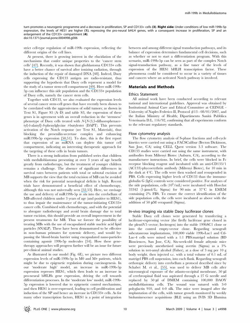
As illustrated in our model (Fig. 6E), we picture two different
expression levels of miR-199b-5p in M0 and M+ patients, which
might be due to epigenetic regulation during carcinogenesis. In
our ‘moderate high’ model, an increase in miR-199b-5p
expression represses HES1, which then leads to an increase in
pro-neural bHLHs gene expression, driving the cell towards
differentiation processes. In the ‘moderate low’ model, miR-199b-
5p expression is lowered due to epigenetic control mechanisms,
and then HES1 is over-expressed, leading to cell proliferation and
induction of the SP and hence an increase in CD133+ cells. As for
many other transcription factors, HES1 is a point of integration
between and among different signal transduction pathways, and its
balance of expression determines fundamental cell decisions, such
as whether or not to start a differentiation program. With this
scenario, miR-199b-5p can be seen as part of the complex Notch
signal-transduction pathway, as a fine tuner of the levels of
expression of the HES1 bHLH transcription factor. These
phenomena could be considered to occur in a variety of tissues
and cancers where an activated Notch pathway is involved.
Materials and Methods
Ethics StatementAll animal work have been conducted according to relevant
national and international guidelines. Approval was obtained by
Institutional Animal Care and Ethical Committee at CEINGE -
University of Naples Federico II, Protocol #13 - 08/01/2007, and
the Italian Ministry of Health; Dipartimento Sanita Pubblica
Veterinaria D.L. 116/92, confirming that all experiments conform
to the relevant regulatory standards.
Flow cytometry analysisThe flow cytometry analysis of S-phase fractions and cell-cycle
kinetics were carried out using a FACSCalibur (Becton Dickinson,
San Jose, CA) using CELL Quest version 3.3 software. The
CD133 studies were carried out using the same instrument, with
antibodies from Miltenyi Biotec (Auburn, CA), according to the
manufacturer instructions. In brief, the cells were blocked in Fc
receptor blocking reagent and incubated with an anti-CD133/1
(AC133)-phycoerythrin antibody (Miltenyi Biotec), for 10 min in
the dark at 4uC. The cells were then washed and resuspended in
PBS. Cells expressing higher levels of CD133 than the immuno-
globulin G (IgG) controls were considered positive. For analyses of
the side populations, cells (106/mL) were incubated with Hoechst
33342 (5 mmol/L; Sigma) for 90 min at 37uC in EMEM
containing 2% FBS. To ensure the correct identification of the
side population cells, the cells were incubated as above with the
addition of 50 mM verapamil (Sigma).
In-vivo imaging via stable Daoy luciferase clonesStable Daoy cell clones were generated by transfecting a
luciferase-expressing vector (the firefly luciferase gene cloned in
the plentiV5 vector; Invitrogen) into the stable 199bSC1 clone or
into the control empty-vector clone. Regarding xenograft
subcutaneous implantations, 100,000 viable 199b-Luc1 and Ctl-
Luc-4 cells were mixed with a 1:1 PBS:matrigel solution (BD
Biosciences, San Jose, CA). Six-week-old female athymic mice
were previously anesthetised using avertin (Sigma) as a 3%
solution in tert-amyl alcohol (Fisher), at a dose of 3 mg per 10 g
body weight. then injected s.c. with a total volume of 0.1 mL of
matrigel PBS cell suspension, into each flank. Regarding xenograft
orthotopic delivery into cerebellum a protocol described since by
Schabet M. et al., [55], was used to deliver MB cells after
microsurgical exposure of the atlanto-occipital membrane, 50 ml
of cerebrospinal fluid was aspirated through a 27 G needle and
replaced by 50 ml of DMEM containing 100,000 DAOY
medulloblastoma cells. The wound was sutured with 3-0
polyglactin 910, and 4-0 silk. The mice were imaged after the
implantation of the cells, with tumor growth monitored by weekly
bioluminescence acquisitions (BLI) using an IVIS 3D Illumina
turn promotes a neurogenic programme and a decrease in proliferation, SP and CD133+ cells (3). Right side: Under conditions of low miR-199b-5pexpression, the levels of HES1 are higher (1), repressing the pro-neural bHLH genes, with a consequent increase in proliferation, SP and anenlargement of the CD133+ compartment (4).doi:10.1371/journal.pone.0004998.g006
miR-199b in Medulloblastoma
PLoS ONE | www.plosone.org 10 March 2009 | Volume 4 | Issue 3 | e4998

Imaging System (Xenogen Corp. Alameda, CA). For the
acquisitions, the mice were isofluorane-anaesthetised i.p., injected
with 100 mL D-luciferin (15 mg/mL stock) per 10 g body weight,
and imaged for 30 s, 10 min after luciferin injection; four
acquisitions per mouse were made (ventral, dorsal and each
flank). To quantify the bioluminescence, the integrated fluxes of
photons (photons per s) within each area of interest were
determined using the Living Images Software Package 3.0
(Xenogen-Caliper). The emission data from the start of tumor
growth were collected for at least 6 weeks, and then were
normalized to the bioluminescence on the injection day. Calliper
measurements of tumor sizes were made weekly, along the long
and short axes, and estimations of their volumes were made using
the formula: width26length60.52.
Animals were monitored daily by weights and neurological
examinations to determine their good standard status of health.
Those animals showing sign of health sufferance were promptly
sacrificed. On day 63, after cell inoculation, all animals were
sacrificed with an overdose of ketanest and xylazin.
Animal preparation and PET/CT imagingThe mice were kept in a ventilated cage (26uC) for 1 h prior to
imaging studies. Anesthesia was performed with intraperitoneal
administration of a mixture of ketamine (100 mg/kg) and xylazine
(10 mg/kg) (injection volume, 100 ml/10 g). PET was performed
1 h after administration of 39-deoxy-39-[18F]fluorothymidine
([18F]FLT), a marker of tumor proliferation (50 mL; 7.4 MBq;
scan time, 18 min), in a lateral caudal vein, using an animal PET
scanner (GE Healthcare eXplore Vista, FWHM 1.6 mm). High
resolution CT studies (GE Healtcare eXplore Locus; spatial
resolution, 45 mm) were performed within 24 h from the PET.
Data AnalysisMaximum (SUVmax) and mean (SUVmean) standardized
uptake values (SUVs) were calculated from the PET studies
(SUV = tissue activity (MBq/cc)/[injected dose (MBq)/body
weight (g)]). The PET/CT images were post-processed to obtain
multiplanar reconstructions (MPRs), maximum intensity projec-
tions (MIPs), 3D volume rendering, and fusion images, using
Osirix 3.3 (MAC OS 10.5 operating system). Additional 3D
reconstructions were obtained using MicroView (GE eXplore
Locus).
Lesion volumes were calculated from PET data using in-house-
developed software (based on IDL, ITT Vis Inc), by summarizing
all spatially connected voxels with SUV .50% SUVmax. Lesion
profiles defined with these procedures were used for ROI-based
comparison between AdV5-Mock and AdV5-199b.
Supporting Information
Text S1 Supporting Information
Found at: doi:10.1371/journal.pone.0004998.s001 (0.12 MB
DOC)
Figure S1 A) Representative Western blots from transient
transfection of HEK293 cells with expression constructs for miR-
199b-5p and miR-199a. MiR-199b-5p over-expression lowered the
levels of the endogenous HES1 protein; in contrast, miR-199a
induced an increase in HES1 levels 48 h after transfection. Right:
Quantification through densitometric analyses. B) Representative
Western blot from transient transfection of D283 cells with the
expression construct for miR-199b-5p and using an empty vector.
Again, miR-199b-5p over-expression lowered the levels of the
endogenous HES1 protein. Below a quantification through
densitometric analyses. C) D283 and SH-SY5Y cells express similar
levels of miR-199b-5p and miR-124a, with the latter known to be
preferentially expressed in the central nervous system. In contrast,
the D283Med cells showed considerably lower levels of miR-199b-
5p. The data shown are means6SD from two independent
experiments, each carried out in triplicate. D) Representative
MiR-199b-5p expression profiles across a panel of human tissues
(Ambion); miR-199b-5p was expressed to different degrees, with
relatively high expression in the duodenum, lymph nodes, lung,
skeletal muscle, right ventricle (highest), kidney, total heart and
thyroid. MiR-199b-5p effects on the 39UTR of its putative target
gene, HES1. E–F) Luciferase activity from a reporter vector
containing wild-type HES1 39UTR and HES1 39UTR mutated in
the miR-199b binding site, co-transfected or not with an expression
vector for miR-199b-5p. The luciferase from the wild-type 39UTR
activity was reduced by 50% with miR-199b-5p expression, while
29-O-methyl-oligoribonucleotide (2-OM; 400 nM) blocks this effect.
There is no effect with the miR-199b-5p together with the HES1
39UTR mutated in the binding site. A representative experiment is
shown where the data are means6SD from three replicates in
HEK293 and Daoy cell lines respectively. G–I) Pre-miR-199 was
cloned and transfected into Daoy cells, and three stable clones were
evaluated for HES1 expression by qRT PCR and Western blotting.
There was a significant decrease in HES1 protein levels, as revealed
using an anti-HES1 polyclonal antibody; the decrease was also
revealed by densitometry analysis (G; right panel) H) A 29-O-
methyl-oligoribonucleotide (2-OM) directed against miR-199b-5p
was transfected into the stable 199bSC1 clone, with a representative
Western blot and the quantification by densitometric analysis
showing restored HES1 expression. The quantification data shown
are means6SD from two independent experiments, each carried
out in triplicate.
Found at: doi:10.1371/journal.pone.0004998.s002 (7.18 MB TIF)
Figure S2 Mmu-miR-199b expression in mouse embryonic
cerebellum and regulation of other potential targets by human
miR-199b-5p. A) Mmu-miR-199b in situ mRNA expression is
detectable at E14.5 and in newborn mouse (p0) cerebellum. The
staining was diffuse, and in all areas of the cerebellum; expression
decreased from E14.5 to p0. Mmu-miR-124a (a brain specific
miRNAs) was used as control. Left: magnification, 506; Right:
magnification, 2006. B) Quantification of decrease in mmu-miR-
199b expression during mouse development and differentiation of
the cerebellum. The levels of expression of mature miR-199b-5p
are given relative to let-7A, with the data shown as means6SD
from two independent experiments, each carried out in triplicates
C) Representative Western blot showing the protein levels of
GSK3-Beta which is predicted to be a target, in the higher
expressing miR-199b-5p stable clone. GSK3-Beta levels were not
down-regulated, and were instead slightly increased, as is clear
from the quantification by densitometric analysis shown in the
right panel, where the data shown are means6SD from two
independent experiments, each carried out in triplicate D) MiR-
199b-5p is predicted to bind other UTRs (see Supporting Table
S1). When miR-199b-5p over-expression in Daoy cells was
examined for down-regulation of productive translation from a
reporter gene carrying the full-length 39UTRs indicated, none of
them were seen to be affected. The data shown are means6SD
from two independent experiments, each carried out in triplicate.
E) Cell motility assays comparing the 199bSC1 cell line with the
Daoy empty vector CTR (control) line using a Boyden chamber
system (0.5% FBS as chemoattractant). The higher motility cells
were fixed and hematoxylin stained and counted under the
microscope. The data shown are means6SD from two indepen-
dent experiments, each carried out in triplicate, and they indicate
no differences between control (CTR) and the 199bSC1 cell line.
miR-199b in Medulloblastoma
PLoS ONE | www.plosone.org 11 March 2009 | Volume 4 | Issue 3 | e4998

F) Proliferation assay (MTS) of D283MED and ONS76 cells over-
expressing miR-199b-5p after transient transfections, as indicated.
The D283MED cells transfected with 199b-5p (blue diamonds)
show appreciable reduction in cell proliferation, as compared to
the control, empty vector, transfection (green triangles). ONS76
cells show a higher proliferation rate that is nonetheless affected by
the 199b-5p transfectant (compare orange crosses of empty vector
transfection to purple squares). The data shown are means6SD
from two independent experiments, each carried out in triplicate
G) Silencing of endogenous expression of miR-199b-5p via
transfection of a 2-O-methyl oligoribonucleotide antisense (2-
OM-a) leads to an increase in Daoy cells proliferation, probably
relieving the control of endogenous miR-199b-5p on HES1
39UTR. The data shown are means6SD from two independent
experiments, each carried out in triplicate.
Found at: doi:10.1371/journal.pone.0004998.s003 (8.54 MB TIF)
Figure S3 FACS analysis for the role of miR-199b-5p on the
Daoy cell side population. A, C, E) Decrease in the SP cells of the
Daoy 199bSC1 stable clone, as determined by Hoechst 33342
staining. B, D, F) Addition of verapamil to force dye incorporation
also in the SP cells. The Daoy cells showed a 5.2% fraction of SP
cells, while the stable 199bSC1 and 199bMC1 clones over-
expressing miR-199b-5p do not show significant levels of SP cells
(0.2%, 0.4%, respectively)
Found at: doi:10.1371/journal.pone.0004998.s004 (8.54 MB TIF)
Figure S4 Creation of bioluminescent Daoy cells over-express-
ing miR-199b-5p for in-vivo studies. The Daoy stable clone with
the empty vector (Ctl) and the Daoy stable clone over-expressing
miR-199b-5p (199b) were transfected with a luciferase expression
vector (Luc), generating, respectively, the Ctl-Luc#4 clone, and
the199bLuc-1 and 199b-Luc-3 clones. These were all evaluated
for their expression of bioluminescence when incubated with the
enzyme substrate, luciferin. A) Light emission of serial dilutions of
the cells indicated in a microplate. The bioluminescence signal was
acquired by an IVIS 200 Imaging System (Xenogen Corp.
Alameda, CA). B) Correlation between cell number and photon
emission. The 199b-Luc1 and Ctl-Luc#4 clones where used for
the in-vivo studies, with the data shown as means6SD from two
independent experiments C) Representative data of the expression
of miR-199b-5p in the luciferase clones, relative to the luciferase
clone carrying the empty vector. D) Representative immunoblot
analysis showing lowered expression of HES1 in the 199b-Luc
clones, compared to the empty-vector clone. E, F) Hematoxylin
and eosin staining of xenografts derived from mouse #4: the
control-injected side shows the xenograft infiltrating the muscle
tissue, while the 199b-injected side does not. G, H) Nestin is a
marker of neuroblasts, and its expression correlates with a lesser
differentiated state: the xenograft from the mouse #4 199b-
injected side shows a decrease in Nestin positivity. Nestin staining
was perfomed by immunohistochemistry with a polyclonal
antibody (Abcam). Magnification, 1006. I) Representative gene
expression levels of the transgene miR-199b-5p in the tumor
explant from mouse #5. The over-expression of miR-199b-5p was
lost. L, M, N) Immunohistochemistry with anti-HES1, anti-KI67,
anti-Gabra6, anti-Nestin, anti-Math3 antibodies and hematoxylin
staining of the xenografts from the cerebellum of AdV5-Mock and
AdV5-199b mice. Significant expression of Hes1, Ki67, Nestin
and Math3 proteins is seen in the AdV5-Mock cerebellum tumoral
tissue, while very low Hes1, Ki67 Nestin and Math3 expression is
seen in the AdV5-199b tumor cells in the cerebellum. Differences
of expression of Gabra6 are barely observed between in the AdV5-
Mock cerebellum tumoral tissue and AdV5-199b mice. Left:
magnification, 256; Right: magnification, 1006.
Found at: doi:10.1371/journal.pone.0004998.s005 (9.53 MB TIF)
Figure S5 Over-expression of endogenous miR199b upon
DAPT treatment induces down-regulation of genes involved to
embryonic stem cells and cancer stem cells in the Daoy cell line. A)
Representative data of a time course experiment of DAPT
treatment of Daoy cell lines (6 h, 12 h, 24 h) with media
supplemented and replaced every 4 h, and fold of expression of
miR199b determined using quantitive real time detection, relative
to time 0. B) Representative Western blot showing HES1 down-
regulation after 12 h of induction of DAPT in the Daoy cell line.
C) Relative gene expression levels of CD133, c-Myc Oct4, KFL5,
Nanog, TCF7L1, HMGA1, HMGB3, ZIC1, MYBL2, TEAD4,
ILF3 in the Daoy cell line with and without DAPT treatment for
12 h. D) Relative gene expression of PDGFR-A, PDGFR-B and
SPARC in the Daoy cell line with and without DAPT treatment
for 12 h The primers used and their DDct values of relative
expression using real-time cDNA quantitative detection, are listed
in Supporting Table S3.
Found at: doi:10.1371/journal.pone.0004998.s006 (0.47 MB
PDF)
Figure S6 Mir-199-b-5p interferes with the engraftment poten-
tial of Daoy cells injected into mouse cerebellum. A) In-vivo
bioluminescence analyses using IVIS 3D of 199b-Luc1 mouse
xenografted with Daoy Luc1 cell line overexpressing miR-199b by
stable clone analyses. (see mouse #3; see additional data in Fig. 4F,
main text). The mouse was scanned once a week for a total of 8
weeks. B) BLI measurements (photon/sec/cm-2) from mice
carrying cells infected with ADV5-199b show reductions to week
8. C) Images taken by IVIS Spectrum at 560 nm and 660 nm to
generate the topography of the subject. Clt-Luc-4 AdV5-Mock #5
xenografted mice shown on a 3D axis (x,y,z), with the extension of
the tumor burden shown in comparison with a digital mouse atlas
that enabled the display of the 3D skeleton and organs on the 3D
reconstruction, using Living Images software. (See also Movie S1
and Figure 4 panel F for BLI fold changes values) D) As described
above, analyses are taken on Ctl-luc AdV5 199b #5 xenografted
mice (See also Movie S2 and Figure 4 panel F for BLI fold changes
values).
Found at: doi:10.1371/journal.pone.0004998.s007 (8.30 MB TIF)
Table S1 Cancer related targets of miR-199b-5p. MiRanda and
Pita algorithms were applied to the selected ‘‘cancer-related’’ miR-
199b-5p targets. The gene targets predicted by both of these
algorithms are listed, including: MIRANDA score, P values, Pita
analyses with sequence matches and Delta-Delta G values.
Found at: doi:10.1371/journal.pone.0004998.s008 (0.04 MB
DOC)
Table S2 Patients characteristics recruited for the study. A) In
columns Age at diagnosis in month, Follow-up time in months,
State at last news, Mestatasis (M) stage, Histology, Relative
expression of miR-199b-5p compared to the level of (U6) and
22Delta Ct values. B) Normal healthy cerebellum mRNA from the
Brain and tissue Bank, University of Maryland, Baltimore (USA)
were used for this study. In columns, ID of material, age in year
and relative expression of miR-199b-5p compared to the level of
(U6) and 22Delta Ct values.
Found at: doi:10.1371/journal.pone.0004998.s009 (0.15 MB
DOC)
Table S3 Stem and cancer stem genes, and genes associated
with MB tumor development in Daoy cell lines over-expressing
endogenous miR-199b-5p under treatment with DAPT. The
genes involved in stem cell biology selected for the study of their
expression after over-expression of miR-199b-5p. The Unigene ID
miR-199b in Medulloblastoma
PLoS ONE | www.plosone.org 12 March 2009 | Volume 4 | Issue 3 | e4998

and sequence of primers used for real-time PCR are shown, the
22Delta Ct values are obtained from the analysis of Daoy cells
treated with DAPT for 12 h and analysed with the 7700 Real
Time TaqMan Applied Biosystem.
Found at: doi:10.1371/journal.pone.0004998.s010 (0.05 MB
DOC)
Movie S1
Found at: doi:10.1371/journal.pone.0004998.s011 (26.01 MB
MOV)
Movie S2
Found at: doi:10.1371/journal.pone.0004998.s012 (21.61 MB
MOV)
Acknowledgments
The authors would like to thank: the Brain Tumor Committee at the Curie
Institute (Paris, France), the Brain Tumor Databank from the Necker
Hospital (Paris, France), NICHD Brain and Tissue Bank, University of
Maryland, Department of Paediatrics, (Baltimore, USA) for sharing
healthy control cerebellum tissues, and Dr. Roberta Migliorati, Azienda
Ospedaliera Santobono-Pausillipon (Naples, Italy), for help in collecting
the MB samples; Prof. Giancarlo Troncone, Dr. Donatella Montanaro and
Dr. Nicola Ivan Orlotti, CEINGE (Naples, Italy) for technical support in
the immunohistochemistry staining of Daoy-tumor xenografts and with the
miR-199b targets identification approach; the ‘‘Diagnostic Imaging Core’’
at CEINGE and Drs. Michele Larobina, Anna Nardelli, Giovanni
Ortosecco, Elena Castaldi from the Diagnostic Imaging Department
(Federico II Naples and IBB-CNR Naples, Italy) for helping with the
mouse imaging by PET-TC analyses. We also thank Dr. Tetsuo Sudo,
Toray Industries, Japan, for the anti-HES1 antibody.
Author Contributions
Conceived and designed the experiments: LG IA EC NM VE MZ.
Performed the experiments: LG IA EC NM GP DDM VE AG LN SE SG
SF PAN. Analyzed the data: LG IA EC NM GP DDM AG LN SE SG SF
VD AB LDV PAN OD MT AI. Contributed reagents/materials/analysis
tools: LG IA GC. Wrote the paper: MZ.
References
1. Bartel DP (2004) MicroRNAs: genomics, biogenesis, mechanism, and function.
Cell 116: 281–297.
2. Hammond SM (2006) MicroRNAs as oncogenes. Curr Opin Genet Dev 16:
4–9.
3. O’Donnell KA, Wentzel EA, Zeller KI, Dang CV, Mendell JT (2005) c-Myc-
regulated microRNAs modulate E2F1 expression. Nature 435: 839–843.
4. Calin GA, Dumitru CD, Shimizu M, Bichi R, Zupo S, et al. (2002) Frequent
deletions and down-regulation of micro- RNA genes miR15 and miR16 at
13q14 in chronic lymphocytic leukemia. Proc Natl Acad Sci U S A 99:
15524–15529.
5. Ciafre SA, Galardi S, Mangiola A, Ferracin M, Liu CG, et al. (2005) Extensive
modulation of a set of microRNAs in primary glioblastoma. Biochem Biophys
Res Commun 334: 1351–1358.
6. Michael MZ, OC SM, van Holst Pellekaan NG, Young GP, James RJ (2003)
Reduced accumulation of specific microRNAs in colorectal neoplasia. Mol
Cancer Res 1: 882–891.
7. Fabbri M, Garzon R, Cimmino A, Liu Z, Zanesi N, et al. (2007) MicroRNA-29
family reverts aberrant methylation in lung cancer by targeting DNA
methyltransferases 3A and 3B. Proc Natl Acad Sci U S A 104: 15805–15810.
8. Ma L, Teruya-Feldstein J, Weinberg RA (2007) Tumour invasion and metastasis
initiated by microRNA-10b in breast cancer. Nature 449: 682–688.
9. Gilbertson RJ, Ellison DW (2008) The origins of medulloblastoma subtypes.
Annu Rev Pathol 3: 341–365.
10. Buhren J, Christoph AH, Buslei R, Albrecht S, Wiestler OD, et al. (2000)
Expression of the neurotrophin receptor p75NTR in medulloblastomas is
correlated with distinct histological and clinical features: evidence for a
medulloblastoma subtype derived from the external granule cell layer.
J Neuropathol Exp Neurol 59: 229–240.
11. Katsetos CD, Krishna L, Frankfurter A, Karkavelas G, Wolfe DE, et al. (1995) A
cytomorphological scheme of differentiating neuronal phenotypes in cerebellar
medulloblastomas based on immunolocalization of class III beta-tubulin isotype
(beta III) and proliferating cell nuclear antigen (PCNA)/cyclin. Clin Neuro-
pathol 14: 72–81.
12. Marino S (2005) Medulloblastoma: developmental mechanisms out of control.
Trends Mol Med 11: 17–22.
13. MacDonald TJ (2008) Aggressive infantile embryonal tumors. J Child Neurol 23:
1195–1204.
14. Yang SY, Wang KC, Cho BK, Kim YY, Lim SY, et al. (2005) Radiation-
induced cerebellar glioblastoma at the site of a treated medulloblastoma: case
report. J Neurosurg 102: 417–422.
15. Patrice SJ, Tarbell NJ, Goumnerova LC, Shrieve DC, Black PM, et al. (1995)
Results of radiosurgery in the management of recurrent and residual
medulloblastoma. Pediatr Neurosurg 22: 197–203.
16. Kombogiorgas D, Sgouros S, Walsh AR, Hockley AD, Stevens M, et al. (2007)
Outcome of children with posterior fossa medulloblastoma: a single institution
experience over the decade 1994–2003. Childs Nerv Syst 23: 399–405.
17. Huntly BJ, Gilliland DG (2005) Cancer biology: summing up cancer stem cells.
Nature 435: 1169–1170.
18. Singh SK, Clarke ID, Hide T, Dirks PB (2004) Cancer stem cells in nervous
system tumors. Oncogene 23: 7267–7273.
19. Yang ZJ, Ellis T, Markant SL, Read TA, Kessler JD, et al. (2008)
Medulloblastoma can be initiated by deletion of Patched in lineage-restricted
progenitors or stem cells. Cancer Cell 14: 135–145.
20. Solecki DJ, Liu XL, Tomoda T, Fang Y, Hatten ME (2001) Activated Notch2
signaling inhibits differentiation of cerebellar granule neuron precursors by
maintaining proliferation. Neuron 31: 557–568.
21. Ishibashi M, Moriyoshi K, Sasai Y, Shiota K, Nakanishi S, et al. (1994)
Persistent expression of helix-loop-helix factor HES-1 prevents mammalian
neural differentiation in the central nervous system. Embo J 13: 1799–1805.
22. Ishibashi M, Ang SL, Shiota K, Nakanishi S, Kageyama R, et al. (1995)
Targeted disruption of mammalian hairy and Enhancer of split homolog-1
(HES-1) leads to up-regulation of neural helix-loop-helix factors, premature
neurogenesis, and severe neural tube defects. Genes Dev 9: 3136–3148.
23. Nakamura Y, Sakakibara S, Miyata T, Ogawa M, Shimazaki T, et al. (2000)
The bHLH gene hes1 as a repressor of the neuronal commitment of CNS stem
cells. J Neurosci 20: 283–293.
24. Fan X, Mikolaenko I, Elhassan I, Ni X, Wang Y, et al. (2004) Notch1 and
notch2 have opposite effects on embryonal brain tumor growth. Cancer Res 64:
7787–7793.
25. Hallahan AR, Pritchard JI, Hansen S, Benson M, Stoeck J, et al. (2004) The
SmoA1 mouse model reveals that notch signaling is critical for the growth and
survival of sonic hedgehog-induced medulloblastomas. Cancer Res 64:
7794–7800.
26. Leung C, Lingbeek M, Shakhova O, Liu J, Tanger E, et al. (2004) Bmi1 is
essential for cerebellar development and is overexpressed in human medullo-
blastomas. Nature 428: 337–341.
27. Griffiths-Jones S (2004) The microRNA Registry. Nucleic Acids Res 32:
D109–111.
28. Yanaihara N, Caplen N, Bowman E, Seike M, Kumamoto K, et al. (2006)
Unique microRNA molecular profiles in lung cancer diagnosis and prognosis.
Cancer Cell 9: 189–198.
29. Sandberg AA (2002) Cytogenetics and molecular genetics of bladder cancer: a
personal view. Am J Med Genet 115: 173–182.
30. Gramantieri L, Ferracin M, Fornari F, Veronese A, Sabbioni S, et al. (2007)
Cyclin G1 is a target of miR-122a, a microRNA frequently down-regulated in
human hepatocellular carcinoma. Cancer Res 67: 6092–6099.
31. Jiang J, Gusev Y, Aderca I, Mettler TA, Nagorney DM, et al. (2008) Association
of MicroRNA expression in hepatocellular carcinomas with hepatitis infection,
cirrhosis, and patient survival. Clin Cancer Res 14: 419–427.
32. Murakami Y, Yasuda T, Saigo K, Urashima T, Toyoda H, et al. (2006)
Comprehensive analysis of microRNA expression patterns in hepatocellular
carcinoma and non-tumorous tissues. Oncogene 25: 2537–2545.
33. Izant JG, McIntosh JR (1980) Microtubule-associated proteins: a monoclonal
antibody to MAP2 binds to differentiated neurons. Proc Natl Acad Sci U S A 77:
4741–4745.
34. Li XN, Parikh S, Shu Q, Jung HL, Chow CW, et al. (2004) Phenylbutyrate and
phenylacetate induce differentiation and inhibit proliferation of human
medulloblastoma cells. Clin Cancer Res 10: 1150–1159.
35. Hatakeyama J, Bessho Y, Katoh K, Ookawara S, Fujioka M, et al. (2004) Hes
genes regulate size, shape and histogenesis of the nervous system by control of
the timing of neural stem cell differentiation. Development 131: 5539–5550.
36. Fan X, Matsui W, Khaki L, Stearns D, Chun J, et al. (2006) Notch pathway
inhibition depletes stem-like cells and blocks engraftment in embryonal brain
tumors. Cancer Res 66: 7445–7452.
37. Kondo T, Setoguchi T, Taga T (2004) Persistence of a small subpopulation of
cancer stem-like cells in the C6 glioma cell line. Proc Natl Acad Sci U S A 101:
781–786.
38. Goodell MA, Brose K, Paradis G, Conner AS, Mulligan RC (1996) Isolation and
functional properties of murine hematopoietic stem cells that are replicating in
vivo. J Exp Med 183: 1797–1806.
39. Singh SK, Hawkins C, Clarke ID, Squire JA, Bayani J, et al. (2004)
Identification of human brain tumour initiating cells. Nature 432: 396–401.
miR-199b in Medulloblastoma
PLoS ONE | www.plosone.org 13 March 2009 | Volume 4 | Issue 3 | e4998

40. Eberhart CG (2007) In search of the medulloblast: neural stem cells and
embryonal brain tumors. Neurosurg Clin N Am 18: 59–69, viii–ix.
41. Kertesz M, Iovino N, Unnerstall U, Gaul U, Segal E (2007) The role of site
accessibility in microRNA target recognition. Nat Genet 39: 1278–1284.
42. Kim S, Lee UJ, Kim MN, Lee EJ, Kim JY, et al. (2008) MicroRNA miR-199a*
regulates the MET proto-oncogene and the downstream extracellular signal-
regulated kinase 2 (ERK2). J Biol Chem 283: 18158–18166.
43. Pogoriler J, Millen K, Utset M, Du W (2006) Loss of cyclin D1 impairs
cerebellar development and suppresses medulloblastoma formation. Develop-
ment 133: 3929–3937.
44. Gilbertson RJ, Clifford SC (2003) PDGFRB is overexpressed in metastatic
medulloblastoma. Nat Genet 35: 197–198.
45. MacDonald TJ, Brown KM, LaFleur B, Peterson K, Lawlor C, et al. (2001)
Expression profiling of medulloblastoma: PDGFRA and the RAS/MAPK
pathway as therapeutic targets for metastatic disease. Nat Genet 29: 143–152.
46. Hatton BA, Villavicencio EH, Tsuchiya KD, Pritchard JI, Ditzler S, et al. (2008)
The Smo/Smo model: hedgehog-induced medulloblastoma with 90% incidence
and leptomeningeal spread. Cancer Res 68: 1768–1776.
47. Fan X, Eberhart CG (2008) Medulloblastoma stem cells. J Clin Oncol 26:
2821–2827.
48. Bao S, Wu Q, McLendon RE, Hao Y, Shi Q, et al. (2006) Glioma stem cells
promote radioresistance by preferential activation of the DNA damage response.Nature 444: 756–760.
49. Blazek ER, Foutch JL, Maki G (2007) Daoy medulloblastoma cells that express
CD133 are radioresistant relative to CD1332 cells, and the CD133+ sector isenlarged by hypoxia. Int J Radiat Oncol Biol Phys 67: 1–5.
50. Ben-Porath I, Thomson MW, Carey VJ, Ge R, Bell GW, et al. (2008) Anembryonic stem cell-like gene expression signature in poorly differentiated
aggressive human tumors. Nat Genet 40: 499–507.
51. Gao JX (2008) Cancer stem cells: the lessons from pre-cancerous stem cells. J CellMol Med 12: 67–96.
52. Grill J, Dufour C, Kalifa C (2006) High-dose chemotherapy in children withnewly-diagnosed medulloblastoma. Lancet Oncol 7: 787–789.
53. Grill J, Sainte-Rose C, Jouvet A, Gentet JC, Lejars O, et al. (2005) Treatment ofmedulloblastoma with postoperative chemotherapy alone: an SFOP prospective
trial in young children. Lancet Oncol 6: 573–580.
54. Zimmermann TS, Lee AC, Akinc A, Bramlage B, Bumcrot D, et al. (2006)RNAi-mediated gene silencing in non-human primates. Nature 441: 111–114.
55. Schabet M, Martos J, Buchholz R, Pietsch T (1997) Animal model of humanmedulloblastoma: clinical, magnetic resonance imaging, and histopathological
findings after intra-cisternal injection of MHH-MED-1 cells into nude rats. Med
Pediatr Oncol 29: 92–7.
miR-199b in Medulloblastoma
PLoS ONE | www.plosone.org 14 March 2009 | Volume 4 | Issue 3 | e4998