
αVβ5 and CD44 Are Oxygen-Regulated Human Embryonic Stem Cell Attachment Factors
10
Hindawi Publishing Corporation BioMed Research International Volume 2013, Article ID 729281, 9 pages http://dx.doi.org/10.1155/2013/729281 Research Article V5 and CD44 Are Oxygen-Regulated Human Embryonic Stem Cell Attachment Factors Deepak Kumar, Saniya Gupta, Ying Yang, and Nicholas R. Forsyth Guy Hilton Research Centre, Institute of Science and Technology in Medicine, University of Keele, ornburrow Drive, Hartshill, Stoke-on-Trent, Staffordshire ST4 7QB, UK Correspondence should be addressed to Nicholas R. Forsyth; [email protected] Received 29 April 2013; Revised 19 September 2013; Accepted 4 October 2013 Academic Editor: Louise E. Glover Copyright © 2013 Deepak Kumar et al. is is an open access article distributed under the Creative Commons Attribution License, which permits unrestricted use, distribution, and reproduction in any medium, provided the original work is properly cited. Human embryonic stem cells (hESCs) have great potential for clinical therapeutic use. However, relatively little is known of the mechanisms which dictate their specificity of adhesion to substrates through adhesion proteins including integrins. Previous observations demonstrated enhanced clonogenicity in reduced oxygen culture systems. Here, we demonstrated via antibody blocking experiments that V5 and 6 significantly promoted hESC attachment in 2% O 2 only, whereas blockage of CD44 inhibited cell attachment in 21% O 2 alone. Immunofluorescence confirmed expression of V5 and CD44 in both 2% O 2 and 21% O 2 cultured hESCs while flow cytometry revealed significantly higher V5 expression in 2% O 2 versus 21% O 2 cultured hESCs and higher CD44 expression in 21% O 2 versus 2% O 2 cultured hESCs. Adhered hESCs following blockage of V5 in 2% O 2 displayed a reduction in nuclear colocalisation of Oct-4 and Nanog with little effect observed in 21% O 2 . Blockage of CD44 had the converse effect with dramatic reductions in nuclear colocalisation of Oct-4 and Nanog in 21% O 2 cultured hESC which retained adherence, but not in 2% O 2 cultured cells. Identification of oxygen-dependent substrate attachment mechanisms in hESCs has the potential to play a role in the development of novel substrates to improve hESC attachment and culture. 1. Introduction Human embryonic stem cells (hESCs), derived from the inner cell mass of preimplantation blastocysts, have an inher- ent capacity for indefinite self-renewal [1]. Due to their differ- entiation capacity, immortality and immunological privilege, hESCs hold great promise for clinical therapeutics when used in combination with tissue engineering and regenera- tive medicine approaches [1–3]. e single currently active approved clinical safety trial incorporating hESCs is for age- related macular degeneration [4, 5]. Typical in vitro expansion of hESCs involves either direct coculture with mitotically inactivated mouse (or human) embryonic fibroblasts (MEFs) or feeder-free meth- ods, where preconditioned media and biological substrates such as Matrigel are employed [1, 6]. Matrigel, a loosely defined gel sourced from Engelbreth-Holm-Swarm tumours, is comprised of extracellular matrix (ECM) proteins includ- ing laminin-111, collagen IV, heparin sulphate proteogly- cans, entactin, fibronectin, growth factors, matrix-degrading enzymes and their inhibitors, and other yet to be defined components [6]. Limitations of culturing hESCs using Matrigel (and other biological substrates) include batch to batch variability, xenogenic contamination, expression of foreign oligosaccharide residues, and scale-up issues [7, 8]. Alternatives include collagen IV, fibronectin, laminin, vit- ronectin [9], recombinant vitronectin [8], human serum con- taining medium conditioned by human embryonic fibrob- lasts derived from hESCs [10], and hyaluronic acid hydrogels [11]. hESCs ECM attachment is primarily mediated by inte- grins (heterodimeric, transmembrane glycoproteins) and other surface receptors [12]. e integrin family, comprised of 18 alpha () subunits and 8 beta () subunits, has 24 recog- nised distinct heterodimer arrangements each with a specific set of functions [12–14]. Integrin functions include mediating cell-cell, cell-ECM, and cytoskeletal-ECM interactions. ECM proteins essential for hESC adhesion and pluripotency reten- tion include laminin-111, collagen IV, fibronectin, and vit- ronectin [15]. Laminin is an essential component of virtually all basement membranes [16]. Laminin functions include the mediation of cell adhesion, cell spreading, cell migration, and
Transcript of αVβ5 and CD44 Are Oxygen-Regulated Human Embryonic Stem Cell Attachment Factors

Hindawi Publishing CorporationBioMed Research InternationalVolume 2013, Article ID 729281, 9 pageshttp://dx.doi.org/10.1155/2013/729281
Research Article𝛼V𝛽5 and CD44 Are Oxygen-Regulated Human Embryonic StemCell Attachment Factors
Deepak Kumar, Saniya Gupta, Ying Yang, and Nicholas R. Forsyth
Guy Hilton Research Centre, Institute of Science and Technology in Medicine, University of Keele, Thornburrow Drive,Hartshill, Stoke-on-Trent, Staffordshire ST4 7QB, UK
Correspondence should be addressed to Nicholas R. Forsyth; [email protected]
Received 29 April 2013; Revised 19 September 2013; Accepted 4 October 2013
Academic Editor: Louise E. Glover
Copyright © 2013 Deepak Kumar et al.This is an open access article distributed under the Creative Commons Attribution License,which permits unrestricted use, distribution, and reproduction in any medium, provided the original work is properly cited.
Human embryonic stem cells (hESCs) have great potential for clinical therapeutic use. However, relatively little is known of themechanisms which dictate their specificity of adhesion to substrates through adhesion proteins including integrins. Previousobservations demonstrated enhanced clonogenicity in reduced oxygen culture systems. Here, we demonstrated via antibodyblocking experiments that 𝛼V𝛽5 and 𝛼6 significantly promoted hESC attachment in 2% O
2only, whereas blockage of CD44
inhibited cell attachment in 21% O2alone. Immunofluorescence confirmed expression of 𝛼V𝛽5 and CD44 in both 2% O
2and
21% O2cultured hESCs while flow cytometry revealed significantly higher 𝛼V𝛽5 expression in 2% O
2versus 21% O
2cultured
hESCs and higher CD44 expression in 21% O2versus 2% O
2cultured hESCs. Adhered hESCs following blockage of 𝛼V𝛽5 in 2%
O2displayed a reduction in nuclear colocalisation of Oct-4 and Nanog with little effect observed in 21% O
2. Blockage of CD44 had
the converse effect with dramatic reductions in nuclear colocalisation of Oct-4 and Nanog in 21%O2cultured hESC which retained
adherence, but not in 2%O2cultured cells. Identification of oxygen-dependent substrate attachment mechanisms in hESCs has the
potential to play a role in the development of novel substrates to improve hESC attachment and culture.
1. Introduction
Human embryonic stem cells (hESCs), derived from theinner cell mass of preimplantation blastocysts, have an inher-ent capacity for indefinite self-renewal [1]. Due to their differ-entiation capacity, immortality and immunological privilege,hESCs hold great promise for clinical therapeutics whenused in combination with tissue engineering and regenera-tive medicine approaches [1–3]. The single currently activeapproved clinical safety trial incorporating hESCs is for age-related macular degeneration [4, 5].
Typical in vitro expansion of hESCs involves eitherdirect coculture with mitotically inactivated mouse (orhuman) embryonic fibroblasts (MEFs) or feeder-free meth-ods, where preconditioned media and biological substratessuch as Matrigel are employed [1, 6]. Matrigel, a looselydefined gel sourced from Engelbreth-Holm-Swarm tumours,is comprised of extracellular matrix (ECM) proteins includ-ing laminin-111, collagen IV, heparin sulphate proteogly-cans, entactin, fibronectin, growth factors, matrix-degradingenzymes and their inhibitors, and other yet to be defined
components [6]. Limitations of culturing hESCs usingMatrigel (and other biological substrates) include batch tobatch variability, xenogenic contamination, expression offoreign oligosaccharide residues, and scale-up issues [7, 8].Alternatives include collagen IV, fibronectin, laminin, vit-ronectin [9], recombinant vitronectin [8], human serum con-taining medium conditioned by human embryonic fibrob-lasts derived from hESCs [10], and hyaluronic acid hydrogels[11].
hESCs ECM attachment is primarily mediated by inte-grins (heterodimeric, transmembrane glycoproteins) andother surface receptors [12]. The integrin family, comprisedof 18 alpha (𝛼) subunits and 8 beta (𝛽) subunits, has 24 recog-nised distinct heterodimer arrangements each with a specificset of functions [12–14]. Integrin functions includemediatingcell-cell, cell-ECM, and cytoskeletal-ECM interactions. ECMproteins essential for hESC adhesion and pluripotency reten-tion include laminin-111, collagen IV, fibronectin, and vit-ronectin [15]. Laminin is an essential component of virtuallyall basement membranes [16]. Laminin functions include themediation of cell adhesion, cell spreading, cell migration, and

2 BioMed Research International
cell proliferation. The laminin-specific integrin receptor is𝛼6𝛽1 while 𝛼1𝛽1, 𝛼2𝛽1, and 𝛼3𝛽1 are also recognised in a non-specificmanner. Integrin subunits and heterodimers detectedon the surface of hESCs include 𝛼2, 𝛼3, 𝛼5, 𝛼6, 𝛼11, 𝛽1, and𝛼V𝛽5. These subunits can heterodimerise to form receptorsfor fibronectin (𝛼5𝛽1), vitronectin (𝛼V𝛽5), collagen andlaminin (𝛼2𝛽1), laminin-111 (𝛼6𝛽1), and collagen, laminin,and VCAM1 (𝛼9𝛽1) [8, 17–19]. Antibody-directed blockageof the 𝛼5𝛽1 heterodimer impacted hESC attachment acrossa range of defined substrate coatings including collagen IV,laminin, and entactin, when cultured with MEF-conditionedmedia, demonstrating that fibronectin was secreted by feedercells which subsequently adsorbed onto surfaces promotinghESC adherence [8]. In defined media (mTeSR1), blocking𝛼5𝛽1 had no effect on hESC attachment to a vitronectin-coated substrate but hindered adhesion to all other ECMpro-tein substrates (laminin, entactin, and collagen IV), suggest-ing that hESC substrate adhesion via 𝛼V𝛽5 was essential forexpansion of hESCs when cultured using defined media [8].
To this point, however, there are very few reports whichhave explored the role, if any, that oxygen concentration hason integrin expression level in hESC. Reports from mouseembryonic stem cells (𝛽1), human mesenchymal stem cells(hMSC) (𝛼1, 𝛼3, 𝛼5, 𝛼6, 𝛼11, 𝛼V, 𝛽1, and 𝛽3), and chondro-cytes (𝛽1) have indicated that specific integrin subunits, indi-cated in parentheses, display an oxygen sensitivity, hypoxiainducible factor 1-dependent in some instances, resultingin selected increased transcriptional expression of indicatedsubunits [20, 21]. Reports have also indicated that reducedoxygen culture has resulted in selected transcriptional down-regulation of integrin subunits, indicated in parentheses, inhMSC (𝛼2), trophoblast (𝛼1, 𝛼4, and 𝛼5), breast carcinomacells (𝛼5), gastric cancer cells (𝛼5), cytotrophoblasts (𝛼6), andmelanoma cell lines (𝛼V, 𝛽1, and 𝛼V𝛽3) [22–28].
Previous studies of the effects of reduced oxygen on hESCculture have demonstrated enhanced clonogenicity, reducedchromosomal aberration frequency, and improved consis-tency of embryoid body formation, significant transcriptionalalterations, and permissive single-cell derived progenitorisolation [29, 30]. A previous study by our group describedreduced transcriptional heterogeneity between hESC lines(H1, H9, and RH1) cultured in physiological normoxia whichwas accompanied by significant upregulation, but with mod-est fold change differences, of specific integrin subunits (𝛼6,𝛼E, 𝛼V, and 𝛽5) in comparison to hyperoxia (21% O
2) [31].
We have now extended these observations and describedthe translational consequences of physiological oxygen onintegrin subunit dependency and CD44 reliance for adhesionand pluripotent marker expression, in hESC (SHEF1). Wedemonstrate that adhesion, pluripotent marker expressionand localisation in hESCs were 𝛼V𝛽5 dependent in 2% O
2
and CD44 dependent in 21% O2. These findings will help
drive the future development of novel substrates to improvehESC attachment and expansion during in vitro expansion.
2. Materials and Methods
2.1. Human Embryonic Stem Cell Culture. Conditioned cul-ture media were prepared usingmouse embryonic fibroblasts
(MEFs) as previously described [6]. In brief, hESC mediacomprised Knock-out DMEM (KO-DMEM) (Gibco-Invi-trogen, UK) supplemented with 20% Knock-out SerumReplacement (Gibco-Invitrogen, UK), 1% L-glutamine(Lonza, UK), 1% nonessential amino acids (Lonza, UK),4 ng/mL basic fibroblastic growth factor (Lonza, UK), and0.1mM 𝛽-mercaptoethanol (Gibco-Invitrogen, UK). hESCmedia were conditioned overnight on semiconfluent MEFsand then further supplemented with 4 ng/mL of bFGF andsterile filtered (Millipore, Watford, UK) before use [6].hESCs (SHEF1) were cultured on Matrigel (BD Biosciences,Oxford, UK) coated flasks in two different oxygen tensions;2% O2(using the SCI-TIVE workstation; Ruskinn, Pencoed,
UK) and 21% O2(Heraeus Cytoperm 2 incubator; Thermo
Electron Corporation, UK). Media were changed on adaily basis and cells passaged every 2-3 days after reaching90% confluence using a brief 0.25% trypsin and EDTAtreatment for 1-2 minutes at room temperature, followed bycentrifugation for 3 minutes at 1200 rpm and replated at a1 : 2 ratio.
2.2. Integrin Blocking and Cell Attachment. hESCs werepretreated with blocking antibodies raised against inte-grin subunits including anti-integrin 𝛼V (R&D Biosystems,Abingdon, UK), anti-integrin 𝛼V𝛽5 (Chemicon Interna-tional, Watford, UK), anti-integrin 𝛽5 (R&D Biosystems,Abingdon, UK), anti-integrin 𝛼E (Lifespan Bioscience, Not-tingham UK), anti-integrin 𝛼6 (Autogen Bioclear, Calne,UK), and anti-CD44 (HCAM) (Santa Cruz Biotechnology,Heidelberg, Germany). hESCs were incubated with either 0,1, or 25𝜇g/mL concentrations of antibody (in PBS) in either2% O
2or 21% O
2at 37∘C for 30 minutes in KO-DMEM.
Cells were then re-plated into Matrigel coated 6-well platesat a density of 4 × 10⁵ cells per well and incubated at either2% O
2or 21% O
2for 24 hours. After 24 hours, cells were
trypsinised (as described above) and counts recorded with ahaemocytometer. Cell viability and nontoxicity of antibodysolution were determined by staining hESCs with TrypanBlue at a 1 : 1 ratio to cell solution.
2.3. Immunofluorescence Staining. hESCs (400 000 cells/well) were seeded onto Matrigel coated 24-well plates andexpanded to approximately 70% confluence in both oxygenconcentrations (2% O
2and 21% O
2). Media were removed
and the cells were fixed in 4% paraformaldehyde (in PBS) for40minutes. Cells were washed with PBS and nonspecific pro-teins were blocked with 3% bovine serum albumin for 1 hourat room temperature. Cells were incubated with 50𝜇g/mLanti-𝛼V𝛽5 anti-human monoclonal antibody solution at4∘C overnight. Primary antibody binding was visualisedby incubation with donkey anti-human IgG (NL557; R&DBiosystems), at 5 𝜇g/mL for 2 hours at room temperature.Nuclei were visualised by counterstaining with DAPI andmounted with Vectashield (Vector Laboratories, Burlingame,CA). Images were recorded on a fluorescent microscope(Nikon TZ1; Leica, Germany).
To assess expression of pluripotency-associated proteinsafter antibody blocking, cells were first fixed (as above)and subsequently incubated with 1 𝜇g/100 𝜇L of either goat
BioMed Research International 3
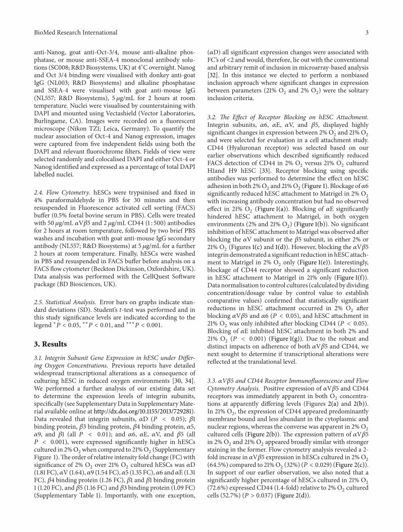
anti-Nanog, goat anti-Oct-3/4, mouse anti-alkaline phos-phatase, or mouse anti-SSEA-4 monoclonal antibody solu-tions (SC008; R&DBiosystems, UK) at 4∘C overnight. Nanogand Oct 3/4 binding were visualised with donkey anti-goatIgG (NL003; R&D Biosystems) and alkaline phosphataseand SSEA-4 were visualised with goat anti-mouse IgG(NL557; R&D Biosystems), 5𝜇g/mL for 2 hours at roomtemperature. Nuclei were visualised by counterstaining withDAPI and mounted using Vectashield (Vector Laboratories,Burlingame, CA). Images were recorded on a fluorescentmicroscope (Nikon TZ1; Leica, Germany). To quantify thenuclear association of Oct-4 and Nanog expression, imageswere captured from five independent fields using both theDAPI and relevant fluorochrome filters. Fields of view wereselected randomly and colocalised DAPI and either Oct-4 orNanog identified and expressed as a percentage of total DAPIlabelled nuclei.
2.4. Flow Cytometry. hESCs were trypsinised and fixed in4% paraformaldehyde in PBS for 30 minutes and thenresuspended in Fluorescence activated cell sorting (FACS)buffer (0.5% foetal bovine serum in PBS). Cells were treatedwith 50𝜇g/mL 𝛼V𝛽5 and 2𝜇g/mL CD44 (1 : 500) antibodiesfor 2 hours at room temperature, followed by two brief PBSwashes and incubation with goat anti-mouse IgG secondaryantibody (NL557; R&D Biosystems) at 5𝜇g/mL for a further2 hours at room temperature. Finally, hESCs were washedin PBS and resuspended in FACS buffer before analysis on aFACS flow cytometer (BecktonDickinson, Oxfordshire, UK).Data analysis was performed with the CellQuest Softwarepackage (BD Biosciences, UK).
2.5. Statistical Analysis. Error bars on graphs indicate stan-dard deviations (SD). Student’s 𝑡-test was performed and inthis study significance levels are indicated according to thelegend ∗𝑃 < 0.05, ∗∗𝑃 < 0.01, and ∗∗∗𝑃 < 0.001.
3. Results
3.1. Integrin Subunit Gene Expression in hESC under Differ-ing Oxygen Concentrations. Previous reports have detailedwidespread transcriptional alterations as a consequence ofculturing hESC in reduced oxygen environments [30, 34].We performed a further analysis of our existing data setto determine the expression levels of integrin subunits,specifically (see SupplementaryData in SupplementaryMate-rial available online at http://dx.doi.org/10.1155/2013/729281).Data revealed that integrin subunits, 𝛼D (𝑃 < 0.05); 𝛽1binding protein, 𝛽3 binding protein, 𝛽4 binding protein, 𝛼5,𝛼9, and 𝛽1 (all 𝑃 < 0.01); and 𝛼6, 𝛼E, 𝛼V, and 𝛽5 (all𝑃 < 0.001), were expressed significantly higher in hESCscultured in 2%O
2when compared to 21%O
2(Supplementary
Figure 1).Theorder of relative intensity fold change (FC)withsignificance of 2% O
2over 21% O
2cultured hESCs was 𝛼D
(1.81 FC),𝛼V (1.64),𝛼9 (1.54 FC),𝛼5 (1.35 FC),𝛼6 and𝛼E (1.31FC), 𝛽4 binding protein (1.26 FC), 𝛽1 and 𝛽1 binding protein1 (1.20 FC), and 𝛽5 (1.16 FC) and 𝛽3 binding protein (1.09 FC)(Supplementary Table 1). Importantly, with one exception,
(𝛼D) all significant expression changes were associated withFC’s of <2 and would, therefore, lie out with the conventionaland arbitrary remit of inclusion in microarray-based analysis[32]. In this instance we elected to perform a nonbiasedinclusion approach where significant changes in expressionbetween parameters (21% O
2and 2% O
2) were the solitary
inclusion criteria.
3.2. The Effect of Receptor Blocking on hESC Attachment.Integrin subunits, 𝛼6, 𝛼E, 𝛼V, and 𝛽5, displayed highlysignificant changes in expression between 2% O
2and 21% O
2
and were selected for evaluation in a cell attachment study.CD44 (Hyaluronan receptor) was selected based on ourearlier observations which described significantly reducedFACS detection of CD44 in 2% O
2versus 21% O
2cultured
H1and H9 hESC [33]. Receptor blocking using specificantibodies was performed to determine the effect on hESCadhesion in both 2%O
2and 21%O
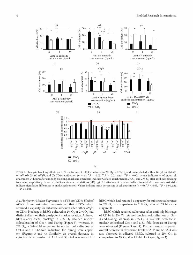
2(Figure 1). Blockage of𝛼6
significantly reduced hESC attachment to Matrigel in 2% O2
with increasing antibody concentration but had no observedeffect in 21% O
2(Figure 1(a)). Blocking of 𝛼E significantly
hindered hESC attachment to Matrigel, in both oxygenenvironments (2% and 21% O
2) (Figure 1(b)). No significant
inhibition of hESC attachment toMatrigel was observed afterblocking the 𝛼V subunit or the 𝛽5 subunit, in either 2% or21% O
2(Figures 1(c) and 1(d)). However, blocking the 𝛼V𝛽5
integrin demonstrated a significant reduction in hESC attach-ment to Matrigel in 2% O
2only (Figure 1(e)). Interestingly,
blockage of CD44 receptor showed a significant reductionin hESC attachment to Matrigel in 21% only (Figure 1(f)).Data normalisation to control cultures (calculated by dividingconcentration/dosage value by control value to establishcomparative values) confirmed that statistically significantreductions in hESC attachment occurred in 2% O
2after
blocking 𝛼V𝛽5 and 𝛼6 (𝑃 < 0.05), and hESC attachment in21% O
2was only inhibited after blocking CD44 (𝑃 < 0.05).
Blocking of 𝛼E inhibited hESC attachment in both 2% and21% O
2(𝑃 < 0.001) (Figure 1(g)). Due to the robust and
distinct impacts on adherence of both 𝛼V𝛽5 and CD44, wenext sought to determine if transcriptional alterations werereflected at the translational level.
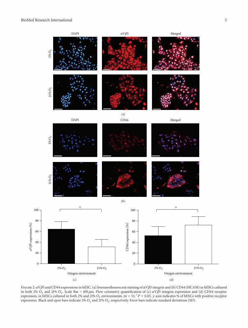
3.3. 𝛼V𝛽5 and CD44 Receptor Immunofluorescence and FlowCytometry Analysis. Positive expression of 𝛼V𝛽5 and CD44receptors was immediately apparent in both O
2concentra-
tions at apparently differing levels (Figures 2(a) and 2(b)).In 21% O
2, the expression of CD44 appeared predominantly
membrane bound and less abundant in the cytoplasmic andnuclear regions, whereas the converse was apparent in 2%O
2
cultured cells (Figure 2(b)). The expression pattern of 𝛼V𝛽5in 2% O
2and 21% O
2appeared broadly similar with stronger
staining in the former. Flow cytometry analysis revealed a 2-fold increase in 𝛼V𝛽5 expression in hESCs cultured in 2%O
2
(64.5%) compared to 21%O2(32%) (𝑃 < 0.029) (Figure 2(c)).
In support of our earlier observation, we also noted that asignificantly higher percentage of hESCs cultured in 21% O
2
(72.6%) expressed CD44 (1.4-fold) relative to 2%O2cultured
cells (52.7%) (𝑃 > 0.037) (Figure 2(d)).
4 BioMed Research International
30
20
10
00 1 25
Cel
l atta
chm
ent (%
) ∗∗∗𝛼6
Anti-𝛼6 antibodyconcentration (𝜇g/mL)
(a)
30
20
10
00 1 25
Cel
l atta
chm
ent (%
)
∗ ∗
∗∗∗∗∗
∗∗∗
𝛼E
Anti-𝛼E antibodyconcentration (𝜇g/mL)
(b)
30
20
10
00 1 25
Cel
l atta
chm
ent (%
)
Anti-𝛼V
𝛼V
antibodyconcentration (𝜇g/mL)
(c)
30
20
10
0
0 1 25
Cel
l atta
chm
ent (%
)
Anti-𝛽5
𝛽5
antibodyconcentration (𝜇g/mL)
(d)
21%O2
2%O2
30
20
10
00 1 25
Cel
l atta
chm
ent (%
) ∗
Anti-𝛼V𝛽5
𝛼V𝛽5
antibodyconcentration (𝜇g/mL)
(e)
21%O2
2%O2
0 1 25
Cel
l atta
chm
ent (%
) ∗40
30
20
10
0
Anti-CD44 (HCAM)
CD44
antibody concentration (𝜇g/mL)
(f)
21%O2
2%O2
𝛼6𝛼E𝛼V 𝛽5𝛼V𝛽5
1.2
1
0.8
0.6
0.4
0.2
0
Nor
mal
ized
cell
atta
chm
ent (%
)
CD44
∗
∗ ∗∗∗
∗
∗∗∗∗
∗∗∗
1𝜇
g/m
L
25𝜇
g/m
L
1𝜇
g/m
L
25𝜇
g/m
L
1𝜇
g/m
L
25𝜇
g/m
L
1𝜇
g/m
L
25𝜇
g/m
L
1𝜇
g/m
L
25𝜇
g/m
L
1𝜇
g/m
L
25𝜇
g/m
L
(g)
Figure 1: Integrin blocking effects on hESCs attachment. hESCs cultured in 2% O2or 21% O
2and preincubated with anti- (a) 𝛼6, (b) 𝛼E,
(c) 𝛼V, (d) 𝛽5, (e) 𝛼V𝛽5, and (f) CD44 antibodies. (𝑛 = 6); ∗𝑃 < 0.05, ∗∗𝑃 < 0.01, and ∗∗∗𝑃 < 0.001. 𝑦-axis indicates % of input cellattachment 24 hours after antibody blocking. Black and open bars indicate % of cell attachment in 2%O
2and 21% O
2after antibody blocking
treatment, respectively. Error bars indicate standard deviations (SD). (g) Cell attachment data normalised to unblocked controls. Asterisksindicate significant differences to unblocked controls. Values indicate mean percentage of cell attachment (𝑛 = 6); ∗𝑃 < 0.05, ∗∗𝑃 < 0.01, and∗∗∗𝑃 < 0.001.
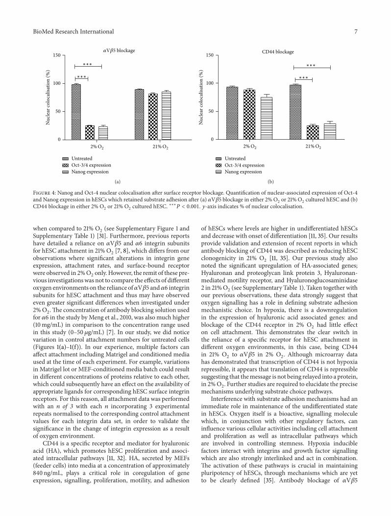
3.4. PluripotentMarker Expression in𝛼V𝛽5 andCD44BlockedhESCs. Immunostaining demonstrated that hESCs whichretained a capacity for substrate adhesion after either 𝛼V𝛽5or CD44 blockage in hESCs cultured in 2%O
2or 21%O
2had
distinct effects on their pluripotentmarker location. AdheredhESCs after 𝛼V𝛽5 blockage in 21% O
2retained nuclear
colocalisation of Oct-4 and Nanog (Figure 3), whereas, in2% O
2, a 3.44-fold reduction in nuclear colocalisation of
Oct-4 and a 3.63-fold reduction for Nanog were appar-ent (Figures 3 and 4). Similarly, an overall decrease incytoplasmic expression of ALP and SSEA-4 was noted for
hESC which had retained a capacity for substrate adherencein 2% O
2in comparison to 21% O
2after 𝛼V𝛽5 blockage
(Figure 3).hESC which retained adherence after antibody blockage
of CD44 in 2% O2retained nuclear colocalisation of Oct-
4 and Nanog, whereas, in 21% O2, a 3.62-fold decrease in
nuclear colocalised Oct-4 and a 3.4-fold decrease in Nanogwere observed (Figures 3 and 4). Furthermore, an apparentoverall decrease in expression levels of ALP and SSEA-4 wasalso observed in adhered hESCs, cultured in 21% O
2, in
comparison to 2% O2after CD44 blockage (Figure 3).
BioMed Research International 5
21%
O2
2%
O2
𝛼V𝛽5DAPI Merged
(a)
21%
O2
2%
O2
CD44DAPI Merged
(b)
𝛼V𝛽5
expr
essio
n (%
)
100
80
60
40
20
0
Oxygen environment
∗
21%O22%O2
(c)
CD44
expr
essio
n (%
)
100
80
60
40
20
0
Oxygen environment
∗
21%O22%O2
(d)
Figure 2: 𝛼V𝛽5 and CD44 expressions in hESC. (a) Immunoflourescent staining of 𝛼V𝛽5 integrin and (b) CD44 (HCAM) in hESCs culturedin both 2% O
2and 21% O
2. Scale Bar = 100𝜇m. Flow cytometry quantification of (c) 𝛼V𝛽5 integrin expression and (d) CD44 receptor
expression, in hESCs cultured in both 2% and 21% O2environments. (𝑛 = 5); ∗𝑃 < 0.05. 𝑦-axis indicates % of hESCs with positive receptor
expression. Black and open bars indicate 2% O2and 21% O
2, respectively. Error bars indicate standard deviations (SD).

6 BioMed Research International
Oct-3/4 DAPI21%O2 2%O2 21%O2 2%O2
21%O2 2%O221%O2 2%O2
hESC
hESC
hESC
hESC
𝛼V𝛽5
bloc
ked
hESC
CD44
bloc
ked
hESC
𝛼V𝛽5
bloc
ked
hESC
CD44
bloc
ked
hESC
𝛼V𝛽5
bloc
ked
hESC
CD44
bloc
ked
hESC
𝛼V𝛽5
bloc
ked
hESC
CD44
bloc
ked
hESC
ALP DAPI
Nanog DAPI
SSEA-4 DAPI
Figure 3: Pluripotent marker expression in 𝛼V𝛽5 blocked hESCs and CD44 blocked hESCs. Immunoflourescent staining of pluripotentmarker expression (Oct-3/4, Nanog, ALP, and SSEA-4) in hESCs, 24 hours after 𝛼V𝛽5 blockage and CD44 blockage when cultured in 2% O
2
or 21% O2. Nuclei are counterstained with DAPI (indicated in blue). Scale bar = 100𝜇m.
4. Discussion
Pluripotentiality, self-renewal, and relative ease of scale-upof hESC represent the main drivers behind regenerativemedicine industry association and potential applicability inwidespread disease treatments. Limitations behind immedi-ate application remain and include tumorigenicity, xenogenicrisk, and poorly defined mechanisms of action. Reductionof these limitations is associated with better understandingof the mechanisms of substrate adhesion. We have revealedthat hESC substrate adhesion operates through integrindependent and independent pathways where oxygen tensionplays a key role in mechanism choice with implications forpluripotential retention.
Integrin receptors expressed by hESC play a vital rolein adhesion to ECM proteins such as laminin (𝛼6𝛽1), vit-ronectin (𝛼V𝛽5) and fibronectin (𝛼V𝛽1, 𝛼5𝛽1), and colla-gen and laminin (𝛼2𝛽1), nidogen, laminin, collagen 1 andfibronectin (𝛼3𝛽1), collagen (𝛼11𝛽1) [8, 17, 18]. It is evident,therefore, that substantial redundancies exist across thesignalling and cell-matrix interaction pathways that are asso-ciatedwith hESC adhesion and self-renewal. Previous reportshave detailed transcriptional alterations resulting in reducedtranscriptional heterogeneity following culture of hESC in areduced oxygen environment [30, 34]; this includes a study byour group which specifically revealed that integrin subunits,𝛼5 (𝑃 < 0.01) and 𝛼6, 𝛼E, 𝛼V, and 𝛽5 (all 𝑃 < 0.001), wereexpressed significantly higher in hESCs cultured in 2% O
2
BioMed Research International 7
𝛼V𝛽5 blockage150
100
50
0
UntreatedOct-3/4 expressionNanog expression
21%O22%O2
∗∗∗
∗∗∗
Nuc
lear
colo
calis
atio
n (%
)
(a)
∗∗∗
∗∗∗
CD44 blockage150
100
50
0
UntreatedOct-3/4 expressionNanog expression
21%O22%O2
Nuc
lear
colo
calis
atio
n (%
)
(b)
Figure 4: Nanog and Oct-4 nuclear colocalisation after surface receptor blockage. Quantification of nuclear-associated expression of Oct-4and Nanog expression in hESCs which retained substrate adhesion after (a) 𝛼V𝛽5 blockage in either 2% O
2or 21% O
2cultured hESC and (b)
CD44 blockage in either 2% O2or 21% O
2cultured hESC. ∗∗∗𝑃 < 0.001. 𝑦-axis indicates % of nuclear colocalisation.
when compared to 21% O2(see Supplementary Figure 1 and
Supplementary Table 1) [31]. Furthermore, previous reportshave detailed a reliance on 𝛼V𝛽5 and 𝛼6 integrin subunitsfor hESC attachment in 21% O
2[7, 8], which differs from our
observations where significant alterations in integrin geneexpression, attachment rates, and surface-bound receptorwere observed in 2%O
2only. However, the remit of these pre-
vious investigationswas not to compare the effects of differentoxygen environments on the reliance of𝛼V𝛽5 and𝛼6 integrinsubunits for hESC attachment and thus may have observedeven greater significant differences when investigated under2%O2.The concentration of antibody blocking solution used
for 𝛼6 in the study byMeng et al., 2010, was also much higher(10mg/mL) in comparison to the concentration range usedin this study (0–50 𝜇g/mL) [7]. In our study, we did noticevariation in control attachment numbers for untreated cells(Figures 1(a)–1(f)). In our experience, multiple factors canaffect attachment including Matrigel and conditioned mediaused at the time of each experiment. For example, variationsin Matrigel lot or MEF-conditioned media batch could resultin different concentrations of proteins relative to each other,which could subsequently have an effect on the availability ofappropriate ligands for corresponding hESC surface integrinreceptors. For this reason, all attachment data was performedwith an 𝑛 of 3 with each 𝑛 incorporating 3 experimentalrepeats normalised to the corresponding control attachmentvalues for each integrin data set, in order to validate thesignificance in the change of integrin expression as a resultof oxygen environment.
CD44 is a specific receptor and mediator for hyaluronicacid (HA), which promotes hESC proliferation and associ-ated intracellular pathways [11, 32]. HA, secreted by MEFs(feeder cells) into media at a concentration of approximately840 ng/mL, plays a critical role in coregulation of geneexpression, signalling, proliferation, motility, and adhesion
of hESCs where levels are higher in undifferentiated hESCsand decrease with onset of differentiation [11, 35]. Our resultsprovide validation and extension of recent reports in whichantibody blocking of CD44 was described as reducing hESCclonogenicity in 21% O
2[11, 35]. Our previous study also
noted the significant upregulation of HA-associated genes;Hyaluronan and proteoglycan link protein 3, Hyaluronan-mediated motility receptor, and Hyaluronoglucosaminidase2 in 21%O
2(see Supplementary Table 1). Taken together with
our previous observations, these data strongly suggest thatoxygen signalling has a role in defining substrate adhesionmechanistic choice. In hypoxia, there is a downregulationin the expression of hyaluronic acid associated genes: andblockage of the CD44 receptor in 2% O
2had little effect
on cell attachment. This demonstrates the clear switch inthe reliance of a specific receptor for hESC attachment indifferent oxygen environments, in this case, being CD44in 21% O
2to 𝛼V𝛽5 in 2% O
2. Although microarray data
has demonstrated that transcription of CD44 is not hypoxiarepressible, it appears that translation of CD44 is repressiblesuggesting that themessage is not being relayed into a protein,in 2%O
2. Further studies are required to elucidate the precise
mechanisms underlying substrate choice pathways.Interference with substrate adhesion mechanisms had an
immediate role in maintenance of the undifferentiated statein hESCs. Oxygen itself is a bioactive, signalling moleculewhich, in conjunction with other regulatory factors, caninfluence various cellular activities including cell attachmentand proliferation as well as intracellular pathways whichare involved in controlling stemness. Hypoxia induciblefactors interact with integrins and growth factor signallingwhich are also strongly interlinked and act in combination.The activation of these pathways is crucial in maintainingpluripotency of hESCs, through mechanisms which are yetto be clearly defined [35]. Antibody blockage of 𝛼V𝛽5
8 BioMed Research International
(in 2% O2) and CD44 (in 21% O
2) significantly decreased
the nuclear localisation of Oct-3/4 and Nanog which stronglysuggests an oxygen-linked substrate adhesion mechanismchoice pathway. To function as pluripotency regulators,Nanog andOct-3/4 require nuclear colocalisation [36]. In thisinstance, nuclear localisation was lost as a direct consequenceof substrate adhesion receptor blockage in an oxygen-specificmanner. In addition to these, a substantial decrease inAlkaline phosphatase and SSEA-4 expression was also notedin both. The consistency of response indicates that either𝛼V𝛽5 orCD44 is signalling via similar pathways, for instance,interfering with FGF-2 signalling pathway resulting in inacti-vation of pathways MAPK/ERK, PI3/AKT kinase, and NFkBor through distinct, though mechanistically identical, self-renewal maintenance pathways [37, 38]. More specifically, in2%O2, HIFs are able to activate signalling pathways including
FGF and notch through upregulating the expression of tran-scriptional factors such as NFkB, activator protein-1, p53, andC-myc [39, 40]. Therefore, it is apparent that the inhibitionof receptor 𝛼V𝛽5 results in the outside-in signalling effectresulting in the inactivation of these intracellular pathwayswhich cause the inactivation in the expression of pluripotentgenes. As for CD44, we believe that blocking this receptor in21%O
2interferes with the FGF-2 signalling pathway which is
known to cause inactivation ofMAPK, ERK, PI3/AKTkinase,andNFKBpathwayswhich has been evidently shown to causea downregulation of pluripotent markers.
Recognition of the specific integrin mediators and ECMproteins, required for hESC attachment and growth whilstretaining pluripotency, is fundamental for scaling up cul-turing protocols for potential therapeutic applications. Con-current research has attempted to implement these find-ings into designing or investigating alternate defined sub-strates to Matrigel for hESC culture coupled to eliminationof xenogenic contamination. Recent studies have definedindividual ECM proteins such as laminin, vitronectin,fibronectin, and collagen as alternative hESC culture sub-strates [8]. Uniquely, we have demonstrated in this studythat oxygen is an additional factor that drives the substrateadhesion ability of hESCs. The definition of essential hESCintegrin receptors in relation to oxygen will enable the effec-tive and constructive design of novel engineered substrates(synthetic or natural) which would present tailored ligandsites for corresponding hESC receptors resulting in enhancedhESC attachment and self-renewal at an enhanced rate withthe elimination of xenogenic substrates. The development ofdefined substrates will enhance the definition and standardis-ation of hESC culture and assist in clinical translation of hESCproducts.
5. Conclusion
hESC substrate-attachment mechanisms are related to theoxygen environment in which hESCs are cultured.This studydescribes the specific, key integrins and adhesion moleculeswhich mediate this function in an oxygen concentration-dependent manner. These data will assist in the design ofnovel substrates with the potential to eliminate xenogenicsubstrate components and promote hESC adhesion in
the relevant oxygen environments. The optimised oxygenenvironment, nonbiologicial xeno-free substrate, and definedmedia would result in a simpler, in vitro hESC culturetechnique and improve the scale-up of hESCs, which couldprovide exciting opportunities for in vivo cell therapy andregenerative medicine applications.
Conflict of Interests
The authors namedDeepak Kumar, Saniya Gupta, Ying Yang,and Nicholas R. Forsyth confirm that they do not havea direct financial relation with the commercial identitiesidentified asMatrigel, GraphPadPrism, andThermoElectronCorporation.
Acknowledgments
Theauthorswish to thank and acknowledge the EPSRC,KeeleAcorn Funding, and EXPERTISSUESNoE (FP6) for financialassistance. They are also grateful to Dr. Jenson Lin for hisvaluable assistance with FACS analysis.
References
[1] J. A.Thomson, J. Itskovitz-Eldor, S. S. Shapiro et al., “Embryonicstem cell lines derived from human blastocysts,” Science, vol.282, no. 5391, pp. 1145–1147, 1998.
[2] A. M. Wobus and K. R. Boheler, “Embryonic stem cells:prospects for developmental biology and cell therapy,” Physio-logical Reviews, vol. 85, no. 2, pp. 635–678, 2005.
[3] M. Stojkovic, M. Lako, T. Strachan, and A. Murdoch, “Deriva-tion, growth and applications of human embryonic stem cells,”Reproduction, vol. 128, no. 3, pp. 259–267, 2004.
[4] J. Alper, “Geron gets green light for human trial of ES cell-derived product,” Nature Biotechnology, vol. 27, no. 3, pp. 213–214, 2009.
[5] Advanced Cell Technology, Advanced Cell Technology ReceivesFDA Clearance for Clinical Trials Using Embryonic Stem Cells toTreat Age-Related Macular Degeneration, 2011.
[6] C. H. Xu, M. S. Inokuma, J. Denham et al., “Feeder-freegrowth of undifferentiated human embryonic stem cells,”Nature Biotechnology, vol. 19, no. 10, pp. 971–974, 2001.
[7] Y. Meng, S. Eshghi, Y. J. Li, R. Schmidt, D. V. Schaffer, and K. E.Healy, “Characterization of integrin engagement during definedhuman embryonic stem cell culture,” FASEB Journal, vol. 24, no.4, pp. 1056–1065, 2010.
[8] S. R. Braam, L. Zeinstra, S. Litjens et al., “Recombinant vit-ronectin is a functionally defined substrate that supports humanembryonic stem cell self-renewal via 𝛼V𝛽5 integrin,” StemCells,vol. 26, no. 9, pp. 2257–2265, 2008.
[9] T. E. Ludwig, M. E. Levenstein, J. M. Jones et al., “Derivationof human embryonic stem cells in defined conditions,” NatureBiotechnology, vol. 24, no. 2, pp. 185–187, 2006.
[10] P. Stojkovic, M. Lako, S. Przyborski et al., “Human-serummatrix supports undifferentiated growth of human embryonicstem cells,” Stem Cells, vol. 23, no. 7, pp. 895–902, 2005.
[11] S. Gerecht, J. A. Burdick, L. S. Ferreira, S. A. Townsend, R.Langer, and G. Vunjak-Novakovic, “Hyaluronic acid hydrogelfor controlled self-renewal and differentiation of human embry-onic stem cells,” Proceedings of the National Academy of Sciences

BioMed Research International 9
of the United States of America, vol. 104, no. 27, pp. 11298–11303,2007.
[12] J. D. Humphries, A. Byron, and M. J. Humphries, “Integrinligands at a glance,” Journal of Cell Science, vol. 119, no. 19, pp.3901–3903, 2006.
[13] R. O. Hynes, “Integrins: bidirectional, allosteric signalingmachines,” Cell, vol. 110, no. 6, pp. 673–687, 2002.
[14] J. C. Y. Wong, S. Y. Gao, J. G. Lees, M. B. Best, R. Wang, and B.E. Tuch, “Definitive endoderm derived from human embryonicstem cells highly express the integrin receptors 𝛼V and 𝛽5,” CellAdhesion and Migration, vol. 4, no. 1, pp. 39–45, 2010.
[15] A. B. J. Prowse, F. Chong, P. P. Gray, and T. P. Munro, “Stemcell integrins: implications for ex-vivo culture and cellulartherapies,” Stem Cell Research, vol. 6, no. 1, pp. 1–12, 2011.
[16] H. M. Cooper, R. N. Tamura, and V. Quaranta, “The majorlaminin receptor of mouse embryonic stem cells is a novelisoform of the 𝛼6𝛽1 integrin,” Journal of Cell Biology, vol. 115,no. 3, pp. 843–850, 1991.
[17] S. T. Lee, J. I. Yun, Y. S. Jo et al., “Engineering integrin signalingfor promoting embryonic stem cell self-renewal in a preciselydefined niche,” Biomaterials, vol. 31, no. 6, pp. 1219–1226, 2010.
[18] T.Miyazaki, S. Futaki, K. Hasegawa et al., “Recombinant humanlaminin isoforms can support the undifferentiated growth ofhuman embryonic stem cells,” Biochemical and BiophysicalResearch Communications, vol. 375, no. 1, pp. 27–32, 2008.
[19] J. Kruegel and N. Miosge, “Basement membrane componentsare key players in specialized extracellular matrices,” Cellularand Molecular Life Sciences, vol. 67, no. 17, pp. 2879–2895, 2010.
[20] K. Suzuma, H. Takagi, A. Otani, and Y. Honda, “Hypoxiaand vascular endothelial growth factor stimulate angiogenicintegrin expression in bovine retinal microvascular endothelialcells,” Investigative Ophthalmology and Visual Science, vol. 39,no. 6, pp. 1028–1035, 1998.
[21] M. Kubo, T. Li, T. Kamota,M.Ohshima, S. Qin, andK.Hamano,“Increased expression of CXCR4 and integrin 𝛼M in hypoxia-preconditioned cells contributes to improved cell retention andangiogenic potency,” Journal of Cellular Physiology, vol. 220, no.2, pp. 508–514, 2009.
[22] B. A. Kilburn, J. Wang, Z. M. Duniec-Dmuchkowski, R. E.Leach, R. Romero, and D. R. Armant, “Extracellular matrixcomposition and hypoxia regulate the expression of HLA-Gand integrins in a human trophoblast cell line,” Biology ofReproduction, vol. 62, no. 3, pp. 739–747, 2000.
[23] T. Iwaki, K. Yamamoto, T. Matsuura, M. Sugimura, T.Kobayashi, and N. Kanayama, “Alteration of integrins underhypoxic stress in early placenta and choriocarcinoma cell lineBeWo,”Gynecologic andObstetric Investigation, vol. 57, no. 4, pp.196–203, 2004.
[24] N. M. Hasan, G. E. Adams, M. C. Joiner, J. F. Marshall, and I. R.Hart, “Hypoxia facilitates tumour cell detachment by reducingexpression of surface adhesion molecules and adhesion toextracellular matrices without loss of cell viability,” The BritishJournal of Cancer, vol. 77, no. 11, pp. 1799–1805, 1998.
[25] G. E. Lash, T. E. Fitzpatrick, and C. H. Graham, “Effect ofhypoxia on cellular adhesion to vitronectin and fibronectin,”Biochemical and Biophysical Research Communications, vol. 287,no. 3, pp. 622–629, 2001.
[26] J. Hu, F. Bianchi, M. Ferguson et al., “Gene expression signaturefor angiogenic and nonangiogenic non-small-cell lung cancer,”Oncogene, vol. 24, no. 7, pp. 1212–1219, 2005.
[27] N. Rohwer, M. Welzel, K. Daskalow et al., “Hypoxia-induciblefactor 1𝛼 mediates anoikis resistance via suppression of 𝛼5integrin,”Cancer Research, vol. 68, no. 24, pp. 10113–10120, 2008.
[28] M.M. Saller, W. C. Prall, D. Docheva et al., “Increased stemnessand migration of human mesenchymal stem cells in hypoxiais associated with altered integrin expression,” Biochemical andBiophysical Research Communications, vol. 423, no. 2, pp. 379–385, 2012.
[29] T. Ezashi, P. Das, and R. M. Roberts, “Low O2tensions and
the prevention of differentiation of hES cells,” Proceedings of theNational Academy of Sciences of the United States of America,vol. 102, no. 13, pp. 4783–4788, 2005.
[30] S. D.Westfall, S. Sachdev, P. Das et al., “Identification of oxygen-sensitive transcriptional programs in human embryonic stemcells,” Stem Cells and Development, vol. 17, no. 5, pp. 869–881,2008.
[31] N. R. Forsyth and J. McWhir, “Human embryonic stem celltelomere length impacts directly on clonal progenitor isolationfrequency,” Rejuvenation Research, vol. 11, no. 1, pp. 5–17, 2008.
[32] L. Y. W. Bourguignon, K. Peyrollier, W. Xia, and E. Gilad,“Hyaluronan-CD44 interaction activates stem cell markerNanog, Stat-3-mediated MDR1 gene expression, and ankyrin-regulated multidrug efflux in breast and ovarian tumor cells,”The Journal of Biological Chemistry, vol. 283, no. 25, pp. 17635–17651, 2008.
[33] M. J. Grimshaw and R. M. Mason, “Modulation of bovinearticular chondrocyte gene expression in vitro by oxygentension,” Osteoarthritis and Cartilage, vol. 9, no. 4, pp. 357–364,2001.
[34] N. R. Forsyth, A. Musio, P. Vezzoni, A. H. R. W. Simpson, B.S. Noble, and J. McWhir, “Physiologic oxygen enhances humanembryonic stem cell clonal recovery and reduces chromosomalabnormalities,” Cloning and Stem Cells, vol. 8, no. 1, pp. 16–23,2006.
[35] K. M. Yamada and S. Even-Ram, “Integrin regulation of growthfactor receptors,”Nature Cell Biology, vol. 4, no. 4, pp. E75–E76,2002.
[36] G. Pan and J. A.Thomson, “Nanog and transcriptional networksin embryonic stem cell pluripotency,” Cell Research, vol. 17, no.1, pp. 42–49, 2007.
[37] L. Armstrong, O. Hughes, S. Yung et al., “The role of PI3K/AKT,MAPK/ERK andNF𝜅𝛽 signalling in themaintenance of humanembryonic stem cell pluripotency and viability highlightedby transcriptional profiling and functional analysis,” HumanMolecular Genetics, vol. 15, no. 11, pp. 1894–1913, 2006.
[38] L. Eiselleova, K.Matulka, V. Kriz et al., “A complex role for FGF-2 in self-renewal, survival, and adhesion of human embryonicstem cells,” Stem Cells, vol. 27, no. 8, pp. 1847–1857, 2009.
[39] T. Ma, W. L. Grayson, M. Frohlich, and G. Vunjak-Novakovic,“Hypoxia and stem cell-based engineering of mesenchymaltissues,” Biotechnology Progress, vol. 25, no. 1, pp. 32–42, 2009.
[40] I. Szablowska-Gadomska, V. Zayat, and L. Buzanska, “Influenceof low oxygen tensions on expression of pluripotency genes instem cells,”ActaNeurobiologiae Experimentalis, vol. 71, no. 1, pp.86–93, 2011.

Submit your manuscripts athttp://www.hindawi.com
Hindawi Publishing Corporationhttp://www.hindawi.com Volume 2013
Oxidative Medicine and Cellular Longevity
Hindawi Publishing Corporation http://www.hindawi.com Volume 2013Hindawi Publishing Corporation http://www.hindawi.com Volume 2013
The Scientific World Journal
International Journal of
EndocrinologyHindawi Publishing Corporationhttp://www.hindawi.com
Volume 2013
ISRN Anesthesiology
Hindawi Publishing Corporationhttp://www.hindawi.com Volume 2013
OncologyJournal of
Hindawi Publishing Corporationhttp://www.hindawi.com Volume 2013
PPARRe sea rch
Hindawi Publishing Corporationhttp://www.hindawi.com Volume 2013
OphthalmologyJournal of
Hindawi Publishing Corporationhttp://www.hindawi.com Volume 2013
ISRN Allergy
Hindawi Publishing Corporationhttp://www.hindawi.com Volume 2013
BioMed Research International
Hindawi Publishing Corporationhttp://www.hindawi.com Volume 2013
ObesityJournal of
Hindawi Publishing Corporationhttp://www.hindawi.com Volume 2013
ISRN Addiction
Hindawi Publishing Corporationhttp://www.hindawi.com Volume 2013
Hindawi Publishing Corporationhttp://www.hindawi.com Volume 2013
Computational and Mathematical Methods in Medicine
ISRN AIDS
Hindawi Publishing Corporationhttp://www.hindawi.com Volume 2013
Clinical &DevelopmentalImmunology
Hindawi Publishing Corporationhttp://www.hindawi.com
Volume 2013
Diabetes ResearchJournal of
Hindawi Publishing Corporationhttp://www.hindawi.com Volume 2013
Evidence-Based Complementary and Alternative Medicine
Volume 2013Hindawi Publishing Corporationhttp://www.hindawi.com
Hindawi Publishing Corporationhttp://www.hindawi.com Volume 2013
Gastroenterology Research and Practice
Hindawi Publishing Corporationhttp://www.hindawi.com Volume 2013
ISRN Biomarkers
Hindawi Publishing Corporationhttp://www.hindawi.com Volume 2013
MEDIATORSINFLAMMATION
of