
Availability, Formulation, Labeling, and Price of Low-sodium ...
Upload
independentCategory
view
1download
0
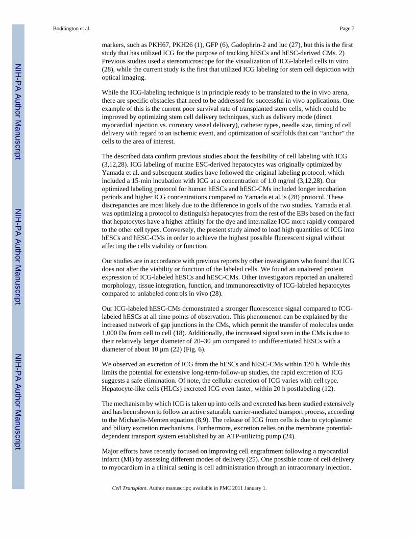
Labeling Human Embryonic Stem Cell-Derived CardiomyocytesWith Indocyanine Green for Noninvasive Tracking With OpticalImaging: An FDA-Compatible Alternative to Firefly Luciferase
Sophie E. Boddington*, Tobias D. Henning*,†, Priyanka Jha*, Christopher R. Schlieve‡, LydiaMandrussow*, David DeNardo§, Harold S. Bernstein¶, Carissa Ritner¶, Daniel Golovko*, YingLu*, Shoujun Zhao*, and Heike E. Daldrup-Link** Department of Radiology, University of California, San Francisco, CA, USA† Department of Radiology, Technical University of Munich, Munich, Germany‡ Gladstone Institute of Cardiovascular Disease, Gladstone Stem Cell Core, University of California,San Francisco, CA, USA§ Cancer Research Institute, University of California, San Francisco, CA, USA¶ Cardiovascular Research Institute, University of California, San Francisco, CA, USA
AbstractHuman embryonic stem cell-derived cardiomyocytes (hESC-CMs) have demonstrated the ability toimprove myocardial function following transplantation into an ischemic heart; however, thefunctional benefits are transient possibly due to poor cell retention. A diagnostic technique that couldvisualize transplanted hESC-CMs could help to optimize stem cell delivery techniques. Thus, thepurpose of this study was to develop a labeling technique for hESCs and hESC-CMs with the FDA-approved contrast agent indocyanine green (ICG) for optical imaging (OI). hESCs were labeled with0.5, 1.0, 2.0, and 2.5 mg/ml of ICG for 30, 45, and 60 min at 37°C. Longitudinal OI studies wereperformed with both hESCs and hESC-CMs. The expression of surface proteins was assessed withimmunofluorescent staining. hESCs labeled with 2 mg ICG/ml for 60 min achieved maximumfluorescence. Longitudinal studies revealed that the fluorescent signal was equivalent to controls at120 h postlabeling. The fluorescence signal of hESCs and hESC-CMs at 1, 24, and 48 h wassignificantly higher compared to precontrast data (p < 0.05). Immunocytochemistry revealedretention of cell-specific surface and nuclear markers postlabeling. These data demonstrate thathESCs and hESC-CMs labeled with ICG show a significant fluorescence up to 48 h and can bevisualized with OI. The labeling procedure does not impair the viability or functional integrity of thecells. The technique may be useful for assessing different delivery routes in order to improve theengraftment of transplanted hESC-CMs or other stem cell progenitors.
KeywordsOptical imaging; Cell labeling; Stem cell; Cardiomyocytes; Indocyanine green; Contrast agent
Address correspondence to Sophie E. Boddington, Department of Radiology, University of California, San Francisco, 185 Berry Street,Suite 350, San Francisco, CA, 94107-0946, USA. Tel: 415-353-4854; Fax: 415-353-9425; [email protected].
NIH Public AccessAuthor ManuscriptCell Transplant. Author manuscript; available in PMC 2011 January 1.
Published in final edited form as:Cell Transplant. 2010 ; 19(1): 55–65. doi:10.3727/096368909X478579.
NIH
-PA Author Manuscript
NIH
-PA Author Manuscript
NIH
-PA Author Manuscript

INTRODUCTIONStem cell therapies hold unprecedented potential to provide therapeutics for a wide variety ofdiseases. Due to the limited mitotic abilities of cells in many organs and thus a limitedregeneration capacity, injury resulting from disease or trauma often leads to irreversible celldamage. Human embryonic stem cells (hESCs) have the unique ability to regenerate into cellsof almost all human tissues (2,5,21). hESC-CMs are a particularly promising source oftherapeutics for myocardial regeneration following cardiac ischemia. When compared tocardiac progenitors derived from human mesenchymal stem cells (hMSC), hESC-CMs have adistinct advantage because their in vitro differentiation model closely resembles in vivo cardiacdevelopment at a structural and electrophysiological level. Few studies, however, currentlydemonstrate this potential for hMSC (4,11,14,19).
A major challenge for successful hESC-derived myocardial regeneration is the developmentof an efficient delivery technique. Previous studies have shown poor outcomes of hESC-CMdelivery, such that the majority of the injected cells are either washed away to extra cardiacorgans or die soon after injection (7,17). More specifically, the present cell survival rate ofhESC-CM transplants in cardiac infarcts is bleak, with only 10% of the cells surviving theinjection. This poor survival rate is thought to be the primary reason behind the limited benefitsobserved in cardiac function following hESC-CM-based therapy (7). A diagnostic techniquethat could directly visualize the transplanted hESC-CM in vivo is needed in order to help directand optimize stem cell delivery techniques and confirm successful stem cell deposition intothe myocardium (20).
Among various available imaging techniques, optical imaging (OI) has the advantage of beingquick, inexpensive, easy to perform, noninvasive, and does not involve ionizing radiation. Thefirst clinical OI scanners have recently entered clinical applications and pilot studies showpromising results for investigations of breast tissue in patients (13).
In order to track hESCs and/or their progeny with OI, cells must be labeled with an exogenousor endogenous marker. While many investigators have focused on bioluminescence OItechniques (e.g., firefly luciferase) for in vivo stem cell tracking, this technique has little clinicalrelevance because it involves the introduction of nonhuman virus substrates into the cells ofinterest (D-luciferin), which alters the host’s DNA permanently (7). Exogenous fluorescentmarkers represent a potentially clinically applicable alternative to firefly luciferase.Indocyanine green (ICG) is an exogenous fluorescent marker with a favorable near-infrared(NIR) absorption and emission spectrum (600–950 nm) and is FDA approved for investigationsof cardiac perfusion in patients (23).
The purpose of this study was to establish a labeling technique of hESCs and hESC-CMs withICG for subsequent OI. The major goal of this investigation was to develop an ICG-labelingprotocol, which should provide maximal fluorescence of labeled hESCs and hESC-CMs onOI scans and, at the same time, preserve cell viability and expression of appropriate surfacemarkers. If successful, this technique would be in principle readily applicable for monitoringstem cell-based therapies, particularly new injection techniques.
MATERIALS AND METHODSContrast Agent
ICG is an FDA-approved, hydrophilic, anionic, tricarbocyanine NIR dye with a molecularweight of 774.97 Da. The structure of ICG is composed of two polycyclic moieties connectedby a carbon chain. ICG shows an absorbance peak at 780 nm and an emission peak at 830 nm(22). The ICG solution used in this study was prepared by dissolving ICG powder (laser grade
Boddington et al. Page 2
Cell Transplant. Author manuscript; available in PMC 2011 January 1.
NIH
-PA Author Manuscript
NIH
-PA Author Manuscript
NIH
-PA Author Manuscript

I-25; Sigma-Aldrich Co., St. Louis, MO) in 20% dimethyl sulfoxide (DMSO, 99.9%, purchasedfrom Sigma-Aldrich Co.) and 80% Dulbecco’s modified Eagles medium (D-MEM HighGlucose; D5648) with resulting stock solutions at concentrations of 0.5, 1, 2, and 2.5 mg/ml.
Culture of Undifferentiated hESCsAll studies were in accordance with and approved by our institution’s Committee on HumanResearch. For conduction of the described experiments, HSF-6 hESCs (UC06, a federal line/NIH approved) were kept in an undifferentiated and pluripotent state, cultured on gelatin-coated (0.1% gel) 10-cm (Falcon) petri dishes, and plated on irradiated mouse embryonicfibroblasts (MEFs ~800,000 cells/plate). The feeder cells were mitotically inactivated using acesium source gamma irradiator (3000 rad for 15 min). Culture medium [Knockout SerumReplacement (KSR) media] contained: Knockout DMEM (Invitrogen, Carlsbad, CA, USA)supplemented with 20% KSR (Invitrogen), 1% nonessential amino acids (Invitrogen), 1 mML-glutamine (Invitrogen), 0.1 mM mercaptoethanol (Sigma), and 4 ng/ml human basic FGF(Invitrogen). Cells were maintained at 37°C in 5% CO2 and split by manual dissection at aratio of 1:3 approximately every 4 days. hESCs were grown until 10-cm petri dishes were 80%confluent. Three days prior to labeling experiments the hESCs were dissected with the EZ tool(Invitrogen) to ensure that the colonies were divided into homogeneous subcolonies. Followingdissection, an equal amount of cell solution was transferred to six-well plates (Corning Inc.VWR Int., West Chester, PA, USA) that were pregelatinized with 0.1% gel and plated with~100,000 feeders. Once colonies formed, roughly 3–4 days later, the cells were ready to belabeled.
Optimization of hESC LabelinghESCs were incubated with increasing concentrations of 0.5, 1.0, 1.5, 2.0, and 2.5 mg ICG/mlfor either 30, 45, or 60 min in standard cell culture conditions. The stock cell labeling solutionwas added to cell culture dishes (60 μl/ml). Following incubation the cells were trypisinized(0.25% trypsin), washed three times in PBS, resuspended in 10 ml of prewarmed KSR, andtransferred to gelatinized-coated (0.1% gel) 10-cm petri dishes. hESCs were separated fromfeeders after an incubation of 60 min at 37°C/5% CO2, at which time the feeder cells adheredtightly to the plate, while the labeled hESCs only adhered weakly. The hESCs were collectedby pipetting and were transferred to a 15-ml centrifuge tube (26). hESCs were then countedand transferred into microcentrifuge tubes for OI (VWR Int.) where they contained 300,000cells/1 ml KSR media.
Cardiomyocyte DifferentiationhESCs were allowed to spontaneously differentiate into beating cardiomyocytes usingestablished methods (16). Briefly, embryoid bodies (EBs) were first produced in six-well lowattachment plates. On day 7 EBs were transferred to a standard six-well plate precoated with0.1% gelatin. Cardiomyocyte media was similar to the KSR used in the undifferentiated culture,but lacked bFGF and contained 20% defined FBS (Hyclone) instead of 20% KSR.
Beating cardiomyocytes (CMs) were labeled on day 25 with ICG under previously optimizedconditions that were established using the hESCs. CMs were grown in six-well plates that wereprecoated with 0.10% gelatin. Following labeling beating CMs were dissected out with a P-100micropipette and transferred to a 15-ml conical tube. The cell suspension was thenenzymatically dispersed using TrypLE (GIBCO, 12604) for 5 min at 37°C and then neutralizedwith CM media containing 20% FBS. Cells were allowed to suspend by gravity and werewashed with phosphate-buffered saline (PBS) three times and were then resuspended in freshCM media for imaging.
Boddington et al. Page 3
Cell Transplant. Author manuscript; available in PMC 2011 January 1.
NIH
-PA Author Manuscript
NIH
-PA Author Manuscript
NIH
-PA Author Manuscript

Optical ImagingIn vitro fluorescence imaging was performed with an IVIS 50 Imaging System (Xenogen,Alameda, CA). The optical imager is an integrated fluorescence system (400–900 nm) that iscomposed of a light-tight specimen chamber (dark box) and a 0.5-inch charge-coupled device(CCD) camera. To minimize electronic background and maximize sensitivity, the CCD camerais thermoelectrically cooled to −70°C.
All in vitro experimental samples were imaged in 1 ml of KSR or CM media in microcentrifugetubes (VWR Int.). All images were acquired using the filter setting preset for ICG with abackground wavelength at 665–695 nm, an excitation wavelength at 710–760 nm, and anemission wavelength set at 810–875 nm. Consistent illumination parameters were used for allNIR fluorescent acquisitions. For all experiments, the field of view (FOV) was set at 7,exposure time was 2 s, f/stop was 2, lamp voltage was set to “high” and binning was kept onmedium. Following acquisition, all images were normalized to units of average efficiency,displayed in the same scale of fluorescent intensity, and analyzed using the Living Image 2.5software (Xenogen, Alameda, CA).
Optimization of the Cell Labeling Protocol—hESCs labeled with different ICGconcentrations and incubation times underwent OI directly after the labeling process.
Longitudinal Follow-up Studies—hESCs and hESC-CMs were labeled with the opticallabeling protocol, determined based on experiments from the above protocol, and in separatetubes 100,000 hESCs or hESC-CMs were resuspended in 1 ml of fresh KSR or CM media andimaged until the fluorescent signal matched that of the nonlabeled control cells. All experimentswere performed in triplicate (Fig. 1).
Optical Imaging Data AnalysisFor quantitative analyses of OI data, regions of interest (ROI) were placed on each ICG testsample (micro-centrifuge tube). Quantitative measures of fluorescence intensity of labeledcells in test tubes were normalized to units of average efficiency, displayed in the same scaleof fluorescent intensity, and analyzed using the Living Image 2.5 software (Xenogen, Alameda,CA). The unit of “efficiency” represents the fractional ratio of fluorescent-emitted photons perincident excitation photon (Xenogen).
Statistical AnalysisMeans and SDs were used to describe the variation in fluorescent signal over time and viabilityfor the following groups: 1) fluorescent signal for hESCs compared to nonlabeled controls, 2)fluorescent signal for CMs compared to nonlabeled controls, 3) fluorescent signal for hESCscompared to CMs, and 4) viability of CMs compared to nonlabeled controls. The multivariateanalysis of variance (MANOVA) was used to describe the variation in fluorescent signal andviability for hESCs compared to nonlabeled controls for the various labeling protocols (variousICG concentrations and incubation times). Multiple comparison p-values were determined bythe Tukey-Kramer test. All analyses were performed by SAS software (SAS V9.3; Cary, NC,USA).
Cell Viability TestingTrypan blue tests were performed on all cells before and after the labeling procedure to verifyviability. Cells were counted in a hemocytometer and then underwent subsequent opticalimaging studies.
Boddington et al. Page 4
Cell Transplant. Author manuscript; available in PMC 2011 January 1.
NIH
-PA Author Manuscript
NIH
-PA Author Manuscript
NIH
-PA Author Manuscript

ImmunohistochemistryAnalysis of hESC Pluripotency Postlabeling—To ensure that the hESCs retainedmarker expression patterns characteristic of undifferentiated hESCs postlabeling, the cells wereanalyzed using immunofluorescence techniques followed by fluorescent microscopy. hESCunderwent labeling under the optimized conditions as described above. The ICG-labeledhESCs and unlabeled controls were manually dissected out, washed three times, and replatedonto 24-well plates that were pregelatinized with 0.1% gel and contained 20,000 irradiatedfeeder cells per well. After colony formation occurred (approximately 4 days later) the cellswere incubated with primary antibodies according to the manufacturer’s instructions (ES CellMarker Sample Kit protocol; Chemicon, Billerica, MA) followed by incubation with thesecondary antibody Alexa Fluor® 488 Goat anti-mouse IgG. Confocal images were acquiredusing a Leica microscope.
Assessment of Cardiomyocyte Protein Expression Postlabeling—Followinglabeling with ICG, the beating CM areas were dissected out with a P-100 micropipette andtransferred to a 15-ml conical tube. The cell suspension was then enzymatically dispersed usingTrypLE (GIBCO, 12604) for 5 min at 37°C and then neutralized with CM media containing20% FBS. Cells were allowed to settle by gravity and were then resuspended in fresh CMmedia. ROC inhibitor was added at a concentration of 10 μM (EMD: y-27632) to preventapoptosis. Cells were plated at a low density on pregelatinized-coated wells of Lab-Tek IIeight-well glass chamber slide system and incubated for 24 h. Cells were fixed using 4%paraformaldyhyde for 15 min. Cells were washed two times for 1 min with PBS followed bywashing three times for 1 min each with washing buffer 1 (0.10% Triton X-100 in PBS at pH7.4). Cells were blocked with 10% PBS, 0.10% Triton X-100, 5% normal goat serum, and inPBS for 1 h at room temperature. Primary antibodies were added to chamber slides in a totalof 200 μl/well of antibody buffer and incubated overnight at 4°C. Primary antibodies wereTroponin T (Thermo Scientific, MS-295-PO), GATA4 (Abcam, ab61170), Connexin43(Millipore, AB1728), Alexa Fluor® 488 Phalloidin (Invitrogen, A12379), Islet1(Developmental Studies Hybridoma Bank), Lin28 (R&D Systems, AF3757), Wnt-8A (R&DSystems, AF2248), TBX5 (GenWay, 18-003-42369), SMA. All were used at 1:100. The nextday cells were washed three times for 1 min each in washing buffer 2 (0.03% Triton X-100 inPBS at pH 7.4) followed by an additional wash in PBS for 1 min. Cells were incubated withproper secondary antibodies in 1× PBS. Secondary antibodies used were Alexa Fluor® anti-rabbit 555 (Molecular Probes, A21429), Alexa Fluor® anti-mouse IgG 488 (Molecular Probes,A11029), and Alexa Fluor® anti-goat 555 (Invitrogen Molecular Probes, A21431). All wereused at 1:500. Slides were placed in humidifying trays/covered and left on benchtop for 1–2h. Cells were washed in washing buffer 2 for 1 min and then washed again in PBS for 1 min,postfixed with 4% PFA for 1 min, followed by washing with PBS for 1 min. Samples werecombined with VECTASHIELD® mounting medium containing DAPI (Vector LaboratoriesInc., H-1500) and wells were sealed with coverslips. Samples were then imaged using a ZeissAxioObserver Z1 microscope.
RESULTSOptical Imaging
Optimization of hESC Labeling Protocol—Labeling of hESCs with varyingconcentrations and incubation times showed a maximal fluorescence signal with thecombination of the 60-min incubation time and an ICG concentration of 2.0 mg/ml (Fig. 2).The average fluorescence signal was significantly higher with the 60-min incubation timecompared to lower incubation times (p < 0.0001), and also significantly higher with the ICGconcentration of 2.0 mg/ml than lower or higher concentrations (p < 0.0001).
Boddington et al. Page 5
Cell Transplant. Author manuscript; available in PMC 2011 January 1.
NIH
-PA Author Manuscript
NIH
-PA Author Manuscript
NIH
-PA Author Manuscript

Longitudinal OI Studies—Labeling of hESCs with ICG at a concentration of 2.0 mg/mlafter a 60-min incubation time revealed that the fluorescent signal slowly decreased over time,compatible with slow release of the contrast agent from the cells. The signal decreased by 50%within 48 h postlabeling and was equivalent to controls at 120 h postlabeling (Fig. 3). Thefluorescence signal at 1, 24, and 48 h was significantly higher compared to precontrast data(p < 0.05). The signal at 120 h postlabeling was not significantly different from baseline (p >0.05).
Twelve days after induction of hESC differentiation as described above (16), beatingcardiomyocytes appeared. Longitudinal studies of CMs, labeled on day 25 with 2.0 mg ICG/ml for 60 min, revealed similar fluorescence signal kinetics compared to labeled hESCs (Fig.4). The cells’ fluorescence signal was significantly elevated at 1, 24, and 48 h (p < 0.05), butshowed no significant difference compared to baseline data at 120 h postlabeling (p > 0.05).
Fluorescence—The fluorescence signal of CMs was significantly higher compared to thefluorescence signal of hESCs at 1 h (p < 0.0001), 48 h (p < 0.05), 72 h (p < 0.01), and 96 h(p < 0.001) while there was no statistically significant difference in signal at 24 h postlabeling(Fig. 5).
Viability TestingTrypan blue exclusion testing showed no significant difference in viability between labeledhESCs or labeled CMs compared to unlabeled controls. All groups exhibited viabilities greaterthan 88%, which were not significantly different between experimental groups (p > 0.05) (Fig.2).
ImmunocytochemistryAnalysis of hESC Pluripotency—Fluorescent microscopy experiments followingimmunohistochemistry revealed that ICG-labeled and unlabeled HSF-6 cells expressed thesurface antigens SSEA-3, SSEA-4, TRA-1-60, and TRA-1-81 and did not express SSEA-1(Fig. 6). These expression patterns are consistent with undifferentiated hESCs or “pluripotent”stem cells. Furthermore, there were no observed morphologic abnormalities of labeled hESCs(colonies had clearly defined borders and the cells within each colony were homogenouslysized) compared to unlabeled controls.
Retention of Cardiomyocyte Protein Expression Post-labeling—The presence ofcardiac-specific proteins was studied using immunocytochemistry. Figure 7 shows positivestaining for Connexin43, Lin28, Troponin T, Actin + Lin28, Wnt8a, TBX5, GATA4, and Islet1.The staining patterns recapitulate previous publications on the location of the targeted proteins(Fig. 7). There was no difference in antigen expression patterns between la- beled and unlabeledCMs.
DISCUSSIONThe results of this study indicate that hESCs and hESC-CMs can be labeled by simpleincubation with the FDA-approved fluorescent marker ICG without impairing the cellsviability, hESCs pluripotency, or cardiomyocyte function. The ICG-labeled hESCs andcardiomyocytes can be detected up to 48 h postlabeling with optical imaging.
Our study is novel with regard to two aspects. 1) To the best of our knowledge, this is the firststudy that utilized the FDA-approved fluorochrome ICG for the labeling of hESCs and hESC-derived CMs. Murine ESC-derived hepatocytes have been previously labeled with ICG andadult mesenchymal stem cells have been previously labeled with other organic fluorescent
Boddington et al. Page 6
Cell Transplant. Author manuscript; available in PMC 2011 January 1.
NIH
-PA Author Manuscript
NIH
-PA Author Manuscript
NIH
-PA Author Manuscript
markers, such as PKH67, PKH26 (1), GFP (6), Gadophrin-2 and luc (27), but this is the firststudy that has utilized ICG for the purpose of tracking hESCs and hESC-derived CMs. 2)Previous studies used a stereomicroscope for the visualization of ICG-labeled cells in vitro(28), while the current study is the first that utilized ICG labeling for stem cell depiction withoptical imaging.
While the ICG-labeling technique is in principle ready to be translated to the in vivo arena,there are specific obstacles that need to be addressed for successful in vivo applications. Oneexample of this is the current poor survival rate of transplanted stem cells, which could beimproved by optimizing stem cell delivery techniques, such as delivery mode (directmyocardial injection vs. coronary vessel delivery), catheter types, needle size, timing of celldelivery with regard to an ischemic event, and optimization of scaffolds that can “anchor” thecells to the area of interest.
The described data confirm previous studies about the feasibility of cell labeling with ICG(3,12,28). ICG labeling of murine ESC-derived hepatocytes was originally optimized byYamada et al. and subsequent studies have followed the original labeling protocol, whichincluded a 15-min incubation with ICG at a concentration of 1.0 mg/ml (3,12,28). Ouroptimized labeling protocol for human hESCs and hESC-CMs included longer incubationperiods and higher ICG concentrations compared to Yamada et al.’s (28) protocol. Thesediscrepancies are most likely due to the difference in goals of the two studies. Yamada et al.was optimizing a protocol to distinguish hepatocytes from the rest of the EBs based on the factthat hepatocytes have a higher affinity for the dye and internalize ICG more rapidly comparedto the other cell types. Conversely, the present study aimed to load high quantities of ICG intohESCs and hESC-CMs in order to achieve the highest possible fluorescent signal withoutaffecting the cells viability or function.
Our studies are in accordance with previous reports by other investigators who found that ICGdoes not alter the viability or function of the labeled cells. We found an unaltered proteinexpression of ICG-labeled hESCs and hESC-CMs. Other investigators reported an unalteredmorphology, tissue integration, function, and immunoreactivity of ICG-labeled hepatocytescompared to unlabeled controls in vivo (28).
Our ICG-labeled hESC-CMs demonstrated a stronger fluorescence signal compared to ICG-labeled hESCs at all time points of observation. This phenomenon can be explained by theincreased network of gap junctions in the CMs, which permit the transfer of molecules under1,000 Da from cell to cell (18). Additionally, the increased signal seen in the CMs is due totheir relatively larger diameter of 20–30 μm compared to undifferentiated hESCs with adiameter of about 10 μm (22) (Fig. 6).
We observed an excretion of ICG from the hESCs and hESC-CMs within 120 h. While thislimits the potential for extensive long-term-follow-up studies, the rapid excretion of ICGsuggests a safe elimination. Of note, the cellular excretion of ICG varies with cell type.Hepatocyte-like cells (HLCs) excreted ICG even faster, within 20 h postlabeling (12).
The mechanism by which ICG is taken up into cells and excreted has been studied extensivelyand has been shown to follow an active saturable carrier-mediated transport process, accordingto the Michaelis-Menten equation (8,9). The release of ICG from cells is due to cytoplasmicand biliary excretion mechanisms. Furthermore, excretion relies on the membrane potential-dependent transport system established by an ATP-utilizing pump (24).
Major efforts have recently focused on improving cell engraftment following a myocardialinfarct (MI) by assessing different modes of delivery (25). One possible route of cell deliveryto myocardium in a clinical setting is cell administration through an intracoronary injection.
Boddington et al. Page 7
Cell Transplant. Author manuscript; available in PMC 2011 January 1.
NIH
-PA Author Manuscript
NIH
-PA Author Manuscript
NIH
-PA Author Manuscript

Limited benefits from this approach have prompted investigators to assess different deliverymethods, such as direct intramyocardial injections. A recent study investigated the distributionpatterns of transplanted bone marrow stem cells transfected with firefly luciferase followingthree delivery methods: intra-aortic, intravenous, and intramyocardial. Within 48 h of delivery,the large majority of the implanted cells localized to extracardiac organs with all of thesedelivery methods. Interestingly, this study also revealed the importance of the timing ofdelivery with regard to the ischemic event. When cells were injected into an acute infarct therewas a 50% survival rate at 2 h, but improved retention was observed when cells were injectedinto a more mature infarct (15). These results confirm the critical need of a cell trackingtechnique in the early phase after delivery of therapeutic cells, which would be possible withour ICG-labeling technique.
The ICG-labeling technique is advantageous over other techniques being developed becauseit is cost effective, technically straightforward, and rapid (<2 h). The most notable advantageof the ICG-labeling technique compared to others in the field, namely labeling with iron oxide-based nanoparticles for MR imaging, is the magnitudes increased sensitivity that one achieveswith OI. While MRI has the advantage of high anatomical resolution, it provides limitedsensitivity. Single cell sensitivity is especially important in the field of stem cell research dueto the unpredictable migrational dynamics of the cells, even after local injections. Whilelabeling cells for PET imaging does offer sensitive cell detection, PET techniques involveradiation exposure with unknown side effects on the stem cells. More specifically, causingdamage to the DNA of the cells from repeated or even acute radiation exposure is extremelyrisky in a field where the behavioral dynamics of the cells are not currently well understood.Other advantages of the ICG-labeling technique are its compatibility with conventionalfluorescent microscopy, which enables direct correlations of imaging findings withpathological specimens (10).
We recognize that there are several limitations to our study. Firstly, our data showed that thefluorescent marker is slowly released from the labeled cells. The cells could be detected withOI for at least 48 h; however, the fluorescent signal rapidly decayed after this time interval.Thus, potential applications of our proposed technique would be limited to this time interval.Our ICG-based cell tracking technique could contribute to short-term posttransplantinvestigations by assessing factors that may increase stem cell engraftment, such as differentdelivery modes, the timing of delivery, various scaffolds and growth factors, and different stemcell types. In addition, several investigators noted that the transplanted stem cells do not engraftinto the desired area but migrate to other areas in the early time period after transplantation(7). The ICG-labeling method could be used to monitor such migrational dynamics in a fast,inexpensive, and relatively easy manner.
Our applied marker ICG is FDA approved for cardiac imaging and thus would be in theoryreadily accessible to patients under “off label use” conditions. Clinical scanners for breastimaging have just entered clinical practice. However, clinical OI scanners for evaluations ofpatients’ hearts have yet to be developed; therefore, potential future clinical applications of theICG-labeling technique await further development of OI scanners for cardiac applications(13). Our studies were obtained in vitro and future studies are being conducted to investigatein vivo applications of the proposed cell tracking technique, such as tracking of hESC-CMsafter implantation into ischemic myocardium of experimental animals.
CONCLUSIONSIn summary, the derived data establish an optimized technique for depiction of the labeled cellswith optical imaging and illustrates the feasibility of hESC-based labeling with the FDA-approved fluorescent dye ICG. Other potential applications of the ICG-labeling technique
Boddington et al. Page 8
Cell Transplant. Author manuscript; available in PMC 2011 January 1.
NIH
-PA Author Manuscript
NIH
-PA Author Manuscript
NIH
-PA Author Manuscript

include, but are not limited to, investigations of other stem cell lineages and progenitors, suchas neural precursors, chondrocytes, osteoblasts, and neutrophils.
AcknowledgmentsThis study was supported by a research grant from the California Institute for Regenerative Medicine (CIRM), grant#RS1-00381-1. The Islet-1 monoclonal antibody 40.2D6 was developed by Thomas Jessel and was obtained from theDevelopmental Studies Hybridoma Bank developed with support of the NICHD and maintained by the University ofIowa, Department of Biological Sciences, Iowa City, IA.
References1. Askenasy N, Farkas DL. Optical imaging of PKH-labeled hematopoietic cells in recipient bone marrow
in vivo. Stem Cells 2002;20(6):501–513. [PubMed: 12456958]2. Attema JL, Papathanasiou P, Forsberg EC, Xu J, Smale ST, Weissman IL. Epigenetic characterization
of hematopoietic stem cell differentiation using miniChIP and bisulfite sequencing analysis. Proc NatlAcad Sci USA 2007;104(30):12371–12376. [PubMed: 17640913]
3. Baharvand H, Hashemi SM, Kazemi Ashtiani S, Farrokhi A. Differentiation of human embryonic stemcells into hepatocytes in 2D and 3D culture systems in vitro. Int J Dev Biol 2006;50(7):645–652.[PubMed: 16892178]
4. Bettiol E, Clement S, Krause KH, Jaconi ME. Embryonic and adult stem cell-derived cardiomyocytes:Lessons from in vitro models. Rev Physiol Biochem Pharmacol 2006;157:1–30. [PubMed: 17236648]
5. Boyer LA, Mathur D, Jaenisch R. Molecular control of pluripotency. Curr Opin Genet Dev 2006;16(5):455–462. [PubMed: 16920351]
6. Brazelton TR, Blau HM. Optimizing techniques for tracking transplanted stem cells in vivo. Stem Cells2005;23(9):1251–1265. [PubMed: 16109764]
7. Cao F, Wagner RA, Wilson KD, Xie X, Fu JD, Drukker M, Lee A, Li RA, Gambhir SS, WeissmanIL, Robbins RC, Wu JC. Transcriptional and functional profiling of human embryonic stem cell-derived cardiomyocytes. PLoS ONE 2008;3(10):e3474. [PubMed: 18941512]
8. Desmettre T, Devoisselle JM, Mordon S. Fluorescence properties and metabolic features ofindocyanine green (ICG) as related to angiography. Surv Ophthalmol 2000;45(1):15–27. [PubMed:10946079]
9. Fickweiler S, Szeimies RM, Baumler W, Steinbach P, Karrer S, Goetz AE, Abels C, Hofstadter F,Landthaler M. Indocyanine green: Intracellular uptake and phototherapeutic effects in vitro. JPhotochem Photobiol B 1997;38(2–3):178–183. [PubMed: 9203378]
10. Frangioni JV, Hajjar RJ. In vivo tracking of stem cells for clinical trials in cardiovascular disease.Circulation 2004;110(21):3378–3383. [PubMed: 15557385]
11. Furlani D, Li W, Pittermann E, Klopsch C, Wang L, Knopp A, Jungebluth P, Thedinga E, HavensteinC, Westien I, Ugurlucan M, Li RK, Ma N, Steinhoff G. A transformed cell population derived fromcultured mesenchymal stem cells has no functional effect after transplantation into the injured heart.Cell Transplant 2009;18(3):319–331. [PubMed: 19558780]
12. Hay DC, Zhao D, Ross A, Mandalam R, Lebkowski J, Cui W. Direct differentiation of humanembryonic stem cells to hepatocyte-like cells exhibiting functional activities. Cloning Stem Cells2007;9(1):51–62. [PubMed: 17386014]
13. Intes X. Time-domain optical mammography SoftScan: Initial results. Acad Radiol 2005;12(8):934–947. [PubMed: 16023382]
14. Laflamme MA, Chen KY, Naumova AV, Muskheli V, Fugate JA, Dupras SK, Reinecke H, Xu C,Hassanipour M, Police S, O’Sullivan C, Collins L, Chen Y, Minami E, Gill EA, Ueno S, Yuan C,Gold J, Murry CE. Cardiomyocytes derived from human embryonic stem cells in pro-survival factorsenhance function of infarcted rat hearts. Nat Biotechnol 2007;25(9):1015–1024. [PubMed:17721512]
15. Li SH, Lai TY, Sun Z, Han M, Moriyama E, Wilson B, Fazel S, Weisel RD, Yau T, Wu JC, Li RK.Tracking cardiac engraftment and distribution of implanted bone marrow cells: Comparing intra-aortic, intravenous, and intramyocardial delivery. J Thorac Cardiovasc Surg 2009;137(5):1225–1233. [PubMed: 19379996]
Boddington et al. Page 9
Cell Transplant. Author manuscript; available in PMC 2011 January 1.
NIH
-PA Author Manuscript
NIH
-PA Author Manuscript
NIH
-PA Author Manuscript

16. Maltsev VA, Wobus AM, Rohwedel J, Bader M, Hescheler J. Cardiomyocytes differentiated in vitrofrom embryonic stem cells developmentally express cardiac-specific genes and ionic currents. CircRes 1994;75(2):233–244. [PubMed: 8033337]
17. Martens TP, Godier AF, Parks JJ, Wan LQ, Koeckert MS, Eng GM, Hudson BI, Sherman W, Vunjak-Novakovic G. Percutaneous cell delivery into the heart using hydrogels polymerizing in situ. CellTransplant 2009;18(3):297–304. [PubMed: 19558778]
18. Nicholson BJ. Gap junctions—from cell to molecule. J Cell Sci 2003;116(Pt. 22):4479–4481.[PubMed: 14576341]
19. Nistor GI, Totoiu MO, Haque N, Carpenter MK, Keirstead HS. Human embryonic stem cellsdifferentiate into oligodendrocytes in high purity and myelinate after spinal cord transplantation. Glia2005;49(3):385–396. [PubMed: 15538751]
20. Patel AN, Sherman W. Cardiac stem cell therapy: Advances from 2008. Cell Transplant 2009;18(3):243–244. [PubMed: 19558772]
21. Pellettieri J, Sanchez Alvarado A. Cell turnover and adult tissue homeostasis: from humans toplanarians. Annu Rev Genet 2007;41:83–105. [PubMed: 18076325]
22. Satin J, Itzhaki I, Rapoport S, Schroder EA, Izu L, Arbel G, Beyar R, Balke CW, Schiller J, GepsteinL. Calcium handling in human embryonic stem cell-derived cardiomyocytes. Stem Cells 2008;26(8):1961–1972. [PubMed: 18483424]
23. Saxena V, Sadoqi M, Shao J. Degradation kinetics of indocyanine green in aqueous solution. J PharmSci 2003;92(10):2090–2097. [PubMed: 14502548]
24. Shinohara H, Tanaka A, Kitai T, Yanabu N, Inomoto T, Satoh S, Hatano E, Yamaoka Y, Hirao K.Direct measurement of hepatic indocyanine green clearance with near-infrared spectroscopy:Separate evaluation of uptake and removal. Hepatology 1996;23(1):137–144. [PubMed: 8550033]
25. Silva SA, Sousa AL, Haddad AF, Azevedo JC, Soares VE, Peixoto CM, Soares AJ, Issa AF, FelipeLR, Branco RV, Addad JA, Moreira RC, Tuche FA, Mesquita CT, Drumond CC, Junior AO, RochitteCE, Luz JH, Rabischoffisky A, Nogueira FB, Vieira RB, Junior HS, Borojevic R, Dohmann HF.Autologous bone-marrow mononuclear cell transplantation after acute myocardial infarction:Comparison of two delivery techniques. Cell Transplant 2009;18(3):343–352. [PubMed: 19558782]
26. Tompers DM, Labosky PA. Electroporation of murine embryonic stem cells: A step-by-step guide.Stem Cells 2004;22(3):243–249. [PubMed: 15153600]
27. Wang X, Rosol M, Ge S, Peterson D, McNamara G, Pollack H, Kohn DB, Nelson MD, Crooks GM.Dynamic tracking of human hematopoietic stem cell engraftment using in vivo bioluminescenceimaging. Blood 2003;102(10):3478–3482. [PubMed: 12946998]
28. Yamada T, Yoshikawa M, Kanda S, Kato Y, Nakajima Y, Ishizaka S, Tsunoda Y. In vitrodifferentiation of embryonic stem cells into hepatocyte-like cells identified by cellular uptake ofindocyanine green. Stem Cells 2002;20(2):146–154. [PubMed: 11897871]
Boddington et al. Page 10
Cell Transplant. Author manuscript; available in PMC 2011 January 1.
NIH
-PA Author Manuscript
NIH
-PA Author Manuscript
NIH
-PA Author Manuscript

Figure 1.Experimental scheme followed for the labeling with indocyanine green (ICG), imaging, andprotein analysis of undifferentiated human embryonic stem cells (hESCs) and cardiomyocytes.
Boddington et al. Page 11
Cell Transplant. Author manuscript; available in PMC 2011 January 1.
NIH
-PA Author Manuscript
NIH
-PA Author Manuscript
NIH
-PA Author Manuscript

Figure 2.Cell viability of human embryonic stem cells (hESCs) maintained postlabeling withindocyanine green (ICG). Fluorescent signal (units in efficiency) seen with optical imaging(OI) and viability (% viable cells) of hESCs, labeled with different ICG concentrations (0 =control as well as 0.5, 1.0, 1.5, 2.0, and 2.5 mg ICG/ml) for different incubation intervals (30,45, and 60 min). Data are displayed as means and SD of triplicate samples containing 300,000cells in a pellet per sample in 1 ml of KSR.
Boddington et al. Page 12
Cell Transplant. Author manuscript; available in PMC 2011 January 1.
NIH
-PA Author Manuscript
NIH
-PA Author Manuscript
NIH
-PA Author Manuscript

Figure 3.Representative optical images of ICG-labeled hESCs in 1 ml of KSR using the optimizedlabeling protocol (2.0 mg ICG/ml, 60-min incubation) at different time points after labeling(1, 24, 48, 72, 96, and 120 h) showing decreasing fluorescent signal over time. The three rowsrepresent triplicates.
Boddington et al. Page 13
Cell Transplant. Author manuscript; available in PMC 2011 January 1.
NIH
-PA Author Manuscript
NIH
-PA Author Manuscript
NIH
-PA Author Manuscript

Figure 4.Representative optical images of ICG-labeled hESC-CMs in 1 ml of CM media using theoptimized labeling protocol (2.0 mg ICG/ml, 60-min incubation) at different time points afterlabeling (1, 24, 48, 72, 96, and 120 h) showing decreasing fluorescent signal over time. Thethree rows represent triplicates.
Boddington et al. Page 14
Cell Transplant. Author manuscript; available in PMC 2011 January 1.
NIH
-PA Author Manuscript
NIH
-PA Author Manuscript
NIH
-PA Author Manuscript

Figure 5.OI fluorescent signal (units in efficiency) of ICG-labeled human embryonic stem cells (hESC),ICG-labeled hESC-derived cardiomyocytes (CM), and nonlabeled controls at different timepoints after labeling (1, 24, 48, 72, 96, and 120 h). Data are displayed as means and SD oftriplicate samples with 100,000 cells per sample.
Boddington et al. Page 15
Cell Transplant. Author manuscript; available in PMC 2011 January 1.
NIH
-PA Author Manuscript
NIH
-PA Author Manuscript
NIH
-PA Author Manuscript

Figure 6.Fluorescent microscopy following immunohistochemistry revealed that the hESCs labeledunder the optimized conditions expressed the hESC cell-specific surface antigens, includingSSEA-3, SSEA-4, TRA-1-60, and TRA-1-81, and did not express SSEA-1. These expressionmarkers are consistent with undifferentiated hESCs. Scale bar: 100 μm.
Boddington et al. Page 16
Cell Transplant. Author manuscript; available in PMC 2011 January 1.
NIH
-PA Author Manuscript
NIH
-PA Author Manuscript
NIH
-PA Author Manuscript

Figure 7.Cardiomyocyte protein expression maintained post-ICG labeling. Positive staining wasobserved for (a) Connexin43, (b) Wnt8a, (c) Troponin T, (d) GATA4, (e) Lin28, (f) TBX5, (g)Actin, (h) Islet1. Scale bar: 100 μm.
Boddington et al. Page 17
Cell Transplant. Author manuscript; available in PMC 2011 January 1.
NIH
-PA Author Manuscript
NIH
-PA Author Manuscript
NIH
-PA Author Manuscript
Copyright © 2022 FDOKUMEN