Transplanted Oligodendrocytes and Motoneuron Progenitors Generated from Human Embryonic Stem Cells...
9
EMBRYONIC STEM CELLS/INDUCED PLURIPOTENT STEM CELLS Transplanted Oligodendrocytes and Motoneuron Progenitors Generated from Human Embryonic Stem Cells Promote Locomotor Recovery After Spinal Cord Transection SLAVEN ERCEG, a,b MOHAMMAD RONAGHI, a MARC ORIA, c,d MIREIA GARCI ´ A ROSELLO ´ , a MARIA AMPARO PE ´ REZ ARAGO ´ , a MARIA GOMEZ LOPEZ, a IVANA RADOJEVIC, a VICTORIA MORENO-MANZANO, a FRANCISCO-JAVIER RODRI ´ GUEZ- JIME ´ NEZ, a SHOM SHANKER BHATTACHARYA, b JUAN CORDOBA, c MIODRAG STOJKOVIC a,e,f * a Cellular Reprogramming Laboratory, Centro de Investigacio ´n Prı ´ncipe Felipe (CIPF), Valencia, Spain; b CABIMER (Centro Andaluz de Biologı ´a Molecular y Medicina Regenerativa), Avda. Americo Vespucio s/n, Parque Cientı ´fico y Tecnolo ´gico Cartuja, Sevilla, Spain; c Liver Unit, Department of Medicine, Hospital Universitari Vall d’Hebron, Universitat Auto ´noma de Barcelona, Barcelona, Spain; d Centro de Investigacio ´n Biome ´dica en Red de Enfermedades Hepa ´ticas y Digestivas (CIBEREHD), Instituto de Salud Carlos III, Madrid, Spain; e Spebo Medical, Leskovac, Serbia; f Human Genetics, Medical School University of Kragujevac, Serbia Key Words. Embryonic stem cells • Oligodendrocytes • Stem cell transplantation • Neural differentiation ABSTRACT Human embryonic stem cells (hESC) hold great promise for the treatment of patients with many neurodegenerative diseases particularly those arising from cell loss or neural dysfunction including spinal cord injury. This study evalu- ates the therapeutic effects of transplanted hESC-derived oligodendrocyte progenitors (OPC) and/or motoneuron progenitors (MP) on axonal remyelination and functional recovery of adult rats after complete spinal cord transec- tion. OPC and/or MP were grafted into the site of injury in the acute phase. Based on Basso-Beattie-Bresnahan scores recovery of locomotor function was significantly enhanced in rats treated with OPC and/or MP when com- pared with control animals. When transplanted into the spinal cord immediately after complete transection, OPC and MP survived, migrated, and differentiated into mature oligodendrocytes and neurons showing in vivo electrophysiological activity. Taken together, these results indicate that OPC and MP derived from hESC could be a useful therapeutic strategy to repair injured spinal cord. STEM CELLS 2010;28:1541–1549 Disclosure of potential conflicts of interest is found at the end of this article. INTRODUCTION Spinal cord transection, besides the loss in central control of motor, sensory and autonomic function below the injury site, produces limited exogenous repair and poor functional recov- ery. The cumulative death of neurons, astroglia, and oligoden- droglia in and around the lesion site disrupts neural circuitry and leads to neurological dysfunction [1]. Damaged axons are unable to grow, regenerate and to reconnect themselves with the structures that were innervated before the injury, causing a permanent interruption of the injured nervous route [1–3]. Many factors contribute to this condition including cellular destruction, demyelination, inability to remyelinate spared axons, and failure of axons to overcome the conduction block by astrocyte scar [4–6]. Introducing new cells as progenitors of motoneurons (motoneurons progenitor, MP) or oligoden- drocyte progenitor cells (OPC) into a damaged spinal cord to treat a disease could be a good strategy to establish disrupted synaptic connections between the central nervous system (CNS) functions above and below the lesion. Compared with other stem cell types, human embryonic stem cells (hESC) and induced pluripotency stem (IPS) cells currently show the greatest potential for differentiation and cell replacement therapies. These cells are pluripotent and can give rise to cells of three germinal layers, they can be propa- gated indefinitely in culture and can provide a large quantity Author contributions: S.E.: conception and design, data analysis and interpretation, manuscript writing, final approval of manuscript, experiments; M.R.: experiments; M.O.: experiments, data analysis and interpretation; M.G.R.: experiments, surgery; M.A.P.A., M.G.L., I.R., V.M.-M., and F.J.R.-J.: experiments; S.S.B. and J.C.: data analysis and interpretation; M.S.: conception and design, data analysis and interpretation, manuscript writing, final approval of manuscript. *Present address: CABIMER (Centro Andaluz de Biologı ´a Molecular y Medicina Regenerativa), Avda. Americo Vespucio s/n, Parque Cientı ´fico y Tecnolo ´gico Cartuja, Sevilla, Spain. Correspondence: Slaven Erceg, Ph.D., CABIMER (Centro Andaluz de Biologı ´a Molecular y Medicina Regenerativa), Avda. Americo Vespucio s/n, Parque Cientı ´fico y Tecnolo ´gico Cartuja, Sevilla, Spain. Telephone: þ34-95-446-76-21; Fax: þ34-95-446-16-64; e-mail: [email protected] or Miodrag Stojkovic, Ph.D., SPEBO Medical, Norvezanska 16, Leskovac, Serbia and Medical Faculty, University of Kragujevac, Kragujevac, Serbia. Telephone: +381-16-21-81-71; e-mail: [email protected] Received April 8, 2010; accepted for publication July 15, 2010; first published online in STEM CELLS EXPRESS July 27, 2010. V C AlphaMed Press 1066-5099/2009/ $30.00/0 doi: 10.1002/stem.489 STEM CELLS 2010;28:1541–1549 www.StemCells.com
Transcript of Transplanted Oligodendrocytes and Motoneuron Progenitors Generated from Human Embryonic Stem Cells...

EMBRYONIC STEM CELLS/INDUCED PLURIPOTENT STEM CELLS
Transplanted Oligodendrocytes and Motoneuron Progenitors
Generated from Human Embryonic Stem Cells Promote Locomotor
Recovery After Spinal Cord Transection
SLAVEN ERCEG,a,b MOHAMMAD RONAGHI,a MARC ORIA,c,d MIREIA GARCIA ROSELLO,a MARIA AMPARO PEREZ
ARAGO,aMARIA GOMEZ LOPEZ,
aIVANA RADOJEVIC,
aVICTORIA MORENO-MANZANO,
aFRANCISCO-JAVIER RODRIGUEZ-
JIMENEZ,a SHOM SHANKER BHATTACHARYA,b JUAN CORDOBA,c MIODRAG STOJKOVICa,e,f*
aCellular Reprogramming Laboratory, Centro de Investigacion Prıncipe Felipe (CIPF), Valencia, Spain;bCABIMER (Centro Andaluz de Biologıa Molecular y Medicina Regenerativa), Avda. Americo Vespucio s/n,
Parque Cientıfico y Tecnologico Cartuja, Sevilla, Spain; cLiver Unit, Department of Medicine, Hospital
Universitari Vall d’Hebron, Universitat Autonoma de Barcelona, Barcelona, Spain; dCentro de Investigacion
Biomedica en Red de Enfermedades Hepaticas y Digestivas (CIBEREHD), Instituto de Salud Carlos III, Madrid,
Spain; eSpebo Medical, Leskovac, Serbia; fHuman Genetics, Medical School University of Kragujevac, Serbia
Key Words. Embryonic stem cells • Oligodendrocytes • Stem cell transplantation • Neural differentiation
ABSTRACT
Human embryonic stem cells (hESC) hold great promisefor the treatment of patients with many neurodegenerativediseases particularly those arising from cell loss or neural
dysfunction including spinal cord injury. This study evalu-ates the therapeutic effects of transplanted hESC-derivedoligodendrocyte progenitors (OPC) and/or motoneuron
progenitors (MP) on axonal remyelination and functionalrecovery of adult rats after complete spinal cord transec-
tion. OPC and/or MP were grafted into the site of injuryin the acute phase. Based on Basso-Beattie-Bresnahan
scores recovery of locomotor function was significantlyenhanced in rats treated with OPC and/or MP when com-pared with control animals. When transplanted into the
spinal cord immediately after complete transection, OPCand MP survived, migrated, and differentiated intomature oligodendrocytes and neurons showing in vivo
electrophysiological activity. Taken together, these resultsindicate that OPC and MP derived from hESC could be a
useful therapeutic strategy to repair injured spinal cord.STEM CELLS 2010;28:1541–1549
Disclosure of potential conflicts of interest is found at the end of this article.
INTRODUCTION
Spinal cord transection, besides the loss in central control ofmotor, sensory and autonomic function below the injury site,produces limited exogenous repair and poor functional recov-ery. The cumulative death of neurons, astroglia, and oligoden-droglia in and around the lesion site disrupts neural circuitryand leads to neurological dysfunction [1]. Damaged axons areunable to grow, regenerate and to reconnect themselves withthe structures that were innervated before the injury, causinga permanent interruption of the injured nervous route [1–3].Many factors contribute to this condition including cellulardestruction, demyelination, inability to remyelinate spared
axons, and failure of axons to overcome the conduction block
by astrocyte scar [4–6]. Introducing new cells as progenitors
of motoneurons (motoneurons progenitor, MP) or oligoden-
drocyte progenitor cells (OPC) into a damaged spinal cord to
treat a disease could be a good strategy to establish disrupted
synaptic connections between the central nervous system
(CNS) functions above and below the lesion.Compared with other stem cell types, human embryonic
stem cells (hESC) and induced pluripotency stem (IPS) cellscurrently show the greatest potential for differentiation andcell replacement therapies. These cells are pluripotent and cangive rise to cells of three germinal layers, they can be propa-gated indefinitely in culture and can provide a large quantity
Author contributions: S.E.: conception and design, data analysis and interpretation, manuscript writing, final approval of manuscript,experiments; M.R.: experiments; M.O.: experiments, data analysis and interpretation; M.G.R.: experiments, surgery; M.A.P.A., M.G.L.,I.R., V.M.-M., and F.J.R.-J.: experiments; S.S.B. and J.C.: data analysis and interpretation; M.S.: conception and design, data analysisand interpretation, manuscript writing, final approval of manuscript.
*Present address: CABIMER (Centro Andaluz de Biologıa Molecular y Medicina Regenerativa), Avda. Americo Vespucio s/n, ParqueCientıfico y Tecnologico Cartuja, Sevilla, Spain.
Correspondence: Slaven Erceg, Ph.D., CABIMER (Centro Andaluz de Biologıa Molecular y Medicina Regenerativa), Avda. AmericoVespucio s/n, Parque Cientıfico y Tecnologico Cartuja, Sevilla, Spain. Telephone: þ34-95-446-76-21; Fax: þ34-95-446-16-64; e-mail:[email protected] or Miodrag Stojkovic, Ph.D., SPEBO Medical, Norvezanska 16, Leskovac, Serbia and Medical Faculty,University of Kragujevac, Kragujevac, Serbia. Telephone: +381-16-21-81-71; e-mail: [email protected] Received April 8, 2010;accepted for publication July 15, 2010; first published online in STEM CELLS EXPRESS July 27, 2010. VC AlphaMed Press 1066-5099/2009/$30.00/0 doi: 10.1002/stem.489
STEM CELLS 2010;28:1541–1549 www.StemCells.com

of differentiated cells for transplantation including specificcells of neuronal or glial fates [7–14].
Recently, few studies have demonstrated that transplanta-tion of OPC derived from hESC and MPs derived from mouseembryonic stem cells (mESC) can provide both trophic supportfor spared axons and participate in remyelination of the injuredspinal cord [15–18]. The use of hESC-derived neural progeni-tors for treatment of spinal cord injury (SCI) has beendescribed [10, 19] in which authors showed that transplantedhESC-OPC survived, integrated, differentiated, and remyelateddamaged tissue resulting in a significant improvement of loco-motor function of rats with spinal cord contusions. However,there is no study that describes transplantation of hESC-OPCand hESC-MP using an animal model with complete spinalcord transection. Therefore, in this study, we sought to evaluatethe behavior and efficiency of grafted hESC-OPC and/orhESC-MP into female rats after spinal cord transection.
MATERIALS AND METHODS
Cell Culture and Differentiation
Primary hESC colonies (H9, H9-green fluorescent protein (GFP)and H1 lines, WiCell Inc., Madison, WI) were mechanically dis-persed into several small clumps, which were cultured on freshcommercially available human foreskin fibroblasts (AmericanType Culture Collection, Manassas, VA, USA), inactivated bymitomycin C in ES medium containing Knockout-Dulbecco’sModified Eagle Medium (DMEM) (Invitrogen, Carlsbad, CA,http://www.inivitrogen.com), 100 lM ß-mercaptoethanol (Sigma,St. Louis, http://www.sigmaaldrich.com), 1 mM L-glutamine(Invitrogen), 100 mM nonessential amino acids, 20% serumreplacement (SR; Invitrogen), 1% penicillin-streptomycin (Invi-trogen), and 8 ng/ml basic fibroblast growth factor (bFGF; Invi-trogen). Embryonic stem cell (ESC) medium was changed everysecond day. Human ESC were passaged by incubation in 1 mg/ml collagenase IV (animal-free, Invitrogen) for 5–8 minutes at37�C or mechanically dissociated and then moved to freshly pre-pared human foreskin fibroblast layer.
Cells were differentiated toward OPC according to already pub-lished protocols [10, 19] (Fig. 1). Briefly, cell clumps were placedfor 2 days in 50% hESC growth media and 50% glial restrictionmedia (GRM) [10] in ultralow attachment six-well plates (Corning,NY, USA, http://www.corning.com). This medium was thenreplaced with 100% GRM supplemented with 20 ng/ml epidermalgrowth factor (EGF; Sigma-Aldrich) and 10 lM/ml all-trans-reti-noic acid (RA) in dimethyl sulfoxide (DMSO) for an additional 7days. During 25 days the cells were exposed to GRM supplementedwith 20 ng/ml EGF. Then, floating yellow spheres were plated insix-well plates (BD, Franklin Lakes, NJ, USA, http://www.bd.com)1:30 Matrigel for 1 week. The progenitors were migrated fromspheres. Cell cultures were then replated on 1:30 Matrigel substrateand cultured for 1 week in GRM supplemented with 20 ng/ml EGF.The duration of the protocol was 42 days. At this point the cellswere ready for transplantation. For transplantation, the cells weredisaggregated mechanically using a glass pipette.
For motoneuron differentiation, we used modified protocol ofLi et al. [12]. Briefly, the H9 cell line was permanently trans-fected with plasmid carrying GFP (H9-GFP). Cell clumps wereplaced in normal hESC medium for 4 days without bFGF to formembryoid bodies (EB) in ultralow attachment six-well plates(Corning). The medium was changed daily. Then the EB wereplaced in normal six-well plates on 1:30 Matrigel substrate toattach in motor induction medium (MIM) composed ofDMEM:F12 with Glutamax, N2 supplement, heparin (2 lg/mL),and bFGF (20 ng/mL). After 1 day, EB attached and after 4 daysrosettes start to appear. From day 8, 0.1 lM all-trans-RA wasapplied in MIM. The medium with RA was changed daily. At
day 16, the rosettes were mechanically cut and placed in ultralowattachment six-well plates as round structure-neuromasses inmotor medium (MM) consisting of neurobasal medium, N2 sup-plement, 1 lM cAMP, 0.1 lM RA, and supplemented with 200ng/mL sonic hedgehog (SHH; R&D Systems Minneapolis, MN,USA, http://www.RnDSystems.com) for 7 days. The neuromasseswere maintained in MM medium supplemented with SHH (50ng/ml) until the day of transplantation. At the day of transplanta-tion, these neuromass-like structures were disaggregated mechani-cally with a glass pipette. This is the stage when the early MPs(ISL1þ and Tuj1þ) start to mature [12].
Experimental Groups
To determine whether OPC and/or eGFP-expressing MP are ca-pable to improve motor function when immediately transplantedafter SCI, �1.5 million cells were transplanted into the spinal cordin the acute phase after a complete transection of the spinal cordat the thoracic level [20, 21]. Fourteen rats per group were used.Three different transplantation experiments were performed: therats were treated with a single cell type (OPC; n ¼ 14 or MP; n¼ 14) or in combination (OPC þ MP; n ¼ 14) and each was fol-lowed after transplantation for immunohistochemical evidence ofcell incorporation in the lesion site and their survival. We per-formed electrophysiological and behavioral studies of functional re-covery from hindlimb paralysis. We, therefore, defined five groupsof animals, including sham and controls (n ¼ 14). Acute transplan-tation controls included animals that received vehicle-only injec-tions. For more details of surgery procedure and other methodsused in this study please see Supporting Information.
Behavioral Assessment
Functional recovery was assessed by evaluators blinded to treat-ment groups. Open field locomotor test using the Basso-Beattie-Bresnahan (BBB) locomotor rating scale [22] was performed in aplastic tray (50 � 80 � 40). One week before injury, each animalwas acclimated to the open field and scored. The BBB test wasperformed every week after injury during 4 months when two inde-pendent examiners observed and recorded, with video digital cam-era (Sony), the hindlimb movement of the rats, which range from0 (no hind movement) to 21 (normal gait). The videos were ana-lyzed frame by frame using ImageMixer 3SE software and scoredindependently by two observers blinded to the treatment group.
Electrophysiology Measurements In Vivo
The motor potentials were evoked and recorded according to aprior study [23]. The main difference in our study was that thecranial screw was not implanted and a needle electrode was used.According to the anesthetics study of Oria et al. [24], the protocolwas administered intravenously as a bolus dose of 10 mg/kg. Forthe recording of evoked potential (motor-evoked potential [MEP]and compound motor action potential [CMAP]) one needle elec-trode was placed in the tibialis anterior muscle (cathode) andone-needle electrode subcutaneously at the foot pad level (anode).For the induction of CMAP following peripheral nerve stimula-tion, one electrode was placed in the muscle (cathode) andanother subcutaneously (anode), both near the sciatic nerve. Forthe induction of MEP (after central stimulation), one-needle elec-trode was placed subcutaneously at the level of the lower jaw(anode) and a needle electrode (cranial level) was used for thecathode. For ground, an electrode was placed subcutaneously inthe lumbar region. The electrophysiological recordings were per-formed with an electromyographer (Medtronic Keypoint Portable,Denmark) and the bandpass used was 2 Hz to 10 KHz. Through-out the experiments, the duration of the pulse was 0.1 ms. Therecordings were started by measuring the maximum amplitude ofthe CMAP. This was achieved by stimulating the sciatic nervewith a single pulse of supramaximal intensity. To induce MEP, astimulation of 25 mA intensity was applied at the needle elec-trode (cranial level). Results were expressed as latency (ms) andamplitude (%) (MEP/CMAP ratio).
1542 Locomotor Recovery After Spinal Cord Transection

Figure 1. hESCs were differentiated toward OPC in hESC growth media and glial restriction media (GRM) supplemented with EGF and all-trans-RA. During 25 days, the EBs were exposed to GRM supplemented with EGF. Floating yellow spheres were plated for 1 week. Cell cultures werereplated on Matrigel substrate, and cultured for 1 week in GRM supplemented with EGF. The duration of the protocol was 42 days. For transplanta-tion, the cells were disaggregated mechanically using a glass pipette. For motoneuron differentiation, hESC were permanently transfected with a plas-mid carrying GFP. Cell clumps were grown for an additional 4 days to form EBs. The EB were placed on a Matrigel substrate to attach in motorinduction medium. From day 8, RA was added. At day 16 the rosettes were placed as round structure-neuromasses in MM consisting of neurobasalmedium, N2 supplement, cyclic AMP (cAMP), RA, and SHH. The neuromasses were maintained in MM medium supplemented with SHH until theday of transplantation. Differentiated cells were characterized and �1.5 million cells were transplanted. Three different transplantation experimentswere performed: The rats that were treated with a single-cell type (OPC or MP) or in combination (OPC þ MP). Immunohistochemical evidence ofcell incorporation in the LS, and electrophysiological and behavioral studies were performed after 4 months. Abbreviations: bFGF, basic fibroblastgrowth factor; EB, embryoid body; EGF, epidermal growth factor; GFAP, glial fibrillary acidic protein; GFP, green fluorescent protein; hESC, humanembryonic stem cell; MBP, myelin basic protein; MP, motoneuron progenitor; OPC, oligodendrocyte progenitors; RA, retinoic acid; RIP, receptorinteracting protein; SHH, sonic hedgehog.
Statistical Methods
BBB scores were analyzed by repeated measures two-wayANOVA with Bonferroni multiple comparison test at each timepoint. The differences were significant when p < .05.
RESULTS
The experimental procedure including differentiation and celltransplantation is presented in Figure 1. Characterization ofhESC-OPC and hESC-MP used for cell transplantation is pre-sented in Supporting Information (Supporting InformationFigs. 1 and 2).
Animals
The majority (80%) of animals survived following injury.Some animals died due to ulcers, autophagia, or considerableweight loss 1 month after surgery. There was a loss of about10%–20% in animal body weight (data not shown) during thefirst month, but the animals recovered following 4 monthspostinjury. No formation of teratoma was observed 120 daysafter cell transplantation. Complete spinal cord transectionlesion was characterized by an obvious traverse scar at the T8lesion epicenters and neuronal necrosis and cavitations rostraland caudal (below and above) the lesion site (Supporting In-formation Fig. 3A–E). The transaction site was characterizedby the presence of the white tissue between cord stumps. Re-active gliosis was detected by immunohistochemistry usinganti-GFAP. These results confirmed that as a consequence ofthe transection of spinal cord, abundant loss of oligodendro-cytes [25] occurred at considerable distance from the lesion.
Transplanted Cells Survived, Migrated, andDifferentiated Within the Spinal Cord
A total of 1.5 million cells were transplanted rostrally andcaudally to the lesion site at T8 in 5- to 7-week-old femalerats in the acute phase of SCI. We specifically tested threetransplantation strategies including control and sham groups.The first group included rats transplanted with GFP-expressedMP (MP group). In the second group of animals, OPCs previ-ously labeled with Hoechst were transplanted (OPC group). Inthe third group of rats, equal quantity of OPCs was given,previously labeled with Hoechst and GFP-expressing MP(OPC þ MP group). In all animals, including the control ani-mals, we administered subcutaneously the phosphodiesterasetype 4 inhibitor (Rolipram), an axonal growth supporter [15].Four months after lesion, control animals treated with or with-out Rolipram showed only scattered neurofilament (NF)-posi-tive fibers in the central scar. Many NF-immunoreactive fiberswere stopped at the host-scar interface (Fig. 2A). Rolipramdid not dramatically increase amounts of NF-positive fibers incontrol rats compared with the rats without Rolipram, in ac-cordance with behavioral tests (see below). The survival ofthe transplanted cell was less than 1% (Supporting Informa-tion Fig. 4). The lesion site of control rats without transplantswas negative for anti-human nuclei staining, GFP fluorescenceand NF70 (data not shown).
MP Group
Either GFP-expressing MP or MP, previously dissociated,were injected into the rostral and caudal areas 1–2 mm awayfrom the center of injury to avoid central cavitation. Survivingmature MP-derived GFP cells were observed within the graymatter of the spinal cord (Fig. 2C–2G). Four months aftertransplantation, GFP-labeled MPs were visualized as a densemass of elongated, brightly fluorescent cells extending from
the lesion site (Fig. 2C). These MP-derived neurons persis-tently expressed GFP, which confirmed that MPs were capa-ble of surviving and engrafting for at least 4 months aftertransplantation (Supporting Information Fig. 4). Most of thecells in the lesion were immunoreactive for glial fibrillaryacidic protein (GFAP) (Fig. 2B, 2C; 46% 6 6% of GFPþ).The MPs were gathered in the lesion gap, which was sur-rounded by an intensively GFAP-positive border of reactiveastrocytes reaching 4 mm caudal and 5 mm cranial. GFP andNF70 immunostaining (the lesion samples injected with MP)suggests that these cells differentiated toward neurons in theglial scar (Fig. 2E, 2F; 11% 6 2% of GFPþ cells). The phe-notype of these neurons remains unknown as these GFP neu-rons were neither interneurons (Pax2� neurons) nor motoneur-ons (HB9�) 4 months after transplantation (data not shown).Many GFPþ cells coexpressed oligodendrocyte markers O4 orNG2 (Fig. 2D, 2G; 36% 6 8% of GFPþ cells). An additionalproof of dual terminal differentiation of hESC-derived MP isthe presence of GFPþ cells surrounding NF70þ filaments(Fig. 2F) that suggests that transplanted cells also have thecapacity to mature to oligodendrocytes.
OPC Group
Transplanted OPC previously labeled with Hoechst survivedand migrated over short distances of the spinal cord duringthe postimplantation survival period in all treated animals(Fig. 2). Most of the human nuclei positive cells were locatedin the lesion site (Fig. 2I; Supporting Information Fig. 4).Double staining to human nuclei and GFAP or O4 revealedthat the majority of transplanted cells differentiated to astro-cytes and oligodendrocytes in the lesion site (Fig. 2I, 2L;28% 6 3%, 45% 6 6%, respectively). Interestingly, immuno-histochemical analysis revealed NF70þ fibers in the lesionsite suggesting that human OPC also differentiated towardneuronal cells (Fig. 1H, 1J, 1M; 23% 6 4%) often in closeassociation with NG2þ cells (Fig. 2H). Specificity of NF70antibody to human neurons confirms human origin of thesecells. Curiously, many adematous polyposis coli (APC)þ cellswere observed providing myelin sheet to those NF70þ fibers(Fig. 2K) suggesting that a considerable number of OPC-transplanted cells differentiated toward neurons and oligoden-drocytes (Fig. 1). The NF70þ fibers were localized only inthe lesion site and �2 mm rostrally and 3 mm caudally, sug-gesting that observed neuronal cells were not motoneurons(Fig. 2A; Supporting Information Fig. 4). Many NF70þ cellscolocalized with PAX2 (Fig. 2J) suggesting the interneuronnature of differentiated neurons. These observations corrobo-rate with reverse transcription polymerase chain reaction (RT-PCR) analysis of OPC used in transplantation, where werevealed expression of NF and bIII-tubulin (TUJ1þ) charac-teristic for neuronal progenitors (Supporting Information Fig.5).
OPC 1 MP Group
For the OPC þ MP group, we obtained the same immunohis-tological results regarding the OPC cells and MP. Trans-planted cells differentiated to mature oligodendrocytes as con-firmed by staining of spinal cord sections for theoligodendroglial markers. Clusters of GFPþ cells were dou-ble-labeled with both NG2 (Fig. 3A–3C) and APC (Fig. 3D).Detection of APC-CC1-positive cells negative for antihumannuclear antigen reveals the possible endogenous origin ofthese cells.
The GFP-positive axons, double-labeled with NF70, werefound in OPC þ MP transplanted rats (Fig. 2E–2I). NF-
1544 Locomotor Recovery After Spinal Cord Transection

positive axons were often found in bundles (Fig. 3E–3G, 3I)
and exceeded the number of GFPþ cells, thus confirming their
OPC origin.
RT-PCR of the Spinal Tissue
To ensure that the functional recovery was caused by trans-planted human cells we isolated total RNA from the lesion
Figure 2. Acute transplantation of human embryonic stem cell-derived OPC or MP resulted in differentiation toward oligodendrocytes and neu-rons. (A): The neuronal fibers (NF200þ) were stopped at host-scar interface in control (nontransplanted) animals. (B): Neuronal fibers positivefor human-specific NF70þ (green) were detected in the lesion site surrounded with glial GFAPþ cells in MP-transplanted animals. In the lesionof the GFP-MP treated animals many GFPþ cells were double-labeled with glial GFAP (C) and oligodendrocyte (NG2) markers ([D], marked as‘‘*’’). Dual fates of the transplanted MP were confirmed with double labeling of the GFP (marked as ‘‘*’’) and NF70 (red; arrow; [E] and [F])surrounded by oligodendrocytes (G) (marked as ‘‘*’’). In the lesion site of the OPC-transplanted animals, human-specific neurons were observedbecause the cells were positive for NF70 ([H]; marked as ‘‘*’’). The majority of the cells in the lesion site were astrocytes, GFAPþ (green), andoligodendrocytes O4þ (red; [I]; marked as ‘‘*’’). (J): Many cells were positive for the human-specific neuronal marker NF70 and interneuronalmarker PAX2 (marked as ‘‘*’’). (K): In the lesion site, we observed myelination of transplanted cells. The NF70þ fibers (marked as arrow) weresurrounded with oligodendrocytes (APCþ, marked as ‘‘*’’). (L): The majority of the cells were oligodendrocytes and double-labeled with NG2and hNU (marked as ‘‘*’’). (M): Many NFþ fibers were localized only in the lesion site with GFAPþ cells. Observations: (B): Immunofloures-cence was done with the spinal cord samples, where MP (without GFP) were injected. Abbreviations: APC, adematous polyposis coli; GFAP,glial fibrillary acidic protein; GFP, green fluorescent protein; LS, lesion site; MP, motoneuron progenitors; OPC, oligodendrocyte progenitors.
Erceg, Ronaghi, Oria et al. 1545
www.StemCells.com

site and converted this to cDNA 4 months after transplanta-tion. We found the expression of human GAPDH, GFAP,NG2, TUJ1, and MAP2 but not in the control rats or nonle-sioned part of the spinal cord (Supporting Information Fig. 5)confirming the presence of differentiated hESC. These cellsfinally matured to astrocytes, oligodendrocytes, and neurons(expressing GFAP, NG2, and TUJ1, respectively; SupportingInformation Fig. 5) but only OPC and OPC þ MP expressedhuman MAP2 (marker for postmitotic neurons), which is anadditional proof of a triple fate of OPC and MP.
Behavioral Assessment
Hindlimb motor function was assessed using the BBB loco-motor rating scale [22, 26]. The behavioral results (Fig. 4E)were collected by weekly BBB tests during the 4-month mon-itoring period for each group of animals. Before the injury,all animals showed normal locomotor activity, scored as 21on the BBB scale, although all injured rats manifested com-plete hindlimb paralysis 7 days after injury, resulting in ascore of 0. The BBB scores were in the range of 0–1 or 2 inthe control animals during the 4 months after SCI. In contrast,MP, OPC, and OPC þ MP groups showed hindlimb func-tional locomotor recovery that increased gradually after 3weeks of transplantation. Four months after transplantation all42 transplanted animals displayed BBB scores significantly (p< .001) higher than that achieved by the control group. OPCanimals reached a final average BBB score of 6, which is sig-nificantly (p < .001) higher than the control group thatreached only 1.5. MP rats partially recovered hindlimb move-ments and after 4 months reached a final average score of 6,significantly higher than control animals. Also, the OPC þMP group regained significantly (p < .001) more functionalrecovery than the control group and versus single cell type
treatment (Fig. 4E) reaching a final average BBB score of 9.Significantly higher (p < .001) BBB score in OPC þ MPscore was reached after 5 weeks of transplantation comparedwith control animals and after 12 weeks compared with singlecell treatment (Fig. 4E and Supporting Information Table 1).
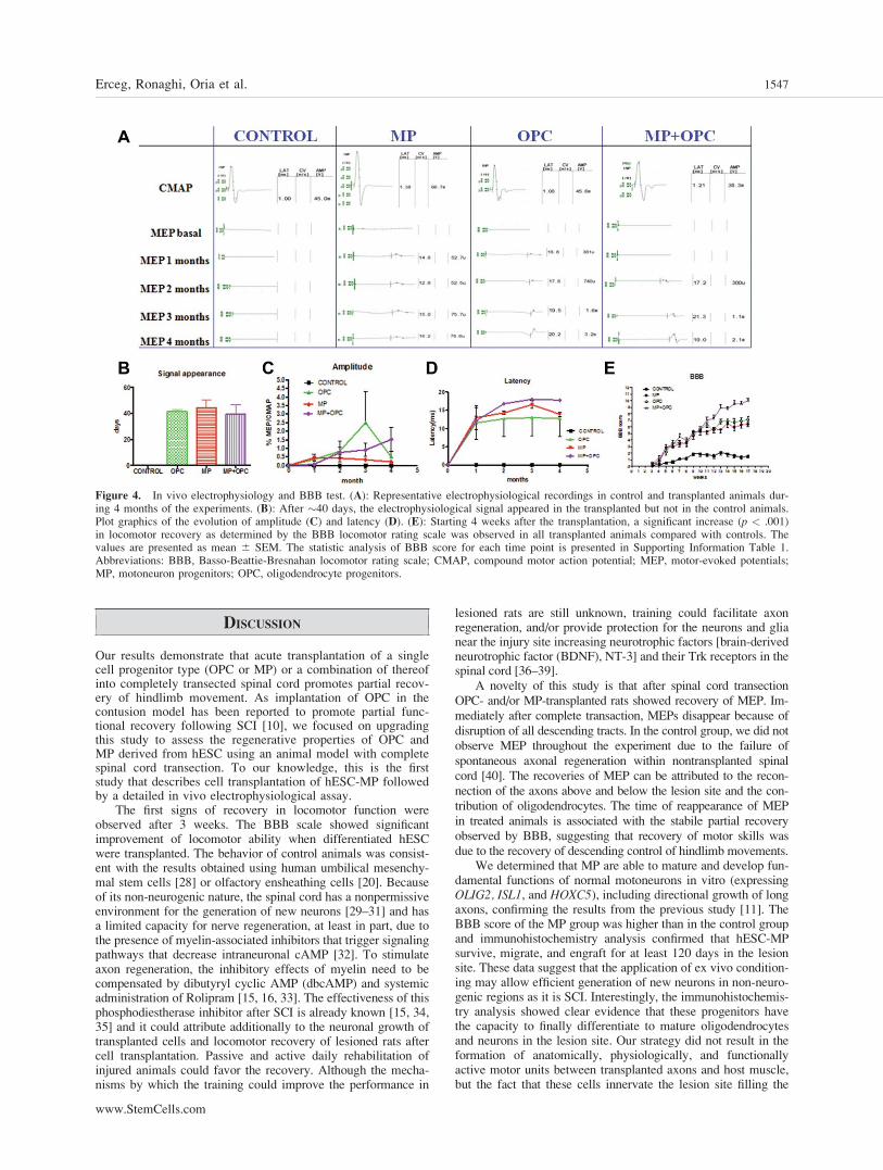
Electrophysiological Evaluation
Electrophysiological tests were performed in all rats beforeand after the surgery and transplantation to ensure the com-plete transection of the spinal cord. MEPs were used to evalu-ate the function of spinal cord descending tracts [27]. Electro-physiological tests were performed for 4 months in allanimals at monthly intervals. The electrophysiological testsdemonstrated complete interruption of spinal motor pathwaysafter the injury (Fig. 4A). In control animals, MEP did notrecover over time, indicating that interruption of supraspinalaxons was maintained until the end of the observation period(Fig. 4A). In contrast, in the OPC group and/or MP group,MEP reappeared �40 days after grafting (Fig. 4A, 4B), indi-cating functional spinal reconnection and partial restitution ofmotor pathways. Four months after injury, the amplitude ofthe evoked action potentials showed recovery in OPC and/orMP groups. Comparatively, animals that received OPC andMP tended to show MEP of higher amplitude than those witha single cell progenitor type treatment (Fig. 4C). Although therats treated with OPC showed higher amplitude after 3months compared with other treatments, the mean amplitudeof evoked potentials after 4 months were higher, but not stat-istically significant (p > .05) in the OPC þ MP group com-pared with OPC or MP (Fig. 4C) groups. Also the animalstransplanted with a single type of cells, OPC or MP, showedshorter latency in comparison with OPC þ MP (Fig. 4D).
Figure 3. Acute transplantation of humanembryonic stem cell-derived oligodendro-cyte progenitors (OPC) and motoneuronprogenitors (MP) resulted in differentiationtoward oligodendrocytes and neurons.Many GFPþ cells were observed in thelesion site and colabeled with oligodendro-cyte markers NG2 (A, B, C) and APC (D)(marked as ‘‘*’’). We observed NG2þ
(marked as ‘‘*’’) cells surrounding GFPþ
(arrow; [E]). Many NF70þ cells wereobserved in the lesion site often localizedin bundles (G, H). All NF70þ cells werenot GFPþ revealing that other human neu-rons proceed from OPC (E, F, I). Observa-tions: (C, G): Immunoflourescence wasdone with the spinal cord samples, whereMPs (without GFP) were injected. Abbrevi-ations: APC, adematous polyposis coli;GFP, green fluorescent protein; LS, lesionsite.
1546 Locomotor Recovery After Spinal Cord Transection
DISCUSSION
Our results demonstrate that acute transplantation of a singlecell progenitor type (OPC or MP) or a combination of thereofinto completely transected spinal cord promotes partial recov-ery of hindlimb movement. As implantation of OPC in thecontusion model has been reported to promote partial func-tional recovery following SCI [10], we focused on upgradingthis study to assess the regenerative properties of OPC andMP derived from hESC using an animal model with completespinal cord transection. To our knowledge, this is the firststudy that describes cell transplantation of hESC-MP followedby a detailed in vivo electrophysiological assay.
The first signs of recovery in locomotor function wereobserved after 3 weeks. The BBB scale showed significantimprovement of locomotor ability when differentiated hESCwere transplanted. The behavior of control animals was consist-ent with the results obtained using human umbilical mesenchy-mal stem cells [28] or olfactory ensheathing cells [20]. Becauseof its non-neurogenic nature, the spinal cord has a nonpermissiveenvironment for the generation of new neurons [29–31] and hasa limited capacity for nerve regeneration, at least in part, due tothe presence of myelin-associated inhibitors that trigger signalingpathways that decrease intraneuronal cAMP [32]. To stimulateaxon regeneration, the inhibitory effects of myelin need to becompensated by dibutyryl cyclic AMP (dbcAMP) and systemicadministration of Rolipram [15, 16, 33]. The effectiveness of thisphosphodiestherase inhibitor after SCI is already known [15, 34,35] and it could attribute additionally to the neuronal growth oftransplanted cells and locomotor recovery of lesioned rats aftercell transplantation. Passive and active daily rehabilitation ofinjured animals could favor the recovery. Although the mecha-nisms by which the training could improve the performance in
lesioned rats are still unknown, training could facilitate axonregeneration, and/or provide protection for the neurons and glianear the injury site increasing neurotrophic factors [brain-derivedneurotrophic factor (BDNF), NT-3] and their Trk receptors in thespinal cord [36–39].
A novelty of this study is that after spinal cord transectionOPC- and/or MP-transplanted rats showed recovery of MEP. Im-mediately after complete transaction, MEPs disappear because ofdisruption of all descending tracts. In the control group, we did notobserve MEP throughout the experiment due to the failure ofspontaneous axonal regeneration within nontransplanted spinalcord [40]. The recoveries of MEP can be attributed to the recon-nection of the axons above and below the lesion site and the con-tribution of oligodendrocytes. The time of reappearance of MEPin treated animals is associated with the stabile partial recoveryobserved by BBB, suggesting that recovery of motor skills wasdue to the recovery of descending control of hindlimb movements.
We determined that MP are able to mature and develop fun-damental functions of normal motoneurons in vitro (expressingOLIG2, ISL1, and HOXC5), including directional growth of longaxons, confirming the results from the previous study [11]. TheBBB score of the MP group was higher than in the control groupand immunohistochemistry analysis confirmed that hESC-MPsurvive, migrate, and engraft for at least 120 days in the lesionsite. These data suggest that the application of ex vivo condition-ing may allow efficient generation of new neurons in non-neuro-genic regions as it is SCI. Interestingly, the immunohistochemis-try analysis showed clear evidence that these progenitors havethe capacity to finally differentiate to mature oligodendrocytesand neurons in the lesion site. Our strategy did not result in theformation of anatomically, physiologically, and functionallyactive motor units between transplanted axons and host muscle,but the fact that these cells innervate the lesion site filling the
Figure 4. In vivo electrophysiology and BBB test. (A): Representative electrophysiological recordings in control and transplanted animals dur-ing 4 months of the experiments. (B): After �40 days, the electrophysiological signal appeared in the transplanted but not in the control animals.Plot graphics of the evolution of amplitude (C) and latency (D). (E): Starting 4 weeks after the transplantation, a significant increase (p < .001)in locomotor recovery as determined by the BBB locomotor rating scale was observed in all transplanted animals compared with controls. Thevalues are presented as mean 6 SEM. The statistic analysis of BBB score for each time point is presented in Supporting Information Table 1.Abbreviations: BBB, Basso-Beattie-Bresnahan locomotor rating scale; CMAP, compound motor action potential; MEP, motor-evoked potentials;MP, motoneuron progenitors; OPC, oligodendrocyte progenitors.
Erceg, Ronaghi, Oria et al. 1547
www.StemCells.com

gap between the rostral and caudal stumps as well as signifi-cantly improved locomotor function of lesioned rats suggeststhat hESC-MP have promising regenerative potential.
Interestingly, transplanted OPC had significant recoveringeffects too. In a contusion model, even after severe contusiveSCI, surviving axons persist in the subpial rim of white matterand restoration of the oligodendrocyte population by replacementtherapy has been considered as a potentially attractive strategy topromote remyelination after SCI [10, 19, 41]. But in the case ofthe spinal transection model there is no evidence of the presenceof spared host axons. After analysis of control rats, survival ofhost axons or spontaneous regeneration was not observed. Immu-nohistochemical analysis showed differentiation of transplantedcells toward neuronal cells, which was confirmed by the presenceof human specific NF70þ neurofilaments in the lesion site. Thisresult contrasts to previous claims that hESC differentiate exclu-sively toward a pure population of OPC [10, 19]. We hypothe-sized that the potential of OPC to recover is due to the presenceof heterogeneous cell types or multiple character of transplantedprogenitors. Analyzing the transcription phenotype of obtainedOPC at day 42, we observed strong expression of OLIG2 [42–44], which gives rise to motoneurons during the neurogenicphase. Nevertheless, OLIG2 is expressed in human OPC [45] andis necessary for specification and differentiation of human OPC[44]. In rodents, OLIG2-expressing spinal progenitors from theMP (pMN) domain are a source of both motoneurons and OPC[46, 47]. pMN cells in the spinal cord initially produce motoneur-ons, but switch later in development to form oligodendrocytes[48–50]. It has been proposed that transplanted OPC may undergotheir last division to adapt to a new environment by respondingappropriately to environmental cues [51] (Fig. 1). Shihabuddinet al. [29] have showed that isolated and in vitro expanded neuralprogenitors from adult spinal cord (non-neurogenic zone) havethe ability to respond to epigenetic signals in vivo, after transplan-tation, by site-specific differentiation, generating proper neuronsand/or glial cells depending on the transplantation site. Althoughwe can conclude the commitment of generated OPC to oligoden-droglial fate [10, 19], neuronal cells were also observed in vitroindicating that nearly 20% of differentiated cells were TUJ1þ.We believe that the presence of neuronal progenitors within trans-planted OPC caused significant recovery of animals. OPC couldgenerate a paracrine/trophic environment and positively modulatethe local immune response, as well as promote neuronal protec-tion and activation of endogenous neurogenesis [41, 52] suggest-ing that regenerative mechanisms do not depend exclusively on aspecific cell fate. Possible percentage of Tujþ (spontaneouslygenerated or not) could have origin from early neural processes.By the way, all cells arise from the same pool of neural stemcells, and it is not excluded that neurons arose from this pool.Finally, the cell transplantation strategy showed similar recoveryeffects that included synergic action of OPC and MP becausethese two cells types formed myelin-axon interaction.
The functional locomotor recovery analysis of the rats trans-planted with OPC and MP showed significantly better hindlimb re-covery than the animal groups treated with a single cell type. Theseresults have shown that combination of OPC and MP is superiorapproach when comparing with a single type strategy used in ourstudy or when compared with the studies where other cell stem
types strategies used in the same rat model of SCI [20, 28, 53, 54].As demonstrated in our results, locomotor improvement after trans-plantation, the OPC and MP is associated with abundant presenceof human NF-positive fibers and oligodendrocytes in the lesion.That means that hESC-derived OPC and MP were able to differen-tiate into mature oligodendrocytes and neurons. This was not thecase in our previous study where we used ependymal stem cells totreat animals with spinal cord contusion [41].
CONCLUSION AND PERSPECTIVES
Therapeutic use of stem cells to regenerate neural cells in thedamaged spinal cord requires efficient delivery of stem cells intothe injured area. The cells should differentiate into mature andfunctional cells that engraft for extended periods of time. Futurestudies must examine: (a) whether hESC-derived motoneuronshave the ability to extend toward anatomically and functionallyappropriate distal muscle targets; (b) whether they can form junc-tions on reaching these muscles as previously shown usingmESC [15]; and (c) whether the functional locomotor improve-ment can be enhanced using OPC. Therefore, the mechanismsunderlying the promotive effects on the functional recovery aftertransplantation of the hESC-derived OPC and MP is rather theability of these cells to differentiate into neuronal and glial cellsthan promotion of endogenous axonal regeneration.
Taken together, our study demonstrates that hESC-OPCand hESC-MP, when transplanted into the spinal cord immedi-ately after the injury, survived for at least 4 months; migrated atleast 3 mm away from the lesion; differentiated into appropriatecell types without forming teratomas, and improved locomotorfunction. These results open up new possibilities and strategiesfor future clinical applications in SCI and constitute a promis-ing approach for repairing the damaged spinal cord especially iftransplantation is shortly delayed after the lesion.
ACKNOWLEDGMENTS
We are thankful to Richard Griffeth and Drs. Majlinda Lako andLyle Armstrong for critical reading and Laura Nunez, EvaEscorihuela, andMaria Teresa Calvo Fernandez for the excellenttechnical support. We also thank Elena Herrero y Viviana Bisbalfor excellent veterinary support. The analysis of the samples wasperformed by the Confocal Microscopy Service of the ResearchCentre ‘‘Principe Felipe.’’ This work was supported by funds forresearch in the field of Regenerative Medicine from the RegionalGovernment Health Department (Generalitat Valenciana), theInstituto Carlos III belonging to the Spanish Ministry of Healthand Consumer Affairs, and through the grants from the SpanishMinistry of Science and Innovation (SAF2007-63193) and LaMarato (TV3 070,330).
DISCLOSURE OF POTENTIAL CONFLICTS OF
INTEREST
The authors indicate no potential conflicts of interest.
REFERENCES
1 Beattie MS, Li Q, Bresnahan JC. Cell death and plasticity after exper-imental spinal cord injury. Prog Brain Res 2000;128:9–21.
2 Blight AR. Spinal cord injury models: Neurophysiology. J Neuro-trauma 1992;9:147–149; discussion 149–150.
3 Grossman SD, Rosenberg LJ, Wrathall JR. Relationship of altered glu-tamate receptor subunit mRNA expression to acute cell loss after spi-nal cord contusion. Exp Neurol 2001;168:283–289.
4 Deumens R, Koopmans GC, Jaken RJ et al. Stimulation of neuriteoutgrowth on neonatal cerebral astrocytes is enhanced in the presenceof BDNF. Neurosci Lett 2006;407:268–273.
5 Dietz V, Curt A. Neurological aspects of spinal-cord repair: Promisesand challenges. Lancet Neurol 2006;5:688–694.
1548 Locomotor Recovery After Spinal Cord Transection

6 Harel NY, Strittmatter SM. Can regenerating axons recapitulate devel-opmental guidance during recovery from spinal cord injury? Nat RevNeurosci 2006;7:603–616.
7 Benzing C, Segschneider M, Leinhaas A et al. Neural conversion ofhuman embryonic stem cell colonies in the presence of fibroblastgrowth factor-2. Neuroreport 2006;17:1675–1681.
8 Gerrard L, Rodgers L, Cui W. Differentiation of human embryonicstem cells to neural lineages in adherent culture by blocking bonemorphogenetic protein signaling. Stem Cells 2005;23:1234–1241.
9 Itsykson P, Ilouz N, Turetsky T et al. Derivation of neural precursorsfrom human embryonic stem cells in the presence of noggin. Mol CellNeurosci 2005;30:24–36.
10 Keirstead HS, Nistor G, Bernal G et al. Human embryonic stem cell-derived oligodendrocyte progenitor cell transplants remyelinate andrestore locomotion after spinal cord injury. J Neurosci 2005;25:4694–4705.
11 Lee H, Shamy GA, Elkabetz Y et al. Directed differentiation andtransplantation of human embryonic stem cell-derived motoneurons.Stem Cells 2007;25:1931–1939.
12 Li XJ, Du ZW, Zarnowska ED et al. Specification of motoneuronsfrom human embryonic stem cells. Nat Biotechnol 2005;23:215–221.
13 Ludwig TE, Levenstein ME, Jones JM et al. Derivation of human em-bryonic stem cells in defined conditions. Nat Biotechnol 2006;24:185–187.
14 Erceg S, Lainez S, Ronaghi M et al. Differentiation of human embry-onic stem cells to regional specific neural precursors in chemicallydefined medium conditions. Plos One 2008;3:e2122.
15 Deshpande DM, Kim YS, Martinez T et al. Recovery from paralysisin adult rats using embryonic stem cells. Ann Neurol 2006;60:32–44.
16 Harper JM, Krishnan C, Darman JS et al. Axonal growth of embry-onic stem cell-derived motoneurons in vitro and in motoneuron-injured adult rats. Proc Natl Acad Sci USA 2004;101:7123–7128.
17 Liu S, Qu Y, Stewart TJ et al. Embryonic stem cells differentiate intooligodendrocytes and myelinate in culture and after spinal cord trans-plantation. Proc Natl Acad Sci USA 2000;97:6126–6131.
18 McDonald JW, Liu XZ, Qu Y et al. Transplanted embryonic stemcells survive, differentiate and promote recovery in injured rat spinalcord. Nat Med 1999;5:1410–1412.
19 Nistor GI, Totoiu MO, Haque N et al. Human embryonic stem cellsdifferentiate into oligodendrocytes in high purity and myelinate afterspinal cord transplantation. Glia 2005;49:385–396.
20 Lopez-Vales R, Fores J, Verdu E et al. Acute and delayed transplanta-tion of olfactory ensheathing cells promote partial recovery after com-plete transection of the spinal cord. Neurobiol Dis 2006;21:57–68.
21 Ramon-Cueto A, Cordero MI, Santos-Benito FF et al. Functional re-covery of paraplegic rats and motor axon regeneration in their spinalcords by olfactory ensheathing glia. Neuron 2000;25:425–435.
22 Basso DM, Beattie MS, Bresnahan JC. A sensitive and reliable loco-motor rating scale for open field testing in rats. J Neurotrauma 1995;12:1–21.
23 Oria M, Raguer N, Chatauret N et al. Functional abnormalities of themotor tract in the rat after portocaval anastomosis and after carbontetrachloride induction of cirrhosis. Metab Brain Dis 2006;21:297–308.
24 Oria M, Chatauret N, Raguer N et al. A new method for measuringmotor evoked potentials in the awake rat: Effects of anesthetics. JNeurotrauma 2008;25:266–275.
25 Kakulas BA. The applied neuropathology of human spinal cord injury.Spinal Cord 1999;37:79–88.
26 Basso DM, Beattie MS, Bresnahan JC. Graded histological and loco-motor outcomes after spinal cord contusion using the NYU weight-drop device versus transection. Exp Neurol 1996;139:244–256.
27 Garcia-Alias G, Verdu E, Fores J et al. Functional and electrophysio-logical characterization of photochemical graded spinal cord injury inthe rat. J Neurotrauma 2003;20:501–510.
28 Yang CC, Shih YH, Ko MH et al. Transplantation of human umbilicalmesenchymal stem cells from Wharton’s jelly after complete transec-tion of the rat spinal cord. Plos One 2008;3:e3336.
29 Shihabuddin LS, Horner PJ, Ray J et al. Adult spinal cord stem cellsgenerate neurons after transplantation in the adult dentate gyrus. JNeurosci 2000;20:8727–8735.
30 Song H, Stevens CF, Gage FH. Astroglia induce neurogenesis fromadult neural stem cells. Nature 2002;417:39–44.
31 Horner PJ, Power AE, Kempermann G et al. Proliferation and differ-entiation of progenitor cells throughout the intact adult rat spinal cord.J Neurosci 2000;20:2218–2228.
32 Bito H, Furuyashiki T, Ishihara H et al. A critical role for a Rho-asso-ciated kinase, p160ROCK, in determining axon outgrowth in mamma-lian CNS neurons. Neuron 2000;26:431–441.
33 Pearse DD, Pereira FC, Marcillo AE et al. cAMP and Schwann cellspromote axonal growth and functional recovery after spinal cordinjury. Nat Med 2004;10:610–616.
34 Hannila SS, Filbin MT. The role of cyclic AMP signaling in promot-ing axonal regeneration after spinal cord injury. Exp Neurol 2008;209:321–332.
35 Nikulina E, Tidwell JL, Dai HN et al. The phosphodiesterase inhibitorrolipram delivered after a spinal cord lesion promotes axonal regener-ation and functional recovery. Proc Natl Acad Sci USA 2004;101:8786–8790.
36 Gomez-Pinilla F, Ying Z, Opazo P et al. Differential regulation byexercise of BDNF and NT-3 in rat spinal cord and skeletal muscle.Eur J Neurosci 2001;13:1078–1084.
37 Gomez-Pinilla F, Ying Z, Roy RR et al. Voluntary exercise induces aBDNF-mediated mechanism that promotes neuroplasticity. J Neuro-physiol 2002;88:2187–2195.
38 Ying Z, Roy RR, Edgerton VR et al. Voluntary exercise increasesneurotrophin-3 and its receptor TrkC in the spinal cord. Brain Res2003;987:93–99.
39 Kubasak MD, Jindrich DL, Zhong H et al. OEG implantation and steptraining enhance hindlimb-stepping ability in adult spinal transectedrats. Brain 2008;131:264–276.
40 Valero-Cabre A, Fores J, Navarro X. Reorganization of reflexresponses mediated by different afferent sensory fibers after spinalcord transection. J Neurophysiol 2004;91:2838–2848.
41 Moreno-Manzano V, Rodriguez-Jimenez FJ, Garcia-Rosello M et al.Activated spinal cord ependymal stem cells rescue neurological func-tion. Stem Cells 2009;27:733–743.
42 Lu QR, Yuk D, Alberta JA et al. Sonic hedgehog–regulated oligoden-drocyte lineage genes encoding bHLH proteins in the mammalian cen-tral nervous system. Neuron 2000;25:317–329.
43 Zhou Q, Wang S, Anderson DJ. Identification of a novel family of oli-godendrocyte lineage-specific basic helix-loop-helix transcription fac-tors. Neuron 2000;25:331–343.
44 Hu BY, Du ZW, Li XJ et al. Human oligodendrocytes from embry-onic stem cells: Conserved SHH signaling networks and divergentFGF effects. Development 2009;136:1443–1452.
45 Jakovcevski I, Zecevic N. Olig transcription factors are expressed inoligodendrocyte and neuronal cells in human fetal CNS. J Neurosci2005;25:10064–10073.
46 Lu QR, Sun T, Zhu Z et al. Common developmental requirement forOlig function indicates a motor neuron/oligodendrocyte connection.Cell 2002;109:75–86.
47 Zhou Q, Anderson DJ. The bHLH transcription factors OLIG2 andOLIG1 couple neuronal and glial subtype specification. Cell 2002;109:61–73.
48 Richardson WD, Pringle NP, Yu WP et al. Origins of spinal cord oli-godendrocytes: Possible developmental and evolutionary relationshipswith motor neurons. Dev Neurosci 1997;19:58–68.
49 Richardson WD, Smith HK, Sun T et al. Oligodendrocyte lineage andthe motor neuron connection. Glia 2000;29:136–142.
50 Rowitch DH, Lu QR, Kessaris N et al. An ‘oligarchy’ rules neural de-velopment. Trends Neurosci 2002;25:417–422.
51 Brustle O, Maskos U, McKay RD. Host-guided migration allows tar-geted introduction of neurons into the embryonic brain. Neuron 1995;15:1275–1285.
52 Pluchino S, Zanotti L, Rossi B et al. Neurosphere-derived multipotentprecursors promote neuroprotection by an immunomodulatory mecha-nism. Nature 2005;436:266–271.
53 Hamada M, Yoshikawa H, Ueda Y et al. Introduction of the MASH1gene into mouse embryonic stem cells leads to differentiation ofmotoneuron precursors lacking Nogo receptor expression that can beapplicable for transplantation to spinal cord injury. Neurobiol Dis2006;22:509–522.
54 Lopez-Vales R, Fores J, Navarro X et al. Chronic transplantation ofolfactory ensheathing cells promotes partial recovery after completespinal cord transection in the rat. Glia 2007;55:303–311.
See www.StemCells.com for supporting information available online.
Erceg, Ronaghi, Oria et al. 1549