Targeted Ablation of Oligodendrocytes Induces Axonal Pathology Independent of Overt Demyelination
14
Neurobiology of Disease Targeted Ablation of Oligodendrocytes Induces Axonal Pathology Independent of Overt Demyelination Laura-Jane Oluich, 1,2 Jo Anne S. Stratton, 1,2 Yao Lulu Xing, 1,2 Sze Woei Ng, 1 Holly S. Cate, 1,2 Pankaj Sah, 3 François Windels, 3 Trevor J. Kilpatrick, 1,2 and Tobias D. Merson 1,2 1 Florey Neuroscience Institutes, University of Melbourne, Parkville, Victoria 3010, Australia, 2 Centre for Neuroscience Research, University of Melbourne, Parkville, Victoria 3010, Australia, and 3 Queensland Brain Institute, The University of Queensland, Brisbane, Queensland, 4072, Australia The critical role of oligodendrocytes in producing and maintaining myelin that supports rapid axonal conduction in CNS neurons is well established. More recently, additional roles for oligodendrocytes have been posited, including provision of trophic factors and metabolic support for neurons. To investigate the functional consequences of oligodendrocyte loss, we have generated a transgenic mouse model of conditional oligodendrocyte ablation. In this model, oligodendrocytes are rendered selectively sensitive to exogenously administered diphtheria toxin (DT) by targeted expression of the diphtheria toxin receptor in oligodendrocytes. Administration of DT resulted in severe clinical dysfunction with an ascending spastic paralysis ultimately resulting in fatal respiratory impairment within 22 d of DT challenge. Pathologically, at this time point, mice exhibited a loss of 26% of oligodendrocyte cell bodies throughout the CNS. Oligoden- drocyte cell-body loss was associated with moderate microglial activation, but no widespread myelin degradation. These changes were accompanied with acute axonal injury as characterized by structural and biochemical alterations at nodes of Ranvier and reduced somatosensory-evoked potentials. In summary, we have shown that a death signal initiated within oligodendrocytes results in subcellular changes and loss of key symbiotic interactions between the oligodendrocyte and the axons it ensheaths. This produces profound func- tional consequences that occur before the removal of the myelin membrane, i.e., in the absence of demyelination. These findings have clear implications for the understanding of the pathogenesis of diseases of the CNS such as multiple sclerosis in which the oligodendro- cyte is potentially targeted. Introduction The oligodendrocyte is a specialized glial cell within the CNS that is responsible for producing the myelin that ensheaths axons and forms the nodes of Ranvier that permit saltatory nerve conduc- tion. Dysfunction and death of oligodendrocytes has been de- scribed in various demyelinating pathologies including the leukodystrophies and multiple sclerosis (MS) (Lucchinetti et al., 1996; Dowling et al., 1999; Feigenbaum et al., 2000). Autoimmune-induced inflammatory demyelination has tradi- tionally been the assumed pathogenic mechanism in MS (Weiner, 2004; Frohman et al., 2006). However, recent detailed examinations of pathological specimens have challenged this view. On the one hand, significant axonal degeneration occurs early in disease (Trapp et al., 1998; 1999; Kuhlmann et al., 2002), even in the absence of demyelination (Nikic ´ et al., 2011), raising the possibility of axonal attack by activated microglia and/or macrophages. On the other hand, oligodendrocyte death in acute MS can occur without significant immune cell infiltration and may precede the development of inflammatory demyelinated plaques (Barnett and Prineas, 2004; Marik et al., 2007; Henderson et al., 2009). Moreover, periplaque tissue in evolving MS lesions exhibits oxidative damage that is often localized within apoptotic oligoden- drocytes (Haider et al., 2011). Thus, oligodendrocytopathy could represent the primary event in newly forming MS lesions. Experimental models of primary oligodendrocytopathy could assist in unraveling the complex pathological sequelae in MS. To this end, specific oligodendrocyte-targeting models have recently been developed using diphtheria toxin (DT) as the driver of oligodendrocyte death (Buch et al., 2005; Traka et al., 2010; Pohl et al., 2011). These binary genetic approaches involve oligodendrocyte-restricted Cre or CreER-regulated expression of the simian DT receptor (DTR) or the A subunit of DT (DT-A). In the former scenario, expression of DTR in oligodendrocytes renders naturally DT-resistant mouse cells susceptible to intra- peritoneally injected DT. Once internalized, DT catalyzes ADP- ribosylation of eukaryotic elongation factor 2, which results in inhibition of protein translation and ultimately, apoptosis (Collier, 2001). Induction of oligodendrocyte apoptosis in these mice causes demyelination, myelin splitting, and/or vacuoliza- tion resulting in motor pathologies of variable severity (Buch et al., 2005; Traka et al., 2010; Pohl et al., 2011). Received March 2, 2012; revised March 22, 2012; accepted March 28, 2012. Author contributions: H.S.C., P.S., T.J.K., and T.D.M. designed research; L.-J.O., J.A.S.S., Y.L.X., S.W.N., F.W., and T.D.M. performed research; L.-J.O. and T.D.M. analyzed data; T.J.K. and T.D.M. wrote the paper. This work was supported by the Australian Postgraduate Award and Rotary Health Scholarship (L.J.O), the National Health and Medical Research Council of Australia (NHMRC), Multiple Sclerosis Research Australia (MSRA) Betty Cuthbert Fellowship (T.D.M.), NHMRC and MSRA Project Grants (T.D.M.), and an NHMRC Program Grant (T.J.K. and P.S.). Anna Friedhuber and Dennis Kemper provided technical assistance with electron microscopy. Charity- works for MS and the Victorian Operational Infrastructure Support Program provided additional support. The authors declare no competing financial interests. Correspondence should be addressed to either Tobias D. Merson or Trevor J. Kilpatrick, Multiple Sclerosis Division, Kenneth Myer Building, University of Melbourne, Parkville, Victoria 3010, Australia. E-mail: [email protected] or [email protected]. DOI:10.1523/JNEUROSCI.1053-12.2012 Copyright © 2012 the authors 0270-6474/12/328317-14$15.00/0 The Journal of Neuroscience, June 13, 2012 • 32(24):8317– 8330 • 8317
Transcript of Targeted Ablation of Oligodendrocytes Induces Axonal Pathology Independent of Overt Demyelination

Neurobiology of Disease
Targeted Ablation of Oligodendrocytes Induces AxonalPathology Independent of Overt Demyelination
Laura-Jane Oluich,1,2 Jo Anne S. Stratton,1,2 Yao Lulu Xing,1,2 Sze Woei Ng,1 Holly S. Cate,1,2 Pankaj Sah,3
François Windels,3 Trevor J. Kilpatrick,1,2 and Tobias D. Merson1,2
1Florey Neuroscience Institutes, University of Melbourne, Parkville, Victoria 3010, Australia, 2Centre for Neuroscience Research, University of Melbourne,Parkville, Victoria 3010, Australia, and 3Queensland Brain Institute, The University of Queensland, Brisbane, Queensland, 4072, Australia
The critical role of oligodendrocytes in producing and maintaining myelin that supports rapid axonal conduction in CNS neurons is wellestablished. More recently, additional roles for oligodendrocytes have been posited, including provision of trophic factors and metabolicsupport for neurons. To investigate the functional consequences of oligodendrocyte loss, we have generated a transgenic mouse model ofconditional oligodendrocyte ablation. In this model, oligodendrocytes are rendered selectively sensitive to exogenously administereddiphtheria toxin (DT) by targeted expression of the diphtheria toxin receptor in oligodendrocytes. Administration of DT resulted insevere clinical dysfunction with an ascending spastic paralysis ultimately resulting in fatal respiratory impairment within 22 d of DTchallenge. Pathologically, at this time point, mice exhibited a loss of �26% of oligodendrocyte cell bodies throughout the CNS. Oligoden-drocyte cell-body loss was associated with moderate microglial activation, but no widespread myelin degradation. These changes wereaccompanied with acute axonal injury as characterized by structural and biochemical alterations at nodes of Ranvier and reducedsomatosensory-evoked potentials. In summary, we have shown that a death signal initiated within oligodendrocytes results in subcellularchanges and loss of key symbiotic interactions between the oligodendrocyte and the axons it ensheaths. This produces profound func-tional consequences that occur before the removal of the myelin membrane, i.e., in the absence of demyelination. These findings haveclear implications for the understanding of the pathogenesis of diseases of the CNS such as multiple sclerosis in which the oligodendro-cyte is potentially targeted.
IntroductionThe oligodendrocyte is a specialized glial cell within the CNS thatis responsible for producing the myelin that ensheaths axons andforms the nodes of Ranvier that permit saltatory nerve conduc-tion. Dysfunction and death of oligodendrocytes has been de-scribed in various demyelinating pathologies including theleukodystrophies and multiple sclerosis (MS) (Lucchinetti etal., 1996; Dowling et al., 1999; Feigenbaum et al., 2000).Autoimmune-induced inflammatory demyelination has tradi-tionally been the assumed pathogenic mechanism in MS(Weiner, 2004; Frohman et al., 2006). However, recent detailedexaminations of pathological specimens have challenged thisview. On the one hand, significant axonal degeneration occursearly in disease (Trapp et al., 1998; 1999; Kuhlmann et al., 2002),
even in the absence of demyelination (Nikic et al., 2011), raisingthe possibility of axonal attack by activated microglia and/ormacrophages. On the other hand, oligodendrocyte death in acuteMS can occur without significant immune cell infiltration and mayprecede the development of inflammatory demyelinated plaques(Barnett and Prineas, 2004; Marik et al., 2007; Henderson et al.,2009). Moreover, periplaque tissue in evolving MS lesions exhibitsoxidative damage that is often localized within apoptotic oligoden-drocytes (Haider et al., 2011). Thus, oligodendrocytopathy couldrepresent the primary event in newly forming MS lesions.
Experimental models of primary oligodendrocytopathy couldassist in unraveling the complex pathological sequelae in MS. Tothis end, specific oligodendrocyte-targeting models have recentlybeen developed using diphtheria toxin (DT) as the driver ofoligodendrocyte death (Buch et al., 2005; Traka et al., 2010;Pohl et al., 2011). These binary genetic approaches involveoligodendrocyte-restricted Cre or CreER-regulated expressionof the simian DT receptor (DTR) or the A subunit of DT (DT-A).In the former scenario, expression of DTR in oligodendrocytesrenders naturally DT-resistant mouse cells susceptible to intra-peritoneally injected DT. Once internalized, DT catalyzes ADP-ribosylation of eukaryotic elongation factor 2, which results ininhibition of protein translation and ultimately, apoptosis(Collier, 2001). Induction of oligodendrocyte apoptosis in thesemice causes demyelination, myelin splitting, and/or vacuoliza-tion resulting in motor pathologies of variable severity (Buch etal., 2005; Traka et al., 2010; Pohl et al., 2011).
Received March 2, 2012; revised March 22, 2012; accepted March 28, 2012.Author contributions: H.S.C., P.S., T.J.K., and T.D.M. designed research; L.-J.O., J.A.S.S., Y.L.X., S.W.N., F.W., and
T.D.M. performed research; L.-J.O. and T.D.M. analyzed data; T.J.K. and T.D.M. wrote the paper.This work was supported by the Australian Postgraduate Award and Rotary Health Scholarship (L.J.O), the
National Health and Medical Research Council of Australia (NHMRC), Multiple Sclerosis Research Australia (MSRA)Betty Cuthbert Fellowship (T.D.M.), NHMRC and MSRA Project Grants (T.D.M.), and an NHMRC Program Grant (T.J.K.and P.S.). Anna Friedhuber and Dennis Kemper provided technical assistance with electron microscopy. Charity-works for MS and the Victorian Operational Infrastructure Support Program provided additional support.
The authors declare no competing financial interests.Correspondence should be addressed to either Tobias D. Merson or Trevor J. Kilpatrick, Multiple Sclerosis
Division, Kenneth Myer Building, University of Melbourne, Parkville, Victoria 3010, Australia. E-mail:[email protected] or [email protected].
DOI:10.1523/JNEUROSCI.1053-12.2012Copyright © 2012 the authors 0270-6474/12/328317-14$15.00/0
The Journal of Neuroscience, June 13, 2012 • 32(24):8317– 8330 • 8317
We have generated a novel transgenic mouse denoted MBP-DTR (Line 100A) for targeted ablation of oligodendrocytes thatdoes not rely upon Cre/loxP transgenics. In these mice, expres-sion of human DTR is regulated by the proximal 1.96 kb pro-moter of mouse myelin basic protein (MBP) gene which directsspecific expression to mature oligodendrocytes (Gow et al., 1992;Shults et al., 2005; Emery et al., 2006). Administration of DT toMBP-DTR mice leads to loss of oligodendrocyte cell bodies andthe development of severe clinical pathology characterized byspastic paralysis of the hindlimbs, tremor, ataxia, and kyphosisthat correlates with impairment of electrophysiological proper-ties of neurones and ultrastructural alterations at the nodes ofRanvier. Importantly, these changes occurred in the absence ofinflammatory demyelination and demonstrate that axonal pa-thology occurring in the absence of demyelination can still besecondary to primary pathology of the oligodendrocyte.
Materials and MethodsGeneration of MBP-DTR transgenic mice. The MBP-DTR construct wasgenerated by excising a LacZ transgene from the pMG2 vector (kindlyprovided by Associate Professor Alexander Gow, Wayne State Univer-sity, Detroit, Michigan; Gow et al., 1992) and replacing it with the codingsequence (CDS) for human heparin-binding epidermal growth factor(HB-EGF), a high-affinity DTR. The HB-EGF/DTR CDS was amplifiedby high-fidelity PCR from the pRC/CMV/hHB-EGF plasmid (kindlyprovided by Professor Eisuke Mekada, Osaka University, Japan). Theresultant MBP-DTR construct was validated by DNA sequencing tocomprise the 1.94 kb proximal promoter of the mouse myelin basicprotein promoter (MBP, corresponding to bases �1907 to �36 relativeto ATG), the 627 bp human HB-EGF/DTR CDS, and 1.43 kb 3� splicingand the polyadenylation sequence derived from the human �-globingene (exon 2, intron 2, exon 3). MBP-DTR plasmid DNA isolated frommaxiprep culture (Qiagen) was further purified by CsCl gradient centrif-ugation. The DNA was digested with ScaI and SphI producing a 1.7 kbfragment comprising the plasmid backbone and a 4.7 kb fragment com-prising the proximal MBP promoter, human HB-EGF (DTR) CDS, andhuman �-globin polyadenylation sequence. The DNA fragments wereseparated on a 1.5% agarose (Lonza SeaPlaque; Fisher Scientific)/TBE geland the 4.7 kb ScaI/SphI MBP-DTR fragment was extracted from thecorresponding gel slice by �-Agarase I digestion (New England BioLabs).The extracted DNA was dialyzed (“v” series membrane, 0.05 �m poresize; Millipore) with microinjection buffer and diluted to 2 �g/ml beforemicroinjecting into the male pronuclei of fertilized C57BL/6 oocytes. Tailbiopsies from the offspring of oocyte recipients were assessed for incor-poration of the transgene by Southern blot and PCR analysis. For South-ern blot analysis, genomic DNA was digested with BstXI followed byagarose gel electrophoresis. Gel blots prepared using nitrocellulose mem-branes (Hybond N�; GE Healthcare) were hybridized with a 32P-labeledDNA probe generated by PCR amplification of a 629 bp product fromthe MBP-DTR construct that encompassed part of the murine MBPpromoter and human DTR sequence (sense primer: 5�-CAGGCCCACATTCATATC-3�; antisense primer: 5�-TCTTCCCTAGCCCCTTGCCTTTCT-3�). PCR genotyping of tail biopsy DNA was performed using primerstargeting a 512 bp amplicon encompassing part of the vector between thepromoter and HB-EGF CDS and part of the human HB-EGF CDS with 80%homology to murine HB-EGF (sense primer: 5�-GCTCGAATAATTCTAGGGTC-3�; antisense primer: 5�-GGTCATAGGTATATAAGCGA-3�). Ofthe 18 founders that exhibited germline transmission of the MBP-DTRtransgene, we describe the characterization of one line denoted MBP-DTRLine 100A which is hereafter referred to as MBP-DTR mice.
Animal cohorts. All experimental mice were maintained on a pureC57BL/6 background on a 12 h light-dark cycle and provided with rodentchow (Barastoc Products) and water ad libitum. Experimental cohortswere generated by crossing MBP-DTR heterozygotes with wild-typeC57BL/6 to produce heterozygous MBP-DTR and wild-type litter-mates or, alternatively, by crossing MBP-DTR heterozygotes withPdgfra-CreERT2�/�:R26R-YFP�/� mice to generate Pdgfra-CreERT2�/�:
R26R-YFP�/�:MBP-DTR�/� and Pdgfra-CreERT2 �/�:R26R-YFP �/�:wild-type littermates. Pdgfra-CreERT2 mice (Rivers et al., 2008) and R26R-YFP mice (Srinivas et al., 2001) were kindly provided by Professor WilliamD. Richardson (Wolfson Institute for Biomedical Research, University Col-lege of London) and Professor Frank Costantini (Columbia University Med-ical Center, New York), respectively. Experimental procedures wereconducted in accordance with National Health and Medical Research Coun-cil guidelines and were approved by the animal ethics committees of theFlorey Neuroscience Institutes and University of Queensland.
Intraperitoneal injection and gavaging. For the initial experimental co-horts, mice aged 8 –10 weeks were given a single intraperitoneal injectionof 200 ng DT (2 �g/ml in saline; Sigma) or an equal volume (100 �l) ofsaline as control. For subsequent cohorts, DT was administered as asingle intraperitoneal injection at a fixed dose of 10 �g/kg (prepared at 1�g/ml in saline); vehicle controls received saline at a dose of 10 �l/g.Three experimental groups were typically assessed: MBP-DTR mice ad-ministered DT, MBP-DTR mice administered saline, and wild-type lit-termates administered DT. To induce recombination in Pdgfra-CreERT2:R26R-YFP:MBP-DTR and Pdgfra-CreERT2:R26R-YFP:wild-type mice,tamoxifen (prepared at 40 mg/ml in 50°C corn oil; Sigma) was adminis-tered by oral gavage (300 mg/kg/d) for 4 consecutive days, commencing7 d before DT challenge. Following DT or saline challenge, mice wereweighed and monitored daily for signs of neurological impairment. Theclinical course was scored as follows: 0 � healthy, 1� pelvis lowered, 2 �pelvis on ground, 3 � hindpaw slippage, 4 � kyphosis, 5 � tail spasticity,6 � upper torso lowered, and 7 � labored breathing.
Immunohistochemistry. Mice were deeply anesthetized with 100 mg/kgsodium pentobarbitone then transcardially perfused with PBS followedby 4% paraformaldehyde (PFA)/PBS. Extracted brains, spinal cords, andsciatic nerves were postfixed in 4% PFA/PBS for 2 h on ice, rinsed brieflyin PBS, and then equilibrated in 20% sucrose/PBS for 24 h before em-bedding in Tissue-Tek O.C.T. compound (Sakura FineTek) and freezingon an isopentane bath over dry ice. Tissue was stored at �80°C untilsectioned. Ten-micron-thick cryosections of the brain (coronal), spinalcord (coronal and longitudinal), and sciatic nerve (longitudinal) werecollected onto Superfrost Plus slides (Menzel Glaser) and air-dried for1 h before storing at �80°C until stained.
Cryosections were blocked for 1 h with PBS containing 0.3% TritonX-100 and 10% normal serum (goat or donkey depending upon host ofsecondary antibodies). In some instances serum was replaced with 1%BSA and 0.1% gelatin. Primary antibodies diluted in 10% normal serum/PBS were applied for 16 h at 4°C or at room temperature. Slides wererinsed three times in PBS (5 min each) before application of secondaryantibodies for 30 min at room temperature. Slides were rinsed again,counterstained with Hoechst 33342 (1 �g/ml; Invitrogen), and cover-slipped with Mowiol mounting medium. For DTR immunohistochem-istry, an Alexa Fluor 488-conjugated goat anti-FITC antibody (1:200;Invitrogen) was used as a tertiary antibody after the secondary antibodyrinse to amplify the DTR signal.
The following primary antibodies were used: rabbit anti-pan-neurofascin (1:2000; a generous gift from Professor Peter Brophy, Uni-versity of Edinburgh), mouse anti-APC/CC-1 (1:100; Calbiochem),mouse anti-ankyrin G (1:200; Santa Cruz Biotechnology), mouse anti-oligodendrocytes (1:5000, clone NS-1/RIP; Millipore), rabbit anti-�-amyloid precursor protein (1:300; Zymed), rat anti-PDGFR� (1:500,clone APA5; BD PharMingen), rabbit anti-NG2 (1:200; Millipore), rab-bit anti-cleaved caspase-3 (1:200; Cell Signaling Technology), rabbitanti-CD3 (1:200; Dako), rabbit anti-Krox20 (1:200; Covance), rat anti-CD11b (1:200; BD PharMingen), mouse anti-glial fibrillary acidic pro-tein (GFAP) (1:200, clone GA5; Millipore), rabbit anti-GFAP (1:500;Dako), goat anti-HB-EGF (1:200; R&D Systems), rabbit anti-Iba1 (1:1000; Wako Industries), mouse anti-myelin-associated glycoprotein(MAG) (1:1000; Millipore), rabbit anti-MBP (1:200; Millipore Biosci-ence Research Reagents), mouse anti-NeuN (1:100; Millipore BioscienceResearch Reagents), mouse anti-S100� (1:1000, clone SH-B1; Sigma),rabbit anti-Olig2 (1:200; Millipore), mouse anti-Kv1.2 (1:500; Neuro-Mab), rabbit anti-Nav1.6 (1:100; Alomone Labs), and mouse anti-SMI312 (1:1000; Covance). Secondary antibodies raised in goat or
8318 • J. Neurosci., June 13, 2012 • 32(24):8317– 8330 Oluich et al.• Axonal Pathology Independent of Demyelination following Oligodendrocyte Apoptosis

donkey and conjugated to FITC, TRITC, or NL557 were purchased fromJackson ImmunoResearch or R&D Systems and used at 1:200 dilution.
Slides were examined using an AxioPlan2 fluorescent microscope(Zeiss), and images were captured using an Axiocam HRc camera (Zeiss)using Axiovision 7.2 software. For comparative analysis of the intensityof myelin protein labeling, photomicrographs were converted to gray-scale and analyzed using ImageJ software (National Institutes of Health;NIH). Intensity was defined as the average gray value obtained from allpixels within the region of interest above background. Confocal analysisof nodal protein expression was performed using a FluoView FV1000confocal microscope (Olympus) under a 40� oil objective using Flu-oView confocal software (Olympus).
TUNEL. For combined immunohistochemistry and TUNEL, 10-�m-thick coronal cryosections of fresh frozen brain tissue were collected ontoSuperfrost Plus slides, air-dried for 5 min, postfixed in 4% PFA/PBS for20 min at room temperature, and rinsed three times in PBS. Immuno-histochemistry was conducted as previously described using TRITC-conjugated secondary antibodies then processed for TUNEL using theFluorescein In Situ Cell Death Detection Kit (Roche) according to themanufacturer’s protocol. Slides were counterstained with Hoechst 33342and coverslipped as described previously. To quantify the extent of apo-ptosis, the number of TUNEL � Hoechst � cells in the cerebral cortex andcorpus callosum were counted (bregma level �0.5 mm) in MBP-DTRand wild-type mice (n � 4 per genotype) at both 5 and 15 d following DTchallenge and expressed as TUNEL � cells per millimeter squared. Forstatistical analysis, the data were analyzed by one-way ANOVA followedby Bonferroni’s post hoc tests.
Electron microscopy. Following perfusion/fixation, the lumbar spinalcord and sciatic nerves were postfixed in Karnovsky’s buffer (2.5% glu-taraldehyde, 4% PFA, 0.1 M sodium cacodylate, pH 7.3) for 16 h at 4°C,rinsed three times in PBS, and stored in 0.1 M sodium cacodylate, pH 7.3,at 4°C for 24 h. The tissue was postfixed in a solution of 2% w/v osmiumtetraoxide and 1.5% w/v potassium ferricyanide and embedded inSpurr’s resin. The dorsal funiculus of the lumbar spinal cord and sciaticnerves were sectioned at 0.5 �m thickness on an ultramicrotome (Ul-tracut S; Reichert) in transverse orientation for analysis of g-ratios (dor-sal funiculus, sciatic nerve) or longitudinally (dorsal funiculus) forassessment of nodes of Ranvier.
Semithin (0.5 �m) transverse sections were stained with methyleneblue and imaged at 10,000� magnification. Fibers within a 121.5 �m 2
region of interest (ROI) within each dorsal funiculus (two ROI assessedper animal, n � 2 animals per group) or a 3.07 mm 2 ROI within eachsciatic nerve (four ROI assessed per animal, n � 2 animals per group)were assessed using ImageJ software (NIH). For each axon, the axonalarea excluding the myelin sheath and the entire fiber area including themyelin sheath were independently outlined and the area calculated usingthe “measurement” function within ImageJ. In addition, as a measure ofmyelin integrity, the axonal and fiber diameters were measured and usedto calculate g-ratios (ratio of axonal diameter to fiber diameter) (Sher-man and Brophy, 2005). Results were analyzed using multivariateANOVA followed by Tukey’s post hoc analysis using SPSS 15.0.
For nodal analysis, the ROI in the dorsal funiculus was confirmed bymethylene blue staining before collection of ultrathin (90 nm) sections.Ultrathin sections were postfixed in lead nitrate and urinyl acetate,mounted on copper grids, and examined in an Elmskop 102 transmissionelectron microscope (Siemens). Photomicrographs were captured at25,000� magnification and negatives were scanned to generate high-resolution computer images for analysis. Nodes of Ranvier were exam-ined qualitatively for indications of ultrastructural abnormalities. Thepercentage of nodes within the dorsal funiculi of DT-challenged MBP-DTR (n � 2) and wild-type (n � 2) mice exhibiting paranodal loopeversion was determined on the basis of assessments by two expert my-elin biologists who were blinded to the identity of each group.
Somatosensory-evoked potentials. Six DT-challenged MBP-DTR miceat clinical end point and five matching DT-challenged wild-type litter-mates were anesthetized with an intraperitoneal injection of ketamine(0.1 mg/kg) and xylazine (0.01 mg/kg). Depth of anesthesia was checkedby the loss of response to rear paw pinch and additional injection of theanesthetic solution was provided as required. The body temperature of
animals was maintained at 36.5°C using a controlled heating pad. Ani-mals were mounted in a stereotaxic frame (model 962; Kopf Instru-ments) and the scalp incised longitudinally and retracted to expose theskull. A hole was drilled above the somatosensory cortex (anteroposte-rior, �0.5; lateral, 2.5; and dorsal, 1 mm) to allow insertion of a borosili-cate glass recording electrode filled with 3 M saline. A second hole wasdrilled on the opposite side above the contralateral frontal lobe to allowfor the insertion of the reference electrode between the skull and themeninges. Stimulating electrodes made of stainless steel (25 g) were in-serted at the sciatic notch and connected to a constant current generator(A385; World Precision Instrument). Local field potentials were re-corded at 5 kHz and bandpass filtered (30 Hz–1.8 kHz). Each animalreceived 50 sciatic notch stimulations (5 mA; 0.2 ms; interspike interval,15 s); the onset of the field potential immediately following the stimula-tion artifact was used to measure the nerve conduction velocity (valuesare mean � SEM).
Measurement of compound action potentials in isolated sciatic nerve.Eight DT-challenged MBP-DTR mice at clinical end point and threematching DT-challenged wild-type littermates were overdosed with so-dium pentobarbitone (100 mg/kg). Sciatic nerves were isolated andplaced in saline at room temperature. Nerves were coated in liquid par-affin oil, and the proximal and distal ends were placed onto gold-platedfixed electrodes (proximal end, stimulating electrode; distal end, record-ing electrode 10 mm from proximal electrode; medial position, ground-ing electrode) in a humidified 37°C chamber. Square-wave stimulatingpulses of 100 �s duration were delivered at 30 s intervals using aconstant-voltage stimulus isolator (PowerLab 4/20; ADInstruments) atprogressive intensities from 0.2 mV up to 10 V. Monophasic compoundaction potentials (CAPs) generated at suprathreshold stimulus intensity(6 V) were amplified using a differential bioamplifier (Bio Amp; Maclab)and digitized using Scope software (v4.1; ADInstruments). Post hoc anal-ysis was performed using LabChart Reader for Mac (ADInstruments).
ResultsMBP-DTR mice express DTR exclusively in oligodendrocytesin the CNSTo assess the consequences of oligodendrocyte ablation in theadult CNS, we generated transgenic mice that express DTR underthe regulatory control of the proximal promoter for the MBPgene (see Materials and Methods; Fig. 1A). We used a 1.94 kbMBP promoter that had previously been shown to direct trans-gene expression specifically to the mature oligodendrocyte lin-eage (Gow et al., 1992; Wrabetz et al., 1998; Emery et al., 2006).Eighteen germline-transmitting MBP-DTR transgenic founderswere identified by Southern blot (Fig. 1B) and PCR analysis (Fig.1C; see Materials and Methods). We performed a series of immu-nohistochemical analyses to assess the expression pattern of DTRin the adult CNS of MBP-DTR and wild-type littermates. Immu-nolabeling was conducted using an antibody that specifically de-tects human HB-EGF/DTR, but not its murine homolog. Of theMBP-DTR lines identified, five lines expressed DTR in 100% ofthe oligodendrocytes. Here we characterize the phenotype of oneof these five transgenic lines denoted MBP-DTR Line 100A. As-sessment of coronal sections of the adult forebrain in MBP-DTRLine 100A mice, referred to hereafter as MBP-DTR, revealed thatDTR was highly expressed in white matter tracts (Fig. 1D),whereas only low-level, nonspecific labeling was observed inwild-type littermates (Fig. 1 E). We confirmed that DTR wasexpressed in mature oligodendrocytes in MBP-DTR mice bymultilabel immunohistochemistry. Specifically, all CC-1� oligo-dendrocytes identified in sections from MBP-DTR mice werefound to coexpress DTR (Fig. 1F), whereas no DTR was detectedin any CC-1� oligodendrocytes in wild-type littermates (Fig.1G). DTR expression in transgenic mice was found not onlywithin the cell bodies of CC-1� oligodendrocytes but also withinthe processes of oligodendrocytes identified by colocalization of
Oluich et al.• Axonal Pathology Independent of Demyelination following Oligodendrocyte Apoptosis J. Neurosci., June 13, 2012 • 32(24):8317– 8330 • 8319

DTR and MBP (Fig. 1H–J) as well asof DTR and RIP (Fig. 1K–M). In contrast,DTR was not expressed by any other neu-ral cell population examined includingNeuN� neurons (Fig. 1N), PDGFR�� oli-godendrocyte progenitors cells (Fig. 1O),Iba1� microglia (Fig. 1P), or GFAP� astro-cytes (data not shown). Together, theseexperiments demonstrate the successfulgeneration of MBP-DTR transgenic micethat express DTR exclusively within the ma-ture oligodendrocyte population of theCNS, in a pattern that closely reflects the ex-pression of myelin proteins.
Severe clinical and motor deficits inMBP-DTR mice following DT challengeTo ablate oligodendrocytes, we adminis-tered a single 200 ng dose of DT to MBP-DTR transgenic mice (n � 9) and wild-typelittermates (n � 8), or an equivalent vol-ume of saline to MBP-DTR mice (n � 6).The two control groups, wild-type miceadministered DT and MBP-DTR mice ad-ministered saline, did not shown any clin-ical changes over the subsequent 3 weeks.In contrast, MBP-DTR mice injected withDT exhibited progressive motor deficitsthat typically began at 9 d after DT admin-istration and became profound in the next5– 6 d. The maximum survival time fol-lowing the onset of clinical impairmentwas 8 d. DT-challenged transgenic ani-mals exhibited hindlimb clasping whenheld by the tail, an ataxic gait, hindlimbparalysis, tail spasticity, and thoracic ky-phosis (Fig. 2A). The phenotype rapidlyprogressed to include forelimb weakness,respiratory impairment (labored breath-ing), reduced mobility, lethargy, andweight loss, at which time disabled ani-mals and their matching controls werekilled, a time point we have termed theclinical end point. A plot of survival timeto clinical end point following DT chal-lenge demonstrates that all MBP-DTR
Figure 1. Generation of MBP-DTR transgenic mice expressing DTR within the mature oligodendrocyte population of the adultCNS. A, Schematic representation of the ScaI/SphI-linearized MBP-DTR construct used to generate MBP-DTR founders. The con-struct comprises the 1.94 kb proximal promoter of the mouse myelin basic protein promoter, the 627 bp human HB-EGF/DTR CDS,and the 1.43 kb 3� splicing and the polyadenylation sequence derived from the human �-globin gene. Arrowheads depict thesense and antisense primers used for PCR genotyping. The solid black line indicates the size and position of the 32P-labeled probeused for Southern blot analysis of BstXI-digested mouse tail DNA. B, Southern blot analysis of BstXI-digested genomic DNA isolatedfrom mouse tail biopsies used to identify MBP-DTR (tg) founders; note the absence of signal at 1.7 kb in the wild-type (wt) sample.Hybridization was performed using a 629 bp 32P-labeled DNA probe that identified a 1.7 kb BstXI fragment generated from theMBP-DTR sequence. C, PCR genotyping using primers that amplify a 512 bp product from the MBP-DTR transgene enableddiscrimination of MBP-DTR (tg) from wild-type (wt) mice. D, E, Immunohistochemical assessment of DTR expression in theforebrain using an anti-human HB-EGF antibody revealed DTR expression within white matter tracts of MBP-DTR (D) but not
4
wild-type (E) mice. F, G, Immunohistochemical assessment ofDTR (green) expression among CC-1 (red) immunoreactive oli-godendrocytes in the corpus callosum of MBP-DTR (F) versuswild-type (G) mice. Nuclei were stained with Hoechst 33258(blue). H–J, DTR expression (H, green) in the corpus callosumcolocalized with MBP (I, red), as revealed in the merged image(J). K–M, DTR expression (K, green) was localized to both thecell body and processes of RIP-labeled oligodendrocytes (L,red), as shown in the merged image (M). Nuclei were counter-stained with Hoechst. N–P, DTR expression (green) was notobserved in NeuN-labeled (red) neuronal cell bodies (N), inPDGFR�-labeled (red) oligodendrocyte progenitor cells (O) orin Iba1-labeled (red) microglia (P, confocal stack). CC, corpuscallosum, Ctx, neocortex; Str, striatum; z, z-axis projection.Scale bars: F, G, K–P, 50 �m; (in J) H–J, 200 �m.
8320 • J. Neurosci., June 13, 2012 • 32(24):8317– 8330 Oluich et al.• Axonal Pathology Independent of Demyelination following Oligodendrocyte Apoptosis
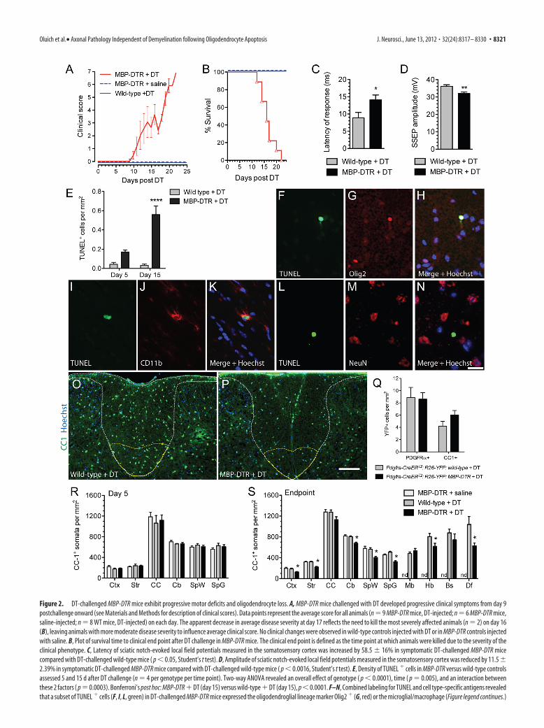
Figure 2. DT-challenged MBP-DTR mice exhibit progressive motor deficits and oligodendrocyte loss. A, MBP-DTR mice challenged with DT developed progressive clinical symptoms from day 9postchallenge onward (see Materials and Methods for description of clinical scores). Data points represent the average score for all animals (n � 9 MBP-DTR mice, DT-injected; n � 6 MBP-DTR mice,saline-injected; n � 8 WT mice, DT-injected) on each day. The apparent decrease in average disease severity at day 17 reflects the need to kill the most severely affected animals (n � 2) on day 16(B), leaving animals with more moderate disease severity to influence average clinical score. No clinical changes were observed in wild-type controls injected with DT or in MBP-DTR controls injectedwith saline. B, Plot of survival time to clinical end point after DT challenge in MBP-DTR mice. The clinical end point is defined as the time point at which animals were killed due to the severity of theclinical phenotype. C, Latency of sciatic notch-evoked local field potentials measured in the somatosensory cortex was increased by 58.5 � 16% in symptomatic DT-challenged MBP-DTR micecompared with DT-challenged wild-type mice ( p � 0.05, Student’s t test). D, Amplitude of sciatic notch-evoked local field potentials measured in the somatosensory cortex was reduced by 11.5 �2.39% in symptomatic DT-challenged MBP-DTR mice compared with DT-challenged wild-type mice ( p � 0.0016, Student’s t test). E, Density of TUNEL � cells in MBP-DTR versus wild-type controlsassessed 5 and 15 d after DT challenge (n � 4 per genotype per time point). Two-way ANOVA revealed an overall effect of genotype ( p � 0.0001), time ( p � 0.005), and an interaction betweenthese 2 factors ( p � 0.0003). Bonferroni’s post hoc: MBP-DTR � DT (day 15) versus wild-type � DT (day 15), p � 0.0001. F–N, Combined labeling for TUNEL and cell type-specific antigens revealedthat a subset of TUNEL � cells (F, I, L, green) in DT-challenged MBP-DTR mice expressed the oligodendroglial lineage marker Olig2 � (G, red) or the microglial/macrophage (Figure legend continues.)
Oluich et al.• Axonal Pathology Independent of Demyelination following Oligodendrocyte Apoptosis J. Neurosci., June 13, 2012 • 32(24):8317– 8330 • 8321

mice were killed between 12 and 22 d after DT injection (Fig. 2B),with a median survival time of 16 d.
Disabled mice exhibit electrophysiological abnormalitiesTo directly assess whether symptomatic MBP-DTR mice chal-lenged with DT exhibited electrophysiological abnormalities,we performed in vivo recordings of somatosensory-evoked po-tentials (SSEPs) by stimulating at the sciatic notch and record-ing the response in the somatosensory cortex of both MBP-DTR (n � 6) and wild-type animals (n � 5) challenged withDT. In control animals the onset of the local field potential wasdetected 8.11 � 0.29 ms after sciatic notch stimulation,whereas in symptomatic MBP-DTR animals SSEPs were re-corded 13.42 � 0.30 ms after stimulation, representing a39.6% increase in latency (Fig. 2C). Statistical analysis dem-onstrated that transmission of the SSEP was significantly fasterin control versus symptomatic MBP-DTR animals (unpairedStudent’s t test; p � 0.037). We also observed a statisticallysignificant reduction in the peak amplitude of SSEPs in symp-tomatic DT-challenged MBP-DTR animals (31.98 � 0.86 mV)compared with control animals (36.13 � 0.97 mV, p � 0.0016,unpaired Student’s t test) (Fig. 2 D).
Induction of oligodendrocyte apoptosis in MBP-DTR micefollowing DT challengeAdministration of DT would be expected to result in widespreadoligodendrocyte loss throughout the CNS of MBP-DTR trans-genic mice. To confirm this, apoptosis within the brains of MBP-DTR mice was assessed 5 and 15 d after administration of DT,corresponding to time points before and during overt clinicaldisease, respectively (n � 4 per time point). Wild-type animalsadministered DT and assessed 5 and 15 d postinjection served ascontrols (n � 4 per time point). At day 15, the number ofTUNEL� cells in the neocortex of MBP-DTR mice was increased17.5 relative to wild-type controls (Fig. 2E). Two-way ANOVA ofTUNEL� cell density identified a significant effect of both geno-type (p � 0.0001) and time (p � 0.0005) and also a significantinteraction (p � 0.0003) between these two variables. Bonferro-ni’s post hoc analysis confirmed that the increase in apoptotic celldensity in MBP-DTR mice compared with wild-type mice at 15 dpost-DT was highly significant (p � 0.0001).
We next assessed the phenotype of TUNEL� cells and identi-fied two distinct cell types by combined TUNEL and immuno-histochemical analysis. Some TUNEL� nuclei coexpressed thetranscription factor Olig2 (Fig. 2F–H) suggesting that oligoden-
drocytes were undergoing apoptosis following DT challenge. Inaddition, some TUNEL� cells expressed CD11b (Fig. 2 I–K) orIba1 (data not shown), consistent with a microglial/macrophageidentity. Notably, we never observed colocalization of TUNELwith NeuN � neuronal nuclei (Fig. 2 L–N ), and TUNEL wasnot identified in GFAP � astrocytes or NG2� oligodendrocyteprogenitor cells (data not shown). Since DTR expression inMBP-DTR mice is restricted to the mature oligodendrocytepopulation, the presence of TUNEL � microglia likely reflectsan indirect response to the induction of DT-mediated oligo-dendrocyte apoptosis.
Numbers of CC-1-positive oligodendrocytic cell bodies arereduced in DT-challenged MBP-DTR miceTo assess the extent of oligodendrocyte ablation following DTchallenge in MBP-DTR mice, we quantified the density of CC-1�
oligodendrocytes in the forebrain, cerebellum, and spinal cord at5 d post-DT injection, and in the forebrain, cerebellum, spinalcord, midbrain, hindbrain, and brainstem at the clinical endpoint. MBP-DTR mice injected with saline and wild-type miceinjected with DT were assessed at the same time points. Repre-sentative photomicrographs of CC-1 staining in the dorsal funic-ulus at the clinical end point are shown (Fig. 2O,P). At the day 5time point, the number of oligodendrocytes per millimetersquared was not different in any region of the CNS of DT-challenged MBP-DTR mice when compared with controls (Fig.2R; multivariate ANOVA, p � 0.915). On the other hand, therewas a significant reduction in the total number of CC-1� oligo-dendrocytes in the CNS of the DT-challenged MBP-DTR mice atthe clinical end point, with an average reduction in cell density of26 � 1.6% (multivariate ANOVA, p � 0.030). A region-specificanalysis indicated that the numbers of CC-1-positive somatawere significantly reduced in the neocortex (Tukey-HSD, p �0.006), striatum (p � 0.001), cerebellar white matter (p � 0.001),spinal cord white matter (p � 0.001), spinal cord gray matter(p � 0.001), and hindbrain (p � 0.05), but were not significantlyreduced in the corpus callosum (p � 0.102), midbrain (p �0.396), and brainstem (p � 0.373; Fig. 2S). Interestingly, in thedorsal funiculus of the spinal cord white matter which containsthe corticospinal tract there was a 39.5 � 5.2% loss of CC-1-positive oligodendrocytes in comparison with DT-challengedwild-type mice (p � 0.05; Fig. 2S).
To assess whether newly differentiated oligodendrocyteprecursor cells (OPCs) contribute to the pool of mature oligoden-drocytes at clinical end point, we used Pdgfra-CreERT2:R26R-YFP:MBP-DTR transgenic mice and Pdgfra-CreERT2:R26R-YFP:wild-type littermates to map the fate of OPCs, which all express thePDGFR� receptor, following DT-challenge. Under these condi-tions we have shown that 52.8% of all PDGFR�� cells expressYFP within 7 d from the first tamoxifen gavage. We found nodifference in the density of YFP� cells that were immunoreactivefor the oligodendrocyte precursor cell marker, PDGFR� (p �0.910; Fig. 2Q), or mature oligodendrocyte marker, CC-1 (p �0.124; Fig. 2Q), in the lumbar spinal cord of Pdgfra-CreERT2:R26R-YFP:MBP-DTR (n � 4) or Pdgfra-CreERT2:R26R-YFP:wild-type (n � 3) mice. These findings indicate that OPCs presentat the time of oligodendrocyte ablation do not appreciably differ-entiate into mature oligodendrocytes in compensation for themature oligodendrocytes that are lost following DT challenge inthe MBP-DTR mice.
4
(Figure legend continued.) marker CD11b (J, red) but not the neuronal marker NeuN (M, red).Merged images with Hoechst 33342 nuclear counterstain (blue) reveal a TUNEL � Olig2 � cell(H) in the cerebral cortex, a TUNEL � CD11b � cell in the corpus callosum (K), and a TUNEL �
NeuN � cell in the cortex (N) of an MBP-DTR mouse assessed 15 d post-DT. O, P, Representativephotomicrographs of immunolabeling against the oligodendrocyte marker CC-1 (green) in thedorsal column (white outline) of wild-type � DT (O) and MBP-DTR � DT (P) mice. The dorsalfuniculus is outlined in yellow. Nuclei were counterstained with Hoechst 33342 (blue). Q, Den-sity of YFP � cells that are immunoreactive for the oligodendrocyte precursor cell marker(PDGFR�) or mature oligodendrocyte marker (CC-1) in the white and gray matter of the lumbarspinal cord of DT-challenged Pdgfra-CreERT2:R26R-YFP:MBP-DTR (n � 4) or DT-challengedPdgfra-CreERT2:R26R-YFP:wild-type (n � 3) mice. R, S, Density of CC-1 � oligodendrocyte so-mata at day 5 (R) and at clinical end point (S) in DT-challenged MBP-DTR mice compared withcontrols. Nd, not determined. Error bars indicate mean � SE of the mean. Cb, cerebellum; CC,corpus callosum; Ctx, cerebral cortex; Str, striatum; SpW, spinal cord white matter; SpG, spinalcord gray matter; Mb, midbrain; Hb, hindbrain; Bs, brainstem; Df, dorsal funiculus of spinal cordwhite matter. Scale bars: (in N) F–N, 20 �m; (in P) O, P, 100 �m.
8322 • J. Neurosci., June 13, 2012 • 32(24):8317– 8330 Oluich et al.• Axonal Pathology Independent of Demyelination following Oligodendrocyte Apoptosis

Reactive changes in microglia/macrophages and astrocytes indisabled MBP-DTR miceWe next assessed whether oligodendrocyte loss induced an innateimmune response by examining Iba1 immunoreactivity at theclinical end point in DT-challenged MBP-DTR mice andmatched controls (Fig. 3A–C). Morphological changes in theIba1-positive microglial cell population were seen in MBP-DTR;DT-challenged mice that were not present in control mice. Thesechanges were characterized by shortened thickened processes andincreased Iba1 immunoreactivity; however, we did not observeany microglia with the amoeboid morphology typically associ-ated with phagocytic activity (Stence et al., 2001). We also foundthat the average density of Iba� cells throughout the CNS ofDT-challenged MBP-DTR mice relative to DT-challenged wild-type mice was increased by 39 � 7% (Fig. 3D; multivariateANOVA, p � 0.015). Examination of specific regions of interestrevealed that the density of Iba1� cells was significantly increasedin the neocortex (150% of control; p � 0.001), striatum (137%;p � 0.001), corpus callosum (151%; p � 0.001), cerebellar whitematter (140%; p � 0.037), and spinal cord white (124%; p �0.035) and gray matter (134%; p � 0.009) (Fig. 3D).
We performed similar morphological and quantitative analy-ses of Iba1� cells at the day 5 time point and observed an overall35 � 10.6% increase in Iba1� cells specifically in DT-challengedMBP-DTR mice relative to controls; however, this increase wasnot statistically significant (multivariate ANOVA, p � 0.737). Wealso assessed the relationship between Iba1� microglia and oli-
godendrocytes at this early time point by colabeling for Iba1 andCC-1 (Fig. 3E–G). Microglia were frequently in close associationwith CC-1� somata. Occasional local aggregations of microgliain contact with CC-1� oligodendrocyte cell bodies were identi-fied in oligodendrocyte-dense areas such as the corpus callosumand cerebellar white matter (Fig. 3G, arrowheads).
We next assessed for evidence of reactive astrogliosis followingoligodendrocyte ablation by qualitative analysis of GFAP immuno-reactivity in sections of the brain and spinal cord in DT-challengedMBP-DTR mice at clinical end point and in matched controls.Increased cellular GFAP immunoreactivity was evident in nu-merous regions, including the neocortex, striatum, and spinalcord (Fig. 3H–J ); although in some regions, most notably thecorpus callosum, no such response was identified.
Absence of CD3 � T-cell infiltration into the CNS followingoligodendrocyte ablationWe next sought to assess whether the oligodendrocyte-initiatedinjury in the MBP-DTR model was associated with the recruit-ment of peripheral T-cells into the CNS. We examined the brainsand spinal cords of DT-challenged MBP-DTR (n � 10), saline-injected MBP-DTR (n � 6), and DT-challenged wild-type con-trols (n � 9) by immunohistochemical analysis for the T-cellantigen CD3. Animals were assessed both at 5 d following injec-tion and at the clinical end point. CD3� cells were rarely ob-served in any tissue samples examined. At both time points onlyone CD3� T-cell was identified for every two or three tissue
Figure 3. Increased microglial density and GFAP immunoreactivity following oligodendrocyte apoptosis. A–C, Immunohistochemistry against the microglial/macrophage marker Iba1 in theventrolateral aspect of the spinal cord of MBP-DTR � saline (A), wild-type � DT (B), and MBP-DTR � DT (C) mice. MBP-DTR � DT mice were collected at clinical end point and matching controlswere examined at the same time point following saline or DT injection. Nuclei were identified with Hoechst (blue). Note the increased intensity of Iba1 staining in the section from a DT-challengedMBP-DTR mouse (C). D, Density of Iba1 � cells in gray and white matter regions of the brain and spinal cord were increased in DT-challenged MBP-DTR mice at clinical end point. E–G, Multicellularaggregates of Iba1 � cells (E, red) among CC-1 � oligodendrocytes (F, green) assessed 5 d after DT challenge in MBP-DTR mice. Note the close cell– cell contact between Iba1 � microglia and CC-1 �
oligodendrocytes (G, arrowheads). H–J, Assessment of GFAP immunoreactivity (green) in the ventrolateral aspect of the spinal cord of MBP-DTR � saline (H), wild-type � DT (I), and MBP-DTR �DT (J) mice reveals evidence of reactive astrogliosis in MBP-DTR � DT mice (J) at clinical end point. Scale bars: (in C, J) A–C, H–J, 100 �m; (in G) E–G, 25 �m. Cb, cerebellum; CC, corpus callosum;Ctx, cerebral cortex; GM, gray matter; Str, striatum; SpG, spinal cord gray matter; SpW, spinal cord gray matter; WM, white matter.
Oluich et al.• Axonal Pathology Independent of Demyelination following Oligodendrocyte Apoptosis J. Neurosci., June 13, 2012 • 32(24):8317– 8330 • 8323

sections assessed, thus providing no evidence for T-cell recruit-ment into the CNS in this model.
Maintenance of myelin and neuronal preservation followingoligodendrocyte ablationDemyelination with neuronal preservation can occur as a resultof oligodendrocyte death or dysfunction (Blakemore, 1973;Traka et al., 2010). To investigate demyelination in the MBP-DTR model, sections from DT-challenged MBP-DTR mice (n �7) and wild-type counterparts (n � 6), as well as saline-injectedMBP-DTR mice (n � 3), were analyzed immunohistochemicallyfor expression of the myelin proteins, 2�,3�-cyclic nucleotide 3�-phosphodiesterase (CNPase; Fig. 4A–C) and MAG (Fig. 4E–G)at clinical end point. CNPase and MAG proteins were chosen foranalysis as they are both lost early during experimental demyeli-nation (Felts et al., 2005) and in pattern III lesions in MS (Luc-
chinetti et al., 2000; Aboul-Enein et al., 2003; Marik et al., 2007),and are therefore considered robust immunohistochemicalmarkers of demyelination. Quantitative analysis of the fluores-cence intensity in photomicrographs using ImageJ revealed nochanges in RIP (CNPase) or MAG labeling in any of the analyzedregions of the DT-challenged MBP-DTR transgenic mice whencompared with controls (Fig. 4D,H; two-way ANOVA, p � 0.728and p � 0.602, respectively). Immunohistochemical analysistherefore failed to provide any evidence of overt demyelination insymptomatic mice.
To further investigate the integrity of the myelin sheath, weexamined axial sections of the dorsal funiculus of the lumbarspinal cord using electron microscopic analysis. The dorsal funic-ulus was selected for ultrastructural analysis since it contains ax-ons integrally involved in the control of motor function andCC-1� cell counts revealed a 39.5 � 5.2% reduction in oligoden-
Figure 4. Maintenance of normal levels of CNS myelin and neuronal preservation at clinical end point in symptomatic mice. A–C, Immunohistochemistry against the myelin marker RIP (CNPase)on coronal forebrain sections of MBP-DTR � saline (A), wild-type � DT (B), and MBP-DTR � DT (C) mice revealed no distinguishable change in RIP immunoreactivity in the primary motor cortex(M1) among the 3 experimental groups. D, Quantitative analysis of photomicrographs of RIP-labeled CNS tissue revealed no significant change in mean fluorescent pixel density in gray and whitematter regions of the brain and spinal cord. E–G, Immunohistochemistry against the myelin marker MAG in spinal cord sections from MBP-DTR � saline (E), wild-type � DT (F), and MBP-DTR �DT (G) mice revealed no obvious change in immunoreactivity among the 3 experimental groups. H, Quantitative analysis of photomicrographs of MAG-labeled CNS tissue revealed no significantchange in mean fluorescent pixel density in gray and white matter regions of the brain and spinal cord. I, J, Electron micrographs of ultrathin transverse sections of the dorsal funiculus of the lumbarspinal cord of wild-type �DT (I) and MBP-DTR � DT (J) mice at clinical end point revealed normal-appearing myelinated axons. K, Comparison of g-ratios for axons in the dorsal funiculus of thespinal cord revealed a 2.1% reduction in mean g-ratio for axons of DT-challenged MBP-DTR mice versus DT-challenged wild-type controls. L, Scatterplot of g-ratio against axonal diameter of neuronsin the dorsal funiculus. Linear regression analysis revealed that the slope of the line of best fit was significantly lower for DT-challenged MBP-DTR mice compared with wild-type mice ( p � 0.0057).This analysis indicates that the g-ratios of larger caliber axons are reduced in DT-challenged MBP-DTR mice relative to controls. M, Evidence of a left shift in the frequency distribution of axons witha given g-ratio divided into 0.025 bins suggests moderate swelling of oligodendrocyte membranes at the myelin internode. N, Frequency distribution of axon diameters (divided into 0.05 �m bins)was similar for DT-challenged MBP-DTR mice and controls indicating no differences in the proportion of axons of a given diameter. O, Immunohistochemical analysis of the spinal cord revealed nodifference in the number of neuronal cell bodies (NeuN �) per millimeter squared in DT-challenged MBP-DTR versus wild-type mice. Immunohistochemical analysis of the spinal cord revealed nodifference in the number of axons (SMI312 �) per millimeter squared in DT-challenged MBP-DTR versus wild-type mice. Nd, not determined. Scale bars: (in A, G) A–C, E–G, 200 �m; (in J) I, J, 200�m. AC, anterior commissure; CbW, cerebellar white matter; CC, corpus callosum; Ctx, cerebral cortex; SpW, spinal cord gray matter.
8324 • J. Neurosci., June 13, 2012 • 32(24):8317– 8330 Oluich et al.• Axonal Pathology Independent of Demyelination following Oligodendrocyte Apoptosis

drocyte cell bodies in this region (seeabove). Sections were prepared from bothMBP-DTR (n � 2) and wild-type (n � 2)mice challenged with DT and examined atthe clinical end point for the MBP-DTRmice (Fig. 4 I, J). Measurements of fiberand axon areas were used to calculateg-ratios (Fig. 4K–M). The mean g-ratio ofaxons from DT-challenged MBP-DTRmice was 2.1% less than that of DT-challenged wild-type controls (Fig. 4K;p � 0.0103, Mann–Whitney test). Plot-ting g-ratios against axonal diameters re-vealed that g-ratios of larger caliber axonswere reduced in DT-challenged MBP-DTR mice relative to controls (Fig. 4L;slope of line of best fit: MBP-DTR � DT,0.035 � 0.014; wild-type � DT, 0.087 �0.013; p � 0.0057). Consistent with thesedata, frequency distributions of g-ratiosrevealed a left shift in the populationsuggestive of moderate swelling of oli-godendrocyte membranes at the myelininternode (Fig. 4M). This conclusion wassupported by the finding that mean axondiameter was not different in the twogroups (MBP-DTR � DT, 0.72 � 0.02;wild-type � DT, 0.68 � 0.02; p � 0.223,Mann–Whitney test) and the distributionof axonal diameters remained unaltered(Fig. 4N). Collectively, these findings in-dicate that in regions of oligodendrocytecell body loss in symptomatic MBP-DTRmice challenged with DT there is no overtdemyelination and that compact myelinremains essentially intact (Fig. 4 I, J).
To investigate neuronal loss in theMBP-DTR model, spinal cord sectionsfrom DT-challenged MBP-DTR mice(n � 7) and wild-type counterparts (n �6) were analyzed immunohistochemicallyfor expression of neuronal markers (Fig.4O,P). This revealed no difference in thenumber of neuronal cell bodies (NeuN�,p � 0.4714) or the number of axons(SMI31 2�, p � 0.7753) per millimetersquared in DT-challenged MBP-DTR ver-sus wild-type mice. Similarly, assessment
Figure 5. Pathological changes in axons and nodes of Ranvier following oligodendrocyte ablation. A–F, Immunohistochemis-try against APP (A, D, red) and RIP (B, E, green) in the cerebral cortex (A–C) and corpus callosum (D–F) of a DT-challenged MBP-DTRmouse. Note the APP � spheroid (A, arrow) proximal to an RIP-labeled myelinated fiber (B, arrowheads) shows evidence of APPaccumulation along its length. C and F show the merged images with Hoechst 33342 counterstain. G, H, Immunohistochemicalassessment of Nav1.6 (red) and Kv1.2 (green) expression at nodes of Ranvier in longitudinal sections of the dorsal funiculus of thelumbar spinal cord revealed lengthening of the Nav1.6-expressing domain at the node in symptomatic DT-challenged MBP-DTRmice (H) compared with the DT-challenged wild-type mice (G). Note the difference in linear measures of Nav1.6 labeling for givennodes in H versus G. I, J, Immunohistochemical labeling of neurofascin (Nfasc, green) and ankyrin G (red) in longitudinal sectionsof the dorsal funiculus of the lumbar spinal cord revealed lengthening of the ankyrin G-labeled domain at the node of DT-challenged MBP-DTR mice (J, indicated by white bars) and reduced or abnormally distributed Nfasc expression. K, Quantification ofthe density of nodal Nav1.6 expression revealed no change in node density within dorsal, lateral, or ventral aspects of the lumbarspinal cord. L, Measurement of the length of ankyrin G expression at nodes demonstrated a significant overall effect of genotype
4
(p � 0.0001, two-way ANOVA) and post hoc analysis con-firmed genotype-specific differences in the dorsal funiculus(p � 0.0001) and lateral column (p � 0.01, Bonferroni’s posthoc analysis). M, Frequency plot of node length demonstrateda right shift in the distribution of node lengths in MBP-DTR �DT mice as opposed to wild-type � DT mice. N, Percentage ofnodes defined by discrete ankyrin G expression that exhibitedbilateral neurofascin (Nfasc) labeling. Two-way ANOVA re-vealed a significant overall effect of genotype ( p � 0.0001)and a significant effect of genotype in both dorsal ( p � 0.01)and lateral ( p � 0.05) spinal cord (Bonferroni’s post hoc anal-ysis). Dors., dorsal; Lat., lateral; Vent., ventral. Data representmean � SEM. Scale bars: (in C) A–E, 25 �m; (in J) G–J, 5 �m.
Oluich et al.• Axonal Pathology Independent of Demyelination following Oligodendrocyte Apoptosis J. Neurosci., June 13, 2012 • 32(24):8317– 8330 • 8325

of axonal density in coronal sections of thehindbrain of DT-challenged MBP-DTRand wild-type mice, a region that exhibited a23.3% reduction in CC-1� cell bodies, didnot reveal any difference in the density ofSMI312� axons (data not shown). Collec-tively these data indicate that functional def-icits at the clinical end point are not due toneuronal or axonal loss.
Axonal injury is observed followingoligodendrocyte ablationThe severe clinical phenotype observed inDT-challenged MBP-DTR mice suggeststhat a significant neuropathology inducedby oligodendrocyte cell death could occureven though gross structural integrity andmyelination were preserved. To investi-gate this issue further, we looked for evi-dence of an underlying axonal pathologyin symptomatic DT-challenged MBP-DTR mice. First, we assessed sections forthe accumulation of axonal spheroidscontaining amyloid precursor protein(APP), a robust indicator of acute damageto axonal structure (Trapp et al., 1998).Throughout the CNS of symptomaticDT-challenged transgenic mice, but notcontrols, APP� axonal spheroids of �3 to 5 �m in diameter werevisible but at very low density (Fig. 5A,D; approximately two tothree per coronal section). When sections were colabeled with themyelin marker RIP and visualized under high magnification,APP� spheroids were found in regions that remained heavilymyelinated (Fig. 5B,E). Accumulation of APP along the length ofmyelinated (RIP�) axons was also observed (Fig. 5C), indicatingthat the infrequent observation of axonal pathology was not re-stricted to rare demyelinated fibers.
Axonal node length is increased and paranodal neurofascinlabeling is reduced in the spinal cord of symptomatic MBP-DTR miceWe next assessed evidence for changes in the expression profile ofproteins normally clustered at the node of Ranvier and paranodethat are required for fast axonal conduction (Vabnick andShrager, 1998). Longitudinal sections of the spinal cord of DT-challenged MBP-DTR mice (n � 5) and DT-challenged wild-typelittermates (n � 4) were collected at the clinical end point andimmunolabeled for Nav1.6 and Kv1.2 (Fig. 5G,H), proteins nor-mally localized to the node and juxtaparanode, respectively (Po-liak and Peles, 2003). In animals treated with DT, we observed anincrease in the average length of the Nav1.6 expression domain atthe node, suggesting dispersal of sodium channels following DTadministration (Fig. 5G,H, linear measurements). In contrast,Kv1.2 labeling appeared normal in symptomatic animals.
Given the dispersed pattern of Nav1.6 expression at the node,we next examined the expression of ankyrin G, a structural pro-tein that plays a critical role in clustering voltage-gated sodiumchannels at the node. To identify nodes, ankyrin G-stained sec-tions were colabeled with a pan-neurofascin (Nfasc) antibodythat detects both oligodendrocyte-specific (Nfasc155) andneuron-specific (Nfasc186) isoforms (Tait et al., 2000) (Fig.5 I, J). First, we quantified node density on the basis of Nav1.6labeling and observed no significant differences between DT-
challenged MBP-DTR mice and controls in the dorsal, lateral, orventral column of the lumbar spinal cord (Fig. 5K; overall effectof genotype, p � 0.5202; 2-way ANOVA), consistent with thenormal density of SMI312� axons previously described. Mea-surement of the length of the ankyrin G-expressing domain atnodes revealed an increase in node length in symptomatic DT-challenged MBP-DTR animals relative to controls (Fig. 5L; over-all effect of genotype, p � 0.0001; 2-way ANOVA). Bonferroni’spost hoc analysis revealed that relative to controls, node length inDT-challenged MBP-DTR mice was significantly increased in thedorsal funiculus (p � 0.0001) and lateral column (p � 0.01) butnot in the ventral column (p 0.05). To analyze node lengthen-ing in greater detail, the lengths of ankyrin G-labeled nodes indorsal, lateral, and ventral columns were grouped into 0.4 �mbins and plotted to generate a frequency distribution of overallnode lengths in the lumbar spinal cord of DT-challenged MBP-DTR animals compared with wild-type controls (Fig. 5M). Theplot reveals a right-shifted distribution of node lengths for DT-challenged MBP-DTR animals relative to controls. Analysis of thecurve of best fit (data not shown) revealed that the two frequencydistributions were significantly different (extra sum-of-squares Ftest, p � 0.0001).
We also noted changes in the profile of neurofascin expressionin regions outside the ankyrin G-expressing domain, consideredto represent the oligodendrocyte-specific Nfasc155 isoform at theparanode. Many nodes in DT-challenged MBP-DTR mice dem-onstrated almost complete loss of Nfasc155 labeling. This was ofparticular interest given the integral role of Nfasc155 in establish-ing axoglial structure at the paranode (Zonta et al., 2008). Incontrast, the expression profile of neurofascin within the ankyrinG-expressing domain, likely reflecting the expression of neuron-specific Nfasc186, was preserved. Overall, compared with DT-challenged wild-type controls, symptomatic DT-challengedMBP-DTR mice exhibited a 12 � 1.2% reduction in the percent-age of nodes at which Nfasc155 was present (Fig. 5N; overall effect
Figure 6. Dissociation of paranodal loops from the axolemma of myelinated neurons in the spinal cord following oligodendro-cyte ablation. A–D, Electron microscopic assessment of nodes of Ranvier in longitudinal sections of the dorsal funiculus of thelumbar spinal cord revealed the nature of the interaction of paranodal loops and the axolemma of DT-challenged wild-type (A) andDT-challenged MBP-DTR mice (B–D). In all nodes examined in DT-challenged wild-type mice the paranodal loops closely abut theaxon (A, arrowheads). A subset of nodes in DT-challenged MBP-DTR mice also maintained this regular structure (B); however,many nodes in these animals demonstrated alterations in axonal caliber, accumulation of organelles at the node and paranode (C,D, arrows), and unfurling or frank disconnection of paranodal loops from the axon (C, D, arrowheads). There was a 59.4% increasein the number of nodes described as abnormal (unpaired Student’s t test). N, node; PN, paranode. Scale bar: (in D) A–D, 1 �m.
8326 • J. Neurosci., June 13, 2012 • 32(24):8317– 8330 Oluich et al.• Axonal Pathology Independent of Demyelination following Oligodendrocyte Apoptosis

of genotype: p � 0.0001, 2-way ANOVA; specific effects of geno-type: dorsal, p � 0.01; lateral, p � 0.05, Bonferroni’s post hocanalysis).
Abnormalities of myelination in disabled MBP-DTR mice arerestricted to the paranodal regionTo clarify the nature of axonal pathology occurring at the nodegiven the evidence of abnormal protein localization of Nav1.6,ankyrin G, and neurofascin, we assessed nodes of Ranvier at theultrastructural level. We prepared ultrathin sections from thedorsal spinal cord of two DT-challenged MBP-DTR mice, twoDT-challenged wild-type littermates, and one vehicle-treatedcontrol. In control DT-challenged wild-type mice and a vehicle-treated transgenic mouse, normal nodal structure was main-tained (Fig. 6A; wild-type � DT). Paranodal myelin loops werepresent abutting the node and tightly juxtaposed to the axonalmembrane, and the axonal caliber was regular. Some nodes fromDT-challenged transgenic mice maintained this regular pattern(Fig. 6B). However, in a significant proportion of nodes exam-ined in DT-challenged MBP-DTR mice, substantial abnormalitieswere evident with disruption of axonal caliber and accumulationof organelles at both the node and paranode (Fig. 6C,D). Therewas also evidence that the most proximal paranodal myelin loops(i.e., those closest to the node) were “disconnected” from theaxon and everted away from the axon, disrupting the regularstructure present at normal nodes (Fig. 6C,D, arrowheads). Insome cases, paranodes were completely dissociated from the axo-lemma, reminiscent of the loss of oligodendrocyte-specific neu-rofascin labeling characterized immunohistochemically. We nextextended this assessment to a quantitative analysis of ultrastruc-tural changes in the nodes of Ranvier in longitudinal sections ofthe dorsal funiculus within the lumbar spinal cord of both vehicleand DT-challenged MBP-DTR mice. We found that 59.4% ofnodes in the DT-challenged MBP-DTR mice exhibited paranodalloop eversion which was never observed in DT-challenged wild-type mice (n � 32 MBP-DTR � DT, n � 14 wild-type � DTparanodal domains). Collectively, these data indicate that admin-istration of DT leads to significant changes in structure of thenodes and paranodes and the distribution of organizational pro-
teins. As a result, there are significant changes in the distributionof ion channels that determine saltatory conduction velocity inthese axons.
Peripheral nerve myelination and conduction are unaffectedin DT-challenged MBP-DTR miceGiven that the endogenous MBP gene is expressed not only inoligodendrocytes but also in myelinating Schwann cells (Lemke,1988) we also assessed the consequences of DT challenge in theperipheral nervous system (PNS) of the MBP-DTR mice. Immu-nohistochemical staining of the sciatic nerve revealed that therewas low-level expression of DTR proximal to the node of Ranvier(Fig. 7A,B). Double labeling confirmed that DTR was localizedwithin perinodal microvilli of Schwann cells defined by expres-sion of ezrin (Fig. 7C–E), surrounding the domain of axonalNav1.6 expression (Fig. 7F–H). To assess whether Schwann cellswere ablated following DT challenge, we quantified the density ofKrox20� cells in the sciatic nerve and found a 33.1 � 7.3% re-duction in the number of Krox20� Schwann cells at clinical endpoint (585 � 54/mm 2 in wild-type � DT mice versus 391 �43/mm 2 MBP-DTR � DT mice, n � 3 per group; p � 0.048,unpaired Student’s two-tailed t test). Ultrastructural analysis ofthe sciatic nerves revealed that myelination is normal in the DT-challenged MBP-DTR mice compared with DT-challenged wild-type (Fig. 7 I, J), consistent with our observations concerningmyelination in the CNS. Plotting g-ratio against axon diameterrevealed similar distributions for both experimental groups (Fig.7K) with a mean g-ratio of 0.6085 � 0.0036 in MBP-DTR � DTmice compared with a mean g-ratio of 0.6031 � 0.003 in wild-type � DT mice (p � 0.258, unpaired Student’s two-tailed t test).
To assess peripheral nerve function in these mice we measuredcompound action potential conduction in isolated sciatic nervesfrom DT-challenged MBP-DTR and wild-type mice (n � 8 andthree nerves per group, respectively). In contrast to the signifi-cant reduction in the velocity and amplitude of central conduc-tion measured by SSEP assessment, no significant alteration inperipheral nerve CAPs was observed in nerves isolated from theDT-challenged MBP-DTR mice (Fig. 7L; latency of CAP re-sponse: 1.247 � 0.009 ms in MBP-DTR � DT (n � 8) compared
Figure 7. Preservation of peripheral myelination and conduction in the sciatic nerve of MBP-DTR mice. A, B, Immunohistochemical analysis of DTR expression in the sciatic nerve of unchallengedMBP-DTR (A) and wild-type control (B) mice revealed restricted foci of DTR protein within the perinodal regions of MBP-DTR nerves (B, arrowheads). C–E, Colocalization of perinodal DTR (C, green)with the Schwann cell microvillar marker ezrin (D, red). F–H, Microvillar DTR expression (F, green) in the sciatic nerve surrounds domains of axonal Nav1.6 expression (G, red). I, J, Electronmicrographs of transverse sections of the sciatic nerve of wild-type �DT (I) and MBP-DTR � DT (J) mice at clinical end point revealed normal-appearing myelinated axons. K, Scatterplot of g-ratioagainst axon diameter revealed no differences between DT-challenged MBP-DTR mice (red) and DT-challenged wild-type controls (blue). L, Electrophysiological assessment of acutely isolated sciaticnerves revealed no significant alteration in peripheral nerve CAPs in DT-challenged MBP-DTR mice versus wild-type mice. Scale bar, (in A, E, H, I) A–J, 10 �m.
Oluich et al.• Axonal Pathology Independent of Demyelination following Oligodendrocyte Apoptosis J. Neurosci., June 13, 2012 • 32(24):8317– 8330 • 8327

with 1.225 � 0.014 ms in wild-type � DT mice (n � 3); p �0.231, Student’s two-tailed t test). Collectively these data suggestthat Schwann cells are targeted by DT challenge but that bothmyelination and peripheral nerve electrophysiological parame-ters are unaltered. We therefore conclude that Schwann cell tar-geting in the PNS is likely to be, at most, a minor contributor tothe overall phenotype of the DT-challenged mice.
DiscussionHere we describe the generation and characterization of a novelmodel of conditional oligodendrocyte ablation using transgenicmice that express DTR in oligodendrocytes throughout the CNS.In response to systemic DT administration, MBP-DTR mice pro-gressively developed profound clinical disability characterized byataxia, spastic paralysis of the hindlimbs, kyphosis, weight loss,and ultimately respiratory paralysis within 22 d of DT injection.At clinical end point, mice exhibited the loss of �26% of oligo-dendrocyte cell bodies throughout the CNS but maintained nor-mal g-ratios and normal levels of MAG and CNP proteinsindicating no overt demyelination. Schwann cells were also tar-geted; however, their loss was not associated with abnormalitiesof peripheral nerve conduction and the animals displayed a spas-tic rather than a flaccid paralysis, indicative of a central ratherthan a peripheral clinical deficit. Disabled mice displayed ever-sion of paranodal loops away from the axolemma and altereddistribution of ion channels and structural proteins at both thenodes of Ranvier and paranodes resulting in nodal lengthening.Although axonal density within the spinal cord was unaltered,axonal pathology was evident from APP accumulation withinaxonal swellings and an increased SSEP latency indicative of re-duced conduction velocity. Collectively, these data reveal a criti-cal role for oligodendrocytes in maintaining the structural andfunctional integrity of axons that is distinct from the provision ofmyelin.
Comparison with other models of conditionaloligodendrocyte ablationPreviously described binary genetic models of conditional oligo-dendrocyte ablation were established by crossing two transgeniclines to direct Cre-mediated expression of DT-A or DTR in ma-ture oligodendrocytes (Buch et al., 2005; Traka et al., 2010; Pohlet al., 2011). Despite the different genetic strategies used to in-duce oligodendrocyte ablation, all transgenic models exhibit asimilar clinical phenotype but differ with respect to time of symp-tom onset and clinical course, with MBP-DTR mice exhibitingthe most rapid and severe phenotype. Whereas the former mod-els are associated with overt CNS demyelination and/or myelinvacuolation, DT-challenged MBP-DTR mice exhibited profoundclinical disability and axonal pathology before these histopatho-logical features appear. Myelin preservation in symptomatic DT-challenged MBP-DTR mice at clinical end point (�16 d post-DTadministration) suggests that degenerative mechanisms respon-sible for degradation of compact myelin following oligodendro-cyte apoptosis are not fully induced before the clinical end point.Comparatively, demyelination in MOGi-Cre:iDTR (oDTR) mice(Buch et al., 2005; Locatelli et al., 2012) and PLP/CreERT:ROSA26-eGFP-DTA mice (Traka et al., 2010) was first observed28 and 35 d postchallenge, respectively, whereas reduced MBPimmunoreactivity in PLP-CreERT2:R26-LacZ/DT-A mice wasprominent at 39 – 42 d post tamoxifen (Pohl et al., 2011). Therapid onset of clinical pathology in MBP-DTR mice within 9 –12d after a single DT injection likely reflects the high-level consti-tutive expression of DTR by oligodendrocytes that renders the
population highly sensitive to DT-mediated cytotoxicity. In con-trast, the binary genetic models rely upon repetitive administra-tion of tamoxifen (Traka et al., 2010; Pohl et al., 2011) or DT(Buch et al., 2005; Locatelli et al., 2012) over several days thatwould be expected to induce an asynchronous course of DT-mediated cytotoxicity and cell death rather that the acute toxicityobserved in the MBP-DTR model. In addition to differences inkinetics, the percentage depletion of oligodendrocytes across var-ious regions of gray and white matter will reflect the specific Credrivers and floxed alleles that are used and the precise regimen oftamoxifen or DT that is administered (Saito et al., 2001; Leone etal., 2003).
Conduction impairment is associated with disruption ofnodes and paranodesThe clinical presentation of ataxia and hindlimb paralysis wascorroborated by increased latency of SSEP recordings. Structur-ally, we observed the presence of axonal spheroids at low densitythroughout the CNS of DT-challenged MBP-DTR mice, a markerof early axonal pathology and transection (Trapp et al., 1998).Ultrastructural analysis revealed disorganization of paranodalstructure within spinal cord white matter characterized by ever-sion of the paranodal loops from the axolemma. Since node andfiber density were unaltered in symptomatic mice, the observa-tion that only a subset of nodes exhibited increased nodal Nav1.6and ankyrin G expression suggests that paranodal eversion likelyprecedes redistribution of Nav1.6 and ankyrin G. Current under-standing of paranodal structure and function support this con-clusion. Tight axoglial junctions at the paranode form via highmolecular weight complexes between the axonal proteins casprand contactin and oligodendrocyte Nfasc155 (Tait et al., 2000;Bhat et al., 2001). By preventing lateral membrane diffusion, theparanode maintains voltage-gated Na� and K� channels at highconcentration at the node and juxtaparanode, respectively,thereby enabling fast saltatory conduction (Sherman andBrophy, 2005). In symptomatic MBP-DTR mice, maintenance ofKv1.2 expression within the juxtaparanode but dispersal of nodalNav1.6 therefore likely reflects the selective disruption of paran-odal loops located closest to the node but preservation of distalloop attachment to the axolemma.
The nodal and paranodal changes observed in MBP-DTRmice bear similarities to defects in a number of myelin and para-nodal protein-mutant mice and with changes observed in MStissue. Notably, mice deficient in Nfasc155 or Caspr do not formnormal paranodes and exhibit aberrant clustering of voltage-gated ion channels on axons resulting in various motor deficits(Bhat et al., 2001; Rios et al., 2003; Pillai et al., 2009). In MSsubjects, Caspr and Nfasc155 are abnormally distributed onmyelinated axons that border chronic demyelinated lesions(Wolswijk and Balesar, 2003; Howell et al., 2006) suggesting thatchanges in paranodal integrity could occur at the leading edge ofMS lesion formation before demyelination. Paranodal eversionhas also been described in transgenic mice with additional copiesof the plp gene (Tanaka et al., 2009) and in CGT mutant mice thathave aberrant but viable oligodendrocytes (Dupree et al., 1998).Dispersal of nodal proteins away from the node and paranodaldisruption characterized by loss of the axoglial junctions there-fore appear to be a common mechanism leading to functionaldeficits that can occur in isolation or coincident with demyelina-tion. Independent of the initiating event, these pathologicalchanges at the node and paranode are likely to contribute tofailure of saltatory conduction and reduced conduction velocity.
8328 • J. Neurosci., June 13, 2012 • 32(24):8317– 8330 Oluich et al.• Axonal Pathology Independent of Demyelination following Oligodendrocyte Apoptosis

Immune activity and myelinationThe activation, proliferation, recruitment, and conversion of mi-croglia to a phagocytic amoeboid phenotype is an important cel-lular response that contributes to clearance of myelin debris invarious models of CNS demyelination (Merson et al., 2010). Incontrast, DT-challenged MBP-DTR mice exhibited only a mod-est increase in microglial cell density that retained a ramifiedmorphology with evidence of process thickening reminiscent of“prephagocytic” lesions in early MS cases (Barnett and Prineas,2004), and “predemyelinating” lesions described by Marik et al.(2007). Nevertheless, close apposition between microglia and oli-godendrocyte cell bodies 5 d following DT administration sug-gests that microglia are responsive to early alterations inoligodendrocyte physiology. Importantly, microglia are capableof phagocytosing damaged cells and membranes without exhibitingan amoeboid morphology (Davalos et al., 2005; Nimmerjahn et al.,2005; Kim and Dustin, 2006) and rapid clearance of apoptotic cellbodies does not stimulate a proinflammatory response in macro-phages (Gregory and Devitt, 2004; Town et al., 2005). Thus micro-glia in MBP-DTR mice could participate in restricted “surgical”phagocytosis of the cell bodies of apoptotic oligodendrocytes with-out conversion to an amoeboid phenotype required for efficientphagocytosis of compact myelin debris.
It was also notable that there was no adaptive immune re-sponse consistent with previous observations in oligodendrocyteablation models (Buch et al., 2005; Traka et al., 2010; Pohl et al.,2011; Locatelli et al., 2012). The absence of lymphocyte infiltra-tion has also been observed in acute MS lesions (Barnett andPrineas, 2004; Henderson et al., 2009) and therefore does notmitigate against the potential relevance of this model to MS. Theessential issue that remains in testing the veracity of Barnett andPrineas’ (2004) hypothesis of oligodendrocytopathy as a poten-tial cause of MS is to determine whether there are downstreamcircumstances in which the adaptive immune system does be-come activated, as occurs in chronic MS. The recent study byLocatelli et al. (2012), demonstrating that oligodendrocyte celldeath failed to potentiate anti-CNS immunity, even in the con-text of potent induction of T-cell activation and inflammation,suggests that autoimmunity is not readily modulated by oligo-dendrocyte death. Thus an important issue in addressing Barnettand Prineas’ (2004) hypothesis will be to establish how engage-ment with the immune system differs between models of oligo-dendrocytopathy that induce T-cell recruitment into the CNS (Ipet al., 2006; Kassmann et al., 2007) and models of oligodendro-cyte cell death that do not.
Concluding remarksWe have demonstrated that oligodendrocyte cell death disruptsthe normal structural and molecular integrity of paranodes andnodes of Ranvier, leading to impaired conduction of action po-tentials and a gross clinical phenotype despite maintenance ofnormal myelin internodes. The direct expression of DTR in ma-ture oligodendrocytes has many attractive features as a model foroligodendrocyte ablation. Unlike existing binary genetic ap-proaches to ablate oligodendrocytes, the MBP-DTR model pro-vides unlimited freedom to apply Cre/loxP genetics to furtherexplore pathogenic mechanisms. Moreover, this phenotype iselicited with a single dose of DT, enabling clear dissociation ofreactive changes that could otherwise occur contemporaneouslyin the binary genetic models. The MBP-DTR model will facilitatethe analysis of the early events involved in the breakdown of thesymbiotic relationship between oligodendrocytes and axons. Ourresults clearly demonstrate that axonal impairment occurs be-
fore, or in the absence of, demyelination. This phenotype closelymirrors the early MS lesion, i.e., the prephagocytic lesions de-scribed by Barnett and Prineas (2004), as well as the predemyeli-nating lesions described by Marik et al. (2007) and Pattern III MSlesions (Lucchinetti et al., 2000), providing an opportunity toelucidate the processes that lead to demyelination and axonaldegeneration and the relationship between the events. Impor-tantly, the data also serve to illustrate that the genesis of axonalpathology in the absence of demyelination in recent onset diseasecould be induced by an oligodendrocytopathy.
Oligodendrocytes have been proposed to play an integral rolein the maintenance of axonal function by providing trophic sup-port to axons via the provision of energy-rich metabolites tomyelinated axons (Nave, 2010). Further evaluation of the MBP-DTR model will help clarify the molecular mechanisms that governoligodendrocyte–axon interactions, and the types of metabolitesthat might be supplied to axons. Ascertaining the nature of thecytokine profile that predominates in the MBP-DTR model, andcontrasting it with that which prevails in demyelinating modelscould delineate the molecular mechanisms regulating axonal de-generation on the one hand and myelin breakdown andmicroglial-mediated phagocytosis on the other. Such informa-tion could facilitate our understanding of how these various pro-cesses are controlled and the circumstances in which it isappropriate to modulate each of them.
ReferencesAboul-Enein F, Rauschka H, Kornek B, Stadelmann C, Stefferl A, Bruck W,
Lucchinetti C, Schmidbauer M, Jellinger K, Lassmann H (2003) Prefer-ential loss of myelin-associated glycoprotein reflects hypoxia-like whitematter damage in stroke and inflammatory brain diseases. J NeuropatholExp Neurol 62:25–33.
Barnett MH, Prineas JW (2004) Relapsing and remitting multiple sclerosis:pathology of the newly forming lesion. Ann Neurol 55:458 – 468.
Bhat MA, Rios JC, Lu Y, Garcia-Fresco GP, Ching W, St Martin M, Li J,Einheber S, Chesler M, Rosenbluth J, Salzer JL, Bellen HJ (2001) Axon-glia interactions and the domain organization of myelinated axons re-quires neurexin IV/Caspr/Paranodin. Neuron 30:369 –383.
Blakemore WF (1973) Demyelination of the superior cerebellar peduncle inthe mouse induced by cuprizone. J Neurol Sci 20:63–72.
Buch T, Heppner FL, Tertilt C, Heinen TJAJ, Kremer M, Wunderlich FT, JungS, Waisman A (2005) A Cre-inducible diphtheria toxin receptor medi-ates cell lineage ablation after toxin administration. Nat Meth 2:419 – 426.
Collier RJ (2001) Understanding the mode of action of diphtheria toxin: aperspective on progress during the 20th century. Toxicon 39:1793–1803.
Davalos D, Grutzendler J, Yang G, Kim JV, Zuo Y, Jung S, Littman DR, DustinML, Gan WB (2005) ATP mediates rapid microglial response to localbrain injury in vivo. Nat Neurosci 8:752–758.
Dowling P, Ming X, Raval S, Husar W, Casaccia-Bonnefil P, Chao M, Cook S,Blumberg B (1999) Up-regulated p75NTR neurotrophin receptor onglial cells in MS plaques. Neurology 53:1676 –1682.
Dupree JL, Coetzee T, Blight A, Suzuki K, Popko B (1998) Myelin galacto-lipids are essential for proper node of Ranvier formation in the CNS.J Neurosci 18:1642–1649.
Emery B, Cate HS, Marriott M, Merson T, Binder MD, Snell C, Soo PY,Murray S, Croker B, Zhang JG, Alexander WS, Cooper H, Butzkueven H,Kilpatrick TJ (2006) Suppressor of cytokine signaling 3 limits protectionof leukemia inhibitory factor receptor signaling against central demyeli-nation. Proc Natl Acad Sci U S A 103:7859 –7864.
Feigenbaum V, Gelot A, Casanova P, Daumas-Duport C, Aubourg P, Dubois-Dalcq M (2000) Apoptosis in the central nervous system of cerebral ad-renoleukodystrophy patients. Neurobiol Dis 7:600 – 612.
Felts PA, Woolston A-M, Fernando HB, Asquith S, Gregson NA, Mizzi OJ,Smith KJ (2005) Inflammation and primary demyelination induced bythe intraspinal injection of lipopolysaccharide. Brain 128:1649 –1666.
Frohman EM, Racke MK, Raine CS (2006) Multiple sclerosis—the plaqueand its pathogenesis. N Engl J Med 354:942–955.
Gow A, Friedrich VL Jr, Lazzarini RA (1992) Myelin basic protein gene
Oluich et al.• Axonal Pathology Independent of Demyelination following Oligodendrocyte Apoptosis J. Neurosci., June 13, 2012 • 32(24):8317– 8330 • 8329

contains separate enhancers for oligodendrocyte and Schwann cell ex-pression. J Cell Biol 119:605– 616.
Gregory CD, Devitt A (2004) The macrophage and the apoptotic cell: aninnate immune interaction viewed simplistically? Immunology 113:1–14.
Haider L, Fischer MT, Frischer JM, Bauer J, Hoftberger R, Botond G, Ester-bauer H, Binder CJ, Witztum JL, Lassmann H (2011) Oxidative damagein multiple sclerosis lesions. Brain 134:1914 –1924.
Henderson AP, Barnett MH, Parratt JD, Prineas JW (2009) Multiple sclero-sis: distribution of inflammatory cells in newly forming lesions. Ann Neu-rol 66:739 –753.
Howell OW, Palser A, Polito A, Melrose S, Zonta B, Scheiermann C, Vora AJ,Brophy PJ, Reynolds R (2006) Disruption of neurofascin localizationreveals early changes preceding demyelination and remyelination in mul-tiple sclerosis. Brain 129:3173–3185.
Ip CW, Kroner A, Bendszus M, Leder C, Kobsar I, Fischer S, Wiendl H, NaveKA, Martini R (2006) Immune cells contribute to myelin degenerationand axonopathic changes in mice overexpressing proteolipid protein inoligodendrocytes. J Neurosci 26:8206 – 8216.
Kassmann CM, Lappe-Siefke C, Baes M, Brugger B, Mildner A, Werner HB,Natt O, Michaelis T, Prinz M, Frahm J, Nave KA (2007) Axonal loss andneuroinflammation caused by peroxisome-deficient oligodendrocytes.Nat Genet 39:969 –976.
Kim JV, Dustin ML (2006) Innate response to focal necrotic injury insidethe blood-brain barrier. J Immunol 177:5269 –5277.
Kuhlmann T, Lingfeld G, Bitsch A, Schuchardt J, Bruck W (2002) Acuteaxonal damage in multiple sclerosis is most extensive in early diseasestages and decreases over time. Brain 125:2202–2212.
Lemke G (1988) Unwrapping the genes of myelin. Neuron 1:535–543.Leone DP, Genoud S, Atanasoski S, Grausenburger R, Berger P, Metzger D,
Macklin WB, Chambon P, Suter U (2003) Tamoxifen-inducible glia-specific Cre mice for somatic mutagenesis in oligodendrocytes andSchwann cells. Mol Cell Neurosci 22:430 – 440.
Locatelli G, Wortge S, Buch T, Ingold B, Frommer F, Sobottka B, Kruger M,Karram K, Buhlmann C, Bechmann I, Heppner FL, Waisman A, Becher B(2012) Primary oligodendrocyte death does not elicit anti-CNS immu-nity. Nat Neurosci. Advance online publication. Retrieved February 26,2012. doi:10.1038/nn.3062.
Lucchinetti CF, Bruck W, Rodriguez M, Lassmann H (1996) Distinct pat-terns of multiple sclerosis pathology indicates heterogeneity on pathogen-esis. Brain Pathol 6:259 –274.
Lucchinetti C, Bruck W, Parisi J, Scheithauer B, Rodriguez M, Lassmann H(2000) Heterogeneity of multiple sclerosis lesions: implications for thepathogenesis of demyelination. Ann Neurol 47:707–717.
Marik C, Felts PA, Bauer J, Lassmann H, Smith KJ (2007) Lesion genesis ina subset of patients with multiple sclerosis: a role for innate immunity?Brain 130:2800 –2815.
Merson TD, Binder MD, Kilpatrick TJ (2010) Role of cytokines as media-tors and regulators of microglial activity in inflammatory demyelinationof the CNS. Neuromolecular Med 12:99 –132.
Nave KA (2010) Myelination and support of axonal integrity by glia. Nature468:244 –252.
Nikic I, Merkler D, Sorbara C, Brinkoetter M, Kreutzfeldt M, Bareyre FM,Bruck W, Bishop D, Misgeld T, Kerschensteiner M (2011) A reversibleform of axon damage in experimental autoimmune encephalomyelitisand multiple sclerosis. Nat Med 17:495– 499.
Nimmerjahn A, Kirchhoff F, Helmchen F (2005) Resting microglial cellsare highly dynamic surveillants of brain parenchyma in vivo. Science308:1314 –1318.
Pillai AM, Thaxton C, Pribisko AL, Cheng JG, Dupree JL, Bhat MA (2009)Spatiotemporal ablation of myelinating glia-specific neurofascin(Nfasc NF155) in mice reveals gradual loss of paranodal axoglial junc-tions and concomitant disorganization of axonal domains. J NeurosciRes 87:1773–1793.
Pohl HB, Porcheri C, Mueggler T, Bachmann LC, Martino G, Riethmacher D,Franklin RJ, Rudin M, Suter U (2011) Genetically induced adult oligo-
dendrocyte cell death is associated with poor myelin clearance, reducedremyelination, and axonal damage. J Neurosci 31:1069 –1080.
Poliak S, Peles E (2003) The local differentiation of myelinated axons atnodes of Ranvier. Nat Rev Neurosci 4:968 –980.
Rios JC, Rubin M, St Martin M, Downey RT, Einheber S, Rosenbluth J,Levinson SR, Bhat M, Salzer JL (2003) Paranodal interactions regulateexpression of sodium channel subtypes and provide a diffusion barrier forthe node of Ranvier. J Neurosci 23:7001–7011.
Rivers LE, Young KM, Rizzi M, Jamen F, Psachoulia K, Wade A, Kessaris N,Richardson WD (2008) PDGFRA/NG2 glia generate myelinating oligo-dendrocytes and piriform projection neurons in adult mice. Nat Neurosci11:1392–1401.
Saito M, Iwawaki T, Taya C, Yonekawa H, Noda M, Inui Y, Mekada E, KimataY, Tsuru A, Kohno K (2001) Diphtheria toxin receptor-mediated con-ditional and targeted cell ablation in transgenic mice. Nat Biotechnol19:746 –750.
Sherman DL, Brophy PJ (2005) Mechanisms of axon ensheathment andmyelin growth. Nat Rev Neurosci 6:683– 690.
Shults CW, Rockenstein E, Crews L, Adame A, Mante M, Larrea G,Hashimoto M, Song D, Iwatsubo T, Tsuboi K, Masliah E (2005) Neuro-logical and neurodegenerative alterations in a transgenic mouse modelexpressing human alpha-synuclein under oligodendrocyte promoter: im-plications for multiple system atrophy. J Neurosci 25:10689 –10699.
Srinivas S, Watanabe T, Lin CS, William CM, Tanabe Y, Jessell TM, Costan-tini F (2001) Cre reporter strains produced by targeted insertion ofEYFP and ECFP into the ROSA26 locus. BMC Dev Biol 1:4.
Stence N, Waite M, Dailey ME (2001) Dynamics of microglial activation: aconfocal time-lapse analysis in hippocampal slices. Glia 33:256 –266.
Tait S, Gunn-Moore F, Collinson JM, Huang J, Lubetzki C, Pedraza L, Sher-man DL, Colman DR, Brophy PJ (2000) An oligodendrocyte cell adhe-sion molecule at the site of assembly of the paranodal axo-glial junction.J Cell Biol 150:657– 666.
Tanaka H, Ma J, Tanaka KF, Takao K, Komada M, Tanda K, Suzuki A,Ishibashi T, Baba H, Isa T, Shigemoto R, Ono K, Miyakawa T, Ikenaka K(2009) Mice with altered myelin proteolipid protein gene expression dis-play cognitive deficits accompanied by abnormal neuron-glia interac-tions and decreased conduction velocities. J Neurosci 29:8363– 8371.
Town T, Nikolic V, Tan J (2005) The microglial “activation” continuum:from innate to adaptive responses. J Neuroinflammation 2:24.
Traka M, Arasi K, Avila RL, Podojil JR, Christakos A, Miller SD, Soliven B,Popko B (2010) A genetic mouse model of adult-onset, pervasivecentral nervous system demyelination with robust remyelination.Brain 133:3017–3029.
Trapp BD, Peterson J, Ransohoff RM, Rudick R, Mork S, Bo L (1998)Axonal transection in the lesions of multiple sclerosis. N Engl J Med338:278 –285.
Trapp BD, Ransohoff R, Rudick R (1999) Axonal pathology in multiplesclerosis: relationship to neurologic disability. Curr Opin Neurol12:295–302.
Vabnick I, Shrager P (1998) Ion channel redistribution and function duringdevelopment of the myelinated axon. J Neurobiol 37:80 –96.
Weiner HL (2004) Multiple sclerosis is an inflammatory T-cell-mediatedautoimmune disease. Arch Neurol 61:1613–1615.
Wolswijk G, Balesar R (2003) Changes in the expression and localization ofthe paranodal protein Caspr on axons in chronic multiple sclerosis. Brain126:1638 –1649.
Wrabetz L, Taveggia C, Feltri ML, Quattrini A, Awatramani R, Scherer SS,Messing A, Kamholz J (1998) A minimal human MBP promoter-lacZtransgene is appropriately regulated in developing brain and after opticenucleation, but not in shiverer mutant mice. J Neurobiol 34:10 –26.
Zonta B, Tait S, Melrose S, Anderson H, Harroch S, Higginson J, Sherman DL,Brophy PJ (2008) Glial and neuronal isoforms of Neurofascin have dis-tinct roles in the assembly of nodes of Ranvier in the central nervoussystem. J Cell Biol 181:1169 –1177.
8330 • J. Neurosci., June 13, 2012 • 32(24):8317– 8330 Oluich et al.• Axonal Pathology Independent of Demyelination following Oligodendrocyte Apoptosis