Broad-Specificity mRNA–rRNA Complementarity in Efficient Protein Translation
Upload
independentCategory
view
1download
0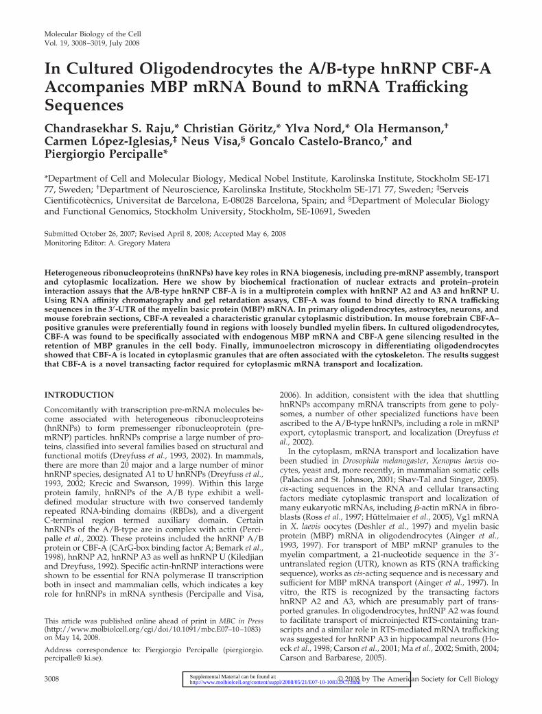
Molecular Biology of the CellVol. 19, 3008–3019, July 2008
In Cultured Oligodendrocytes the A/B-type hnRNP CBF-AAccompanies MBP mRNA Bound to mRNA TraffickingSequencesChandrasekhar S. Raju,* Christian Goritz,* Ylva Nord,* Ola Hermanson,†Carmen Lopez-Iglesias,‡ Neus Visa,§ Goncalo Castelo-Branco,† andPiergiorgio Percipalle*
*Department of Cell and Molecular Biology, Medical Nobel Institute, Karolinska Institute, Stockholm SE-17177, Sweden; †Department of Neuroscience, Karolinska Institute, Stockholm SE-171 77, Sweden; ‡ServeisCientificotecnics, Universitat de Barcelona, E-08028 Barcelona, Spain; and §Department of Molecular Biologyand Functional Genomics, Stockholm University, Stockholm, SE-10691, Sweden
Submitted October 26, 2007; Revised April 8, 2008; Accepted May 6, 2008Monitoring Editor: A. Gregory Matera
Heterogeneous ribonucleoproteins (hnRNPs) have key roles in RNA biogenesis, including pre-mRNP assembly, transportand cytoplasmic localization. Here we show by biochemical fractionation of nuclear extracts and protein–proteininteraction assays that the A/B-type hnRNP CBF-A is in a multiprotein complex with hnRNP A2 and A3 and hnRNP U.Using RNA affinity chromatography and gel retardation assays, CBF-A was found to bind directly to RNA traffickingsequences in the 3�-UTR of the myelin basic protein (MBP) mRNA. In primary oligodendrocytes, astrocytes, neurons, andmouse forebrain sections, CBF-A revealed a characteristic granular cytoplasmic distribution. In mouse forebrain CBF-A–positive granules were preferentially found in regions with loosely bundled myelin fibers. In cultured oligodendrocytes,CBF-A was found to be specifically associated with endogenous MBP mRNA and CBF-A gene silencing resulted in theretention of MBP granules in the cell body. Finally, immunoelectron microscopy in differentiating oligodendrocytesshowed that CBF-A is located in cytoplasmic granules that are often associated with the cytoskeleton. The results suggestthat CBF-A is a novel transacting factor required for cytoplasmic mRNA transport and localization.
INTRODUCTION
Concomitantly with transcription pre-mRNA molecules be-come associated with heterogeneous ribonucleoproteins(hnRNPs) to form premessenger ribonucleoprotein (pre-mRNP) particles. hnRNPs comprise a large number of pro-teins, classified into several families based on structural andfunctional motifs (Dreyfuss et al., 1993, 2002). In mammals,there are more than 20 major and a large number of minorhnRNP species, designated A1 to U hnRNPs (Dreyfuss et al.,1993, 2002; Krecic and Swanson, 1999). Within this largeprotein family, hnRNPs of the A/B type exhibit a well-defined modular structure with two conserved tandemlyrepeated RNA-binding domains (RBDs), and a divergentC-terminal region termed auxiliary domain. CertainhnRNPs of the A/B-type are in complex with actin (Perci-palle et al., 2002). These proteins included the hnRNP A/Bprotein or CBF-A (CArG-box binding factor A; Bemark et al.,1998), hnRNP A2, hnRNP A3 as well as hnRNP U (Kiledjianand Dreyfuss, 1992). Specific actin-hnRNP interactions wereshown to be essential for RNA polymerase II transcriptionboth in insect and mammalian cells, which indicates a keyrole for hnRNPs in mRNA synthesis (Percipalle and Visa,
2006). In addition, consistent with the idea that shuttlinghnRNPs accompany mRNA transcripts from gene to poly-somes, a number of other specialized functions have beenascribed to the A/B-type hnRNPs, including a role in mRNPexport, cytoplasmic transport, and localization (Dreyfuss etal., 2002).
In the cytoplasm, mRNA transport and localization havebeen studied in Drosophila melanogaster, Xenopus laevis oo-cytes, yeast and, more recently, in mammalian somatic cells(Palacios and St. Johnson, 2001; Shav-Tal and Singer, 2005).cis-acting sequences in the RNA and cellular transactingfactors mediate cytoplasmic transport and localization ofmany eukaryotic mRNAs, including �-actin mRNA in fibro-blasts (Ross et al., 1997; Huttelmaier et al., 2005), Vg1 mRNAin X. laevis oocytes (Deshler et al., 1997) and myelin basicprotein (MBP) mRNA in oligodendrocytes (Ainger et al.,1993, 1997). For transport of MBP mRNP granules to themyelin compartment, a 21-nucleotide sequence in the 3�-untranslated region (UTR), known as RTS (RNA traffickingsequence), works as cis-acting sequence and is necessary andsufficient for MBP mRNA transport (Ainger et al., 1997). Invitro, the RTS is recognized by the transacting factorshnRNP A2 and A3, which are presumably part of trans-ported granules. In oligodendrocytes, hnRNP A2 was foundto facilitate transport of microinjected RTS-containing tran-scripts and a similar role in RTS-mediated mRNA traffickingwas suggested for hnRNP A3 in hippocampal neurons (Ho-eck et al., 1998; Carson et al., 2001; Ma et al., 2002; Smith, 2004;Carson and Barbarese, 2005).
This article was published online ahead of print in MBC in Press(http://www.molbiolcell.org/cgi/doi/10.1091/mbc.E07–10–1083)on May 14, 2008.
Address correspondence to: Piergiorgio Percipalle (piergiorgio.percipalle@ ki.se).
3008 © 2008 by The American Society for Cell Biology http://www.molbiolcell.org/content/suppl/2008/05/21/E07-10-1083.DC1.htmlSupplemental Material can be found at:
MBP, together with the proteolipid protein (PLP) and itsisoform DM20 are major constituents of purified myelin, andthey are required to stabilize the apposed myelin mem-branes in compact myelin (Gielen et al., 2004). The molecularmechanisms underlying the regulation of MBP biogenesisare still unclear. In an attempt to identify transacting factorsassociated with transported granules, recent proteomic stud-ies showed that hnRNP U and CBF-A are among thehnRNPs that are found in RNP granules isolated from de-veloping and adult mouse brains (Kanai et al., 2004; Elvira etal., 2006). CBF-A is a shuttling hnRNP component of pre-mRNP/mRNP particles (Percipalle et al., 2002). However,despite of the above observations, at this stage, its potentialrole in cytoplasmic mRNA transport and MBP biogenesishas not yet been investigated.
In this study, we show evidence that CBF-A binds theMBP mRNA RTS sequence and is required in vivo for MBP
mRNA transport in oligodendrocytes. Based on these obser-vations, we propose a general role for CBF-A as a cellulartransacting factor in cytoplasmic RNA transport and local-ization.
MATERIALS AND METHODS
AntibodiesThe rabbit polyclonal peptide specific antibody to CBF-A was raised using thepeptide YQQGYGPGYGGYDY as antigen. The same peptide antigen conju-gated to Sulfolink resin (Pierce, Rockford, IL) was used for affinity purificationof the serum. The rabbit polyclonal antibody (SAK22) against p37 and p42CBF-A isoforms was a gift from J. Dean (Imperial College of Science, London;see also Supplementary Information).
Cell CultureHeLa and oli-neu cells (gift from J. Trotter, University of Mainz) were main-tained as previously shown (Jung et al., 1995; Percipalle et al., 2002). Briefly,
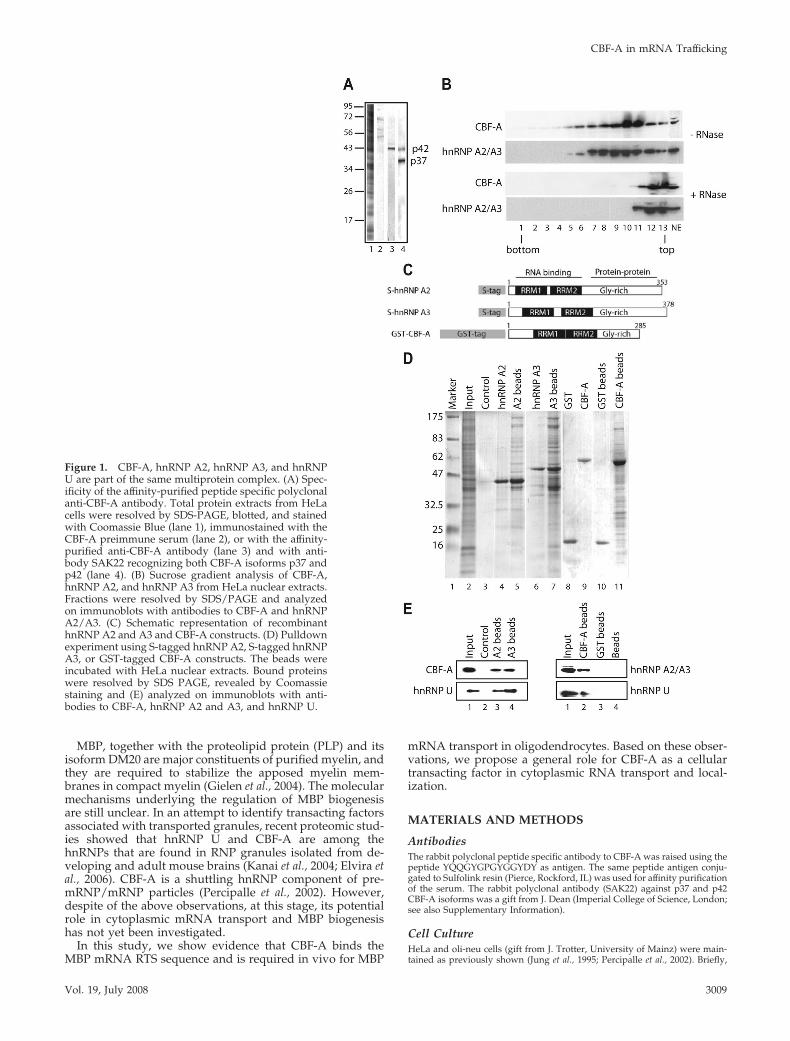
Figure 1. CBF-A, hnRNP A2, hnRNP A3, and hnRNPU are part of the same multiprotein complex. (A) Spec-ificity of the affinity-purified peptide specific polyclonalanti-CBF-A antibody. Total protein extracts from HeLacells were resolved by SDS-PAGE, blotted, and stainedwith Coomassie Blue (lane 1), immunostained with theCBF-A preimmune serum (lane 2), or with the affinity-purified anti-CBF-A antibody (lane 3) and with anti-body SAK22 recognizing both CBF-A isoforms p37 andp42 (lane 4). (B) Sucrose gradient analysis of CBF-A,hnRNP A2, and hnRNP A3 from HeLa nuclear extracts.Fractions were resolved by SDS/PAGE and analyzedon immunoblots with antibodies to CBF-A and hnRNPA2/A3. (C) Schematic representation of recombinanthnRNP A2 and A3 and CBF-A constructs. (D) Pulldownexperiment using S-tagged hnRNP A2, S-tagged hnRNPA3, or GST-tagged CBF-A constructs. The beads wereincubated with HeLa nuclear extracts. Bound proteinswere resolved by SDS PAGE, revealed by Coomassiestaining and (E) analyzed on immunoblots with anti-bodies to CBF-A, hnRNP A2 and A3, and hnRNP U.
CBF-A in mRNA Trafficking
Vol. 19, July 2008 3009
oli-neu cells were propagated in Sato medium containing 1% horse serum anddifferentiated for 7–10 d with dibutyryl cAMP (Sigma-Aldrich, St. Louis, MO),as described (Jung et al., 1995).
Immunohistochemistry on Mouse Forebrain SectionsAdult C57bl6 mice (Charles River, Wilmington, MA) were transcardiallyperfused with PBS followed by 4% formaldehyde in PBS, and brains weredissected and postfixed overnight at 4°C. Coronal sections were made at 35�m using a vibratom (Leica, Heidelberg, Germany). Immunostaining forCBF-A, MBP, and CNPase was performed on free-floating sections (see Sup-plementary Information).
Primary CellsAdult mouse neural stem cells were prepared and cultured as neurospheresand differentiated as described (Rietze and Reynolds, 2006), with minormodifications (see Supplementary Information for a detailed description ofthe protocol used). Fetal rat neural stem cells were prepared as described inthe Supplementary Information.
Immunofluorescence Microscopy, Immuno-FISH, and GeneSilencingImmunofluorescence and immuno-FISH on cells grown on coverslips werealready described (Singer and Ward, 1982; Percipalle et al., 2002). For in situdetection of MBP mRNA, the RNA probe 5�-CCAUGCUCUCUGGCUCCU-
UGGCGGUGUGCCUGUCU-3� was digoxigenin-labeled in the 5� end. Con-focal images were collected using a Zeiss LSM 510 META confocal laserscanning microscope (Jena, Germany; Percipalle et al., 2002).
For posttranscriptional silencing of the CBF-A gene, the siGENOMESMART pool duplex against the mouse HNRPAB gene (NM_010448) or thecontrol ECFP (enhanced cyan fluorescent protein) gene were transfected intooli-neu cells according to the manufacturer’s instructions (Dharmacon Re-search, Boulder, CO). Sequences can be found in Supplementary Information.
Cloning, Expression, and Protein PurificationFull-length hnRNP A2 (forward primer 5�-GGAATTCTTAGCGACTGAGTC-CGCGATG, reverse primer 5�-ATAAGAATGCGGCCGCTGAAGCTGTTCT-GTTACCTCTG) and hnRNP A3 (Ma et al., 2002) were cloned in pGEM-T(Promega, Madison, WI) and subsequently in pET30a (�) for expression(Novagen, Madison, WI). Constructs were verified by DNA sequencing.Full-length glutathione S-transferase (GST)-tagged CBF-A (gift of Tomas Le-anderson, Lund University) was expressed from a pGEX plasmid vector (seeSupplementary Information).
Protein–Protein Interaction AssaysFor pulldown experiments, recombinant hnRNP A2, hnRNP A3, or CBF-Aconstructs were coupled to protein S agarose or glutathione beads (Novagen;GE Healthcare, Waukesha, WI) and incubated with HeLa nuclear extracts (seeSupplementary Information).
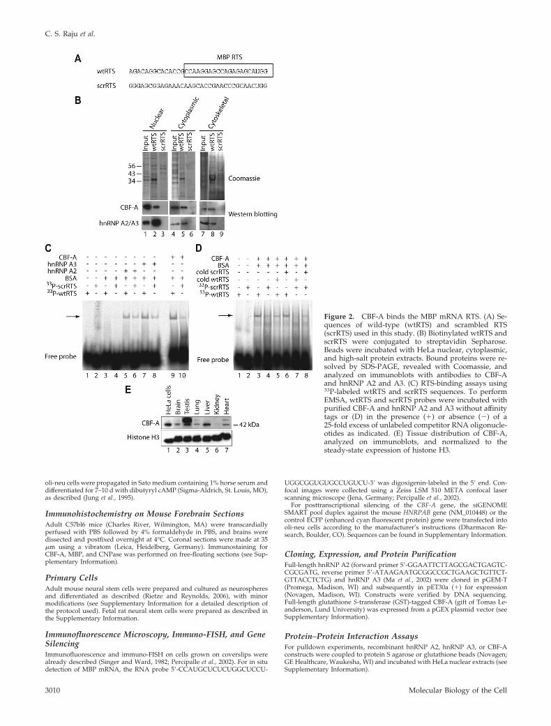
Figure 2. CBF-A binds the MBP mRNA RTS. (A) Se-quences of wild-type (wtRTS) and scrambled RTS(scrRTS) used in this study. (B) Biotinylated wtRTS andscrRTS were conjugated to streptavidin Sepharose.Beads were incubated with HeLa nuclear, cytoplasmic,and high-salt protein extracts. Bound proteins were re-solved by SDS-PAGE, revealed with Coomassie, andanalyzed on immunoblots with antibodies to CBF-Aand hnRNP A2 and A3. (C) RTS-binding assays using33P-labeled wtRTS and scrRTS sequences. To performEMSA, wtRTS and scrRTS probes were incubated withpurified CBF-A and hnRNP A2 and A3 without affinitytags or (D) in the presence (�) or absence (�) of a25-fold excess of unlabeled competitor RNA oligonucle-otides as indicated. (E) Tissue distribution of CBF-A,analyzed on immunoblots, and normalized to thesteady-state expression of histone H3.
C. S. Raju et al.
Molecular Biology of the Cell3010
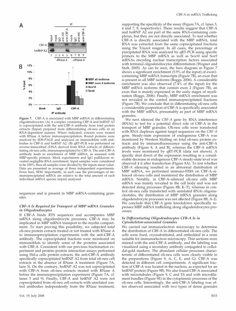
Protein and RNA ImmunoprecipitationFor protein immunoprecipitations, total protein extracts prepared from oli-neu cells were incubated with the anti-CBF-A antibody, with the anti-HA tagantibody, and with nonspecific mouse IgGs. Where indicated, extracts wereincubated with RNase A (see Supplementary Information). The immunopre-cipitated fractions from untreated and RNase A–treated extracts were re-solved by SDS-PAGE and analyzed on immunoblots with antibodies againstCBF-A and hnRNP A2. For analysis of the RNA species associated withCBF-A, the RNA was extracted from the fractions immunoprecipitated withthe anti-CBF-A antibody or mock and IgG control immunoprecipitation as-says using the Trizol reagent, followed by DNase I (Invitrogen, Carlsbad, CA)treatment and subsequently made into cDNA by Superscript II (Invitrogen)using random primers. The samples were then analyzed by quantitativeRT-PCR (qRT-PCR) with primers specific to MBP mRNA exon 1, MBP mRNAexon 2, and Sox 10 and Sox9 mRNA (see Supplementary Information forprimer sequences and qRT PCR analysis).
Protein–RNA Interaction AssaysRNA affinity chromatography was performed as described (Hoek et al., 1998).For each RNA-binding experiment, 500 pmol of 5�-biotinylated wild-type RTS(wtRTS; 5�-AGACAGGCACACCGCCAAGGAGCCAGAGAGCAUGG-3�)and scrambled RTS (scrRTS; 5�-GGGAGCGGAGAAACAAGCACCGAAC-CCGCAACUGG-3�) were coupled to streptavidin-coated Sepharose (GEHealthcare) and incubated with nuclear, cytoplasmic, and high-salt extracts.Bound proteins were resolved by SDS-PAGE and analyzed on immunoblots(see Supplementary Information).
For electrophoretic mobility shift assay (EMSA), wtRTS, and scrRTS were 5�end-labeled using �-33P-ATP (GE Healthcare) and T4 polynucleotide kinase(New England Biolabs, Beverly, MA). Purified hnRNP A2, hnRNP A3, and
CBF-A were incubated with �50 fmol of 33P-labeled wtRTS or scrRTS inEMSA buffer (20 mM HEPES, pH 7.6, 5 mM MgCl2, 40 mM KCl, 1 mM DTT,5% glycerol) containing heparin (5 �g/�l) and BSA (100 �g/ml) for 30 min onice. Protein–RNA complexes were resolved by native gel electrophoresis andanalyzed with a Fuji-BAS 2000 phosphorimager (Tokyo, Japan).
Immunoelectron MicroscopyAfter fixation, the cells were either cryosectioned or cryosubstituted andembedded in Lowicryl K4M (Polysciences, Warrington, PA; see Supplemen-tary Information). Cryosectioned samples provided a good definition of nu-clear and membrane structures, whereas cryosubstitution offered a superiorpreservation of the cytoskeleton. For immunolabeling, binding of the anti-CBF-A antibody was detected with either a secondary antibody or protein Acoupled to 10-nm diameter colloidal gold particles. Sections were observed ina JEM-1010 electron microscope (Jeol, Tokyo, Japan), and photographic neg-atives were scanned with an Artixcan 2500f scanner (Microtek International,Hsinchu, Taiwan; see Supplementary Online Information).
RESULTS
CBF-A, hnRNP A2 and A3, and hnRNP U Are Present in aMultiprotein ComplexCBF-A is present in two alternatively spliced isoforms, p37and p42, that differ only for the presence of a 47-amino acidinsert in the p42 C-terminus located outside their RNA-binding domains (Dean et al., 2002). Evidence that CBF-A is
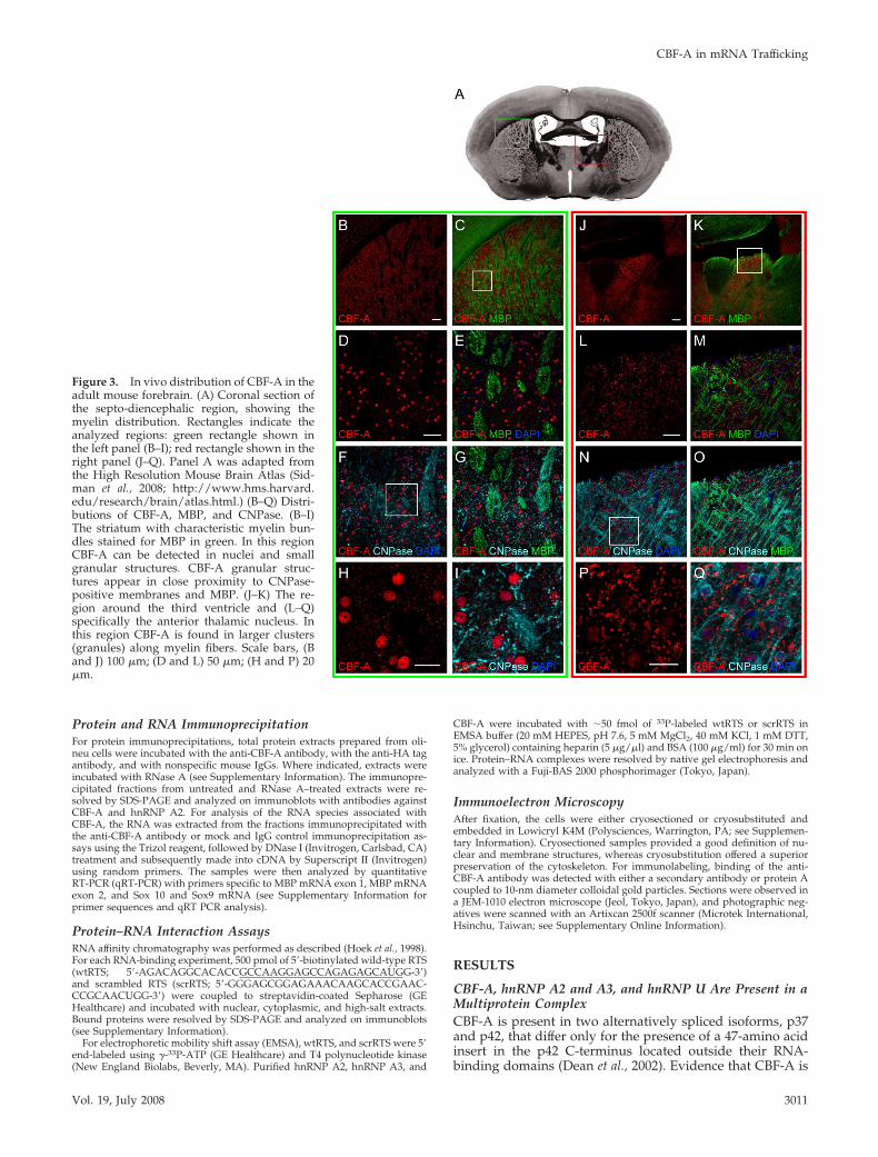
Figure 3. In vivo distribution of CBF-A in theadult mouse forebrain. (A) Coronal section ofthe septo-diencephalic region, showing themyelin distribution. Rectangles indicate theanalyzed regions: green rectangle shown inthe left panel (B–I); red rectangle shown in theright panel (J–Q). Panel A was adapted fromthe High Resolution Mouse Brain Atlas (Sid-man et al., 2008; http://www.hms.harvard.edu/research/brain/atlas.html.) (B–Q) Distri-butions of CBF-A, MBP, and CNPase. (B–I)The striatum with characteristic myelin bun-dles stained for MBP in green. In this regionCBF-A can be detected in nuclei and smallgranular structures. CBF-A granular struc-tures appear in close proximity to CNPase-positive membranes and MBP. (J–K) The re-gion around the third ventricle and (L–Q)specifically the anterior thalamic nucleus. Inthis region CBF-A is found in larger clusters(granules) along myelin fibers. Scale bars, (Band J) 100 �m; (D and L) 50 �m; (H and P) 20�m.
CBF-A in mRNA Trafficking
Vol. 19, July 2008 3011
a component of pre-mRNP/RNP particles prompted us todetermine whether CBF-A is in a multiprotein complex to-gether with core hnRNP proteins such as hnRNP A2 andhnRNP A3. We first fractionated nuclear proteins preparedfrom HeLa cells by ultracentrifugation on a sucrose gradientand monitored coelution of CBF-A on immunoblots, using anovel affinity-purified peptide-specific anti-CBF-A antibodythat recognizes the larger p42 isoform (Figure 1A, cf. lane 3).CBF-A was found to coelute with hnRNP A2/A3 in anRNA-dependent manner (Figure 1B). To demonstrate thesuggested association of CBF-A with hnRNP A2/A3, taggedCBF-A, hnRNP A2, and hnRNP A3 full-length constructswere recombinantly expressed and affinity-purified for invitro protein–protein interactions (Figure 1C). GST-tagged
CBF-A and S-tagged hnRNP A2 and A3 were coupled toeither glutathione beads or protein S beads and incubatedwith HeLa nuclear protein extracts. Coprecipitated proteinswere resolved by SDS-PAGE and monitored on immuno-blots. Figure 1E shows that recombinant CBF-A, hnRNP A2,and hnRNP A3 reciprocally coprecipitated the endogenousproteins, as well as hnRNP U, a newly identified proteincomponent of transported RNA granules (Elvira et al., 2006;Kanai et al., 2004). None of the proteins could be coprecipi-tated from nuclear extracts using protein S beads or GSTbeads, further supporting the specificity of the reaction (Fig-ure 1D, cf. lanes 3and 10, and Figure 1E). Overall, theseresults are consistent with the view that subcellular fractionsof CBF-A, hnRNP A2, hnRNP A3, and hnRNP U are phys-
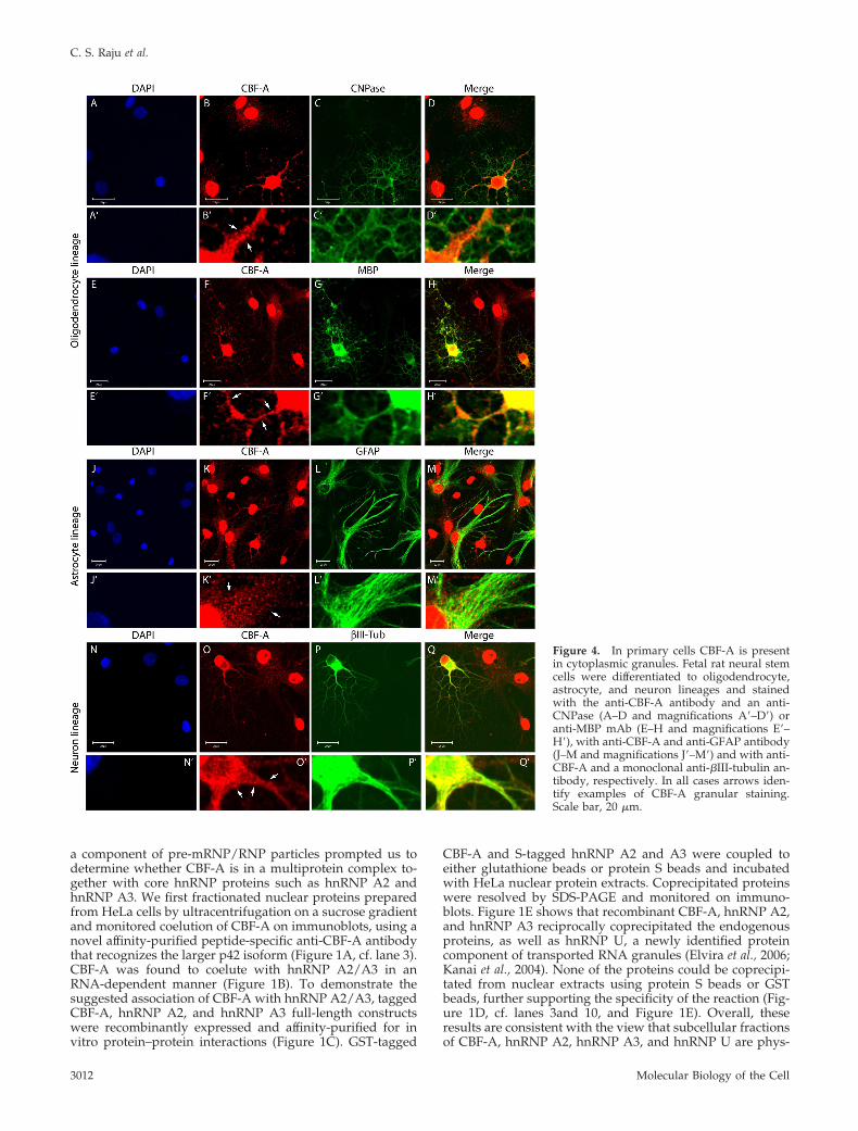
Figure 4. In primary cells CBF-A is presentin cytoplasmic granules. Fetal rat neural stemcells were differentiated to oligodendrocyte,astrocyte, and neuron lineages and stainedwith the anti-CBF-A antibody and an anti-CNPase (A–D and magnifications A�–D�) oranti-MBP mAb (E–H and magnifications E�–H�), with anti-CBF-A and anti-GFAP antibody(J–M and magnifications J�–M�) and with anti-CBF-A and a monoclonal anti-�III-tubulin an-tibody, respectively. In all cases arrows iden-tify examples of CBF-A granular staining.Scale bar, 20 �m.
C. S. Raju et al.
Molecular Biology of the Cell3012
ically associated and are contained in a nuclear multiproteincomplex.
CBF-A Binds the MBP RTSThe finding that CBF-A is in a complex with hnRNP A2,hnRNP A3, and hnRNP U suggests that CBF-A may also beassociated with MBP mRNA either indirectly or directlybound to the RTS sequence. To analyze this possibility, wesynthesized biotinylated 35-nt RNA oligonucleotides en-compassing the wtRTS from MBP mRNA and scrRTS withidentical nucleotide composition but a different primarysequence (Figure 2A). To test whether endogenous CBF-Aassociated with RTS sequences, we coupled wtRTS andscrRTS to streptavidin-coated Sepharose beads and incu-bated the beads with HeLa nuclear, cytoplasmic, or high-salt(or cytoskeletal) protein extracts. The blots in Figure 2Bshow that endogenous CBF-A and hnRNP A2 and A3 co-precipitated with wtRTS from all cellular extracts (cf. lanes 2,5, and 8), whereas no significant levels of CBF-A and hnRNPA2 and A3 were coprecipitated with scrRTS beads (Figure2B, cf. lanes 3, 6, and 9).
We next applied EMSAs to determine whether CBF-Adirectly binds the RTS motif. We incubated 33P-labeledwtRTS and scrRTS RNA oligonucleotides with purifiedCBF-A (37-kDa isoform), hnRNP A2, or hnRNP A3, wherethe affinity tags had been proteolytically removed. Figure 2C
shows that the electrophoretic mobility of the wtRTS oligo-nucleotide was considerably retarded when incubated withCBF-A, whereas the electrophoretic mobility of scrRTS wasonly marginally affected by CBF-A (Figure 2C, cf. lanes 9and 10). Similar results were obtained with hnRNP A2 andA3 used as control (Figure 2C). In comparison with wtRTS,the electrophoretic mobility of scrRTS was only slightlyretarded when incubated with CBF-A (Figure 2D, cf. lanes 3and 4). Second, retardation in the electrophoretic mobility ofwtRTS incubated with CBF-A was altered in the presence ofunlabeled wtRTS but not in the presence of unlabeledscrRTS when added in large excess to the reaction mixture(Figure 2D, cf. lanes 3, 5 and 3, 6, respectively). Finally,incubation of CBF-A with labeled scrRTS was sensitive tocompetition with unlabeled wtRTS but was not sensitive tocompetition with unlabeled scrRTS (Figure 2D, cf. lanes 4, 7and 4, 8). These results indicate that CBF-A specifically bindsthe MBP mRNA RTS, presumably as part of a multiproteincomplex.
In Oligodendrocytes CBF-A Associates with MBP mRNAGranulesIf CBF-A specifically binds RTS sequences found within theMBP mRNA, it is possible that in oligodendrocytes CBF-Aremains associated with transported mRNA granules out-side the cell nucleus. Consistently, immunoblots of protein
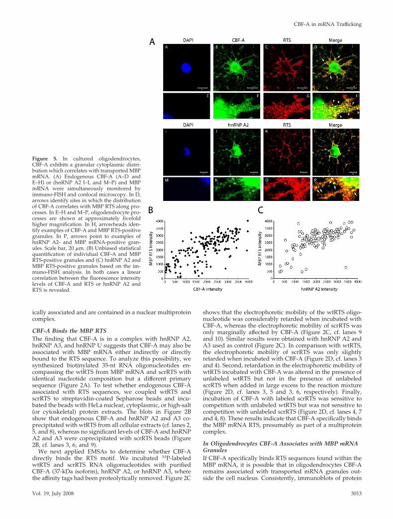
Figure 5. In cultured oligodendrocytes,CBF-A exhibits a granular cytoplasmic distri-bution which correlates with transported MBPmRNA. (A) Endogenous CBF-A (A–D andE–H) or (hnRNP A2 I–L and M–P) and MBPmRNA were simultaneously monitored byimmuno-FISH and confocal microscopy. In D,arrows identify sites in which the distributionof CBF-A correlates with MBP RTS along pro-cesses. In E–H and M–P, oligodendrocyte pro-cesses are shown at approximately fivefoldhigher magnification. In H, arrowheads iden-tify examples of CBF-A and MBP RTS-positivegranules. In P, arrows point to examples ofhnRNP A2- and MBP mRNA-positive gran-ules. Scale bar, 20 �m. (B) Unbiased statisticalquantification of individual CBF-A and MBPRTS-positive granules and (C) hnRNP A2 andMBP RTS-positive granules based on the im-muno-FISH analysis. In both cases a linearcorrelation between the fluorescence intensitylevels of CBF-A and RTS or hnRNP A2 andRTS is revealed.
CBF-A in mRNA Trafficking
Vol. 19, July 2008 3013

extracts prepared from different mouse tissues revealed thatCBF-A is ubiquitously expressed (Figure 2E) and that theanti-CBF-A antibody is monospecific in brain tissue (Figure2E, lane 2).
On the basis of the above, we analyzed the in vivo distri-bution of CBF-A in the myelin-rich regions present in theforebrain of adult mice. In agreement with its ubiquitousexpression, immunostaining of mouse brain sections withthe anti-CBF-A antibody followed by confocal microscopyrevealed that cellular CBF-A is expressed all over in theforebrain, showing distinctive nuclear localization (Figure3). Interestingly, a fraction of CBF-A was also found outsidethe cell nucleus in discrete particles, reminiscent of trans-ported granules in oligodendrocytes, with different sizesand signal intensities depending on the brain regions ana-lyzed (Figure 3, cf. E and M). To test whether in oligoden-drocytes CBF-A displays a granular distribution, we per-formed triple-immunostaining of brain sections with theanti-CBF-A antibody, a rat mAb against MBP, which specif-ically labels myelin fibers, and a mouse mAb against 2�,3�-cyclic nucleotide 3�-phosphodiesterase (CNPase), which la-bels oligodendrocytes membranes and is commonly used asmarker for oligodendrocytes. Remarkably, in nuclei encap-sulated by CNPase we detected very little CBF-A, whereasmost of the CBF-A–positive granules were found in closeproximity to CNPase and/or MBP, suggesting a specializedcytoplasmic function for CBF-A, presumably in processesemanating from the cell body (Figure 3, cf. I and Q). Con-sistent with the in vivo distribution detected in mouse fore-brain, CBF-A–positive granules were also revealed in pri-mary oligodendrocytes, astrocytes, and neurons obtained byin vitro differentiation of fetal rat and adult mouse neuralstem cells (Figure 4 and Supplementary Figure S1). In allthese cases, CBF-A–positive granules were revealed alongthe microtubule-rich processes.
To study whether CBF-A is present in MBP mRNA gran-ules, we used a stable mouse oligodendroglial precursor cellline, oli-neu cells, which can differentiate into myelin-asso-ciated glycoprotein (MAG)-positive oligodendrocytes (Junget al., 1995). Differentiated oli-neu cells were subjected toimmunofluorescence in situ hybridization (immuno-FISH)experiments with the anti-CBF-A antibody and a specific 5�digoxigenin end-labeled RNA probe hybridizing with theendogenous MBP mRNA RTS. Confocal microscopy re-vealed that CBF-A–positive granules distributed along oli-godendrocytes processes partly colocalize with MBP mRNAin granular structures (Figure 5, A–H, see arrowheads in H),similar to hnRNP A2 (Figure 5, I–P, see arrows in P). Insupport of a colocalization between endogenous CBF-A andRTS-containing MBP mRNA, we next performed unbiasedstatistical analysis on the fluorescence intensity levels de-rived from the corresponding confocal images and as pre-viously described (Ma et al., 2002). The results revealedlinear correlations between the CBF-A and RTS signals (Fig-ure 5B). Quantification of the number of colocalizationevents occurring in individual particles showed that roughly88% of the granules contains CBF-A and RTS-containingmRNA. As expected, a similar correlation was revealedbetween the hnRNP A2 and RTS signals (Figure 5C), where�90% of the granules contained both hnRNP A2 and RTS-containing mRNA. Furthermore, double immunostainingexperiments demonstrated a linear correlation also betweenthe granular distributions of CBF-A and hnRNP A2 (Figure6, A and B), and CBF-A was found to colocalize with �-tu-bulin along oli-neu processes (Supplementary Figure 2). Fi-nally triple oli-neu immunofluorescence staining for theMBP mRNA RTS, hnRNP A2, and CBF-A showed that morethan 80% of endogenous MBP mRNA granules were posi-tively stained for RTS RNA, CBF-A, and hnRNP A2 (Sup-plementary Figure S3). We conclude that CBF-A binds RTS
Figure 6. In oli-neu cells, the distribution ofendogenous CBF-A correlates with hnRNPA2. (A and E) DAPI staining, (B and F) oli-neucells stained with a mAb to hnRNP A2. (C andG) Oli-neu cells stained with the rabbit poly-clonal peptide-specific anti-CBF-A antibodyand (D and H) merged images. Scale bar, 20�m. (B) Statistical quantification of CBF-A andhnRNP A2-positive granules based on thedouble immunofluorescence analysis and con-focal microscopy in A. A linear correlationbetween the fluorescence signals of CBF-Aand hnRNP A2 is revealed.
C. S. Raju et al.
Molecular Biology of the Cell3014
sequences and is present in MBP mRNA-containing gran-ules.
CBF-A Is Required for Transport of MBP mRNA Granulesin OligodendrocytesIf CBF-A binds RTS sequences and accompanies MBPmRNA along oligodendrocyte processes, CBF-A may beimplicated in MBP mRNA transport to the myelin compart-ment. To start proving this possibility, we subjected totaloli-neu protein extracts treated or not treated with RNase Ato immunoprecipitation experiments with the anti-CBF-Aantibody. The coprecipitated fractions were monitored onimmunoblots to identify some of the proteins associatedwith CBF-A. Consistent with our previous fractionation ex-periment and protein–protein interaction assays performedusing HeLa cells protein extracts, the anti-CBF-A antibodyspecifically coprecipitated hnRNP A2 from total oli-neu cellextracts in the absence of RNase A treatment (Figure 7A,lane 5). On the contrary, hnRNP A2 was not coprecipitatedwith CBF-A from oli-neu extracts treated with RNase Abefore the immunoprecipitation experiment (Figure 7A, cf.lanes 5 and 9). Finally, CBF-A and hnRNP A2 were notcoprecipitated from oli-neu cell extracts with unrelated con-trol antibodies independently from the RNase treatment,
supporting the specificity of the assay (Figure 7A, cf. lanes 3,4 and 7, 8, respectively). These results suggest that CBF-Aand hnRNP A2 are part of the same RNA-containing com-plexes, but they are not directly associated. To test whetherCBF-A is directly associated with the MBP mRNA, totalRNA was extracted from the same coprecipitated fractionsusing the Triazol reagent. In all cases, the percentage ofprecipitated RNA was analyzed by qRT-PCR using specificprimers to the MBP mRNA as well as Sox10 and Sox9mRNAs encoding nuclear transcription factors associatedwith terminal oligodendrocytes differentiation (Wegner andStolt, 2005). As can be seen, the bars diagram in Figure 7shows a significant enrichment (5.9% of the input) of exon1-containing MBP mRNA transcripts (Figure 7B), an exon thatis present in all MBP isoforms (Boggs, 2006). A considerableenrichment was also observed (7.4% of the input) for theMBP mRNA isoforms that contain exon 2 (Figure 7B), anexon that is mainly expressed in the early stages of myeli-nation (Boggs, 2006). Finally, MBP mRNA enrichment wasnot revealed in the control immunoprecipitated fractions(Figure 7B). We conclude that in differentiating oli-neu cellsa considerable proportion of CBF-A is specifically associatedwith the MBP mRNA, presumably as part of MBP mRNAgranules.
We next silenced the CBF-A gene by RNA interference(RNAi) to test for a potential direct role of CBF-A in thetransport of MBP granules. Oli-neu cells were transfectedwith RNA duplexes against target sequences on the CBF-Agene. Steady-state expression of endogenous CBF-A wasmonitored by Western blotting on total oli-neu protein ex-tracts and by immunofluorescence using the anti-CBF-Aantibody (Figure 8, A and B), whereas the CBF-A mRNAlevels were monitored by qRT-PCR (data not shown). Aspecific shut down of the expression resulting in a consid-erable decrease in endogenous CBF-A steady-state level wasobserved 4 d after transfection (Figure 8A). To test whetherCBF-A silencing resulted in an abnormal distribution ofMBP mRNA, we performed immuno-FISH on CBF-A–si-lenced oli-neu cells and monitored the distribution of MBPmRNA. Notably, in CBF-A–silenced oli-neu cells MBPmRNA was mainly revealed in the cell body and was notdetected along processes (Figure 8B, K–T), whereas in con-trol oli-neu cells transfected with unrelated RNAi oligonu-cleotides, the distribution of MBP mRNA granules alongoligodendrocyte processes was not affected (Figure 8B, A–J).We conclude that CBF-A gene knockdown specifically re-presses MBP mRNA trafficking along oligodendrocytes pro-cesses.
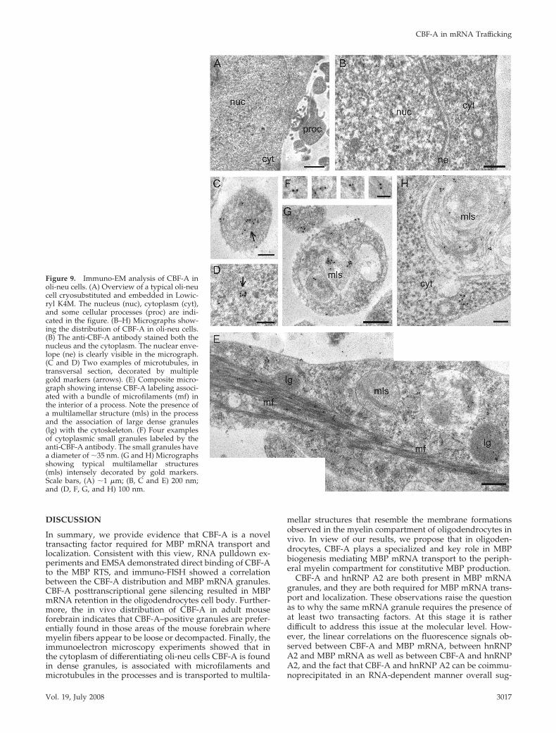
In Differentiating Oligodendrocytes CFB-A Is inCytoskeleton-associated GranulesWe carried out immunoelectron microscopy to determinethe distribution of CBF-A in differentiated oli-neu cells. Thecells were fixed, cryosubstituted, and embedded in a resinsuitable for immunoelectron microscopy. Thin sections werestained with the anti-CBF-A antibody, and the labeling wasvisualized using a secondary antibody conjugated to colloi-dal-gold markers. The abundant cellular processes charac-teristic of differentiated oli-neu cells were clearly visible inthe preparations (Figure 9, A, C, E, and G). CBF-A wasdetected in different cell compartments. A significant frac-tion of CBF-A was located in the nucleus, as expected for anhnRNP protein (Figure 9B). We also found CBF-A associatedwith microtubules (Figure 9, C and D) and with microfila-ment bundles (Figure 9E) in the cytoplasmic processes of theoli-neu cells. Interestingly, the anti-CBF-A labeling was of-ten observed associated with two types of dense granules
Figure 7. CBF-A is associated with MBP mRNA in differentiatingoligodendrocytes. (A) A complex containing CBF-A and hnRNP A2is coprecipitated with the anti-CBF-A antibody from total proteinextracts (Input) prepared from differentiating oli-neu cells in anRNA-dependent manner. Where indicated, extracts were treatedwith RNase A before immunoprecipitation. Bound proteins wereresolved by SDS-PAGE and analyzed on immunoblots with anti-bodies to CBF-A and hnRNP A2. (B) qRT-PCR was performed onreverse-transcribed cDNA derived from RNA extracts of differen-tiating oli-neu cells, immunoprecipitated by CBF-A. The anti-CBF-Aantibody leads to enrichment of MBP mRNA, as assessed withMBP-specific primers. Mock experiments and IgG pulldowns re-vealed negligible RNA enrichment. Input samples were consideredto be 100%; thus all samples were divided by the inputs mean value.Data are presented as average of three independent experiments.Error bars, SEM. Importantly, in each case the percentages of im-munoprecipitated mRNA are relative to the total amount of eachindividual mRNA species (input) analyzed.
CBF-A in mRNA Trafficking
Vol. 19, July 2008 3015

with diameters of 30–40 nm (Figure 9F) and 300–400 nm (lgin Figure 9E), respectively. These large CBF-A–positivegranules correspond to the RNA transport granules ob-served in our immunofluorescence experiments (see Discus-sion), and their dimensions are in agreement with previousreports of MBP granules in oligodendrocytes (Ainger et al.,1993; Barbarese et al., 1995). Both large and small granulesare found in association with the cytoskeleton. Finally, theimmunoelectron microscopy experiment also revealed thatCBF-A reaches the myelin compartment. Indeed, significantlabeling was observed in association with the concentricmultilamellar formations that are characteristic of the differ-entiating oligodendrocytes (Figure 9, E, G, and H).
We also analyzed the distribution of CBF-A in HeLa cells(Supplementary Figure S4). Both the nucleus and the cyto-plasm were significantly labeled (Supplementary Figure S4,A and B), and gold markers were occasionally observed inthe proximity of the nuclear envelope (Supplementary Fig-ure S4C). In the cytoplasm of HeLa cells, CBF-A could alsobe seen in dense granules that resemble the small granulesdescribed in oli-neu cells based on dimensions, texture, andelectron density. The large CFB-A granules observed in oli-godendrocytes were not present in HeLa cells. Interestingly,the small CBF-A–containing granules were associated withcortical filaments and filipodia at the periphery of HeLa cells(Supplementary Figure S4, F and G).
Figure 8. (A) Steady-state expression levelsof CBF-A in mock-transfected oli-neu cells(lane 1), control oli-neu cells transfected withnonspecific siRNA oligonucleotides (lane 2),and in CBF-A silenced oli-neu cells (lane 3).The bars diagram shows the relative amountsof CBF-A determined in independent experi-ments as ratios between transfected (eitherwith control siRNA oligonucleotides or withspecific siRNA oligonucleotides against CBF-A)and mock-transfected oli-neu cells. (B) In CBF-A–silenced oli-neu cells, MBP mRNA is ex-cluded from oligodendrocytes processes asmonitored by immuno-FISH and confocal mi-croscopy. White arrows point toward siRNA-transfected cells in which the expression ofCBF-A is considerably decreased (N and S).Black arrows point toward processes of oli-neu cells that are transfected with controlsiRNA oligonucleotides or specific siRNA oli-gonucleotides to silence CBF-A (J and T). Scalebars, (A–E and K–O) 10 �m; (F–J and P–T) 10�m. In the right column, D–T show silencedoli-neu cells at fivefold magnification. Overall,these experiments were repeated three timesand on average as much as 70% of cellsshowed decreased expression of CBF-A andMBP mRNA exclusion from processes.
C. S. Raju et al.
Molecular Biology of the Cell3016
DISCUSSION
In summary, we provide evidence that CBF-A is a noveltransacting factor required for MBP mRNA transport andlocalization. Consistent with this view, RNA pulldown ex-periments and EMSA demonstrated direct binding of CBF-Ato the MBP RTS, and immuno-FISH showed a correlationbetween the CBF-A distribution and MBP mRNA granules.CBF-A posttranscriptional gene silencing resulted in MBPmRNA retention in the oligodendrocytes cell body. Further-more, the in vivo distribution of CBF-A in adult mouseforebrain indicates that CBF-A–positive granules are prefer-entially found in those areas of the mouse forebrain wheremyelin fibers appear to be loose or decompacted. Finally, theimmunoelectron microscopy experiments showed that inthe cytoplasm of differentiating oli-neu cells CBF-A is foundin dense granules, is associated with microfilaments andmicrotubules in the processes and is transported to multila-
mellar structures that resemble the membrane formationsobserved in the myelin compartment of oligodendrocytes invivo. In view of our results, we propose that in oligoden-drocytes, CBF-A plays a specialized and key role in MBPbiogenesis mediating MBP mRNA transport to the periph-eral myelin compartment for constitutive MBP production.
CBF-A and hnRNP A2 are both present in MBP mRNAgranules, and they are both required for MBP mRNA trans-port and localization. These observations raise the questionas to why the same mRNA granule requires the presence ofat least two transacting factors. At this stage it is ratherdifficult to address this issue at the molecular level. How-ever, the linear correlations on the fluorescence signals ob-served between CBF-A and MBP mRNA, between hnRNPA2 and MBP mRNA as well as between CBF-A and hnRNPA2, and the fact that CBF-A and hnRNP A2 can be coimmu-noprecipitated in an RNA-dependent manner overall sug-
Figure 9. Immuno-EM analysis of CBF-A inoli-neu cells. (A) Overview of a typical oli-neucell cryosubstituted and embedded in Lowic-ryl K4M. The nucleus (nuc), cytoplasm (cyt),and some cellular processes (proc) are indi-cated in the figure. (B–H) Micrographs show-ing the distribution of CBF-A in oli-neu cells.(B) The anti-CBF-A antibody stained both thenucleus and the cytoplasm. The nuclear enve-lope (ne) is clearly visible in the micrograph.(C and D) Two examples of microtubules, intransversal section, decorated by multiplegold markers (arrows). (E) Composite micro-graph showing intense CBF-A labeling associ-ated with a bundle of microfilaments (mf) inthe interior of a process. Note the presence ofa multilamellar structure (mls) in the processand the association of large dense granules(lg) with the cytoskeleton. (F) Four examplesof cytoplasmic small granules labeled by theanti-CBF-A antibody. The small granules havea diameter of �35 nm. (G and H) Micrographsshowing typical multilamellar structures(mls) intensely decorated by gold markers.Scale bars, (A) �1 �m; (B, C and E) 200 nm;and (D, F, G, and H) 100 nm.
CBF-A in mRNA Trafficking
Vol. 19, July 2008 3017
gest that CBF-A and hnRNP A2 are simultaneously presentin the same MBP mRNP complex. If this is the case, CBF-Aand hnRNP A2 may cooperate for MBP mRNA transportand localization. Their roles could be dependent on the localenvironment and the set of interactions required for theestablishment of trafficking intermediates, for instance, todetermine polarity during RNA trafficking (Carson and Bar-barese, 2005).
CBF-A is ubiquitously expressed and has been suggestedto have a role in transcriptional control (Kamada and Miwa,1992; Bemark et al., 1998; Leverrier et al., 2000; Mikheev et al.,2000). Because it is also a component of shuttling pre-mRNP/mRNP particles (Percipalle et al., 2002), our resultssuggest that CBF-A associates with mRNAs in the cell nu-cleus and accompanies its target mRNAs to their cytoplas-mic destination. In support of this hypothesis the immuno-electron microscopy performed with our anti-CBF-Aantibody in oli-neu and HeLa cells showed that CBF-Alocalizes in the nucleoplasm (Figure 9B and SupplementaryFigure S4B). Occasionally, CBF-A was found at or near nu-clear pores (Supplementary Figure S4C), which is consistentwith the proposal that CBF-A remains associated with themRNP during nucleo-cytoplasmic transport. In the cyto-plasm of oli-neu and HeLa cells, CBF-A was often found indense granules with a diameter of 30–40 nm (Figure 9F andSupplementary Figure S4, D and E). These CBF-A–contain-ing granules, which were often seen in association withcytoskeletal fibers, are likely to be mRNA transport gran-ules. In either case, our observations underscore the generalidea that the cytoplasmic fate of each mRNA is predeter-mined by the assembly of the mRNP complex in the cellnucleus (Daneholt, 2001).
We revealed CBF-A–positive granules in astrocytes andneurons where mRNA transcripts containing RTS-like se-quences were identified by computational approaches(Ainger et al., 1997). Moreover, CBF-A was found associatedwith the cytoskeleton in HeLa cells. Altogether these obser-vations, strongly suggest that CBF-A has a role in cytoplas-mic mRNA transport and its function may not be exclusiveof oligodendrocytes.
Given the ubiquitous distribution of CBF-A and its spe-cific role in MBP mRNA transport in oligodendrocytes, wesuggest that CBF-A has a general role as a transacting factorin the establishment of asymmetric mRNA and/or proteindistributions. CBF-A is likely to act on the localization ofmultiple mRNAs in HeLa cells as well as in other cell types,the MBP mRNA being a specific case related to a veryspecialized cellular function.
ACKNOWLEDGMENTS
We thank Sonia Ruız and Gema Martınez (Serveis Cientificotecnics, Univer-sitat de Barcelona) for EM sample preparation. This work was supported bygrants from the Swedish Research Council (Vetenskapsrådet) to P.P. andN.V. G.C.B. is supported by postdoctoral fellowships from the Swedish BrainFoundation (Hjarnfonden) and from the David and Astrid Hagelens Foun-dation.
REFERENCES
Ainger, K., Avossa, D., Morgan, F., Hill, S. J., Barry, C., Barbarese, E., andCarson, J. H. (1993). Transport and localization of exogenous myelin basicprotein mRNA microinjected into oligodendrocytes. J. Cell Biol. 123, 431–441.
Ainger, K., Avossa, D., Diana, A. S., Barry, C., Barbarese, E., and Carson, J. H.(1997). Transport and localization elements in myelin basic protein mRNA.J. Cell Biol. 138, 1077–1087.
Barbarese, E., Koppel, D. E., Deutscher, M. P., Smith, C. L., Ainger, K.,Morgan, F., and Carson, J. H. (1995). Protein translation components arecolocalized in granules in oligodendrocytes. J. Cell Sci. 108, 2781–2790.
Boggs, J. M. (2006). Myelin basic protein: a multifunctional protein. Cell Mol.Life Sci. 63, 1945–1961.
Bemark, M., Olsson, H., Heinegård, D., and Leanderson, T. (1998). Purifica-tion and characterization of a protein binding to the SP6 k promoter. J. Biol.Chem. 273, 18881–18890.
Carson, J. H., Cui, H., Krueger, W., Schlepchenko, B., Brumwell, C., andBarbarese, E. (2001). RNA trafficking in oligodendrocytes. Results Probl. CellDiffer. 34, 69–81.
Carson, J. H., and Barbarese, E. (2005). Systems analysis of RNA trafficking inneural cells. Biol. Cell 97, 51–62.
Daneholt, B. (2001). Assembly and transport of a pre-messenger RNP particle.Proc. Natl. Acad. Sci. USA 98, 7012–7017.
Dean, J.L.E., Sully, G., Wait, R., Rawlinson, L., Clark, A. R., and Saklatvala, J.(2002). Identification of a novel AU-rich-element-binding protein which isrelated to AUF1. Biochem. J. 366, 709–719.
Deshler, J. O., Highett, M. I., and Scnapp, B. J. (1997). Localization of XenopusVg1 mRNA by Vera protein and the endoplasmic reticulum. Science 276,1128–1131.
Dreyfuss, G., Matunis, M. J., Pinol-Roma, S., and Burd, C. G. (1993). hnRNPproteins and the biogenesis of mRNA. Annu. Rev. Biochem. 62, 289–321.
Dreyfuss, G., Kim, V. N., and Kataoka, N. (2002). Messenger-RNA-bindingproteins and the messenger they carry. Nat. Rev. Mol. Cell Biol. 3, 195–205.
Elvira, G. et al. (2006). Characterization of an RNA granule from developingbrain. Mol. Cell Proteomics 5, 635–651.
Gielen, E., Baron, W., Vandeven, M., Steels, P., Hoekstra, D., and Ameloot, M.(2004). Rafts in oligodendrocytes: evidence and structure-function relation-ship. Glia 54, 499–512.
Hoek, K. S., Kidd, G. J., Carson, J. H., and Smith, R. (1998). hnRNP A2selectively binds the cytoplasmic transport sequence of myelin basic proteinmRNA. Biochemistry 37, 7021–7029.
Huttelmaier, S., Zenklusen, D., Lederer, M., Dictenberg, J., Lorenz, M., Meng,X., Bassell, G. J., Condeelis, J., and Singer, R. H. (2005). Spatial regulation ofbeta-actin translation by Src-dependent phosphorylation of ZBP1. Nature 438,432–435.
Jung, M. et al. (1995). Lines of murine oligodendroglial precursor cells im-mortalized by an activated neu tyrosine kinase show distinct degrees ofinteraction with axons in vitro and in vivo. Eur. J. Neurosci. 7, 1245–1265.
Kamada, S., and Miwa, T. (1992). A protein binding to CArG box motifs andto single-stranded DNA functions as transcriptional repressor. Gene 119,229–236.
Kanai, Y., Dohmae, N., and Hirokawa, N. (2004). Kinesin transports RNA:isolation and characterization of an RNA-transporting granule. Neuron 43,513–525.
Kiledjian, M., and Dreyfuss, G. (1992). Primary structure and binding activityof the hnRNP U protein: binding RNA through RGG box. EMBO J. 11,2655–2664.
Krecic, A. M., and Swanson, M. S. (1999). hnRNP complexes: composition,structure and function. Curr. Opin. Cell Biol. 11, 363–371.
Leverrier, S., Cinato, E., Paul, C., Derancourt, J., Bemark, M., Leandersson, T.,and Legraverend, C. (2000). Purification and cloning of type A/B hnRNPproteins involved in transcriptional activation from the rat spi2 gene GAGAbox. Biol. Chem. 381, 1031–1040.
Ma, A.S.W., Moran-Jones, K., Shan, J., Munro, T. P., Snee, M. J., Hoek, K. S.,and Smith, R. (2002). Heterogeneous nuclear ribonucleoprotein A3, a novelRNA trafficking response element-binding protein. J. Biol. Chem. 277, 18010–18020.
Mikheev, A. M., Mikheev, S. A., Zhang, Y., Aebersold, R., and Zarbl, H.(2000). CArG binding factor A (CBF-A) is involved in transcriptional regula-tion of the rat Ha-ras promoter. Nucleic Acids Res. 28, 3762–3770.
Palacios, I., and St. Johnson, D. (2001). Getting the message across: theintracellular localization of mRNAs in higher eukaryotes. Annu. Rev. CellDev. Biol. 17, 569–614.
Percipalle, P., Jonsson, A., Nashchekin, D., Karlsson, C., Bergman, T., Guialis,A., and Daneholt, B. (2002). Nuclear actin is associated with a specific subsetof hnRNP A/B-type proteins. Nucleic Acids Res. 30, 1725–1734.
Percipalle, P., and Visa, N. (2006). Molecular functions of nuclear actin. J. CellBiol. 172, 967–971.
Rietze, R. L., and Reynolds, B. A. (2006). Neural stem cell isolation andcharacterization. Methods Enzymol. 419, 3–23.
C. S. Raju et al.
Molecular Biology of the Cell3018
Ross, A., Oleynikov, Y., Kislauskis, E., Taneja, K., and Singer, R. (1997).Characterization of a beta actin mRNA zipcode-binding protein. Mol. Cell.Biol. 17, 2158–2165.
Shav-Tal, Y., and Singer, R. H. (2005). RNA localization. J. Cell Sci. 118,4077–4081.
Sidman, R. L., Kosaras, B., Misra, B., and Senft, S. (2008). High resolutionmouse brain atlas. Harvard University Brain Atlas Project. http://www.hms.harvard.edu/research/brain/atlas.html/. (accessed 5/9/08).
Singer, R. H., and Ward, D. C. (1982). Actin gene expression visualized inchicken muscle tissue culture by using in situ hybridization with a biotin-ylated nucleotide analog. Proc. Natl. Acad. Sci. USA 79, 7331–7335.
Smith, R. (2004). Moving molecules: mRNA trafficking in Mammalian oligo-dendrocytes and neurons. Neuroscientist 10, 495–500.
Wegner, M., and Stolt, C. C. (2005). From stem cells to neurons and glia: aSoxist’s view of neural development. Trends Neurosci. 28, 583–588.
CBF-A in mRNA Trafficking
Vol. 19, July 2008 3019
Copyright © 2022 FDOKUMEN