optmization of stabilization of highway embankment slopes ...
Upload
khangminh22Category
view
0download
0
Open Research OnlineThe Open University’s repository of research publicationsand other research outputs
Targeting cancer through tumor-selective mRNAstabilizationThesisHow to cite:
Ahmed, Atique U. (2007). Targeting cancer through tumor-selective mRNA stabilization. PhD thesis TheOpen University.
For guidance on citations see FAQs.
c© 2006 Atique U. Ahmed
https://creativecommons.org/licenses/by-nc-nd/4.0/
Version: Version of Record
Link(s) to article on publisher’s website:http://dx.doi.org/doi:10.21954/ou.ro.0000fb3a
Copyright and Moral Rights for the articles on this site are retained by the individual authors and/or other copyrightowners. For more information on Open Research Online’s data policy on reuse of materials please consult the policiespage.
oro.open.ac.uk

Targeting Cancer through Tumor-selective mRNA Stabilization
Atique U. Ahmed
A thesis submitted in partial fulfillment of the requirement of the Open University for thedegree of Doctor of Philosophy
Molecular Medicine Program Mayo Foundation 200 first street SW
Rochester, Minnesota

ProQuest Number: 13889368
All rights reserved
INFORMATION TO ALL USERS The quality of this reproduction is dependent upon the quality of the copy submitted.
In the unlikely event that the author did not send a com p le te manuscript and there are missing pages, these will be noted. Also, if material had to be removed,
a note will indicate the deletion.
uestProQuest 13889368
Published by ProQuest LLC(2019). Copyright of the Dissertation is held by the Author.
All rights reserved.This work is protected against unauthorized copying under Title 17, United States C ode
Microform Edition © ProQuest LLC.
ProQuest LLC.789 East Eisenhower Parkway
P.O. Box 1346 Ann Arbor, Ml 48106- 1346

This thesis is dedicated to my parents and my wife, whose courage, love and patience have been an inspiration to me.
ii

Abstract
The success of cancer gene therapy is hindered by various physiological barriers to
therapeutic vector transport from the site of injection to the nucleus of all the tumor cells.
The application of replicating viruses for the treatment of cancers can overcome this
problem. But this approach is limited by normal tissue tolerance of toxicity determined
by local concentration of transgene products and viral proteins. Major improvements in
vector targeting technology are required before any clinical success. On this basis, this
thesis tests the hypothesis to target transformed tumor cells by using a novel post-
translational mRNA stabilizing mechanism, which is occasionally deregulated in cancer.
The overexpression of various proteins associated with rapid responses to inflammation
and/or proliferation can be controlled at the level of mRNA stability. Since tumor cells
continually recapitulate intracellular programs of proliferation, we hypothesize that we
can use the tumor cell selective stabilization of mRNA as a novel means to target
different malignant diseases.
Cyclooxygenase (COX) is the key enzyme in the conversion of arachidonic acid to
prostaglandin during inflammation and many studies have linked elevated expression of
COX-2 to the pathology of breast, colorectal, head and neck and other types of cancer. It
has been shown that the up-regulation of COX-2 is a downstream effect of RAS-
mediated transformation. Although the COX-2 over-expression in cancer is associated
with increased transcription of the COX-2 gene, a large component of RAS-induced up-
regulation is also mediated by a selective stabilization of the mRNA of the COX-2 gene
in RAS-transformed cells. In this project, we show tumor selective mRNA stability via
COX-2 3’ UTR by fusing it with the adenovirus early essential gene El A, thereby

obtaining a conditionally replicating adenovirus vector which will preferentially replicate
in the RAS transformed cells.
There are wide range of genes reported in the literature with 3'UTR, which confers
destabilized activity on their cognate mRNA, but whose actions are reversed under
certain physiological conditions. These include hypoxia responsive 3'UTR, radiation
responsive elements and 3' UTR, which mediate increased mRNA stability in
proliferating cells. Therefore, the linkage to 3’UTRs is a general strategy that could be
used to confer tumor cell specificity to expression of therapeutic and/or replicative genes
in a wide variety of vectors and to target specific physiological situations within tumors.
iv

Acknowledgements
This thesis is the result of a three and half years long journey whereby I have been
accompanied and supported by many people. It is with pleasure that I have now the
opportunity to express my gratitude to all of them. This is an impossible task, given the
many people that have helped me to reach this milestone. I am going to try anyway, and
if your name is not listed, rest assured that my gratitude is not less than for those listed
below.
The first person I would like to thank is my mentor Dr, Richard Vile. I worked with him
for six years when I first started to work as a research technologist in his lab. During
these years, I have known Dr. Vile as one of the most creative and innovative person. His
overly enthusiastic and integral view of science and his mission for helping people
through his research has made a profound impact on my life, which I would like to
pursue through out my carrier. I owe him lots of gratitude for having shown me this way
of research. He could not even realize how much I have learned from him. Beside of
being an excellent supervisor, Dr. Vile has been a good friend to me. I am really glad that
I have come to know Richard Vile in my life.
I also like to thank Dr. Stephen Russell who monitored my work and always was
available when I needed his advise. My colleagues in the Vile lab who gave me the
feeling of being at home at work. Alan Melcher, M Gough, E. Lineardakis, Luis Sanchez-
Perez, Tim Kottke, Jill Thompson, Jain Qiao, Rosa Diaz, Uma Thanarajasingam, many
thanks for being good friends to me. I am very grateful to Stephen Murphy who taught
me all about the Adenovirus vector technology.

I feel a deep sense of gratitude for my late father Ashfaq Uddin Ahmed and mother
Zebunessa Ahmed who shaped the development of my vision and taught me the values
that really matter in life. The memory of my father still provides a persistent inspiration
for my journey in this life. I am grateful for my bother Imon and sister Tumpa for their
continued support.
I am very grateful for my wife Simona, for her love, support and patience during the PhD
period. One of the life changing experiences that we lived in this period was the birth of
our son Nico Ahmed, who provided additional and joyful inspiration to our life mission.
The chain of my gratitude would be definitely incomplete without thanking whom
Aristotle referred to as The Prime Mover. Thanks for inspiring and guiding this humble
being.
vi

TABLE OF CONTENTS
Page
Title Page i
Abstract iii
Acknowledgements v
Contents vii
List of illustrations xii
CHAPTER 1: INTRODUCTION TO GENE THERAPY AND 13’ UTR-MEDIATED mRNA STABILITY
1.1 Introduction to Gene Therapy 2
1.1.2 A Brief History of Human Gene Therapy 2
1.1.2.1 The early development of gene therapy 3
1.1.2.2 Virus as a vehicle for gene transfer 4
1.1.2.3 The development of viral vector for gene therapy 5
1.1.2.4 Early gene transfer studies in animals and humans 7
1.1.2 5 Gene therapy begins to come of age 8
1.1.3 Viral Vectors for Gene Therapy 8
1.1.3.1 Retrovirus 10
1.1.3.2 Lentivirus 12
1.1.3.3 Adeno-associated virus 13

1.1.3.4 Herpes Simples Virus 14
1.1.4 Adenovirus 15
1.1.4.1 Adenovirus structure 15
1.1.4.2 Adenovirus binding and entry 16
1.1.4.3 Adenovirus genome and replication 17
1.1.4.4 Adenovirus vector development and production 19
1.1.5 Gene Therapy Application 21
1.1.5.1 Gene therapy for genetic diseases 21
1.1.5.1.1 Cystic Fibrosis 22
1.1.5.1.2 Hemophilia 23
1.1.5.1.3 Muscular Dystrophy 25
1.1.5.1.4 Severe Combined Immunodeficiency 26
1.1.6 Gene Therapy for Neoplastic Diseases 28
1.1.6.1 Strategies for cancer gene therapy 28
1.1.6.1.1 Transfer of tumor suppressor gene 29
1.1.6.1.2 Suicide genes-enzyme/pro-drug approach 29
1.1.6.1.3 Anti-angiogeneic therapy 31
1.1.6.1.4 Immunomodulatory approaches 31
1.1.6.1.5 Fusogenic membrane glycoprotein 31
1.1.7 Oncolytic Virus 33
1.1.7.1 Mutation/deletion derived oncolytic viruses 34
1.1.7.2 Transcriptional targeted oncolytic viruses 37
1.1.7.3 Transductional targeted oncolytic viruses 38
1.1.7.4‘Naturally smart’ viruses 39
1.1.7.5 Conclusion and future direction 40
1.2 mRNA Degradation and Stability in Regulation of Gene
Expression 411.2.1 Introduction 41
1.2.2 mRNA decay pathway in mammalian cells 42
1.2.3 Control of mRNA by c/s-acting element (ARE) 43
viii

1.2.4 Trans-acting factors regulating mRNA stability 45
1.2.4.1 Tristetraprolin 45
1.2.4.2 AUF1 45
1.2.4.3 HuR 46
1.2.4.4 Hsp70 47
1.2.5 Signal transduction pathways in regulation of mRNA stability 47
1.2.6 Physiological significance 49
1.2.6.1 Regulated mRNA stability 50
1.2.6.1.1 £-to-a Hemoglobin gene switching 50
1.2.6.1.2 Cytokines expression profile in newborns versus adults 50
1.2.6.1.3 Control of c-myc proto-oncogene during differentiation 51
1.2.6.1.4 Iron-responsive element (IRE) and iron-regulatory protein (IRP) 52
1.2.6.1.5 mRNA stability during replicative-senescence 53
1.2.6.2 Deregulated mRNA stability: A disease mechanism? 53
1.2.6.2.1 a-Thalassemia 54
1.2.6.2.2 Myotonic Dystrophy 54
1.2.6.2.3 Alzheimer’s Diseases (AD) 55
1.2.7 mRNA stability in cancer 56.
1.2.7.1 Alteration of regulatory elements in the 3’UTRs 57
1.2.7.1.1 Cyclin D 57
1.2.7.1.2 C-myc 59
1.2.7.2 Alteration in the RNA binding factors 60
1.2.7.2.1 HuR 60
1.2.7.2.2 Tristetraprolin 62
1.2.7.2.3 AUF1 62
1.2.7.3 Alteration in the signaling pathways and mRNA stability 63
1.2.7.3.1 p38 MAPK signaling 63
1.2.7.3.2 Wnt/p-catenin pathway 65
1.2.8 MicroRNA and the 3’UTR mediated mRNA stability 66
1.2.9 Conclusion and future direction 68
ix

1.3 Cyclooxygenase-2 691.3.1 Introduction 69
1.3.2 Gene, enzyme and structure 71
1.3.3 Prostaglandin biosynthesis 72
1.3.4 Regulation of COX-2 expression 73
1.3.4.1 Transcriptional regulation 73
1.3.4.2 Post-transcriptional regulation 73
1.3.5 Physiological and pathophysiological function of COX-2 74
1.3.5.1 COX-2 in pain management 74
1.3.5.2 COX-2 in kidney functions 74
1.3.5.3 COX-2 in cardiovascular systems 75
1.3.5.4 COX-2 and the Alzheimer’s diseases 75
1.3.6 COX-2 and cancer 75
1.3.6.1 Epidemiological evidence for an association between COX-2 and cancer 76
1.3.6.2 Genetic evidence for an association between COX-2 and cancer 77
1.3.6.3 Proposed mechanisms for the role of COX-2 in cancer 77
1.3.7.3.1 Support angiogenesis 78
1.3.7.3.2 Anti-apoptotic 78
1.3.6.4 Conclusion 78
1.3.7 Deregulated mRNA stability and COX-2 expression in cancer 79
1.3.7.1 3’UTR-mediated deregulation of COX-2 expression in cancer 79
1.3.7.2 Altered interaction between the COX-2 AREs and ARE-binding protein 80
1.3.7.2.1 HuR 80
1.3.7.2.2 Tristertaprolin 81
1.3.7.3 Signaling pathways effecting the post-transcriptional regulation of COX-2
in cancer 82
1.3.7.3.1 Wnt/p-catenin pathway 82
1.3.8 Ras signaling 83
1.3.9 Ras signaling and COX-2 expression in cancer 86
1.4 Hypothesis 89
x

CHAPTER 2: MATERIALS AND METHODS 902.1 Cell Biology
2.1.1 Eukaryotic cell culture-General Procedure 91
2.1.2 Storage and recovery of cells stored in liquid nitrogen 92
2.1.3 Gene transfer into eukaryotic cells
2.1.3.1 Growth selection system 93
2.1.3.2 Transfection protocol 93
2.1.3.2.1 Calcium phosphate/DNA transfection protocol 93
2.1.3.2.2 Effectine transfection 94
2.1.4 Assays 94
2.1.4.1 Cell survival assay 95
2.1.4.2MTT assay 95
2.1.4.3 GM-CSF ELISA 96
2.1.4.4 Flow cytometry for cell cycle analysis 97
2.1.5 Quantitative analysis of mRNA by Northern blot analysis 97
2.1.6 Quantitative analysis of protein by Western blot 99
2.1.7 Preparation of complementary DNA for analysis with PCR 99
2.2 Molecular Biology 101
2.2.1 General procedures 101
2.2.2 Determination of nucleic acid concentration 101
2.2.3 Amplification of DNA sequences by the polymerase chain reaction(PCR) 101
2.2.4 Ligation of PCR products 102
2.2.5 Agarose gel electropheresis of DNA 103
2.2.6 Transformation of bacteria 103
2.2.7 Small scale preparation of plasmid DNA (“miniprep”) 104
2.2.8 Large scale preparation of plasmid DNA (“maxiprep”) 105
2.2.9 Digestion of DNA with restriction enzymes 106
2.2.10 Removal of 5’ terminal phosphate groups 106
2.2.11 Purification of DNA restriction fragments 106
2.2.12 Ligation of DNA fragments into vectors 107
2.2.13 Plasmid construction 107
xi

2.3 Construction and production of recombinant adenovirus 108
2.3.1 Ad-ElA-COX virus 108
2.3.2 AdDNMT virus 109
2.3.3 Wild type replication competent adenovirus 109
2.4 In vivo study 110
2.5 Biodistribution and liver toxicity studies 111
CHAPTER 3: CLONING AND IN VIVO CHARACTERIZATION
OF THE COX-2 3’UTR ELEMENT 113
3.1 Introduction 114
3.2 Results 116
3.2.1 H-Ras mediated conditional cellular transformation and COX-2 induction 116
3.2.2 E l A-COX complements adenoviral replication in trans 117
3.2.3 COX-2 3’UTR-mediated E l A stabilization in H-Ras transformed cell 119
3.2.4 COX-2 activity in different cancer cell lines 120
3.2.5 Effect of copy number of COX-2 3’UTR element on tumor cell selective gene
expression 122
3.3 Figures 123
3.4 Discussion 133
CHAPTER 4: A CONDITIONALLY REPLICATING ADENOVIRUS
TARGETED TO TUMOR CELLS THROUGH TUMOR
CELL SELECTIVE mRNA STABILIZATION 1394.1 Introduction 140
4.2 Results 142
4.2.1 Adenoviral vector expressing E l A ligated to COX-2 3’UTR 142
4.2.2 Confirmation of recombinant adenovirus by Hirt extraction 142
4.2.3 E1A expression can be controlled within the adenoviral genome by the COX-2
3’UTR 143
xii

4.2.4 Oncolytic selectivity of Ad-ElA-COX virus in vivo 145
4.2.5 Biodistribution of Ad-ElA-COX virus in nude mice 146
4.2.6 Toxicity and biodistribution study of Ad-ElA-COX virus in immunocompetent
murine model 147
3.3 Figures 150
3.4 Discussion 164
CHAPTER 5: RETARGETED INTRATUMORAL EXPRESSION OFA FUSOGENIC MEMBRANE GLYCOPROTEIN BY TUMOR CELL SELECTIVE MRNA STABILIZATION ENHANCE THE EFFICACY OF REPLICATINGADENOVIRAL THERAPY 174
5.1 Introduction 175
5.2 Results 176
5.2.1 FMG expression in combination with replicating adenovirus therapy leads to
extensive tumor cell killing in vitro 176
5.2.2 FMG enhance in vivo therapeutic efficacy of the adenoviral therapy 176
5.2.3 FMG induced syncytia formation enhances spread of adenoviral vector through a
monolayer 179
5.2.4 Syncytia culture produce increased levels of viral titer 180
5.2.5 Increased titer associated with syscytia occurs through post-transcriptional
upregulation of E l A expression 181
3.3 Figures 200
3.4 Discussion 211
CHAPTER 6: A CELL CYCLE DEPENDENT CONDITIONALLY
REPLICATING ADENOVIRAL VECTOR FOR
CANCER GENE THERAPY 2146.1 Introduction 215
6.2 Results 217

6.2.1 DNMT 3’UTR is hyperactive in tumor cells 218
6.2.2 DNMT 3’UTR can regulate transgene expression selectively in the tumor cells 218
6.2.3 Construction of AdDNMT virus 218
6.2.4 Oncolytic selectivity of AdDNMT virus in vitro 219
6.2.5 Tumor selective E1A expression by AdDNMT viruses 220
6.2.6 Cell cycle dependent AdDNMT replication and oncolysis 221
3.3 Discussion 230
CHAPTER 7: DISCUSSION AND FUTURE DIRECTION 234
REFERENCES 246
xiv

LIST OF ILLUSTRATION Page
Chapter 1
Figure 1.1: Principle of generating a viral vector for gene therapy 10
Figure 1.2 Schematic of the adenovirus particle 16
Figure 1.3 Map of the adenovirus genome and transcription unit 17
Figure 1.4 Genomic structure of first-generation, second-generation
and helper-dependent vectors 19
Figure 1.5 Replication-defective versus replication-competent vector
for cancer gene therapy 33
Figure 1.6 Retargeting of adenoviral vector for cancer gene therapy 35
Figure 1.7 Interaction between adenovirus-encoded proteins and cellular
factors that facilitate ONYX-015 replication and host cell disruption 36
Figure 1.8 Mechanisms of post-transcriptional regulation and their alteration
in cancer 56
Figure 1.9 Prostaglandin biosynthesis pathway 73
Figure 1.10 Ras signaling in human cancer 85
Figure 1.11 Mechanisms of COX-1 upregulation by Ras mutation in
human cancer 87
CHAPTER 3
Figure 1 Growth of RIE-iRAS cells in 5mM IPTG in culture 122
Figure 2 Construction of CMV-E1A and CMV-E1A-COX plasmids 124
xv

Figure 3 Induction of the Ha-RasVa112 gene in RIE-iRAS cells stabilizes E l A
expression sufficiently to allow mobilization of a replication
incompetent adenoviral vector 125
Figure 4A Activated MAPK level in RIE-iRas cells treated with IPTG in the
presence of DMSO or PD98059 (50/aM) 128
Figure 4B Inhibition of Ha-RasVall2-induced P-MAPK activation by PD98059
blocks COX-2 3’UTR-mediated stabilization of E l A expression 129
Figure 5A COX-2 3’ UTR ability to regulate GM-CSF expression in different
human tumor cell lines with elevated level of activated Ras/MAPK
oncogenic signal 131
Figure 5B Effect of COX-2 3’UTR dosage on the tumor cell selective
transgene expression 132
CHAPTER 4
Figure 1 Schematic diagram of in vivo homologous recombination steps
between a linearized transfer vector carrying adenoviral E l A gene
ligated to 469 bp COX-2 3’UTR and an intact supercoiled Ad plasmid in
bacteria 150
Figure 2 Replication of Ad-El A-COX correlates with the inducible
activated Ras oncogene 151
Figure 3 Tumor selective oncolysis f Ad-El A-COX virus 153
Figure 4 Northern blot analysis of E l A mRNA from cell infected with
Ad-ElA-COX virus 154
xvi

Figure 5A
Figure 5B-C
Figure 6
Figure 7
CHAPTERS
Figure 1
Figure 2
Figure 3
Figure 4
Figure 5
CHAPTER 6
Figure 1
Figure 2
Figure 3
Western blot analysis of E l A protein from cells infected with
Ad-El A-COX virus
In vivo therapeutic efficacy of Ad-ElA-COX virus
Replication of Ad-El A-COX in the nude mice after systemic
Administration
Biodistribution and toxicity study of Ad-El A-COX virus
in the immunocompetent model
Synergistic interaction between FMG and adenovirus enhance
the oncolytic activity of adenoviral vector in vitro
Construction of CMV-GALV-COX construct
FMG induced syncytia enhances the therapeutic efficacy
of replication adenoviral vector in vivo
GALV-induced syncytia increases intratumoral spread
of a therapeutic adenoviral vector
Increased viral titer and spread in syncytial cultures are
associate with elevated levels of E l A protein but not mRNA
Level of DNMT 1 expression analyzed by Western blot in the
immortalized primary cell lines and different cancer cell lines
Schematic representation of the adenoviral vector AdDNMT.
Tumor-selective cytotoxicity of AdDNMT virus
155
156
158
160
200
202
203
206
209
221
224
229
xvii

Figure 4
Figure 5
Oncolytic specificity of the AdDNMT vector in different
tumor cell lines 208
Cell cycle dependent oncolytic activity of AdDNMT in A549 cell 209
xviii

Chapter 1
Introduction
1

1.1 Gene therapy
1.1.1 Introduction to gene therapy
The basic principle of gene therapy is to introduce a functional gene into a diseased target
cell and restore the normal physiological function of that gene in order to revert the disease
state or slowdown the progression of the disease. Over the years, a large number of
inherited and acquired diseases have been targeted by gene therapy to provide new therapy.
But success is considerably limited by the inability to develop a safe and effective gene
transfer vehicle with which to transport genetic material into the target cells. The first
major success in the field of gene therapy was the retrovirus-based therapy for infants
suffering from the X-linked severe combined immune deficiency (SCID-X1) and showed
real promise for long-term and even permanent cure of hereditary diseases (Cavazzana-
Calvo et al., 2000). However, the field suffered serious setbacks from recent findings that
two of the SCID-X1 -treated patients developed a leukemia-like condition due to the vector-
mediated insertional mutagenesis (Hacein-Bey-Abina et al., 2003). It has rapidly become
obvious that major improvements are required in all aspects of gene delivery vector
development and targeting of gene expression in order to treat any disease successfully
using gene therapy. Also, various features of each vector and type of disease need to be
defined before decisions are made as to which vector should be applied. The purpose of
this part of the chapter is to describe the development of the field of gene therapy by
discussing the history, the problems and the premise of this field.
1.1.2 A brief history of human gene therapy.
The concept that genes can be used to treat human diseases goes back several decades.
In the early 60s, when the fundamentals of molecular genetics and gene transfer technology
2

in bacteria were established, gene transfer into animal and human became inevitable. But it
wasn’t until the discovery of reverse transcriptase and sequence-specific restriction
endonucleases in the late 60s and 70s that gene therapy became a reality. In this part of the
chapter, I will briefly review the revolution of molecular genetics and technology that gave
birth to the field of human gene therapy.
1.1.2.1 The early development of gene therapy
The concept of gene therapy came before the molecular genetics revolution, beginning
with the discovery by Oswald Avery and his colleagues that a gene could be transferred
within nucleic acids during the Second World War (Avery and McCarthy 1944). They were
able to demonstrate that transferring genomic DNA from one strain of bacteria to another
changes the recipient’s phenotypes into that of the donor’s. The ability of viruses to
transmit genes was first demonstrated in Salmonella by Zinder and Lederberg, which is
thought to be a critical point of reference in terms of the development of gene transfer
technology (Zinder & Lederberg, 1952). Then Elizabeth Szybalska and Wadaw Szybalski
performed the earliest mammalian gene transfer experiments, where they were able to
transform mammalian cells with a foreign DNA (Szybalska & Szybalski, 1962). In 1968,
researchers from Salk Institute were able to immortalize mammalian cells by SV40 viral
DNA and demonstrated the integration of proviral DNA into the host genome (Sambrook et
al., 1968) . The ability of virus to transfer foreign genetic materials in the target cells and
the fact that such genetic information can be stably expressed sparked the idea of treating
genetic diseases by substituting a normal gene for a defective one.
3

1.1.2.2 Virus as a vehicle of gene transfer
In the late 1960s, Stanfield Rogers and his research group in the Oak Ridge National
Laboratory in Tennessee were working with the Shope papiloma virus, which had long
been known to cause warts in rabbits when applied topically to rabbit skin. In the late
1950s, Rogers’s group reported that Shope papilloma virus infected rabbits skin cells
contained high levels of active arginase (Rogers, 1959). Because no detectable arginase
activity was observed in the normal rabbit skin, they concluded that the virus genome
contained an arginase gene, which was introduced in the skin cells by transduction. At the
same time several other investigators had demonstrated that the arginase found in the
Shope papilloma virus infected rabbit skin cells showed very similar physical and chemical
properties of the arginase found in the normal rabbit liver (Satoh & Ito, 1968). These
observations were contradictory to Rogers’s conclusions about the Shope papilloma virus-
mediated arginase induction and were somehow overlooked by his group. However, one
reproducible observation made by Roger’s group was the decreased serum arginine level
after the systemic administration of Shope papilloma virus in rabbits. In 1966, they also
reported that researchers who worked with the Shope papilloma virus in Roger’s laboratory
had prolonged decreased levels of serum arginine. Based on these observations, in 1970s
Rogers and his colleagues had become involved in testing the Shope gene transfer model
for clinical application to cure hyperargininemia, a human disease caused by a defect in
urea cycle in the liver and characterized by elevated serum arginine levels and deficiency in
cellular arginase enzyme activity (Terheggen et al., 1972). Large amounts of purified
Shope papilloma virus were injected systemically into two sick children with
hyperargininemia (New York Times, 20 September 1970). The clinical argument behind
4

this premature and probably very first human gene therapy clinical trial was no alternative
treatment available at that time for hyperargininemia and aggressive new approaches were
the only way to help those patients (Rogers & Pfuderer, 1968) (Rogers, 1968). Several
years later, in their final publication on hyperargininemia gene therapy study, Rogers and
his collogues reported that the systemic administration of the Shope papilloma virus into
the patients with hyperargininemia did not reduce the serum arginine levels or alter the
course of the disease (Terheggen et al., 1975).
The scientific community heavily criticized this clinical study at that time. It is not
difficult for us to understand why the hyperargininemia clinical trial failed. Rogers’s group
neither had enough information about the Shope papilloma virus, nor they had any clear
ideas about the source and the mechanism of the Shope papilloma virus-induced arginase
activity. Now the Shope papilloma virus genome has been sequenced (Giri et al., 1985) and
we know that viral genome does not contain any arginine gene. There is no doubt that these
experiments were attempted too early, before the necessary technologies were developed.
But one should remember Rogers’s attempt to cure hyperargininemia as we commemorate
the Wright brothers’ innovation. It was crude, but taught us how to fly, inspired us to
explore space. As Theodore Friedmann said (Friedmann, 2001), “perhaps the most
interesting part of this history, even in the face of the flawed design and failure of the
clinical study, was Rogers’s insight into the potential use of viruses as vectors to add new
genetic information into human cells for therapy.”
1.1.2.3 The development of viral vector for gene therapy
The controversies surrounding the first human gene therapy clinical trial by Rogers’s
group concealed their groundbreaking attempt to produce one of the earliest viral vector for
5

gene therapy (Jackson et al., 1972). The tobacco mosaic virus (TMV), which causes
diseases in tobacco plants was one of the best understood eukaryotic viruses at that time
and all the assays required for studying this virus were readily available. Rogers and his
colleagues elected to chemically modify the RNA genome of the TMV virus and to show
that in Jackson’s word, “ sequences of nucleotides can be added to a virus RNA in vitro
and the virus used as a vector to transmit the desired information”. They were able to add
poly (A) sequences to purified TMV RNA genome by using the polynucleotide
phosphorylase and reported that plants infected with this modified virus contained elevated
levels of tetra-lysine and penta-lysine oligomers, indicating expression of the modified
poly(A) added to viral genome.
In the early 1970s, Paul Berg and his group from Stanford University foresaw the potential
of using virus to introduce the corrective genetic materials into victims of genetic diseases
and developed the first viral vector system based upon the rhesus polyoma virus (SV40).
They were able to introduce new genetic information such as the X phage DNA and the
galactose operon gene from E. coli into the SV40 DNA (Jackson et al., 1972). But the
alarming ability of the SV40 virus to transform cells in the culture and cause cancer in
rodent delayed the testing of these vectors (Watson, DNA 2003). In 1976, Burg was able
to show that the recombinant SV40 vectors carrying lambda phage DNA were able to
propagate in cultured kidney cells (Goff & Berg, 1976). In the late 1970s, the reverse
transcriptase enzyme was discovered and Berg and his colleagues were able to show that in
vitro synthesis of rabbit globin cDNA. Then they exchanged the globin cDNA with the
major capsid protein, VP1 of the SV40 virus and showed that cells infected with this
recombinant virus expressed the rabbit-globin protein (Mulligan et al., 1979). Both Roger’s
6

and Berg’s experiments were milestones for the development of viral vectors in gene
therapy.
1.1.2.4 Early gene transfer studies in animals and human
In 1980, the head of the department of hematology at UCLA, Martin Cline and his
group successfully transferred the DHFR gene by using calcium phosphate transfection into
mouse bone marrow cells in vitro. Then they transplanted the modified cells into irradiated
mice and showed that the recipient animals had an increased percentage of donor marrow
cells with elevated DFHR enzymatic activity (Cline et al., 1980). Based on this
experimental data, Cline and his colleagues transferred the p-globin gene in human bone
marrow with a calcium phosphate transfection method, which was then transplanted back
into the thalassaemia patients in Italy and Israel (Beutler, 2001). Although the patients
suffered no adverse effects from the therapy, this clinical trial was criticized for both the
scientific and the procedural design. Cline was disciplined by his home institution and by
NIH (Wade, 1981) for breaking the institution guidelines, therefore, NIH established a new
rule for all new gene therapy trials to be approved by the Recombinant DNA Advisory
Committee (RAC).
In the early 1980s, two other successful gene transfer experiments in animal models are
worth mentioning in this overview. In the first study carried out by Rubin et al., where they
transferred a normal xanthine dehydrogenase gene, which is responsible for wild-type eye
color in Drosophila, into embryos containing the defective gene by using transposon and
were able to restore the wild-type eye color (Rubin et al., 1982). The second experiment
was a retrovirus-mediated growth factor transferred into transgenic dwarf mice, which
mimicked human pituitary dwarfism and resulted in the correction of murine dwarfism
7

(Hammer et al., 1984). But the mice grew 50% larger than the normal size due the
deregulated expression of the corrective gene.
1.1.2.5 Gene therapy begins to come of age
Steven Rosenberg and French Anderson did the first approved clinical trial for human
gene therapy on May 22, 1989, where they used a retrovirus to transduce the tumor-
infiltrating lymphocytes (TIL) with the neomycin resistance gene as a marker for the
infused cells (Morecki et al., 1991). The aim of this trial was not therapy, but to evaluate
the applicability and side effects of this method. Five patients received the gene-modified
TIL. The presence and expression of the neomycin-resistance gene were detected in TIL
from all the patients with southern blot and cells from four out of five patients grew
successfully in high concentration of a neomycin analogue, G418. This study was able to
demonstrate the safety of the retroviral vector-mediated gene transduction for human gene
therapy. The first RAC-approved human gene therapy trial with a therapeutic aim began 18
month later. This time, Anderson and his colleagues at NIH attempted to cure severe
combined immune deficiency (SCDD), a monogenic disease caused by the lack of the
enzyme adenosine deaminase (ADA). They were able to insert an ADA gene into T cells of
two children suffering from SCID (Blaese et al., 1995). Although the infusion of corrective
gene-modified T cells did not fully reverse the disease symptoms, it did significantly
reduce the amount of the drug PGE-ADA needed to treat them (Blaese et al., 1995).
1.1.3 Viral Vectors for Gene Therapy
Viruses have evolved for thousands of years to become a biological machine that
efficiently gains access to host cells and hijacks the cellular machinery to support their
replication. The idea behind virus-based vectors for gene therapy application utilizes the
8

viral infection pathways to deliver desired genetic information, but avoids the subsequent
expression of viral genes, which leads to viral replication and toxicity. This is achieved by
replacing all, or some, of the coding regions from the viral genome with the genetic
information of a desired therapeutic gene, but leaving intact those genes that are required
for the packaging of vector genome with the therapeutic gene into the viral capsid. The
deleted genes encoding proteins are usually essential for virus replication or
capsid/envelope proteins, which are included in a separate construct in the
packaging/producer cells to provide helper function in trans. The recombinant
nonreplication vector particles carrying a therapeutic gene can be produced by introducing
the modified vector sequence into the producer cells (Figure 1.1). The ability to insert
desired genetic information into a replication defective viral vector is the backbone of
developing virus-based gene transfer technology.
All the currently available viral vectors for gene therapy are based on the different
which viruses can be categorized into two groups: I) integrating vectors and II)
nonintegrating vectors (Verma & Weitzman, 2005) Vectors based on oncoretrovirus,
lentivirus and adeno-associated virus can integrate packaged sequences into the host cell
chromosomal DNA and maintain lifelong gene expression. Adenovirus and herpes simplex
virus based vectors are nonintegrating vectors. The packaged genetic information delivered
by these vectors remains episomal in the nucleus of the target cells. In this part of the
chapter, I will give a brief overview of the development of all the major gene therapy
vectors derived from different viruses.
9

a bViral genome
Replication Virion Pathogenecity structure
Packaging cell
Packaging constructs
MOOVector constracts
Transgene cassette
Viral ITRs Replication origins Packaging signals
I
Figure 1.1 Principle of generating a viral vector for gene therapy, (a) Different viral genes that are involved in replication, production of the virion, and pathogenicity after viral infection. The packaging constract contains only genes that requires for replication and structural proteins. The vector construct contains the essential cis-acting packaging signals and the transgene cassette that contains the therapeutic gene, (b) The packing and vector constructs are introduced into the packaging cells by transfection, which stably expressed proteins required for replication and assembly for the recombinant vectors. Adapted from Kootstra (2003).
1.1.3.1 Retrovirus
Retroviruses are a large family of enveloped viruses, which contain two copies of the
RNA viral genome flanked by 5’ and 3’ terminal repeats (LTR) (Pages & Bru, 2004). The
RNA genome holds three essential genes: the gag gene that encodes for the core proteins
10

capsid, matrix and nucleocapsid; the pol gene which encodes for the viral enzymes
protease, reverse transcriptase and integrase; and the env gene encodes for envelope
glycoproteins, which mediate virus entry. After binding to its receptor, the viral capsid
containing the RNA genome enters the cell through membrane fusion and the RNA
genome converts into double-stranded proviral DNA by the viral enzyme reverse
transcriptase. The proviral DNA then translocates to the nucleus with the preintegration
complex during mitosis and integrates into the host cell genome by the viral integrase.
Retrovirus based vectors are amongst the earliest and most widely used viral vectors for
gene therapy (Shimotohno & Temin, 1981). These vectors were initially based on gamma
retrovirus genus, mainly Moloney murine leukaemia virus (MoMuLV). To generate
recombinant retroviral vectors, the gag, pol and env genes are replaced by the cDNA of
therapeutic gene up to 6-7 kb in a vector, which only contains the packaging signals and
two LTRs. All three deleted viral genes are constitutively expressed in a cell line known as
packaging cell, which provides the necessary helper function for the propagation and
production of retroviral vector. Because these vectors are capable of integrating the
therapeutic gene into the host genome, they are the ideal vectors for the long-term gene
expression to correct monogenic diseases. But the main concern in using retroviral vector is
the insertional mutagenesis caused by accidental random integration into the host
chromosome resulting in the activation of certain protooncogenes. Another limitation is the
proviral integration and gene expression required for active cell division (Verma &
Weitzman, 2005).
11

1.1.3.2 Lentiviruses
Lentiviruses are members of retrovirus family, which encode three to six more viral
proteins in addition to gag, pol and env (Kootstra & Verma, 2003). I will focus on the
human immunodeficiency virus type 1 (HlV-l)-based vectors because, they have been
most extensively used for gene therapy. In addition of all the advantages described above,
which are common to all retroviral vectors, vectors derived from lentiviruses offer one
great advantage over their oncoretroviral counterparts: they can transduce nondividing
cells, an important requirement for genetically modifying tissues for potential targets for
gene therapy. Vectors derived from HIV-1 allow for the efficient in vivo delivery,
integration, and stable expression of transgenes into cells such as neurons, hepatocytes and
myocytes (Blomer et al., 1997, Kafri et al., 1997)). But the safety of HIV-based vectors
requires a most careful evaluation, considering the pathogenicity of the parental virus.
HIV-1 encodes six accessory proteins (Tat, Rev, Vif, Vpr, Nef, and Vpu). Using a
similar strategy that is used for the production of retroviral vector can generate the HIV-
based vector. The third-generation packaging unit of HIV-1-based vectors conserves only
three of the nine genes present in the genome of the parental virus, which eliminates
possibility of reconstitution of a wild-type virus through recombination. These vectors are
deleted for all the viral genes except the LTRs, the packaging signal and the Rev responsive
element (RRE). The Rev proteins, if provided in trans during vector production, ensure
efficient nuclear export of the viral RNA through binding to the RRE. Initially, the vector
RNA was derived by the endogenous LTR promoter, but the next generation HIV-based
vector utilizes a CMV/LTR hybrid promoter to make the vector Tar-independent. The
presence of the nuclear target signals in the viral accessory protein Vpr allows the
12

integration of the viral/therapeutic genome into the host genome in both dividing and non
dividing cells making them an attractive vector system. However, the safety of the HIV-
based vectors in the clinical setting is a major concern. This problem is addressed by the
recent development of self-inactivated vectors, where the 3’ LTR is partially deleted to
prevent mobilization following infection with HIV-based vector (Miyoshi et al., 1998) and
minimize the risk of insertional mutagenesis.
1.1.3.3 Adeno-associated virus (AAV)
AAV is a small, no enveloped, nonpathogenic DNA virus that belongs to the
Parvoviridae family (Kootstra & Verma, 2003). The viral genome is a linear, 4580 base
pairs single-stranded DNA (ssDNA), which is inserted between two T-shaped ITRs
carrying two major viral open reading frames (ORF). The cap ORF encodes for the
structural proteins that form capsid, whereas the Rep ORF produces the regulatory proteins.
After the virus enters the cell, the ssDNA is converted into the double-stranded DNA and is
directed to host chromosome by Rep proteins, where it integrates by nonhomologous
recombination. Successful AAV replication requires coinfection with a helper virus such as
adenovirus or herpes virus. To generate recombinant AAV vector (rAAV), the cDNA of a
therapeutic gene is inserted between the two AAV ITRs in an expression plasmid. The
second plasmid is the helper plasmid that provides all the necessary AAV proteins like Cap
and Rev in trans. These two plasmids are cotransfected in a permissive cell line such as 293
followed by helper adenovirus infection. Most recombinant AAV vectors have been
derived from serotype 2 capsid (Carter & Samulski, 2000). But, so far, a total of eight
different AAV serotypes have been identified that utilize different cellular receptors for cell
entry, which give each serotype a unique tropism (Grimm & Kay, 2003). Pesudotyping the
13

rAAV2 with capsids from other serotypes to achieve more efficient gene transfer in
targeted tissues is becoming a common strategy (Grimm & Kay, 2003). One of the major
problems of using the AAV vectors for gene therapy is contamination with the wild type
AAV and helper virus in purified rAAV stocks. However, new vector systems and
packaging cell lines have been designed to overcome these problems.
1.1.3.4 Herpes Simplex Virus
Human herpesviruses are a class of large double-stranded DNA viruses with the ability
to accommodate a large amount of foreign DNA (Epstein et al., 2005). The viral genome is
about 152 kb in size and is divided into unique long (U l) and unique short (Us) regions that
are flanked by terminal repeats. The virus encodes at least 80 viral proteins with very little
gene splicing. Natural herpes virus infection can be lytic in epithelial cells or persist in a
latent state in the neuronal cells. All of the gene therapy vectors are derived from type 1
herpes simplex virus. Two different strategies have been used to generate recombinant viral
vectors. The first strategy uses the replication defective HSV-1 vectors contained deletion
of all, or the five immediate early genes (ICP0, ICP4, ICP22, ICP27 and ICP47) that are
responsible for lytic infection (Berto et al., 2005). They can carry large transgenes up to 30
kb in size and can be produced in high titers by using complementary cell lines that provide
the deleted early genes in trans. But these vectors still contain large proportions of wild
type HSV-1 genome and can express many different viral genes, which induce cytotoxicity
and immune responses against the therapeutic vector. The second HSV vector system is
known as the HSV-1 amplicon vector system, which is based on the ability of HSV-1 to
package defective genomes carrying the cis-acting sequences ori (origin of viral DNA
replication) and pac (packaging and cleavage signal). Beside the m-acting sequences, all
14

other wild type viral genes are deleted from the amplicon vectors. For this reason,
packaging and production of the amplicon vectors require a replicating helper virus
infection, which can result in high-level contamination with the helper virus. This problem
is overcome by the development of a bacterial artificial chromosome carrying all the viral
genes without the pac signal. The HSV vector systems have been applied to gene therapy
for multiple diseases, including brain tumors, neurological diseases and spinal nerve
diseases. The major limitation for recombinant HSV-1 vector is the host immune response.
But the large packaging capacity of HSV-1 based vectors may be useful for delivering
complex genes.
1.1.4 Adenovirus
Half a century ago Rowe and his colleagues first isolated adenovirus from culture
adenoid tissue in the laboratory (Rowe et al., 1953). Since then, this virus has been used as
a powerful model system to study basic cellular processes such as transcription, RNA
processing, DNA replication, cell cycle and oncogenesis. In some earlier studies, it was
observed that adenovirus could recombine in tissue culture setting (Lewis & Rowe, 1970)
and that became the foundation for the use of the adenovirus as a vector for gene therapy.
In this part of this chapter, I will briefly discuss the structure and life cycle for the human
adenovirus and then give an overview of the use of adenovirus vectors as a gene therapy
vehicle.
1.1.4.1 Adenovirus structure
Adenovirus is a nonenveloped icosahedral particle about 70-90 nm in size with a viral
capsid that surrounds the viral core containing the large double-stranded DNA genome of
15

FiberProtein V
HexonProtein VIT
Terminal protei Pentonbase
Figure 1.2 Schematic of the adenovirus particle, showing major component of the capsid and the core. Adapted from Shenk (1996).
36 kb (Figure 1.2) (Berk, 2005). The capsid is made of three types of proteins: the hexon
proteins, which form homotrimers and 240 of these hexomers form the basis of the
icosahedral structure; the fiber form trimers which is associated with each of the 12 penton
vertices and is responsible for the initial attachment of virions to the cell surface; and the
penton base which form 12 pentomers that anchors the fiber. So far, at least 51 distinct
serotypes of human adenoviruses have been isolated and classified into six groups (A-F)
based on the sequence homology and their ability to agglutinate red blood cells.
1.1.4.2 Adenovirus binding and entry
Except for the group B adenoviruses, the initial attachment of all other groups to the
cell surface occurs through binding of the fiber knob to the Coxsackie and Adenovirus
Receptor (CAR) (Coyne & Bergelson, 2005). CAR is a type 1 transmembrane protein in
the immunoglobulin superfamily, which normally functions as a cell-to-cell adhesion
molecule (Honda et al., 2000) and is expressed in many human tissues including heart,
lung, liver and brain (Howitt et al., 2003). After the initial binding to CAR, the RGD motif
16

on the penton base interacts with the otvp3 and oivPs integrin molecules and triggers virus
internalization by the clathrin-dependent, receptor-mediated endocytosis (Meier et al.,
2002).Then the virions escape from the endosome to cytoplasm by an unknown
mechanism, traffic toward the nucleus by a dynein-mediated, microtubles dependent
migration where they subsequently dock with the Nuclear Pore Complex (NCP) (Trotman
et al., 2001). The viral capsid disassembles at the NPC, transports the viral genome in the
nucleus and initiates the viral transcriptional program.
1.1.4.3 Adenovirus genome and replication
Adenovirus genes can be divided into three major groups depending on the time course of
their gene expression during the viral replicative cycle: early (El A, E1B, E2, E3, and E4),
delayed (IX and Iva2), and the late transcription unit. (Figure 1.3). El A is the first viral
transcription unit that is expressed after infection, which then fra^s-activates the other
adenovirus early genes and pushes the infected cells to enter S phase by sequestering the
LP
E1AE1B E3
E4 ITRE2ITR
Figure 1.3 Map of the adenovirus genome and transcription unit. Position of the left and right ITRs, the packing sequence (\j/), the early transcription units (El, E2, E3 and E4) and the major late transcription unit (MLP, L1-L5) are shown. Arrows indicate the direction of transcription. Adapted from Mcconnell (2004).
17

Rb proteins and subsequently releasing the E2F transcription factor in order to create an
optimal environment for virus replication (McConnell & Imperiale, 2004). But this E1A
mediated cell cycle deregulation results in an accumulation of the tumor suppressor p53
and the activation of p53-dependent apoptosis pathway, which prevents the survival of
infected cells. The adenoviral ElB-55k protein and the E4 region product E4orf6 together
block the p53-dependent apoptosis by directly binding p53 and inhibiting its transcription
activity to express proapoptotic genes (Sarnow et al., 1982). The E2 region contains DNA
polymerase, preterminal protein (pTP), and the 72-kDa DNA-binding protein, which are
necessary for the replication of the viral DNA. The pTP acts as a telomere to maintain the
integrity of the viral DNA. The products of the viral E3 region suppress the host immune
response by interfering with the antigen processing and presentation in order to allow the
virus to replicate more efficiently. The E4 gene products have been known to play a role in
cell cycle control and transformation; however, the mechanisms underlying these functions
are not well understood.
The adenoviral major late promoter (MLP) derived most of the late genes. This major
late transcription unit encodes approximately 15 to 20 different mRNAs, all of which are
derived from a single pre-mRNA by differential splicing. Most of the late gene products are
viral structural proteins and other proteins involved in virion assembly, which include
hexon, penton base, knobbed fiber, 100k protein, pIX, pIV and IVa2.
Adenoviruses enter cells by receptor-mediated endocytosis. Once the viral DNA is
released into the nucleus, the viral early genes are transcribed, leading to DNA replication
by E2 gene products. The viral DNA replication also initiates the late phase where gene
expression by the MLP increases, which in turn results in a high production of all the
18

structural proteins that assemble together with viral genome in the nucleus. The newly
synthesized virions are released from the cell by induction of cell lysis.
1.1.4.4 Adenoviral vector development and production
Most of the early generation adenoviral vectors are derived from Ad serotype 5. The
replication-defective adenovirus (Ad) vector can be generated.by replacing viral sequences
such as El, E2, E3 or E4 in viral DNA by the foreign cDNA. As described above, the
adenoviral El A genes are necessary for the activation of most of the viral promoters and
the expression of both early and late genes. Thus, the removal of the El coding region
results in replication defective virus. The first generation adenoviral vectors were
specifically designed to replace their El region with the sequence of the gene of interest, so
Firstgeneration AEl AE3
ITR ITR
Secondgeneration ^ —
AEl AE2
ITR ITR
Helper-dependent Vectors
ITR
■<ITR
Figure 1.4 Genomic structure of first-generation, second-generation and helper-dependent vectors. Regions that have deleted are indicated by open boxes. Adapted from Mcconnell (2004).

that the recombinant virus could not replicate but was able to express the inserted transgene
upon infection. The ability to generate El deleted vectors is made possible by the existence
of cell lines that provide the El gene products in trans. One of the most frequently used
cell line for this purpose is the 293A cell line, which is a human embryonic kidney-derived
line that is immortalized by sheared fragments of adenovirus type 5 DNA (Graham et al.,
1977). Production of El-deleated vectors is usually carried out by homologous
recombination or site directed recombination in the mammalian cells between constructs
carrying the left and right end of the adenoviral genome. The cloning capacity of the first
generation Ad vectors can be further increased by deletion of additional nonessential genes
such as the E3 region. Combining the El and E3 deletions allows approximately 8.3 kb for
insertion of therapeutic genes in one recombinant adenovirus. However, recent data has
suggested that the expression of E3 genes from the recombinant vector may be beneficial in
vivo because of its ability to dampen the host immune responses against the viral vector
(Bruder et al., 1997).
Although first-generation vectors have been proven to be highly efficient as vehicles
for gene delivery, one of the major challenges for using these vectors at a clinical level is
the host immune response against the therapeutic vector. To overcome this problem,
vectors deleted for multiple genes have been created to inhibit viral gene expression more
effectively. These vectors, known as second-generation adenoviral vectors (Figure 1.4),
are usually deleted in E2 and E4 coding sequences to reduce the host immune response
against the adenovector and also have the benefit of a large capacity for transgene insertion
due to the deletion. Experiments in immune-competent mice demonstrate that the transgene
expression from the second-generation adenovectors was sustained much longer than the
20

first-generation vectors (Amalfitano et al., 1998). But the most promising approach for
long-term gene expression with the adenoviral vectors is that of gutted, or helper-dependent
adenovirus vectors (Alba et al., 2005), where all of the viral structural genes are deleted
from the viral chromosome, leaving just the two ITRs and the packing signals. Such
vectors can accommodate up to 37 kb of transgene sequences. The presence of a helper
virus that provides the functions that required for replication and assembly of the helper-
dependent adenovector. The main problem to date is the inability to completely separate
virions containing the helper-dependent chromosome from those containing the helper
virus genome (Sandig et al., 2000). However, in vivo studies using helper-dependent
vectors have produced some promising results (Ehrhardt & Kay, 2002).
1.1.5 Gene therapy application:
1.1.5.1 Gene therapy for genetic diseases
Gene therapy is very attractive particularly for diseases that currently do not have any
effective treatment options, and it is probably easier for targeting monogenic disorders than
for complex diseases with multiple defective genes. The most obvious application for gene
therapy is the replacement of a defective gene with its functional counterpart in order to
restore normal physiological function and thereby reverse the diseases state. A successful
gene therapy approach for monogenic diseases requires a stable transfer of the gene into
target cells to insure the permanent correction of the disorder. There are two types of gene
therapy approaches for inherited genetic disorders. The first approach is germ cell gene
therapy, where the corrective gene is inserted into a sperm cell or ova and will therefore be
incorporated into each cell of the new individual thus upon. However, genetic modification
of the human germline is not allowed in any country yet. The second method is somatic
21

gene therapy. This approach involves a corrective gene transfer into somatic cells of an
individual in order to restore the normal gene function. So far, about 90 trials for inherited
monogenic disorders have been reported in the literature (Edelstein et al., 2004). Here, I
briefly summarize the current advances and the future direction of gene therapy for
different inherited monogenic diseases.
1.1.5.1.1 Cystic Fibrosis
Cystic fibrosis (CF) is caused by mutations in a gene named cystic fibrosis
transmembrane regulator (CFTR), which is located on chromosome 7 and acts as a
membrane chloride channel (Drumm et al., 1991). Mutations in the CFTR gene result in
chronic lung infection, pancreatic dysfunction and diabetes mellitus. It is the most common
autosomal recessive disorder in Caucasians, which affects about 1 in 2500 live births. In
theory, CF states can be improved by restoring the mutant CFTR gene function, which can
be achieved by transferring a wild type CFTR gene in the target cells with the gene therapy
vector. In the early days, adenovirus-based vectors were widely used in most of the CF
gene therapy experiments. But, it has become clear that these vectors are not best suited for
CF gene therapy due to their poor gene transfer ability into the airway epithelial cells and
also due to the host immune response against the adenovirus-based vectors (Crystal et al.,
1994). In contrast, AAV-based vectors become more attractive for CF gene therapy
because of their safety profile, broad tissue tropism, long-term gene expression and their
ability to escape the host immune surveillance. Early trials for CF gene therapy using AAV
did not induce inflammation, but showed inadequate amount of CFTR-gene transfer (Flotte
et al., 1996). Target Genetics Corporation carried out several clinical trials with an AAV-
22

based vector tgAAVCF, which contains the complete human CFTR cDNA and uses AAV
ITR-promoter elements to express the therapeutic gene. They administered this vector in an
aerosolized form and were able to show that a single administration of the virus was well
tolerated and safe, but no therapeutic benefits were observed in any patients due to lack of
CFTR gene expression (Aitken et al., 2001). A recently reported phase I clinical trial using
second-generation AAV2 vector expressing CFTR gene also showed the limited
transduction of the therapeutic gene into the airway epithelial cells. The low transduction
efficiency is thought to result in the increased inflammation and sputum barrier in patients
with moderate to severe disease. Several nanoparticle formulations have been developed to
increase the transduction efficiency for the CFTR gene into the target cells (Zabner et al.,
1997). But no treatment efficacy has been noted. For a successful CF gene therapy, we
have to overcome the problems associated with gene transfer to airway epithelial cells.
1.1.5.1.2 Hemophilia
Hemophilia A and B are inherited bleeding disorders that affect one out of 34,500 men
(Bell et al., 1995). This disease is caused by a mutation in the Factor VIII and IX gene
respectively, which are involved in blood clotting. Several characteristics make hemophilia
an ideal candidate for a gene therapeutic approach: i) only a relatively small amount of
proteins is required for phenotypic correction, ii) the therapeutic gene is small (Factor IX
cDNA 1.4kb long) and it can easily be packaged into different viral vectors, and iii)
because the Factor IX protein is secreted into the blood stream, one can target different
organs such as muscle or liver to express the therapeutic gene. Most of the earlier attempts
for gene therapy for hemophilia were based on adenoviral vectors. Adenovectors were
initially attractive for hemophilia gene therapy because of their natural ability to transduce
23

hepatocytes. Although, preclinical studies with the first generation replication incompetent
adenoviral vectors in both murine and canine hemophilia models showed promising results,
the host immune response against these vectors compromised their safety and also inhibited
long term therapeutic gene expression. The third generation gutless adenovector carrying
Factor VIII gene for hemophilia A has shown a significant reduction in the immune
response and hepatotoxicity (Ehrhardt et al., 2003), but its long term therapeutic gene
expression is compromised by the formation of neutralizing antibodies against the
therapeutic Factor VIII protein. The host immune response against the AAV-based vectors
is much lower than the adenovectors and produces some very promising preclinical results
for hemophilia gene therapy. So far, two clinical trials have been reported using AAV2
vector to deliver Factor IX to the muscle (Manno et al., 2003) and to the liver, via hepatic
artery infusion (Kaiser, 2004). In the first clinical trial with adenovirus, muscle delivery
increased the Factor IX expression only about 1%, whereas a 12% increase was detected in
one of the patient treated by hepatic artery delivery of the rAAV vector. However, this
increased Factor IX expression was followed by a transient elevation in serum
transaminase levels and a loss of the therapeutic gene expression; it was speculated that the
host immune response against the AAV vector lead to the destruction of the transduced
cells (Sabatino et al., 2005). The main challenge for hemophilia gene therapy is how to
avoid the induction of the host immune responses after the delivery of the therapeutic gene.
Reduction of the therapeutic gene expression is often attributed to cell-mediated and
humoral immune responses against the transduced target cells and transgene produced.
Viral vectors used to deliver the therapeutic gene act as an immunological adjuvant and
24

amplify this response. Several strategies have been investigated for inducing immune
tolerance to the Factor VIII and IX transgene products (Manno et al., 2006).
1.1.5.1.3 Muscular Dystrophy
Muscular Dystrophy (MD) is caused by a large deletion of the dystrophin gene that
leads to a destabilization and subsequent degeneration of muscle tissue (Culligan et al.,
1998). MD is a good candidate for gene therapy because the successful transfer of the wild
type dystrophin gene to the muscle tissue should lead to the correction of the disease’s
state. But, the dystrophin gene is a very large gene that encodes a 3685 amino acid protein
and 14 kb cDNA (Nobile et al., 1997). This is a significant challenge for MD gene therapy,
because only a few viral vectors are currently available that will be able to package such a
large transgene as dystrophin. A phase I trial has been initiated using a plasmid dystrophin
DNA directly injected into the muscle to determine the tolerability and safety as well as
gene expression (Thioudellet et al., 2002). In this trial, a low level of dystrophin expression
was detected for up to 3 weeks in the muscle fibers of six out of nine patients (Romero et
al., 2004). Gutless adenoviral vectors have been used to deliver dystrophin genes into the
muscle of the mdx transgenic model of MD. But the success rate was limited by the host
immune response against the viral vectors and inefficient infection of mature muscle
tissues by this vector (Dudley et al., 2004). AAV vector system is very attractive because
of its tropism to infect muscle cells and it’s ability to escape immunological response in
vivo. Unfortunately AAV vectors have a cloning capacity of only about 4 kb and cannot
package full-length dystrophin cDNA. However, recent advances in creating mini- and
microdystrophin genes have resulted in dystrophin expressing cassettes that can be
packaged in the AAV vector (Gregorevic et al., 2006). But, it is yet to be determined if
25

these minimal dystrophin proteins can fully compensate for the lack of full-length
dystrophin.
1.1.5.1.4 Severe combined Immunodeficiency
Severe Combined Immunodeficiency (SCID) is a group of rare monogenic disorders,
which is commonly characterized by a block in T cell differentiation and a direct and
indirect impairment of B cell immunity. Mutations in ten different genes have been found
to cause ten distinct SCID phenotypes (Fisher et al., 2005). Recent understanding of the
molecular basis of different SCID conditions has opened the door toward an alternative
gene therapy, which is allogeinc hematopoietic stem cell transplantation.
The adenosine-deaminase (ADA) deficiency was the first inherited SCID disease to be
treated with a gene therapy approach (Culver et al., 1991). This fatal inherited disorder is
characterized by a defect in purine metabolism pathway that leads to impaired immune
functions, recurrent infections and systemic metabolic abnormalities. A strong rationale for
somatic gene therapy and the need for alternative therapy led to the design of the earliest
human gene therapy clinical trial, which was based on retroviral-mediated gene transfer of
the normal ADA gene into autologus hematopoietic stem cells (HSCs) or peripheral blood
lymphocytes (PBLs). Most of these early gene therapy trials, which mainly used retroviral
vector to transfer ADA gene into HSCs and PBLs, have proven to be safe and feasible
(Bordignon & al., 1995). However, all patients in these trials received conventional
enzyme-replacement therapy, which abolished the growth advantage for gene corrected
cells and interfered with the proper evaluation of the gene transfer efficacy. It is only
recently that the clinical efficacy of the ADA gene therapy has been examined in the
absence of enzyme replacement therapy (Aiuti et al., 2002a). Results from these trials
26

showed that engineered PBLs were able to restore the T-cell defect, but were insufficient
for systemic correction and detoxification. However, gene transfer in bone marrow stem
cells showed a full immunological and metabolic correction of the ADA defect (Aiuti et al.,
2002b).
In 2000, Cavazzana-Calvo and colleagues from the Necker Hospital in Paris
(Cavazzana-Calvo et al., 2000) used a gene therapy approach to treat ten infants that were
bom with X-linked SCID. This type of SCID is the most common of all SCID syndromes,
which is caused by mutations in the jc subunit of the interleukin 2 cytokines receptor
family that leads to impairment of T, B and NK-cell development. In their gene therapy
protocols, the authors used cytokines-stimulated, bone marrow-derived, autologous CD34+
cells that were repeatedly transduced with an oncoretroviral vector carrying the normal yc
gene. Approximately 15-20 X 106 engineered CD34+ cells were infused back to the patient
without any conditioning. Significant immune reconstitution occurred in all except one
infant. T-cell levels increased from near undetectable levels to normal limits within 3
months of infusion and these modified T-cells were able to induce an appropriate immune
response against a variety of antigens. The therapeutic transgene was detectable in almost
100% of circulating T cells. Unfortunately, almost 5 years after therapy, three patients that
were involved in this trial developed T-cell leukaemia, both associated with an insertional
mutagenesis event (Hacein-Bey-Abina et al., 2003, Marshall el al., 2003, Check et al.,
2005). The activation of the LM02 proto-oncogene at the site of vector integration played a
key role in the development of leukaemia (Hacein-Bey-Abina et al., 2003), but other
factors may have contributed as well including the effect of yc transgene (Dave et al., 2004)
or abnormal proliferative advantage of corrected cells. This serious adverse side effect in a
27

trial that initially produced such promising results has slowed down the expansion of
human gene therapy.
1.1.6 Gene therapy in neoplastic Diseases
Cancer is a multi-stage genetic disease that involves alteration in the multiple molecular
pathways related to growth control and apoptosis in order to support uncontrolled growth
of the cancer cells and support their ability to invade and metastasize (Hanahan &
Weinberg, 2000). The interaction of cancer cells with their microenvironment, including
stroma, extrcellular matrix, immune system, and cells necessary for induction of
angiogenesis to support tumor growth is critical for tumorogenesis. Thus, there are many
genes that have been identified in recent years, which can be a potential target for novel
cancer therapy. Catalyzed by the finding of these new targets for cancer therapy, there has
been a dramatic increase in developing gene therapy approaches for the treatment of
cancer. The new knowledge of the molecular mechanisms underlying oncogenesis and the
development of viral vector as a vehicle for gene delivery has permitted the formulation of
the concept of cancer gene therapy. In this part of the chapter, I will discuss various
strategies of cancer gene therapy.
1.1.6.X Strategies for cancer gene therapy
Strategies for cancer gene therapy adopt ideas and technologies ranging from
generating the immune response against tumor antigens to directly attacking on tumor cells.
All the new understandings about the mechanisms of molecular alteration in tumorgenesis
have contributed a great deal to the various approaches to cancer gene therapies. Some of
the most popular approaches that have been explored to date are described below.
28

1.1.6.1.1 Transfer of tumor suppressor gene
The loss of tumor suppressor gene functions is one of the key characteristics in many
human malignancies. In the most direct “gene replacements” approach of cancer gene
therapy, tumor-suppressor genes are delivered and expressed in cancer cells in which these
genes are defective, resulting in cell death and growth arrest. Several tumor suppressor
genes have been isolated in recent years. Examples of tumor suppressor genes are -the
retinoblastoma pRB, APC, PTEN and the p53. The expression of each of these genes in
tumor cells in vitro causes an acute change in cell physiology and gene expression,
resulting in cell cycle arrest or death (McCormick, 2001). Vectors expressing tumor
suppressor genes such as p53 injected directly into the tumor show some promise both in
vivo and clinically (Roth et al., 1999). But one of the major problems of the gene
replacement approach of cancer gene therapy is that almost every cell in the tumor would
need to be infected with vector carrying tumor-suppressor gene in order to achieve
clinically relevant therapeutic efficacy, which is an enormous technical hurdle with the
current vector technology in hand.
1.1.6.1.2 Suicide genes-enzyme/pro-drug approach
One of the most popular ways to achieve direct targeting of cancer cells with gene
therapy is the delivery of suicide genes to cancer cells. In this approach, vectors expressing
suicide genes allow the cancer cells to metabolize a harmless prodrug, administered
separately, into a potent cytotoxic that does not only kill the transduced cell with the
suicide gene, but also can diffuse into the neighboring cells and create a bystander effect.
Several prodrug-enzyme combinations are evaluated for this approach, but the herpes
simplex virus thymidine kinase (HSV-tk) has been the most popular and most extensively
29

used in the clinic. The HSV-tk gene converts inactive prodrug ganciclovir to the
phosphorylated active form, which becomes incorporated into DNA during cell division,
thereby blocking DNA synthesis. The vector carrying suicide genes can be injected directly
into the tumor mass or delivered systemically and targeted to tumor cells by using the
tumor-specific expression elements. Clinical studies with suicide gene therapy have proven
to be safe, but not sufficient enough to show clinically relevant therapeutic efficacy. A
Phase III clinical study of retrovirus- encoded HSV-tk showed no patients benefit (Roth et
al., 1996) Adenovirus expressing HSV-tk injected directly into brain looked slightly more
promising, with some survival benefit in a small number of patients (Sandmair et al.,
1999). However, suicide gene activity needs to be enhanced to improve its efficacy.
1.1.6.1.3 Anti-angiogeneic therapy
In the last twenty years, remarkable progress has been made in understanding the
molecular mechanisms of angiogenesis. Molecules such as vascular endothelial growth
factors (VEGF) or TIMP-2 have been identified as a culprit that supports angiogenesis in
the tumor microenvironment. These molecules are excellent candidates for cancer gene
therapy because the inhibition of their activity can lead to significant tumor growth
suppression and result in bystander effects. Angiogenesis can also be inhibited by a
constitutive expression of anti-angiogeneic molecules such as angiostatin or endostatin.
Adenovirus vector expressing a soluble form of VEGF receptor were recently shown to
inhibit tumor growth in mouse models (Ogawa et al., 2002). An adenovirus expressing
secretable endostatin was able to inhibit tumor growth in vivo (Li et al., 2006). Vectors
have been designed to express siRNA to target VEGF and VEGF receptors. But these
approaches so far are not very successful in the clinic because a relatively long-term
30

expression of the therapeutic gene, at least until regression or apoptosis of tumor cells are
deprived of their nutrition.
1.1.6.1.4 Immunomodulatory approaches
Recent research has identified numerous mechanisms used by cancer cells to escape
host immune surveillance. These include expressing high amounts of immunosuppressive
cytokines like interleukin (IL)-IO or TGF-p, lowering the expression of MHC class I and II
protein, impairing growth and differentiation of effectors immune cells that down regulate
expression of different costimulatory molecules (Rosenberg, 1992). Thus, using viral
vectors to express some of these immunostimualtory genes to activate the host immune
system is an attractive strategy. The types of immunostimulatory genes used in the clinical
and pre-clinical settings in cancer gene therapy are IL-2. IL-4, IL-12, IL-6 and tumor
necrosis factor, interferon-y and T-cell costimulatory molecules such as B7.1 and B7.2.
The viral vectors carrying these molecules are also being used ex-vivo to develop cell-
based vaccines, in which case the patient can be vaccinated with autologous tumor cells
infected with gene therapy vector. If successful, immunomodulation approaches have the
potential to develop as systemic cancer therapies.
1.1.6.1.5 Fusogenic membrane glycoprotein
Fusogenic membrane glycoprotein (FMG) is derived from viral genes, which mediate
viral binding and subsequent internalization via viral envelope fused with cell membrane.
In vitro, when FMG is expressed in cell monolayer containing the appropriate receptor,
massive cell-cell fusion occurs and multi-nuclei syncitia are formed. All enveloped viruses
enter cells by protein mediated membrane fusion. These include viruses from diverse
groups such as retroviruses, paramyxoviruses and orthomyxoviruses. These viruses enter
31

cells primarily by one of two pathways: entry following direct fusion between the viral and
cell membranes at the cell surface or entry by endocytosis and fusion between the viral and
endosomal membrane. The first mechanisms is pH independent and unlike the second one.
But both routs of entry require initial binding via the receptor-binding domain to a cellular
receptor. The N-terminus of each fusion contains a hydrophobic region, the ‘fusion
peptide’, which in the stable conformation inserts into the target membrane during
membrane fusion.
The idea of using FMG as a therapeutic gene against cancer relies on the induction of
extensive cell-cell fusion of tumor cells to generate nonviable syncitia, resulting in a desire
cytoreductive effect (Bateman et al., 2002). For this to occur, it is more likely to be
effective using FMG that undergo pH independent fusion. The FMG used in the future
studies in this thesis is the envelope of a C-type retrovirus, Gibbon ape leukemia virus
(GALV), which will be discussed in detail (Hanger et al., 2000).
The GALV envelope shows very close homology to other C- type retrovirus envelopes
such as Murine leukaemia virus and Feline leukemia virus. This envelope is composed of
two polypeptides formed following the cleavage of the 85 kDa precursor protein. The large
70 kDa surface polypeptides are completely extracellular and correspond to the N-terminal
region of the precursor. The smaller 15 kDa transmembrane polypeptide corresponds to the
C- terminal region of the precursor. The receptor for GALV is a sodium-dependent
phosphate symporter, PiT 1 (Takeuchi et al., 1992) and is widely expressed in many
tissues.
32

1.1.7 Oncolytic virus
The therapeutic efficacy of most of the cancer gene therapy approaches is significantly
compromised by the inability of the current viral vectors to deliver gene efficiently in vivo.
To overcome this problem, people have used the viruses’ ability to spread from their site of
infection and infect the neighboring cells. As a consequence of virus infection, infected
cells are killed as they become the factories for producing the new infectious viral particles
(Figure 1.5). This approach not only amplifies the application therapeutic genes in a tumor
selective manner, but also uses its ability to lyses and kills the infected cell and eliminates
the tumor cells selectively. The success of this approach depends on our ability to engineer
or select viruses that replicate specifically in tumor cells, but not in the normal cells.
Replication incompetent vector
Viral spread & oncolysis
Replication competent vector
Figure 1.5 Replication-defective verses replication-competent vector for cancer gene therapy. Replication-defective vectors are limited transduction efficiency, oncolytic viruses are capable of progressive replicative spread, resulting in an amplification of the initial input dose and more effective in eliminating tumor. Adapted from Yoon (2001).
33

These viruses, termed oncolytic viruses, are essentially tumor-specific, self-replicating,
lysis-inducing cancer killers. In this part of the chapter, we will summarize what is
currently known about many of these oncolytic viruses and discuss some of the obstacles of
this approach of cancer gene therapy. Many oncolytic viruses belonging to several viral
families have been identified or engineered. They include herpes simplex viruses,
adenovirus, retroviruses paramyxoviruses and poxviruses. These viruses can be categorized
into four major groups on the basis of their oncolytic restriction: I) mutation/deletion
derived viruses, II) transcriptional targeted oncolytic viruses, III) transductional targeted
oncolytic viruses and IV) ‘naturally smart’ viruses.
1.1.7.1 Mutation/deletion derived oncolytic viruses
One of the earliest approaches takes advantage of tumor specific changes that allow
preferential replication of the virus only in target cancer cells by introducing strategic
mutation or deletion in the viral genome, thus inhibiting natural viral replication in the
nontargeted normal cells.
The tumor suppressor gene p53, so-called guardian of the genome, is responsible for
inducing cell-growth arrest and apoptosis in response to cellular stress, DNA damage or
viral infection. The wild-type adenovirus naturally suppresses p53 activity by expressing
E1B, which prevents apoptosis and allows the virus replication to occur (Yew & Berk,
1992). Since almost 50% of all tumors have deletion or mutations in the p53 gene, E1B is
not necessary for the virus to replicate in these cells and the virus should replicate
efficiently. However, in normal, quiescent cells, E1B deleted viruses are unable to replicate
34

A. Tumor targeting strategies
Transcriptional targeting
Transductional targeting
Inherent or targeted deletion
of viral genes
Normal cell Cancer cell
Viral spread & oncolysis
Figure 1.6 Retargeting of adenoviral vector for cancer gene therapy. Targeting can be achieve at three levels: (1) expression of adenoviral genes can be limited to target tissue by the introduction of tumor/tissue specific promoters (transcriptional); (2) adenovirus can be conjugated with retargeting moieties to avoid binding to its native receptor CAR and introducing binding to new receptors present on target cells (transductional); (3) replicative essential viral gene can be deleted selectively so the vector can not replicate in any cells except the target cells (targeted deletion).
35

because of their inability to inhibit wt p53 activity and prevent apoptosis in the infected
cells - at least in theory (Figure 1.7). A virus - known as ONYX -015 or d ll520 (Bischoff
et a l, 1996) deleted for E1B gene is one of the first engineered oncolytic virus used in
clinical trials. The ONYX-015 has been used successfully to treat different tumor models in
animals, but it has been less successful in treating cancer in humans (Ries & Korn, 2002).
ONYX-015 showed some therapeutic benefits when used in combination with standard
chemotherapies (Heise et al., 1997). But several reports are showing that ONYX-015 can
replicate in some tumor cell lines that retain wild-type p53 (Goodrum & Ornelles, 1998)
(Turned et al., 1999). These findings have raised concerns about safety and replication
selectivity of ONYX-015 and the specificity of ONYX-015 for p53-deficient cells remains
controversial (Dix et al., 2001). Many different gene deletion strategies have been used to
develop the gene deleted viruses show, which some promising selectivity and killing
activity in vitro culture systems, even against the tumor in animal models.
A. ONYX replication in normal cell^►P-L V 1^ . L3»L4 »L5 » ONYX
Ea*.
ITR E2 E4 ITR
(g ) wrcwM™
E2F * S-phase entry ... VS- *•+ Virus .pefcjicatioi
— I ApoptosisNo virus reo i
Cell cycle arrest
No virus replication
36

ONYX replication in cancer cell
E2FS-phase entry
* Virus replication
\upregulation
Cell cyde^arrest Apoptcpf^No virus replication ►
Figure 1.7 Interaction between adenovirus-encoded proteins and cellular factor that facilitate ONYX-015 replication and host cell disruption. A. Early gene El A expression induces forced entry into S phase, resulting in activation of the p53 pathway leading to cell death. Adenovirus counteracts p53 by ElB-55kD and E4orf6 that bind to, and inactivate p53. ONYX-015 is ElB-55kD deleted, which cannot replicate in normal cells with wild type p53. But tumor cell with dysfunction p53 pathway able to support ONYX-105 replication because they cannot activate p53 dependent apoptotic pathway.
However, none of them stand out when they are used in clinical trials. Additional genetic
modifications need to be developed for increasing the effectiveness of the
mutation/deletion derived oncolytic viruses.
1.1.7.2 Transcriptionally targeted oncolytic viruses
Another approach to achieve tumor-selective replication involves expressing viral genes
under promoters that are only functional in tumor cells. This strategy has been used
primarily with the adenovirus and herpes simplex virus (HSV). Researchers have employed
various tumor-specific promoters (hTERT, Try, CEA) or tissue-specific promoters (MUC1,
PSA, AFP) to drive the replication-essential viral gene such as the adenoviral El A in order
37
to achieve tumor selective replication. One such tumor-specific promoters, which shows a
lot of promise in vitro and animal models is the promoter that derived the gene that encodes
the telomerase reverse transcriptase (hTERT). The hTERT protein is necessary for the
extension telomeres, and expresses at high levels in cells that are rapidly dividing. Over
85% of human tumors overexpress the hTERT gene (Irving et al., 2004). A number of
studies have demonstrated that the oncolytic selectivity can be achieved by using the
hTERT promoter to derive the expression of early gene, such as adenoviral El gene.
Another tissue specific promoter MUC1 has been used to express El A, supporting virus
replication selectively in breast cancer cells in which MUC1 is aberrantly expressed (Chen
et al., 2005). Using these tumor selective promoters to develop conditionally replicating
viral vectors shows success in both the preclinical and the clinical settings, but the
commercial and clinical development and use of these viral vectors for cancer gene therapy
might be complicated by a dominating patent on the use of tissue-specific promoters for
gene therapy (Novartis US patent 5,998,205, Hallenbeck, Chang and Chiang 1999).
1.1.7.3 Transductional targeted oncolytic viruses
Different viruses use different cellular receptors and distinct sequential steps to infect
target cells. For examples, two different cellular receptors are responsible for adenovirus
entry. First, the knob domain of the adenovirus fiber capsid protein binds to the cellular
receptor CAR on the cell membrane of the target cells and then the RGD peptide sequences
in the penton base interacts with the cellular integrin receptors to initiate internalization of
the virion (Wickham et al., 1993). During tumorigenesis, various genetic and epigenetic
events cause overexpression of so-called ‘tumor antigens’ on the surface of tumor cells. As
recognition of specific cell surface receptors is the key initial step for virus entry and
38

productive infection, researchers are able to engineer viruses that only recognize and use
the antigen present on the tumor cell surface to enter the target cells, which then restrict
replication of an oncolytic virus to malignant cells. The first step of this targeting strategy
requires creating a blind virus that no longer recognizes its normal cellular receptors for
entry. This receptor blind virus then can be retargeted to tumor cells by using bifunctional
crosslinkers or structural modification to recognize the antigens that are predominantly
expressed on the tumor cell surface. The oncolytic measles virus has been successfully
modified by this strategy. During normal infection, measles virus binds by its
hameagglutinin (H) attachment protein to one of two cellular receptors: CD46 and SLAM
(Dorig et al., 1993) (Tatsuo et al. 2000). By introducing mutations in SLAM and CD46
binding domains of the measles virus H-protein, Vongpunsawad et al created a blind virus,
which was then subsequently engineered to express H-protein fused with growth factors
such as EGF and IGF, as well as single-chain antibodies against tumor antigens such as
CEA, CD20 or CD38 ( Vongpunsawad et at., 2004, Nakamura et al., 2005).
1.1.7.4 ‘Naturally smart’ viruses
Many viruses have a natural tropism for tumor cells for different reasons. For example,
RNA viruses such as vesicular stomatitis virus (VSV) (Lichty et al., 2004), reovirus
(Coffey et al., 1998) or Newcastle diseases virus (Cassel & Garrett, 1965) replicate
selectively in tumor cells because these cells fail to mount a protective interferon response
against the viral infection. These viruses demonstrated oncolytic potency in studies from
tissue culture up to human clinical trials, but difficulties in engineering and uncleared
restriction mechanisms somewhat hampered the progress to take these viruses into the
clinic (Russell, 2002).
39

1.1.7.5 Conclusion and future direction
As vectors of cancer gene therapy, oncolytic viruses face similar challenges as any
nonreplicating virus would. The major problem is that the host immune responses to
circulating viral particles prevent vectors from having any prolonged persistence. To
replicate, oncolytic viruses must produce viral protein intracellularly. These protein can
also be presented on major histocompatibility complex class I (MHC I) protein as viral
antigen, attract cytotoxic T lymphocytes (CTLs), which are responsible for clearing
intracellular pathogens and will arbitrarily destroy infected cells that are attempting to
produce therapeutic oncolytic viruses. Conversely, attracting CTLs at the site of infection
in the tumor could be therapeutically beneficial to patients. The viral infection could
produce wide variety of chemo- and cytokines, which can create a suitable environment for
CTLs to eliminate cancer cells more efficiently and also help antigen-presenting cells to
recognize tumor associated antigens that lead to generate immune response against the
tumor and break the immune tolerance for tumor. Thus, the advantages and disadvantages
of immune responses against the vectors in oncolytic virotherapy must be further examined
for each vector used. Also, the preexisting humoral immunity against the viral vectors such
as adenoviral vector is a major hurdle for systemic delivery of these vectors. To evade
these antibodies against the therapeutic vectors, formulations based on cationic liposomes
have been produced to encapsulate in a lipid bilayer and protecting the vector from initial
detection and agglutination (Yotnda et al., 2002). Formulating adenovirus in collagen-
based matrices (Siemens et al., 2001) and polyethylene glycol (PEG) has been shown to
protect the viral vector from the host immune system (Croyle et al., 2002). In addition,
different immunosuppressive drugs have been used to inhibit B-cell maturation and mast
40

cell activation (Bouvet et al., 1998). Viral immunomodulatory gene, such as adenoviral E3
gene that has been deleted from most of the adenoviral vectors, could be useful for
controlling the host immune response against the therapeutic vectors (Wang et al., 2003b).
In the future, it is likely that a successful oncolytic virus will use a combination of
different methods of selectivity to ensure the maximum safety and therapeutic efficacy. The
oncolytic activity itself may not be sufficient enough to eradicate the tumor burden. To
enhance the therapeutic potency at the level where clinically relevant therapeutic efficacy
can be achieved by an oncolytic virus, the therapeutic vector may need to carry an
additional therapeutic genes such as suicide gene HSV-TK or immunostimulatory gene
such as TNF-a, IL-24/mda-7 (Lambright et al., 2001). Also, multiple genetic restriction
points for enhanced tumor specificity and vector-specific immune suppressant to overcome
the host immune response is the key to create a vector that will be safe and effective for
cancer gene therapy in the clinic.
1.2 mRNA degradation and stability in regulation of gene expression.
1.2.1 Introduction
It is becoming increasingly clear that controlling the rates of mRNA decay is a key factor in
the regulation of gene expression (Shim & Karin, 2002) (Wilusz & Wilusz, 2004). Controlling
gene expression at mRNA level is faster and more economic than other regulatory mechanisms
such as selective protein degradation in the proteasoms. The rate of mRNA decay varies
considerably from one mRNA species to another and can be altered by many extracellular
stimuli ranging from development, hormonal and nutritional, to environmental stresses
41

(Guhaniyogi & Brewer, 2001). Genes like most cytokines and many proto-oncogenes produce
mRNAs that are unstable in quiescent environments, but are considerably stabilized upon
appropriate stimulation. In this part of the chapter, I will provide a brief overview of all the
important components involving the process of mRNA decay and stability.
1.2.2 mRNA decay pathway in mammalian cells
The regulation of mRNA decay in mammalian cells is a tightly controlled process, which is
orchestrated by the interaction between the mRNA’s structural components and the frans-acting
factors. The mRNA components include the 5’ untranslated region (UTR), the 5’-cap structure,
the protein-coding region, 3’UTR and the 3’ polyadenylation [poly(A)] tail. Experiments from
both yeast and mammalian systems reveal that the majority of the physiological mRNA decay is
initiated by a deadenylation-dependent pathway, which starts with the selective removal of the
3’ poly (A) tail followed by the 3’—>5’ decay by the poly(A) specific exonucleases. The length
of the poly(A) tail is very important for deciding the stability of any mRNA (Komer & Wahle,
1997). In the stable transcripts, the 3’ poly(A) tails form a complex with a highly conserved 70-
kDa poly(A)-binding protein (PABP) that stabilizes the mRNA by protecting 3’ poly(A) tail
from deadenylation (Bernstein et al., 1989). The exosome, a complex of 10 or more 3’—>5’
exonucleases plays a key role in the mRNA turnover. Also the PABP bound to poly(A) tail
interacts with the translation initiation factor eIF4 family proteins, which are bound at the 5’-
cap region of the mRNA (Wells et al., 1998) and circularize the mRNA molecule, thereby
initiating the assembly of the translation initiation complex at the 5’ cap and increase the
translation rate. The 5’-cap structure can also be the target for an exonucclease-dependent
decapping activity, which then initiates the 5’—>3’ decay of the mRNA. The cap-binding eIF4
family protein can inhibit the 5’ decapping activity and the presence of the adenylate-uridylate
42

rich (AU-rich) elements (ARE) in the 3’UTR of the mRNA can stimulate the decapping activity
(Gao et al., 2001). In mammalian cells, the exosome mediated 3’—>5’ mRNA decay is more
active than the 5’—>3’ decapping pathway (Mukherjee et al., 2002).
1.2.3 Control of mRNA decay by cA-acting elements (ARE)
Many cytokines and proto-oncogenes respond to the extracellular stimuli by transiently
stabilizing their transcripts and inducing protein expression. But in the resting environment, the
rapid disappearance of these mRNA is mediated by specific ds-acting elements found either in
the coding region or, more frequently, within the 3’ UTR region of the RNA. The most well
studied cis-acting element involved in controlling the half-life of many mRNAs is AU-rich
element (ARE) found in the 3’UTRs of these unstable mRNA. The first ARE was identify from
the human granulocyte/macrophage colony stimulating factor mRNA, which was 51-nucleotide
long and able to destabilize a very stable p-globin mRNA by its incorporation into the 3’ UTR
region (Shaw & Kamen, 1986). Since then, many different AREs have been discovered and
extensively examined. These AREs can be quite variable in length and sequence, but a common
feature of carrying multiple copies of AU-rich elements characterizes most of them. These
AREs are divided into three major subclasses: (i) Class I AREs contains 1-3 copies of AUUUA
repeats within the AU-rich region and are present in the proto-oncogene (c-fos, c-myc) and
cytokines coding genes (IL-4, IL-6), (ii) Class II AREs contain 5-8 copies of AUUUA repeats
and are only found in the cytokines mRNAs (GM-CSF, TNF-a), (iii) Class III AREs lack
AUUUA repeats, but contain a long U-rich region- such elements are found in the c-jun proto
oncogene mRNA (Peng et al., 1996). The AREs in mammalian cells are more conserved than
the sequences in the open reading frames of the mRNAs: for examples, only 45% of sequence
homology was found in the coding region between murine and human IL-3, but the AU-rich
43

region in the 3’UTR were highly conserved with 93% sequence homology (Dorssers et al.,
1987). The ARE-directed mRNA turnover is extremely heterogeneous in the rate of decay as
well as in the decay mechanisms (Wilusz et al., 2001). This heterogeneity presumably arises
from the diverse RNA-protein and protein-protein interactions that regulate this process.
Specific protein binding to ARE-elements in the 3’UTR may be either sequence specific or
dependent on the secondary structure of the ARE-elements formed in the mRNA. The
mammalian 70-kDa heat shock protein (Hsp) has been identified in a cellular complex
containing an ARE-binding protein as well as being detected in direct contact with ARE
sequences (Laroia et al., 1999). It has been hypothesized that Hsp 70 and other chaperones may
be recruited for unwinding the complex secondary structure of AREs to expose critical ex
acting sequences, which may then facilitate binding of all the necessary trans-acting factors
involved in the mRNA turnover. The AREs activity can also be differentially regulated in
response to specific extracellular stimuli and is cell lines dependent (Nair et al., 1994). In vitro,
the ARE sequences are able to stimulate decapping {Gao, 2001 #131}, exosome recruitment
{Mukherjee, 2002 #132} and poly(A) shortening. The ARE mediated mRNA destabilization
involves AU-binding proteins (AUBPs) that physically recruit the exosome on the RNA to be
degraded - as opposed to AUBPs involving in stabilizing ARE containing mRNAs are not able
to interact with the exosome complex, thus protecting the transcript degradation (van Hoof &
Parker, 2002). The ability of specific AREs to stabilize or destabilize a target mRNA is
governed by the synchronized interaction between the trans-acting RNA-binding proteins,
extracellular stimuli and intercellular signaling pathways.
44

1.2.4 Trans-acting factors regulating mRNA stability
Several AU-binding proteins have been identified, characterized and cloned over the past
decade (Wilson et al., 1999). The binding of these proteins to mRNA containing an ARE can
have either negative or positive effects on the turnover, translation and localization of the
mRNA. Here we discuss a few well-characterized AUBPs known to play a significant role in
influencing the stability of the ARE bearing mRNA.
1.2.4.1 Tristetraprolin
Tristetraprolin (TTP) belongs to a group of zinc finger proteins that is characterized by two
copies of CCCH zinc finger motif (Thompson et al., 1996). TTP inhibits the TNF-a production
from macrophages by directly binding to the ARE and destabilizing the TNF-a mRNA
(Carballo et al., 1998) (Lai & Blackshear, 2001). The macrophages from the TTP deficient mice
are inefficient in regulating the TNF-a mRNA stability, which leads to aberrant expression of
TNF-a and develop a complex phenotype consisting of dermatitis, cachexia, myeloid
hyperplasia and autoimmunity (Taylor et al., 1996). TTP serves as an adaptor protein that
bridges the ARE-mRNA to exosome and initiates the mRNA degradation (Chen et al., 2001).
This protein can exist in numerous phosphorylated forms and be affected by the signaling
pathways such as mitogen-activated protein kinase (MAPK) or the p38 MAPK (Mahtani et al.,
2001). The phosphorylation statuses of TTP also determine its ability to decay the ARE-mRNA.
1.2.4.2 AUF1
AUF1 is the most extensively characterized AUBP. It was first identified from an enriched
cytosolic protein fraction, which is able to accelerate ARE-dependent c-myc mRNA decay in
vitro (Zhang et al., 1993). AUF1 is expressed as four related isoforms (p37, p40, p42 and p45)
generated by alternative splicing (Wagner et al., 1998). Different isoforms appear to display
45

different effects on mRNA turnover. AUF1 induced mRNA-decay is initiated by AUF1 forming
multi-subunit complexes on ARE with itself as homodimers or heterodimers and with additional
cellular factors such as eIF4G, PABP and the hsp70 (Laroia et al., 1999). Although the
destabilizing function of AUF1 is well-documented, recent reports suggest that AUF1 may also
be involved in the interaction with mRNA stabilizing factors, such as embryonic lethal
abnormal vision (ELAV) (Levine et al., 1993) and HuR (Ma et al., 1996) and initiate
stabilization of ARE bearing mRNAs (Myer et al., 1997). In vitro, the stabilizing effect of
AUF1 is thought to be highly selective for class II AREs. The AUF1 expression is increased
during T-cell activation where AUF1 selectively stabilizes the class II ARE containing
cytokines mRNAs (Ming et al., 2001).
1.2.4.3 HuR
HuR belongs to the ELAV family protein that is known to bind the ARE motifs with
three RNA recognition motif (RRM)-type RNA-binding domains; also it stabilizes and/or
activates the translation of the target mRNA. This protein is ubiquitously expressed in all
cell types (Antic & Keene, 1997) and has a high binding affinity for several AREs (Levy et
al., 1998). By using the immunoprecipitation of the HuR containing RNA/protein complex
to identify the global target mRNA for HuR, Gorospe’s lab was able to show that 15% of
9600 analyzed genes are able to form a complex with HuR (Lopez de Silanes et al., 2004).
The overexpression of HuR protein leads to the accumulation of ARE bearing target
mRNAs. Although predominately nuclear (>90%), it shuttles between nucleus and
cytoplasm, influencing mRNA translation and stability (Fan & Steitz, 1998); so moreover
the subcellular localization is closely linked to its functions. The nucleocytoplasmic
trafficking of HuR is modulated by HuR-binding proteins that interact with the nuclear
46

export factor CRM1 and initiates shuttling to cytoplasm (Gallouzi et al., 2001). The
cytoplasmic localization of HuR is also known to be influenced by the activity of AMP-
activated kinase (AMPK), an enzyme that has a key physiological role in the cellular
response to stress (Hardie & Hawley, 2001). HuR is shown to interact with the
endonucleolytic site in the target mRNA and to protect this site from endonuclease
cleavage (Zhao et al., 2000).
1.2.4.4 Hsp70
During different cellular stress, mammalian cells respond by elevating the synthesis of a
highly conserved set of proteins, known as heat shock proteins (Hsp). The primary function
of the four major groups of Hsps (Hsp27, Hsp60, Hap70 and Hsp90) is to help proper
protein folding in vivo. But recently, this family of proteins and more specially Hsp70, is
implicated in playing an important role in the cytoplamic metabolism of lymphokine and
other short-lived mRNAs by physiologically regulating the interaction between the AU-
binding protein, AUF1 and the ARE-mRNAs. It also directly competes with other AUBPs
during the heat shock, thus influencing the ability of these factors to stabilize or destabilize
the ARE-mRNAs. It is thought that the competitive equilibrium between AUF1 and Hsp70
for ARE substrates is rapidly shifting in favor of Hsp70 under heat shock conditions, which
contributes to the stabilization of these mRNAs at the elevated temperatures. Alternatively,
the chaperone activity of Hsp70 alters the local RNA structures and influences the binding
of the AUBPs to the ARE bearing mRNAs (Wilson et al., 2001).
1.2.5 Signal transduction pathways in regulation of mRNA stability
The ARE-mediated RNA decay is highly dependent on the stimuli induced signal
transduction pathways. The ability of these signaling pathways to post-translationally modify
47

AUBPs by phosphorylation and alter the binding affinity towards ARE-mRNAs has a
profound effect on the stability of many ARE-bearing mRNAs. Early reports describing the
signaling pathway altering ARE-mRNAs included phorbol ester, antibodies induced T cell
activation and TNF-a. In this section I will describe some examples of this regulation.
One of the most well studied signaling pathways that alter the stability of many ARE-bearing
mRNAs is the p38-signaling cascade. p38 belongs to the large family of Mitogen-Activated
Protein Kinases (MAPK). Different stresses, such as heat shock or UV rays activate this
signaling cascade by dual tyrosine/threonine phosphorylation of p38, which then activates the
downstream effectors and alter the interaction between AU-binding proteins and ARE-
mRNAs. As described before, TTP directly interacts with the ARE of the TNF-a mRNA and
induces decay. During arsenite stress, p38-MAPK mediates TTP phosphorylation, which
leads to its association with 14-3-3 proteins and result in exclusion from stress granules
(Stoecklin et al., 2004). Under these conditions, another AUBP HuR binds to the ARE of the
TNF-a mRNAs in the stress granules and stabilizes the transcripts. But under mitochondrial
stress, TTP remains unphosphorylated and can be localized to the stress granules where it
binds to AREs and acts as a potent destabilizing factor (Stoecklin et al., 2004).
The AREs of other cytokines such as GM-CSF or IL-3 are able to regulate mRNA stability in
response to calcium signaling {Bickel, 1992 #159}. During the T-cell activation, the
stabilization of the IL-2 mRNA is mediated via two different cis-acting elements and
signaling pathways (Chen et al., 2000). One of the pathways involves JNK signaling cascade
activation and binding of two proteins, YB-1 and nucleolin to the 5’UTR of the IL-2 mRNA.
The second pathway requires activation of protein kinase C (PKC) and/or calcineurin
48

signaling pathway and also binding an AUBP known as NF90 to the ARE at the 3’UTR to
stabilize the IL-2 mRNA.
Many different environmental stresses induce signaling pathways also known to alter the rate
of ARE-mRNAs decay. Among them, hypoxia is one of the well-characterized stresses that
is known to induce VEGF through mRNA stabilization. A recent study demonstrated that the
hypoxia-induced activation of stress signaling pathways like JNK and p38 is required for the
selective VEGF mRNA stabilization (Pages et al., 2000). When exposed to UV light (UVC),
the AU-binding protein HuR-dependent p21 mRNA stability was shown to be associated
with the elevated retention of the HuR protein in the cytoplasms (Wang et al., 2000b).
The stimuli induce a signaling pathway and also change the ARE-mRNA turnover by post-
translational modulating the activity of specific endonucleases. One such example is
endonuclease G3BP, which interacts with the oncogene RAS. G3BP is a 52kDa
endoribinuclease known to require site-specific phosphorylation for its catalytic activity
(Gallouzi et al., 1998). The c-myc mRNA, which contains a high-affinity G3BP binding site
at the 3’UTR region, can be cleaved by G3BP in a phosphorylation-dependent manner. In the
serum stimulated cells, the G3BP gets phosphorylated by RAS and localizes to the nucleolus
where G3BP cleaves the 3’UTR and destabilizes the c-myc mRNA (Touniere et al., 2001).
1.2.6 Physiological significance
The ARE-dependent mRNA degradation functions as a sensor for regulating the adaptive
cellular response to different physiological stimuli. Many biological processes such as
development, aging, host defense, cell cycle regulation, apoptosis and angiogenesis require
transient response in the gene expression; moreover the ARE-mediated control of mRNA
stability is proven to be one of the most economic and efficient way of regulating gene
49

expression. The following are few examples of physiological conditions where ARE-mediated
mRNA stability is proven to be extremely important.
1.2.6.1 Regulated mRNA stability
Throughout the development specific and timely gene expression is very critical. This
regulation of gene expression is coordinated at multiple levels including transcriptional, post-
transcriptional, mRNA and protein turnover. The aim of this section is to introduce few
examples that illustrate the developmental role of mRNA turnover in the gene regulation.
1.2.6.1.1 £-to-a Hemoglobin gene switching
Hemoglobin exists as a tetramer, which is composed of two a chains and two (3 chains,
encoded by a-globin and p-globin gene clusters. In humans, the a-chains consist of a and £
forms, which express simultaneously during the embryonic yolk sac development. But a switch
to exclusive production of a-chains occurs in enucleated erythroid cells of the fetal liver at 6-7
weeks of gestation (Russell et al., 1997). The exclusive production of the a-chains then persists
throughout adulthood in the bone marrow. The total shutdown of the ^-globin gene and
exclusive synthesis of the a-globin gene requires the 3’UTR sequence of these genes to
destabilize the ^-globin mRNA and stabilize the a-globin mRNA selectively in concert with the
transcriptional silencing of the £-globin gene. Both the 3’UTR sequence of £-globin and a-
globin mRNA contain a polypyrimidine-rich element (PRE), but they differ in sequence and
binding affinity to different RNA-binding proteins. The disassociation constant for a-PRE and
£-PRE is about 0.5 nM and3.0 nM respectively, a difference of six-fold (Russell et al., 1998).
The reduced affinity of RNA-binding protein complex for the £-PRE allows rapid degradation
of ^-globin mRNA via deadenylation. It is also observed that the poly(A) tails of the ^-globin
mRNA are shorter than those of a-globin mRNA, which may also contribute to the rapid decay

of the ^-globin mRNA (Russell et al., 1998). This is one of the elegant examples of how
differences in RNA-protein binding affinity control an important physiological process during
development.
1.2.6.1.2 Cytokines expression profile in newborns versus adults
GM-CSF plays a very important role in generation of immune responses against infection
by modulating myeloid activation, proliferation and differentiation of neutrophils, monocytes
and platelets. The expression of GM-CSF mRNA and protein in activated neonatal mononuclear
cells (MNCs) from umbilical cord blood are about seven and four fold less compared to adult
peripheral blood MNCs (Buzby et al., 1999). The expression of GM-CSF is partly controlled by
the rate of the mRNA decay, which is regulated by the ARE sequence within the 3’UTR region
of the GM-CSF mRNA (Shaw et al., 1986). The transcription rate of GM-CSF is very similar in
the MNC to the two groups, but the half-life of the GM-CSF mRNA is three-time shorter in the
neonatal MNC (19 min as oppose to 79 min in the adults MNC) (Buzby et al., 1999). The rapid
decay in vitro is both ARE and RNA-binding protein AUF1 dependent. Wagner et al were able
to show that the p37 isoform of AUF1 is predominately expressed in neonatal MNC, whereas
the p45 AUF1 is exclusive to adults MNC (Wagner et al., 1998). They were also able to show
that p37 possesses a higher ARE-binding affinity than p45 isoform, which promote rapid ARE-
bearing mRNA degradation in the neonatal MNC (Wagner et al., 1998). The half-life of other
cytokines such as MIPla, IL-3 IL-8 and IL-12 are shorter in neonatal MNC as well. Thus,
mRNA stability can play a major role in age dependent immune functions.
1.2.6.1.3 Control of c-myc proto-oncogene during differentiation
As mentioned before, the embryonic development and cellular differentiation are totally
regulated by the coordinated control of gene expression and cellular proliferation. One of the
51

most intensely studied master regulators of gene that has a significant influence on cellular
proliferation and differentiation is the c-myc transcription factor. Experiments in the human
K562 cell line are able to show elegantly how the c-myc silencing controls the fate of
differentiating into the erythroid or the platelet precursors during development. The activation
of protein kinase C (PKC) signaling pathway in the K562 cells reduces the half-life of the c-myc
mRNA by four fold, resulting in 90% decline of c-myc mRNA, which leads to loss of erythroid
properties and differentiation into megakaryoblast (Brewer, 1998). The PKC induced
destabilization of c-myc mRNA is mediated by a deadenylation-independent pathway.
1.2.6.1.4 Iron-responsive element (IRE) and iron-regulatory protein (IRP)
The 3’UTR of the transferring receptor (TfR) contains five iron response elements (IRE),
three of which are reported to regulate the mRNA half-life, another one is located in the 5’UTR
and is known to affect translation. These IREs interact with two specific iron-regulatory
proteins known as IRE-binding proteins (IRP1 and IRP-2) and modulate the intercellular iron
concentration by assembling an iron-sulfur cluster in IRP1 and stabilizing the IRP2 protein. The
IRP responds to low intercellular iron concentration in two different ways, (i) it interacts with
the three IREs located at the 3’UTR of the TfR mRNA, thereby stabilizing it and allow the iron
uptake, (ii) it also can bind to the IRE in the 5’-UTR of the ferritin mRNA, an iron sequestration
protein, thereby blocking the translation initiation and preventing iron sequestration. On the
other hand, when the iron concentration is very high, the iron-sulfur cluster in the IRP1 is
disassembled and the IRP2 is a target for ubiquitin-directed proteolysis. This inhibits the
interaction between IRPs and TfR mRNA, which then becomes more accessible for
endonucleolytic cleavage, thus limiting iron uptake (reviewed in Hardie et al., 1998).
52

1.2.6.1.5 mRNA stability during replicative-senescence
The AMP-activated protein kinase (AMPK), is an enzyme that works as a metabolic sensor
for low fuel status in the cell (Hardie et al., 1998). The AMPK is activated by the high
concentration of AMP and is inhibited by the elevated levels of ATP. The AMP:ATP ratios are
2-3 fold higher in the high-passage senescent old fibroblasts compared to young fibroblast,
which leads to the activation of the AMPK pathway. By using the in vitro human IDH4
fibroblast model system, Wang and his colleagues were able to show that AMPK influences the
ARE-containing mRNA turnover by inhibiting the cytoplasmic export of the RNA-binding
protein HuR (Wang et al., 2002). As mentioned previously, the HuR is predominantly a nuclear
protein (>90%) in the unstimulated cells and cytoplasomic translocation is necessary for the
HuR to stabilize ARE-mRNAs. The AMPK-induced inhibition of the HuR migration and
decreased cytoplasomic mRNA turnover of many different prolifirative genes including cyclins
A, and Bl, c-Fos and DP-1 have been observed (Jain et al., 1997). They were also able to show
that aged or high passage cells express less total HuR (both cytoplasmic and nuclear). The
combined effects of both decreased expression and cytoplasmic localization of HuR allow faster
decay of these mRNAs (Wang et al., 2003a). The aged cells contain decreased levels of HuR
dependent ARE-RNA-binding and stabilizing activity compared to young cells. Interestingly,
the level of another AUBP AUF1 also decreases with age, which is shown to increase the
stability of cyclin-dependent kinase inhibitors p21WAF1 and pl6INK4a, which may contribute to a
cell-cycle arrest and induce replicative senescence (Wang et al., 2005).
1.2.6.2 Deregulated mRNA stability: A diseases mechanism?
As repeatedly mentioned throughout this review, many of the early and transient response
genes are regulated at the level of mRNA stability by the ARE located in the 3’UTR of their
53

mRNA, which is critical for controlling important biological responses such as cell growth, cell
cycle regulation and apoptosis, host defense, angiogenesis, ionic homeostasis and responses to
exogenous agents like radiation, virus and inflammatory stimuli. A recent report showed that
ARE-mRNA represents 8% of all the mRNAs transcribed from functional human genes
(Khabar, 2005). So the deregulation in the ARE-mediated control of gene expression could have
a dire consequence in maintaining a normal physiology that lead to undesirable pathologic
states. Following are few examples of human disease conditions resulting from unwanted
changes in ARE-mediated events.
1.2.6.2.1 a-Thalassemia
a-Thalassemia is a disease that arises from decreased stability of the normally very stable a-
globin mRNA by the presence of an anti-termination sequence. The expression of goblin genes
is regulated both transcriptionally and post-transcriptionally. The diseased alleles of the a-
globin gene contain termination mutation of UAA to CAA that allows translating ribosomes to
proceed into the 3’UTR, which mask the binding sites for stabilizing protein at the C-rich
regions of the 3’UTR. By preventing the interaction between the RNA-binding proteins and the
a-globin mRNA, the poly(A) undergoes rapid shortening and premature degradation of mRNA,
which leads to a-thalassemia (Forget, 1979).
1.2.6.2.2 Myotonic Dystrophy
Myotonic dystrophy (DM) is a dominant inherited multi-system disorder, which is caused
by an expanded number of trinucleotide (CTG) repeats in the 3’UTR of a cAMP-dependent
protein kinase gene (DMPK). Most of the normal alleles of the DMPK gene contain 5-30
repeats, but with the most severe forms of disease, characterized by respiratory distress,
endocrine dysfunction and mental retardation, the number of repeats reaches 1000 or more (Lu
54

et al., 1999). The mutated mRNA is thought to bind to an ELAV-like ribonucleoprotein and is
retained within the DM myoblast nuclei, which leads to impaired kinase synthesis and reduces
its function in ion channel phosphorylation (Davis et al., 1997) that is thought to be cause of
change in the muscle excitability.
1.2.6.2.3 Alzheimer’s Diseases (AD)
In the brain of Alzheimer’s patients, iron cannot be properly sequestered by ferritin, which
leads to an excessive iron accumulation, cells therefore become vulnerable to oxidative stress
(Percy et al., 1998). The iron homeostasis in normal physiological conditions is achieved by the
simultaneous regulation of ferritin, an iron sequestration protein, and the transferrin receptor
(TfR), which is the iron uptake protein. These two proteins allow the cell to obtain iron when
needed and sequester it when in excess. It has been hypothesized that a more stable form of
iron-responsive element/ iron-regulatory protein complex in the Alzheimer’s patient brains
could stabilize the TfR mRNA while inhibiting ferritin synthesis (Guhaniyogi & Brewer, 2001).
Such alteration would result in an increased iron uptake without appropriate sequestration of
iron by ferritin.
Another AD related gene that has received much attention is the amyloid precursor protein
(APP) (Rajagopalan et al., 1998). APP synthesis is deregulated in the AD patient brains and
APP deposits of extracellular a-amyloid within the central nervous system in the AD patients
were observed. The transgenic mice that overexpress APP has accelerated deposition of a-
amyloid. Elevated levels of APP mRNA was detected in the brain tissue of AD patients. A
29-nt, C+U-rich sequence located approximately 200-nt downstream of the stop codon in the
3’UTR of the APP mRNA is required and sufficient to control the stability of the APP
mRNA (Zaidi & Malter, 1994). It has been reported that the stabilization of APP mRNA in
55

the AD patients is regulated by heterogeneous ribonucleoprotein C (hnRNP C) and its
interaction with the U-rich sequences in the 3’UTR of the APP mRNA may contribute to
elevated APP levels in the AD (Rajagopalan et al., 1998).
1.2.7 mRNA stability in cancer
The fundamental abnormality resulting in the development of cancer is the continual
unregulated gene expression that leads to an uncontrolled proliferation of cancer cells. The
genes required for the maintenance of the abnormal cell growth are known as oncogenes.
Oncogene overexpression is one of the hallmarks of cancer; cancer cells utilize variety of
different molecular pathways to achieve that. A most efficient and economic way of
regulating gene expression is controlling the stability of the transcribed mRNAs, which has
been exploited by the cancer cell very efficiently. The stability of the many different ARE
bearing mRNAs including poto-oncogenes and growth factors are significantly altered by the
presence of the unregulated trans-acting factors in the cancer cell, leading to the cancer
causing protein overexpression and cellular transformation.
Figure 1.8 Mechanisms of post-transcriptionalregulation and their alteration in cancer
ORF AAAAE AAA
A-B
RAS-MAPKSignalingPathw ays
Alteration of regulatory elements by point mutations or deletions
(2 ) Alteration of the RNA Binding Protein- Mutation leading to a change in the affinity of the RNA-BP for the target- Titration of RNA-BP by overexpression of a target sequence- Over or underexpression of the RNA-BP
( 3 ) Alteration of signaling pathways- Change in the binding specificity of the RNA-BP via post-translational modifications- Change in the cofactors (or protein partners) interacting with the RNA-BP
56

The regulation of mRNA stability is controlled by a complex network of RNA/RNA-binding
protein interactions, which are influenced by the signal transduction pathway that can post-
translationally modify RNA-BPs and the interaction with the target mRNA. The mechanisms
of altering the mRNA stability and gene regulation in cancer cells can be divided into three
major groups: (i) altering mRNA stability by point mutation or deletions in the regulatory
element of the mRNA, (ii) modification of the RNA/RNA-BPs interaction by mutations in the
RNA-BPs which change the affinity of the RNA-BP for its target mRNA or by
over/underexpressing the RNA-BPs, and (iii) alteration of signaling pathways that affect the
RNA/RNA-BPs interaction (Audic & Hartley, 2004). In this part of the chapter I will briefly
review the mechanisms of altering the mRNA stability as a means to up or downregulate gene
expression in cancer.
1.2.7.1 Alteration of regulatory elements in the 3’UTRs:
Although the regulatory sequence that influences the mRNA stability can be found in the
all-different parts of the mRNA, the elements located in the 3’ untranslated region are
considered to be the most important mRNA stability determinants. From a mechanistic point
of view, any RNA/protein interaction that takes place at the 3’UTR, which determines the
stability of the mRNA, can persist throughout the translation, because it is very unlikely that
ribosomes scan the 3’UTR region (Poyry et al., 2004). So the regulatory elements in the
3’UTR can influence the mRNA stability at any time. We will discuss few examples of how
alterations in regulatory sequence can promote tumorigenesis.
1.2.7.1.1 Cyclins D
A fundamental aspect of cancer is the impaired cell cycle checkpoint. Unlike normal
cells that only proliferate in the presence of mitogenic signals in response to the
57

extracellular stimuli or during development. The proliferation of cancer cell is uncontrolled
due to the alteration in the many aspects of the cell cycle. The endpoint of these alterations
is the inappropriate proliferation commonly associated with carcinogenesis. Cyclin and
cyclin-dependant kinases (cdks) complexes are central for the progression and control of
the mammalian cell cycle (Malumbres & Barbacid, 2001). The activation of each cdk is
controlled by the availability of a cyclin partner and a complex network of post-
translational modification of these proteins, which then control the progress from one cell
cycle phase to other. The expression of different cyclins is regulated by the phase of the
cell cycle; cell cycle dependent mRNA stability plays an important role in controlling the
cyclin expression. D-type cyclin (Dl, D2 and D3) expression is highest in the early G1
phase where they interact and activate cdk4 and cdk6. The complexes then phosphorylate
the Rb tumor suppressor and release E2F transcription factors to initiate the S-phase of cell
cycle.
The 3’UTR elements of the cyclin Dl and D3 mRNA can regulate the mRNA turnover and
gene expression in response to different extracellular stimuli (Langenfeld et al., 1997).
Several human cancers are known to overexpress the cyclin Dl gene (Donnellan & Chetty,
1998). Rearrangement in the 3’UTR of the cyclin Dl gene have been reported in patients
with mantle cell lymphomas and t(llql3)-associated leukemia, ((Tsujimoto et al., 1985)
(Nakagawa et al., 2006). In both cases, deletion of the AU-rich region of the 3’UTR is
responsible for stabilizing the cyclin Dl mRNA, which leads to an overexpression of cyclin
Dl protein. The half-life of the rearranged cyclin Dl mRNA is greater than 3 hours
compared to 0.5 hour for cyclin Dl mRNA from normal tissue (Rimokh et al., 1994). A
similar rearrangement is also reported in the 3’UTR region of the cyclin Dl in
58

neuroblastoma (Molenaar et al., 2003). MDA MB-453 a human breast cancer cell line,
contained cyclin Dl mRNA where the AU-rich region is deleted. This truncated cyclin Dl
mRNA is more stable than the full length mRNA and is considered to be the cause of the
cyclin Dl overexpression in the MDA MB-453 cell line (Lebwohl et al., 1994).
Prostaglandin A2 (PGA2) is an experimental chemotherapy that causes cell cycle arrest in
several human tumor cell line by decreasing the cyclin Dl expression (Gorospe et al.,
1996). Cells treated with PGA2 are shown to upregulate ARE-binding protein AUF1, which
leads to a decrease in stability of the cyclin Dl mRNA through a specific 390 bp region in
the 3’ UTR (Lin et al., 2000). Glucocorticoids is another chemotherapeutic agent that is
know to inhibit cyclin D3 expression and induces a G0/G1 cell cycle arrest in the T
lymphoma cells by destabilizing its mRNA (Reisman & Thompson, 1995). These examples
highlight the potential importance of mRNA stability in cancer development and also in
cancer therapy.
1.2.7.1.2 C-myc
C-myc is a master transcription factor known to dimerize with its cellular partner, binds to
E-box DNA and transactivate many different genes in different stages of the development
(Levens, 2002). In normal cells c-myc mRNA is extremely labile. The stability of c-myc
mRNA is controlled by three different elements, which include AREs in the 3’UTR that
control instability (Herrick & Ross, 1994) a coding region elements (Wisdom & Lee, 1991)
and a 5’UTR internal ribosomal entry site (Nanbru et al., 1997), which controls translation.
Deregulated c-myc expression is associated with cancer, which supports many aspects of
tumorogenesis including proliferation, growth, metabolisms and differentiation. Both
deletion (Aghib et al., 1990) and translocation in the 3’UTR region of the c-myc gene
59

(Hollis et al., 1988) have been reported in human T-cell leukemia and human myeloma cell
line respectively. These observations suggest that the deletion in the 3’UTR region may be
playing a role in the cancer cell selective c-myc mRNA stabilization and overexpression of
c-myc proteins. But, transgenic mice carrying a similar deletion in the c-myc gene show no
phenotype and the half-life of the c-myc mRNA was normal (Langa et al., 2001). It has
been speculated that beside mRNA stability, another mechanisms may be involved in
deregulating the c-myc expression in cancer cells.
1.2.7.2 Alteration in the RNA binding factors
AREs facilitate the binding of cellular RNA-binding proteins that recruit other proteins at
the site and guide degradation or stabilization of the target mRNA. The overall protein
production from a specific mRNA is ultimately determined by the competition between the
stabilization and degradation factors binding at the site of the ARE of this mRNA. In the
human genomes about 500 proteins have been identified to contain RNA-binding domain
(Anantharaman et al., 2002). At least 14 of these proteins known to alter their activity in
cancer in various ways, which than affect the stability of different ARE-containing
mRNAs. We will discuss a few well characterize RNA-binding proteins that known to play
a significant role in cancer development.
1.2.7.2.1 HuR
Human-antigen R (HuR), is one of the best characterized RNA-binding proteins. It was
first isolated as a tumor antigen in lung carcinoma of individuals with paraneoplastic
neurological disorder (Szabo et al., 1991). HuR is predominately a nuclear protein and
60

thought to bind to ARE containing mRNAs in the nucleus and export through the nuclear
pores to the cytoplasm and induced mRNA stabilization (Fan & Steitz, 1998).
Increased expression of cyclooxygenase-2 (COX-2) has been observed in several human
tumors (Thun et al., 2002). COX-2 affects many aspects of carcinogenesis including
angiogenesis, apoptosis, immune escape and tumor invasiveness (Gupta & Dubois, 2001).
Different oncogenes, cytokines, growth factors and tumor-associated genes known to
induce COX-2 expression by activating transcription and also by enhancing mRNA
stability transiently (Ristimaki et al., 2002). In human colon cancer, Increased HuR
expression is directly correlated with the COX-2 expression (Dixon et al., 2000). Increased
binding of HuR to a conserved AU-rich element in the 3’UTR region within the COX-2
mRNA decreased COX-2 mRNA decay and increased COX-2 protein levels. Other ARE-
containing, tumor promoting factors such as angiogenic factor VEGF and proliferative
factors IL-8 also upregulated in colon and brain tumor cells presumably due to mRNA
stabilization by the overexpressed HuR protein (Dixon et al., 2000). Cyclin A and B1 play
a central role in controlling the cell cycle progression from the S to the G2 phase and their
expression are tightly regulated during the cell division. The post-transcriptional regulation
by mRNA stability is very important for the regulated expression of these proteins (Maity
et al., 1995). During the S phase, the HuR protein localizes from nuclear to cytoplasm and
selectively stabilizes the cyclin A and B1 mRNA (Maity et al., 1997). In the RKO
colorectal cancer cell, the antisense mRNA mediated inhibition of the HuR expression
leads to a reduced half-life of the cyclin A and B 1 mRNA and an inhibition of cell growth
and proliferation (Wang et al., 2000a). These results illustrate the importance of HuR in the
cell cycle regulation by post-transcriptional mechanisms.
61

1.2.7.2.2 Tristetraprolin (TTP)
It has been reported that the defects in the ARE-mediated mRNA stabilization result in
an increased expression of the autocrine hematopoietic growth factors GM-CSF, which is
known to contribute to the pathogenesis of leukemia (Hoyle et al., 1997). The ARE-binding
protein tristetraprolin has been shown to regulate the GM-CSF mRNA stability by
interacting with the AU-rich region of the ARE and induce deadenylation mediated mRNA
decay (Schuler & Cole, 1988). The GM-CSF mRNA is markedly stabilized in the TTP
knockout transgenic animals (Carballo et al., 2000), which establishes a clear role of TTP
in regulating GM-CSF expression via mRNA stabilization.
In a human colorectal cell line HCA-7, two different COX-2 mRNA with different lengths
of 3’UTR have been identified. One of the mRNA with a longer 3’UTR contains the
binding site for the TTP and is subject to TTP mediated mRNA degradation. The COX-2
mRNA with the shorter version of 3’UTR without the TTP binding site cannot be targeted
by the TTP for degradation and is responsible for a sustaining elevated level of COX-2
expression in the cancer cells (Boutaud et al., 2003).
1.2.7.2.3 AUF1
AUF1 is an ARE-binding protein that is shown to cause mRNA destabilization. The
target mRNAs for AUF1 encode many important proteins that are involved in mitogenic
signaling, immune response, cancer-associated and cell-cycle regulation. As mentioned
before, this protein is expressed in four isoforms (p37, p40, p42 and p45) arising from the
same mRNA by alternative splicing (Wilson et al., 1999). In vitro, the p37 isoform has the
highest affinity for the AREs and is most efficient in destabilizing the ARE-mRNA (Loflin
et al., 1999). The p37 AUF1 overexpressing transgenic mice develop sarcomas by
62

upregulating the cyclin Dl expression (Gouble et al., 2002). Elevated levels of interleukin-
10 (IL-10) production were observed in the malignant melanoma cells, which is responsible
for inhibiting immune surveillance and tumor rejection (Steinbrink et al., 2002). The IL-10
mRNA half-life of the melanoma cells increases dramatically compared to normal
melanocytes (75 min. vs 7 min.). This abnormal stabilitization of IL-10 mRNA in the
melanoma cell appears to be due to the very low cytoplasmic levels of AUF1 (Brewer et
al., 2003).
1.2.7.3 Alteration in the signaling pathways and mRNA stability
The third most important component of the ARE-mediated regulation of mRNA
turnover is comprised of the intracellular cell signaling pathways. Compared to our current
understanding of how intracellular signaling regulates cellular processes such as
transcription, very little is known about the signaling events regulating mRNA stability.
However, several reports have provided increasing support for signal transduction
pathways as being possibly involved in regulating the mRNA stability. The following are
the few examples of the best characterize signaling pathways that are involved in regulating
stability of the different ARE-mRNAs.
1.2.7.3.1 p38 MAPK signaling
The p38 MAPK pathway has been shown to regulate the half-life of a number of ARE-
bearing mRNAs including COX-2, IL-3, IL-6, IL8, TNF-oc, VEGF, GM-CSF, c-fos and
uPA (Dean et al., 2004). Extracellular ligands such as different pro-inflammatory stimuli
can bind to the target receptor and activate upstream MAP kinases such as MKK6 and
MKK3, which then activate the p38 MAPK and regulate the stability of the p38 responsive
63

mRNAs. More than 40 ARE-mRNAs have been recently reported as potential targets for
the p38 MAPK pathway (Frevel et al., 2003). The ARE-mediated deadenylation directly
correlates with the p38 MAPK activity. The inhibition of p38 MAPK pathways leads to a
reduction of mRNA decay and an increase in translation efficiency (Dean et al., 2003).
Several studies identify the MK2 as the key downstream effectors molecule in the p38
mediated mRNA stabilization. Both the endotoxin-induced COX-2 mRNA stabilization
and the COX-2 expression can be abolished by expressing dominant-negative MK2 or
inhibition of the p38 pathway using pharmacological inhibitor SB203580 (Dean et al.,
1999). The COX-2 expression is linked to poor prognosis of many human cancers. Also
treatment with the selective COX-2 inhibitor celecoxib reduces the number of colorectal
polyps in patients with familial adenomatous polyosis (Phillips et al., 2002). The p38
MAPK pathway has been reported to play an important role in the cancer cell selective
COX-2 mRNA stabilization and COX-2 induction. In HeLa cell, IL-1 induced COX-2
expression can be reduced by an inhibition of p38 signaling by destabilizing the COX-2
mRNA (Ridley et al., 1998). In Barrett’s esophagus, acid-activated p38 MAPK stabilized
COX-2 mRNA and increases the COX-2 expression, suggesting potential mechanisms
whereby acid reflux might promote carcinogenesis (Souza et al., 2004).
The transforming growth factor-pl (TGF-pl) is a member of a large family of
multifunctional polypeptides that promotes tumor growth, immune suppression,
angiogenesis and metastasis (Steams et al., 1999). Both the intercellular and serum levels
of TGF-pl are elevated in prostate cancer patients and even more so in patients with
metastasis (Eastham et al., 1995). In the prostate cancer, TGF-pl can elevate the expression
of ARE-binding protein HuR (Park et al., 2003), which binds to ARE elements within the
64

3’ untranslated region of the tumor-promoting cytokine IL-6 mRNA and stabilize the
mRNA. The inhibition of the p38 signaling pathway abrogate TGF-pl-induced IL-6
mRNA expression (Park et al., 2003). Matrix metalloproteinase protein-9 is involved in
disrupting the basement membrane during angiogenesis and tumor invasion. The TGF-pl
induces MMP-9 mRNA stabilization and elevated MMP-9 expression is reported in
different malignancies (Sehgal & Thompson, 1999).
The urokinase-type plasminogen activator (uPA) interacts with its receptor (uPAR) to
promote cell migration as well as proliferation and contributes to the pathogenesis of
neoplastic growth and invasiveness. The expression of both uPA and its receptor at the
tumor sites correlates with poor prognosis of cancer. In the invasive breast cancer cells,
constitutive p38 MAPK activity is essential for uPA expression by promoting stability of
the ARE-containing uPA mRNA (Han et al., 2002). The expression of the dominant
negative MK2 inhibits uPA expression as it destabilizes the uPA mRNA.
The precise mechanism of how the p38 pathway regulates the ARE-containing mRNA
turnover remains unknown. Hypothetically, RNA-binding proteins could be an ideal target
for the post-translational modification by the p38 pathway, which eventually leads to
alteration in their ability to stabilize or destabilize ARE-mRNAs. In recent years, there has
been much progress in identifying RNA-binding proteins that are affected by the
intercellular signaling. One such example is the p38-mediated modification of the mRNA
binding protein tristetraprolin (TTP). TTP exists in numerous phosphorylated forms and
known to be targeted by several signaling pathways including p38. The
hypophosphorylated form of TTP binds to ARE sequences more efficiently than the
65

hyperphosphorylated form (Carballo et al., 2000), which alters its ability to destabilize
ARE-mRNAs.
1.2.7.3.2 Wnt/p-catenin pathway
The wnt/p-catenin pathway has been extensively linked to cancer (Kikuchi et al., 2006).
This pathway rapidly induces expression of cell type specific transcription factor Pitx2,
cyclins Dl and D2 by stabilizing the mRNA of these genes. Wnt activation induces the
cytoplasmic localization of the ARE-binding protein. Wnt-induced Pitx2 mRNA
stabilization is due to a decreased interaction of its ARE with the destabilizing ARE-BPs
like TTP and an increased interaction with the stabilizing ARE-BPs HuR (Briata et al.,
2003).
1.2.8 MicroRNA and the 3’UTR mediated mRNA stability:
MicroRNAs (miRNAs) represent a new class of highly conserved, about 22-nucleotide
non-coding RNAs that are thought to negatively regulate the expression of protein-coding
genes by targeting mRNAs for cleavage or translation repression (Bartel, 2004). By using
bioinformatics prediction and molecular cloning strategies, hundreds of miRNAs have been
identified in worms, flies, fish, plants and mammals (Ambros et al., 2003). The primary
miRNAs (pre-miRNAs) are first transcribed in the nucleus to produce stem-loop structure
of about 80nt (Bartel, 2004) and immediately processed by the RNase III enzyme Drosha,
which excises the stem-loop from pre-miRNA (Bartel, 2004). This pre-miRNA is then
exported from the nucleus to cytoplasm by export receptor Exportin-5 (Bartel, 2004) and
are further processed by another RNase III endonuclease, Dicer, to generate the mature
miRNAs as part of a short RNA duplex. The mature miRNA is then subsequently unwound
by a helicase-like enzyme and is incorporated into a RNA-induced silencing complex
66

(RISC), where they can direct RISC to downregulate target gene expression by either
mRNA cleavage or inhibition of effective mRNA translation (Bartel, 2004). The
mammalian miRNAs are thought to inhibit gene expression by repressing productive
translation through imperfect complementary sequences with the 3’-UTR of the target
mRNAs (Doench et al., 2003). Hundreds of miRNAs have been identified, but their targets
are yet to be determined. But theoretically based on bioinformatics approach to predict
miRNA targets, a given miRNA can target one to hundreds of different genes. About 1-5%
of all animal genes are comprised of miRNA, making it one of the most abundant classes of
regulators (Bartel, 2004).
In a recent study, Jiahuai Han and colleagues observed that miR16, a human miRNA
containing an UAAAUAUU complementary sequence to the ARE sequence of the TNF-a
mRNA, is required for ARE-mediated mRNA turnover (Jing et al., 2005). An
overexpression or inhibition of miR16 expression decreases or increases, respectively, the
stability of a reporter RNA containing AREs (class II ARE containing multiple AUUUA
repeats) of both TNF and COX-2, but has no effect on the reporter RNA containing uPA
ARE (class III ARE with no AUUUA repeats). The miR16-mediated ARE-mRNA decay is
sequence-specific and the ARE-binding protein tristetraprolin (TTP) plays an essential role
in the process ARE-mRNA degradation. By using very elegant knockdown experiments in
both Drosophila and mammalian systems with the siRNA, they were also able to show that
many cellular components involved in the miRNA-mediated regulation gene expression are
required or even essential for ARE-mediated RNA degradation, suggesting these two
mechanisms of gene regulation are somehow interconnected. Comprehending the
relationship between these two pathways of gene regulation will not only be important for
67

improving our knowledge of regulation of gene expression globally, but also it will give us
new insights about their involvement in human diseases. For example, it has been reported
that a cluster of two miRNAs, miR-15 and miR16-l located on chromosome 13ql4.3, is
most frequently deleted genomic region in human chronic lymphocytic leukemia (CLL),
which leads to a downregulation of both genes in the majority of CLL cases (approximately
68%) (Calin et al., 2005). The antiapoptotic protein bcl-2 expression in CLL is inversely
correlated with the miR15-a and miR16-l expression and both of the miRNAs negatively
regulate bcl-2 at a posttranscriptional level. Many other genes (e.g. MRP, COX-2) have
been reported to overexpress in CLL by increasing the stability of their mRNA and are
important for the tumorogenesis (Ryan et al., 2006). Based on these reports, it might not be
premature to speculate that miRNAs like miR16, may be an important component of the
ARE-mediated mRNA decay.
1.2.9 Conclusion and future direction
The mechanism of mRNA stability as a means to regulate gene expression is found in
all living organisms. The kinetics of mRNA accumulation following transcription is
determined by the stability of the mRNA, which is one of the key factors that control the
quantity of protein produced from that mRNA. A given mRNA may also be controlled by
several post-transcriptional mechanisms, such as microRNA or mRNA stability, in order to
achieve the appropriate expression in both time and space. The ARE-mediated mRNA
turnover is an important component of the post-transcriptional process of gene regulation,
which is control by a dynamic equilibrium among cis-acting elements in the mRNA, RNA-
binding proteins and the signaling pathways that are capable of modifying the interaction
between mRNA and RNA-BPs in a particular cellular environment.
68

Compared with the amount of knowledge we have in regards to the regulation of gene
expression by transcription, our understanding on the post-transcriptional regulation of
gene expression is still in its infancy. Many questions remain to be answered. There is very
little information about the secondary structure of the ARE and how that may play a role in
the mRNA turnover. Increasing knowledge on the structure of ARE will be very important
for better understanding of the interaction with the RNA-binding proteins and the ARE
within the 3’UTR of the target mRNA. Many RNA-BPs seem to shuttle between the
nucleus and cytoplasm; different cellular signaling pathways have been shown to modulate
this process. It has become clear that some signaling events modify specific RNA-BPs by
phosphorylation, which controls their interaction with the AREs. But the relationship
between intracellular signaling and the compartmentalization of the RNA-BPs is not well
understood. We also need to address the role of microRNAs in the process of ARE-
mediated mRNA decay. Recent reports have identified cellular factors that are involved in
both of these regulatory processes. It is yet to be determined how these two post-
transcriptional pathways of gene regulation are interconnected. And finally, as pointed out
in this review, several human diseases are caused by the defects in the process of mRNA
turnover. In-depth knowledge about the post-transcriptional regulation of gene expression
will provide a solid ground for designing effective therapy for these diseases.
1.3 Cyclooxygenase-2
1.3.1 Introduction:
Aspirin has been used as an analgesic and anti-pyretic medicine for over a century with
very little knowledge about its mechanisms of action. In the late sixty’s, Samuelsson and
69

Bergstrom discovered the prostaglandin (PG) synthesis pathways (Nugteren et al., 1966)
and few years later JR Vane and his colleagues identified cyclooxygenase (COX) as the
molecular target for Aspirin, the rate-limiting enzyme for prostaglandin biosynthesis
(Ferreira et al., 1971). In 1982 the Noble committee acknowledged the importance of this
discovery by awarding Drs. Vane, Samuelsson and Bergstrom the Noble Prize for
Physiology and Medicine. Now a family of drug exists with the similar properties,
collectively known as non-steroidal anti-inflammatory drugs (NSAIDs).
The enzyme COX exists as two distinct isoforms, COX-1 and COX-2. COX-1 is
constitutively expressed as a housekeeping enzyme in most of the tissues and controls
normal physiological functions such as platelet aggregation, and regulation of renal blood
flow (Smith et al., 2000). By contrast, COX-2 is expressed by cells that are involved in
inflammation and responsible for the synthesis of the PGs associated with pain and fever.
The suppression of COX-2 activity is thought to be the main therapeutic action of NSAIDs,
on the other hand the inhibition COX-1 results in unwanted side effects such as ulceration,
bleeding, and obstruction at the gastrointestinal tract (Smith et al., 2000). The expression of
COX-2 has been reported to be elevated in many different human cancers including
colorectal, breast, cervical, prostate and lung (Zha et al., 2004). Multiple lines of
epidemiological evidence indicated that the use of COX-2 inhibitor NSAIDs is associated
with a reduced risk of several malignancies (Zha et al., 2004). Also, transgenic animals
with knockout COX-2 or animals treated with selective COX-2 inhibitor, tumor formation
and growth are significantly reduced, indicating a clear association between COX-2
expression and carcinogenesis (Oshima et al., 1996). Here we review the fundamental
70

properties of COX enzyme, especially COX-2 as related to tumorogenesis and discuss the
proposed mechanisms behind their roles in cancer.
1.3.2 Gene, enzyme and structure:
The human gene encoding COX-1 is located on chromosome 9 (9q32-9q33.3), which
contains 11 exones spreads across 40 kb and its mRNA is approximately 2.8 kb (Smith et
al., 2000). The gene encoding COX-2 is located on chromosome 1 (lq25.2-25.3) and
contains 10 exones with a 4.5 kb transcript (Smith et al., 2000). Although the two members
of COX family enzymes are very different in their genomics structure and transcript size,
the proteins for both enzymes are about 600 amino acids with the calculated molecular
weight of about 6 8 kDa. But after the post-translational modifications by glycosylation, the
molecular weight becomes 75-80 kDa (Smith et al., 2000).
Both of the COX isoforms catalyze the same reaction by using the same substrate and
generate the same products. The X-ray crystal structures of both of the isoforms are near
superimposible with few profound differences. First of all, the isoleucine 590 in the
substrate channel of COX-1 is replaced by a smaller amino acid valine in COX-2
(Kurumbail et al., 1996), which gives COX-2 larger substrate binding pocket and ability to
use a broader spectrum of substrate. The isolucine/valine substitution is also thought to be
the structural basis for the COX-2 selective inhibitors. Another important structural
difference between these two enzymes is that COX-2 contains an additional 18 amino acids
towards its C-terminus end, while COX-1 enzyme contains an insertion of extra 17 amino
acids towards its N-terminus end (Smith et al., 2000). COX-2 can be found in both the
endoplasmic reticulum (ER) and the nuclear envelope, but COX-1 is only localized to the
71

ER. It has been hypothized that the C-terminal insertion might carry a nuclear localization
signal that initiate the COX-2 nuclear membrane localization (Smith et al., 2000).
As mentioned above, COX-1 is constitutively expressed with constant levels in many
tissues, whereas COX-2 is an inducible enzyme, which is only expressed in response to
different cellular stimuli. But the high amount of constitutive expression of COX-2 is also
documented in the central nervous system, the kidney and in the seminal vesicles. The
substrate for COX-2 enzyme is present in the ER and the nuclear membrane where COX-2
catalyzes the rate-limiting step for prostaglandin synthesis.
1.3.3 Prostaglandin biosynthesis:
Both COX isoforms catalyze the synthesis of prostaglandins (PGs) from arachidonic
acid, which is a 2 0 -carbon polyunsaturated fatty acid released by phospholipase A2 from
the cell membrane (Figure 1.9). COX catalyzes the oxidative cyclization of arachidonic
acid to form the unstable intermediate PGG2 , which is then rapidly reduced to a more stable
PGH2 by the peroxidase activity of COX. Under different physiological conditions,
different cell types metabolize PGH2 differently to produce dramatically different products.
For examples, PGD is usually found in the mast cells and in the brain; PGF is produced in
the uterus; PGI is found in the endothelial cells; thromboxane is commonly produced by
platelets and macrophages (Smith et al., 2000).
Membrane phospholipids
1
COX-21
Arachidonic acidCyclooxygenase ^
ProstaglandinPeroxidase ^
Prostaglandin \ \72

Figure 1.9 Prostaglandin biosynthesis pathway.
1.3.4 Regulation of COX-2 expression:
1.3.4.1 Transcriptional regulation
The expression of COX-2 is regulated by a broad-spectrum stimulus associated with
the inflammation. The most well studied COX-2 inducers are bacterial lipopolysaccharide
(LPS), pro-inflammatory cytokines such as IL-lp, TNF-a, IL-2, growth factors (e.g.
epidermal growth factor, platelet derived growth factor), and some tumor promoting agents
such as PM A. On the other hand, anti-inflammatory cytokines like IL-10 have been
reported to inhibit COX-2 expression (Zha et al., 2004). The promoter of the COX-2 gene
contains a TATA box and binding sites for several transcription factors including activator
protein l(AP-l), nuclear factor kB, nuclear factor for activated T cells (NFAT), which can
modulate the transcription of COX-2 (Smith et al., 2000). As a negative regulator wild
type, but not mutant, p53 markedly suppresses COX-2 transcription by competing with the
TATA-binding protein for binding to the TATA-box (Smith et al., 2000). Several reports
highlight the importance of the p53 status in human tumors as one of the determinant factor
of COX-2 overexpression (Erkinheimo et al., 2004).
1.3.4.2 Post-transcriptional regulation
When Needleman and his colleagues first discovered the inducible isoform of the
cyclooxygenase gene COX-2, they also recognized that the COX-2 expression was
temporally regulated at both transcriptional and post-transcriptional levels (Raz et al.,
1989). Then the identification of multiple regulatory elements within the 3’UTR of the
COX-2 mRNA solidified the notion that COX-2 might be regulated at the post-
transcriptional levels. The 3’UTR of the human COX-2 gene is located in the exon 10,
73

which contains 22 copies of the AUUUA Shaw-Kamen sequences (Gou et al., 1998). The
post-transcriptional regulation of the COX-2 mRNA is dependent on these elements since
its presence induces rapid decay of a naturally stable reporter mRNA (Dixon et al., 2000).
This AU-rich region (ARE) is highly conserved among human, mouse, rat chicken, pig and
sheep COX-2 transcript, implying that the function of these regulatory elements has been
conserved throughout the evolution. I will discuss later in this chapter how all the trans
acting regulatory elements and the signal transduction pathways interact with the 3’UTR
and initiate post-transcriptional regulation of the COX-2 expression.
1.3.5 Physiological and pathophysiological functions of COX-2
1.3.5.1 COX-2 in pain management
COX-2 is constitutively expressed in the dorsal horn of the spinal cord and becomes
upregulated briefly after trauma and during inflammation. This causes an increased
synthesis of the COX-2-dependent PGs, which sensitize peripheral nociceptor terminals
and produces localized pain. Experimental evidences suggesting that the increased COX-2
expression in the spinal cord may facilitate transmission of the nociceptive input. The
specific COX-2 inhibitor celecoxib inhibits the inflammation-induced PGs synthesis in the
cerebrospinal fluid and suppresses pain sensation (Smith et al., 1998).
1.3.5.2 COX-2 in kidney function
The kidney is one of the organs that have been reported to express COX-2
constitutively. In the human kidney, the COX-2 expression is observed in the renal
vasculature, medullary interstitial cells and in the macular dense by immunohistochemistry.
The limited evidence in humans indicated that the COX-2 is involved in the sodium
regulation and the kidney perfusion under stress, but do not contribute in maintaining the
74

basal renal blood flow (Khan et al., 2001b). Various clinical studies clarify the involvement
of COX-2 in human renal function and show that specific inhibitors cause peripheral
edema, hypertension, and worsen a pre-existing hypertension by inhibition water and salt
excretion by the kidney.
1.3.5.3 COX-2 and the cardiovascular system
It has been suggested that the COX-2 expressed by the endothelium to plays a
vasoprotective and anti-atherogenic role by catalyzing the production of PGI2 , which is a
potent inhibitor of platelet aggregation, activation and adhesion of leukocytes, and
accumulation of cholesterol in vascular cells. In the clinical studies, specific COX-2
inhibitors have been shown to decrease systemic PGI2 (McAdam et al., 1999).
1.3.5.4 COX-2 and the Alzheimer’s diseases
The involvement of COX-2 with Alzheimer’s diseases has been mostly based on
epidemiological studies. A study conducted with 1686 participants showed that the risk of
developing Alzheimer’s disease was significantly reduced in the group of NSAIDs user.
Also, COX-2 is upregulated in brain areas like the hippocampus and the cortex, which are
related to memory. COX-2 expression is also correlated with the deposition of (3-Amyloid
protein in the Alzheimer’s plaques (Xiang et al., 2002). However, the precise role of COX-
2 in Alzheimer’s disease is not yet known.
1.3.6 COX-2 and cancer
The idea that the COX-2 may play a role in carcinogenesis was first suggested by the
epidemiological studies demonstrating the effectiveness of different NSAIDs in reducing
the relative risk of clone cancer (Gupta & Dubois, 2001). Now, compelling evidence is
75

indicating that COX-2 is an important contributor to the process of carcinogenesis. In this
part of the chapter, I will focus on two sources of evidence: epidemiological studies and
results from transgenic animal models, which indicate that the COX-2 could be one of the
rate-limiting factor and also will discuss proposed mechanisms for the role of COX-2 in
cancer development.
1.3.6.1 Epidemiological evidence for an association between COX-2 and
carcinogenesis
One of the earliest epidemiological study that evaluated the anti-cancer properties of a
selective COX-2 inhibitor sulindac was reported by Dr. Willlliam Waddell and his
colleagues, where they were able to show the regression of rectal polyps in a small number
of familial adenomatous polyposis patients (FAP) in response to sulindac (Waddell &
Loughry, 1983). This work initiated a number of additional epidemiological studies as well
as clinical trials. The results from one of the randomized double-blind placebo controlled
trials on FAP patients suggested that treatment with celecoxib 400 mg twice daily for 6
months reduced the number of colorectal polyps by 28% (Steinbach et al., 2000). Similar
studies on other high-risk populations have shown some beneficial reduction of the number
and the size of adenoma, but the effects were inconsistent (Baron et al., 2003). A number of
epidemiological studies on colorectal cancer patients also showed about 30-50% reduction
in risk of colorectal cancer or adenomatous polyps or death from colorectal cancer. Based
on these results, the FDA approved celecoxib as an adjunctive therapy for FAP patients.
These beneficial effects of NSAIDs as anti-cancer agents directly correlate with escalating
doses and duration of the treatment (Giovannucci et al., 1995). Although the early results
76

look very promising, currently there is not enough evidence out there to draw any
conclusion on the chemo-preventative value of NSAIDs.
The COX-2 overexpression in human colon cancer was first reported by the Eberhart’s
group in 1994. Among all the clinical colon cancer samples tested in different studies, the
percentage of COX-2 positive cells varied from 40-100%. There was a great deal of
variability among these studies, even though most cases are from the FAP patient group. In
these patients, increased COX-2 expression correlated with larger polyp size and also the
invasiveness of the polyp (Khan et al., 2001a). But it is still unclear which cell-type
actually expresses COX-2 in the tumor microenvironment or when COX-2 expression is
required during the tumor progression.
1.3.6.2 Genetic evidence for an association between COX-2 and carcinogenesis
The first genetic evidence of the involvement of COX-2 in the process of
carcinogenesis came from a transgenic murine model of FAP (mice carrying inactive tumor
suppressor gene APCA716). When these APCA716 mice were crossed with another transgenic
model carrying an inactivated COX-2 gene, the number and the size of the intestinal polyps
were reduced in a dose-dependent manner (Oshima et al., 1996). In the transgenic mice
model, where COX-2 is placed downstream of the murine mammary tumor virus promoter
and a high level of COX-2 expression is induced in the mammary gland only during
pregnancy, development of mammary gland hyperplasia, dysplasia and metastatic tumors
were observed. The overexpression of anti-apoptotic protein Bcl-2 is seen in only in the
tumor of these mice. In addition to these genetic evidences, it was found that treatment with
the selective COX-2 inhibitor suppressed the growth of the different tumors in the animal
model, which supports the role of COX-2 in tumorigenesis.
77

1.3.6.3 Proposed mechanisms for the role of COX-2 in cancer
1.3.6.3.1 Support angiogenesis
Tumor angiogenesis not only supports tumor growth by supplying all the necessary
nutrients, but also provides an important path for tumor metastasis. The first evidence that
COX-2 is involved in tumor angiogenesis came from a study where the growth of the
COX-2 positive tumor cells was suppressed in the nude mice by COX inhibitor diclofenac
through blocking angiogenesis. Subsequently, numerous reports showed by
immunohistochemistry that COX-2 is co-localized with angiogenic factor like VEGF,
PDGF or bFGF in different cancer cells. The COX-2 up-regulation leads to prostaglandins
synthesis, which has been shown to play an important role in ocVp3 integrin-induced
endothelial cell migration, a necessary step for tumor angiogenesis (Dormond et al., 2001).
The overexpression of COX-2 also leads to the production of matrix metalloproteinase,
which is known to initiate the ECM invasion. Moreover, tumor angiogenesis is thought to
be a major target in the clinical trials for different COX-2 inhibitors in many cancers
(Masferrer et al., 2000).
1.3.6.3.2 Anti-apoptotic
The COX-2 overexpression in the tumor microenvironment makes the tumor cells
resistant to apoptosis via altering the membrane death receptor pathway and shifting the
balance of the pro-apoptotic to the anti-apoptotic protein expression. Treatment of human
colon cancer cells with the PGE2 leads to increased expression of the anti-apoptotic protein
Bcl-2 and reduction in the basal apoptotic rate (Sheng et al., 1998b). Also in the transgenic
mice, where selective COX-2 overexpression in the mammary gland leads to mammary
gland hyperplasia and metastatic tumors, reduced expression of pro-apoptotic genes Bcl-
78

x(L), Bax and increased levels of anti-apoptotic protein Bcl-2 were observed only in the
COX-2-induced tumor tissue.
1.3.6.4 Conclusion:
In the last 15 years, our understanding of the biology of the COX-2 enzyme has begun
to shed some light on the mechanisms of the pro-cancerous roles of this enzyme. But it is
still not clear at what stage of carcinogenesis the COX-2 is required or expressed.
Inhibiting COX-2 by NSAIDs blocked or slowed down the development of different stages
of cancer, but more clinical trials are required to draw a conclusion. All these findings
highlight the importance of the chronic inflammation in cancer development and that
NSAIDs may become an important part of preventative care for cancer.
1.3.7 Deregulated mRNA stability and expression of COX-2 gene in cancer
Collective evidence from epidemiological studies, transgenic animal models and cell
culture studies repeatedly indicates that the overexpression of COX-2 is an important step
in tumorigenesis. The regulation of COX-2 expression is maintained both at the
transcriptional and post-transcriptional levels and requires contribution from the multiple
signaling pathways. Recent findings indicate that the increased expression of COX-2 in
cancer is a combined effect of the loss of both the transcriptional and post-transcriptional
regulation. This part of the chapter is intended to summarize the recent finding of
deregulated COX-2 expression in cancer at the post-transcriptional levels.
1.3.7.1 3’UTR-mediated post-transcriptional regulation of COX-2 expression in
cancer
The loss of AU-rich element (ARE)-mediated post-transcriptional regulation is one of
the major causes of aberrant expression of different growth-associated genes in cancer
79

(Khabar, 2005). In the human T-cell leukemia, the deletion of the 3’UTR of c-myc proto-
oncogene leads to an increase in mRNA stability and enhances oncogenecity (Aghib et al.,
1990). With regard to aberrant expression of COX-2 in human colon carcinoma cells,
similar findings have also been observed. Due to the alternative polyadenylation sites
usage, the human COX-2 gene is able to produce two different primary transcripts (4.6kb
and 2.6 kb). The 2.6 kb COX-2 mRNA is missing the distal part of the of the 3’UTR,
which leads to the stabilization of its message and an increase in expression of COX-2 in
the colon cancer (Sawaoka et al., 2003). This is one of the rare examples of naturally
occurring alteration in the ARE region of the COX-2 mRNA resulting in deregulated
protein expression in cancer. Most of the reported mechanisms of loss of COX-2 ARE
function in tumor cells are primarily due to the alteration in cellular signaling or the trans
acting regulator factors that influence the post-transcriptional regulation.
1.3.7.2 Altered interaction between the COX-2 AREs and ARE-binding protein:
The post-transcriptional regulation of any ARE-containing mRNA is a dynamic
process, which is maintained by the cellular signaling pathway and the interaction between
various ARE-binding proteins with the AREs-mRNA. So far, eight cellular factors have
been identified that bind to ARE of the COX-2 3’UTR and influence the function of the
3’UTR to promote rapid mRNA decay, increased mRNA stability or regulate translation
efficiency. The role of these factors in deregulating COX-2 expression, and their
importance in carcinogenesis are discussed below.
1.3.7.2.1 HuR
Both in vitro and in vivo study showed that HuR binds to COX-2 ARE and stabilizes the
COX-2 transcript (Dixon et al., 2000). Elevated levels of HuR expression is the main cause
80

of enhanced stabilization of the COX-2 mRNA, which leads to the overexpression of COX-
2 protein in colon cancer cells. Inhibiting CRM 1-mediated nuclear export of the HuR
protein with the anti-tumor agent leptomycin B significantly impaired the COX-2
expression (Sengupta et al., 2003). And recentstudies have shown that, an increase in
cytoplasmic localization of HuR protein is shown to be associated with COX-2
overexpression and poor clinical outcome in different cancers (Denkert et al., 2004).
Several recent reports indicated that an increased cytoplasmic expression of HuR is
associated with a poor histological differentiation, large tumor size, and a decreased overall
survival in ductal breast and ovarian carcinomas (Heinonen et al., 2005). In a study with
the 83 primary ovarian carcinomas and the 9 ovarian carcinoma cell lines, a significant
correlation between cytoplasmic HuR expression and the increased COX-2 expression
(P=0.002) as well as histological grade (P=0.008) and mitotic activity (P=0.002) was
observed (Denkert et al., 2004). Thus, HuR is the first mRNA stability protein, of which
the expression in the different subcellular compartments is associated with a poor prognosis
in cancer.
1.3.8.2.2 Tristetraprolin (TTP)
TTP is a zinc finger containing protein, which was originally identified as an
immediate-early response gene. Extracellular stimuli induced activated ERK, p38 MAPK
signaling pathway rapidly phosphorylated TTP and altered its function by reducing the
affinity for the ARE-containing mRNA. In the human colorectal cancer cell line, HCA-7,
TTP is shown to bind in a distal region of the COX-2 3’UTR and promote rapid
degradation of full-length (4.6 kb) COX-2 transcript, whereas a 3’UTR-truncted (2.6 kb)
mRNA escapes TTP-induced decay and maintains elevated COX-2 level in the tumor cells.
81

Although, TTP expression is downregulated in many colon cancer cells and tumor samples
(Zhang et al., 1997), the exact role of TTP in deregulated expression of COX-2 in cancer is
still unclear.
AUF1 is another ARE-binding protein that is known to bind the COX-2 ARE (Sully et
al., 2004). A recent report demonstrated that the overexpression of AUF-1 in transgenic
mice promotes sarcoma development (Gouble et al., 2002) by increasing the cyclin D1
mRNA and protein expression. Other noticeable RNA-binding proteins, known to interact
with the ARE within the COX-2 3’UTR are CArG box-binding factor-A (CBF-A) (Sully et
al., 2004), CUGBP2 (Mukhopadhyay et al., 2003) and T-cell intracellular antigen-1 (TIA-
1). Most of the studies describing the interaction between these RNA-binding proteins and
COX-2 ARE are under normal physiological condition and the mechanisms of deregulating
COX-2 expression by these proteins in the tumor microenvironment are currently
unknown.
1.3.7.3 Signaling pathways effecting the post-transcriptional regulation of COX-2
The majority of human tumors support their aberrant cell growth by strategically
introducing genetic mutations that results in an altered signal transduction. The signaling
pathways involved in maintaining the post-transcriptional regulation of genes is also
targeted by the tumor cells to increase the expression of the growth-promoting genes such
as COX-2. Here we discuss the signal transduction pathways that are involved in regulating
the COX-2 expression in human cancers at the post-transcriptional levels.
1.3.7.3.1 Wnt/APC
Mutation in the tumor suppressor gene adenomatous polyposis coli (APC) is a
common phenomenon in both sporadic and familial colorectal cancers (Cottrell et al.,
82

1992). The upstream of the APC gene is the Wnt mammary oncogene, secretes the
signaling factor and promotes mammary cell carcinoma. The alteration in both molecules is
reported in many different cancers, which then leads to a nuclear accumulation of the
transcriptional activator (3-catenin and the expression of a number of cancer-associated
genes (Polakis, 2000).
COX-2 seems likely to be a common downstream target for the altered Wnt/APC
pathways in cancer. An elevated COX-2 expression due to increased transcriptional
activation was observed in Wnt transformed murine epithelial cells (Howe et al., 1999).
The nuclear localization of (3-catenin due to APC mutation is directly correlated with the
increased COX-2 expression in the human colon cancer cells (Dimberg et al., 2001).
Recent reports demonstrate that mutated (3-catenin cooperates with K-RAS and
synergistically stabilizes the COX-2 mRNA to promote COX-2 overexpression (Araki et
al., 2003).
1.3.8. Ras signaling
Ras proteins are approximately 21-kDa membrane associated proteins, which act as a
molecular switch that converts extracellular stimuli to intracellular signaling responses and
modulate many important aspects of cell functions including cell proliferation,
differentiation, survival and apoptosis. The membrane bound Ras proteins cycle between a
GDP-bound inactive to a GTP-bound active state. The GDP/GTP cycling is regulated by a
wide range of cell surface receptors those belong to receptor tyrosine kinases, G protein
coupled receptor, cytokines receptors and integrins. Activated Ras interacts with more than
2 0 downstream effectors proteins including mitogen-activated protein kinase
83

(MAPK)/extracellular signal-regulated kinase (ERK1), PI3-kinase, Raf-1 serine/threonin
kinase and Ral GDS.
Three different Ras genes encode three classical Ras proteins: Harvey (H)-Ras,
Neuroblastoma (N)-Ras and Kirstan (K)-Ras. In humans, these genes are located on
chromosome 11 (H-Ras), chromosome 1 (N-Ras) and chromosome 12 (K-Ras). These
proteins display approximately 85% sequence homology and most of the differences are
clustered in the C-terminal hypervariable regions. The sequence differences also correlate
with functional differences of each Ras isoform. Inactivation of K-Ras is embryonically
lethal, whereas both H-Ras and N-Ras knockout mice are viable (Koera et al., 1997)
(Esteban et al., 2001). After the Ras proteins synthesize, they undergo posttranslational
modification by prenylation and famesylation. The appropriate posttranscriptional
modification is essential for the intercellular transport of Ras proteins to the plasma
membrane (Winter-Vann & Casey, 2005). H-Ras and N-Ras are targeted to plasma
membrane via exocytotic pathway through the golgi apparatus; K-Ras reaches the plasma
membrane by microtubule-dependent mechanisms. The femesyl moieties of the proteins
serve as a membrane anchor inside the plasma membrane where proteins become
functionally active. Once activated, H-Ras and K-Ras need to translocate into the
endosomal or other intracellular compartments to complete their signaling functions by
activating the downstream effector molecules.
Mutated forms of Ras proteins are found in 30% of all cancers. About 90% of
pancreatic cancers, 50% of colon cancers and 25% of adenomas carry K-Ras mutations.
Most of these mutations are in codon 12, 13 and 61, which abolish GAPs mediated GTP-
hydrolysis and cause the mutated Ras proteins to stay constantly in their active GTP-bound
84

state, independently of extracellular stimuli. This leads to an inappropriate signal
transduction and a constitutive activation of multiple downstream MAPK signaling
cascades, which initiate cellular transformation. It is becoming clear that oncogenic Ras
requires cooperation from other poto-oncogene and signaling pathways such as c-myc, p53
or transforming growth factor-p (TGF-P) to promote all phases of the carcinogenesis,
including the transformation of primary cells, migration, invasion and metastasis. For
example, in an animal model the coexpression of c-myc with mutated Ras synergistically
enhances the tumor formation (Sinn et al., 1987). The epithelial-to-mesenchymal transition
is a necessary step for tumor invasion and metastasis requires cooperation between the
active-Ras and the TGF-p-mediated signaling (Janda et al., 2002).
RAS Mutation in Human CancerGrowth Factors
^ fg f ) OVER EXPRESSED RECEPTORS:EGFR, HER-2, PDGFR
2DGE
s Amino acids: 12,13, 59 &61 Abolish GAP-induced GTP hydrolisis
Cytoplasm
Ral /Cdc42 . , RafI / \ |
I I I , ICellular Response
o Proliferation o Apoptosis o Adhesions ° Cell CycleC-jun, fos, c-myc Bad, Bcl-XL Catenin P21, Cyclin D1
o Angiogenesis o Resistance ° Invasion o GrowthVEGF to Therapy Metastasis
Figure 1.10 Ras signaling in human cancer.
85

The two downstream pathways targeted by the activated Ras that have been extensively
linked to tumorigenesis are: (i) the Raf/MEK/ERK pathway and (ii) the PI3-kinase
pathway. Mutation in Ras or aberrant signal transduction from the overexpressed receptor
tyrosine kinases or G protein-coupled receptors in the surface of the tumor cells are the
primary source for the oncogenic activation of Ras proteins. Once activated, Ras targets
Raf in the plasma membrane, where Raf is activated and than phosphorylates the duel-
specific kinases MEK 1 and 2. MEK then activates ERK 1 and 2 by phosphorylation. The
activated ERK translocates into the nucleus and it interacts with different transcription
factors and induces gene expression that subsequently promotes cell growth, differentiation
and apoptosis. Uncontrolled Raf/MEK/ERK signaling has been observed in several tumors.
The second downstream signaling cascade of Ras is PI3-K, which is extremely important
for translating signals from a variety of extracellular stimuli into different cellular
responses. PI3-K controls the phosphoinositide lipid metabolism and production of PIP3 at
the plasma membrane, which is involved in the recruitment and activation of a wide variety
of downstream targets, including serine/threonine kinase Akt/PKB. The main effects of
aberrant activation of the PI3-K/Akt pathway in the process of transformation are the
support of cell survival and cell proliferation.
1.3.9 Ras signaling and COX-2 expressing in the tumor
Both the transcriptional and post-transcriptional mechanisms are involved in Ras-
mediated COX-2 induction during carcinogenesis. The transcription of COX-2 gene has
been reported to be induced by the activation of ERK, p38 MAPK, Rho and JNK pathways,
whereas the mRNA stability of COX-2 is increased through the activation of ERK, p38
MAPK and Akt/PKB signaling pathways, which known to contribute in the aberrant
86

induction of COX-2 synergesticelly in cancer. The post-trancriptional mechanisms that
mainly located downstream of the Ras also maintain a cross talk with the Ras signaling
cascade. Recent work in the Ras-transformed intestinal epithelial cells has demonstrated
that TGF-P, a signaling pathway required for Ras-mediated invasion and metastasis, also
synergistically enhances the COX-2 mRNA stabilization and COX-2 expression (Sheng et
al., 2000). In the colorectal cancer cells, the constitutive activation of ERK pathway results
in an elevated expression of COX-2. The inhibition of the ERK pathway reduces the COX-
2 mRNA stability and the COX-2 induction in these cell lines (Dixon et al., 2000; Sheng et
al., 2000). In the Ras-transformed intestinal epithelial cells, the induction of both the COX-
2 promoter and COX-2 mRNA stabilization is depended on the ERK activation (Sheng et
al., 1998b). The Ras induced sequential activation of the PI3-K/PDK/AKT/PKB by various
growth factor receptors or mutated Ras signaling facilitates a number of cellular events
associated with the cellular transformation. Constitutive activation of the Akt/PKB pathway
Figure 1.11 Mechanisms of COX-1 upregulation by Ras mutation in human cancer.
RAS mutation and COX-2Growth Factors
■Amino acids: 12, 13, 59 &61
Abolish GAP-induced GTP hydro
OVER EXPRESSED RECEPTORS: EGFR, HER-2, PDGFR
Abolish GAP-induced GTP hydrolisis
Cytoplasm
Ral/Cdc42I
PI3K Raf/
Transcriptional and Post-transctiptional deregulation * Cyclooxygenase-2
> Pro-angiogeneic>Anti-apoptotic
87

has been reported in colon cancer cells, which is associated with the COX-2 overexpression
by modulating COX-2 mRNA stability (Sheng et al., 2000). The activation of this pathway
is also required for the Ras-mediated stabilization of the COX-2 mRNA in the intestinal
epithelial cells (Sheng et al., 2000).Activation of p38 MAPK works as a sensor for different
cellular stresses and inflammations. Many inflammatory genes have been found to be
regulated by p38 MAPK both transcriptionally and post-trancriptionally. A number of
reports demonstrated the
ability of pro-inflammatory signals such as p38 MAPK to induce different pro-
inflammatory genes expression by promoting a stabilization of different ARE-containing
mRNAs including COX-2 mRNA. In the LPS-treated human monocytes, the p38 MAPK
activation is required for selective COX-2 mRNA stabilization (Dean et al., 1999 #281).
The association between chronic inflammations with cancer development is well
documented. Constitutive activation of pro-inflammatory signaling cascades such as p38
MAPK has been observed in neoplastic tissues (Hardwick et al., 2001). In both intestinal
and breast cancer cells, the aberrant activity of p38 MAPK promotes a COX-2 stabilization
(Sheng et al., 2000). The anti-inflammatory drug dexamethasone abolishes p38 MAPK
activity by inducing the expression of mitogen-activated protein kinase phosphatase 1
(MKP-1), which inhibits the mRNA stabilizing functions of p38 MAPK and decreases
COX-2 expression (Lasa et al., 2001).
88

1.4 Hypothesis
Inflammation/Transformeienvironment
More proteins/ transgenes
mRNA decay by 3 (DJTR
Stabilization by 3CUTR
Normal environmentLess or no proteins/
transgenes
Over-expression of various proteins associated with rapid responses to inflammation and/or
proliferation can be controlled at the level of mRNA stability. Since tumor cells
continually recapitulate intracellular programmed of proliferation, we propose that tumor
cell selective stabilization of mRNA can be used as a novel mean to control therapeutic
gene expression in cancer gene therapy.
89

Chapter 2
MATERIALS AND METHODS
90

2.1 CELL BIOLOGY
2.1.1 Eukaryotic cell culture - General procedures
All manipulations involving cell culture were carried out in a sterile environment provided
by a laminar flow hood. All tissue culture reagents were filter sterilized by passage
through a 0 . 2 2 pm filter and stored in sterile autoclaved containers.
The cell lines used in this work were:
293 (Graham et al., 1977)
293A (Quantum Biotechnologies, qbiogene)
293T (Klages et al., 2000)
Human melanoma cell lines, a kind gift from Professor Hart, London)
Mel 624
Mel 8 8 8
A378M
RIE-iRas cell line with an inducible activated Ha-RasVa112 cDNA was generated by using
the LacSwitch eukaryotic expression system (Stratagene, La Jolla, CA) and was maintained
in DMEM containing 400 pg/ml G418 (Life Technologies,Inc), 150 pg/ml hygromycin B
(Invitrogen, Carlsbad, CA) and 10% FBS. A kind gift from Professor Beauchamp from
Vandarbil Medical school.
The following human tumor cell lines were obtained from the American Type Culture
Collection (ATCC; Manassas, VA) and were maintained as a monolayer in Dulbecco's
modified Eagle's medium (DMEM, Life Technologies, Inc.) containing 10% fetal bovine
serum:
HT1080 human osteosarcoma cell line
91
HCT116 colorectal cancer cell line
LnCap, PC-3 prostate cancer cell line
BEAS-2B is an immortalized normal human bronchial epithelial cell line.
U118,U87,U251 gliomas
hTER: Primary human retina pigment epithelial cell hTER were purchased from Clonetech.
Adherent cell lines were grown as monolayers in plastic tissue culture flasks or dishes
(Nunc, Nalge Nunc, Napeville, IL) in DMEM supplemented with 10% v/v heat-inactivated
fetal calf serum (Gibco BRL, Life Technologies, Grand Island, NY) and incubated at 37 °C
in 5 or 10% CO2 . Cells were grown until just subconfluent (approximately 2 to 4 days) and
were subcultured 1:10, using trypsin (0.05% W/v)/5mM EDTA to detach the cells. Cell
counts were performed using an Improved Neubauer haemocytometer and an inverted
microscope (Olympus 1X70).
2.1.2 Storage and recovery of cells stored in liquid nitrogen
Cells were trypsinised, pelleted and resuspended at approximately 106 cells/ml in medium
containing 10% v/v dimenthylsulphoxide (DMSO). 1 ml aliquots were transferred to 1.5ml
Nunc cryotubes, which were then placed within a 1 °C Freezing Container (Nalgene) and
stored in a 70°C freezer. Using this apparatus, the cells cooled at approximately 1 °C per
minute. Frozen cells were then transferred to liquid nitrogen tanks (-196 °C) the following
day.
Rapid thawing in a 37 0 C water bath performed recovery of cells from liquid nitrogen
storage. Thawed cells were washed in 10ml of medium, harvested by centrifugation (llOg
for 5 minutes) and were then transferred to 25 cm flasks containing fresh culture medium.
92

2.1.3 Gene transfer into eukaryotic cells
2.1.3.1 Growth selection system
I. Geneticin (G418 sulphate)
Geneticin is an aminoglycosied antibiotic related to Gentamicin and is toxic to both
prokaryotic and eukaryotic cells. Introduction of the neomycin phosphotransferase gene
into eukaryotic cells can confer resistance to Geneticin added to normal medium
(Southern and Bert, 1982). Geneticin (Gibco, Life Technologies, Scotland) was added
to DMEM to a concentration of 5 mg/ml for selective growth of B16 cells and to 1
mg/ml for other cell lines, these being the concentrations previously determined to be
optimal for selective growth of these cells.
II. Puromycin
Purmycin inhibits protein synthesis in eukarotic cells by acting as an analogue of
amunoacyl-tRNA thus causing premature chain termination. The purmycin-N-acetyl-
transferase gene from Streptomyces alboniger may be expressed in mammalian cells
and used as a selectable marker for purmycin resistance (Vara et al., 1986). For
2.1.3.2 Transfection protocols
2.1.3.2.1 Calcium phosphate/DNA co-precipitation (ProFection)
This method involves mixing DNA with CaCL and a phosphate buffer to form a fine
precipitate, which is deposited, onto the cultured cells. Reagents provided in a ProFection
kit (promega) were used. Twenty-four hours prior to transfection, 5X10⁵ cells were plated
out in a 25cm2 flask. 10 pg of the plasmid DNA to be transfected were made up to 263 pi
using sterile distilled water followed by the addition of 37 pi of 2M CaCL. 300 pi of 2 x
93

HEPES (N-(2-hydroxyethyl)piperazine- N’- (2-ethanesulphonic acid)) buffered saline
(supplied in the kit) was then added drop wise to the mixture, during which time a fine
precipitate became visible. The sample was incubated at room temperature for 30 minutes
and then added drop wise to the medium in the cell culture flask. On the following day the
medium was removed and replaced with fresh medium.
2.1.3.2.2 Effectine Transfection
This method involves complexing DNA with a non-liposomal lipid and was performed as
recommended by the manufacturer’s guidelines (Qiagen). Briefly cells were prepared as for
the calcium phosphate protocol. For the transfection of a 25cm2 flask 1 pg of DNA is first
mixed with buffer EC to a total volume of 150 1. Next 8 pi of Enhancer was added to
condense the DNA before mixing with 10 pi of Effectene. After standing for 5 minutes at
room temperature 800 pi of media was added and the mixture added to the cells.
If the aim was to obtain stable transfectants, the cells were split into selection medium after
another 48 hrs. One method was to serially dilute the cells in selection medium and plate
them in 96 well plates. After about 10-14 days wells containing a single colony were
identified and transferred to a 24 well then 25cm2 flask. The other method used was to plate
cells in selection medium into 6 cm dishes. After about 10-14 days resistant colonies were
either pooled or individually lifted using trypsin-soaked filter paper microsquares and
transferred to individual wells of 24-well plate, followed by expansion into larger cell
culture flasks.
Transfections and Luciferase Reporter assays: Plasmids used in DNA transfections were
purified by DNA maxi kit (QIAGEN, Valencia, CA). DNA concentrations were
determined by UV spectrophotometers and confirmed by analytical agarose gel
94

electrophoresis. For transient transfection, cells were seeded and grown to 50-80%
confluent for 24 h prior to transfection. For 6 well plates, cells were transfected with 1 pg
of DNA per well with Lipofectin (QIAGEN, Valencia, CA) at a 2:1 to 4:1 Lipofectin:DNA
mass ratio (depending on the cell type). Twenty-four hours after transfection, cells were
washed with PBS and lysed in the lysis buffer provided with the luciferase kit (Promega,
Madison, WI). The luciferase activity of pGL3-control plasmid in each cell line was
considered as 1 0 0 %.
2.1.4 ASSAYS
2.1.4.1 Cell survival assay
293 cells were plated at a density of 5X10⁴ cell/well of a 5 well plate. After overnight
incubation they were transiently transfected using the ProFection protocol detailed above.
After 24 hours the cells were washed. Those wells containing cells tranfected with suicide
genes were then incubated in media containing the appropriate prodrug: for HSVtk this was
5 pg/ml ganciclovir, for CD this was 3 pmM 5-fluorocytosine. All other cells were
incubated in normal media. 5 days after transfection-surviving cells were determined using
trypan blue exclusion: cells were washed, trypsinised and collected in 1ml of media. 15 pi
was then mixed with ~ 1 pi of trypan blue and the number of viable cells counted using an
Improved Neubauer haemocytometer and an inverted microscope (Olympus 1X70). Counts
were performed 2 times per sample.
2.1.4.2 MTT assay
To evaluate the selectivity of the cytophatic effect of the conditionally replicating
adenovirus, 500-1000 cells were seeded in 96-well plates. On the next day cells were
infected with AdDNMT or WT Ad5 at 1.0 MOI. At indicated times post infection, 200 pi
95

of 3-(4,5-dimethylthiazol-2-yl)-2,5-diphenyl-tetrazolium bromide (MTT, Roche,
Indianapolis, IN) in cell culture media (2mg/ml) was added to each well. After 4 h
incubation at 37°C, the precipitate was dissolved with 200 pi of dissolving solution
provided by the manufacture (Roche, Indianapolis, IN). Plates were than read on a
microplate reader at 540 nm, All assay were performed triplicate and plotted as percent of
non-infected cells treated with MTT in the same condition.
2.1.4.3 GM-CSF ELISA
ELISA plates (Rainin) were first prepared with the addition of 100 pi capture antibody/well
(R&D Systems anti-human GM-CSF antibody MAB615) at 2 pg/ml in PBS. The plate was
then sealed and incubated overnight at RT. The plate was then aspired and washed three
times with wash buffer(0.05% Tween 20 in PBS) using a MultiWash Plus microplate
washer. The plate was then blocked with 300 pi of PBS containing 1% BSA, 5% sucrose
and incubated at RT for 60 mins. The aspiration / wash procedure was repeated as
described above and 1 0 0 pl/well of test sample and standards were added in triplicate and
incubated for 2 hours at RT. Standards were made up from a stock of 118 ng/ml
recombinant human CM-CSF (R&D Systems): 20 pi of stock was added to 2.36mls of
diluent (0.1% BSA, 0.05% Tween 20 in PBS) giving an upper standard value of lng/ml.
serial dilutions of 1 : 2 were then performed down to 15.625pg/ml.
The aspiration / wash procedure was repeated as described. The ‘detection’ Biotinylated
anti-human GM-CFS antibody (R&D Systems antibody BAM215) was then added as
lOOpl to each well from a working stock of 1 pg/ml in diluent (0.1% BSA, 0.05% Tween
20 PBS). This was sealed and incubated for 2 hours at RT.
96

The aspiration / wash procedure was repeated as described. Streptaviding-Horseradish
peroxidase (Zymed laboratories Inc., San Francisco, CA) was then added as 100 pi to each
well: 1:5000 in diluent (0.05% Tween at 20 in PBS) of a 1.25mg/ml stock. This was sealed
and incubated for 20 minutes at RT.
The aspiration / wash procedure was repeated as described. 100 pi of ‘substrate’ solution
(1:1 mixture of H2O2 and Tetramethylbensidine (BD PHamingen, San Diego, CA)) was
then added.
2.1.5 Flow cytometry for cell cycle analysis
For cell cycle analysis, cell were collected by trypsinizing, washed twice with PBS, fixed
with 95% ethanol and stored at 4°C for 24 h. These cells were treated with Rnase A
(Roche, Indianapolis, IN), and were stained with propidium iodide (PI) (Sigma) at the final
concentration of 25 pg/ml. The PI fluorescence of nuclei was measured using FACScane
with FL2.
2.1.6 Quantitative analysis of mRNA by Northern blot
Total cellular mRNA was extracted by using an Rneasy kit (QIAGEN, Valencia, CA),
according to the manufacture's protocol. The mRNA samples (5-10 pg/lane) were separated
on formaldehyde-agarose gels and blotted onto nitrocellulose membranes. Next the RNA
concentration was estimated by absorbance at 260 nm as previously described. 1 0 pg of
total RNA was made up to 20 pi with diethyl pyrocarbonate (DEPC) treated distilled water
for each sample. 2.5 pi of 5x RNA loading buffer (64 pi 5% bromophenol blue, 80 pi 0.5
M EDTA, 720 pi 37% formaldehyde, 2 ml glycerol, 3.084 ml fromamide, 4ml lOx MOPS,
made up to 10ml with DEPC dFbO) was added. (lOx MOPS is 200mM 3-[N-morhilino]
propanesulfonic acid (MOPS), 50mM sodium acetate, lOmM EDTA). The samples were
97

then heated to 65 °C for 4 minutes and kept on ice before loading on to a 1.2% agarose gel
(1.6g agarose, 15ml lOx MOPS, made up to 150 ml with DEPC dFLO was heated to fully
dissolve the agarose. After cooling to ~ 65 °C 2.7 ml formaldehyde and 5 pi ethidium
bromide was added and the gel poured). The gel was then equilibrated by running for 30
minutes at 80V in lx running buffer (100ml lOx MOPS, 20 ml 37% formaldehyde, 880 mo
DEPC dH^O). after equilibration the samples were loaded on to the gel and run at ~80V for
~ 2 hours.
Northern blot of luciferase mRNA: The blots were hybridized with the 1.7-kb HindHI/XhoI
fragment from pGL3 control containing the full length luciferase ORF cDNA probes
labeled with [a-32P] dCTP by random primer extension (Stratagene, La Jolla, CA) (Mawji
et al). After hybridization and wash, the blot was subjected to autoradiograph. 18S rRNA
signals were used to determine integrity of RNA and equality of the loading. For
determination of mRNA stability, cells were transfected with thelpg of pGL3 control or
pGL3 DNMT 3’UTR plasmids. 12-16 h post transfection the transcription was stopped by
the addition of 10 pg/ml Act D (Sigma). The RNA samples were isolated at 0, 3, 6 , 9 and
12 hour following the Act D treatment and analyzed for mRNA levels by Northern blotting.
Northern blot of El A mRNA: For determination of mRNA stability, infected (10 moi of
Ad-ElA-COX) and transfected (lpg of CMV-E1A or CMV-E1A-COX plasmids) RIE-
iRas cells were treated with or without IPTG for 24h, then the transcription was stopped by
the addition of lOOpM DRB (5,6-dichlorobenzimidazole riboside; Sigma). The RNA
samples were isolated at 0,1, 2, 3,4, and 5 hour following the DRB treatment and analyzed
for mRNA levels by Northern blotting.
98

2.1.6 Quantitative analysis of protein by western blot
Protein extracts were prepared at 11-14 h postinfection by lysis of infected cells with
radioimmunoprecipitation assay buffer (lOmM Tris buffer [pH7.4], 425mM NaCl, 1% NP-
40, 1% deoxycholate, 0.1% SDS, lOOpl of protease inhibitor cocktail [Roche, Indianapolis,
IN], 5 mM EGTA, 100 pM Na3V0 4 , 50 mM NaPyrophosphate, 50 mM NaF) and protein
expression was determined by Western Blot after separation of lOpg of cell lysate on 10%
SDS-PAGE and transferred to a PVDF membrane (Millipore, Bedford, MA). The protein
concentration was determined using a BCA protein assay with bovine serum albumin as a
standard (Pierce, Rockford, IL).
The detection of adenovirus E1A and H-RAS proteins were accomplished using rabbit
polyclonal antibody (Santa Cruz Biotech., Santa Cruz, CA) against the target proteins.
Immunoreactive bands were visualized by enhanced chemiluminescence (Pierce, Rockford,
IL). The detection of adenovirus fiber protein, a monoclonal antibody from NeoMarker was
used. For the detection of Phospho-MAP kinase a monoclonal antibody from New England
Biolabs Inc. was used. The MAP kinase specific inhibitor PD98059 was also purchased
from New England Biolabs Inc.
2.1.7 Preparation of complementary DNA for analysis with PCR
The RNA sample was first incubated with 1 pi DNAse (RNAse Free) (Boerhinger
Mannheim) and incubated at 37 °C for one hour. Next the RNA concentration was
estimated by absorbance at 260 mm as previously described. A First strand cDNA was
generated from an RNA template using a First Strand cDNA Synthesis Kit supplied by
Boerhinger Mannheim Roche (Indianapolis, IN). For each RNA sample two aqueous
solutions containing 1 pg of total RNA were made up to 10 pi with sterile water. To one
99

sample 10 pi of the ‘Reaction Mixture’ containing RNAase inhibitor, magnesium chloride,
DNTPs, aqueous buffer and 2 pi Oligo-p(dT) 15 primer was added; this was the rt negative
control. To the other sample was added the same reaction mixture plus 1 pi AMV reverse
transcriptase; this was the rt positive sample. All samples were then incubated at 25 °C for
10 minutes and then at 42 °C for 60 min. For analysis with polymerase chain reaction
(rtPCR) 2 pi of the reaction mixture was used in each PCR sample. Both rt positive and
negative samples were first analyzed for glyceraldehydes phosphate dehydrogenace
(GAPDH) to confirm a lack of DNA contamination of the mRNA and identify equal
quantities of input RNA to the rtPCR procedure. The GAPDH primers used were from the
human GAPGH PCR primer pair (R&D system, Minneapolis, MN) with the following
sequence:
Forward: AAAGGGTCATCATCTCTGCC
Reverse: TGACAAAGTGGTCGTTGAGG
PCR was performed in a 50pl reaction mixture with 250pM of each dNTP, lOOnM of
primers, 5pl of lOx buffer (HT Biotechnology Ltd, Cambridge, U.K.) and 1 unit of super
Taq DNA polymerase (HT Biotechnology Ltd, Cambridge, U.K.) using 30 cycles (94°C, 1
minute denaturation; 58°C, 1.5 minute annealing; and 72°C 2 minutes extension). For
GAPDH positive PCR is identified by a band at 576 base pairs. Subsequent analysis by
PRC of the rt samples was performed using primers of interest with the appropriate PCR
conditions.
The reaction mix (25pi samples) was analyzed by agarose gel electrophoresis (1%) in TAE
buffer containing 0.2pg/ml ethidium bromide. In all experiments, a mock PCR (without
added DNA) was performed to exclude contamination. To exclude carry over of genomic
100

DNA during the RNA preparation step, controls were also carried out in which the reverse
transcriptase enzyme was omitted.
2.2 MOLECULAR BIOLOGY
2.2.1 General Procedures
Distilled water was used to prepare all the solutions utilized to prepare and manipulate
nucleic acids. The solutions were stored in a sterile container after being either autoclaved
before use or filter sterilized when thermolabile. Sigma (St.Louis, MO) supplied all
chemical reagents and New England BioLabs (Beverly, MA) supplied all the enzymes,
unless differently stated.
2.2.2 Determination of nucleic acid concentration
When measuring the absorbance of an aqueous solution of the nucleic acid at 260nm, it
was determined that a double standard DNA concentration of 50 pg/ml is equivalent to an
absorbance on one unit while 40 pg/ml represented an RNA concentration.
2.2.3 Amplification of DNA sequences by the polymerase chain reaction
Two types of Thermus aquaticus (Taq) DNA polymerase (AmpliTaq for diagnostic
purpose or AmpligTaq for cloning purposes) were used to perform polymerase chain
reactions (PCR) when samples containing template DNA mixed with sequence-specific
oligonucleotide primers were cycled through three temperature incubations: 1 .
Denaturation of double stranded DNA; 2. Annealing of primers to DNA 3. Extension of
target sequences by Taq DNA polymerase.
The PCR was carried out in a Biometra TRIO-thermoblock (Biometra, Gottingen, ERG)
101

The optimal cycle number and exact annealing and extensions were as described for each
individual reaction (see Results). Primers were synthesized by the Molecular biology Core
Facility, Mayo Foundation , on an Applied Biosystems 380B Synthesizer.
The reaction mixtures were prepared in a laminar flow hood isolated from normal areas of
DNA handling. Each reaction sample consisted of template DN (1 jLig of genomic DNA or
0.1-0.5pg of plasmid DNA; for semi-quantitative rtPCR the cDNA equivalent of 0.1 pg
RNA was used), 8 |il dNTPs (40 mM), 5 pi of lOx PCR buffer, 0.2 pg 5’ primer, 0.2pg
3’promer, 0.5 pi Taq DNA polymerase (5 units/pl) and distilled water added to a total
volume of 50 pi. The reaction was then heated to 94°C for 10 minutes and then allowed to
proceed through 20 to 30 cycles of denaturation, annealing and extension to produce the
required degree of amplification. If the PCR product was required for cloning experiment a
final 10-minute extension cycle at 72°C was added. The amplified PCR products were
evaluated by mixing 1 2 pi of the reaction mixture with 2 pi of the reaction mixture with 2
pi of 6 x loading buffer stock solution and run on an agarose gel.
2.2.4 Ligation of PCR products
PCR products were ligated into the pCR3.1 vector using a TA Cloning Kit (invitrogen,
Carlsbad, CA). This system takes advantage of the nontemplate-dependent activity of Taq
polymerase that adds a single deoxyadenoisine to the 3’ termini of the double stranded
molecules. The linearised vectors that are supplied possess single overhanging
deoxythimidine residues at the 3’ termini, thus allowing the PCR product to ligate
efficiently with the vector. The ligation reactions were performed according to the
manufacturer’s instructions in 1 0 pi volumes consisting o f : 1 pi of lOx ligation buffer, lpl
T4 DNA ligase, 2 pi linearised vector (60 ng pCR3.1), 1 pi PCR reaction mixture and 5 pi
102

distilled water. The reaction mixture was incubated overnight at 14°C and was then
transformed into competent e.coli (TOPIOF’ strain for pCR3.1) and plated onto L-agar
containing kanamycin.
2.2.5 Agarose gel electropheresis of DNA
Gels were prepared by adding agarose (0.7 to 1.8% w/v) to 150ml x TAE (Tris-acetate-
EDTA) buffer (diluted from 50X TAE stock solution: 2M Tris base, 2M glacial acetic acid,
50 mM EDTA) and boiled in microwave cooker for r5 minutes. On cooling to below 50°C,
2 pi of ethidium bromide stock solution (10 mg/ml) was added. Gels were poured into a gel
former with a well-comb in place. After setting, the gel was submerged in an
electropheresis tank containing 1 x TAE buffer. Loading buffer (1/6 volume of 6 x stock
solution: 0.25% bromophenol blue, 40% w/v sucrose in water) was added to the DNA
solutions, which were then transferred into the wells, and electropheresis was performed
using a voltage between 70 and 110 volts. Geh gel was transilluminted with short wave
ultraviolet lights and the DNA was visualized by 2uv transilluminator (UVP, Upland, CA)
and Alpha Ease 5.04 Software (Alpha Innotech Corporation, San Leandro, CA), DNA
fragment were sized by reference to a ‘DNA ladder’.
2.2.6 Transformation of bacteria
The plasmid DNA was added to 100 pi of competent E.coli. The suspension was cooled on
ice for 45 minutes, warmed at 42 °C for 1 minute and then returned to ice for 2 minutes.
400 pi of L-broth was then added to the samples followed by incubation in a shaking
incubator at 37 °C for 1 h to permit expression of the antibiotic resistance gene on the
plasmid. The bacteria were then plated out onto 90mm Petri dishes (Becton Dickenson
Lab ware, NJ) containing L-agar (L-broth with 1.5% w/v agar) with ampicillin (final
103

concentration of 100 pl/ml) or Kanamycin (final concentration of 25 pl/ml). The plates
were incubated overnight at 37°C.
2.2.7 Small scale preparation of plasmid DNA (“miniprep”)
Plasmid DNA was prepared from small cultures of bacteria using a QIAprep 8 plasmid
minipreparation kit and QIAvac Manifold 6 S (Qiagen, Valencia, CA), following the
protocol supplied by the manufacturer. This procedure was based on the alkaline lysis
method for rapid extraction of plasmid DNA from bacterial cells followed by the
absorption of DNA onto silica in the presence of high salt.
Single bacterial colonies were inoculated into 5ml of L-broth containing ampicillin and
incubated overnight in a shaking incubator at 37 °C. 1.4ml of the overnight cultures were
centrifuged at 10,000g for 5 minutes and bacteria were then resuspended in 250 pi of
resuspension buffer PI (50mM Tris-HCl pH 8.0, lOmM EDTA, 100 pg/ml RNAse). 250 pi
of lysis buffer P2 (200mM NaOH, 1% SDS) was then added and mixed, followed by
adding 500 pi of neutralization buffer N3 which adjusts the sample to high salt binding
conditions and causes precipitation of denatured proteins, SDS, cellular debris and
chromosomal DNA. The samples were then centrifuged at 10,000g for 10 minutes and the
supernatants were then transferred to individual wells of a QIAprep 8 strip placed in a
QIAvac Manifold 6 S. Vacuum suction was applied to cause flow through the silica
membrane, which forms the floor of the wells. After washing with 2 ml of buffer PE to
remove salts, the DNA was eluted by applying 100 pi of distilled water to the silica
membrane.
104

2.2.8 Large scale preparation of plasmid DNA (“maxiprep”)
Qiagen Plasmid Maxi kit was used which is based on the modified alkaline procedure
followed by binding of plasmid DNA to an anion-exchange resin. A single bacterial colony
was used to inoculate a 2 ml volume of L-broth containing ampicillin which was incubated
for 8 h in a shaking incubator at 37°C. 1ml of this culture was used to inoculate 100 ml of L-
broth containing ampicillin which was then incubated overnight. The bacteria was pelleted
by centrifugation at 6,000g for 20 minutes (J2-HS centrifuge, Beckman)and resuspended in
10 ml of resuspension buffer PI. 10ml of lysis buffer P2 was then added and left at room
temperature for 5 minutes. 10 ml of neutralization buffer P3 (3M potassium acetate pH 5.5)
(pre-chilled to 4°C) was added and the lysate poured into a QIAfilter Maxi cartridge and
incubated at room temperature for 10 minutes. The cell lysate was then filtered onto a
QIAGEN-tip which had been pre-equilibrated with 10ml buffer QBT (750ml NaCl, 50mM
MOPS pH 7.0, 15% ethanol, 0.15% Triton X-100) and allowed to enter the anion-exchange
resin by gravity flow. Under these conditions, the plasmid DNA binds to the anion-
exchange resin. The resin was then washed with 0 ml of medium salt buffer QC (1M NaCl,
50mM MOPS pH 7.0, 15% ethanol) to remove RNA, proteins and low molecular weight
impurities. The DNA was eluted with 15ml of high salt buffer QF (1.25M NaCl, 50mM
Tris-HCl pH 8.5, 15% ethanol), and was then desalted by precipitation with 10.5ml
isopropanol. The DNA was pelleted by centrifugation at 15,000g for 30 minutes at 4°C,
washed with 70% v/v ethanol, air dried and then dissolved in TE buffer.
2.2.9 Digestion of DNA with restriction enzymes
105

Plasmid DNA was digested in volumes of 30 pi using 1-2 units of enzymes per pg of
DNA, buffers supplied by the manufacturer and incubated for 60 minutes at the appreciate
temperature; BSA was added when indicated.
2.2.10 Removal of 5’ terminal phosphate groups
To reduce re-ligation of the vector DNA in cases where cohesive ends were present,
treatment with calf intestinal alkaline phophatase (CLAP) to remove the 5’ phosphate
groups of linear double stranded DNA was performed. At the end of restriction enzyme
digestion, 1 unit of CLAP (Promega, Madison, WI) was added to the reaction sample with
5pl of lOx reaction buffer (50mM Tris-HCl pH 9.3, ImM MgCl2 and ImM spermidine) and
the reaction mixture made up to 50 pi with DH2O. This was then incubated for a further 60
minutes at 37°C. the sample was then run on an agarose gel and the appropriate fragment
was purified as described above.
2.2.11 Purification of DNA restriction fragments
Agarose gels were visualized by UV transillumination and the bands of interest excised
using a scalper blade. The DNA was purified from the gel using the QIAquick gel
extraction kit (Qiagen, Valencia, CA) following the instructions provided by the
manufacturer. The method is based on the binding of DNA to silica under high salt
conditions. The excised portion of gel was dissolved in 3 volumes of buffer QG and
incubated at 50 °C for 10 minutes. Once the gel had completely dissolved 1 volume of
isopoppanol was added if the DNA fragment was between 500-4000 base pairs. The
sample was then added to the QLAquick column and centrifuged at >10,000g for 1 minute.
The column was then washed with 500 pi of buffer QG and centrifuged as before. 750 pi
of buffer PE was then added and centrifuged as before. The DNA was elute form the
106

column by the addition of 30 jil TE, waiting 1 minute before recentrifugation, 1 pi of the
elute was run on an agarose gel to confirm successful purification of the DNA fragment.
2.2.12 Ligation of DNA fragments into vectors
Ligations were performed overnight at 14 °C in volumes of 15 pi using 1 unit of T4
DNAligase and ligase buffer (50mM Tris-HCl pH 7.8, 10 mM MgCL lOmM DTT, lmM
ATP, 25pg/ml BSA). Reaction samples were such that the concentration of the 5’ temrinin
was 0.1-1.0 pm. The molar ration of vector to insert was in the range of 1:3 to 1:10.
2.2.13 Plasmid construction
The 469bp human COX-2 3'UTR cDNA clone was isolated by reverse transcription-
polymerase chain reaction (PCR) amplification using human COX-2 sequence-specific
primers. PCR products were legated into the TOPO TA-cloning vector (Invitrogen,
Carlsbad, CA) and subsequently excised with Xhol. The DNA fragments were purified by
agarose gel electrophoresis and extracted using Gene clean Kit (QIAGEN, Valencia, CA).
DNA inserts were ligated into the unique Xhol site of the pElA-K2 vector (pElA-K2-
COX), located in the 3'-end of the adenovirus type 5 El A gene.
The pCR3.1-GALV expression plasmid consists of the human CMV promoter driving
expression of the hyperfusogenic Gibbon Ape Leukemia Virus (GALV) envelope cDNA as
described in {Fielding, 2000 #2372} and {Bateman, 2000 #2629}. The 469bp human
COX-2 3'UTR cDNA clone was isolated by reverse transcription-polymerase chain
reaction (PCR) amplification using human COX-2 sequence-specific primers.
The plasmid pGL3-control was purchased from Promaga. For the construction of pGL-3
DNMT 3’UTR, human DNMT-3’ UTR (5090-5408) was amplified by reverse transcription
(RT) -PCR from ljixg of total RNA prepared from human melanoma Mel624 cell line. Reverse
107

transcription was carried out with random primers (Roche Molecular Biochemicals), AMV
Reverse transcriptase (Roche Molecular Biochemicals) and 1.0 pg of total RNA as
recommended by the manufacturer in a total volume of 20 pi. Two micro liter of reverse
transcribed cDNA was used for subsequent PCR amplification of the 3’UTR with Taq
polymerase (Roche), 50 ng of the sense primer (5’-CTCGAGTCTGC CCTCCCGTCACCC-
3’) and antisense primer (5’-CTCGAGGGTTT ATAGGAGAGATTT-3’), 1 mM dNTPs, and
the manufacturer’s amplification buffer. Cycling conditions were as followed: 95°C for 1 min,
53.9°C for 1.5 min, and 72°C for 1.5 min for 30 cycles. The amplified fragment was subcloned
into pCR2.1 TOPO using the TA cloning kit (Invitrogen, Carlsbad, CA) as recommended by
the manufacturer. To generate pGL3 DNMT1 3’UTR, the 318 bp 3’UTR was excised from
pCR2.1 TOPO DNMT 3’UTR with Xbal and Spel and legated into the Xbal digested pGL3-
control (Promega, Madison, WI) vector.
2.3 Construction and production of recombinant adenovirus
2.3.1 Ad-ElA-COX virus
The 469bp human COX-2 3'UTR was cloned as described in the previous chapter. The Ad-
E1A-COX is an E1/E3 deleted, serotype 5 vector that contains the cytomegalovirus (CMV)
immediate-early gene promoter-enhancer driving the adenovirus El A cDNA (llOObp) which
is fused with COX-2 3' UTR (469bp). This vector was constructed by using an AdEasy kit,
according to the manufacturer's protocol (Qbiogene,CA). Briefly, E1A-COX-2 gene was PCR
cloned from plasmid pElA-K2-COX and inserted into the transfer plasmid, pShuttle (AdEasy
kit, Qbiogene,CA) by using the unique HindUI-EcoKV sites. The resulting plasmid (pShuttle-
E1A-COX) was than linearized with Pme 1 and co-transfected into E.coli strain BJ5183
together with pAdEasy-1 (Qbiogene,CA), the viral DNA plasmid. The recombinant adenoviral
108

construct was then cleaved with Pacl to expose its Inverted Terminal Repeats and transfected
into 293A cells to produce viral particles. The viral clones were screened by PCR diagnosis of
Hirt extracts. The selective vector clone was then plaque purified at least three times before it
was used in experiments. For in vivo experiments, the virus was purified on cesium chloride
gradient columns.
2.3.2 AdDNMT virus
The novel conditionally replicating adenovirus was constructed using the Microbix
(Hamilton, Canada) system, according to the manufacture's protocol (Qbiogene,CA).
Briefly, the 1498bp adenovirus-5 E1A transcriptional unit with the El A promoter was PCR
amplified from the Adenovirus-5 genomic DNA (Sigma) using 50 ng of sense primer (5’-
CATC ATAATATACCTTATTTTGG-3 ’) and antisense primer (5’-
GCTAGCCCATGAGGTCAG ATGTAACCAAGA-3’). The PCR product than gene
cleaned (QIAGEN, Valencia, CA) and subcloned into pCR2.1 TOPO vector. After the
sequence was verified, the El A transcriptional unit was excised as a Notl/EcoRV fragment
and legated into the shuttle vector pDC512, which also contain an Xbal/Hindlll fragment
of DNMT1 3’-UTR upstream of the SV40 polyA (p512-ElA-DMNT3’). The control
shuttle vector do not contain any DNMT1 3’-UTR (p512-ElA). Recombinant viruses
(AdDNMT and Ad El A) were generated by site directed homologous recombination in
293 cells followed by calcium phosphate transfection of p512-El A or p512-ElA-DMNT3’
with the adenoviral genome contain in pBGHfrt (Microbix, Hamilton, Canada). Individual
plaques were expended in 293 cells and purified using cesium chloride gradient
ultracentrifugation (McGrory, 1988 #18). Viral titers were determined by the plaque-
109

forming assay in 293 cells and expressed as pfu/volume. The viral titer range from 1010 to
1011 pfu/ml. Wild type adenovirus was purchased from Microbix (Hamilton, Canada).
2.3.3 Wild type replication competent adenovirus
Wild type adenovirus serotype 5 was purchased from Qbiogene, CA. The virus was grown
on 293 cells and purified on cesium chloride gradient columns for use in in vitro and in
vivo experiments.
2.4 In vivo studies
To establish subcutaneous tumors, 4-5 week old athymic nu/nu female mice (Harlan
Sprague Dawley, Inc., Indianapolis, IN) were injected with 2X10⁶ tumor cells (U118, U87,
U251). When the tumors measured 0.3 cm in diameter, lX108pfu of wild type Ad-5 or Ad-
E1A-COX were injected intratumorally in a 0.05 ml volume. Control tumors were injected
with equal volume of PBS only. Animals were examined every other day and euthanized if
tumor size reached 1.OX 1.0 cm. An animal was scored as tumor-free when tumor size
remained <0 . 2 cm.
For evaluation of the relative uptake of virus into liver after i.v. injection, athymic nu/nu
female mice (Harlan Sprague Dawley, Inc., Indianapolis, IN) were given 1X10⁸ pfu of wild
type Ad-5 or Ad-ElA-COX in single tail vein injection and euthanized at 24 and 72 hours
postinoculation. Liver, blood and spleens were excised, divided, and processed for viral
titer, western blot or RT-PCR. To determine the virus titers, tissues were weighed,
homogenate and freeze/thawed three times, centrifuged (3000Xg), and the virus titer in the
supernatant was determined by a plaque assay using 293A cells.
To establish subcutaneous tumors, 4-5 week old athymic nu/nu female mice (Harlan
Sprague Dawley, Inc., Indianapolis, IN) were injected with 2xl06 tumor cells (U118,
110

U251). When the tumors measured the appropriate size (palpable (0.2cm)) or 0.5-0.6cm,
plasmid DNA, wild type Ad-5 or a combination was injected intratumorally in 50pl or
lOOpl volume. In order to administer the combination treatment intratumorally, the
pGALV and Ad5-Wt components were mixed in a syringe and injected simultaneously.
Animals were examined every other day and euthanized if tumor size reached 1.Ox 1.0 cm.
2.5 Biodistribution and liver toxicity studies
Several doses were evaluated to assess a maximum tolerated i.v. dose of Ad-ElA-COX in
immunocompetent C57BL/6J (Jackson Laboratory, Bar Harbor, Maine) mice. Desired
amount of virus was diluted in PBS and injected into the tail vain in a volume of 0.1 ml.
Mice were weighed and observed daily for signs of acute toxicities (such as lethargy or
anorexia). When signs of acute toxicity were observed, mice were euthanized. Surviving
animals were euthanized 14 days after virus injection. Gross pathological changes were
recorded at necropsy. Liver were collected from all the mice, half of the liver was
preserved in formalin for histopathological analysis and the other half was weighted, flash-
frozen in liquid nitrogen, and stored at -70°C. These liver samples were then weighed,
homogenated and used for determining the virus titer and examined for the presence of the
viral DNA by southern blot.
For the evaluation of the relative uptake of virus into all the major organs (liver, lung, heart
and kidney) after i.v. injection, immunocompetent C57BL/6J (Jackson Laboratory, Bar
Harbor, Maine) mice were given 5X10⁹ pfu of Ad-ElA-COX and wt-Ad5 virus in a single
tail vain injection and euthanized at 24, 48 and 72 hours post inoculation (n=3 per time
point). Serum was collected by cardiac bleeds and was analyzed for selective clinical
111

chemistry parameters including aspartate aminotransferase (AST) and alanine
aminotransferase (ALT). All the organs were excise, divided and processed as described
above for viral titer and southern blot for viral DNA.
112

Chapter 3
Cloning and in vitro characterization of the COX-2
3’untranstated region
113

3.1 Introduction:
Uncontrolled gene expression is a hallmark of cancer cells. Acquiring a new profile of
gene expression helps cancer cells to overcome different cellular checkpoints and give
them a growth advantage over normal cells. An efficient and rapid way to alter gene
expression is via altering the stability of transcribed mRNAs. Many proto-oncogenes and
proinflammatory cytokines require rapid and transient induction in response to extracellular
stimuli, which involves both transcriptional and post-transcriptional mechanisms of gene
expression. The half-life of these labile mRNAs is partly dependent upon specific cis-
acting elements found in the 3’ untranslated region (3’UTR). The cis-acting destabilizing
elements can be variable in sequence and length, but are well characterized by AU-rich
regions (ARE), which contain multiple copies of AUUUA repeats within the U-rich
sequence (Shaw & Kamen, 1986). ARE-mediated mRNA stability and translation are
controlled through a complex network of RNA/protein interactions involving recognition
of ARE-containing specific target mRNAs by specific RNA-binding proteins (RNA-BPs)
in the presence of the appropriate signals. Abnormal expression of RNA-BPs, mutation in
the cis-acting regulatory sequence in the target mRNAs or modulation of signaling
pathways are all known to alter mRNA stability and induce deregulated gene expression in
cancer cells. These alterations occur in diverse cancer types, which result in a deregulation
of many genes involved in cancer progression.
It has been reported that the ARE sequence from some cancer associated or
proinflammatoty genes is sufficient to change a stable long-lived mRNA, such as p-globin
mRNA to an unstable form (Shaw & Kamen, 1986; Sheng et al., 2001). But in the presence
of appropriate stimuli such as oncogenic or proinflammatory signals, the same ARE is able
114

to reverse the destabilizing effects and to increase the reporter mRNA half-life
significantly. In this chapter, we hypothize that we may be able to use the ARE sequence
within the 3’UTR region of a tumor associated gene to stabilize therapeutic/viral mRNA
within the tumor and thus achieve tumor selective expression of therapeutic/viral genes.
We used the ARE from a well documented cancer associated gene cyclooxygenase 2
(COX-2) to show that an essential replicative viral gene can be expressed selectively in Ras
activated transformed cells.
Many studies have linked elevated expression of COX-2, a key enzyme in
prostaglandin synthesis, to the pathology of breast, colorectal, head and neck and other
cancer types (reviewed in Turini & DuBois, 2002). Expression of COX-2 is normally
induced in cells by cytokines, growth factors and tumor promoters (Turini & DuBois,
2002). Up-regulation of COX-2 is a downstream effect of RAS-mediated transformation
(Sheng et al., 2001; Sheng et al., 1997b; Sheng et al., 1998b). Although RAS-mediated
overexpression of COX-2 is also associated with an increased transcription of the COX-2
gene, a large component of its up-regulation is mediated by selective stabilization of the
mRNA of the COX-2 gene in RAS-transformed cells (Sheng et al., 2000) (Dixon et al.,
2000; Sheng et al., 2001) mRNA stability has been mapped to the 3’Untranslated Region of
the COX-mRNA (Dixon et al., 2000) This mRNA stabilization was mediated in part
through the activation of the mitogen activated protein kinase P-MAPK pathway, which is
a well characterized downstream effectors of both RAS-, and EGF-receptor mediated
intracellular signaling (Sheng et al., 2001) (Montero & Nagamine, 1999). Finally, the P-
MAPK signaling cascade has previously been shown to be involved in preferential
115

stabilization of other growth promoting mRNAs (Brook et al., 2000) and proteins that link
RAS-mediated oncogenesis.
In this chapter, we describe a novel mean to achieve tumor restricted gene expression
by using COX-2 ARE-mediated tumor selective mRNA stabilization. We constructed the
CMV promoter driven E1A-COX2 plasmid by inserting 469bp of the human COX-2
3'UTR cDNA in the 3'-end of the adenovirus type 5 E1A gene (Figure 2). By using the
RIE-iRAS model cell system, we demonstrated that the CMV-E1A-COX2 construct could
complement, in trans, the mobilization of a replication incompetent Ad-GFP adenoviral
vector by expressing appreciable functional levels of El A protein only in the presence of
IPTG induced activated Ha-RasVa112 protein. Moreover, using inhibition studies, we
confirmed that COX-2 3'UTR-mediated stabilization of E1A expression in this system
requires the activated P-MAPK signaling pathway, which is up-regulated in the presence of
an activated Ha-RasVall2oncogene. Our results support the use of tumor selective mRNA
stabilization mechanisms as a targeting strategy in the context of cancer gene therapy.
3.2 Results:
3.2.1 H-Ras mediated Conditional Cellular Transformation and COX-2 induction
It was previously reported that the increased expression of COX-2 during cellular
transformation is a downstream effect of the aberrant activity of the RAS oncogene and a
conserved AU-rich region located in the COX-2 3’UTR is required for RAS mediated
COX-2 mRNA stabilization and tumor selective COX-2 induction (Sheng et al., 1997a)
(Dixon et al., 2000). Based on these observations, we hypothesized that a therapeutic, or
viral gene can preferentially be expressed by ligation to the COX-2 3’UTR to exploit tumor
116

cell selective mRNA stabilization. To test our hypothesis, we used an in vitro model system
where rat intestinal epithelial cells (RIE-1) were stably transfected with an inducible Ha-
RasVa1' 12 cDNA and are referred to as RIE-iRas cells (Sheng et al., 1997a). In normal
culture condition the RIE-iRas cells behave similarly to the parental RIE-1 cells and show
nontransformed phenotypes, which include intact cell-cell contact inhibition (Figure IB-1
and (Sheng et al., 1997a). But in the presence of 5mM IPTG, an induction of the activated
Ha-RasV aM 2 was observed by the western blot (Sheng et al., 1997a)(and Figure 1A). After
24-48 hours of treatment with IPTG, RIE-iRas cells acquire morphologic changes, such as
a spindly appearance, growth in overlapping clusters and loss of contact inhibition (Figure
IB-2). The IPTG induced activated Ha-RasVa1' 12 also gave the RIE-iRas cells significant
growth advantages over the noninduced RIE-iRas cells (Fig. 1C) and the transformation
could be completely reversed upon withdrawal of IPTG from the media for 48-72 hours
(Sheng et al., 1997a and data not shown). In the Ha-RasVa112 induced RIE-iRas cells, an
elevation of COX-2 protein was detected by western blot 8-12 hours after the addition of
IPTG and the half-life of the COX-mRNA was almost doubled (13 min. to 30 minute)
(Sheng et al., 1997a). For all the future experiments, we used the RIE-iRas inducible
system to evaluate our hypothesis.
3.2.2 E1A-COX complements adenoviral replication in trans only in the presence of
activated RAS expression
To investigate effect of the COX-2 3’UTR on transgene expression, we inserted the 469 bp
COX-2 3’UTR downstream of the adenoviral El A gene, which is expressed under the
CMV promoter (Figure 2). This construct was used to do the following vector mobilization
experiment in the RIE-iRAS cells. Although RIE-iRAS cells are of rodent origin, they are
117

still able to support wild type adenoviral replication but at reduced levels compared to
human cell lines (data not shown). RIE-iRAS cells transfected with CMV-E1A or CMV-
E1A-COX were subsequently infected with an ElA-deleted replication incompetent
adenoviral vector carrying a marker gene GFP. ElA-expressing cells would be converted
into transient adenoviral producer cells if they subsequently become infected with the Ad-
GFP construct and would, therefore, mobilize the GFP reporter gene through the cell
monolayer. FACS analysis of transfected/infected RIE-iRAS cells demonstrated that
CMV-E1A supported considerable mobilization of the incoming Ad-GFP vector
irrespective of the presence of IPTG (Figure 3A-3). In contrast, CMV-E1A-COX was
unable to mobilize the Ad-GFP vector to any significantly enhanced levels compared to
mock transfected cells unless cells were previously induced with IPTG to express the Ha-
RasVa1' 12 oncogene (Figure 3A-6 and 7). To confirm that the mobilization of the GFP
reporter gene was due to complementation in trans by the El A proteins, supernatants were
removed from the transfected/infected RIE-iRAS cultures and plated on HT1080 cells
(Figure 3B). FACS analysis of the infected HT1080 cells indicated that similar titers of
Ad-GFP were present in the supernatants removed from CMV-EIA/Ad-GFP treated RTH-
iRAS cells irrespective of the induction of Ha-RasVa112; however, there was only a
significant titer of Ad-GFP released from CMV-ElA-COX/Ad-GFP-transduced RIE-iRAS
cells if these cells were treated with IPTG to induce expression of Ha-RasVa1' 12 (Figure
3B). No cytopathic effect was observed in the infected HT1080 cells, indicating that the
virus released from the RIE-iRAS cells was replication incompetent and derived from
complementation of the Ad-GFP vector by the El A proteins.
118
3.2.3 COX-2 3’UTR-mediated E l A stabilization in Ha-RASva1'12 transformed cells is
dependent upon the MAP kinase pathway
Working downstream of RAS to mediate cellular transformation is the mitogen-activated
protein kinase (MAPK) cascade. Activation of RAS by different growth factors and
cytokines leads to phosphorylation and activation of the MAPK (P-MAPK) signaling
cascade, which subsequently activate downstream different effectors molecules. Inhibition
of the MAPK pathway had been reported to block RAS-mediated COX-2 mRNA
stabilization and expression (Sheng et al., 2001; Sheng et al., 1998a). Therefore, we
investigated whether the effects we observed with Ha-RasVal'12-mediated control of E1A-
COX expression operate through the P-MAPK signaling pathway. RIE-iRAS cells express
minimal levels of P-MAPK in the absence of Ha-RasVa1' 12 induction (Figure 4A).
However, the expression of the activated oncogene, even at relatively low levels, induces
high levels of P-MAPK protein (Figure 4A). PD98059, an inhibitor of P-MAPK activity
effectively blocked MAPK phosphorylation and activation in RIE-iRAS cells (Sheng et al.,
1998a), even when the cells were induced to express high levels of Ha-RasVa1' 12 (Figure
4A). Therefore, we used PD98059 inhibition to demonstrate that the E1A expression from
CMV-E1A-COX is also dependent upon the P-MAPK pathway. Mobilization of the Ad-
GFP adenoviral vector through RIE-iRAS cultures by the CMV-E1A construct was
independent of IPTG induced Ras signaling (Figure 4B-1 & 2) and the MAPK inhibitor
PD98059 did not have any effect on the mobilization (Figure 4B-3) (Figure 4B). As
shown before, transfection of CMV-E1A-COX required IPTG induction of Ha-RasVa1' 12 to
be effective in vector mobilization (Figure 4B-4). However, inhibition of P-MAPK
activity by PD98059 greatly reduced the ability of CMV-E1A-COX to support Ad-GFP
119

mobilization even in the presence of high levels of expression of Ha-RasVa1'12 induced by
IPTG (Figure 4B-5). From the results of these experiments, we can conclude that RAS
mediated MAPK activation is essential for the COX-2 3’UTR to stabilize the E1A gene
and support viral replication.
3.2.4 COX-2 3’ UTR able to regulate gene expression in different human tumor cell
lines with elevated level of activated Ras/MAPK oncogenic signal
In our previous study we show that in an inducible model cell line, COX-2 3’ UTR can
stabilize a viral gene in the presence of activated RAS, MAPK signaling. To prove that we
can achieve a similar degree of specificity by the COX-2 3’ UTR in human tumor cell lines
and this specificity is not transgene specific, we tested the ability of COX-2 3’UTR to
regulate cytokine gene expression in human primary and tumor cell lines. The COX-2 3’
UTR was inserted downstream of a human cytokine cDNA GM-CSF (GMCSF-COX2
3’UTR). Two human tumor cell lines (LnCap and HCT116) with elevated level of
activated MAPK and a human primary cell line (BEAS) with basal level of MAPK (Figure
3B, chapter 4, page-152) activity were then transfected with the pCR3.1-hGMCSF vectors
with and without the COX-3 3’ UTR, allowed to recover for 24 hours after transfection,
and assayed for GM-CSF expression by ELISA. In the presence of the COX-2 3’ UTR,
significant increase in GMC-SF production over time was detected only in the two tumor
cell lines (Figure 5A). Although there was some GM-CSF expression detected in the
primary cell line, the expression level did not increase over time. At 96 hours post
transfection the tumor cell lines were able to produce about two fold more GM-CSF than
the primary cells (Figure 5A).
120

3.2.5 Effects of copy number of COX-2 3’UTRs on tumor cell selective gene
expression
To test weather increasing the length or the number of repeated copies COX-2 3’UTR
element will increase the tumor cell selectivity, we generated a series of reporter vectors
carrying 1, 2 and 3 copies of COX-2 3’UTR ligated downstream of the GM-CSF transgene.
We transfect an equal amount of these plasmids into the human colorectal cancer cell line
HCT116 and their effects on the GM-CSF expression by ELISA over time. As shown in
figure 5B, the insertion of multiple copies of the COX-2 3’UTR did not improve its ability
to express the GM-CSF gene selectively in the tumor cells. There was an inverse
correlation between the number of copies of the COX-2 3’UTR and the amount of GMC-
SF expression overtime in the tumor cell.
121

Figure 1
1 A .
24H 48H 72H
IPTG:
■■■
Ha-RASVal-12
IB.
(-)IPTG (+)IPTG (48h)
122

1C.
w«—<D
6Zi ■<
u
RIE-iRas
RIE-iRas+IPTG (5mM)
2
1
Time (hrs)Figure 1. A. Growth of RIE-iRAS cells in 5mM IPTG in culture leads to induction of Ha-
RasVa112 as assessed by Western Blot. B. Morphological transformation of RIE-iRas cells.
The cells were treated with 5mM IPTG 0 and 48 h. the pictures were taken by using an
inverted microscope (original magnification X200) C. Induction of the Ha-RasVa112 gene in
RIE-iRAS cells by 5mM IPTG induces transformation as seen by increased growth rate in
culture.
123

Figure 2E1A
MCS
pEl A-k2
CMV'C Z h
polyA
PCR3.11.14 kbp
PCR PCR Clonins
CMV El A poly ACMV-E1A
CMVCox-2
El A 3’UTR polyA
CMV-E1 A-Cox
Cox2 3’UTR
469 bp I I— ► —
8047 8516
Genomic PCR
Figure 2. Construction of plasmids CMV-E1A and CMV-E1 A-COX. The adenoviral
El A cDNA was PCR cloned into the expression plasmid pCR3.1 to generate CMV-E1A.
A 469bp fragment of the 3’UTR of the COX-2 gene was cloned from genomic DNA by
PCR and ligated downstream of the El A gene to give CMV-E1 A-COX.
124

Figure 3
(-) ve CONTROL
2 ''3 ,10u 101 10 10J 10
FL1-Height
(+)ve CONTROL (100 Ad-GFP)o<\l
wc3o
M2
o
FL1-Height
CMV-E1A
3
CMV-E1A + IPTG
4
% GFP+ve cellsoC\J
55.3%M1
M2OT
O
FL1 -Height
FL1-Height
125

CMV-E1A %GFP+ve cells
iou 10' i<r ic r 10FL1-Height
CMV-E1A-C0X2
’ *2r ......... 3 .........10u 101 1(T 1<T 10
FL1-Height
CMV-E1A-C0X2 + IPTG
10u 101 1CT 10J 10F L 1-H eigh t
Figure 3. Induction of the Ha-RasVa112 gene in RIE-iRAS cells stabilizes El A expression
sufficiently to allow mobilization of a replication incompetent adenoviral vector. A.
5X10⁵ RIE-iRas cells were plated in the presence or absence of IPTG(5mM). 24 hours
following transfection with 1.0 pg of CMV-E1A (3-5) or CMV-E1 A-COX (6-7) DNA the
cells were infected with a replication-defective Ad-GFP (10 m.o.i.) vector (3-7). Spread of
the GFP reporter gene through the culture was assayed with time using FACS analysis.
Expression of GFP is shown at 72 hours following Ad-GFP infection. Control cells were
transfected with an irrelevant plasmid and infected with Ad-GFP. Results shown are
representative of four different experiments.
126

Med
ian
Fluo
resc
ence
In
tens
ity
3B.
48h FACS 96h FACS
250
200
150
100
50
0
□ (-)IPTG
□ (+)IPTG
CONTROL
m/ / / /Y / / / /' / / /v / /
■ M ■ / / / /
V Am
A Z -A A4a .If a y i
/ / / /Hi/ / / /' / / / /A /A/ / / /
1800
1500
1200
900
600
300
0CM VE 1A CMV E1A-COX2 CONTROL
v / y /'/ / / / ,■V///
m L
'///A
/ / / /
W /m____
C M V E 1A CMV E1A-COX2
Figure 3B. HT1080 cells were exposed to 72 hour supernatants following Ad-GFP
infection of the RIE-iRAS cells transfected with either CMV-E1A or CMV-E1 A-COX and
treated +/- IPTG as shown. Infected HT1080 cells were analyzed by FACS to detect levels
of Ad-GFP.
127

Figure 4
4A.
PD98059(50gM)
IPTG (5m M ): + +
H-RAS Vail 2
P-MAPK
Figure 4. A. Levels of Ha-RasVa112 and activated P-MAPK were determined by western
blot in RIE-iRas cells treated with IPTG in the presence of DMSO or PD98059 (50pM) for
48 h.
128

10u 101 10^ 10J 10 FL1-Heigh<
10u 1 0 ' 10 10J 10 FL1-Height
CMV-E1A-COX2+ IPTG + PD98059
5
CMV-E1A-COX2 + IPTG
10° 101 To2 103 10<FL1 -Height
CMV-E1A + IPTG
CMV-E1A+ IPTG + PD98059
53.77%
10u 10 ' 102 10J 10 FL 1 -Height
CMV-E1A % GFP+ve cellso _____________
55.3%
129

Figure 4. Inhibition of Ha-RasVall2-induced P-MAPK activation by PD98059 blocks COX-
2 3’UTR-mediated stabilization of E1A expression. B. Mobilization of an Ad-GFP
replication-incompetent vector through RIE-iRAS cells treated as shown was measured
using fluorescence and FACS analysis. RIE-iRAS cells were transfected with (1-3) CMV-
E1A or (4-5) CMV-E1 A-COX (1.0 pg), infected with Ad-GFP at an m.o.i. of 10 and then
treated with IPTG (2-5) or IPTG and PD98059 [50pM] (3 and 5). 72 hours later, the
number of cells expressing GFP (a measure of the mobilization of the Ad-GFP by El A
expression) was measured as shown. For represents uninfected cells and cells infected with
8xl05 pfu Ad-GFP, see figure 3A (1 and 2).
130

Figure 5
5A. GMCSF-COX2 3U TRlOO-i
BEAS
HCT116
LnCap
12 24 36 48 60 10872 84 96
TIM E (Hours)
Figure 5. A. COX-2 3’ UTR able to regulate GM-CSF expression in different human
tumor cell lines with elevated level of activated Ras/MAPK oncogenic signal. One
primary cell line BEAS with low level of activated Ras/MAPK and two tumor cell lines
(HCT116 and LnCap) with elevated level of activated Ras/MAPK were transiently
transfected with pCR3.1 plasmids containing GM-CSF cDNA alone or ligated to COX-2
3’UTR. The supernatant was collected every 24 hours for 96 hours and assayed for the
GM-CSF by ELISA at day 5. Results of different treatment groups were expressed as a
percentage of the control plasmid (pCR3.1 GM-CSF)(**p<.025).
131

5B.
1500 -i
1250 --
1 Oftamiy 5 0 0 -
1000 -
750 -
G M C -SF E L ISA (H C T 116)
O 250-
□ GM-1XCOX
□ GM-2XCOX
□ GM-3XCOX -T-
i a24
a
i-T-
-=?-
48 72
T IM E (H ours)
:F-1
Z796
Figure 5. B. Effect of COX-2 3’UTR dosage on the tumor cell selective transgene
expression. HCT116 cells transfected with pCR3.1 plasmid carrying GM-CSF transgene,
which was ligated to multimers of COX-2 3’UTR. The supernatant from the transfected
cells were collected every 24 hours for 96 hours and assayed for the GM-CSF by EFISA at
day 5. (**/?<• 012).
132

3.4 Discussion
In this chapter, we showed that inserting 469 bp of the ARE-containing 3’untranslated
region from a tumor associated gene COX-2 downstream of the adenoviral early gene El A,
is sufficient enough to restrict E1A expression in COX-2 overexpressed transformed cells.
This restricted expression of El A was able to support in-trans tumor cell selective
replication of a replication defective adenoviral vector. Tumor specific Ras signaling is
essential for the COX-2 3’UTR-mediated tumor cell selective expression of the El A gene
and inhibition of MAP kinase, a downstream effectors molecule of Ras, which could
abolish the El A-COX2 dependent restricted adenovirus replication in tumor cells.
The association between aberrant COX-2 expression and carcinogenesis is well
documented. A significant amount of both experimental and epidemiological data
identified COX-2 as an important player in cancer initiation and progression. Deregulated
induction of COX-2 in the tumor microenvironment is initiated by both transcriptional and
post-transcriptional mechanisms. The post-transcriptional mechanism of COX-2 induction
is mainly mediated by the highly conserved AU-rich cw-acting sequence, which is located
in the 3’UTR of the COX-2 mRNA. In cancer cells, the presence of constitutively active
Ras-MAPK signaling selectively stabilized very unstable COX-2 mRNA and increased
COX-2 expression through the ARE of the COX-2 3’UTR. The results presented in this
chapter show some in vitro evidence for the use of tumor specific COX-2 ARE-activity to
stabilize mRNA and thus achieve tumor targeted therapeutic gene expression.
It has been previously reported that the 3’ UTR of the COX-2 alone can destabilize
reporter luciferase and a-globin mRNAs, but also can stabilize the same mRNA in the
presence of activated Ras signaling (Zhang et al., 2000) (Sheng et al., 2000). The 3’UTR of
133

the COX-2 mRNA is about 1455-nucleotide and containes 12 highly conserved AU-rich
elements (AUUUA repeats) (Jones et al., 1993). Six of these AU-rich elements (ARE) are
cluster in the 116-nucleotide sequence located close to the COX-2 termination codon and
play an important role in regulation of COX-2 expression by altering the mRNA stability
and protein translation (Dixon et al., 2000). Based on this information, we cloned 469-
nucleotide COX-2 3’ UTR downstream of the adenovirus El A gene, which contains these
six AREs. One of our specific aims of this project is to develop a conditionally replicating
adenoviral vector by using the COX-2 3’UTR. The adenoviral El A gene has been
extensively and successfully used for this purpose and we choose to use this transgene to
test our hypothesis by using in vitro mobilization assay, which measures the ability to
support the replication and mobilization of a El A deleted replication-incompetent
adenovirus vector expressing marker gene GFP (AdGFP) in the RIE-iRas cells. The E1A
protein expressed from the control plasmid CMV-E1A was able to supports considerable
mobilization of the incoming Ad-GFP vector independent of the inducible Ha-RasVa112
(Figure 3A- 4 & 5). But in the presence of COX-2 3’UTR, the ability of the E1A to
support Ad-GFP mobilization was decreased about 100 fold (Figure 3A-6) and the
mobilization of Ad-GFP was partially regained about 60 fold once the Ha-RasVa112 was
induced by IPTG (Figure 3A-7). Thus, El A expression in the presence of COX-2 3’UTR
was dependent on the IPTG induced Ha-RasVa112 expression and able to support in-trans the
replication and mobilization of a replication defective AD-GFP vector.
To test our hypothesis, we selected a model cell system where the normal rat intestinal
epithelial cell (RIE-1) can be transformed by inducible expression Ha-RasVa112 (Sheng et
al., 1997a). The induction of H-Ras in the RIE-iRas by IPTG led to a COX-2 expression,
134

which is a direct consequence of Ras-mediated COX-2 mRNA stabilization (Sheng et al.,
2000). Although, this model cell system is artificial and does not have all the characteristics
of other human cancer cells, it was an ideal in vitro system to evaluate our hypothesis
because of its ability to induced COX-2 expression via oncogenic Ras-dependent post-
transcriptional mechanisms (Sheng et al., 1997a). Our results suggest that the suppression
of Ad-GFP mobilization by the COX-2 3’UTR element in the absence of IPTG-induced
Ha-RasVa112 is relatively stronger than the activation of Ad-GFP mobilization after the
induction of Ha-RasVa112 (100 fold decrease vs. 60 fold increase). Also, we do not know
what is the minimal level of Ras activity required for the adequate amount of El A
expression to support Ad-GFP expression. A more sensitive system such as rapamycin
inducible system could be used to express Ras more controlled manner and address this
issue. This information will be important to identify what kind of cancer cells we will be
able to target with the COX-2 3’UTR-mediated tumor selective mRNA stabilization.
PD98059, a highly specific chemical inhibitor for Ras/MAP kinase signaling cascade,
can prevent activation of a downstream effectors MAP kinase kinase-1 by blocking access
to activating enzymes (Alessi et al., 1995). It has been shown that in colorectal cancer cell
lines, constitutive activation of MAP kinase pathway induces elevated levels of COX-2
expression (Dixon et al., 2000) and blocking MAPK activation by PD98059 decrease the
level of COX-2. In RIE-iRAS cells, expression of the activated Ras oncogene, even at
relatively low levels, induces high levels of MAPK activation and PD98059 can effectively
block the MAPK activation (Figure 4A). When we treat IPTG-induced RIE-iRAS cell with
PD98059, the ability of the COX-2 3’UTR to mobilize Ad-GFP is significantly reduced
even in the presence of the activated Ha-RasVa112 (Figure 4B- 3 & 5). The MAPK inhibitor
135

does not have any effects on the controlled plasmid CMV-ElA-mediated Ad-GFP vector
mobilization and replication (Figure 4B- 2 & 3), which rules out the possibility of any
nonspecific effects of the PD98059. These experiments demonstrate the requirement of the
activated Ras/MAPK signaling cascade for the COX-2 3’UTR-mediated selective El A
expression and Ad-GFP vector mobilization in the IPTG induced Ras-transformed cells.
The next question we asked has to do with selectivity of the COX-2 3’ UTR to regulate
gene expression in human tumor and primary cell lines. Also, to prove that the COX-2
3’UTR can be used to regulate the selective expression of other therapeutic genes in tumor
cells, we inserted the COX-2 3’UTR downstream of the human cytokine gene GM-CSF
and tested the effects of the COX-2 3’UTR on GM-CSF expression. Compared to primary
cells, the COX-2 3’UTR was more active in the two different human cancer cell lines we
tested (Figure 5A). GM-CSF expression from CMVp-GMCSF-COX2 plasmid was
increased over time and reached about 80% of the control CMVp-GM, in both of the tumor
cell lines. In the primary cells BEAS-2B, in the presence of the COX-2 3’UTR, GM-CSF
expression never reached over 40% of the controlled plasmid. From these results we can
conclude that the COX2 3’UTR is more active in human tumor cells than in normal cell
and can regulate another transgene in a tumor cell selective manner. Next, we tried to
improve the ability of COX-2 3’UTR element to stabilize specific mRNA selectively in the
tumor cells. It has been previously reported that a targeting element such as COX-2 3’UTR
can be used in multiple copies in a row in order to improve their ability to regulate gene
expression in specific environments. For examples, a hypoxia-responsive element (HRE)
derived from the 5’UTR of the human vascular endothelial growth factor improved its
induction ability as the number of copies of HRE increased at tumor hypoxia (Shibata et
136

al., 2000). Based on these reports, we thought we might be able to improve the Ras
responsiveness of the COX-2 3’UTR by increasing the number of copies of the ARE-
element from one to three. We constructs and compared the Ras responsiveness of a series
of constructs carrying 1, 2 and 3 copies of 469 bp COX-2 3’UTR downstream of GM-CSF
gene expressed under CMV promoter and test these constructs in the HCT116 by transient
transfection and monitored the GM-CSF expression by ELISA. By increasing the number
of 3’UTR element, we saw the GM-CSF expression was significantly suppressed in the Ras
activated HCT116 cancer cells. Compared to one copy of COX-2 3’UTR, there was about
50% reduction of GM-CSF expression with three copies of COX-2 3’UTR at 96 hours post
transfection (Figure 5B). So the increased copy number of COX-2 3’UTR has a negative
effect on tumor selective transgene expression.
Data from Chapter 3 indicates that tumor selective mRNA stabilization can be used as
a novel strategy in tumor-selective gene expression for cancer gene therapy. We believe
that such construct can be used to drive therapeutic genes or replicative essential viral gene
to generate conditionally replicating viral vectors. We propose to exploit the tumor
selective mRNA stabilization via COX-2 3’ UTR by fusing it with the adenovirus early
essential gene El A, thereby obtaining a conditionally replicating adenovirus vector, which
will preferentially replicate in the RAS transformed cells. To that end, the priority is to
develop a conditionally replicating adenoviral vector by using the COX-2 3’UTR and this
will be the focus of the following chapter.
137

The data presented in this chapter from parts of the following paper:
Ahmed A, Thompson J, Emiliusen L, Murphy S, Beauchamp DR, Suzuki K, Alemany R,
Harrington K and Richard G. Vile. A Conditionally Replicating Adenovirus Targeted to
Tumor Cells Through Activated RAS/P- MAPK-Selective mRNA Stabilization. Nature
Biotech; 2003 Jul; 21(7): 771-7.
138

Chapter 4
A Conditionally Replicating Adenovirus Targeted to Tumor
Cells Through Tumor Cell Selective mRNA Stabilization
139

4.1 Introduction
As described in previous chapters, COX-2 3’UTR can be used in vitro to regulate gene
expression by tumor cell selective mRNA stabilization mechanisms and to control
replication of an adenoviral vector. In order to assess the utility of the COX-2 3’UTR as an
element to control viral replication when it is inserted in to the viral genome, it was
necessary to develop adenoviral vector with E1A-COX2 3’UTR. In this chapter, we focus
on the development and the characterizations of adenoviral vector carrying the El A gene
ligated to 469 bp COX-2 3’UTR element.
Gene therapy for cancer has emerged as a targeted approach that would significantly reduce
undesired side effects. In this approach, it is essential to ensure that the vectors used for
gene therapy be targeted very efficiently in order to reduce toxicity compared to
conventional therapies (Verma & Somia, 1997). In general, tumor-selective gene
expression from adenoviral vectors (replication competent or incompetent) has been
achieved through transcriptional regulation using selective promoters driving either
essential replicative (Sadeghi & Hitt, 2005) or therapeutic genes (Bilsland et al., 2003).
The promoter/enhancer used for these vectors is derived from genes whose expression is
selectively upregulated in tumor cells as opposed to normal counterparts (Matsubara et al.,
2001) (Wirth et al., 2003) However, it is also clear that tissue specific promoter/enhancer
elements inserted into adenoviral genomes are affected by viral enhancers requiring the
addition of other insulator elements, thereby complicating the efficacy of such approaches
(Sadeghi & Hitt, 2005) (Wirth et al., 2003). In contrast, the control of mRNA stability of
tumor-associated proteins has not yet been exploited for the design of tumor specific
replicating viruses. In the previous chapter, we demonstrated that in vitro the adenoviral
140

El A gene can be selectively expressed by the COX-2 3’UTR element in the tumor cells
with an elevated level of Ras/MAPK signaling and this tumor specific El A expression can
support replication and mobilization of a replication incompetent adenoviral vector. Based
on this, we hypothesize that we might be able to use tumor cell selective stabilization of
mRNA as a novel mean to control viral gene expression and to develop a conditionally
replicating viral vector for cancer gene therapy. We describe here the construction of a
conditionally replication competent adenoviral vector in which expression of the essential
El A gene is regulated by ligation to the 3’UTR of the COX-2 gene, allowing RAS/P-
MAPK-specific stabilization of the mRNA. This is the first description of a replicating
(adeno)virus whose tumor selectivity is based upon control of gene expression at the level
of mRNA stability. This strategy has great potential for expansion since there are many
different genes whose 3’UTRs control selective mRNA stability under different
physiological, pathological and tumor-associated conditions
141

4.2 Results
4.2.1 Construction of recombinant adenoviral shuttle vector expressing E1A ligated to
COX-2 3’UTR
The 469bp human COX-2 3'UTR was cloned as described in the previous chapter. The Ad-
E1A-COX is an E1/E3 deleted, serotype 5 vector that contains the cytomegalovirus (CMV)
immediate-early gene promoter-enhancer driving the adenovirus El A cDNA (llOObp)
which is fused with COX-2 3' UTR (469bp). This vector was constructed by using an
AdEasy kit, according to the manufacturer's protocol (Qbiogene,CA). Briefly, E1A-COX-2
gene was PCR cloned from plasmid pEl A-K2-COX and inserted into the transfer plasmid,
pShuttle (AdEasy kit, Qbiogene,CA) by using the unique Hindlll-EcoRV sites. The
resulting plasmid (pShuttle-El A-COX) was then linearized with Pme 1 and co-transfected
into E.coli strain BJ5183 together with pAdEasy-1 (Qbiogene,CA), the viral DNA plasmid.
The recombinant adenoviral construct was then cleaved with Pacl to expose its Inverted
Terminal Repeats and transfected into 293A cells to produce viral particles. The selective
vector clone was then plaque purified at least three times before it was used in experiments.
For in vivo experiments, the virus was purified on cesium chloride gradient columns.
4.2.2 Confirmation of recombinant adenovirus by Hirt extraction
Incorporation of the El A gene with or without COX-2 3’UTR within the recombinant
adenovirus was confirmed by Hirt extraction. The recovered DNA was analyzed in a PCR:
2 pi of sample DNA was added per PCR reaction mix. Specific primers that were design to
anneal in the shuttle vector just outside the inserted transgene were used to test for
incorporation of the El A gene. PCR were performed by using AmpliTaq with the
following condition: 94° for 10 minutes, then 30 cycles of 94° for 1 minute, 55° for 1.5
142

minutes, 72° for 2 minutes. Samples from the PCR were run on the gel (See Figure IB).
The PCR detects the incorporation of 1.0 kb fragment corresponding to El A gene and 1.5
kb fragment corresponding to El A-COX2 3’UTR in their designated adenovirus.
4.2.3 E1A expression can be destabilized within an adenoviral genome by the COX-2
3’UTR and re-stabilized in the presence of activated RAS and high levels of P-MAPK
Next, we incorporated these E1A and E1A-COX cassettes into ElA-deleted adenoviral
genomes using a protocol to generate recombinant adenoviral vector as described in the
method and materials section. Both the Ad-EIA and Ad-El A-COX viruses recovered from
transfection of 293 cells were purified (Figure IB) and used to infect RIE-iRAS cells in the
presence or absence of IPTG with 10 MOI (described in the previous chapter). 7 days post
infection, the surviving cells from infected culture were counted. As shown in figure 2A,
replication and oncolysis of Ad-El A-COX in RIE-iRAS cells was heavily dependent upon
IPTG induction of Ha-RasVa1'12. In addition to the cytotoxicity assay described in Figure
2A, we also assayed replication of the Ad-El A-COX virus directly in infected RIE-iRAS
cells (+/-) IPTG by measuring the viral titer (Figure 2B). Ad-El A-cox-infected RIE-iRAS
cells in the presence of IPTG consistently produced in excess of 3 logs more virus (105
plaques per 105 lysed infected cells) per cell than the same cultures in the absence of IPTG
induction (102 plaques per 105 lysed infected cells) (Figure 2B). We also wanted to
confirm that the effects we observed in the model RIE-iRAS system were reproducible in
human cell lines with different levels of RAS or P-MAPK activity. Therefore, the levels of
P-MAPK in several different human cell lines were measured by Western blot analysis
(Figure 3B). Of these lines, uninduced RIE-iRAS, U118 glioma and the normal epithelial
BEAS cell lines expressed low or undetectable levels of P-MAPK. The remainder

expressed moderate (HT1080, U87, U251 and HCT116) or high (IPTG-induced REE-iRAS,
LnCap and PC3) levels of P-MAPK (Figure 3B). These lines were infected with the Ad-
E1A or Ad-ElA-COX viruses (at a lower m.o.i. than with the RIE-iRAS line because of
the improved ability of human lines to support adenoviral replication). 7 days following
infection with an m.o.i. of 0.1, surviving cells were counted (Figure 3A). The wild type
El A gene supported ongoing viral replication that caused lysis and killing of every cell line
although the efficacy of the wild type virus was reduced in the human prostatic line PC3
(Figure 3A). In contrast, the replication of Ad-El A-COX was much more heavily
dependent upon the cell line; in general, oncolysis correlated very closely with the line’s P-
MAPK activity status. Thus, cultures of normal bronchial epithelial cells (BEAS) with no
detectable activated P-MAPK were completely eradicated by Ad-El A virus infection
(Figure 3A) but Ad-El A-COX was significantly less toxic to these cells (BEAS cells are
very sensitive to adenoviral infection). Ad-El A-COX also replicated only very poorly
relative to the wild type Ad-El A in the U118 (glioma) (Figure 3A) and uninduced RIE-
iRAS cells lines (Figure 2 A&B). We did observe some killing of U118 cells by Ad-ElA-
COX due to the fact that these cells express low, but still detectable, levels of P-MAPK
(see, for example, Figure 5A & B below). Infection of the U87 glioma line (moderate P-
MAPK activity) with Ad-El A-COX was effective at killing these cells although not as well
as the wild type virus. In contrast, one other glioma cell lines U251, the fibrosarcoma
HT1080 and colorectal HCT-116 cell lines and two prostate cell lines LnCap and PC3 were
as good substrates for replication of Ad-El A-COX as for Ad-El A and all 5 lines express
elevated levels of P-MAPK. As for the RIE-iRAS cells in Figure 2A above, viral burst
assays from the infected human cell lines confirmed the cytotoxicity data in that P-MAPK

expressing cells produced in general 3-4 or 2 logs more virus per infected cell in a
replication assay (Sheng et al., 2000) than the BEAS or U118 cell lines respectively. In
addition, Northern blot analysis confirmed a direct correlation between the levels of
oncolysis of different cell lines, the levels of P-MAPK activity and the expression of steady
state levels of El A mRNA species at early time points following infection with Ad-El A or
Ad-El A-COX (Figure 4A).
4.2.4 Ad-ElA-COX is oncolytic in vivo against human tumors expressing high levels of
P-MAPK activity
Although the RIE-iRAS cell line grew in nude mice, we observed induction of Ha-RasVa1'
12 within all of the tumors in vivo irrespective of whether IPTG was administered.
Therefore, to test the selectivity of the Ad-El A-COX virus, we used tumor lines that are
closely matched histologically, but which differ in levels of P-MAPK activity. The glioma
cell lines U118 with low level of P-MAPK and U87 or U251 with high level of P-MAPK
moderate/high) (Figure 5A) were used to test the in vivo efficacy and selectivity of the Ad-
E1A-COX virus. Western Blotting confirmed in vitro that the level of E1A expression
supported by Ad-El A and Ad-El A-COX infection (Figure 5B) reflects very closely the P-
MAPK activity of these lines (U 118« U87<U251) (Figure 5A) data, which was
confirmed at the RNA level by Northern Blotting (data not shown).
There was a significant difference in the growth rate of established U118 tumors following
a single intratumoral injection (108 pfu) of wild type Ad-El A compared to tumors injected
with PBS (p<0.001) (Figure 5 C & D). However, injection with Ad-ElA-COX virus gave
no statistically significant difference compared to the PBS injected control (Figure 5 C &
D) in U118 (low P-MAPK activity) tumors. In the U251 model, the oncolytic effects of a
145
\

single intratumoral injection of wild type adenovirus were reduced compared to those in the
U118 model (Figure 5 D&E), but, consistent with the high levels of P-MAPK activity in
this tumor (Figure 5A), the Ad-ElA-COX virus either matched, the example shown in
Figure 5 C with U87 model, or exceeded the efficacy of the wild type virus (Figure 5E
with U251 model). These findings were also confirmed in the second P-MAPK high
glioma model, U87. Thus, combining the results of several experiments, the injection of
wild type Ad-El A virus was effective at reducing the size of U118 tumors (>75%
reduction in final tumor size relative to PBS injected control tumors). This therapeutic
effect was less pronounced in the U87 model (-30% reduction with respect to PBS injected
tumors) (Figure 5C). Ad-ElA-COX was, however, as effective as wild type adenovirus
when used to treat U87 tumors (moderate/high P-MAPK activity) but had no significant
effect on the treatment of subcutaneous U118 tumors (Figure 5C) (low levels of P-MAPK
activity and de-stabilized E1A expression as shown in Figure 5B). The fact that Ad-EIA-
COX was even more effective than wild type virus in the U251 model (Figure 5E), but that
Ad-El A-COX was only similar to wild type virus in efficacy in the U87 model (Figure
5C) is consistent with the observation that U251 tumors express somewhat higher levels of
P-MAPK than U87 tumors (Figures 3B and 5A) and accordingly support higher levels of
adenoviral replication. Taken together, these in vivo results are consistent with the in vitro
data demonstrating a strong correlation between the P-MAPK status of a tumor and its
ability to support the replication of the Ad-El A-COX virus.
146

4.2.5 AdElA-COX shows reduced E1A expression in normal tissues following
systemic administration in nude mice.
Given the particular sensitivity of the liver as a potential site of toxicity following therapy
with adenoviral vectors, we tested whether the selectivity of Ad-El A-COX for non
transformed cells was also maintained in normal liver in vivo. Athymic nude mice were
injected intravenously with either wild type Ad-EIA or Ad-ElA-COX virus (106 pfu per
mouse) in order to infect normal hepatocytes and other tissues. Three days later, livers were
removed from the animals and assayed for expression of El A mRNA by rtPCR. Hepatic
expression of El A could be detected following infection with Ad-El A virus in both treated
mice (Figure 6A). However, the presence of the COX-2 3’UTR was sufficient to lower
levels of expression of E1A mRNA to below detectable levels in both mice injected with
Ad-ElA-COX virus (Figure 6A). Serum was also collected from the treated mice and
tested for the presence of replicating virus. Serial dilutions of samples plated onto 293
cells indicated that mice treated with Ad-El A had very low, but detectable titers of
circulating virus (Figure 6B), presumably as a result of low level replication in the liver or
elsewhere. In contrast, no detectable virus could be recovered from either mouse treated
i.v. with Ad-ElA-COX virus. Therefore, the presence of the COX-2 3’UTR reduces
significantly levels of El A expression and viral replication in normal - albeit murine - liver
tissue.
4.2.6 Systemic administration of AdElA-COX in the immunocompetent murine
model induced hepatotoxicity
One of the major problems of using any conditionally replicating adenovirus to treat
patients with metastatic disease is the toxicity caused by the viral vector after systemic
147

administration in the immunocompetent host. So the safety of every viral vector must be
evaluated very carefully in the animal model before it is considered for use in humans. To
establish the maximum tolerated dose of the AdElA-COX virus in a immunocompetent
murine model, C57BL/6J mice were injected with escalating doses (1X10⁹-5X10⁹
pfu/mice) of wild type Ad-5 or recombinant AdElA-COX virus into the tail vain. Theo
highest dose of Ad-ElA-COX virus evaluated that resulted in 100% survival was 5X10
pfu. Mice receiving 5X10⁹ pfu of Ad-EIA- COX virus as five daily injections of 109 pfu
resulted in 70% lethality (Figure 7A). There was no difference in the overall survival and
the lethal dose 50 (LD50) between the groups treated with retargeted Ad-ElA-COX or Wt
Ad-5 virus. This was true for all the different doses tested. At the highest dose of 5X10⁹
pfu/mice, the percentage of overall survival from each treatment group was the same
(Figure 7A), but mice from the group treated with AdElA-COX virus started to die 5 days
earlier than the group with wild type Ad-5. Histophatological analysis of livers from mice
that died acutely revealed that the AdElA-COX virus induced significantly more
hepatotoxicity as shown by increased amount of immune infiltrate and severe, diffuse
necrosis than the wild type Ad-5 or PBS treated control (Figure 7B). Mice that survived
the treatment were sacrificed 2 weeks after virus injection and showed normal liver
histology. The elevated level of serum liver enzyme ALT was also observed 3 days post i.v
administration also confirmed the hepatotoxicity induced by AdElA-COX virus. But at a
later time the ALT level was very similar or even lower (day 14) than the mice treated with
wild type Ad-5 (Figure 7C). However, there was no statically significant difference in the
amount of virus recovered from the liver tissue of the mice treated with either of the virus
(Figure 7D). If anything, we recovered higher amounts of wt Ad-5 compared to
148

recombinant AdElA-COX virus at 48 and 72 hours post administration. But because of the
smaller sample size (n=3) and large standard deviation, the difference between these two
groups was not statistically significant. When we looked at the viral DNA synthesis in the
liver tissues of the same mice from these two groups, the AdElA-COX virus able to
synthesize significantly more DNA than the wt Ad-5 virus at 24h post administration. But,
in the later time point at 48 and 72h after the i.v. injection, the viral DNA load was much
higher in mice treated with wt Ad-5 than the AdElA-COX virus. There was no difference
in the recovered viral titer between the two virus groups in other major organs such as the
heart, lung and kidney.
149

1A IB
,d5^E1/ EE
Ad-E1-C0X
1.5kb1.0kb
rtPCR for E1A or E1 A-COX from HIRT extracted DNA
Ad EASY Vector system Qbiogene
Figure 1. A. Schematic diagram of in vivo homologous recombination step between a
linearized transfer vector carrying adenoviral El A gene ligated to 469 bp COX-2 3’UTR
and an intact supercoiled Ad plasmid in bacteria. B. Diagnostic PCR performed on Hirt
extracted DNA from HT1080 cells infected with recombinant adenovirus. Lane A: 1Kb
ladder, lane B: lOObp ladder, lane C: 1 pg of wild type adenovirus DNA. lane D: Hirt DNA
extracted from HT1080 cells infected with 10 MOI of wild type adenovirus, lane E-H: Hirt
DNA extracted from HT1080 cells infected with 10 MOI of recombinant Ad-El A-COX
virus.
WT.Ad-5 1 2 3 4
150

2A
0 AdEl A-Cox
RIE-iRAS+IPTG RIE-iRAS
Figure 2. Replication of Ad-El A-COX correlates with the P-MAPK status of tumor cell
lines. A. RIE-iRAS cells grown in the presence or absence of IPTG to induce expression
of the Ha-RasVal12 oncogene were infected with Ad-El A or Ad-El A-COX viruses at an
m.o.i. of 10. 7 days later, surviving cells were counted.
151

2B
£3
■H—ma&_CD
1E+09
1E+08
1E+07
1E+06
(I) 1E+053
1E+04
oC 1E+03<D T3 1E+02
V / ' / /
" / / / / ,v/ / / a/ / / / , ' / / / / ./ / / / s
' / / / // / / / ’ / / / /
□ (-)PD
E3 (+)PD
Figure 2B. HT1080 cells were exposed to 72 hour supernatants following Ad-El A-COX
infection of the RIE-iRAS cells transfected with either CMV-E1A or CMV-E1 A-COX and
treated IPTG or IPTG and PD98059 as shown. Virus titer was measured by plaque assay
by using 293A cells.
152

3A
10000 -
1000 -
100 -
10 -
1 -
0.1 -
m
3B
m
Q</ SX Q) / \ ^ O fh
i :-L _
X
HCT116 U118 U87 U251 HT1080 LnCap PC3 BEAS
+ - + + + + + -WB for p-MAPK
Figure 3A. The range of tumor cell lines, characterized in B below were infected with Ad-
E1A or Ad-ElA-COX viruses at an m.o.i. of 0.1. 7 days later, surviving cells were
counted. B. Levels of P-MAPK activity in the cell lines used to assess the in vitro
cytotoxicity of the Ad-El A and Ad-El A-COX viruses were measured by Western blot
analysis. Lanes 1-6, human lines: normal bronchial epithelial cells (BEAS); 2,
fibrosarcoma, HT1080; 3, glioma U118; 4, glioma U87; 5 glioma U251; 6, colorectal
HCT116. Lanes 7 and 8 rat intestinal epithelial RIE-iRAS cells grown in the absence (7) or
presence (8) of IPTG to induce expression of Ha-RasVa112. Lane 9, 10 human prostatic
LnCap and PC3 cell respectively.
153

4A
BEAS HCT116 LnCap
Figure 4. Northern blot analysis for expression of El A mRNA of representative low P-
MAPK (BEAS) or high P-MAPK (HCT116 and LnCap) cell lines infected with Ad-El A
(lanes 1, 3, 5) or Ad-El A-COX (lanes 2, 4, 6) viruses as described in Fig. 3.
154
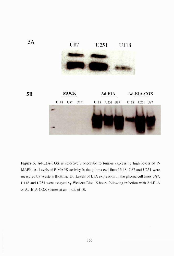
5A U87 U251 U118
5B MOCK Ad-EIA Ad-ElA-COX
Ul 18 U87 U251 U 118 U25I U87 U118 U251 U87
Figure 5. Ad-El A-COX is selectively oncolytic to tumors expressing high levels of P-
MAPK. A. Levels of P-MAPK activity in the glioma cell lines Ul 18, U87 and U251 were
measured by Western Blotting. B. Levels of El A expression in the glioma cell lines U87,
Ul 18 and U251 were assayed by Western Blot 15 hours following infection with Ad-El A
or Ad-El A-COX viruses at an m.o.i. of 10.
155

5C
120
C/5
O§ 100o073<D<D 5-i
4 -1100 0QOh
£0>N
00 Vh3O£3H
80
60
40
20
0
PBSAd-El A-COX
<Si-a<
PBS
<Ei73<
Ul 18 U87
Tumor Type
156
Ad-
El A
-CO
X

5C
CDNCOO§ 5D
60U 1 1 8 PBS
50
40 Ad-E1 A-COX30
(p<0.001)20
10Ad-E1A
o o 5 10 15 20 25 30 35 40 45
U 2 5 180 PBS70
60 Ad-E1 A50
Ad-E1 A-COX40
30
20
10
0 0 20 4010 30 50 60 70 80 90
DAYS
Figure 5C, 5D. U118 (C) or U251 (D) tumors were implanted subcutaneously in nude
mice (10 mice per group) and allowed to develop to size between 0.2-.04 cm. These
established tumors were injected directly with equal doses of Ad-EIA, Ad-ElA-COX
(108pfu) or PBS, in a total volume oflOOpl and tumor growth was followed with time. E.
The same experiments as described in C and D were carried out with the U 118 and U87
tumor lines. Experiments were terminated 60 days following virus injection when mean
tumor sizes in all groups were measured. Results of different treatment groups, over
different experiments, were expressed as a percentage of the mean size of the PBS injected
control groups.
157

Circulating Titre of Virus (pfu/ml)
i.v. Injection
(106 p.f.u.)Blood
(day 3)
Liver
(day 3) (day 6)
Mouse #1 10Ad E1A
Mouse #2 103 2X 10² -
Mouse #1 0 - -
Ad-E1 A-coxMouse #2 0 “
i.v. administration of 1 .0 X 10 6 pfu of different viruses.
158

6B
Expression of E1A Cannot Be Detected in Livers of Nude Mice Given i.v Injections of Ad-E1 A-COX
Mouse:
Ad-E1A Ad-E1 A-COX
1 2 1 2 +ve
Figure 6. Replication of Ad-ElA-COX cannot be detected following systemic
administration. A. Mice (2 per group) were injected i.v with Ad-EIA or Ad-ElA-COX
virus(106 pfu/mouse). Serum from these mice was recovered and plated in serial dilutions
onto 293 cells. Presence of virus in the blood was assessed as cytopathic effect on the 293
cells and titer of circulating virus determined. B. 3 days later, livers were recovered and
used for preparation of cDNA, which was subsequently screened by PCR for levels of
mRNA of El A.
159

7A% Mice Surviving Post IV injection of
different viruses
1 1 0 -1 0 0 -
90“80-70-60-50-40-30-20 -
10 -
- 1X10A9 wt
5X10A9 wt<0>E3V)
1X10A9 Ad-COX
5X10A9 Ad-OOX
0 1 2 3 4 5 6 7 8 9 10 11 12 13 14 15
Time (days)
Figure 7A. i.v dosing studies of Ad-ElA-COX and wt Ad-5 administered in C57BL/6J
mice. Data points, percent of mice surviving following tail vain injection with Ad-EIA-
COX or wt Ad-5, mice were euthanized immediately if they show signs of acute toxicides
such as anorexia or lethargy. Surviving mice were euthanized 14 days after virus injection.
160

Figure 7B. Liver histology in C57BL/6J mice following a single i.v. injection of Ad-
E1A-COX virus. Mice were injected with 5X10⁹ pfu of different viruses. Representative
photomicrographs of H&E-stained liver sections from mice died acutely (3-6 days after
injection). Arrow showing the immune infiltrates in the liver of the mice treated with Ad-
E1 A-COX virus.
161

7C
1400-
1200-
1000"
H 800--1< 600-
400-
200"
0
ALT
AVERAGE
PBS-CONTROLo
8WT-3D RV251-3D WT-6D RV251-6D WT-9D RV251-9D WT-14D RV251-14D
3 DAYS 6 DAYS 9 DAYS 14 DAYS
Days post i.v. administration
Figure 1C. Animals were treated with PBS of 5X10⁹pfu of the indicated virus. Serum was
collected for every three days interval for 14 days. Each data point represented individual
mice.
162

VIRU
S TI
TER
(pfu
)
7D
24h post injection
WT-A15
KV251
W T-A d5 RV251
Virus
7EC M1 M2 M3 M1 M2 M3
10000000- 1000000-j
« lOOOOtH
S ioooo-j
iKXXH
1 OH
IO—
48hpost Lv.
WT-.Ad5
RV251
WT Ad-5 Ad-El A-COX
WT-Ad5 RV251
Virus
10000000CE
10000000J
k•I 1000000J
g tOOOOOj
> 10000-
1000-
M1 M2 M3 M1 M2 M3
WT Ad-5 Ad-El A-COX
100-1
72hpost Lv.
WT-.A15
RV251
WT-Ad5 RV251
"Virus
C M1 M2 M3 M1 M2 M3
■ 0 “WT Ad-5 Ad-El A-COX
Figure 7D. Virus titer in liver in C57BL/6J following a single i.v. injection of Ad-El A-
COX (RV251) or wt Ad-5 virus. E. Southern blot analysis of mouse liver total DNA, 24,
48 and 72 hours following the administration of 5X10⁹pfu of Ad-ElA-COX or wt Ad-5
virus in C57BL/6J mice. Probing for the 8-kb fragment of adenovirus DNA. Each lane
represents individual mice.
163

Discussion:
In this chapter, we show that tumor cell selective stabilization of mRNA can be used as a
novel means to regulate viral gene expression and thus restricting viral replication in the
tumor cells. Ligation of the E1A gene to a 469bp region of the 3’UTR of the COX-2 gene
(Dixon et al., 2000) is sufficient enough to regulate adenoviral replication to tumor cells
expressing activated RAS oncoprotein or, more generally, increased P-MAPK activity.
Activated RAS and/or receptor tyrosin kinase signaling, resulting in elevated P-MAPK
activity, is characteristic of a very wide variety of human tumor types. The up-regulation of
COX-2 is a downstream effect of RAS-mediated transformation (Sheng et al., 2001; Sheng
et al., 1998b). Although RAS-mediated overexpression of COX-2 is also associated with
increased transcription of the COX-2 gene, a large component of its up-regulation is
mediated by selective stabilization of the mRNA of the COX-2 gene in RAS-transformed
cells (Dixon et al., 2000; Sheng et al., 2001). Given the clear association in the literature
between the proliferative response of cancer cells, P-MAPK activation and regulation of
gene expression through selective stabilization of mRNA, we hypothesized that we might
be able to use tumor cell selective stabilization of mRNA as a novel means to control
therapeutic gene expression in viral vectors for cancer gene therapy. We describe here the
construction of a conditionally replication competent adenoviral vector, in which
expression of the essential E1A gene is regulated by ligation to the 3’UTR of the COX-2
gene, allowing RAS/P-MAPK-specific stabilization of the mRNA. This is the first
description of a replicating adenovirus whose tumor selectivity is based upon control of
gene expression at the level of mRNA stability. This strategy has great potential for

expansion since there are many different genes whose 3’UTRs control selective mRNA
stability under different physiological, pathological and tumor-associated conditions.
The data presented in this chapter demonstrates that inserting the COX-2 3’UTR element
downstream of the El A gene in the adenoviral genome can restrict viral replication
selectively in the tumor cells. Using a panel of human tumor cell lines with different levels
of MAPK signaling, we are able to show that the Ad-ElA-COX virus is preferentially
oncolytic in vitro in human tumor cells with high levels of P-MAPK activity. In vivo, the
Ad-El A-COX virus is at least as effective oncolytically as the wild type virus in high P-
MAPK expressing tumors (U87 and U251), but generates no significant therapeutic effects
in low P-MAPK expressing tumors (U118). It is not yet clear exactly what levels of P-
MAPK activity are required for sufficient stabilization of the E1A-COX-2 mRNA to
support viral replication in order to achieve therapeutic efficacy. Such information will be
important in order to identify the types of cancer that can be targeted by the Ad-El A-COX
oncolytic vectors.
Although adenoviral replication in murine cells is greatly reduced as compared to human
cells, different murine models have been extensively used to evaluate the safety and
biodistribution of recombinant adenoviral vectors after systemic administration. Previous in
vivo toxicity studies have shown that the intravenous administration of adenoviral vectors
results mostly in hepatocyte transduction (Fechner et al., 1999). We first used the
immunocompromised nude murine model to study the toxicity and biodistribution of the
Ad-ElA-COX virus after systemic administration. Following intravenous injection of
adenovirus, virus could be detected in the blood of the animals receiving Ad-El A, but not
Ad-El A-COX virus. Moreover, the livers of injected mice expressed appreciable levels of
165

E1A from the Ad-EIA virus. In contrast, undetectable amount of E1A mRNA could be
found in the livers of mice that received similar doses of Ad-El A-COX. Given the
particular sensitivity of the liver as a potential site of toxicity following therapy with
adenoviral vectors, this data shows that the presence of the COX-2 3’UTR is sufficient to
reduce significantly the levels of El A expression in nude mice - liver tissue. This would be
expected to translate into significantly reduced levels of toxicity should such vectors
become disseminated through the circulation.
A majority of patients dying to cancer do so because of metastasis diseases. The treatment
of patients with systemic metastasis remains a difficult challenge. One of the goals for any
new cancer gene therapy strategy is to target systemic metastasis for more effective and
selective treatment against cancer. In the clinical setting, patient’s immune system is a
major obstacle for any systemic gene therapy. Host immune system eliminates most of the
therapeutic viral vectors after systemic administrations, which not only reduces the
therapeutic efficacy, but also is the main cause of systemic toxicity induced by the viral
vector. Both the systemic metastasis and the host immune system have proven to be the
major challenges for the development of successful gene therapy for cancer treatment. So,
the safety and therapeutic activity after the systemic administration of any new viral vector
for the cancer gene therapy needs to be tested very thoroughly. For this purpose, an
evaluation of the toxicity and biodistribution of the Ad-El A-COX virus were carried out in
the presence of the immune system by delivering the virus intravenously in the
immunocompetent C57/B6 mice. In this murine model, the dosing regimen of 2.5X10⁹ pfu
resulted in 50% lethality (LD50) for both the Ad-El A COX and the wt Ad-5 virus.
Although there was no difference in the LD50 value for both of the viruses, the mice
166

treated with the Ad-El A-COX virus started to die much earlier (5 days earlier) than the
mice treated with wt Ad-5 virus (Figure 7A). The mice that died acutely after systemic Ad-
El A-COX virus administration showed signs of hepatic toxicity including gross
pathological changes (liver tissue looked more yellow), histophathological changes and
elevated liver enzymes in the serum.
The reported LD50 for the conditionally replicating ONYX-015 in the
immunocompromised nude mice is about 5X10⁹ pfu (Heise et al., 1999a), which is two fold
higher than the LD50 of the Ad-El A-COX virus measured in the immunocompetent host.
The anti-vector host immuoresponse associated with the liver toxicity in nude mice is very
different and probably much less intense than the immunocompetent C57B/6 mice, which
is probably due to the fact that nude mice do not have all the necessary immune
components. After systemic administration of the Ad-ElA-COX and Ad-EIA viruses in
nude mice, we only measured the presence of the El A mRNA by RT-PCR in the liver
tissues and we were not able to detect any El A mRNA in the mice treated with Ad-El A-
COX virus (Figure 6A & 6B). These mice also show no signs of systemic toxicity. So, we
think that the discrepancy between the toxicity results in the immunocompromised and
immunocompetent murine model is mainly due to the difference in the immune system of
these two models.
Another possible way to explain the toxicity and the early onset of mortality by the
recombinant Ad-ElA-COX virus is the absence of the immunoregulatory gene E3 in the
viral vector. The adenoviral E3 gene products are the most important genes that suppress
the host immune response against the virus, which regulates many aspects of the host
immune system and creates an environment ideal for viral replication. Ad E3 gene products
167

such as gpl9K block the transport of MHC class I major from the ER to the plasma
membrane and downregulate CTL response. Another E3 gene product, the 10.4K/14.5K
protein, down-regulates death receptor and its ligand Fas and TRAIL by receptor
internalization and inhibits TNF-induced cytolysis and production of chemokines by
blocking NF-kB signal transduction. Most of the first generation adenoviral vectors are E3
deleted because E3 is not necessary for viral replication and also to create some space for
the transgene. So the early onset of the toxicity induced by the Ad-El A-COX virus may be
due to the absence of the E3 genes. In contrast, the control wt Ad-5 virus may be able to
suppress the early toxicity induced by the immune system because of the intact E3 gene in
the viral genome. So, it is probably not fair to compare the toxicity between these two
viruses due to the difference in the E3 gene. A proper way to evaluate the toxicity of the
AdElA-COX virus would be a virus with the same first generation viral backbone as
AdElA-COX virus, but without any COX-2 3’UTR.
We also cannot exclude the possibility that the Ad-El A-COX virus may replicate in some
specific organ non-selectively than the wt Ad-5 and may induce the toxicity in the
immunocompetant model. There are anatomical areas, such as the kidney, the central
nervous system and the seminal vesicles, where COX-2 expression is elevated compared to
other organs and may support Ad-El A-COX replication. But there was no sign of
replication for both viruses in major organs such as heart, lung and the kidney. Liver was
the only organ where we able to detect some non-specific Ad-El A-COX virus replication.
Mice treated with either of the viruses showed increased amounts of viral titer and viral
DNA measured in the liver. But the replication kinetics of these two viruses in the liver was
very different. Compared to wt As-5 virus, the Ad-El A-COX virus showed an increase in
168

viral DNA synthesis at 24 hours post injection (Figure 7E), but there was no difference
between the viral titer at this time point (Figure 7D). We do not know why at 24 hours
post injection, the recovered viral DNA was higher in the liver of the mice treated with Ad
El A-COX virus than the wild type Ad-5 virus or why this increased Ad-El A-COX viral
DNA in the liver do not correlate with the recovered viral titer at this time point. One
possibility may be that at 24h post administration, the increased viral DNA synthesis in the
liver of the mice treated with Ad-El A-COX virus is due to the higher replication of that
virus, but the titer is not changed because of the inability of this virus to control the host
innate immune response, which also may have cleaned up the vims and resulted in more
intense hepatotoxicity. Together, these data confirm that there is an early onset of liver
toxicity after systemic administration of recombinant Ad-El A-COX vims compared to wt
Ad-5 vims in the immunocompetent murine model.
Host immune response is a major obstacle to the systemic administration of recombinant
adenovims. Ad vector induced acute inflammation not only reduces the gene transfer
efficiency and vector persistence, but also causes profound damage to normal tissue and
significant morbidity in the transduced hosts (Raper et al., 2002). The specific mechanisms
underlying the acute immune response against the Ad viral vector are not well understood.
Both viral entry and the adenoviral gene products induce proinflammatory cytokines and
chemokines cascade that have harmful effects. During viral entry adenovims fiber proteins
interact with cellular Coxsackie adenovims receptor (CAR) (Bewley et al., 1999) and the
RGD motifs of the penton base interact with the a vP3 and the a vp5 integrins (Li et al.,
2001a). These interactions activate some key intercellular signaling pathways, which are
critical for viral endocytosis (Li et al., 1998) and facilitate viral particles transport to the
169
nucleus by altering the actin cytoskeleton (Suomalainen et al., 1999). But the host also uses
the vector activated intercellular signaling pathways as the alarm signals for viral infection
and induces innate immune responses, the first line of host defense against any viral
infection. The rapid and potent innate immune reactions induced by the adenoviral vectors
can have devastating consequences as seen in the clinical trial of gene therapy for ornithine
transcarbamylase (OTC) deficiency (Raper et al., 2002).
In vitro and in vivo studies have demonstrated that during cell entry, the adenovirus
activates the intercellular MAPK-signaling cascade for the efficient transport of viral
particles to the nucleus. The host cells also use the same signaling pathway to trigger early
inflammation and subsequent antiviral immunity. Within 10 min of Ad-LacZ transduction,
MAPK dependent IL-8 induction was observed in HeLa cells. Similar studies have showed
that the activation of p38 MAPK within minutes after adenovirus entry in epithelial cells
(Suomalainen et al., 1999). A chemical inhibitor against the p38 MAPK signaling pathway
blocked Ad vector induced chemokine IP-10 expression, providing a link between early
vector-induced signaling and proinflammatory gene expression (Tibbies et al., 2002).
MAPK signaling plays an important role in regulating different gene expressions that
supports both the viral life cycle and the host immune responses. Once activated, MAPKs
can directly activate transcription factors and transcriptional co-regulator via
phosphorylation or activate the downstream kinases that can regulate transcription and
mRNA stability. Crofford and Roessler showed that the adenovirus treatment of the liver
synoviocytes increased levels of COX-2 mRNA and protein (Crofford et al., 2005). Viral
gene expression was not required for the early COX-2 induction and inhibition of the p38
MAPK pathways to suppress the COX-2 expression.
170

During the systemic administration of the recombinant viral vector, the host cannot
distinguish between the infectious virus and the therapeutic virus and therefore inducing
potent immune responses. Also, the immune responses generated by the host against the
recombinant adenoviral vectors are more intense than the wild type virus infection because
of the deletion of E3 immunosuppressive genes. One possibility is that the MAPK
signaling cascade which is activated during the vector entry process through the interaction
with the host and the o^ps integrins also creates an environment to stabilize COX-2
mRNA as a part of the host immune response. Here, we used the COX-2 3’UTR to
selectively stabilize the mRNA of the adenoviral replicative essential El A gene in the RAS
transformed environment and thus controlled viral replication in the tumor cells.
Inflammatory signaling cascades like MAPK, that get activated during the viral entry
process can also induce inflammatory molecules like COX-2 or the adenoviral gene El A
that is fused with the COX-2 3’ UTR by stabilizing the mRNA via the AU-rich sequences
in the 3’UTR. This may lead to the nonspecific stabilization of E1A mRNA in any cells or
tissues that contain viral entry receptors and are capable of responding to viral infection. So
the enhanced toxicity observed by the Ad-El A-COX after the systemic administration in
the immunocompetent model may be due to an inflammatory environment induced by the
viral vector in the target organ such as liver, which then stabilizes El A mRNA, supporting
nonspecific virus replication. Also in the immunocompetent animal, the host immune
response is more intense due to the presence of all the necessary immune components,
which creates a much more favorable condition to stabilize the mRNA of the
proinflammatory genes like COX-2 or El A fused with the COX-2 3’UTR.
171

Many different genes with the AU-rich 3'UTR confer destabilizing activity on their cognate
mRNA but their actions are reversed under certain physiological conditions. These include
hypoxia responsive 3'UTR (Claffey et al., 1998) (Maity & Solomon, 2000), radiation
responsive elements and 3' UTR which increased mRNA stability in proliferating cells (Lee
et al., 1998; Maity et al., 1997). In this report, we were able to show that it is possible to
control transgene expression and viral vector replication by using tumor cell selective
mRNA stabilization via the COX-2 3’UTR. It will be important to choose these elements
thoughtfully in order to develop a conditionally replication viral vector. During
inflammation and cellular transformation, the expression of many inflammatory genes is
controlled at the level of their mRNA stability. These genes will probably not be the best
candidates for targeting tumor through their 3’ UTR.
To our knowledge, this is the first time that conditionality of adenoviral replication has
been conferred solely at the post-transcriptional level of control of El A expression.
However, based on previous experience with designing tumor cell specific vectors where
initially unpredicted results can surface upon broader testing, it seems likely that there will
be apparently suitable tumors (i.e. mutated RAS or elevated P-MAPK activity) in which the
level of control conferred by the 3’UTR is insufficient to allow completely controlled
levels of replication. Additionally, there are likely to be anatomical sites, physiological
conditions and inflammatory environments which are amenable to stabilization of the
COX-2 3’UTR region (Cao & Prescott, 2002), thereby allowing viral replication at extra-
tumoral sites where COX-2 is normally induced (Turini & DuBois, 2002). Therefore, we
envisage that the 3’UTR strategy will be most effective in the context of a mosaics of
regulatory elements combined to confer multiple levels of specificity to the resultant virus.
172

Other targeting strategies such as transcriptional regulation of the El A gene can very
readily be combined with the COX-2 3’UTR (indeed the COX-2 promoter has itself been
used to target recombinant vectors (Cao & Prescott, 2002)), as can incorporation of
molecular features that target tumor cell specific mutations, such as loss of p53 or
downstream effectors. Thus, multi-component targeting will be most effective at
generating truly tumor selective vectors, which can be delivered to extra-tumoral sites
without allowing unacceptable levels of viral replication. Based on our studies here, we
propose that mRNA (de-) stabilizing elements be considered as one of the component
cassettes of such mosaically regulated viruses.
The data presented in this chapter from part of the following paper:
Ahmed A, Thompson J, Emiliusen L, Murphy S, Beauchamp DR, Suzuki K, Alemany R,
Harrington K and Richard G. Vile. A Conditionally Replicating Adenovirus Targeted to
Tumor Cells Through Activated RAS/P- MAPK-Selective mRNA Stabilization. Nature
Biotech; 2003 Jul; 21(7): 771-7.
173

Chapter 5
Retargeted Intratumoral Expression of a Fusogenic Membrane
Glycoprotein by Tumor Cell Selective mRNA Stabilization
Enhances the Efficacy of Replicating Adenovirus Therapy.
174

5.1 Introduction
This chapter describes a novel approach to enhance and retarget the oncolytic potency of
adenovirus vector by combining the ability of the COX-2 3’UTR to stabilize mRNA and
regulate gene expression specifically in cells with elevated levels of activated Ras/MAPK
with a viral Fusogenic Membrane Glycoproteins (FMG), which induces tumor cell killing
through induction of fusion of tumor cells to form large multinucleated syncytia (Fielding
et al., 2000) (Bateman et al., 2000b). In the previous two chapters, we show that the 469 bp
COX-2 3’UTR is able to regulate the expression of a viral gene, thus controlling viral
replication and oncolysis. Here we demonstrate that the FMG mediated fusion and tumor
cell killing can also be control by the COX-2 3’UTR element and this FMG-induced tumor
selective syncytia is able to enhance the oncolytic potency of replication competent
adenoviral vector both in vitro and in vivo.
The use of conditionally replication-competent viruses for the treatment of cancer has been
more prominently considered due to the problems associated with poor intratumoral spread
and low transduction efficiency in vivo (Vile et al., 2002). In order to achieve clinically
relevant therapeutic efficacy, it is essential to improve spread of the therapeutic virus
within tumors where other cell types, cell matrix, and areas of necrosis exist and viral
vector tends to be trapped in between the sub-cellular compartment (Sauthoff et al., 2003).
Many different strategies used to increase efficacy of adenovirus include engineering viral
genes to enhance virus release and spread, and aiming viruses with additional therapeutic
genes that might either enhance viral cytotoxicity and/or provide additional therapeutic
benefits (Ramachandra et al., 2001) (Heise et al., 2000). However, poor intratumoral spread
175

remains an important hurdles that need to be overcome before replicating adenoviral vector
can be used efficiently even against the localized tumor burdens (Vile et al., 2002).
Previously, several laboratories including ours have shown that tumor cell killing can be
obtained by gene transfer of the FMG and the FMG-induced tumor cell killing also create a
immunostimulatory environment, which can enhance the therapeutic efficacy (Bateman et
al., 2000b) (Higuchi et al., 2000). Moreover, a recent report was able to demonstrate that
the HIV gpl20 FMG induced fusion facilitates the spread of adenoviral vectors through a
monolayer of tumor cells and increases viral release from the infected cells (Li et al.,
2001b). Based on these reports, we therefore explored the use of FMG in combination with
the COX-2 3’UTR to selectively enhance the therapeutic efficacy of the oncolytic
adenoviral vector. The data presented in this chapter demonstrates that by using the COX-2
3’UTR mediated, FMG-induced retargeted tumor cell fusion in tandem with the oncolytic
properties of replicating adenovirus enhance the therapeutic efficacy of both approaches
synergistically by increasing the release of viral particles from infected cells that are fused
by FMG and/or the spread of viral particles through the tumor via the syncytia.
5.2 Results
5.2.1 Co-transduction of human tumor cells with sub-therapeutic doses of plasmid
DNA expressing an FMG and replicating adenovirus leads to extensive tumor cell
killing in vitro
Transfection of 106 confluent tumor cells from various different lines (Mel624 or Mel888,
human melanoma; LnCap human prostate carcinoma; U118 or U251 glioma) in vitro with
0.5pg of plamsid DNA expressing the hyperfusogenic Gibbon Ape Leukemia Virus
176

(GALV) FMG (Fielding et al., 2000) (Bateman et al., 2000a) (pCR3.1-GALV) routinely
led to over 90% of the cells being killed 96 hours following transfection. Similarly,
infection of these cell lines with wild type adenovirus serotype 5 (Ad5-Wt) at an m.o.i. of
0.1 led to cytopathicity of over 95% of cells (Figure 1A). To study the interaction of FMG
expression with Ad5-Wt infection, we selected treatment doses (0.01 |ig plasmid DNA and
0.001 m.o.i of Ad5-Wt) at which less than 10% of the tumor cell cultures were killed by
either treatment alone (Figure IB). However, co-transduction with these suboptimal doses
of both pCR3.1-GALV (0.01 pig) and Ad5-Wt (m.o.i. 0.001) induced significantly increased
cytotoxicity compared to either treatment alone (>95% cell killing) (Figure IB).
5.2.2 Suboptimal doses of plasmid FMG DNA and replicating adenovirus co-operate
to eradicate small-established tumors.
To investigate whether these in vitro results also have relevance to in vivo virotherapy, we
first used direct intratumoral injection of suboptimal doses of Ad5-Wt or pCR3.1-GALV
into very small established (0.2cm diameter) human tumor xenografts. Three intratumoral
injections of low doses of either pCR3.1-GALV (0.5|ig/injection) or Ad5-Wt virus at a low
dose (3xl07 pfu/injection) had no significant effect on tumor cure (Figure 3). However,
simultaneous co-transduction of tumors with replicating adenovirus stocks and syncytia-
inducing GALV plasmid DNA led to regression of all tumors under these conditions
(Figure 3) such that, at the end of the experiments shown in Figure 3 A and B, 10 of 10
mice were tumor free in the (pGALV + Ad5-Wt) treatment groups.
We confirmed that the therapeutic effects of pCR3.1-GALV and Ad5-Wt co-transduction
were dependent upon expression of the GALV FMG by including an additional treatment
group in the experiments of Figure 3. In previous two chapter, we have reported that gene
177

expression can be effectively targeted to cells expressing an activated mutant RAS
oncogene and/or high levels of P-MAPK by ligation of a therapeutic gene to the
3’Untranslated Region of the Cyclooxygenase 2 gene (COX2-3’UTR). The COX2-3’UTR
efficiently destabilizes mRNA molecules in cells with low P-MAPK activity but selectively
restabilises the mRNA molecules and, therefore, protein expression in cells with high P-
MAPK activity. We constructed an expression plasmid, pCR3.1-GALV-COX (Figure 2)
in which 469bp of the COX-2 3’UTR destabilizes expression of the GALV FMG in low P-
MAPK expressing cells (such as the U118 glioma) but selectively restabilises expression in
high P-MAPK expressing cells (such as U251). Cotransduction of small established U251
tumor xenografts with Ad5-Wt and pCR3.1-GALV-COX was as effective therapeutically
as treatment with Ad5-Wt and pCR3.1-GALV (10 of 10 mice tumor free at the end of the
experiment in Figure 3B); however, treatment of the low P-MAPK tumor U118 with Ad5-
Wt and pCR3.1-GALV-COX, in which GALV expression is significantly reduced by
COX-2 3’UTR-mediated destabilization of the mRNA, was no more effective than
treatment with Ad5-Wt alone (Figure 3A). These results confirm that GALV expression
can be targeted to tumor cells overexpressing P-MAPK and indicate that the therapeutic
effects observed with co-transduction of tumors with Ad5-Wt and GALV expressing
plasmids are dependent upon expression of the FMG. To confirm the in vitro findings of
enhanced viral spread through co-expression of the pGALV vector, the experiment of
Figure 3A was repeated except that the tumors were resected two days following injection.
Tumor cells were dissociated in vitro and analyzed by FACS for expression of the viral
E1A protein. As can be seen from Figure 3C, even at early times after transduction in
vivo, significantly enhanced numbers of infected cells were seen in tumors co-transduced
178

with replicating virus and pGALV (-8% of the dissociated cells) compared to the virus
alone (-1%).
5.2.3 Syncytial formation enhances spread of adenoviral vector through a monloayer
The increased potency of GALV transfection in combination with infection by replicating
virus might be explained simply by enhanced transfection efficiency of tumor cells in the
presence of lysosomal disrupting adenoviral infection. To address this possibility, we
measured GFP expression 48 hours following transfection in cultures transfected with
pCR3.1-GFP plasmid DNA (0.01 jig) in the presence or absence of co-infecting Ad5-Wt
(moi 0.001). Although results differed slightly for each cell line, the presence of Ad5-Wt
(at these low m.o.i) increased the transfection efficiency with plasmid DNA by, at most,
about two fold (typically increasing the number of GFP positive cells from 4% to 6-7% in
the case of Mel624 cells) (Figure 4A). However, transfection with at least 50 fold more
plasmid DNA was required to generate levels of cytotoxicity by GALV alone comparable
to those seen in the combination of low dose pCR3.1-GALV and Ad5-Wt (Figure 1A)
suggesting that enhanced transfection efficiency alone was not sufficient to explain the co
operation between GALV transfection and Ad5-Wt infection.
It has recently been reported that syncytial formation enhances the dispersion of adenoviral
particles through a monolayer in vitro (Li et al., 2001b). Therefore, we examined the ability
of a replicating adenovirus to mobilize a replication-incompetent Ad-GFP vector from
human tumor cells into murine B16 melanoma cells in co-culture. Human tumor cells are
highly susceptible to GALV-mediated cell fusion but B16 murine melanoma cells do not
express the receptor for GALV and so will not fuse to GALV-expressing cells. In the
experiment described in Figure 4B, the only way that the GFP reporter gene would
179

transduce B16 cells in the mixed cultures would be through mobilization of Ad-GFP from
the human tumor cells by the replication competent Ad5-Wt virus. The number of GFP-
transduced B16 cells at the end of the experiment was assessed by FACS analysis. The
presence of ongoing cell fusion within cultures of human tumor cells significantly
enhanced mobilization of Ad-GFP for infection of bystander B16 cells when replicating
Ad5-Wt was present (Figure 4B).
5.2.4 Syncytial cultures produce increased levels of viral titer.
The increased mobilization of the Ad-GFP vector (Figure 4B) in the presence of syncytia
may be due to an increase in the viral titer released on a per cell basis, an increase in the
dispersal of the same amount of virus through the culture (Li et al., 2001b), or both. The
titer of cell free virus released into the supernatants from the cultures of Figure 4B were
significantly higher in the presence of syncytia relative to the same number of cells in non
fusing cultures (p<0.03 for U87 cells and p<0.01 for U118 cells) (Table 1). The difference
in the amount of cell-associated virus (recovered from the cell pellets) was less marked
between fusing and non-fusing cells but was still significantly enhanced from fusing cells
(p<0.05 for U87 cells and pc.0.01 for U118 cells) (Table 1). The increase in cell free virus
may in part be due to the increased lysis of the human tumor cells by FMG-mediated cell
killing and release of cell associated virus. These data show that presence of fusing cells
increases both the total amount of virus produced and increases the amount of virus
released into the culture supernatant, which at least partly will explain the more efficient
spread of adenovirus through the culture (Figure 4B) (Li et al., 2001b).
180
5.2.5 Increased titer associated with syncytia occurs through posttranscriptional
upregulation of E l A expression.
RtPCR analysis indicated that the increased viral titers described in Table 1 were not due
to greater levels of transcription of the viral E1A gene within Ad5-Wt-infected, fusing
human tumor cell cultures (Figure 5A), data confirmed by Northern blotting (not shown).
In contrast, levels of El A protein were significantly enhanced (up to 10 fold depending
upon the experiment) within fusing, as opposed to non-fusing, tumor cell cultures (Figure
5B). These data are consistent with our previous observations (Higuchi et al., 2000), as
well as those of others (Mi et al., 2000) that as syncytia develop they become highly
metabolically active (Higuchi et al., 2000) and generate high levels of protein production
compared to the same number of non-fused cells.
199

Num
ber
of Ce
lls
Surv
ivin
g (96
hr
s)
Figure 1A
2.00e+7
Mock
1.00e+7
0.00e+0
pCR3.1-GALV - 0.5 pg
Ad5-W t moi 0.1 pCR3.1-GALV
+ Ad5-Wt
Treatment
Figure IB
2.00e+7 i
vOON
0)UStf)
. £
’ >Ih300
<DJCE3
,00e+7
0.00e+0
MockpCR3.1-GALV
0.01 (Ag
Ad5-Wtmoi 0.001
l i i i i i i
pCR3.1 -GALV+ Ad5-Wt
200
Treatment

Figure 1. A. 106 confluent tumor cells (Mel624 shown here) were either transfected using
the Effectene reagent in vitro with 0.5pg of plamsid DNA expressing the GALV FMG
(pCR3.1-GALV), were infected with wild type adenovirus serotype 5 (Ad5-Wt) at an m.o.i.
of 0.1 or were transfected with 0.5pg plasmid DNA admixed with Ad5-Wt (pCR3.1 + Ad5-
Wt). 96 hours later, surviving cells were counted. B. The same experiment was
performed using 0.01 pg of plasmid DNA, 0.001 moi of Ad5-Wt or both admixed. With
the low dose of plasmid DNA, individual pockets of syncytia were formed but these were
too infrequent to cause wide scale fusion of the entire population of cells and were
eventually overwhelmed by proliferating tumor cells.
201

Fieure 2
CMV GALV polyA]—I pCR3.1-GALV
CMV GALV> -c
Cox-23’UTR polyA- c z m H U
pCR3.1-GALV-COX
Cox2 3’UTR
469 bp
8047 8516
Genomic PCR
Figure 2. Construction of the CMV-GALV-COX construct.
202

Fieure 3A
U118
C-3O
C/5
100
80
60
40
20
pGALV
pGALV-COX + Ad:>
Ad5-Wt + pGALV
Ad5-Wt
20 30 40
Days Following Intratumoral Injection
50 60
Fieure 3B
100U251
pGALV + Ad5-W t
tpG A LV -C O X + A d5-W t
60PBS
a 40 >|
GO
20 pGALV
Ad5-W t
10 20 30 40
Days Following Intratumoral Injection
50 60
203

Figure 3. Co-administration of plasmid GALV and Ad5-Wt virus can cure small
palpable tumors. 5xl06 U118 (A) or U251 (B) tumor cells were injected subcutaneously
in athymic nude mice. When tumors were palpable (~0.2cm diameter) different groups (10
mice per group) were injected with PBS (PBS); or with low dose pCR3.1-GALV
(0.5pg/injection) plasmid DNA (pGALV); or with Ad5-Wt virus (3xl07 pfu/injection)
(Ad5-Wt); or with pCR3.1-GALV (0.5pg/injection) + Ad5-Wt virus (3xl07 pfu/injection)
(pGALV+Ad5-Wt). Another group of tumors was also injected with 0.5pg of the plasmid
pCR3.1 -GALV-COX (Figure 2) along with the Ad5-Wt (pGALV-COX + Ad5-Wt) in
which 469 bp of the COX-2 3’UTR is ligated downstream of the GALV cDNA (see text).
Tumor growth was monitored for a further 62 days. Animals were sacrificed when tumor
size reached 1.0cm in any diameter.
204

Figure 3C
E l A-FITC
o?cmTS
Untreated Negative Positive
0 %
j f s iCO
0 . 5 9 % c f 1£ 2 " CO
* ° ]
5 ) N
= F 2 1
| TTT M M I I * » f
e ! ° i
o o - " T T i ■ i | i i 1 1 | > 1 1 1 | . i . I I
i o 1
O o -
r \ ■ •
Forward Scatter1000
Forward Scatter
50.21%
Forward Scatter
GALV
0.44%
1000Forward Scatter
Ad5-W t
1.23%
Forward Scatter
GALV+Ad5-W t
8.44%
1000Forward Scatter
Figure 3C. The experiment of 3 A and B. above was repeated except that the tumors were
resected two days following injection with pGALV (GALV), replicating virus (Ad5-wt) or
pGALV and Ad5-wt (GALV + Ad5-wt). Tumor cells were dissociated in vitro and
analyzed by FACS for expression of the viral El a protein using a rabbit polyclonal
antibody against E la (Santa Cruz Biotech. Santa Cruz, CA) and a FTTC labeled anti rabbit
IgG. As controls for the FACS analysis, uninfected, cultured U251 cells were treated with
no secondary antibody (Untreated) or with both primary and secondary antibodies
(Negative). The positive control was U251 cells infected with wild type Ad virus 24 hours
previously (Positive).
205

Figure 4Atk 1008.001
MOCK1.14%
-®CM
400 600Forward S c a tte r
tk 1008.003
Ad5-W t 0.44%
1000
tk 1008.002
400 600 800Forward Scatter
1000
CM V-GFP4.07%
400Forward S catter
tk1008.004
CM V-GFP+Ad5-W t6.26%
400 600Forward S ca tte r
1000
Figure 4B
30
a• i 20
a.xW
u'OCQ
io
Ad-GFP + Ad5-Wt + GALV
Ad-GFP
Ad-GFF^ - ‘ v s ' ; . •- .
Ad5
Ad-GFP +GALV
Treatment

Figure 4. Co-transduction of human tumor cells with a fusogenic membrane
glycoprotein and replicating adenovirus increases the spread of virus through the
culture. A. GFP expression was measured 48 hours following transfection of Mel624
cultures with pCR3.1-GFP plasmid DNA (0.01 pg) (CMV-GFP) in the presence or absence
of co-infecting Ad5-Wt (moi 0.001) (Ad5-Wt). B. 106 cells were transfected with pCR3.1-
GALV plasmid DNA (0.01 pg) to induce syncytia formation or with or pCR3.1-empty as a
control and/or were infected with a mixture of replicating adenoviral stocks (m.o.i. of
0.001) mixed with replication defective Ad-GFP (m.o.i. 0.1). 24 hours later, cultures were
washed 3x in PBS and then 5xl04 B16 cells, pre-labeled with Cell Tracker Orange dye,
were added to the cultures. Since B16 cells will not be incorporated into syncytia, the only
way that the GFP reporter gene would transduce these cells would be through mobilization
of Ad-GFP from the human tumor cells by the replication competent Ad5-Wt. The number
of GFP transduced B16 cells at the end of the experiment (4 days later) was assessed by
FACS for double stained (Orange and Green) cells. Similar results were also seen with
U118 and U251 cells.
207

Table 1(Virus Recovered p.f.u/ml on 293 cells)
Original Transduction Treatment
GALV + Ad5-Wt + AdGFP
GALV + — + AdGFP
MOCK + Ad5-Wt + AdGFP
MOCK + —- + AdGFP
Cell Free Cell Associated
U87 U118 U87
9 x 10⁴ 2X 10⁵
1 x 10⁶ 7 x 10⁵l x l O 3 3 x 10³
Table. Syncytial formation is associated with increased viral production. 106 U87 (high
level of activated MAPK) or U118 (low level of activated MAPK) cells were transfected
with pCR3.1-GALV plasmid DNA (0.01 pig) to induce syncytia formation or with or
pCR3.1-empty as a control and/or were infected with a mixture of replicating adenoviral
stocks (m.o.i. of 0.001) mixed with replication defective Ad-GFP (m.o.i. 0.1), cell culture
supernatants were recovered and cells were trypsinised, washed in PBS and lysed by three
cycles of freeze thawing. Viral titers from the supernatants (Cell Free) and from the cell
pellets (Cell Associated) were then determined by a plaque assay on 293 cells as described
in material and methods.
208

Fieure 5A
GAPDH
E1A
Fieure 5B
+ - Loading control
209

Figure 5. Increased viral titer and spread in syncytial cultures is associated with
elevated levels of E l A protein but not mRNA. 106 confluent Mel624 tumor cells were
either transfected using the Effectene reagent in vitro with 0.01 jig of plamsid DNA
expressing the GALV FMG (pCR3.1-GALV) (lane 1), were infected with wild type
adenovirus serotype 5 (Ad5-Wt) at an m.o.i. of 0.001 (lane 2) or were transfected with
0.01 ng plasmid DNA admixed with Ad5-Wt (pCR3.1 + Ad5-Wt) (lane 3). 48 hours later
RNA was prepared from the cultures and used A. for production of cDNA. Primers
specific for E1A (5’ -TTCCTCAAGAGGCCACTCTTG-3 ’ and 5’-
CACGCCATGCAAGTTAAACA-3’) were used to assess the amount of El A message
present in the cultures. Equal loading of samples was assessed by riper for GAPDH. B. In
addition, protein was prepared from cell lysates and Western Blot analysis used to
determine the relative levels of expressed El A protein. Equal loading of samples was
confirmed as shown in the upper panel.
210

5.4 Discussion
Oncolytic adenoviruses are a promising new concept for the treatment of cancer. One of the
most attractive advantage of replicating oncolytic viruses is that a single virus can
selectively amplify several thousand-fold within a tumor cell and the progeny viruses then
have the capacity to spread and infect uninfected tumor cells, thus enhancing the
therapeutic effects. But the early data from both of the clinical trials (Kim et al., 2001) and
animal models (Sauthoff et al., 2003) indicated that barriers within the tumor prevent viral
spread, which is a major obstacle for achieving clinically relevant therapeutic efficacy. In
this chapter, we described a novel approach to enhance the in vivo efficacy of replicating
adenovirus therapy by combining a fusogenic viral glycoprotein-induced tumor cell fusion
and also showed that the viral therapy can be retargeted by expressing FMG selectively in
tumor cells with elevated level of Ras/MAPK via the COX-2 3’UTR element.
Our in vitro studies show that the FMG-induced fusion significantly enhances the oncolytic
activity of replicating adenovirus. By ligating the GALV FMG with the COX-2 3’ UTR,
we were able to regulate GALV-induced tumor cell fusion selectively in cells that have
activated Ras/MAPK signaling cascade. In vivo, we demonstrated that the efficacy of
intratumoral injection of replicating adenovims is greatly enhanced by the co-injection of
an FMG-encoding plasmid. This combinatorial approach was sufficient to eradicate very
small-established tumors at viral doses where injection of the vims, or plasmid alone, was
not effective. And, the selective expression of GALV by the COX-2 3’UTR element only
in tumor with elevated level of activated Ras/MAPK was sufficient enough to regulate this
combined therapeutic approach.
211

The enhanced therapeutic effects that observed in this combination therapy were not due to
increased destruction of the stroma between tumor cells which might act in patient tumors
to impede virus spread, because the stroma in these experiments are of murine origin which
do not express fusion competent receptors for the GALV FMG. These data suggest that
replicating adenoviruses expressing the GALV FMG will be significantly more potent than
viruses without FMG. In addition, we have shown that targeting of GALV expression to
tumor cells is possible at posttranscriptional levels as demonstrated in this work. This
strategies could be combined with pre-existing conditionally replicating adenoviruses
which have tumor targeting already built in either at the level of transcriptional regulation
using selective promoters driving essential replicative (Rodriguez et al., 1997) (Hallenbeck
et al., 1999) or therapeutic genes (Freytag et al., 2002) or at the genetic level using viral
mutants with tumor cell selective replicating properties (Alemany et al., 2000).
The results from the in vitro studies to understand the interaction between FMG and
adenovirus replication show that the FMG-mediated syncytial formation significantly
enhances the efficacy of replicating adenoviral therapy through a combination of
mechanisms. One of these is a moderate increase in the transfectability of cells with the
GALV plasmid in the presence of co-infecting adenoviruses. This effect will boost the
cytotoxicity associated with the FMG-mediated component of the therapy (Bateman, 2002)
(Diaz et al., 2000). However, quantitatively more significant, the levels of tumor cell that
expressed the viral El A protein were markedly increased in the presence of FMG-mediated
cell-cell fusion. Elevated cellular levels of E1A were associated with significantly
increased total viral titers (total virus produced from infected cells) as well as greater viral
release from cultures of infected syncytial cultures compared to normal non-fusing
212

monolayer. Finally, as a result of all of these effects, and consistent with report using the
HIV gpl20 FMG (Li et al., 2001b), syncytia also promoted enhanced spread of adenoviral
particles through the tumor cell cultures. Beside the overall increase in viral titer, syncytial
formation also increased release of adenoviral particles. This is most probably due in part at
least to lysis of infected cells thereby releasing intracellular particles, which would
otherwise remain cell associated. Therefore, the increase level of viral production in
combination with the accelerated release of viral particles through tumor cell cultures
would suggest that combination therapy with FMG expression and replicating adenovirus
infection may overcome some of the problems associated with the use of replicating
adenovirotherapy, which have been seen to date.
213

Chapter 6
A Cell Cycle Dependent Conditionally Replicating Adenoviral
Vector for Cancer Gene Therapy.
214

6.1 Introduction:
We previously showed that successful construction of a conditionally replicating
adenovirus to target the RAS-MAPK pathway in the cell can be achieved by the
cyclooxygenase-2 3’-UTR. In order to expand our hypothesis and prove that the 3’UTR
element from other tumor associated genes can also be used to target tumor cells, in the
studies presented in this chapter we used the 318 bp 3’UTR element from the DNA
methyltransferasel gene (DNMT1) and demonstrated in vitro that genes can be selectively
expressed in rapidly dividing tumor cells. Also, by introducing the DNMT1 3’-UTR in
between the adenoviral El A coding sequence and the poly A signal, El A expression, and
subsequently viral replication, becomes restricted to the tumor cells derived from multiple
tissue types.
DNA methyltransferasel is a major enzyme that is responsible for maintaining the
methylation pattern from the parent cell into the daughter cell during cell division. The
expression of DNMT1 is tightly coordinated with the DNA replication (Araujo et al., 1999)
during cell division (Szyf et al., 1985). Several lines of evidences link DNMT1 with the
tumorigenic process. First, increased levels of DNMT1 mRNA, protein and activity were
observed in many different types of cancer. Second, overexpression of DNMT1 in the
mouse fibroblast NIH3T3 cells caused cellular transformation (Wu et al., 1993). Also the
DNMT1 promoter regions contain multiple activator protein 1 (AP-1) elements, which can
be transcriptionally activated by proto-oncogene and mitogenic signals (Girault et al.,
2003). And finally, downregulation of DNMT1 expression by RNA interference, antisense
or pharmacological inhibitor induced demethylation of DNA and re-expression of various
tumor suppressor genes (Robert et al., 2003), which lead to inhibition of DNA replication
215

(Knox et al., 2000) and cell growth (Laird et al., 1995). Thus, DNMT1 has been suggested
to play an important role in cancer development.
DNMT1 mRNA levels are significantly downregulated during the Go/ Gi phase of the cell
cycle, but the abundance of DNMT1 mRNA is dramatically increased after cells enter into
the S phase (Robertson et al., 2000; Szyf et al., 1991). The 3’-untranslated region (3’-UTR)
of the DNMT1 mRNA plays an important role in the cell cycle-dependent regulation of
DNMT1 expression. A 54-nucleotide highly conserved AU-rich element (ARE) within the
DNMT1 mRNA is responsible for orchestrating cell cycle-dependent DNMT1 expression
by destabilizing its own mRNA in the quiescent cells {Detich, 2001 #3867}. 40 kd protein
(p40) interacting with this conserved region of DNMT1 3’UTR may be involved in
destabilizing DNMT1 mRNA (Detich et al., 2001). As the cell enters into S phase, the
binding of p40 to the 54-nucleotide element is significantly reduced by some unknown
mechanisms and the mRNA is restabilized. Because tumor tissues are presumed to contain
a greater proportion of dividing cells than their normal counterparts and increased DNMT1
expression is observed in many different tumor samples, targeting cancer via tumor cell
specific mRNA stabilization by the DNMT1 3’-UTR could be a viable strategy.
In this chapter we are able to show that DNMT 3’UTR element can be used to regulate
transgenes expression selectively in various tumor cells. By inserting this element in the
adenoviral genome we were able to control El A expression and viral replication in vitro.
We were also able to demonstrate that DNMT1 3’UTR-mediated regulation of adenoviral
replication was correlated with cell cycle dependent DNMT1 protein expression in the
A549 cancer cell.
216

6.2 Results:
6.2.1 DNMT 3’UTR is active in tumor cells
To evaluate tumor cell specific activity of DNMT 3’UTR, a transient transfection of
luciferase reporter assay was performed. The luciferase activity of the positive control
plasmid with the SV40 enhancer/promoter (Fig. 1A) in each cell line was considered as
100%. As shown in Fig. IB, the DNMT 3’UTR containing pGL3 construct showed similar
luciferase reporter activity compared to the control vector in most of the tumor cell lines
tested. In contrast, luciferase activity was significantly inhibited (10-12% of control
vector) in the primary human retina pigment epithelial cells hTERT-RPEl and IMR90.
The other primary cell line used in this experiment was BEAS-2B, which is a human
bronchial epithelial cell immortalized by SV-40 large T antigen, and showed similar
luciferase reporter activity to control plasmid in the presence of DNMT1 3’-UTR. It has
been reported previously that SV-40 large T antigen transformed cells overexpressed
DNMT-1 during cellular transformation (Chuang et al., 1997) by stabilizing the DNMT-1
mRNA at the posttranscriptional level (Slack et al., 1999). So, SV-40 large T antigen may
also stabilized DNMT1 massages in the BEAS-2B cell and responsible for the increased
luciferase activity. Western blot analysis of DNMT1 expression in the various tumor and
primary cell lines revealed that the DNMT1 expression is significantly elevated in most of
the tumor cell lines tested compared to primary cell lines except BEAS-2B cells.
217

6.2.2 The DNMT1 3’-UTR can selectively down-regulate the stable luciferse mRNA in
primary cells but not in tumor cells
To validate the hypothesis that the cis-acting sequence within the DNMT1 3’-UTR can
stabilize the mRNA levels in rapidly dividing tumor cells, but destabilize the same mRNA
in slow growing primary cells, we performed a Northern blot for luciferase mRNA,
extracted from transiently transfected human primary cells (hTRE) and tumor cells
(HCT116). In the presence of the DNMT1 3’-UTR, steady-state reporter luciferase mRNA
expression was significantly inhibited only in the primary cells (Figure 1C). In contrast, in
tumor cells the DNMT1 3’-UTR did not alter the luciferase mRNA expression. These
finding suggested that the DNMT1 3’-UTR possesses a potent inhibitory effects on the
reporter mRNA expression only in primary cells, but not in tumor cells.
6.2.3 Construction of AdDNMT virus
Therefore, we constructed AdDNMT, a conditionally replicating adenovirus where the
replicative essential viral gene El A was ligated to the DNMT 1-3’UTR using a PCR
strategy. Figure 2 depicts the genome of the recombinant adenoviruses. The viral DNA was
isolated by Hirt extraction and the modified sequences were confirmed by PCR (data not
shown).
6.2.4 AdDNMT induced tumor-specific cytotoxicity
To evaluate the cytolytic activity of the AdDNMT in the context of oncolytic adenovirus, a
human colorectal cell line HCT116, and the primary cell line hTRE were infected with
AdDNMT or wild type Ad-5 (wt Ad-5) at an MOI of 1. After 7 days, cells were examined
for CPE. Figure 4A depicts representative results of selective oncolysis of the AdDNMT
virus. As expected, wt Ad-5 virus induced cytotoxicity in both tumor and normal cells
218

without any specificity. In contrast, the AdDNMT infection induced selective oncolysis in
the HCT116 tumor cell line, but no significant oncolysis was observed on the normal hTRE
cell line. The morphology of AdDNMT-infected hTRE normal cells appeared to be very
similar to the uninfected cells (Figure 4A).
To further monitor the oncolytic activity of Ad-DNMT by the quantitative measurement of
the cell viability, MTT assays were performed exposing a panel of human primary (hTRE,
hEPC, HUVAC and IMR90) and tumor cell lines (HCT116, Mel 88, Mel 624, huh7 and
U251) to AdDNMT or wt Ad-5 virus at MOI of 1. In most of the tumor cell lines tested, the
oncolytic activity of the AdDNMT virus was as efficient as wt Ad-5 virus, with some
differences in the kinetics of cell killing between different cell lines (Fig. 3B and 4B).
While greater than 90% of HCT116 and U251 tumor cells were killed within 6-8 days,
longer times were required with the Mel 888 and huh-7 cell lines to observe comparable
levels of cytolysis. But in all the tumor cell lines, AdDNMT and wt Ad-5 virus showed
very similar kinetics of oncolysis. In contrast, in most of the human primary cell lines,
AdDNMT showed some toxicity at the earlier time point, but in the later time point
AdDNMT induced oncolysis was significantly reduced compared to wt Ad-5 virus. The
cytotoxicity observed by AdDNMT infection at the earlier time points in some primary cell
lines could be due to the viral particle induced toxicity rather than oncolysis. These
findings confirmed that AdDNMT mediates selective oncolysis in tumor cells, but the
oncolysis is significantly reduced in normal cells.
6.2.5 AdDNMT mediated tumor cell-specific E1A expression
To ensure the specificity of DNMT1 3’-UTR, human tumor (HCT116) and primary (hTRE)
cells were infected with AdDNMT at an MOI of 10. After 48h of incubation, the
219

expression of El A protein was evaluated by Western blot. As shown in figure 3C, the
expression of E1A in AdDNMT infected HCT116 cells was detectable but lower than wt
Ad-5 infected cells. In contrast, E1A expression was completely undetectable in AdDNMT
infected primary hTRE cells, thus demonstrating the specificity of DNMT 3’-UTR (Figure
3C).
6.2.6 Cell cycle dependent DNMT1 expression correlate with AdDNMT replication
and oncolysis in A549 cells
We observed that the lytic activity of the AdDNMT virus in A549 cells is dependent on the
seeding density of the A549 cells at the time of AdDNMT infection. If the cell culture
monolayer of the A549 cells reached full confluence during infection, the oncolytic activity
of the AdDNMT virus was significantly reduced compared to Wt Ad-5 virus (Figure SC-
100 % confluent). But if the monolayer was sub-confluent at the time of infection, the
oncolytic activity of the AdDNMT virus was very similar to the Wt Ad-5 virus.
Immunoblot analysis revealed that DNMT1 expression was significantly lower in the
confluent cultures (Figure 5B), which also contained fewer number of cells in the S-phase
of the cell cycle. Based on these results, we conclude that the oncolytic activity of the
AdDNMT virus in A549 cells depend on the percentage of cells are in S-phase of the cell
cycle and also correlate with the cell cycle dependent DNMT1 expression.
220

Figure 1. Immunoblot analysis for DNMT-1.
A. Primarycell lines Cancer cell lines
0-4
Figure 1 A. Level of DNMT 1 expression analyzed by Western blot in the immortalized
primary cell lines and different cancer cell lines. Equal amount of protein (15 pg) loaded
into each lane.
221

Figure 1. The DNMT 3’UTR destabilizes a reporter mRNA in Primary human cells.
B.SV40p Luc-ORF pA
pGL3-ctrl 1 I— I,- H............ .... I I
3’UTRpGL3-DNMT 3 ’UTR 1 I— I ~ L . . ^ I
C.150
125
100
75
50
25
pGL3 CONTROL DNMT1
LnCap
CE L L LINES
□
Figure 1. B. Schematic representation of DNMT1 3’-UTR luciferase reporter plasmids. A
318-bp PCR product coding for DNMT1 3’-UTR was inserted between the luciferase
reporter gene and SV40 poly A signal to investigate the selectivity of the DNMT1 3’-UTR.
C. the DNMT1 3’-UTR activity was assessed by luciferase assays. The DNMT1 3’-UTR
activity in each cell line was calculated as percentage of the positive control ( pGL3-ctrl).
The data shown are MEAN ± s.d. (bars) and values (n=3).
222

c. The reporter mRNA half life study.
hTRE HCT116
Luc-
18s-
m m ^
r nmmmm rnmmmrnAct D (hr) 0 3 6 9 12 0 3 6 9 12
Luc DNMT 3’UTR-
18s-
Act D (hr) 0 3 6 9 12 0 3 6 9 12
Figure 1. C. Differential DNMT1 3’UTR decay rates in vitro. The primary cells hTRE
and the tumor cells HCT116 were transiently transfected 1 pg of pGL3 plasmid with
reporter luciferase gene and luciferase gene ligated to DNMT 3’UTR. After incubation for
18 hours, cells were harvested (Act D Oh) or further incubated with 5 pg actinomycin D/ml.
The cells were harvested every 3 hours for 12 hours and the reporter luciferase mRNA
levels were determined by Northern blot analysis.
223

Figure 2. S ch em a tic r ep re sen ta tio n o f th e A d D N M T v e c to r an d th e th e A d D N M T -tk v ecto r g en om es.
DNMT 3’UTR
ElAp)ElA pA (AE3<
AdD NM T vector
ElAp)ElA E 4 p ) tk
AdDNM T-tk vector
Fig. 2. Schematic representation of the adenoviral vector AdDNMT.
224

Figure 3. Cytopathic effects of AdDNMT on human primary and tumor cell line.
mock Wt-Ad5 AdDNMT
hTRE(Primary cell)
HCT116(Tumor cell)
o 2 4 6 8 10 12
Chys p o s t i n f e c t i o n
hTRE
2 4 6 8 10
D ay s p o s t i n f e c t i o n
225

Figure 3C. Immunoblot analysis for E1A protein.
hTRE HCT116
Figure 3. Tumor-selective cytotoxicity of AdDNMT. A, subconfluent tumor cells
(HCT116) and normal cells (hTRE-RPE) were infected with MOI 1 of AdDNMT or wt
Ad5 virus. Mock-infected cells were introduced as control. After 7 days, the appearance of
cytopathic effect (rounding and detachement) was monitored, and documented as
photographs. B, Monolayers of tumor (HCT116) and normal cells were infected with MOI
1 of wt Ad5 (■) or AdDNMT (A) virus. MTT assay was performed to measure viable
cells, and results are the mean of triplicate experiments and expressed as percentage of non
infected cells. C. . Levels of El A expression in the HCT116 cancer cell lines U87, U118
and U251 were assayed by Western Blot 15 hours following infection with Ad-EIA or Ad-
E1A-COX viruses at an m.o.i. of 10. Lane 1 and 5 Mock infected, lane 2 and 6 AdElAp-
E1A: cells infected with recombinant virus that express E1A gene under E1A promoter,
226

lane 3 and 7 Wt-Ad5: cells infected with wild type adenovirus 5 and lane 4 and 8
AdDNMT: cells infected with recombilant virusthat express E1A gene under E1A
promoter and fused with DNMT 3’ UTR.
227
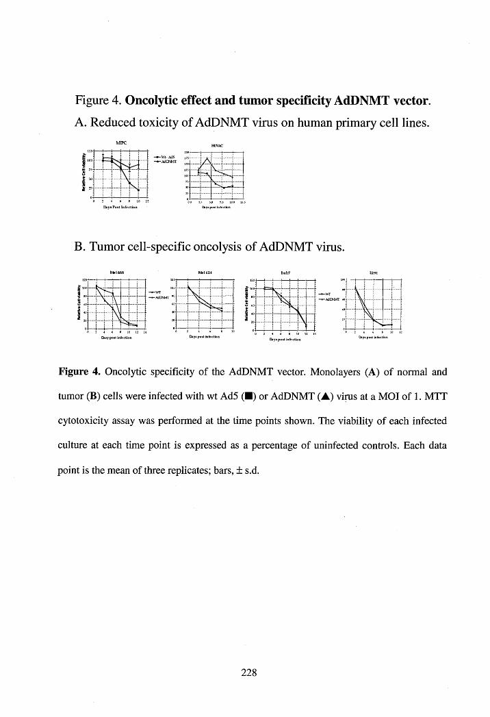
Figure 4. Oncolytic effect and tumor specificity AdDNMT vector.
A . R educed to x ic ity o f A d D N M T virus on hum an prim ary c e ll lines.
Ebyi P ost In fection
B. Tum or ce ll-sp ec ific on co lysis o f A d D N M T virus.
2 S 14Disy post infection Chys post infection
!1
2 10 12Days post infection
Figure 4. Oncolytic specificity of the AdDNMT vector. Monolayers (A) of normal and
tumor (B) cells were infected with wt Ad5 (■) or AdDNMT (A) virus at a MOI of 1. MTT
cytotoxicity assay was performed at the time points shown. The viability of each infected
culture at each time point is expressed as a percentage of uninfected controls. Each data
point is the mean of three replicates; bars, ± s.d.
228

Figure 5. Cell cycle dependent oncolytic activity of AdDNMT in A549 cell.
A. 50% Confluent 100% Confluent
EL,G0-G1: 40.27% S: 4.57%
B.
DNMTl-
Actin-
Confluency50% 100%/G0-G 1:26.26%
S: 26.98%
50%> Confluent 100% Confluent
C. I3
i122 106 84
-WT-.AdDNMT
0 2 s 10 124
Days p o s t in fec tionDays post infection
Figure 5. Cell cycle dependent oncolytic activity of AdDNMT in A549 cell. A. Cell cycle
analysis by flow cytometry of A549 cells plated in different density. B. Immunoblot
analysis of the DNMT1 protein level in the same population of A549 cells as figure 5A. C.
Monolayers of A549 cell with different density (as figure 5A) were infected with MOI 1 of
wt Ad5 (■) or AdDNMT (A) virus. MTT assay was performed to measure viable cells,
and results are the mean of triplicate experiments and expressed as percentage of non
infected cells.
229

6.4 Discussion:
The studies presented in this chapter provide in vitro evidence supporting the use of tumor
cell selective mRNA stabilization by the DNMT1 3’UTR for developing a conditionally
replicating adenovirus vector for cancer treatment. Our results demonstrate that (a) the
minimal 318 bp DNMT1 3’-UTR can induce tumor cell selective mRNA stabilization of
El A gene to drive adenovirus replication and oncolysis, (b) the 3’-UTR also causes
inhibition of the E1A gene expression and adenovirus replication in primary cells derived
from different tissues, and (c) AdDNMT replication and oncolysis in the A549 tumor cell
line correlate with cell cycle dependent expression of DNMT1.
The overexpression of the DNMT1 mRNA and protein has been reported in many different
types of cancers (Etoh et al., 2004; Girault et al., 2003; Nakagawa et al., 2003; Nakagawa
et al., 2005; Shieh et al., 2005). It is speculated that in cancers, DNMT1 is involved in the
DNA hypermethylation and maintenance of CpG islands that are not methylated in normal
cells (Etoh et al., 2004). The ubiquitous expression of DNMT1 in many different types of
tumor has made the enzyme an attractive pan-tumor target for developing oncolytic
adenovirus.
The DNMT1 3’UTR used in this report was significantly more active in a variety of cancer
cell lines with higher expression of DNMT1. By inserting the DNMT1 3’UTR in between
the luciferase reporter gene and polyA signal, the luciferase activity was 8 to 10-fold
reduced in normal hTRE-RPE cells, which do not express any DNMT1 (Figure 1). But in
another primary cell line BEAS-2B, the luciferase activity was not controlled by the
DNMT1 3’UTR. The BEAS-2B cell line is a normal human bronchial epithelial cell, which
is immortalized by SV40 large T antigen. It has been reported that the DNMT1 expression
230
is a downstream effect of SV40 large T antigen expression and the DNMT1 3’UTR plays
an important role in DNMT1 expression in cells that express SV40 large T antigen
(Chuang et al., 1997). We believe that the inability to repress reporter gene activity by the
DNMT1 3’UTR in the BEAS-2B cells is due to SV40 large T antigen-mediated DNMT1
mRNA stabilization. On the other hand, DNMT1 3’UTR did not affect the reporter gene
expression in most of the tumor cell lines that express DNMT1 (Figure 1A and IB). In the
oncolytic activity measured by the MTT assay, the wild-type Ad5 virus showed a similar
killing potency in normal and cancer cells, thus lacking tumor cell specificity. In contrast,
all the tumor cell lines tested, AdDNMT adenovirus induced cell killing as potently as the
wt Ad-5 virus, but was approximately 10-50 times less effective in normal cells,
confirming tumor cell-specificity of the DNMT1 3’UTR. We were also able to show that in
A549 cells, the replication and the oncolytic activity of the AdDNMT virus correlate with
the DNMT1 protein expression in cells plated with different density. Thus, the A549 cells
plated in higher density became cytostatic by increasing the number of cells in the Go/ Gi
phase of the cell cycle (Figure 5A) in the reducing the number of cells metabolically active
(data not shown). Under these conditions, A549 cells downregulate DNMT1 protein
expression. The mechanism of DNMT1 downregulation in the confluent A549 cells is not
yet understood. But we are able to show that in the confluent cultures of A549 cells, the
oncolytic activity of the AdDNMT virus was significantly reduced compared to the control
wt Ad-5 adenovirus. In contrast, the semi-confluent A549 cells the oncolytic activity of the
AdDNMT virus was identical to the wt Ad-5 correlating the correspondingly high levels of
DNMT1. Thus, from these results we can conclude that the novel DNMT1 3’UTR tested in
231

this study can control transgene expression, and viral replication, in a tumor-specific
manner.
A successful viral vector for cancer gene therapy not only needs to demonstrate the
therapeutic efficacy by efficiently eliminating all the cancer cells, but also needs to be
selective enough to use it systemically. Most of the current strategies for developing cancer
gene therapy vector are not tumor-selective enough to fulfill all the safety requirements for
systemic use. Major efforts have been directed toward improving or finding new and better
tumor targeting strategies to achieve higher degree of tumor selectivity and lower the
toxicity caused by therapeutic vectors. The tumor selective mRNA stabilization by the
DNMT 3’UTR presented in this study can not only be used as a novel pan-targeting
approach to control therapeutic or viral gene expression in tumor cells, but it can also be
used for improving the selectivity of currently available tumor targeting strategies. Many
tumor specific promoters that are being used for developing oncolytic virus for cancer gene
therapies are not entirely selective for tumor cells and generally active in one, or at most, a
few normal tissue types. Due to this leakiness, systemic use of the oncolytic vectors
developed by these tumor specific promoters is difficult because of the toxicity induced by
these vectors. Although tumor cells tend to overexpress DNMT1, it has been reported that
some proliferating normal cells constitutively express DNMT1 (Leonhardt et al., 1992).
The difference in the mechanisms involved in controlling the expression of DNMT1 in the
normal proliferating cells and cancer cells is not well understood, but it is a possibility that
the AdDNMT virus may replicate and show some toxicity in these normal cells. We
believed that the combination of tumor specific mRNA stabilization mechanisms of the
DNMT 3’-UTR with a tissue specific promoter could be a solution for developing a safer
232

oncolytic vector. The tumor cell specificity of different tumor specific promoters can be
enhanced by adding the translational specificity of DNMT1 3’UTR, which should help
each other to express therapeutic gene more selectively in the tumor. The data presented
here suggest that DNMT 3’UTR mediated tumor specific mRNA stabilization could be use
as a means to achieve tumor-specific expression of therapeutics, or viral genes, for cancer
gene therapy.
233

Chapter 7
Discussion and future direction
234

In this thesis, the hypothesis to be tested is that tumor cell selective stabilization of mRNA
can be used to control therapeutic/viral gene expression in order to develop targeted viral
vectors for cancer gene therapy. The data presented indicates that linkage to the AU-rich 3’
untranslated region (UTR) from a tumor-associated gene, cyclooxygenase-2 (COX-2), of a
replicative essential viral gene is sufficient to achieve tumor cell selective viral replication
and oncolysis.
The expression of COX-2, which has been associated with poor prognosis in many
different types of tumor and is a downstream effect of RAS-mediated transformation
(Sheng et al., 2001). Upregulation of COX-2 in the transformed environment is partly
mediated by a selective stabilization of the COX-2 mRNA (Sheng et al., 2000) (Dixon et
al., 2000) and the AU-rich region of the 3’UTR of COX-2 mRNA is essential for
stabilizing the COX-2 mRNA in an activated RAS-MAPK-dependent manner (Lasa et al.,
2000). So, we hypothesized that tumor selective transgene expression can be achieved by
using an AU-rich stabilization element such as the COX-2 3’UTR in a viral vector for
cancer gene therapy. Our hypothesis relies on the proven ability of the COX-2 3’UTR
region to destabilize a message in the quiescent environment, but restabilize the same
mRNA in the presence of the appropriate oncogenic stimuli.
The initial studies were designed to clone the AU-rich element from the 3’ UTR of the
tumor-associated gene COX-2 and evaluate it’s the ability to regulate gene expression in
transformed and non-transformed cells. 469 base pairs of the COX-2 3’UTR was cloned
(Dixon et al., 2000) and inserted downstream of the replicative essential adenoviral gene
El A, which was driven by the human CMV to generate the CMV-E1A-COX construct.
The effect of the COX-2 3’UTR insertion on El A expression was characterized in a model
235

system where exactly matched rat intestinal epithelial cell lines (RIE-iRAS), differing only
in the expression of an activated inducible variant of the H-Ras oncogene were used (Sheng
et al., 2000). Plasmid transfection of the adenoviral E1A gene, with or without, the COX-2
3’UTR in the RIE-iRAS cells, followed by infection with an ElA-deleated, replication-
incompetent adenoviral vector expressing GFP reporter gene (Ad-GFP) highlighted a
number of points. First of all, transient expression of E1A (CMV-E1A) in RIE-iRAS cells
was able to support the replication and mobilization of the incoming Ad-GFP vector, which
was independent of the inducible expression of Ras oncogene. In contrast, cells transfected
with CMV-E1A-COX plasmid were able to support replication and mobilization of the Ad-
GFP vector only when RIE-iRAS cells were previously induced to express activated Ras.
Secondly, experiments designed to assess viral titers showed a clear difference between the
titer of Ad-GFP released with, or without, inducible RAS signaling in the RIE-iRAS cells
transfected with CMV-E1A-COX plasmid. And finally, inhibition of Mitogen-activated
protein kinase (MAPK) activation, a downstream effectors molecule for Ras signaling
cascade, greatly reduced the ability of CMV-E1A-COX to support Ad-GFP replication and
mobilization even in the presence of inducible activated Ras. From these experiments, we
were able to conclude that, at least in vitro, the COX-2 3’UTR is able to control gene
expression, which was highly dependent on oncogenic Ras signaling.
To test whether the regulation of gene expression by the COX-2 3’UTR is sufficient to
control viral replication, we incorporated the CMV-E1A and CMV-E1A-COX cassettes
into ElA-deleted adenoviral genomes. The production of recombinant adenovirus
expressing these cassettes was performed using standard methods and the recoverable titer
of adenovirus expressing CMV-E1A (Ad-EIA) or CMV-E1 A-COX (Ad-El A-COX) was

very similar to the wild type Ad-5 titer. The replication selectivity of the Ad-EIA and the
Ad-ElA-COX virus were tested in the model cell culture system RIE-iRAS cells.
Replication and oncolysis of the Ad-El A-COX virus was heavily dependent upon the
expression of the inducible activated Ras and consistently produced an excess of 3 log
more viruses compared to the same cultures without any inducible activated Ras. Ad-El A-
COX virus replication in the Ras induced RIE-iRas cells was also inhibited (about 2 log
inhibition) by blocking MAPK activation with a chemical inhibitor, which again validated
the requirement for activated Ras/MAPK intracellular signaling in COX-2 3’UTR mediated
regulation of gene expression. We were also able to show that the oncolytic activity of the
Ad-El A-COX virus in different human tumor cell lines correlated very closely with the
cell lines’ MAPK activity status. In vivo in two human tumor xenograft models with
elevated levels of activated MAPK (U87 and U251), the Ad-El A-COX virus showed
similar therapeutic efficacy as the wild type Ad-5 virus, but showed no significant
therapeutic effects in the tumor model with very minimal activated MAPK (U118). Taken
together, these data suggest that El A expression can be regulated in an adenoviral genome
by the COX-2 3’UTR and Ad-El A-COX virus is oncolytic both in vitro and in vivo against
human tumors with elevated level of activated RAS-MAPK signals.
Results from the toxicity and the biodistribution studies in the immunocompetent
murine model show that the systemic administration of Ad-ElA-COX virus induces some
hepatic toxicity compared to the wild type Ad-5 virus. Although the lethal dose 50 (LD50)
for both of the viruses were very similar, the mice treated with Ad-El A-COX virus had
elevated levels of liver enzyme in the blood and showed abnormal liver histopathology
with increased immune infiltrate. This toxicity was not associated with the organ-specific
237

non-selective replication of the Ad-El A-COX virus because the amount of virus recovered
from the liver of the mice treated with the Ad-El A-COX were lower than the mice treated
with the wild type Ad-5 virus. However, at the early time point (24 h post i.v. challenge)
we observed an increased amount of viral DNA and El A protein in the livers of the Ad-
E1A-COX treated group compared to the wt Ad-5 group. It is possible that this early
elevation of viral DNA and protein could trigger a stronger innate immune response, which
then would lead to hepatic toxicity. It is known that during cell entry, the interaction
between the adenoviral entry receptors (fiber knob and penton base (Roelvink et al., 1998))
and the cellular entry receptors (CAR and avp 3 and ocyp 5 (Wickham et al., 1993))
activates various intercellular signaling pathways including the MAPK signaling cascade
(Bruder et al., 1997) (Tibbies et al., 2002). Based on this, one working hypothesis is that
the activation of a MAPK pathway during the initial adenoviral interaction with the cellular
entry receptors might alter the resting cell microenvironment and therefore even hyper-
stabilize the viral E1A protein ligated to the COX-2 3’UTR. This hypothesis is recently
supported by Crofford et al, showing that the initial interaction between adenovirus and
liver synoviocytes is sufficient to increase COX-2 expression both at the mRNA and
protein levels, and MAPK signaling is vital for this COX-2 induction (Crofford et al.,
2005). Taken together, results from the initial in vivo toxicity show that the 3’UTR needs
to be selected very carefully in order to develop tumor selective conditionally replicating
viral vectors. The 3’UTR from different genes will have different restrictions, which will
depend on the level of expression of that gene in the tumor microenvironment and the
expression profile in the normal physiological condition (Denigo et al., 2000). Also, the
kind of vector used to target tumor will be important for the selection of the 3’UTR
238

elements. It will be probably wise to avoid genes involved in inducing innate immune
response for developing retargeted adenoviral vectors, because adenovirus is known to
induce potent innate immune responses (Muruve, 2004), which may compromise the
selectivity of the 3’UTR element. Recently, several reports show that in order to develop
safe and less toxic adenoviral vectors for cancer therapy, multiple viral genes expression
need to be regulated (Irving et al., 2004). One possible way to reduce the Ad-ElA-COX
induced liver toxicity after systemic administration will be to use the COX-2 3’UTR
element to regulate multiple adenoviral genes. For example, the adenoviral late
transcription unit will be a very attractive candidate for this targeting approach as the late
transcription unit encodes approximately 15-20 different mRNAs, all of which derive from
a single pre-mRNA by differential splicing (Young, 2003). Most of the late gene products
are viral structural proteins and essential for virion assembly (Rux & Burnett, 2004). If an
mRNA stabilizing element, such as COX-2 3’UTR, can be inserted down stream of the late
transcription unit, all of these late gene products can be regulated by one single tumor
selective element and may lead to improved tumor selectivity and reduced toxicity.
To our knowledge, this is the first example of achieving tumor-selective gene expression,
and development of a conditionally replicating adenovirus, through a tumor specific post-
transcriptional mRNA stabilization element. There are several issues that need to be
addressed in order to optimize post-transcriptional targeting strategies for more efficient
tumor targeting. First of all, we have no knowledge about the minimal level of activated
RAS-MAPK signaling required for achieving sufficient stabilization of the transgene
mRNA in order to target tumor cells through the COX-2 3’UTR. This will give us a clearer
idea about the kind of cancer that can be targeted with the Ad-El A-COX vector. One
!239

possible way to investigate this will be using a more sensitive inducible system such as
repaying regulated, where transgene can be expressed in a dose-responsive manner
(Pollock et al., 2002) and use these systems to generate stable cell line where constitutively
activated Ras or MAPK expression can be more tightly controlled. Also, the receptor
tyrosine kinase (RTK) family of proteins such as epidermal growth factor receptor (EGFR)
is an upstream signaling molecule of Ras/MAKP signaling cascade and plays an important
role in cancer pathogenesis. Several reports postulate a link between the EGFR
upregulation and subsequent COX-2 induction in different cancers (Yuan et al., 2005) and
the activation of EGFR signaling pathway can induce COX-2 expression by stabilizing the
COX-2 mRNA (Matsuura et al., 1999). So the RTK overexpressed tumor cell can be a
suitable target for the Ad-El A-COX virus and we need to test the level of different RTK
expression in different cancer cells and try to correlate with the oncolytic selectivity of Ad-
El A-COX virus. Second of all, we did not optimize the length of the COX-2 3’UTR
element needed to achieve the maximum levels of tumor selectivity. The 469 bp COX-2
3’UTR used in this study was based on the published data by Dixon et al (Dixon et al.,
2000) who reported the position of the six AU-rich sequence motifs (AUUUA) in the 469
bp of the COX-2 3’UTR. Several other AU-rich motifs located outside of the 469 bp COX-
2 3’UTR have also been identified. It will be logical to include the rest of the 1000 bp AU-
rich motifs from the COX-2 3’UTR in our tumor targeting strategy to see whether we can
improve the tumor selectivity of this element. And finally, what kind of viral vector will be
the best fit for these pos-transcriptional targeting strategies that will give us the maximum
control and tumor selectivity for viral replication. To use mRNA stabilization as a mean to
control viral gene expression and tumor selective viral replication, it is possible that
240

oncolytic RNA viral vectors such as vesicular steatites virus (VSV) or measles virus may
be better than DNA viruses because the whole viral RNA genome could be restabilized or
destabilized by the 3’UTR element and that may enhance the selectivity of tumor cell
specific viral replication. But the most challenging thing for using the RNA virus in this
targeting approach will be the production of high titer recombinant vectors. It may be
necessary to alter the producer cell by introducing the appropriate intercellular signals such
as activated Ras/MAPK signaling, so the viral genome can be stabilized in the producer
cell and will allow to generate high titer therapeutic viral vectors.
In order to expand the tumor targeting strategy using selective mRNA stability with other
tumor associated genes, we cloned a second 3’UTR element from the DNA
methyltransferase 1 (DNMT1) gene, an important enzyme responsible for maintaining the
cellular epigenetic pattern from parent cell to daughter cell during cell division (Chen & Li,
2006). Several studies have linked this enzyme with tumorogenesis (Szyf, 2002). Increased
DNMT1 expression in various transformed environments is associated with abnormal cell
cycle (Szyf, 2001) and increased rate of cell division in tumor cells as compared to their
normal counterparts. In dividing cells, the stability of the DNMT1 mRNA is dramatically
increased after the cell entry into the S phase and the 3’UTR of the DNMT1 mRNA is
crucial for this cell cycle dependent increased mRNA turnover (Detich et al., 2001)
(Robertson et al., 1999). Because most cancerous tissues contain a higher percentage of
dividing cells compared to normal tissues (Robertson et al., 2000), we thought it might be
possible to express therapeutic/viral genes selectively in rapidly dividing tumor cells by
using the DNMT1 3’UTR. By inserting the DNMT1 3’UTR element downstream of
adenoviral E1A gene we showed that the E1A gene can be expressed selectively in cancer
241

cells and a recombinant adenovirus (AdDNMT) generated by using the E1A-DNMT
3’UTR construct was able to replicate selectively in various tumor cells. The oncolytic
activity of the AdDNMT virus was significantly reduced in three immortalized primary cell
lines in the tissue culture. The oncolytic activities also correlated with DNMT1 protein
expression and the percentage of cells that were in the S phase of the cell cycle. These in
vitro data support the conclusion that the DNMT1 3’UTR is able to control transgene
expression and adenoviral replication selectively in tumor cells. Currently we are
evaluating the selectivity of the AdDNMT virus in the in vivo animal model.
One of the major obstacles to effective therapeutic use of replicating viruses as anti
cancer agents is the poor intra-tumoral spread of the released virus where other cell types,
cell matrix and areas of necrosis exist (Sauthoff et al., 2003). Even after local
administration into the tumor, the virus usually gets trapped between various compartments „
composed of these different cell-types (Heise et al., 1999b). To overcome this problem, we
exploited a viral fusogenic membrane glycoprotein (FMG), which is a cytotoxic gene that
induces cell killing through induction of tumor cell fusion by forming large multinuclear
syncytia. Our laboratory previously demonstrated that the gene transfer of FMG in a
xenograft tumor model inhibits tumor growth (Bateman et al., 2000a). A recent report also
showed that the HIV gpl20 -mediated syncytia formation facilitates the dispersal and
increases the infection efficiency of an adenoviral vector in vitro (Li et al., 2001b). Also, an
oncolytic herpesvirus expressing Gibbon Ape Leukemia Virus (GALV) hyperfusogenic
FMG enhances the therapeutic efficacy of the oncolytic virus in vivo (Fu et al., 2003).
Based on these results, we hypothesized that the FMG expression during viral replication
inside the tumor might enhance the therapeutic efficacy of the oncolytic viral therapy by
242

increasing both the release of the viral particles from the infected cells and the spread of the
viral particles through out the tumor via the syncytia. We were able to show that GALV
induced syncytia enhances viral titer and spread of adenoviral vector through a monolayer.
In the presence of GALV induced syncytia, the titer of the virus released into the culture
medium is two logs higher than without syncytia. Increased viral titer in syncytia was
associated with the upregulation of adenoviral El A protein expression through unknown
post-transcriptional mechanisms. We do not know whether the FMG-induced syncytia
affect the expression level of the other viral proteins both in early and late phases. It has
been previously reported by our laboratory that transgene expression can be upregulated in
syncytia culture due to the increased metabolic activity induced by syncytia formation,
which can convert the syncytia into transient protein production factories (Bateman, 2002;
Higuchi et al., 2000). We were also able to show that a combination of different doses of
FMG gene therapy with replicating adenovirotherapy leads to regression of both small and
large established tumor xenografts. Moreover, this combined therapy can be effectively
targeted to cells expressing activated oncogenic signals (RAS/MAPK) by ligation of the
FMG to the 3’ untranslated region of the COX-2 gene. We believe that FMG expression
needs to be regulated transcriptionally, or post-transcriptionally, in order to avoid toxicity.
For safety purpose, it may be wise to keep the FMG expression separate from virus
replication, but it could be difficult to get approval from the regulatory committee to use
two separate therapeutic arms (intratumoral FMG+ adenoviral therapy) as a therapy for
cancer. Therefore, I proposed to develop a conditionally replicating adenovirus expressing
FMG selectively in the tumor. In addition, we have no notion about the time, or amount, of
FMG expression required for the maximum interaction between the viral life cycles and the

FMG-induced syncytia, which will give us the optimal synergistic enhancement of the
therapeutic efficacy. These questions can be addressed by developing a replicating
adenovirus that expresses FMG at different stages of the viral life cycle by using different
internal viral promoter (FMG expresses at the early phase of the viral life cycle by the El A
promoter or expresses at the late phase of the viral life cycle by using the viral major late
promoter (Wills et al., 1994)). Taken together, we used FMG-induced syncytia to enhance
the in vivo therapeutic efficacy of an oncolytic adenovirus for the treatment of cancer. The
FMG-induced syncytia not only act as a potent cytotoxic and immunostimulatory gene for
cancer gene therapy, but also improve oncolytic viral replication, intratumoral spread and
production, which enhance the oncolytic properties of the conditionally replicating
adenovirus.
The application of gene therapy in the treatment of diseases like cancer is currently
limited due to our inability to efficiently target systemic metastasis. Because different types
of cancer have diverse biological properties, and since even the most efficient targeting
strategy currently available has its limitations, it is unrealistic to imagine that an individual
targeting approach will eventually emerge as the most efficient way to target cancer.
Besides, most of the tumor specific elements currently used to target cancer cells have
proven to be leaky due to their expression in the normal anatomical site (Zeh & Bartlett,
2002). Moreover, most tumors contain a very heterogeneous population of transformed
cells (Bignold, 2003) (Khong & Restifo, 2002). Targeting cancer with an individual tumor
specific element will create selection pressures, which will drive the cancer to become
resistant to therapy (Konson et al., 2004). So, a successful cancer gene therapy approach
will likely rely on the ability to combine different targeting strategies to create highly
244

specialized “mosaic” vectors that safely incorporate different targeting strategies to address
specific features of each type of cancer (Borovjagin et al., 2005). We think that the post-
transcriptional targeting strategy of using the 3’UTR will be most efficient in combination
with other tumor targeting approaches such as transcriptional or transductional targeting.
One unique feature of the tumor selective mRNA stabilization element such as COX-2
3’UTR is that we can regulate individual gene expression both at the transcriptional and
post-transcriptional levels. This may help us target systemic cancer more selectively and
reduce toxicity from the therapeutic viral vectors.
In summary, this thesis described tumor cell selective mRNA stabilization as a novel
means to achieve tumor specific therapeutic gene expression. By using two different
3’UTR mRNA stability elements, we were able to regulate three different transgenes
selectively in tumor cells. Incorporation of the COX-2 3’UTR in the adenovirus genome
was sufficient enough to control viral replication and oncolysis both in vitro and in vivo.
Many other genes have been reported to use tumor cell selective mRNA stabilization as a
mean to control their expression in the tumor microenvironment. Therefore, targeting
cancer through tumor cell selective mRNA stabilization could be a viable strategy, which
may have broad applicability and potential in vector development for cancer gene therapy.
245

Reference:
Aghib DF, Bishop JM, Ottolenghi S, Guerrasio A, Serra A and Saglio G. (1990).Oncogene, 5, 707-11.
Aitken ML, Moss RB, Waltz DA, Dovey ME, Tonelli MR, McNamara SC, Gibson RL, Ramsey BW, Carter BJ and Reynolds TC. (2001). Hum Gene Ther, 1 2 ,1907-16.
Aiuti A, Slavin S, Aker M, Ficara F, Deola S, Mortellaro A, Morecki S, Andolfi G,Tabucchi A, Carlucci F, Marinello E, Cattaneo F, Vai S, Servida P, Miniero R, Roncarolo MG and Bordignon C. (2002a). Science, 296,2410-3.
Aiuti A, Vai S, Mortellaro A, Casorati G, Ficara F, Andolfi G, Ferrari G, Tabucchi A,Carlucci F, Ochs HD, Notarangelo LD, Roncarolo MG and Bordignon C. (2002b). Nat Med, 8,423-5.
Alba R, Bosch A and Chillon M. (2005). Gene Ther, 12 Suppl 1, SI8-27.Alemany R, Balague C and Curiel DT. (2000). Nat Biotechnol, 18, 723-7.Alessi DR, Cuenda A, Cohen P, Dudley DT and Saltiel AR. (1995). J Biol Chem, 270,
27489-94.Amalfitano A, Hauser MA, Hu H, Serra D, Begy CR and Chamberlain JS. (1998). J Virol,
72,926-33.Ambros V, Lee RC, Lavanway A, Williams PT and Jewell D. (2003). CurrBiol, 13, 807-
18.Anantharaman V, Koonin EV and Aravind L. (2002). Nucleic Acids Res, 30,1427-64.Antic D and Keene JD. (1997). Am J Hum Genet, 61, 273-8.Araki Y, Okamura S, Hussain SP, Nagashima M, He P, Shiseki M, Miura K and Harris CC.
(2003). Cancer Res, 63, 728-34.Araujo FD, Knox JD, Ramchandani S, Pelletier R, Bigey P, Price G, Szyf M and Zannis-
Hadjopoulos M. (1999). J Biol Chem, 274,9335-41.Audic Y and Hartley RS. (2004). Biol Cell, 96,479-98.Baron JA, Cole BF, Sandler RS, Haile RW, Ahnen D, Bresalier R, McKeown-Eyssen G,
Summers RW, Rothstein R, Burke CA, Snover DC, Church TR, Allen JI, Beach M, Beck GJ, Bond JH, Byers T, Greenberg ER, Mandel JS, Marcon N, Mott LA, Pearson L, Saibil F and van Stolk RU. (2003). N Engl J Med, 348, 891-9.
Bartel DP. (2004). Cell, 116,281-97.Bateman A. (2002). PhD Thesis, Open University, UK.Bateman A, Bullough F, Murphy S, Emiliusen L, Lavillette D, Cosset FL, Cattaneo R,
Russell SJ and Vile RG. (2000a). Cancer Res, 60, 1492-7.Bateman A, Harrington K, Kottke T, Ahmed A, Melcher A, Gough M, Linardakis E,
Riddle D, Dietz A, Lohse C, Strome S, Peterson T, Simari R and Vile R. (2002). Cancer Research, 62, 5466-6578.
Bateman AR, Harrington KJ, Melcher AA and Vile RG. (2000b). Expert Opin Investig Drugs, 9, 2799-813.
Bell B, Canty D and Audet M. (1995). Pediatr Rev, 16,290-8.Berk AJ. (2005). Oncogene, 24, 7673-85.Berto E, Bozac A and Marcori P. (2005). Gene Ther S98-102.Bernstein P, Peltz SW and Ross J. (1989). Mol Cell Biol, 9 ,659-70.Beutler E. (2001). Mol Ther, 4, 396-7.
246

Bewley MC, Springer K, Zhang YB, Freimuth P and Flanagan JM. (1999). Science, 286, 1579-83.
Bignold LP. (2003). Cell Mol Life Sci, 60, 883-91.Bilsland AE, Anderson CJ, Fletcher-Monaghan AJ, McGregor F, Evans TR, Ganly I, Knox
RJ, Plumb JA and Keith WN. (2003). Oncogene, 22, 370-80.Bischoff JR, Kim DH, Williams A, Heise C, Horn S, Muna M, Ng L, Nye JA, Sampson-
Johannes A, Fattaey A and McCormick F. (1996). Science, 274, 373-6.Blaese RM, Culver KW, Miller AD, Carter CS, Fleisher T, Clerici M, Shearer G, Chang L,
Chiang Y, Tolstoshev P, Greenblatt JJ, Rosenberg SA, Klein H, Berger M, Mullen CA, Ramsey WJ, Muiul L, Morgan RA and Anderson WF. (1995). 270,475-480.
Blomer BF, Naldini T, Gage FH. (1997). J Virol. 71: 6641-49.Bordignon C and al. e. (1995). 270,470-475.Borovjagin AV, Krendelchtchikov A, Ramesh N, Yu DC, Douglas JT and Curiel DT.
(2005). Cancer Gene Ther, 12,475-86.Boutaud O, Dixon DA, Oates JA and Sawaoka H. (2003). Adv Exp Med Biol, 525,157-60. Bouvet M, Fang B, Ekmekcioglu S, Ji L, Bucana CD, Hamada K, Grimm EA and Roth JA.
(1998). Gene Ther, 5, 189-95.Brewer G. (1998). J Biol Chem, 273,34770-4.Brewer G, Saccani S, Sarkar S, Lewis A and Pestka S. (2003). J Interferon Cytokine Res,
23,553-64.Briata P, Ilengo C, Corte G, Moroni C, Rosenfeld MG, Chen CY and Gherzi R. (2003).
Mol Cell, 12,1201-11.Brook M, Sully G, Clark AR and Saklatvala J. (2000). FEBS Lett, 483,57-61.Bruder JT, Jie T, McVey DL and Kovesdi I. (1997). J Virol, 71, 7623-8.Buzby JS, Brewer G and Nugent DJ. (1999). J Biol Chem, 274, 33973-8.Calin GA, Ferracin M, Cimmino A, Di Leva G, Shimizu M, Wojcik SE, Iorio MV, Visone
R, Sever NI, Fabbri M, Iuliano R, Palumbo T, Pichiorri F, Roldo C, Garzon R, Sevignani C, Rassenti L, Alder H, Volinia S, Liu CG, Kipps TJ, Negrini M and Croce CM. (2005). N Engl J Med, 353,1793-801.
Cao Y and Prescott SM. (2002). J Cell Physiol, 190, 279-86.Carballo E, Lai WS and Blackshear PJ. (1998). Science, 281,1001-5.Carballo E, Lai WS and Blackshear PJ. (2000). Blood, 95,1891-9.Carter PJ and Samulski RJ. (2000). In tJ Mol Med, 6,17-27.Cassel WA and Garrett RE. (1965). Cancer, 18, 863-8.Cavazzana-Calvo M, Hacein-Bey S, de Saint Basile G, Gross F, Yvon E, Nusbaum P, Selz
F, Hue C, Certain S, Casanova JL, Bousso P, Deist FL and Fischer A. (2000). Science, 288, 669-72.
Check E (2005). Nature. 433,561-3.Chen C Y, Gherzi R, Andersen JS, Gaietta G, Jurchott K, Royer HD, Mann M and Karin M.
(2000). Genes Dev, 14, 1236-48.Chen CY, Gherzi R, Ong SE, Chan EL, Raijmakers R, Pruijn GJ, Stoecklin G, Moroni C,
Mann M and Karin M. (2001). Cell, 107,451-64.Chen L, Liu Q, Qin R, Le H, Xia R, Li W and Kumar M. (2005). In tJ Mol Med, 15,617-
26.Chen T and Li E. (2006). Curr Top Microbiol Immunol, 301, 179-201.Chuang LS, Ian HI, Koh TW, Ng HH, Xu G and Li BF. (1997). Science, 277,1996-2000.
247

Claffey KP, Shih SC, Mullen A, Dziennis S, Cusick JL, Abrams KR, Lee SW and Detmar M. (1998). Mol Biol Cell, 9,469-81.
Cline MJ, Stang H, Mercola K, Morse L, Ruprecht R, Brown J and Salser W. (1980).Nature, 284,422-5.
Coffey MC, Strong JE, Forsyth PA and Lee PWK. (1998). 282,1332-1334.Cottrell S, Bicknell D, Kaklamanis L and Bodmer WF. (1992). Lancet, 340,626-30.Coyne CB and Bergelson JM. (2005). Adv Drug Deliv Rev, 57, 869-82.Crofford LJ, McDonagh KT, Guo S, Mehta H, Bian H, Petruzelli LM and Roessler BJ.
(2005). J Gene Med, 7,288-96.Croyle MA, Chirmule N, Zhang Y and Wilson JM. (2002). Hum Gene Ther, 13, 1887-900. Crystal RG, McElvaney NG, Rosenfeld MA, Chu CS, Mastrangeli A, Hay JG, Brody SL,
Jaffe HA, Eissa NT and Danel C. (1994). Nat Genet, 8,42-51.Culligan KG, Mackey AJ, Finn DM, Maguire PB and Ohlendieck K. (1998). In tJ Mol
Med, 2, 639-48.Culver K, Cometta K, Morgan R, Morecki S, Aebersold P, Kasid A, Lotze M, Rosenberg
SA, Anderson WF and Blaese RM. (1991). Proc Natl Acad Sci USA, 88,3155-9. Dave UP, Jenkins NA and Copeland NG. (2004). Science, 303,333.Davis BM, McCurrach ME, Taneja KL, Singer RH and Housman DE. (1997). Proc Natl
Acad Sci USA, 94, 7388-93.Dean JL, Brook M, Clark AR and Saklatvala J. (1999). J Biol Chem, 274,264-9.Dean JL, Sarsfield SJ, Tsounakou E and Saklatvala J. (2003). J Biol Chem, 278, 39470-6. Dean JL, Sully G, Clark AR and Saklatvala J. (2004). Cell Signal, 16,1113-21.Denkert C, Weichert W, Pest S, Koch I, Licht D, Kobel M, Reles A, Sehouli J, Dietel M
and Hauptmann S. (2004). Cancer Res, 64, 189-95.Denigo M, Cestelli A, Savettieri G and Di Liegro I. (2000). In tJ Mol Med, 5, 111-23. Detich N, Ramchandani S and Szyf M. (2001). J Biol Chem, 276, 24881-90.Diaz RM, Bateman A, Emiliusen L, Fielding A, Trono D, Russell SJ and Vile RG. (2000).
Gene Therapy, 7,1656-1663.Dimberg J, Hugander A, Sirsjo A and Soderkvist P. (2001). Anticancer Res, 21, 911-5.Dix BR, Edwards SJ and Braithwaite AW. (2001). J Virol, 75,5443-7.Dixon DA, Kaplan CD, McIntyre TM, Zimmerman GA and Prescott SM. (2000). J Biol
Chem, 275, 11750-7.Doench JG, Petersen CP and Sharp PA. (2003). Genes Dev, 17,438-42.Donnellan R and Chetty R. (1998). Mol Pathol, 51,1-7.Dorig RE, Marcil A, Chopra A and Richardson CD. (1993). Cell, 75,295-305.Dormond O, Foletti A, Paroz C and Ruegg C. (2001). Nat Med, 7, 1041-7.Dorssers L, Burger H, Bot F, Delwel R, Geurts van Kessel AH, Lowenberg B and
Wagemaker G. (1987). Gene, 55,115-24.Drumm ML, Wilkinson DJ, Smit LS, Worrell RT, Strong TV, Frizzell RA, Dawson DC
and Collins FS. (1991). Science, 254,1797-9.Dudley RW, Lu Y, Gilbert R, Matecki S, Nalbantoglu J, Petrof BJ and Karpati G. (2004).
Hum Gene Ther, 15,145-56.Eastham JA, Truong LD, Rogers E, Kattan M, Flanders KC, Scardino PT and Thompson
TC. (1995). Lab Invest, 73, 628-35.Edelstein ML, Abedi MR, Wixon J and Edelstein RM. (2004). J Gene Med, 6,597-602. Ehrhardt A and Kay MA. (2002). Blood, 99, 3923-30.
248

Ehrhardt A, Xu H, Dillow AM, Bellinger DA, Nichols TC and Kay MA. (2003). Blood, 102,2403-11.
Epstein AL, Marconi P, Argnani R and Manservigi R. (2005). Curr Gene Ther, 5,445-58.Erkinheimo TL, Lassus H, Finne P, van Rees BP, Leminen A, Ylikorkala O, Haglund C,
Butzow R and Ristimaki A. (2004). Clin Cancer Res, 10, 538-45.Esteban LM, Vicario-Abejon C, Femandez-Salguero P, Femandez-Medarde A,
Swaminathan N, Yienger K, Lopez E, Malumbres M, McKay R, Ward JM, Pellicer A and Santos E. (2001). Mol Cell Biol, 21,1444-52.
Etoh T, Kanai Y, Ushijima S, Nakagawa T, Nakanishi Y, Sasako M, Kitano S and Hirohashi S. (2004). Am J Pathol, 164,689-99.
Fan XC and Steitz JA. (1998). Proc Natl Acad Sci USA, 95, 15293-8.Fechner H, Haack A, Wang H, Wang X, Eizema K, Pauschinger M, Schoemaker R, Veghel
R, Houtsmuller A, Schultheiss HP, Lamers J and Poller W. (1999). Gene Ther, 6, 1520-35.
Ferreira SH, Moncada S and Vane JR. (1971). Nat New Biol, 231, 237-9.Fielding AK, Chapel-Femandes S, Chadwick MP, Bullough FJ, Cosset F-L and Russell SJ.
(2000). Human Gene Therapy, 11, 817-826.Fischer A, Le Deist F, Hacein-Bey-Abina S, Andre-Schmutz I, Basile Gde S, de VillartayJP, Cavazzana-Calvo M. Immonol Rev, 203, 98-109Flotte T, Carter B, Conrad C, Guggino W, Reynolds T, Rosenstein B, Taylor G, Walden S
and Wetzel R. (1996). Hum Gene Ther, 7,1145-59.Forget BG. (1979). Ann Intern Med, 91,605-16.Frevel MA, Bakheet T, Silva AM, Hissong JG, Khabar KS and Williams BR. (2003). Mol
Cell Biol, 23,425-36.Freytag SO, Khil M, Strieker H, Peabody J, Menon M, dePeralta-Venturina M, Nafziger D,
Pegg J, Paielli D, Brown S, Barton K, Lu M, Aguilar-Cordova E and Kim JH.(2002). Cancer Research, 62,4968-4976.
Friedmann T. (2001). Mol Ther, 4, 285-8.Fu X, Tao L, Jin A, Vile R, Brenner MK and Zhang X. (2003). Molecular Therapy, 7, 748-
754.Gallouzi IE, Brennan CM and Steitz JA. (2001). Rna, 7,1348-61.Gallouzi IE, Parker F, Chebli K, Maurier F, Labourier E, Barlat I, Capony JP, Tocque B
and Tazi J. (1998). Mol Cell Biol, 18, 3956-65.Gao M, Wilusz CJ, Peltz SW and Wilusz J. (2001). Embo J, 20, 1134-43.Giovannucci E, Egan KM, Hunter DJ, Stampfer MJ, Colditz GA, Willett WC and Speizer
FE. (1995). N Engl J Med, 333,609-14.Girault I, Tozlu S, Lidereau R and Bieche I. (2003). Clin Cancer Res, 9,4415-22.Giri I, Danos O and Yaniv M. (1985). Proc Natl Acad Sci USA, 82,1580-4.Goff SP and Berg P. (1976). Cell, 9,695-705.Goodrum FD and Omelles DA. (1998). J Virol, 72,9479-90.Gorospe M, Wang X, Guyton KZ and Holbrook NJ. (1996). Mol Cell Biol, 16,6654-60.Gou Q, Liu CH, Ben-Av P and Hla T. (1998). Biochem Biophys Res Commun, 242, 508-12.Gouble A, Grazide S, Meggetto F, Mercier P, Delsol G and Morello D. (2002). Cancer
Res, 62, 1489-95.Graham FL, Smiley J, Russell WC and Naim R. (1977). J Gen Virol, 36,59-74.
249

Gregorevic P, Allen JM, Minami E, Blankinship MJ, Haraguchi M, Meuse L, Finn E, Adams ME, Froehner SC, Murry CE and Chamberlain JS. (2006). Nat Med, 12, 787-9.
Grimm D and Kay MA. (2003). Curr Gene Ther, 3, 281-304.Guhaniyogi J and Brewer G. (2001). Gene, 265, 11-23.Gupta RA and Dubois RN. (2001). Nat Rev Cancer, 1, 11-21.Hacein-Bey-Abina S, von Kalle C, Schmidt MR, McCormack MP, Wulffraat N, Leboulch
P, Lim A, Osborne CS, Pawliuk R, Morillon E, Sorensen R, Forster A, Fraser P, Cohen JI, de Saint Basile G, Alexander I, Wintergerst U, Frebourg T, Aurias A, Stoppa-Lyonnet D, Romana S, Radford-Weiss I, Gross F, Valensi F, Delabesse E, Macintyre E, Sigaux F, Soulier J, Leiva LE, Wissler M, Prinz C, Rabbitts TH, Le Deist F, Fischer A and Cavazzana-Calvo M. (2003). Science, 302,415-419.
Hallenbeck PL, Chang Y-N, Hay C, Golightly D, Stewart D, Lin J, Phipps S and Chiang YL. (1999). 10,1721-1733.
Hammer RE, Palmiter RD and Brinster RL. (1984). Nature, 311, 65-7.Han Q, Leng J, Bian D, Mahanivong C, Carpenter KA, Pan ZK, Han J and Huang S.
(2002). J Biol Chem, 277,48379-85.Hanahan D and Weinberg RA. (2000). Cell, 100,57-70.Hanger JJ, Bromham LD, McKee JJ, O'Brien TM and Robinson WF. (2000). J Virol, 74,
4264-72.Hardie DG, Carling D and Carlson M. (1998). Annu Rev Biochem, 67, 821-55.Hardie DG and Hawley SA. (2001). Bioessays, 23,1112-9.Hardwick JC, van den Brink GR, Offerhaus GJ, van Deventer SJ and Peppelenbosch MP.
(2001). Oncogene, 20, 819-27.Heinonen M, Bono P, Narko K, Chang SH, Lundin J, Joensuu H, Fumeaux H, Hla T,
Haglund C and Ristimaki A. (2005). Cancer Res, 65,2157-61.Heise C, Hermiston T, Johnson L, Brooks G, Sampson-Johannes A, Williams A, Hawkins
L and Kim D. (2000). Nat Med, 6, 1134-1139.Heise C, Sampson-Johannes A, Williams A, McCormick F, Von Hoff DD and Kim DH.
(1997). Nat Med, 3, 639-45.Heise C, Sampson-Johannes A, Williams A, McCormick F, Von Hoff DD and Kim DH.
(1999a). 3,639-645.Heise CC, Williams A, Olesch J and Kim DH. (1999b). Cancer Gene Ther, 6,499-504.Herrick DJ and Ross J. (1994). Mol Cell Biol, 14,2119-28.Higuchi H, Bronk S, Bateman A, Harrington KJ, Vile RG and Gores GJ. (2000). Cancer
Research, 60,6396-6402.Hollis GF, Gazdar AF, Bertness V and Kirsch IR. (1988). Mol Cell Biol, 8, 124-9.Honda T, Saitoh H, Masuko M, Katagiri-Abe T, Tominaga K, Kozakai I, Kobayashi K,
Kumanishi T, Watanabe YG, Odani S and Kuwano R. (2000). Brain Res Mol Brain Res, 77, 19-28.
Howe LR, Subbaramaiah K, Chung WJ, Dannenberg AJ and Brown AM. (1999). Cancer Res, 59,1572-7.
Howitt J, Anderson CW and Freimuth P. (2003). Curr Top Microbiol Immunol, 272, 331- 64.
Hoyle PE, Steelman LS and McCubrey JA. (1997). Cytokines Cell Mol Ther, 3, 159-68.
250

Irving J, Wang Z, Powell S, O'Sullivan C, Mok M, Murphy B, Cardoza L, Lebkowski JS and Majumdar AS. (2004). Cancer Gene Ther, 11, 174-85.
Jackson DA, Symons RH and Berg P. (1972). Proc Natl Acad Sci USA, 69,2904-9.Jain RG, Andrews LG, McGowan KM, Pekala PH and Keene JD. (1997). Mol Cell Biol,
17,954-62.Janda E, Litos G, Grunert S, Downward J and Beug H. (2002). Oncogene, 21, 5148-59.Jing Q, Huang S, Guth S, Zarubin T, Motoyama A, Chen J, Di Padova F, Lin SC, Gram H
and Han J. (2005). Cell, 120,623-34.Jones DA, Carlton DP, McIntyre TM, Zimmerman GA and Prescott SM. (1993). J Biol
Chem, 268, 9049-54.Kafri TU, Blumer DA and Verma IM. (1997). Nat. Genet. 17, 314-17.Kaiser J. (2004). Science, 304,1423-5.Khabar KS. (2005). J Interferon Cytokine Res, 25,1-10.Khan KN, Masferrer JL, Woemer BM, Soslow R and Koki AT. (2001a). Scand J
Gastroenterol, 36, 865-9.Khan KN, Stanfield KM, Harris RK and Baron DA. (2001b). Ren Fail, 23, 321-30.Khong HT and Restifo NP. (2002). Nat Immunol, 3, 999-1005.Kikuchi A, Kishida S and Yamamoto H. (2006). Exp Mol Med, 38,1-10.Kim D, Martuza RL and Zwiebel J. (2001). Nature Medicine, 7,781-787.Klages N, Zufferey R and Trono D. (2000). Mol Ther, 2,170-6.Knox JD, Araujo FD, Bigey P, Slack AD, Price GB, Zannis-Hadjopoulos M and Szyf M.
(2000). J Biol Chem, 275,17986-90.Koera K, Nakamura K, Nakao K, Miyoshi J, Toyoshima K, Hatta T, Otani H, Aiba A and
Katsuki M. (1997). Oncogene, 15,1151-9.Konson A, Ben-Kasus T, Mahajna JA, Danon A, Rimon G and Agbaria R. (2004). Cancer
Gene Ther, 11, 830-40.Kootstra NA and Verma IM. (2003). Annu Rev Pharmacol Toxicol, 43, 413-39.Korner CG and Wahle E. (1997). J Biol Chem, 272,10448-56.Kurumbail RG, Stevens AM, Gierse JK, McDonald JJ, Stegeman RA, Pak JY, Gildehaus
D, Miyashiro JM, Penning TD, Seibert K, Isakson PC and Stallings WC. (1996). Nature, 384, 644-8.
Lai WS and Blackshear PJ. (2001). J Biol Chem, 276, 23144-54.Laird PW, Jackson-Grusby L, Fazeli A, Dickinson SL, Jung WE, Li E, Weinberg RA and
Jaenisch R. (1995). Cell, 81,197-205.Lambright ES, Amin K, Wiewrodt R, Force SD, Lanuti M, Propert KJ, Litzky L, Kaiser
LR and Albelda SM. (2001). Gene Ther, 8, 946-53.Langa F, Lafon I, Vandormael-Poumin S, Vidaud M, Babinet C and Morello D. (2001).
Oncogene, 20,4344-53.Langenfeld J, Kiyokawa H, Sekula D, Boyle J and Dmitrovsky E. (1997). Proc Natl Acad
Sci US A, 94,12070-4.Laroia G, Cuesta R, Brewer G and Schneider RJ. (1999). Science, 284,499-502.Lasa M, Brook M, Saklatvala J and Clark AR. (2001). Mol Cell Biol, 21,771-80.Lasa M, Mahtani KR, Finch A, Brewer G, Saklatvala J and Clark AR. (2000). Mol Cell
Biol, 20,4265-74.Lebwohl DE, Muise-Helmericks R, Sepp-Lorenzino L, Serve S, Timaul M, Bol R, Borgen
P and Rosen N. (1994). Oncogene, 9, 1925-9.
251

Lee CH, Bradley G and Ling V. (1998). J Cell Physiol, 177,1-12.Leonhardt H, Page AW, Weier HU and Bestor TH. (1992). Cell, 71, 865-73.Levens D. (2002). Proc Natl Acad Sci USA, 99, 5757-9.Levine TD, Gao F, King PH, Andrews LG and Keene JD. (1993). Mol Cell Biol, 13, 3494-
504.Levy NS, Chung S, Fumeaux H and Levy AP. (1998). J Biol Chem, 273,6417-23.Lewis AM, Jr. and Rowe WP. (1970). J Virol, 5,413-20.Li E, Brown SL, Stupack DG, Puente XS, Cheresh DA and Nemerow GR. (2001a). J Virol,
75,5405-9.Li E, Stupack D, Bokoch GM and Nemerow GR. (1998). J Virol, 72, 8806-12.Li H, Haviv YS, Derdeyn CA, Lam J, Coolidge C, Hunter E, Curiel DT and Blackwell JL.
(2001b). Human Gene Therapy, 12, 2155-2165.Li X, Raikwar SP, Liu YH, Lee SJ, Zhang YP, Zhang S, Cheng L, Lee SD, Juliar BE,
Gardner TA, Jeng MH and Kao C. (2006). Mol Cancer Ther, 5,676-84.Lichty BD, Stojdl DF, Taylor RA, Miller L, Frenkel I, Atkins H and Bell JC. (2004). Hum
Gene Ther, 15, 821-31.Lin S, Wang W, Wilson GM, Yang X, Brewer G, Holbrook NJ and Gorospe M. (2000).
Mol Cell Biol, 20,7903-13.Loflin P, Chen CY and Shyu AB. (1999). Genes Dev, 13, 1884-97.Lopez de Silanes I, Zhan M, Lai A, Yang X and Gorospe M. (2004). Proc Natl Acad Sci U
SA, 101,2987-92.Lu X, Timchenko NA and Timchenko LT. (1999). Hum Mol Genet, 8, 53-60.Ma WJ, Cheng S, Campbell C, Wright A and Fumeaux H. (1996). J Biol Chem, 271, 8144-
51.Mahtani KR, Brook M, Dean JL, Sully G, Saklatvala J and Clark AR. (2001). Mol Cell
Biol, 21,6461-9.Maity A, McKenna WG and Muschel RJ. (1995). Embo J, 14,603-9.Maity A, McKenna WG and Muschel RJ. (1997). Cell Growth Differ, 8, 311-8.Maity A and Solomon D. (2000). Exp Cell Res, 255, 250-7.Malumbres M and Barbacid M. (2001). Nat Rev Cancer, 1, 222-31.Manno CS, Chew AJ, Hutchison S, Larson PJ, Herzog RW, Arruda VR, Tai SJ, Ragni MV,
Thompson A, Ozelo M, Couto LB, Leonard DG, Johnson FA, McClelland A, Scallan C, Skarsgard E, Flake AW, Kay MA, High KA and Glader B. (2003).Blood, 101,2963-72.
Manno CS, Pierce GF, Arruda VR, Glader B, Ragni M, Rasko JJ, Ozelo MC, Hoots K,Blatt P, Konkle B, Dake M, Kaye R, Razavi M, Zajko A, Zehnder J, Rustagi PK, Nakai H, Chew A, Leonard D, Wright JF, Lessard RR, Sommer JM, Tigges M, Sabatino D, Luk A, Jiang H, Mingozzi F, Couto L, Ertl HC, High KA and Kay MA.(2006). Nat Med, 12, 342-7.
Marshall E (2003). Science, 299, 320-3.Masferrer JL, Leahy KM, Koki AT, Zweifel BS, Settle SL, Woemer BM, Edwards DA,
Flickinger AG, Moore RJ and Seibert K. (2000). Cancer Res, 60,1306-11.Matsubara S, Wada Y, Gardner TA, Egawa M, Park MS, Hsieh CL, Zhau HE, Kao C,
Kamidono S, Gillenwater JY and Chung LW. (2001). Cancer Res, 61, 6012-9.Matsuura H, Sakaue M, Subbaramaiah K, Kamitani H, Eling TE, Dannenberg AJ, Tanabe
T, Inoue H, Arata J and Jetten AM. (1999). J Biol Chem, 274, 29138-48.
252
McAdam BF, Catella-Lawson F, Mardini IA, Kapoor S, Lawson JA and FitzGerald GA.(1999). Proc Natl Acad Sci U S A , 96, 272-7.
McConnell MJ and Imperiale MJ. (2004). Hum Gene Ther, 15, 1022-33.McCormick F. (2001). Nat Rev Cancer, 1, 130-41.Meier O, Boucke K, Hammer SV, Keller S, Stidwill RP, Hemmi S and Greber UF. (2002).
J Cell Biol, 158,1119-31.Mi S, Lee X, Li X, Veldman GM, Finnerty H, Racie L, LaVallie E, Tang XY, Edouard P,
Howes S, Keith JC, Jr. and McCoy JM. (2000). Nature, 403, 785-9.Ming XF, Stoecklin G, Lu M, Looser R and Moroni C. (2001). Mol Cell Biol, 21, 5778-89. Miyoshi H, Blomer U, Takahashi M, Gage FH and Verma IM. (1998). J Virol, 72, 8150-7. Molenaar JJ, van Sluis P, Boon K, Versteeg R and Caron HN. (2003). Genes Chromosomes
Cancer, 36, 242-9.Montero L and Nagamine Y. (1999). Cancer Res, 59, 5286-93.Morecki S, Karson E, Cometta K, Kasid A, Aebersold P, Blaese RM, Anderson WF and
Rosenberg SA. (1991). Cancer Immunol Immunother, 32, 342-52.Mukherjee D, Gao M, O'Connor JP, Raijmakers R, Pruijn G, Lutz CS and Wilusz J. (2002).
Embo J, 21,165-74.Mukhopadhyay D, Houchen CW, Kennedy S, Dieckgraefe BK and Anant S. (2003). Mol
Cell, 11, 113-26.Mulligan RC, Howard BH and Berg P. (1979). Nature, 277,108-14.Muruve DA. (2004). Hum Gene Ther, 15,1157-66.Myer VE, Fan XC and Steitz JA. (1997). Embo J, 16, 2130-9.Nair AP, Hahn S, Banholzer R, Hirsch HH and Moroni C. (1994). Nature, 369,239-42. Nakagawa M, Seto M and Hosokawa Y. (2006). Leukemia, 20,929-36.Nakagawa T, Kanai Y, Saito Y, Kitamura T, Kakizoe T and Hirohashi S. (2003). J Urol,
170,2463-6.Nakagawa T, Kanai Y, Ushijima S, Kitamura T, Kakizoe T and Hirohashi S. (2005). J
Urol, 173,1767-71.Nakamura T, Peng KW, Harvey M, Greiner S, Lorimer LA, James CD and Russell SJ.
(2005). Nat Biotechnol, 23, 209-14.Nanbru C, Lafon I, Audigier S, Gensac MC, Vagner S, Huez G and Prats AC. (1997). J
Biol Chem, 272, 32061-6.Nobile C, Marchi J, Nigro V, Roberts RG and Danieli GA. (1997). Genomics, 45,421-4. Nugteren DH, Van Dorp DA, Bergstrom S, Hamberg M and Samuelsson B. (1966).
Nature, 212, 38-9.Ogawa T, Takayama K, Takakura N, Kitano S and Ueno H. (2002). Cancer Gene Ther, 9,
633-40.Oshima M, Dinchuk JE, Kargman SL, Oshima H, Hancock B, Kwong E, Trzaskos JM,
Evans JF and Taketo MM. (1996). Cell, 87, 803-9.Pages G, Berra E, Milanini J, Levy AP and Pouyssegur J. (2000). J Biol Chem, 275, 26484-
91.Pages JC and Bru T. (2004). J Gene Med, 6 Suppl 1, S67-82.Park JI, Lee MG, Cho K, Park BJ, Chae KS, Byun DS, Ryu BK, Park YK and Chi SG.
(2003). Oncogene, 22,4314-32.Peng SS, Chen CY and Shyu AB. (1996). Mol Cell Biol, 16, 1490-9.
253

Percy ME, Wong S, Bauer S, Liaghati-Nasseri N, Perry MD, Chauthaiwale VM, Dhar M and Joshi JG. (1998). Analyst, 123,41-50.
Phillips RK, Wallace MH, Lynch PM, Hawk E, Gordon GB, Saunders BP, Wakabayashi N, Shen Y, Zimmerman S, Godio L, Rodrigues-Bigas M, Su LK, Sherman J, Kelloff G, Levin B and Steinbach G. (2002). Gut, 50, 857-60.
Polakis P. (2000). Genes Dev, 14, 1837-51.Pollock R, Giel M, Linher K and Clackson T. (2002). Nat Biotechnol, 20,729-33.Poyry TA, Kaminski A and Jackson RJ. (2004). Genes Dev, 18,62-75.Rajagopalan LE, Westmark CJ, Jarzembowski JA and Malter JS. (1998). Nucleic Acids
Res, 26, 3418-23.Ramachandra M, Rahman A, Zou A, Vaillancourt M, Howe JA, Antelman D, Sugarman B,
Demers GW, Engler H, Johnson D and Shabram P. (2001). Nat Biotechnol, 19, 1035-41.
Raper SE, Yudkoff M, Chirmule N, Gao GP, Nunes F, Haskal ZJ, Furth EE, Propert KJ,Robinson MB, Magosin S, Simoes H, Speicher L, Hughes J, Tazelaar J, Wivel NA, Wilson JM and Batshaw ML. (2002). Hum Gene Ther, 13, 163-75.
Raz A, Wyche A and Needleman P. (1989). Proc Natl Acad Sci USA, 86, 1657-61.Reisman D and Thompson EA. (1995). Mol Endocrinol, 9, 1500-9.Ridley SH, Dean JL, Sarsfield SJ, Brook M, Clark AR and Saklatvala J. (1998). FEBS Lett,
439,75-80.Ries S and Korn WM. (2002). Br J Cancer, 86, 5-11.Rimokh R, Berger F, Bastard C, Klein B, French M, Archimbaud E, Rouault JP, Santa
Lucia B, Duret L, Vuillaume M and et al. (1994). Blood, 83, 3689-96.Ristimaki A, Sivula A, Lundin J, Lundin M, Salminen T, Haglund C, Joensuu H and Isola
J. (2002). Cancer Res, 62,632-5.Robert MF, Morin S, Beaulieu N, Gauthier F, Chute IC, Barsalou A and MacLeod AR.
(2003). Nat Genet, 33, 61-5.Robertson KD, Keyomarsi K, Gonzales FA, Velicescu M and Jones PA. (2000). Nucleic
Acids Res, 28,2108-13.Robertson KD, Uzvolgyi E, Liang G, Talmadge C, Sumegi J, Gonzales FA and Jones PA.
(1999). Nucleic Acids Res, 27, 2291-8.Rodriguez R, Schuur ER, Lim HY, Henderson GA, Simons JW and Henderson DR. (1997).
57,2559-2563.Roelvink PW, Lizonova A, Lee JG, Li Y, Bergelson JM, Finberg RW, Brough DE,
Kovesdi I and Wickham TJ. (1998). J Virol, 72, 7909-15.Rogers S. (1959). Nature, 183,1815-6.Rogers S. (1968). Nature, 220, 1321-2.Rogers S and Pfuderer P. (1968). Nature, 219,749-51.Romero NB, Braun S, Benveniste O, Leturcq F, Hogrel JY, Morris GE, Barois A, Eymard
B, Payan C, Ortega V, Boch AL, Lejean L, Thioudellet C, Mourot B, Escot C, Choquel A, Recan D, Kaplan JC, Dickson G, Klatzmann D, Molinier-Frenckel V, Guillet JG, Squiban P, Herson S and Fardeau M. (2004). Hum Gene Ther, 15, 1065- 76.
Rosenberg SA. (1992). 268,2416-2419.
254

Roth JA, Nguyen D, Lawrence DD, Kemp BL, Carrasco CH, Ferson DZ, Hong WK,Komaki R, Lee JJ, Nesbitt JC, Pisters KMW, Putnam JB, Schea R and et al. (1996). 2,985-991.
Roth JA, Swisher SG and Meyn RE. (1999). Oncology (Williston Park), 13, 148-54.Rowe WP, Huebner RJ, Gilmore LK, Parrott RH and Ward TG. (1953). Proc Soc Exp Biol
Med, 84, 570-3.Russell JE, Morales J and Liebhaber SA. (1997). Prog Nucleic Acid Res Mol Biol, 57,249-
87.Russell JE, Morales J, Makeyev AV and Liebhaber SA. (1998). Mol Cell Biol, 18,2173-83. Russell SJ. (2002). Cancer Gene Ther, 9, 961-6.Rux JJ and Burnett RM. (2004). Hum Gene Ther, 15, 1167-76.Ryan EP, Pollock SJ, Kaur K, Felgar RE, Bernstein SH, Chiorazzi N and Phipps RP.
(2006). Clin Immunol, 120, 76-90.Sabatino DE, Mingozzi F, Hui DJ, Chen H, Colosi P, Ertl HC and High KA. (2005). Mol
Ther, 12, 1023-33.Sadeghi H and Hitt MM. (2005). Curr Gene Ther, 5,411-27.Sambrook J, Westphal H, Srinivasan PR and Dulbecco R. (1968). Proc Natl Acad Sci U S
A, 60, 1288-95.Sandig V, Youil R, Bett AJ, Franlin LL, Oshima M, Maione D, Wang F, Metzker ML,
Savino R and Caskey CT. (2000). Proc Natl Acad Sci USA, 97,1002-7.Sandmair AM, Loimas S, Poptani H, Vainio P, Vanninen R, Turunen M, Tyynela K,
Vapalahti M and Yla-Herttuala S. (1999). Acta Neurochir (Wien), 141, 867-72; discussion 872-3.
Samow P, Ho YS, Williams J and Levine AJ. (1982). Cell, 28,387-94.Satoh PS and Ito Y. (1968). Virology, 35, 335-7.Sauthoff H, Hu J, Maca C, Goldman M, Heitner S, Yee H, Pipiya T, Rom WN and Hay JG.
(2003). Hum Gene Ther, 14,425-33.Sawaoka H, Dixon DA, Oates JA and Boutaud O. (2003). J Biol Chem, 278, 13928-35. Schuler GD and Cole MD. (1988). Cell, 55,1115-22.Sehgal I and Thompson TC. (1999). Mol Biol Cell, 10,407-16.Sengupta S, Jang BC, Wu MT, Paik JH, Fumeaux H and Hla T. (2003). J Biol Chem, 278,
25227-33.Shaw G and Kamen R. (1986). Cell, 46,659-67.Sheng GG, Shao J, Sheng H, Hooton EB, Isakson PC, Morrow JD, Coffey RJ, Jr., DuBois
RN and Beauchamp RD. (1997a). Gastroenterology, 113, 1883-91.Sheng H, Shao J, Dixon DA, Williams CS, Prescott SM, DuBois RN and Beauchamp RD.
(2000). J Biol Chem, 275, 6628-35.Sheng H, Shao J and Dubois RN. (2001). Cancer Res, 61,2670-5.Sheng H, Shao J, Hooton EB, Tsujii M, DuBois RN and Beauchamp RD. (1997b). Cell
Growth Differ, 8,463-70.Sheng H, Shao J, Morrow JD, Beauchamp RD and DuBois RN. (1998a). Cancer Res, 58,
362-6.Sheng H, Williams CS, Shao J, Liang P, DuBois RN and Beauchamp RD. (1998b). J Biol
Chem, 273, 22120-7.Shibata T, Giaccia AJ and Brown JM. (2000). Gene Ther, 7,493-8.
255

Shieh YS, Shiah SG, Jeng HH, Lee HS, Wu CW and Chang LC. (2005). Cancer, 104, 1013-21.
Shim J and Karin M. (2002). Mol Cells, 14, 323-31.Shimotohno K and Temin HM. (1981). Cell, 26,67-77.Siemens DR, Elzey BD, Lubaroff DM, Bohlken C, Jensen RJ, Swanson AK and Ratliff TL.
(2001). J Immunol, 166,731-5.Sinn E, Muller W, Pattengale P, Tepler I, Wallace R and Leder P. (1987). Cell, 49,465-75.Slack A, Cervoni N, Pinard M and Szyf M. (1999). J Biol Chem, 274,10105-12.Smith CJ, Zhang Y, Koboldt CM, Muhammad J, Zweifel BS, Shaffer A, Talley JJ,
Masferrer JL, Seibert K and Isakson PC. (1998). Proc Natl Acad Sci USA, 95, 13313-8.
Smith WL, DeWitt DL and Garavito RM. (2000). Annu Rev Biochem, 69, 145-82.Souza RF, Shewmake K, Pearson S, Sarosi GA, Jr., Feagins LA, Ramirez RD, Terada LS
and Spechler SJ. (2004). Am J Physiol Gastrointest Liver Physiol, 287, G743-8.Steams ME, Garcia FU, Fudge K, Rhim J and Wang M. (1999). Clin Cancer Res, 5,711-
20.Steinbach G, Lynch PM, Phillips RK, Wallace MH, Hawk E, Gordon GB, Wakabayashi N,
Saunders B, Shen Y, Fujimura T, Su LK and Levin B. (2000). N Engl J Med, 342, 1946-52.
Steinbrink K, Graulich E, Kubsch S, Knop J and Enk AH. (2002). Blood, 99, 2468-76.Stoecklin G, Stubbs T, Kedersha N, Wax S, Rigby WF, Blackwell TK and Anderson P.
(2004). Embo J, 23,1313-24.Sully G, Dean JL, Wait R, Rawlinson L, Santalucia T, Saklatvala J and Clark AR. (2004).
Biochem J, 377, 629-39.Suomalainen M, Nakano MY, Keller S, Boucke K, Stidwill RP and Greber UF. (1999).
144,657-672.Szabo A, Dalmau J, Manley G, Rosenfeld M, Wong E, Henson J, Posner JB and Fumeaux
HM. (1991). Cell, 67,325-33.Szybalska EH and Szybalski W. (1962). Proc Natl Acad Sci USA, 48,2026-34.Szyf M. (2001). Front Biosci, 6, D599-609.Szyf M. (2002). Methods, 27,184-91.Szyf M, Bozovic V and Tanigawa G. (1991). J Biol Chem, 266, 10027-30.Szyf M, Kaplan F, Mann V, Giloh H, Kedar E and Razin A. (1985). J Biol Chem, 260,
8653-6.Takeuchi Y, Vile RG, Simpson G, O'Hara B, Collins MKL and Weiss RA. (1992). 66,
1219-1222.Taylor GA, Carballo E, Lee DM, Lai WS, Thompson MJ, Patel DD, Schenkman DI,
Gilkeson GS, Broxmeyer HE, Haynes BF and Blackshear PJ. (1996). Immunity, 4, 445-54.
Terheggen HG, Lavinha F, Colombo JP, Van Sande M and Lowenthal A. (1972). J Genet Hum, 20,69-84.
Terheggen HG, Lowenthal A, Lavinha F, Colombo JP and Rogers S. (1975). Z Kinderheilkd, 119,1-3.
Thioudellet C, Blot S, Squiban P, Fardeau M and Braun S. (2002). Neuromuscul Disord, 12 Suppl 1, S49-51.
Thompson MJ, Lai WS, Taylor GA and Blackshear PJ. (1996). Gene, 174,225-33.
256

Thun MJ, Henley SJ and Patrono C. (2002). J Natl Cancer Inst, 94, 252-66.Tibbies LA, Spurrell JC, Bowen GP, Liu Q, Lam M, Zaiss AK, Robbins SM, Hollenberg
MD, Wickham TJ and Muruve DA. (2002). J Virol, 76, 1559-68.Tourriere H, Gallouzi IE, Chebli K, Capony JP, Mouaikel J, van der Geer P and Tazi J.
(2001). Mol Cell Biol, 21,7747-60.Trotman LC, Mosberger N, Fomerod M, Stidwill RP and Greber UF. (2001). Nat Cell Biol,
3,1092-100.Tsujimoto Y, Gorham J, Cossman J, Jaffe E and Croce CM. (1985). Science, 229, 1390-3.Turini ME and DuBois RN. (2002). Annu Rev Med, 53, 35-57.Tumell AS, Grand RJ and Gallimore PH. (1999). J Virol, 73,2074-83.van Hoof A and Parker R. (2002). Curr Biol, 12, R285-7.Verma I and Somia N. (1997). Nature, 389, 239-242.Verma IM and Weitzman MD. (2005). Annu Rev Biochem, 74,711-38.Vile RG, Ando D and Kim DH. (2002). Cancer Gene Therapy, 9,1062-1067.Wadded WR and Loughry RW. (1983). J Surg Oncol, 24, 83-7.Wade N, (1981). Science, 212,24-5.Wagner BJ, DeMaria CT, Sun Y, Wilson GM and Brewer G. (1998). Genomics, 48,195-
202.Wang W, Caldwell MC, Lin S, Fumeaux H and Gorospe M. (2000a). Embo J, 19, 2340-50.Wang W, Fan J, Yang X, Furer-Galban S, Lopez de Silanes I, von Kobbe C, Guo J, Georas
SN, Foufelle F, Hardie DG, Carling D and Gorospe M. (2002). Mol Cell Biol, 22, 3425-36.
Wang W, Fumeaux H, Cheng H, Caldwell MC, Hutter D, Liu Y, Holbrook N and Gorospe M. (2000b). Mol Cell Biol, 20,760-9.
Wang W, Martindale JL, Yang X, Chrest FJ and Gorospe M. (2005). EMBO Rep, 6,158- 64.
Wang W, Yang X, Lopez de Silanes I, Carling D and Gorospe M. (2003a). J Biol Chem, 278,27016-23.
Wang Y, Hadden G, Hid R, Anand A, Liu TC, Francis J, Brooks G, Lemoine N and Kim D. (2003b). Nat Biotechnol, 21, 1328-35.
Weds SE, Hillner PE, Vale RD and Sachs AB. (1998). Mol Cell, 2,135-40.Wickham TJ, Mathias P, Cheresh DA and Nemerow GR. (1993). Cell, 73,309-19.Wills KN, Maneval DC, Menzel P, Harris MP, Sutjipto S, Vaillancourt MT, Huang WM,
Johnson DE, Anderson SC, Wen SF and et al. (1994). Hum Gene Ther, 5, 1079-88.Wilson GM, Sun Y, Seders J, Lu H, Penkar N, Dillard G and Brewer G. (1999). Mol Cell
Biol, 19,4056-64.Wilson GM, Sutphen K, Bolikal S, Chuang KY and Brewer G. (2001). J Biol Chem, 276,
44450-6.Wilusz CJ and Wilusz J. (2004). Trends Genet, 20,491-7.Wilusz CJ, Wormington M and Peltz SW. (2001). Nat Rev Mol Cell Biol, 2, 237-46.Winter-Vann AM and Casey PJ. (2005). Nat Rev Cancer, 5, 405-12.Wirth T, Zender L, Schulte B, Mundt B, Plentz R, Rudolph KL, Manns M, Kubicka S and
Kuhnel F. (2003). Cancer Res, 63, 3181-8.Wisdom R and Lee W. (1991). Genes Dev, 5, 232-43.Wu J, Issa JP, Herman J, Bassett DE, Jr., Nelkin BD and Baylin SB. (1993). Proc Natl
Acad Sci US A, 90, 8891-5.
257

Xiang Z, Ho L, Yemul S, Zhao Z, Qing W, Pompl P, Kelley K, Dang A, Qing W, Teplow D and Pasinetti GM. (2002). Gene Expr, 10, 271-8.
Yew PR and Berk AJ. (1992). Nature, 357, 82-5.Yotnda P, Chen DH, Chiu W, Piedra PA, Davis A, Templeton NS and Brenner MK.
(2002). Mol Ther, 5,233-41.Young CS. (2003). Curr Top Microbiol Immunol, 272, 213-49.Yuan A, Yu CJ, Shun CT, Luh KT, Kuo SH, Lee YC and Yang PC. (2005). Int J Cancer,
115,545-55.Zabner J, Cheng SH, Meeker D, Launspach J, Balfour R, Perricone MA, Morris JE,
Marshall J, Fasbender A, Smith AE and Welsh MJ. (1997). J Clin Invest, 100, 1529-37.
Zaidi SH and Malter JS. (1994). J Biol Chem, 269,24007-13.Zeh HJ and Bartlett DL. (2002). Cancer Gene Ther, 9, 1001-12.Zha S, Yegnasubramanian V, Nelson WG, Isaacs WB and De Marzo AM. (2004). Cancer
Lett, 215,1-20.Zhang L, Zhou W, Velculescu VE, Kern SE, Hruban RH, Hamilton SR, Vogelstein B and
Kinzler KW. (1997). Science, 276,1268-72.Zhang W-W, Fang X, Branch CD, Mazur W, French BA and Roth JA. (1993). 15, 868-872.Zhang Z, Sheng H, Shao J, Beauchamp RD and DuBois RN. (2000). Neoplasia, 2, 523-30.Zhao Z, Chang FC and Fumeaux HM. (2000). Nucleic Acids Res, 28,2695-701.Zinder ND and Lederberg J. (1952). J Bacteriol, 64,679-99.
258
Copyright © 2022 FDOKUMEN