
Decreased cortical muscarinic M1 receptors in schizophrenia are associated with changes in gene...
9
Decreased cortical muscarinic M1 receptors in schizophrenia are associated with changes in gene promoter methylation, mRNA and gene targeting microRNA E Scarr 1,10 , JM Craig 2,3,10 , MJ Cairns 4,5,10 , MS Seo 1,6 , JC Galati 7,8 , NJ Beveridge 4,5 , A Gibbons 1,6 , S Juzva 6 , B Weinrich 2 , M Parkinson-Bates 9 , AP Carroll 4,5 , R Saffery 9,3 and B Dean 1,6 Many studies have shown decreased cortical muscarinic M1 receptors (CHRM1) in schizophrenia (Sz), with one study showing Sz can be separated into two populations based on a marked loss of CHRM1 (B75%) in B25% of people (Def-Sz) with the disorder. To better understand the mechanism contributing to the loss of CHRM1 in Def-Sz, we measured specific markers of gene expression in the cortex of people with Sz as a whole, people differentiated into Def-Sz and people with Sz that do not have a deficit in cortical CHRM1 (Non-Def-Sz) and health controls. We now report that cortical CHRM1 gene promoter methylation and CHRM1 mRNA are decrease in Sz, Def-Sz and Non-Def-Sz but levels of the micro RNA (miR)-107, a CHRM1 targeting miR, are increased only in Def-Sz. We also report in vitro data strongly supporting the notion that miR-107 levels regulate CHRM1 expression. These data suggest there is a reversal of the expected inverse relationship between gene promoter methylation and CHRM1 mRNA in people with Sz and that a breakdown in gene promoter methylation control of CHRM1 expression is contributing to the global pathophysiology of the syndrome. In addition, our data argues that increased levels of at least one miR, miR-107, is contributing to the marked loss of cortical CHRM1 in Def-Sz and this may be a differentiating pathophysiology. These latter data continue to support the hypothesis that microRNAs (miRNA) have a role in the underlying neurobiology of Sz but argue they are differentially affected in subsets of people within that syndrome. Translational Psychiatry (2013) 3, e230; doi:10.1038/tp.2013.3; published online 19 February 2013 Introduction It has been argued that being able to dissect the syndrome of schizophrenia (Sz) into its component disorders would be a significant advance towards understand the aetiologies of what should be more homogeneous component disorders. 1 Significantly, a growing body of data from genetic, neuroima- ging and postmortem central nervous system (CNS) studies suggest that cortical muscarinic receptors (CHRMs) are involved in the pathophysiology of Sz. 2 More recently, we have identified a subgroup of people with Sz (B25%) that can be separated into a discrete population because they have had a marked loss (B75%) of cortical CHRM (Def-Sz). 3 Thus, for the first time, it has become possible to study Sz as a whole and then to subsequently interrogate data divided into those with Def-Sz and those people with the disorder who do not have the deficits in CHRM (Non-Def-Sz). In perusing the discovery of Def-Sz it is important to acknowledge that the radioligand binding assay used to delineate the two groups of people with Sz is highly selective for the CHRM1. 4,5 In addition, it has been reported that CHRM1, 6 but not CHRM2, CHRM3 or CHRM4, 6,7 protein is decreased in the cortex of people with the disorder. From these data we hypothesised that people with Def-Sz have a marked loss of steady-state cortical CHRM1 and that such a loss of receptor must reflect a change in either gene expression or protein degradation. To begin to address this issue, we decided to determine if critical markers of gene expression (CHRM1 gene promoter methylation, CHRM1 mRNA, CHRM1 targeting microRNA (miRNA)) were altered in the cortex of people with Sz compared with age/sex matched controls and whether these changes were more marked in Def-Sz. 1 The Department of Psychiatry, The University of Melbourne, Parkville, Victoria, Australia; 2 The Early Life Epigenetic Group, The Murdoch’s Children’s Research Institute, The Royal Children’s Hospital, Parkville, Victoria, Australia; 3 The Department of Paediatrics, The University of Melbourne, Parkville, Victoria, Australia; 4 The School of Biomedical Sciences and Pharmacy, and Hunter Medical Research Institute, University of Newcastle, Callaghan, New South Wales, Australia; 5 The Schizophrenia Research Institute, University of Sydney, Sydney, New South Wales, Australia; 6 The Molecular Psychiatry Laboratory, The Florey Institute of Neuroscience and Mental Health, Parkville, Victoria, Australia; 7 The Murdoch Children’s Research Institute, Royal Children’s Hospital, Parkville, Victoria, Australia; 8 Department of Mathematics and Statistics, La Trobe University, Bundoora, Victoria, Australia and 9 The Cancer and the Developmental Epigenetics Group, The Murdoch Children’s Research Institute, Royal Children’s Hospital, Parkville, Victoria, Australia Correspondence: Professor B Dean, Molecular Psychiatry Laboratory, The Mental Health Research Institute, The Kenneth Myer Building, The University of Melbourne, Genetics Lane, Parkville, Victoria 3010, Australia. E-mail: [email protected] 10 These authors contributed equally to this work. Received 29 November 2012; accepted 2 Decemeber 2012 Keywords: cortex; CHRM1; muscarinic M1 receptor; postmortem CNS; schizophrenia Citation: Transl Psychiatry (2013) 3, e230; doi:10.1038/tp.2013.3 & 2013 Macmillan Publishers Limited All rights reserved 2158-3188/13 www.nature.com/tp
-
Upload
independent -
Category
Documents
-
view
5 -
download
0
Transcript of Decreased cortical muscarinic M1 receptors in schizophrenia are associated with changes in gene...

Decreased cortical muscarinic M1 receptors inschizophrenia are associated with changes in genepromoter methylation, mRNA and gene targetingmicroRNA
E Scarr1,10, JM Craig2,3,10, MJ Cairns4,5,10, MS Seo1,6, JC Galati7,8, NJ Beveridge4,5, A Gibbons1,6, S Juzva6, B Weinrich2,M Parkinson-Bates9, AP Carroll4,5, R Saffery9,3 and B Dean1,6
Many studies have shown decreased cortical muscarinic M1 receptors (CHRM1) in schizophrenia (Sz), with one study showingSz can be separated into two populations based on a marked loss of CHRM1 (B75%) in B25% of people (Def-Sz) with thedisorder. To better understand the mechanism contributing to the loss of CHRM1 in Def-Sz, we measured specific markers ofgene expression in the cortex of people with Sz as a whole, people differentiated into Def-Sz and people with Sz that do not have adeficit in cortical CHRM1 (Non-Def-Sz) and health controls. We now report that cortical CHRM1 gene promoter methylation andCHRM1 mRNA are decrease in Sz, Def-Sz and Non-Def-Sz but levels of the micro RNA (miR)-107, a CHRM1 targeting miR, areincreased only in Def-Sz. We also report in vitro data strongly supporting the notion that miR-107 levels regulate CHRM1expression. These data suggest there is a reversal of the expected inverse relationship between gene promoter methylation andCHRM1 mRNA in people with Sz and that a breakdown in gene promoter methylation control of CHRM1 expression iscontributing to the global pathophysiology of the syndrome. In addition, our data argues that increased levels of at least one miR,miR-107, is contributing to the marked loss of cortical CHRM1 in Def-Sz and this may be a differentiating pathophysiology. Theselatter data continue to support the hypothesis that microRNAs (miRNA) have a role in the underlying neurobiology of Sz butargue they are differentially affected in subsets of people within that syndrome.Translational Psychiatry (2013) 3, e230; doi:10.1038/tp.2013.3; published online 19 February 2013
Introduction
It has been argued that being able to dissect the syndrome ofschizophrenia (Sz) into its component disorders would be asignificant advance towards understand the aetiologies ofwhat should be more homogeneous component disorders.1
Significantly, a growing body of data from genetic, neuroima-ging and postmortem central nervous system (CNS) studiessuggest that cortical muscarinic receptors (CHRMs) areinvolved in the pathophysiology of Sz.2 More recently, wehave identified a subgroup of people with Sz (B25%) that canbe separated into a discrete population because they havehad a marked loss (B75%) of cortical CHRM (Def-Sz).3 Thus,for the first time, it has become possible to study Sz as a wholeand then to subsequently interrogate data divided into thosewith Def-Sz and those people with the disorder who do nothave the deficits in CHRM (Non-Def-Sz).
In perusing the discovery of Def-Sz it is important toacknowledge that the radioligand binding assay used todelineate the two groups of people with Sz is highly selectivefor the CHRM1.4,5 In addition, it has been reported thatCHRM1,6 but not CHRM2, CHRM3 or CHRM4,6,7 protein isdecreased in the cortex of people with the disorder. Fromthese data we hypothesised that people with Def-Sz have amarked loss of steady-state cortical CHRM1 and that such aloss of receptor must reflect a change in either geneexpression or protein degradation. To begin to address thisissue, we decided to determine if critical markers of geneexpression (CHRM1 gene promoter methylation, CHRM1mRNA, CHRM1 targeting microRNA (miRNA)) were altered inthe cortex of people with Sz compared with age/sex matchedcontrols and whether these changes were more marked inDef-Sz.
1The Department of Psychiatry, The University of Melbourne, Parkville, Victoria, Australia; 2The Early Life Epigenetic Group, The Murdoch’s Children’s ResearchInstitute, The Royal Children’s Hospital, Parkville, Victoria, Australia; 3The Department of Paediatrics, The University of Melbourne, Parkville, Victoria, Australia; 4TheSchool of Biomedical Sciences and Pharmacy, and Hunter Medical Research Institute, University of Newcastle, Callaghan, New South Wales, Australia; 5TheSchizophrenia Research Institute, University of Sydney, Sydney, New South Wales, Australia; 6The Molecular Psychiatry Laboratory, The Florey Institute ofNeuroscience and Mental Health, Parkville, Victoria, Australia; 7The Murdoch Children’s Research Institute, Royal Children’s Hospital, Parkville, Victoria, Australia;8Department of Mathematics and Statistics, La Trobe University, Bundoora, Victoria, Australia and 9The Cancer and the Developmental Epigenetics Group, TheMurdoch Children’s Research Institute, Royal Children’s Hospital, Parkville, Victoria, AustraliaCorrespondence: Professor B Dean, Molecular Psychiatry Laboratory, The Mental Health Research Institute, The Kenneth Myer Building, The University of Melbourne,Genetics Lane, Parkville, Victoria 3010, Australia.E-mail: [email protected] authors contributed equally to this work.
Received 29 November 2012; accepted 2 Decemeber 2012Keywords: cortex; CHRM1; muscarinic M1 receptor; postmortem CNS; schizophrenia
Citation: Transl Psychiatry (2013) 3, e230; doi:10.1038/tp.2013.3& 2013 Macmillan Publishers Limited All rights reserved 2158-3188/13
www.nature.com/tp
Materials and methods
Postmortem tissue collection and appraisal. Approval forthis study was obtained from the Ethics Committee of theVictorian Institute of Forensic Medicine and the MentalHealth Research and Ethics Committee of Melbourne Health.All tissue was collected following permission from the next ofkin and, where a psychiatric history was identified, anextensive case history review was conducted using astructured diagnostic instrument (Diagnostic Instrument forBrain Studies8) allowing a consensus diagnosis according toDSM-IV criteria.9 Comprehensive medication histories wereobtained, the most recently prescribed doses of antipsychoticdrugs were converted to a standardized drug dose,10 thoseof anticholinergic drugs converted to benztropine equivalentsand benzodiazepine doses converted to diazepam equiva-lents (Table 1). Duration of illness (time from first hospitaladmission to death) was also calculated.
Cadavers were refrigerated within 5 h of being found andCNS tissue was rapidly processed according to a standar-dised protocol,11 ensuring all CNS tissue was frozen to� 70 1C within 30 min of being removed at autopsy. Consis-tent refrigeration of cadavers within 5 h was importantbecause data suggests such rapid refrigeration wouldsignificantly inhibit autolysis.12 To determine the quality oftissue preservation, the pH of CNS tissue was determined asdescribed previously.13 For witnessed deaths, postmorteminterval (PMI) was the time between death and autopsy butwhen death was not witnessed PMI was taken as the intervalmid-way between the donor last being seen alive and beingfound dead.
Tissue processing. For all components of this study tissuewas taken from Brodmann’s Area 9 (BA 9), which wasdefined as the lateral surface of the frontal lobe, including themiddle frontal gyrus superior to the inferior frontal sulcus, ofthe left CNS hemisphere.
CHRM1 gene methylation and mRNA levels. Measure-ment of DNA methylation was completed using tissue from132 donors; 69 people meeting the DSM-IV criteria for thediagnoses of Sz and 63 people with no history of psychiatricillness (controls) (Table 1, cohort A), these cases were allincluded in our previous study that identified a subset ofsubjects with Def-Sz.3 The 69 people with Sz consisted of 49people with Sz who did not have a deficit in [3H]pirenzepinebinding (Non-Def-Sz) and 20 people with Sz who had Def-Sz.3
Genomic DNA extraction and bisulphite conversion. Tomeasure CHRM1 promoter methylation, BA 9 was homo-genised in DNA extraction buffer (100 mM NaCl, 10 mM Tris-HCl (pH 8.0), 25 mM EDTA, 0.5% SDS), containing200mg ml� 1 Proteinase K and incubated for 3 h after whichDNA was isolated using a standard phenol: chloroformmethod. The DNA was precipitated with absolute ethanolcontaining 10% sodium acetate (3 M, pH 5.2) and the DNApellets were washed with 70% ethanol and resuspended in100ml TE buffer (10 mM Tris-HCl (pH7.5), 1 mM EDTA). DNAwas quantitated and its quality assessed spectroscopically
using a Nanodrop (ThermoFisher Scientific, Walthan, MA,USA).
For each case, 2 mg of genomic DNA was bisulphiteconverted using the MethylEasyXceed Rapid bisulphitemodification kit (Human Genetic Signatures, Sydney, NSW,Australia) following the manufacturer’s instructions. Using thisapproach sodium bisulphite selectively converts unmethy-lated cytosine residues to uracil nucleotides, leaving methy-lated cytosine unchanged. All samples were processed inparallel to ensure consistency.
PCR amplification. Primers for CHRM1 were designedusing PrimerExpress 3.0 following sequence bisulphiteconversion using Methyl Primer Express v1.0 software.(Supplementary Table 1) for primer sequences. Threeamplicons were designed, subsequent to a bioinformaticanalysis, to characterise DNA methylation level and distribu-tion at the CHRM1 gene promoter (See SupplementaryFigure 1). To reduce technical variability, three replicateamplifications were performed in 15 ml reactions containing10 ng bisulphite-converted DNA, 1X FastStart PCR mastermix (Roche Diagnostics Australia Pty Castle Hill, NSW,Australia) and 3 nM each of forward and reverse primers.The amplification conditions were: initial denaturationat 95 1C for 2 min, followed by five cycles of 95 1C for 30 s,56 1C for 30 s, 72 1C for 90 s, then 35 cycles of 95 1C for30 s, 60 1C for 30 s, 72 1C for 90 s, and a final extension at72 1C for 7 min.
DNA methylation. DNA methylation levels were determinedas described previously14 using MassARRAY EpiTYPER(Sequenom, QLD, San Diego, CA, USA), based on MALDI-TOF mass spectrometry. In short, bisulphite converted DNAwas amplified with primers containing a T7-promoter tag andthe amplification products cleaned with shrimp alkalinephosphatase (SAP, Sequenom, San Diego, CA, USA). Afterpurification, in vitro transcription and T specific cleavage wasperformed. Each reaction was then spotted onto a Maldima-trix-containing SpectroCHIPS (Sequenom) and subjectedto MALDI-TOF MS. The mass spectra was collected byMassARRAY Spectrometer and analysed by EpiTYPERv.1.0 software (Sequenom). Data was obtained for eachCpG-containing fragment (unit), with most units containingsingle CpGs, and two units (0304_CpG5_6 and 06_CpG3_4)containing two CpGs. For simplicity, all CpG units arereferred to as ‘sites’ in the Results.
The Sequenom technology, like all sequencing technolo-gies,15 gives results on single nucleotides that is of varyingcertainty and there are issues relating to the difficulties inmeasuring the status of every potential nucleotide methylationstatus.16 Therefore to ensure our data analyses only includedhigh quality data we applied a stringent quality control processto remove potentially unreliable measurements before analy-sis.14 Firstly, all data from CpG-containing fragments flaggedby EpiTYPER as having low or high mass (outside the MSanalytical window), or overlapping MS peaks were discarded.Secondly, CpG analytic units that failed to produce data for430% of samples were also discarded (unreliable CpG unit),and samples with more than 30% missing data points withinan amplicon (unreliable sample) had all methylation values for
Muscarinic receptors in schizophreniaE Scarr et al
2
Translational Psychiatry
that sample set to ‘missing’. Finally, technical replicatesshowing X5% absolute difference from the median value of aset of technical replicates were set to ‘missing’ and onlysamples with at least two successful technical replicates wereanalysed.
Quantifying CHRM1 mRNAIn situ hybridisation. In situ hybridisation was performed asdescribed previously.17 Briefly, 6� 10 mm sections were cutfrom the BA 9 of each subject, fixed in 4% paraformaldehyde,placed in fresh 0.25% acetic anhydride in 0.1 M triethanola-mine HCl (pH 8.0), dehydrated, delipidated and equilibratedin 95% ethanol before being dried.
To increase specificity, the oligonucleotide probe containeda mix of three antisense sequences, the probes werecomplementary to bases 4–51, 721–768 and 811–853 ofhuman CHRM1 mRNA (accession no.: X52068).6 Theoligonucleotides were labelled with [35S]ATP using the NENOligonucleotide 30 end labelling system and purified using aNENSORB 20 purification cartridge (Perkin Elmer, GlenWaveryly, VIC, Australia).
For hybridisation, the probe was prepared at 1� 106 dpm of[35S]ATP per 50ml of hybridisation buffer (formamide (50%),NaCl (600 mM), Tris-HCl (80 mM), ethylenediamine tetraace-tate (4 mM), sodium pyrophosphate (0.1%), SDS (0.2%),sodium heparin (2 mg ml� 1), dextran sulphate (10%) anddithiothreitol (100 mM)) and incubated with tissue sections for20 h at 37 1C, in a humidified chamber. Two sections wereincubated with the radioactive oligonucleotide probe (totalbinding) while two sections were incubated with radioactiveprobes in the presence of excess non-radioactive probes(non-specific binding). Hybridisation to mRNA was confirmedby incubating two sections, preincubated in 0.02% RNaseA,with the radioactive probe (negative control).
After hybridisation, all sections were washed18 and driedbefore being apposed to BAS-SR2025 imaging plates(Fujifilm, Tokyo, Japan), with [14C]microscales (GE Health-care, Rydalmere, NSW, Australia) for 4 weeks before beingscanned in the BAS 5000 high resolution phosphoimager(Fujifilm).19 The signal intensity of the phosphoimages weremeasured against those on the [14C]microscales using AISimage analysis software. Results were expressed as TB
minus NSB in dpm � 103 per mg estimated wet weight tissueequivalents.
miRNA. This component of the study used Brodmann’sArea 9 from 14 people with Def-Sz, 13 people with Non-DefSz and 15 control people (Table 1B).
CHRM1 miRNA target site analysis. CHRM1 target siteprediction was accomplished using the miRNA targetdatabase miRGen (http://diana.pcbi.upenn.edu/cgi-bin/miR-Gen/v3/Targets.cgi) (Supplementary Table 2). This serviceprovides meta-analysis of established miRNA target predic-tion algorithms that search the 3́ UTR sequences forhomology and putative affinity with known miRNAsequences.20 These algorithms also have weightings thatenable the appreciation of conservation between 3́ UTRmotifs in vertebrate species that are more likely to havefunctional significance. An important feature of the miRGendatabase is that it provides output on the amalgamation ofpredictions from multiple algorithms to yield a broad lowstringency appraisal of a given target gene or more stringentoutputs that consider the intersection of miRNA target sitesthat satisfy multiple algorithms including miRanda, PictaR,TargetScanS and DIANA-microT.
RNA purification and first-strand complementary DNAsynthesis. Total RNA was isolated from 100 mg frozencortical samples with 1.0 ml TRIzol reagent (Invitrogen,Mulgrave, VIC, Australia) for human tissue and QIAGEN(Doncaster, VIC, Australia) RNA extraction kit for mousetissue, according to the manufacturer’s instructions. Follow-ing Trizol extraction, the RNA was treated with 0.1 U ml� 1
DNAse in 1� DNAse buffer (Invitrogen, Australia) to removegenomic DNA contamination, extracted using phenol (pH4.2): chloroform extraction and ethanol precipitated. com-plementary DNA was synthesised as described previously.21
miRNA expression. miRNA expression analysis was per-formed using miRNA specific relative quantitative real-timePCR as described previously.22 Primer sequences used inreverse transcription reaction and quantitative real-time PCR
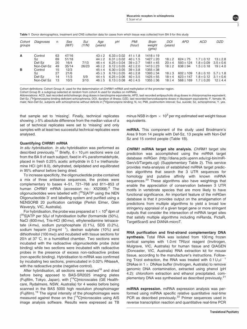
Table 1 Donor demographics, treatment and CNS collection data for cases from which tissue was collected from BA 9 for this study
Cohortgroups
Diagnoses n Sex(M/F)
Sui(Y/N)
Age(years)
pH PMI(hour)
Brainweight(gms)
DOI(years)
APD ACD DZD
A Control 63 47/16 43±2 6.33±0.02 41±1.8 1418±19Sz 69 51/18 44±2 6.31±0.02 40±1.5 1427±20 18±2 624±75 1.7±0.12 13±2.8Def-Sz 20 16/4 7/13 46±4 6.25±0.04 39±2.7 1461±45 20±4 593±124 1.8±0.09 3.5±0.6Non-Def-Sz 49 35/14 26/23 46±2 6.12±0.05 42±2.0 1413±23 18±2 638±94 1.5±0.18 19±4.0
B Control 15 12/3 45±4 6.30±0.05 39±3.8 1355±38Sz 27 21/6 45±3 6.19±0.05 40±2.8 1393±34 18±3 602±109 1.8±0.10 5.7±1.8Def-Sz 14 11/3 5/9 44±5 6.25±0.06 40±3.5 1425±55 18±4 623±147 1.8±0.12 3.1±0.6Non-Def Sz 13 10/3 3/10 46±5 6.13±0.08 40±4.5 1355±36 18±4 568±169 1.7±0.20 12±4.4
Cohort definitions: Cohort Group A: used for the determination of CHRM1 mRNA and methylation of the promoter region.Cohort Group B: a subgroup selected at random from cohort A used for studies on miRNAs.Abbreviations: ACD, last recorded anticholinergic drug doses in benztropine equivalents; APD, last recorded antipsychotic drug doses in chlorpromazine equivalent;Def-Sz, [3H]pirenzepine binding deficient schizophrenia; DOI, duration of illness; DZD, last recorded benzodiazepine doses in diazepam equivalents; F, female; M,male; Non-Def-Sz, subjects with schizophrenia without deficits in [3H]pirenzepine binding; N, no; PMI, postmortem interval; Sui, suicide; Sz, schizophrenia; Y, yes.
Muscarinic receptors in schizophreniaE Scarr et al
3
Translational Psychiatry

are provided in Supplementary Table 3. Relative expressionwas determined with respect to the geometric means of threeconstitutively expressed small nuclear RNA (U6, U44 andU49) by subtracting the geometric means of their cyclethreshold value from the cycle threshold value derived for themiRNA.23 All reactions were subjected to post amplificationdissociation curve analysis to provide guidance on reactionspecificity. Initial confirmation of amplicon length was alsoaccomplished through gel electrophoresis.
Reporter gene assay. Luciferase reporter gene analysis ofmiRNA function was conducted as described previously.24
Briefly, HEK-293 cell cultures were seeded into 24-wellplates and maintained in DMEM with 10% (vol/vol) foetal calfserum, 20 mM HEPES, 0.15% (wt/vol) sodium bicarbonateand 2 mM L-glutamine at 37 1C with 5% CO2 and 90%humidity. Transfections were performed 24 h postseedingwith recombinant CHRM1-Firefly luciferase reporter geneconstructs, 100 nM synthetic miRNA or LNA modified anti-miR oligonucleotide in Lipofectamine 2000 according tomanufacturer’s instructions (Invitrogen). Reporter gene con-structs containing 3́ UTR sequences encoding target genemiRNA recognition elements were produced through theligation of a synthetic double stranded cassette compatiblewith SpeI and Hind III digested pMIR-REPORT vector(Invitrogen) (see Supplementary Figure 2). Reporter genesilencing in response to miRNA cotransfection was monitoredwith respect to a control plasmid expressing Renilla luciferase(pRL-TK) using the dual luciferase reporter assay (Promega,Alexandria, NSW, Australia). Non-specific effects associatedwith transfection were controlled by comparison to cellscotransfected with mutant miRNAs or mutant anti-miRs.
[3H]pirenzepine binding. The data on [3H]pirenzepinebinding used in this study was originally generated in anearlier study.3 The data pertaining to the relevant cohortswas collated and then reanalysed within the parameters ofeach portion of this study.
Statistics. The distribution of all data was determined usingthe D’Agostino and Pearson omnibus normality test as this isbest for determining data distribution in small cohorts.25
Demographic, pharmacological and CNS collection datawere compared using a student’s t-test at the levels of Szand a one-way analysis of variance when comparing Def-Szand Non-Def-Sz to controls. Where data were found to bemixed between parametric and non-parametric distributionsthe General Linear Model was used to identify significantvariance whereas Mann–Whitney U or Kruskal–Wallis testswere used when all data to be analysed was shown to have anon-parametric distribution. Gender frequency across diag-nostic cohorts was assessed using the w2 test. Potentialbiological relationships and confounding factors were inte-grated using linear regression analysis. Due to small cohortsizes only strong relationships (r2¼ 0.49) where the regres-sion deviated significantly were considered as being ofinterest.26
Most analyses were conducted using Prism 5.01 (Graph-pad Software, La Jolla, CA, USA) but the General LinearModel was computed using Minitab 15 (Minitab, State
College, PA, USA) while the methylation data was analysedusing Stata 11 (StataCorp., 2010; College Station, TX, USA).
Results and discussion
Demographics, pharmacological history and CNScollection datamRNA and methylation cohorts:. The cases used in thiscomponent of the study were part of the cohorts used in thestudy that identified Def-Sz27 with the cohort sizes beinglimited by availability of tissue from BA 9. There were nosignificant differences in mean age (P¼ 0.63; P¼ 0.92), PMI(P¼ 0.75; P¼ 0.84), CNS pH (P¼ 0.19; P¼ 0.10), CNSweight (P¼ 0.54, P¼ 0.23) or gender frequency (P¼ 1.00;P¼ 0.11) when comparing Sz to control or Def-Sz, Non-Def-Sz and controls, respectively, for the larger cohorts (Table 1:Cohort Groups A). In comparing Def-Sz and Non-Def-Szthere were no significant differences in mean duration ofillness (P¼ 0.87), the last recorded dose of antipsychotic(P¼ 0.73) or anticholinergic (P¼ 0.15) drugs but the Def-Szhad lower final recorded doses of benzodiazepines(P¼ 0.01). The frequency of benzodiazepine prescriptiondid not differ between Def-Sz and Non-Def-Sz (P¼ 0.60).This would suggest that the symptoms of schizophreniatargeted by benzodiazepines (distress, insomnia and beha-vioural disturbances secondary to psychosis28) are moreresponsive to benzodiazepine treatment in subjects with Def-Sz by a mechanism that has yet to be understood.
miRNA cohort:. The cohorts for this study were selectedbased on diagnoses, whether or not they were shown to beDef-Sz or Non-Def-Sz by measuring [3H]pirenzepinebinding, the availability of tissue from BA 9 and limitationsin sample processing capacity of the technique. Togetherthese factors contributed to resulted in smaller cohorts beingstudied when measuring miRNA. There were no significantdifferences in mean age (P¼ 0.55; P¼ 0.83), PMI (P¼ 0.71;P¼ 0.93), CNS pH (P¼ 0.11; P¼ 0.16), CNS weight(P¼ 0.44; P¼ 0.29) or gender frequency (P¼ 1.00; P¼ 0.99)when comparing Sz to control or Def-Sz, Non-Def-Sz andcontrols, respectively (Table 1: Cohort Groups B). Incomparing Def-Sz and Non-Def-Sz there were no significantdifferences in mean duration of illness (P¼ 0.96), the lastrecorded dose of antipsychotic drugs (P¼ 0.59) or antic-holinergic drugs (P¼ 0.50) but the Def-Sz had lower finalrecorded doses of benzodiazepines (P¼ 0.04).
CHRM1 Gene methylation. Ten potential CpG sites wereidentified across the putative CHRM1 promoter region(Supplementary Figure 1). There was a significant reductionin levels of methylation in four of these sites in people with Sz(Table 2) with further analyses revealing a significantreduction in levels of DNA methylation in three sites inpeople with Def-Sz and Non-Def-Sz (Table 3), plus anaddition site in the Non-Def-Sz. Examining the pooledmethylation data showed that there was a significantdecrease in methylation associated with the diagnoses ofSz but not with other factors such as suicide, gender, age,tissue pH or PMI (Table 4). In addition, there were verysimilar reductions in DNA methylation in all CpG sites in
Muscarinic receptors in schizophreniaE Scarr et al
4
Translational Psychiatry
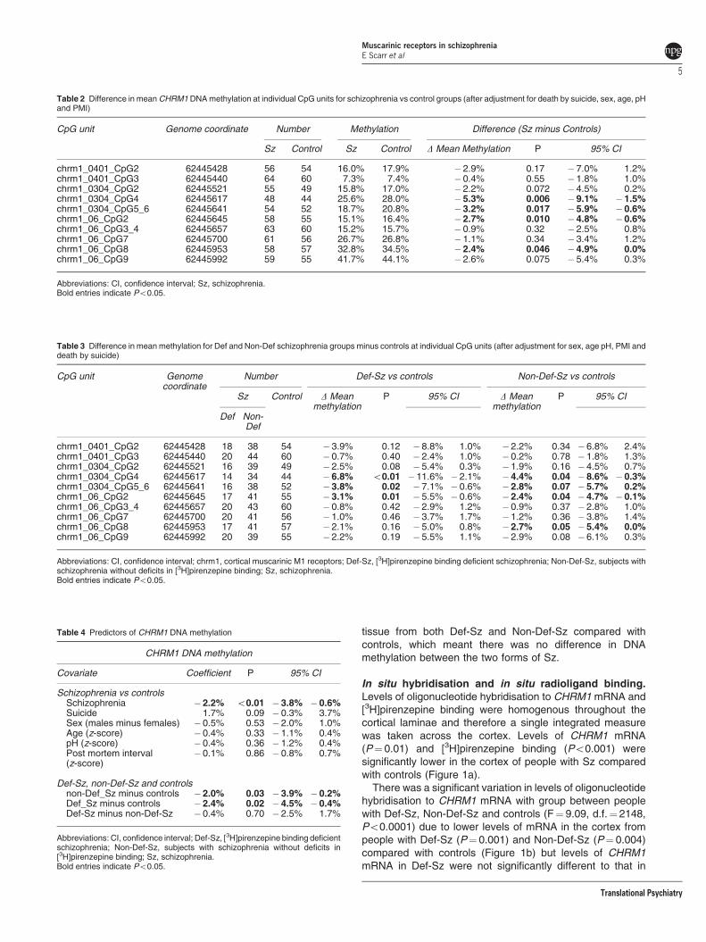
tissue from both Def-Sz and Non-Def-Sz compared withcontrols, which meant there was no difference in DNAmethylation between the two forms of Sz.
In situ hybridisation and in situ radioligand binding.Levels of oligonucleotide hybridisation to CHRM1 mRNA and[3H]pirenzepine binding were homogenous throughout thecortical laminae and therefore a single integrated measurewas taken across the cortex. Levels of CHRM1 mRNA(P¼ 0.01) and [3H]pirenzepine binding (Po0.001) weresignificantly lower in the cortex of people with Sz comparedwith controls (Figure 1a).
There was a significant variation in levels of oligonucleotidehybridisation to CHRM1 mRNA with group between peoplewith Def-Sz, Non-Def-Sz and controls (F¼ 9.09, d.f.¼ 2148,Po0.0001) due to lower levels of mRNA in the cortex frompeople with Def-Sz (P¼ 0.001) and Non-Def-Sz (P¼ 0.004)compared with controls (Figure 1b) but levels of CHRM1mRNA in Def-Sz were not significantly different to that in
Table 2 Difference in mean CHRM1 DNA methylation at individual CpG units for schizophrenia vs control groups (after adjustment for death by suicide, sex, age, pHand PMI)
CpG unit Genome coordinate Number Methylation Difference (Sz minus Controls)
Sz Control Sz Control D Mean Methylation P 95% CI
chrm1_0401_CpG2 62445428 56 54 16.0% 17.9% �2.9% 0.17 � 7.0% 1.2%chrm1_0401_CpG3 62445440 64 60 7.3% 7.4% �0.4% 0.55 � 1.8% 1.0%chrm1_0304_CpG2 62445521 55 49 15.8% 17.0% �2.2% 0.072 � 4.5% 0.2%chrm1_0304_CpG4 62445617 48 44 25.6% 28.0% �5.3% 0.006 � 9.1% �1.5%chrm1_0304_CpG5_6 62445641 54 52 18.7% 20.8% �3.2% 0.017 � 5.9% �0.6%chrm1_06_CpG2 62445645 58 55 15.1% 16.4% �2.7% 0.010 � 4.8% �0.6%chrm1_06_CpG3_4 62445657 63 60 15.2% 15.7% �0.9% 0.32 � 2.5% 0.8%chrm1_06_CpG7 62445700 61 56 26.7% 26.8% �1.1% 0.34 � 3.4% 1.2%chrm1_06_CpG8 62445953 58 57 32.8% 34.5% �2.4% 0.046 � 4.9% 0.0%chrm1_06_CpG9 62445992 59 55 41.7% 44.1% �2.6% 0.075 � 5.4% 0.3%
Abbreviations: CI, confidence interval; Sz, schizophrenia.Bold entries indicate Po0.05.
Table 3 Difference in mean methylation for Def and Non-Def schizophrenia groups minus controls at individual CpG units (after adjustment for sex, age pH, PMI anddeath by suicide)
CpG unit Genomecoordinate
Number Def-Sz vs controls Non-Def-Sz vs controls
Sz Control D Meanmethylation
P 95% CI D Meanmethylation
P 95% CI
Def Non-Def
chrm1_0401_CpG2 62445428 18 38 54 � 3.9% 0.12 � 8.8% 1.0% �2.2% 0.34 �6.8% 2.4%chrm1_0401_CpG3 62445440 20 44 60 � 0.7% 0.40 � 2.4% 1.0% �0.2% 0.78 �1.8% 1.3%chrm1_0304_CpG2 62445521 16 39 49 � 2.5% 0.08 � 5.4% 0.3% �1.9% 0.16 �4.5% 0.7%chrm1_0304_CpG4 62445617 14 34 44 � 6.8% o0.01 �11.6% � 2.1% �4.4% 0.04 �8.6% � 0.3%chrm1_0304_CpG5_6 62445641 16 38 52 � 3.8% 0.02 � 7.1% � 0.6% �2.8% 0.07 �5.7% 0.2%chrm1_06_CpG2 62445645 17 41 55 � 3.1% 0.01 � 5.5% � 0.6% �2.4% 0.04 �4.7% � 0.1%chrm1_06_CpG3_4 62445657 20 43 60 � 0.8% 0.42 � 2.9% 1.2% �0.9% 0.37 �2.8% 1.0%chrm1_06_CpG7 62445700 20 41 56 � 1.0% 0.46 � 3.7% 1.7% �1.2% 0.36 �3.8% 1.4%chrm1_06_CpG8 62445953 17 41 57 � 2.1% 0.16 � 5.0% 0.8% �2.7% 0.05 �5.4% 0.0%chrm1_06_CpG9 62445992 20 39 55 � 2.2% 0.19 � 5.5% 1.1% �2.9% 0.08 �6.1% 0.3%
Abbreviations: CI, confidence interval; chrm1, cortical muscarinic M1 receptors; Def-Sz, [3H]pirenzepine binding deficient schizophrenia; Non-Def-Sz, subjects withschizophrenia without deficits in [3H]pirenzepine binding; Sz, schizophrenia.Bold entries indicate Po0.05.
Table 4 Predictors of CHRM1 DNA methylation
CHRM1 DNA methylation
Covariate Coefficient P 95% CI
Schizophrenia vs controlsSchizophrenia �2.2% o0.01 � 3.8% � 0.6%Suicide 1.7% 0.09 � 0.3% 3.7%Sex (males minus females) �0.5% 0.53 � 2.0% 1.0%Age (z-score) �0.4% 0.33 � 1.1% 0.4%pH (z-score) �0.4% 0.36 � 1.2% 0.4%Post mortem interval(z-score)
�0.1% 0.86 � 0.8% 0.7%
Def-Sz, non-Def-Sz and controlsnon-Def_Sz minus controls �2.0% 0.03 � 3.9% � 0.2%Def_Sz minus controls �2.4% 0.02 � 4.5% � 0.4%Def-Sz minus non-Def-Sz �0.4% 0.70 � 2.5% 1.7%
Abbreviations: CI, confidence interval; Def-Sz, [3H]pirenzepine binding deficientschizophrenia; Non-Def-Sz, subjects with schizophrenia without deficits in[3H]pirenzepine binding; Sz, schizophrenia.Bold entries indicate Po0.05.
Muscarinic receptors in schizophreniaE Scarr et al
5
Translational Psychiatry

Non-Def-Sz (P¼ 0.63). As would be predicted, there was alsosignificant variation in levels of [3H]pirenzepine binding withgroup (F¼ 95.6, d.f.¼ 2148, Po0.0001) due to decreasedlevels of binding in Def-Sz (Po0.0001) and Non-Def-Sz(P¼ 0.02) compared with controls.
miRNA. Initially, to identify potential miRNA regulators ofCHRM1 we performed a low stringency search of themiRGen miRNA target prediction database, using the ‘union’of their algorithms.20 This suggested that CHRM1 could beinfluenced by up to 32 different miRNA (SupplementaryTable 2). However, a more stringent appraisal of targetsusing the ‘intersection’ of different algorithms identified asingle miR-107 as the miRNA most likely to target CHRM1expression. The miR107 target site in the CHRM1 3́ UTR isonly one of two that display a highly conserved complete sevennucleotide seed-paring region (Supplementary Figure 2) and,additionally, has previously been shown to be increased inpostmortem superior temporal cortex and dorsolateral pre-frontal cortex from people with Sz.23 Similar analyses sug-gested it was highly unlikely that miR-16 would target CHRM1expression and so levels of this miR were measured todetermine if any change in miR-107 was due to a generalisedchange in levels of miR in the cortex of people with Sz.
In the cohorts used in the study of miRNA, levels of cortical[3H]pirenzepine binding (P¼ 0.0004) and CHRM1 mRNA(P¼ 0.03) were decreased in BA 9 from people with Sz(Figure 2a) but neither levels of miR-16 (P¼ 0.50) nor miR-107 (P¼ 0.19) varied significantly with diagnosis (Figure 2b).
In comparing data from Def-Sz, Non-Def-Sz and controls,levels of miR-107 (F¼ 4.25, d.f.¼ 2,40, P¼ 0.02) and [3H]pirenzepine binding (F¼ 72.3, d.f.¼ 2,40, Po0.0001) variedwith group, levels of CHRM1 mRNA showed a trend to varyingwith group (F¼ 2.68, d.f.¼ 2,40, P¼ 0.08) whereas levels ofmiR-16 did not vary with group (F¼ 0.65, d.f.¼ 2,40, P¼ 0.52)(Figures 2c and d). The variance in miR-107 was due toincreased levels of that miRNA in the cortex of people withDef-Sz compared with controls (Po0.05) but levels of miR-107 were not significantly different between the Non-Def-Szand controls. The variation in [3H]pirenzepine binding withgroup was due to decreased levels of binding in cortex frompeople with Def-Sz (Po0.001), but not Non-Def-Sz, com-pared with controls. The trend to variance in levels of CHRM1
Figure 1 Levels (Mean±s.e.m.) of [3H]pirenzepine binding and oligonucleotideCHRM1 probe hybridisation to Brodmann’s area 9 (BA 9) from subjects with. (a):schizophrenia and controls and (b): Def-Sz, Non-Def-Sz and controls.
Figure 2 (a): Levels (Mean±s.e.m.) of [3H]pirenzepine binding and oligonucleotide CHRM1 probe hybridisation to BA 9 from the cohort of subjects with schizophreniaand control subjects used for the study of miRNA and. (b): Levels (Mean±s.e.m.) of miR-16 and miR-107 in the same region from the individuals. (c): Levels (Mean±s.e.m.)of [3H]pirenzepine binding and oligonucleotide CHRM1 probe hybridisation to BA 9 from the cohort of subjects with Def-Sz, Non-Def-Sz and control subjects used for the studyof miRNA and (d): Levels (Mean±s.e.m.) of miR-16 and miR-107 in the same region from the individuals.
Muscarinic receptors in schizophreniaE Scarr et al
6
Translational Psychiatry

mRNA was predominantly due to lower levels in the Def-Szgroup (t-test compared with controls P¼ 0.03) rather from theNon-Def-Sz (t-test compared with controls P¼ 0.67).
Potential biological relationships. For the cohorts ofpeople used for the study of [3H]pirenzepine binding, CHRM1mRNA and gene promoter methylation, there were no strongcorrelations between levels of [3H]pirenzepine binding andlevels of mRNA (Supplementary Table 4). There were nocorrelations between any levels of CpG methylation and anydemographic, brain collection of pharmacological variables(data not shown). Significantly, in the cohorts used for thestudy of miRNA there were correlations between levels ofmiR-16 and miR-107, but not any of the other measures in alldiagnostic cohorts (Figure 3 and Supplementary Table 5).
CHRM1 reporter expression is modulated by intracellu-lar miR-107. To further investigate the role of miR-107 in theregulation of CHRM1 we cloned a segment of its 3́ UTRdownstream of the Firefly luciferase reporter gene in pMIR-REPORT and analysed reporter gene expression in HEK293cells cotransfected with synthetic miR-107 or LNA modifiedantimiR-107 (miR-107 inhibitor). In each case, the Firefly
luciferase gene dosage was normalised by Renilla luciferaseexpressed from cotransfected pRL-TK vector and comparedwith control transfections containing mutant versions of miR-107 (and the corresponding LNA modified antimiRs).Following transfection, analysis of luciferase activity demon-strated that the addition of miR-107 was capable of reducingCHRM1 reporter gene expression by 20% (P¼ 0.004)(Figure 4). When the endogenous miR-107 was depleted orinhibited by its cognate anti-miRs, the reporter geneexpression was increased by 62% (P¼ 0.03). Theseobservations, showing gene silencing in the presence ofmiR-107 over-expression and a reduction of this effect whenmiR-107 is inhibited, demonstrate that this region of CHMR13’ UTR is potently regulated by miR-107, at least in vitro.
Potential confounds. The only strong and significantrelationships between experimental measures and potentialnumeric confounds was between [3H]pirenzepine bindingand final recorded benzodiazepine drug dose (r2¼ 0.68,Po0.001) in people with Sz and between miR-107 and PMI(r2¼ 0.47, Po0.01) in Non-Def-Sz (Supplementary Table 4).Effects of these relationships on the original analyses usingan analysis of covariance showed that final recordedbenzodiazepine drug dose as a covariate did not affect thevariation of [3H]pirenzepine binding with diagnoses (F¼ 101,d.f.¼ 12 129, Po0.0001). When PMI was included as acovariate in the analyses of miR-107 it is notable that there isan increase in the variance associated with group (F¼ 4.94,d.f.¼ 1,2,39, P¼ 0.01).
There was no effect of either gender (P¼ 0.63) or suicide(P¼ 0.17) on levels of [3H]pirenzepine binding in eitherpeople with Sz as a whole or their age/ sex matched controls.Levels of CHRM1 mRNA did not vary with gender or suicide inthese cohorts (P¼ 0.41 and P¼ 0.26, respectively). In thecohorts used for the study of miRNA; levels of [3H]pirenzepinebinding (P¼ 0.63), CHRM1 mRNA (P¼ 0.87), miR-16(P¼ 0.78) or miR-107 (P¼ 0.30) did not vary with genderwhen people with Sz were compared with controls. Similarly,levels of [3H]pirenzepine binding (P¼ 0.46), CHRM1 mRNA(P¼ 0.50), miR-16 (P¼ 0.26) and miR-107 (P¼ 0.13) did notvary with suicide.
This study explores the potential mechanisms that couldcontribute to widely replicated finding of reduced levels ofcortical [3H]pirenzepine binding in people with Sz3,6,29–32
taking into account that, under the conditions used in mostprotocols, the radioligand will show aX80% selectivity forCHRM1.4,5 In addition, it explores whether changes in thestatus of markers of CHRM1 expression may differentiatepeople with Def-Sz, who have a B75% loss of cortical [3H]pirenzepine binding,3 from Non-Def-Sz. Consistent withprevious findings6,33 we show a decrease in levels of corticalCHRM1 mRNA in Sz that is equivocal across Def-Sz andNon-Def-Sz. Our data also shows a decrease in levels ofmethylation of the putative promoter region of the CHRM1gene in Sz. There was a significant decrease in the levels ofmethylation in four CpG sites in Non-Def-Sz whereas onlythree of these sites had significant decrease in levels of genemethylation in Def-Sz. This being noted, our overall analysessuggested differences in CHRM1 gene promoter methylationdid not permit separation of the two groups. The presence of
Figure 3 The relationship between levels of miR-16 and miR-107 in BA 9 fromsubjects with schizophrenia, Def-Sz, Non-Def-Sz and controls.
Figure 4 Effect (Mean±s.e.m.) of miR-107 and antimiR-107 on the activity of aCHRM1 reported gene construct. (a): Po0.05; (b): Po0.005.
Muscarinic receptors in schizophreniaE Scarr et al
7
Translational Psychiatry

low levels of CHRM1 binding and mRNA has a symmetry but itis generally argued that increased gene methylation isassociated with gene silencing and lower levels of mRNA.34
Thus, on the one hand our data is consistent with thesuggestion that Sz is associated with a hypo-methylationstate35 but, on the other hand, our data also suggests such astate may not be associated with the expected increase ingene expression in the CNS of people with the disorder. Thus,our data leads us to postulate that the pathophysiology of Szmay interfere with the relationship between gene promotermethylation and levels of the gene expression.
Our study showed levels of miR-107, but not miR-16, wereincreased in the cortex from people with Def-Sz, but not Non-Def-Sz. Significantly, from sequence homology, miR-107 waspredicted to target CHRM1 expression whereas miR-16 wouldnot target CHRM1 mRNA. The notion that miR-107 targetsCHRM1 expression is supported by our data showing chang-ing miR-107 levels appropriately changes the activity of areporter gene linked to levels of CHRM1 expression. There-fore our data suggests that CHRM1 gene expression in peoplewith Def-Sz is affected by the increased activity of at leastone CHRM1 targeting miRNA and this unique pathophysiol-ogy could be causing the large decrease in cortical CHRM1 inthese people. Overall, our data suggests that the changes inthe regulation of CHRM1 expression is not a straightforwardprogression whereby changes in levels of mRNA predictchanges in levels of protein; rather our data is in keeping withthe concept of a derangement of the highly complex system ofchecks and balances regulating expression in the CNS.36
This study, for the first time, to our knowledge, offers anexplanation as to an underlying mechanism that may becontributing to the marked decrease in cortical CHRM1 levelsin Def-Sz. Our data suggests this mechanism involvesincreased levels of at least one miR, miR-107, in the cortexof people with Def-Sz and our in vitro data shows this increasein miR-107 would act to decrease CHRM1 expression and/ortranslation. Such a functional link is supported by our datashowing a negative correlation between [3H]pirenzepinebinding and levels of miR-107 (r¼ � 0.3671, P¼ 0.001) inthe cortex of people with Def-Sz that was not detectable intissue from Non-Def-Sz and controls. We interpret these dataas suggesting a stronger relationship between miR-107 andlevels of CHRM1 in Def-Sz that would be expected if miR-107was a significant factor in its pathophysiology. Importantly,levels of a miR-16, that does not target CHRM1 expression, isnot altered in the cortex of Def-Sz and Non-Def-Sz showingthe changes in miR-107 are not reflecting generalisedchanges in miR in the cortex of people with Def-Sz.Coalescing our data on CHRM1 binding, CHRM1 mRNAand miR-107 we now hypothesise that the marked decreasein CHRM1 levels in Def-Sz is due to the cumulative effects ofdecrease gene expression being amplified by the effects ofincreased CHRM1 targeting miRNA activity.
Our data also adds to a growing body of data implicatinggene methylation in the pathophysiology of Sz.37–41 However,our data differs in that it shows a decrease in CHRM1expression (as measured by levels of mRNA) in the presenceof decreased gene methylation, which would normally beexpected to cause a decrease in gene silencing (that is,increased expression).34 Significantly, the expected inverse
relationship between methylation and levels of gene expres-sion has been reported in the face of decreased gene copynumber in transgenic animals.42 Levels of gene methylationhave long been known to be subject to feed-back loopregulation and it has been suggested that disruption of suchprocesses could result in disease states.43 Thus, our datamay reflect a disruptive feed-back loop involving methylationof CHRM1 as part of the pathophysiology of Sz because CNSgene methylation is a dynamic process that varies during life44
and the decreased methylation of the CHRM1 promoterregion we report in Sz could be part of a feedback mechanismtrying to turn on the expression of CHRM1 in an attempt tocompensate for low levels of cortical mRNA and/or protein.
In conclusion, our study has identified changes in multiplemarkers (gene promoter methylation, levels of mRNA andprotein) of CHRM1 gene expression in the dorsolateralprefrontal cortex from people with Sz. However, being ableto analyse our data at the subsyndrome level has revealed anincrease in a CHRM1 mRNA targeting miR (miR-107) only inthe cortex of people with Def-Sz. We therefore hypothesisethat this extra burden on CHRM1 gene expression is asignificant contributor to the marked loss in cortical CHRM1 bywhich this subset of people is defined. Our data also supportsthat of others that have shown changes in levels of miRNA inthe CNS of subjects with schizophrenia the suggest a role formiRNAs in the pathophysiology of Sz23 but extends thishypothesis to suggest that changes in miRNA levels maydifferentiate some subsets of people within the syndrome.However, we acknowledge that our sample size for thisportion of our study is small and therefore our results must beregarded with caution until further data is obtained.
Our findings on the mechanisms underpinning changes inCHRM1 in Sz comes at a critical time given the developmentof CHRM1 allosteric modulates which, for the first time, openup the possibility of specifically targeting that receptor to bringabout therapeutic benefits.45 Significantly, data on these newcompounds support the hypothesis that they will be useful inreducing the symptoms of Sz46 but our data may indicate theymay be useful in treating selective subgroups of people withthe disorder. Hence, a high priority should be given todeveloping more selective CHRM1 ligands for neuroimaging,rather than the pan-muscarinic receptor ligand used to showdecreased CHRMs in living people with Sz,47 to facilitate thetesting of these new drugs in subjects who have a marked lossof CHRM1 compared to those who have not. Finally, our datastrongly supports the argument that subdividing the syndromeof Sz will facilitate an improved understanding of itscomponent disorders1 and that this understanding may leadto more personalised medicines with which to treat thesecomponent disorders.48
Conflict of interest
The authors declare no conflict of interest.
Acknowledgements. The authors are grateful for the excellent technicalassistance of Mr Geoff Pavey. Tissues were received from the Victorian Brain BankNetwork, which is supported by the Mental Health Research Institute, The Alfred,Victorian Forensic Institute of Medicine and The University of Melbourne and fundedby Australia’s National Health and Medical Research Council, Helen Macpherson
Muscarinic receptors in schizophreniaE Scarr et al
8
Translational Psychiatry
Smith Trust, Parkinson’s Victoria and Perpetual Philanthropic Services. Sources ofsupport: BD is an NHMRC Senior Research Fellow (APP1002240). ES is an ARCFuture Fellow (FT100100689). MSS is a recipient of a University of MelbourneInternational Research Scholarship. This research was principally funded by theNHMRC (Project Grant no. 509333 and 628669) but was also funded in part by theVictorian Government’s Operational Infrastructure Support Programme, theRebecca L Cooper Medical Research Foundation and the Wood’s Family ResearchProgram.
1. Tamminga CA. Accelerating new knowledge in schizophrenia. Am J Psychiatry 2008; 165:949–951.
2. Raedler TJ, Bymaster FP, Tandon R, Copolov D, Dean B. Towards a muscarinichypothesis of schizophrenia. Mol Psychiatry 2007; 12: 232–246.
3. Scarr E, Cowie TF, Kanellakis S, Sundram S, Pantelis C, Dean B. Decreased corticalmuscarinic receptors define a subgroup of subjects with schizophrenia. Mol Psychiatry2009; 14: 1017–1023.
4. Scarr E, Dean B. Muscarinic receptors: do they have a role in the pathology and treatmentof schizophrenia? J Neurochem 2008; 107: 1188–1195.
5. Gibbons AS, Scarr E, Boer S, Money T, Jeon WJ, Felder C et al. Widespread decreasesin cortical muscarinic receptors in a subset of people with schizophrenia. Int JNeuropsychopharmacol; S1461145712000028 [pii]; doi:10.1017/S1461145712000028(in press).
6. Dean B, McLeod M, Keriakous D, McKenzie J, Scarr E. Decreased muscarinic(1) receptorsin the dorsolateral prefrontal cortex of subjects with schizophrenia. Mol Psychiatry 2002; 7:1083–1091.
7. Scarr E, Keriakous D, Crossland N, Dean B. No change in cortical muscarinic M2, M3receptors or [35S]GTPgammaS binding in schizophrenia. Life Sci 2006; 78: 1231–1237.
8. Hill C, Keks N, Roberts S, Opeskin K, Dean B, Mackinnon A et al. Problem of diagnosis inpostmortem brain studies of schizophrenia. Am J Psychiatry 1996; 153: 533–537.
9. Roberts SB, Hill CA, Dean B, Keks NA, Opeskin K, Copolov DL. Confirmation of thediagnosis of schizophrenia after death using DSM-IV: a Victorian experience. Aust N Z JPsychiatry 1998; 32: 73–76.
10. Remington GJ. Antipsychotics (Neuroleptics). In B-Bjai JJ (ed) Handbook of PsychotropicDrugs. Hogrefe & Huber: Seattle, Toronto, Gottingen, Bern, 1999, pp 55–84.
11. Dean B, Pavey G, Chai SY, Mendelsohn FAO. The localisation and quantification ofmolecular changes in the human brain using in situ radioligand binding andautoradiography. In Dean B, Kleinman JE, Hyde TM (ed) Using CNS tissue in psychiatricresearch: A practical guide. Harwood Academic Press: Amsterdam, 1999, pp 67–83.
12. Stan AD, Ghose S, Gao XM, Roberts RC, Lewis-Amezcua K, Hatanpaa KJ et al. Humanpostmortem tissue: what quality markers matter? Brain Res 2006; 1123: 1–11.
13. Kingsbury AE, Foster OJ, Nisbet AP, Cairns N, Bray L, Eve DJ et al. Tissue pH as anindicator of mRNA preservation in human post-mortem brain. Brain Res Mol Brain Res1995; 28: 311–318.
14. Ollikainen M, Smith KR, Joo EJ, Ng HK, Andronikos R, Novakovic B et al. DNA methylationanalysis of multiple tissues from newborn twins reveals both genetic and intrauterinecomponents to variation in the human neonatal epigenome. Hum Mol Genet 2010; 19:4176–4188.
15. Ewing B, Green P. Base-calling of automated sequencer traces using phred. II. Errorprobabilities. Genome Res 1998; 8: 186–194.
16. Fraga MF, Esteller M. DNA methylation: a profile of methods and applications.Biotechniques 2002; 33: 632, 634, 636–649.
17. Scarr E, Sundram S, Keriakous D, Dean B. Altered hippocampal muscarinic m4, but notm1, receptor expression from subjects with schizophrenia. Biol Psychiatry 2007; 61:1161–1170.
18. Loiacono RE, Gundlach AL. In situ hybridisation histochemistry: application to human braintissue. In Dean B, Hyde TM, Kleinman JE (ed) Using CNS Tissue in Psychiatric Research:A Practical Guide. Harwood Academic Press: Sydney, 1999, pp 85–106.
19. Dean B, Pavey G, Opeskin K. [3H]raclopride binding to brain tissue from subjects withschizophrenia: methodological aspects. Neuropharmacology 1997; 36: 779–786.
20. Megraw M, Sethupathy P, Corda B, Hatzigeorgiou AG. miRGen: a database for the studyof animal microRNA genomic organization and function. Nucleic Acids Res 2007; 35:D149–D155.
21. Udawela M, Scarr E, Hannan AJ, Thomas EA, Dean B. Phospholipase C beta 1 expressionin the dorsolateral prefrontal cortex from patients with schizophrenia at different stages ofillness. Aust N Z J Psychiatry 2011; 45: 140–147.
22. Santarelli DM, Beveridge NJ, Tooney PA, Cairns MJ. Upregulation of dicer and microRNAexpression in the dorsolateral prefrontal cortex Brodmann area 46 in schizophrenia. BiolPsychiatry 2011; 69: 180–187.
23. Beveridge NJ, Gardiner E, Carroll AP, Tooney PA, Cairns MJ. Schizophrenia is associatedwith an increase in cortical microRNA biogenesis. Mol Psychiatry 2010; 15: 1176–1189.
24. Beveridge NJ, Tooney PA, Carroll AP, Tran N, Cairns MJ. Down-regulation of miR-17family expression in response to retinoic acid induced neuronal differentiation. Cell Signal2009; 21: 1837–1845.
25. D’Agostino RB, Belanger A, D’Agostino RB Jnr. A suggestion for using powerful andinformative tests of normality. Am Stat 1990; 44: 316–321.
26. Cook RD, Weisberg S. Applied Regression Including Computing and Graphics. Wiley:Hoboken, 2011.
27. Dean B, Boer S, Gibbons A, Money T, Scarr E. Recent advances in postmortem pathologyand neurochemistry in schizophrenia. Curr Opin Psychiatry 2009; 22: 154–160.
28. Falkai P, Wobrock T, Lieberman J, Glenthoj B, Gattaz WF, Moller HJ. World Federation ofSocieties of Biological Psychiatry (WFSBP) guidelines for biological treatment ofschizophrenia, Part 1: acute treatment of schizophrenia. World J Biol Psychiatry 2005;6: 132–191.
29. Crook JM, Tomaskovic-Crook E, Copolov DL, Dean B. Low muscarinic receptorbinding in prefrontal cortex from subjects with schizophrenia: a study of Brodmann’s areas8, 9, 10, and 46 and the effects of neuroleptic drug treatment. Am J Psychiatry 2001; 158:918–925.
30. Zavitsanou K, Katsifis A, Filomena M, Xu-Feng H. Investigation of M1/M4 muscarinicreceptors in the anterior cingulate cortex in schizophrenia, bipolar disorder, and majordepression disorder. Neuropsychopharmacology 2004; 29: 619–625.
31. Deng C, Huang XF. Decreased density of muscarinic receptors in the superior temporalgyrus in schizophrenia. J Neurosci Res 2005; 81: 883–890.
32. Newell KA, Zavitsanou K, Jew SK, Huang XF. Alterations of muscarinic and GABA receptorbinding in the posterior cingulate cortex in schizophrenia. Prog NeuropsychopharmacolBiol Psychiatry 2007; 31: 225–233.
33. Mancama D, Arranz MJ, Landau S, Kerwin R. Reduced expression of the muscarinic 1receptor cortical subtype in schizophrenia. Am J Med Genet B Neuropsychiatr Genet 2003;119: 2–6.
34. Turker MS. Gene silencing in mammalian cells and the spread of DNA methylation.Oncogene 2002; 21: 5388–5393.
35. Melas PA, Rogdaki M, Osby U, Schalling M, Lavebratt C, Ekstrom TJ. Epigeneticaberrations in leukocytes of patients with schizophrenia: association of global DNAmethylation with antipsychotic drug treatment and disease onset. FASEB J 2012; 26:2712–2718.
36. Qureshi IA, Mehler MF. The emerging role of epigenetics in stroke: II. RNA regulatorycircuitry. Arch Neurol 2010; 67: 1435–1441.
37. Huang HS, Akbarian S. GAD1 mRNA expression and DNA methylation in prefrontal cortexof subjects with schizophrenia. PLoS ONE 2007; 2: e809.
38. Iwamoto K, Bundo M, Yamada K, Takao H, Iwayama-Shigeno Y, Yoshikawa T et al. DNAmethylation status of SOX10 correlates with its downregulation and oligodendrocytedysfunction in schizophrenia. J Neurosci 2005; 25: 5376–5381.
39. Grayson DR, Chen Y, Costa E, Dong E, Guidotti A, Kundakovic M et al. The human reelingene: transcription factors (þ ), repressors (� ) and the methylation switch (þ /� ) inschizophrenia. Pharmacol Ther 2006; 111: 272–286.
40. Veldic M, Caruncho HJ, Liu WS, Davis J, Satta R, Grayson DR et al.DNA-methyltransferase 1 mRNA is selectively overexpressed in telencephalicGABAergic interneurons of schizophrenia brains. Proc Natl Acad Sci USA 2004; 101:348–353.
41. Abdolmaleky HM, Yaqubi S, Papageorgis P, Lambert AW, Ozturk S, Sivaraman V et al.Epigenetic dysregulation of HTR2A in the brain of patients with schizophrenia and bipolardisorder. Schizophr Res 2011; 129: 183–190.
42. Garrick D, Fiering S, Martin DI, Whitelaw E. Repeat-induced gene silencing in mammals.Nat Genet 1998; 18: 56–59.
43. Jaenisch R, Bird A. Epigenetic regulation of gene expression: how the genome integratesintrinsic and environmental signals. Nat Genet 2003; 33(Suppl): 245–254.
44. Puckett RE, Lubin FD. Epigenetic mechanisms in experience-driven memory formationand behavior. Epigenomics 2011; 3: 649–664.
45. Conn PJ, Christopoulos A, Lindsley CW. Allosteric modulators of GPCRs: a novelapproach for the treatment of CNS disorders. Nat Rev Drug Discov 2009; 8: 41–54.
46. Jones CK, Brady AE, Davis AA, Xiang Z, Bubser M, Tantawy MN et al. Novel selectiveallosteric activator of the M1 muscarinic acetylcholine receptor regulates amyloidprocessing and produces antipsychotic-like activity in rats. J Neurosci 2008; 28:10422–10433.
47. Raedler TJ, Knable MB, Jones DW, Urbina RA, Gorey JG, Lee KS et al. In vivodetermination of muscarinic acetylcholine receptor availability in schizophrenia. Am JPsychiatry 2003; 160: 118–127.
48. Insel TR. Disruptive insights in psychiatry: transforming a clinical discipline. J Clin Invest2009; 119: 700–705.
Translational Psychiatry is an open-access journalpublished by Nature Publishing Group. This work is
licensed under the Creative Commons Attribution-NonCommercial-NoDerivative Works 3.0 Unported License. To view a copy of this license,visit http://creativecommons.org/licenses/by-nc-nd/3.0/
Supplementary Information accompanies the paper on the Translational Psychiatry website (http://www.nature.com/tp)
Muscarinic receptors in schizophreniaE Scarr et al
9
Translational Psychiatry