The leader region of Laminin B1 mRNA confers cap-independent translation
10
Published online 29 March 2007 Nucleic Acids Research, 2007, Vol. 35, No. 8 2473–2482 doi:10.1093/nar/gkm096 The leader region of Laminin B1 mRNA confers cap-independent translation Michaela Petz 1 , Daniela Kozina 2 , Heidemarie Huber 1 , Tanja Siwiec 1 , Joachim Seipelt 3 , Wolfgang Sommergruber 2 and Wolfgang Mikulits 1, * 1 Department of Medicine I, Division: Institute of Cancer Research, Medical University of Vienna, Borschke-Gasse 8a, A-1090 Vienna, 2 Boehringer Ingelheim Austria, Dr Boehringer Gasse 5-10, A-1120 Vienna and 3 Max F. Perutz Laboratories, University Departments at the Vienna Biocenter, Department of Medical Biochemistry, Medical University of Vienna, Dr Bohr-Gasse 9, A-1030 Vienna, Austria Received November 23, 2006; Revised February 2, 2007; Accepted February 3, 2007 ABSTRACT Translation initiation of eukaryotic mRNAs generally occurs by cap-dependent ribosome scanning. However, certain mRNAs contain internal ribosome entry sites (IRES) allowing cap-independent transla- tion. Several of these IRES-competent transcripts and their corresponding proteins are involved in tumourigenesis. This study focused on IRES- driven translation control during the epithelial to mesenchymal transition (EMT) of hepatocytes that reflects crucial aspects of carcinoma progression. Expression profiling of EMT revealed Laminin B1 (LamB1) to be translationally upregulated. The 5 0 -untranslated region (UTR) of LamB1 was potent to direct IRES-dependent mRNA utilization of a bicistronic reporter construct. Stringent assays for cryptic promoter and splice sites showed no aberrantly expressed transcripts, suggesting that the reporter activity provided by the leader region of LamB1 mRNA exclusively depends on IRES. In accordance, LamB1 expression increased upon negative interference with cap-dependent transla- tion by expression of human rhinovirus 2A protease or heat shock of cells. Finally, the enhanced expression of LamB1 during EMT correlated with an elevated IRES activity. Together, these data provide first evidence that the 5 0 -UTR of LamB1 contains a bona fide IRES that directs translational upregulation of LamB1 during stress conditions and neoplastic progression of hepatocytes. INTRODUCTION Ribosome scanning is the widely used mechanism of translation initiation in eukaryotes. The scanning mechanism requires recognition of the cap structure at the 5 0 end of the mRNA by the cap-binding protein eIF4E, followed by recruitment of the activated 40S ribosomal subunit and subsequent downstream scanning along the 5 0 -untranslated region (UTR) to a suitable start codon (1). Nucleotide composition, length and secondary structures of the 5 0 -UTR determine the efficiency of translation initiation. Long, GC-rich and highly struc- tured 5 0 -UTRs reduce migration of the 43S pre-initiation complex along the RNA (2). Alternatively, a structural motif of the 5 0 -UTR, referred to as internal ribosome entry site (IRES), allows ribosomes to directly bind upstream of the start codon and to initiate translation independently of the cap structure (3). Initially described in picornavirus, the IRES-mediated translation initiation has been identified in cellular mRNAs (4,5). Escaping multiple mechanisms to regulate cap-dependent translation, internal initiation selectively allows sustained or enhanced translation of transcripts during situations with high need of corresponding proteins. Cellular IRES have been demonstrated to be active during apoptosis (6), angiogenesis (7), the G2/M phase of the cell cycle (8) or stress conditions such as hypoxia (9), amino acid starvation and heat shock (10). The observation that IRES elements are recognized in gene products regulating these processes suggests an important function of internal translational initiation in the posttranscriptional control of gene expression. IRES- driven translation further might have severe implications in tumourigenesis, since it affects the expression of platelet-derived growth factor (PDGF-B) or c-myc, which are involved in proliferation, and of apoptotic protease activating factor 1 (Apaf1) and vascular endothelial growth factor (VEGF), which govern pro- grammed cell death and angiogenesis, respectively (11–14). Evidence for the relevance of IRES-mediated translation in cancer is particularly provided by the proto- oncogene c-myc. Although deregulated transcription of c-myc is frequently observed in many cancer types, *To whom correspondence should be addressed. Tel: þ43 1 4277 65250; Fax: þ43 1 4277 65239; Email: [email protected] ß 2007 The Author(s) This is an Open Access article distributed under the terms of the Creative Commons Attribution Non-Commercial License (http://creativecommons.org/licenses/ by-nc/2.0/uk/) which permits unrestricted non-commercial use, distribution, and reproduction in any medium, provided the original work is properly cited. by guest on March 13, 2016 http://nar.oxfordjournals.org/ Downloaded from
-
Upload
independent -
Category
Documents
-
view
4 -
download
0
Transcript of The leader region of Laminin B1 mRNA confers cap-independent translation
Published online 29 March 2007 Nucleic Acids Research, 2007, Vol. 35, No. 8 2473–2482doi:10.1093/nar/gkm096
The leader region of Laminin B1 mRNA conferscap-independent translationMichaela Petz1, Daniela Kozina2, Heidemarie Huber1, Tanja Siwiec1,
Joachim Seipelt3, Wolfgang Sommergruber2 and Wolfgang Mikulits1,*
1Department of Medicine I, Division: Institute of Cancer Research, Medical University of Vienna, Borschke-Gasse8a, A-1090 Vienna, 2Boehringer Ingelheim Austria, Dr Boehringer Gasse 5-10, A-1120 Vienna and 3Max F. PerutzLaboratories, University Departments at the Vienna Biocenter, Department of Medical Biochemistry, MedicalUniversity of Vienna, Dr Bohr-Gasse 9, A-1030 Vienna, Austria
Received November 23, 2006; Revised February 2, 2007; Accepted February 3, 2007
ABSTRACT
Translation initiation of eukaryotic mRNAs generallyoccurs by cap-dependent ribosome scanning.However, certain mRNAs contain internal ribosomeentry sites (IRES) allowing cap-independent transla-tion. Several of these IRES-competent transcriptsand their corresponding proteins are involved intumourigenesis. This study focused on IRES-driven translation control during the epithelial tomesenchymal transition (EMT) of hepatocytes thatreflects crucial aspects of carcinoma progression.Expression profiling of EMT revealed Laminin B1(LamB1) to be translationally upregulated. The50-untranslated region (UTR) of LamB1 was potentto direct IRES-dependent mRNA utilization of abicistronic reporter construct. Stringent assays forcryptic promoter and splice sites showed noaberrantly expressed transcripts, suggesting thatthe reporter activity provided by the leader regionof LamB1 mRNA exclusively depends on IRES.In accordance, LamB1 expression increased uponnegative interference with cap-dependent transla-tion by expression of human rhinovirus 2A proteaseor heat shock of cells. Finally, the enhancedexpression of LamB1 during EMT correlated withan elevated IRES activity. Together, these dataprovide first evidence that the 50-UTR of LamB1contains a bona fide IRES that directs translationalupregulation of LamB1 during stress conditions andneoplastic progression of hepatocytes.
INTRODUCTION
Ribosome scanning is the widely used mechanism oftranslation initiation in eukaryotes. The scanning
mechanism requires recognition of the cap structureat the 50 end of the mRNA by the cap-binding proteineIF4E, followed by recruitment of the activated 40Sribosomal subunit and subsequent downstream scanningalong the 50-untranslated region (UTR) to a suitable startcodon (1). Nucleotide composition, length and secondarystructures of the 50-UTR determine the efficiency oftranslation initiation. Long, GC-rich and highly struc-tured 50-UTRs reduce migration of the 43S pre-initiationcomplex along the RNA (2). Alternatively, a structuralmotif of the 50-UTR, referred to as internal ribosomeentry site (IRES), allows ribosomes to directly bindupstream of the start codon and to initiate translationindependently of the cap structure (3).Initially described in picornavirus, the IRES-mediated
translation initiation has been identified in cellularmRNAs (4,5). Escaping multiple mechanisms to regulatecap-dependent translation, internal initiation selectivelyallows sustained or enhanced translation of transcriptsduring situations with high need of correspondingproteins. Cellular IRES have been demonstrated to beactive during apoptosis (6), angiogenesis (7), the G2/Mphase of the cell cycle (8) or stress conditions such ashypoxia (9), amino acid starvation and heat shock (10).The observation that IRES elements are recognized ingene products regulating these processes suggests animportant function of internal translational initiation inthe posttranscriptional control of gene expression. IRES-driven translation further might have severe implicationsin tumourigenesis, since it affects the expression ofplatelet-derived growth factor (PDGF-B) or c-myc,which are involved in proliferation, and of apoptoticprotease activating factor 1 (Apaf1) and vascularendothelial growth factor (VEGF), which govern pro-grammed cell death and angiogenesis, respectively(11–14). Evidence for the relevance of IRES-mediatedtranslation in cancer is particularly provided by the proto-oncogene c-myc. Although deregulated transcription ofc-myc is frequently observed in many cancer types,
*To whom correspondence should be addressed. Tel: þ43 1 4277 65250; Fax: þ43 1 4277 65239; Email: [email protected]
� 2007 The Author(s)
This is an Open Access article distributed under the terms of the Creative Commons Attribution Non-Commercial License (http://creativecommons.org/licenses/
by-nc/2.0/uk/) which permits unrestricted non-commercial use, distribution, and reproduction in any medium, provided the original work is properly cited.
by guest on March 13, 2016
http://nar.oxfordjournals.org/D
ownloaded from
oncogenic gain-of-function can arise from changes intranslation such as the C–T mutation in the IRES ofc-myc that results in increased expression in cells derivedfrom patients with multiple myeloma (15).Ras and PI3K/AKT signalling pathways, which are
frequently activated in tumours, play an important role intranslational regulation and malignant transformation(16). For example, PI3K/AKT/mTOR regulates proteinsynthesis by activating the ribosomal protein S6 andtranslation initiation factors important for the ribosomerecruitment to the mRNA (17). Overexpression of theeukaryotic initiation factors (eIF)4G and eIF4E has beendemonstrated in various types of tumours which leadsto the increased efficiency of cap-dependent translation(16,17). Elevated levels of rate-limiting eIF4E selectivelyenhance the translation of highly structured mRNAswhich even contain IRES such as VEGF, ornithinedecarboxylase (ODC) or fibroblast growth factor 2 (18).The acquisition of invasive and metastatic properties
of carcinoma cells is a frequent event in the latestage tumourigenesis. Loss of epithelial characteristicsand the gain of a fibroblastoid phenotype during theprogression in malignancy represent a phenomenonreferred to as epithelial to mesenchymal transition(EMT) (19). A predominantly occurring molecular altera-tion in hepatocellular carcinoma is the overexpression oftransforming growth factor (TGF)-b that induces EMT incooperation with active Ras (20,21). In this study,we performed expression profiling of hepatocellularEMT by employing total versus polysome-bound tran-scripts on DNA microarrays, and found increasedtranslation of several components of the extracellularmatrix (ECM) among them laminin B1 (LamB1). LamB1is one of the three different b-subunits that form togetherwith a- and g-chains over 14 heterotrimeric lamininisoforms with diverse functions (22). Integrin- and non-integrin-mediated laminin signalling activate several reg-ulatory pathways that are involved in metastasis of cancercells (23,24). Here, we demonstrate that the 50-UTR ofLamB1 directs translation of a bicistronic mRNA that isdevoid of cryptic promoter or splice sites, indicating thepresence of a functional IRES. This finding was corrobo-rated by the persistent expression of LamB1 after cleavageof eIF4G by 2A protease or during heat shock. Inaddition, upregulated LamB1 was found to be associatedwith increased IRES activity upon hepatocellular EMT,demonstrating the involvement of cap-independent trans-lation of LamB1 transcripts.
MATERIALS AND METHODS
Construction of the plasmids
The LamB1 50-UTR was amplified by employing a cDNAlibrary of human HeLa cells. Primers were used accordingto the GenBank sequence NM_002291. Amplificationproducts were cloned into pGem-Easy and transformedinto E. coli JM109. The human LamB1 50-UTR containeda short intron that was present after the cloningprocedure, probably due to incomplete splicing. Toobtain the correct intronless 50-UTR, the sequence was
stepwise amplified. For construction of bicistronic plas-mids, Firefly luciferase was inserted into pIRES (Promega,Madison, USA), resulting in pEMCV-F, followed byinsertion of Renilla luciferase resulting in pR-EMCV-F.Bicistronic pR-Lam-F was constructed by replacing theEMCV sequence of pR-EMCV-F with the amplifiedLamB1-50UTR. A bicistronic control plasmid pR-F wasobtained by deletion of the LamB1 50-UTR sequence frompR-Lam-F. Monocistronic plasmids harbouring either theEMCV or the LamB1-50-UTR upstream of Fireflyluciferase were constructed by removal of the Renillaluciferase sequence from pR-Lam-F or pR-EMCV-F,resulting in pLam-F or pEMCV-F, respectively. Themonocistronic control plasmid pF exclusively containingFirefly luciferase was constructed by excision of EMCVfrom pEMCV-F. Plasmids for the cryptic promoter assaywere constructed by inserting Firefly luciferase intopGEM-3Zf(�) (Promega, Madison, USA), resulting inpGEM-F. Cloning of the LamB1 50-UTR into pGEM-Fled to pGEM-Lam-F. Vectors expressing 2A protease ofhuman rhinovirus serotype 2 were generated by an in-frame fusion of N-terminal-enhanced green fluorescentprotein (GFP) with either wild-type 2A protease cDNA(p2Awt) or the inactive mutant C106A (p2Amut) in pCI-neo (Promega).
Cell culture
Immortalized murine MIM-1-4 hepatocytes were grownon collagen-coated culture dishes in RPMI 1640 plus 10%foetal calf serum (FCS), 40 ng/ml human TGF-a (Sigma,St. Louis, USA), 30 ng/ml human insulin-like growthfactor II (IGF-II, Sigma, St. Louis, USA), 1.4 nM insulin(Sigma, St. Louis, USA) and antibiotics, as describedpreviously (25). Malignant epithelial MIM-R hepatocyteswere generated by stable retroviral transmission ofMIM-1-4 cells with oncogenic v-Ha-Ras and GFP asoutlined recently (26). Human SW480 colon carcinomacells were cultured in DMEM and 10% FCS. All cellswere kept at 378C and 5% CO2 and routinely screened forthe absence of mycoplasma. Human TGF-b 1 (R&DSystems, Minneapolis, USA) was used at a concentrationof 2.5 ng/ml for the first 72 h of EMT induction. For long-term treatment of MIM-R hepatocytes, TGF-b 1 wassupplemented to the medium at a concentration of1 ng/ml, resulting in fibroblastoid MIM-RT cells aftercytokine administration for42 weeks.
Heat shock
Medium supplemented with 10mM HEPES was used forheat shock experiments. Cells were incubated for 8min at448C in a water bath and subsequently for 2min at roomtemperature (27).
RNA isolation and DNAmicroarray analysis
Epithelial MIM-R hepatocytes and metastatic fibro-blastoid MIM-RT cells were used for expression profiling.Total RNA of each cell population was isolated usingRNeasy as recommended by the manufacturer (Quiagen,Hilden, Germany). Polysome-associated mRNA popula-tions were isolated through fractionation of cytoplasmic
2474 Nucleic Acids Research, 2007, Vol. 35, No. 8
by guest on March 13, 2016
http://nar.oxfordjournals.org/D
ownloaded from
extracts in sucrose gradients as described (28). Briefly,cellular extracts were prepared and supplemented with20mM dithiothreitol, 150 mg/ml cycloheximide, 665 mg/mlheparin and 1mM phenylmethylsulphonyl fluorideafter removal of nuclei and mitochondria. The resultingsupernatants were layered onto 10ml linear sucrosegradients [15–40% sucrose (w/v)] and centrifuged in aSW41Ti rotor (Beckman, Palo Alto, USA) at 38 000 r.p.m.for 120min at 48C. The RNA extracted from 19harvested fractions were subsequently analyzed forintegrity and association with (poly)ribosomes. Poly(A)þ
mRNA from total RNA preparations and thosepooled from fractions 12–19 corresponding to polysome-associated RNA was isolated using oligo-dT beads(Quiagen, Hilden, Germany). Each cRNA preparationfor hybridiztion was performed with 5 mg RNA accordingto the protocol provided by manufacturer (Affymetrix,Santa Clara, USA). RNA labelled by incorporationof biotinylated CTP and UTP was purified withRNeasy columns (Qiagen, Hilden, Germany) and quanti-fied by a spectrophotometer. Affymetrix Mu11KGeneChips consisting of two individual subarrays(subA and subB), in total containing roughly 11 000probe sets of oligonucleotides, were subsequently hybri-dized at 458C for 16 h. Following washes, probearrays were scanned three times at 6-mm resolutionusing the GeneChip confocal scanner (Hewlett-Packard,Waldbronn, Germany). GeneChip 3.0 (Affymetrix, SantaClara, USA) was used to quantify the scanned image.To correct for minor differences in overall chip fluores-cence, intensity values were scaled to a level that theoverall fluorescence intensity of each chip of the sametype was equivalent. Differentially translated mRNAswere considered as significantly induced or repressedby �2-fold or �0.5-fold regulation of polysome-boundRNA, respectively, and a �2-fold or �0.5-fold variationof total RNA.
Reverse transcriptase polymerase chain reaction (RT-PCR)
Poly(A)þ -mRNA was extracted and reverse transcribedusing an mRNA isolation and first-strand cDNA synthesiskit (Roche, Mannheim, Germany). Aliquots of theresulting products were employed as templates for specificPCR amplifications using Ready-To-Go PCR beads(Amersham Pharmacia Biotech, Uppsala, Sweden). Theconditions for PCR reaction were optimized for eachprimer pair. The following forward and reverse primerswere used for specific amplifications: mouse LamB1 50-ATGAAGCGGAGGAAGCCAAC-30 and 50-TCACACTGCCGAGCATACAC-30; mouse RhoA 50-GTGGAATTCGCCTTGCATCTGAGAAGT-30 and 50-CACGAATTCAATTAACCGCATGAGGCT-30; pR-Lam-F andpR-F plasmid 50-GCTAACGCAGTCAGTGCTTC-30
and 50-CTCACGCAGGCAGTTCTATG-30. The amplifi-cation products were subsequently analyzed by electro-phoresis on 1% agarose gels and staining with ethidiumbromide. Signals were quantified with ImageQuant 5.0(Amersham Biosciences, Little Chalfont, UK).
Real-time PCR
PCR reactions were performed with Taq Manaccording to the recommendations of the manufacturer(Invitrogen Corporation, CA, USA), and quantified withthe ABI prism 7700 sequence detection system and the7500 Fast Real Time PCR System (Applied Biosystems,Foster City, CA, USA). The following probes, forwardand reverse primer sequences were used: mouse RhoA:50-FAM-ACCTGAAGAAGGCAGAGATATGGCAAA-TAMRA-30, 50-AATGAAGCAGGAGCCGGTAA-30
and 50-CCCAAAAGCGCCAATCC-30; mouse LamB1:50-FAM-ATATCCAAGGAACCCAAAACCTGCTAAC-TAMRA-30, 50-AGGCGATTAAACAAGCTGATGAG-30 and 50-AAGCTGCCGTTTCAGATTCAA-30; Renillaluciferase: 50-FAM-TCAAGATAAGATCAAAGCAATAGTTCACGCTGAAAGT-TAMRA-30, 50-GCTTGTTTGGCATTTCATTATAGCT-30 and 50- TTCACGAGGCCATGATAATGTT-30; Firefly luciferase: 50-FAM-AAACGGATTACCAGGGATTTCAGTCGATGTAC-TAMRA-30, 50-GCAAAAAAAGCTCCCAATCATC-30
and 50-AAGGACTCTGGCACAAAATCGT-30.
Western blot analysis
The preparation of cellular extracts, separation of proteinsby SDS-polyacrylamide gel electrophoresis and immuno-blotting were carried out as described recently (20).The protein extract from 1� 105 cells per sample wasloaded onto gels and immunological detection of proteinswas performed with the SuperSignal detection system(Pierce Chemical Company, Rockford, USA). The follow-ing primary antibodies were used: anti-LamB1 (SantaCruz Biotechnology, California, USA), 1:1000;anti-LamB1 (Neo Markers, Fremont, USA), 1:1000;anti-actin (Sigma, St. Louis, USA), 1:1000; anti-hsp70(Neo Markers, Fremont; USA), 1:1000; anti eIF4G (29),1:5000. Secondary antibodies (Calbiochem, LaJolla, USA)were used at dilutions of 1:10 000. Signals on theautoradiographs were scanned and quantified withImageQuant 5.0 (Amersham Biosciences, LittleChalfont, UK).
Transient transfections and luciferase reporter assays
Cells were plated at a density of 2.5� 104 cells per 24-wellplate or 1� 105 cells per 6-well plate 1 day beforetransfection. Lipofectamine Plus was used for transienttransfections as recommended by the manufacturer(Invitrogen, Carlsbad, USA). Relative Luciferase activityof mono- or bicistronic plasmids was determined byco-transfection of 0.4 mg plasmid and 0.1mg b-galactosi-dase reporter (30) per 24-well or 1 mg plasmid and 0.25 mgb-galactosidase reporter per 6-well. For assays tointerfere with cap-dependent translation, vectors of wildtype (p2Awt) or mutated 2A protease (p2Amut) wereco-transfected with bicistronic plasmid with amounts asdescribed above. Cells were lysed 48 h post-transfectionand the luciferase activity was determined using aLuminoskan (Labsystems, Farnborough, UK) as pre-viously described (31). Light emission was measuredfor 3 s after addition of each luciferase substrates and
Nucleic Acids Research, 2007, Vol. 35, No. 8 2475
by guest on March 13, 2016
http://nar.oxfordjournals.org/D
ownloaded from
integrated over an interval of 10 s. Assays wereperformed in duplicate and results represent the averagesof two independent experiments after normalization tob-galactosidase activities. In case of heat shock andectopic 2A protease expression, Firefly luciferase activitywas normalized to the RNA level after reverse transcrip-tion and quantitation of cDNA. Renilla and Fireflyluciferase activities of bicistronic plasmids were deter-mined using a Dual-Luciferase� Reporter Assay Systemaccording to the protocol of the manufacturer (Promega,Madison, USA). The expression ratio of the two reportergenes was normalized to the empty control reporter(pRF).
RESULTS
Identification of candidate mRNAs for differentialtranslation upon hepatocellular EMT
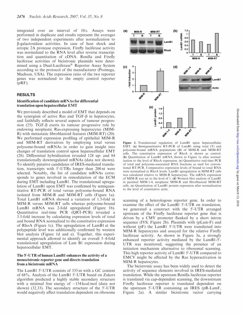
We previously described a model of EMT that depends onthe synergism of active Ras and TGF-b in hepatocytes,and faithfully reflects several aspects of tumour progres-sion (25). TGF-b exerts its tumour progressive role byendowing neoplastic Ras-expressing hepatocytes (MIM-R) with metastatic fibroblastoid features (MIM-RT) (20).We performed expression profiling of epithelial MIM-Rand MIM-RT derivatives by employing total versuspolysome-bound mRNAs in order to gain insight intochanges of translation control upon hepatocellular EMT(28). Differential hybridization revealed 128 up- and 84translationally downregulated mRNAs (data not shown).To identify putative candidates of IRES-mediated transla-tion, transcripts with 50-UTRs longer than 200 nt wereselected. Notably, the list of candidate mRNAs corre-sponds to genes involved in remodulation of the ECMduring EMT including LamB1. The translational upregu-lation of LamB1 upon EMT was confirmed by semiquan-titative RT-PCR of total versus polysome-bound RNAisolated from MIM-R and MIM-RT cells (Figure 1a).Total LamB1 mRNA showed a variation of 1.5-fold inMIM-R versus MIM-RT cells whereas polysome-boundLamB1 mRNA was 2-fold upregulated (Figure 1b).Quantitative real-time PCR (QRT-PCR) revealed a3.5-fold increase by calculating expression levels of totaland bound RNA normalized to the constitutive expressionof RhoA (Figure 1c). The upregulation of LamB1 at thepolypeptide level was additionally confirmed by westernblot analysis (Figure 1d and e). Together, this experi-mental approach allowed to identify an overall 3–4-foldtranslational upregulation of Lam B1 expression duringhepatocellular EMT.
The 50-UTR of human LamB1 enhances the activity of amonocistronic reporter gene and directs translationfrom a bicistronic mRNA
The LamB1 50-UTR consists of 335 nt with a GC contentof 68%. Analysis of the LamB1 50-UTR based on Zukersalgorithm predicted a highly stable secondary structurewith a minimal free energy of �154 kcal/mol (data notshown) (32,33). The secondary structure of the 50-UTRwould negatively affect translation dependent on ribosome
scanning of a heterologous reporter gene. In order toexamine the effect of the LamB1 50-UTR on translation,we generated a construct with the 50-UTR arrangedupstream of the Firefly luciferase reporter gene that isdriven by a CMV promoter flanked by a short intronsequence (IVS; Figure 2b). Plasmids with (pLam-F) andwithout (pF) the LamB1 50-UTR were transfected intoMIM-R hepatocytes and assayed for the relative Fireflyluciferase activity. As shown in Figure 3a, a stronglyenhanced reporter activity mediated by the LamB1-50-UTR was monitored, suggesting the presence of aninitiation mechanism alternative to ribosomal scanning.This high reporter activity of LamB1 50-UTR compared toEMCV might be affected by the Ras hyperactivation inMIM-R hepatocytes.
The bicistronic assay has been widely used to detect theactivity of sequence elements involved in IRES-mediatedtranslation. While the upstream Renilla luciferase reporteris translated via cap-dependent scanning, the downstreamFirefly luciferase reporter is translated dependent onthe upstream 50-UTR containing an IRES (pR-LamF,Figure 2a). A similar bicistronic vector carrying
Figure 1. Translational regulation of LamB1 upon hepatocellularEMT. (a) Semiquantitative RT-PCR of LamB1 using total (T) andpolysome-bound mRNA populations (B) of MIM-R and MIM-RTcells. The constitutive expression of RhoA is shown as control.(b) Quantitation of LamB1 mRNA shown in Figure 1a after normal-ization to the level of RhoA expression. (c) Quantitative real-time PCRof total and polysome-associated RNA fractions as used for conven-tional RT-PCR. Comparative expression levels of bound to total RNAwere normalized to RhoA levels. LamB1 upregulation in MIM-RT cellswas calculated relative to MIM-R hepatocytes. The mRNA expressionof MIM-R was set to the level of 1. (d) Western blot analysis of LamB1in parental MIM-1-4, neoplastic MIM-R and fibroblastoid MIM-RTcells. (e) Quantitation of LamB1 protein expression after normalizationto the level of constitutive actin.
2476 Nucleic Acids Research, 2007, Vol. 35, No. 8
by guest on March 13, 2016
http://nar.oxfordjournals.org/D
ownloaded from
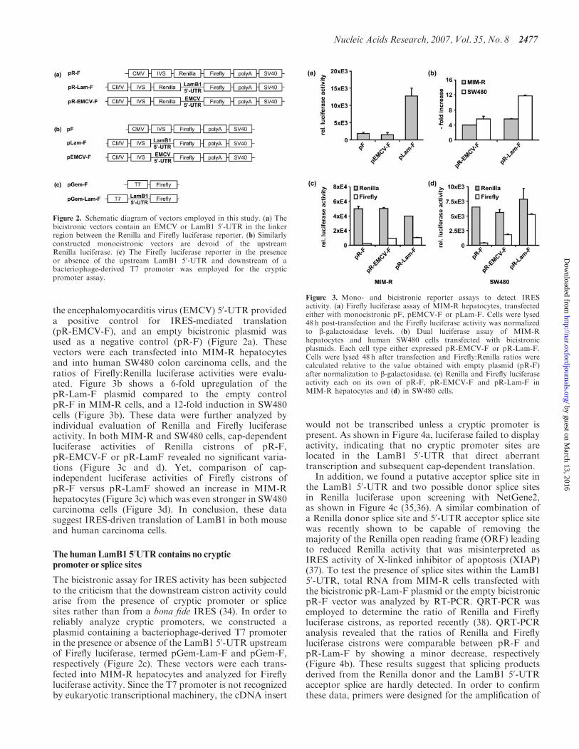
the encephalomyocarditis virus (EMCV) 50-UTR provideda positive control for IRES-mediated translation(pR-EMCV-F), and an empty bicistronic plasmid wasused as a negative control (pR-F) (Figure 2a). Thesevectors were each transfected into MIM-R hepatocytesand into human SW480 colon carcinoma cells, and theratios of Firefly:Renilla luciferase activities were evalu-ated. Figure 3b shows a 6-fold upregulation of thepR-Lam-F plasmid compared to the empty controlpR-F in MIM-R cells, and a 12-fold induction in SW480cells (Figure 3b). These data were further analyzed byindividual evaluation of Renilla and Firefly luciferaseactivity. In both MIM-R and SW480 cells, cap-dependentluciferase activities of Renilla cistrons of pR-F,pR-EMCV-F or pR-LamF revealed no significant varia-tions (Figure 3c and d). Yet, comparison of cap-independent luciferase activities of Firefly cistrons ofpR-F versus pR-LamF showed an increase in MIM-Rhepatocytes (Figure 3c) which was even stronger in SW480carcinoma cells (Figure 3d). In conclusion, these datasuggest IRES-driven translation of LamB1 in both mouseand human carcinoma cells.
The human LamB1 50UTR contains no crypticpromoter or splice sites
The bicistronic assay for IRES activity has been subjectedto the criticism that the downstream cistron activity couldarise from the presence of cryptic promoter or splicesites rather than from a bona fide IRES (34). In order toreliably analyze cryptic promoters, we constructed aplasmid containing a bacteriophage-derived T7 promoterin the presence or absence of the LamB1 50-UTR upstreamof Firefly luciferase, termed pGem-Lam-F and pGem-F,respectively (Figure 2c). These vectors were each trans-fected into MIM-R hepatocytes and analyzed for Fireflyluciferase activity. Since the T7 promoter is not recognizedby eukaryotic transcriptional machinery, the cDNA insert
would not be transcribed unless a cryptic promoter ispresent. As shown in Figure 4a, luciferase failed to displayactivity, indicating that no cryptic promoter sites arelocated in the LamB1 50-UTR that direct aberranttranscription and subsequent cap-dependent translation.In addition, we found a putative acceptor splice site in
the LamB1 50-UTR and two possible donor splice sitesin Renilla luciferase upon screening with NetGene2,as shown in Figure 4c (35,36). A similar combination ofa Renilla donor splice site and 50-UTR acceptor splice sitewas recently shown to be capable of removing themajority of the Renilla open reading frame (ORF) leadingto reduced Renilla activity that was misinterpreted asIRES activity of X-linked inhibitor of apoptosis (XIAP)(37). To test the presence of splice sites within the LamB150-UTR, total RNA from MIM-R cells transfected withthe bicistronic pR-Lam-F plasmid or the empty bicistronicpR-F vector was analyzed by RT-PCR. QRT-PCR wasemployed to determine the ratio of Renilla and Fireflyluciferase cistrons, as reported recently (38). QRT-PCRanalysis revealed that the ratios of Renilla and Fireflyluciferase cistrons were comparable between pR-F andpR-Lam-F by showing a minor decrease, respectively(Figure 4b). These results suggest that splicing productsderived from the Renilla donor and the LamB1 50-UTRacceptor splice are hardly detected. In order to confirmthese data, primers were designed for the amplification of
Figure 3. Mono- and bicistronic reporter assays to detect IRESactivity. (a) Firefly luciferase assay of MIM-R hepatocytes, transfectedeither with monocistronic pF, pEMCV-F or pLam-F. Cells were lysed48 h post-transfection and the Firefly luciferase activity was normalizedto b-galactosidase levels. (b) Dual luciferase assay of MIM-Rhepatocytes and human SW480 cells transfected with bicistronicplasmids. Each cell type either expressed pR-EMCV-F or pR-Lam-F.Cells were lysed 48 h after transfection and Firefly:Renilla ratios werecalculated relative to the value obtained with empty plasmid (pR-F)after normalization to b-galactosidase. (c) Renilla and Firefly luciferaseactivity each on its own of pR-F, pR-EMCV-F and pR-Lam-F inMIM-R hepatocytes and (d) in SW480 cells.
Figure 2. Schematic diagram of vectors employed in this study. (a) Thebicistronic vectors contain an EMCV or LamB1 50-UTR in the linkerregion between the Renilla and Firefly luciferase reporter. (b) Similarlyconstructed monocistronic vectors are devoid of the upstreamRenilla luciferase. (c) The Firefly luciferase reporter in the presenceor absence of the upstream LamB1 50-UTR and downstream of abacteriophage-derived T7 promoter was employed for the crypticpromoter assay.
Nucleic Acids Research, 2007, Vol. 35, No. 8 2477
by guest on March 13, 2016
http://nar.oxfordjournals.org/D
ownloaded from
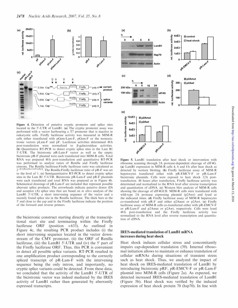
the bicistronic construct starting directly at the transcrip-tional start site and terminating within the Fireflyluciferase ORF (position þ633). As outlined inFigure 4c, the resulting PCR product includes (i) theshort intervening sequence located in the vector down-stream of the CMV promoter, (ii) the ORF of Renillaluciferase, (iii) the LamB1 50-UTR and (iv) the 50 part ofthe Firefly luciferase ORF. Thus, this PCR is convenientto detect all possible splice variants. RT-PCR resulted inone amplification product corresponding to the correctlyspliced transcript of pR-Lam-F with the interveningsequence being the only intron, and importantly, nocryptic splice variants could be detected. From these data,we concluded that the activity of the LamB1 50-UTR ofthe bicistronic vector was indeed mediated by the IRESactivity of LamB1 rather than generated by aberrantlyexpressed transcripts.
IRES-mediated translation of LamB1 mRNAincreases during heat shock
Heat shock induces cellular stress and concomitantlyimpairs cap-dependent translation (39). Internal riboso-mal initiation allows to maintain or enhance translation ofcellular mRNAs during situations of transient stresssuch as heat shock. Thus, we analyzed the impact ofheat shock on IRES-mediated translation of LamB1 byintroducing bicistronic pRF, pR-EMCV-F or pR-Lam-Fplasmid into MIM-R cells (Figure 2a). As expected, wedetected increased IRES-mediated translation of LamB1(Figure 5b). Heat shock was verified by the inducedexpression of heat shock protein 70 (hsp70). In line with
Figure 5. LamB1 translation after heat shock or intervention withribosome scanning through 2A protease-dependent cleavage of eIF4G.(a) LamB1 expression in MIM-R cells 4, 6 and 8 h after heat shock asdetected by western blotting. (b) Firefly luciferase assay of MIM-Rhepatocytes transfected either with pR-EMCV-F or pR-Lam-Fbicistronic plasmids. Cells were exposed to heat shock 12 h post-transfection. 48 hours after transfection, Firefly luciferase activity wasdetermined and normalized to the RNA level after reverse transcriptionand quantitation of cDNA. (c) Western blot analysis of MIM-R cellsshowing the cleavage of eIF4GI/II. MIM-R cells were transfected withwild-type 2A protease expressing plasmid (p2Awt) and lysed atthe indicated times. (d) Firefly luciferase assay of MIM-R hepatocytesco-transfected with pR-F and either p2Amut or p2Awt. (e) Fireflyluciferase assay of MIM-R cells co-transfected either with pR-EMCV-For pR-Lam-F and p2Amut or p2Awt, respectively. Cells were lysed48 h post-transfection and the Firefly luciferase activity wasnormalized to the RNA level after reverse transcription and quantita-tion of cDNA.
Figure 4. Detection of putative cryptic promoter and splice siteslocated in the 50-UTR of LamB1. (a) The cryptic promoter assay wasperformed with a vector harbouring a T7 promoter that is inactive ineukaryotic cells. Firefly luciferase activity was measured in MIM-Rcells either transfected with pGem-Lam-F, pGem-F or the monocis-tronic vectors pLam-F and pF. Luciferase activities determined 48 hpost-transfection were normalized to b-galactosidase activities.(b) Quantitative RT-PCR to detect cryptic splice sites in the Lam B150-UTR. The bicistronic pR-Lam-F vector as well as the emptybicistronic pR-F plasmid were each transfected into MIM-R cells. TotalRNA was prepared 48 h post-transfection and quantitative RT-PCRwas performed to analyze ratios of Renilla and Firefly luciferasecistrons. The Renilla luciferase:Firefly luciferase ratio was calculated as2�[Ct(Renilla-Ct(Firefly)]. The Renilla:Firefly luciferase ratio of pR-F was setto the level of 1. (c) Semiquantitative RT-PCR to detect cryptic splicesites in the Lam B1 50-UTR. Bicistronic pR-Lam-F and pR-F plasmidswere each transfected and total RNA was prepared as in Figure 4b.Schematical drawings of pR-Lam-F are included that represent possibleaberrant splice products. The arrowheads indicate putative donor (D)and acceptor (A) splice sites that are based on in silico analysis of theLamB1 50-UTR, a short intervening sequence of the vector and arecently found splice site in the Renilla luciferase. The thick bars at the50 end close to the cap and in the Firefly luciferase indicate the positionof the forward and reverse primers.
2478 Nucleic Acids Research, 2007, Vol. 35, No. 8
by guest on March 13, 2016
http://nar.oxfordjournals.org/D
ownloaded from
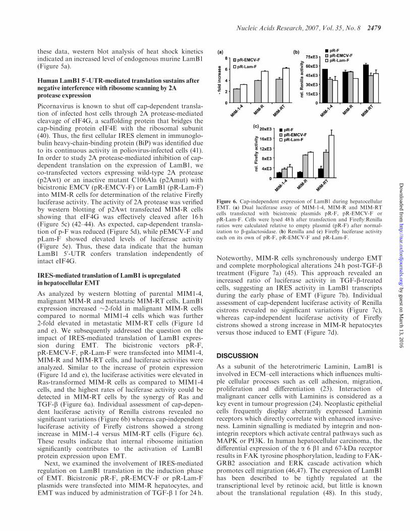
these data, western blot analysis of heat shock kineticsindicated an increased level of endogenous murine LamB1(Figure 5a).
Human LamB1 50-UTR-mediated translation sustains afternegative interference with ribosome scanning by 2Aprotease expression
Picornavirus is known to shut off cap-dependent transla-tion of infected host cells through 2A protease-mediatedcleavage of eIF4G, a scaffolding protein that bridges thecap-binding protein eIF4E with the ribosomal subunit(40). Thus, the first cellular IRES element in immunoglo-bulin heavy-chain-binding protein (BiP) was identified dueto its continuous activity in poliovirus-infected cells (41).In order to study 2A protease-mediated inhibition of cap-dependent translation on the expression of LamB1, weco-transfected vectors expressing wild-type 2A protease(p2Awt) or an inactive mutant C106Ala (p2Amut) withbicistronic EMCV (pR-EMCV-F) or LamB1 (pR-Lam-F)into MIM-R cells for determination of the relative Fireflyluciferase activity. The activity of 2A protease was verifiedby western blotting of p2Awt transfected MIM-R cellsshowing that eIF4G was effectively cleaved after 16 h(Figure 5c) (42–44). As expected, cap-dependent transla-tion of p-F was reduced (Figure 5d), while pEMCV-F andpLam-F showed elevated levels of luciferase activity(Figure 5e). Thus, these data indicate that the humanLamB1 50-UTR confers translation independently ofintact eIF4G.
IRES-mediated translation of LamB1 is upregulatedin hepatocellular EMT
As analyzed by western blotting of parental MIM1-4,malignant MIM-R and metastatic MIM-RT cells, LamB1expression increased �2-fold in malignant MIM-R cellscompared to normal MIM1-4 cells which was further2-fold elevated in metastatic MIM-RT cells (Figure 1dand e). We subsequently addressed the question on theimpact of IRES-mediated translation of LamB1 expres-sion during EMT. The bicistronic vectors pR-F,pR-EMCV-F, pR-Lam-F were transfected into MIM1-4,MIM-R and MIM-RT cells, and luciferase activities wereanalyzed. Similar to the increase of protein expression(Figure 1d and e), the luciferase activities were elevated inRas-transformed MIM-R cells as compared to MIM1-4cells, and the highest rates of luciferase activity could bedetected in MIM-RT cells by the synergy of Ras andTGF-b (Figure 6a). Individual assessment of cap-depen-dent luciferase activity of Renilla cistrons revealed nosignificant variations (Figure 6b) whereas cap-independentluciferase activity of Firefly cistrons showed a strongincrease in MIM-1-4 versus MIM-RT cells (Figure 6c).These results indicate that internal ribosome initiationsignificantly contributes to the activation of LamB1protein expression upon EMT.
Next, we examined the involvement of IRES-mediatedregulation on LamB1 translation in the induction phaseof EMT. Bicistronic pR-F, pR-EMCV-F or pR-Lam-Fplasmids were transfected into MIM-R hepatocytes, andEMT was induced by administration of TGF-b 1 for 24 h.
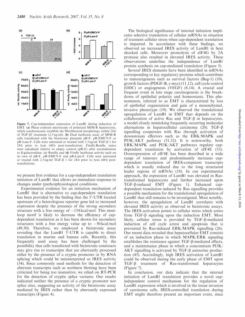
Noteworthy, MIM-R cells synchronously undergo EMTand complete morphological alterations 24 h post-TGF-btreatment (Figure 7a) (45). This approach revealed anincreased ratio of luciferase activity in TGF-b-treatedcells, suggesting an IRES activity in LamB1 transcriptsduring the early phase of EMT (Figure 7b). Individualassessment of cap-dependent luciferase activity of Renillacistrons revealed no significant variations (Figure 7c),whereas cap-independent luciferase activity of Fireflycistrons showed a strong increase in MIM-R hepatocytesversus those induced to EMT (Figure 7d).
DISCUSSION
As a subunit of the heterotrimeric Laminin, LamB1 isinvolved in ECM–cell interactions which influences multi-ple cellular processes such as cell adhesion, migration,proliferation and differentiation (23). Interaction ofmalignant cancer cells with Laminins is considered as akey event in tumour progression (24). Neoplastic epithelialcells frequently display aberrantly expressed Lamininreceptors which directly correlate with enhanced invasive-ness. Laminin signalling is mediated by integrin and non-integrin receptors which activate central pathways such asMAPK or PI3K. In human hepatocellular carcinoma, thedifferential expression of the a 6 b1 and 67-kDa receptorresults in FAK tyrosine phosphorylation, leading to FAK-GRB2 association and ERK cascade activation whichpromotes cell migration (46,47). The expression of LamB1has been described to be tightly regulated at thetranscriptional level by retinoic acid, but little is knownabout the translational regulation (48). In this study,
Figure 6. Cap-independent expression of LamB1 during hepatocellularEMT. (a) Dual luciferase assay of MIM-1-4, MIM-R and MIM-RTcells transfected with bicistronic plasmids pR-F, pR-EMCV-F orpR-Lam-F. Cells were lysed 48 h after transfection and Firefly:Renillaratios were calculated relative to empty plasmid (pR-F) after normal-ization to b-galactosidase. (b) Renilla and (c) Firefly luciferase activityeach on its own of pR-F, pR-EMCV-F and pR-Lam-F.
Nucleic Acids Research, 2007, Vol. 35, No. 8 2479
by guest on March 13, 2016
http://nar.oxfordjournals.org/D
ownloaded from
we present first evidence for a cap-independent translationinitiation of LamB1 that allows an immediate response tochanges under (patho)physiological conditions.Experimental evidence for an initiation mechanism of
LamB1 that is alternative to cap-dependent translationhas been provided by the finding that the 50-UTR locatedupstream of a heterologous reporter gene led to increasedexpression despite the presence of the strong secondarystructure with a free energy of �154 kcal/mol. This stem-loop motif is likely to decrease the efficiency of cap-dependent translation as it has been shown for secondarystructures with a free energy value up to �50 kcal/mol(49,50). Therefore, we employed a bicistronic assayrevealing that the LamB1 50-UTR is capable to directtranslation in murine and human cells. Recently, thisfrequently used assay has been challenged by thepossibility that cells transfected with bicistronic constructsmay give rise to transcripts that are aberrantly processedeither by the presence of a cryptic promoter or by RNAsplicing which could be misinterpreted as IRES activity(34). Since commonly used methods for the detection ofaberrant transcripts such as northern blotting have beencriticized for being too insensitive, we relied on RT-PCRfor the detection of cryptic splice variants. Our resultsindicated neither the presence of a cryptic promoter norsplice sites, suggesting an activity of the bicistronic assaymediated by IRES rather than by aberrantly expressedtranscripts (Figure 4).
The biological significance of internal initiation impli-cates selective translation of cellular mRNAs in situationof transient cellular stress when cap-dependent translationis impaired. In accordance with these findings, weobserved an increased IRES activity of LamB1 in heatshocked cells. Moreover proteolysis of eIF4G by 2Aprotease also resulted in elevated IRES activity. Theseobservations underline the independence of LamB1protein synthesis on cap-mediated translation (Figure 5).
Several IRES elements have been identified in mRNAscorresponding to key regulatory proteins which contributeto tumourigenesis such as survival factors (Bag-1) (10),growth factors (PDGF-B, c-myc) (11,12), cell cycle control(ODC) or angiogenesis (VEGF) (8,14). A crucial andfrequent event in late stage carcinogenesis is the break-down of epithelial polarity and homeostasis. This phe-nomenon, referred to as EMT is characterized by lossof epithelial organization and gain of a mesenchymal,invasive phenotype (19). We observed the translationalupregulation of LamB1 in EMT that depends on thecollaboration of active Ras and TGF-b in hepatocytes,a model closely mimicking frequently occurring molecularalternations in hepatocellular carcinoma (20). TGF-bsignalling cooperates with Ras through activation ofdownstream effectors such as the ERK/MAPK andPI3K/AKT pathway (51,52). It is noteworthy, bothERK/MAPK and PI3K/AKT pathways regulate cap-dependent translation by activation of eIF4E (53).Overexpression of eIF4E has been described in a widerange of tumours and predominantly increases cap-dependent translation of IRES-competent transcriptswhich is usually reduced due to the long structuredleader regions of mRNAs (18). In our experimentalapproach, the expression of LamB1 was elevated in Ras-transformed hepatocytes and further increased uponTGF-b-induced EMT (Figure 1). Enhanced cap-dependent translation induced by Ras signalling providesa possible mechanism for the translational upregulation ofLamB1 that still remains to be investigated. Most notably,however, the upregulation of LamB1 correlates withelevated IRES activity as observed in bicistronic assays.The IRES activation points to cellular stress which arisesfrom TGF-b signaling upon the induction EMT. Mostlikely, cellular stress is provided by TGF-b-mediatedinduction of cell cycle arrest and apoptosis that isprevented by Ras-induced ERK/MAPK signalling (26).Our recent data revealed that hepatocellular EMT consistsof an induction phase in which MAPK/ERK signalingestablishes the resistance against TGF-b-mediated effects,and a maintenance phase in which a concomitant PI3K/AKT signalling is activated by TGF-b autocrine produc-tion (45). Accordingly, high IRES activation of LamB1could be observed during the early phase of EMT uponTGF-b treatment of Ras-transformed hepatocytes(Figure 7).
In conclusion, our data indicate that the internalinitiation of LamB1 translation provides a novel cap-independent control mechanism for the regulation ofLamB1 expression which is involved in the tissue invasionof carcinoma cells. IRES-controlled translation duringEMT might therefore present an important event, since
Figure 7. Cap-independent expression of LamB1 during induction ofEMT. (a) Phase contrast microscopy of polarized MIM-R hepatocytes,which synchronously establish the fibroblastoid morphology within 24 hof TGF-b1 treatment (2.5 ng/ml). (b) Dual luciferase assay of MIM-Rcells transfected with the bicistronic plasmids pR-F, pR-EMCV-F orpR-Lam-F. Cells were untreated or treated with 2.5 ng/ml TGF-b 1 for24 h prior to lysis (48 h post-transfection). Firefly:Renilla ratioswere calculated relative to empty control (pR-F) after normalizationto b-galactosidase. (c) Renilla and (d) Firefly luciferase activity each onits own of pR-F, pR-EMCV-F and pR-Lam-F. Cells were untreatedor treated with 2.5 ng/ml TGF-b 1 for 24 h prior to lysis (48 h post-transfection).
2480 Nucleic Acids Research, 2007, Vol. 35, No. 8
by guest on March 13, 2016
http://nar.oxfordjournals.org/D
ownloaded from
additional mRNA species predicted to internal ribosomeentry have an impact in tumour progression.
ACKNOWLEDGEMENTS
We thank Rudolf Oehler for helpful advice on heat shockexperiments and stimulating discussions. This work wassupported by a grant from the ‘Herzfelder FamilyFoundation’, Austria. Funding to pay the Open Accesspublication charge was provided by the Herzfelder FamilyFoundation.
Conflict of interest statement. None declared.
REFERENCES
1. Merrick,W.C. (2004) Cap-dependent and cap-independent transla-tion in eukaryotic systems. Gene, 332, 1–11.
2. Kozak,M. (1986) Influences of mRNA secondary structure oninitiation by eukaryotic ribosomes. Proc. Natl. Acad. Sci. USA, 83,2850–2854.
3. Komar,A.A. and Hatzoglou,M. (2005) Internal ribosome entry sitesin cellular mRNAs: mystery of their existence. J. Biol. Chem., 280,23425–23428.
4. Baird,S.D., Turcotte,M., Korneluk,R.G. and Holcik,M. (2006)Searching for IRES. RNA, 12, 1755–1785.
5. Pelletier,J. and Sonenberg,N. (1988) Internal initiation of transla-tion of eukaryotic mRNA directed by a sequence derived frompoliovirus RNA. Nature, 334, 320–325.
6. Holcik,M. (2003) Translational upregulation of the X-linkedinhibitor of apoptosis. Ann. N.Y. Acad. Sci., 1010, 249–258.
7. Stein,I., Itin,A., Einat,P., Skaliter,R., Grossman,Z. and Keshet,E.(1998) Translation of vascular endothelial growth factor mRNA byinternal ribosome entry: implications for translation under hypoxia.Mol. Cell. Biol., 18, 3112–3119.
8. Pyronnet,S., Pradayrol,L. and Sonenberg,N. (2000) A cell cycle-dependent internal ribosome entry site. Mol. Cell, 5, 607–616.
9. Lang,K.J., Kappel,A. and Goodall,G.J. (2002) Hypoxia-induciblefactor-1alpha mRNA contains an internal ribosome entry site thatallows efficient translation during normoxia and hypoxia. Mol. Biol.Cell, 13, 1792–1801.
10. Coldwell,M.J., deSchoolmeester,M.L., Fraser,G.A., Pickering,B.M.,Packham,G. and Willis,A.E. (2001) The p36 isoform of BAG-1 istranslated by internal ribosome entry following heat shock.Oncogene, 20, 4095–4100.
11. Bernstein,J., Sella,O., Le,S.Y. and Elroy-Stein,O. (1997)PDGF2/c-sis mRNA leader contains a differentiation-linked inter-nal ribosomal entry site (D-IRES). J. Biol. Chem., 272, 9356–9362.
12. Stoneley,M., Paulin,F.E., Le Quesne,J.P., Chappell,S.A. andWillis,A.E. (1998) C-Myc 50 untranslated region contains aninternal ribosome entry segment. Oncogene, 16, 423–428.
13. Coldwell,M.J., Mitchell,S.A., Stoneley,M., MacFarlane,M. andWillis,A.E. (2000) Initiation of Apaf-1 translation by internalribosome entry. Oncogene, 19, 899–905.
14. Akiri,G., Nahari,D., Finkelstein,Y., Le,S.Y., Elroy-Stein,O. andLevi,B.Z. (1998) Regulation of vascular endothelial growth factor(VEGF) expression is mediated by internal initiation of translationand alternative initiation of transcription. Oncogene, 17, 227–236.
15. Chappell,S.A., LeQuesne,J.P., Paulin,F.E., deSchoolmeester,M.L.,Stoneley,M., Soutar,R.L., Ralston,S.H., Helfrich,M.H. andWillis,A.E. (2000) A mutation in the c-myc-IRES leads to enhancedinternal ribosome entry in multiple myeloma: a novel mechanism ofoncogene de-regulation. Oncogene, 19, 4437–4440.
16. Clemens,M.J. (2004) Targets and mechanisms for the regulation oftranslation in malignant transformation. Oncogene, 23, 3180–3188.
17. Gingras,A.C., Raught,B. and Sonenberg,N. (2001) Regulation oftranslation initiation by FRAP/mTOR. Genes Dev., 15, 807–826.
18. Graff,J.R. and Zimmer,S.G. (2003) Translational control andmetastatic progression: enhanced activity of the mRNA cap-bindingprotein eIF-4E selectively enhances translation of metastasis-relatedmRNAs. Clin. Exp. Metastasis, 20, 265–273.
19. Thiery,J.P. (2002) Epithelial-mesenchymal transitions in tumourprogression. Nat. Rev. Cancer, 2, 442–454.
20. Gotzmann,J., Huber,H., Thallinger,C., Wolschek,M., Jansen,B.,Schulte-Hermann,R., Beug,H. and Mikulits,W. (2002) Hepatocytesconvert to a fibroblastoid phenotype through the cooperation ofTGF-beta1 and Ha-Ras: steps towards invasiveness. J. Cell Sci.,115, 1189–1202.
21. Ito,N., Kawata,S., Tamura,S., Takaishi,K., Shirai,Y., Kiso,S.,Yabuuchi,I., Matsuda,Y., Nishioka,M. et al. (1991) Elevated levelsof transforming growth factor beta messenger RNA and itspolypeptide in human hepatocellular carcinoma. Cancer Res., 51,4080–4083.
22. Malinda,K.M. and Kleinman,H.K. (1996) The laminins. Int. J.Biochem. Cell Biol., 28, 957–959.
23. Patarroyo,M., Tryggvason,K. and Virtanen,I. (2002) Lamininisoforms in tumor invasion, angiogenesis and metastasis. Semin.Cancer Biol., 12, 197–207.
24. Givant-Horwitz,V., Davidson,B. and Reich,R. (2005) Laminin-induced signaling in tumor cells. Cancer Lett., 223, 1–10.
25. Mikula,M., Fuchs,E., Huber,H., Beug,H., Schulte-Hermann,R. andMikulits,W. (2004) Immortalized p19ARF null hepatocytes restoreliver injury and generate hepatic progenitors after transplantation.Hepatology, 39, 628–634.
26. Fischer,A.N., Herrera,B., Mikula,M., Proell,V., Fuchs,E.,Gotzmann,J., Schulte-Hermann,R., Beug,H. and Mikulits,W. (2005)Integration of Ras subeffector signaling in TGF-beta mediated latestage hepatocarcinogenesis. Carcinogenesis, 26, 931–942.
27. Oehler,R., Zellner,M., Hefel,B., Weingartmann,G., Spittler,A.,Struse,H.M. and Roth,E. (1998) Influence of heat shock on cellvolume regulation: protection from hypertonic challenge in ahuman monocyte cell line. FASEB J., 12, 553–560.
28. Mikulits,W., Pradet-Balade,B., Habermann,B., Beug,H., Garcia-Sanz,J.A. and Mullner,E.W. (2000) Isolation of translationallycontrolled mRNAs by differential screening. FASEB J., 14,1641–1652.
29. Yan,R., Rychlik,W., Etchison,D. and Rhoads,R.E. (1992) Aminoacid sequence of the human protein synthesis initiation factor eIF-4gamma. J. Biol. Chem., 267, 23226–23231.
30. Eger,A., Stockinger,A., Schaffhauser,B., Beug,H. and Foisner,R.(2000) Epithelial mesenchymal transition by c-Fos estrogen receptoractivation involves nuclear translocation of beta-catenin andupregulation of beta-catenin/lymphoid enhancer binding factor-1transcriptional activity. J. Cell Biol., 148, 173–188.
31. Mikula,M., Gotzmann,J., Fischer,A.N., Wolschek,M.F.,Thallinger,C., Schulte-Hermann,R., Beug,H. and Mikulits,W.(2003) The proto-oncoprotein c-Fos negatively regulates hepatocel-lular tumorigenesis. Oncogene, 22, 6725–6738.
32. Mathews,D.H., Disney,M.D., Childs,J.L., Schroeder,S.J., Zuker,M.and Turner,D.H. (2004) Incorporating chemical modificationconstraints into a dynamic programming algorithm for predictionof RNA secondary structure. Proc. Natl. Acad. Sci. USA, 101,7287–7292.
33. Mathews,D.H., Sabina,J., Zuker,M. and Turner,D.H. (1999)Expanded sequence dependence of thermodynamic parametersimproves prediction of RNA secondary structure. J. Mol. Biol., 288,911–940.
34. Kozak,M. (2003) Alternative ways to think about mRNA sequencesand proteins that appear to promote internal initiation oftranslation. Gene, 318, 1–23.
35. Brunak,S., Engelbrecht,J. and Knudsen,S. (1991) Prediction ofhuman mRNA donor and acceptor sites from the DNA sequence.J. Mol. Biol., 220, 49–65.
36. Hebsgaard,S.M., Korning,P.G., Tolstrup,N., Engelbrecht,J.,Rouze,P. and Brunak,S. (1996) Splice site prediction in Arabidopsisthaliana pre-mRNA by combining local and global sequenceinformation. Nucleic Acids Res., 24, 3439–3452.
37. Van Eden,M.E., Byrd,M.P., Sherrill,K.W. and Lloyd,R.E. (2004)Demonstrating internal ribosome entry sites in eukaryotic mRNAsusing stringent RNA test procedures. RNA, 10, 720–730.
38. Holcik,M., Graber,T., Lewis,S.M., Lefebvre,C.A., Lacasse,E. andBaird,S. (2005) Spurious splicing within the XIAP 50 UTR occurs inthe Rluc/Fluc but not the betagal/CAT bicistronic reporter system.RNA, 11, 1605–1619.
Nucleic Acids Research, 2007, Vol. 35, No. 8 2481
by guest on March 13, 2016
http://nar.oxfordjournals.org/D
ownloaded from

39. Kim,Y.K. and Jang,S.K. (2002) Continuous heat shock enhancestranslational initiation directed by internal ribosomal entry site.Biochem. Biophys. Res. Commun., 297, 224–231.
40. Gradi,A., Svitkin,Y.V., Imataka,H. and Sonenberg,N. (1998)Proteolysis of human eukaryotic translation initiation factoreIF4GII, but not eIF4GI, coincides with the shutoff of host proteinsynthesis after poliovirus infection. Proc. Natl. Acad. Sci. USA, 95,11089–11094.
41. Sarnow,P. (1989) Translation of glucose-regulated protein 78/immunoglobulin heavy-chain binding protein mRNA is increased inpoliovirus-infected cells at a time when cap-dependent translation ofcellular mRNAs is inhibited. Proc. Natl. Acad. Sci. USA, 86,5795–5799.
42. Marissen,W.E. and Lloyd,R.E. (1998) Eukaryotic translationinitiation factor 4G is targeted for proteolytic cleavage by caspase 3during inhibition of translation in apoptotic cells. Mol. Cell. Biol.,18, 7565–7574.
43. Gradi,A., Svitkin,Y.V., Sommergruber,W., Imataka,H., Morino,S.,Skern,T. and Sonenberg,N. (2003) Human rhinovirus 2A proteinasecleavage sites in eukaryotic initiation factors (eIF) 4GI and eIF4GIIare different. J. Virol., 77, 5026–5029.
44. Sommergruber,W., Ahorn,H., Klump,H., Seipelt,J., Zoephel,A.,Fessl,F., Krystek,E., Blaas,D., Kuechler,E. et al. (1994) 2Aproteinases of coxsackie- and rhinovirus cleave peptides derivedfrom eIF-4 gamma via a common recognition motif. Virology, 198,741–745.
45. Gotzmann,J., Mikula,M., Eger,A., Schulte-Hermann,R.,Foisner,R., Beug,H. and Mikulits,W. (2004) Molecular aspects ofepithelial cell plasticity: implications for local tumor invasion andmetastasis. Mutat. Res., 566, 9–20.
46. Ozaki,I., Yamamoto,K., Mizuta,T., Kajihara,S., Fukushima,N.,Setoguchi,Y., Morito,F. and Sakai,T. (1998) Differential expressionof laminin receptors in human hepatocellular carcinoma. Gut, 43,837–842.
47. Carloni,V., Mazzocca,A., Pantaleo,P., Cordella,C., Laffi,G. andGentilini,P. (2001) The integrin, alpha6beta1, is necessary forthe matrix-dependent activation of FAK and MAP kinase andthe migration of human hepatocarcinoma cells. Hepatology, 34,42–49.
48. Li,C. and Gudas,L.J. (1996) Murine laminin B1 gene regulationduring the retinoic acid- and dibutyryl cyclic AMP-induceddifferentiation of embryonic F9 teratocarcinoma stem cells. J. Biol.Chem., 271, 6810–6818.
49. Kozak,M. (1989) Circumstances and mechanisms of inhibition oftranslation by secondary structure in eucaryotic mRNAs. Mol. Cell.Biol., 9, 5134–5142.
50. Kozak,M. (1991) Structural features in eukaryotic mRNAsthat modulate the initiation of translation. J. Biol. Chem., 266,19867–19870.
51. Macaluso,M., Russo,G., Cinti,C., Bazan,V., Gebbia,N. andRusso,A. (2002) Ras family genes: an interesting link between cellcycle and cancer. J. Cell. Physiol., 192, 125–130.
52. Bakin,A.V., Tomlinson,A.K., Bhowmick,N.A., Moses,H.L. andArteaga,C.L. (2000) Phosphatidylinositol 3-kinase function isrequired for transforming growth factor beta-mediated epithelial tomesenchymal transition and cell migration. J. Biol. Chem., 275,36803–36810.
53. Mamane,Y., Petroulakis,E., Rong,L., Yoshida,K., Ler,L.W. andSonenberg,N. (2004) eIF4E – from translation to transformation.Oncogene, 23, 3172–3179.
2482 Nucleic Acids Research, 2007, Vol. 35, No. 8
by guest on March 13, 2016
http://nar.oxfordjournals.org/D
ownloaded from