Identification of functional domains in Arabidopsis thaliana mRNA decapping enzyme (AtDcp2
14
Published online 19 November 2007 Nucleic Acids Research, 2008, Vol. 36, No. 1 203–216 doi:10.1093/nar/gkm1002 Identification of functional domains in Arabidopsis thaliana mRNA decapping enzyme (AtDcp2) Dilantha Gunawardana, Heung-Chin Cheng and Kenwyn R. Gayler* Department of Biochemistry and Molecular Biology, Bio21 Molecular Science and Biotechnology Institute, University of Melbourne, Parkville, Victoria 3010, Australia Received September 28, 2007; Revised October 19, 2007; Accepted October 22, 2007 ABSTRACT The Arabidopsis thaliana decapping enzyme (AtDcp2) was characterized by bioinformatics ana- lysis and by biochemical studies of the enzyme and mutants produced by recombinant expression. Three functionally significant regions were detected: (i) a highly disordered C-terminal region with a putative PSD-95, Discs-large, ZO-1 (PDZ) domain-binding motif, (ii) a conserved Nudix box constituting the putative active site and (iii) a putative RNA binding domain consisting of the conserved Box B and a preceding loop region. Mutation of the putative PDZ domain-binding motif improved the stability of recombinant AtDcp2 and secondary mutants expressed in Escherichia coli. Such recombinant AtDcp2 specifically hydrolysed capped mRNA to produce 7-methyl GDP and decapped RNA. AtDcp2 activity was Mn 2+ - or Mg 2+ -dependent and was inhibited by the prod- uct 7-methyl GDP. Mutation of the conserved glutamate-154 and glutamate-158 in the Nudix box reduced AtDcp2 activity up to 400-fold and showed that AtDcp2 employs the catalytic mechan- ism conserved amongst Nudix hydrolases. Unlike many Nudix hydrolases, AtDcp2 is refractory to inhibition by fluoride ions. Decapping was depen- dent on binding to the mRNA moiety rather than to the 7-methyl diguanosine triphosphate cap of the substrate. Mutational analysis of the putative RNA- binding domain confirmed the functional signi- ficance of an 11-residue loop region and the conserved Box B. INTRODUCTION One of the key processes in mRNA turnover is decapping, that is, the removal of the 7-methyl guanosine cap from the 5 0 end of the mRNA strand. Capped mRNA is estimated to constitute 95–97% of cellular mRNA and it has been proposed that the availability of the 7-methyl guanosine cap in mRNA is a dominant feature in competition for limited translation initiation factors, primarily eIF4E (1). Cap-independent translation mechanisms such as re-initiation and internal initiation generally lack the efficiency associated with cap-dependent translation (1). Since capping is co-transcriptional and occurs in the nucleus (2), while decapping occurs in granules known as the mRNA processing bodies (P bodies) (3,4), decapped mRNAs are unlikely to be recapped for cap-dependent translation. Rather, dead- enylation followed by removal of 7-methyl guanosine diphosphate from the 5 0 end of mRNA appears to initiate degradation of mRNA by exoribonucleases in the 5 0 to 3 0 direction (5). There are four primary pathways of mRNA decay in eukaryotes: two deadenylation- dependent mechanisms classified in accordance to the orientation of decay—5 0 !3 0 or 3 0 !5 0 , endonucleolytic digestion and quality control pathways (6). Of these, deadenylation followed by decapping is the pathway gaining importance in yeast, humans and now in plants as a mechanism of controlling both gene expression and protein synthesis (5,7,8). In yeast, the major mRNA decay pathway involves a deadenylation-dependent decapping mechanism, where the mRNA is initially deadenylated, then decapped by an enzyme named Dcp2p and ultimately degraded by a highly processive exoribonuclease in the 5 0 !3 0 direction (5,9–11). A similar enzyme in humans, hDcp2, is active on capped mRNA and is again deadenylation-dependent (7,12). Although decapping by Dcp2 enzymes is a mandatory step in the degradation of capped mRNA in the 5 0 !3 0 direction, the mechanism of action of this enzyme is still poorly understood and this enzyme is yet to be characterized in plants. Annotation of the Arabidopsis thaliana genome in the NCBI database includes one gene, At5g13570, with strong sequence homology to the Dcp2- type mRNA decapping enzymes. In this article, the protein encoded by gene At5g13570 is studied in detail. Dcp2-type decapping enzymes are members of the Nudix superfamily of enzymes (13). Nearly 1800 open reading frames coding for Nudix hydrolases have been *To whom correspondence should be addressed. Tel: +61 3 9457 3169; Fax: +61 3 9348 1421; Email: [email protected] ß 2007 The Author(s) This is an Open Access article distributed under the terms of the Creative Commons Attribution Non-Commercial License (http://creativecommons.org/licenses/ by-nc/2.0/uk/) which permits unrestricted non-commercial use, distribution, and reproduction in any medium, provided the original work is properly cited. by guest on July 25, 2014 http://nar.oxfordjournals.org/ Downloaded from
-
Upload
independent -
Category
Documents
-
view
0 -
download
0
Transcript of Identification of functional domains in Arabidopsis thaliana mRNA decapping enzyme (AtDcp2

Published online 19 November 2007 Nucleic Acids Research, 2008, Vol. 36, No. 1 203–216doi:10.1093/nar/gkm1002
Identification of functional domains in Arabidopsisthaliana mRNA decapping enzyme (AtDcp2)Dilantha Gunawardana, Heung-Chin Cheng and Kenwyn R. Gayler*
Department of Biochemistry and Molecular Biology, Bio21 Molecular Science and Biotechnology Institute,University of Melbourne, Parkville, Victoria 3010, Australia
Received September 28, 2007; Revised October 19, 2007; Accepted October 22, 2007
ABSTRACT
The Arabidopsis thaliana decapping enzyme(AtDcp2) was characterized by bioinformatics ana-lysis and by biochemical studies of the enzyme andmutants produced by recombinant expression.Three functionally significant regions weredetected: (i) a highly disordered C-terminal regionwith a putative PSD-95, Discs-large, ZO-1 (PDZ)domain-binding motif, (ii) a conserved Nudixbox constituting the putative active site and(iii) a putative RNA binding domain consisting ofthe conserved Box B and a preceding loop region.Mutation of the putative PDZ domain-binding motifimproved the stability of recombinant AtDcp2 andsecondary mutants expressed in Escherichia coli.Such recombinant AtDcp2 specifically hydrolysedcapped mRNA to produce 7-methyl GDP anddecapped RNA. AtDcp2 activity was Mn2+- orMg2+-dependent and was inhibited by the prod-uct 7-methyl GDP. Mutation of the conservedglutamate-154 and glutamate-158 in the Nudixbox reduced AtDcp2 activity up to 400-fold andshowed that AtDcp2 employs the catalytic mechan-ism conserved amongst Nudix hydrolases. Unlikemany Nudix hydrolases, AtDcp2 is refractory toinhibition by fluoride ions. Decapping was depen-dent on binding to the mRNA moiety rather than tothe 7-methyl diguanosine triphosphate cap of thesubstrate. Mutational analysis of the putative RNA-binding domain confirmed the functional signi-ficance of an 11-residue loop region and theconserved Box B.
INTRODUCTION
One of the key processes in mRNA turnover is decapping,that is, the removal of the 7-methyl guanosine cap fromthe 50 end of the mRNA strand. Capped mRNA isestimated to constitute 95–97% of cellular mRNA and it
has been proposed that the availability of the 7-methylguanosine cap in mRNA is a dominant feature incompetition for limited translation initiation factors,primarily eIF4E (1). Cap-independent translationmechanisms such as re-initiation and internal initiationgenerally lack the efficiency associated with cap-dependenttranslation (1). Since capping is co-transcriptional andoccurs in the nucleus (2), while decapping occursin granules known as the mRNA processing bodies(P bodies) (3,4), decapped mRNAs are unlikely to berecapped for cap-dependent translation. Rather, dead-enylation followed by removal of 7-methyl guanosinediphosphate from the 50 end of mRNA appears to initiatedegradation of mRNA by exoribonucleases in the 50 to30 direction (5). There are four primary pathways ofmRNA decay in eukaryotes: two deadenylation-dependent mechanisms classified in accordance to theorientation of decay—50!30 or 30!50, endonucleolyticdigestion and quality control pathways (6). Of these,deadenylation followed by decapping is the pathwaygaining importance in yeast, humans and now in plantsas a mechanism of controlling both gene expression andprotein synthesis (5,7,8).In yeast, the major mRNA decay pathway involves
a deadenylation-dependent decapping mechanism, wherethe mRNA is initially deadenylated, then decapped by anenzyme named Dcp2p and ultimately degraded by a highlyprocessive exoribonuclease in the 50!30 direction (5,9–11).A similar enzyme in humans, hDcp2, is active on cappedmRNA and is again deadenylation-dependent (7,12).Although decapping by Dcp2 enzymes is a mandatorystep in the degradation of capped mRNA in the 50!30
direction, the mechanism of action of this enzyme isstill poorly understood and this enzyme is yet to becharacterized in plants. Annotation of the Arabidopsisthaliana genome in the NCBI database includes one gene,At5g13570, with strong sequence homology to the Dcp2-type mRNA decapping enzymes. In this article, theprotein encoded by gene At5g13570 is studied in detail.Dcp2-type decapping enzymes are members of the
Nudix superfamily of enzymes (13). Nearly 1800 openreading frames coding for Nudix hydrolases have been
*To whom correspondence should be addressed. Tel: +61 3 9457 3169; Fax: +61 3 9348 1421; Email: [email protected]
� 2007 The Author(s)
This is an Open Access article distributed under the terms of the Creative Commons Attribution Non-Commercial License (http://creativecommons.org/licenses/
by-nc/2.0/uk/) which permits unrestricted non-commercial use, distribution, and reproduction in any medium, provided the original work is properly cited.
by guest on July 25, 2014http://nar.oxfordjournals.org/
Dow
nloaded from

identified by bioinformatics searches in over 360 species(14). Nudix enzymes catalyse the hydrolysis of a widerange of small nucleotide substrates composed of anucleoside diphosphate linked to another moiety X,hence the name Nudix (14). All members of this super-family share a conserved amino acid sequence(GX5EX7REUXEEXGU) termed the Nudix motif/box,forming the catalytic site of these enzymes (15,16). In theDcp2-type decapping enzymes from yeast and humans,the Nudix box has been shown to be essential for thehydrolysis of the pyrophosphate bond linking the capto the 50 extremity of mRNA (7,11).It is proposed that hDcp2, the decapping enzyme from
humans, binds to the RNA body first and subsequentlypositions the active site against the cap structure fordecapping (17). An intact cap structure on an RNA strandof 23 nucleotides is mandatory for decapping by hDcp2(7). The hDcp2 enzyme is unable to bind to cap analog[m7G(50)ppp(50)G] alone (17) and decapping by hDcp2 isnot regulated by the addition of cap analog competitor(7), again suggesting the importance of a short RNAregion for preliminary binding. In addition, uncappedtranscripts are efficient competitors for capped RNA,suggesting that binding of mRNA to hDcp2 is indepen-dent of the cap itself (17). A region essential for RNAbinding by the hDcp2 protein was demonstrated bydeletion mutagenesis. The deleted region was C-terminalto the Nudix motif in a region comprising an a-helicalstructure and a preceding loop region (17,18). The regioncorresponding to the above a-helix was designated asBox B (17,18). The contributions of structural motifs andindividual residues to mRNA binding within this regionhave not been established.In vivo studies in plants suggest an important role
for the Dcp2 type decapping enzyme in post-embryonicdevelopment (8,19). Heterozygous knockouts of the dcp2allele coding for the AtDcp2 protein, which resultedin small leaves and short hypocotyls and roots, andhomozygous T-DNA insertions, which resulted in a lethalphenotype, demonstrate the significance of the AtDcp2decapping enzyme for growth and elongation of plants (8).In this article, we present studies on the enzymaticproperties and structural motifs central to the mechanismof action of this Dcp2-type decapping enzyme from theplant A. thaliana and confirm that gene At5g13570 doesindeed encode an active Dcp2-type decapping enzyme. Weconduct in addition a mutational analysis on AtDcp2 as afull length recombinant protein. Previous biochemicalstudies largely used truncated Dcp2 proteins with greaterinherent stability (11,17). Indeed, we detected significantproteolysis of the recombinant full length AtDcp2expressed in E. coli. To minimize the proteolysis of thefull length AtDcp2 protein in a bacterial expressionsystem, we first conducted bioinformatics analyses toidentify the structurally disordered regions that aresusceptible to proteolysis in the AtDcp2 sequence. Inaddition to the regions with high propensity of structuraldisorder, we also discovered a putative PSD-95, Discs-large, ZO-1 (PDZ) domain-binding motif in the AtDcp2sequence. This motif could potentially target the recom-binant AtDcp2 protein to the PDZ domain-containing
proteases in Escherichia coli. Thus, its mutation to abolishsuch interaction could minimize degradation of therecombinant AtDcp2 protein in E. coli. We found thatmutation of the putative PDZ domain-binding motif didminimize degradation of recombinant AtDcp2 protein inE. coli. The adoption of such a strategy permits us toexpress full length AtDcp2 protein and its mutants forbiochemical analysis.
Bioinformatics analysis was used to identify furtherstructural motifs within the sequence of the full lengthAtDcp2 protein. The functional significance of thesemotifs was investigated by biochemical characterizationof the recombinant AtDcp2 mutants carrying mutationsof key residues in these motifs. The mechanism ofhydrolysis was investigated by mutation of key residuesin the consensus REXXEE found within the conservedNudix box of this enzyme superfamily. In addition,biochemical analyses to decipher the molecular basisof substrate recognition by AtDcp2 and the ability ofthe reaction products to selectively suppress AtDcp2decapping activity were conducted. Our results demon-strated the involvement of a putative loop region and theneighbouring Box B region in decapping.
MATERIALS AND METHODS
Bioinformatics analysis
Structural disorder prediction was performed with theDISOPRED (20) and DisEMBL (21) disorder predictionservers. Information on introns/exons and intron phaseswas downloaded from the Xpro database (22). Homologymodelling of the 3D structures of proteins was performedusing SWISS-MODEL (23) and the structures furtheranalysed using PyMOLTM version 0.97.
Cloning of cDNAs
A cDNA library in the pSPORT-P vector prepared frommRNA from 42-day-old seedlings of A. thaliana cvColumbia was obtained from Invitrogen, USA. ThecDNA corresponding to the At5g13570 gene was ampli-fied by PCR from the Arabidopsis cDNA library withprimers introducing a BamH I site at the 50 end and anEcoR I site at the 30 end. PCR reactions were performed in50 ml reaction volumes containing the PCR amplificationbuffer, 400 nM of each dNTP, 100 ng of the 50 and 30 gene-specific primers, 2 ml of the cDNA template and 1–5U ofTaq DNA polymerase (Invitrogen, USA). The cyclingconditions were 958C for 30 s, 45–558C for 30–45 s and728C for 1min/kb of the expected products. Theamplification conditions consisted of 25–40 cycles, withfinal 5min incubation at the end of the amplificationcycles. PCR products were ligated into digested vectorsusing the T4 DNA ligase (Roche, Switzerland). Reactionswere carried out with 10–100 ng of the vector with 3- to20-fold molar excess of the insert in the ligation buffersupplied by the manufacturer. The ligation reactionswere incubated at 148C overnight and immediately usedfor transformation of competent bacterial cells. Plasmidpurification was performed using the PerfectPrep mini kit(Eppendorf Scientific Inc., USA). The plasmid clones
204 Nucleic Acids Research, 2008, Vol. 36, No. 1
by guest on July 25, 2014http://nar.oxfordjournals.org/
Dow
nloaded from
containing the entire At5g13570 coding region weresequenced to confirm authenticity of the insert and thatthe insert was in frame for expression as a glutathioneS-transferase (GST)-fusion in E. coli.
Site-directed mutagenesis
The QuikChangeTM site-directed mutagenesis kit(Stratagene, USA) was used to create the sequencescoding for the mutant proteins. PCR reactions comprisedof 40–180 ng of plasmid DNA, 100 ng of each mutagenicprimer, 200 mM dNTPs, 10mM KCl, 6mM (NH4)2SO4,20mM Tris–HCl and 2.5U of Pfu Turbo DNA poly-merase in a total volume of 50 ml. The reaction wassubjected to an initial heating step of 30 s at 958C and21 cycles of 958C for 30 s, 558C for 1min, 688C for 14minin a GeneampR PCR system (Perkin-Elmer, USA). A 5 mlaliquot of the reaction was obtained for analysis byagarose gel electrophoresis and the remaining 45 ml of thereaction was subjected to digestion, with Dpn I endonu-clease for 1–2 h at 378C. A total of 1–10 ml of the reactionwas used to transform 60 ml of E. coli XL1-Blue cells foramplification of the mutant plasmids. Mutant cDNAsisolated from bacterial cells were sequenced to verify themutations.
In vitro transcription of mRNA
All transcripts were synthesized by in vitro transcriptionusing an SP6 polymerase. Omp (Outer membrane protein)gene (Saccharomyces cerevisiae YBR230C) cloned into thepSP73 vector, for SP6 driven transcription, was donatedby Dr Lena Burri. The Omp transcripts were synthesizedby SP6 polymerase driven transcription on linearizedplasmids using the MEGAscript� in vitro transcription kit(Ambion, USA) as described by the manufacturer.
Capping reactions
The capping reaction mixture contained 50mM Tris–HCl,pH 8, 6mM KCl, 2.5mM DTT, 1.25mM MgCl2,0.1mg/ml BSA, 20 U RNaseOUTTM ribonuclease inhi-bitor, 0.4mM SAM, 3.33 pmol (3000Ci/mmol) [a-32P]GTP and 10U of vaccinia virus capping enzyme (Ambion,USA) in a final volume of 200 ml. The reactions wereincubated at 378C for 3 h and the total RNA precipitatedwith 3.25M LiCl. The precipitated RNA was resuspendedand dissolved in 200ml of H2O prior to its use as thesubstrate for the decapping activity assay.
Expression and purification of the AtDcp2 protein
Escherichia coli BL-21(DE3)pLysS transformed with thewild-type/mutant plasmids was grown with orbital shak-ing (180–200 r.p.m.) at 378C to an A600nm of 0.3–0.6 andinduced with 0.5–1mM IPTG. Expression was inducedfor 3–40 h, depending on the post-induction temperature.Post-induction temperatures ranging from 158C to 378Cwere used. Cells were harvested, sonicated to disrupt thecells and the soluble and insoluble fractions separatedby centrifugation at 13 000g for 15min. The expressedGST-AtDcp2 and GST-AtDcp2-PD fusion proteins werepurified by affinity chromatography on a glutathione
agarose column. The GST-AtDcp2-PD-His10 protein waspurified by affinity chromatography using a Co-IDAagarose column. The histidine-tagged fusion proteins weresupplemented with Tris(2-carboxyethyl) phosphine hydro-chloride (TCEP) to a final concentration of 5.7mM.
Decapping reaction and separation of the componentsof the decapping reaction by thin layer chromatography
Decapping of the capped mRNA by the AtDcp2 proteinwas assayed in reactions containing 50mM Tris–HCl,pH 8, 5mM MnCl2 or 5mM MgCl2, 0.0167 pmol (finalconcentration 0.14 nM) capped mRNA containing 32P atthe a-phosphate group ([a-32P]-7-methyl-Gppp-RNA) andup to 8 mg of the decapping enzyme or its mutants in afinal volume of 120 ml. For comparative mutationalanalyses, 8 mg of the recombinant enzyme was used foreach reaction. All assays were conducted using the cappedOmp mRNA transcripts of 548 nucleotides. Reactionswere incubated at 258C for up to 3 h. Aliquots (2 ml) weredirectly blotted onto PEI-Cellulose (Merck & Co. Inc.,USA) and TLC performed using 0.75M LiCl or 0.45M(NH)4SO4 as the mobile phase. The TLC plates were driedand the distribution of radioactivity determined usinga Typhoon 8600 phosphorimager and ImageQuant 5.2software (Amersham International, UK). The extent ofdecapping at the respective time points was calculatedusing the following formula.
% Decapping
¼½a�32P� 7�methyl GDP
½½a�32P� 7�methyl GDP
þRemnant ½��32P� capped mRNA�
� �� 100
For competition studies of mRNA decapping, reactionswere run with a fixed amount (0.14 nM) of radiolabelledcapped Omp mRNA and 164 mM to 2mM of each non-radioactive competitor. The competitors were uncappedOmp mRNA, cap analog dinucleotide (7-methyl digua-nosine triphosphate) and 7-methyl GDP. Inhibition ofdecapping by fluoride ions was assayed under standardconditions in the presence of 50 mM to 25mM NaF.
Identification of the major reaction product of theAtDcp2-catalysed decapping of capped mRNA as7-methyl-GDP
The radioactive capped mRNA ([a-32P]-m7Gppp-RNA)was incubated with GST-AtDcp2-PD-His10 in the pre-sence of Mn2+ for a prolonged period of time (>3 h)to allow complete decapping of [a-32P]-m7Gppp-RNA bythe enzyme. An aliquot of the reaction mixture was mixedwith up to 1 mg of 7-methyl-GDP standard prior toapplication onto the TLC plate. After chromatography,the 7-methyl-GDP standard on the TLC plate wasdetected by Abs254nM and its location was marked andthe mobilities of the radioactive reaction products of thedecapping reaction were detected by autoradiography.Co-migration of a radioactive spot with the 7-methyl-GDP standard indicates that the radioactive spot
Nucleic Acids Research, 2008, Vol. 36, No. 1 205
by guest on July 25, 2014http://nar.oxfordjournals.org/
Dow
nloaded from

corresponds to 7-methyl-GDP generated in the AtDcp2-catalysed decapping of [a-32P]-m7Gppp-RNA.
RESULTS
The recombinant AtDcp2 protein preparation fromexpression in E. coli contains a significant amount ofdegradation products
The cDNA corresponding to gene At5g13570, annotatedin the NCBI database as a putative Dcp2 type mRNAdecapping enzyme from the genome of A. thaliana,was amplified by PCR from a cDNA library preparedfrom seedlings of A. thaliana cv Columbia. The encodedprotein, a putative Dcp2-type mRNA decapping enzyme,was expressed in E. coli as a GST fusion protein, GST-AtDcp2. Expression at post-induction temperatures of238C and 378C did not produce any soluble GST-AtDcp2protein. Co-expression with chaperones GroES andGroEL did not significantly increase the yield of solubleGST-AtDcp2. When the expression temperature waslowered to 158C, a significant amount of solubleGST-AtDcp2 protein was obtained. As shown inFigure 1A, purification by glutathione-agarose affinitychromatography generated an enzyme preparation thatcontains the intact recombinant GST-AtDcp2 of �69 kDaand a significant amount of lower molecular weightcontaminating proteins. As all the proteins bound toglutathione agarose, we therefore conclude that thecontaminating proteins retained an intact N-terminalGST moiety and were degradation products of therecombinant protein with a portion of the C-terminusend truncated. The most likely source of these contam-inating proteins was either proteolysis of the C-terminalend of the proteins following expression or prematuretermination of translation. In agreement with our obser-vation, extensive proteolysis of the decapping enzymesfrom yeast (Dcp2p) and human (hDcp2) was observedwhen these proteins were expressed as recombinantproteins in bacterial expression systems (7,11).
Bioinformatics analysis revealed structural disorderand identified a putative PDZ domain-binding motif atthe C-terminus
Since the purified GST-AtDcp2 preparation contained asignificant amount of proteolytic products, we conducteda bioinformatics analysis to identify regions in the AtDcp2
sequence that are prone to proteolysis. The rationale isthat mutation or deletion of the putative proteolysis-proneregions may allow generation of intact recombinantAtDcp2 proteins for biochemical analysis. Our analysisrevealed a region of extensive structural disorder at theC-terminus of the AtDcp2 protein. Seventy-six percentof the C-terminus from residues 251–373 was predictedby DISOPRED (20) to be disordered, while only 8%of the N-terminal region spanning residues 1–250 wasdisordered. The C-terminus was similarly predicted byDisEMBL (21) to contain a high percentage of structuraldisorder in the form of loops and coils, hot loops thatconstitute a subset of loops comprising inherent highmobility and missing coordinates in X-ray structures(Figure 2A). In comparison to other segments in theAtDcp2 sequence, the C-terminal extremity was predictedto exhibit the highest degree of structural disorder.From this analysis, it was concluded that there are twotopologically different regions present in the AtDcp2protein, a structured globular N-terminus and a highlydisordered C-terminus of 123 amino acids.
The presence of such a significant region of structuraldisorder is highly unusual for Nudix hydrolases. Mostmembers of the Nudix superfamily are compact globularenzymes. When the structural disorder profile of each ofthe 25 members of the Nudix hydrolase superfamily inA. thaliana was assessed, it was apparent that the AtDcp2protein was the only Nudix protein in Arabidopsis topossess a region with a high degree of structural disorder(data not shown). To assess the genetic basis for thepresence of unique structural features at the protein level,the intron–exon architecture of the At5g13570 gene,coding for AtDcp2, was investigated. The majority ofthe disordered C-terminal extremity of AtDcp2 wasencoded by a longer exon (coding for 111 residues)comprising of a phase 1 intron at the 50 end and a minorsegment encoded by a smaller exon containing a phase 2intron at the 50 end (Figure 2B). In contrast, the Nudixfold contained phase 0 introns between exons (Figure 2B).Whereas the presence of phase 0 introns in the Nudix foldis consistent with its more ancient origins—it is found ineukarya, prokarya and archaea—the phase 1 and 2 intronsfound between exons forming the disordered C-terminusof the decapping enzyme are consistent with its morerecent formation in the eukaryotic lineage alone.
The structural disorder within the AtDcp2 protein wasconsistent with its putative role as an RNA chaperone that
Figure 1. SDS–PAGE analysis under non-reducing conditions of the purified variants of recombinant AtDcp2 enzyme expressed in E. coli.(A) GST-AtDcp2. (B) GST-AtDcp2-PD. (C) GST-AtDcp2-PD-His10. The arrow indicates the mobility of the intact recombinant protein in each case.
206 Nucleic Acids Research, 2008, Vol. 36, No. 1
by guest on July 25, 2014http://nar.oxfordjournals.org/
Dow
nloaded from

binds to the unwound 50 RNA backbone prior to caphydrolysis. Large disordered regions are a characteristicfeature of RNA and protein chaperones (24). In theAtDcp2 protein, this region of disorder maps outside ofthe region inclusive of an a-helix named Box B and anadjacent loop region that had previously been demon-strated to be essential for RNA binding by the humanhDcp2 enzyme (17) and may function separately in RNA
binding. Many unfolded and misfolded regions of proteinsundergo proteolysis more readily than regions that adoptwell-defined secondary and tertiary structures (25–27).Thus, the presence of such a long C-terminal segment withhigh propensity of structural disorder may contributeto its susceptibility to proteolysis by the endogenousproteases when expressed as a recombinant protein inE. coli.In addition to this, we have also identified another
structural feature that could potentially contribute to itssusceptibility to proteolysis in E. coli—the C-terminalGNSA motif, which is a putative PDZ domain-bindingmotif (Figure 2C). The GNSA motif could furtherenhance proteolysis of recombinant AtDcp2 by directingthe recombinant protein to bind the PDZ domain-containing proteases. PDZ domains are protein–proteininteraction domains, chiefly responsible for the binding ofpartner proteins (28). The protein–protein interactionsmediated by PDZ domains rely on the low-binding affinityof PDZ domains to apolar PDZ domain-binding motifsof partner proteins, in particular, those which contain ahydrophobic residue at the C-terminal extremity (28).Several of the cytoplasmic and periplasmic proteases inE. coli responsible for the degradation of disordered ormisfolded proteins, contain PDZ or PDZ-like domains.For example, both the tail-specific protease (Tsp) and theisolated PDZ domain from the Tsp protease bind to theapolar residues of substrates with a dissociation constantKD of 1.8–1.9 mM (29). Further, the periplasmic PDZdomain-containing protease DegS preferentially recog-nizes the sequence motif YYF-COOH at the C-terminalextremity of protein substrates (30,31).Taken together, the susceptibility of recombinant
AtDcp2 to proteolysis is likely due to the presence of(i) a long segment with high propensity of structuraldisorder and (ii) a putative PDZ domain-binding motif atthe C-terminus.
Mutation of the putative PDZ domain-binding motifsignificantly reduces proteolysis of the recombinant AtDcp2protein expressed inE. coli
The inclusion of broad-specificity proteases inhibitors inthe extraction buffer did not prevent proteolysis of therecombinant GST-AtDcp2 protein (data not shown). Wedecided to mutate the PDZ domain-binding motif ofAtDcp2 to minimize proteolysis of the recombinantenzyme by the endogenous proteases in E. coli. TheC-terminal residues Ser-Ala were mutated to Pro-Asp toreplace the apolar tail with an acidic residue to abolish anypossible interaction of the recombinant enzyme with thePDZ domain-containing proteases. Indeed, the resultantmutant protein GST-AtDcp2-PD was less susceptible todegradation than the parent molecule with an intact PDZdomain-binding motif at the C-terminus. The purifiedGST-AtDcp2-PD preparation (Figure 1B) contains pre-dominantly the intact recombinant enzyme of �69 kDaand a much smaller amount of degradation products thanthe GST-AtDcp2 preparation (Figure 1A). Consistentwith our observation, similar approaches (i.e. insertion ofa polar tail motif) was adopted by other researchers
Figure 2. Bioinformatics analysis predicts region of structural disorderand existence of a putative PDZ binding domain in the AtDcp2sequence. (A) Prediction of disordered regions using the DisEMBLserver—in the graphical output from DisEMBL, the disorder predictionis calculated using the three criteria: (i) secondary structural types ofloops/coils (blue), (ii) hot loops which constitute a subset of loopscomprising inherent high mobility (red) and (iii) missing coordinates inX-ray structures (green). (B) Schematic representation of the proteinfragments encoded by each exon of the At5g13570 gene coding forAtDcp2 mRNA decapping enzyme—labels indicate the size of proteinsfragments encoded by each exon (below) and the intron phases (above)of the At5g13570 gene. The positions of the Nudix fold and thedisordered tail are shown. (C) The sequence corresponding to thedisordered region of the AtDcp2 protein (underlined) with emphasis onthe putative PDZ domain-binding motif. (D) The putative PDZdomain-binding motif in the wild-type AtDcp2 protein and mutationsintroduced to generate the recombinant GST-AtDcp2-PD and GST-AtDcp2-PD-His10 proteins.
Nucleic Acids Research, 2008, Vol. 36, No. 1 207
by guest on July 25, 2014http://nar.oxfordjournals.org/
Dow
nloaded from

to prevent proteolysis of the recombinant proteins byproteases containing PDZ or PDZ-like domains. Forexample, the sequence KNQHE was attached to theC-terminal extremity of the Arc repressor to abolish itsrapid proteolysis by the Tsp (32). Similarly, the presenceof a polar C-terminal extremity in the � repressorN-terminal domain prevented its degradation by Tsp (32).
The inclusion of a poly-histidine tag and the addition oftris-(2-carboxyethyl) phosphine hydrochloride (TCEP)facilitates purification and prevents aggregation ofrecombinant AtDcp2 proteins
A poly-histidine tag (His10) was engineered at theC-terminus of GST-AtDcp2-PD to facilitate purificationof the intact recombinant AtDcp2 protein. The resultantrecombinant enzyme GST-AtDcp2-PD-His10 contains(i) a GST moiety at the N-terminus, (ii) mutations of thelast two residues at the C-terminus of the AtDcp2sequence and (iii) a poly-histidine tag at the C-terminus.The presence of the poly-histidine tag permitted efficientpurification of the intact recombinant GST-AtDcp2-PD-His10 proteins from crude bacterial lysate (Figure 1C).We also included 5.7mM of TCEP in the buffer forstorage of the final purified enzyme preparation to preventaggregation of the recombinant enzyme. A summary ofthe further mutations carried out on GST-AtDcp2-PD-His10 is shown in Figure 3
7-methyl GDP is a major product of the divalentcation-dependent decapping reaction catalysedby GST-AtDcp2-PD-His10
The activity of expressed GST-AtDcp2-PD-His10protein as a decapping enzyme was assessed with anassay procedure adapted from the method developed
by Zhang et al. (33). This method involves the use ofradioactive [a-32P]-labelled capped mRNA (referred to as[a-32P]-m7Gppp-RNA) as the substrate and separation ofthe reaction products from the substrate by TLC. Sincelonger capped transcripts (those of more than 99 bases)were preferred over shorter capped transcripts (20–29bases) by the yeast decapping enzyme Dcp2p (11), wechose the yeast outer membrane protein (Omp) transcriptof 548 bases as the substrate for all decapping reactionsdescribed in this article. To generate the capped RNA,in vitro transcribed Omp mRNA was radiolabelled atthe a-phosphate group of the cap with [a-32P] GTP usinga capping enzyme from vaccinia virus.
As shown in Figure 4A, GST-AtDcp2-PD-His10catalysed formation of several radioactive reaction pro-ducts from [a-32P]-m7Gppp-RNA in the presence of Mg2+
or Mn2+. These reaction products were separated by TLCand depicted in the figure as spots a, b and c. Since [a-32P]-m7Gppp-RNA could not migrate during chromatogra-phy, it remained at the origin. Thus, the radioactive spotat the origin corresponded to residual [a-32P]-m7Gppp-RNA left at the end of the reaction. Among spots a, b andc, spot b represents the major reaction product. Toidentify reaction product in spot b, [a-32P]-m7Gppp-RNAwas incubated with GST-AtDcp2-PD-His10 in the pre-sence of Mn2+ for a prolonged period of time (>3 h) toallow complete decapping of [a-32P]-m7Gppp-RNA. Analiquot of the reaction mixture was mixed with up to 1 mgof 7-methyl-GDP standard prior to application onto theTLC plate. After chromatography, the 7-methyl-GDPstandard on the TLC plate was detected by Abs254nM andits location was marked (data not shown). When thelocation of radioactive spots a, b and c on the TLC platewas monitored by autoradiography, we found that spot bco-localized with 7-methyl GDP (The key in Figure 4A),
Figure 3. Summary of the mutations and purity of the purified preparations of GST-AtDcp2-PD-His10 and its mutants. (A) The location of thefunctional motifs and mutated residues in the AtDcp2 protein. All proteins also contained an additional 10 histidine residues C-terminal to residue373. (B) SDS–PAGE analysis of the preparations of GST-AtDcp2-PD-His10 protein (His10) and mutant variants purified with buffers containingTCEP to minimize aggregation. The single amino acid substitutions are presented using the conventional mutational nomenclature. Del 11 indicatesthe mutant AtDcp2�222–232. The arrow indicates the mobility of the intact recombinant proteins (molecular masses ranging from � 67 to 69 kDa).
208 Nucleic Acids Research, 2008, Vol. 36, No. 1
by guest on July 25, 2014http://nar.oxfordjournals.org/
Dow
nloaded from

suggesting that 7-methyl-GDP was generated in thedecapping reaction catalysed by GST-AtDcp2-PD-His10in vitro.
When the conserved glutamate-154 essential for decap-ping activity was replaced by alanine (the rationale of thismutation is presented in the next section), the mutantcould only generate a negligible amount of spot b afterprolonged incubation (120min) (Figure 4B). Thus, thegeneration of radioactive reaction products in spots a, band c from [a-32P]-m7Gppp-RNA as shown in Figure 4Awas indeed catalysed by the intrinsic enzymatic activityof GST-AtDcp2-PD-His10 and not by the activity of anycontaminating enzyme in the protein preparation.
In addition to TLC analysis, the mixture generatedby prolonged incubation of [a-32P]-m7Gppp-RNA withGST-AtDcp2-PD-His10 was subject to analysis bySDS/urea polyacrylamide gel electrophoresis followingalkaline phosphatase treatment. It is noteworthy that freeinorganic phosphate migrates at the dye front, while the
migration of small nucleotides such as 7-methyl GMPand 7methy GDP is retarded in the SDS/urea gel electro-phoresis (34). Being a non-specific phosphatase, alkalinephosphatase can fully dephosphorylate all possible32P-labelled nucleotides arising from decapping of[a-32P]-m7Gppp-RNA, including [32P]-7-methyl GMP,[a-32P]-7-methyl GDP and [a-32P]-7-methyl GTP. Asshown in Figure 4C, treatment of reaction mixture withalkaline phosphatase resulted in the formation of the fast-migrating [32P]-inorganic phosphate ([32P]-Pi), while themixture without alkaline phosphatase treatment containeda negligible amount of [32P]-Pi.Taken together, the results shown in Figure 4A–C
confirm that 7-methyl GDP and by inference 50-monophos-phate mRNA, are the major products of the AtDcp2-catalysed decapping reaction. In addition to the majorradioactive spot (spot b), corresponding to 7-methyl-GDP, two minor radioactive spots (spots a and c) werealso detected in the TLC plate. Since they correspond to
Figure 4. Characterization of the mRNA decapping activity of AtDcp2. (A) TLC identification of the products of hydrolysis by GST-AtDcp2-PD-His10 protein of capped mRNA labelled with 32P at the a-phosphate of the cap. Reactions were carried out in the presence of 5mM Mg2+ or 5mMMn2+ and in the absence of both ions. The ‘key’ shows the mobilities of the standard 7-methyl GDP (corresponding to spot b) and capped mRNAon the TLC plate. According to the report by Bergman et al. (34), spots a and c likely correspond to [32P]7-methyl-GMP and [a-32P]7-methyl-GTP,respectively. (B) TLC profile of the time course of decapping of [32P]-labelled capped RNA by the GST-AtDcp2-PD-His10 mutant carryingthe catalytic site mutation (E154A) in the presence of 5mM Mn2+ ions. (C) Twenty percent SDS/Urea polyacrylamide gel electrophoresis ofthe products of the AtDcp2-catalysed decapping of [32P]-labelled capped RNA before (�) and after (+) treatment with alkaline phosphatase.The multiple radioactive bands in the lane labelled (�) correspond to [a-32P]7-methyl-GDP and other nucleotides including [32P]7-methyl-GMPand [a-32P]7-methyl-GTP generated in this extended (3 h) decapping reaction. (D) Time course of decapping by the GST-AtDcp2-PD-His10 proteinin the presence of Mg2+ or Mn2+ determined by TLC as in panel A. In the absence of Mn2+ and Mg2+ ions, production of 7-methyl GDP wasundetectable (data not shown).
Nucleic Acids Research, 2008, Vol. 36, No. 1 209
by guest on July 25, 2014http://nar.oxfordjournals.org/
Dow
nloaded from
the minor components of the products of the AtDcp2-catalysed decapping reaction, we did not conduct furtherexperiments to confirm their identities. Comparison oftheir mobilities in the TLC (Figure 4A) and thosereported by other researchers, spots a and c likelycorrespond to 7-methyl GTP and 7-methyl GMP,respectively. It is noteworthy that in all analyses, onlythe radioactivity associated with spot b was used tomeasure the decapping activity of AtDcp2 and itsmutants.Both the human and the yeast decapping enzymes
require the presence of either Mg2+ or Mn2+ ions foractivity (11,17). For the human enzyme, Mn2+ is thepreferred cofactor in vitro (17). For the truncated yeastdecapping enzyme Dcp2�Cp, both by itself and in thepresence of the yeast Dcp1p protein, Mn2+ was thepreferred bivalent cation for decapping (11). The decap-ping activity of the GST-AtDcp2-PD-His10 protein wasabsolutely dependent on either Mn2+ or Mg2+ cofactors,with activity four fold higher with Mn2+ compared toMg2+ (Figure 4D). Reaction rates show an initial rapidrate of decapping followed by a second more gradual rateof decapping (Figure 4D). Similar biphasic kinetics wasalso observed for the yeast decapping enzyme Dcp2�Cpin the presence of Dcp1p (11).
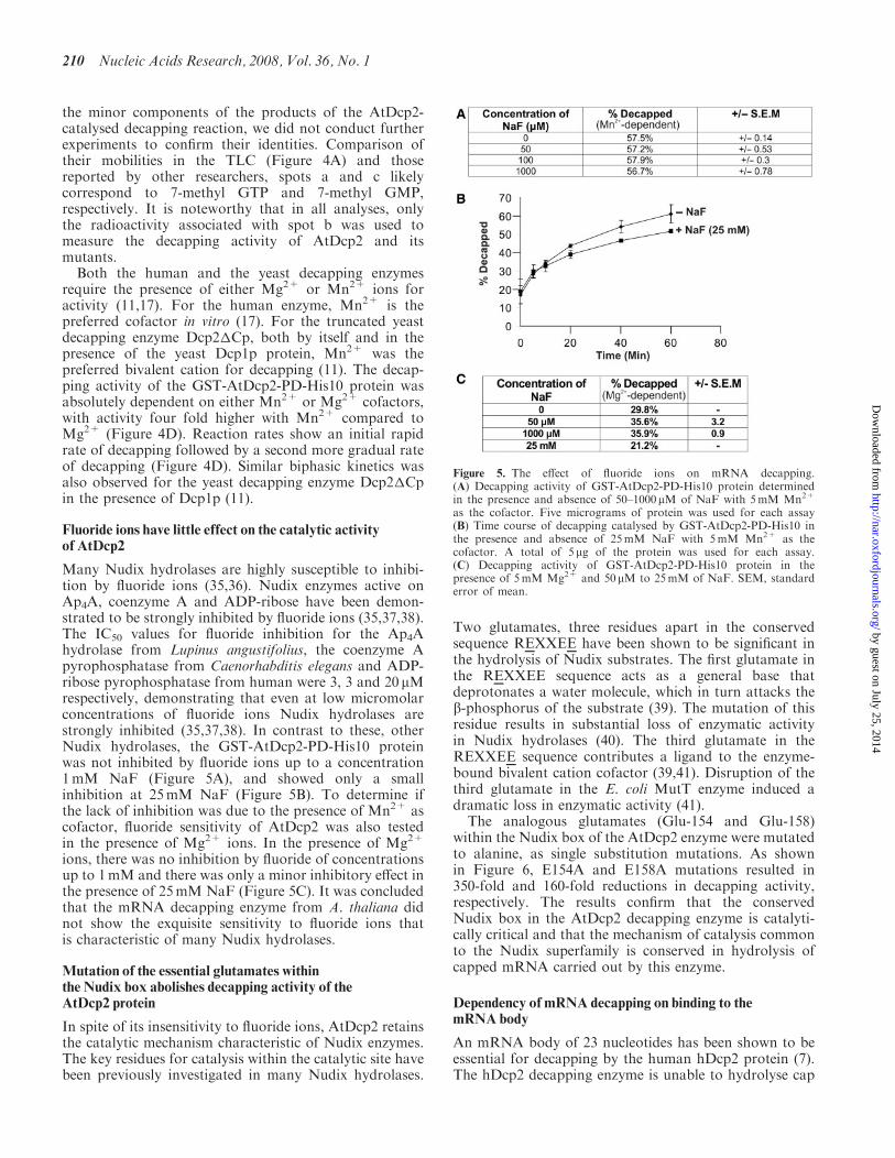
Fluoride ions have little effect on the catalytic activityof AtDcp2
Many Nudix hydrolases are highly susceptible to inhibi-tion by fluoride ions (35,36). Nudix enzymes active onAp4A, coenzyme A and ADP-ribose have been demon-strated to be strongly inhibited by fluoride ions (35,37,38).The IC50 values for fluoride inhibition for the Ap4Ahydrolase from Lupinus angustifolius, the coenzyme Apyrophosphatase from Caenorhabditis elegans and ADP-ribose pyrophosphatase from human were 3, 3 and 20 mMrespectively, demonstrating that even at low micromolarconcentrations of fluoride ions Nudix hydrolases arestrongly inhibited (35,37,38). In contrast to these, otherNudix hydrolases, the GST-AtDcp2-PD-His10 proteinwas not inhibited by fluoride ions up to a concentration1mM NaF (Figure 5A), and showed only a smallinhibition at 25mM NaF (Figure 5B). To determine ifthe lack of inhibition was due to the presence of Mn2+ ascofactor, fluoride sensitivity of AtDcp2 was also testedin the presence of Mg2+ ions. In the presence of Mg2+
ions, there was no inhibition by fluoride of concentrationsup to 1mM and there was only a minor inhibitory effect inthe presence of 25mM NaF (Figure 5C). It was concludedthat the mRNA decapping enzyme from A. thaliana didnot show the exquisite sensitivity to fluoride ions thatis characteristic of many Nudix hydrolases.
Mutation of the essential glutamates withinthe Nudix box abolishes decapping activity of theAtDcp2 protein
In spite of its insensitivity to fluoride ions, AtDcp2 retainsthe catalytic mechanism characteristic of Nudix enzymes.The key residues for catalysis within the catalytic site havebeen previously investigated in many Nudix hydrolases.
Two glutamates, three residues apart in the conservedsequence REXXEE have been shown to be significant inthe hydrolysis of Nudix substrates. The first glutamate inthe REXXEE sequence acts as a general base thatdeprotonates a water molecule, which in turn attacks theb-phosphorus of the substrate (39). The mutation of thisresidue results in substantial loss of enzymatic activityin Nudix hydrolases (40). The third glutamate in theREXXEE sequence contributes a ligand to the enzyme-bound bivalent cation cofactor (39,41). Disruption of thethird glutamate in the E. coli MutT enzyme induced adramatic loss in enzymatic activity (41).
The analogous glutamates (Glu-154 and Glu-158)within the Nudix box of the AtDcp2 enzyme were mutatedto alanine, as single substitution mutations. As shownin Figure 6, E154A and E158A mutations resulted in350-fold and 160-fold reductions in decapping activity,respectively. The results confirm that the conservedNudix box in the AtDcp2 decapping enzyme is catalyti-cally critical and that the mechanism of catalysis commonto the Nudix superfamily is conserved in hydrolysis ofcapped mRNA carried out by this enzyme.
Dependency of mRNA decapping on binding to themRNA body
An mRNA body of 23 nucleotides has been shown to beessential for decapping by the human hDcp2 protein (7).The hDcp2 decapping enzyme is unable to hydrolyse cap
Figure 5. The effect of fluoride ions on mRNA decapping.(A) Decapping activity of GST-AtDcp2-PD-His10 protein determinedin the presence and absence of 50–1000mM of NaF with 5mM Mn2+
as the cofactor. Five micrograms of protein was used for each assay(B) Time course of decapping catalysed by GST-AtDcp2-PD-His10 inthe presence and absence of 25mM NaF with 5mM Mn2+ as thecofactor. A total of 5 mg of the protein was used for each assay.(C) Decapping activity of GST-AtDcp2-PD-His10 protein in thepresence of 5mM Mg2+ and 50 mM to 25mM of NaF. SEM, standarderror of mean.
210 Nucleic Acids Research, 2008, Vol. 36, No. 1
by guest on July 25, 2014http://nar.oxfordjournals.org/
Dow
nloaded from

analog, m7G(50)ppp(50)G and decapping of cappedmRNA by the hDcp2 enzyme is not inhibited by theaddition of cap analog as a competitor (7,12,17). Takentogether, these studies suggest the requirement of aminimal number of nucleotides in the capped mRNA forits recognition by hDcp2 as substrates. In addition,uncapped transcripts are efficient competitors of cappedmRNA in the hDcp2-catalysed decapping reaction,suggesting that mRNA binding by hDcp2 can beindependent of the 7-methyl diguanosine triphosphatecap (17). To determine whether the Arabidopsis AtDcp2enzyme bound to the mRNA moiety of the capped mRNAor to the cap, the GST-AtDcp2-PD-His10 protein wasassayed for decapping activity with a fixed amount(0.14 nM) of [a-32P] capped mRNA in the presence orabsence of 164 mM of uncapped Omp mRNA or capanalog as competitors. As shown in Table 1, production of[a-32P] 7-methyl GDP from [a-32P] capped mRNA wasreduced by uncapped Omp mRNA even though it doesnot have the 7-methyl diguanosine triphosphate cap. Incontrast, the cap analog, 7-methyl diguanosine tripho-sphate, did not reduce mRNA decapping. From theseresults, we conclude that the RNA strand rather than thecap mediates binding of the capped mRNA to AtDcp2.Such property is consistent with the mechanism proposedby Piccirillo et al. (17) for the human decapping enzyme—the decapping enzyme first binds the RNA moiety of thesubstrate and then scans the RNA body to locate the cap.
AtDcp2 activity is inhibited by the reaction product7-methyl GDP
In contrast to the inability of the 7-methyl diguanosinetriphosphate to inhibit AtDcp2 activity, the reactionproduct 7-methyl GDP at 200 mM effectively suppressedthe AtDcp2-catalysed decapping activity (Table 1). Ourresults suggest that the active site of the decapping enzymeadopts a configuration that binds poorly to the cap
structure of capped mRNA but relatively efficiently to thereaction product 7-methyl GDP.
Deletion of a key loop region in the putative RNA-bindingdomain interferes with the AtDcp2 catalysed decappingactivity
Multiple sequence alignment of Dcp2 enzymes from arange of species was used to identify the RNA bindingdomain in the AtDcp2 protein (Figure 7A). In the humandecapping enzyme, hDcp2, a region consisting of Box Band the preceding loop region was shown to be importantfor RNA binding (17). The Box B region is conservedin the Dcp2-type decapping enzymes (Figure 7A). Fromthe structure of the Schizosaccharomyces pombe Dcp2penzyme, it is clear that Box B forms the C-terminal helix
Figure 6. Effects of mutation of two conserved glutamates in the Nudix box on decapping activity of AtDcp2. (A) The mutated residues (underlined)within the Nudix box. (B) Decapping activities of the GST-AtDcp2-PD-His10 protein and the mutants. The decapping assays were carried out with8 mg of purified recombinant enzyme from three separate expressions. SEM, standard error of mean.
Table 1. The dependency of decapping on mRNA/cap/product binding
Competitor Decapped (%) SEM (+/�)
Panel ANone 68.4 0.7Uncapped Omp mRNA(164 mM) 37.8 2.17-methyl diguanosine triphosphate(164 mM)
69 1
Panel BNone 44.3 0.37-methyl GDP (2mM) 25.3 1.47-methyl GDP (0.2mM) 29.4 0.1
(A) The extent of decapping of the substrate was measured after 60minin assays containing GST-AtDcp2-PD-His10 protein, 5mM Mn2� anda fixed amount (0.14 nM) of radiolabelled capped Omp mRNA,supplemented with 164mM each of the competitors: uncapped Ompand cap analog dinucleotide. The SEM of three separate assays foreach competitor are also indicated. All other conditions are asdescribed in the ‘Materials and methods’ section.(B) The extent of decapping of the substrate by the recombinantenzyme in the presence and absence of the reaction product 7-methylGDP after 60min.
Nucleic Acids Research, 2008, Vol. 36, No. 1 211
by guest on July 25, 2014http://nar.oxfordjournals.org/
Dow
nloaded from

in the a/b/a sandwich of the Nudix fold (18). To definethe structural features in the loop region precedingthe Box B region responsible for RNA binding, weconducted bioinformatics analysis of this region in theAtDcp2 sequence. The analysis revealed a motif(VITHGVSGLKL) in this loop region of AtDcp2 thatclosely resembles the VIGXXGXXUK segment(Figure 7B) commonly found in the KH domain of
several RNA-binding proteins including ERA, Fragile Xprotein, hnRNP K and Poly(C) binding proteins (42–45).We therefore termed this motif as KH-like motif.Homology modelling of the Nudix fold of the AtDcp2protein (residues 98–250) using the known structure of theS. pombe Dcp2p decapping enzyme, revealed that thisKH-like motif likely resides in the loop region that extendsto the start of the Box B helix (Figure 7C). We postulate
Figure 7. Effects of mutation of residues within the putative RNA-binding KH-like domain and Box B domain on catalytic activity of AtDcp2.(A) Sequence alignment of Arabidopsis thaliana (AtDcp2) to reveal the putative KH-like domain. Oryza sativa (osDcp2), human (hDcp2), Xenopuslaevis (xlDcp2) and Caenorhabditis elegans (ceDcp2) Dcp2 decapping enzymes were used for the alignments. The position of the KH-like motif andBox B within the AtDcp2 protein is shown in the sequence alignment. (B) The consensus sequence of the conserved 10 residue region found in KHdomains (42,43), the sequence corresponding to the consensus sequence found in the KH domain in the RNA-binding site of ERA protein fromE. coli (42) and the sequence of a KH-like motif in the AtDcp2 decapping enzyme. (C) Comparison of the structure of the Ap4A hydrolase fromL. angustifolius (PDB ID–1JKN) with the homology model of the Nudix fold of the AtDcp2 protein (residues 98–250) modelled after the structure ofthe S.pombe Dcp2p enzyme (PDB ID – 2A6T). Box B helix in the AtDcp2 homology model and the analogous helix 4 in the lupin enzyme structureare marked in red. The 11-residue region forming the near-consensus of a KH motif, found within the loop region preceding Box B, is coloured inyellow. This region was deleted in mutant AtDcp2�222–232. The side chains of three lysines and a single glycine, which were mutated individually toalanines, are also shown. (D) Decapping activities of the GST-AtDcp2-PD-His10 protein and the mutant proteins carrying mutations in the putativeRNA-binding site. All decapping efficiencies were obtained from decapping assays carried out using 8 mg of recombinant enzyme from three separateexpressions. SEM, standard error of mean.
212 Nucleic Acids Research, 2008, Vol. 36, No. 1
by guest on July 25, 2014http://nar.oxfordjournals.org/
Dow
nloaded from

that the KH-like motif in AtDcp2 (Figure 7B) containsdeterminants that facilitate the decapping reaction bybinding to the RNA moiety of the capped mRNAsubstrate.
To ascertain the functional significance of the 11-residueKH-like motif, it was deleted or mutated in AtDcp2. Asshown in Figure 7D, deletion of the KH-like motif led to a92-fold reduction in the decapping activity. Replacementof Lys-231 and Gly-226 in this segment (VITHGVSGLK)by alanine reduced the decapping activity of AtDcp2 by�68% and 34%, respectively. In summary, our resultssuggest that the KH-like motif contains determinantsessential for decapping activity. Presumably, these deter-minants facilitate decapping by mediating association ofAtDcp2 with the RNA backbone of the capped mRNAsubstrate. This notion is supported by comparison of thehomology model of the AtDcp2 protein with the structureof the related Nudix enzyme Ap4A hydrolase fromLupinus angustifolius (46,47). It was evident from thecomparison that the Box B helix of AtDcp2 is equivalentto helix 4 in the lupin enzyme structure (Figure 7C). Theloop region preceding helix 4 in the lupin Ap4A hydrolasewas demonstrated to contribute critical substrate bindingresidues that lined the hydrophobic pocket, where theATP-MgFX substrate bound (47). It is likely that theKH-like motif in this loop region is suitably located inthe AtDcp2 structure to mediate or facilitate binding ofthe decapping enzyme to the RNA substrate.
Two lysine residues in Box B region contribute to efficienthydrolysis of capped mRNA by AtDcp2
Since the Box B region in human decapping enzyme wasfound to participate in RNA binding, we conducted amutagenesis study to ascertain if the Box B region ofAtDcp2 contains determinants that are important foractivity. Both Lys-243 and Lys-248 in the Box B region ofAtDcp2 were individually modified to alanine to eliminatetheir polar side chains. As shown in Figure 7D, themutations resulted in 56–68% reductions in decappingactivity (Figure 7D), supporting the notion that bothlysine residues in the Box B region contribute to the abilityof AtDcp2 to efficiently hydrolyse capped mRNA.Presumably, the positively charged side chains of bothlysines mediate binding of AtDcp2 to the RNA moiety ofthe capped mRNA substrate.
DISCUSSION
The roles of Glu-154 and Glu-158 in the decapping reaction
This study describes the functional characterization ofa Dcp2 type decapping enzyme from plants. The AtDcp2enzyme hydrolyses the mRNA cap by substitution atb-phosphorus, the conserved mechanism of catalysisfor the Nudix superfamily, yielding 7-methyl GDP and50-monophosphate mRNA as the products. The individualconversion of both essential catalytic site glutamates,Glu-154 and Glu-158, to alanine resulted in morethan 150-fold reductions in catalytic activity of theAtDcp2 enzyme, consistent with their functions asthe effector of catalysis (Glu-154) and a ligand donor
(Glu-158) that coordinates a divalent cation, respec-tively (39). Relevant to our findings presented inFigure 6, mutations of homologous glutamates in theconserved Nudix box motif of both the human andyeast decapping enzymes abolished their decappingactivities (7,11).
ImplicationsofproductinhibitionofAtDcp2by7-methylGDP
Decapping was inhibited by the product 7-methyl GDP,by uncapped mRNA but not by the cap analog, 7-methyldiguanosine triphosphate. It appears that presentation ofthe cap to the cleavage site requires the cooperation of themRNA body and that the catalytic site binds to theproduct 7-methyl GDP more strongly than to the cap incapped mRNA. The 7-methyl GDP similarly inhibits theinherent mRNA decapping activity in the L-A virus (48)and the activity of the nuclear Nudix small nucleolar RNAdecapping enzyme X29 (49). Such product inhibition by7-methyl GDP constitutes a potential mechanism toprevent decapping of intact translatable mRNAs andaccordingly 7-methyl GDP has a short cellular lifetime.The 7-methyl GDP formed by Dcp2 enzymes is convertedrapidly to 7-methyl GMP by the scavenger mRNAdecapping activity in vivo (50).
The lack of sensitivity to fluoride ions is a distinguishingfeature of AtDcp2 within the Nudix hydrolase superfamilyof enzymes
Extreme sensitivity to fluoride has been shown previouslyto be a widespread characteristic of Nudix hydrolases.All enzymes so far tested have shown this (35,36,38,51).Contrary to other Nudix enzymes, the AtDcp2 proteinwas not inhibited by fluoride ions at concentrations of upto 1mM. Only at 25mM, did the fluoride ions impose aminor inhibitory effect. Fluoride compounds mimicphosphate groups. It has been proposed that Nudixenzymes and ATPases are inhibited by complexes formedwith the fluoride compounds and the reaction products(36,52). Since our results demonstrate that AtDcp2 isrefractory to inhibition by fluoride ions, we propose thatAtDcp2 active site adopts a configuration with limitedaccessibility to the complexes formed by fluoride ionswith 7-methyl GDP. It is likely all other Dcp2-typedecapping enzymes also share such a property.In this respect, the Dcp2-type decapping enzymeappears to be distinct from many other Nudix hydrolases.Future investigations to solve the 3D structure ofAtDcp2 will provide the structural basis for this distinctproperty.
The structural basis of RNA binding by AtDcp2
RNA-binding proteins interact with RNA molecules intwo structurally defined modes: (i) groove binding and(ii) b-sheet binding (53,54). For the proteins adopting thegroove-binding mode, a secondary structural element suchas a helix or a loop is inserted into the groove of the RNAsubstrate (53,54). It is proposed here that AtDcp2 adoptsthe groove-binding mode to bind RNA—the loop regionpreceding Box B and/or Box B extending into grooves ofthe RNA molecule. Katsamba and co-workers (55)
Nucleic Acids Research, 2008, Vol. 36, No. 1 213
by guest on July 25, 2014http://nar.oxfordjournals.org/
Dow
nloaded from
proposed a two-step ‘lure and lock’ model of protein–RNA association. In this model, electrostatic interactionsare the key for the initial association (the lure step).This is then followed by rapid induced fit binding (the lockstep), resulting in a stable protein–RNA complex (55).Since mutation of Lys-231 in the loop region and Lys-243and Lys-248 in the Box B region reduces the decappingactivity of AtDcp2 (Figure 7), we propose that theyare responsible for the initial electrostatic interactionsin the lure step to ‘lure’ the RNA molecule to AtDcp2,the bound RNA molecule then induces conforma-tional changes to establish additional interactionsbetween the RNA moiety with determinants inother parts of AtDcp2 to form a stable protein–RNAcomplex.Piccirillo et al. (17) reported that the carboxyl-terminal
portion including Box B in the human decapping enzymehDcp2 can mediate RNA binding. They also demon-strated that hDcp2 and single-stranded DNA could formstable complexes, and the ability of hDcp2 to form thecomplexes was significantly diminished upon deletion ofthe C-terminal segment containing Box B. We haveattempted to use the three different methods describedby Piccirillo et al. (17) and Moller et al. (56) and toascertain if AtDcp2 can form stable complexes with[a-32P]-m7Gppp-RNA. These methods include: (i) theNorthwestern RNA-binding assay, (ii) cross-linking ofAtDcp2 with [a-32P]-m7Gppp-RNA by ultraviolet lightirradiation and (iii) cross-linking of AtDcp2 with [a-32P]-m7Gppp-RNA by formaldehyde treatment. In contrast tothe findings by Piccirillo and co-workers, we failed todetect stable AtDcp2-[a-32P]-m7Gppp–RNA complexes.A possible explanation for our failure to detect thestable complexes is that the binding of AtDcp2 tocapped mRNA is transient in nature. An appropriateapproach for future studies to measure the affinity ofAtDcp2 for capped mDNA and to define the structuralmotifs critical for RNA binding is the use of surfaceplasmon resonance to determine the affinities of AtDcp2and its mutants for capped mRNA immobilized on sensorchips.
Possible functional significance of the putative PDZdomain-binding motif at the C-terminus of AtDcp2
Bioinformatics analysis identified the ‘GNSA’ motif at theC-terminus of AtDcp2 as a putative PDZ domain-bindingmotif. In addition, we conducted further analysis byhomology searches in the NCBI database and found thatsimilar putative PDZ domain-binding motifs also exist inalmost 80% of vertebrate and plant Dcp2 decappingenzymes (data not shown). One approach to ascertain ifthe GNSA can indeed mediate AtDcp2 binding to PDZdomain-containing proteins is to use the recombinantGST-AtDcp2 proteins as the baits to search for bindingproteins in Arabidopsis tissue lysates. Proteins that canbind the GST-AtDcp2 but not GST-AtDcp2-PD(Figure 1) are likely those that target the GNSA-motif.Identification of these proteins will shed light onregulatory properties of AtDcp2.
CONCLUSIONS
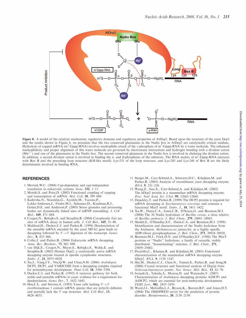
Based upon the findings described in this article, wepropose a model of the catalytic mechanism and regula-tion of AtDcp2 (Figure 8). We have demonstrated thatAtDcp2 catalyses the following reaction.
m7Gppp�RNAþH2O ! m7GDPþ p�RNAþHþ
In the reaction, AtDcp2 selectively catalyses cleavage ofthe diphosphate linkage between the b- and g-phosphatesin the cap of the capped mRNA substrate (m7Gppp-RNA) to generate 7-methyl GDP and 50-monophosphatemRNA (p-RNA) as the reaction products. Similar toother known Nudix hydrolase superfamily members, theAtDcp2-catalysed decapping reaction involves nucleophi-lic attack of the pyrophosphate bond by a water molecule(Equation 1). One of the two conserved glutamates in theNudix box is involved in polarizing and appropriatelypositioning the water molecule, while the other conservedglutamate coordinates a divalent cation (Figure 8). It isproposed that the recognition of the substrates by AtDcp2involves binding of the RNA moiety of the capped mRNAto the Box B region and a KH-like motif in the looppreceding the Box B region. One lysine in the KH-likemotif and two lysines in the Box B region likely mediatein part the binding of AtDcp2 to the RNA moiety.Furthermore, the activity of AtDcp2 is subject to feedbackinhibition by its reaction product 7-methyl GDP anduncapped RNA.
In spite of these findings, several important outstandingquestions remain: (i) What are the exact roles of Glu-154and Glu-158 of the Nudix box in catalysis? (ii) How doesAtDcp2 selectively target the pyrophosphate bond linkingthe b- and g-phosphates but not that linking the a- andb-phosphates and the phosphoester linkage betweeng-phosphate and the RNA moiety of the capped RNAsubstrate? (iii) What is the structural basis of RNAbinding to the loop and Box B regions? (iv) How does theactive site of AtDcp2 target the cap at the 50-end of thecapped RNA substrates? (v) What are the upstreamregulators of AtDcp2? Xu et al. (8) recently reported thatAtDcp2 interacts with Dcp1 and VARICOSE (VCS), theplant homolog of an important component called Hedls/Ge-1 of the mRNA processing bodies (P bodies) in thecytoplasm. Furthermore, they demonstrated that AtDcp2activity is stimulated upon binding to these proteins.Exactly how these regulatory proteins modulate AtDcp2activity is unclear. The availability of the intact recombi-nant AtDcp2 permits future investigation to answer thesequestions by biochemical and structural approaches.
ACKNOWLEDGEMENTS
We are indebted to Dr Lena Burri, University ofMelbourne for the pSP73-Omp clone and the Sir Johnand Lady Higgins Foundation for sponsoring the PhDstudies of the candidate Dilantha Gunawardana. Fundingto pay the Open Access publication charges for this articlewas provided by The University of Melbourne.
Conflict of interest statement. None declared.
214 Nucleic Acids Research, 2008, Vol. 36, No. 1
by guest on July 25, 2014http://nar.oxfordjournals.org/
Dow
nloaded from
REFERENCES
1. Merrick,W.C. (2004) Cap-dependent and cap-independenttranslation in eukaryotic systems. Gene, 332, 1–11.
2. Moteki,S. and Price,D. (2002) Functional coupling of cappingand transcription of mRNA. Mol. Cell, 10, 599–609.
3. Kedersha,N., Stoecklin,G., Ayodele,M., Yacono,P.,Lykke-Andersen,J., Fitzler,M.J., Scheuner,D., Kaufman,R.J.,Golan,D.E. and Anderson,P. (2005) Stress granules and processingbodies are dynamically linked sites of mRNP remodeling. J. CellBiol., 169, 871–884.
4. Cougot,N., Babajko,S. and Seraphin,B. (2004) Cytoplasmic foci aresites of mRNA decay in human cells. J. Cell Biol., 165, 31–40.
5. Muhlrad,D., Decker,C.J. and Parker,R. (1994) Deadenylation ofthe unstable mRNA encoded by the yeast MFA2 gene leads todecapping followed by 50–>30 digestion of the transcript. GenesDev., 8, 855–866.
6. Coller,J. and Parker,R. (2004) Eukaryotic mRNA decapping.Annu. Rev. Biochem., 73, 861–890.
7. van Dijk,E., Cougot,N., Meyer,S., Babajko,S., Wahle,E. andSeraphin,B. (2002) Human Dcp2: a catalytically active mRNAdecapping enzyme located in specific cytoplasmic structures.Embo. J., 21, 6915–6924.
8. Xu,J., Yang,J.Y., Niu,Q.W. and Chua,N.H. (2006) ArabidopsisDCP2, DCP1, and VARICOSE form a decapping complex requiredfor postembryonic development. Plant Cell, 18, 3386–3398.
9. Decker,C.J. and Parker,R. (1993) A turnover pathway for bothstable and unstable mRNAs in yeast: evidence for a requirement fordeadenylation. Genes Dev., 7, 1632–1643.
10. Hsu,C.L. and Stevens,A. (1993) Yeast cells lacking 50–>30
exoribonuclease 1 contain mRNA species that are poly(A) deficientand partially lack the 50 cap structure. Mol. Cell Biol., 13,4826–4835.
11. Steiger,M., Carr-Schmid,A., Schwartz,D.C., Kiledjian,M. andParker,R. (2003) Analysis of recombinant yeast decapping enzyme.RNA, 9, 231–238.
12. Wang,Z., Jiao,X., Carr-Schmid,A. and Kiledjian,M. (2002)The hDcp2 protein is a mammalian mRNA decapping enzyme.Proc. Natl Acad. Sci. USA, 99, 12663–12668.
13. Dunckley,T. and Parker,R. (1999) The DCP2 protein is required formRNA decapping in Saccharomyces cerevisiae and contains afunctional MutT motif. Embo. J., 18, 5411–5422.
14. Xu,W., Dunn,C.A., Jones,C.R., D’Souza,G. and Bessman,M.J.(2004) The 26 Nudix hydrolases of Bacillus cereus, a close relativeof Bacillus anthracis. J. Biol. Chem., 279, 24861–24865.
15. Sheikh,S., O’Handley,S.F., Dunn,C.A. and Bessman,M.J. (1998)Identification and characterization of the Nudix hydrolase fromthe Archaeon, Methanococcus jannaschii, as a highly specificADP-ribose pyrophosphatase. J. Biol. Chem., 273, 20924–20928.
16. Bessman,M.J., Frick,D.N. and O’Handley,S.F. (1996) The MutTproteins or ‘‘Nudix’’ hydrolases, a family of versatile, widelydistributed, ‘‘housecleaning’’ enzymes. J. Biol. Chem., 271,25059–25062.
17. Piccirillo,C., Khanna,R. and Kiledjian,M. (2003) Functionalcharacterization of the mammalian mRNA decapping enzymehDcp2. RNA, 9, 1138–1147.
18. She,M., Decker,C.J., Chen,N., Tumati,S., Parker,R. and Song,H.(2006) Crystal structure and functional analysis of Dcp2p fromSchizosaccharomyces pombe. Nat. Struct. Mol. Biol., 13, 63–70.
19. Iwasaki,S., Takeda,A., Motose,H. and Watanabe,Y. (2007)Characterization of Arabidopsis decapping proteins AtDCP1 andAtDCP2, which are essential for post-embryonic development.FEBS Lett., 581, 2455–2459.
20. Ward,J.J., McGuffin,L.J., Bryson,K., Buxton,B.F. and Jones,D.T.(2004) The DISOPRED server for the prediction of proteindisorder. Bioinformatics, 20, 2138–2139.
Figure 8. A model of the catalytic mechanism, regulatory domains and regulatory properties of AtDcp2. Based upon the structure of the yeast Dcp2and the results shown in Figure 6, we postulate that the two conserved glutamates in the Nudix box in AtDcp2 are catalytically critical residues.Hydrolysis of capped mRNA (m7-Gppp-RNA) involves nucleophilic attack of the g-phosphate of m7-Gppp-RNA by a water molecule. The enhancednucleophilicity and proper alignment of this water molecule are governed by electrostatic interactions and hydrogen bonding with a divalent cation(Mn2+) and one of the glutamates in the Nudix box. The second conserved glutamate in the Nudix box is involved in chelating the divalent cation.In addition, a second divalent cation is involved in binding the a- and b-phosphates of the substrate. The RNA moiety of m7-Gppp-RNA interactswith Box B and the preceding loop structure (KH-like motif). Lys-231 of the loop structure, and Lys-243 and Lys-248 of Box B are the likelydeterminants involved in binding RNA.
Nucleic Acids Research, 2008, Vol. 36, No. 1 215
by guest on July 25, 2014http://nar.oxfordjournals.org/
Dow
nloaded from
21. Linding,R., Jensen,L.J., Diella,F., Bork,P., Gibson,T.J. andRussell,R.B. (2003) Protein disorder prediction: implications forstructural proteomics. Structure, 11, 1453–1459.
22. Gopalan,V., Tan,T.W., Lee,B.T. and Ranganathan,S. (2004) Xpro:database of eukaryotic protein-encoding genes. Nucleic Acids Res.,32, D59–D63.
23. Schwede,T., Kopp,J., Guex,N. and Peitsch,M.C. (2003)SWISS-MODEL: an automated protein homology-modeling server.Nucleic Acids Res., 31, 3381–3385.
24. Tompa,P. and Csermely,P. (2004) The role of structural disorder inthe function of RNA and protein chaperones. Faseb. J., 18,1169–1175.
25. Gottesman,S. (1996) Proteases and their targets in Escherichia coli.Annu. Rev. Genet., 30, 465–506.
26. Levchenko,I, Smith,C.K., Walsh,N.P., Sauer,R.T. and Baker,T.A.(1997) PDZ-like domains mediate binding specificity in the Clp/Hsp100 family of chaperones and protease regulatory subunits. Cell,91, 939–947.
27. Longhi,S., Receveur-Brechot,V., Karlin,D., Johansson,K.,Darbon,H., Bhella,D., Yeo,R., Finet,S. and Canard,B. (2003) TheC-terminal domain of the measles virus nucleoprotein is intrinsicallydisordered and folds upon binding to the C-terminal moiety of thephosphoprotein. J. Biol. Chem., 278, 18638–18648.
28. Harris,B.Z. and Lim,W.A. (2001) Mechanism and role of PDZdomains in signaling complex assembly. J. Cell Sci., 114,3219–3231.
29. Beebe,K.D., Shin,J., Peng,J., Chaudhury,C., Khera,J. and Pei,D.(2000) Substrate recognition through a PDZ domain in tail-specificprotease. Biochemistry, 39, 3149–3155.
30. Walsh,N.P., Alba,B.M., Bose,B., Gross,C.A. and Sauer,R.T. (2003)OMP peptide signals initiate the envelope-stress response byactivating DegS protease via relief of inhibition mediated by itsPDZ domain. Cell, 113, 61–71.
31. Wilken,C., Kitzing,K., Kurzbauer,R., Ehrmann,M. and Clausen,T.(2004) Crystal structure of the DegS stress sensor: how a PDZdomain recognizes misfolded protein and activates a protease. Cell,117, 483–494.
32. Keiler,K.C., Silber,K.R., Downard,K.M., Papayannopoulos,I.A.,Biemann,K. and Sauer,R.T. (1995) C-terminal specific proteindegradation: activity and substrate specificity of the Tsp protease.Protein Sci., 4, 1507–1515.
33. Zhang,S., Williams,C.J., Wormington,M., Stevens,A. andPeltz,S.W. (1999) Monitoring mRNA decapping activity. Methods,17, 46–51.
34. Bergman,N., Opyrchal,M., Bates,E.J. and Wilusz,J. (2002) Analysisof the products of mRNA decapping and 30-to-50 decay bydenaturing gel electrophoresis. RNA, 8, 959–965.
35. Maksel,D., Gooley,P.R., Swarbrick,J.D., Guranowski,A.,Gange,C., Blackburn,G.M. and Gayler,K.R. (2001)Characterization of active-site residues in diadenosinetetraphosphate hydrolase from Lupinus angustifolius. Biochem. J.,357, 399–405.
36. Guranowski,A. (1990) Fluoride is a strong and specific inhibitor of(asymmetrical) Ap4A hydrolases. FEBS Lett., 262, 205–208.
37. Gasmi,L., Cartwright,J.L. and McLennan,A.G. (1999) Cloning,expression and characterization of YSA1H, a human adenosine50-diphosphosugar pyrophosphatase possessing a MutT motif.Biochem. J., 344 (Pt 2), 331–337.
38. AbdelRaheim,S.R. and McLennan,A.G. (2002) The Caenorhabditiselegans Y87G2A.14 Nudix hydrolase is a peroxisomal coenzymeA diphosphatase. BMC Biochem., 3, 5.
39. Mildvan,A.S., Xia,Z., Azurmendi,H.F., Saraswat,V., Legler,P.M.,Massiah,M.A., Gabelli,S.B., Bianchet,M.A., Kang,L.W. et al.
(2005) Structures and mechanisms of Nudix hydrolases. Arch.Biochem. Biophys., 433, 129–143.
40. Harris,T.K., Wu,G., Massiah,M.A. and Mildvan,A.S. (2000)Mutational, kinetic, and NMR studies of the roles ofconserved glutamate residues and of lysine-39 in themechanism of the MutT pyrophosphohydrolase. Biochemistry, 39,1655–1674.
41. Lin,J., Abeygunawardana,C., Frick,D.N., Bessman,M.J. andMildvan,A.S. (1996) The role of Glu 57 in the mechanism of theEscherichia coli MutT enzyme by mutagenesis and heteronuclearNMR. Biochemistry, 35, 6715–6726.
42. Chen,X., Court,D.L. and Ji,X. (1999) Crystal structure of ERA: aGTPase-dependent cell cycle regulator containing an RNA bindingmotif. Proc. Natl Acad. Sci. USA, 96, 8396–8401.
43. Siomi,H., Matunis,M.J., Michael,W.M. and Dreyfuss,G. (1993)The pre-mRNA binding K protein contains a novel evolutionarilyconserved motif. Nucleic Acids Res., 21, 1193–1198.
44. Siomi,H., Siomi,M.C., Nussbaum,R.L. and Dreyfuss,G. (1993)The protein product of the fragile X gene, FMR1, hascharacteristics of an RNA-binding protein. Cell, 74, 291–298.
45. Sidiqi,M., Wilce,J.A., Vivian,J.P., Porter,C.J., Barker,A.,Leedman,P.J. and Wilce,M.C. (2005) Structure and RNA bindingof the third KH domain of poly(C)-binding protein 1. Nucleic AcidsRes., 33, 1213–1221.
46. Fletcher,J.I., Swarbrick,J.D., Maksel,D., Gayler,K.R. andGooley,P.R. (2002) The structure of Ap4A hydrolase complexedwith ATP-MgF(x) reveals the basis of substrate binding. Structure,10, 205–213.
47. Swarbrick,J.D., Bashtannyk,T., Maksel,D., Zhang,X.R.,Blackburn,G.M., Gayler,K.R. and Gooley,P.R. (2000) Thethree-dimensional structure of the Nudix enzyme diadenosinetetraphosphate hydrolase from Lupinus angustifolius L. J. Mol.Biol., 302, 1165–1177.
48. Naitow,H., Tang,J., Canady,M., Wickner,R.B. and Johnson,J.E.(2002) L-A virus at 3.4 A resolution reveals particlearchitecture and mRNA decapping mechanism. Nat. Struct. Biol., 9,725–728.
49. Peculis,B.A., Reynolds,K. and Cleland,M. (2007) Metal determinesefficiency and substrate specificity of the nuclear NUDIX decappingproteins X29 and H29K (Nudt16). J. Biol. Chem., 282,24792–24805.
50. van Dijk,E., Le Hir,H. and Seraphin,B. (2003) DcpS can act in the50–30 mRNA decay pathway in addition to the 30–50 pathway. Proc.Natl Acad. Sci. USA, 100, 12081–12086.
51. AbdelRaheim,S.R., Cartwright,J.L., Gasmi,L. and McLennan,A.G.(2001) The NADH diphosphatase encoded by the Saccharomycescerevisiae NPY1 Nudix hydrolase gene is located in peroxisomes.Arch. Biochem Biophys., 388, 18–24.
52. Coll,R.J. and Murphy,A.J. (1992) Fluoride-inhibited calciumATPase of sarcoplasmic reticulum. Magnesium and fluoridestoichiometry. J. Biol. Chem., 267, 21584–21587.
53. Jones,S., Daley,D.T., Luscombe,N.M., Berman,H.M. andThornton,J.M. (2001) Protein-RNA interactions: a structuralanalysis. Nucleic Acids Res., 29, 943–954.
54. Draper,D.E. (1999) Themes in RNA-protein recognition. J. Mol.Biol., 293, 255–270.
55. Katsamba,P.S., Myszka,D.G. and Laird-Offringa,I.A. (2001) Twofunctionally distinct steps mediate high affinity binding of U1Aprotein to U1 hairpin II RNA. J. Biol. Chem., 276, 21476–21481.
56. Moller,K., Rinke,J., Ross,A., Buddle,G. and Brimacombe,R. (1977)The use of formaldehyde in RNA-protein cross-linking studieswith ribosomal subunits from Escherichia coli. Eur. J. Biochem., 76,175–187.
216 Nucleic Acids Research, 2008, Vol. 36, No. 1
by guest on July 25, 2014http://nar.oxfordjournals.org/
Dow
nloaded from