Analysis of the RNA-recognition motif and RS and RGG domains: conservation in metazoan pre-mRNA...
14
Nucleic Acids Research, 1993, Vol. 21, No. 25 5803-5816 Analysis of the RNA-recognition motif and RS and RGG domains: conservation in metazoan pre-mRNA splicing factors Ewan Birney+, Sanjay Kumar§ and Adrian R.Krainer* Cold Spring Harbor Laboratory, PO Box 100, 1 Bungtown Road, Cold Spring Harbor, NY 11724-2208, USA Received July 30, 1993; Revised and Accepted October 27, 1993 ABSTRACT We present a systematic analysis of sequence motifs found in metazoan protein factors involved in constitutive pre-mRNA splicing and in alternative splicing regulation. Using profile analysis we constructed a database enriched in protein sequences containing one or more presumptive copies of the RNA- recognition motif (RRM). We provide an accurate alignment of RRMs and structure-based criteria for identifying new RRMs, including many that lack the prototype RNP-1 submotif. We present a comprehensive table of 125 sequences containing 252 RRMs, including 22 previously unreported RRMs in 17 proteins. The presence of a putative RRM in these proteins, which are implicated in a variety of cellular processes, strongly suggests that their function involves binding to RNA. Unreported homologies in the RRM-enriched database to the metazoan SR family of splicing factors are described for an Arg-rich human nuclear protein and two yeast proteins (S. pombe mei2 and S. cerevisiae Npl3). We have rigorously tested the phylogenetic relationships of a large sample of RRMs. This analysis indicates that the RRM is an ancient conserved region (ACR) that has diversified by duplication of genes and intragenic domains. Statistical analyses and classification of repeated Arg - Ser (RS) and RGG domains in various protein splicing factors are presented. INTRODUCTION Several metazoan protein factors have been identified that are required for pre-mRNA splicing in vitro, or that regulate the selection of alternative splice sites in vivo and/or in vitro (reviewed in 1,2). In addition, several polypeptides associated with all, or with specific snRNPs have been described, as have a number of RNA-binding proteins, termed hnRNPs, which are associated with nuclear pre-mRNAs (reviewed in 1-4). These proteins, and many others, are present in spliceosomes, although perhaps not in all spliceosomes. Some may participate directly in splicing whereas others may be involved for example in snRNP assembly and transport to the nucleus, or in mRNA export to the cytoplasm. The generic or essential metazoan protein splicing factors for which complete amino acid sequences are available to date include human SF2/ASF (also known as SRp3Oa) (5,6), human and mouse U2AF65 (7,8), human and chicken SC35 (also known as PR264 or SRp30b) (9,10), human PSF (1 1), Drosophila SRp55 (a variant of which is known as B52) (12,13), mouse and human X16 (also known as SRp2O) (14,15) and its probable Drosophila homolog, RBP1 (16). Partial peptide sequences are also available for human SRp40, SRp55, and SRp75 (15). With the exception of U2AF65 and PSF, all of the above proteins belong to the same protein family, and many of them, perhaps all, have similar functions in constitutive and alternative splicing in vitro (15-21). Functional roles for constitutive and/or alternative splicing in vitro have also been reported for hnRNPAI and hnRNP C1/C2, for which sequences are available from several species (22 -24). In addition, the complete sequences of several other hnRNP and snRNP polypeptides have been known for some time (reviewed in 3,4). Finally, several regulators of alternative splicing in Drosophila, first identified genetically, have been cloned and sequenced, including Transformer (Tra), Transformer-2 (Tra-2), Sex-lethal (Sxl), and Suppressor of white apricot (Su(wa)) (reviewed in 25). Several of the above proteins are closely related in sequence, and many share structural features among themselves and with other RNA-binding proteins. Other sequence features are unique to a small subset of these proteins. The most notable features present in many of these proteins include one or more RNP-type RNA-recognition motifs (RRM or CS-RBD or RNP-80), and clusters of Arg and Ser residues (RS domains). Gly-rich clusters, sometimes referred to as hinge regions, are also common, as are domains with many Gly-Gly dipeptides interspersed with aromatic and Arg residues (RGG or GAR domains). The RRMs of various proteins have been extensively studied (reviewed in 26-30). As more members of the RRM superfamily have been * To whom correspondence should be addressed Present addresses: +Balliol College, Oxford, OXI 3BJ, UK and §New England Biolabs, Beverly, MA 01915, USA k. 1993 Oxford University Press
-
Upload
independent -
Category
Documents
-
view
2 -
download
0
Transcript of Analysis of the RNA-recognition motif and RS and RGG domains: conservation in metazoan pre-mRNA...

Nucleic Acids Research, 1993, Vol. 21, No. 25 5803-5816
Analysis of the RNA-recognition motif and RS and RGGdomains: conservation in metazoan pre-mRNA splicingfactors
Ewan Birney+, Sanjay Kumar§ and Adrian R.Krainer*Cold Spring Harbor Laboratory, PO Box 100, 1 Bungtown Road, Cold Spring Harbor,NY 11724-2208, USA
Received July 30, 1993; Revised and Accepted October 27, 1993
ABSTRACT
We present a systematic analysis of sequence motifsfound in metazoan protein factors involved inconstitutive pre-mRNA splicing and in alternativesplicing regulation. Using profile analysis weconstructed a database enriched in protein sequencescontaining one or more presumptive copies of the RNA-recognition motif (RRM). We provide an accuratealignment of RRMs and structure-based criteria foridentifying new RRMs, including many that lack theprototype RNP-1 submotif. We present acomprehensive table of 125 sequences containing 252RRMs, including 22 previously unreported RRMs in 17proteins. The presence of a putative RRM in theseproteins, which are implicated in a variety of cellularprocesses, strongly suggests that their functioninvolves binding to RNA. Unreported homologies in theRRM-enriched database to the metazoan SR family ofsplicing factors are described for an Arg-rich humannuclear protein and two yeast proteins (S. pombe mei2and S. cerevisiae Npl3). We have rigorously tested thephylogenetic relationships of a large sample of RRMs.This analysis indicates that the RRM is an ancientconserved region (ACR) that has diversified byduplication of genes and intragenic domains. Statisticalanalyses and classification of repeated Arg - Ser (RS)and RGG domains in various protein splicing factorsare presented.
INTRODUCTIONSeveral metazoan protein factors have been identified that arerequired for pre-mRNA splicing in vitro, or that regulate theselection of alternative splice sites in vivo and/or in vitro(reviewed in 1,2). In addition, several polypeptides associatedwith all, or with specific snRNPs have been described, as havea number of RNA-binding proteins, termed hnRNPs, which areassociated with nuclear pre-mRNAs (reviewed in 1-4). Theseproteins, and many others, are present in spliceosomes, although
perhaps not in all spliceosomes. Some may participate directlyin splicing whereas others may be involved for example in snRNPassembly and transport to the nucleus, or in mRNA export tothe cytoplasm.The generic or essential metazoan protein splicing factors for
which complete amino acid sequences are available to date includehuman SF2/ASF (also known as SRp3Oa) (5,6), human andmouse U2AF65 (7,8), human and chicken SC35 (also known asPR264 or SRp30b) (9,10), human PSF (1 1), Drosophila SRp55(a variant of which is known as B52) (12,13), mouse and humanX16 (also known as SRp2O) (14,15) and its probable Drosophilahomolog, RBP1 (16). Partial peptide sequences are also availablefor human SRp40, SRp55, and SRp75 (15). With the exceptionof U2AF65 and PSF, all of the above proteins belong to thesame protein family, and many of them, perhaps all, have similarfunctions in constitutive and alternative splicing in vitro (15-21).Functional roles for constitutive and/or alternative splicing in vitrohave also been reported for hnRNPAI and hnRNP C1/C2, forwhich sequences are available from several species (22 -24). Inaddition, the complete sequences of several other hnRNP andsnRNP polypeptides have been known for some time (reviewedin 3,4). Finally, several regulators of alternative splicing inDrosophila, first identified genetically, have been cloned andsequenced, including Transformer (Tra), Transformer-2 (Tra-2),Sex-lethal (Sxl), and Suppressor of white apricot (Su(wa))(reviewed in 25).
Several of the above proteins are closely related in sequence,and many share structural features among themselves and withother RNA-binding proteins. Other sequence features are uniqueto a small subset of these proteins. The most notable featurespresent in many of these proteins include one or more RNP-typeRNA-recognition motifs (RRM or CS-RBD or RNP-80), andclusters of Arg and Ser residues (RS domains). Gly-rich clusters,sometimes referred to as hinge regions, are also common, as aredomains with many Gly-Gly dipeptides interspersed witharomatic and Arg residues (RGG or GAR domains). The RRMsof various proteins have been extensively studied (reviewed in26-30). As more members of the RRM superfamily have been
* To whom correspondence should be addressed
Present addresses: +Balliol College, Oxford, OXI 3BJ, UK and §New England Biolabs, Beverly, MA 01915, USA
k. 1993 Oxford University Press

5804 Nucleic Acids Research, 1993, Vol. 21, No. 25
Table 1. Sequences in the comprehensive RRM database
SR proteinshumSF2=gp:m69401l7:96)(122:1971)droSRP55sw.-. drom[gp:x62599X5:79)(116:193!)araSRugp:m96340(8:89)(122:1971)humSc35gp.-x2447[gp:x62446](15:98)murXl6=sw:xl6_mou4Wgp:110838](1 1:88)
droRBPl=gp:104929(12:91)Other RS domain proteins
droTRA=sw:t2dromm(98:1 80)humU2AF=.wruafhumhn(gp:x64587J(151:237)(261:343)(378:4721)sacYCIllcswycbl_Vjt1 23:203)(220:301)(350:431 I)
# humARGNP=gp:m74002(34:1201)snRNP-associated proteins
humUlAasw.ruljhuman(1 1 :94)(208:287)droUlA=gp:m89775(8:93)(144:216)xenUlAugp:x57963(1 1:96)(209:284+)
humU2=azw:ru2b_human(8:.90)(1 52:229)soIU2B=gp:m72892(12:103)(209:284+)humUl70k=awwrul7.humanWgp:x15776][gp:x06815](281:363)
droUl70krsw:ru7Adrome(101 :187)xenUl70 w:ru17xenIa(103:191)
araU170k=gp:m93439(33:1 17)sacUl7Ok=gp:x59986(108:195)
hnRNP proteinshumHNRNPAl=w.vroaL_humawngp:m99167](14:95)(105:186)
droHNRNPA1=roal1.drol(224:107)(115:198)graHNRNPAclwoal-wham(18:101)(109:192)droHRP6p:m25545(27:1 10)(118:201)droHRP40gp:x62637(57:136)(138:220)droHRP36igp:x62636(25:108)droHRP97igp:102106(33:1 16)(12:207)humHNRNPA2/31Suw:roa2_human(22:104)(1 13:195)xenHNRNPAAmwroa&..xwni4sw:roabjcenla(15:98)(106:89)droPllgp:x5991[gp:x58183](28:118)(1 17:197)casRBP-gp:d10677(21:104)(1 11:185)xenNRP=gp:m348521:104)(1 11:185)
humHNRNPABargp:m65025(69:152)(153:237)murCBF=gp.d90151(76:159)(160:243)rstHNRNPAD=aw.roc_rd(33:1 17)
humE2BPugp:m94630(77:160)(162243)humHNRNPC=wrochumIn(1 7:87)
xenHNRNPC=sw:roc_xenla(18:88)humHNRANPLsw.roL huwnn(71:1501)(164:2421)(352:4301)(472:5591)humHn NPw.:ptb_hwnmsw ptp-mouseJ(60:1421)(185:2631)(338:4161)(455:5331)humHNRNPM gp:N03532(72:155)(204:287)(653:729)
Other hnRNA-assochated proteinsdroELAV=aw.esav-dromnusw:eIav-drovl(150:245)(259:332)(403:485)
mSXL=swsxL_dhm(126:208)(212294)humHUD=sw.hudhnuman(47:129)(133 215)(298:380)droCPO=gp:z14974(452:536)
dreBJ6gP:.x55902(303:375)(377:460)humPSF=gp:x70944[#sw:c_s24-hwnan(298:367)(372:451)
scNPL3iWp:m86731(126:200)(#201 2801)Chloroplast RNA-blnding proteins
nlc2kd=aw:ro2Ujnica98:181)(192275)Spl2Skdl=ro28*sple(49:133)(143:227)zea3l kd=gp:m74566(126:209)(220:296)
plu3Okd=gp:x651 18[gp:x61113](88:172)(195 279)
tob3 kdmip:x65117(89:173)(209:293)nlc3lkdsw:.ro3Lnksycp:s381221(137:220)(231 :315)araRNABgp:m94554(gp:x5255(108:192)(202:286)nIc33kd=wr33_nksy(1 14:197)(218:301)
tab33K6=gp:x61 1 15(104:180)(205:289)Stress-induced plant proteins
z&aABI=.w:abaLmalzw(10:86)zeaGRP=gp:x61 121(5:84)
carGRPxgp:x58146(8:84)nhpGLYumgp:z14143(8:84)wrCCR=gp:104171[gp:100649J[z14988](2:91)vulGLYV=p:x57663[gp:x57662](+1:75)
Pre-rRNA processing factorshumNUCL=w.nucLhunan(308:388)(394:471)(487:566)(573:649)
ratNUCLe-swnucLrasw:nucLmousej(311:393)(397:476)(489:569)(575:656)maNLUCLx w:nucL_mau(308:389)(394:467)(486:559)(572:646)gaiWNL=w:nuct chlck(280:364)(372:452)(462.542)(554:635)xenNUCL=sw.nucLxenb(,109:191)(201:281)(291:370)(379:460)
scNSR1=sw.rwtyas(169:251)(268:350)sacRNA12+=gp:s92205(1962791)scSSBlw.wusbl-tyewu38:124)(186:278)
Poly(A)-binding proteinshumPABP=sw.pepb_humaiagp:x65553](12:94)(100:180)(192 270)(292:372)
droPABP=sw:papbAdrome(4:90)(92:174)(178:266)(286:372)xenPABP=sw:pabp_xenlagp:x57483](12:97)(100:184)(188:276)(295:379)swPABPrsw.pbpjws(39:123)(127:210)(220:303)(323:406)schPADP=gp:m64603(67:151)(154 238)(248:331)(354:434)arPABP=gp:m97657(46:129)(133 216)(226:309)(329:412)
sacRNAl15Sw:m15_y9wt(19:103)Other proteins
humTIA-1=gb:m77142=pir:a39293(8:85)(96:174)(206:276)hunTlARigp:m96964(11:89)(99:179)(207280)sacPUDl.ip:113725(gp:1017971(76:159)(164247)(342:420)
humRO=sw:ro6_human(203:30011)humLA=sw:la_human(230:30011)
bosLA-gp:x13698(203:33011)humEIF4BDsw:Itb_hwnen(97:179)
# droMODL=wwmodu_dromw(1772581)(261:3391)(343:4191)(421:5081)# sacCDC63sw:cc63_.yeast(80:168)# hanPRTl=wptrl_hanpo(40:126)
humRDisw:rdphtuman[gp:m21332](262:339)humSCF=gp:m85085(17:99)
# schMEI2=sw:mei2_schpo(1 96:2771)(296:3731)# humMSSP-1--p:x64652(25:109)(109:198)# murBF41=sw:bf41_mouse(+1 :69)
plaARP2usw:awp2plahf(25:1211)(347:4511)MAcNM4 .x6475X#56:1541IX164:249X314:392)sacNGR=gp:z14097(#36:17911)(193:272)(361:432)
murPle.g:x52102(2:77)(79:155)sacRNP1=p:m88608(36:121)(142:230)humEWS=gpx6M899(362:446)droORB=gp:x64412(577:65611)(689:83711)
Possible RRM-containing proteins* humXE7A=gp:K03426(160:26811)# humLSPRO=gp:m99578(153:26011)# sacSEN3=gp:106321(476:55611)# araDDP=gp:m98455(281:371 II)* humU2AF35=gp:m96982(60:15411)
Sequences are grouped by the known or presumed functions of the proteins. These groupings are generally not phylogenetic. Phylogenetic relationships are shownby indentation under a representative sequence. Homologs that only differ in species of origin and/or by only a few amino acids are given in [ ] after the sequence.Sequences are given an abbreviated name in UPPER case (usually the appropiate gene name) preceded by a species code of three lower case letters. SWISS-PROTlocus names are given wherever possible, in the form sw:locus-name; otherwise GenPept or GenBank accession numbers are given as gp:accession number orgb:accession-number, respectively. Finally, the coordinates of the RRMs are given as (resl:res2). An ! inside a bracket indicates an atypical RRM, a !! indicatesa very atypical/questionable RRM. A + at either end denotes a truncation within the RRM, usually because the sequence was a cDNA or protein fragment. A# in the margin indicates a sequence in which no RRMs had been previously reported; a # inside a bracket indicates that that particular RRM had not been previouslyreported. Sequences that were used in the first databas search are in italics, and those in the second search are in beld (see Methods). This table is available ona file-server. Database codes: sw, SwissProt; gp, GenPept; gb, GenBank. Species codes: ara, Arabidopsis thaliana; bos, bovine; cae, Caenorhabditis elegans; car,Daucus carota; dro, Drosophila melanogaster; gal, Gallus galUs; han, Hansenula polymorpha; hum, human; mes, Mesocricetus auratus; mur, mouse; nic, Nicotianasylvestis; plu, Mcodanaphwnbagbdfola; rat, rat; sac, Saccharoryces cerevsae; sch, Schizosacchronyces pombe; sci, Schirtocerca wnericana; sol, Solawun tuberoswn;spi, Spinacia oleracea; tab, Nicotiana abacwn; vul, Sorghwn vulgare; xen, Xenopus laevis; zea, Zea mays.
discovered, the range of sequence variation has greatly increased.Thus, a systematic reevaluation of sequence criteria foridentifying bonafide RRMs has become necessary. In contrastto RRMs, much less is known at present about the structure andfunction of RS (7,31,32) and RGG domains (33,34).
We report a systematic comparison of sequence features presentin these splicing factors, in an attempt to understan the structural,functional and evolutionary relationships among these proteins.Conventional global pairwise alignment searches (e.g., FASTA(35)) do not properly identify homologies between proteins with
Nucleic Acids Research, 1993, Vol. 21, No. 25 5805
similar domains that are arranged differently, or between proteinsthat share one domain but differ otherwise. Even programs basedon local alignments (e.g., BLAST (36), BLAZE (37), or BLITZ(38)) have difficulty identifying degenerate motifs composed ofconserved but dispersed residues. Since most of the proteins ofinterest are composed of multiple degenerate domains, suchsearches are of limited value. Our approach was to analyze eachidentifiable domain separately, and to build a database of allproteins containing one of the domains, the RRM, using profileanalysis (39). This database was then searched by conventionalpairwise alignments with other domains or full-length proteinsof interest. This approach was effective in identifying proteinswith unreported homologies to splicing factors.
METHODS
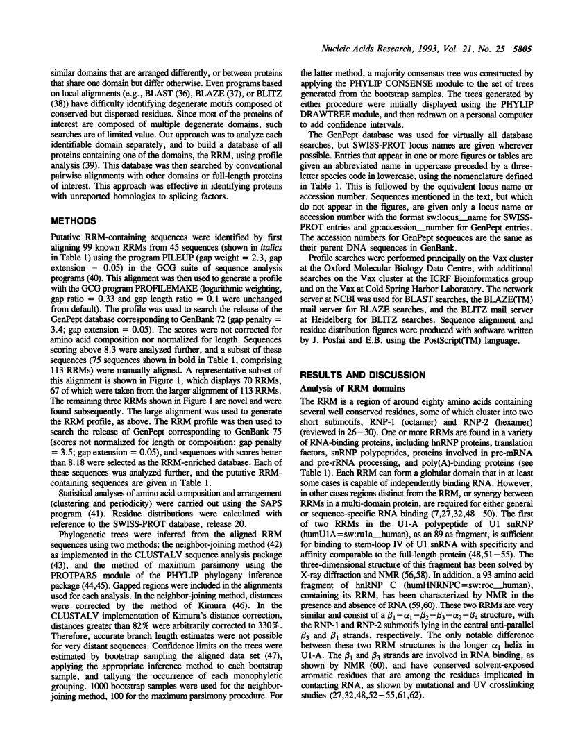
Putative RRM-containing sequences were identified by firstaligning 99 known RRMs from 45 sequences (shown in italicsin Table 1) using the program PILEUP (gap weight = 2.3, gapextension = 0.05) in the GCG suite of sequence analysisprograms (40). This alignment was then used to generate a profilewith the GCG program PROFILEMAKE (ogarithmic weighting,gap ratio = 0.33 and gap length ratio = 0.1 were unchangedfrom default). The profile was used to search the release of theGenPept database corresponding to GenBank 72 (gap penalty =3.4; gap extension = 0.05). The scores were not corrected foramino acid composition nor normalized for length. Sequencesscoring above 8.3 were analyzed further, and a subset of thesesequences (75 sequences shown in bold in Table 1, comprising113 RRMs) were manually aligned. A representative subset ofthis alignment is shown in Figure 1, which displays 70 RRMs,67 of which were taken from the larger alignment of 113 RRMs.The remaining three RRMs shown in Figure 1 are novel and werefound subsequently. The large alignment was used to generatethe RRM profile, as above. The RRM profile was then used tosearch the release of GenPept corresponding to GenBank 75(scores not normalized for length or composition; gap penalty= 3.5; gap extension = 0.05), and sequences with scores betterthan 8.18 were selected as the RRM-enriched database. Each ofthese sequences was analyzed further, and the putative RRM-containing sequences are given in Table 1.
Statistical analyses of amino acid composition and arrangement(clustering and periodicity) were carried out using the SAPSprogram (41). Residue distributions were calculated withreference to the SWISS-PROT database, release 20.
Phylogenetic trees were inferred from the aligned RRMsequences using two methods: the neighbor-joining method (42)as implemented in the CLUSTALV sequence analysis package(43), and the method of maximum parsimony using thePROTPARS module of the PHYLIP phylogeny inferencepackage (44,45). Gapped regions were included in the alignmentsused for each analysis. In the neighbor-joining method, distanceswere corrected by the method of Kimura (46). In theCLUSTALV implementation of Kimura's distance correction,distances greater than 82% were arbitrarily corrected to 330%.Therefore, accurate branch length estimates were not possiblefor very distant sequences. Confidence limits on the trees wereestimated by bootstrap sampling the aligned data set (47),applying the appropriate inference method to each bootstrapsample, and tallying the occurrence of each monophyleticgrouping. 1000 bootstrap samples were used for the neighbor-joining method, 100 for the maximum parsimony procedure. For
the latter method, a majority consensus tree was constructed byapplying the PHYLIP CONSENSE module to the set of treesgenerated from the bootstrap samples. The trees generated byeither procedure were initially displayed using the PHYLIPDRAWTREE module, and then redrawn on a personal computerto add confidence intervals.The GenPept database was used for virtually all database
searches, but SWISS-PROT locus names are given whereverpossible. Entries that appear in one or more figures or tables aregiven an abbreviated name in uppercase preceded by a three-letter species code in lowercase, using the nomenclature definedin Table 1. This is followed by the equivalent locus name oraccession number. Sequences mentioned in the text, but whichdo not appear in the figures, are given only a locus, name oraccession number with the format sw:locus_name for SWISS-PROT entries and gp:accession..number for GenPept entries.The accession numbers for GenPept sequences are the same astheir parent DNA sequences in GenBank.
Profile searches were performed principally on the Vax clusterat the Oxford Molecular Biology Data Centre, with additionalsearches on the Vax cluster at the ICRF Bioinformatics groupand on the Vax at Cold Spring Harbor Laboratory. The networkserver at NCBI was used for BLAST searches, the BLAZE(TM)mail server for BLAZE searches, and the BLITZ mail serverat Heidelberg for BLITZ searches. Sequence alignment andresidue distribution figures were produced with software writtenby J. Posfai and E.B. using the PostScript(TM) language.
RESULTS AND DISCUSSIONAnalysis of RRM domainsThe RRM is a region of around eighty amino acids containingseveral well conserved residues, some of which cluster into twoshort submotifs, RNP-1 (octamer) and RNP-2 (hexamer)(reviewed in 26-30). One or more RRMs are found in a varietyof RNA-binding proteins, including hnRNP proteins, translationfactors, snRNP polypeptides, proteins involved in pre-mRNAand pre-rRNA processing, and poly(A)-binding proteins (seeTable 1). Each RRM can form a globular domain that in at leastsome cases is capable of independently binding RNA. However,in other cases regions distinct from the RRM, or synergy betweenRRMs in a multi-domain protein, are required for either generalor sequence-specific RNA binding (7,27,32,48-50). The firstof two RRMs in the U1-A polypeptide of Ul snRNP(humU1A=sw:ru1a-human), as an 89 aa fragment, is sufficientfor binding to stem-loop IV of U1 snRNA with specificity andaffinity comparable to the full-length protein (48,51-55). Thethree-dimensional structure of this fragment has been solved byX-ray diffraction and NMR (56,58). In addition, a 93 amino acidfragment of hnRNP C (humHNRNPC=sw:rochuman),containing its RRM, has been characterized by NMR in thepresence and absence ofRNA (59,60). These two RRMs are verysimilar and consist of a Al -ac1-_2-(03-t2-34 structure, withthe RNP-1 and RNP-2 submotifs lying in the central anti-parallel(3 and (31 strands, respectively. The only notable differencebetween these two RRM structures is the longer cal helix inU1-A. The Al and (3 strands are involved in RNA binding, asshown by NMR (60), and have conserved solvent-exposedaromatic residues that are among the residues implicated incontacting RNA, as shown by mutational and UV crosslinkingstudies (27,32,48,52-55,61,62).

5806 Nucleic Acids Research, 1993, Vol. 21, No. 25
ac, oop2 02 loop3 J3 0op4 a2
humSF2(1) 17droSRP55(1) 5araSR(1) 8humSC35 15murX16 11droRBPl 12droTRA2 98humU170K 281sacU170K 108sacNPL3(1) 126humU1A(1) 11humUlA(2) 209hurnU2BO(1) 8hurnU2BO(2) 152humU2AF65(1) 150humU2AF65(2) 260sacYCL1IC(1) 123sacYCL1lC(2) 220humSF2(2) 122droSRP55(2) 116araSR(2) 120sacNPL3(2) 201humHNRNPM(1) 71humHNRNPM(2) 204humHNRNPM(3) 653humHNRNPA1(1) 15humHNRNPAI(2) 106humHNRNPC 17xenNRP(1) 21xenNRP(2) 110droPl1(1) 25droPl 1(2) 116humHUD(1) 47humHUD(2) 133humHUD(3) 298droSXL(l) 126droSXL(2) 212droELAV(1) 151droELAV(2) 249droELAV(3) 403droRBP9(1) 1droRBP9(2) 1droRBP9(3) 1humPABP(1) 12humPABP(2) 100humPABP(3) 192humPABP(4) 292sacPABP(1) 39sacPABP(2) 127sacPABP(3) 220sacPABP(4) 323humNUCL(l) 308humNUCL(2) 394humNUCL(3) 486humNUCL(4) 573tob28RNP(1) 108tob28RNP(2) 192tob31RNP(1) 137tob3l RNP(2) 231tob33RNP(1) 115tob33RNP(2) 218sacPRP24(1) IsacPRP24(2) 1sacPRP24(3) 1schME12(1) 198schMEI2(2) 291hum241 D5 61droBJ6(1) 303droBJ6(2) 377humARGNP 34
CORE
I 5
F
F
11
U U LRNP-2
9 abcdlO 15 20 22a23 25
IPP --- DIRTK ED K-IPY----GVRER R G-PG---- DIRER D K-Y---- RTSPD R K-N----NGNKT R Y-
- ---SASKH EG K-T----NTSQH RE NK-----DTTES R V-
Y---.-DLDEI K K-:PL .-.--DVQES El P-EKIKKDELKK Al 0-
E---- ETNEL SM 0_-P
NDKIKKEELKR A Q-
----ETNEI _8I-F----GITEEAMMD A-QM----YLNDDC KELLTS-
F- --DCTPE E L3T----SMNWO DD E----SGSWQ KDHMRE---- RVSWQ DYMRQ-
B----SASAQ DHIAK-E----GCSWQ DLARE-N-
PF---- DVKWO KDLVKEK'--.. KVGWK E M-F-- - DFTWK D E-F--- -ETTDE RSE----DTEEH D 0-
- -- LVVKKS EAl K-V----QTTQE E H--.-NTTVE KQ 0-
Y----RTTDD A K-0-.- -DHDEE E D-PQ -- NMTQEEFRS BS-K----TMTQK Q-P----DSDES BP0----DMTDR A A-R---- TITDD TI K-
PO ----TMTEDE S S-K----TMTQQ Al P-P ---ETEEA PO- - TMSQD RS S-K- -. NMTQS S P-P----DTEEN P_P----DVTEA E SP-K----SIDNK D SA-
'GE---- DMDDE D P-ALD----GIDDE K P-
P----SVSEA DI P-I
-P - -DIDNK D V- IIS.-- ETTDEQFQE K-D--- -SVDDE EE P-FNKSAPELKT D KNDLY---- KVTQD E D-AAY----SATEE E K-ATE----DTTEE E G-SVY----DIDSE 0 Q
PW----DIDDA 0 E-
Y----DVDSE R Q-
PW--- -GIDDA 0 E-F----SMTSSS El E-----ALTSO D D-Q
K--- -SYNON K N-cPP----SYTOR DLLD-INST---ELLDEN ES3G-F EPR----IVPYA LELSK-IOG--GSVLSIL SILQT- tSP----YVSNE E a-TN---- DITDD E 1KP-TP---- FVSNE K I-SP----SASSEOMRTL F-I9 abcdlo 15 20 22a23 25
DDVQ
D'
N'RRKElIDElDIElGIAFE)IR)YiYOHFTlYiRJY)DC
EVIGCECDJDNSNSCIT'N%TC
IQT
SNEBc
ITSN
IB
LSCTVS1SBILSEVSATSPSIB
VD\RL\KVF
El\VSJ
VSAENlMSIIH%ALSEKIGI
ERA
ERADEl
Z L F G U Z
30 32abcdof ghij kl mnopqr stu33K-------- - NRRGGPI
----------------- VPPRPV ------------- RDRYTKES
--RNP..A- - - - - RNP-I........... . IDAQTQR
---YSKRSGKP-.............-KDKITQK S
---------------- RSLKV ------------------ PGRHI
L-------------- KTMK4V ------------------ PGRHIL -------- PGNPVLAVQINQDF V- KDSATGLS
I ----- ----- - TSKGHIA ---------- - LDFNGFSA Y -- -- -- -- -- -- -- -- -- --
A H - -- -- -- -- -- -- ------KaT -R-------------TDARITr .. . .. . .. .VNTRDFV --------------MDAEGKA I .-.-.-.-.....-EDKDGKA ------- --- - MENGKC ------.----- - RDPNTKR
- .----------- TDRGSGKC H-------- - - - - - -
C ------------- RDPLTKRA ------------ FDKTTNRV M------------ KDPKTKRVV ---------- SDKDTGKC ------------ RDKITGQ
IL.- VDQVTGVV I------------- RDFNTNKC ------------- RDYKTGYK ------------- RDKLTGRPV RDKSQVY I DPLNPQAPSKGQS L------------QNAGNDTQV ------------- KDPTTNQC I------------ RDKVTGQSS L---------- - CDNITGLSV I--------- - RDLQSNK
---------- RDMITRRSC V -------- ---- - CDENG
E----------------- SGK--- ------------- MEGGRS
C--..........-RDAITKTS---------------TDENGKS--------------- KDADGKI--------------- RTENGKS
V -------------------MV ------------------GKSp ----------------- NGKV ---------------ETGS
I------------- YNRETDRS------------- FDRESGR-------------YNRDTDQS-------------YDRETGR-V------------YDRVTDRS
------------- YDRSSGRSV --------------- DSLKKNFFI L------------- PSLRFNTSIF
----------- AGQREHSFNNCC---------------- SLSTDI
A L--------------- - RSQNVc-I-------------- VDDRGRSTI
----------------- LDKNFI..T- VDDRGKHt4
L F----------- PPDDSPLPVSSF30 32abc def ghi J kl mnopqr 8tu33 35 40a41 45 50 53abcdef ghiS4
Z U V F Z A
RNP-1
loopi loops I0460 62
EFPRSG 91EPARGS 74ELAHGG 82QMARYG 92ELSNGE 83EMSSGR 84DFSITQ 175DVERGR 358CIVDIE 185VYSKLP 195OYAKTD 89ISFAKK 281OYAKTD 86ITYAKK 224RRPHDY 231VQRASV 336RQDNPP 198REGRFN 296AYIRVK 192VEDRRG 188YVRVRE 193ERDDNP 275KEDPDG 148KMDERA 280RIDRNA 728KRAVSR 91RKALSK 1824LAAEP 86KVAFPR 97KKAQPK 185RAVPRQ 102KKAIAK 192SYARPS 124TVKFAN 210SFKTNK 375SYARPG 203SVRLAE 289SFARPS 240VVKFSN 327SFKTNK 480SIARPS 78TVKFAN 78SFKTNK 78USQRD 893RFKSR 1753RAQKK 265kLAQRK 367MSQRD 116kPHLSR 2033RAQKK 296kIAQRK 399EKPKGK 383YYTGEK 474ELQGPR 560)WAKPK 644IKAAPR 175IAAEER 269IKAARR 214IVAEDR 308IFPEVP 192IVAGOK 295iLTECT 76IVSNPL 793LADKK 80FOFCOR 269kYYDAM 366'VIVEP 137IFAPNA 374'CLVDP 453'YAEGV 113
60 62_m LT.C-=-

Nucleic Acids Research, 1993, Vol. 21, No. 25 5807
An alignment of RRMs. An alignment of 70 RRMs (Figure 1)was constructed with special emphasis on tertiary structuralrequirements, modeled on the two available structures. Thealignment includes sequences of proteins thought to be involvedin pre-mRNA splicing, spliceosome-associated factors, and somehnRNP proteins, as well as some additional putative RNAprocessing factors. Human and yeast poly(A)-binding proteinswere included, together with nucleolin, to give some samplingof other RRM sequences, and to illustrate phylogeneticrelationships (see below). All the RRMs present in these proteinswere included, except for RRM3 of U2AF65 and RRM3 of ahomologous protein in yeast, YCLlIc, which are highly atypical(7,63). Alignments with different sets of RRMs have beenconstructed and all the features described below are consistentwith all the alignments (data not shown).The alignment shown in Figure 1 differs from previous
alignments ofRRMs (27) in the positioning of conserved residuesin cal and loop2. The alignment shown here benefits from therecent availability of the hnRNP C RRM structure (59), inaddition to that of the N-terminal U1-A RRM (56,58), whichallowed us to look at the relative positions of residues within bothstructures. The most significant change implicates the conservedGly in the tight turn at the end of loop2 leading into 02, ratherthan in the last turn of the a1 helix (27). This alignment alsoshows that the Ul-A sequence is atypical in its longer a1I helix,which gives rise to the first gapped region. The other major gapin the alignment corresponds to loop3 (also known as the variableloop), which can be easily accommodated into the model tertiarystructure. Other small alignment gaps occur between (xl and (2,(3 and a2, and a2 and 04, all of which are plausible sites forinsertions or deletions within the conserved tertiary structure.In some RRMs (not shown in Figure 1), there is also a gap atposition 28 within the (-bulge in (2 (positions 27-29). TheRNP-1 and RNP-2 submotifs lie in 03 and (31 strands,respectively. The conserved aromatic residues at positions 3, 35,and 37 protrude from the (3 sheet to interact with the RNA, asshown by crosslinking and mutational studies (32,48,52-55,61,62). Ring-stacking interactions between these solvent-exposedresidues and single-stranded bases have been postulated (27).
A consensus RRM structural core sequence. The most conservedpositions in the alignment (Leu7, Leu 16, Phe2O, Val38, Phe4O,and Ala49) (Figure 1) correspond precisely to residues that formthe hydrophobic core of the U1-A tertiary structure (56,58), aspreviously noted (27). In addition, Gly24 seems to be requiredfor the turn into (2. Based on the analysis of the RRM
alignment and the model three-dimensional RRM structure, wepropose the following consensus structural core sequence forRRMs:
UxUxxLxxx[xo 6]Z[x]xxxLxxxFxxx[x]GxUx[x]Zxxxxxx[x021 +]UxVxF[x]xxxxxxZxxA
(x = any residue; U = uncharged residues: L,I,V,A,G,F,W,Y,C,M; Z = U + S,T; + indicates that loop3 may beexpanded further)
We note that this is a degenerate consensus, i.e., no singleposition is absolutely invariant. Although position 34 has a highlyconserved Gly residue, we did not include it in the consensusbecause its role appears to be to connect loop3 to .(3. Sinceloop3 does not appear to be involved in the structure of thedomain (see below), only a subset of RRMs might require a Glyat this position. There are many additional positions in thealignment that show conservation, and in general, additionalconserved residues must be required to form the two a-helicesand four (3-strands, and to fold them into a correct RRM structure.However, it is unlikely that an RRM can exist without at leastconservative substitutions in the above consensus sequence.Although this consensus is too permissive to be used as the solecriterion to identify RRMs, it will identify most, if not all RRMs,including those with atypical RNP-1 submotifs. The RNP-1submotif remains the most obvious signature for typical RRMs,but many RRMs contain atypical RNP-1 submotifs (see Table 1)and sequences matching the RNP- 1 submotif consensus are foundin proteins that lack an RRM (see below).
RRM positions with potential to contact RNA. All the solvent-exposed positions in and near the (3-sheet have the potential tocontribute to RNA binding. The three solvent-exposed aromaticpositions (3 in RNP-2; 35 and 37 in RNP-1), which have beenimplicated in RNA binding, are predominantly Phe or Tyr (73 %,60%, and 74%, respectively; taken from a weighted dataset of179 sequences; data not shown) but they can tolerate substitutions(Figure 1). Thus, these conserved aromatic residues are notalways required for RNA binding, as they are absent from manyputative RRMs. For example, hnRNP L (sw:roL human), whichlacks these conserved aromatic residues in all but one positionof one of its four RRMs, can bind RNA (64). The variabilityseen at these usually aromatic positions could reflect sequence-specific contacts. For example, Gln54 of the U1-A polypeptide,which is in the usual aromatic position 35, has been implicatedin sequence-specific hydrogen bonding to stem-loop II of Ul
Figure 1. Alignment of 70 selected RRMs. Selected RRMs were aligned manually with special emphasis on tertiary structural requirements modeled after two availablestructures, as described in the text. Alignment gaps are indicated by dashes. Sequence names are as in Table 1. For sequences with more than one RRM, eachone is numbered from the N-terminus, with the number given in parentheses after the sequence name. Beginning and end residue positions within the parent proteinare given for each RRM at left and right of the alignment. The sacPRP24 and droRBP9 sequences are from reference (27); the accession numbers for the remainingsequences are given in Table 1. The alignment positions are numbered above and below, following the nomenclature of Kenan et al. (27), in which conserved positionsare given sequential numbers, whereas positions that are not present in all RRMs are alphabetized. However, as the alignment in reference (27) differs from theone above, the numbers are not interchangeable between the two. The positions of conserved residues are highlighted by vertical shading. Black shading indicatespositions in which a single residue occurs in at least 75% of the sequences. Grey shading at the same positions represent conservative substitutions. Elsewhere,grey shading indicates positions in which residues belonging to a single conservative grouping are present in at least 50% of the sequences. When a single conservativegrouping represents at least 75% of the sequences, this is denoted by grey shading in boxed columns. Acceptable conservative groupings wereI=V=L,F=Y=W,Q=N,R=K,D=E,S=T. Three additional positions at which the consensus is split between two residues were also shaded: positions 6 and 53g(G=N) and position 36 (A=G). The consensus structural core residues are shown below the alignment (U = uncharged residues: L,I,V,A,G,F,W,Y,C,M; Z =U + S,T), along with the position of the RNP-1 and RNP-2 submotifs. Secondary structure, modeled primarily after the humUlA(l) tertiary structure (56,58) exceptfor cal, which was based on the humHNRNPC secondary structure (59), is given above the alignment. In humUlA(l) cil extends another 2 residues towards theN-terminus. Most positions of the RRM lack a firm consensus. The range of sequence similarity between two otherwise unrelated RRMs is 10-20% identity.

5808 Nucleic Acids Research, 1993, Vol. 21, No. 25
snRNA, and mutating this residue to Phe drastically reducesbinding (52). In some RRMs, solvent-exposed aromatic residuesin (2 (humSC35, murX16, sacNPL3) or (4 (sacPABP,humPABP) might have similar functions to those of the aromaticresidues in al and (3.
Since residues within loop3 are not involved in the overallstructure of the RRM, substitutions, insertions and deletions inthis loop may be easily tolerated. Thus, alignments between directhomologs, such as human Xenopus, chick and rat nucleolin, orhuman, Xenopus, Drosophila and yeast U1-70K, show greatestconservation in the (3 strands, and greatest variability in loop3of the corresponding RRMs, suggesting that accumulatedmutations in these regions do not affect RRM function (data notshown; see Table 1 for accession numbers). Variability is alsoseen in the et-helices, as expected, since they are on the oppositeside of the RNA-binding surface. Furthermore, in the yeastU1-70K homolog (sacU170K=gp:x59986), whose RRM can befunctionally replaced by the human U 1-70K RRM(humU170K=sw:rul7_human), loop3 is one of the mostvariable regions compared to the human RRM (65,66). However,in other protein families, such as the hnRNP Al-like proteins,loop3 sequences do show conservation.Replacement of loop3 of RRM1 of the U1-A polypeptide
(humUlA=sw:rula_human) by the analogous region of theU2-B" polypeptide (humU2B=sw:ru2b_human) abolishes itsability to distinguish between U1 and U2 snRNAs (55). Furtherreplacement of residues in part of 02, in addition to loop3,reverses the RNA-binding specificity of this RRM (54). However,this is one of the few regions of divergence between the N-terminal RRMs of these two highly homologous proteins, andone of the only divergent solvent-exposed regions near the (-sheet. The issue of binding specificity is further complicated inthis case by the fact that the U2-B" protein requires the U2-A'protein for specific binding to U2 snRNA (55,56). Interestingly,the Drosophila protein dro25 (droUlA=gp:m89775), whose firstRRM has a loop3 region that is almost identical to that of humanU2-B", in fact binds to Drosophila Ul RNA in vivo (57). In short,although in general loop3 shows the greatest variability insequence, this need not mean that it is the major determinant ofsequence-specific binding.
Construction of an RRM-enriched databaseThe RRM has only a few well-conserved residues, mostly in theRNP-1 and RNP-2 submotifs (27; Figure 1). Pairwise alignmentsof unrelated RRM sequences are often incorrect, due to theadditional residues within the RRM. To overcome theselimitations, we employed profile analysis (39). A profile is aposition-dependent scoring table that summarizes the preferencesfor amino acid residues and the acceptability of gaps at eachposition in a set of aligned sequences. By constructing a profilefrom a set of aligned RRM sequences, flexibility for residue typeand gaps is promoted in some regions whereas conserved residuesare strongly enforced. In addition subtle preferences for broadresidue type (e.g., uncharged, small residues) become apparentand are represented in the profile. By using a profile generatedfrom an alignment of 75 RRM-containing sequences (1 13 RRMsin total), essentially a larger version of the alignment in Figure1, we could identify many, if not all sequences with potentialRRMs in a large database. The resulting limited set of sequences,which represents only 0.66% of the entries in the GenPeptdatabase, can then be used to search for other domains and motifs.The reduced database size increases the statistical significance
of otherwise weak similarities found among these proteins outsidetheir RRMs.As expected, known RRMs (both present in and absent from
the profile alignment) consistently produced high scores (exceptfor E. coli rho protein, Drosophila bicoid, Drosophila Suppressorof sable and bacteriophage 029gp 1O, see below). Atypical RRMs,whether present in the alignment, such as hnRNP L(sw:roLJhuman), or absent from it, such as La protein(sw:la_human), gave lower scores. The first 434 scores (cutoffscore of 8.18) were arbitrarily selected to produce the RRM-enriched database. This database was over twice the sizenecessary to include the last known positive in the search (Laprotein) (67,68). This RRM-enriched database included sequencesthat lack well-defined RNP-1 and RNP-2 submotifs, which arethe hallmarks of RRMs, but that contain the virtually invarianthydrophobic residues located in the RRM core. The cutoff waschosen liberally to include most, if not all sequences with potentialRRMs. As a result, only 29% of the sequences in this RRM-enriched database appear to contain an RRM. However, thiscontrasts with 0.0019% putative RRM-containing sequences inthe GenPept database.
In none of the searches to identify RRM-containing sequenceswere either the bacterial rho protein (sw:rho-ecoli) (69), theq029gplO protein (sw:vglO0bpph2) (70), the bicoid protein(sw:hmbc-drome) (71) or Suppressor of sable (gp:m57889) (72)identified, even though they were all previously reported to haveRRMs (69-72). They scored 7.00, 7.83, 6.87 and 7.46,respectively, against the RRM profile using identical searchconditions as above. In each case, randomized sequences thatmaintained the composition but altered the order of residues gavesignificantly lower scores than the original sequence. The abovecore consensus can be made to fit the 029gp 1O sequence, whereasfor rho, bicoid, and Suppressor of sable, unprecedented gaps andsubstitutions must be accommodated. Mutations of the twosolvent-exposed Phe residues in the putative RNP-1 sequence ofrho protein reduce RNA binding (69). It is unclear if the criteriaproposed here would support the presence of an RRM in029gplO, but neither bicoid, nor rho, nor Suppressor of sableproteins satisfy these criteria. This may indicate that the consensusfor an RRM is broader than suggested here, that these regionshave diverged from an ancestral RRM, or that the sequencehomology found is spurious, despite the requirement for two Pheresidues for RNA binding by rho.The presence of RNP-l submotifs in prokaryotic cold shock
proteins and eukaryotic Y box transcription factors has been noted(73). However, in these proteins the submotif differs in the finalposition, which is predominantly Phe in RRM RNP- 1 submotifsand Arg in cold shock proteins and Y box factors. Neither thecold shock proteins nor the Y box factors were identified in ourRRM searches. The crystal and solution structures of the B.subtilis major cold shock protein have been recently reported(74,75) and show that the RNP-l submotif lies in the second (strand, but with an entirely different overall topology from thatof the prototype RRM. Interestingly, the position of the residuesin this (2 strand match very closely with those in the Ul-ARNP-1, and 03 of the cold shock protein has residues inanalogous positions to those of the RNP-2 submotif in U1-A(74,75), although in a very different position relative to the RNP-lalong the primary sequence. The conservation of these RNP-1submotifs could be a case of convergent evolution of nucleic acid-binding domains. This illustrates one limitation of using theRNP-1submotif as the sole signature for an RRM. For example,

Nucleic Acids Research, 1993, Vol. 21, No. 25 5809
humSF2droSRP55araSRhumSC35murXl6droRBP1schMEI2humU170K
604251615253335224
765867776869351240
Figure 2. A conserved octapeptide motif. The alignment shows a partiallyconserved octapeptide motif found in all SR proteins in the current databases(human SF2/ASF, Drosophila SRp55, an Arabidopsis SR protein, human SC35,murine X16 and Drosophila RBP1), as well as in S. pombe mei2 and humanUl-70K polypeptide. In all but the U1-70K polypeptide this motif is found withinan RRM, overlapping the last two amino acids of the RNP-1 submotif. Thealignment was extended by nine residues towards the C-terminus to illustrate thefurther homology found among the first seven sequences, which is higher thanbetween unrelated RRMs. Sequences upstream of the octapeptide are not shownbecause they comprise the rest of the RNP-1 submotif (except in U1-70K) andthe homology is high, as expected. Black boxes indicate identities, whereas grey
boxes denote conservative groupings, as in Figure 1. Positions were shaded ifidentities or conservative groupings were present in four or more sequences.
bacteriophage T4gp32, which has been noted to have a sequencewith similarity to the RNP-1 submotif (26,76), does not satisfythe structural criteria for an RRM, and binds RNA by amechanism that does not involve an RRM-like structure (26,77).A comprehensive RRM databaseAlthough the entire RRM-enriched database was employed forsubsequent searches, we attempted to identify all sequencescontaining bonafide RRMs by manually examining each entryfor the presence of the motif using the structure-based RRMconsensus given above. A comprehensive list of the resultingRRM-containing sequences is given in Table 1. This RRMdatabase contains 22 previously unreported RRMs, stronglysuggesting that the function of the corresponding proteins, whichare implicated in a variety of cellular processes, involves bindingto RNA. For example, the S. cerevisiae gene RNA12+(gp:s92205), which is involved in rRNA maturation, containsa previously unreported RRM, suggesting that the gene productdirectly binds rRNA. This analysis also revealed previouslyunreported RRMs in Drosophila modulo (sw:modu-drome) andhuman MPSS-1 (gp:x64652), both of which have been shownto bind DNA. Given the presence of multiple RRMs in theseproteins, their relative affinities for dsDNA, ssDNA and RNAshould be measured. A fourth example is a partial human cDNAthat was originally identified as a myoblast cell surface protein(hum241D5=sw:cs24_human), and has a good fit to the RRMconsensus (Figure 1), suggesting that it was cloned fortuitously.Recently, it was shown that this cDNA represents a fragmentof a recently cloned human splicing factor, PSF (gp:x70944) (11).
Unreported homologies between sequences in the RRM-enriched database and known splicing factorsThe RRM-enriched database was searched using FASTA (35),with the sequences of known splicing factors and spliceosomalproteins, or domains thereof. Unexpected sequences identifiedthrough these searches were then manually examined for thepresence of one or more RRMs using the consensus derivedabove, and for other sequence features.
Human Arg-rich nuclear protein. When the RRM-enricheddatabase was searched with the RS domains from various
proteins, an Arg-rich human nuclear protein of 54 kDa(humARGNP=gp:m74002) (78) gave consistently high scores.This sequence has both an extensive RS domain and a slightlyatypical, previously unreported RRM upstream of the RS domain(residues 34-113; Figure 1; see also Figure 5, below). Thisprotein localizes to the nucleoplasmic speckled region of themammalian nucleus (78), a region enriched in many splicingcomponents (reviewed in 79). This subnuclear localization isconsistent with the presence of an RS domain (31), and togetherwith the presence of an RRM, suggests a possible role in pre-mRNA splicing. The humARGNP RRM does not resemble theRRMs ofSR proteins, nor was any additional homology observedbetween humARGNP and other SR proteins, besides the RSdomain and the expected similarity between otherwise unrelatedRRMs (data not shown).
S. pombe mei2. Another sequence we identified as havinghomology to splicing factors was mei2, the product of a geneinvolved in meiosis in S. pombe (schMEI2=sw:mei2_schpo)(80). It showed a partial match to a region previously shown tobe homologous between SF2/ASF (humSF2=gp:m69040) andU1-70K (humU170K =sw:ru17ilhuman), the octapeptideEFEDPRDA (5). Exact or almost exact fits to this octapeptidewere also found in the six other sequenced SR proteins, all justafter the RNP-1 submotif, extending from the end of 03 into a2(Figure 2). The octapeptide in mei2 is in the identical positionin a putative RRM (residues 296-373) as in the SR proteins.Therefore, the mei2 octapeptide is present in the same structuralcontext, although the sequence of the putative RRM is veryatypical. The octapeptide in U1-70K is located upstream of thesingle RRM, and is strongly conserved in human, Xenopus, andDrosophila proteins (5), but not in the more divergent S. cere-visiae homolog (65,66) (see Table 1 for accession numbers). Anungapped alignment of the homologous segments of yeast mei2,the six SR proteins, and human U1-70K is shown in Figure 2.The homology between U1-70K and the other proteins does notextend beyond either side of the octapeptide (Figure 2 and datanot shown). No other exact matches to the octapeptide were foundeither in the RRM-enriched database, in SWISS-PROT, or insix-frame translations of GenBank. The function of the octa-peptide motif in any of the above proteins is presently unknown.
S. pombe mei2 has at least two previously unreported RRMs:the one containing the octapeptide, and another immediatelypreceding it (Figure 1). Neither of these RRMs contains a goodfit to the RNP-1 consensus (DGICIVAF and VSQIICEF).However, the correct spacing of the hydrophobic core residuesstrongly suggests that these putative RRMs can fold into thecorrect prototype structure, which in turn suggests that the abilityto bind RNA has also been conserved. A good fit to the RNP-1submotif (VGYAFINF) is found towards the C-terminus of mei2.It is unclear whether this is part of a third RRM, as the requisiteAla residue in a2 iS replaced by a Phe residue, which would notfit in the model RRM tertiary structure.
Recent work showed that wild type mei2 is required forefficient splicing of the mesI intron during meiosis in S. pombe(C. Shimoda, personal communication). Alternative splicing ofthe mesi intron is of the intron retention type (2). The presenceof two RRMs (Figure 1) and the additional homology to SRproteins (Figure 2) are consistent with a direct role of mei2 inpre-mRNA splicing in S. pombe. Unlike SR proteins, mei2 lacksan RS domain, but extensive RS domains have not yet been foundin fission or budding yeasts.

5810 Nucleic Acids Research, 1993, Vol. 21, No. 25
RNP-2
1 MSGGGVIRGPAGNNDa1.MVG1 .MSSRSSI
110 GRPPMHHRQEGELSN
so . .*. .
K Al RRGGP _GRTL .....P P vJ P PR PPK!PMKU ...
RNP-2-1
79 1 FP* S............ GGTGR GGGAPRGRYGP Si61 LL PAfGSARGSNRDRYDDRY R RYNEKSSSRYGP .
0 LAHG ..... GRRSSDDTR SFN GRGRGDGGSRG Si183 AN E VYSEL ................... P,
PPS(SSRIkSS1PEG(
145 . C ADEY...138 . T ADAHKQ.143 *. CSQUYRDA224 SLJTT SSINTRDF
JRK TY SHEGETAYIflKV. GP S 198
kL
T E llNR...............HLV DR G 185rcY KY NAFSNGYV. EREY SR D 200
'S E V E E NN G ....I TV RDDN 272
Flgue 3. Sequence similarity between three metazoan SR proteins and yeast Npl3. The alignment shows N-terminal regions of human SF2/ASF, Drosophila SRp55,an Arabidopsis SR protein, and a centrl portion of S. cerevisiae Npl3. Residue positions for each protein are shown at left and at the end of the alignment. Blackboxes indicate identities, whereas grey boxes show conservative groupings, as in Figure 1. Positions were shaded if identities or conservative groupings were presentinthree or more sequences. The posit of the two RRMs are shown by horizo lines above the aligme, and the RNP-2 and RNP-l smhifs ofeach RRM are boxed.
S. cerevisiae Npl3. A third striking homology was detectedbetween S. cereWsiae Npl3 (sacNPL3A=gp:m86731), alsoknown as NOP3 (gp:x66019), and three SR proteins: humanSF2/ASF (humSF2=gp:m69040), Drosophila SRp55 (droSRP55=sw:sr55-drome), and an Arabidopsis protein (araSR=gp:m98340). Npl3 was isolated in a genetic screen for factorsinvolved in nuclear protein localization (81). However, thisfunction may be indirect, as gene disruption did not affect proteinlocalization, although mutant forms of the protein blockedtransport. Npl3 has a good fit to the RRM consensus (81)followed by a second unreported, atypical RRM, similar to theabove SR proteins (Figure 1). However, the proteins differ inthat Npl3 has an N-terminal domain rich in Pro (22%) and Gln(16%), with imperfect Ala-Pro-Gln-Glu repeats unique inthe da e, and a C-trminal RGG domain (see below), whereasthe SR proteins have characteristic C-terminal RS domains(5,6,12).The alignment of the central region of S. cerevisiae Npl3 with
the first 185-200 residues of human SF2/ASF, DrosophilaSRp55, and the Arabidopsis SR protein is shown in Figure 3.Witiin this region, Npl3 is 30%, 29%, and 24% identical toaraSR, SRp55 and SF2/ASF, respectively. This level ofhomology is far greater an expected for unrelated RRMs, andis particularly striking in the case of the second atypical RRM.Npl3 lacks the Gly hinge region between the RRMs, resultingin a large gap in the alignment. The two other significant gapslie in the presumptive loop3 in the RRM structure. None of thesegaps would therefore disrupt the expected RRM tertiary structre.To date, proteins containing extensive RS domains have not
been identified in eitier fission or budding yeast. The N-terminusof S. cerevisiae YCLI lc (sacYCL1lC=sw:ycbl yeast) is Arg-rich (26% Arg in the first 52 amino acids), with four RSdipeptides (63). The observed homology between the RRMs ofmetazoan SR proteins and yeast Npl3 suggests that hese proteinshave a common ancestor. In addition to its expected ability tobind RNA, Npl3 may be involved in some aspect of mRNAprocessing, although its auxiliary domains suggest that its function
is different from those of SR proteins. Recent experiments haveimplicated Npl3 in mRNA nuclear-cytoplasmic export, inaddition to nuclear protein import, with several temperature-sensitive alleles mapping to Gly241 and Ala254 (P. Silver,personal communication). These residues are located witiin thesecond RRM of Npl3 (Figure 3), and are two of the positionsof the structural core consensus defined above (Figure 1). Thetemperature-sensitive phenotpe of these mutations is consistentwith the requirement for the structural core consensus for theintegrity of the tertiary structure of the RRM.A conserved heptapeptide in RRMs ofcertain SR proteins, Npl3,YCLllc, and hnRNP M. The second RRMs of SF2/ASF(humSF2=gp:m69040), SRp55 (droSRP55=sw:sr55_drome),the Arabidopsis SR protein (araSR=gp:m98340) and yeast Npl3(sacNPL3A=gp:m86731) includes the heptapeptide SWQDLKD,which is completely conserved in location and sequence (Figure3). Exact matches to this heptapeptide were not found in any otherprotein in the RRM-enriched dabase. However, partial matcheswere found in YCL1 lc (sacYCLl lC=sw:ycbl_yeast), an openreading frame in chromosomem of S. cerevisiae, which showssimilarity to human U2AF*5 (63), and in all three RRMs ofhnRNP M (humHNRNPM=gp:103532) (see under a, region inFig. 1). Although the sequence similarity between the aboveheptapeptides is low, the motif lies in precisely the same locationin each of the RRMs, i.e., in a1I on the opposite side of the (8-sheet where the RNA is thought to lie. Only partial fits to theheptapeptide were found in six-frame translations of GenBank.The function of the heptapeptide remains unknown, but it isunlikely to be involved in directly containg RNA, given itsposition within the tertiary structure of these proteins.
Human U24FI5. The sequence U2AF35 (humU2AF35 =gp:m96983) yielded a high score against the RRM profile. U2AF35is a subunit of U2AF not required for biochemical complement-ation of splicing extracts depleted of strong poly(U)-bindingproteins, and has an extensive RS domain (82). Further analysisdid not identify an RRM, although we note that the region
humSF2droSRP55araSRsacNPL3
humSF2droSRP55araSRsacNPL3
RRM(1 )
humSF2droSRP55araSRsacNPL3
RRM(2)
IM:- QAI AKG
EL AlE N
9

Nucleic Acids Research, 1993, Vol. 21, No. 25 5811
Bootstrap confidence intervals (%)
100 75 74 50 49 - 25 24 0
,
Figure 4. Phylogenetic tree of 70 selected RRMs. The phylogeny was derived by the neighbor-joining method from the alignment of all the RRMs shown in Fig.1. Sequence names are as in Fig. 1. Confidence limits on the phylogeny were obtained by the bootstrap method, as described in Methods, and are represented bylines of different thickness, as shown at the bottom of the tree. A low bootstrap confidence interval for a particular grouping indicates that the homology seen isnot consistent across the alignment. The central node for this unrooted tree was chosen arbitrarily. The groupings that have a 50% or greater confidence intervalare in broad agreement with the phylogenies derived by the maximum parsimony method (see Methods). The overall similarity of the alignment used to generate
this phylogeny is insufficient to derive accurate lengths for all branches (see Methods).
between residues 110-144 is very reminiscent of the (3-
loop4 -a2 region of the model RRM (not shown). Thissimilarity could indicate a very atypical RRM or perhaps a
structure that has evolved from an RRM to fulfill a different role.It will be interesting to see whether or not U2AF35 directly bindsRNA.
Phylogenetic analysis of RRMsThe modular nature of RRMs has led to the proposal that thesedomains have evolved by duplication and diversification from
an ancestral RNA-binding protein (reviewed in 26). Theavailability of a large set of RRM-containing proteins affordedus the opportunity to examine this model and the evolutionaryrelationships among RRMs. Phylogenetic analysis of the RRMalignment of Figure 1 by both the neighbor-joining method (42)and by the method of maximum parsimony (44,45) was carriedout. The two trees were in broad agreement and the neighbor-joining tree is shown in Figure 4. Confidence intervals on thephylogeny were estimated by the bootstrap method (47), and are
represented in the figure.
ft-j
sacPRP24(3)
PAO
dropll(l)
9P11)
1%
;
I

5812 Nucleic Acids Research, 1993, Vol. 21, No. 25
Table 2. Sequence conservation among SR proteins
0s~~~~oNdroSRP55 141121086 6droSRP55 47% 50% 48% 52% 490/
145 109 80 70araSR 58% 49% 49% 52%
humSF2 38% 748% 52%
humSC35 47% 47%murX16 62%
The length in amino acids for each protein is given in parentheses. For eachpairwise score the upper number shows the number of identical amino acids,and the lower number expresses this as a percentage of the smaller sequence.Alignments were constructed with full length sequences using the global alignmentalgorithm of Needleman and Wunsch, as implemented in the GCG GAP programwith the Dayhoff scoring matrix. For the sake of consistency each alignment wasmade with a gap penalty of 1.7 and gap extension of 0.03. These parameterswere found to give in nearly all cases alignments consistent with the domainstructure of the proteins. These numbers do not accurately reflect homology andcannot be used to infer phylogenetic relationships. See Fig. 1 and Fig. 4 for thealignment and phylogeny of the first RRM for each of these sequences.
Domain duplication. RRM duplications witiin individual proteinsappear to be common, as seen in the following groupings: (i)the last tiree RRMs of poly(A)-binding proteins (humPABP andsacPABP); (ii) both RRMs of the hnRNP Al-like proteins(humHNRNPA1, droPll, and xenNRP); (iii) the second andtiird RRMs of nucleolin (humNUCL); (iv) the tobaccochloroplast RNA-binding proteins (tob28RNP, tob31RNP, andtob33RNP), except for tob33RNP(l). The level of amino acidsimilarity between corresponding RRMs in two homologousproteins that contain multiple RRMs is often greater than thesimilarity between the multiple RRMs of either protein (83). Thisis most obvious in the poly(A)-binding proteins, in which thegrouping of corresponding yeast and human RRMs implies thatRRM duplication preceded the divergence between yeasts andmetazons. The observed phylogenetic pattem is consistent withthe idea that each RRM evolved independently after theduplication event.
Gene duplication. RRM relatedness between different proteins,reflecting gene duplication, is also evident for: (i) the SR familyof splicing factors as seen in the grouping of their first RRMs(humSF2, droSRP55, murX16, droRBPl, and araSR; humSC35does not group with this phylogeny, and other groupings ofhumSC35 are not supported by the bootstrap analysis), and, whenpresent, of their second RRMs; (ii) hnRNP Al-type proteins(humHNRNPAl, droP 1i, and xenNRP) (84,85); (iii) chloroplastRNA-binding proteins (tob28RNP, tob31RNP, and tob33RNP)(86); (iv) droSXL, droELAV, humHUD, and droRBP9 (87,88).Gene duplication provides an opportunity to evolve restricteddevelopmental or tissue-specific expression patterns for certainRRM-containing proteins that may acquire unique functions orbinding specificities. For example, mammalian hnRNP Al(humHNRNPAl=sw:roaluman) is expressed in manytissues, whereas its amphibian relative, NRP-1 (xenNRP=gp:m34895), is expressed only in neuronal tissues (89).When related multi-RRM proteins from different species are
aligned (e.g. poly(A)-binding proteins, hnRNP Al-like proteins),not only are the RRMs highly conserved, but also the length andsequence of the linkdng regions between RRMs (data not shown)
(85). This phylogenetic conservation suggests that the linkingregions are important and at individual RRMs in these proteinsdo not act independently but rather require interactions between,and precise positioning of, the RRMs. Indeed, a number of multi-RRM proteins show synergy between two or more of their RRMsfor binding to RNA. Examples include yeast poly(A)-bindingprotein (49), U2AF65 (7), and SF2/ASF (32). In the chloroplastRNA-binding protein cp29B, the 37 amino acid Gly-rich linkerbetween the two RRMs is required for the synergistic effect onRNA binding (50).The phylogenetic data support a model in which at least several
of the major groups of RNA-binding proteins arose through acombination of gene duplication and intragenic domainduplication. The presence of RRMs in proteins from a varietyof distant phyla, combined with the modular organization ofmultiple RRMs suggests that the entire domain, rather than simplythe RNP-1 submotif, is an ancient conserved region (ACR) (90).After this dataset was assembled, the sequences of RRM-containing proteins from several species of cyanobacteria weresubmitted to Genbank (gp: 120890, gp: 120891, gp: 120892) (M.Mulligan, personal communication). This is in contrast to ourearlier searches, which failed to idenf RRMs in any eubacteria(with the possible exception of bacteriophage 429gplO, seeabove), despite the success of these sensitive searches inidentifying a broad range ofRRMs in eukaryotes. The presenceof RRM-conaining proteins in both cyanobacteria and eukaryotesunderscores the ancient origin of the RRM.
Phylogeny and finction of SR proteins. The SR family ofphosphoproteins in metazoans is a striking example of geneduplication and phylogenetic conservation (15) (Table 2). MouseX16, which is 100% identical at the amino acid level to humanSRp2O, is 62% identical to RBP1, a probable Drosophila SRp20homolog (14,15,16). Human and avian SC35 (PR264) (9,10) are98% homologous. An Arabidopsis SR protein is 58% identicalto human SF2/ASF (59% without RS domain) and 47% (48%without RS domain) identical to Drosophila SRp55 (12) (foraccession numbers see SR protein group in Table 1). Mouse(gb:x66091) and human SF2/ASF are 100% identical at the aminoacid level and 95% identical at the DNA level within the codingregion (data not shown). Table 2 gives an indication of theconservation in sequence between SR proteins, and the phylogenyof their RRMs is shown in Figure 4. The evolution ofSR proteinsprobably involved complex events such as domain duplicationand subsequent deletion, as well as extension of the RS domain.As a consequence, numerical pairwise homology scores cannotaccurately reflect phylogenetic relationships among these proteins.
Partial amino acid sequences of human SRp4O and SRp75 showthe presence of at least part of the second atypical RRM (15).However, an analogous RRM is absent from SC35 and X16,despite the fact that human SF2/ASF, Drosophila SRp55, andhuman SC35 have equivalent in vitro activities in general andalternative splicing, and bovine SRp4O and SRp75 have equivalentgeneral splicing activity (15,20,21). The single RRM in SC35and X16 probably substitutes for the two synergistic RRMs inthe other SR proteins (32).
All SR proteins studied so far have both constitutive andalternative splicing activities in vitro (15,16,17-21). Forexample, human SF2/ASF and human SC35, which are only 38%identical have indistinguishable biochemical activities (21). In acomparison ofhuman SF2/ASF and Drosophila RBP1 activitiesin human extracts, some qualitative and quantitative differences
Nucleic Acids Research, 1993, Vol. 21, No. 25 5813
were reported, although the possibility that these differences aredue to the use of mixed human and Drosophila factors has notbeen ruled out (16). Quantitative differences in splice site selectionpreferences among several SR proteins in vitro were recentlyreported (91). The in vivo expression of individual SR proteinshas been studied in a few cases at the level ofmRNA or protein(10, 14,16,91; A. Hanamura and A.R.K., unpublished data). Ineach case, a wide range of expression was observed in differenttissues or cell lines. In addition, the activities of these proteinsmay be regulated by phosphorylation or nuclear localization. Theselective pressure to maintain such a high degree of sequenceconservation between individual members of the SR family fromdifferent species is inconsistent with the apparently redundantbiochemical activities of less homologous members from the samespecies. This strongly suggests that individual SR proteins haveunique functions in vivo.
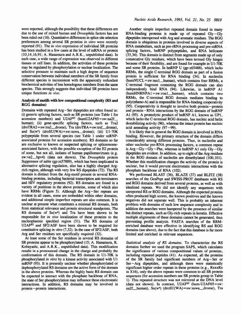
Analysis of motifs with low compositional complexity (RS andRGG domains)Domains with repeated Arg -Ser dipeptides are often found in:(i) generic splicing factors, such as SR proteins (see Table 1 foraccession numbers) and U2AF65 (humU2AF65 = sw:ua2fhuman); (ii) gene-specific splicing factors, such as Tra2(droTRA2 =sw:tra2 drome), Tra (droTRA=sw:trsf_drome),and Su(wa) (droSU(WA) = sw:suwa-drome); (iii) U1-70Kpolypeptide from several species (see Table 1 under snRNP-associated proteins for accession numbers). RS domains so farare exclusive to known or suspected splicing or spliceosome-associated factors, with the possible exception of the E2 proteinof some, but not all, isolates of human papillomaviruses (e.g.sw:ve2 bpv4) (data not shown). The Drosophila proteinSuppressor of sable (gp:m57889), which has been implicated inalternative splicing regulation, also has a highly charged Arg-rich region, although with very few RS dipeptides (72). The RSdomain is distinct from the Arg-motif present in several RNA-binding proteins, including bacterial transcription anti-terminatorsand HIV regulatory proteins (92). RS domains are found in avariety of positions in the above proteins, some of which alsohave RRMs (Figure 5). Although the Arg-Ser repeats areevident in all cases, some are embedded within other domains,and additional simple imperfect repeats are also common. It isunclear at present what constitutes a minimal RS domain, bothfrom statistical relevance and protein structural standpoints. TheRS domains of Su(wa) and Tra have been shown to beresponsible for in vivo localization of these proteins to thenucleoplasmic speckled region (31). The RS domains ofU2AF65 and SF2/ASF have been shown to be required forconstitutive splicing in vitro (7,32). In the case of SF2/ASF, bothArg and Ser residues are specifically required (32).At least some of the Ser residues in several RS domains of
SR proteins appear to be phosphorylated (15; A. Hanamura, R.Kobayashi, and A.R.K., unpublished data). This modificationresults in a pronounced change in the charge and probably theconformation of this domain. The RS domain in U1-70K isphosphorylated in vitro by a kinase activity associated with UlsnRNP (93). It is presently unclear whether phosphorylated ordephosphorylated RS domains are the active form of the domainin the above proteins. Whereas the highly basic RS domain canbe expected to interact with the phosphate backbone of RNA,the state of Ser phosphorylation may influence these electrostaticinteractions. In addition, RS domains may be involved inprotein -protein interactions.
Another simple imperfect repeated domain found in manyRNA-binding proteins is made up of repeated Gly-Glydipeptides interspersed with Arg and aromatic residues. The RGGdomain is ubiquitous in proteins involved in diverse aspects ofRNA metabolism, such as pre-rRNA processing and pre-mRNAsplicing factors, hnRNP polypeptides, and RNA helicases(33,34). This domain is distinct from segments made up of onlyconsecutive Gly residues, which have been termed Gly hingesbecause of their flexibility, and are found for example in U1-70Kand some SR proteins. In hnRNP U (gp:x65488), which lacksRRMs, the single C-terminal RGG domain as part of a fusionprotein is sufficient for RNA binding (34). In nucleolin(humNUCL=sw:nucl human), which contains four RRMs, aC-terminal fragment containing the RGG domain can alsoindependently bind RNA (94). Likewise, in hnRNP Al(humHNRNPAI =sw:roal-human), which contains twoRRMs, the C-terminal RGG domain mediates binding topoly(etheno-A) and is responsible for RNA-binding cooperativity(95). Cooperativity is thought to involve both protein-proteinand protein-RNA interactions by the RGG domain of hnRNPAl (95). A proteolytic product of hnRNP Al, known as UPI,which lacks the C-terminal RGG domain, has nucleic acid helixdestabilizing activity (96), whereas the intact protein has nucleicacid annealing activity (97-99).
It is likely that in general the RGG domain is involved in RNAbinding. However, the primary structure of the domain differsconsiderably among different proteins. Thus, in nucleolin andother nucleolar pre-RNA processing factors, a common repeatis Arg-Gly-Gly-Phe, whereas in hnRNP Al only Gly-Glydipeptides are evident. In addition, up to eight of the Arg residuesin the RGG domain of nucleolin are dimethylated (100,101).Whether this modification changes the activity of the protein isunclear, but it would prevent electrostatic interactions with thephosphate backbone of RNA (102).We performed BLAST (36), BLAZE (37) and BLITZ (38)
searches of the GenPept and SWISS-PROT databases with RSand RGG domains derived from several proteins, as well as withidealized repeats. We did not identify any sequences withunreported RS or RGG domains. Although the expected proteinsoften produced high scores, the known positives and the knownnegatives did not separate well. This is probably an inherentproblem with domains of such low sequence complexity and inaddition the searches were hampered by the presence of similarbut distinct repeats, such as Gly-rich repeats in keratin. Effectivemultiple alignments of these domains cannot be generated, thuspreventing profile analysis. FASTA searches of the RRM-enriched database were effective in identifying RS and RGGdomains (see above), due to the fact that this database is far morelimited and enriched in relevant sequences.
Statistical analysis of RS domains. To characterize the RSdomains further we used the program SAPS, which calculatesthe significance of various compositional values of proteins,including repeated peptides (41). As expected, all the proteinsof the SR family had significant numbers of Arg -Ser orSer-Arg dipeptides, and although there were statisticallysignificant higher order repeats in these proteins (e.g., RxxxRxin X16), only the above repeats were common to all SR proteinsequences (for accession numbers see SR protein group in Table1). This repeated structure was not mirrored at the DNA level(data not shown). In contrast, U2AF65 (hum-U2AF65 = sw:ua2fthuman), Su(wa) (droSU(WA)= sw:suwa_drome), Tra

5814 Nucleic Acids Research, 1993, Vol. 21, No. 25
humSF2
droSRP55
araSR
humSC35
murX16
droRBPI
humU2AF65
humARGNP
droTRA2
droTRA
humU170K
droSU(WA)
(cont)
humU2AF35I II-1lMll1T|II11
F4gure 5. Distribution of RS or SR dipeptides and RRMs in proteins with RS domains. Each protein is represented by a horizontal box drawn to scale. Sequencenames are as in Table 1. The Drosophila Su(wa) protein (droSU(WA)) was split into two boxes due to its size. Thin vertical black lines represent a single RS orSR dipeptide. The thickness of the black lines is proportional to the number of consecutive RS or SR dipeptides. The peptdes can overlap, so that XRSRX is representedby two lines. Grey shading represents RRMs, or in the case of U2AF35, a region reminiscent of an RRM (see text). The RS domain of Ul-70K is nested withina region high in RD and RE repeats, which are not shown. The RS domains in some of these proteins include regions with two or more consecutive Arg or twoor more consecutive Ser residues, which are not represented in this diagram.
(droTRA=sw:trsf-drome) and Tra2 (droTRA2=sw:tra2drome) lacked statistically significant repeats of Arg-Ser, butdid contain significant repeats of either Rx or Sx. In all theseproteins the RS domain is near one of the termini (Figure 5),and given its extremely charged nature, one would expect it tobe solvent-exposed.
In U1-70K (humU170K=sw:rul7-human) the RS domainis embedded within an RD/E domain (103), and although Rx isa very significant repeat throughout the RD and RS domains,both of the repeats DR and RE were significant in the same region(data not shown). Without other knowledge, one would expectthe function of this domain to be mediated by the RD repeats.Given that the Ser residues in this domain ofU1-70K are heavilyphosphorylated (93), the RS domain mimics the alternating chargestructure of the surrounding RD domain. The RD and RSdomains may have separate functions, in which case it is unclearif they have to be nested. Perhaps this RS domain acts like thesurrounding RD domain, but in a manner that is subject toregulaton by reversible Ser phosphorylation. Another possibility,since the RS domain of U1-70K is uncharacteristically far fromthe protein termini (Figure 5), is that the RD domain serves toensure that the RS domain is solvent-exposed and flexible.Interestingly, RS and RD domains are lacking in the S. cerevisiaehomolog of U1-70K (sacU17OK=gp:x59986) (65,66).The S. cerevisiae YCL1c protein (sacYCLllC=sw:ycbl.
yeast), for which we previously noted an architectural similarityto human U2AF65 (63), did not have significant repeats of eitherRx or Sx. In addition, Suppressor of sable (gp:m57889) did notcontain significant repeats of either Rx or Sx or (R/K)x. Thehighly charged region in Suppressor of sable is a very smallregion in a large protein with many other highly distinctiveregions of low sequence complexity.
Sequence criteria for SR proteins. Given that several proteins,in addition to SR proteins, contain both RS domains and RRMs
(Figure 5), what structural features distinguish SR proteins?Currendy sequenced SR proteins are characterized by an N-terminal RRM and an extensive C-terminal RS domain. The RSdomains of these proteins are rich in consecutive RS dipeptides,in contrast to other proteins, in which Arg and Ser residues aredispersed and show less periodicity. The RRM is characterizedby a partially conserved octapeptide (EFEDxRDA) that overlapsthe RNP-l submotif (see above). Several, though not all, SRproteins contain a distinctive atypical central RRM, whichincludes a conserved heptapeptide (SWQDLKD) (see above).While this manuscript was in preparation, the full sequences
ofhuman SRp75 (104) and HRS (105) were published. We notethat HRS appears to be identical to human SRp4O, based on thereported partial amino acid sequence of the latter (9). Thesesequences were not retrieved in our searches because they werenot available in the databases at the time. Both sequences fullysatisfy the above criteria for SR proteins (data not shown). Theconserved SWQDLKD heptapeptide appears to be an invariantsignature for all SR proteins that contain the central atypicalRRM, including SRp75 and HRS, in addition to the proteinsshown in Figure 3.
Statistical analysis ofRGG domains. A similar SAPS analysis(41) of RGG domains showed limited significant similaritiesbetween different proteins. Often GG or GGx, or other spacingsof Gly were found to be significant. Gly repats that also involvedArg or aromatic residues were seldom found to be statisticallysignificant. Witiin the Gly-rich domain found at the C-terminusof hnRNP Al from several species, only Gly repeats weresignificant, and no other repeats were common to all theseproteins (data not shown).The statistical importance of the Gly residues in these repeats
is consistent with structural data for nucleolin (humNUCL=sw:nucL-human) (94). Circular dichroism and Fourier transforminfrared spectroscopy studies of the RGG domain of nucleolin
11 lllin 11 0 7:1s 11 I El= I
I I iimi II IN]m I
I in F'Tr7 ----E--7M M-71iiii IIlmill III I I
I I II III El- --- 0 I I II irnI in II -2 I in I I II I 0 1 1 1 1 11 I

Nucleic Acids Research, 1993, Vol. 21, No. 25 5815
are consistent with a secondary structure of repeated (3-turnsstacked together to form a (-spiral (94). Computer-modelingstudies of (3-spirals indicate that these structures are very flexible(94,106). The fact that each Gly -Gly repeat constitutes anindependent unit in the (3-spiral model is consistent with ourfinding that no alignment is possible among the sequences ofRGGdomains of nucleolin, fibrillarin, and hnRNP Al-type proteins,although all contain aromatic and Arg residues interspersed withthe Gly-Gly dipeptides. Since the (3-spiral model tolerates otherresidues, including Gly, outside the Gly -Gly repeats, it isdifficult to derive statistically significant consensus repeats. Insummary, the common features of all these domains consist oftheir position near one terminus of the protein, Gly-richness, thepresence of few acidic residues, and usually a repeated patternof Gly residues.
A comprehensive RRM database on a file-serverA comprehensive and frequently updated table ofRRM sequencesis available on a file-server by sending an e-mail message [email protected] with the words SEND RRM in the bodyof the message. Every effort has been made to ensure that thistable is comprehensive and non-redundant. Comments, queries,and new or missing sequences are very welcome; send e-mailto [email protected]
ACKNOWLEDGEMENTSWe are grateful to R.J. Roberts, J. Pflugrath, and D.S. Horowitzfor helpful comments on the manuscript; P.R. Evans forproviding the Ul-A structural coordinates; J. Felsenstein foradvice on phylogenetic analysis; C. Shimoda, P. Silver, and M.Mulligan for communicating results prior to publication; D.Higgins for providing modified Clustal V software prior to itsrelease, and for advice; S. Goodbourn, C. Rawlings, J. Rees,C. Edge, and J. Pflugrath for sharing computing facilities andfor advice; L. Cowe and B. O'Connor for assistance with GCGsoftware; and D. Frendewey for helpful discussions. E.B. wassupported in part by a fellowship from Baring Brothers and bythe Cold Spring Harbor Undergraduate Research Program. S.K.was supported by postdoctoral fellowship grant GM15262 fromthe N.I.H. A.R.K. is a Pew Scholar in the Biomedical Sciences.This work was supported by grant GM42699 from the N.I.H.
REFERENCES1. Moore,M.J., Query,C.C. and Sharp,P.A. (1993) In Gesteland,R.F. and
Atkins,J.F. (eds.), The RNA World. Cold Spring Harbor Laboratory Press,New York, pp. 303-357.
2. Rio,D.C. (1992) Gene Expr. 2, 1-5.3. Dreyfuss,G., Matunis,M.J., Piniol-Roma,S. and Burd,C.G. (1993) Annu.
Rev. Biochem. 62, 289-321.4. Liilumann,R., Kastner,B. and Bach,M. (1990) Biochim. Biophys. Acta
1087, 265-292.5. Krainer,A.R., Mayeda,A., Kozak,D. and Binns,G. (1991) Cell 66,
383-394.6. Ge,H., Zuo,P. and Manley,J.L. (1991) Cell 66, 373-382.7. Zamore,P.D., Patton,J.G. and Green,M.R. (1992) Nature 355, 609-614.8. Sailer,A., MacDonald,N.J. and Weissmann,C. (1992) Nucleic Acids Res.
20, 2374.9. Vellard,M., Sureau,A., Soret,J., Martinerie,C. and Perbal,B. (1992) Proc.
Natl. Acad. Sci. USA 89, 2511-2515.10. Fu,X.-D. and Maniatis,T. (1992) Science 256, 535-538.11. Patton,J.G., Porro,E.B., Galceran,J., Tempst,P. and Nadal-Ginard,B. (1993)
Genes Dev. 7, 393-406.
12. Roth,M.B., Zahler,A.M. and Stolk,J.A. (1991) J. Cell Biol. 115, 587-596.13. Champlin,D.T., Frasch,M., Saumweber,H. and Lis,J.T. (1991) Genes Dev.
5, 1611-1621.14. Ayane,M., Preuss,U., Kohler,G. and Nielsen,P.J. (1991) Nucleic Acids
Res. 19, 1273-1278.15. Zahler,A.M., Lane,W.S., Stolk,J.A. and Roth,M.B. (1992) Genes Dev.
6, 837-847.16. Kim,Y.-J., Zuo,P., Manley,J.L. and Baker,B.S. (1992) Genes Dev. 6,
2569-2579.17. Krainer,A.R., Conway,G.C. and Kozak,D. (1990) Genes Dev. 4,
1158-1171.18. Krainer,A.R., Conway,G.C. and Kozak,D. (1990) Cell 62, 35-42.19. Ge,H. and Manley,J.L. (1990) Cell 62, 25-34.20. Mayeda,A., Zahler,A.M., Krainer,A.R. and Roth,M.B. (1992) Proc. Natl.
Acad. Sci. USA 89, 1301-1304.21. Fu,X.-D., Mayeda,A., Maniatis,T. and Krainer,A.R. (1992) Proc. Natl.
Acad. Sci. USA 89, 11224-11228.22. Choi,Y.D., Grabowski,P.J., Sharp,P.A. and Dreyfuss,G. (1986) Science
231, 1534-1539.23. Sierakowska,H., Szer,W., Furdon,P.J. and Kole,R. (1986) Nucleic Acids
Res. 14, 5241-5254.24. Mayeda,A. and Krainer,A.R. (1992) Cell 68, 365-375.25. Mattox,W., Ryner,L. and Baker,B.S. (1992) J. Biol. Chem. 267,
19023-19026.26. Bandziulis,R.J., Swanson,M.S. and Dreyfuss,G. (1989) Genes Dev. 3,
431-437.27. Kenan,D.J., Query,C.C. and Keene,J.D. (1991) Trends Biochem. Sci. 16,
214-220.28. Nagai,K. (1992) Current Biol. 2, 131-137.29. Haynes,S.R. (1992) New Biol. 4, 421-429.30. Mattaj,I.W. (1993) Cell 73, 837-840.31. Li,H. and Bingham,P.M. (1991) Cell 67, 335-342.32. Caceres,J. and Krainer,A.R. (1993) EMBO J., in press.33. Steinert,P.M., Mack,J.W., Korge,B.P., Gan,S.Q., Haynes,S.R. and
Steven,A.C. (1991) Int. J. Biol. Macromol. 13, 130-139.34. Kiledjian,M. and Dreyfuss,G. (1992) EMBO J. 11, 2655-2664.35. Pearson,W.R. and Lipman,D.J. (1988) Proc. Natl. Acad. Sci. USA 85,
2444-2448.36. Altschul,S.F., Gish,W., Miller,W., Myers,E.W. and Lipman,D.J. (1990)
J. Mol. Biol. 215, 403-410.37. Brutlag,D.L., Dautricourt,J.P., Diaz,R., Fier,J., Moxon,B. and Stamm,R.
(1993) Computers Chem., in press.38. Sturroc,S.S. and Collins,J.F. (1993) BLITZ MPsren version 1.3
Biocomputing Research unit, University of Edinburgh, UK.39. Gribskov,M.. McLachlan,A.D. and Eisenberg,D. (1987) Proc. Nail. Acad.
Sci. USA 84, 4355-4358.40. Devereux,J., Haeberli,P. and Smithies,O. (1984) Nucleic Acids Res. 12,
387-395.41. Brendel,V., Bucher,P., Nourbakhsh,I.R., Blaisdell,B.E. and Karlin,S.
(1992) Proc. Nail. Acad. Sci. USA 89, 2002-2006.42. Saitou,N. and Nei,M. (1987) Mol. Biol. Evol. 4, 406-425.43. Higgins,D.G. and Sharp,P.M. (1988) Gene 73, 237-244.44. Felsenstein,J. (1993) PHYLIP (Phylogeny Inference Package) version 3.5c.
Distributed by the author. Dept of Genetics, University of Washington,Seattle.
45. Felsenstein,J. (1989) Cladistics 5, 164-166.46. Kimura,M. (1983) The Neutl Theory of Molecular Evolution. Cambridge
University Press, Cambridge, U.K.47. Felsenstein,J. (1985) Evolution 39, 783-791.48. Scherly,D., Kambach,C., Boelens,W., van Venrooij,W.J. and Mattaj,I.W.
(1991) J. Mol. Biol. 219, 577-584.49. Burd,C.G., Matunis,E.L. and Dreyfuss,G. (1991) Mol. Cell. Biol. 11,
3419-3424.50. Ye,L. and Sugiura,M. (1992) Nucleic Acids Res. 20, 6275-6279.51. Lutz-Freyermuth,C., Query,C.C. and Keene,J.D. (1990) Proc. Nad. Acad.
Sci. USA 87, 6393-6397.52. Jessen,T.H., Oubridge,C., Teo,C.H., Pritchard,C. and Nagai,K. (1991)
EMBO J. 10, 3447-3456.53. Boelens,W., Scherly,D., Jansen,E.J., Kolen,K., Mattaj,I.W. and van
Venrooij,W.J. (1991) Nucleic Acids Res. 19, 4611-4618.54. Scherly,D., Boelens,W., Dathan,N.A., van Venrooij,W.J. and Mattaj,I.W.
(1990) Nature 345, 502-506.55. Bentley,R.C. and Keene,J.D. (1991) Mol. Cell. Biol. 11, 1829-1839.56. Nagai,K., Oubridge,C., Jessen,T.H., Li,J. and Evans,P.R. (1990) Natre
348, 515-520.

5816 Nucleic Acids Research, 1993, Vol. 21, No. 25
57. Harper,D.S., Fresco,L.D. and Keene,J.D. (1992) Nucleic Acids Res. 20,3645-3650.
58. Hoffman,D.W., Query,C.C., Golden,B.L., White,S.W. and Keene,J.D.(1991) Proc. Natl. Acad. Sci. USA 88, 2495-2499.
59. Wittekind,M., Gorlach,M., Friedrichs,M., Dreyfuss,G. and Mueller,L.(1992) Biochemistry 31, 6254-6265.
60. Gorlach,M., Wittekind,M., Beckman,R.A., Mueller,L. and Dreyfuss,G.(1992) EMBO J. 11, 3289-3295.
61. Merrill,B.M., Stone,K.L., Cobianchi,F., Wilson,S.H. and Williams,K.R.(1988) J. Biol. Chem. 263, 3307-3313.
62. Query,C.C., Bentley,R.C. and Keene,J.D. (1989) Cell 57, 89-101.63. Biney,E., Kumar,S. and Krainer,A.R. (1992) Nucleic Acids Res. 20, 4663.64. Pintol-Roma,S., Swanson,M.S., Gall,J.G. and Dreyfuss,G. (1989) J. Cell
Biol. 109, 2575-2587.65. Smith,V., Barrell,B.G. (1991) EMBO J. 10, 2627-2634.66. Kao,H.Y. and Siliciano,P.G. (1992) Nucleic Acids Res. 20,4009-4013.67. Chambers,J.C., Kenan,D., Martin,B.J. and Keene,J.D. (1988) J. Biol.
Chem. 263, 18043-18051.68. Chan,E.K., Sullivan,K.F. and Tan,E.M. (1989) Nucleic Acids Res. 17,
2233-2244.69. Brennan,C.A. and Platt,T. (1991) J. Biol. Chem. 266, 17296-17305.70. Grimes,S. and Anderson,D. (1990) J. Mol. Biol. 215, 559-566.71. Rebagliati,M. (1989) Cell 58, 231-232.72. Voelker,R.A., Gibson,W., Graves,J.P., Sterling,J.F. and Eisenberg,M.T.
(1991) Mol. Cell. Biol. 11, 894-905.73. Landsman,D. (1992) Nucleic Acids Res. 20, 2861-2864.74. Schindelin,H., Marahiel,M. and Heinemann,U. (1993) Nature 364,
164-168.75. Schnuchel,A., Wiltscheck,R., Czisch,M., Herrier,M., Willimsky,G.,
Graumann,P., Marahiel,M. and Holak,T. (1993) Nature 364, 169-171.76. Kim,Y.-J. and Baker,B.S. (1993) Mol. Cell. Biol. 13, 174-183.77. Shamoo,Y., Ghosaini,L.R., Keatng,K.M., Williams,K.R., Sturtevant,J.M.
and Konigsberg,W.H. (1989) Biochem. 28, 7409-7417.78. Chaudhary,N., McMahon,C. and Blobel,G. (1991) Proc. Nail. Acad. Sci.
USA 88, 8189-8193.79. Huang,S. and Spector,D.L. (1992) Curr. Biol. 2, 188-190.80. Shimoda,C., Uehira,M., Kishida,M., Fujioka,H., Iino,Y., Watanabe,Y.
and Yamamoto,M. (1987) J. Bacteriol. 169, 93-96.81. Bossie,M.A., DeHoratius,C., Barcelo,G. and Silver,P. (1992) Mol. Biol.
Cell 3, 875-893.82. Zhang,M., Zamore,P.D., Carmo-Fonseca,M., Lamond,A.I. and
Green,M.R. (1992) Proc. Natl. Acad. Sci. USA 89, 8769-8773.83. Dreyfuss,G., Swanson,M.S., Pifiol-Roma,S. (1988) Trends Biochem. Sci.
13, 86-91.84. Good,P.J., Rebbert,M.L. and Dawid,I.B. (1993) Nucleic Acids Res. 21,
999-1006.85. Matunis,E.L., Matunis,M.J. and Dreyfuss,G. (1992) J. Cell Biol. 116,
257-269.86. Ye,L., Li,Y., Fukami-Kobayashi,K., Go,M., Konishi,T., Watanabe,A. and
Sugiura,M. (1991) Nucleic Acids Res. 19, 6485-6490.87. Szabo,A., Dalmau,J., Manley,G., Rosenfeld,M., Wong,E., Henson,J.,
Posner,J.B. and Furneaux,H.M. (1991) Cell 67, 325-333.88. Kim,Y.-J. and Baker,B.S. (1993) J. Neurosci. 13, 1045-1056.89. Richter,K., Good,P.J. and Dawid,I.B. (1990) New Biol. 2, 556-565.90. Green,P., Lipnan,D., Hillier,L., Waterston,R., States,D. and Claverie,J.M.
(1993) Science 259, 1711-1716.91. Zahler,A.M., Neugebauer,K.M., Lane,W.S. and Roth,M.B. (1993) Science
260, 219-222.92. Lazinski,D., Grzadzielska,E. and Das,A. (1989) Cell 59, 207-218.93. Woppmnann,A., Patschinsky,T., Bringmann,P., Godt,F. and Liihmann,R.
(1990) Nucleic Acids Res. 18, 4427-4438.94. Ghisolfi,L., Joseph,G., Amalric,F. and Erard,M. (1992) J. Biol. Chem.
267, 2955-2959.95. Kumar,A., Casas-Finet,J.R., Luneau,C.J., Karpel,R.L., Merrill,B.M.,
Williams,K.R. and Wilson,S.H. (1990) J. Biol. Chem. 265,17094-17100.96. Karpel,R.L. and Burchard,A.C. (1980) Biochem. 19, 4674-4682.97. Pontius,B.W. and Berg,P. (1990) Proc. Nad. Acad. Sci. USA 87,
8403-8407.98. Kumar,A. and Wilson,S.H. (1990) Biochem. 29, 10717-10722.99. Munroe,S.H. and Dong,X.F. (1992) Proc. Nad. Acad. Sci. USA 89,
895-899.100. Lischwe,M.A., Cook,R.G., Ahn,Y.S., Yeoman,L.C. and Busch,H. (1985)
Biochem. 24, 6025-6028.101. Lapeyre,B., Arnalric,F., Ghaffari,S.H., Rao,S.V., Dumbar,T.S. and
Olson,M.O. (1986) J. Biol. Chem. 261, 9167-9173.
102. Calnan,B.J., Tidor,B., Biancalana,S., Hudson,D. and Frankel,A.D. (1991)Science 252, 1167-1171.
103. Mancebo,R., Lo,P.C. and Mount,S.M. (1990) Mol. Cell. Biol. 10,2492-2502.
104. Zahler,A.M., Neugebauer,K.M., Stolk,J.A. and Roth,M.B. (1993) Mol.Cell. Biol. 13, 4023-4028.
105. Diamond,R.H., Du,K., Lee,V.M., Mohn,K.L., Haber,B.A., Tewari,D.S.and Taub,R. (1993) J. Biol. Chem. 268, 15185-15192.
106. Matsushima,N., Creutz,C.E. and Kretsinger,R.H. (1990) Proteins 7,125-155.