Shuttling SR proteins: more than splicing factors
10
REVIEW ARTICLE Shuttling SR proteins: more than splicing factors Laure Twyffels 1 , Cyril Gueydan 1 and Ve ´ ronique Kruys 1,2 1 Laboratoire de Biologie Mole ´ culaire du Ge ` ne, Faculte ´ des Sciences, Universite ´ Libre de Bruxelles, Gosselies, Belgium 2 Center of Microscopy and Molecular Imaging, Gosselies, Belgium Keywords cytoplasmic; NMD; nucleocytoplasmic; RNA; shuttling; splicing; SR proteins; SRSF; sumoylation; translation Correspondence V. Kruys, Laboratoire de Biologie Mole ´ culaire du Ge ` ne, Institut de Biologie et de Me ´ decine Mole ´ culaires, Universite ´ Libre de Bruxelles, 12 rue des Profs Jeener et Brachet, 6041 Gosselies, Belgium Fax: +322 650 9800 Tel: +322 650 9804 E-mail: [email protected] (Received 20 May 2011, revised 21 June 2011, accepted 25 July 2011) doi:10.1111/j.1742-4658.2011.08274.x Serine–arginine (SR) proteins commonly designate a family of eukaryotic RNA binding proteins containing a protein domain composed of several repeats of the arginine–serine dipeptide, termed the arginine–serine (RS) domain. This protein family is involved in essential nuclear processes such as constitutive and alternative splicing of mRNA precursors. Besides par- ticipating in crucial activities in the nuclear compartment, several SR pro- teins are able to shuttle between the nucleus and the cytoplasm and to exert regulatory functions in the latter compartment. This review aims at discussing the properties of shuttling SR proteins with particular emphasis on their nucleo-cytoplasmic traffic and their cytoplasmic functions. Indeed, recent findings have unravelled the complex regulation of SR protein nucleo-cytoplasmic distribution and the diversity of cytoplasmic mecha- nisms in which these proteins are involved. SR proteins: definition and general features Serine–arginine (SR) proteins were first described as a family of co-purifying proteins capable of restoring splicing in S100 splicing-deficient extracts. These factors are structurally highly related as they are com- posed of a carboxy-terminal domain enriched with the arginine–serine (RS) dipeptide, preceded by at least one RNA binding domain of the RNA recognition motif (RRM) type [1]. These proteins play an essential role in constitutive and alternative splicing of mRNA precursors [2]. In the last 10 years, however, other functions have been unravelled for these proteins. Indeed, SR protein prototypes such as SRSF1 (ASF ⁄ SF2) and SRSF2 (SC35) were reported to stimu- late transcriptional elongation (for a review see [3]). Certain SR proteins participate in mRNA translation (see [4] and below). Finally, a recent study indicates that SRSF1 promotes microRNA processing by facili- tating Drosha-mediated cleavage [5]. Altogether, these findings highlight the broader roles of SR proteins in gene expression. Genome-wide searches for proteins containing RS domains revealed several other ‘non-classical’ SR proteins, termed SR-like proteins because of differ- ences in the structure of the RS domain and ⁄ or lack of a prototypical RRM. While a large subset of SR-like proteins is involved in pre-mRNA processing, several members of this protein family are associated with other cellular functions [4,6]. ‘Classical’ SR proteins exist in plants [7], metazoans [1] and in some unicellular eukaryotes, such as the fission yeast Schizosaccharomyces pombe [8,9]. How- ever, they are not present in all eukaryotes and are Abbreviations ARE, AU-rich element; NMD, nonsense-mediated decay; PIAS1, protein inhibitor of activated STAT-1; RRM, RNA recognition motif; RS, arginine–serine; SG, stress granule; SR, serine–arginine; UTR, untranslated region. 3246 FEBS Journal 278 (2011) 3246–3255 ª 2011 The Authors Journal compilation ª 2011 FEBS
-
Upload
independent -
Category
Documents
-
view
1 -
download
0
Transcript of Shuttling SR proteins: more than splicing factors
REVIEW ARTICLE
Shuttling SR proteins: more than splicing factorsLaure Twyffels1, Cyril Gueydan1 and Veronique Kruys1,2
1 Laboratoire de Biologie Moleculaire du Gene, Faculte des Sciences, Universite Libre de Bruxelles, Gosselies, Belgium
2 Center of Microscopy and Molecular Imaging, Gosselies, Belgium
Keywords
cytoplasmic; NMD; nucleocytoplasmic;
RNA; shuttling; splicing; SR proteins; SRSF;
sumoylation; translation
Correspondence
V. Kruys, Laboratoire de Biologie
Moleculaire du Gene, Institut de Biologie et
de Medecine Moleculaires, Universite Libre
de Bruxelles, 12 rue des Profs Jeener et
Brachet, 6041 Gosselies, Belgium
Fax: +322 650 9800
Tel: +322 650 9804
E-mail: [email protected]
(Received 20 May 2011, revised 21 June
2011, accepted 25 July 2011)
doi:10.1111/j.1742-4658.2011.08274.x
Serine–arginine (SR) proteins commonly designate a family of eukaryotic
RNA binding proteins containing a protein domain composed of several
repeats of the arginine–serine dipeptide, termed the arginine–serine (RS)
domain. This protein family is involved in essential nuclear processes such
as constitutive and alternative splicing of mRNA precursors. Besides par-
ticipating in crucial activities in the nuclear compartment, several SR pro-
teins are able to shuttle between the nucleus and the cytoplasm and to
exert regulatory functions in the latter compartment. This review aims at
discussing the properties of shuttling SR proteins with particular emphasis
on their nucleo-cytoplasmic traffic and their cytoplasmic functions. Indeed,
recent findings have unravelled the complex regulation of SR protein
nucleo-cytoplasmic distribution and the diversity of cytoplasmic mecha-
nisms in which these proteins are involved.
SR proteins: definition and generalfeatures
Serine–arginine (SR) proteins were first described as a
family of co-purifying proteins capable of restoring
splicing in S100 splicing-deficient extracts. These
factors are structurally highly related as they are com-
posed of a carboxy-terminal domain enriched with the
arginine–serine (RS) dipeptide, preceded by at least
one RNA binding domain of the RNA recognition
motif (RRM) type [1]. These proteins play an essential
role in constitutive and alternative splicing of mRNA
precursors [2]. In the last 10 years, however, other
functions have been unravelled for these proteins.
Indeed, SR protein prototypes such as SRSF1
(ASF ⁄SF2) and SRSF2 (SC35) were reported to stimu-
late transcriptional elongation (for a review see [3]).
Certain SR proteins participate in mRNA translation
(see [4] and below). Finally, a recent study indicates
that SRSF1 promotes microRNA processing by facili-
tating Drosha-mediated cleavage [5]. Altogether, these
findings highlight the broader roles of SR proteins in
gene expression.
Genome-wide searches for proteins containing RS
domains revealed several other ‘non-classical’ SR
proteins, termed SR-like proteins because of differ-
ences in the structure of the RS domain and ⁄or lack of
a prototypical RRM. While a large subset of SR-like
proteins is involved in pre-mRNA processing, several
members of this protein family are associated with
other cellular functions [4,6].
‘Classical’ SR proteins exist in plants [7], metazoans
[1] and in some unicellular eukaryotes, such as the
fission yeast Schizosaccharomyces pombe [8,9]. How-
ever, they are not present in all eukaryotes and are
Abbreviations
ARE, AU-rich element; NMD, nonsense-mediated decay; PIAS1, protein inhibitor of activated STAT-1; RRM, RNA recognition motif;
RS, arginine–serine; SG, stress granule; SR, serine–arginine; UTR, untranslated region.
3246 FEBS Journal 278 (2011) 3246–3255 ª 2011 The Authors Journal compilation ª 2011 FEBS

apparently missing from the budding yeast Saccharo-
myces cerevisiae, which lacks alternative splicing. In
this species, splicing is activated by SR-like proteins
such as Npl3p [10].
To clarify the boundaries of the SR protein family,
a new definition based solely on sequence criteria has
recently been proposed. According to this new defini-
tion, SR proteins contain one or two conserved RNA
binding domains followed by a carboxy-terminal RS
domain of at least 50 amino acids with > 40% RS
content. In human, this strict definition yields a family
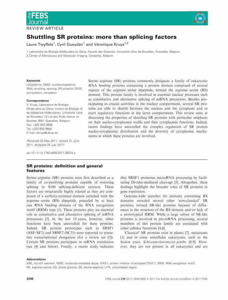
of 12 SR proteins [11] (see Fig. 1).
An important question is whether SR proteins have
unique or redundant functions. The intrinsic capacity
of SR proteins to activate splicing was first revealed
by complementation assays with cytoplasmic S100
splicing-deficient extract that contains all the splicing
machinery except SR proteins [1]. However, several
differences distinguish these proteins as to their ability
to promote splicing both in vitro and in vivo, support-
ing unique functions in splicing processes [12–15]. In
Caenorhabditis elegans, gene inactivation of SR
protein homologues by dsRNA interference reveals
that partial functional redundancy ensures viability
upon inactivation of single SR proteins, except in the
case of ASF ⁄SF2 whose expression is essential for
nematode development [16]. The function of SR
proteins has also been studied in mouse model sys-
tems. All SR-null mice for SRSF1, SRSF2 or SRSF3
(SRp20) show an early embryonic phenotype indicat-
ing that SR proteins are not redundant. However, the
essential functions exerted by each SR protein appear
to be tissue and ⁄or developmental stage specific (see
[17] for a review).
SR proteins ensure the coupling of splicing to tran-
scription as they are recruited together with U1
snRNP by RNA polymerase II and co-transcription-
ally deposited on the exon ⁄ 5¢ splice sites of nascent
transcripts [15]. Besides promoting the splicing process,
the co-transcriptional deposition of SR proteins on
nascent pre-mRNA transcripts has been shown to pre-
vent the formation of R loops due to hybridization of
the neosynthesized RNA to the complementary strand
of the DNA template and thereby to contribute to
genome stability by avoiding DNA double-stranded
breaks [18].
Interestingly, SRSF1 was recently reported as a
modulator of protein sumoylation. SRSF1 associates
with Ubc9 and enhances sumoylation of specific
substrates. In addition, SRSF1 interacts with PIAS1
(protein inhibitor of activated STAT-1), regulating
PIAS1-induced overall protein sumoylation. SRSF1
plays a role in heat-shock-induced sumoylation and
promotes SUMO conjugation to RNA-processing fac-
tors. This additional role of SRSF1 further expands
the versatility of this multifunctional protein [19] and
raises the interesting possibility that this activity could
be carried out by other SR proteins.
The expression of SR proteins varies widely among
cell types [20] and it has become clear that the abun-
dance of SR proteins greatly conditions the splicing
programme executed within different cell types. This is
clearly exemplified by SRSF1 which is a proto-onco-
gene whose expression is upregulated in various human
RRM 1 RRM 2 RS
RRM RS
RRM RS
RRM 1 RRM 2 RS
RRM 1 RRM 2 RS
RRM 1 RS
ZnRRM RS
RRM
RRM 2
RS
RRM
RRM 2 RS
RS
RRM RS
RRM RS
RRM
Name Synonym UniProt
SRSF1 (ASF/SF2) Q07955
SRSF2 (SC35) Q01130
SRSF3 (SRp20) P84103
SRSF4 (SRp75) Q08170
SRSF5 (SRp40) Q13243
SRSF6 (SRp55) Q13247
SRSF7 (9G8) Q16629
SRSF8 (SRp46) Q9BRL6
SRSF9 (SRp30c) Q13242
SRSF10 (SRp38,SRrp40) O75494
SRSF11 (p54) Q05519
SRSF12 (SRrp35) Q8WXF0
Fig. 1. Schematic representation of the 12
human SR proteins as defined by Manley
and Krainer [11]. Zn, zinc finger.
L. Twyffels et al. Shuttling SR proteins
FEBS Journal 278 (2011) 3246–3255 ª 2011 The Authors Journal compilation ª 2011 FEBS 3247

tumours, partly because of gene amplification. Overex-
pression of SRSF1 leads to abnormal accumulation of
alternatively spliced transcripts, including the mRNA
encoding the oncogenic isoform 2 of ribosomal protein
S6 kinase (S6K1) [21]. In contrast, several autoregula-
tory mechanisms mostly acting at the post-transcrip-
tional level maintain homeostatic levels of SR proteins.
A common mechanism relies on the generation of
alternatively spliced non-productive transcripts which
are either retained in the nucleus or targeted to
nonsense-mediated decay (NMD) due to the inclusion
of premature termination codons [22–24]. This splicing
reprogramming leads to a decrease of the translation-
competent mRNA isoform and thereby to the
repression of SR protein expression. Recent reports
indicate that the expression of SR proteins such as
SRSF1 can also be downmodulated at the post-tran-
scriptional level by microRNAs targeting the SRSF1 3¢untranslated region (UTR) [5,25] or by the autoregula-
tory translational repression of SRSF1 of its own
transcript ([23], and see later). Furthermore, the RS
domain of SR proteins is extensively phosphorylated
on serine residues and this post-translational modifica-
tion plays an important role in regulating the subcellu-
lar localization and activities of SR proteins. Several
protein kinase families have been shown to phosphory-
late the RS domain of SR proteins, including the
SRPK family [26,27], the Clk ⁄Sty family of dual
specificity kinases [28], the DNA topoisomerase I [29]
and the mitogen-activated AKT kinase [30,31]. More
recently, arginine methylation was also reported to
regulate SRSF1 nucleo-cytoplasmic distribution ([32]
and later). All these mechanisms contribute to the fine-
tuning of SR protein functions in various cell types
and upon different cellular conditions.
SR protein localization andnucleo-cytoplasmic shuttling
SR proteins are predominantly located in the nucleus
[33–39] and their nuclear import is mediated by trans-
portin-SR, a member of the b-karyopherin family. This
importin specifically contacts the RS domain to convey
them through the nuclear pore [40,41]. Several lines of
Splicing
Genome stability
Translational control
Localization intostress granules
Transcriptional elongation
mRNA export
SRSF1
SRSF1
SRPK
RISC
(A)nm7g
(A)nm7g
SRSF1 (A)nm7g SRSF1 (A)nm7g
(A)nm7g
+ +
SRSF1
Trn-SR1
Trn-SR2
mTOR+
target targetSUMO
SUMO
SumoylationE1/E2/E3
SRSF1
+
Nonsense-mediated decay
mRNP remodelling
PTC (A)nm7gSRSF1
?
PKCI-r
P
PP
SRSF1+ ? + ?
mRNA degradation
– –
pre-miR-7
pri-miR-7
Pri-miRNAprocessingDrosha
?SRSF1
SRSF1
P
SRSF1
SRSF1
(A)n
(A)n
m7g
m7g
(A) n
m7 g
SRSF1
TIAR
?SRSF1
ESE
?
Xpo5NXF1 NXF1
Fig. 2. Roles of SRSF1 in various cellular processes and compartments. ESE, exonic splicing enhancer; NXF1, nuclear export factor 1; P,
phosphate group; RISC, RNA-induced silencing complex; Xpo5, exportin 5.
Shuttling SR proteins L. Twyffels et al.
3248 FEBS Journal 278 (2011) 3246–3255 ª 2011 The Authors Journal compilation ª 2011 FEBS

evidence demonstrate that phosphorylation of the RS
domain by the cytoplasmic SR protein kinase 1
(SRPK1) is a prerequisite to SR protein nuclear
import [41–43].
Heterokaryon assays revealed that at least four SR
proteins (SRSF1, SRSF3, SRSF7 and SRSF10) shuttle
between the nucleus and the cytoplasm at a high
dynamic rate [34,44,45]. SRSF4 and SRFS6 also shuttle
between the two compartments but with slower kinetics
[44]. Earlier reports indicate that the RS domain is
important for nucleo-cytoplasmic shuttling [34]. Upon
recruitment of SR proteins for splicing, the RS domain
is hyperphosphorylated [26,28]. However, it becomes
partially dephosphorylated during splicing and this step
appears necessary for SR protein nuclear export
[46,47]. The RS domain is not sufficient, however, to
promote the export of a reporter protein. Therefore, it
regulates, rather than mediates, SR protein nuclear
export [34]. Interestingly, the non-shuttling SRSF2 is
confined to the nucleus due to the presence of a nuclear
retention sequence within its RS domain, thereby dem-
onstrating that RS domains are functionally distinct
with respect to nuclear export [45].
The shuttling ability of SR proteins is also dependent
on their RNA binding activity. This has been clearly
established for SRSF1 by earlier studies revealing that
the RNA binding activity of the first RRM is necessary
for nuclear export [34]. We recently revisited the deter-
minants of SRSF1 nuclear export by comparing the
subcellular localization of several mutants to the one of
the wild-type protein after transcription inhibition by
actinomycin D. As previously observed, this treatment
inhibits SRSF1 nuclear accumulation, leading to a
massive relocalization of SRSF1 into the cytoplasm. In
contrast, the W134A mutant in which the RNA bind-
ing capacity of RRM2 is disrupted [48] remains nuclear
under the same conditions [49]. Altogether, these data
suggest that SRSF1 nucleo-cytoplasmic shuttling
requires intact RNA binding activity of both RNA
binding domains.
Shuttling SR proteins interact with the TAP ⁄NFX1,
the primary receptor for general mRNA nuclear export
[50]. This interaction is mediated by the same domain
of TAP which is involved in the recruitment of the
Aly ⁄Ref adaptor protein. Moreover, SRSF3 and
SRSF7 were shown to activate the export of the intron-
less H2A mRNA [51]. Therefore, shuttling SR proteins
contribute to mRNA nuclear export via the interaction
with the nuclear receptor TAP. The TAP-binding
domains of SRSF1 and SRSF7 were first mapped to
the amino-terminal region composed of their RRM
and the downstream linker region [50]. Structural anal-
yses further indicated that this interaction involved
arginine residues located downstream from the RRM
of SRSF7 and SRSF3 [52] and an arginine-rich region
located between the two RNA binding domains for
SRSF1 [48]. A recent report indicates that three argi-
nine residues (R93, R97 and R109) located in SRSF1
inter-RRM linker region are methylated and strongly
influence SRSF1 nucleo-cytoplasmic distribution.
Indeed, an SRSF1 mutant in which these three arginine
residues were substituted by unmethylable and
uncharged alanine residues is predominantly located in
the cytoplasm. In contrast, a mutant in which these
arginine residues are substituted by lysines remains
predominantly nuclear, thereby suggesting that disrup-
tion of both methylation and charge of these arginine
residues modifies SRSF1 localization [32]. This study
emphasizes the importance of arginine residues located
in the TAP-binding motif for SRSF1 subcellular
distribution [48]. As hyperphosphorylated SR proteins
do not bind TAP [46,47], one could speculate that neg-
atively charged phosphoserines could associate with
these arginines, thereby blocking their interaction with
TAP. It is still to be explained, however, how the meth-
ylation status of these arginines conditions SRSF1
nucleo-cytoplasmic shuttling and how it modulates its
function as mRNA nuclear export adaptor.
In conclusion, the nuclear export of SR proteins
appears to be tightly coupled to the TAP-mediated
mRNA export pathway and to rely on a complex net-
work of sequence determinants combined with several
post-translational modifications.
Roles of SR proteins in the cytoplasm
In the last 10 years, several functions were attributed
to shuttling SR proteins in the cytoplasmic phase of
RNA metabolism. Some of these effects rely on
indirect mechanisms involving alternative splicing. For
example, high levels of SRSF1 favour the production
of an MNK2 isoform that phosphorylates the transla-
tion initiation factor eiF4E, thereby enhancing cap-
dependent translation [21]. Direct effects of SRSF1 on
cytoplasmic mRNA metabolism are diverse and
include regulation of mRNA stability and translation.
First, SR proteins modulate the NMD pathway,
whereby mRNAs containing premature termination
codons are targeted for degradation. Indeed, overex-
pression of a subset of SR proteins such as SRSF1
and SRSF2 strongly enhances NMD [53]. The NMD-
promoting effect of SRSF1 requires the presence of the
RS domain. However, this mechanism does not appear
to be dependent on SRSF1 nucleo-cytoplasmic shut-
tling, as the non-shuttling fusion protein consisting of
full-length SRSF1 fused at its C-terminus to the
L. Twyffels et al. Shuttling SR proteins
FEBS Journal 278 (2011) 3246–3255 ª 2011 The Authors Journal compilation ª 2011 FEBS 3249

nuclear retention sequence of SRSF2 retains the ability
to activate NMD. Although it remains unclear how
SR proteins activate NMD, one can hypothesize that
their recruitment promotes the nuclear modelling of
the nonsense mRNP to target it efficiently to NMD.
SRSF1 has been shown to bind to a purine-rich
sequence located in the 3¢ UTR of the mRNA encoding
a protein related to the protein-kinase-C-interacting
protein (PKCI-r) and to induce the degradation of this
transcript [54]. Furthermore, fractionation of cytosolic
extracts by sedimentation on sucrose gradients revealed
the association of SRSF1 and SRSF7 with ribosomal
subunits, monosomes and to a lesser extent polysomes,
suggesting a role for these SR proteins in mRNA
translation [55–57]. Interestingly, mutants of SRSF1
and SRSF7 lacking their RS domain are associated to
heavier polysomes than their native counterparts
[56,57]. Moreover, a hypophosphorylated SRSF1
mutant in which arginine–serine dipeptides are substi-
tuted by glycine–serines is markedly shifted to the much
heavier polysome-containing fractions, thereby suggest-
ing that RS domain hypophosphorylation favours
SRSF1 association with the translational machinery
[56]. Tethering assays indicate that SRSF1 binding to
the coding region of a reporter mRNA leads to
increased mRNA translation in cell extracts as well as
in intact cells. Moreover, while RRM1 and RS domains
are not required for this translation-enhancing effect,
RRM2 is indispensable [56]. This direct effect of SRSF1
is mediated by the recruitment of components of the
mTOR signalling pathway, resulting in phosphorylation
and release of 4E-BP, a competitive inhibitor of cap-
dependent translation [58]. This translation-activating
effect of SRSF1 is counteracted by overexpression of
SRPK2, which phosphorylates SRSF1 RS domain and
probably contributes to the dissociation of SR protein
from polysomes [56]. In contrast, it is upregulated by
mitogenic stimuli which promote SRSF1 phosphoryla-
tion by AKT kinase [30]. It thus appears that SR-
dependent translation activation is finely regulated in
response to extracellular signals and that this effect
might result from the competition between AKT- and
SRPK-mediated phosphorylation of SRSF1.
SRSF1 tethering to mRNA 3¢ UTR also activates
mRNA translation in vitro [56,58]. In intact cells, how-
ever, the opposite effect is observed [49]. The apparent
contradiction between in vitro and in vivo observations
might reflect the importance of the nuclear origin of
the target mRNA for the functional outcome of
SRSF1 recruitment to mRNA 3¢ UTR. Of note, trans-
lation inhibition mediated by SRSF1 tethering to
mRNA 3¢ UTR is markedly alleviated upon deletion
of the RS domain [49].
As mentioned above, SRSF1 controls its gene
expression partly by down-modulating the translation
of its own mRNA. This mechanism depends on the
presence of intact SRSF1 mRNA 3¢ UTR and affects
the initiation step of SRSF1 mRNA translation.
SRSF1 RRM2 is required for this regulation and
unexpectedly a nuclear-retained version of SRSF1 is
still able to autoregulate its translation. It thus appears
that nuclear SRSF1 affects the mRNP composition of
its own transcript which in turn conditions its transla-
tion efficiency in the cytoplasm [23].
SRSF1 interacts with several cellular factors includ-
ing RNA binding proteins such as TIAR and TIA-1.
These proteins are known translational repressors of
mRNAs bearing AU-rich elements (ARE) in their 3¢UTR. We have shown that overexpression of SRSF1
downregulates the expression of an ARE-containing
reporter mRNA. Interestingly, the same effect is repro-
duced upon overexpression of an RNA binding defec-
tive mutant but not upon overexpression of a mutant
lacking the RS domain [49]. Altogether, these observa-
tions suggest that SRSF1 participates in the post-tran-
scriptional control of ARE-containing mRNAs most
probably by protein–protein interactions with ARE-
binding proteins such as TIAR and TIA-1.
Finally, some viruses exploit SR proteins to promote
the translation of their mRNAs by different mecha-
nisms. SRSF3, on one hand, participates in the internal
ribosome entry site (IRES)-dependent translation of
picornavirus mRNAs. This effect is mediated by the
interaction of SRSF3 with the cellular RNA binding
protein PCBP2 that binds to IRES sequences within
viral RNAs [59]. On the other hand, SRSF5 and SRSF6
proteins activate the translation of gag protein from un-
spliced HIV-1 mRNAs. This mechanism appears to
depend on the gag-pol coding region as codon optimiza-
tion abolishes the translation-enhancing effect [60].
In conclusion, shuttling SR proteins are multifunc-
tional proteins which participate in a wide diversity of
regulatory mechanisms controlling messenger RNA
metabolism both in the nucleus and in the cytoplasm
(see Fig. 2). The regulatory output is conditioned by
multiple parameters such as the abundance of SR pro-
teins, their phosphorylation profile, their binding site
within the mRNA molecule and their association with
other factors within the mRNP.
SR proteins migrate into cytoplasmicstress granules
As they reach the cytoplasm, most mRNAs are
programmed for immediate translation, which involves
substantial RNP remodelling and assembly into
Shuttling SR proteins L. Twyffels et al.
3250 FEBS Journal 278 (2011) 3246–3255 ª 2011 The Authors Journal compilation ª 2011 FEBS

functional polysomes. However, in response to
environmental stress (heat, oxidative conditions, UV
irradiation, osmotic shock and hypoxia), eukaryotic
cells reprogramme their mRNA metabolism to repair
stress-induced damage and adapt to novel conditions.
During this process, the translation of mRNAs encod-
ing ‘housekeeping’ proteins is aborted, whereas the
translation of mRNAs encoding proteins involved in
the damage repair is stimulated. The untranslated
mRNAs concentrate in cytoplasmic foci called stress
granules (SGs) in association with stalled 48S
ribosomal pre-initiation complexes. Most of the trans-
lational machinery is recruited to SGs with the excep-
tion of the 60S subunit. In addition, SGs contain
PABP1 and several RNA-binding proteins that
regulate mRNA metabolism, including HuR, TTP,
G3BP and TIA proteins. In many cases, stress-induced
translation arrest is signalled by the phosphorylation
of translation initiator factor eIF2a. Phospho-eiF2areduces the availability of the eIF2–GTP–tRNAmet
i
ternary complex, thereby blocking translation initiation
and promoting polysome disassembly. SGs are not
stable repositories of untranslated mRNAs but rather
highly dynamic structures, as drugs that stabilize or
destabilize polysomes inhibit or promote SG assembly,
respectively, which is indicative of a dynamic equilib-
rium between these structures [61]. Moreover, upon
stress recovery, SGs are disassembled, and sequestered
mRNAs reassociate with active translational machin-
ery (see [62,63] for reviews).
On the basis of SRSF1 capacity to interact with
TIA proteins, we investigated SRSF1 migration into
SGs and found that this SR protein is recruited in
these structures in response to various stresses such as
oxidative stress, osmotic shock and heat. SRSF1 is a
bona fide SG component and does not get associated
with other cytoplasmic structures such as processing
bodies [64]. SRSF1 migration into SGs strongly
depends on its ability to bind RNA. Therefore, this
process most probably results from the sequestration
of SRSF1-bound transcripts in such cytoplasmic
structures. Accordingly, downregulation of SRSF1
expression does not alter SG assembly upon stress [49].
Other SR proteins such as SRSF2, SRSF3, SRSF7
and SRSF10 share the capacity to migrate into SGs
([65], Twyffels L. et al., unpublished results) (Table 1).
However, their propensity to assemble in such
structures is highly variable. Indeed, the shuttling
SRSF1, SRSF3 and SRSF10 are significantly relocal-
ized in cytoplasmic SGs upon stress. In contrast, the
non-shuttling SRSF2 and the shuttling SRSF7 display a
much lower propensity to assemble into SGs (Twyffels L.
et al., unpublished data). The nucleo-cytoplasmic
shuttling thus appears necessary for massive accumula-
tion of SR proteins into SGs. However, other parame-
ters seem to condition this process as shuttling SRSF7
only moderately migrates in SGs in response to oxida-
tive stress. Additionally, the fact that the non-shuttling
SRSF2 can be detected in SGs suggests that a minor
fraction of SR proteins normally retained in the
nucleus are redistributed to the cytoplasm in response
to stress. It should be noted that the nucleo-cytoplas-
mic distribution of transportin-SR is markedly altered
upon oxidative stress with significant relocalization to
the cytoplasm [66]. Therefore, the nuclear import
mediated by transportin-SR might be partially
impaired during oxidative stress, which might contrib-
ute to abnormal cytoplasmic accumulation of SR
proteins and further assembly into SGs. Altogether,
these observations suggest that the nucleo-cytoplasmic
distribution of several RNA binding proteins
undergoes major alterations upon stress and that SR
proteins participate to a variable extent in mechanisms
of translational repression ⁄derepression under these
conditions.
Conclusions
SR proteins make up a family of regulators with
important functions in RNA metabolism both in the
nucleus and in the cytoplasm. This review emphasized
the emerging roles of shuttling SR proteins in the cyto-
plasm. We have seen that SR proteins can be involved
in all phases of mRNA metabolism from synthesis to
degradation, including intermediate steps such as
nuclear export and translation. Most strikingly, the
role of SR proteins in NMD and translation regulation
perfectly exemplifies the importance of mRNP nuclear
assembly in the determination of mRNA cytoplasmic
fate and reveals some molecular links coupling gene
expression machineries.
While the parameters conditioning SRSF1 nucleo-
cytoplasmic shuttling are getting well characterized,
this is still not the case for the other shuttling SR
proteins (see Table 1). Moreover, several aspects of SR
protein distribution and activities remain unclear.
Most importantly, the signalling pathways controlling
SR domain phosphorylation as well as the phosphory-
lated residues within SR proteins are not fully identi-
fied although this post-translational modification is
known to play a major role in the control of SR
protein traffic, interactions and activities. Similarly, the
methylation of specific residues in the SRSF1 inter-
RRM region conditions SR protein localization and
thus probably modulates other aspects of SR protein
biology. Therefore, the detailed characterization of the
L. Twyffels et al. Shuttling SR proteins
FEBS Journal 278 (2011) 3246–3255 ª 2011 The Authors Journal compilation ª 2011 FEBS 3251

Table 1. Nucleo-cytoplasmic distribution, shuttling characteristics and reported functions of human SR proteins. References for the pre-
sented data are given in the text.
Name
Location
at the
equilibrium
Relocalization
to the
cytoplasm
upon RNA
PolII blockade
Shuttling in
interspecies
heterokaryon
assays
Association
with mRNA
export
machinery
Cytosolic
distribution
Accumulation
into stress
granules Reported functions
SRSF1 Nuclear
(endogenous
and tagged)
+ ++ + Free, 40S, 60S,
80S: +++
Light polysomes: +
Upon
overexpression: +
Upon various
stress: ++
Activates constitutive
and alternative splicing
Stimulates transcriptional
elongation
Contributes to genome
stability
Contributes to
mRNA export
Enhances NMD
Regulates translation
Promotes PKCI-r mRNA
degradation
Stimulates PIAS1- and
Ubc9-dependent
sumoylation
SRSF2 Nuclear
(endogenous
and tagged)
) ) ) ND Upon
overexpression: )Upon oxidative
stress: +
Upon heat
shock: )
Activates constitutive
and alternative splicing
Stimulates transcriptional
elongation
Contributes to
genome stability
Enhances NMD
SRSF3 Nuclear
(tagged)
+ ++ + Free, 40S: +++ Upon
overexpression: +
Upon oxidative
stress: ++
Activates constitutive
and alternative splicing
Contributes to mRNA export
Promotes IRES-mediated
translation of viral mRNA
SRSF4 Nuclear
(tagged)
ND + ND ND ND Activates constitutive and
alternative splicing
SRSF5 Nuclear
(tagged)
) ND ND ND ND Activates constitutive and
alternative splicing
Stimulates translation
of unspliced HIV-1 mRNA
SRSF6 Nuclear
(endogenous
and tagged)
) + ND ND ND Activates constitutive
and alternative splicing
Stimulates translation of
unspliced HIV-1 mRNA
SRSF7 Nuclear
(tagged)
) ++ + Free: ++
40S, 60S,
80S: +++
Polysomes: +
Upon
overexpression: )Upon oxidative
stress: +
Activates constitutive and
alternative splicing
Contributes to mRNA export
Stimulates translation
SRSF8 ND ND ND ND ND ND Regulates constitutive and
alternative splicing
SRSF9 Nuclear
(tagged)
ND ND ND ND ND Regulates constitutive and
alternative splicing
SRSF10 Nuclear
(tagged)
+ ++ ND Upon heat
shock: ++
Represses splicing
SRSF11 Nuclear
(endogenous)
ND ND ND ND ND Represses alternative
splicing
SRSF12 ND ND ND ND ND ND Represses alternative
splicing
Shuttling SR proteins L. Twyffels et al.
3252 FEBS Journal 278 (2011) 3246–3255 ª 2011 The Authors Journal compilation ª 2011 FEBS

post-translational modifications (e.g. phosphorylation,
methylation) of SR proteins as well as the identifica-
tion of physiological mRNA targets should provide a
more comprehensive view on how these factors
combine multiple activities in RNA metabolism. These
key issues should not wait too long to be addressed as
SR proteins exert essential cellular functions and have
been clearly established as key regulators whose unbal-
anced expression has been associated with cancer
[21,67,68] and other severe human diseases [69–71].
Acknowledgements
Laure Twyffels is an FNRS research fellow.
References
1 Zahler AM, Lane WS, Stolk JA & Roth MB (1992) SR
proteins: a conserved family of pre-mRNA splicing
factors. Genes Dev 6, 837–847.
2 Shepard PJ & Hertel KJ (2009) The SR protein family.
Genome Biol 10, 242.
3 Zhong X-Y, Wang P, Han J, Rosenfeld MG & Fu X-D
(2009) SR proteins in vertical integration of gene
expression from transcription to RNA processing to
translation. Mol Cell 35, 1–10.
4 Long JC & Caceres JF (2009) The SR protein family of
splicing factors: master regulators of gene expression.
Biochem J 417, 15–27.
5 Wu H, Sun S, Tu K, Gao Y, Xie B, Krainer AR &
Zhu J (2010) A splicing-independent function of
SF2 ⁄ASF in microRNA processing. Mol Cell 38, 66–77.
6 Boucher L, Ouzounis CA, Enright AJ & Blencowe BJ
(2001) A genome-wide survey of RS domain proteins.
RNA 7, 1693–1701.
7 Lopato S, Mayeda A, Krainer AR & Barta A (1996)
Pre-mRNA splicing in plants: characterization of
Ser ⁄Arg splicing factors. Proc Natl Acad Sci USA 93,
3074–3079.
8 Gross T, Richert K, Mierke C, Lutzelberger M &
Kaufer NF (1998) Identification and characterization of
srp1, a gene of fission yeast encoding a RNA binding
domain and a RS domain typical of SR splicing factors.
Nucleic Acids Res 26, 505–511.
9 Lutzelberger M, Gross T & Kaufer NF (1999) Srp2, an
SR protein family member of fission yeast: in vivo
characterization of its modular domains. Nucleic Acids
Res 27, 2618–2626.
10 Kress TL, Krogan NJ & Guthrie C (2008) A single
SR-like protein, Npl3, promotes pre-mRNA splicing in
budding yeast. Mol Cell 32, 727–734.
11 Manley JL & Krainer AR (2010) A rational nomencla-
ture for serine ⁄ arginine-rich protein splicing factors (SR
proteins). Genes Dev 24, 1073–1074.
12 Tacke R & Manley JL (1995) The human splicing fac-
tors ASF ⁄SF2 and SC35 possess distinct, functionally
significant RNA binding specificities. EMBO J 14,
3540–3551.
13 Fu X-D (1993) Specific commitment of different pre-
mRNAs to splicing by single SR proteins. Nature 365,
82–85.
14 Wang J & Manley JL (1995) Overexpression of the SR
proteins ASF ⁄SF2 and SC35 influences alternative splic-
ing in vivo in diverse ways. RNA 1, 335–346.
15 Das R, Yu J, Zhang Z, Gygi MP, Krainer AR, Gygi
SP & Reed R (2007) SR proteins function in coupling
RNAP II transcription to pre-mRNA splicing. Mol Cell
26, 867–881.
16 Longman D, Johnstone IL & Caceres JF (2000)
Functional characterization of SR and SR-related genes
in Caenorhabditis elegans. EMBO J 19, 1625–1637.
17 Moroy T & Heyd F (2007) The impact of alternative
splicing in vivo: mouse models show the way. RNA 13,
1155–1171.
18 Li X & Manley JL (2005) Inactivation of the SR pro-
tein splicing factor ASF ⁄SF2 results in genomic instabil-
ity. Cell 122, 365–378.
19 Pelisch F, Gerez J, Druker J, Schor IE, Munoz MJ,
Risso G, Petrillo E, Westman BJ, Lamond AI, Arzt E
et al. (2010) The serine ⁄ arginine-rich protein SF2 ⁄ASF
regulates protein sumoylation. Proc Natl Acad Sci USA
107, 116119–116124.
20 Hanamura A, Caceres JF, Mayeda A, Franza BR Jr &
Krainer AR (1998) Regulated tissue-specific expression
of antagonistic pre-mRNA splicing factors. RNA 4,
430–444.
21 Karni R, de Stanchina E, Lowe SW, Sinha R, Mu D &
Krainer AR (2007) The gene encoding the splicing fac-
tor SF2 ⁄ASF is a proto-oncogene. Nat Struct Mol Biol
14, 185–193.
22 Ni JZ, Grate L, Donohue JP, Preston C, Nobida N,
O’Brien G, Shiue L, Clark TA, Blume JE & Ares M
(2007) Ultraconserved elements are associated with
homeostatic control of splicing regulators by alternative
splicing and nonsense-mediated decay. Genes Dev 21,
708–718.
23 Sun S, Zhang Z, Sinha R, Karni R & Krainer AR
(2010) SF2 ⁄ASF autoregulation involves multiple layers
of post-transcriptional and translational control. Nat
Struct Mol Biol 17, 306–312.
24 McGlincy NJ & Smith CWJ (2008) Alternative splicing
resulting in nonsense-mediated mRNA decay: what is the
meaning of nonsense? Trends Biochem Sci 33, 385–393.
25 Meseguer S, Mudduluru G, Escamilla JM, Allgayer H
& Barettino D (2011) MicroRNAs-10a and -10b con-
tribute to retinoic acid-induced differentiation of neuro-
blastoma cells and target the alternative splicing
regulatory factor SFRS1 (SF2 ⁄ASF). J Biol Chem 286,
4150–4164.
L. Twyffels et al. Shuttling SR proteins
FEBS Journal 278 (2011) 3246–3255 ª 2011 The Authors Journal compilation ª 2011 FEBS 3253

26 Gui JF, Tronchere H, Chandler SD & Fu XD (1994)
Purification and characterization of a kinase specific for
the serine- and arginine-rich pre-mRNA splicing factors.
Proc Natl Acad Sci USA 91, 10824–10828.
27 Wang HY, Lin W, Dyck JA, Yeakley JM, Songyang Z,
Cantley LC & Fu XD (1998) SRPK2: a differentially
expressed SR protein-specific kinase involved in mediat-
ing the interaction and localization of pre-mRNA splic-
ing factors in mammalian cells. J Cell Biol 140, 737–
750.
28 Colwill K, Pawson T, Andrews B, Prasad J, Manley JL,
Bell JC & Duncan PI (1996) The Clk ⁄ Sty protein kinase
phosphorylates SR splicing factors and regulates their
intranuclear distribution. EMBO J 15, 265–275.
29 Rossi F, Labourier E, Forne T, Divita G, Derancourt
J, Riou JF, Antoine E, Cathala G, Brunel C & Tazi J
(1996) Specific phosphorylation of SR proteins by mam-
malian DNA topoisomerase I. Nature 381, 80–82.
30 Blaustein M, Pelisch F, Tanos T, Munoz MJ, Wengier
D, Quadrana L, Sanford JR, Muschietti JP, Kornblihtt
AR, Caceres JF et al. (2005) Concerted regulation of
nuclear and cytoplasmic activities of SR proteins by
AKT. Nat Struct Mol Biol 12, 1037–1044.
31 Patel NA, Kaneko S, Apostolatos HS, Bae SS, Watson
JE, Davidowitz K, Chappell DS, Birnbaum MJ, Cheng
JQ & Cooper DR (2005) Molecular and genetic studies
imply Akt-mediated signaling promotes protein kinase
CbII alternative splicing via phosphorylation of serine ⁄arginine-rich splicing factor SRp40. J Biol Chem 280,
14302–14309.
32 Sinha R, Allemand E, Zhang Z, Karni R, Myers MP &
Krainer AR (2010) Arginine methylation controls the
subcellular localization and functions of the oncoprotein
splicing factor SF2 ⁄ASF. Mol Cell Biol 30, 2762–2774.
33 Caceres JF, Misteli T, Screaton GR, Spector DL &
Krainer AR (1997) Role of the modular domains of SR
proteins in subnuclear localization and alternative splic-
ing specificity. J Cell Biol 138, 225–238.
34 Caceres JF, Screaton GR & Krainer AR (1998) A spe-
cific subset of SR proteins shuttles continuously
between the nucleus and the cytoplasm. Genes Dev 12,
55–66.
35 Zimowska G, Shi J, Munguba G, Jackson MR, Alpativ
R, Simmons MN, Shi Y & Sugrue SP (2003)
Pinin ⁄DRS ⁄memA interacts with SRp75, SRm300 and
SRrp130 in corneal epithelial cells. Invest Ophthalmol
Vis Sci 44, 4715–4723.
36 Sakashita E & Endo H (2010) SR and SR-related pro-
teins redistribute to segregated fibrillar components of
nucleoli in a response to DNA damage. Nucleus 1, 367–
380.
37 Barbe L, Lundberg E, Oksvold P, Stenius A, Lewin E,
Bjorling E, Asplund A, Ponten F, Brismar H, Uhlen M
et al. (2008) Towards a confocal subcellular atlas of the
human proteome. Mol Cell Proteomics 7, 499–508.
38 Lai M-C, Lin R-I & Tarn W-Y (2003) Differential
effects of hyperphosphorylation on splicing factor
SRp55. Biochem J 371, 937–945.
39 Cowper AE, Caceres JF, Mayeda A & Screaton GR
(2001) Serine-arginine (SR) protein-like factors that
antagonize authentic SR proteins and regulate alterna-
tive splicing. J Biol Chem 276, 48908–48914.
40 Kataoka N, Bachorik JL & Dreyfuss G (1999) Trans-
portin-SR, a nuclear import receptor for SR proteins.
J Cell Biol 145, 1145–1152.
41 Lai M-C, Lin R-I, Huang S-Y, Tsai C-W & Tarn W-Y
(2000) A human importin-b family protein, Transpor-
tin-SR2, interacts with the phosphorylated RS domain
of SR proteins. J Biol Chem 275, 7950–7957.
42 Koizumi J, Okamoto Y, Onogi H, Mayeda A, Krainer
AR & Hagiwara M (1999) The subcellular localization
of SF2 ⁄ASF is regulated by direct interaction with SR
protein kinases (SRPKs). J Biol Chem 274, 11125–11131.
43 Ngo JCK, Chakrabarti S, Ding J-H, Velazquez-Dones
A, Nolen B, Aubol BE, Adams JA, Fu X-D & Ghosh
G (2005) Interplay between SRPK and Clk ⁄Sty kinases
in phosphorylation of the splicing factor ASF ⁄SF2 is
regulated by a docking motif in ASF ⁄ SF2. Mol Cell 20,
77–89.
44 Sapra AK, Anko M-L, Grishina I, Lorenz M, Pabis M,
Poser I, Rollins J, Weiland E-M & Neugebauer KM
(2009) SR protein family members display diverse activ-
ities in the formation of nascent and mature mRNPs
in vivo. Mol Cell 34, 179–190.
45 Cazalla D, Zhu J, Manche L, Huber E, Krainer AR &
Caceres JF (2002) Nuclear export and retention signals
in the RS domain of SR proteins. Mol Cell Biol 22,
6871–6882.
46 Huang Y, Yario TA & Steitz JA (2004) A molecular
link between SR protein dephosphorylation and mRNA
export. Proc Natl Acad Sci USA 101, 9666–9670.
47 Lai M-C & Tarn W-Y (2004) Hypophosphorylated
ASF ⁄ SF2 binds TAP and is present in messenger ribo-
nucleoproteins. J Biol Chem 279, 31745–31749.
48 Tintaru AM, Hautbergue GM, Hounslow AM, Hung
M-L, Lian L-Y, Craven CJ & Wilson SA (2007) Struc-
tural and functional analysis of RNA and TAP binding
to SF2 ⁄ASF. EMBO Rep 8, 756–762.
49 Delestienne N, Wauquier C, Soin R, Dierick J-F, Guey-
dan C & Kruys V (2010) The splicing factor ASF ⁄ SF2is associated with TIA-1-related ⁄TIA-1-containing ribo-
nucleoproteic complexes and contributes to post-tran-
scriptional repression of gene expression. FEBS J 277,
2496–2514.
50 Huang Y, Gattoni R & Steitz JA (2003) SR Splicing
factors serve as adapter proteins for TAP-dependent
mRNA export. Mol Cell 11, 837–843.
51 Huang Y & Steitz JA (2001) Splicing factors SRp20
and 9G8 promote the nucleocytoplasmic export of
mRNA. Mol Cell 7, 899–905.
Shuttling SR proteins L. Twyffels et al.
3254 FEBS Journal 278 (2011) 3246–3255 ª 2011 The Authors Journal compilation ª 2011 FEBS

52 Hargous Y, Hautbergue GM, Tintaru AM, Skrisovska
L, Golovanov AP, Stevenin J, Lian L-Y, Wilson SA &
Allain FH-T (2006) Molecular basis of RNA recogni-
tion and TAP binding by the SR proteins SRp20 and
9G8. EMBO J 25, 5126–5137.
53 Zhang Z & Krainer AR (2004) Involvement of SR pro-
teins in mRNA surveillance. Mol Cell 16, 597–607.
54 Lemaire R, Prasad J, Kashima T, Gustafson J, Manley
JL & Lafyatis R (2002) Stability of a PKCI-1-related
mRNA is controlled by the splicing factor ASF ⁄ SF2:a novel function for SR proteins. Genes Dev 16, 594–607.
55 Sanford JR, Gray NK, Beckmann K & Caceres JF
(2004) A novel role for shuttling SR proteins in mRNA
translation. Genes Dev 18, 755–768.
56 Sanford JR, Ellis JD, Cazalla D & Caceres JF (2005)
Reversible phosphorylation differentially affects nuclear
and cytoplasmic functions of splicing factor 2 ⁄ alterna-tive splicing factor. Proc Natl Acad Sci USA 102,
15042–15047.
57 Swartz JE, Bor Y-C, Misawa Y, Rekosh D & Hammar-
skjold M-L (2007) The shuttling SR protein 9G8 plays
a role in translation of unspliced mRNA containing a
constitutive transport element. J Biol Chem 282, 19844–
19853.
58 Michlewski G, Sanford JR & Caceres JF (2008) The
splicing factor SF2 ⁄ASF regulates translation initiation
by enhancing phosphorylation of 4E-BP1. Mol Cell 30,
179–189.
59 Bedard KM, Daijogo S & Semler BL (2007) A nucleo-
cytoplasmic SR protein functions in viral IRES-medi-
ated translation initiation. EMBO J 26, 459–467.
60 Swanson CM, Sherer NM & Malim MH (2010) SRp40
and SRp55 promote the translation of unspliced human
immunodeficiency virus type 1 RNA. J Virol 84, 6748–
6759.
61 Kedersha N, Cho MR, Li W, Yacono PW, Chen S,
Gilks N, Golan DE & Anderson P (2000) Dynamic
shuttling of TIA-1 accompanies the recruitment of
mRNA to mammalian stress granules. J Cell Biol 151,
1257–1268.
62 Anderson P & Kedersha N (2006) RNA granules. J Cell
Biol 172, 803–808.
63 Anderson P & Kedersha N (2008) Stress granules: the
Tao of RNA triage. Trends Biochem Sci 33, 141–150.
64 Kulkarni M, Ozgur S & Stoeklin G (2010) On track
with P-bodies. Biochem Soc Trans 38, 242–251.
65 Shin C, Kleiman FE & Manley JL (2005) Multiple
properties of the splicing repressor SRp38 distinguish
it from typical SR Proteins. Mol Cell Biol 25, 8334–
8343.
66 Chang W-L & Tarn W-Y (2009) A role for Transportin
in deposition of TTP to cytoplasmic RNA granules and
mRNA decay. Nucleic Acids Res 37, 6600–6612.
67 Faustino NA & Cooper TA (2003) Pre-mRNA splicing
and human disease. Genes Dev 17, 419–437.
68 Fischer DC, Noack K, Runnebaum IB, Watermann
DO, Kieback DG, Stamm S & Stickeler E (2004)
Expression of splicing factors in human ovarian cancer.
Oncol Rep 11, 1085–1090.
69 Cartegni L & Krainer AR (2002) Disruption of an
SF2 ⁄ASF-dependent exonic splicing enhancer in SMN2
causes spinal muscular atrophy in the absence of
SMN1. Nat Genet 4, 377–384.
70 Gabut M, Mine M, Marsac C, Brivet M, Tazi J &
Soret J (2005) The SR protein SC35 is responsible for
aberrant splicing of the E1a pyruvate dehydrogenase
mRNA in a case of mental retardation with lactic
acidosis. Mol Cell Biol 25, 3286–3294.
71 Watanuki T, Funato H, Uchida S, Matsubara T,
Kobayashi A, Wakabayashi Y, Otsuki K, Nishida A &
Watanabe Y (2008) Increased expression of splicing fac-
tor SRp20 mRNA in bipolar disorder patients. J Affect
Disord 110, 62–69.
L. Twyffels et al. Shuttling SR proteins
FEBS Journal 278 (2011) 3246–3255 ª 2011 The Authors Journal compilation ª 2011 FEBS 3255