the intersection of gender and italian/americaness - Digital ...

Intersection of Small RNA Pathways in Arabidopsisthaliana Sub-Nuclear DomainsOlga Pontes1,2*., Alexa Vitins1.¤a, Thomas S. Ream1¤b, Evelyn Hong1, Craig S. Pikaard1,3, Pedro Costa-
Nunes1,2
1Department of Biology, University of New Mexico, Albuquerque, New Mexico, United States of America, 2 Biology Department, Washington University in St. Louis, St.
Louis, Missouri, United States of America, 3Department of Biology and Department of Molecular and Cellular Biochemistry, Indiana University, Bloomington, Indiana,
United States of America
Abstract
In Arabidopsis thaliana, functionally diverse small RNA (smRNA) pathways bring about decreased RNA accumulation oftarget genes via several different mechanisms. Cytological experiments have suggested that the processing of microRNAs(miRNAs) and heterochromatic small interfering RNAs (hc-siRNAs) occurs within a specific nuclear domain that can presentCajal Body (CB) characteristics. It is unclear whether single or multiple smRNA-related domains are found within the same CBand how specialization of the smRNA pathways is determined within this specific sub-compartment. To ascertain whethernuclear smRNA centers are spatially related, we localized key proteins required for siRNA or miRNA biogenesis byimmunofluorescence analysis. The intranuclear distribution of the proteins revealed that hc-siRNA, miRNA and trans-actingsiRNA (ta-siRNA) pathway proteins accumulate and colocalize within a sub-nuclear structure in the nucleolar periphery.Furthermore, colocalization of miRNA- and siRNA-pathway members with CB markers, and reduced wild-type localizationpatterns in CB mutants indicates that proper nuclear localization of these proteins requires CB integrity. We hypothesizethat these nuclear domains could be important for RNA silencing and may partially explain the functional redundancies andinteractions among components of the same protein family. The CB may be the place in the nucleus where Dicer-generatedsmRNA precursors are processed and assigned to a specific pathway, and where storage, recycling or assembly of RNAinterference components takes place.
Citation: Pontes O, Vitins A, Ream TS, Hong E, Pikaard CS, et al. (2013) Intersection of Small RNA Pathways in Arabidopsis thaliana Sub-Nuclear Domains. PLoSONE 8(6): e65652. doi:10.1371/journal.pone.0065652
Editor: Eric Jan, University of British Columbia, Canada
Received December 31, 2012; Accepted April 25, 2013; Published June 12, 2013
Copyright: � 2013 Pontes et al. This is an open-access article distributed under the terms of the Creative Commons Attribution License, which permitsunrestricted use, distribution, and reproduction in any medium, provided the original author and source are credited.
Funding: OP, EH and PC-N were supported by an Edward Mallinckrodt, Jr. Foundation award and Monsanto Company. TR is a Gordon and Betty MooreFoundation Fellow of the Life Sciences Research Foundation. The funders had no role in study design, data collection and analysis, decision to publish, orpreparation of the manuscript.
Competing Interests: OP, EH and PCN were supported by an Edward Mallinckrodt, Jr. Foundation award and Monsanto Company. This does not alter theauthors’ adherence to all PLOS ONE policies on sharing data and materials.
* E-mail: [email protected]
¤a Current address: Laboratory for Health Protection Research, National Institute of Public Health and the Environment, Bilthoven, the Netherlands¤b Current address: Department of Biochemistry, University of Wisconsin, Madison, Wiscosin, United States of America
. These authors contributed equally to this work.
Introduction
Arabidopsis thaliana small RNA (smRNA) silencing pathways are
essential for regulating development and establishing epigenetic
modifications, such as DNA and histone methylation [1,2]. Major
pathways include the microRNA (miRNA), trans-acting small
interfering RNA (ta-siRNA), natural cis-antisense siRNA (nat-
siRNA), and heterochromatic small interfering RNA (hc-siRNA)
pathways [3,4]. Each pathway shares a common set of steps that
begin with the processing of double-stranded RNA by a DICER-
LIKE (DCL) enzyme, generating 21–24 nucleotide (nt) double-
stranded RNA (dsRNA) duplexes. DCL proteins interact with
dsRNA-binding proteins (DRB) that aid in the dicing process
[5,6]. The smRNAs are unwound and one strand binds an
ARGONAUTE (AGO) protein, which facilitates small-RNA
guided target RNA cleavage, translational repression or the
establishment of epigenetic modifications [7]. A. thaliana has four
DCLs, ten AGOs, and six RNA DEPENDENT RNA POLY-
MERASES (RDRs) that collaborate in various permutations
within different pathways. How each class of smRNA is
successfully channeled through its unique pathway remains largely
unknown.
Mature miRNAs are distinct from other smRNA classes by their
stem-loop precursors. Those originate from Pol II-transcribed
precursors that are cleaved by DCL1 to produce an miRNA,
typically 21-nt in length, whose 39-end is subsequently methylated
by HEN1 (HUA ENHANCER 1). The 39-end methylation is a
unifying feature of all smRNA pathways, promoting stability of the
smRNA molecule [8]. AGO1 binds the miRNA and uses the
smRNA sequence to guide cleavage or translational arrest of
complementary target mRNAs [1]. The miRNA pathway is
essential for regulating specific transcription factors and other
genes required for plant development [4].
The ta-siRNAs are produced from endogenous loci and act in
trans to direct post-transcriptional gene silencing, being involved in
developmental phase changes and organ polarity. This specific
pathway is initiated upon the cleavage of non-coding primary TAS
gene transcripts by AGO1-bound and AGO7-bound miRNA
PLOS ONE | www.plosone.org 1 June 2013 | Volume 8 | Issue 6 | e65652
complexes [9,10]. AGO7 binds specifically to miRNA390 and is
involved in targeting TAS3 transcripts [10]. SUPPRESSOR OF
GENE SILENCING 3 (SGS3) protects the single-stranded RNA
cleavage products from degradation that RDR6 utilizes as a
template for double-stranded RNA production. Subsequent dicing
into 21-nt ta-siRNAs occurs as a result of DCL4 activity [12,13].
The resulting ta-siRNAs are loaded into and guide AGO1 proteins
to complementary transcripts targeted for cleavage [11].
In response to biotic and abiotic stresses, transcription of two
overlapping genes in reverse orientation can produce nat-siRNAs
[14,15]. For example, the opposing transcription of P5CDH, a
stress-related gene, and SRO5, a gene induced by salt, results in the
production of double-stranded RNAs that are processed into
smRNAs [14]. In turn, these siRNAs direct the cleavage of the
P5CDH transcripts through an unidentified AGO. In an ampli-
fication step, the cleaved P5CDH transcripts are stabilized by
SGS3, processed into dsRNA by RDR6 and cleaved by DCL1
into 21-nt RNAs, resulting in a cis regulatory feedback loop to
ultimately lead to salt tolerance. The nat-siRNA pathway is also
dependent on RNA POLYMERASE IV (NRPDIV) [14], which is
best know for its role in hc-siRNA biogenesis.
The hc-siRNA pathway is responsible for transcriptional
silencing by guiding DNA methylation and changes in local
chromatin structure [2,16]. Hc-siRNAs are 24 nt in length and
their biogenesis requires the activity of Pol IV [17,18], CLASSY1
(CLSY1, a putative chromatin-remodeler) [19], RDR2, and
DCL3 [3,20]. SiRNAs bound to AGO4, coupled with RNA-
directed DNA METHYLATION 1 (RDM1) and SUPPRESSOR
OF TY INSERTION 5-LIKE (SPT5L) bind to transcripts
produced by another plant-specific RNA polymerase, Pol V, or
RNA polymerase II (Pol II), ultimately recruiting the de novo DNA
methyltransferase DRM2 (DOMAINS REARRANGED METH-
YLTRANSFERASE 2) [2,20,21,22,23,24,25]. Pol V transcription
is dependent on DRD1 (DEFECTIVE IN RNA-DIRECTED
DNA METHYLATION1) [26,27], a putative chromatin remo-
deler, and DEFECTIVE IN MERISTEM SILENCING 3
(DMS3), a protein similar to the Structural Maintenance of
Chromosomes (SMC) [2,22,28].
The complexity of smRNA silencing raises important questions
of how double-stranded RNA molecules can be channeled
through the correct pathway. An important clue arose from
studies showing that preferential binding of members of the
Arabidopsis AGO family to smRNAs is dependent on the 59
terminal nucleotide [29,30]. Nevertheless, the redundancy found
among DCL proteins and the contribution of RDR6 to multiple
smRNA silencing pathways strongly suggests that additional
factors are required for specification of a smRNA to its pathway.
Interestingly, both miRNA and hc- siRNA pathway components
were found to localize in nucleolus-associated structures
[31,32,33]. Furthermore, in the case of the hc-siRNA pathway,
the specific domains associated with the nucleolus are Cajal bodies
(CBs).
CBs are conspicuous nuclear domains often localized to the
nucleolar periphery or within the nucleoli [34,35,36]. These
structures contain a wide variety of different proteins and small
RNAs: both small nuclear RNAs (snRNAs) and small nucleolar
RNAs (snoRNAs), which are factors involved in pre-mRNA
splicing, pre-ribosomal RNA (pre-rRNA) processing, or histone
pre-mRNA 39 maturation; basal transcription factors for RNA
polymerase I, II and III; and telomerase RNA [37,35,38]. It is
currently thought that CBs have roles in the metabolism of
different classes of small ribonucleoprotein (snRNP) particles,
snoRNPs, Sm proteins, telomerase, and the U7 snRNP. In
addition, CBs were found to be associated with gene-specific loci,
such as histone and U2 snRNA gene clusters. It was previously
shown that the hc-siRNA-processing center shares some features
with CBs [32], and an analogous nuclear domain for miRNA
processing has been described [31]. However, it is unknown
whether all the major components of the miRNA and siRNA (ta-
siRNA and hc-siRNA) pathways localize to the same or to distinct
CBs at the same point in time, but it has been hypothesized that
different types of CBs might co-exist [39]. Furthermore, the role of
CB assembly in miRNA and siRNA component localization and
biogenesis is unknown.
In this paper, we analyzed the nuclear localization and
colocalization of the main components of each of the smRNA
silencing pathways in A. thaliana to assess how these pathways are
organized in the cellular compartment and whether a specific
nuclear location provides a site for crosstalk among smRNA
pathways. Our results suggest that a specific nuclear domain
provides a site where A. thaliana RNAi pathways colocalize and
intersect. This domain possibly constitutes a site for smRNA
biogenesis, modification, effector complex assembly or storage of
smRNA-related components.
Results
Components of the A. thaliana Trans-acting siRNAPathway Localize to a Discrete Perinucleolar Structure
To evaluate where ta-siRNA pathways occur within the
nucleus, we used immunofluorescence to determine the nuclear
location of the proteins involved in ta-siRNA biogenesis. To avoid
protein localization artifacts caused by over-expression of proteins
driven by strong constitutive expression promoters, we generated
A. thaliana transgenic lines expressing FLAG-tagged SGS3 and
RDR6 under the control of their native promoters in the
corresponding null mutants. Epitope-tagged proteins complement
sgs3 and rdr6 mutant phenotypes as verified by the restoration of
smRNA biogenesis and are thus functional (Figure S1). In addition
to the epitope-tagged proteins, antibodies specific to native RDR6,
SGS3, DCL4 or AGO7 were raised and the specificity of each
antibody was determined by immunoblotting and immunolabeling
in the respective mutants (Figures S2 and S3). To avoid artifacts
derived from cross reaction between antibodies raised in the same
organism, immunolocalizations were performed such that mouse
monoclonal antibodies detected the epitope of recombinant
protein in transgenic lines, whereas native proteins were detected
by specific antibodies raised in rabbit.
As shown in Figure 1, SGS3 is uniformly distributed throughout
the nucleoplasm and enriched within a prominent, round-shaped
domain located in the nucleolar periphery (Figure 1). The
nucleolus is the prominent nuclear compartment devoid of
DNA-staining DAPI (Figure 1, in blue). Similar localization
patterns were observed for DCL4 and AGO7 (Figure 1). In
contrast, RDR6 does not show perinucleolar accumulation and is
distributed in the nucleoplasm. The same nuclear distribution
patterns of RDR6 and SGS3 were observed upon immunolocal-
ization of epitope-tagged or the native proteins (Figure 1 and
Table S1). The specificity of the antibodies recognizing the native
forms of the proteins was confirmed by the absence of fluorescence
signals in nuclei of the corresponding null mutant background
(Figure S3).
We performed colocalization analysis to determine whether
RDR6, SGS3, DCL4 and AGO7 were present at the same nuclear
location. SGS3, DCL4 and AGO7 were colocalized within the
perinucleolar domain in a fraction of nuclei (Figure 1). RDR6
failed to colocalize with DCL4 or AGO7 (data not shown). Only a
smRNA Pathways and Nuclear Domains
PLOS ONE | www.plosone.org 2 June 2013 | Volume 8 | Issue 6 | e65652

few colocalization foci were observed between RDR6 and SGS3 in
the nucleoplasm (Figure 1 top row, arrows).
Proteins Functioning in miRNA and siRNA PathwaysColocalize within a Nuclear Region Associated with theNucleolar Periphery
The nuclear distribution of some ta-siRNA pathway compo-
nents, such as SGS3, DCL4 and AGO7, resembles the patterns
previously described for key proteins involved in miRNA and hc-
siRNA biogenesis [32,20]. Two independent studies showed that
both miRNA and hc-siRNA pathway proteins are enriched in a
domain near the nucleolar periphery designated as a ‘‘Dicing
Body’’ [31] or ‘‘siRNA-processing center’’ [20], respectively.
However, it is not clear whether these nuclear domains correspond
to distinct smRNA processing centers or to the same entity.
To determine whether a unique multi-functional processing
center or multiple centers exist, we colocalized all four A. thaliana
DCL proteins, DCL1, DCL2, DCL3 and DCL4, in pairwise
combinations using epitope-tagged proteins from transgenes and
antibodies recognizing the native forms of the proteins. As
depicted in Figure 2, DCL1, DCL3 and DCL4 colocalize near
the nucleolar periphery with a frequency of 39%–46%. When
colocalization was not observed, the DCL signals still occurred in
the nucleolar periphery but did not colocalize, or the proteins were
diffusely distributed throughout the nucleoplasm (not shown).
Although DCL1 and DCL3 both colocalize with DCL2 in the
round-shaped signal in the nucleolar periphery, the observed
frequency was lower, between 17% and 23% (Figure 2, Fisher’s
test P,0.005). This observation correlates with the high frequency
of nucleoplasmic signals showed by DCL2 (67%, see Table S1).
Duplex miRNAs are methylated at the 29 OH of its 39 end by
HEN1 [40]. HEN1 was shown to localize within a so-called
‘‘Dicing Body’’ which colocalizes with DCL1 [31]. To determine
whether HEN1 is colocalized with other DCL enzymes, dual
immunolocalization was performed between HEN1 and DCL1,
DCL3 and DCL4. To localize HEN1, we produced a transgenic
line bearing the protein tagged with a FLAG epitope. The
complementation of the hen1 mutation was verified by the rescue
of miR167 and flower development phenotypes (Figure S4).
We found that DCL3 and DCL4 colocalized with HEN1 within
a focus at the nucleolar periphery at a frequency of 43% and 53%,
respectively (Figure 3). HEN1 colocalized with DCL1 at a
frequency of 49% in the nucleolar periphery (Figure 3) and 21%
in nucleoplasmic foci (not shown). When colocalization was not
observed, HEN1 was distributed throughout the nucleoplasm
while DCL proteins were found enriched in the nucleolar-
associated signal or dispersed in the nucleoplasm (not shown).
To test whether AGO proteins utilized by different pathways
might colocalize, we performed double immunolocalization,
combining epitope-tagged AGO4 detected by a mouse monoclo-
nal antibody and antibodies specific to native AGO1 and AGO7,
raised in rabbit. The specificity of AGO1 and AGO7 antibodies
Figure 1. Nuclear localization of trans-acting siRNA pathway members. Double immunolocalization in interphase nuclei of RDR6, SGS3,DCL4 and AGO7 proteins indicate that, with the exception of RDR6, all the proteins are colocalized within a round-shaped signal in the nucleolarperiphery (in yellow in the merged panels). In the top nuclei, RDR6 was detected with a mouse anti-Flag antibody, while SGS3 was detected with anantibody raised in rabbit against the native protein. In the middle and bottom panels, SGS3 was detected using a mouse anti-Flag antibody, whereasDCL4 and AGO7 were visualized by making use of specific native antibodies raised in rabbit. The arrow denotes colocalization foci between RDR6 andSGS3. n =number of nuclei analyzed and % indicates the percentage of nuclei with the representative immunolocalization pattern. Nuclear DNA wascounterstained by DAPI (in blue). Scale bar denotes 5 mm.doi:10.1371/journal.pone.0065652.g001
smRNA Pathways and Nuclear Domains
PLOS ONE | www.plosone.org 3 June 2013 | Volume 8 | Issue 6 | e65652

was confirmed by the absence of fluorescence signals in nuclei of
the corresponding null mutant background (Figure S3). AGO4
and AGO1 were detected in the nucleolar domain and nucleo-
plasm and found to colocalize in 37% of interphase nuclei. AGO7
also colocalized with AGO1 and AGO4 but at a lower frequency,
29% and 14%, respectively (Fisher’s test P,0.005, Figure 4).
AGO proteins can also be dispersed in the nucleoplasm or
enriched in the nucleolar periphery without displaying colocaliza-
tion (not shown).
In contrast with the results obtained with DCLs and AGOs,
we observed a lack of colocalization between RDR6 and RDR2
(Figure S5), although we cannot rule out transient interactions.
Figure 2. Nuclear localization of Dicer proteins. Immunofluorescence analysis of DCL1, DCL2, DCL3 and DCL4 shows that these functionallyredundant proteins are colocalized within a round-shaped signal in the nuclear periphery, as suggested by the strong yellow signal displayed aftermerging the green and red channels. DCL proteins were detected using a specific antibody combination originated from different hosted species toavoid cross reactivity issues. n = number of nuclei analyzed and % indicates the percentage of nuclei with the representative immunolocalizationpattern. Nuclear DNA was counterstained by DAPI (in blue). Scale bar denotes 5 mm.doi:10.1371/journal.pone.0065652.g002
smRNA Pathways and Nuclear Domains
PLOS ONE | www.plosone.org 4 June 2013 | Volume 8 | Issue 6 | e65652

Components of the miRNA and siRNA PathwaysColocalize with Cajal Bodies
To investigate the relationship between CBs and small RNA
pathway proteins, we performed systematic coimmunolocalization
of all the main components of the miRNA and siRNA pathways
with known CB proteins. These studies made use of antibodies
recognizing specific CB components such as coilin, which
molecularly defines these structures [41]; U2B’’, a spliceosomal
protein which is a component of the U2 snRNP complex [42,43];
and Y12, which recognizes methylated Arginines in Sm proteins
[44]. We also developed a transgenic plant line expressing epitope
Flag-tagged SmD3, a core protein of small nuclear ribonucleo-
protein (snRNP) essential for splicing of primary transcripts [45],
in an smD3 loss-of-function background. Protein immunoprecip-
itation and western blot analysis allowed the detection of an smD3
Flag-tagged recombinant protein of the expected molecular
weight, indicating that the transgenic line is functional (Figure S6).
To determine if there are multiple types of plant CB-related
entities with unique protein compositions, we performed immu-
nofluorescence to determine the degree of colocalization between
the different CB markers. As depicted in Figure 5, coilin
colocalized with smD3 and U2B’’ within a round-shaped signal
located in the nucleolar periphery, in more than 86% of nuclei.
Nuclear colocalization was also observed between coilin and Y12,
but at a lower frequency (71%) (Fisher’s test P,0.005, Figure 5).
In nuclei where CB markers were not colocalized, independent
foci were observed indicating that distinct CB-like structures might
coexist (Figure 5, bottom nuclei).
We undertook colocalization analysis between the proteins
functioning in siRNA and miRNA biogenesis with those that
identify CBs to examine the possible presence of a unique or
multiple siRNA-processing centers that overlap with CBs
(Figure 6). All components of the miRNA and siRNA pathways
colocalized with coilin and smD3 within the round-shaped signal
located at the nucleolar periphery (Figure 6A and 6B). However,
the frequency of colocalization differed for the two markers and
among components of different pathways. Components of the hc-
siRNA pathway colocalized more frequently with coilin (between
71% and 91%, Figure 6A) than those involved in miRNA and ta-
siRNA biogenesis, which colocalize less frequently (between 29%
and 56%, Figure 6A). Generally, smRNA pathway components
were found more frequently colocalized with smD3 than with
coilin, with frequencies ranging from 65% to 89% (t test, P,0.005)
(Figure 6B). It is possible that spliceosomal complexes have an
unknown role in RNA-mediated silencing in A. thaliana, similar to
what has been described in fission yeast [46]. The colocalization
frequencies observed with the Cajal body marker Y12 were similar
to smD3 (not shown). Overall, these data indicate that the round-
shaped nucleolar-associated signal, observed for the large majority
of smRNA pathway components in A. thaliana, contains CB
features.
To further investigate whether Cajal bodies are important for
smRNA components’ nuclear distribution, we localized all the
Figure 3. Relative localization of HEN1 and Dicer proteins. Simultaneous immunofluorescence of HEN1 and DCL1, DCL3 and DCL4 indicatesthese proteins colocalize within the nucleolar periphery (in yellow in the merged panels). HEN1 was visualized by immunolocalization using an anti-Flag antibody. DCL1, DCL3 and DCL4 were detected within the nucleus by using specific native antibodies. n = number of nuclei analyzed and %indicates the percentage of nuclei with the representative immunolocalization pattern. Nuclear DNA was counterstained by DAPI (in blue). Scale bardenotes 5 mm.doi:10.1371/journal.pone.0065652.g003
smRNA Pathways and Nuclear Domains
PLOS ONE | www.plosone.org 5 June 2013 | Volume 8 | Issue 6 | e65652

smRNA pathways’ components in coilin (47) and smD3 loss-of-
function mutant backgrounds (Figure 7). In both cases, we
observed a reduction in the frequency of nuclei displaying the
wild type distribution pattern of the siRNA and miRNA proteins
(Class II, Figure 7 - a prominent round-shaped signal located near
the nucleolar periphery) and an increased frequency of Class III
nuclei (Figure 7). In particular, the enrichment in the nucleolar
periphery foci was reduced upon coilin disruption (compare
frequencies in the table of Figure 7), suggesting that these
structures may be required for stability/integrity of the siRNA
and miRNA pathway proteins in that nuclear domain. The
interphase localization of the proteins involved in the biogenesis of
hc-siRNAs was the most affected in the coilin background, as lower
frequencies of nuclei displaying wild type patterns were observed
when compared to other smRNA pathway members (compare
frequency of nuclei in the table of Figure 7). These observations
are in agreement with the previously suggested role of coilin-
containing CBs as being required for the efficiency of RNA-
dependent DNA methylation [47].
Discussion
Overall, our analysis shows that the majority of the components
that genetically define the A. thaliana smRNA pathways are
partially colocalized within a defined region in the nucleolar
periphery. The localization of DCL and AGO proteins at a
common nuclear location suggests that the accumulation of
smRNA precursors potentially occurs within that domain,
facilitating smRNA processing and/or RISC complex assembly.
DCL enzymes participate in distinct, yet partially redundant
processes, and produce miRNAs or siRNAs of characteristic sizes,
ranging from 21–24 nt. This ‘‘dicing step’’ is generally considered
a key event in generating the diversity of smRNA pathways. The
colocalization of DCL proteins is a strong indication that the
dicing steps of all the smRNA pathways could potentially occur in
the same site in the nucleus in a significant number of cells. One
possibility that may additionally account for the localization of the
smRNA components in one particular nuclear domain is the
requirement of 59-RNA methylation by HEN1 in every smRNA
biogenesis pathway, which could be facilitated by the physical
proximity of the different players. Nevertheless, the colocalization
between HEN1 and DCL proteins is consistent with the fact that
these proteins act in concert with one another at the same site
within the nucleus and HEN1’s requirement for proper function-
ing of miRNA and siRNA pathways in A. thaliana [48].
Upon dicing, smRNAs are loaded onto AGO proteins forming
the core of RISC complexes which guide gene silencing in a
sequence-specific manner. In A. thaliana, AGO4 mainly functions
in the heterochromatic siRNA pathway, in contrast to AGO1 and
AGO7, which are involved in the miRNA and ta-siRNA
pathways. The observed colocalization frequencies between the
three AGOs may help explain previous results suggesting that
AGO proteins can bind to smRNAs with which they are not
typically associated [49]. For instance, 3% of AGO4-associated
Figure 4. Nuclear localization of Argonaute proteins. The analysis of sub-nuclear localization of Argonaute proteins was performed byimmunofluorescence. The overlay of green and red channels (resulting in the displayed yellow signal) indicates that, in a fraction of the nuclei, theproteins were colocalized within the nucleolar periphery. AGO4 was detected using an anti-cMyc antibody, whereas AGO1 and AGO7 were visualizedby making use of specific native antibodies raised in chicken and rabbit, respectively. n = number of nuclei analyzed and % indicates the percentageof nuclei with the representative immunolocalization pattern. Nuclear DNA was counterstained by DAPI (in blue). Scale bar denotes 5 mm.doi:10.1371/journal.pone.0065652.g004
smRNA Pathways and Nuclear Domains
PLOS ONE | www.plosone.org 6 June 2013 | Volume 8 | Issue 6 | e65652

smRNAs are known miRNAs that mostly associate with AGO1,
and AGO4 can actively cleave targets of specific miRNAs [49].
The colocalization of AGO4 and AGO1 may help explain their
partial functional redundancy.
Our data indicates that RDR6, SGS3 and AGO7 localize in the
nucleus in addition to the cytoplasm [50,51]. RDR6 is involved in
multiple pathways, including PTGS of transgenes and viruses and
perception and transmission of long-distance silencing signals
[52,53], hence a much broader distribution is not completely
unexpected. One can hypothesize that the widespread localization
of RDR6 in the nucleoplasm and the reported localization in the
cytoplasm [50,54] is a result of the involvement in multiple
smRNA pathways. Furthermore, RDR6 appears to have cyto-
plasmic as well as nuclear functions. For instance, RDR6-
dependent degradation of de-adenylated RNA occurs in the
nucleus [55], and a recent report shows that dsRNAs derived from
intron sequences are sufficient to trigger PTGS of the soybean
FAD2-1 gene, with corresponding siRNAs detected in the nucleus
[54]. In addition, RDR6 and Pol IV, a nuclear protein [20],
function together in nat-siRNA biogenesis [14]. The nuclear
localization of both Pol IV and RDR6 would facilitate the
coordination of their activity.
We did not observe extensive colocalization between RDR6 and
SGS3 within the nucleus. However, genetic experiments [12] have
shown that SGS3 is required for RDR6 activity by stabilizing
template RNAs, and both proteins were found at the same
cytoplasmic foci, suggesting their functional dependence [50]. The
absence of widespread colocalization between RDR6 and SGS3 in
the nucleus may reflect transient interactions that our analysis was
unable to capture or that proteins interact and colocalize mainly in
Figure 5. Nuclear colocalization of Cajal body components. Immunoflourescence of specific Cajal body components, U2B’’, smD3 and coilin,indicates that those are colocalized in a large fraction of nuclei (in yellow in the merged panels). SmD3 was detected using an anti-Flag antibody andboth coilin and U2B’’ were visualized by making use of specific native antibodies. Nuclear DNA was counterstained by DAPI (in blue). Scale bardenotes 5 mm.doi:10.1371/journal.pone.0065652.g005
smRNA Pathways and Nuclear Domains
PLOS ONE | www.plosone.org 7 June 2013 | Volume 8 | Issue 6 | e65652
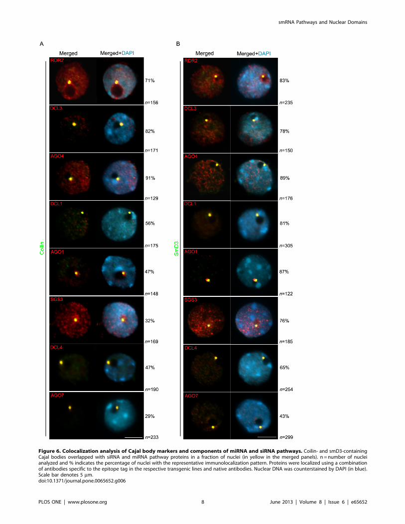
Figure 6. Colocalization analysis of Cajal body markers and components of miRNA and siRNA pathways. Coilin- and smD3-containingCajal bodies overlapped with siRNA and miRNA pathway proteins in a fraction of nuclei (in yellow in the merged panels). n = number of nucleianalyzed and % indicates the percentage of nuclei with the representative immunolocalization pattern. Proteins were localized using a combinationof antibodies specific to the epitope tag in the respective transgenic lines and native antibodies. Nuclear DNA was counterstained by DAPI (in blue).Scale bar denotes 5 mm.doi:10.1371/journal.pone.0065652.g006
smRNA Pathways and Nuclear Domains
PLOS ONE | www.plosone.org 8 June 2013 | Volume 8 | Issue 6 | e65652

the cytoplasm, as previously suggested [50]. Nevertheless, the few
colocalization foci may correspond to sites of dsRNA synthesis.
We hypothesize that the sub-nuclear compartmentalization of
smRNA pathway components might play a role in their gene
silencing activities (Figure 8). It is possible a stepwise process
involving a cytoplasmic and a nuclear phase is required for
smRNA biogenesis. Many of the smRNA components described in
this study were localized in the cytoplasm in previous studies
[50,51,56] including AGO4, which is devoted to the production of
hc-siRNAs that act in chromatin [57]. Under such a model for
instance, the physical proximity of DCL proteins – which seem to
be exclusively localized in the nucleus [13,54,55,58] – to AGOs
within the same nuclear compartment could facilitate RISC
complex assembly/function. DCL-produced smRNAs at the
nucleolar periphery might then be exported to the cytoplasm via
the exportin-5 homolog HASTY for post-transcriptional gene
silencing [59,60] and/or by action of SDE5, which is similar to a
human mRNA export factor TAP. SDE5 is proposed to have a
role in the transport of RNA molecules, targeting precursor or
secondary RNAs to be converted into a double-stranded form by
RDR6/SGS3 in the cytoplasm [61,53]. Other possible compo-
nents involved in the nucleus-cytoplasmic transport of smRNAs or
their precursors could include the THO/TREX RNA trafficking
complex [53,62,63] but its exact role in plant smRNA pathways
remains unknown.
We addressed the question of whether one or more smRNA-
related centers exist by testing the possible association of A. thaliana
DCL, AGO and RDR proteins with one another and with a set of
Cajal body markers. Overall, our data indicate that CBs are a
point of convergence for all the different smRNA biogenesis
pathways, presumably where miRNAs, ta-siRNAs and hc-siRNAs
are generated or stored in close proximity (Figure 8). Cajal bodies
are evolutionary conserved nuclear domains present in yeast,
animal and plants cells [35,36,64,65]. These conspicuous struc-
tures play major roles in a variety of RNA processing and/or
ribonucleoprotein assembly processes and thereby regulate gene
expression as well as assembly and transport of macromolecular
complexes. There are many kinds of Cajal bodies present with a
diverse array of functions. These include involvement in pre-
mRNA splicing, pre-rRNA processing and modification (including
methylation and pseudouridylation), telomerase assembly, viral
trafficking and histone mRNA 39 end formation [66,67,68,69,70].
A common thread to Cajal body function is the assembly of
ribonucleoprotein complexes, which now possibly includes RNA
Induced Silencing Complexes. If CBs are important for localiza-
tion of smRNA-related proteins, we might observe defects of
smRNA biogenesis in the CB mutant background. However, we
did not observe any developmental phenotypes, altered siRNA
accumulation or change in DNA methylation patterns in smd3
mutant plants (Figure S7). A similar result was also previously
described for coilin mutant lines [41,47]. One hypothesis is that CB
integrity is sufficient but not necessary for smRNA biogenesis. It is
possible that CBs are involved in smRNA and/or protein
processing or a yet-to-be identified function that differentially
regulates the multiple endogenous siRNA pathways in A. thaliana.
Nevertheless, as components of the smRNA machinery are not
always colocalized with CBs, our data support the idea that there
are multiple different types of Cajal body-related entities or
smRNA-related nuclear structures and that these share different
subsets of proteins. Consistent with the aforementioned hypothesis,
Figure 7. Effect of Cajal body mutants in the localization of miRNA and siRNA pathway components. Immunofluorescence of severalproteins functioning in siRNA and miRNA pathways using specific native antibodies showed that the loss of function of coilin and smD3 interferedwith their nuclear localization. Representative nuclei for each class are displayed. In the table, n = number of nuclei analyzed and % indicates thepercentage of nuclei with the representative immunolocalization pattern. Nuclear DNA was counterstained by DAPI (in blue). Scale bar denotes 5 mm.doi:10.1371/journal.pone.0065652.g007
smRNA Pathways and Nuclear Domains
PLOS ONE | www.plosone.org 9 June 2013 | Volume 8 | Issue 6 | e65652

a nuclear body, the AB-body, was described as enriched in
particular RdDM components such as AGO4 and DRM2 but
does not colocalize with CB markers [47].
Targeting of siRNAs and miRNAs into Cajal bodies may be an
important mechanism in determining different RNA silencing
outcomes. The colocalization of the miRNA and siRNA pathway
components may partially explain some of the redundancy
observed among pathways, which could generate a backup system
in case of loss of function of any component member. The
association of the RNAi-silencing machinery with CBs suggests
that the latter may be a storage site, as for instance AGO7 is not
functional when targeted to the nucleus [51]. Nevertheless, as
miRNAs, hc-siRNAs and DCL and AGO proteins colocalize to
those nuclear domains [20,31], the possibility that the assembly of
RNAi components and/or smRNA precursor processing occurs in
CB cannot be ruled out.
However, the idea of smRNA processing proteins being
restricted to a unique nuclear compartment may not be entirely
correct, as the colocalization between proteins or with CB markers
was not observed in all analyzed nuclei. In that context, smRNA
pathway proteins were observed colocalizing within a round
nuclear structure that does not colocalize with the CB marker or
were dispersed in the nucleoplasm with variable frequencies.
These observations could indicate that our results are a snapshot
and the lack of complete overlapping localization patterns suggests
that interactions could be transient and/or restricted to an
unknown step of siRNA biogenesis. In that scenario, smRNA
pathway proteins not colocalizing with the CB may correspond to
a non-functional state or different steps of the corresponding
smRNA processing. CBs are known to be highly dynamic in both
number and size, also undergoing fusion and division [71,72]. It is
possible that the assembly of smRNA-related centers is dynamic,
reflecting transient fusion/synthesis between distinct foci that are
not detected by our cytological analysis. This dynamic behavior
could account for the partial colocalization between and within
DCL and AGO family proteins. This points to important, albeit
poorly understood, functions for these nuclear sub-structures.
Future work aimed at the characterization of proteins or even
Figure 8. Multiple endogenous small RNA-directed silencing pathways in A. thaliana plants potentially intersect in a specific nucleardomain corresponding to a CB. The model depicts the scenario where pathways possibly converge in the same CB. A. RNA polymerase IV (Pol IV)transcription provides a single-stranded RNA template to RDR2. RDR2 is localized around the nucleolus and in the CB and acts exclusively in the hc-siRNA pathway. RDR2 activity produces a dsRNA precursor, processed by DCL3 into 24nt siRNA duplexes. B. SGS3 stabilization of a RDR6-deriveddsRNA precursor can occur in the nucleoplasm and/or in the Cajal body. C. Dicer activity hypothetically occurs within the CB nuclear domain, withdifferent DCLs directed to its respective substrates by DRB proteins, like HYL1 and DRB4. Upon dicing, smRNAs are methylated by HEN1 and loadedonto AGO proteins according to their size and 59- end nucleotide. D. Loaded AGO proteins will exit the CB and be assigned to the nucleus orcytoplasm to carry out its function. Traffic to the cytoplasm might involve HASTY and other proteins not yet identified. E. In the cytoplasm, AGO7 hasbeen found associated with RDR6 and SGS3 within a specific compartment related to post-transcriptional gene silencing [50,51]. F. AGO4 localizationto the cytoplasm as part of RISC complex maturation has been reported [57] followed by its return to the nucleus to target epigenetic modifications.doi:10.1371/journal.pone.0065652.g008
smRNA Pathways and Nuclear Domains
PLOS ONE | www.plosone.org 10 June 2013 | Volume 8 | Issue 6 | e65652

RNAs associated with the smRNA compartment will help to
distinguish among the several functional possibilities and give
further insight into the role of Cajal bodies in regulating miRNA
and siRNA pathways.
Materials and Methods
Plant LinesThe mutant lines used in this study were: nrpd1-3 and nrpd2/
nrpd2b [17]; dcl3-1 and rdr2-1 [3]; ago4-1 [73]; ago1-8 [74], ago7
(zip-1; [75]), coilin, SALK 148630 [47]; hen1-1 [76]; and smd3-1
SALK_025193. dcl4-1, sgs2-1 and sgs3, SALK 001394 were kindly
provided by Herve Vaucheret (INRA). All mutant lines were in a
Col-0 background with the exception of ago4-1, ago1-8 and hen1-1
that were in a Ler background.
Production of Arabidopsis Epitope-tagged TransgenicLines
The genes were PCR-amplified from A. thaliana (ecotype Col-0)
genomic DNA and cDNA using Platinum Pfx (Invitrogen) or Pfu
Ultra (Stratgene) polymerases and primers as follows: SmD3
cDNA 59-CACCATGAGTCGGAGTTTGGGGATTCC-39 and
59- CTCCTCACAGGTGGCACAGCCCCTC-39; HEN1 prim-
ers were 59- CACCCGCCTTACCAATCAGAGCCTTAAACC-
39 and 59- AAGATCAGTCTTTTTCTTTTCTA-
CATCTTCTTTCTTCCA-39. The DCL2, RDR6 and SGS3
genomic fragments lacked a stop codon and were amplified with
DCL2-F 59-CACCGATTTGAATCTATG-
GAAGTTTTGGTGTTTTTA-39 and DCL2-R 59-GTAGTC-
CAGGCTGTTCTTAAGTAAGTT-39; RDR6-F 59-
CACCTGTTCTCTGTTGCTACCTTTACTGTG -39 and
RDR6-R 59- GAGACGCTGAGCAAGAAACTTAG-39; and
SGS3-F 59- CACCGAAAAGGCTTTAGTTGTTGGGC-39
and SGS3-R 59- ATCATCTTCATTGTGAAGGCCATG -39.
All the PCR fragments were cloned into pENTR-D TOPO
(Invitrogen) and sequenced. The genes in pENTR vectors were
then recombined into the pEarleyGate 302 or 202 destination
vectors [77] using LR clonase and transformed into each
respective mutant line by the floral dip method [78] using A.
tumefaciens strain GV3101. RDR6 transgenic plants were generated
using Agro strain LBA4404.
Antibody ProductionDCL3 and RDR2 antibodies raised in chicken were described
previously [20]. DCL1 and AGO1 antibodies were raised in
chicken as described in [20] against peptides CIGEPMPSVK-
KAKDS and MVRKRRTDAPSEGGC, respectively. Rabbit
antibodies recognizing RDR6, SGS3, DCL4, and AGO7 (Sigma)
were generated against peptides conjugated to keyhole limpet
hemocyanin (KLH). Peptides were as follows: RDR6-
MGSEGNMKKSVV; SGS3-GPMSKEKNVQGG; DCL4-
RGLPQAPSKTEDR; and AGO7-KHIPSSKSRTPLLHK. Anti-
bodies were affinity purified using peptides immobilized on
SulfoLink Coupling Gel (Pierce).
Immunofluorescence and MicroscopyMesophyll leaf nuclei were isolated as described previously
[20,79]. Upon 4% paraformaldehyde post-fixation, the nuclei
were incubated overnight at 4uC or RT with primary antibodies
raised specifically for proteins involved in A. thaliana miRNA and
siRNA pathways using a dilution of 1:50 or 1:100 mouse anti-Flag
antibodies (Sigma). AGO4 was detected using mAb Myc
(Millipore) in a dilution of 1:200. Secondary antibodies anti-rabbit
or anti-mouse or anti-chicken Alexa 594 (Invitrogen) or Alexa 488
(Invitrogen) were diluted at 1:250 in PBS and incubated for 2 h at
37uC. DNA was counterstained with 1 mg/ml DAPI in Prolong
mounting medium (Invitrogen).
Preparations were inspected with a Nikon Eclipse E800i and
Deltavision (Applied Precision) epifluorescence microscopes
equipped with a Photometrics Coolsnap HQ2 Mono digital
camera. Images were acquired using SoftWorx software and
pseudocolored and merged in Adobe Photoshop 7. Statistical
analysis was performed using Fisher’s exact test and Student’s t test
in nuclei extracted from 3–4 different plants.
Supporting Information
Figure S1 Mutant phenotype complementation assaysof epitope-tagged lines expressing tasiRNA pathwaycomponents driven by its native promoter. A–C. Western
blot of anti-FLAG immunoprecipitated protein fractions from
SGS3-FLAG (A), RDR6-FLAG (B) or DCL2-FLAG (C) trans-
genic lines or non-transgenic, Col-0 wt control plants. Blots were
probed with anti-FLAG-HRP (1:2000) and detected with ECL+chemiluminescent detection reagent (GE Healthcare). The doublet
band observed in the size range of SGS3 (kDa) could be due to
post-translational modifications of the SGS3 protein or a cleavage
event in the SGS3-FLAG protein.
(DOC)
Figure S2 Western blot analysis of native antibodyspecificity. Each antibody specifically recognizes its appropriate
protein. Specificity was confirmed by the absence of band in the
immunoprecipiation of wild-type tissue without the transformation
of the corresponding FLAG-tagged protein. A. RDR6 protein is
137 kDa, B. SGS3 protein is 72 kDa, and C. DCL4 protein is
191 kDa.
(DOC)
Figure S3 Native antibody labeling specificity. Immuno-
staining of interphase nuclei was performed using RDR2, DCL3,
AGO4, DCL1, AGO1, SGS3, DCL4 and AGO7 (all in red) native
antibodies in each of the respective loss-of-function mutants. No
signal was observed, indicating that the antibodies are specific for
those proteins. Nuclear DNA was counterstained by DAPI (in
blue). Scale bar denotes 5 mm.
(DOC)
Figure S4 Rescue of hen1 morphological and small RNAdefects with a genomic HEN1-FLAG construct. A.
Inflorescence images of wild type Ler, hen1-1, and hen1-1
transformed with the genomic HEN1-FLAG construct. Visible
in the third panel is the rescue by the genomic construct of the
mutant inflorescence phenotype. B. Small RNA blot probing for
microRNA167 shows decreased levels in hen1-1 when compared to
wild type. Multiple lines of hen1-1 transformed with the genomic
HEN1-FLAG construct show wild type levels of microRNA167.
(DOC)
Figure S5 Colocalization of A. thaliana RNA dependentRNA polymerases RDR2 and RDR6. Immunofluorescence
analysis indicates that while RDR2 (red) is localized in the
nucleoplasm and at the nucleolar periphery, RDR6 (green) is
mainly nucleoplasmic. RDR6 localization was performed by an
anti-Flag antibody in a RDR6-epitope tagged transgenic line and
RDR2 by making use of a native antibody. Interestingly, the two
RDRs do not colocalize within the nucleus. ‘‘n’’ denotes number
of nuclei analyzed and % indicates the percentage of nuclei with
representative immunolocalization pattern. Nuclear DNA was
counterstained by DAPI (in blue). Scale bar denotes 5 mm.
(DOC)
smRNA Pathways and Nuclear Domains
PLOS ONE | www.plosone.org 11 June 2013 | Volume 8 | Issue 6 | e65652

Figure S6 Protein Immunoprecipitation of FLAG-taggedSmD3 recombinant protein in A. thaliana smd3 mutantbackground. SmD3-flag was immunoprecipitated from total
protein extracts using anti-FLAG antibodies and detected on
immunoblots using FLAG M2 antibody.
(DOC)
Figure S7 Analysis of the smd3-1 mutant line. A. Two-
step RT-PCR was used to evaluate knockout of smd3-1 T-DNA
line. Primers were designed to amplify transcription products
located downstream of the T-DNA insertion but within the ORF.
The absence of PCR product amplification indicates that smd3-1 is
a null allele. Genomic DNA was amplified as a control. B and C.
DNA methylation levels at AtSN1 and 5S rDNA loci are unaffected
in an smd3-1 genomic background. nrpd1 (nrpd1a-3) and nrpd2a/
nrpd2b were used as controls. For the AtSN1 assay, gDNA was
digested with HaeIII (CpNpN) and PCR amplified with primers
specific for AtSN1 or a control gene lacking HaeIII restriction sites
[17]. 5S rDNA methylation analysis was performed by Southern
blot as previously described [17]. D. smd3 knockout does not affect
smRNA accumulation.
(DOC)
Table S1 Interphase localization of siRNA and miRNApathway components.
(DOC)
Methods S1
(DOC)
Author Contributions
Conceived and designed the experiments: OP CSP. Performed the
experiments: OP AV TR EH PC-N. Analyzed the data: OP AV TR EH
PC-N CSP. Contributed reagents/materials/analysis tools: OP AV TR
EH PC-N CSP. Wrote the paper: OP AV TR EH PC-N CSP.
References
1. Jones-Rhoades MW, Bartel DP, Bartel B (2006) MicroRNAS and theirregulatory roles in plants. Annu Rev Plant Biol 57: 19–53.
2. Law JA, Ausin I, Johnson LM, Vashisht AA, Zhu JK, et al. (2010) A protein
complex required for polymerase V transcripts and RNA- directed DNAmethylation in arabidopsis. Curr Biol 20: 951–6.
3. Xie Z, Johansen LK, Gustafson AM, Kasschau KD, Lellis AD, et al. (2004)
Genetic and functional diversification of small RNA pathways in plants. PLoSBiol 2: E104.
4. Xie Z, Grotewold E (2008) Serial chip as a tool to investigate the co-localization
or exclusion of proteins on plant genes. Plant Methods 4: 25.
5. Hiraguri A, Itoh R, Kondo N, Nomura Y, Aizawa D, et al. (2005) Specificinteractions between dicer-like proteins and HYL1/drb-family dsrna-binding
proteins in arabidopsis thaliana. Plant Mol Biol 57: 173–88.
6. Ruiz-Ferrer V, Voinnet O (2019) Roles of plant small rnas in biotic stressresponses. Annu Rev Plant Biol 60: 485–510.
7. Vaucheret H (2008) Plant ARGONAUTES. Trends Plant Sci 13: 350–8.
8. Li J, Yang Z, Yu B, Liu J, Chen X (2005) Methylation protects mirnas and sirnasfrom a 39-end uridylation activity in arabidopsis. Curr Biol 15: 1501–7.
9. Vaucheret H (2005) MicroRNA-dependent trans-acting sirna production. Sci
STKE 300: pe43.
10. Montgomery TA, Howell MD, Cuperus JT, Li D, Hansen JE, et al. (2008)Specificity of argonaute7-mir390 interaction and dual functionality in TAS3
trans-acting sirna formation. Cell 133: 128–41.
11. Peragine A, Yoshikawa M, Wu G, Albrecht HL, Poethig RS (2004) SGS3 andSGS2/SDE1/RDR6 are required for juvenile development and the production
of trans-acting sirnas in arabidopsis. Genes Dev 18: 2368–79.
12. Yoshikawa M, Peragine A, Park MY, Poethig RS (2005) A pathway for thebiogenesis of trans-acting sirnas in arabidopsis. Genes Dev 19: 2164–75.
13. Elmayan T, Adenot X, Gissot L, Lauressergues D, Gy I, et al. (2009) A
neomorphic sgs3 allele stabilizing mirna cleavage products reveals that SGS3acts as a homodimer. FEBS J 276: 835–44.
14. Borsani O, Zhu J, Verslues PE, Sunkar R, Zhu JK (2005) Endogenous sirnas
derived from a pair of natural cis-antisense transcripts regulate salt tolerance inarabidopsis. Cell 123: 1279–91.
15. Katiyar-Agarwal S, Morgan R, Dahlbeck D, Borsani O, et al. (2006) A
pathogen-inducible endogenous sirna in plant immunity. Proc Natl AcadSci U S A 103: 18002–7.
16. Matzke M, Kanno T, Daxinger L, Huettel B, Matzke AJ (2009) RNA-mediated
chromatin-based silencing in plants. Curr Opin Cell Biol 21: 367–76.
17. Onodera Y, Haag JR, Ream T, Nunes PC, Pontes O, et al. (2005) Plant nuclear
RNA polymerase IV mediates sirna and DNA methylation-dependent
heterochromatin formation. Cell 120: 613–22.
18. Herr AJ, Jensen MB, Dalmay T, Baulcombe DC (2005) RNA polymerase IV
directs silencing of endogenous DNA. Science 308: 118–20.
19. Smith LM, Pontes O, Searle I, Yelina N, Yousafzai FK, et al. (2007) An SNF2protein associated with nuclear RNA silencing and the spread of a silencing
signal between cells in arabidopsis. Plant Cell 19: 1507–21.
20. Pontes O, Li CF, Nunes PC, Haag J, Ream T, et al. (2006) The Arabidopsischromatin-modifying nuclear sirna pathway involves a nucleolar RNA
processing center. Cell 126: 79–92.
21. Bies-Etheve N, Pontier D, Lahmy S, Picart C, Vega D, et al. (2009) RNA-directed DNA methylation requires an ago4-interacting member of the SPT5
elongation factor family. EMBO Rep 10: 649–54.
22. Wierzbicki AT, Ream TS, Haag JR, Pikaard CS (2009) RNA polymerase Vtranscription guides ARGONAUTE4 to chromatin. Nat Genet 41: 630–4.
23. Gao Z, Liu HL, Daxinger L, Pontes O, He X, et al. (2010) An RNA polymerase
II- and ago4-associated protein acts in rna-directed DNA methylation. Nature465: 106–9.
24. Zheng B, Wang Z, Li S, Yu B, Liu JY, et al. (2009) Intergenic transcription by
RNA polymerase II coordinates pol IV and pol V in sirna-directed
transcriptional gene silencing in arabidopsis. Genes Dev 23: 2850–60.
25. He XJ, Hsu YF, Pontes O, Zhu J, Lu J, et al. (2009) NRPD4, a protein related to
the RPB4 subunit of RNA polymerase II, is a component of RNA polymerases
IV and V and is required for rna-directed DNA methylation. Genes Dev 23:
318–30.
26. Kanno T, Mette MF, Kreil DP, Aufsatz W, Matzke M, et al. (2004) Involvement
of putative SNF2 chromatin remodeling protein DRD1 in rna-directed DNA
methylation. Curr Biol 14: 801–5.
27. Wierzbicki AT, Haag JR, Pikaard CS (2008) Noncoding transcription by RNA
polymerase pol ivb/pol V mediates transcriptional silencing of overlapping and
adjacent genes. Cell 135: 635–48.
28. Kanno T, Bucher E, Daxinger L, Huettel B, Bohmdorfer G, et al. (2008) A
structural-maintenance-of-chromosomes hinge domain-containing protein is
required for rna-directed DNA methylation. Nat Genet 40: 670–5.
29. Mi S, Cai T, Hu Y, Chen Y, Hodges E, et al. (2008) Sorting of small rnas into
arabidopsis argonaute complexes is directed by the 59 terminal nucleotide. Cell
133: 116–27.
30. Takeda A, Iwasaki S, Watanabe T, Utsumi M, Watanabe Y (2008) The
mechanism selecting the guide strand from small RNA duplexes is different
among argonaute proteins. Plant Cell Physiol 49: 493–500.
31. Fang Y, Spector DL (2007) Identification of nuclear dicing bodies containing
proteins for microrna biogenesis in living arabidopsis plants. Curr Biol 17: 818–
23.
32. Li CF, Pontes O, El-Shami M, Henderson IR, Bernatavichute YV, et al. (2006)
An argonaute4-containing nuclear processing center colocalized with cajal
bodies in arabidopsis thaliana. Cell 126: 93–106.
33. Pontes O, Pikaard CS (2008) SiRNA and mirna processing: New functions for
cajal bodies. Curr Opin Genet Dev 18: 197–203.
34. Ochs RL, Stein TW, Tan EM (1994) Coiled bodies in the nucleolus of breast
cancer cells. J Cell Sci 107: 385–99.
35. Gall JG (2000) Cajal bodies: The first 100 years. Annu Rev Cell Dev Biol 16:
273–300.
36. Verheggen C, Lafontaine DL, Samarsky D, Mouaikel J, Blanchard JM, et al.
(2002) Mammalian and yeast U3 snornps are matured in specific and related
nuclear compartments. EMBO J 21: 2736–45.
37. Matera AG (1998) Of coiled bodies, gems, and salmon. J Cell Biochem 70: 181–
92.
38. Zhu Y, Tomlinson RL, Lukowiak AA, Terns RM, Terns MP (2004) Telomerase
RNA accumulates in cajal bodies in human cancer cells. Mol Biol Cell 15: 81–
90.
39. Fujioka Y, Utsumi M, Ohba Y, Watanabe Y (2007) Location of a possible mirna
processing site in smd3/smb nuclear bodies in arabidopsis. Plant Cell Physiol 48:
1243–53.
40. Yu B, Yang Z, Li J, Minakhina S, Yang M, et al. (2005) Methylation as a crucial
step in plant microrna biogenesis. Science 307: 932–5.
41. Collier S, Pendle A, Boudonck K, van Rij T, Dolan L, et al. (2006) A distant
coilin homologue is required for the formation of cajal bodies in arabidopsis.
Mol Biol Cell 17: 2942–51.
42. Beven AF, Simpson GG, Brown JW, Shaw PJ (1995) The organization of
spliceosomal components in the nuclei of higher plants. J Cell Sci 108: 509–18.
43. Boudonck K, Dolan L, Shaw PJ (1999) The movement of coiled bodies
visualized in living plant cells by the green fluorescent protein. Mol Biol Cell 10:
2297–307.
44. Brahms H, Raymackers J, Union A, de Keyser F, Meheus L, et al. (2000) The c-
terminal RG dipeptide repeats of the spliceosomal sm proteins D1 and D3
smRNA Pathways and Nuclear Domains
PLOS ONE | www.plosone.org 12 June 2013 | Volume 8 | Issue 6 | e65652
contain symmetrical dimethylarginines, which form a major b-cell epitope for
anti-sm autoantibodies. J Biol Chem 275: 17122–9.
45. Scofield DG, Lynch M (2008) Evolutionary diversification of the sm family of
rna-associated proteins. Mol Biol Evol 25: 2255–67.
46. Bayne EH, Portoso M, Kagansky A, Kos-Braun IC, Urano T, et al. (2008)
Splicing factors facilitate rnai-directed silencing in fission yeast. Science 322:
602–6.
47. Li CF, Henderson IR, Song L, Fedoroff N, Lagrange T, et al. (2008) Dynamic
regulation of ARGONAUTE4 within multiple nuclear bodies in arabidopsis
thaliana. PLoS Genet 4: e27.
48. Yang Z, Ebright YW, Yu B, Chen X (2006) HEN1 recognizes 21–24 nt small
RNA duplexes and deposits a methyl group onto the 29 OH of the 39 terminal
nucleotide. Nucleic Acids Res 34: 667–75.
49. Qi Y, He X, Wang XJ, Kohany O, Jurka J, et al. (2006) Distinct catalytic and
non-catalytic roles of ARGONAUTE4 in rna-directed DNA methylation.
Nature 443: 1008–12.
50. Kumakura N, Takeda A, Fujioka Y, Motose H, Takano R, et al. (2009) SGS3
and RDR6 interact and colocalize in cytoplasmic SGS3/rdr6-bodies. FEBS Lett
583: 1261–6.
51. Jouannet V, Moreno AB, Elmayan T, Vaucheret H, Crespi MD, et al. (2012)
Cytoplasmic arabidopsis AGO7 accumulates in membrane-associated sirna
bodies and is required for ta-sirna biogenesis. EMBO J 31: 1704–13.
52. Dunoyer P, Schott G, Himber C, Meyer D, Takeda A, et al. (2010) Small RNA
duplexes function as mobile silencing signals between plant cells. Science 328:
912–6.
53. Jauvion V, Elmayan T, Vaucheret H (2010) The conserved RNA trafficking
proteins HPR1 and TEX1 are involved in the production of endogenous and
exogenous small interfering RNA in arabidopsis. Plant Cell 22: 2697–709.
54. Hoffer P, Ivashuta S, Pontes O, Vitins A, Pikaard C, et al. (2011)
Posttranscriptional gene silencing in nuclei. Proc Natl Acad Sci U S A 108:
409–14.
55. Luo Z, Chen Z (2007) Improperly terminated, unpolyadenylated mrna of sense
transgenes is targeted by rdr6-mediated RNA silencing in arabidopsis. Plant Cell
19: 943–58.
56. Schott G, Mari-Ordonez A, Himber C, Alioua A, Voinnet O, et al. (2012)
Differential effects of viral silencing suppressors on sirna and mirna loading
support the existence of two distinct cellular pools of ARGONAUTE1. EMBO J
31: 2553–65.
57. Ye R, Wang W, Iki T, Liu C, Wu Y, et al. (2012) Cytoplasmic assembly and
selective nuclear import of arabidopsis ARGONAUTE4/sirna complexes. Mol
Cell 46: 859–70.
58. Song L, Han MH, Lesicka J, Fedoroff N (2007) Arabidopsis primary microrna
processing proteins HYL1 and DCL1 define a nuclear body distinct from the
cajal body. Proc Natl Acad Sci U S A 104: 5437–42.
59. Bollman KM, Aukerman MJ, Park MY, Hunter C, Berardini TZ, et al. (2003)
HASTY, the arabidopsis ortholog of exportin 5/MSN5, regulates phase change
and morphogenesis. Development 130: 1493–504.
60. Park MY, Wu G, Gonzalez-Sulser A, Vaucheret H, Poethig RS (2005) Nuclear
processing and export of micrornas in arabidopsis. Proc Natl Acad Sci U S A
102: 3691–6.
61. Hernandez-Pinzon I, Yelina NE, Schwach F, Studholme DJ, Baulcombe D, et
al. (2007) SDE5, the putative homologue of a human mrna export factor, isrequired for transgene silencing and accumulation of trans-acting endogenous
sirna. Plant J 50: 140–8.
62. Yelina NE, Smith LM, Jones AM, Patel K, Kelly KA, et al. (2010) Putativearabidopsis THO/TREX mrna export complex is involved in transgene and
endogenous sirna biosynthesis. Proc Natl Acad Sci U S A 107: 13948–53.63. Kota P, Wagner R, Huerta E, Underwood M, Nickerson A (2008) Binding of
ATP to UAP56 is necessary for mrna export. J Cell Sci 121: 1526–37.
64. Matera AG (1998) Of coiled bodies, gems, and salmon. J Cell Biochem 70: 181–92.
65. Ogg SC, Lamond AI (2002) Cajal bodies and coilin–moving towards function.J Cell Biol 159: 17–21.
66. Cioce M, Lamond AI (2005) Cajal bodies: A long history of discovery. Annu RevCell Dev Biol 21: 105–31.
67. Kiss T, Fayet E, Jady BE, Richard P, Weber M (2006) Biogenesis and
intranuclear trafficking of human box C/D and H/ACA rnps. Cold SpringHarb Symp Quant Biol 71: 407–17.
68. Lukowiak AA, Narayanan A, Li ZH, Terns RM, Terns MP (2001) The snornadomain of vertebrate telomerase RNA functions to localize the RNA within the
nucleus. RNA 7: 1833–44.
69. Shaw PJ, Brown JW (2004) Plant nuclear bodies. Curr Opin Plant Biol 7: 614–20.
70. Stanek D, Neugebauer KM (2006) The cajal body: A meeting place forspliceosomal snrnps in the nuclear maze. Chromosoma 115: 343–54.
71. Sleeman JE, Trinkle-Mulcahy L, Prescott AR, Ogg SC, Lamond AI (2003) Cajalbody proteins SMN and coilin show differential dynamic behaviour in vivo.
J Cell Sci 116: 2039–50.
72. Stanek D, Pridalova-Hnilicova J, Novotny I, Huranova M, Blazıkova M, et al(2008) Spliceosomal small nuclear ribonucleoprotein particles repeatedly cycle
through cajal bodies. Mol Biol Cell 19: 2534–43.73. Zilberman D, Cao X, Jacobsen SE (2003) ARGONAUTE4 control of locus-
specific sirna accumulation and DNA and histone methylation. Science 299:
716.74. Lynn K, Fernandez A, Aida M, Sedbrook J, Tasaka M, et al. (1999) The
PINHEAD/ZWILLE gene acts pleiotropically in arabidopsis development andhas overlapping functions with the ARGONAUTE1 gene. Development 126:
469–81.75. Hunter C, Sun H, Poethig RS (2003) The arabidopsis heterochronic gene
ZIPPY is an ARGONAUTE family member. Curr Biol 13: 1734–9.
76. Chen X, Liu J, Cheng Y, Jia D (2002) HEN1 functions pleiotropically inarabidopsis development and acts in C function in the flower. Development 129:
1085–94.77. Earley K, Lawrence RJ, Pontes O, Reuther R, Enciso AJ, et al. (2006) Erasure of
histone acetylation by arabidopsis HDA6 mediates large-scale gene silencing in
nucleolar dominance. Genes Dev 20: 1283–93.78. Clough SJ, Bent AF (1998) Floral dip: A simplified method for agrobacterium-
mediated transformation of arabidopsis thaliana. Plant J 16: 735–43.79. Pontes O, Lawrence RJ, Silva M, Preuss S, Costa-Nunes P, et al. (2007)
Postembryonic establishment of megabase-scale gene silencing in nucleolardominance. PLoS One 2: e1157.
smRNA Pathways and Nuclear Domains
PLOS ONE | www.plosone.org 13 June 2013 | Volume 8 | Issue 6 | e65652
Copyright © 2022 FDOKUMEN