Comparison of nonviral transfection and adeno-associated viral transduction on cardiomyocytes
11
MOLECULAR BIOTECHNOLOGY Volume 28, 2004 AAV Transduction vs Nonviral Techniques 21 RESEARCH 21 Molecular Biotechnology © 2004 Humana Press Inc. All rights of any nature whatsoever reserved. 1073–6085/2004/28:1/21–31/$25.00 Abstract *Author to whom all correspondence and reprint requests should be addressed: 1 Department/Institute of Medical Genetics and 2 Department of Microbiology, Ullevaal University Hospital, Kirkeveien 166, N-0407 Oslo, Norway. E-mail: [email protected]; 3 Department of Oncology, Gene Therapy Program, Haukeland University Hospital, Bergen, Norway; 4 Institute for Experimental Medical Research and 5 Center for Heart Failure Research, Ullevaal University Hospital, Oslo, Norway. Comparison of Nonviral Transfection and Adeno-Associated Viral Transduction on Cardiomyocytes Srdjan Djurovic,* ,1 Nina Iversen, 1 Stig Jeansson, 2 Frank Hoover, 3 and Geir Christensen 4,5 Cardiomyocytes are terminally differentiated cells that to date have been characterized as poor targets for nonviral gene transfer. This study was therefore designed to determine the optimal nonviral gene transfer parameters in cell cultures of neonatal rat cardiomyocytes and to compare them with the efficiency of gene transfer using adeno-associated viral vectors (AAV). Transfection efficiency was measured by quantitative chloramphenicol acetyltransferase type I (CAT)–enzyme-linked immunosorbent assay and β-galactosidase staining, based on overexpression of reporter genes (CAT and LacZ). The efficiency of CAT/LacZ overexpression was assessed using the following techniques: (1) liposomal reagents, such as: FuGENE 6, LipofectAMINE 2000, LipofectAMINE PLUS, GenePORTER, Metafectene, and LipoGen; (2) electroporation and nucleofector techniques; and (3) an AAV2 vector harboring a lacZ reporter gene. Toxicity was monitored by total protein measurement and by analyzing cell metabolism. On average, Lipofectamine 2000 was the most effective nonviral method examined yielding consistently high transfection rates (8.1% β-galactosidase-positive cells) combined with low toxicity. Electroporation also resulted in high transfection values (7.5%); however, cellular toxicity was higher than that of Lipofectamine 2000. Finally, transduction with AAV2 vectors provided the highest levels of transduction (88.1%) with no cellular toxicity. We conclude that although transduction with AAV is more efficient (88.1%), transfections with nonviral techniques, when optimized, may provide a useful alternative for overexpression of therapeutic genes in neonatal cardiomyocytes. Index Entries: Liposomes; lipofection; electroporation; gene therapy; nucleofection; adeno-associated virus. 1. Introduction Although several methods have been described to introduce deoxyribonucleic acid (DNA) ex- pression vectors into mammalian cells in vitro and in vivo (1,2), the transfection of cardiomyocytes in primary culture has remained an obstacle using conventional chemical techniques (3–7): Trans- fection rates of 0–15% with lipofection and 5–8% with calcium phosphate precipitation have been reported. The use of DNA–liposome complexes (lipo- fection) has become a popular method of deliver- ing genes and is being tested in various clinical trials. Cationic lipids condense DNA through ionic interaction and thus form cationic lipoplexes (1,7). The resulting complexes fuse with the anionic surfaces of cells, delivering DNA into the cells via endocytosis. The major advantage of cationic lipoplexes is simplicity of production, low toxic- ity, and immunogenicity. However, the data on liposome-mediated gene transfer are not clear and consistent, and obviously more optimization would be necessary to obtain maximum transfection. Another transfection method, electroporation (8), also termed electrotransfer (9) or electropermea- bilization (10), is an experimental technique using
-
Upload
independent -
Category
Documents
-
view
5 -
download
0
Transcript of Comparison of nonviral transfection and adeno-associated viral transduction on cardiomyocytes

MOLECULAR BIOTECHNOLOGY Volume 28, 2004
AAV Transduction vs Nonviral Techniques 21RESEARCH
21
Molecular Biotechnology © 2004 Humana Press Inc. All rights of any nature whatsoever reserved. 1073–6085/2004/28:1/21–31/$25.00
Abstract
*Author to whom all correspondence and reprint requests should be addressed: 1Department/Institute of Medical Genetics and 2Department ofMicrobiology, Ullevaal University Hospital, Kirkeveien 166, N-0407 Oslo, Norway. E-mail: [email protected]; 3Departmentof Oncology, Gene Therapy Program, Haukeland University Hospital, Bergen, Norway; 4Institute for Experimental Medical Research and5Center for Heart Failure Research, Ullevaal University Hospital, Oslo, Norway.
Comparison of Nonviral Transfectionand Adeno-Associated Viral Transduction on Cardiomyocytes
Srdjan Djurovic,*,1 Nina Iversen,1 Stig Jeansson,2
Frank Hoover,3 and Geir Christensen4,5
Cardiomyocytes are terminally differentiated cells that to date have been characterized as poor targets fornonviral gene transfer. This study was therefore designed to determine the optimal nonviral gene transferparameters in cell cultures of neonatal rat cardiomyocytes and to compare them with the efficiency of genetransfer using adeno-associated viral vectors (AAV). Transfection efficiency was measured by quantitativechloramphenicol acetyltransferase type I (CAT)–enzyme-linked immunosorbent assay and β-galactosidasestaining, based on overexpression of reporter genes (CAT and LacZ).
The efficiency of CAT/LacZ overexpression was assessed using the following techniques: (1) liposomalreagents, such as: FuGENE 6, LipofectAMINE 2000, LipofectAMINE PLUS, GenePORTER, Metafectene,and LipoGen; (2) electroporation and nucleofector techniques; and (3) an AAV2 vector harboring a lacZreporter gene. Toxicity was monitored by total protein measurement and by analyzing cell metabolism.
On average, Lipofectamine 2000 was the most effective nonviral method examined yielding consistentlyhigh transfection rates (8.1% β-galactosidase-positive cells) combined with low toxicity. Electroporationalso resulted in high transfection values (7.5%); however, cellular toxicity was higher than that ofLipofectamine 2000. Finally, transduction with AAV2 vectors provided the highest levels of transduction(88.1%) with no cellular toxicity.
We conclude that although transduction with AAV is more efficient (88.1%), transfections with nonviraltechniques, when optimized, may provide a useful alternative for overexpression of therapeutic genes inneonatal cardiomyocytes.
Index Entries: Liposomes; lipofection; electroporation; gene therapy; nucleofection; adeno-associated virus.
1. IntroductionAlthough several methods have been described
to introduce deoxyribonucleic acid (DNA) ex-pression vectors into mammalian cells in vitro andin vivo (1,2), the transfection of cardiomyocytesin primary culture has remained an obstacle usingconventional chemical techniques (3–7): Trans-fection rates of 0–15% with lipofection and 5–8%with calcium phosphate precipitation have beenreported.
The use of DNA–liposome complexes (lipo-fection) has become a popular method of deliver-ing genes and is being tested in various clinical
trials. Cationic lipids condense DNA through ionicinteraction and thus form cationic lipoplexes (1,7).The resulting complexes fuse with the anionicsurfaces of cells, delivering DNA into the cellsvia endocytosis. The major advantage of cationiclipoplexes is simplicity of production, low toxic-ity, and immunogenicity. However, the data onliposome-mediated gene transfer are not clear andconsistent, and obviously more optimization wouldbe necessary to obtain maximum transfection.
Another transfection method, electroporation (8),also termed electrotransfer (9) or electropermea-bilization (10), is an experimental technique using
03/JW685/Djurovic/21-32 8/23/04, 1:53 PM21
MOLECULAR BIOTECHNOLOGY Volume 28, 2004
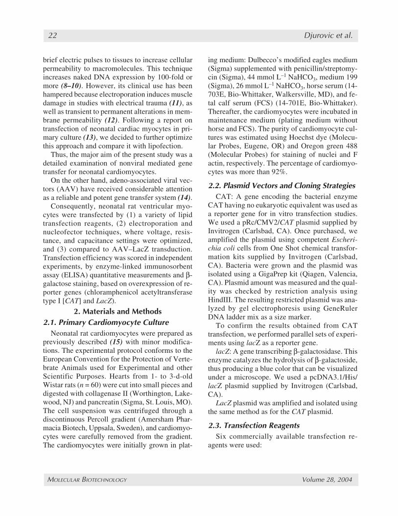
22 Djurovic et al.
brief electric pulses to tissues to increase cellularpermeability to macromolecules. This techniqueincreases naked DNA expression by 100-fold ormore (8–10). However, its clinical use has beenhampered because electroporation induces muscledamage in studies with electrical trauma (11), aswell as transient to permanent alterations in mem-brane permeability (12). Following a report ontransfection of neonatal cardiac myocytes in pri-mary culture (13), we decided to further optimizethis approach and compare it with lipofection.
Thus, the major aim of the present study was adetailed examination of nonviral mediated genetransfer for neonatal cardiomyocytes.
On the other hand, adeno-associated viral vec-tors (AAV) have received considerable attentionas a reliable and potent gene transfer system (14).
Consequently, neonatal rat ventricular myo-cytes were transfected by (1) a variety of lipidtransfection reagents, (2) electroporation andnucleofector techniques, where voltage, resis-tance, and capacitance settings were optimized,and (3) compared to AAV–LacZ transduction.Transfection efficiency was scored in independentexperiments, by enzyme-linked immunosorbentassay (ELISA) quantitative measurements and β-galactose staining, based on overexpression of re-porter genes (chloramphenicol acetyltransferasetype I [CAT] and LacZ).
2. Materials and Methods2.1. Primary Cardiomyocyte Culture
Neonatal rat cardiomyocytes were prepared aspreviously described (15) with minor modifica-tions. The experimental protocol conforms to theEuropean Convention for the Protection of Verte-brate Animals used for Experimental and otherScientific Purposes. Hearts from 1- to 3-d-oldWistar rats (n = 60) were cut into small pieces anddigested with collagenase II (Worthington, Lake-wood, NJ) and pancreatin (Sigma, St. Louis, MO).The cell suspension was centrifuged through adiscontinuous Percoll gradient (Amersham Phar-macia Biotech, Uppsala, Sweden), and cardiomyo-cytes were carefully removed from the gradient.The cardiomyocytes were initially grown in plat-
ing medium: Dulbecco’s modified eagles medium(Sigma) supplemented with penicillin/streptomy-cin (Sigma), 44 mmol L–1 NaHCO3, medium 199(Sigma), 26 mmol L–1 NaHCO3, horse serum (14-703E, Bio-Whittaker, Walkersville, MD), and fe-tal calf serum (FCS) (14-701E, Bio-Whittaker).Thereafter, the cardiomyocytes were incubated inmaintenance medium (plating medium withouthorse and FCS). The purity of cardiomyocyte cul-tures was estimated using Hoechst dye (Molecu-lar Probes, Eugene, OR) and Oregon green 488(Molecular Probes) for staining of nuclei and Factin, respectively. The percentage of cardiomyo-cytes was more than 92%.
2.2. Plasmid Vectors and Cloning StrategiesCAT: A gene encoding the bacterial enzyme
CAT having no eukaryotic equivalent was used asa reporter gene for in vitro transfection studies.We used a pRc/CMV2/CAT plasmid supplied byInvitrogen (Carlsbad, CA). Once purchased, weamplified the plasmid using competent Escheri-chia coli cells from One Shot chemical transfor-mation kits supplied by Invitrogen (Carlsbad,CA). Bacteria were grown and the plasmid wasisolated using a GigaPrep kit (Qiagen, Valencia,CA). Plasmid amount was measured and the qual-ity was checked by restriction analysis usingHindIII. The resulting restricted plasmid was ana-lyzed by gel electrophoresis using GeneRulerDNA ladder mix as a size marker.
To confirm the results obtained from CATtransfection, we performed parallel sets of experi-ments using lacZ as a reporter gene.
lacZ: A gene transcribing β-galactosidase. Thisenzyme catalyzes the hydrolysis of β-galactoside,thus producing a blue color that can be visualizedunder a microscope. We used a pcDNA3.1/His/lacZ plasmid supplied by Invitrogen (Carlsbad,CA).
LacZ plasmid was amplified and isolated usingthe same method as for the CAT plasmid.
2.3. Transfection ReagentsSix commercially available transfection re-
agents were used:
03/JW685/Djurovic/21-32 8/23/04, 1:53 PM22
MOLECULAR BIOTECHNOLOGY Volume 28, 2004
AAV Transduction vs Nonviral Techniques 23
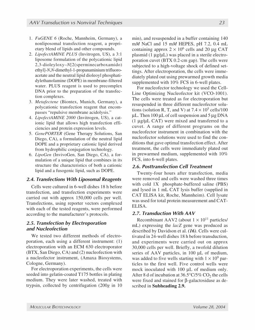
1. FuGENE 6 (Roche, Mannheim, Germany), anonliposomal transfection reagent, a propri-etary blend of lipids and other compounds.
2. LipofectAMINE PLUS (Invitrogen, US), a 3:1liposome formulation of the polycationic lipid2,3-dioleyloxy–N[2(sperminecarboxamido)ethyl]-N,N-dimethyl-1-propanaminium trifluoro-acetate and the neutral lipid dioleoyl phosphati-dylethanolamine (DOPE) in membrane-filteredwater. PLUS reagent is used to precomplexDNA prior to the preparation of the transfec-tion complexes.
3. Metafectene (Biontex, Munich, Germany), apolycationic transfection reagent that encom-passes “repulsive membrane acidolysis.”
4. LipofectAMINE 2000 (Invitrogen, US), a cat-ionic lipid that allows high transfection effi-ciencies and protein expression levels.
5. GenePORTER (Gene Therapy Solutions, SanDiego, CA), a formulation of the neutral lipidDOPE and a proprietary cationic lipid derivedfrom hydrophilic conjugation technology.
6. LipoGen (InvivoGen, San Diego, CA), a for-mulation of a unique lipid that combines in itsstructure the characteristics of both a cationiclipid and a fusogenic lipid, such as DOPE.
2.4. Transfections With Liposomal ReagentsCells were cultured in 6-well dishes 18 h before
transfection, and transfection experiments werecarried out with approx 150,000 cells per well.Transfections, using reporter vectors complexedwith each of the tested reagents, were performedaccording to the manufacturer’s protocols.
2.5. Transfection by Electroporationand Nucleofection
We tested two different methods of electro-poration, each using a different instrument: (1)electroporation with an ECM 630 electroporator(BTX, San Diego, CA) and (2) nucleofection witha nucleofector instrument, (Amaxa Biosystems,Cologne, Germany).
For electroporation experiments, the cells wereseeded into gelatin-coated T175 bottles in platingmedium. They were later washed, treated withtrypsin, collected by centrifugation (200g in 10
min), and resuspended in a buffer containing 140mM NaCl and 15 mM HEPES, pH 7.2, 0.4 mLcontaining approx 2 × 10⁶ cells and 20 µg CATplasmid (1 µg/µL) was placed in a sterile electro-poration cuvet (BTX 0.2-cm gap). The cells weresubjected to a high-voltage shock of defined set-tings. After electroporation, the cells were imme-diately plated out using prewarmed growth mediasupplemented with 10% FCS in 6-well plates.
For nucleofector technology we used the Cell-Line Optimizing Nucleofector kit (VCO-1001).The cells were treated as for electroporation butresuspended in three different nucleofector solu-tions (solution R, T, and V) at 7.4 × 10⁵ cells/100µL. Then 100 µL of cell suspension and 5 µg DNA(1 µg/µL CAT) were mixed and transferred to acuvet. A range of different programs on thenucleofector instrument in combination with thenucleofector solutions were used to find the con-ditions that gave optimal transfection effect. Aftertreatment, the cells were immediately plated outin prewarmed medium, supplemented with 10%FCS, into 6-well plates.
2.6. Posttransfection Cell TreatmentTwenty-four hours after transfection, media
were removed and cells were washed three timeswith cold 1X phosphate-buffered saline (PBS)and lysed in 1 mL CAT lysis buffer (supplied inCAT ELISA kit, Roche, Mannheim). Cell lysatewas used for total protein measurement and CAT-ELISA.
2.7. Transduction With AAVRecombinant AAV2 (about 1 × 10¹¹ particles/
mL) expressing the lacZ gene was produced asdescribed by Davidson et al. (16). Cells were cul-tivated in 24-well dishes 18 h before transduction,and experiments were carried out on approx30,000 cells per well. Briefly, a twofold dilutionseries of AAV particles, in 100 µL of medium,was added to five wells starting with 1 × 10⁹ par-ticles to the first well. Five control wells weremock inoculated with 100 µL of medium only.After 8 d of incubation at 36.5°C/5% CO2 the cellswere fixed and stained for β-galactosidase as de-scribed in Subheading 2.9.
03/JW685/Djurovic/21-32 8/23/04, 1:53 PM23

MOLECULAR BIOTECHNOLOGY Volume 28, 2004
24 Djurovic et al.
2.8. CAT–ELISA MeasurementsConcentrations of CAT in cell lysate were mea-
sured by CAT–ELISA (Roche, Mannheim) as rec-ommended by the producer. All measurementswere done in duplicate. Concentrations of un-known samples were calculated from a standardcurve run with each plate.
2.9. β-Galactosidase StainingCells were fixed and stained using a β-galac-
tosidase staining kit (Invitrogen, Carlsbad, CA).Any cells containing β-galactosidase stained blue.After incubation, cells were counted and a trans-duction percentage was described by comparingthe number of blue cells to the number of un-stained cells.
2.10. Cell Survival CalculationsTo evaluate possible detrimental effects of each
transfection method, cellular total protein wasmeasured for the test and control samples. Whencomparing these, it was assumed that cells in thecontrol well were unaffected by the experiment.Test results were then compared to the control re-sults and a percentage survival was calculated.These measurements where then confirmed usinga cytotoxicity detection kit (Roche, Mannheim),which measures lactate dehydrogenase activityreleased from damaged cells (results not shown).
2.11. Reporting of ResultsThe experiments were performed in duplicates
or triplicates and were repeated twice. All resultsare expressed as mean ±SEM. To effectively com-pare the results found in the different nonviralmethods used, results were standardized. This wasneeded because the different methods tested usedvarying numbers of cells. The transfection re-agents required 150,000 cells per well, but electro-poration and the nucleofector used between 1 and2 million cells per well. To standardize, we used aratio of CAT produced (in ng) divided by totalprotein of surviving cells (in ng). This essentiallygives us the amount of CAT produced per livingcell. This value was then multiplied by 1 × 10⁶ tomake the numbers more manageable.
3. Results3.1. Optimization of Lipofectionin Cardiomyocyte Cultures
Primarily, DNA/liposome ratios used werethose recommended by the manufacturers. Experi-ments were designed using two different amountsof plasmid, but ratios of DNA and lipoformulationwere held constant. The aim of this procedure wasto couple the most effective amount of plasmidwith the best liposomes for neonatal cardiomyo-cytes gene delivery. Amounts of plasmid wereascertained from those recommended by the manu-facturers. Results are presented in Table 1.
After the initial experiments, we set about opti-mizing the transfection efficiency of the two bestperforming transfection reagents. Table 2 showsdifferent DNA amounts, DNA:liposome ratiostested, and their corresponding results. Regardingtransfection efficiency, LipofectAMINE 2000gave the best results. It achieved a 4.3- and 9.4-fold higher CAT activity compared with the sec-ond best performing liposome, FuGENE-6, aswell as a 5–10% lower cytotoxicity value.
These findings were later confirmed with β-ga-lactosidase staining (Table 2).
3.2. Optimization of Electroporation inCardiomyocyte Cultures
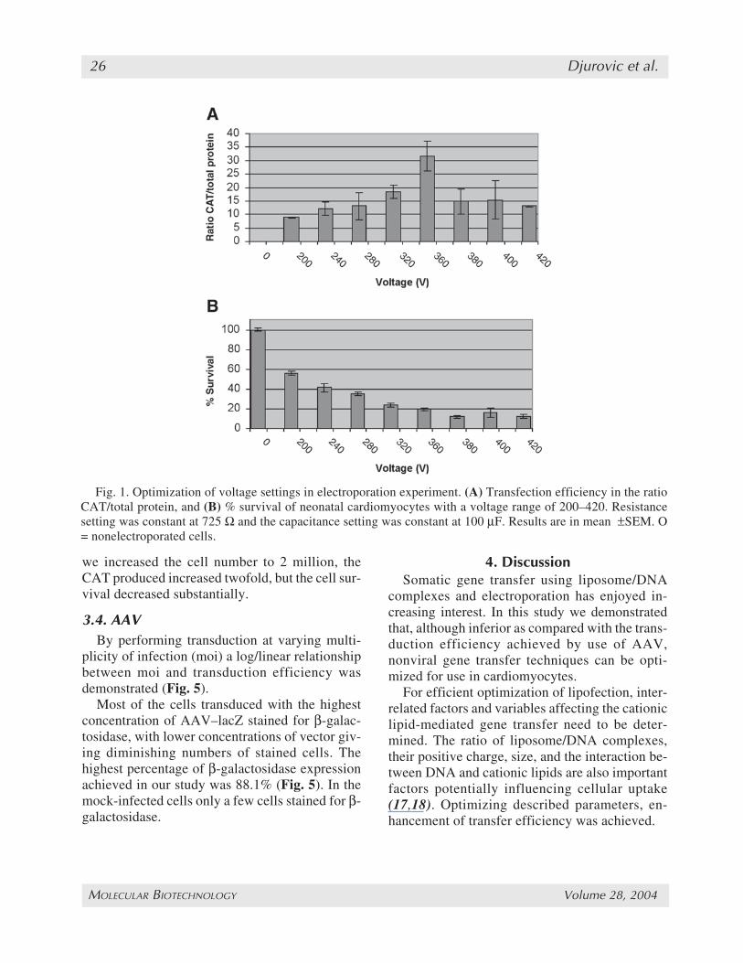
To optimize the electroporation procedure, arange of voltage, capacitance, and resistance set-tings were investigated. The initial resistance andcapacitance were set at 725 Ω and 50–100 µF tofind the optimal voltage, and the voltage was var-ied from 200 to 420 V. From these experimentsthe optimal voltage was found be 360 V (Fig. 1).After the voltage settings had been established, theoptimal resistance and capacitance were found tobe 725 Ω and 50 µF, respectively (Figs. 2 and 3).Optimization of the resistance showed that weachieved a much better transfection efficiency at725 Ω compared to 450 Ω and slightly but not sig-nificantly better than 1000 Ω based on the ratioCAT/total protein (Fig. 2A). The cell survival wasnot significantly influenced by the resistance (Fig.2B). The transfection efficiency was only slightly
03/JW685/Djurovic/21-32 8/23/04, 1:53 PM24
MOLECULAR BIOTECHNOLOGY Volume 28, 2004
AAV Transduction vs Nonviral Techniques 25
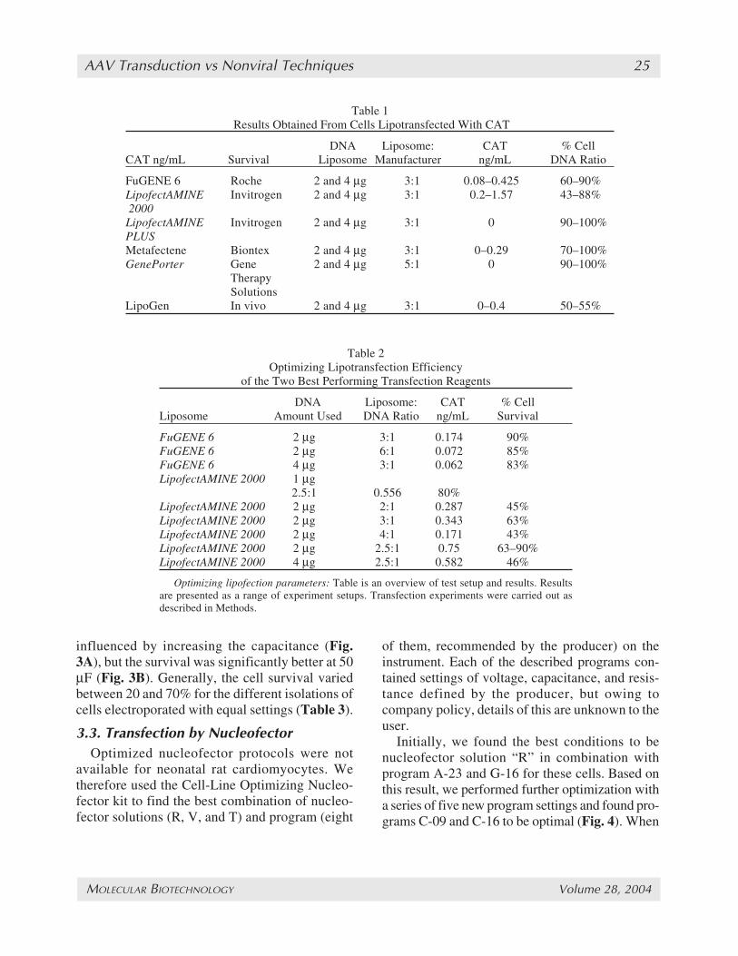
Table 1 Results Obtained From Cells Lipotransfected With CAT
DNA Liposome: CAT % CellCAT ng/mL Survival Liposome Manufacturer ng/mL DNA Ratio
FuGENE 6 Roche 2 and 4 µg 3:1 0.08–0.425 60–90%LipofectAMINE Invitrogen 2 and 4 µg 3:1 0.2–1.57 43–88% 2000LipofectAMINE Invitrogen 2 and 4 µg 3:1 0 90–100%PLUSMetafectene Biontex 2 and 4 µg 3:1 0–0.29 70–100%GenePorter Gene 2 and 4 µg 5:1 0 90–100%
TherapySolutions
LipoGen In vivo 2 and 4 µg 3:1 0–0.4 50–55%
Table 2Optimizing Lipotransfection Efficiency
of the Two Best Performing Transfection Reagents
DNA Liposome: CAT % CellLiposome Amount Used DNA Ratio ng/mL Survival
FuGENE 6 2 µg 3:1 0.174 90%FuGENE 6 2 µg 6:1 0.072 85%FuGENE 6 4 µg 3:1 0.062 83%LipofectAMINE 2000 1 µg
2.5:1 0.556 80%LipofectAMINE 2000 2 µg 2:1 0.287 45%LipofectAMINE 2000 2 µg 3:1 0.343 63%LipofectAMINE 2000 2 µg 4:1 0.171 43%LipofectAMINE 2000 2 µg 2.5:1 0.75 63–90%LipofectAMINE 2000 4 µg 2.5:1 0.582 46%
Optimizing lipofection parameters: Table is an overview of test setup and results. Resultsare presented as a range of experiment setups. Transfection experiments were carried out asdescribed in Methods.
influenced by increasing the capacitance (Fig.3A), but the survival was significantly better at 50µF (Fig. 3B). Generally, the cell survival variedbetween 20 and 70% for the different isolations ofcells electroporated with equal settings (Table 3).
3.3. Transfection by NucleofectorOptimized nucleofector protocols were not
available for neonatal rat cardiomyocytes. Wetherefore used the Cell-Line Optimizing Nucleo-fector kit to find the best combination of nucleo-fector solutions (R, V, and T) and program (eight
of them, recommended by the producer) on theinstrument. Each of the described programs con-tained settings of voltage, capacitance, and resis-tance defined by the producer, but owing tocompany policy, details of this are unknown to theuser.
Initially, we found the best conditions to benucleofector solution “R” in combination withprogram A-23 and G-16 for these cells. Based onthis result, we performed further optimization witha series of five new program settings and found pro-grams C-09 and C-16 to be optimal (Fig. 4). When
03/JW685/Djurovic/21-32 8/23/04, 1:53 PM25
MOLECULAR BIOTECHNOLOGY Volume 28, 2004
26 Djurovic et al.
Fig. 1. Optimization of voltage settings in electroporation experiment. (A) Transfection efficiency in the ratioCAT/total protein, and (B) % survival of neonatal cardiomyocytes with a voltage range of 200–420. Resistancesetting was constant at 725 Ω and the capacitance setting was constant at 100 µF. Results are in mean ±SEM. O= nonelectroporated cells.
we increased the cell number to 2 million, theCAT produced increased twofold, but the cell sur-vival decreased substantially.
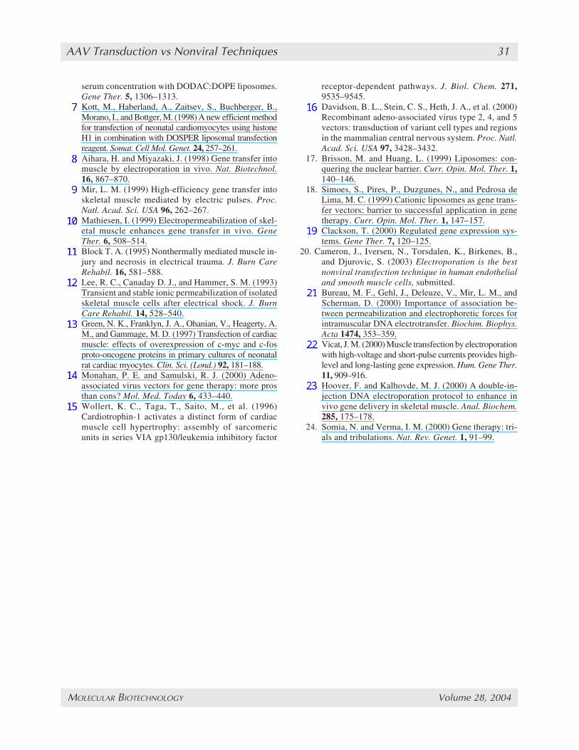
3.4. AAVBy performing transduction at varying multi-
plicity of infection (moi) a log/linear relationshipbetween moi and transduction efficiency wasdemonstrated (Fig. 5).
Most of the cells transduced with the highestconcentration of AAV–lacZ stained for β-galac-tosidase, with lower concentrations of vector giv-ing diminishing numbers of stained cells. Thehighest percentage of β-galactosidase expressionachieved in our study was 88.1% (Fig. 5). In themock-infected cells only a few cells stained for β-galactosidase.
4. DiscussionSomatic gene transfer using liposome/DNA
complexes and electroporation has enjoyed in-creasing interest. In this study we demonstratedthat, although inferior as compared with the trans-duction efficiency achieved by use of AAV,nonviral gene transfer techniques can be opti-mized for use in cardiomyocytes.
For efficient optimization of lipofection, inter-related factors and variables affecting the cationiclipid-mediated gene transfer need to be deter-mined. The ratio of liposome/DNA complexes,their positive charge, size, and the interaction be-tween DNA and cationic lipids are also importantfactors potentially influencing cellular uptake(17,18). Optimizing described parameters, en-hancement of transfer efficiency was achieved.
03/JW685/Djurovic/21-32 8/23/04, 1:53 PM26

MOLECULAR BIOTECHNOLOGY Volume 28, 2004
AAV Transduction vs Nonviral Techniques 27
Fig. 2. Optimization of resistance settings in electroporation experiment. (A) Transfection efficiency inthe ratio CAT/total protein, and (B) % survival of neonatal cardiomyocytes with a resistance range of 450–1000 Ω. Voltage setting was constant at 360 V, and the capacitance setting was constant at 100 µF. Resultsare in mean ±SEM.
Toxicity of cationic lipids is another importantaspect when using liposomes. Some cationic lip-ids, such as GenePORTER showed low toxicity,but very low transfer efficiency (Table 1). Butothers, such as LipofectAMINE 2000, performedwell in this system, combining high transfer effi-ciencies with relatively low toxicities. Toxicity ismainly caused by free liposomes, which do notcomplex DNA. One solution would be to removefree liposomes from the complexing solution (19).Discarding these free liposomes resulted in re-duced toxicity but transfer efficiencies decreasedmarkedly as well (data not shown). On the otherhand, for optimal transfer efficiencies, higher
DNA concentrations require higher amounts of li-posomes, which inevitably increase toxicity.
We optimized the electroporation procedure forthe neonatal rat cardiomyocytes, studying influ-ence and balance of the best possible transfer effi-ciency and survival rate.
Our results using the ECM 630 electroporatorare better than those obtained using the nucleo-fector instrument (Table 3). Transfection usingthe nucleofector is a patented commercial tech-nique requiring proprietary buffers and programs,the constituents of which are not known to finalusers. But when we used the ECM 630, an “inhouse” method was developed. We believe that
03/JW685/Djurovic/21-32 8/23/04, 1:53 PM27

MOLECULAR BIOTECHNOLOGY Volume 28, 2004
28 Djurovic et al.
Fig. 3. Optimization of capacity settings in electroporation experiment. (A) Transfection efficiency in the ratioCAT/total protein, and (B) % survival of neonatal cardiomyocytes with a capacitance range of 50–200 µF. Thevoltage setting was constant at 360 V, and the resistance setting was constant at 725Ω. Results are in mean ±SEM.
Table 3Optimized Nonviral Transfection Methods: Their Efficiency
and % Cell Survival in Neonatal Cardiomyocytes*
Ratio % CellMethod CAT/protein × 106 Survival % β-galactosidase
Electroporation 30 20–70% 7.5%Lipofection (L-2000) 19 88 8.1%Lipofection (FuGENE 6) 19 57 3.3%Nucleofection 19 37 4.8%
*Results are obtained from cells transfected with CAT and LacZ.
03/JW685/Djurovic/21-32 8/23/04, 1:53 PM28

MOLECULAR BIOTECHNOLOGY Volume 28, 2004
AAV Transduction vs Nonviral Techniques 29
optimizing of this method is possible. In this studywe kept buffer, cell numbers, and plasmid amountsconstant and tested the variables available on theinstrument (voltage, capacitance, and resistance).We found electroporation to be an easy, quick,effective, and affordable method for transfectingneonatal rat cardiomyocytes. However, the highnumber of cells and high plasmid amounts re-quired could be considered as limitations.
Many obstacles must be overcome before non-viral techniques can be considered for routinegene therapy. One critical issue is achieving anadequate efficiency of gene transfer. Transfectionusing reagents/liposomes needs to be optimizedfor each targeted cell type and reagent used (20).Electroporation may prove difficult to use in aclinical setting. However, investigations are cur-
rently designed to address this issue and initialresults using electroporation show that a low volt-age (<200 V) is required to minimize tissue death,but this lowers transfection efficiency (personalcommunication). This method may be painful tothe patient, as strong currents are required to fa-cilitate good transfection rates. The extent ofmuscle damage may depend on pulsing param-eters and electrode design (21–23).
Interestingly, we obtained similar transfectionefficiency and cell survival with both electro-poration with BTX 630 and lipofection reagent forneonatal rat cardiomyocytes as for human endot-helial and smooth muscle cells (20).
Viral vectors are derived from modified animalor human viruses, resulting in replication-defi-cient vectors, and they represent a powerful mo-
Fig. 4. Results from cell line optimizing nucleofector kit with neonatal rat cardiomyocytes. A combination ofdifferent nucleofector programs and solution R, which have been proved to be the best solution, were used. (A)CAT produced in ng/mL for each setting (mean), and (B) the corresponding % survival of the cells. * Indicates thebest settings.
03/JW685/Djurovic/21-32 8/23/04, 1:53 PM29

MOLECULAR BIOTECHNOLOGY Volume 28, 2004
30 Djurovic et al.
Fig. 5. Relationship between moi and transduction efficiency. The relationship between multiplicity of infec-tion and percentage of β-galactosidase stained cells. In the mock infected control wells <0.5% of the cells werestained for β-galactosidase.
lecular tool for delivering genes into somatic cells.Nevertheless, increased immunogenic responses,insertional mutagenesis causal for oncogenicproperties, inactivation of vector, development ofreplication-competent virions, are some of the dis-advantages that must be overcome to achieve fa-vorable therapeutic benefit (24). However, AAVvectors are considered to be devoid of this limita-tion. Our findings suggested that AAV transduc-tion was probably efficient because of a high moi,and the efficiency of AAV transduction washighly dependent on the moi. At a relatively lowmoi only a minority of the cells were stained forβ-galactosidase.
Through improved gene transfer, nonviraltransfer techniques may play an increasingly im-portant role in delivering genes to cells in relevantdisease states. Despite the fact that transductionwith AAV is more efficient (88.1%), transfectionswith nonviral techniques, when optimized, couldbe a useful tool for overexpression of certaingenes in neonatal cardiomyocytes.
AcknowledgmentsThis work was funded by a grant from Sosial-
og helsedepartementets arbeid med genterapi(st.prp. nr. 61), as well as Anders Jahre’s Fund for
Promotion of Science and Carl Semb’s MedicalResearch Fund and the Norwegian Cancer Soci-ety. The AAV vectors were a kind gift from Dr.Robert Kotin, NIH, Bethesda, MD, USA.
We also thank Bjørg Austbø, Kari Torsdalen,Marit Sletten, Geir Florholmen, Therese Lundin,and Bård Birkenes for excellent laboratory assis-tance and helpful encouragement.
References1. Felgner, P. L. (1991). Cationic liposome-mediated
transfection with lipofectin reagent. In Gene Transferand Expression Protocols, vol. 7, Methods in Molecu-lar Biology (Murray, E. J., ed.), Humana Press,Clifton, NJ, pp. 81–89.
2. Blaese, R. M., Mullen, C. A., and Ramsey, W. J.(1993) Strategies for gene therapy. Pathol. Biol.(Paris) 41, 672–676.
3. Albert, N. and Tremblay, J. P. (1992) Evaluation ofvarious gene transfection methods into human myo-blast clones. Transplant Proc. 24, 2784–2786.
4. Dodds, E., Dunckley, M. G., Naujoks, K., Michaelis,U., and Dickson, G. (1998) Lipofection of culturedmouse muscle cells: a direct comparison oflipofectamine and DOSPER. Gene Ther. 5, 542–551.
5. Trivedi, R. A. and Dickson, G. (1995) Liposome-me-diated gene transfer into normal and dystrophin-defi-cient mouse myoblasts. J. Neurochem. 64, 2230–2238.
6. Vitiello, L., Bockhold, K., Joshi P. B, and Worton, R.G. (1998) Transfection of cultured myoblasts in high
03/JW685/Djurovic/21-32 8/23/04, 1:53 PM30
MOLECULAR BIOTECHNOLOGY Volume 28, 2004
AAV Transduction vs Nonviral Techniques 31
serum concentration with DODAC:DOPE liposomes.Gene Ther. 5, 1306–1313.
7. Kott, M., Haberland, A., Zaitsev, S., Buchberger, B.,Morano, I., and Bottger, M. (1998) A new efficient methodfor transfection of neonatal cardiomyocytes using histoneH1 in combination with DOSPER liposomal transfectionreagent. Somat. Cell Mol. Genet. 24, 257–261.
8. Aihara, H. and Miyazaki, J. (1998) Gene transfer intomuscle by electroporation in vivo. Nat. Biotechnol.16, 867–870.
9. Mir, L. M. (1999) High-efficiency gene transfer intoskeletal muscle mediated by electric pulses. Proc.Natl. Acad. Sci. USA 96, 262–267.
10. Mathiesen, I. (1999) Electropermeabilization of skel-etal muscle enhances gene transfer in vivo. GeneTher. 6, 508–514.
11. Block T. A. (1995) Nonthermally mediated muscle in-jury and necrosis in electrical trauma. J. Burn CareRehabil. 16, 581–588.
12. Lee, R. C., Canaday D. J., and Hammer, S. M. (1993)Transient and stable ionic permeabilization of isolatedskeletal muscle cells after electrical shock. J. BurnCare Rehabil. 14, 528–540.
13. Green, N. K., Franklyn, J. A., Ohanian, V., Heagerty, A.M., and Gammage, M. D. (1997) Transfection of cardiacmuscle: effects of overexpression of c-myc and c-fosproto-oncogene proteins in primary cultures of neonatalrat cardiac myocytes. Clin. Sci. (Lond.) 92, 181–188.
14. Monahan, P. E. and Samulski, R. J. (2000) Adeno-associated virus vectors for gene therapy: more prosthan cons? Mol. Med. Today 6, 433–440.
15. Wollert, K. C., Taga, T., Saito, M., et al. (1996)Cardiotrophin-1 activates a distinct form of cardiacmuscle cell hypertrophy: assembly of sarcomericunits in series VIA gp130/leukemia inhibitory factor
receptor-dependent pathways. J. Biol. Chem. 271,9535–9545.
16. Davidson, B. L., Stein, C. S., Heth, J. A., et al. (2000)Recombinant adeno-associated virus type 2, 4, and 5vectors: transduction of variant cell types and regionsin the mammalian central nervous system. Proc. Natl.Acad. Sci. USA 97, 3428–3432.
17. Brisson, M. and Huang, L. (1999) Liposomes: con-quering the nuclear barrier. Curr. Opin. Mol. Ther. 1,140–146.
18. Simoes, S., Pires, P., Duzgunes, N., and Pedrosa deLima, M. C. (1999) Cationic liposomes as gene trans-fer vectors: barrier to successful application in genetherapy. Curr. Opin. Mol. Ther. 1, 147–157.
19. Clackson, T. (2000) Regulated gene expression sys-tems. Gene Ther. 7, 120–125.
20. Cameron, J., Iversen, N., Torsdalen, K., Birkenes, B.,and Djurovic, S. (2003) Electroporation is the bestnonviral transfection technique in human endothelialand smooth muscle cells, submitted.
21. Bureau, M. F., Gehl, J., Deleuze, V., Mir, L. M., andScherman, D. (2000) Importance of association be-tween permeabilization and electrophoretic forces forintramuscular DNA electrotransfer. Biochim. Biophys.Acta 1474, 353–359.
22. Vicat, J. M. (2000) Muscle transfection by electroporationwith high-voltage and short-pulse currents provides high-level and long-lasting gene expression. Hum. Gene Ther.11, 909–916.
23. Hoover, F. and Kalhovde, M. J. (2000) A double-in-jection DNA electroporation protocol to enhance invivo gene delivery in skeletal muscle. Anal. Biochem.285, 175–178.
24. Somia, N. and Verma, I. M. (2000) Gene therapy: tri-als and tribulations. Nat. Rev. Genet. 1, 91–99.
03/JW685/Djurovic/21-32 8/23/04, 1:53 PM31