Factors Influencing Recombinant Adeno-Associated Virus Production
Upload
independentCategory
view
1download
0
MOLECULAR PAINVulchanova et al. Molecular Pain 2010, 6:31http://www.molecularpain.com/content/6/1/31
Open AccessR E S E A R C H
ResearchDifferential adeno-associated virus mediated gene transfer to sensory neurons following intrathecal delivery by direct lumbar punctureLucy Vulchanova1, Daniel J Schuster2, Lalitha R Belur3, Maureen S Riedl2, Kelly M Podetz-Pedersen3, Kelley F Kitto2,4,5, George L Wilcox2,4,5,6, R Scott McIvor3 and Carolyn A Fairbanks*2,4,5
AbstractBackground: Neuronal transduction by adeno-associated viral (AAV) vectors has been demonstrated in cortex, brainstem, cerebellum, and sensory ganglia. Intrathecal delivery of AAV serotypes that transduce neurons in dorsal root ganglia (DRG) and spinal cord offers substantial opportunities to 1) further study mechanisms underlying chronic pain, and 2) develop novel gene-based therapies for the treatment and management of chronic pain using a non-invasive delivery route with established safety margins. In this study we have compared expression patterns of AAV serotype 5 (AAV5)- and AAV serotype 8 (AAV8)-mediated gene transfer to sensory neurons following intrathecal delivery by direct lumbar puncture.
Results: Intravenous mannitol pre-treatment significantly enhanced transduction of primary sensory neurons after direct lumbar puncture injection of AAV5 (rAAV5-GFP) or AAV8 (rAAV8-GFP) carrying the green fluorescent protein (GFP) gene. The presence of GFP in DRG neurons was consistent with the following evidence for primary afferent origin of the majority of GFP-positive fibers in spinal cord: 1) GFP-positive axons were evident in both dorsal roots and dorsal columns; and 2) dorsal rhizotomy, which severs the primary afferent input to spinal cord, abolished the majority of GFP labeling in dorsal horn. We found that both rAAV5-GFP and rAAV8-GFP appear to preferentially target large-diameter DRG neurons, while excluding the isolectin-B4 (IB4) -binding population of small diameter neurons. In addition, a larger proportion of CGRP-positive cells was transduced by rAAV5-GFP, compared to rAAV8-GFP.
Conclusions: The present study demonstrates the feasibility of minimally invasive gene transfer to sensory neurons using direct lumbar puncture and provides evidence for differential targeting of subtypes of DRG neurons by AAV vectors.
IntroductionPain signals originate at the peripheral terminals of sen-sory neurons and via their central processes are transmit-ted to second order neurons in the dorsal horn of spinalcord. Gene therapy targeted to the spinal cord or sensoryneurons could, after a single administration, providelong-term pain relief with reduced adverse effects relativeto current standards of practice in chronic pain manage-ment. Additionally, gene transfer directed to specific neu-ronal subtypes within dorsal root ganglia (DRG) couldgreatly empower ongoing basic research focused upon
delineating functional differences between distinct noci-ceptive neuronal populations. With respect to the panelof viral vectors available for gene transfer, AAV has theadvantage of conferring stable, long-term gene expres-sion in the absence of an inflammatory response [1]. Fur-ther, AAV vectors are able to transduce non-dividing cells(such as neurons), and can accommodate gene expressioncassettes of up to 4.3 kB, which is well within the range ofmany genes potentially useful for pain control [2]. AAVserotype 2 (AAV2) has been the most widely studiedserotype. However, recent studies of AAV serotypes 5 and8 (AAV5 and AAV8) have demonstrated significantlyincreased gene transfer efficiency, and wider distributionof transduced cells in the CNS when compared with
* Correspondence: [email protected] Departments of Neuroscience, University of Minnesota, Church Street, Minneapolis, MN 55455, USAFull list of author information is available at the end of the article
© 2010 Vulchanova et al; licensee BioMed Central Ltd. This is an Open Access article distributed under the terms of the Creative Com-mons Attribution License (http://creativecommons.org/licenses/by/2.0), which permits unrestricted use, distribution, and reproduc-tion in any medium, provided the original work is properly cited.

Vulchanova et al. Molecular Pain 2010, 6:31http://www.molecularpain.com/content/6/1/31
Page 2 of 9
AAV2 [3,4]. In addition, self-complementary AAV8-mediated transduction of DRG neurons has been recentlyreported [5].
Viral vector-mediated gene transfer to DRG neurons,spinal cord neurons, glia, and pia mater has beenachieved in animal models using direct tissue injectionmethods (intraparenchymal or intraneural) [2,6,7] orintrathecal (i.t.) injection through an atlanto-occipitalcatheter [5,8-10]. The primary limitations of these deliv-ery approaches include requisite surgery and potential forinflammation and tissue damage [11-16]. Acute intrathe-cal (i.t.) delivery, which is applied clinically as well as forbasic neuropharmacological research [17], offers a rela-tively non-invasive route of administration for viral vec-tor-mediated gene transfer. However, viral vectoradministration by direct lumbar puncture has not previ-ously been reported to result in appreciable transductionof spinal cord and DRG neurons [2]. In the present study,we evaluated intravenous mannitol pretreatment as astrategy for increasing the access of viral particles deliv-ered i.t. via direct lumbar puncture to spinal cord paren-chyma and DRG. Mannitol is used to control intracranialpressure [18] and to disrupt the intercellular tight junc-tions between endothelial cells of the CNS microvascula-ture [19]. It is also used intravenously (i.v.) to facilitatechemotherapeutic drug delivery to the CNS [19]. In ani-mal models, mannitol, given i.v. as a pretreatment,enhanced intraparenchymal diffusion following intrac-erebroventricular (i.c.v.) delivery of AAV constructs[20,21]. Therefore, we hypothesized that i.v. pretreatmentwith mannitol would similarly enhance penetration ofAAV vectors to the spinal cord parenchyma and to thecell bodies of DRG neurons.
Entry of AAV particles into cells is mediated by bindingto a combination of cell-surface molecules acting asreceptors or co-receptors, with different serotypes bind-ing to different combinations of surface molecules. Forexample, AAV2 utilizes heparan sulfate proteoglycans asreceptors and a number of other cell-surface molecules,such as fibroblast growth factor receptor 1 and hepato-cyte growth factor receptor, as co-receptors [22-26]. Lessis known about the receptors and co-receptors of AAV5and AAV8 [22,27,28]. Differential expression of cell-sur-face molecules required for entry of AAV vectors maylead to differential tissue tropism among AAV serotypes.DRG neurons are heterogeneous in terms of cell somasize, myelination level, response properties, and neuro-chemistry, raising the possibility for differential targetingof DRG neuronal subtypes by different AAV vectors. Thepresent study provides a quantitative evaluation of theDRG neuronal subtypes expressing green fluorescentprotein (GFP) after transduction by both rAAV5-GFPand rAAV8-GFP, based on cell soma size and labeling for
the neurochemical markers calcitonin gene-related pep-tide (CGRP), isolectin-B4 (IB4), and substance P (SP).
ResultsrAAV5-GFP or rAAV8-GFP were injected intrathecallyby lumbar puncture as described in Methods. GFPexpression following rAAV5-GFP administration wassubstantially more abundant in spinal cords from manni-tol-pretreated compared to saline-pretreated mice (Fig.1A and 1B). GFP was also present in DRG neurons ofmannitol-pretreated mice (Fig 1C). The number of GFP-positive DRG neurons was significantly higher (Mann-Whitney, U05(1),3,3 = 9, p = 0.05) in lumbar DRG of manni-tol-pretreated mice compared to that of saline-pretreatedmice (Fig. 1C and 1D). Intravenous pretreatment withmannitol similarly resulted in higher levels of GFP in spi-nal cord and DRG following i.t. administration of rAAV8-GFP (data not shown).
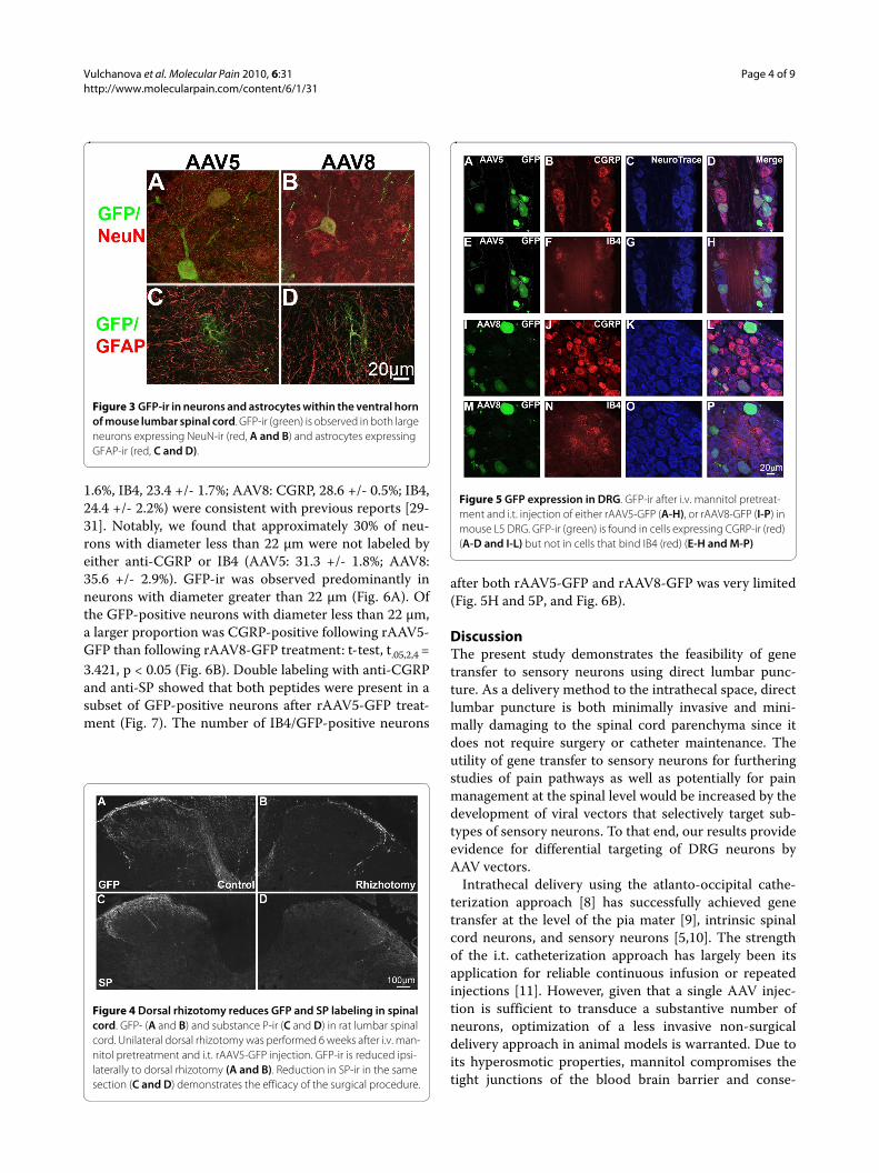
GFP was observed at all levels of mouse spinal cord fol-lowing treatment with rAAV5-GFP (Fig. 2) and rAAV8-GFP (not shown) with notable differences in the caudal-rostral distribution. GFP was abundant in the dorsal hornat sacral (Fig. 2A), lumbar (Fig. 2B), and cervical levels(Fig. 2D). In contrast, labeling in thoracic spinal cord wasmore limited and appeared to be restricted to the dorsalcolumns and Clarke's Columns (Fig. 2C). At all levels ofspinal cord, GFP appeared to be associated predomi-nantly with nerve fibers. However, it was generally absentfrom the substantia gelatinosa (lamina II) of dorsal horn.Labeling of a small number of spinal cord neurons wasmost evident at lumbosacral levels where GFP neuronswere seen in both dorsal (not shown) and ventral horn(Fig. 3A and 3B). In addition, double labeling with GFAPidentified a small number of GFP-positive astrocytes (Fig.3C and 3D). Reducing the injection volume (and conse-quently the viral load) resulted in predominantly lum-bosacral localization of GFP-ir (data not shown).
To determine whether the GFP observed in fibers of thedorsal horn originated from primary afferent neuronswhose cell bodies are found in DRG, dorsal rhizotomieswere performed in rAAV5-GFP injected rats (6 weekspost-injection) to sever input from the DRG. The patternof labeling in rat was consistent with that of mouse at allspinal cord levels. GFP was notably reduced in the dorsalhorn ipsilateral to the severed dorsal roots in contrast toabundant GFP labeling on the contralateral, unoperatedside (Fig. 4A and 4C). Double-labeling with antiserumdirected against substance P (SP) showed that SP-ir wasalso reduced in the ipsilateral dorsal horn of rhizoto-mized rats (Fig. 4B and 4D), confirming the disruption ofafferent input. Further evidence suggesting GFP localiza-tion in the central processes of primary afferent neuronswas the observation of labeling in dorsal roots as well asin the dorsal columns.

Vulchanova et al. Molecular Pain 2010, 6:31http://www.molecularpain.com/content/6/1/31
Page 3 of 9
In mouse DRG, GFP was seen in neuronal cell bodiesand fibers after both rAAV5-GFP (as shown in Fig 1C)and rAAV8-GFP treatment (data not shown). However, inrAAV8-GFP treated DRG, GFP labeling was alsoobserved in non-neuronal cells, which did not co-labelwith markers of glial cells (data not shown). Quantitative
image analysis of DRG from mice injected with rAAV5-GFP (Fig. 5A-H) or rAAV8-GFP (Fig. 5I-P) was per-formed in DRG sections labeled with anti-CGRP and IB4and counterstained with NeuroTrace (Fig. 5). The pro-portions of CGRP-positive and IB4-positive neuronsdetermined in these experiments (AAV5: CGRP, 37.0 +/-
Figure 1 Mannitol enhances GFP expression in mouse spinal cord and DRG. Sections are from mice perfused six weeks after i.t. injection of rAAV5-GFP with or without intravenous mannitol pretreatment. A) GFP immunoreactivity (-ir) in the spinal cord of a mannitol-pretreated mouse. B) GFP-ir in spinal cord of a saline-pretreated mouse. C) GFP-ir in L4 DRG of a mannitol-pretreated mouse. D) Mannitol pretreatment significantly enhanc-es transduction of lumbar DRG neurons when compared to saline pretreatment; Mann-Whitney, U05(1),3,3 = 9, p = 0.05.
Figure 2 GFP-ir in representative sections of mouse spinal cord. A) Sacral, B) lumbar, C) thoracic, and D) cervical spinal cord sections from mice perfused six weeks after i.v. mannitol pretreatment and i.t. rAAV5-GFP injection.
Vulchanova et al. Molecular Pain 2010, 6:31http://www.molecularpain.com/content/6/1/31
Page 4 of 9
1.6%, IB4, 23.4 +/- 1.7%; AAV8: CGRP, 28.6 +/- 0.5%; IB4,24.4 +/- 2.2%) were consistent with previous reports [29-31]. Notably, we found that approximately 30% of neu-rons with diameter less than 22 μm were not labeled byeither anti-CGRP or IB4 (AAV5: 31.3 +/- 1.8%; AAV8:35.6 +/- 2.9%). GFP-ir was observed predominantly inneurons with diameter greater than 22 μm (Fig. 6A). Ofthe GFP-positive neurons with diameter less than 22 μm,a larger proportion was CGRP-positive following rAAV5-GFP than following rAAV8-GFP treatment: t-test, t.05,2,4 =3.421, p < 0.05 (Fig. 6B). Double labeling with anti-CGRPand anti-SP showed that both peptides were present in asubset of GFP-positive neurons after rAAV5-GFP treat-ment (Fig. 7). The number of IB4/GFP-positive neurons
after both rAAV5-GFP and rAAV8-GFP was very limited(Fig. 5H and 5P, and Fig. 6B).
DiscussionThe present study demonstrates the feasibility of genetransfer to sensory neurons using direct lumbar punc-ture. As a delivery method to the intrathecal space, directlumbar puncture is both minimally invasive and mini-mally damaging to the spinal cord parenchyma since itdoes not require surgery or catheter maintenance. Theutility of gene transfer to sensory neurons for furtheringstudies of pain pathways as well as potentially for painmanagement at the spinal level would be increased by thedevelopment of viral vectors that selectively target sub-types of sensory neurons. To that end, our results provideevidence for differential targeting of DRG neurons byAAV vectors.
Intrathecal delivery using the atlanto-occipital cathe-terization approach [8] has successfully achieved genetransfer at the level of the pia mater [9], intrinsic spinalcord neurons, and sensory neurons [5,10]. The strengthof the i.t. catheterization approach has largely been itsapplication for reliable continuous infusion or repeatedinjections [11]. However, given that a single AAV injec-tion is sufficient to transduce a substantive number ofneurons, optimization of a less invasive non-surgicaldelivery approach in animal models is warranted. Due toits hyperosmotic properties, mannitol compromises thetight junctions of the blood brain barrier and conse-
Figure 3 GFP-ir in neurons and astrocytes within the ventral horn of mouse lumbar spinal cord. GFP-ir (green) is observed in both large neurons expressing NeuN-ir (red, A and B) and astrocytes expressing GFAP-ir (red, C and D).
Figure 4 Dorsal rhizotomy reduces GFP and SP labeling in spinal cord. GFP- (A and B) and substance P-ir (C and D) in rat lumbar spinal cord. Unilateral dorsal rhizotomy was performed 6 weeks after i.v. man-nitol pretreatment and i.t. rAAV5-GFP injection. GFP-ir is reduced ipsi-laterally to dorsal rhizotomy (A and B). Reduction in SP-ir in the same section (C and D) demonstrates the efficacy of the surgical procedure.
Figure 5 GFP expression in DRG. GFP-ir after i.v. mannitol pretreat-ment and i.t. injection of either rAAV5-GFP (A-H), or rAAV8-GFP (I-P) in mouse L5 DRG. GFP-ir (green) is found in cells expressing CGRP-ir (red) (A-D and I-L) but not in cells that bind IB4 (red) (E-H and M-P)

Vulchanova et al. Molecular Pain 2010, 6:31http://www.molecularpain.com/content/6/1/31
Page 5 of 9
quently has been used to enhance CNS delivery of che-motherapeutic agents [19]. Furthermore, i.v. mannitol hasbeen used in rodents to expand distribution of AAV vec-tors into brain parenchyma following intracerebroven-tricular injection [20,21]. The present study showed thatthe same concentration of i.v. mannitol similarlyenhanced penetration of AAV5-GFP and AAV8-GFP vec-tors to the spinal cord parenchyma and to the dorsal rootganglia. Although GFP labeling in spinal cord was presentpredominantly in nerve fibers, a small number of trans-duced dorsal and ventral horn neurons as well as astro-cytes was consistently observed, suggesting that bothviral vectors are capable of gaining access to these cells.
The distribution of GFP labeling in spinal cord andDRG suggests that AAV5 and AAV8 target sensory neu-rons differentially. The pattern of GFP-labeled nervefibers in spinal cord was consistent with the subsets ofDRG neurons identified as GFP-positive. At cervical,lumbar and sacral level, GFP labeled fibers were seen pre-dominantly in deeper laminae of dorsal horn and in thedorsal columns, in agreement with the observation thatboth AAV5 and AAV8 preferentially transduce larger
diameter DRG neurons. The pattern of GFP labeling inthoracic spinal cord was notably different from other lev-els (in both rat and mouse) and appeared restricted to thedorsal columns and Clarke's columns. The reason for thisdifference is unknown but may be related to differentialtargeting of proprioceptive primary afferent neurons inthoracic DRG. In contrast to AAV5 and AAV8, a recentstudy reported that AAV6 targets predominantly smalldiameter DRG neurons [10]. AAV5- or AAV8-mediatedgene transfer could be useful for manipulating proteinexpression in large sensory neurons, which commonlysignal innocuous stimuli, but may also carry pain signalsin allodynic conditions [32].
GFP-labeled fibers were also present in lamina I of thespinal cord. These fibers most likely represented collater-als of large diameter neurons as well as terminations ofthe small subset of peptidergic neurons that was targetedby the vectors. The distribution of GFP in superficial dor-sal horn from AAV5- and AAV8-treated mice was similar.However, quantitative analysis in DRG indicated thatwhile nearly all of the rAAV5-GFP transduced neuronswith diameter less than 22 μm were also CGRP-positive,
Figure 6 Quantitative image analysis of GFP-expressing DRG neurons. A) A similar number of neurons smaller than 22 μm (≤22 μm), or larger than 22 μm (>22 μm) expressed GFP after both rAAV5-GFP and rAAV8-GFP injection. Both vectors preferentially targeted large neurons. B) Injection of rAAV5-GFP resulted in more GFP expression in CGRP-positive neurons smaller than 22 μm than was seen with injection of rAAV8-GFP; t-test, t.05,2,4
= 3.421, p = 0.0268. Injection of either vector did not result in GFP expression in a substantial portion of the IB4-binding population. C) Approximately 80% of the DRG neurons expressing GFP were larger than 22 μm, regardless of whether rAAV5-GFP or rAAV8-GFP was injected. Approximately 20% of these GFP-expressing neurons were also CGRP positive.
Figure 7 Colocalization of GFP-ir with SP- and CGRP-ir in DRG Neurons. GFP colocalization with CGRP and SP after i.t. delivery of AAV5-GFP in mice. A) GFP fluorescence. B) CGRP-ir. C) SP-ir. D) Merged image. Of 10 GFP-positive neurons in this image, 6 show CGRP-ir (indicated by single arrows), and of those 6, 1 also displays SP-ir (indicated by arrowhead).

Vulchanova et al. Molecular Pain 2010, 6:31http://www.molecularpain.com/content/6/1/31
Page 6 of 9
rAAV8-GFP transduced a significantly smaller propor-tion of CGRP-positive neurons of this size. Since the pop-ulation of CGRP neurons is heterogeneous, thisobservation may indicate differential expression of recep-tors for AAV5 and AAV8 within the different subsets ofCGRP-positive neurons.
It is noteworthy that GFP labeling was minimal in lam-ina II (substantia gelatinosa, SG) at all levels of the spinalcord. Similar observations have been made in studies ofAAV2-MOR expression following intraneural injection[6] and AAV8-GFP following i.t. injection by the atlanto-occipital catheterization method [5]. The reason forabsence of GFP-ir in the SG is unclear. It is likely that, inour experiments, the low levels of GFP in SG are due tothe limited targeting of neurons smaller than 22 μm, inparticular the minimal transduction of IB4-binding neu-rons. Interestingly, AAV6-GFP delivery to the intrathecalspace via the atlanto-occipital approach resulted in sub-stantial transduction of IB4-positive neurons and in GFPlabeling in the SG of the spinal cord [10].
The observation that neither AAV5 nor AAV8 trans-duced an appreciable number of IB4-positive neuronssuggests that these neurons lack a surface moleculerequired for the entry of these vectors. While AAV5 andAAV8 may have limited application for transduction ofmouse IB4-positive neurons, this limitation can beadvantageous for studies targeting IB4-negative neuronalpopulations. In addition, the targeting of rat IB4-positiveneurons in by AAV5 and AAV8 may be different fromthat of mouse, since there are notable species differencesin the neurochemical subsets of DRG neurons [31]. Thefinding by Towne and colleagues [10] that AAV6 is able totransduce mouse IB4-positive neurons indicates thatAAV6 is able to enter cells via different cell-surface mole-cules than AAV5 and AAV8, and further supports differ-ential targeting selectivity of neuronal subpopulations bydifferent AAV vectors. This evidence highlights theimportance of quantitative analysis of colocalization withhistochemical markers of neuronal sub-populations foraccurate evaluation of the tropism of various AAV vec-tors.
ConclusionAlthough several groups have tried with varied success touse AAV vectors to transduce cells of the DRG or spinalcord following intraparenchymal delivery, intraneuralinjection, or various i.t. delivery approaches [2], none hasreported abundant transduction of the DRG or of thedeeper laminae of the dorsal horn without a deliverymethod requiring surgical intervention. We have demon-strated widespread expression of GFP in the spinal cordand select subsets of DRG neurons following i.t. adminis-tration of rAAV5-GFP or rAAV8-GFP via direct lumbarpuncture in conscious animals pretreated with i.v. manni-
tol. The distinct patterns of transduction observed withAAV5 and AAV8 in the present study and with AAV6 inthe study by Towne and colleagues [10] indicate that dif-ferential targeting of DRG sub-populations by variousAAV vectors should be further investigated with the goalof capitalizing upon such differences for both researchand therapeutic practices. Differential AAV-mediatedgene transfer to spinal cord and DRG neurons may pres-ent multiple opportunities for genetic manipulation ofproteins that drive the processes involved in pain sensa-tion or inhibitory control of pain.
MethodsAAV Vector and PackagingAAV vector TRUF11, containing a CAGS-regulated GFPsequence, has been previously described [1]. Packagingusing AAV5 or AAV8 serotype capsid was carried out atthe University of Florida Vector Core Lab of the GeneTherapy Center (Gainesville, Florida) as previouslydescribed [1].
Animal proceduresExperimental subjects were 20 to 25 g adult male C57BL/6 mice (Harlan, Madison, WI). In one set of experiments,Sprague Dawley rats (male and female), were included asthe subjects for Fig. 4 because their size renders themmore appropriate for dorsal rhizotomy experiments. Allexperiments were reviewed and approved by the Institu-tional Animal Care and Use Committee (IACUC) of theUniversity of Minnesota.
InjectionsSubjects were injected via the tail vein with 25% mannitolsolution (200 μL) twenty minutes prior to i.t. injection ofthe viral constructs. AAV constructs were deliveredintrathecally by direct lumbar puncture in awake mice[17,33] or rats [34] by an experimenter (KFK) with exten-sive experience in this method of drug delivery. A minormodification of the protocol was required to conserveAAV vector. The needle (a 30-gauge, 0.5-inch (mouse) or1.5 inch (rat)) was connected to a length of PE10 tubing,which was then connected to a second needle that wasattached to a 50-μl Luer-hub Hamilton syringe. For bothrAAV5-GFP and rAAV8-GFP, 10 μL of the construct con-taining 1011 viral vector genomes were injected i.t. Theinjection was administered by gripping gently the iliaccrest of the rodent and inserting the needle (bevel sideup) at about a 45° angle centered approximately betweenthe hipbones. A reflexive flick of the tail indicated punc-ture of the dura. Following the injection, the animals werereturned to the vivarium where they remained for sixweeks, until the time of transcardial perfusion, fixation,and extraction of fixed spinal cord and DRG for immuno-histochemical analysis.

Vulchanova et al. Molecular Pain 2010, 6:31http://www.molecularpain.com/content/6/1/31
Page 7 of 9
Dorsal RhizotomyIn rats, i.v. mannitol pretreatment and i.t. rAAV5-GFPinjection was followed 6 weeks later by unilateral dorsalrhizotomy. The rats were approximately 75 g at time ofthe injections and approximately 150-175 g at time of rhi-zhotomy and perfusion. Rats were anaesthetized with 75mg/ml ketamine, 5 mg/kg xylazine and 1 mg/kg acepro-mazine injected intramuscularly. After deep, surgicalanesthesia had been attained, an incision was madethrough the skin overlying T10-L7 of the spinal cord. Thedorsal roots were exposed and severed close to the spinalcolumn. The incision was closed with surgical staples andthe animals allowed to survive for 5 days before perfusionfixation.
ImmunohistochemistryAll animals were sacrificed by perfusion fixation as previ-ously described [35]. Briefly, animals were anaesthetizedwith 100 mg/kg ketamine, 5 mg/kg xylazine, and 1 mg/kgacepromazine injected i.m. and perfused with a solutionof calcium-free tyrodes solution (in mM:NaCl 116, KCl5.4, MgCl2·6H20 1.6, MgSO4·7H2O 0.4, NaH2PO4 1.4, glu-cose 5.6, and NaHCO3 26) followed by fixative (4% para-formaldehyde and 0.2% picric acid in 0.1 M phophatebuffer, pH 6.9) followed by 10% sucrose in PBS. Spinalcord and DRG were removed and incubated in 10%sucrose overnight at 4°C. Sections were cut at 14 μmthickness and thaw mounted onto gel-coated slides. Tis-sue sections were incubated for 1 hour at room tempera-ture in diluent (PBS containing 0.3% Triton, 1% BSA, 1%normal donkey serum) and then incubated overnight at4°C in primary antisera diluted in the same diluent. Insome instances, GFP fluorescence was enhanced byimmunostaining with antisera to GFP. Primary antibodiesused were: rabbit anti-GFP, 1:500 (Invitrogen; Eugene,OR), guinea pig anti-SP; 1:500 (Neuromics; Edina, MN),rabbit anti-CGRP, 1:500 (Immunostar; Hudson, WI),mouse anti-NeuN. 1:500 (Chemicon; Temecula, CA),mouse anti-GFAP, 1:400 (Sigma; St. Louis, MO)), and/orbiotinylated isolectin-B4, 10 μg/ml (Sigma; St. Louis,MO). After rinsing with PBS, sections were incubatedone hour at room temperature with appropriate combi-nations of Cy2-, Cy3-, Cy5- (1:300), and AMCA- (1:400)conjugated secondary antisera (Jackson ImmunoRe-search, West Grove, CA). Sections were rinsed again inPBS and coverslipped using glycerol and PBS containing0.1% p-phenylenediamine (Sigma). DRG sections werealso incubated for 20 minutes at room temperature withNeuroTrace® Red (Invitrogen; Eugene, OR). Images werecollected using confocal microscopy (Biorad MRC1024,or Olympus Fluoview1000), analyzed using Image J(NIH), and processed in Adobe Photoshop.
Cell CountingThree sections from each DRG were selected such thatthey were equally spaced and at least 70 μm apart. Thesesections were labeled with anti-CGRP, biotinylated-IB4,and NeuroTrace® Red for analysis of co-localization andcell size. Seven non-overlapping images were taken acrossthese three sections and analyzed using Image J to mea-sure cell body size and fluorescence intensity. Neuronswere identified and outlined based on their Nissl-likeNeuroTrace® labeling. Only cells with a visible nucleuswere counted. The cutoff of 22 μm for categorization ofneurons by size was chosen based on the width of thelargest peak in size histograms of DRG neurons. ForCGRP fluorescence intensity, a method adapted from thatof Fang and colleagues [36] was used. The average inten-sity of the brightest 10% and the dimmest 10% of neuronsin each image was measured and relative fluorescenceintensity for each cell was defined as 100[(intensity ofselected cell - average of dimmest 10%)/(average ofbrightest 10% - average of dimmest 10%)]. Cells with arelative fluorescence intensity of 28% or greater were con-sidered positive, and this number consistently coincidedwith cells identified as positive by visual inspection.Because the number of GFP expressing cells in each sec-tion was low, an alternative method was used to deter-mine background fluorescence of GFP. The averagefluorescence of three GFP-negative cells in each imagewas calculated, and GFP-positive cells were defined ascells with average intensity greater than that of the 3 neg-ative cells plus 3 standard deviations. IB4 positive cellswere defined using the same method as that for GFP pos-itive cells.
AbbreviationsrAAV: recombinant adeno-associated virus; CGRP: calcitonin gene-related pep-tide; DRG: dorsal root ganglia; GFP: green fluorescent protein; GFAP: glial fibril-lary acidic protein; IB4: isolectin B4; i.t: intrathecal; rAAV5: rAAV serotype 5;rAAV8: rAAV serotype 8.
Competing interestsThe authors declare that they have no competing interests.
Authors' contributionsLV contributed to the experimental design and drafting of the manuscript. Inaddition, she participated in all perfusions, dissections, histochemical analyses,and imaging as well as in interpretation of results and generation of figures.She is primarily responsible for the assurance of quality of the data and themain conclusions.DS assisted in perfusions and dissections and performed immunohistochemis-try, cell quantification and imaging. He assisted in the preparation of the fig-ures, contributed to the analysis and interpretation of the data, and edited themanuscript.LB initiated the studies and contributed to the experimental design and editedthe manuscript.MR performed all perfusions and dissections and participated in histochemicalanalyses and interpretation of results. She along with LV is primarily responsiblefor the assurance of quality of the data and the main conclusions and editedthe manuscipt.KPP performed the IV injections.KFK conducted all intrathecal injections of vector and is responsible for thequality assurance of this key technique.

Vulchanova et al. Molecular Pain 2010, 6:31http://www.molecularpain.com/content/6/1/31
Page 8 of 9
GLW participated in the experimental design and edited the manuscript.RSM initiated the study with GLW and CF as collaborators and edited the man-uscript.CF organized the team, contributed to the experimental design, drafted andedited the manuscript and supported the studies.All authors read and approved the final manuscript.
AcknowledgementsThe authors would like to thank Galina Kalyuzhnaya for her technical assis-tance. This work was supported by the National Institute on Drug Abuse (DA-025164 to C.A.F.) and the University of Minnesota Academic Health Center. An individual NRSA from NINDS (F31NS063634) and a 3 M Fellowship supports D.J.S. A K01 award from NIDA (K01DA017236) supported LV.
Author Details1Departments of Veterinary and Biomedical Sciences, University of Minnesota, Commonwealth Avenue, Saint Paul, MN 55108, USA, 2Departments of Neuroscience, University of Minnesota, Church Street, Minneapolis, MN 55455, USA, 3Departments of Genetics Cell Biology and Development, University of Minnesota, Church Street, Minneapolis, MN 55455, USA, 4Departments of Pharmaceutics, University of Minnesota, Harvard Street, Minneapolis, MN 55455, USA, 5Departments of Pharmacology, University of Minnesota, Church Street, Minneapolis, MN 55455, USA and 6Departments of Dermatology, University of Minnesota, Delaware Street, Minneapolis, MN 55455, USA
References1. Kaemmerer WF, Reddy RG, Warlick CA, Hartung SD, McIvor RS, Low WC: In
vivo transduction of cerebellar Purkinje cells using adeno-associated virus vectors. Mol Ther 2000, 2:446-457.
2. Beutler AS, Banck MS, Walsh CE, Milligan ED: Intrathecal gene transfer by adeno-associated virus for pain. Curr Opin Mol Ther 2005, 7:431-439.
3. Lin D, Fantz CR, Levy B, Rafi MA, Vogler C, Wenger DA, Sands MS: AAV2/5 vector expressing galactocerebrosidase ameliorates CNS disease in the murine model of globoid-cell leukodystrophy more efficiently than AAV2. Mol Ther 2005, 12:422-430.
4. Broekman ML, Comer LA, Hyman BT, Sena-Esteves M: Adeno-associated virus vectors serotyped with AAV8 capsid are more efficient than AAV-1 or -2 serotypes for widespread gene delivery to the neonatal mouse brain. Neuroscience 2006, 138:501-510.
5. Storek B, Reinhardt M, Wang C, Janssen WG, Harder NM, Banck MS, Morrison JH, Beutler AS: Sensory neuron targeting by self-complementary AAV8 via lumbar puncture for chronic pain. Proc Natl Acad Sci USA 2008, 105:1055-1060.
6. Xu Y, Gu Y, Xu GY, Wu P, Li GW, Huang LY: Adeno-associated viral transfer of opioid receptor gene to primary sensory neurons: a strategy to increase opioid antinociception. Proc Natl Acad Sci USA 2003, 100:6204-6209.
7. Finegold AA, Perez FM, Iadarola MJ: In vivo control of NMDA receptor transcript level in motoneurons by viral transduction of a short antisense gene. Brain Res Mol Brain Res 2001, 90:17-25.
8. Yaksh TL, Jessell TM, Gamse R, Mudge AW, Leeman SE: Intrathecal morphine inhibits substance P release from mammalian spinal cord in vivo. Nature 1980, 286:155-157.
9. Finegold AA, Mannes AJ, Iadarola MJ: A paracrine paradigm for in vivo gene therapy in the central nervous system: treatment of chronic pain. Hum Gene Ther 1999, 10:1251-1257.
10. Towne C, Pertin M, Beggah AT, Aebischer P, Decosterd I: Recombinant adeno-associated virus serotype 6 (rAAV2/6)-mediated gene transfer to nociceptive neurons through different routes of delivery. Mol Pain 2009, 5:52.
11. Pogatzki EM, Zahn PK, Brennan TJ: Lumbar catheterization of the subarachnoid space with a 32-gauge polyurethane catheter in the rat. Eur J Pain 2000, 4:111-113.
12. Xu JJ, Walla BC, Diaz MF, Fuller GN, Gutstein HB: Intermittent lumbar puncture in rats: a novel method for the experimental study of opioid tolerance. Anesth Analg 2006, 103:714-720.
13. Storkson RV, Kjorsvik A, Tjolsen A, Hole K: Lumbar catheterization of the spinal subarachnoid space in the rat. Journal of Neuroscience Methods 1996, 65:167-172.
14. Sakura S, Hashimoto K, Bollen AW, Ciriales R, Drasner K: Intrathecal catheterization in the rat. Improved technique for morphologic analysis of drug-induced injury. Anesthesiology 1996, 85:1184-1189.
15. Jones LL, Tuszynski MH: Chronic intrathecal infusions after spinal cord injury cause scarring and compression. Microsc Res Tech 2001, 54:317-324.
16. Tsang BK, He Z, Ma T, Ho IK, Eichhorn JH: Decreased paralysis and better motor coordination with microspinal versus PE10 intrathecal catheters in pain study rats. Anesth Analg 1997, 84:591-594.
17. Fairbanks CA: Spinal delivery of analgesics in experimental models of pain and analgesia. Adv Drug Deliv Rev 2003, 55:1007-1041.
18. Levin AB, Duff TA, Javid MJ: Treatment of increased intracranial pressure: a comparison of different hyperosmotic agents and the use of thiopental. Neurosurgery 1979, 5:570-575.
19. Kroll RA, Neuwelt EA: Outwitting the blood-brain barrier for therapeutic purposes: osmotic opening and other means. Neurosurgery 1998, 42:1083-1099. discussion 1099-1100
20. Fu H, Muenzer J, Samulski RJ, Breese G, Sifford J, Zeng X, McCarty DM: Self-complementary adeno-associated virus serotype 2 vector: global distribution and broad dispersion of AAV-mediated transgene expression in mouse brain. Mol Ther 2003, 8:911-917.
21. Mastakov MY, Baer K, Symes CW, Leichtlein CB, Kotin RM, During MJ: Immunological aspects of recombinant adeno-associated virus delivery to the mammalian brain. J Virol 2002, 76:8446-8454.
22. Akache B, Grimm D, Pandey K, Yant SR, Xu H, Kay MA: The 37/67-kilodalton laminin receptor is a receptor for adeno-associated virus serotypes 8, 2, 3, and 9. J Virol 2006, 80:9831-9836.
23. Asokan A, Hamra JB, Govindasamy L, Agbandje-McKenna M, Samulski RJ: Adeno-associated virus type 2 contains an integrin alpha5beta1 binding domain essential for viral cell entry. J Virol 2006, 80:8961-8969.
24. Kashiwakura Y, Tamayose K, Iwabuchi K, Hirai Y, Shimada T, Matsumoto K, Nakamura T, Watanabe M, Oshimi K, Daida H: Hepatocyte growth factor receptor is a coreceptor for adeno-associated virus type 2 infection. J Virol 2005, 79:609-614.
25. Qing K, Mah C, Hansen J, Zhou S, Dwarki V, Srivastava A: Human fibroblast growth factor receptor 1 is a co-receptor for infection by adeno-associated virus 2. Nat Med 1999, 5:71-77.
26. Summerford C, Samulski RJ: Membrane-associated heparan sulfate proteoglycan is a receptor for adeno-associated virus type 2 virions. J Virol 1998, 72:1438-1445.
27. Di Pasquale G, Davidson BL, Stein CS, Martins I, Scudiero D, Monks A, Chiorini JA: Identification of PDGFR as a receptor for AAV-5 transduction. Nat Med 2003, 9:1306-1312.
28. Walters RW, Yi SM, Keshavjee S, Brown KE, Welsh MJ, Chiorini JA, Zabner J: Binding of adeno-associated virus type 5 to 2,3-linked sialic acid is required for gene transfer. J Biol Chem 2001, 276:20610-20616.
29. Baiou D, Santha P, Avelino A, Charrua A, Bacskai T, Matesz K, Cruz F, Nagy I: Neurochemical characterization of insulin receptor-expressing primary sensory neurons in wild-type and vanilloid type 1 transient receptor potential receptor knockout mice. J Comp Neurol 2007, 503:334-347.
30. Zwick M, Davis BM, Woodbury CJ, Burkett JN, Koerber HR, Simpson JF, Albers KM: Glial cell line-derived neurotrophic factor is a survival factor for isolectin B4-positive, but not vanilloid receptor 1-positive, neurons in the mouse. J Neurosci 2002, 22:4057-4065.
31. Price TJ, Flores CM: Critical evaluation of the colocalization between calcitonin gene-related peptide, substance P, transient receptor potential vanilloid subfamily type 1 immunoreactivities, and isolectin B4 binding in primary afferent neurons of the rat and mouse. J Pain 2007, 8:263-272.
32. Sun H, Ren K, Zhong CM, Ossipov MH, Malan TP, Lai J, Porreca F: Nerve injury-induced tactile allodynia is mediated via ascending spinal dorsal column projections. Pain 2001, 90:105-111.
33. Hylden JL, Wilcox GL: Intrathecal morphine in mice: a new technique. Eur J Pharmacol 1980, 67:313-316.
34. Mestre C, Pelissier T, Fialip J, Wilcox G, Eschalier A: A method to perform direct transcutaneous intrathecal injection in rats. J Pharmacol Toxicol Methods 1994, 32:197-200.
Received: 19 November 2009 Accepted: 28 May 2010 Published: 28 May 2010This article is available from: http://www.molecularpain.com/content/6/1/31© 2010 Vulchanova et al; licensee BioMed Central Ltd. This is an Open Access article distributed under the terms of the Creative Commons Attribution License (http://creativecommons.org/licenses/by/2.0), which permits unrestricted use, distribution, and reproduction in any medium, provided the original work is properly cited.Molecular Pain 2010, 6:31
Vulchanova et al. Molecular Pain 2010, 6:31http://www.molecularpain.com/content/6/1/31
Page 9 of 9
35. Vulchanova L, Riedl MS, Shuster SJ, Stone LS, Hargreaves KM, Buell G, Surprenant A, North RA, Elde R: P2 × 3 is expressed by DRG neurons that terminate in inner lamina II. Eur J Neurosci 1998, 10:3470-3478.
36. Fang X, Djouhri L, McMullan S, Berry C, Waxman SG, Okuse K, Lawson SN: Intense isolectin-B4 binding in rat dorsal root ganglion neurons distinguishes C-fiber nociceptors with broad action potentials and high Nav1.9 expression. J Neurosci 2006, 26:7281-7292.
doi: 10.1186/1744-8069-6-31Cite this article as: Vulchanova et al., Differential adeno-associated virus mediated gene transfer to sensory neurons following intrathecal delivery by direct lumbar puncture Molecular Pain 2010, 6:31
Copyright © 2022 FDOKUMEN