Enhanced Transfection Efficiency of Multicomponent Lipoplexes in the Regime of Optimal Membrane...
7
Enhanced Transfection Efficiency of Multicomponent Lipoplexes in the Regime of Optimal Membrane Charge Density Giulio Caracciolo,* ,† Daniela Pozzi, and Ruggero Caminiti Chemistry Department, UniVersity of Rome “La Sapienza”, P.le A. Moro 5, 00185 Rome, Italy Cristina Marchini, † Maura Montani, and Augusto Amici Department of Molecular Cellular and Animal Biology, UniVersity of Camerino, Via Gentile III da Varano, 62032 Camerino (MC), Italy Heinz Amenitsch Institute of Biophysics and X-ray Structure Research, Austrian Academy of Sciences, Schmiedelstrasse 6, A-8042 Graz, Austria ReceiVed: April 9, 2008; ReVised Manuscript ReceiVed: June 26, 2008 Recently, membrane charge density of lipid membranes, σ M , has been recognized as a universal parameter that controls the transfection efficiency of complexes made of binary cationic liposomes and DNA (binary lipoplexes). Three distinct regimes, most likely related to interactions between complexes and cells, have also been identified. The purpose of this work was to investigate the transfection efficiency behavior of multicomponent lipoplexes in the regime of optimal membrane charge density (1< σ M < 2 × 10 -2 e/Å 2 ) and compare their performance with that of binary lipoplexes usually employed for gene delivery purposes. We found remarkable differences in transfection efficiency due to lipid composition, with maximum in efficiency being obtained when multicomponent lipoplexes were used to transfect NIH 3T3 cells, while binary lipoplexes were definitely less efficient. These findings suggested that multicomponent systems are especially promising lipoplex candidates. With the aim of providing new insights into the mechanism of transfection, we investigated the structural evolution of lipoplexes when interacting with anionic (cellular) lipids by means of synchrotron small-angle X-ray diffraction (SAXD), while the extent of DNA release upon interaction with anionic lipids was measured by electrophoresis on agarose gels. Interestingly, a clear trend was found that the transfection activity increased with the number of lipid components. These results highlight the compositional properties of carrier lipid/cellular lipid mixtures as decisive factors for transfection and suggest a strategy for the rational design of superior cationic lipid carriers. 1. Introduction Gene carriers based on lipids or polymers, rather than on engineered viruses, constitute the latest technique for delivering genes into cells for gene therapy. Cationic liposome-DNA (CL- DNA) complexes (lipoplexes) have been shown to be promising nonviral delivery systems for gene therapy applications. 1-4 Most lipoplexes form a multilayered structure with DNA embedded within cationic lipid membranes (L R C phase), while hexagonal complexes (H II C phase) are more rarely observed (Figure 1). Even though the ease of production and capability of transferring large pieces of DNA represent the most relevant advantages with respect to viral vectors, their low transfection efficiency (TE) is the main concern that remains to be solved. 5 In part, the lack of mechanistic understanding of gene delivery by CL-DNA complexes is due to the large number of parameters involved. Among physical chemical parameters affecting activity of lipoplexes, the membrane charge density (i.e., the average charge per unit area of the lipid membrane, σ M ) has recently been identified as a universal parameter that controls the TE behavior of lamellar binary lipoplexes (whose cationic mem- brane is a binary lipid mixture) in vitro. 6,7 In particular, the existence of an optimal membrane charge density, σ M *, was demonstrated, 7 and TE data were found to merge onto a universal, bell-shaped curve as a function of σ M . This bell curve * To whom correspondence should be addressed. Phone: (+39)06- 49913076. Fax: (+39)06-490631. E-mail: [email protected]. † Authors contributed equally to the work. Figure 1. Schematics of the inner structure of lamellar and hexagonal CL-DNA complexes. In the lamellar phase, L R C , composed of alternative lipid bilayers and DNA monolayers, the lamellar repeat spacing is given by d ) dW + d B . The hexagonal phase, H II C , is composed of cylinders consisting of DNA coated with a lipid monolayer arranged on a hexagonal lattice. J. Phys. Chem. B 2008, 112, 11298–11304 11298 10.1021/jp803077n CCC: $40.75 2008 American Chemical Society Published on Web 08/16/2008
-
Upload
independent -
Category
Documents
-
view
1 -
download
0
Transcript of Enhanced Transfection Efficiency of Multicomponent Lipoplexes in the Regime of Optimal Membrane...

Enhanced Transfection Efficiency of Multicomponent Lipoplexes in the Regime of OptimalMembrane Charge Density
Giulio Caracciolo,*,† Daniela Pozzi, and Ruggero CaminitiChemistry Department, UniVersity of Rome “La Sapienza”, P.le A. Moro 5, 00185 Rome, Italy
Cristina Marchini,† Maura Montani, and Augusto AmiciDepartment of Molecular Cellular and Animal Biology, UniVersity of Camerino, Via Gentile III da Varano,62032 Camerino (MC), Italy
Heinz AmenitschInstitute of Biophysics and X-ray Structure Research, Austrian Academy of Sciences, Schmiedelstrasse 6,A-8042 Graz, Austria
ReceiVed: April 9, 2008; ReVised Manuscript ReceiVed: June 26, 2008
Recently, membrane charge density of lipid membranes, !M, has been recognized as a universal parameterthat controls the transfection efficiency of complexes made of binary cationic liposomes and DNA (binarylipoplexes). Three distinct regimes, most likely related to interactions between complexes and cells, havealso been identified. The purpose of this work was to investigate the transfection efficiency behavior ofmulticomponent lipoplexes in the regime of optimal membrane charge density (1< !M < 2 ! 10-2 e/Å2) andcompare their performance with that of binary lipoplexes usually employed for gene delivery purposes. Wefound remarkable differences in transfection efficiency due to lipid composition, with maximum in efficiencybeing obtained when multicomponent lipoplexes were used to transfect NIH 3T3 cells, while binary lipoplexeswere definitely less efficient. These findings suggested that multicomponent systems are especially promisinglipoplex candidates. With the aim of providing new insights into the mechanism of transfection, we investigatedthe structural evolution of lipoplexes when interacting with anionic (cellular) lipids by means of synchrotronsmall-angle X-ray diffraction (SAXD), while the extent of DNA release upon interaction with anionic lipidswas measured by electrophoresis on agarose gels. Interestingly, a clear trend was found that the transfectionactivity increased with the number of lipid components. These results highlight the compositional propertiesof carrier lipid/cellular lipid mixtures as decisive factors for transfection and suggest a strategy for the rationaldesign of superior cationic lipid carriers.
1. Introduction
Gene carriers based on lipids or polymers, rather than onengineered viruses, constitute the latest technique for deliveringgenes into cells for gene therapy. Cationic liposome-DNA (CL-DNA) complexes (lipoplexes) have been shown to be promisingnonviral delivery systems for gene therapy applications.1-4 Mostlipoplexes form a multilayered structure with DNA embeddedwithin cationic lipid membranes (LRC phase), while hexagonalcomplexes (HII
C phase) are more rarely observed (Figure 1).Even though the ease of production and capability of
transferring large pieces of DNA represent the most relevantadvantages with respect to viral vectors, their low transfectionefficiency (TE) is the main concern that remains to be solved.5
In part, the lack of mechanistic understanding of gene deliveryby CL-DNA complexes is due to the large number of parametersinvolved. Among physical chemical parameters affecting activityof lipoplexes, the membrane charge density (i.e., the averagecharge per unit area of the lipid membrane, !M) has recentlybeen identified as a universal parameter that controls the TEbehavior of lamellar binary lipoplexes (whose cationic mem- brane is a binary lipid mixture) in vitro.6,7 In particular, the
existence of an optimal membrane charge density, !M*, wasdemonstrated,7 and TE data were found to merge onto auniversal, bell-shaped curve as a function of !M. This bell curve
* To whom correspondence should be addressed. Phone: (+39)06-49913076. Fax: (+39)06-490631. E-mail: [email protected].
† Authors contributed equally to the work.
Figure 1. Schematics of the inner structure of lamellar and hexagonalCL-DNA complexes. In the lamellar phase, LR
C, composed of alternativelipid bilayers and DNA monolayers, the lamellar repeat spacing is givenby d ) dW + dB. The hexagonal phase, HII
C, is composed of cylindersconsisting of DNA coated with a lipid monolayer arranged on ahexagonal lattice.
J. Phys. Chem. B 2008, 112, 11298–1130411298
10.1021/jp803077n CCC: $40.75 2008 American Chemical SocietyPublished on Web 08/16/2008

lead to the identification of three distinct regimes, most likelyrelated to interactions between complexes and cells: at low !M
(!M < 10-2 e/Å2), TE increases with increasing !M; atintermediate !M, TE exhibits saturated behavior; at high !M (!M
> 2 ! 10-2 e/Å2), TE decreases with increasing !M.In a recent paper8 we have investigated the structure-activity
relationship of multicomponent lipoplexes, incorporating fromthree to six lipid species, in the regime of low !M. We foundthat TE data points merged onto a single experimental Gaussiancurve, but striking differences with the findings of Ahmad etal.7 emerged: (i) the optimal membrane charge density (!M* "9 ! 10-3 e/Å2) was much lower than that reported (!M* " 17! 10-3 e/Å2); (ii) the extension of the optimal transfectionregime (w " 5 ! 10-4 e/Å2) was found to be about 10-foldlower than that reported by Ahmad et al. (w " 6 ! 10-3 e/Å2);(iii) high TE can be achieved with multicomponent complexesat low membrane charge density. The latter finding wasunprecedented since it had never before been achieved withbinary lipoplexes. Thus, we asked ourselves whether transfectionchanged in the optimal transfection regime (1 < !M < 2 !10-2 e/Å2) if mediated by multicomponent lipoplexes rather thanby binary complexes.
With the aim of providing insights into the mechanism oftransfection, we also investigated the structural evolution oflipoplexes when interacting with anionic (cellular) lipids bymeans of synchrotron small-angle X-ray diffraction (SAXD),while the extent of DNA release upon interaction with anioniclipids was measured by electrophoresis on agarose gels.
2. Experimental Methods2.1. Liposomes Preparation. Cationic 1,2-dioleoyl-3-(tri-
methylammonio)propane (DOTAP) and 3"-[N-(Nʹ′,Nʹ′-dimethy-laminoethyl)carbamoyl]cholesterol (DC-Chol), neutral dio-leoylphosphatidylethanolamine (DOPE), dioleoylphosphocholine(DOPC), 1,2-dilauroyl-sn-glycero-3-phosphocholine (DLPC),1,2-dimyristoyl-sn-glycero-3-phosphocholine (DMPC), and an-ionic dioleoylphosphatidic acid (DOPA) were purchased fromAvanti Polar Lipids (Alabaster, AL) and used without furtherpurification. DOTAP-DOPC (A), DC-Chol-DOPE (B), DOTAP-DLPC (C), and DC-Chol-DMPC (D) CLs were routinelyprepared.9 In brief, each binary mixture, at a molar ratio ofneutral lipid in the bilayer ! ) (neutral lipid/total lipid) (mol/mol) ) 0.25, was dissolved in chloroform, and the solvent wasevaporated in vacuum for 48 h. The obtained lipid films werehydrated with the appropriate amount of Tris-HCl buffer solution(10 mM, pH 7.4) to achieve the desired final concentration ("20mg/mL for X-ray samples and 1 mg/mL for transfectionexperiments). The solutions were incubated at 30 °C for 6 h toallow for the formation of CLs. The obtained liposome solutionswere then stored at 30 °C for 24 h to achieve full hydration.10
The same protocol was followed to prepare anionic liposomes(ALs) made of DOPA.
2.2. Lipoplex Preparation. Calf thymus Na-DNA waspurchased from Sigma (St. Louis, MO). DNA was dissolved inTris-HCl buffer and was sonicated for 5 min, inducing DNAfragmentation with a length distribution between 500 and 1000base pairs, which was determined by gel electrophoresis.Lipoplexes were prepared by mixing 100 µL of calf thymusDNA at 5.3 mg/mL with suitable volumes of liposome dis-persions.
Multicomponent lipoplexes were prepared as elsewheredescribed.9 For instance, premixed DOTAP-DOPC and DC-Chol-DOPE CLs loaded with DNA result in DOTAP-DC-Chol-DOPC-DOPE/DNA complexes.
In Table 1 the multicomponent lipoplexes are listed as afunction of increasing membrane charge density, !M, that wascalculated according to the literature.8 The molar fraction X ofeach lipid species in the lipid bilayer is also specified.
All samples were prepared with the same cationic lipid/DNAratio (mol/mol), i.e., F ) (cationic lipid (by mole)/DNA base)) 2. After storage for 3 days at 4 °C, allowing the samples toreach equilibrium,10 they were transferred to 1.5 mm diameterquartz X-ray capillaries (Hilgenberg, Malsfeld, Germany). Thecapillaries were centrifuged for 5 min at 6000 rpm at roomtemperature to consolidate the samples.
2.3. Lipoplex/Anionic Liposome System Preparation. Li-poplexes and anionic liposomes made of DOPA were mixed atdifferent anionic/cationic charge ratios R. These mixed disper-sions were then equilibrated for 2 days, filled into quartzcapillaries, and flame-sealed. Final concentration of samples was10 mg/mL. To ensure equilibration, the samples were kept at 4°C for 2 days.
2.4. Transfection Efficiency Experiments. Cell lines werecultured in Dulbecco’s modified Eagle’s medium (DMEM)(Invitrogen, Carlsbad, CA) supplemented with 1% penicillin-streptomycin (Invitrogen) and 10% fetal bovine serum (FBS,Invitrogen) at 37 °C and 5% CO2 atmosphere, splitting the cellsevery 2-4 days to maintain monolayer coverage. For lumines-cence analysis, mouse fibroblast NIH 3T3 cells were transfectedwith pGL3 control plasmid (Promega). The day before trans-fection, cells were seeded in 24-well plates (150 000 cells/well)using medium without antibiotics. Cells were incubated untilthey were 75-80% confluent, which generally took 18 to 24 h.For TE experiments, lipoplexes were prepared in Optimem(Invitrogen) by mixing for each well of 24-well plates 0.5 µgof plasmid with 5 µL of sonicated lipid dispersions (1 mg/mL).These complexes were left for 20 min at room temperaturebefore adding them to the cells. The cells were incubated withlipoplexes in Optimem (Invitrogen) for 6 h to permit transienttransfection; the medium was then replaced with DMEMsupplemented with FBS. Luciferase expression was analyzedafter 48 h and measured with the Luciferase Assay System fromPromega, and light output readings were performed on aBerthold AutoLumat luminometer LB-953 (Berthold, BadWildbad, Germany). TE was normalized to milligrams of totalcellular protein in the lysates using the Bio-Rad Protein AssayDye Reagent (Bio-Rad, Hercules, CA).
2.5. Synchrotron Small-Angle X-ray Diffraction Measure-ments. All SAXD measurements were performed at the AustrianSAXS beamline of the synchrotron light source ELETTRA
TABLE 1: Lipid Composition, Molar Fraction of the ithLipid Species in the Lipid Bilayer, Xi (∑i ) 1
6 Xi ) 1) andMembrane Charge Density, !M, of Lipoplexes Useda
lipoplex XDOTAP XDC-Chol XDOPC XDOPE XDMPC XDLPC
!M 10-2
(e/Å2)
A 0.75 0 0.25 0 0 0 1.09A-C 0.75 0 0.125 0 0 0.125 1.10C 0.75 0 0 0 0 0.25 1.12A-B (4:1) 0.6 0.15 0.2 0.05 0 0 1.21A-B (3:1) 0.56 0.19 0.19 0.06 0 0 1.24C-D 0.375 0.375 0 0 0.125 0.125 1.37A-B 0.375 0.375 0.125 0.125 0 0 1.38A-B (1:2) 0.25 0.5 0.08 0.17 0 0 1.47A-B (1:5) 0.125 0.625 0.04 0.21 0 0 1.57D 0. 0.75 0 0 0.25 0 1.62B-D 0 0.75 0 0.125 0.125 0 1.65B 0 0.75 0 0.25 0 0 1.67
a Samples are listed as a function of increasing !M. Where notspecified, cationic liposomes were mixed in equimolar ratio.
Transfection Efficiency of Multicomponent Lipoplexes J. Phys. Chem. B, Vol. 112, No. 36, 2008 11299
(Trieste, Italy).11 SAXD patterns were recorded with a gasdetector based on the delay line principle covering a q range ofbetween 0.05 and 1.5 Å-1. The angular calibration of thedetector was performed with silver behenate [CH3(CH2)20-COOAg] whose d value corresponds to 58.38 Å. Exposure timesfor every sample were 100 s. No evidence of sample degradationdue to radiation damage was observed in any of the samples atthis exposure. The data have been normalized for primary beamintensity and detector efficiency, as well as having the back-ground subtracted. Temperature was controlled in the vicinityof the capillary to within (0.1 °C (Anton Paar, Graz, Austria).
2.6. Agarose Gel Electrophoresis Experiments. Afterequilibration, naked plasmid DNA, lipoplexes, and lipoplexes/AL systems with different R were analyzed by electrophoresis.Electrophoresis studies were conducted on 1% agarose gelscontaining Tris-borate-EDTA (TBE) buffer. After electro-phoresis, ethidium bromide (Et-Br) was added and then visual-ized. Lipoplexes were prepared by mixing 40 µL of lipiddispersions (1 mg/mL, Tris-HCl buffer) with 4 µg of pGL3control plasmid (F ) 2). These complexes were allowed toequilibrate for 1 h at room temperature. Then, 10 µL of eachsample were mixed with 2 µL of loading buffer (glycerol 30%v/v, bromophenol blue 0.25% v/v) and subjected to agarose gelelectrophoresis for 1 h at 80 V. The electrophoresis gel wasvisualized and digitally photographed using a Kodak ImageStation, model 2000 R (Kodak, Rochester, NY). Digitalphotographs were elaborated using a dedicated software (KodakMI, Kodak) that allows calculation of the molar fraction ofreleased DNA, XDNA.
3. Results3.1. Transfection Efficiency Results. In our previous study8
we showed that, at low membrane charge density (!M < 10-2
e/Å2), multicomponent lipoplexes exhibit a transfection ef-ficiency higher than that of binary lipoplexes usually employedfor gene delivery purposes. To extend that research, we askedourselves whether the universal transfection curve obtained byAhmad et al.7 changes in the optimal transfection regime (1 <!M < 2 ! 10-2 e/Å2) when multicomponent lipoplexes ratherthan binary lipoplexes are used. To address this issue, weprepared several lipoplexes varying in composition of the lipidbilayer (Table 1). Then, we performed a transient transfectionassay of mouse fibroblast NIH 3T3 and observed how trans-fection varied.
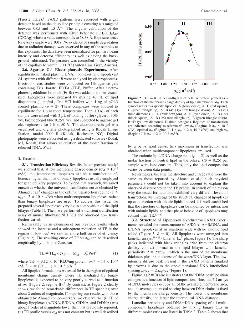
Remarkably, as an extension of our previous results whichshowed the increase and a subsequent reduction of TE in theregime of low !M,8 we saw an entire bell curve of efficiency(Figure 2). The resulting curve of TE vs !M can be describedempirically by a simple Gaussian
TE)TE0 × exp - [(!M - !M! )/w]2 (1)
where TE0 ) 3.12 ! 107 RLU/mg protein, !M* " 14 ! 10-3
e/Å-2, w ) (13 ( 1) ! 10-4 e/Å-2.All lipoplex formulations we tested lie in the region of optimal
membrane charge density where TE mediated by binarylipoplexes is expected to be very poorly curved as a functionof !M (Figure 2, regime II).7 By contrast, as Figure 2 clearlyshows, we found remarkable differences in TE spanning overabout 2 orders of magnitude. Comparing our results with thoseobtained by Ahmad and co-workers, we observe that (i) TE ofbinary lipoplexes (A/DNA, B/DNA, C/DNA, and D/DNA) wasabout 1 order of magnitude lower than that previously reported,(ii) TE profile versus !M was not constant but is well described
by a bell-shaped curve, (iii) maximum in transfection wasobtained when multicomponent lipoplexes are used.
The cationic lipid/DNA charge ratio (F ) 2) as well as themolar fraction of neutral lipid in the bilayer (! ) 0.25) persample were kept constant. Thus, only the lipid compositionvaries between data points.
Nevertheless, because the structure and charge ratio were thesame as those reported by Ahmad et al.,7 such physicalparameters could not be taken into account to explain theobserved discrepancy in the TE profile. In search of the reasonwhy the tested formulations exhibited very different levels oftransfection, we investigated the structural changes of lipoplexesupon interaction with anionic lipids. Indeed, it is well establishedthat the structure of lipoplexes can be modified by interactionwith anionic lipids, and that phase behavior of lipoplexes maycontrol their TE.12-17
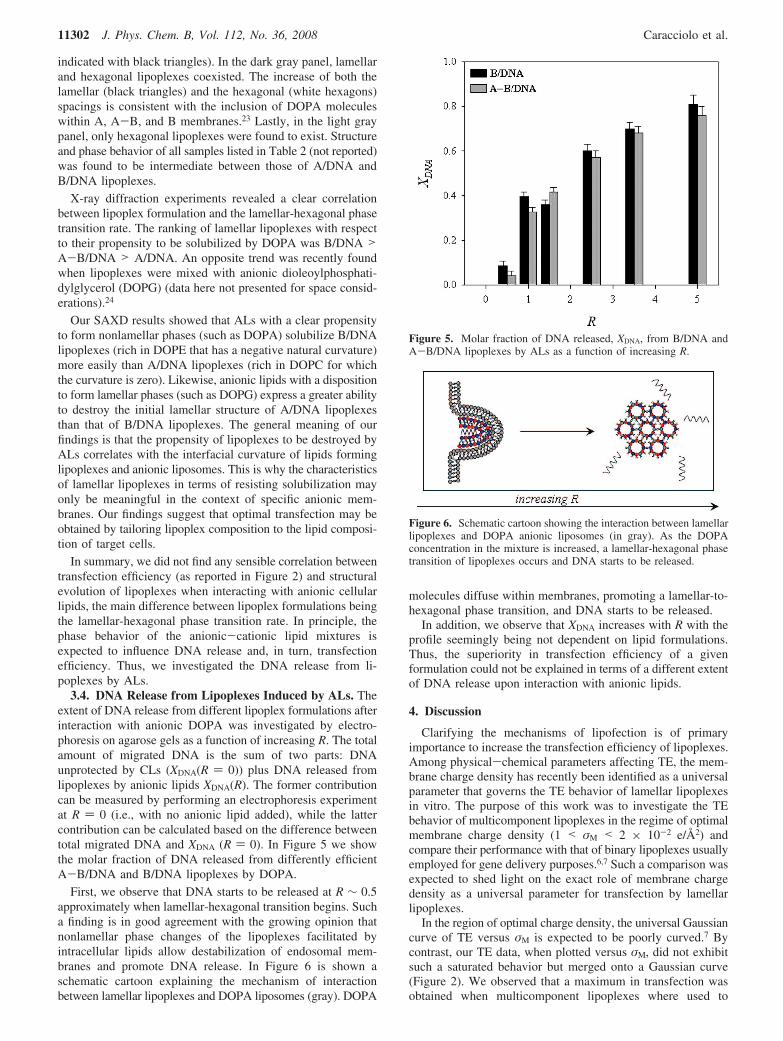
3.2. Structure of Lipoplexes. Synchrotron SAXD experi-ments revealed the nanostructure of A/DNA, A-B/DNA, andB/DNA lipoplexes at an angstrom scale with no anionic lipidadded (Figure 3, R ) 0). All lipoplexes were arranged intolamellar arrays,18-21 (lamellar LRC phase, Figure 1). The sharppeaks indicated with black triangles arise from the electrondensity contrast normal to the lipid bilayer with lamellarperiodicity d ) 2#/q001, which is the sum of the membranethickness plus the thickness of the water/DNA layer. The low-intensity diffuse peak present in the SAXD patterns (markedby arrows) is due to the one-dimensional DNA interhelicalspacing dDNA ) 2#/qDNA (Figure 1).
Figure 3 (R ) 0) also illustrates that the ‘DNA peak’ positionchanges as a function of lipid composition. Thus, the 2D arraysof DNA molecules occupy all of the available membrane area,and the average interaxial spacing between DNA chains is fixedby the membrane charge density. The lower the membranecharge density, the larger the interhelical DNA distance.
Lamellar periodicity and DNA-DNA spacing of all multi-component lipoplexes obtained by mixing binary CLs indifferent molar ratios are listed in Table 2. Table 2 shows that
Figure 2. TE in RLU per milligram of cellular protein plotted as afunction of the membrane charge density of lipid membranes, !M. Eachsymbol refers to a specific lipoplex: A (black circle), A-C (red square),C (green triangle up), A-B (4:1) (yellow triangle down), A-B (3:1)(blue diamond), C-D (pink hexagon), A-B (cyan circle), A-B (1:2)(black square), A-B (1:5) (red triangle up), B (green triangle down),B-D (yellow diamond), D (blue hexagon). Regimes of transfectionare indicated according to reference:7 low !M (Regime I: !M < 10-2
e/Å2), optimal !M (Regime II: 1 < !M < 2 ! 10-2 e/Å2), and high !M
(Regime III: !M > 2 ! 10-2 e/Å2).
11300 J. Phys. Chem. B, Vol. 112, No. 36, 2008 Caracciolo et al.

the physical properties of multicomponent lipoplexes areintermediate between those of binary lipoplexes and mighttherefore be adjusted merely by varying their lipid composition.This indicates that multicomponent lipid/DNA complexes canbe prepared with designed physical properties. In the future,rationally designed vectors shall incorporate the most usefulelements of synthetic systems with variations depending on theirspecific application.
3.3. Phase Evolution of Lipoplexes upon Interaction withAnionic Lipids. Figure 3 shows representative SAXD patternsof A/DNA/DOPA, A-B/DNA/DOPA, and B/DNA/DOPAmixtures as a function of the anionic/cationic charge ratio R.Here experiments with A/DNA/DOPA and B/DNA/DOPA arepresented because these lipid formulations provided the clearestevidence of structural differences upon interaction with anioniclipids, while the A-B/DNA/DOPA system exhibited an inter-mediate behavior.
At low R (R < 0.4), only lamellar lipoplexes were found toexist with the one-dimensional (1D) DNA lattice being dilutedby the anionic lipid (data not reported).8,17,22
As the DOPA concentration was increased in the mixture, alamellar-hexagonal phase transition occurred. At R ) 1, threeBragg peaks (indicated with white hexagons) were clearly visible
onto the SAXD patterns of all investigated samples and wereindexed as the (10), (11), and (20) reflections of an invertedhexagonal phase. Lamellar-to-hexagonal phase transition wascomplete at R ) 1 in the case of both B/DNA/DOPA and A-B/DNA/DOPA systems, while A/DNA lipoplexes partly resistedsolubilization as shown by the presence of the first-order lamellarBragg peak (black triangle). The tendency to form invertedphases observed here is compatible with earlier results in theliterature showing that DOPA holds the potential to promotethe adoption of nonlamellar structure of phospholipids.23 Ad-ditional weak reflections (indicated with gray diamonds) werealso seen. Such reflections are characteristic of the final phaseof DOPA (data elsewhere reported).22
At R ) 5, lamellar lipoplexes were completely solubilized,while hexagonal complexes (white hexagons) were found tocoexist with pure DOPA (gray diamonds).
Figure 4 shows the phase diagram of A/DNA/DOPA, A-B/DNA/DOPA, and B/DNA/DOPA mixtures. In the white panelonly were lamellar lipoplexes detected (lamellar d spacings are
Figure 3. Representative synchrotron SAXD patterns of A/DNA/DOPA, A-B/DNA/DOPA, and B/DNA/DOPA mixtures as a functionof the anionic/cationic charge ratio R. Bragg peaks arising from thelamellar structure of A/DNA, A-B/DNA, and B/DNA lipoplexes areindicated with black triangles. At R ) 0, the ‘DNA peak’ arising frominterstrand correlation is marked by an arrow. Diffraction maxima ofhexagonal complexes are indicated with white hexagons, while reflec-tions of pure DOPA are indicated with gray diamonds.
TABLE 2: Lamellar Periodicity, d, and DNA-DNASpacing, dDNA, of the Multicomponent LipoplexesInvestigated by SAXDa
lipoplex formulation d (Å) dDNA (Å)
A 62.8 37.1A-B (4:1) 63.5 37.0A-B (3:2) 64.1 36.8A-B 64.8 34.9A-B (2:3) 66.2 32.1A-B (1:4) 66.8 31.4B 67.6 29.2
a Where not specified, cationic liposomes were mixed in equi-molar ratio.
Figure 4. Phase diagram of A/DNA, A-B/DNA, and B/DNAlipoplexes with differing DOPA content. Dashed lines separate regionsoccupied by lamellar lipoplexes (white panel), coexisting lamellar andhexagonal lipoplexes (dark gray panel), and hexagonal lipoplexes (lightgray panel). Repeat spacings, d, of lamellar and hexagonal lipoplexesare indicated with black triangles and white hexagons, respectively.To clarify, the R range was restricted to 3.5.
Transfection Efficiency of Multicomponent Lipoplexes J. Phys. Chem. B, Vol. 112, No. 36, 2008 11301
indicated with black triangles). In the dark gray panel, lamellarand hexagonal lipoplexes coexisted. The increase of both thelamellar (black triangles) and the hexagonal (white hexagons)spacings is consistent with the inclusion of DOPA moleculeswithin A, A-B, and B membranes.23 Lastly, in the light graypanel, only hexagonal lipoplexes were found to exist. Structureand phase behavior of all samples listed in Table 2 (not reported)was found to be intermediate between those of A/DNA andB/DNA lipoplexes.
X-ray diffraction experiments revealed a clear correlationbetween lipoplex formulation and the lamellar-hexagonal phasetransition rate. The ranking of lamellar lipoplexes with respectto their propensity to be solubilized by DOPA was B/DNA >A-B/DNA > A/DNA. An opposite trend was recently foundwhen lipoplexes were mixed with anionic dioleoylphosphati-dylglycerol (DOPG) (data here not presented for space consid-erations).24
Our SAXD results showed that ALs with a clear propensityto form nonlamellar phases (such as DOPA) solubilize B/DNAlipoplexes (rich in DOPE that has a negative natural curvature)more easily than A/DNA lipoplexes (rich in DOPC for whichthe curvature is zero). Likewise, anionic lipids with a dispositionto form lamellar phases (such as DOPG) express a greater abilityto destroy the initial lamellar structure of A/DNA lipoplexesthan that of B/DNA lipoplexes. The general meaning of ourfindings is that the propensity of lipoplexes to be destroyed byALs correlates with the interfacial curvature of lipids forminglipoplexes and anionic liposomes. This is why the characteristicsof lamellar lipoplexes in terms of resisting solubilization mayonly be meaningful in the context of specific anionic mem-branes. Our findings suggest that optimal transfection may beobtained by tailoring lipoplex composition to the lipid composi-tion of target cells.
In summary, we did not find any sensible correlation betweentransfection efficiency (as reported in Figure 2) and structuralevolution of lipoplexes when interacting with anionic cellularlipids, the main difference between lipoplex formulations beingthe lamellar-hexagonal phase transition rate. In principle, thephase behavior of the anionic-cationic lipid mixtures isexpected to influence DNA release and, in turn, transfectionefficiency. Thus, we investigated the DNA release from li-poplexes by ALs.
3.4. DNA Release from Lipoplexes Induced by ALs. Theextent of DNA release from different lipoplex formulations afterinteraction with anionic DOPA was investigated by electro-phoresis on agarose gels as a function of increasing R. The totalamount of migrated DNA is the sum of two parts: DNAunprotected by CLs (XDNA(R ) 0)) plus DNA released fromlipoplexes by anionic lipids XDNA(R). The former contributioncan be measured by performing an electrophoresis experimentat R ) 0 (i.e., with no anionic lipid added), while the lattercontribution can be calculated based on the difference betweentotal migrated DNA and XDNA (R ) 0). In Figure 5 we showthe molar fraction of DNA released from differently efficientA-B/DNA and B/DNA lipoplexes by DOPA.
First, we observe that DNA starts to be released at R " 0.5approximately when lamellar-hexagonal transition begins. Sucha finding is in good agreement with the growing opinion thatnonlamellar phase changes of the lipoplexes facilitated byintracellular lipids allow destabilization of endosomal mem-branes and promote DNA release. In Figure 6 is shown aschematic cartoon explaining the mechanism of interactionbetween lamellar lipoplexes and DOPA liposomes (gray). DOPA
molecules diffuse within membranes, promoting a lamellar-to-hexagonal phase transition, and DNA starts to be released.
In addition, we observe that XDNA increases with R with theprofile seemingly being not dependent on lipid formulations.Thus, the superiority in transfection efficiency of a givenformulation could not be explained in terms of a different extentof DNA release upon interaction with anionic lipids.
4. Discussion
Clarifying the mechanisms of lipofection is of primaryimportance to increase the transfection efficiency of lipoplexes.Among physical-chemical parameters affecting TE, the mem-brane charge density has recently been identified as a universalparameter that governs the TE behavior of lamellar lipoplexesin vitro. The purpose of this work was to investigate the TEbehavior of multicomponent lipoplexes in the regime of optimalmembrane charge density (1 < !M < 2 ! 10-2 e/Å2) andcompare their performance with that of binary lipoplexes usuallyemployed for gene delivery purposes.6,7 Such a comparison wasexpected to shed light on the exact role of membrane chargedensity as a universal parameter for transfection by lamellarlipoplexes.
In the region of optimal charge density, the universal Gaussiancurve of TE versus !M is expected to be poorly curved.7 Bycontrast, our TE data, when plotted versus !M, did not exhibitsuch a saturated behavior but merged onto a Gaussian curve(Figure 2). We observed that a maximum in transfection wasobtained when multicomponent lipoplexes where used to
Figure 5. Molar fraction of DNA released, XDNA, from B/DNA andA-B/DNA lipoplexes by ALs as a function of increasing R.
Figure 6. Schematic cartoon showing the interaction between lamellarlipoplexes and DOPA anionic liposomes (in gray). As the DOPAconcentration in the mixture is increased, a lamellar-hexagonal phasetransition of lipoplexes occurs and DNA starts to be released.
11302 J. Phys. Chem. B, Vol. 112, No. 36, 2008 Caracciolo et al.

transfect NIH 3T3 cells, while binary lipoplexes were definitelyless efficient. This finding suggests that, at fixed !M, distinctlevels of transfection can be obtained by adjusting otherphysical-chemical properties of lipoplexes such as, for instance,the number of lipid components. The general meaning of ourfindings is that membrane charge density controls transfectionbehavior of lamellar lipoplexes, with specific variations depend-ing on lipid composition.
To provide a rationale for discrepancy between our findings8
and those previously reported,7 we investigated some physical-chemical features that might account for the superior transfectionefficiency of the most efficient multicomponent lipoplexes suchas their lipid composition, structure, propensity to be disinte-grated by ALs, and ability to release DNA. All investigatedlipoplexes were arranged into lamellar arrays, with distinct DNApacking densities reflecting their different membrane chargedensity (Table 2).
Current work shows that a major impediment to transfectioninvolves the relative inefficient destabilization of membrane-bound compartments in which lipoplexes are confined after theirinternalization. A current opinion is that the structural evolutionof lipoplexes upon interaction with cellular lipids may be acontrolling factor in DNA release.23,25,26 Evidence is accumulat-ing that the lamellar/nonlamellar phase transition of lipoplexesfacilitated by intracellular lipids, allowing destabilization ofendosomal membranes and being helpful in plasmid transloca-tion into the cytosol, is a prerequisite for nuclear delivery.23,25,26
However, after interaction with anionic DOPA vesicles, usedto simulate lipoplex-endosomal membrane interaction, all testedformulations exhibited a practically identical lamellar-invertedhexagonal phase change with minor differences in transitionrate (Figure 4). Thus, we were not able to correlate thepropensity of a given formulation to form nonlamellar phaseswith distinct levels of TE.
As mentioned above, another factor that is likely to beimportant in the efficiency of transfection is the dissociation ofDNA from cationic lipids. It appears highly critical for ac-complishing efficient translocation of nucleic acids across theendosomal membrane into the cytosol for transport to thenucleus. Thus, we measured the extent of DNA release fromdifferent lipoplex formulations after interaction with ALs byelectrophoresis on agarose gels as a function of increasing R.
We observe (Figure 5) that XDNA does not vary significantlywith R with lipid formulations. This observation correlates wellwith our SAXD findings showing a virtually identical phaseevolution of the tested lipoplex formulations as a function ofR. Indeed, we have recently shown8,24 the existence of a strictcorrelation between destabilization of lipoplexes and DNAunbinding: the most stable lipoplexes display the lowest extentof DNA release, while lipoplexes with the highest propensityto be disintegrated by anionic lipids also exhibit much largerDNA release.24
As a result, neither the structural changes of lipoplexes uponinteraction with anionic cellular lipids nor their ability to releaseDNA were found to correlate with TE. Apparently, parametersother than simple structural stability of lipoplexes againstsolubilization by ALs determined the transfection capacity of agiven complex. Specifically, on the basis of observations of TEbehavior of multicomponent lipoplexes in the regime of low8
and optimal membrane charge density, our results suggest thatthe transfection efficiency may be strictly related to thecomposition of lipoplex formulations. To better test thesesuggestions, in Figure 7 we plotted TE as a function of bothDC-Chol/DOTAP and DOPE/PC ratios. We observe that TE
peaked at DC-Chol/DOTAP ) 0.5 and DOPE/PC ) 0.5 whenboth cationic and neutral lipid species are mixed in equimolarratio. We observed that not only the number of lipid componentsbut also their relative molar ratio (i.e., the specific compositionof the lipid carrier) altered transfection activity. First, the choiceof cationic lipid can affect TE significantly. Indeed, DC-Chol-based lipoplexes (DC-Chol/DOTAP ) 1) were found to be moreefficient than those containing DOTAP as cationic lipid (DC-Chol/DOTAP ) 0). Neutral colipids can also alter activity oflipoplexes. For instance, B/DNA lipoplexes (DC-Chol/DOTAP) 1, DOPE/PC ) 1) were found to be 2-fold more efficientthan D/DNA lipoplexes (DC-Chol/DOTAP ) 1, DOPE/PC )0). This finding indicates that, at a fix number of lipidcomponents, DOPE as a colipid gave higher transfection in DC-Chol-containing lipoplexes.
Our findings add a compositional degree of freedom, becauseefficiently transfecting complexes can be prepared from a broadrange of lipids maximizing TE if composition is optimized. Theexact physical-chemical reasons why multicomponent li-poplexes are more efficient than binary lipoplexes cannot bestated unambiguously. However, some speculative argumentscan be proposed. Among the barriers to transfection, escapefrom endosomal compartments is a major obstacle for efficientDNA delivery. The capacity of amphiphile molecules to perturbthe endosomal bilayer structure will be of primary importancein facilitating the plasmid release. However, biological mem-branes are complex multicomponent systems consisting ofmixtures of many different lipids and proteins. Accordingly,the superiority in TE of multicomponent lipoplexes may berelated to the higher fusogenicity and compatibility of vesiclesmade of several lipid components with respect to single lipidsor binary lipoplexes.27,28 This relationship between deliveryactivity and physical property can be rationalized on the basisof the known consequences of packing defects as well as largelocal density fluctuations that could be responsible for theenhanced fusogenicity of multicomponent blends of lipids. Wetherefore emphasize the importance of the lipid composition ofboth lipoplexes and target membranes and suggest that adaptinglipoplex composition to the lipid composition of target cellsmay be a successful strategy for the rational design of superiorcationic lipid carriers. Indeed, even though the significance oflipid diversity in biological membranes still remains obscure,
Figure 7. TE in RLU per milligram of cellular protein plotted as afunction of both DC-Chol/DOTAP and DOPE/PC ratios.
Transfection Efficiency of Multicomponent Lipoplexes J. Phys. Chem. B, Vol. 112, No. 36, 2008 11303
it is expected to play a role in formation of nonbilayer phases,membrane interaction, and fusion and biocompatibility.29
5. Conclusions
In this work, we have investigated the transfection behaviorof multicomponent lipoplexes in the range of optimal membranecharge density. Such complexes have presented a much highertransfection efficiency than binary lipoplexes, which are morecommonly used for gene-delivery purposes. These resultshighlight the compositional properties of carrier lipid/cellularlipid mixtures as decisive factors for transfection and suggestthat multicomponent systems are especially promising lipoplexcandidates.
The biological and cellular responses to the process ofphysical stimulation involving gene transfer also deserve moreconsideration. Innovation in applying the principles of physics,chemistry, and biology to the development of a safe andeffective method for gene delivery is the key to make theurgently needed advances in nonviral gene therapy. In the nextfuture, the engineering of lipoplexes, incorporating the specificproperties of different lipid species, may represent the startingpoint to rationally design highly specific gene vectors.
References and Notes
(1) Felgner, P. L.; Ringold, G. M. Nature 1989, 337, 387–388.(2) Safinya, C. R. Curr. Opin. Struct. Biol. 2001, 11, 440–448.(3) Lin, A. J.; Slack, N. L.; Ahmad, A.; Koltover, I.; George, C. X.;
Samuel, C. E.; Safinya, C. R. J. Drug Target. 2000, 8, 13–27.(4) Caracciolo, G.; Pozzi, D.; Caminiti, R.; Congiu Castellano, A. Eur.
Phys. J. E 2003, 10, 331–336.(5) McManus, J. J.; Radler, J. O.; Dawson, K. A. J. Am. Chem. Soc.
2004, 126, 15966–15967.(6) Ewert, K. K.; Ahmad, A.; Evans, H. M.; Safinya, C. R. Expert
Opin. Biol. Ther. 2005, 5, 33–53.(7) Ahmad, A.; Evans, H. M.; Ewert, K. K.; George, C. X.; Samuel,
C. E.; Safinya, C. R. J. Gene Med. 2005, 7, 739–748.(8) Caracciolo, G.; Marchini, C.; Pozzi, D.; Caminiti, R.; Amenitsch,
H.; Montani, M.; Amici, A. Langmuir 2007, 23, 4498–4508.
(9) Caracciolo, G.; Pozzi, D.; Caminiti, R.; Amenitsch, H. J. Phy-s.Chem. B 2006, 110, 20829–20835.
(10) Pozzi, D.; Amenitsch, H.; Caminiti, R.; Caracciolo, G. Chem. Phys.Lett. 2006, 429, 250–254.
(11) Amenitsch, H.; Rappolt, M.; Kriechbaum, M.; Mio, H.; Laggner,P.; Bernstorff, S. J. Synchrotron Radiat. 1998, 5, 506–508.
(12) Koynova, R.; Wang, L.; Tarahovsky, Y. S.; MacDonald, R. C.Bioconjugate Chem. 2005, 16, 1335–1339.
(13) MacDonald, R. C.; Ashley, G. W.; Shida, M. M.; Rakhmanova,V. A.; Tarahovsky, Y. S.; Pantazatos, D. P.; Kennedy, M. T.; Pozharski,E. V.; Baker, K. A.; Jones, R. D.; Rosenzweig, H. S.; Choi, K. L.; Qiu,R. Z.; McIntosh, T. J. Biophys. J. 1999, 77, 2612–2629.
(14) Xu, Y. H.; Szoka, F. C. Biochemistry 1996, 35, 5616–5623.(15) Tarahovsky, Y.; Koynova, R.; MacDonald, R. C. Biophys. J. 2004,
86, 37A.(16) Caracciolo, G.; Pozzi, D.; Caminiti, R.; Marchini, C.; Montani, M.;
Amici, A.; Amenitsch, H. Appl. Phys. Lett. 2006, 89, 233903–3.(17) Caracciolo, G.; Pozzi, D.; Amenitsch, H.; Caminiti, R. Langmuir
2007, 23, 8713–8717.(18) Salditt, T.; Koltover, I.; Radler, J. O.; Safinya, C. R. Phys. ReV.
Lett. 1997, 79, 2582–2585.(19) Salditt, T.; Koltover, I.; Radler, J. O.; Safinya, C. R. Phys. ReV. E
1998, 58, 889–904.(20) Koltover, I.; Salditt, T.; Radler, J. O.; Safinya, C. R. Science 1998,
281, 78–81.(21) Artzner, F.; Zantl, R.; Rapp, G.; Radler, J. O. Phys. ReV. Lett. 1998,
81, 5015–5018.(22) Caracciolo, G.; Pozzi, D.; Caminiti, R.; Marchini, C.; Montani, M.;
Amici, A.; Amenitsch, H. Appl. Phys. Lett. 2007, 91, 143903–3.(23) Tarahovsky, Y. S.; Koynova, R.; MacDonald, R. C. Biophys. J.
2004, 87, 1054–1064.(24) Caracciolo, G.; Pozzi, D.; Caminiti, R.; Marchini, C.; Montani, M.;
Amici, A.; Amenitsch, H. Biochim. Biophys. Acta 2007, 1768, 2280–2292.(25) Koynova, R.; Wang, L.; MacDonald, R. C. Proc. Natl. Acad. Sci.
U.S.A. 2006, 103, 14373–14378.(26) Wang, L.; Koynova, R.; Parikh, H.; MacDonald, R. C. Biophys. J.
2006, 91, 3692–3706.(27) Lin, W.; Blanchette, C. D.; Longo, M. L. Biophys. J. 2007, 92,
2831–2841.(28) Alonso, M. A.; Millan, J. J. Cell Sci. 2001, 114, 3957–3965.(29) Pautot, S.; Frisken, B. J.; Weitz, D. A. Proc. Natl. Acad. Sci. U.S.A.
2003, 100, 10718–10721.(30) Hed, G.; Safran, S. A. Phys. ReV. Lett. 2004, 93, 138101–4.
JP803077N
11304 J. Phys. Chem. B, Vol. 112, No. 36, 2008 Caracciolo et al.