Isolation and Characterization of an Antigenically Distinct 68- kd Protein from Nonviral...
12
http://vet.sagepub.com/ Veterinary Pathology Online http://vet.sagepub.com/content/37/5/449 The online version of this article can be found at: DOI: 10.1354/vp.37-5-449 2000 37: 449 Vet Pathol E. Wozniak, J. McBride, D. DeNardo, R. Tarara, V. Wong and B. Osburn Retroviridae) Inclusions in Boa Constrictors Chronically Infected with the Inclusion Body Disease Virus (IBDV: Isolation and Characterization of an Antigenically Distinct 68- kd Protein from Nonviral Intracytoplasmic Published by: http://www.sagepublications.com On behalf of: Pathologists. American College of Veterinary Pathologists, European College of Veterinary Pathologists, & the Japanese College of Veterinary can be found at: Veterinary Pathology Online Additional services and information for http://vet.sagepub.com/cgi/alerts Email Alerts: http://vet.sagepub.com/subscriptions Subscriptions: http://www.sagepub.com/journalsReprints.nav Reprints: http://www.sagepub.com/journalsPermissions.nav Permissions: What is This? - Sep 1, 2000 Version of Record >> at UT SCHOOL PUBLIC HLTH LIBRARY on January 3, 2014 vet.sagepub.com Downloaded from at UT SCHOOL PUBLIC HLTH LIBRARY on January 3, 2014 vet.sagepub.com Downloaded from
-
Upload
independent -
Category
Documents
-
view
0 -
download
0
Transcript of Isolation and Characterization of an Antigenically Distinct 68- kd Protein from Nonviral...

http://vet.sagepub.com/Veterinary Pathology Online
http://vet.sagepub.com/content/37/5/449The online version of this article can be found at:
DOI: 10.1354/vp.37-5-449
2000 37: 449Vet PatholE. Wozniak, J. McBride, D. DeNardo, R. Tarara, V. Wong and B. Osburn
Retroviridae)Inclusions in Boa Constrictors Chronically Infected with the Inclusion Body Disease Virus (IBDV:
Isolation and Characterization of an Antigenically Distinct 68- kd Protein from Nonviral Intracytoplasmic
Published by:
http://www.sagepublications.com
On behalf of:
Pathologists.American College of Veterinary Pathologists, European College of Veterinary Pathologists, & the Japanese College of Veterinary
can be found at:Veterinary Pathology OnlineAdditional services and information for
http://vet.sagepub.com/cgi/alertsEmail Alerts:
http://vet.sagepub.com/subscriptionsSubscriptions:
http://www.sagepub.com/journalsReprints.navReprints:
http://www.sagepub.com/journalsPermissions.navPermissions:
What is This?
- Sep 1, 2000Version of Record >>
at UT SCHOOL PUBLIC HLTH LIBRARY on January 3, 2014vet.sagepub.comDownloaded from at UT SCHOOL PUBLIC HLTH LIBRARY on January 3, 2014vet.sagepub.comDownloaded from

449
Vet Pathol 37:449–459 (2000)
Isolation and Characterization of an Antigenically Distinct 68-kd Protein from Nonviral Intracytoplasmic Inclusions in Boa
Constrictors Chronically Infected with the Inclusion BodyDisease Virus (IBDV: Retroviridae)
E. WOZNIAK, J. MCBRIDE, D. DENARDO, R. TARARA, V. WONG, AND B. OSBURN
Department of Pathology, Microbiology and Immunology, School of Veterinary Medicine, University of California,Davis, CA (EW,1 JM, RT, VW, BO); and
Office of Laboratory Animal Care, Northwest Animal Facility, University of California, Berkeley, CA (DD)
Abstract. The relationship between a retroviral infection and the development of nonviral intracytoplasmicinclusion bodies was studied in a Boa constrictor model. Twelve juvenile age- and size-matched inclusion bodydisease (IBD)-negative boas were randomly divided into three groups. Each group was inoculated intraperito-neally with 1 ml of an IBD virus (IBDV)-infected liver homogenate or 1 ml of normal boa liver homogenate(sham-inoculated control) or was left untreated. All boas were monitored for development of IBD by dailyexamination and serial liver biopsy over 1 year. The 4 IBDV-inoculated boas became IBDV and inclusionpositive by 10 weeks postinoculation. The average size and density of inclusion bodies increased with theduration of infection. Ultrastructurally, inclusion bodies ,2 mm in diameter consisted of intracytoplasmic ag-gregates of granular electron-dense material that were not membrane limited. Larger inclusions (3–6 mm indiameter) were characterized as membrane-bound aggregates of amorphous to granular electron-dense materialadmixed with membranelike fragments. The sham-inoculated and untreated control snakes did not becomeinclusion or IBDV positive. Direct comparison of the protein electrophoretograms of IBDV-infected and normalboa tissues demonstrated a prominent 68-kd protein band unique to infected inclusion-positive tissues. Mono-clonal antibodies directed against the 68-kd protein band specifically labeled inclusion bodies. The results ofthis study demonstrate that IBD inclusions represent an intracytoplasmic accumulation of an antigenically dis-tinct IBDV-associated protein.
Key words: Boa constrictor; hyaline inclusions; inclusion body disease; Retroviridae; retrovirus.
A disease entity named for its association with in-tracytoplasmic inclusion bodies of various sizes withinthe epithelial cells of visceral organs and neurons hasbeen recognized in snakes of the family Boidae sincethe 1970s.25 Clinical signs are variable and includechronic regurgitation, neurologic disease characterizedby star gazing, dysequilibrium, tremors, disorientation,and paresis, increased susceptibility to secondary in-fections, and neoplasia.25 In 1994, a C-type retrovirus-like agent that buds from both the endoplasmic retic-ulum and plasma membrane was identified as the eti-ologic agent.25 Experimental transmission studies haveshown that inclusion body disease (IBD) can be effec-tively transmitted by intraperitoneal and subcutaneousinjection of fluids containing mature viral particles.25
Very little is known about the natural history of thevirus and how IBD is transmitted in nature.25 It hasbeen speculated that the blood-feeding macronyssid
1 Present address: Animal Resources Center, University ofTexas Medical Branch, Galveston, TX.
mite Ophionyssus natricis may be involved in thetransmission cycle, but vector incrimination studies arelacking.25 The inclusions after which the disease wasnamed were initially described as non-membrane-bound aggregates of granular electron-dense materialwithout further characterization.25 The morphology ofthe described inclusion bodies most closely resemblesthat described for proteinaceous nonviral inclu-sions.7,9,11,15,20,23
Nonviral inclusions characterized as amorphous in-tracytoplasmic eosinophilic hyaline bodies have beendemonstrated in human and animal tissues afflictedwith a diverse array of disease conditions, includingalcoholism, neoplasia, morbid obesity, Indian child-hood cirrhosis, pyrrolizidine alkaloid and copper ex-posure, and prolonged griseofulvin treatment.2,7–9,11,15,26
These inclusion bodies represent intracytoplasmic ag-gregates of various intermediate filaments, includingcytokeratins, vimentin, and neurofilaments.8,10,11,20 Insome cases, the intermediate filaments within non-membrane-bound inclusions were thickened by a dif-fuse coating of ubiqutin.18,20
at UT SCHOOL PUBLIC HLTH LIBRARY on January 3, 2014vet.sagepub.comDownloaded from

450 Vet Pathol 37:5, 2000Wozniak, McBride, DeNardo, Tarara, Wong, and Osburn
Nonviral inclusions consisting of unidentified gran-ular to fibrillar electron-dense material have been dem-onstrated in cells infected with several retroviruses, in-cluding avian leukosis virus, visna virus, human im-munodeficency virus (HIV), and simian immunodefi-ciency virus (SIV).1,6,12,21,32 The ultrastructuralmorphology of retrovirus-associated inclusions arevariable. One variant known as tubuloreticular inclu-sions was described as fine tubular meshworks of lip-id-rich membranes complexed with unidentified pro-teins that distended the endoplasmic reticulum.13,21
Other retrovirus-associated inclusions were character-ized as basophilic intracytoplasmic bodies of varioussizes composed of non-membrane-limited aggregatesof viral protein and ribosomes.12 Large eosinophilicintracytoplasmic bodies that approximate those ob-served with IBD of boid snakes have not been de-scribed in association with any other retroviral infec-tion.
In the original description of IBD, the virus and in-clusion bodies were usually found together in the sameanimal and often within the same cell, but a clear caus-al relationship between the two entities was not estab-lished. Despite the lack of an established casual rela-tionship between the virus and the inclusion body, thecurrent recommendation is to euthanatize all boidsnakes exhibiting signs compatible with IBD when thecharacteristic inclusion bodies in biopsy specimens arerevealed by light microscopy.26 Because of the mor-phologic and histochemical similarities between IBDinclusions and other types of nonviral intracytoplasmicbodies, we decided to study and better characterize theIBD virus (IBDV)-associated inclusions.2,7–9,11,15,25,26
The objectives of the study were to examine the re-lationship between the retrovirus-like agent and its as-sociated nonviral inclusion bodies and to identify andcharacterize the material within the inclusion bodies.
Materials and Methods
Source of IBDV
An adult female Boa constrictor amarali with a historyof chronic regurgitation, recurrent mouth rot, shedding dif-ficulties, and posterior paresis was euthanatized with pen-tobarbitol, and a complete necropsy examination was per-formed at the owner’s request. Samples of liver, lung, kid-ney, splenopancreas, brain, and spinal cord were collectedand processed for paraffin sectioning. Additional specimensof liver and kidney were either processed for transmissionelectron microscopy (TEM) or frozen at 280 C. Light mi-croscopy revealed numerous intracytoplasmic eosinophilicinclusion bodies (up to 140/high-power field) within hepa-tocytes, neurons, pancreatic acinar cells, and lymphocytes.Ultrastructural evaluation of the liver by TEM revealedabundant C-type retrovirus-like particles that were 100–110nm in diameter with an electron-dense core enclosed by ahexagonal capsid. A membranous envelope bearing short pe-
plomers covered the external surface. The size, morphology,and budding properties of the viruslike particles were allconsistent with IBDV.25 No other lesions or pathogens wereidentified. The signalment, history, clinical syndrome, his-topathology, and ultrastructural findings all supported a finaldiagnosis of IBD. The frozen liver and kidney were labeledas IBDV and inclusion positive and retained as a futuresource of virus and proteins.
Experimental infection
A group of 12 clinically normal age- and size-matchedjuvenile boa constrictors were obtained through a donationfrom the Birmingham Zoo. All boas were individuallyhoused in appropriately sized polypropylene cages equippedwith a water bowl, hide box, a rock, and a focal heat sourceto allow for behavioral thermoregulation. All cages weremaintained in University of California (UC)–Berkeley ani-mal rooms with an ambient temperature of 24–28 C and a12 : 12 hr light–dark cycle. Routine care was providedthrough the UC–Berkeley Office of Laboratory Animal Care.After a 1-month period of acclimation, each snake was anes-thetized with isofluorane, a celiotomy was performed, and abaseline wedge biopsy of liver approximately 6 3 4 3 2mm was collected from each snake. Each biopsy specimenwas halved upon collection and processed for both light andelectron microscopy. Bleeding from the biopsy site was con-trolled with sterile gel foam. The body wall and skin wereclosed with absorbable monofilament suture material. Allsnakes were monitored closely for evidence of infection orother postsurgical complications for 10 days. All 12 snakesremained clinically and behaviorally normal throughout theacclimation and postoperative healing period. No inclusionsor retrovirus-like particles were demonstrated in any of thebaseline biopsy specimens. The persistent clinical normalcyand negative biopsy findings indicated that all 12 boas werefree of IBD.
Approximately 1 g of the frozen IBDV-positive boa liverwas thawed, diluted 1 : 10 in sterile serum free minimumessential medium, and homogenized in a ground glass tissuehomogenizer to prepare an inoculum for experimental trans-mission. The resulting homogenate was clairified by centri-fugation at 1,000 3 g for 10 minutes, and the supernatantwas pushed through a 0.45-mm filter under positive pressure.A sham inoculum prepared from a clinically normal B. con-strictor liver that was determined to be free of inclusionsand IBDV by light microscopy and TEM was prepared byidentical methods.
The group of 12 IBDV-free boas was randomly split intothree groups of four snakes each. In one group, the foursnakes were each given 1 ml of the filtered normal boa liverhomogenate by intraperitoneal (IP) injection (sham-inocu-lated controls).25 To eliminate the possibility of accidentalinfection, the sham inoculation was prepared and adminis-tered before any IBDV-infected material was handled. In thesecond group, each snake was inoculated with 1 ml of thefiltered IBDV–infected liver homogenate by IP injection.25
The third group was left untreated (untreated controls). Allthree groups of boas were monitored daily for clinical evi-dence of IBDV infection following a strict clean-to-dirty or-der of operation. The experimentally infected and control
at UT SCHOOL PUBLIC HLTH LIBRARY on January 3, 2014vet.sagepub.comDownloaded from

Vet Pathol 37:5, 2000 451Nonviral Inclusions in IBDV
animals were housed in different rooms and were nevercared for by the same technician.
All 12 boas were serially monitored for histopathologicchanges and ultrastructural evidence of IBDV infection byrepeated hepatic wedge biopsies taken at 10, 20, and 32weeks postinoculation (PI). Each biopsy specimen was di-vided into three pieces upon collection and processed forparaffin sectioning, frozen sectioning, and TEM. At 52weeks PI, all 12 boas were euthanatized with intracardiacpentobarbital (100 mg/kg) while under a surgical plane ofisofluorane anesthesia, and complete necropsies were per-formed. Samples of liver, gall bladder, lung, kidney, splen-opancreas, brain, and spinal cord were collected and pro-cessed for paraffin sectioning. Additional specimens of liverand kidney from each snake were processed for TEM (liverand kidney), immunoelectron microscopy (liver), frozen sec-tioning (liver), or were frozen at 280 C (liver and kidney).All procedures involving the use of animals were conductedin accordance with an approved animal protocol and the Na-tional Institutes of Health Guide for the Care and Use ofLaboratory Animals. Special permission to conduct multipleceliotomies and liver biopsies was granted and overseen bythe UC–Berkeley Institutional Animal Care and Use Com-mittee.
Tissue processing
Tissue specimens for light microscopy were fixed for 24hours in 10% neutral buffered formalin, embedded in par-affin, sectioned at 3–5 mm, and mounted on glass slides.Sections were stained with Harris hematoxylin and eosin(HE), toludine blue (T-blue), Congo red, thioflaven-T, phos-photungstic acid hematoxylin (PTAH), and periodic acid–Schiff (PAS) using standard histochemical methods.19,28
Specimens for TEM were cut into 1-mm cubes, fixed in2% glutaraldehyde for 24 hours at 4 C, embedded in EPON,sectioned at 1 mm, mounted on glass slides, and stained withT-blue.22 Selected blocks were sectioned at 60 nm, mountedon copper grids, and stained with lead citrate and uranylacetate using standard electron microscopy laboratory meth-ods.22
Liver biopsy analysis
Histopathologic changes within the liver, including inclu-sion body development, were monitored over time in theseries of HE-stained liver sections collected from each of theexperimentally infected and control boas. The average sizeof inclusion bodies was determined by measuring 50 ran-domly selected inclusions in each specimen with a standardocular micrometer and calculating the mean diameter foreach time point. The density of inclusion bodies was deter-mined by counting all inclusions in 30 high-power field foreach specimen. Both the inclusion body measurements andthe density figures were collected using a structured slidereview format to minimize the counting of duplicate and/oroverlapping fields. To further minimize duplicate counts ormeasurements, each section was represented only once ineach data set. The reported values for inclusion body sizesand densities represent the calculated mean 6 2 SD. Theaverage size and density of inclusion bodies at each timepoint were compared directly. The ultrastructural character-
istics of inclusion bodies and the presence or absence ofretrovirus-like particles were determined from the biopsyspecimen halves processed for TEM.
Protein electrophoresis
Specimens of liver and kidney collected from normal(IBDV-free) control boas and IBDV-infected, inclusion-pos-itive boas (the original donor snake and experimentally in-fected snakes at 1 year PI) were homogenized in 1% sodiumdodecyl sulfate (SDS) using a Dounce tissue homogenizer.The total protein in each homogenate was quantified usinga BCA protein assay kit in accordance with the manufactur-er’s protocol (Peirce Biochemicals, Rockford, IL). The initialanalysis of proteins in the crude extracts was accomplishedwith SDS polyacrylamide gel electrophoresis (SDS-PAGE)using an 8- 3 8-cm minigel system in accordance with themanufacturer’s protocol (Bio-Rad, Richmond, CA). The gelswere run under reducing conditions using a discontinuousbuffer system with a 10% resolving gel overlay and a 4%stacking gel.17 Aliquots of each tissue homogenate were di-luted 1 : 4 in reducing sample buffer and heated to 95 C for5 minutes, and each lane was individually loaded with 15mg of protein. One lane of molecular mass standards (Bio-Rad) was run on each gel. Minigels were run at 200 V untilthe bromophenol blue dye front had migrated to approxi-mately 5 mm from the bottom of the gel. Gels were stainedfor total protein with either Coomassie brilliant blue R-250or silver following the manufacturer’s protocols (Bio-Rad).The protein band profiles of infected and normal tissue ex-tracts were compared directly by visual inspection. Bandsunique to IBDV-infected, inclusion-positive tissue were ten-tatively referred to as IBD proteins and given a specific des-ignation number that corresponded to the molecular mass.
Aliquots of IBD proteins were purified by preparativeSDS-PAGE. Batches of IBDV-infected, inclusion-positiveboa liver homogenate containing 700 mg of protein wereprepared as described above and loaded onto a 20- 3 20- 31.5-mm 10% acrylamide gel overlay with a 4% stacking gellayer and run at 35 mA/gel following the manufacturer’sprotocol (Bio-Rad). One lane of molecular mass standards(Bio-Rad) was run on each gel. The gels were negativelystained for protein using a copper stain kit in accordancewith the manufacturer’s protocol (Bio-Rad). Protein bandsof interest (IBD proteins) were excised with a clean scalpelblade, destained, minced, and electroeluted into a dialysismembrane–covered cap with a molecular mass cutoff of 12–15 kd using Tris-glycine buffer and a current setting of 10mA/tube (Bio-Rad). The purity of each lot of protein wasassessed on a 10% acrylamide minigel tube (Bio-Rad). Sam-ples appearing as single bands on Coomassie brilliant blueR-250–stained minigels were dialyzed against 0.9 % NaCl,sterile filtered, and used to immunize mice and as antigensin enyzyme-linked immunosorbent assays (ELISAs).
Mouse immunization and hybridoma derivation
Blood was collected from three adult female BALB/cmice to obtain baseline serum. The mice were then immu-nized with electrophoretically purified inclusion protein (100mg protein/dose) at 2-week intervals. Each mouse was im-munized three times by subcutaneous injection, and blood
at UT SCHOOL PUBLIC HLTH LIBRARY on January 3, 2014vet.sagepub.comDownloaded from

452 Vet Pathol 37:5, 2000Wozniak, McBride, DeNardo, Tarara, Wong, and Osburn
was collected from the retro-orbital space 10 days after thethird immunization at a surgical plane of methoxyfluraneanesthesia. The initial injection was emulsified with an equalvolume of Freund’s complete adjuvant.29 The two subsequentbooster immunizations were made with incomplete Freund’sadjuvant.29 All baseline and postimmunization antisera spec-imens were assayed for specific antibody by solid phaseplate ELISA and western blot. After confirming seroconver-sion to the immunogen, one of the mice was given 100 mgof inclusion protein dissolved in 0.2 ml 0.9 % NaCl intra-venously and euthanatized by carbon dioxide asphyxiation72 hours later.
The freshly euthanatized mouse was exsanguinated bycardiocentesis for antisera preparation, and the spleen wasremoved with sterile surgical instruments. The distal end ofthe spleen was incised and the parenchymal contents wereflushed from the cut end with sterile phosphate-buffered sa-line. The splenic capsule and its remaining contents werethen macerated with sterile scissors and added to the cellsuspension. Splenic lymphocytes were counted and fusedwith P3X myeloma cells with polyethylene glycol followingthe manufacturer’s protocol (PEG 1500; Boehringer Mann-heim Biochemicals, Indianapolis, IN) at a ratio of 7 : 1.16 Theresulting hybridomas were grown in Dulbecco’s modifiedEagles medium supplemented with 10% fetal bovine serum,penicillin, streptomycin amphotericin-B, and hypoxanthineaminopterin and thymidine selective medium supplement(Sigma Chemical Co., St. Louis, MO).4,16 All hybridomaswere screened for antibody production by ELISA and west-ern blot and cloned by limiting dilution using standard lab-oratory procedures.4,16 Hybridoma clones producing anti–IBD protein antibody were selected, expanded in flasks, andgrown in vitro for monoclonal antibody production.4 Allmouse hybridoma and myeloma cells were grown in a hu-midified 37 C incubator under an atmosphere of 5% CO2.4
ELISA
All mouse serum samples and hybridoma supernatantswere screened by solid phase ELISA using a standard pro-tocol.4 Dynatech Immulon II plates (Dynatech Laboratories,Chantilly, VA) were coated with 0.5 mg of electrophoreti-cally purified inclusion protein diluted in 50 ml of 100 mMsodium carbonate/bicarbonate buffer and incubated over-night at 4 C. Unbound sites were blocked with 2% nonfatmilk (2 hours at room temperature), and the plates werewashed three times with Tris-buffered saline (TBS) (pH 8.0),containing 0.05% Tween-20 detergent (TBS-T) (SigmaChemical Co.). The plates were then reacted with a sourceof primary antibody followed by alkaline phosphatase–la-beled goat anti-mouse IgG (g-chain-specific antibody, dilut-ed 1 : 1,000; Kirkegaard & Perry, Gaithersburg, MD) and 5-bromo, 4-chloro, 3-indolyl phosphate (BCIP) microwell sub-strate (Kirkegaard & Perry). All antibody incubations werefor 1 hour at room temperature with three washes in TBS-T (5 minutes/wash) after each incubation. The ELISA resultswere recorded as direct optical density values obtained froman automated ELISA plate reader (Dynatech MR5000, Dy-natech Laboratories) with test and reference wavelength set-tings of 630 nm and 550 nm, respectfully. The serum anti-body titer was approximated by serial dilution using TBS as
a diluent.4 The endpoint antiserum titer was defined as thehighest dilution that yielded an optical density twice thatachieved with the same dilution of baseline mouse serum.4
Hybridoma supernatants were diluted 1 : 1 in TBS and as-sayed in duplicate wells with and without antigen using theELISA protocol. Negative controls for all hybridoma super-natants consisted of P3X myeloma cell–conditioned mediumdiluted 1 : 1 in TBS.30 Serum from an immunized mouse di-luted to its midpoint titer with TBS was used as a positivecontrol for all hybridoma supernatant assays.4 The results foreach well were recorded as a binding ratio. Binding ratioswere calculated by subtracting the average background op-tical density figures (wells without antigen that were treatedwith the test supernatant or conditioned medium) from theaverage test and negative control figures to obtain correctedvalues.30,31 The average corrected test value was then dividedby the corrected control value.30 Any ratio $2 was consid-ered positive.30
Western blot
Proteins separated by SDS-PAGE were transferred fromthe gels to nitrocellulose membranes for western blot anal-ysis of mouse sera and hybridoma supernatants.27 Transferswere made in 1 hour at 100 V using a Tris-glycine tankbuffer containing 20% (vol/vol) methanol. Immediately fol-lowing transfer, each blot was washed three times in TBS-T, reversibly stained for total protein with Ponceau red stainfollowing the manufacturer’s protocol (Bio-Rad), and cutinto strips that were two lanes wide. Each resulting stripcontained one lane of IBDV-infected, inclusion-positive boaliver proteins and one lane of normal boa liver proteins. Allblots were blocked in 2% nonfat milk followed by sequentialincubations with mouse anti-inclusion antibody and alkalinephosphatase–conjugated goat anti-mouse IgG (g-chain-spe-cific antibody, diluted 1 : 1,000; Kirkegaard & Perry).31 Allantibody incubations were for 2 hours at room temperaturewith three washes in TBS-T (5 minutes/wash) between in-cubations and blot color development. Antibody labels werevisualized by incubation of the blots with a precipitatingalkaline phosphatase substrate (50 mg/ml BCIP) and 100 mg/ml nitroblue tetrazolium (Kirkegaard & Perry).31 Band de-velopment was visually monitored, and the reaction wasstopped by rinsing with deionized water.31 Negative controlsfor the hybridoma supernatants and mouse antisera consistedof P3X myeloma cell–conditioned medium diluted 1 : 1 inTBS and baseline mouse serum diluted 1 : 10 with TBS, re-spectfully.30 The mouse antiserum was diluted to its midpointELISA titer with TBS before being employed as a source ofantibody in western blot procedures.4 Once characterized,the diluted antiserum was employed as a positive control inall subsequent blotting procedures.
Immunohistochemistry and immunoelectron microscopy
Immunohistochemical staining was done on both paraffinand frozen sections of normal and IBDV-infected, inclusion-positive boa liver. All slides were immunohistochemicallystained using a modified avidin–biotin–peroxidase complex(ABC) method and kit (Vectastain ABC Kit, Vector Labo-ratories, Burlingame, CA).14,28 Paraffin sections were depar-affinized in xylene, rehydrated in a graded ethanol series,
at UT SCHOOL PUBLIC HLTH LIBRARY on January 3, 2014vet.sagepub.comDownloaded from
Vet Pathol 37:5, 2000 453Nonviral Inclusions in IBDV
rinsed in TBS, treated for 10 minutes in 0.05% protease typeXIV (Sigma Chemical Co.) at 37 C, and rinsed again for 10minutes in TBS.14 Specimens for frozen sectioning were em-bedded in O.C.T. (Miles, Inc., Elkhart, IN), snap frozen in2-methylbutane chilled to its freezing point with liquid ni-trogen cut on a cryostat set at 220 C, air dried, fixed inchilled absolute acetone for 30 seconds, and air dried a sec-ond time. All sections were mounted on poly-lysine–treatedslides (Fisher Scientific, Pittsburgh, PA) and encircled witha wax pen (PAP pen) to create a hydrophobic well. Endog-enous peroxidase activity was blocked by emersion in 0.3%H2O2 in methanol for 30 minutes.14 All sections were thenwashed in TBS for 5 minutes and blocked for 30 minutes in10% normal horse serum. Sets of paraffin and frozen sec-tions were stained with anti–intermediate filament antibodies(high and low molecular mass cytokeratins, pancytokeratin[AE1/AE3], neurofilament triplet, and vimentin) or anti–IBDprotein antibodies followed by biotinylated horse anti-mouseIgG (g-chain-specific antibody diluted 1 : 500; Vector Labo-ratories) and preformed ABCs. All incubations were for 1hour at room temperature with three 10-minute washes inTBS between each step. Baseline mouse serum and condi-tioned medium were used as negative control primary anti-bodies for the mouse antisera and hybridoma supernatants,respectfully. Positive control tissues for the intermediate fil-ament stains consisted of normal canine tissues known tocontain the intermediate filaments of interest (UC–DavisVMTH control tissues) and sets of normal Boa constrictortissues that were predicted to contain intermediate filaments.Intermediate filament antibody labels were visualized by in-cubating the slides with aminoethylcarbozole (AEC) (Zymedkit, Zymed Laboratories, South San Franciso, CA). The anti–IBD protein labels were developed with diaminobenzidine(DAB). Color development was visually monitored andstopped by washing with deionized water (dH2O). All slideswere counterstained with Mayer’s hematoxylin. The AEC-stained slides were washed in dH2O and heat polymerizedin Crystal mount (Biomedia Corp., Foster City, CA) beforebeing coverslipped. The DAB-stained slides were coverslip-ped using standard laboratory methods.
Samples of IBDV-infected, inclusion-positive and samplesof normal boa liver were processed for immunoelectron mi-croscopy. All specimens were fixed in 4% paraformalede-hyde in 0.1 M phosphate buffer (pH 7.3) for 4 hours, em-bedded in LR white resin, thin sectioned, and mounted onnickel grids in accordance with the manufacturer’s protocol(Polysciences, Warrington, PA). All sections were blockedin heat-inactivated goat serum for 1 hour and incubated witheither monoclonal anti–IBD protein antibodies, polyclonalmouse anti–IBD protein antisera (positive control), or P3Xmyeloma cell–conditioned medium (negative control).Bound antibody was visualized by incubation with goat anti-mouse IgG (heavy- and light-chain specific) conjugated to10-nm colloidal gold particles (1 : 50; Vector Laboratories)following the manufacturer’s protocol (BBI Publication G1,Vector Laboratories). All antibody incubations were for 2hours at room temperature with five washes in TBS (5 min-utes/wash) between steps. All immunogold-stained sectionswere washed in TBS for 5 minutes and lightly counterstainedwith uranyl acetate and lead citrate prior to examination.22
Results
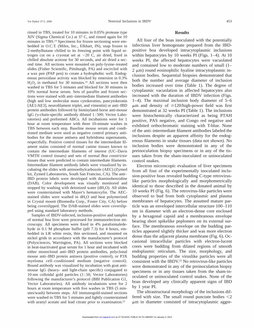
All four of the boas inoculated with the potentiallyinfectious liver homogenate prepared from the IBD-positive boa developed intracytoplasmic inclusionswithin hepatocytes by 10 weeks PI (Figs. 1–4). At 10weeks PI, the affected hepatocytes were vacuolatedand contained low to moderate numbers of small (1–2 mm) round eosinophilic hyaline intracytoplasmic in-clusion bodies. Sequential biopsies demonstrated thatboth the number and average diameter of inclusionbodies increased over time (Table 1). The degree ofcytoplasmic vacuolation in affected hepatocytes alsoincreased with the duration of IBDV infection (Figs.1–4). The maximal inclusion body diameter of 5–6mm and density of $120/high-power field was firstdemonstrated at 32 weeks PI (Table 1). The inclusionswere histochemically characterized as being PTAHpositive, PAS negative, and Congo red negative andexhibited orthochromatic staining with T-blue. Noneof the anti–intermediate filament antibodies labeled theinclusions despite an apparent affinity for the endog-enous filaments in snake tissues (data not shown). Noinclusion bodies were demonstrated in any of thepreinoculation biopsy specimens or in any of the tis-sues taken from the sham-inoculated or uninoculatedcontrol snakes.
Electron microscopic evaluation of liver specimensfrom all four of the experimentally inoculated inclu-sion-positive boas revealed budding C-type retrovirus-like particles morphologically and morphometricallyidentical to those described in the donated animal by10 weeks PI (Fig. 6). The retrovirus-like particles wereobserved to bud from both cytoplasmic and plasmamembranes of hepatocytes. The assumed mature par-ticle was an enveloped intercellular structure 100–110nm in diameter with an electron-dense core enclosedby a hexagonal capsid and a membranous envelopebearing short spikelike peplomers on its external sur-face. The membranous envelope on the budding par-ticles appeared slightly thicker and was more electrondense than the adjacent plasma membrane (Fig. 6). Oc-casional intracellular particles with electron-lucentcores were budding from dilated regions of smoothendoplasmic reticulum. The size, morphology, andbudding properties of the viruslike particles were allconsistent with the IBDV.23 No retrovirus-like particleswere demonstrated in any of the preinoculation biopsyspecimens or in any tissues taken from the sham-in-oculated or uninoculated control snakes. None of theboas developed any clinically apparent signs of IBDby 1 year PI.
The ultrastructural morphology of the inclusions dif-fered with size. The small round punctate bodies ,2mm in diameter consisted of intracytoplasmic aggre-
at UT SCHOOL PUBLIC HLTH LIBRARY on January 3, 2014vet.sagepub.comDownloaded from
454 Vet Pathol 37:5, 2000Wozniak, McBride, DeNardo, Tarara, Wong, and Osburn
Figs. 1–4. Liver; Boa constrictor. Serial liver biopsy specimens from a Boa constrictor experimentally infected withIBDV, showing an increase in the average size and density of intracytoplasmic inclusions (arrows) with increased durationof infection. Fig. 1. Baseline specimen. Fig. 2. Ten weeks PI. Fig. 3. Twenty weeks PI. Fig. 4. Thirty-two weeks PI. HE.Bar 5 10 mm.
Table 1. IBD inclusion body kinetics.
WeeksPostinoc-ulation
Diameter (mm)(x̄ 6 2SD)
No. Inclusions/High-powerField
(x̄ 6 2SD)
010203252
1.5 6 0.52.5 6 0.54.0 6 14.5 6 1
05 6 3
18 6 8120 6 20130 6 20
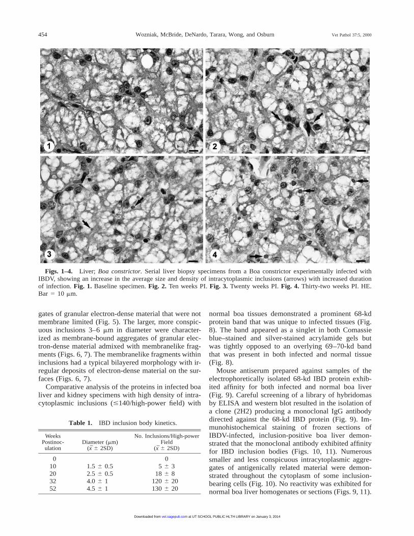
gates of granular electron-dense material that were notmembrane limited (Fig. 5). The larger, more conspic-uous inclusions 3–6 mm in diameter were character-ized as membrane-bound aggregates of granular elec-tron-dense material admixed with membranelike frag-ments (Figs. 6, 7). The membranelike fragments withininclusions had a typical bilayered morphology with ir-regular deposits of electron-dense material on the sur-faces (Figs. 6, 7).
Comparative analysis of the proteins in infected boaliver and kidney specimens with high density of intra-cytoplasmic inclusions (#140/high-power field) with
normal boa tissues demonstrated a prominent 68-kdprotein band that was unique to infected tissues (Fig.8). The band appeared as a singlet in both Comassieblue–stained and silver-stained acrylamide gels butwas tightly opposed to an overlying 69–70-kd bandthat was present in both infected and normal tissue(Fig. 8).
Mouse antiserum prepared against samples of theelectrophoretically isolated 68-kd IBD protein exhib-ited affinity for both infected and normal boa liver(Fig. 9). Careful screening of a library of hybridomasby ELISA and western blot resulted in the isolation ofa clone (2H2) producing a monoclonal IgG antibodydirected against the 68-kd IBD protein (Fig. 9). Im-munohistochemical staining of frozen sections ofIBDV-infected, inclusion-positive boa liver demon-strated that the monoclonal antibody exhibited affinityfor IBD inclusion bodies (Figs. 10, 11). Numeroussmaller and less conspicuous intracytoplasmic aggre-gates of antigenically related material were demon-strated throughout the cytoplasm of some inclusion-bearing cells (Fig. 10). No reactivity was exhibited fornormal boa liver homogenates or sections (Figs. 9, 11).
at UT SCHOOL PUBLIC HLTH LIBRARY on January 3, 2014vet.sagepub.comDownloaded from
Vet Pathol 37:5, 2000 455Nonviral Inclusions in IBDV
Figs. 5–7. Electron micrographs. Liver; Boa constrictor.Three morphologic variants of intracytoplasmic inclusionbodies found in IBDV-infected snakes. Fig. 5. A small roundinclusion similar to those illustrated in Fig. 2. The inclusionis characterized as an intracytoplasmic aggregate of granularelectron-dense material. No limiting membrane is identified.Fig. 6. Large (.3 mm) inclusion body. A C-type retrovirus-like particle (arrowhead) in the process of budding from theplasma membrane of the inclusion-bearing hepatocyte. Largeinclusions are characterized as membrane bound intracyto-plasmic structures that are composed of granular to amor-phous electron-dense material admixed with membranelikefragments (arrows). The bilayered structure of the membra-nelike fragments and the irregular deposits of granular elec-tron-dense material on the surfaces is evident in several re-
Fig. 8. Polyacrylamide gel containing electrophoretical-ly spearated proteins extracted from normal and IBDV-in-fected B. constrictor liver and kidney. Lanes 1 and 3 containnormal boa liver and kidney homogenates, respectfully.Lanes 2 and 4 contain IBDV-infected liver and kidney ho-mogenates, respectfully. Both of the infected tissues have ahigh density of large inclusion bodies. A prominent 68-kdprotein band is present in both IBDV-infected, inclusion-positive tissues; this band is not present in normal boa tis-sues. An aliquot the electrophoretically purified protein frac-tion is shown in lane 5. Coomassie brilliant blue R-250 totalprotein stain. The markers represent the molecular masses inkilodaltons.
←
gions. Fig. 7. Large inclusion body. Multifocal solid densi-ties of electron-dense material containing membrane profilesare present. Uranyl acetate and lead citrate. Bar 5 250 nm.
All attempts at labeling structures in formalin- or par-aformaledehyde-fixed B. constrictor tissues with themonoclonal antibody (2H2) were unsuccessful. Nei-ther baseline mouse serum or conditioned medium ex-hibited any affinity for B. constrictor tissues.
Discussion
All four boas inoculated with the filtered liver ho-mogenate prepared from IBDV-infected boa liver be-came infected with a budding retrovirus-like agentmorphologically and morphometrically consistent withthe IBDV and developed the characteristic intracyto-plasmic inclusion bodies by 10 weeks PI. Increasedcytoplasmic vacuolation was evident in IBDV-infectedhepatocytes by 10 weeks PI and appeared to increaseover time. Cytoplasmic vacuolation has been describedas a cytopathic effect observed with IBDV infection.25
Unlike previous studies, no clinical evidence of inclu-sion body disease was demonstrated in any of the ex-perimentally infected boas during the 1-year study.25
This lack could be attributable to strain differences inthe pathogen, host coadaptation, or other unidentifiedfactors. Also, the incubation period for the onset ofclinically apparent disease may exceed that for the pro-duction of demonstrable inclusion bodies. It has beensuggested that B. constrictor has a greater resistanceto clinically evident IBD than do other boid speciesand may be the natural host to some strains of IBDV.25
Further work on the pathogenesis of IBDV infectionsin boas and the pathogenic properties of different boa-
at UT SCHOOL PUBLIC HLTH LIBRARY on January 3, 2014vet.sagepub.comDownloaded from

456 Vet Pathol 37:5, 2000Wozniak, McBride, DeNardo, Tarara, Wong, and Osburn
Fig. 9. Western blot of normal and IBDV-infected, in-clusion-positive B. constrictor liver proteins. Lanes 1, 3, and5 contain normal boa liver. Lanes 2, 4, and 6 contain IBDV-infected boa constrictor liver with a high density of largeinclusion bodies. Lanes 1 and 2 were stained with baselinemouse serum. Lanes 3 and 4 were stained with polyclonalmouse antisera raised against the 68-kd IBD protein. Lanes5 and 6 were stained with monoclonal antibody 2H2, whichdemonstrates specific affinity for the 68-kd IBD proteinband. The markers represent the molecular masses in kilo-daltons.
Figs. 10, 11. Liver; Boa constrictor. Frozen sections of IBDV-infected, inclusion-positive and normal B. constrictor liverstained with the anti-IBDV monoclonal antibody 2H2. Fig. 10. The anti-IBDV monoclonal antibody prepared against the 68-kd IBD protein band (mab 2H2) demonstrated specific affinity for the intracytoplasmic inclusions (arrows) present in IBDV-infected liver. Fig. 11. No reactivity with normal boa liver was demonstrated. ABC method, DAB chromogen, Mayer’shematoxylin counterstain. Bar 5 10 mm.
and python-derived strains is needed before any defin-itive conclusions can be made in reference to the dis-ease process.
Budding retrovirus-like particles were not demon-strated in all cells bearing the characteristic inclusionbodies, but all snakes with inclusion bodies were dem-
onstrated by electron microscopy to be infected withthe retrovirus-like agent. The presence of inclusionswithout demonstrable viral replication within a givencell could indicate that the inclusions persist in cellsbeyond the period of active replication but could alsobe attributable to timing or simple sectional geometry.Similar results were reported in the original descriptionof IBD and with HIV-infected lymphocytes bearingtubuloreticular inclusions.25,32
Histochemically, IBD inclusions were PTAH posi-tive, orthochromatic with T-blue, PAS negative, andeosinophilic with HE. This pattern of staining suggeststhat the material within the inclusion body is protein-aceous.19,28 The negative results obtained with Congored and thioflaven-T suggest that the proteinaceousmaterial is not in a beta-pleated sheet (amyloid-like)conformation.19,28 The Mallory body, a well-character-ized member of the nonviral inclusion family, yields asimilar histochemical profile.23 Mallory bodies staineosinophilic with HE and are PAS negative, PTAHpositive, Congo red negative, oil red O negative, or-thochromatic with T-blue, and are metenamine silverreduction negative.23 Many of the well-characterized
at UT SCHOOL PUBLIC HLTH LIBRARY on January 3, 2014vet.sagepub.comDownloaded from

Vet Pathol 37:5, 2000 457Nonviral Inclusions in IBDV
proteinaceous intracytoplasmic nonviral inclusion bod-ies are composed of aggregates of intermediate fila-ments.8,10,11,20 Intermediate filaments are cytoskeletalcomponents that range from 6 to 11 mm in diameterand include vimentin, cytokeratins, tonofilaments, andneurofilaments.10 In the case of the Mallory body, theintermediate filaments within the inclusions have beenidentified as high- and low-molecular-mass cytokeratinfilaments.2,8,9,11,15 Ultrastructurally similar aggregates ofvimentin and neurofilaments have been identified inother variants of intracytoplasmic nonviral inclusionbodies.20 The lack of affinity of anti-cytokeratin, -neu-rofilament, and -vimentin antibodies for IBD inclu-sions suggests that they do not contain intermediatefilament proteins. Although a dense coat of ubiquitincould have interfered with antibody binding, this pro-tein has not been demonstrated in membrane-boundvariants of intracytoplasmic inclusion bodies.20
The kinetics of IBD inclusion formation and the an-tigenically unique properties of the 68-kd IBD inclu-sion protein suggest that it may be an overproducedand/or poorly degraded component of the virus thataccumulates in the host cell cytoplasm. Both the av-erage size and the number of inclusions per high-pow-er field increased with the duration of the infection inall four experimentally infected snakes. The ultrastruc-tural morphology of inclusion bodies varied somewhatwith the size of the inclusion body. The small roundpunctate bodies ,2 mm in diameter were composed ofintracytoplasmic aggregates of granular electron-densematerial that were not membrane limited, whereas thelarger inclusions appeared as membrane-bound struc-tures filled with granular electron-dense material ad-mixed with filamentous membranelike structures. Pro-teins that are synthesized within a cell that are notdestined for secretion can accumulate intracytoplasm-ically as non-membrane bound aggregates.24 Mem-brane-bound inclusions, however, often represent ei-ther an accumulation of an undigested substrate in pha-golysosomes or an accumulation of synthesized pro-teins inside cisterns of rough endoplasmic reticulum.24
The presence of granular electron-dense material ad-mixed with membranelike fragments in the same struc-ture would support a phagolysosomal origin for thelarger variants of IBD inclusions. Intracytoplasmic ac-cumulation of unidentified granular electron-dense ma-terial has been demonstrated with other retroviruses.6,12
In some cases, the production and accumulation of ret-roviral proteins results in intracytoplasmic aggregatesadmixed with ribosomes that are detectable by lightmicroscopy as basophilic inclusion bodies of varioussizes.12
A direct comparison of the proteins in normal andIBDV-infected, inclusion-positive boa livers by SDS-PAGE demonstrated the presence of an abundant IBD-
specific 68-kd protein band. Western blots stained withmonoclonal antibody 2H2 demonstrated epitopes ofthe 68-kd IBD protein that appeared to be IBD spe-cific. Immunohistochemical staining of frozen sectionsof IBDV-infected, inclusion-positive liver sectionswith 2H2 resulted in specific labeling of IBD inclu-sions. Antigenically similar material was demonstratedin all size categories of IBD inclusion bodies. None ofthe baseline biopsy specimens and none of the tissuescollected from control or sham-inoculated snakes hadinclusions or retrovirus-like particles. No proteins an-tigenically related to the 68-kd IBD protein were dem-onstrated in any noninfected boa tissues. The 68-kdIBD protein was thereafter referred to as the IBD in-clusion protein.
The genome of retroviruses encodes for severalproducts referred to as PRO, POL, and GAG. TheGAG region is transcribed and translated as a 68-kdpolyprotein that is posttranslationally cleaved into thematrix (MA), capsid, nucleic acid binding, and otherstructural components of the mature particle. The MAprotein associates with and can covalently bind to cellmembrane lipids via a sequence of cationic amino acidresidues that is believed to be a highly conserved re-gion among retroviruses.5 The MA protein is believedto both direct membrane localization of viral particleassembly and form a stable shell-associated layer onthe inner surface of the membrane envelope.5 Recentstudies have shown that retroviruses can complete thebudding process without the transmembrane envelopeproteins, suggesting that it is the core proteins thatdrive the budding process.3
The finding of an antigenically unique 68-kd proteinwithin inclusion bodies that appears to be associatedwith membranes is interesting. In the original descrip-tion of IBD, the inclusions were identified as aggre-gates of electron-dense material.25 Unidentified mate-rial characterized as electron-dense subunits wereshown budding from cytoplasmic membranes ofIBDV-infected cells.25 The size and morphology of thefilamentous structures identified within the larger var-iants of IBD inclusions characterized in this studyclosely approximated cytoplasmic membranes. A dis-tinct bilayered structure was evident on close exami-nation. Irregular deposits of granular electron-densematerial were evident on the surfaces of the membra-nelike filaments. The larger variant of the IBD inclu-sion body may represent an intraphagolysosomal ac-cumulation of IBDV GAG polyprotein and GAG-coat-ed cytoplasmic membranes. If the membranelike struc-tures observed within the large variants of IBDinclusions are indeed GAG-coated membranes, IBDV-infected boa constrictors may be a good alternative(lower vertebrate) animal model for the study ofGAG–membrane interactions and the role of these
at UT SCHOOL PUBLIC HLTH LIBRARY on January 3, 2014vet.sagepub.comDownloaded from

458 Vet Pathol 37:5, 2000Wozniak, McBride, DeNardo, Tarara, Wong, and Osburn
complexes in viral replication and inclusion develop-ment. Further work would be required to more specif-ically identify the 68-kd IBD inclusion protein inIBDV-infected boas.
Because of the potential for IBDV-infected boas topersist as inapparent carriers and the lack of IBDVnatural history transmission data, all newly acquiredspecimens should be quarantined and checked for IBDby liver biopsy before being placed in collections.Minimal biopsy analysis should include histopatholo-gy with a thorough examination of the specimen foreosinophilic intracytoplamsic inclusion bodies and cy-toplasmic vacuolation. Because many different insultsto tissue can cause the formation of similar intracyto-plasmic inclusions, inclusion-positive specimensshould be subjected to additional diagnostic measuressuch as TEM before a diagnosis of IBD is made. Thehighly specific nature of the anti-68-kd IBD inclusionprotein monoclonal antibody may make it a useful re-agent in the immunohistochemical differentiation ofIBD inclusions from other types of nonviral inclusionbodies.
Acknowledgements
We thank K. and C. Pazzi and the Birmingham Zoo fortheir donation of the boa constrictors employed in this study.We also thank D. Naydan of the UC Davis VMTH Histo-pathology Laboratory for her assistance and technical ex-pertise with the immunohistochemical procedures. Specialthanks is also extended to K. Schwendinger (ConsolidatedVeterinary Diagnostics, Sacramento, CA) for the productionof exceptional slides and histochemical stains. We are grate-ful to the UC Berkeley animal care staff for the excellentlevel of animal care and record keeping and to Tom Bed-narek at the UTMB, Department of Pathology Imaging Lab-oratory, for all for his assistance with the illustrations. Thisproject was funded in part by a UC Davis Laboratory Ani-mal Medicine Training Grant awarded by the National In-stitutes of Health.
References
1 Alpers CE, Tsai CC, Hudkins KL, Cui Y, Kuller L, Ben-veniste RE, Ward JM, Morton WR: Focal segmental glo-merulosclerosis in primates infected with simian immu-nodeficiency virus. AIDS Res Hum Retroviruses 13:413–424, 1997
2 Aston NS, Morris PA, Tanner MS, Variend S: An animalmodel for copper-associated cirrhosis in infancy. J Pathol186:215–221, 1998
3 Cadd TL, Skoging U, Lilestrom P: Budding of envelopedviruses from the plasma membrane. Bioessays 19:993–1000, 1997
4 Campbell AM: In: Laboratory Techniques in Biochem-istry and Molecular Biology, ed. Burdon RH and vanKippenber PH, vol. 13, pp. 1–154. Elsevier, New York,NY, 1984
5 Christenson AM, Massiah MA, Turner BG, Sundquist
WI, Summers MF: Three-dimensional structure of theHTLV-II matrix protein and comparative analysis of ma-trix proteins from the different classes of pathogenic hu-man retroviruses. J Mol Biol 264:1117–1131, 1996
6 Coward JE, Harter DH, Hsu KC, Morgan C: Ferratin-conjugated antibody labeling of visna virus. Virology 50:925–930, 1972
7 del Rosario AD, Hai HX, Singh J, Ginsburg R, Ross JS:Intracytoplasmic eosinophilic hyaline globules in carti-lagneous neoplasms: a surgical, pathological, ultrastruc-tural and electron probe X-ray microanalytic study. HumPathol 25:1283–1289, 1994
8 Denk H, Franke WW, Dragosics B, Zeiler I: Pathologyof cytoskeleton of liver cells: demonstration of Mallorybodies (alcoholic hyalin) in murine and human hepato-cytes by immunofluorescence microscopy using antibod-ies to cytokeratin polypeptides from hepatocytes. Hepa-tology 1:9–20, 1981
9 Denk H, Franke WW, Eckerstorfer R, Schmid E, Ker-jaschki D: Formation and involution of Mallory bodies(alcoholic hyaline) in murine and human liver revealedby immunofluorescence microscopy with antibodies toprekeratin. Proc Natl Acad Sci USA 76:4112–4116,1979
10 Franke WW, Schmid E, Osborn M, Weber K: Differentintermediate-sized filaments distinguished by immunofl-uoresence microscopy. Proc Natl Acad Sci USA 75:5034–5038, 1978
11 French SW: Present understanding of the development ofMallory’s body. Arch Pathol Lab Med 107:445–450,1983
12 Gilka F, Spencer J: Viral matrix inclusion bodies in myo-cardium of lymphoid leukosis virus-infected chickens.Am J Vet Res 46:1953–1960, 1985
13 Grimly PM, Schaff Z: Significance of tubuloreticular in-clusions in the pathobiology of human diseases. In:Pathobiology Annual, ed. Iaochim HL, pp. 221–257. Ap-pleton-Century-Crofts, New York, NY, 1976
14 Haines DM, Chelack BJ: Technical considerations forenzyme immunohistochemical staining procedures onformalin-fixed paraffin-embedded tissues for diagnosticpathology. J Vet Diagn Invest 3:101–112, 1991
15 Jensen K, Gluud C: The Mallory body: morphologicaland experimental studies (part 1 of a literature survey).Hepatology 20:1061–1077, 1994
16 Kohler G, Milstein A: Continuous culture of fused cellssecreting antibody of predefined specificity. Nature 256:496–497, 1975
17 Laemmli UK: Cleavage of structural proteins during as-sembly of the head of the bacteriophage T4. Nature 227:680–685, 1970
18 Lowe J, McDermott H, Landon M, Mayer RJ, WilkinsonK: Ubiquiton carboxyl-terminal hydrolase (PGP 9.5) isselectively present in ubiquitinated inclusion bodiescharacteristic of human neurodegenerative diseases. JPathol 161:153–160, 1990
19 Luna L: Manual of Histologic Staining Methods of theArmed Forces Institute of Pathology, 3rd ed. McGraw-Hill, New York, NY, 1968
20 Manetto V, Abdul-Karim FW, Perry G, Tabaton M, Au-
at UT SCHOOL PUBLIC HLTH LIBRARY on January 3, 2014vet.sagepub.comDownloaded from
Vet Pathol 37:5, 2000 459Nonviral Inclusions in IBDV
tilio-Cambetti L, Gambetti P: Selective presence of ubiq-uitin in intracellular inclusions. Am J Pathol 134:505–513, 1989
21 Mantovani G, Santa Cruz G, Piso A, Arangino V, Bal-estrieri A, Del Giacco GS: Hairy cell leukemia with ul-trastructural finding of tubuloreticular inclusions in hairycells: a possible marker of a virus-induced disease. JSubmicrosc Cytol 18:617–624, 1986
22 Mohanty S: Electron Microscopy for Biologists, pp.183–225. Charles C Thomas, Springfield, IL, 1982
23 Norkin SA, Weitzel R, Campagna-Pinto D, MacDonaldRA, Mallory K: Alcoholic hyalin in human cirrhosis:histochemical studies. Am J Pathol 37:49–57, 1960
24 Robbins SL, Cotran RS, Kumar V: Pathologic Basis ofDisease, 3rd ed., pp. 1–21. WB Saunders, Philadelphia,PA, 1984
25 Schumacher J, Jacobson ER, Homer BL, Gaskin JM: In-clusion body disease in boid snakes. J Zoo Wildl Med25:511–524, 1994
26 Scroggs MW, Roggli V, Amando FE, Sanfilippo F: Eo-sinophilic intracytoplasmic globules in pulmonary ade-nocarcinomas: a histochemical, immunohistochemicaland ultrastructural study of six cases. Hum Pathol 20:845–849, 1990
27 Towbin H, Staehelin T, Gordon J: Electrophoretic trans-fer of proteins from polyacrylamide gels to nitrocellulosesheets: procedure and some applications. Proc Natl AcadSci USA 79:4350–4354, 1979
28 Vacca L: Laboratory Manual of Histochemistry. RavenPress, New York, NY, 1985
29 Vaitukaitis J, Robbins JB, Nieschlag E, Ross GT: Amethod for producing specific antisera with small dosesof immunogen. J Clin Endocrinol Metab 33:988–991,1971
30 Voller A, Bidwell D, Barlett A: Enzyme-linked immu-nosorbent assay. In: Manual of Clinical Microbiology,ed. Noel A, Rose B, and Friedman A, 2nd ed. pp. 359–371. American Society for Microbiology, Washington,DC.
31 Wozniak E, Butler JF, Zam SG: Evidence of commonand genus specific epitopes on Ornithodoros spp. tick(Acari: Argasidae) salivary proteins. J Med Entomol 32:484–489, 1995
32 Yoffe B, Petrie BL, Noonan CA, Hollinger FB: In vivoand in vitro ultrastructural alterations induced by humanimmunodeficiency virus in human lymphoid cells. LabInvest 61:303–309, 1989
Request reprints from Dr. E. J. Wozniak, Animal Resources Center, University of Texas Medical Branch, 301 UniversityBlvd., Galveston, TX 77555-0621 (USA).
at UT SCHOOL PUBLIC HLTH LIBRARY on January 3, 2014vet.sagepub.comDownloaded from