Repeated DNA therapeutic vaccination of chronically SIV-infected macaques provides additional...
29
Repeated DNA Therapeutic Vaccination of Chronically SIV- Infected Macaques Provides Additional Virological Benefit Antonio Valentin 1 , Agneta von Gegerfelt 1 , Margherita Rosati 1 , Georgios Miteloudis 1 , Candido Alicea 2 , Cristina Bergamaschi 1 , Rashmi Jalah 2 , Vainav Patel 1 , Amir S. Khan 3 , Ruxandra Draghia-Akli 3 , George N. Pavlakis 1,* , and Barbara K. Felber 2,* 1 Human Retrovirus Section, Vaccine Branch, Center for Cancer Research, National Cancer Institute at Frederick, Frederick, Maryland 21702-1201 2 Human Retrovirus Pathogenesis Section, Vaccine Branch, Center for Cancer Research, National Cancer Institute at Frederick, Frederick, Maryland 21702-1201 3 VGX Pharmaceuticals, Inc., The Woodlands, Texas 77381 Abstract We have previously reported that therapeutic immunization by intramuscular injection of optimized plasmid DNAs encoding SIV antigens effectively induces immune responses able to reduce viremia in antiretroviral therapy (ART)-treated SIVmac251 infected Indian rhesus macaques. We subjected such therapeutically immunized macaques to a second round of therapeutic vaccination using a combination of plasmids expressing SIV genes and the IL-15/IL-15 receptor alpha as molecular adjuvant, which were delivered by the more efficacious in vivo constant-current electroporation. A very strong induction of antigen-specific responses to Gag, Env, Nef, and Pol, during ART (1.2-1.6% of SIV-specific T cells in the circulating T lymphocytes) was obtained with the improved vaccination method. Immunological responses were characterized by the production of IFN-γ, IL–2, and TNFα either alone, or in combination as double or triple cytokine positive multifunctional T cells. A significant induction of CD4 + T cell responses, mainly targeting Gag, Nef, and Pol, as well as of CD8 + T cells, mainly targeting Env, was found in both T cells with central memory and effector memory markers. After release from ART, the animals showed a virological benefit with a further ∼1 log reduction in viremia. Vaccination with plasmid DNAs has several advantages over other vaccine modalities, including the possibility for repeated administration, and was shown to induce potent, efficacious, and long-lasting recall immune responses. Therefore, these data support the concept of adding DNA vaccination to the HAART regimen to boost the HIV-specific immune responses. * Corresponding co-authors: Human Retrovirus Section and Human Retrovirus Pathogenesis Section, Vaccine Branch, Center for Cancer Research, National Cancer Institute at Frederick, P.O. Box B, Building 535, Room 206, Frederick, MD 21702-1201. Phone: (301) 846-5159. Fax: (301) 846-7146. [email protected]; [email protected]. Address reprint requests to: Drs. Barbara K. Felber and George N. Pavlakis, Vaccine Branch, Center for Cancer Research, National Cancer Institute at Frederick, P.O. Box B, Building 535, Room 206, Frederick, Maryland 21702-1201, [email protected] and [email protected] Publisher's Disclaimer: This is a PDF file of an unedited manuscript that has been accepted for publication. As a service to our customers we are providing this early version of the manuscript. The manuscript will undergo copyediting, typesetting, and review of the resulting proof before it is published in its final citable form. Please note that during the production process errors may be discovered which could affect the content, and all legal disclaimers that apply to the journal pertain. NIH Public Access Author Manuscript Vaccine. Author manuscript; available in PMC 2011 February 23. Published in final edited form as: Vaccine. 2010 February 23; 28(8): 1962–1974. doi:10.1016/j.vaccine.2009.10.099. NIH-PA Author Manuscript NIH-PA Author Manuscript NIH-PA Author Manuscript
Transcript of Repeated DNA therapeutic vaccination of chronically SIV-infected macaques provides additional...

Repeated DNA Therapeutic Vaccination of Chronically SIV-Infected Macaques Provides Additional Virological Benefit
Antonio Valentin1, Agneta von Gegerfelt1, Margherita Rosati1, Georgios Miteloudis1,Candido Alicea2, Cristina Bergamaschi1, Rashmi Jalah2, Vainav Patel1, Amir S. Khan3,Ruxandra Draghia-Akli3, George N. Pavlakis1,*, and Barbara K. Felber2,*1Human Retrovirus Section, Vaccine Branch, Center for Cancer Research, National Cancer Instituteat Frederick, Frederick, Maryland 21702-12012Human Retrovirus Pathogenesis Section, Vaccine Branch, Center for Cancer Research, NationalCancer Institute at Frederick, Frederick, Maryland 21702-12013VGX Pharmaceuticals, Inc., The Woodlands, Texas 77381
AbstractWe have previously reported that therapeutic immunization by intramuscular injection of optimizedplasmid DNAs encoding SIV antigens effectively induces immune responses able to reduce viremiain antiretroviral therapy (ART)-treated SIVmac251 infected Indian rhesus macaques. We subjectedsuch therapeutically immunized macaques to a second round of therapeutic vaccination using acombination of plasmids expressing SIV genes and the IL-15/IL-15 receptor alpha as molecularadjuvant, which were delivered by the more efficacious in vivo constant-current electroporation. Avery strong induction of antigen-specific responses to Gag, Env, Nef, and Pol, during ART (1.2-1.6%of SIV-specific T cells in the circulating T lymphocytes) was obtained with the improved vaccinationmethod. Immunological responses were characterized by the production of IFN-γ, IL–2, and TNFαeither alone, or in combination as double or triple cytokine positive multifunctional T cells. Asignificant induction of CD4+ T cell responses, mainly targeting Gag, Nef, and Pol, as well as ofCD8+ T cells, mainly targeting Env, was found in both T cells with central memory and effectormemory markers. After release from ART, the animals showed a virological benefit with a further∼1 log reduction in viremia. Vaccination with plasmid DNAs has several advantages over othervaccine modalities, including the possibility for repeated administration, and was shown to inducepotent, efficacious, and long-lasting recall immune responses. Therefore, these data support theconcept of adding DNA vaccination to the HAART regimen to boost the HIV-specific immuneresponses.
* Corresponding co-authors: Human Retrovirus Section and Human Retrovirus Pathogenesis Section, Vaccine Branch, Center for CancerResearch, National Cancer Institute at Frederick, P.O. Box B, Building 535, Room 206, Frederick, MD 21702-1201. Phone: (301)846-5159. Fax: (301) 846-7146. [email protected]; [email protected] reprint requests to: Drs. Barbara K. Felber and George N. Pavlakis, Vaccine Branch, Center for Cancer Research, NationalCancer Institute at Frederick, P.O. Box B, Building 535, Room 206, Frederick, Maryland 21702-1201, [email protected] [email protected]'s Disclaimer: This is a PDF file of an unedited manuscript that has been accepted for publication. As a service to our customerswe are providing this early version of the manuscript. The manuscript will undergo copyediting, typesetting, and review of the resultingproof before it is published in its final citable form. Please note that during the production process errors may be discovered which couldaffect the content, and all legal disclaimers that apply to the journal pertain.
NIH Public AccessAuthor ManuscriptVaccine. Author manuscript; available in PMC 2011 February 23.
Published in final edited form as:Vaccine. 2010 February 23; 28(8): 1962–1974. doi:10.1016/j.vaccine.2009.10.099.
NIH
-PA Author Manuscript
NIH
-PA Author Manuscript
NIH
-PA Author Manuscript
IntroductionAlthough the introduction of highly active antiretroviral therapy (HAART) resulted in aremarkable decrease of AIDS related deaths, the current pharmacological regimens to treatHIV infection fail to eradicate the virus and are associated with several problems, includingdrug toxicity, and development of resistance with viral rebound. In this context, new strategiesto control viral replication and to restore immune functions are needed. During the period ofantiretroviral therapy (ART), due to efficient control of viral replication, the virus specificcellular immune responses in the periphery are strongly reduced. After interruption of ARTtreatment, there is a rapid increase of viremia within 10 days. The introduction of newtherapeutic interventions that boost immune responses would be beneficial for the clinicalmanagement of HIV-infected individuals. Towards this goal, studies were designed to boostthe immune system of SIV-infected macaques during ART using DNA immunization. It waspreviously observed that vaccination during ART using DNA plasmids (Lisziewicz et al.,2005; Lori et al., 2005; Fuller et al., 2006; Lisziewicz et al., 2007; von Gegerfelt et al.,2007; Halwani et al., 2008; Zur Megede et al., 2008), pox-virus vectors (Hel et al., 2000;Tryniszewska et al., 2002), antigen-pulsed dendritic cells (Lu et al., 2003) or peptide-pulsedblood (De Rose et al., 2008) in SIVmac251-infected macaques was able to evoke SIV-specificrecall immune responses. After release from ART, variable results regarding virological benefitwere reported from no control (Zur Megede et al., 2008), temporal control (Hel et al., 2000;Tryniszewska et al., 2002; Fuller et al., 2006), to long-lasting control (Lori et al., 2003; Lu etal., 2003; Lisziewicz et al., 2005; von Gegerfelt et al., 2007; De Rose et al., 2008). Using DNAonly as vaccine, two reports showed successful immunological and virological benefit, whichare intramuscular injection (von Gegerfelt et al., 2007) and topical administration of DNA-based Dermavir (Lori et al., 2003; Lisziewicz et al., 2005). Importantly, the ability to induceimmune responses able to reduce viremia could therefore offer an opportunity to usevaccination as an additional component to antiretroviral therapy. The use of DNA only asvaccination method is a promising immunization strategy that has advantages (production,stability, repeated use) over other vaccination modalities. Therapeutic vaccination in humansagainst HIV-1 has given mixed results. Some studies have reported an immunological andsometimes also a virological benefit, whereas others did not (Rosenberg et al., 2000; Markowitzet al., 2002; Lu et al., 2003; Lu et al., 2004; Kinloch-De Loes et al., 2005; Levy et al., 2005;Tubiana et al., 2005; Andrieu and Lu, 2007; Hardy et al., 2007; Connolly et al., 2008; Pialouxet al., 2008; Wilson et al., 2008). Several studies suggest that vaccination during highly activeantiretroviral therapy (HAART) induces HIV-specific recall responses. The efficacy oftherapeutic vaccines may be variable, and it is hypothesized that more consistentimmunological and virological benefits could be achieved by improving the vaccinationapproaches.
We focused on DNA vaccination, since previous data in macaques have been encouraging anddemonstrated strong immunogenicity, long-term decrease of viral load and a survival benefitin the animals with robust response to the therapeutic vaccination (Lori et al., 2003; Lisziewiczet al., 2005; von Gegerfelt et al., 2007). Recent developments to improve DNA delivery includein vivo electroporation (Aihara and Miyazaki, 1998; Mathiesen, 1999; Rizzuto et al., 1999;Selby et al., 2000; Widera et al., 2000; Mir, 2001;Wang et al., 2004b; Prud'homme et al.,2006; Draghia-Akli et al., 2008), which showed to be more efficient than traditionalintramuscular injection in SIV/HIV DNAs vaccinating rhesus macaques and inducedsignificantly increased antigen-specific immunity (Selby et al., 2000; Otten et al., 2004; Ottenet al., 2006; Luckay et al., 2007; Hirao et al., 2008; Rosati et al., 2008; Zur Megede et al.,2008).
In this study, we examined whether repeated immunotherapeutic vaccination is of furthervirological benefit. We used macaques animals previously immunized during ART with
Valentin et al. Page 2
Vaccine. Author manuscript; available in PMC 2011 February 23.
NIH
-PA Author Manuscript
NIH
-PA Author Manuscript
NIH
-PA Author Manuscript
plasmid DNAs encoding SIV antigens (von Gegerfelt et al., 2007) and, after release from ART,showed a long-lasting partial control of viremia without any signs of progression towardsimmunodeficiency. Of the eight control animals that were ART-treated only without receivingvaccination (von Gegerfelt et al., 2007), none controlled viremia after ART release and theysubsequently developed AIDS. In this report, we tested whether macaques that benefited fromthe first cycle of immunotherapy could further benefit from a 2nd round of immunotherapeuticimmunization using the combination of improved plasmid DNAs and DNA delivery byelectroporation. Here, we report, the induction of strong and potent increases in SIV-specificimmune responses in the immunized macaques followed by a further significant virologicalbenefit after release from ART.
Materials and MethodsAnimals
The Indian rhesus macaques (Macaca mulatta) included in the study were housed and handledin accordance with the standards of the Association for the Assessment and Accreditation ofLaboratory Animal Care International. Screening for MHC alleles was performed by PCR (D.Watkins, Wisconsin Regional Primate Center). Monkey 538L was Mamu A*01 positive andB*17 negative, whereas the monkeys 920L and 965L were both negative for Mamu A*01 andB*17. These SIVmac251 infected animals were previously involved in a therapeutic SIV DNAvaccination study (von Gegerfelt et al., 2007). Approximately 3 years after release from ART,the animals were enrolled into the present study and subjected to a 2nd round of therapeuticvaccination. During this second antiretroviral treatment period (31 weeks), the animalsreceived the following therapeutic regimen: 20 mg/kg ((R)-9-(2-phosphonylmethoxypropyl)adenine (PMPA), and 50 mg/kg FTC, both injected subcutaneously once daily, and 5 mg/kgDidanosine (ddI), injected intravenously once daily. After two weeks, ddI was discontinued,and the dose of PMPA was reduced to 10mg/kg/day.
DNA vectorsAll plasmids used in the study contain the human CMV promoter without an intron, the bovinegrowth hormone polyadenylation site, and the kanamycin resistance gene, pCMV.kan (Rosatiet al., 2005). The RNA/codon optimized genes (Schwartz et al., 1992a; Schwartz et al.,1992b; Nasioulas et al., 1994; Schneider et al., 1997) for gag, pol, and env were generatedupon introduction of multiple silent point mutations not affecting the sequence of the encodedproteins. The animals were vaccinated with a mixture of DNAs producing secreted andintracellularly degraded variants of the SIV Gag generated by N-terminal fusion with eitherIP10-MCP3 (Biragyn et al., 1999) (MCP3-p39gag, 21S) or with a beta-catenin-derived peptide(aa 18-47) (Aberle et al., 1997) (CATEgagDX, 2S) as previously described (Rosati et al.,2005; von Gegerfelt et al., 2007). The authentic SIVmac239 Env protein sequence (native Env,99S) and a fusion of Env to IP10-MCP3 replacing its native signal peptide with IP10-MCP3(MCP3-Env, 73S) were used. The optimized pol with inactivating mutations in PRT, RT andINT was inserted into pCMVLAMP.kan between the human LAMP-1 luminal domain and theLAMP-1 transmembrane and cytoplasmic (TM/cyt) tail domain (Chikhlikar et al., 2004),generating the plasmid LAMPpol (103S). Plasmid LAMP-NTV (147S) expresses a LAMP-Nef-Tat-Vif fusion protein. LAMP targets the antigen to lysosomes and lysosome-likecompartments, which in antigen presenting cells also contain MHC molecules and was shownto affect the trafficking and immunogenicity of HIV-1 gag (Valentin et al.; Marques et al.,2003; De Arruda et al., 2004; Chikhlikar et al., 2006). The rhesus macaque (rm) IL-15 plasmid(AG65) contains the optimized IL-15 DNA sequence expressing a stable mRNA encoding anIL-15 that has the native signal peptide replaced by the tPA signal and propeptide (Jalah etal., 2007). Plasmid rmIL15Rα (AG120) expresses the optimized rhesus IL-15 receptor alpha(Bergamaschi et al., 2007) chain.
Valentin et al. Page 3
Vaccine. Author manuscript; available in PMC 2011 February 23.
NIH
-PA Author Manuscript
NIH
-PA Author Manuscript
NIH
-PA Author Manuscript

Therapeutic immunizationHighly purified, endotoxin-free DNA plasmid preparations were produced using the Qiagenkit (Hilden, Germany). The 1 ml DNA mixture contained 100 μg of each SIV plasmid and 200μg of the cytokine plasmids, respectively (a total of 1 mg of plasmid DNA). These DNAs wereinjected intramuscularly (0.5 ml per injection) at the left and right thighs using in vivoelectroporation by the CELLECTRA® adaptive constant-current electroporator (VGXPharmaceuticals, Inc., The Woodlands, TX).
Flow cytometric analysisFlow cytometric analysis was performed as described (Rosati et al., 2008). To determine thenumber and phenotype of SIV-specific cells, isolated PBMCs were incubated at a density of106 cells/ml in RPMI-1640 supplemented with 10% FBS, 100 U/ml penicillin/streptomycinand 2 mM L-glutamine, in the presence of pools of 15-aa peptides overlapping by 11 aa, derivedfrom SIV Gag, Pol, Env, Nef, and Tat at a final concentration of 1 μg/ml for each peptide. Cellswere treated overnight with monensin to prevent protein secretion, and cell surface stainingwas performed using the following antibody cocktail: CD3-APCCy7, CD4-PerCPCy5.5,CD45RA-PE, CD28-biotin and Streptavidin-APCCy5.5 (BD Pharmingen, San Jose, CA) andCD8-AF405 (Caltag, Carlsbad, CA). Cells were washed twice, fixed, and permeabilized withCytofix/Cytoperm (BD Pharmingen). Staining for intracellular cytokine detection wasperformed using the following antibody mixture: IFN-γ-FITC, IL-2-APC and TNF-α-PECy7(BD Pharmingen). In some experiments, surface staining was performed with a differentantibody cocktail including CD95-FITC, CCR7-APC, CD3-APCCy7, CD4-PerCPCy5.5,CD45RA-PE, CD8-AF405, CD28-Biotin and Streptavidin-APCCy5.5, and the presence ofantigen specific cells was monitored in permeabilized cells by intracellular staining with anti-IFN-PECy7 mAb. This second staining allows the comparison of memory cell populationsdefined by CD28/CD45RA or CD28/CD95 expression. After intracellular staining, the cellswere washed twice and the samples were analyzed in a FacsAria or LSRII flow cytometers(BD Pharmingen). All data analysis was performed using the FlowJo platform (Tree Star, Inc.,Ashland, OR).
Humoral immune responsesAntibody production against Gag and Env was measured in serial dilutions of plasma byseparate ELISA assays. The plates were analyzed at the absorbance of 450 nm (A450). Thenegative cutoff value was twice the mean A450 values obtained with non-immune sera. Allsamples with A450 value higher than the cutoff were considered positive. The binding antibodytiters are reported as the reciprocal of the highest positive dilution.
Viral load analysisSIV RNA copy numbers were determined by a real-time nucleic acid sequence-basedisothermal amplification (NASBA) assay using SIVmac251-specific primers (Romano et al.,2000) with a threshold of detection of 50 copies/ml.
ResultsStudy design
We used SIVmac251-infected Indian rhesus macaques (538L, 920L, 965L), which werepreviously subjected to one round of therapeutic vaccination (von Gegerfelt et al., 2007).During the previous study, the animals received three DNA immunizations, usingintramuscular needle injection, which resulted in a strong virological benefit as demonstratedby decreased viral loads. These animals were kept and were monitored following the 1st cycleof therapeutic immunization (3.1, 3.7 and 3.8 years for monkey 920L, 965L and 538L,
Valentin et al. Page 4
Vaccine. Author manuscript; available in PMC 2011 February 23.
NIH
-PA Author Manuscript
NIH
-PA Author Manuscript
NIH
-PA Author Manuscript

respectively), and their viral loads remained below the levels measured prior to treatment,demonstrating a long-term benefit of therapeutic DNA vaccination.
In the present study, we report data obtained after a 2nd round of therapeutic vaccination (ART/DNA) outlined in Fig. 1A. The animals were subjected to ART for 31 weeks using PMPA,FTC and ddI as described previously (von Gegerfelt et al., 2007). Starting at week 14 of ART(time point 0), the animals received 4 DNA vaccinations (week 0, 4, 8, and 15) byelectroporation (EP) using the in vivo CELLECTRA® adaptive constant-current electroporator(VGX Pharmaceuticals, Inc.). Two weeks after EP4, ART treatment was terminated and theanimals were monitored for another 16 wks (termination of the study).
The SIV DNA mixture contained optimized expression vectors for different SIV antigens;antigen sequences were modified to alter their intracellular trafficking by fusions to eitherMCP-3, catenin (CATE) or human lysosomal membrane associated protein 1 (LAMP) (Rosatiet al., 2005; von Gegerfelt et al., 2007; Rosati et al., 2008). The plasmid DNAs in vaccinemixture expressed Gag (MCP3gag, CATE-gag), Pol (LAMP-pol), Env (native and MCP3env),and the a Nef-Tat-Vif fusion to LAMP (LAMP-NTV) (Rosati et al., 2005; von Gegerfelt etal., 2007; Rosati et al., 2008). A combination of rhesus IL-15 and IL-15Rα expression plasmids(Bergamaschi et al., 2007; Jalah et al., 2007) was used as a molecular adjuvant. During thecourse of this study, the animals were monitored for the development of SIV-specific immuneresponses and changes in the viral loads.
Virological benefit of the 2nd therapeutic vaccinationFigure 1B shows the viral load data of the vaccinated macaques prior, during and after ART.During the period preceding the start of second ART (13 weeks), the animals showed meanviral loads ranging from 3.6 to 5.1 log10. All animals responded immediately to ART treatment,resulting in persistent control of viremia (<50 copies/ml plasma). After ART release, weobserved a pattern of fluctuating viral loads as previously observed (von Gegerfelt et al.,2007). After the initial virus rebound, the viral load fluctuations were at levels lower than beforeART/DNA, resulting in additional partial control of viremia. Importantly, the improved controlof viremia was maintained during the 16 weeks of follow-up (Fig. 1B). At this point, the animalswere enrolled in another study.
The mean viral loads during the 13 weeks before ART (PRE) and the 16 weeks after ARTrelease (POST) were compared. Figure 1C shows the differential in viral loads (ΔVL) of thethree animals as a result of the 2nd therapeutic vaccination. This analysis revealed a significantdecrease (P=0.0356) in viremia with an average of ∼1 log10 in viral load reduction. A similarbenefit in viremia reduction was found by comparing viral loads during 39 weeks PRE and 16weeks POST ART. In conclusion, the reduction in plasma viral loads demonstrates that the2nd round of therapeutic vaccination provides an additional virological benefit in these animals.
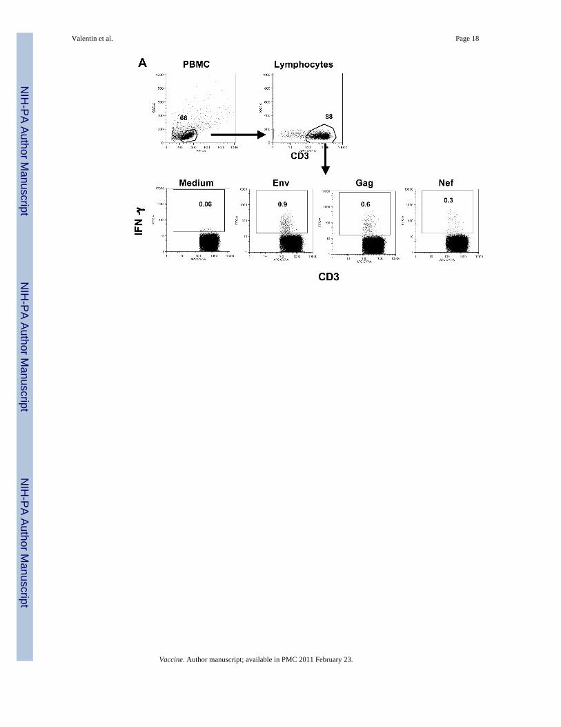
Increase of cellular immune responsesWe studied the development of SIV-specific cellular immune responses in the peripheral bloodfrom the immunized macaques using flow cytometric analysis. Figure 2A outlines the strategyused for the flow cytometric analysis to detect antigen-specific IFN-γ positive T cells using asexample PBMC from animal 965L at 3 weeks post EP3. CD3+ T cells were identified withinthe main lymphocyte population, which was defined by forward and side scatter characteristics.The presence of SIV-specific T cells was determined by intracellular cytokine staining uponstimulation with peptide pools (15-aa peptides overlapping by 11 aa) or medium only, asnegative control.
Valentin et al. Page 5
Vaccine. Author manuscript; available in PMC 2011 February 23.
NIH
-PA Author Manuscript
NIH
-PA Author Manuscript
NIH
-PA Author Manuscript

Figure 2B shows the frequency of IFN-γ secreting T cells of the vaccinated animals determinedupon stimulation with peptide pools spanning five of the six SIV proteins used for vaccination(Gag, Pol, Env, Nef, and Tat). At the day of the first vaccination, none of the animals showeddetectable cellular immune responses, consistent with the absence of replicating SIV due tosuccessful ART treatment. The first DNA vaccination by electroporation (EP1) immediatelyinduced strong immune responses in all animals, at the range of ∼4,000-6,000 SIV-specificIFN-γ producing T cells/106 T cells. These high immune responses persisted for 4 weeks upto the day of EP2. After the 3rd vaccination, the SIV specific immune responses were furtherboosted to ∼12,000-16,000 IFN-γ producing T cells/106 T cells or 1.2-1.6% of SIV-specific Tcells among the total circulating T lymphocyte population. After EP4, we only analyzed the2-week post vaccination sample, since the animals were released from ART at this point oftime. As observed for EP1, subsequent vaccinations not only boosted the recall of cellularimmune responses, but these immune responses persisted.
We noted that individual animals responded to the different vaccine antigens to differentextents. The recall immune responses in 538L were mainly targeted to Gag, whereas 965L hadsimilar Env and Gag responses. In contrast, animal 920L had primarily an Env response anda very poor immune response to Gag (peak 790 and mean 460 IFN-γ+ T cells/106 T cells). Thevaccination was unable to induce recall or de novo Gag-specific immune responses in thismacaque. This observation suggests that the ability of 920L to respond to Gag may beexhausted, since this animal had shown higher Gag responses in the previous ART/DNAtreatment (von Gegerfelt et al., 2007). All animals showed significant immune responses tothe Nef peptide stimulation, while the responses to the Pol peptide pool were lower. We didnot detect any significant immune responses to Tat and the responses to Vif were not analyzed.
To evaluate the efficacy of the DNA delivery by in vivo electroporation, we compared theresponses in these animals to the data obtained previously after direct intramuscular DNAinjection (von Gegerfelt et al., 2007). Immune responses after the 1st round of vaccination weremeasured by the ELIspot assay, therefore the comparison can only be approximate, comparingthe cells producing IFN-γ per million (we could not repeat this measurements usingintracellular cytokine staining because no frozen samples were available from the first DNAtherapeutic vaccination). We noted that DNA vaccination via electroporation resulted in atleast ∼10× higher levels of cellular immune responses compared to the direct IM injection.Therefore, in agreement with observations by us and others (Otten et al., 2004; Otten et al.,2006; Luckay et al., 2007; Hirao et al., 2008; Rosati et al., 2008; Zur Megede et al., 2008),vaccination with SIV/HIV DNA plasmids using electroporation is a potent method to achievehigh immune responses in macaques.
After release from ART, we found a persistence of long-lasting SIV-specific cellular immuneresponses (∼4,000-14,000 IFN-γ+ T cells per million T cells) without any significant changesin the distribution of the antigens recognized by the T cells for at least 2 months (Fig. 2B).While the levels of cellular responses in animals 538L and 920L remain similar during ARTand after release, the cellular immune responses in animal 965L decreased immediately afterrelease from ART. We noted that the animals with higher levels of antigen-specific T cellresponses showed lower mean viral loads, indicating control of viremia by the vaccine-elicitedimmune responses.
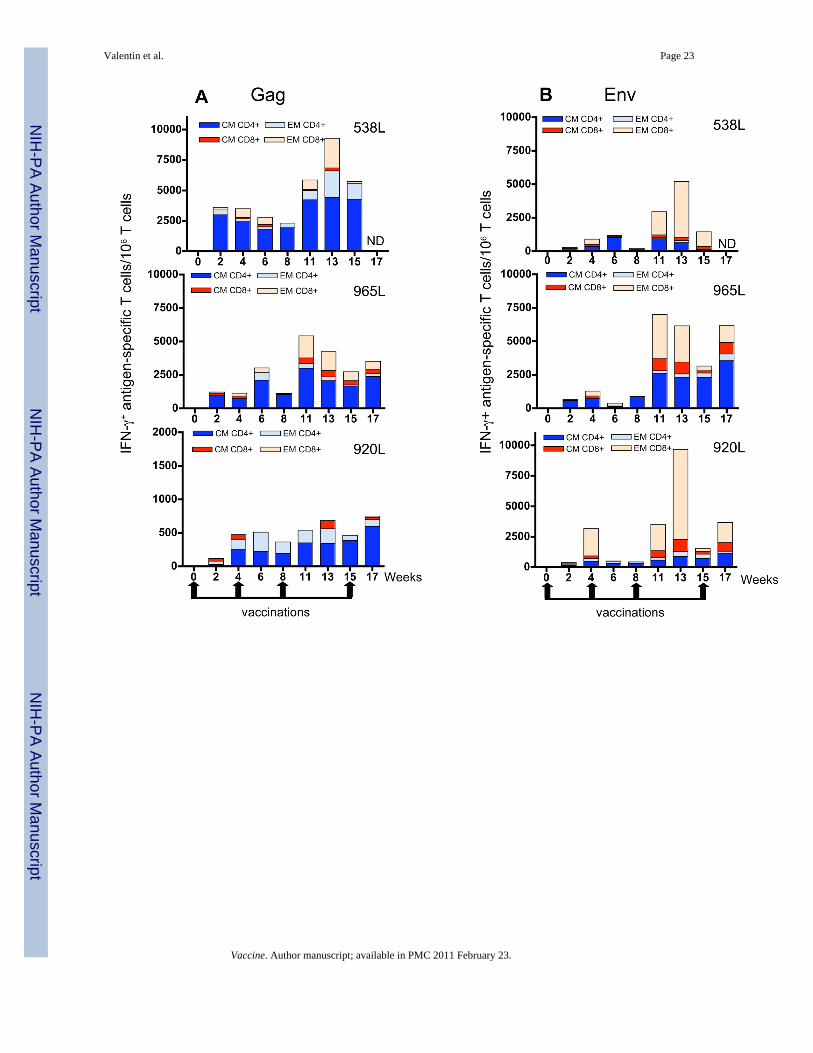
Characterization of T cell subsets induced by DNA vaccinationWe next analyzed the phenotype of the antigen-specific IFN-γ producing T cells in more detail.The gating strategy is shown in Figs. 3A and 3B for animal 965L (3 weeks post EP3). Subsetsof memory T cells were identified based on the staining with CD28 and CD45RA:CD3+CD45RA-CD28+ represents the population of central memory (CM) T cells andCD3+CD28- represents effector memory (EM) T cells (Fig. 3A). Cells with an EM phenotype
Valentin et al. Page 6
Vaccine. Author manuscript; available in PMC 2011 February 23.
NIH
-PA Author Manuscript
NIH
-PA Author Manuscript
NIH
-PA Author Manuscript
were mainly CD8+ T cells (91% in macaque 965L), while the majority of CM cells wereCD4+ T cells. These subsets were further examined for the presence of antigen-specificsubpopulations as shown in the dot plots in Fig. 3B for the same animal (965L) and the dataare summarized in Fig. 4 for all the animals.
A second strategy to define CM and EM cells was also used, employing the CD28 and CD95markers in parallel to the CD28 and CD45RA markers in samples stained with all threeantibodies (Fig. 5). We also analyzed the frequency of the CD4+ and CD8+ T cells within eachpopulation. The comparison of the antigen-specific cells using either definition(CD3+CD45RA-CD28+ versus CD3+CD95+CD28+ for CM and CD3+CD28- versusCD3+CD95+CD28- for EM) showed no significant differences. Thus, both staining strategiesgive similar results for circulating T cells. These data indicate that either approach can be usedto define CM and EM T cell subsets in macaque PBMC (Valentin et al.).
The responses against Gag (Fig. 4A) were dominated by CD4+ T cells with central memoryphenotype, whereas the predominant T cell responses against Env (Fig. 4B) consisted ofCD8+ T cells with effector phenotype. Low levels of CD8+ cells with CM phenotype could bedetected in all three animals in response to both Gag and Env peptide stimulation. Similar toGag, Nef responses (Fig. 4C) were predominantly produced by CD4+ T cells, which for 538Land 965L were almost exclusively cells with CM phenotype. The responses to Pol (Fig. 4D)were quantitatively the lowest and were predominantly CD4+ with CM phenotype in 965L and920L, and a mixture of CD4+ and CD8+ with CM and EM phenotype in macaque 538L. Inconclusion, vaccination by in vivo electroporation during ART potently induced SIV-specificrecall immune responses in different T-cell subsets. The nature of the response was alsoinfluenced by the specific antigen, as shown by comparison of the Gag and Env responses(predominantly CM CD4 for Gag versus EM CD8 for Env).
Vaccination by electroporation induces SIV-specific T cells producing IL-2 or TNFαWe further asked whether the antigen-specific T cells in these vaccinated animals produceother cytokines, such as IL-2 or TNFα (Fig. 6), in addition to IFN-γ production shown above(Figs. 2 through 5). Although we found high levels of IFN-γ-producing SIV-specific T cellsafter EP1 (Fig. 2B), we failed to detect SIV-specific T cells producing either IL-2 (Fig. 6A) orTNFα (Fig. 6B) until 3 weeks post EP3. From this time point on, a significant number of SIV-specific T cells producing IL-2 (Fig. 6A) and to a lesser extent TNFα (Fig. 6B) could bedetected. We found these responses to be induced by all the peptide pools tested (Gag, Env,Pol, Nef, Tat). We observed that in comparison to IFN-γ (see Fig. 2B), the ratio of the responsesto the individual antigens is changed for the IL-2 and TNFα producing cells, i.e. there areproportionally less responses to Gag and Env. As noted for the IFN-γ responses, animal 920Lshowed predominant Env-specific IL-2 and TNFα responses, further supporting the conclusionthat this animal could no longer respond to Gag.
Vaccination by electroporation induces multifunctional SIV-specific T cellsHaving established that vaccination by in vivo electroporation induces SIV-specific T cellsable to produce significant levels of IFN-γ, IL-2 or TNFα, we further analyzed the populationof antigen-specific T cells producing 2 or more cytokines. First, we analyzed double positivecells secreting IFN-γ in combination with IL-2 (Fig. 7A) and with TNFα (Fig. 7B), respectively.We found dual cytokine producing T cells induced by the Gag, Env and Nef peptide pools,respectively, in all three animals. We noted that the levels of IFN-γ+IL-2+ cells weresignificantly higher, reflecting the respective higher single-positive levels. The dual cytokineproducing T cells responded mainly to Gag, Env, and Nef, and no significant responses wereinduced upon stimulation by the Pol and Tat peptide pools. As observed for the single cytokine
Valentin et al. Page 7
Vaccine. Author manuscript; available in PMC 2011 February 23.
NIH
-PA Author Manuscript
NIH
-PA Author Manuscript
NIH
-PA Author Manuscript

producing T cells, the nature of the responses varied among the animals with a major focus onGag in animals 538L and 965L and on Env in animal 920L.
In addition to dual positive cells, DNA vaccination also induced SIV specific triple cytokinepositive cells. Figure 8 shows a comparison of the levels of the double and triple cytokineproducing SIV-specific (Gag, Env and Nef) T cells. For all animals, dual (IFN-γ plus IL-2;IFN-γ plus TNFα) as well as triple (IFN-γ, IL-2 and TNFα) positive cells were induced.Together, these findings indicate that vaccination by electroporation using the optimized DNAmixture is effective and potently induces SIV specific recall responses in T cells withmultifunctional phenotypes. After ART release, the polyfunctional responses persisted in 2 ofthe 3 animals, but not in animal 965L that also had a significant drop of single IFN-γ responses(see Fig. 2B).
Humoral immune responsesWe also monitored the humoral immune responses before, during, and after release from ART/DNA. Figure 9 shows the reciprocal levels of binding antibody against Env (Fig. 9A) and Gag(Fig. 9B), respectively. Our results show that the humoral immune responses decline duringART and the vaccination period, resulting in a reduction of ∼0.5 log10 for Env (A) and ∼1-2log10 for Gag (B). These data indicate that immunization with DNA only is insufficient tomaintain or boost the humoral immune responses, and this is likely due to the relative lowlevels of protein produced upon vaccination. After release from ART, the animals showedanamnestic humoral immune responses to Gag and Env reaching the pre-ART levels asresponse to the replicating SIV after ART release. Therefore, DNA electroporation maintainedthe T helper type 1 nature of DNA vaccination.
DiscussionDNA based immunization is an attractive vaccination approach because its production is simpleand cost effective, it can be repeatedly administered and it can be combined with other vaccinemodalities and molecular adjuvants. Although several trials of DNA vaccination in humanshave shown encouraging, though variable results (Mwau et al., 2004; Graham et al., 2006;Catanzaro et al., 2007; Eller et al., 2007; Tavel et al., 2007; Bansal et al., 2008; Gorse et al.,2008; Jaoko et al., 2008; Kutzler and Weiner, 2008; Wang et al., 2008; Wilson et al., 2008),it appears that naked DNA delivery and expression is inefficient in primates comparednonhuman primates and rodents, which is one key drawback for using DNA vaccination.Different strategies are being developed to improve the efficiency of DNA gene deliveryinclude the combination of antigen expressing plasmids with vectors producing cytokines, orthe use of DNA as prime in combination with recombinant virus or protein boost [(Hartikkaet al., 2001; Fuller et al., 2002; Lori et al., 2003; Bertley et al., 2004; Wang et al., 2004a;Lisziewicz et al., 2005; Dale et al., 2006; Duerr et al., 2006; Girard et al., 2006; Hokey andWeiner, 2006; Liu et al., 2006; Lori et al., 2006; Lu, 2006; Mcmichael, 2006; Rodriguez-Chavez et al., 2006; Brave et al., 2007; Hinkula, 2007; Thorner and Barouch, 2007; Kutzlerand Weiner, 2008; Manrique et al., 2008; Wang et al., 2008)]. The development of DNAdelivery by in vivo electroporation is an important advance for DNA delivery (Aihara andMiyazaki, 1998; Mathiesen, 1999; Rizzuto et al., 1999; Selby et al., 2000; Widera et al.,2000; Mir, 2001; Wang et al., 2004b; Prud'homme et al., 2006; Draghia-Akli et al., 2008), andinitial studies with DNAs producing HIV and SIV antigens have shown great improvement ingene expression as shown in this report and by others (Selby et al., 2000; Widera et al.,2000; Otten et al., 2004; Otten et al., 2006; Luckay et al., 2007; Halwani et al., 2008; Hiraoet al., 2008; Rosati et al., 2008; Zur Megede et al., 2008).
Using DNA only as vaccination modality, we had previously demonstrated a significantvirological benefit in a group of SIVmac251 infected animals (von Gegerfelt et al., 2007),
Valentin et al. Page 8
Vaccine. Author manuscript; available in PMC 2011 February 23.
NIH
-PA Author Manuscript
NIH
-PA Author Manuscript
NIH
-PA Author Manuscript

resulting in ∼1 log10 drop in viremia. Importantly, the animals had maintained this reducedviral loads for more than 3 years. None of the control animals, which were subjected to ARTtreatment only without DNA vaccination, showed reduction of viral loads after release fromART (von Gegerfelt et al., 2007) and subsequently developed AIDS and were no longeravailable. Based on the success of the first therapeutic vaccination (von Gegerfelt et al.,2007), the current study was designed to examine the efficacy of a 2nd round of therapeuticimmunization using macaques from that study. We decided to apply the 2nd therapeutic DNAvaccination by using the more potent in vivo electroporation as DNA delivery methodology,that further allowed detailed measurement of the development of SIV-specific immuneresponses.
We show that combination of optimized DNA vectors and in vivo electroporation induced highlevels of SIV-specific cellular immune responses in the ART-treated SIV-infected animals. Itis noteworthy that direct DNA intramuscular immunization into SIV-infected ART-treatedrhesus macaques induced recall responses to all the antigens produced by the DNA mixture,but to a lower extent (von Gegerfelt et al., 2007; Halwani et al., 2008) when compared to themore effective in vivo electroporation, as shown in this report and by others (Widera et al.,2000; Otten et al., 2004; Otten et al., 2006; Luckay et al., 2007; Hirao et al., 2008; Rosati etal., 2008; Zur Megede et al., 2008). Thus, in vivo electroporation of plasmid DNAs producingSIV antigens induces higher and longer-lasting primary as well as recall immune responses inmacaques. We typically observed that immune responses peaked at 4 weeks post immunization.The large increase in immune responses revealed up to 1.6% of SIV-specific IFN-γ-producingT cells in the blood. Importantly, the efficient immunization method led to the induction ofhigh levels of both CD4+ and CD8+ antigen-specific T cells. A large proportion of these SIVantigen-specific cells had markers of effector memory. Although DNA vaccination resulted inboth CD4+ and CD8+ T cell memory and effector cells, we noted that the type of antigenaffected the responses: Gag, Pol and Nef induced mainly CD4+ T cell responses, whereas Envinduced a higher CD8+ T cell response. Importantly, the vaccination effects were long-lastingand also led to significant development of multifunctional SIV-specific immune responses.These findings demonstrate the potency of DNA vaccination in inducing broad and diversecellular immune responses. In contrast to the changes in the cellular immune responses, weobserved no increase in the humoral immune responses. On the contrary, we found a rathersignificant decline for both Gag and Env binding antibody titers during ART/DNA vaccination,as reported during the 1st immunotherapy cycle (von Gegerfelt et al., 2007). Thus, DNAvaccination by in vivo electroporation in SIV-infected macaques continues to produce apolarized Th1 immune response. It is likely that the combination of DNA with a protein boostis necessary to activate higher antibody responses in chronically infected ART-treated animals.
Upon release from ART, the animals showed an initial virus rebound, followed by several viralload fluctuations, resulting in viral loads lower than before ART/DNA. We previously reportedsuch fluctuations in virus levels that ultimately resulted in reduced viremia (von Gegerfelt etal., 2007). This manifestation is indicative of active immune control. Although the underlyingreason for the fluctuation is not known, we anticipate that the initial virus rebound is eliminatedby the immune system and a new homeostasis is achieved, usually after 2-3 fluctuations,resulting in reduced virus loads compared to pre-ART levels.
It is important to note that the animals involved in this study were infected by SIV for longperiods of time. ART treatment only was reported to lead to complete or partial virus controlafter ART discontinuation uniquely in animals treated very early after infection (Tsai et al.,1998; Van Rompay et al., 1999; Emau et al., 2006), which is different than the experience withchronically infected macaques (Hel et al., 2000; Tryniszewska et al., 2002; Lisziewicz et al.,2005; Fuller et al., 2006; von Gegerfelt et al., 2007) and humans (Chun et al., 1999; Davey etal., 1999; Garcia et al., 1999; Neumann et al., 1999; Ortiz et al., 1999; Ortiz et al., 2001;
Valentin et al. Page 9
Vaccine. Author manuscript; available in PMC 2011 February 23.
NIH
-PA Author Manuscript
NIH
-PA Author Manuscript
NIH
-PA Author Manuscript
Oxenius et al., 2002), where the virus rebounds rapidly to levels similar to those prior to ART.This may be a critical difference with a recently reported study (Zur Megede et al., 2008) inwhich the animals were treated with ART early after infection and DNA vaccination failed toshow a virological benefit over the benefit achieved by ART only.
Important for the success of the immunotherapeutic vaccination is not only the quality of theDNA and the DNA delivery, but also the successful control of viremia during ART. Animalspartially controlling viremia (median viral load of 4.9 log10) and not receiving ART duringthe DNA vaccination period did not show immunological or virologic benefit even with themore efficient EP DNA vaccination method. This observation is in agreement with previousstudies (Hel et al., 2000; von Gegerfelt et al., 2007), which reported that animals that lostcontrol of viremia due to development of drug resistance or non-adherence to drug treatmentdid not benefit from immunotherapy. Therefore, potent control of viremia using effective ARTtreatment is an essential part of the regimen in addition to the use of optimized DNA andefficient DNA delivery to induce high and long-lasting recall immune responses.
An important conclusion of our study is that rhesus macaques chronically infected with SIVbenefited from a 2nd round of immunotherapy during ART. The combination of a cocktail ofoptimized SIV DNA plasmids and of efficient DNA delivery by in vivo electroporation wascritical for achieving high levels of long-lasting recall immune responses. Furthermore DNAvaccination provided a virological benefit, since the therapeutically vaccinated macaques wereable to further lower viremia upon release from ART. The 2nd round of immunotherapydemonstrated that an additional virological benefit can be obtained by repeated ART/DNAvaccination. In summary, our data provide support for a novel immunotherapeutic vaccinationapproach, which could be an addition to anti-retroviral drug therapy.
AcknowledgmentsWe thank D. Weiss, J. Treece, R. Pal, P. Markham and the staff at Advanced BioScience Laboratories, Kensington,MD for their expert help; N. Bischofsberger (Gilead Sciences, Inc.) for PMPA and FTC. We thank T. Jones for editorialassistance. This research was supported by the Intramural Research Program of the NIH, National Cancer Institute,Center for Cancer Research.
ReferencesAberle H, Bauer A, Stappert J, Kispert A, Kemler R. beta-catenin is a target for the ubiquitin-proteasome
pathway. EMBO J 1997;16:3797–3804. [PubMed: 9233789]Aihara H, Miyazaki J. Gene transfer into muscle by electroporation in vivo. Nat Biotechnol 1998;16:867–
870. [PubMed: 9743122]Andrieu JM, Lu W. A dendritic cell-based vaccine for treating HIV infection: background and preliminary
results. J Intern Med 2007;261:123–131. [PubMed: 17241177]Bansal A, Jackson B, West K, Wang S, Lu S, Kennedy JS, Goepfert PA. Multifunctional T-cell
characteristics induced by a polyvalent DNA prime/protein boost human immunodeficiency virus type1 vaccine regimen given to healthy adults are dependent on the route and dose of administration. JVirol 2008;82:6458–6469. [PubMed: 18448544]
Bergamaschi C, Rosati M, Jalah R, Valentin A, Kulkarni V, Alicea C, Zhang GM, Patel V, Felber BK,Pavlakis GN. Intracellular interaction of interleukin IL-15 with its receptor alpha during productionleads to mutual stabilization and increased bioactivity. J Biol Chem 2007;283:4189–4199. [PubMed:18055460]
Bertley FM, Kozlowski PA, Wang SW, Chappelle J, Patel J, Sonuyi O, Mazzara G, Montefiori D, CarvilleA, Mansfield KG, Aldovini A. Control of simian/human immunodeficiency virus viremia and diseaseprogression after IL-2-augmented DNA-modified vaccinia virus Ankara nasal vaccination innonhuman primates. J Immunol 2004;172:3745–3757. [PubMed: 15004179]
Valentin et al. Page 10
Vaccine. Author manuscript; available in PMC 2011 February 23.
NIH
-PA Author Manuscript
NIH
-PA Author Manuscript
NIH
-PA Author Manuscript
Biragyn A, Tani K, Grimm MC, Weeks S, Kwak LW. Genetic fusion of chemokines to a self tumorantigen induces protective, T-cell dependent antitumor immunity. Nat Biotechnol 1999;17:253–258.[PubMed: 10096292]
Brave A, Ljungberg K, Wahren B, Liu MA. Vaccine delivery methods using viral vectors. Mol Pharm2007;4:18–32. [PubMed: 17274663]
Catanzaro AT, Roederer M, Koup RA, Bailer RT, Enama ME, Nason MC, Martin JE, Rucker S, AndrewsCA, Gomez PL, Mascola JR, Nabel GJ, Graham BS. Phase I clinical evaluation of a six-plasmidmulticlade HIV-1 DNA candidate vaccine. Vaccine 2007;25:4085–4092. [PubMed: 17391815]
Chikhlikar P, Barros De Arruda L, Agrawal S, Byrne B, Guggino W, August JT, Marques ET Jr. Invertedterminal repeat sequences of adeno-associated virus enhance the antibody and CD8(+) responses toa HIV-1 p55Gag/LAMP DNA vaccine chimera. Virology 2004;323:220–232. [PubMed: 15193918]
Chikhlikar P, De Arruda LB, Maciel M, Silvera P, Lewis MG, August JT, Marques ET. DNA encodingan HIV-1 Gag/human lysosome-associated membrane protein-1 chimera elicits a broad cellular andhumoral immune response in Rhesus macaques. PLoS ONE 2006;1:e135. [PubMed: 17205139]
Chun TW, Davey RT Jr, Engel D, Lane HC, Fauci AS. Re-emergence of HIV after stopping therapy.Nature 1999;401:874–875. [PubMed: 10553903]
Connolly NC, Whiteside TL, Wilson C, Kondragunta V, Rinaldo CR, Riddler SA. Therapeuticimmunization with human immunodeficiency virus type 1 (HIV-1) peptide-loaded dendritic cells issafe and induces immunogenicity in HIV-1-infected individuals. Clin Vaccine Immunol2008;15:284–292. [PubMed: 17942609]
Dale CJ, Thomson S, De Rose R, Ranasinghe C, Medveczky CJ, Pamungkas J, Boyle DB, Ramshaw IA,Kent SJ. Prime-boost strategies in DNA vaccines. Methods Mol Med 2006;127:171–197. [PubMed:16988455]
Davey RT Jr, Bhat N, Yoder C, Chun TW, Metcalf JA, Dewar R, Natarajan V, Lempicki RA, AdelsbergerJW, Miller KD, Kovacs JA, Polis MA, Walker RE, Falloon J, Masur H, Gee D, Baseler M, DimitrovDS, Fauci AS, Lane HC. HIV-1 and T cell dynamics after interruption of highly active antiretroviraltherapy (HAART) in patients with a history of sustained viral suppression. Proc Natl Acad Sci U SA 1999;96:15109–15114. [PubMed: 10611346]
De Arruda LB, Chikhlikar PR, August JT, Marques ET. DNA vaccine encoding humanimmunodeficiency virus-1 Gag, targeted to the major histocompatibility complex II compartment bylysosomal-associated membrane protein, elicits enhanced long-term memory response. Immunology2004;112:126–133. [PubMed: 15129672]
De Rose R, Fernandez CS, Smith MZ, Batten CJ, Alcantara S, Peut V, Rollman E, Loh L, Mason RD,Wilson K, Law MG, Handley AJ, Kent SJ. Control of viremia and prevention of AIDS followingimmunotherapy of SIV-infected macaques with peptide-pulsed blood. PLoS Pathog2008;4:e1000055. [PubMed: 18451982]
Draghia-Akli R, Khan AS, Brown PA, Pope MA, Wu L, Hirao L, Weiner DB. Parameters for DNAvaccination using adaptive constant-current electroporation in mouse and pig models. Vaccine2008;26:5230–5237. [PubMed: 18450333]
Duerr A, Wasserheit JN, Corey L. HIV vaccines: new frontiers in vaccine development. Clin Infect Dis2006;43:500–511. [PubMed: 16838241]
Eller MA, Eller LA, Opollo MS, Ouma BJ, Oballah PO, Galley L, Karnasuta C, Kim SR, Robb ML,Michael NL, Kibuuka H, Wabwire-Mangen F, Graham BS, Birx DL, De Souza MS, Cox JH.Induction of HIV-specific functional immune responses by a multiclade HIV-1 DNA vaccinecandidate in healthy Ugandans. Vaccine 2007;25:7737–7742. [PubMed: 17920731]
Emau P, Jiang Y, Agy MB, Tian B, Bekele G, Tsai CC. Post-exposure prophylaxis for SIV revisited:animal model for HIV prevention. AIDS Res Ther 2006;3:29. [PubMed: 17132170]
Fuller DH, Rajakumar PA, Wilson LA, Trichel AM, Fuller JT, Shipley T, Wu MS, Weis K, Rinaldo CR,Haynes JR, Murphey-Corb M. Induction of mucosal protection against primary, heterologous simianimmunodeficiency virus by a DNA vaccine. J Virol 2002;76:3309–3317. [PubMed: 11884556]
Fuller DH, Rajakumar PA, Wu MS, Mcmahon CW, Shipley T, Fuller JT, Bazmi A, Trichel AM, AllenTM, Mothe B, Haynes JR, Watkins DI, Murphey-Corb M. DNA immunization in combination witheffective antiretroviral drug therapy controls viral rebound and prevents simian AIDS after treatmentis discontinued. Virology 2006;348:200–215. [PubMed: 16439000]
Valentin et al. Page 11
Vaccine. Author manuscript; available in PMC 2011 February 23.
NIH
-PA Author Manuscript
NIH
-PA Author Manuscript
NIH
-PA Author Manuscript
Garcia F, Plana M, Vidal C, Cruceta A, O'brien WA, Pantaleo G, Pumarola T, Gallart T, Miro JM, GatellJM. Dynamics of viral load rebound and immunological changes after stopping effectiveantiretroviral therapy. Aids 1999;13:F79–86. [PubMed: 10449278]
Girard MP, Osmanov SK, Kieny MP. A review of vaccine research and development: the humanimmunodeficiency virus (HIV). Vaccine 2006;24:4062–4081. [PubMed: 16530298]
Gorse GJ, Baden LR, Wecker M, Newman MJ, Ferrari G, Weinhold KJ, Livingston BD, Villafana TL,Li H, Noonan E, Russell ND. Safety and immunogenicity of cytotoxic T-lymphocyte poly-epitope,DNA plasmid (EP HIV-1090) vaccine in healthy, human immunodeficiency virus type 1 (HIV-1)-uninfected adults. Vaccine 2008;26:215–223. [PubMed: 18055072]
Graham BS, Koup RA, Roederer M, Bailer RT, Enama ME, Moodie Z, Martin JE, Mccluskey MM,Chakrabarti BK, Lamoreaux L, Andrews CA, Gomez PL, Mascola JR, Nabel GJ. Phase 1 safety andimmunogenicity evaluation of a multiclade HIV-1 DNA candidate vaccine. J Infect Dis2006;194:1650–1660. [PubMed: 17109336]
Halwani R, Boyer JD, Yassine-Diab B, Haddad EK, Robinson TM, Kumar S, Parkinson R, Wu L, SidhuMK, Phillipson-Weiner R, Pavlakis GN, Felber BK, Lewis MG, Shen A, Siliciano RF, Weiner DB,Sekaly RP. Therapeutic vaccination with simian immunodeficiency virus (SIV)-DNA + IL-12 orIL-15 induces distinct CD8 memory subsets in SIV-infected macaques. J Immunol 2008;180:7969–7979. [PubMed: 18523260]
Hardy GA, Imami N, Nelson MR, Sullivan AK, Moss R, Aasa-Chapman MM, Gazzard B, Gotch FM.A phase I, randomized study of combined IL-2 and therapeutic immunisation with antiretroviraltherapy. J Immune Based Ther Vaccines 2007;5:6. [PubMed: 17428345]
Hartikka J, Bozoukova V, Ferrari M, Sukhu L, Enas J, Sawdey M, Wloch MK, Tonsky K, Norman J,Manthorpe M, Wheeler CJ. Vaxfectin enhances the humoral immune response to plasmid DNA-encoded antigens. Vaccine 2001;19:1911–1923. [PubMed: 11228361]
Hel Z, Venzon D, Poudyal M, Tsai WP, Giuliani L, Woodward R, Chougnet C, Shearer G, Altman JD,Watkins D, Bischofberger N, Abimiku A, Markham P, Tartaglia J, Franchini G. Viremia controlfollowing antiretroviral treatment and therapeutic immunization during primary SIV251 infection ofmacaques. Nat Med 2000;6:1140–1146. [PubMed: 11017146]
Hinkula J. Clarification of how HIV-1 DNA and protein immunizations may be better used to obtainHIV-1-specific mucosal and systemic immunity. Expert Rev Vaccines 2007;6:203–212. [PubMed:17408370]
Hirao LA, Wu L, Khan AS, Hokey DA, Yan J, Dai A, Betts MR, Draghia-Akli R, Weiner DB. Combinedeffects of IL-12 and electroporation enhances the potency of DNA vaccination in macaques. Vaccine2008;26:3112–3120. [PubMed: 18430495]
Hokey DA, Weiner DB. DNA vaccines for HIV: challenges and opportunities. Springer SeminImmunopathol 2006;28:267–279. [PubMed: 17031649]
Jalah R, Rosati R, Kulkarni V, Patel V, Bergamaschi C, Valentin A, Zhang GM, Sidhu MK, EldridgeJH, Weiner DB, Pavlakis GN, Felber BK. Efficient systemic expression of bioactive IL-15 in miceupon delivery of optimized DNA expression plasmids. DNA Cell Biol 2007;26:827–840. [PubMed:17979522]
Jaoko W, Nakwagala FN, Anzala O, Manyonyi GO, Birungi J, Nanvubya A, Bashir F, Bhatt K, OgutuH, Wakasiaka S, Matu L, Waruingi W, Odada J, Oyaro M, Indangasi J, Ndinya-Achola J, Konde C,Mugisha E, Fast P, Schmidt C, Gilmour J, Tarragona T, Smith C, Barin B, Dally L, Johnson B,Muluubya A, Nielsen L, Hayes P, Boaz M, Hughes P, Hanke T, Mcmichael A, Bwayo J, Kaleebu P.Safety and immunogenicity of recombinant low-dosage HIV-1 A vaccine candidates vectored byplasmid pTHr DNA or modified vaccinia virus Ankara (MVA) in humans in East Africa. Vaccine2008;26:2788–2795. [PubMed: 18440674]
Kinloch-De Loes S, Hoen B, Smith DE, Autran B, Lampe FC, Phillips AN, Goh LE, Andersson J, TsoukasC, Sonnerborg A, Tambussi G, Girard PM, Bloch M, Battegay M, Carter N, El Habib R, Theofan G,Cooper DA, Perrin L. Impact of therapeutic immunization on HIV-1 viremia after discontinuationof antiretroviral therapy initiated during acute infection. J Infect Dis 2005;192:607–617. [PubMed:16028129]
Kutzler MA, Weiner DB. DNA vaccines: ready for prime time? Nat Rev Genet 2008;9:776–788.[PubMed: 18781156]
Valentin et al. Page 12
Vaccine. Author manuscript; available in PMC 2011 February 23.
NIH
-PA Author Manuscript
NIH
-PA Author Manuscript
NIH
-PA Author Manuscript

Levy Y, Gahery-Segard H, Durier C, Lascaux AS, Goujard C, Meiffredy V, Rouzioux C, Habib RE,Beumont-Mauviel M, Guillet JG, Delfraissy JF, Aboulker JP. Immunological and virological efficacyof a therapeutic immunization combined with interleukin-2 in chronically HIV-1 infected patients.Aids 2005;19:279–286. [PubMed: 15718838]
Lisziewicz J, Calarota SA, Lori F. The potential of topical DNA vaccines adjuvanted by cytokines. ExpertOpin Biol Ther 2007;7:1563–1574. [PubMed: 17916048]
Lisziewicz J, Trocio J, Xu J, Whitman L, Ryder A, Bakare N, Lewis MG, Wagner W, Pistorio A, AryaS, Lori F. Control of viral rebound through therapeutic immunization with DermaVir. Aids2005;19:35–43. [PubMed: 15627031]
Liu MA, Wahren B, Karlsson Hedestam GB. DNA vaccines: recent developments and future possibilities.Hum Gene Ther 2006;17:1051–1061. [PubMed: 17032152]
Lori F, Kelly LM, Lisziewicz J. Immunological approaches for HIV therapy. Curr Drug Targets InfectDisord 2003;3:171–178. [PubMed: 12769793]
Lori F, Trocio J, Bakare N, Kelly LM, Lisziewicz J. DermaVir, a novel HIV immunisation technology.Vaccine 2005;23:2030–2034. [PubMed: 15755566]
Lori F, Weiner DB, Calarota SA, Kelly LM, Lisziewicz J. Cytokine-adjuvanted HIV-DNA vaccinationstrategies. Springer Semin Immunopathol 2006;28:231–238. [PubMed: 17053912]
Lu S. Combination DNA plus protein HIV vaccines. Springer Semin Immunopathol 2006;28:255–265.[PubMed: 17021720]
Lu W, Arraes LC, Ferreira WT, Andrieu JM. Therapeutic dendritic-cell vaccine for chronic HIV-1infection. Nat Med 2004;10:1359–1365. [PubMed: 15568033]
Lu W, Wu X, Lu Y, Guo W, Andrieu JM. Therapeutic dendritic-cell vaccine for simian AIDS. Nat Med2003;9:27–32. [PubMed: 12496959]
Luckay A, Sidhu MK, Kjeken R, Megati S, Chong SY, Roopchand V, Garcia-Hand D, Abdullah R, BraunR, Montefiori DC, Rosati M, Felber BK, Pavlakis GN, Mathiesen I, Israel ZR, Eldridge JH, EganMA. Effect of plasmid DNA vaccine design and in vivo electroporation on the resulting vaccine-specific immune responses in rhesus macaques. J Virol 2007;81:5257–5269. [PubMed: 17329330]
Manrique M, Micewicz E, Kozlowski PA, Wang SW, Aurora D, Wilson RL, Ghebremichael M, MazzaraG, Montefiori D, Carville A, Mansfield KG, Aldovini A. DNA-MVA vaccine protection after X4SHIV challenge in macaques correlates with day-of-challenge antiviral CD4+ cell-mediatedimmunity levels and postchallenge preservation of CD4+ T cell memory. AIDS Res HumRetroviruses 2008;24:505–519. [PubMed: 18373436]
Markowitz M, Jin X, Hurley A, Simon V, Ramratnam B, Louie M, Deschenes GR, Ramanathan M Jr,Barsoum S, Vanderhoeven J, He T, Chung C, Murray J, Perelson AS, Zhang L, Ho DD.Discontinuation of antiretroviral therapy commenced early during the course of humanimmunodeficiency virus type 1 infection, with or without adjunctive vaccination. J Infect Dis2002;186:634–643. [PubMed: 12195350]
Marques ET Jr, Chikhlikar P, De Arruda LB, Leao IC, Lu Y, Wong J, Chen JS, Byrne B, August JT.HIV-1 p55Gag encoded in the lysosome-associated membrane protein-1 as a DNA plasmid vaccinechimera is highly expressed, traffics to the major histocompatibility class II compartment, and elicitsenhanced immune responses. J Biol Chem 2003;278:37926–37936. [PubMed: 12824194]
Mathiesen I. Electropermeabilization of skeletal muscle enhances gene transfer in vivo. Gene Ther1999;6:508–514. [PubMed: 10476210]
Mcmichael AJ. HIV vaccines. Annu Rev Immunol 2006;24:227–255. [PubMed: 16551249]Mir LM. Therapeutic perspectives of in vivo cell electropermeabilization. Bioelectrochemistry
2001;53:1–10. [PubMed: 11206915]Mwau M, Cebere I, Sutton J, Chikoti P, Winstone N, Wee EG, Beattie T, Chen YH, Dorrell L, Mcshane
H, Schmidt C, Brooks M, Patel S, Roberts J, Conlon C, Rowland-Jones SL, Bwayo JJ, McmichaelAJ, Hanke T. A human immunodeficiency virus 1 (HIV-1) clade A vaccine in clinical trials:stimulation of HIV-specific T-cell responses by DNA and recombinant modified vaccinia virusAnkara (MVA) vaccines in humans. J Gen Virol 2004;85:911–919. [PubMed: 15039533]
Nasioulas G, Zolotukhin AS, Tabernero C, Solomin L, Cunningham CP, Pavlakis GN, Felber BK.Elements distinct from human immunodeficiency virus type 1 splice sites are responsible for the Revdependence of env mRNA. J Virol 1994;68:2986–2993. [PubMed: 8151769]
Valentin et al. Page 13
Vaccine. Author manuscript; available in PMC 2011 February 23.
NIH
-PA Author Manuscript
NIH
-PA Author Manuscript
NIH
-PA Author Manuscript
Neumann AU, Tubiana R, Calvez V, Robert C, Li TS, Agut H, Autran B, Katlama C. HIV-1 reboundduring interruption of highly active antiretroviral therapy has no deleterious effect on reinitiatedtreatment. Comet Study Group Aids 1999;13:677–683.
Ortiz GM, Nixon DF, Trkola A, Binley J, Jin X, Bonhoeffer S, Kuebler PJ, Donahoe SM, Demoitie MA,Kakimoto WM, Ketas T, Clas B, Heymann JJ, Zhang L, Cao Y, Hurley A, Moore JP, Ho DD,Markowitz M. HIV-1-specific immune responses in subjects who temporarily contain virusreplication after discontinuation of highly active antiretroviral therapy. J Clin Invest 1999;104:R13–18. [PubMed: 10491418]
Ortiz GM, Wellons M, Brancato J, Vo HT, Zinn RL, Clarkson DE, Van Loon K, Bonhoeffer S, MirallesGD, Montefiori D, Bartlett JA, Nixon DF. Structured antiretroviral treatment interruptions inchronically HIV-1-infected subjects. Proc Natl Acad Sci U S A 2001;98:13288–13293. [PubMed:11687611]
Otten G, Schaefer M, Doe B, Liu H, Srivastava I, Zur Megede J, O'hagan D, Donnelly J, Widera G,Rabussay D, Lewis MG, Barnett S, Ulmer JB. Enhancement of DNA vaccine potency in rhesusmacaques by electroporation. Vaccine 2004;22:2489–2493. [PubMed: 15193413]
Otten GR, Schaefer M, Doe B, Liu H, Megede JZ, Donnelly J, Rabussay D, Barnett S, Ulmer JB. Potentimmunogenicity of an HIV-1 gag-pol fusion DNA vaccine delivered by in vivo electroporation.Vaccine 2006;24:4503–4509. [PubMed: 16181711]
Oxenius A, Price DA, Gunthard HF, Dawson SJ, Fagard C, Perrin L, Fischer M, Weber R, Plana M,Garcia F, Hirschel B, Mclean A, Phillips RE. Stimulation of HIV-specific cellular immunity bystructured treatment interruption fails to enhance viral control in chronic HIV infection. Proc NatlAcad Sci U S A 2002;99:13747–13752. [PubMed: 12370434]
Pialoux G, Quercia RP, Gahery H, Daniel N, Slama L, Girard PM, Bonnard P, Rozenbaum W, SchneiderV, Salmon D, Guillet JG. Immunological responses and long-term treatment interruption after humanimmunodeficiency virus type 1 (HIV-1) lipopeptide immunization of HIV-1-infected patients: theLIPTHERA study. Clin Vaccine Immunol 2008;15:562–568. [PubMed: 18184824]
Prud'homme GJ, Glinka Y, Khan AS, Draghia-Akli R. Electroporation-enhanced nonviral gene transferfor the prevention or treatment of immunological, endocrine and neoplastic diseases. Curr Gene Ther2006;6:243–273. [PubMed: 16611045]
Rizzuto G, Cappelletti M, Maione D, Savino R, Lazzaro D, Costa P, Mathiesen I, Cortese R, CilibertoG, Laufer R, La Monica N, Fattori E. Efficient and regulated erythropoietin production by nakedDNA injection and muscle electroporation. Proc Natl Acad Sci U S A 1999;96:6417–6422. [PubMed:10339602]
Rodriguez-Chavez IR, Allen M, Hill EL, Sheets RL, Pensiero M, Bradac JA, D'souza MP. Currentadvances and challenges in HIV-1 vaccines. Curr HIV/AIDS Rep 2006;3:39–47. [PubMed:16522258]
Romano JW, Shurtliff RN, Dobratz E, Gibson A, Hickman K, Markham PD, Pal R. Quantitativeevaluation of simian immunodeficiency virus infection using NASBA technology. J Virol Methods2000;86:61–70. [PubMed: 10713377]
Rosati M, Valentin A, Jalah R, Patel V, von Gegerfelt A, Bergamaschi C, Alicea C, Weiss D, Treece J,Pal R, Markham P, Marques ETA, August JT, Khan A, Draghia-Akli R, Felber BK, Pavlakis GN.Increased immune responses in rhesus macaques by DNA vaccination combined withelectroporation. Vaccine 2008;26:5223–5229. [PubMed: 18468743]
Rosati M, von Gegerfelt A, Roth P, Alicea C, Valentin A, Robert-Guroff M, Venzon D, Montefiori D,Markham P, Felber BK, Pavlakis GN. DNA vaccines expressing different forms of simianimmunodeficiency virus antigens decrease viremia upon SIVmac251 challenge. J Virol2005;79:8480–8492. [PubMed: 15956591]
Rosenberg ES, Altfeld M, Poon SH, Phillips MN, Wilkes BM, Eldridge RL, Robbins GK, D'aquila RT,Goulder PJ, Walker BD. Immune control of HIV-1 after early treatment of acute infection. Nature2000;407:523–526. [PubMed: 11029005]
Schneider R, Campbell M, Nasioulas G, Felber BK, Pavlakis GN. Inactivation of the humanimmunodeficiency virus type 1 inhibitory elements allows Rev-independent expression of Gag andGag/protease and particle formation. J Virol 1997;71:4892–4903. [PubMed: 9188551]
Valentin et al. Page 14
Vaccine. Author manuscript; available in PMC 2011 February 23.
NIH
-PA Author Manuscript
NIH
-PA Author Manuscript
NIH
-PA Author Manuscript

Schwartz S, Campbell M, Nasioulas G, Harrison J, Felber BK, Pavlakis GN. Mutational inactivation ofan inhibitory sequence in human immunodeficiency virus type 1 results in Rev-independent gagexpression. J Virol 1992a;66:7176–7182. [PubMed: 1433510]
Schwartz S, Felber BK, Pavlakis GN. Distinct RNA sequences in the gag region of humanimmunodeficiency virus type 1 decrease RNA stability and inhibit expression in the absence of Revprotein. J Virol 1992b;66:150–159. [PubMed: 1727477]
Selby M, Goldbeck C, Pertile T, Walsh R, Ulmer J. Enhancement of DNA vaccine potency byelectroporation in vivo. J Biotechnol 2000;83:147–152. [PubMed: 11000470]
Tavel JA, Martin JE, Kelly GG, Enama ME, Shen JM, Gomez PL, Andrews CA, Koup RA, Bailer RT,Stein JA, Roederer M, Nabel GJ, Graham BS. Safety and immunogenicity of a Gag-Pol candidateHIV-1 DNA vaccine administered by a needle-free device in HIV-1-seronegative subjects. J AcquirImmune Defic Syndr 2007;44:601–605. [PubMed: 17325604]
Thorner AR, Barouch DH. HIV-1 Vaccine Development: Progress and Prospects. Curr Infect Dis Rep2007;9:71–75. [PubMed: 17254507]
Tryniszewska E, Nacsa J, Lewis MG, Silvera P, Montefiori D, Venzon D, Hel Z, Parks RW, MoniuszkoM, Tartaglia J, Smith KA, Franchini G. Vaccination of macaques with long-standing SIVmac251infection lowers the viral set point after cessation of antiretroviral therapy. J Immunol2002;169:5347–5357. [PubMed: 12391256]
Tsai CC, Emau P, Follis KE, Beck TW, Benveniste RE, Bischofberger N, Lifson JD, Morton WR.Effectiveness of postinoculation (R)-9-(2-phosphonylmethoxypropyl) adenine treatment forprevention of persistent simian immunodeficiency virus SIVmne infection depends critically ontiming of initiation and duration of treatment. J Virol 1998;72:4265–4273. [PubMed: 9557716]
Tubiana R, Carcelain G, Vray M, Gourlain K, Dalban C, Chermak A, Rabian C, Vittecoq D, Simon A,Bouvet E, El Habib R, Costagliola D, Calvez V, Autran B, Katlama C. Therapeutic immunizationwith a human immunodeficiency virus (HIV) type 1-recombinant canarypox vaccine in chronicallyHIV-infected patients: The Vacciter Study (ANRS 094). Vaccine 2005;23:4292–4301. [PubMed:15927325]
Valentin A, Chikhlikar P, Patel V, Rosati M, Maciel M, Chang KH, Silvera P, Felber BK, Pavlakis GN,August JT, Marques ETA. Comparison of DNA vaccines producing HIV-1 gag and LAMP/gagchimera in rhesus macaques reveals antigen-specific T cell responses with distinct phenotypes.submitted.
Van Rompay KK, Dailey PJ, Tarara RP, Canfield DR, Aguirre NL, Cherrington JM, Lamy PD,Bischofberger N, Pedersen NC, Marthas ML. Early short-term 9-[2-(R)-(phosphonomethoxy)propyl]adenine treatment favorably alters the subsequent disease course in simian immunodeficiency virus-infected newborn Rhesus macaques. J Virol 1999;73:2947–2955. [PubMed: 10074144]
von Gegerfelt AS, Rosati M, Alicea C, Valentin A, Roth P, Bear J, Franchini G, Albert PS, BischofbergerN, Boyer JD, Weiner DB, Markham P, Israel ZR, Eldridge JH, Pavlakis GN, Felber BK. Long-lastingdecrease in viremia in macaques chronically infected with simian immunodeficiency virusSIVmac251 after therapeutic DNA immunization. J Virol 2007;81:1972–1979. [PubMed: 17135321]
Wang S, Kennedy JS, West K, Montefiori DC, Coley S, Lawrence J, Shen S, Green S, Rothman AL,Ennis FA, Arthos J, Pal R, Markham P, Lu S. Cross-subtype antibody and cellular immune responsesinduced by a polyvalent DNA prime-protein boost HIV-1 vaccine in healthy human volunteers.Vaccine 2008;26:1098–1110. [PubMed: 18243434]
Wang SW, Bertley FM, Kozlowski PA, Herrmann L, Manson K, Mazzara G, Piatak M, Johnson RP,Carville A, Mansfield K, Aldovini A. An SHIV DNA/MVA rectal vaccination in macaques providessystemic and mucosal virus-specific responses and protection against AIDS. AIDS Res HumRetroviruses 2004a;20:846–859. [PubMed: 15320989]
Wang Z, Troilo PJ, Wang X, Griffiths TG, Pacchione SJ, Barnum AB, Harper LB, Pauley CJ, Niu Z,Denisova L, Follmer TT, Rizzuto G, Ciliberto G, Fattori E, Monica NL, Manam S, Ledwith BJ.Detection of integration of plasmid DNA into host genomic DNA following intramuscular injectionand electroporation. Gene Ther 2004b;11:711–721. [PubMed: 14724672]
Widera G, Austin M, Rabussay D, Goldbeck C, Barnett SW, Chen M, Leung L, Otten GR, Thudium K,Selby MJ, Ulmer JB. Increased DNA vaccine delivery and immunogenicity by electroporation invivo. J Immunol 2000;164:4635–4640. [PubMed: 10779767]
Valentin et al. Page 15
Vaccine. Author manuscript; available in PMC 2011 February 23.
NIH
-PA Author Manuscript
NIH
-PA Author Manuscript
NIH
-PA Author Manuscript

Wilson CC, Newman MJ, Livingston BD, Mawhinney S, Forster JE, Scott J, Schooley RT, Benson CA.Clinical phase 1 testing of the safety and immunogenicity of an epitope-based DNA vaccine in humanimmunodeficiency virus type 1-infected subjects receiving highly active antiretroviral therapy. ClinVaccine Immunol 2008;15:986–994. [PubMed: 18400976]
Zur Megede J, Sanders-Beer B, Silvera P, Golightly D, Bowlsbey A, Hebblewaite D, Sites D, Nieves-Duran L, Srivastava R, Otten GR, Rabussay D, Zhang L, Ulmer JB, Barnett SW, Donnelly JJ. ATherapeutic SIV DNA Vaccine Elicits T-Cell Immune Responses, but No Sustained Control ofViremia in SIVmac239-Infected Rhesus Macaques. AIDS Res Hum Retroviruses 2008;24:1103–1116. [PubMed: 18620495]
Valentin et al. Page 16
Vaccine. Author manuscript; available in PMC 2011 February 23.
NIH
-PA Author Manuscript
NIH
-PA Author Manuscript
NIH
-PA Author Manuscript

FIG. 1.Virological benefit from 2nd round of ART/DNA. (A) Study Outline. SIV-infected macaqueswere subjected to a 2nd round of ART/DNA after more than 3 years of infection. Previously,we reported the data obtained from the 1st cycle of ART/DNA (von Gegerfelt et al., 2007).After 3.1 to 3.8 years, the animals were subjected to a 2nd round of ART/DNA. The durationof the 2nd ART/DNA was 31 weeks and the animals were vaccinated by in vivo electroporationfour times (EP1 to EP4) initiated at week 14 of ART. The animals were released from ARTand monitored for another 16 weeks. (B) Viral loads of the three macaques during 2nd roundof ART/DNA. Viral load data are shown for 39 weeks prior to ART (PRE), 31 weeks of ART/DNA and 16 weeks post release from ART (POST). (C) Mean virus loads and decrease inmean virus load (ΔVL) comparing 13 weeks PRE and 16 weeks POST treatment. Thedifferences are statistically significant (paired T-test).
Valentin et al. Page 17
Vaccine. Author manuscript; available in PMC 2011 February 23.
NIH
-PA Author Manuscript
NIH
-PA Author Manuscript
NIH
-PA Author Manuscript
Valentin et al. Page 18
Vaccine. Author manuscript; available in PMC 2011 February 23.
NIH
-PA Author Manuscript
NIH
-PA Author Manuscript
NIH
-PA Author Manuscript
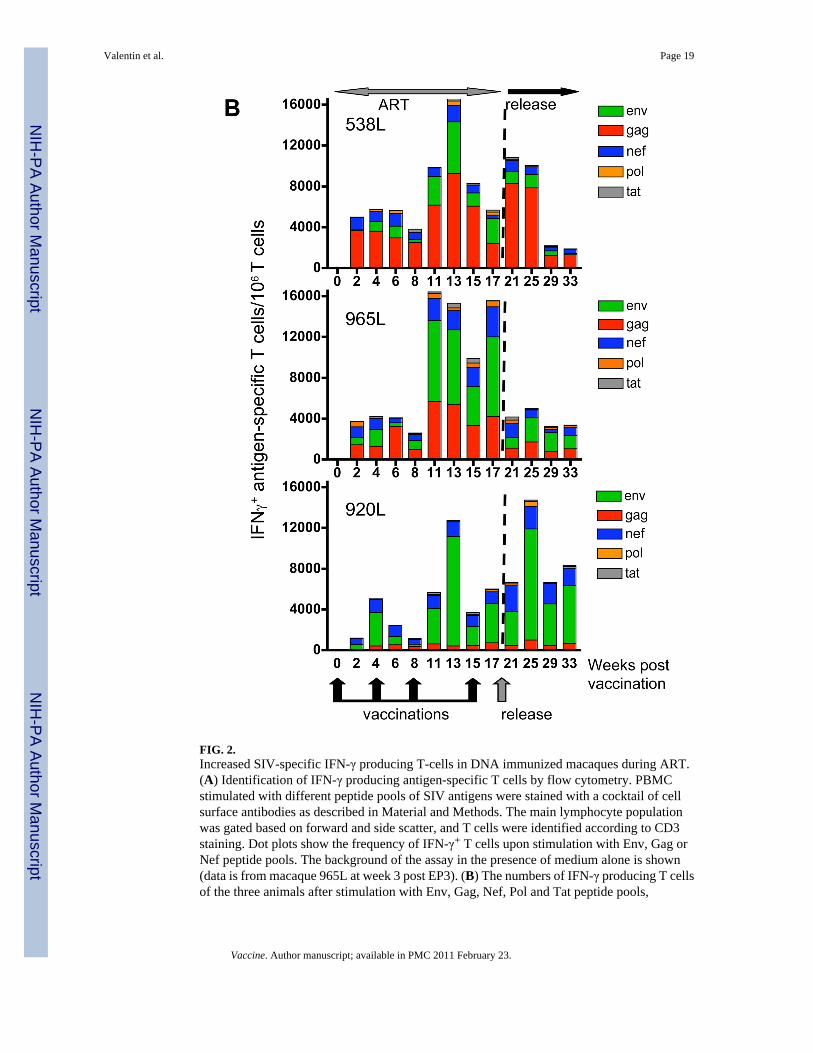
FIG. 2.Increased SIV-specific IFN-γ producing T-cells in DNA immunized macaques during ART.(A) Identification of IFN-γ producing antigen-specific T cells by flow cytometry. PBMCstimulated with different peptide pools of SIV antigens were stained with a cocktail of cellsurface antibodies as described in Material and Methods. The main lymphocyte populationwas gated based on forward and side scatter, and T cells were identified according to CD3staining. Dot plots show the frequency of IFN-γ+ T cells upon stimulation with Env, Gag orNef peptide pools. The background of the assay in the presence of medium alone is shown(data is from macaque 965L at week 3 post EP3). (B) The numbers of IFN-γ producing T cellsof the three animals after stimulation with Env, Gag, Nef, Pol and Tat peptide pools,
Valentin et al. Page 19
Vaccine. Author manuscript; available in PMC 2011 February 23.
NIH
-PA Author Manuscript
NIH
-PA Author Manuscript
NIH
-PA Author Manuscript

respectively, expressed per million circulating T lymphocytes are shown during ART/DNAand after release from ART.
Valentin et al. Page 20
Vaccine. Author manuscript; available in PMC 2011 February 23.
NIH
-PA Author Manuscript
NIH
-PA Author Manuscript
NIH
-PA Author Manuscript

Valentin et al. Page 21
Vaccine. Author manuscript; available in PMC 2011 February 23.
NIH
-PA Author Manuscript
NIH
-PA Author Manuscript
NIH
-PA Author Manuscript
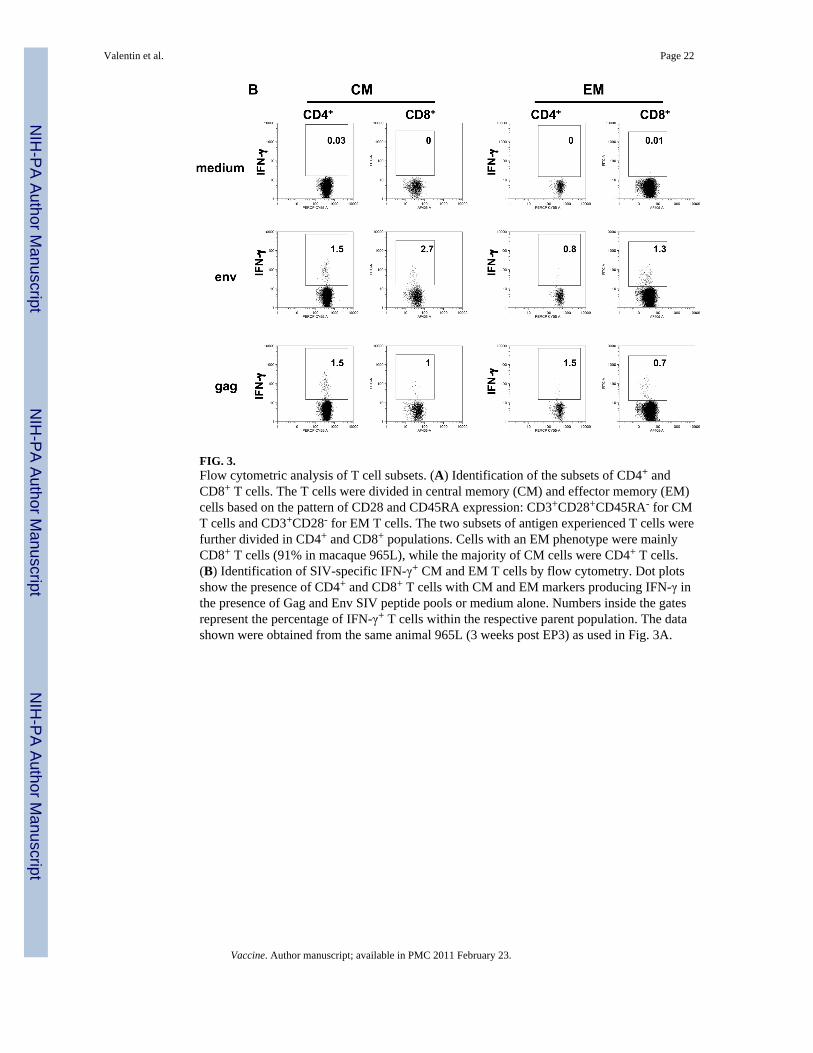
FIG. 3.Flow cytometric analysis of T cell subsets. (A) Identification of the subsets of CD4+ andCD8+ T cells. The T cells were divided in central memory (CM) and effector memory (EM)cells based on the pattern of CD28 and CD45RA expression: CD3+CD28+CD45RA- for CMT cells and CD3+CD28- for EM T cells. The two subsets of antigen experienced T cells werefurther divided in CD4+ and CD8+ populations. Cells with an EM phenotype were mainlyCD8+ T cells (91% in macaque 965L), while the majority of CM cells were CD4+ T cells.(B) Identification of SIV-specific IFN-γ+ CM and EM T cells by flow cytometry. Dot plotsshow the presence of CD4+ and CD8+ T cells with CM and EM markers producing IFN-γ inthe presence of Gag and Env SIV peptide pools or medium alone. Numbers inside the gatesrepresent the percentage of IFN-γ+ T cells within the respective parent population. The datashown were obtained from the same animal 965L (3 weeks post EP3) as used in Fig. 3A.
Valentin et al. Page 22
Vaccine. Author manuscript; available in PMC 2011 February 23.
NIH
-PA Author Manuscript
NIH
-PA Author Manuscript
NIH
-PA Author Manuscript
Valentin et al. Page 23
Vaccine. Author manuscript; available in PMC 2011 February 23.
NIH
-PA Author Manuscript
NIH
-PA Author Manuscript
NIH
-PA Author Manuscript

FIG. 4.Comparison of SIV-specific IFN-γ producing T cell subsets induced by immunization duringART. Frequency of SIV-specific CD4+ and CD8+ T cells with central memory (CM) or effectormemory (EM) markers were determined as outlined in Fig. 4. Numbers indicate IFN-γproducing T cells after stimulation with Gag (A), Env (B), Nef (C), and Pol (D) peptide pools,expressed per million circulating T lymphocytes.
Valentin et al. Page 24
Vaccine. Author manuscript; available in PMC 2011 February 23.
NIH
-PA Author Manuscript
NIH
-PA Author Manuscript
NIH
-PA Author Manuscript
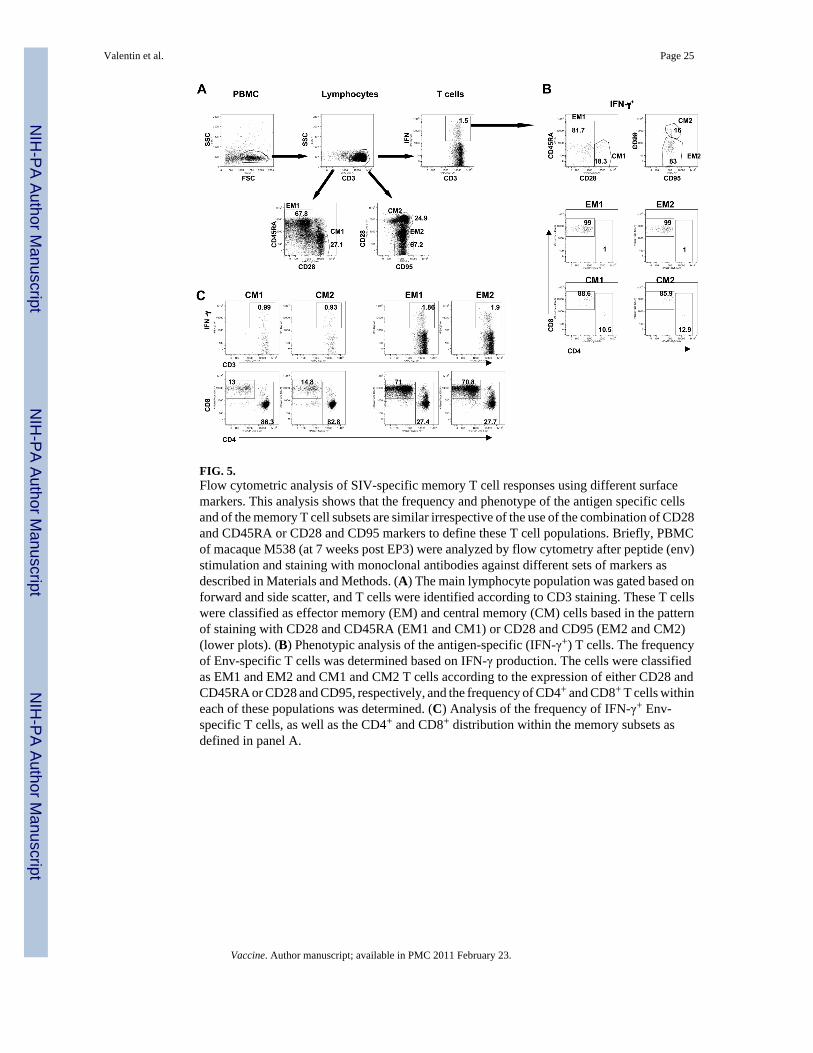
FIG. 5.Flow cytometric analysis of SIV-specific memory T cell responses using different surfacemarkers. This analysis shows that the frequency and phenotype of the antigen specific cellsand of the memory T cell subsets are similar irrespective of the use of the combination of CD28and CD45RA or CD28 and CD95 markers to define these T cell populations. Briefly, PBMCof macaque M538 (at 7 weeks post EP3) were analyzed by flow cytometry after peptide (env)stimulation and staining with monoclonal antibodies against different sets of markers asdescribed in Materials and Methods. (A) The main lymphocyte population was gated based onforward and side scatter, and T cells were identified according to CD3 staining. These T cellswere classified as effector memory (EM) and central memory (CM) cells based in the patternof staining with CD28 and CD45RA (EM1 and CM1) or CD28 and CD95 (EM2 and CM2)(lower plots). (B) Phenotypic analysis of the antigen-specific (IFN-γ+) T cells. The frequencyof Env-specific T cells was determined based on IFN-γ production. The cells were classifiedas EM1 and EM2 and CM1 and CM2 T cells according to the expression of either CD28 andCD45RA or CD28 and CD95, respectively, and the frequency of CD4+ and CD8+ T cells withineach of these populations was determined. (C) Analysis of the frequency of IFN-γ+ Env-specific T cells, as well as the CD4+ and CD8+ distribution within the memory subsets asdefined in panel A.
Valentin et al. Page 25
Vaccine. Author manuscript; available in PMC 2011 February 23.
NIH
-PA Author Manuscript
NIH
-PA Author Manuscript
NIH
-PA Author Manuscript

FIG. 6.Induction of SIV-specific IL-2- and TNFα-producing T cells induced by DNA vaccinationduring ART. The analysis was performed using similar strategy as described for Fig. 3. IL-2(A) and TNFα (B) producing T cells after stimulation with Env, Gag, Nef, Pol, and Tat peptidepools, respectively, were expressed per million circulating T lymphocytes. The ART andrelease periods are indicated. *, Tat-specific immune response was not determined.
Valentin et al. Page 26
Vaccine. Author manuscript; available in PMC 2011 February 23.
NIH
-PA Author Manuscript
NIH
-PA Author Manuscript
NIH
-PA Author Manuscript
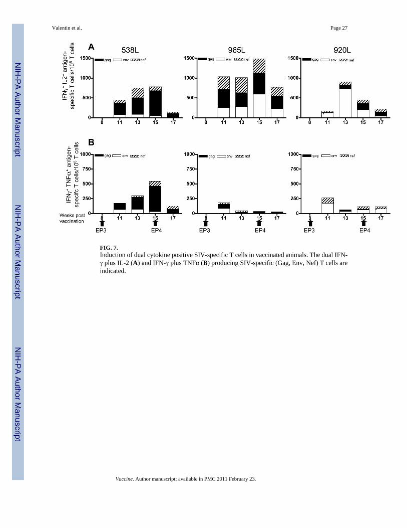
FIG. 7.Induction of dual cytokine positive SIV-specific T cells in vaccinated animals. The dual IFN-γ plus IL-2 (A) and IFN-γ plus TNFα (B) producing SIV-specific (Gag, Env, Nef) T cells areindicated.
Valentin et al. Page 27
Vaccine. Author manuscript; available in PMC 2011 February 23.
NIH
-PA Author Manuscript
NIH
-PA Author Manuscript
NIH
-PA Author Manuscript

FIG. 8.Induction of multifunctional SIV-specific T cells. A comparison of the levels of double positive(from Fig. 7; light grey IFNγ+IL-2+; dark grey and IFNγ+ TNFα+) and triple positive (IFNγ+
IL-2+ TNFα+; striped bar) total SIV specific T cells is shown.
Valentin et al. Page 28
Vaccine. Author manuscript; available in PMC 2011 February 23.
NIH
-PA Author Manuscript
NIH
-PA Author Manuscript
NIH
-PA Author Manuscript

FIG. 9.Humoral immune responses during ART and DNA vaccination and the period after releasefrom ART. The presence of binding antibodies to Env (gp120) (A) and to Gag p27 (B) wasmeasured in plasma prior, during and after therapy.
Valentin et al. Page 29
Vaccine. Author manuscript; available in PMC 2011 February 23.
NIH
-PA Author Manuscript
NIH
-PA Author Manuscript
NIH
-PA Author Manuscript