External brain morphology in rhesus macaques (Macaca mulatta)

Garibal et al. Virology Journal 2012, 9:220http://www.virologyj.com/content/9/1/220
RESEARCH Open Access
IL-2 immunotherapy in chronically SIV-infectedRhesus MacaquesJulie Garibal1, Mireille Laforge4, Ricardo Silvestre6, Shahul Mouhamad1, Laure Campillo-Gimenez1,Yves Lévy1,2,3* and Jérôme Estaquier1,5*
Abstract
Background: Despite inducing a sustained increase in CD4+ T cell counts, intermittent recombinant IL-2 (rIL-2)therapy did not confer a better clinical outcome in HIV-infected patients enrolled in large phase III clinical trialsESPRIT and SILCAAT. Several hypotheses were evoked to explain these discrepancies. Here, we investigated theimpact of low and high doses of IL-2 in Rhesus macaques of Chinese origin infected with SIVmac251 in theabsence of antiretroviral therapy (ART).
Results: We demonstrated that rIL-2 induced a dose dependent expansion of CD4+ and CD8+ T cells withoutaffecting viral load. rIL-2 increased CD4 and CD8 Treg cells as defined by the expression of CD25highFoxP3+CD127low.We also showed that rIL-2 modulated spontaneous and Fas-mediated CD4+ and CD8+ T cell apoptosis. Thehigher dose exhibited a dramatic pro-apoptotic effect on both CD4+ and CD8+ T cell populations. Finally, all theanimals treated with rIL-2 developed a wasting syndrome in the month following treatment simultaneously to adramatic decrease of circulating effector T cells.
Conclusion: These data contribute to the understanding of the homeostatic and dosage effects of IL-2 in thecontext of SIV/HIV infection.
Keywords: SIV, IL-2 immunotherapy, T cells, Apoptosis, Fas, Treg
BackgroundProgressive decrease in the number of CD4+ T lympho-cytes is the hallmark of HIV (Human ImmunodeficiencyVirus) or SIV (Simian Immunodeficiency Virus) infec-tion. During this chronic infection, CD4+ T cell loss,generalized immune activation and increased susceptibil-ity to apoptosis result in impaired T cell homeostasisand subsequent development of opportunistic infections[1]. Mechanisms leading to CD4+ T cell depletion arenot completely understood yet but viral replication playsa key role in this process. Highly Active Antiretroviraltherapy (HAART), by allowing virological suppression,significant rise in CD4+ T cell counts and decrease inapoptosis sensitivity, has dramatically improved clinicaloutcome for HIV patients. However, this treatment par-tially fails to normalize HIV/SIV-associated immune
* Correspondence: [email protected]; [email protected] U955 Equipe 16, Institut Mondor de Recherche Biomédicale,CréteilF-94010, France5Université Laval, Centre de recherche en Infectiologie, Québec, CanadaFull list of author information is available at the end of the article
© 2012 Garibal et al.; licensee BioMed CentralCommons Attribution License (http://creativecreproduction in any medium, provided the or
dysregulation and, in addition, implies risk of drug re-sistance and long terms toxicity [2,3]. This has led toevaluate therapeutic strategies that could reinforceHAART effect or even delay the initiation of antiretro-viral treatment. The best studied of these therapeuticapproaches is intermittent administration of interleukin-2 (IL-2).IL-2 is an autocrine T cell growth factor mainly pro-
duced by activated T cells and implicated in the homeo-stasis and differentiation of Th1, Th2, Th17 andregulatory T cell subsets. In HIV infection, due to pro-gressive loss of CD4+ T cells, levels of IL-2 are reduced.This progressive decrease of IL-2 producing CD4+ Tcells has been directly correlated with activation stateand susceptibility to apoptosis of these cells [4-7]. More-over, IL-2 was found to improve survival of HIVpatients’ T cells ex vivo through the upregulation of theanti-apoptotic protein Bcl-2 [8-10]. Partial restoration ofIL-2 producing CD4+ T cell pool is observed followingtreatment with HAART [11]. On the other hand, IL-2has also been shown to play a key role in pro-apoptotic
Ltd. This is an Open Access article distributed under the terms of the Creativeommons.org/licenses/by/2.0), which permits unrestricted use, distribution, andiginal work is properly cited.

0 7 10 15 30 45 602
3
4
5
6
7
8
Days
Vir
al lo
ad (
Lo
g c
op
ies/
ml)
IL-2
Figure 1 Human rIL-2 does not affect viral load. Four Rhesusmacaques were infected with the pathogenic SIVmac251 strain andthen treated during chronic phase with human rIL-2 (low dose: О,Δor high dose: ●,▲). Each symbol represents one monkey. Viral load(copies/ml) was quantified by RT-PCR at the day of treatment andthereafter. Euthanasia date is represented.
Garibal et al. Virology Journal 2012, 9:220 Page 2 of 15http://www.virologyj.com/content/9/1/220
processes. Indeed, IL-2 is required for priming T cells toundergo Activation-Induced Cell Death (AICD), aprocess that is Fas-mediated and serves as a feedbackregulation of clonal expansion [12]. Mice deficient forIL-2 develop lymphoproliferation and autoimmune dis-eases rather than immunodeficiencies [13]. IL-2 has alsobeen shown to induce directly the expression of pro-grammed death (PD)-1 molecule and its ligand PD-L1,negative regulatory members of the B7 family that playan important role in peripheral tolerance [14]. Inaddition, IL-2, by favoring viral replication in vitro, mayinduce apoptosis in productively HIV/SIV-infected Tcells [15]. IL-2 immunotherapy has been extensivelystudied in phase I/II and III studies. Intermittent treat-ment with IL-2 induces significant and sustained rise inCD4+ T cell counts [16-19]. In particular, recombinantIL-2 therapy raised naïve and central memory CD4+ Tcells [20]. Analysis of T cell populations, using BrdU la-beling in HIV patients treated with IL-2, revealed adecreased sensitivity to apoptosis of CD4+ T cells [21].In addition, in vivo administration of IL-2 was shown toinduce expansion of a CD4 T cell population thatexpressed CD45RO, CD25 and FoxP3 at high levels butexerted weak suppressive activity and were, thus, notconsidered as regulatory T cells (Treg) [22]. On theother hand, a recent study highlighted this point, show-ing that IL-2 therapy led to expansion of two distinctCD4+ CD25high T cell populations that could suppresseffector cells proliferation in vitro [23]. This observationmay partially explain the disappointing results of thelarge phase III trials SILCAAT and ESPRIT. In thesestudies, although the average CD4+ T cell counts weresignificantly higher in IL-2 arms, IL-2 plus HAARTyielded no added clinical benefit as compared withHAART alone [24].Here, in order to better understand effects of IL-2 on
T lymphocytes populations, we used Rhesus macaquesof Chinese origin infected with SIVmac251 treated withhuman recombinant IL-2 (rIL-2) in the absence ofHighly Active Anti-Retroviral Therapy (HAART). Weshowed that IL-2 immunotherapy exerted dosedependent effects on several immunological parameterswithout affecting viral load. Treatment with rIL-2increased CD4+ and CD8+ T cell counts in a dosedependent manner and induced CD4 and, more interest-ingly, CD8 Treg as defined by the expression ofCD25highFoxP3+CD127low. We also showed that IL-2had a dual effect on spontaneous and Fas-induced apop-tosis of CD4+ and CD8+ T cells with low dose exertingan anti-apoptotic effect whereas high dose exhibited adramatic pro-apoptotic effect on both CD4+ and CD8+
T cell populations. Finally, all the animals treated withrIL-2 developed a wasting syndrome in the month fol-lowing treatment.
ResultsHuman rIL-2 transiently increases CD4 and CD8 T cellcounts in a dose-dependent mannerRhesus macaques infected with SIVmac251 at steadystate of chronic phase (four months post infection),received rIL-2 either at low dose (1.4x10⁵ UI/kg bodyweight) (#056212 and #056216) or at higher dose (6.105
UI/kg body weight) (#056230 and #056238) subcutane-ously daily for 5 days.Hematological and physiological parameters such as
platelet, erythrocyte and neutrophil counts, hematocrit,temperature, weight were followed and remained un-changed during IL-2 cycle (see Additional file 1). Viralload was followed in all 4 animals and was not affectedby rIL-2 treatment at either high or low dose (Figure 1).However, after one month, all treated animals developeda wasting syndrome with weight loss and diarrhea andwere euthanized. Therefore, for obvious ethical reasons,no more animals were included in this protocol.Treatment with rIL-2 increased dramatically CD4+
and CD8+ T cell counts in a dose-dependent mannerwhile CD20+ B cell counts remained unchanged (Figure 2).In animals treated with low doses, absolute peripheralCD4+ T cell counts rose from 450 and 560 cells/mm3 (be-fore treatment) to 530 cells/mm3 and 970 cells/mm3 (atday 7) while those treated at the highest dose, experiencedan increase from 690 and 815 to 1800 and 1160 cells/mm3,respectively.In addition, the effect of rIL-2 treatment on CD8 T cell
counts was even more pronounced with 2.6 and 1.4 foldincreases from baseline in the two animals treated at lowdose (Δcounts = +1235 and +2045 cells/mm3) and 7.6

Figure 2 Human rIL-2 transiently increases CD4+ and CD8+ T cell counts in a dose-dependent manner. Percentages and counts ofperipheral blood (A) CD4+, (B) CD8+ T cells and (C) CD20+ B cells were measured at day 0, 7, 15 and 45 after IL-2-treatment (low dose: О,Δ orhigh dose: ●,▲).
Garibal et al. Virology Journal 2012, 9:220 Page 3 of 15http://www.virologyj.com/content/9/1/220
and 3.3 fold increases in those treated at highest doses(Δcounts=+4545 and +3285 cells/mm3). CD4+ and CD8+
changes were transient and T cell counts returned to base-line at day 15. In control SIV-infected Rhesus macaqueswho received a placebo (NaCl) (n=5), CD4 and CD8 T cellcounts remained stable during the same period of time(data not shown).At day 45, all animals experienced a drastic CD4 T cell
lymphopenia (Δcounts =−20 and −290 cells/mm3 in ani-mals treated with low dose and −450 and −425 cells/mm3 and in animals treated with high dose) whereas
CD8 lymphopenia was only observed in recipients ofhighest dose of rIL-2 (Δcounts =−335 and −695 cells/mm3) (Figure 2).
Human rIL-2 has a differential and dose-dependent effecton CD4 and CD8 T cell subpopulationsWe next sought to determine the effects of rIL-2 on Tcell subpopulations defined by the expression ofCD45RA and CD62L. In this analysis, CD45RA+CD62L+
were identified as naïve T cells, CD45RA-CD62L+ wereidentified as central memory T cells, CD45RA-CD62L-

B C
A
Figure 3 Human rIL-2 has a differential, dose-dependent effect on CD4+ and CD8+ T cell subsets. (A) Gating strategy to identify naive (N),central memory (CM), effector memory (EM) and terminal differenciated (TD) T lymphocytes. Cells were first gated on side and forward scatterparameters (gate R1) and further gated on CD4+ (or CD8+) T cells (gate R2). Thereafter, CD4+ (or CD8+) T cell subsets were analyzed for theexpression of CD45RA and CD62L molecules. CD45RA+CD62L+ were identified as naive, CD45RA-CD62L+ as central memory (CM), CD45RA-CD62L-
as effector memory (EM) and CD45RA+CD62L- as terminal differentiated (TD) T cells. (B) Percentages and counts of each CD4+ T cellsubpopulation were followed in IL-2-treated animals - low dose (ОΔ) or high dose (●▲) (C) Similarly, percentages and counts of each CD8+ T cellsubpopulation were followed in IL-2-treated animals - low dose (ОΔ) or high dose (●▲).
Garibal et al. Virology Journal 2012, 9:220 Page 4 of 15http://www.virologyj.com/content/9/1/220

Garibal et al. Virology Journal 2012, 9:220 Page 5 of 15http://www.virologyj.com/content/9/1/220
were identified as effector memory T cells and CD45RA+
CD62L- were identified as terminal differentiated T cells(figure 3A). No significant differences between controls(n = 5) and IL-2 treated animals (n = 4) were seen at
Figure 4 Human rIL-2 transiently increases peripheral blood CD4+ anthe identification of Treg cells. Cells were first gated on side and forward sc(gate R2) to analyze CD25 and FoxP3 expression. CD4+FoxP3+CD25high or CCD4+FoxP3+CD25highCD127low or CD8+FoxP3+CD25highCD127low were idenperipheral blood CD4+CD25high and CD8+CD25 high cells in IL-2-treated madays 0, 7 and 45 after IL-2 treatment. (C) Percentages and counts of CD4 o#056216 – high dose #056230, #056238) at day 0, 7 and 45 after IL-2 treatm
baseline (data not shown). High dose of rIL-2 led to adramatic increase of frequency and absolute counts ofnaïve CD4 T cell at day 7 (Δ%=+25.9 and 39.4%,Δcounts = +765 and 530 cells/mm3). In contrast, in
d CD8+ Tregs in a dose-dependent manner. (A) Gating strategy foratter parameters (gate R1) and further gated on CD4+ T cellsD8+FoxP3+CD25high (R3) were then analyzed for CD127 expression.tified as CD4 or CD8 Tregs respectively. (B) Percentages and counts ofcaques (low dose, #056212, #056216 – high dose, #056230, #056238) atr CD8 Tregs in IL-2-treated macaques (low dose #056212,ent.
Garibal et al. Virology Journal 2012, 9:220 Page 6 of 15http://www.virologyj.com/content/9/1/220
animals treated with low doses of IL-2, percentages andabsolute counts of naïve CD4+ T cells decreased at day 7after treatment (Δ%=−10.2 and −47.3%, Δcounts =−43and −220 cells/mm3).As seen in figure 3B, low dose of rIL-2 increased abso-
lute counts and percentages of effector memory popula-tions at day 7 from 68 and 16% to 77 and 35%. Thiseffect was even more pronounced in central memoryCD4+ T cells as this subpopulation accounted from 10and 14% to 18 and 44% , respectively, of circulating CD4+ Tcells. Animals treated with high dose of rIL-2 also showedan increase in proportion of central memory CD4+ T cells(Δ%=+20.3 and 3.8%) but not effector memory T cells. Inaddition, high dose of rIL-2 also exerted a “long term” effecton central memory CD4+ T cells as proportion of this sub-population increased by 25.5 and 24.1% in the two animalsat day 45.Finally, terminally differentiated CD4+ T cells were
drastically depleted at day 45 after treatment, whateverthe dose of rIL-2 used, with absolute counts remainingunder 10 cells/mm3 corresponding to 5.3% and 35.7%decreases for low and high dose respectively (Figure 3B).High and low dose of rIL-2 therapy exhibited only
modest changes in distribution of CD8+ T populationsat day 7. Percentages of effector memory CD8 T cellsincreased by 5.3 and 12.1% in low and and 11.1 and2.2% in high dose treated animals. Similarly to its effecton CD4 T cell populations, high rIL-2 dose seemed toexert a more long term effect on central memory CD8 Tcell pool as it increased from 7.8 and 16.1% before treat-ment to 33.7 and 38.4% at day 45.
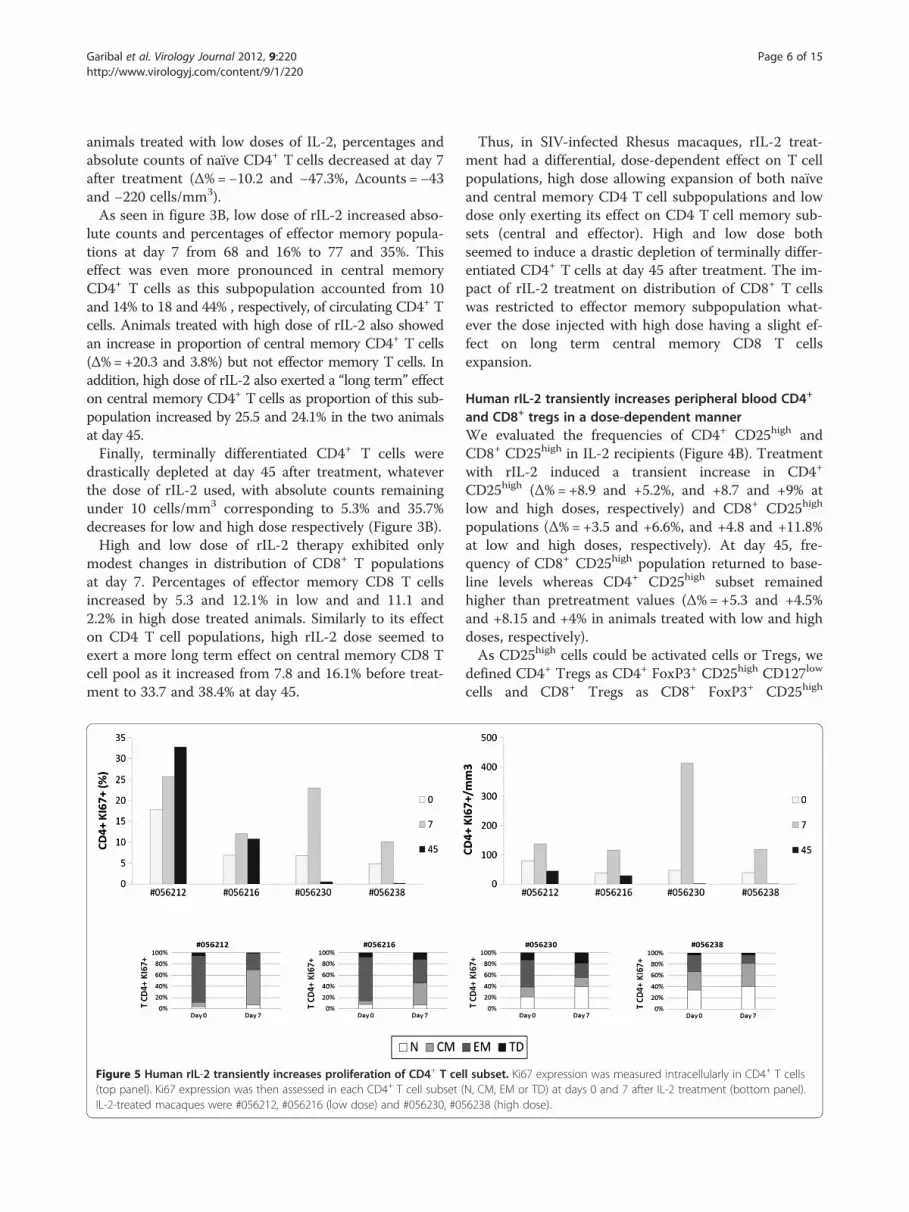
Figure 5 Human rIL-2 transiently increases proliferation of CD4+ T ce(top panel). Ki67 expression was then assessed in each CD4+ T cell subset (IL-2-treated macaques were #056212, #056216 (low dose) and #056230, #05
Thus, in SIV-infected Rhesus macaques, rIL-2 treat-ment had a differential, dose-dependent effect on T cellpopulations, high dose allowing expansion of both naïveand central memory CD4 T cell subpopulations and lowdose only exerting its effect on CD4 T cell memory sub-sets (central and effector). High and low dose bothseemed to induce a drastic depletion of terminally differ-entiated CD4+ T cells at day 45 after treatment. The im-pact of rIL-2 treatment on distribution of CD8+ T cellswas restricted to effector memory subpopulation what-ever the dose injected with high dose having a slight ef-fect on long term central memory CD8 T cellsexpansion.
Human rIL-2 transiently increases peripheral blood CD4+
and CD8+ tregs in a dose-dependent mannerWe evaluated the frequencies of CD4+ CD25high andCD8+ CD25high in IL-2 recipients (Figure 4B). Treatmentwith rIL-2 induced a transient increase in CD4+
CD25high (Δ%=+8.9 and +5.2%, and +8.7 and +9% atlow and high doses, respectively) and CD8+ CD25high
populations (Δ%=+3.5 and +6.6%, and +4.8 and +11.8%at low and high doses, respectively). At day 45, fre-quency of CD8+ CD25high population returned to base-line levels whereas CD4+ CD25high subset remainedhigher than pretreatment values (Δ%=+5.3 and +4.5%and +8.15 and +4% in animals treated with low and highdoses, respectively).As CD25high cells could be activated cells or Tregs, we
defined CD4+ Tregs as CD4+ FoxP3+ CD25high CD127low
cells and CD8+ Tregs as CD8+ FoxP3+ CD25high
ll subset. Ki67 expression was measured intracellularly in CD4+ T cellsN, CM, EM or TD) at days 0 and 7 after IL-2 treatment (bottom panel).6238 (high dose).

Garibal et al. Virology Journal 2012, 9:220 Page 7 of 15http://www.virologyj.com/content/9/1/220
CD127low (Figure 4A). As seen in Figure 4C (top panel),low dose of rIL-2 provoked a robust increase in CD4+
Treg subset frequency (Δ%=+2.5 and +4.8%) and countsat day 7. This frequency then returned to basal levels atday 45. When macaques were treated with high dose ofrIL-2, no increase in frequency of CD4+ Treg subset couldbe observed at day 7 (Δ%=−0.1 and −1.9%). In contrast,peripheral CD8+ Treg population was expanded at day 7after rIL-2 injection, whatever the dose used (Figure 4C –bottom panel) by a 1.5 to 6.6 fold.
Human rIL-2 transiently increases proliferation andactivation of CD4+ and CD8+T cellsNext, we evaluated the proportion of proliferating CD4+
and CD8+ T cells as assessed by the percentages of Tcells expressing nuclear Ki67 Antigen (figures 5 and 6).This revealed a robust and transient rIL-2-associated in-
crease in Ki67 expression in peripheral CD4+ (Figure 5 –top panel) and CD8+ T cells (Figure 6 – top panel).Consistent with the results obtained with global T cellcounts, rIL-2 effect on proliferation seemed to be dose-dependent. Indeed, in animals treated with low rIL-2dose, we observed an increase of Ki67-expressing CD4+
T cells from 17.7 and 6.8% before treatment to 25.7 and12% at day 7 (1.4 and 1.7 fold increase respectively).Using high dose of rIL-2, Ki67 expressing CD4+ T cellpopulation rose from 6.8 and 4.8% before treatment to23 and 10.2% at day 7 (3.4 and 2.1 fold increase respect-ively) (Figure 5 – top panel).Similarly, for CD8+ T cells, rIL-2 treatment transiently
increased frequency of Ki67 expressing CD8+ T cells at
Figure 6 Human rIL-2 transiently increases proliferation of CD8+ T ce(top panel). Ki67 expression was then assessed in each CD8+ T cell subset (IL-2-treated macaques were #056212, #056216 (low dose) and #056230, #05
day 7 by a 1,8 to 3,3 factor when used at low dose whilethis increase was up to 13 fold when animals were trea-ted with high dose of rIL-2 (Figure 6 – top panel).We then assessed cycling cells within each of the CD4+
and CD8+ T cell subsets before and at day 7 after treat-ment. As seen on figure 5 (bottom panel), before treat-ment, the majority of cycling cells were effector memoryCD4+ T cells. After treatment, Ki67-expressing EM CD4+
T cell subset was reduced by half and, consistent with thedata obtained for the distribution of subpopulations, inlow dose treated animals, the percentage of Ki67 expres-sing CD4+ T cells was increased in the central memorysubset (Δ%=+54.8 and 32.5%) while in high dose treatedmacaques, this frequency was mostly augmented in thenaïve subset (Δ%=+18.8 and 7%) and, to a lesser extent,in the central memory subpopulation.Interestingly, in CD8+ T cells treated with low dose of
rIL-2, the pool of cycling terminal differentiated cellsdecreased (Δ%=−10.6 and −4%) whereas it increased whenusing high dose (Δ%=+29.1 and 24%); rIL-2 having a mod-est impact on frequency of cycling EM cells compared tothat observed in CD4+ T cells (Figure 6 – bottom panel).These results showed that rIL-2 led to a transient in-
crease of the proliferation and activation of CD4 andCD8 T cell subsets.
Human rIL-2 modulates CD4+ and CD8+ T cellspontaneous apoptosisIL-2 has been shown to exert pro- or anti-apoptoticeffects in a variety of hematopoietic cells. Furthermore,as we observed a transient activation of CD4+ and CD8+
ll subset. Ki67 expression was measured intracellularly in CD8+ T cellsN, CM, EM or TD) at days 0 and 7 after IL-2 treatment (bottom panel).6238 (high dose).

Garibal et al. Virology Journal 2012, 9:220 Page 8 of 15http://www.virologyj.com/content/9/1/220
T cells after treatment with rIL-2, followed by a contrac-tion of effector memory and Terminally Differentiated Tcell subsets, we wanted to analyze whether cell deathcould be induced in these populations. In this purpose,we evaluated CD4 and CD8 T cell apoptosis by quantify-ing cell surface expression of Phosphatidyl Serine resi-dues by Annexin V staining (Figure 7).Interestingly, our data revealed opposite effect of low
versus high dose of rIL-2 on CD4+ T cells. Indeed, CD4+
T cells from monkeys treated with the lower dose wereless prone to die than CD4+ T cells from macaques trea-ted with the higher dose. As seen in figure 7A, animals
Figure 7 Human rIL-2 modulates spontaneous CD4+ and CD8+ T cellAnnexinV. Total CD4+ (A) and CD8+ (C) T cell apoptosis was measured afteassess the propensity of each subset to undergo apoptosis, apoptosis was(N, CM, EM or TD) at day 0 and day 7 after IL-2 treatment. T cells, gated onanalysed for CD45RA and CD62L expression. Each subpopulation (N, CM, Ewas calculated as follows for each subset:% AnnexinV+ among subset=N (C(CM, EM or TD) AnnexinV- T cells) x100.
treated with low dose of rIL-2 showed a decrease in thefrequency of apoptotic CD4+ T cells at day 7 comparedwith pre treatment values (Δ%=−16.7 and −11.2%)whereas high dose treated macaques exhibited a robustincrease of CD4+ T cell apoptosis (Δ%=+18.5 and16.3%). rIL-2 seemed to exert a long term effect on CD4+
T cell death as levels of apoptosis at day 45 remainedeither increased (Δ%=+11.7 and 4.3%) or decreased(Δ%=−17.7 and −13.9%) compared to basal levels in highor low dose treated animals respectively.This dose-dependent effect on cell death was also
observed in the CD8 T cell compartment as low dose
apoptosis. Apoptotic cell death was assessed using FITC-conjugatedr 16 h of culture at day 0, 7 and 45 after IL-2 treatment. In order tothen measured in each CD4+ (B) or CD8+ (D) T cell subpopulationlymphocytes gate followed by gating on CD4+ or CD8+ Tcells, wereM and TD) was then assessed for AnnexinV expression and sensitivityM, EM or TD) AnnexinV+ T cells/ (N (CM, EM or TD) AnnexinV+ T cells+N

Garibal et al. Virology Journal 2012, 9:220 Page 9 of 15http://www.virologyj.com/content/9/1/220
rIL-2 had no effect on CD8 T cell apoptosis whereashigh dose raised frequency of apoptotic CD8+ T cellsby +31.5 and 35.8% in each animal, respectively. Unlikethe effect on CD4 T cell compartment, this increase wastransient (day 7) and frequencies of apoptotic cellsreturned to basal levels at day 45 (Figure 7C).Given that rIL-2 impacted on T cell subsets distribu-
tion, we next analyzed susceptibility to undergo apop-tosis within each CD4 or CD8 T cell subset. As seen infigure 7B, rIL-2 mainly exerted its effect, either pro- oranti-apoptotic, on the central memory CD4+ T cell sub-population. Indeed, in low dose treated animals, the fre-quency of apoptotic central memory CD4+ T cellsdecreased by a mean of 19.1% at day 7. In macaquestreated with high rIL-2 dose, this frequency rose by amean of 20.4%. Treatment with high dose of rIL-2 alsoinduced an increase in naïve CD4 T cell death at day 7after injection (from 9.7 and 5.6% to 15 and 14.4% -mean Δ%=7%). In contrast to this specific effect on cen-tral memory CD4+ T cells, no difference on CD8 T cellsapoptosis could be observed whatever the rIL-2 doseused (Figure 7D).Thus, low dose of rIL-2 seemed to decrease sensitivity
of central memory CD4+ T cells to spontaneous celldeath while high dose of rIL-2 increased sensitivity ofthis population to undergo apoptosis. Moreover, whilelow dose of rIL-2 had no effect on CD8 T cell apoptosis,high dose clearly potentiated spontaneous CD8 T celldeath. However, unlike its effect on CD4+ T cells, thecytokine did not specifically modulate sensitivity of anyCD8 T cell subset but exerted a global effect on eachCD8 T cell subpopulation.
Human rIL-2 has a dose dependent effect on Fas-inducedCD4 and CD8 T cell apoptosisAs we found that, depending on the dose of rIL-2 used,T cells were more or less sensitive to spontaneous apop-tosis, we next sought to determine whether this differen-tial sensitivity was also observed for Fas-induced celldeath. Indeed, we and others have previously shown thatFas-mediated cell death is associated with progression toAIDS and chronic immune activation observed in HIV/SIV disease could drive T cells into apoptosis via Fas/FasL pathway [25-27].First, we evaluated cell surface expression of CD95/Fas
on CD4+ and CD8+ T cells. We showed that low dose ofrIL-2 had almost no effect on CD95 cell surface expres-sion either on CD4+ or CD8+ T cells. On the contrary,high dose of rIL-2 induced a low increase of CD95 expres-sion at the surface of CD4+ and CD8+ T cells (data notshown).As observed with spontaneous apoptosis, our results
highlighted the dual effect of rIL-2 on Fas-induced CD4+
T cell apoptosis. Indeed, at day 7 after treatment, when
animals were treated with low dose rIL-2, we observed asignificant decrease in Fas-induced CD4+ T cell apop-tosis (−26.7 and −6.1%) whereas when treated with highdose, animals experienced a robust increase in Fas-induced CD4+ cell death (+30.8 and +14.1%). At highdose, this rIL-2 proapoptotic effect seemed to be persist-ent as apoptotic rates were still increased at day 45 com-pared to baseline (Figure 8A).Surprisingly, rIL-2 induced a more questionable effect
on CD8+ T cell population as only high dose showed areproducible effect in all animals with 20.1 and 16.2%increases in Fas-induced CD8+ T cell apoptosis at day 7after treatment (Figure 8C).Thereafter, we studied sensitivity of each subset to
Fas-induced apoptosis. As shown on Figure 8B, treat-ment with low dose of rIL-2 induced an overall slight ef-fect on every CD4 T cell subsets (from −3.5 to −6%depending on the subset) whereas treatment with highdose of rIL-2 led to a more contrasting effect. Indeed, itincreased sensitivity of naïve (Δ%=+7.5 and +7.4%) andcentral memory (Δ%=+18.5 and +46.8%) subsets whileit decreased sensitivity of effector memory (Δ%=−12.7and −20.6%) and terminal differentiated (Δ%=−11.3 and−16.7%) subsets (Figure 8B). Regarding CD8+ T cells, weshowed that treatment with low dose of rIL-2 decreasedsensitivity of naïve CD8+ T cells to Fas-induced apop-tosis (Δ%=−11.3 and −20.2%) whereas it increased sen-sitivity of CD8 central memory subset (Δ%=+26.1 and+12.7%) (Figure 8D). When animals were treated withhigh dose of rIL-2, a dramatic increase in sensitivity toFas-induced apoptosis was observed in CD8 naïve(Δ%=+36.2 and +45.6%), central memory (Δ%=+73.3and +87.4%) and effector memory (Δ%=+61.5 and+48.8 %) subsets (Figure 8D).Thus, we showed here that treatment with rIL-2 could
modulate Fas-induced cell death. This modulation wasparticularly obvious when rIL-2 was used at high dosewith a strong potentiator effect on Fas-induced CD4central memory and CD8 naïve, central and effectormemory T cells apoptosis.
DiscussionBoth enhanced T cell turnover and survival have beenimplicated in regulating the size of the peripheral Tlymphocyte pool following IL-2 immunotherapy in HIV-infected patients. To date, selective expansion of CD4+ Tcells is considered as the hallmark of IL-2-driven immunereconstitution. According to the latest data, the increaseof CD4+ T cell count due to IL-2 immunotherapy in ARTtreated patients does not confer clinical benefit but mayinduce grade 4 clinical side effects [24]. Several hypotheseswere evoked to explain these disappointing results. IL-2side effects might overcome beneficial effects on hostdefenses or the CD4+ T cells induced by IL-2 treatment

Figure 8 Human rIL-2 modulates Fas-mediated CD4+ and CD8+ T cell apoptosis. Fas-induced apoptotic cell death was assessed usingFITC-conjugated AnnexinV. Total CD4+ (A) and CD8+ (C) T cell apoptosis was measured after 16 h of culture in the presence of FasL (200 ng/ml)at day 0, 7 and 45 after IL-2 treatment. In order to assess the propensity of each subset to undergo apoptosis, apoptosis was then measured ineach CD4+ (B) or CD8+ (D) T cell subpopulation (N, CM, EM or TD) at day 0 and day 7 after IL-2 treatment. T cells, gated on lymphocytes gatefollowed by gating on CD4+ or CD8+ Tcells, were analysed for CD45RA and CD62L expression. Each subpopulation (N, CM, EM and TD) was thenassessed for AnnexinV expression and sensitivity was calculated as follows for each subset:% AnnexinV+ among subset=N (CM, EM or TD)AnnexinV+ T cells/ (N (CM, EM or TD) AnnexinV+ T cells+N (CM, EM or TD) AnnexinV- T cells x100.
Garibal et al. Virology Journal 2012, 9:220 Page 10 of 15http://www.virologyj.com/content/9/1/220
may fail to exert protective effector functions. Based onthese observations, we wanted to explore the impact ofIL-2 immunotherapy in a model of infection (i.e.: SIV in-fection of Rhesus macaques) without ART treatment inorder to observe only IL-2-induced changes on variousimmunological parameters.First studies using IL-2 therapy in HIV patients had
shown that this treatment could induce transientincreases in HIV viremia [28] at least due to higherlevels of CCR5 expression [16]. However, other studieshad reported that most of IL-2 treated patients underART showed no increase in plasma viral load [29,30].
Here, although rIL-2 treatment leads to a dramaticincrease of CD4 T cell subset (including cycling cells(Ki67+)), plasmatic viral load is not changed in ARTnaïve SIV-infected monkeys. These results are consistentwith data previously obtained in our laboratory [30] andwith the UK Vanguard study which showed that inter-mittent cycles of subcutaneous IL-2 produced significantincreases in CD4 T cell counts without affecting viralload in ART-naïve HIV-infected patients [31]. Thus,according to our results, adverse IL-2 effects do notseem to be due to its effect on HIV viremia, even in theabsence of ART treatment. Therefore, we addressed

Garibal et al. Virology Journal 2012, 9:220 Page 11 of 15http://www.virologyj.com/content/9/1/220
several questions to help explaining the lack of clinicalbenefit observed in SILCAAT/ESPRIT studies. First, westudied the effect of IL-2 on CD4 and CD8 subpopula-tions. Then, we wanted to confirm and extend resultspreviously obtained in our group [23] concerning IL-2driven Treg induction. Finally, given the controversialliterature concerning IL-2 and apoptosis, we wanted toclarify the role of this cytokine on CD4 and CD8 Tlymphocyte cell death in vivo.First, our data show that treatment of chronically SIV-
infected Rhesus macaques with rIL-2 induced a rapid,transient, dose-dependent increase in peripheral T cellcounts at day 7 after treatment. This immunotherapy notonly induces CD4+ T cell expansion but also affects CD8+
T cell pool early after treatment. These results are consist-ent with data obtained following IL-2 immunotherapy inmetastatic melanoma patients in which levels of perforinand granzyme - which are essential for CD8+ T cell cyto-toxicity - were increased [32]. Since then, many otherstudies have shown that IL-2 immunotherapy mediatedtumor regression via enhanced endogenous tumor specificCD8+ responses [33,34]. Nevertheless, a dramatic CD4lymphopenia occurred at day 45 in all treated animalswhile CD8+ T cell loss only occurred in high dose treatedanimals.We demonstrate that treatment with low or high dose
of rIL-2 has a differential effect on CD4 and CD8 T cellsubpopulations repartition. Indeed, regarding CD4 T cellsubsets, the higher dose of rIL-2 selectively expandednaïve and central memory compartments whereas lowdose only affected memory populations (central and ef-fector). These rIL-2 selectively expanded CD4+ T cellsare activated and express proliferation markers. Thesecells are most likely new thymic emigrants as it wasstrongly suggested that a naive T-cell increase reflectedthymic export after IL-2 therapy in HIV-infectedpatients [35]. In addition, while low dose seemed toexert a transient effect, higher dose induced a sustainedeffect as changes observed in subsets distribution weremaintained at day 45 after treatment. Finally, both dosesinduced a drastic depletion of terminally differentiatedCD4+ T cells (at day 45 after treatment) associated withthe occurrence of a wasting syndrome characterized byweight loss and diarrhea. These results are consistentwith data obtained in the UK-Vanguard trial which sug-gested that after IL-2 discontinuation, CD4 T cell countsdeclined rapidly even below therapeutic threshold [36].We therefore propose that although rIL-2 contributes tocorrect the HIV-driven unbalance within naïve and cen-tral memory CD4+ T compartments, it also leads, in theabsence of antiretroviral treatment, to T cell exhaustionand immune failure through excessive expansion, prolif-eration, activation and apoptosis of CD4 naïve and cen-tral memory subsets.
IL-2 is well-known as a key regulator of immune tol-erance as it promotes the development and peripheralexpansion of CD4+ CD25high Tregs. In the context ofHIV, several reports have suggested that Tregs may havea beneficial role either by suppressing viral replicationor by limiting non specific activation [37,38]. On thecontrary, other studies suggested that Tregs may ac-count for premature and persistent suppression of ef-fector response, thus contributing to establishment of achronic disease state. Indeed, Tregs have been shown toinhibit HIV-specific T-cell responses ex vivo and mayact in vivo by suppressing HIV-specific immunity, allow-ing HIV to persist [39-42]. We have recently reportedthat, following IL-2 immunotherapy, resting and acti-vated Tregs were expanded in HIV-infected patientsunder HAART regimen [23]. They suggest that theseCD4+ Tregs may inhibit the generation of responsesagainst pathogens thereby providing a beginning of ex-planation for the disappointing results of ESPRIT andSILCAAT studies. We largely confirm and extend theseresults in our model as rIL-2, at low dose, drives amassive, transient peripheral CD4 Treg expansion. Atday 45, however, size of the CD4 Treg compartmentreturns to baseline value (low dose treatment) or is evendecreased compared to baseline (high dose treatment).This decrease in peripheral blood may reflect relocationof CD4 Tregs in lymphoid tissues, particularly in thegut [43], thus strengthening loss of the Th17/Treg cellbalance that is already observed in progressive HIV/SIVinfection associated with the expression of TGF-β [44-47].Concerning Treg populations, we also show that treatmentwith rIL-2 induces a sustained and dose-dependent periph-eral expansion of a poorly studied population, namelyCD8+ Tregs. These results are consistent with thestudy of Nigam et al. who recently demonstrated thatperipheral CD8+CD25highFoxP3+ T cells were inducedearly after SIV infection and that the number of thesecells was positively correlated with viral load and asso-ciated with a poor prognosis. This rapid expansion ofCD8 Tregs in the blood was also observed in multiplelymphoid tissues of SIV-infected Rhesus macaques butnot in non pathogenic SIV infection of Sooty Manga-beys. Moreover, they showed that these cells were ableto suppress antiviral immunity thereby suggesting adeleterious role for this CD8+ T cell subset during thecourse of the disease [48]. In addition, following treat-ment with rhIL-2, a dramatic increase in CD8+ Foxp3+
T cell prevalence was observed in the circulation andtumor-draining lymph nodes of subcutaneous tumor-bearing mouse models [49]. Effect of rIL-2 on the Tregulatory compartment thus provides a first piece ofexplanation for the rapid development of wasting syn-drome in our animals. Indeed, rIL-2 may act, on theone hand, on CD4 Treg population, inducing its
Garibal et al. Virology Journal 2012, 9:220 Page 12 of 15http://www.virologyj.com/content/9/1/220
expansion and relocation to gut-associated lymphoidtissues (GALT) thereby contributing to loss of Th17/Treg balance. On the other hand, rIL-2 provokes per-ipheral expansion of CD8+ Treg subset contributing todefective antiviral immunity.Finally, our study underlines the dual role of IL-2 in
the context of spontaneous and Fas-induced apoptosis.Several groups have already investigated the effect ofIL-2 immunotherapy on apoptosis but results were quitediscordant. Indeed, it was shown that, in HIV-infectedpatients, IL-2 treatment was associated with increasedapoptosis rates [50]. On the opposite, some studiesshowed that IL-2 could reduce lymphocyte apoptosis inHIV infected patients [51]. Here, we report that IL-2 caneffectively play both role depending on the dose used,high dose having a proapoptotic effect on both CD4+
and CD8+ T cells and low dose being antiapoptotic inCD4+ T cells only. This modulating effect on cell deathseems to be restricted to CD4+ central memory T cellswhile it exerts a global effect on each CD8+ T cell sub-sets. Excessive apoptosis observed in high dose treatedanimals could be due to higher expression of Fas recep-tor on CM CD4+ T lymphocytes cell surface, indeed weobserved that Fas expression on CD4+ T lymphocyteswas increased after rIL-2 treatment (data not shown).We and others have reported that T cells from HIV-infected individuals or SIV-infected macaques are highlyprone to undergo apoptosis after Fas-ligation [6-8,27]. Arecent study showed that Rhesus macaques treated withneutralizing anti-FasL antibody preserved higher levelsof central memory CD4+ T cells [52] thereby supportinga critical role for Fas in the CD4+ T cell compartment.Herein, we found that in vivo IL-2 treatment has a detri-mental effect on the susceptibility of cells to undergoapoptosis after Fas ligation. However, we cannot excludea role for other death molecules like PD-1 or TRAILthat have been proposed to participate in the death of Tcells during HIV/SIV infection [46,53-56]. Further inves-tigation of these cell death pathways among variouslymphoid populations should provide new insightsabout immunological mechanisms implicated in IL-2immunotherapy.
ConclusionsFollowing rIL-2 treatment in SIV-infected Rhesus maca-ques, T cells - including CD4 and CD8 Tregs - are there-fore not only expanded but, in addition, become moresusceptible to undergo apoptosis providing a rational fordisease progression. Thus, our data contribute to theunderstanding of the homeostatic and dosage effects ofIL-2 in the context of SIV/HIV infection. Although theconclusions drawn in SIV monkey models are not easyto translate to human infected with HIV, we believe thatour data provide new insights in this field regarding the
important and long term commitment in developingIL-2 therapy.
MethodsAnimalsRhesus Macaques (Macaca mulatta) of Chinese originwere inoculated intravenously with 10 AID50 (50% ani-mal infectious dose) of the SIVmac251 strain. All the ani-mals were challenged with the same batch of virus(provided by AM. Aubertin, INSERM U74, Strasbourg,France), titrated in vivo in RMs, and stored in liquid ni-trogen. Animals were seronegative for simian T leukemiavirus type 1, SRV-1 (type D retrovirus), herpesvirus B,and SIVmac. Animals were housed and cared for in com-pliance with existing French regulations (Institut Pasteur,Paris, France). All the animal experiments described inthe present study were conducted at the Institut Pasteuraccording to the European Union guidelines for thehandling of laboratory animals (http://ec.europa.eu/environment/chemicals/lab_animals/home_en.htm). Theprotocol was approved by the committee on the ethics ofanimal experiments of Ile de France. All surgery was per-formed under sodium pentobarbital anesthesia, and allefforts were made to minimize suffering. Macaques inchronic phase of infection were then either treated with6.105 UI/kg body weight (high dose), 1.4.105 UI/kg bodyweight (low dose) recombinant human IL-2 (MacrolinW
Aldesleukine rhIL-2) or saline solution (control) daily for5 days. Blood samples were collected before IL-2 treat-ment (day 0) and at day 7, 10, 15, 30 and 45 aftertreatment.
Lymphocyte immunophenotyping and flow cytometryBlood from monkeys was collected in sterile EDTA-treated vacuum tubes and cell populations were analyzedby flow cytometry using the following antibodies: anti-CD20-FITC (clone 2 H7), anti-CD4-PE (clone L200),anti-CD62L-FITC (clone SK11), anti-CD62L-PE (cloneSK11), anti-CD25-PE (clone M-A251), anti-CD3-PerCP(clone SP34-2), anti-CD4-PerCP (clone SK3), anti-human CD8-PerCP (clone SK1), anti-CD8-APC (cloneRPA-T8), anti-CD95-APC (clone DX2) and anti-CD127-Alexa 647 (clone HIL-7R-M21) were purchased from BDBiosciences/Pharmingen (San Jose, CA). Anti-CD45RA-APC (clone T6D11) was obtained from Miltenyi (BergischGladbach, Germany). For intracellular staining, the cellswere fixed and permeabilized before incubation with anti-Ki67-FITC (clone Ki67, Dako, Glostrup, Denmark), or anti-Fox-P3-Alexa 488 (clone 259D/C7, BD Pharmingen, SanJose, CA).Cells were analyzed by flow cytometry. A total of
30000 events in the lymphocyte gate were analyzedusing CellQuest software (BD Biosciences). All analyses

Garibal et al. Virology Journal 2012, 9:220 Page 13 of 15http://www.virologyj.com/content/9/1/220
were performed with a FACScalibur flow cytometer (BDBiosciences).
Cell death quantificationPBMCs from monkeys were isolated from peripheralblood by density gradient centrifugation LymphoPrep(PAA Laboratories, Pasching, Austria) and cultured inRPMI 1640 supplemented with 10% FCS, 1% glutamine,1% pyruvate, and 1% antibiotics. Up to 500,000 PBMCswere cultured overnight in the absence or presence ofrecombinant Fas-L (10 μg/ml, Alexis Corporation). Celldeath was assessed by flow cytometry, as previouslydescribed [8]. Briefly, after staining with specific anti-bodies (30 min at 4°C), cells were washed and then incu-bated with FITC labeled Annexin-V (10 min at 4°C), and30000 events were analyzed with a FACScalibur flowcytometer (BD Biosciences).
Viral quantificationRNA was extracted from plasma of SIV-infected mon-keys using the TRI Reagent BD Kit (Molecular ResearchCenter, Cincinnati, OH). Real-time quantitative RT-PCRwas used to determine viral loads as previously described[57].
Additional file
Additional file 1: Human rIL-2 does not affect hematologicalparameters. Four Rhesus macaques were infected with the pathogenicSIVmac251 strain and then treated during chronic phase with humanrIL-2 (low dose: ○,Δ or high dose: ●,▲). Each symbol represents oneindividual. Hematological and physiological parameters such as plateleterythrocyte counts, hematocrit, temperature, weight were measured atthe day of treatment and thereafter.
Competing interestsThe authors declare that they have no competing interests.
Authors’ contributionsJG carried out the experimental work and wrote the paper. ML carried outthe viral quantification experiments. RS, SM and LCG helped with themonkeys. YL and JE conceived the study, oversaw its execution and editedthe manuscript. All authors read and approved the final manuscript.
Funding informationThis study was supported by research funding from the ANRS to JE and YLand the “Fondation pour la Recherche Médicale”, to JE. JG and ML weresupported by fellowships from ANRS, SM by SIDACTION and RS by afellowship from the FCT program Ciência 2008.
Author details1INSERM U955 Equipe 16, Institut Mondor de Recherche Biomédicale,CréteilF-94010, France. 2Université Paris-Est, Faculté de Médecine, UMR-S 955,Créteil F-94010, France. 3Assistance Publique-Hôpitaux de Paris (AP-HP),Groupe Henri-Mondor Albert-Chenevier, service d’immunologie clinique,Créteil F-94010, France. 4CNRS FRE 3235, Université Paris Descartes, Paris,France. 5Université Laval, Centre de recherche en Infectiologie, Québec,Canada. 6Instituto de Biologia Molecular e Celular, Universidade do Porto,Porto, Portugal.
Received: 3 February 2012 Accepted: 14 September 2012Published: 28 September 2012
References1. Hurtrel B, Petit F, Arnoult D, Muller-Trutwin M, Silvestri G, Estaquier J:
Apoptosis in SIV infection. Cell Death Differ 2005, 12(Suppl 1):979–990.2. Giralt M, Diaz-Delfin J, Gallego-Escuredo JM, Villarroya J, Domingo P,
Villarroya F: Lipotoxicity on the basis of metabolic syndrome andlipodystrophy in HIV-1-infected patients under antiretroviral treatment.Curr Pharm Des 2010, 16:3371–3378.
3. Veloso S, Peraire J, Vilades C, Lopez-Dupla M, Escote X, Olona M, Garcia-Pardo G, Gomez-Bertomeu F, Soriano A, Sirvent JJ, Vidal F:Pharmacogenetics of the metabolic disturbances and atherosclerosisassociated with antiretroviral therapy in HIV-infected patients. CurrPharm Des 2010, 16:3379–3389.
4. Muro-Cacho CA, Pantaleo G, Fauci AS: Analysis of apoptosis in lymphnodes of HIV-infected persons. Intensity of apoptosis correlates with thegeneral state of activation of the lymphoid tissue and not with stage ofdisease or viral burden. J Immunol 1995, 154:5555–5566.
5. Ledru E, Lecoeur H, Garcia S, Debord T, Gougeon ML: Differentialsusceptibility to activation-induced apoptosis among peripheral Th1subsets: correlation with Bcl-2 expression and consequences for AIDSpathogenesis. J Immunol 1998, 160:3194–3206.
6. Estaquier J, Idziorek T, Zou W, Emilie D, Farber CM, Bourez JM, Ameisen JC:T helper type 1/T helper type 2 cytokines and T cell death: preventiveeffect of interleukin 12 on activation-induced and CD95 (FAS/APO-1)-mediated apoptosis of CD4+ T cells from human immunodeficiencyvirus-infected persons. J Exp Med 1995, 182:1759–1767.
7. Estaquier J, Tanaka M, Suda T, Nagata S, Golstein P, Ameisen JC: Fas-mediated apoptosis of CD4+ and CD8+ T cells from humanimmunodeficiency virus-infected persons: differential in vitro preventiveeffect of cytokines and protease antagonists. Blood 1996, 87:4959–4966.
8. Arnoult D, Petit F, Lelievre JD, Lecossier D, Hance A, Monceaux V, Hurtrel B,Ho Tsong Fang R, Ameisen JC, Estaquier J: Caspase-dependent and-independent T-cell death pathways in pathogenic simianimmunodeficiency virus infection: relationship to disease progression.Cell Death Differ 2003, 10:1240–1252.
9. Boudet F, Lecoeur H, Gougeon ML: Apoptosis associated with ex vivodown-regulation of Bcl-2 and up-regulation of Fas in potential cytotoxicCD8+ T lymphocytes during HIV infection. J Immunol 1996,156:2282–2293.
10. Zaunders JJ, Moutouh-de Parseval L, Kitada S, Reed JC, Rought S, Genini D,Leoni L, Kelleher A, Cooper DA, Smith DE, et al: Polyclonal proliferationand apoptosis of CCR5+ T lymphocytes during primary humanimmunodeficiency virus type 1 infection: regulation by interleukin (IL)-2,IL-15, and Bcl-2. J Infect Dis 2003, 187:1735–1747.
11. Ledru E, Christeff N, Patey O, de Truchis P, Melchior JC, Gougeon ML:Alteration of tumor necrosis factor-alpha T-cell homeostasis followingpotent antiretroviral therapy: contribution to the development of humanimmunodeficiency virus-associated lipodystrophy syndrome. Blood 2000,95:3191–3198.
12. Lenardo MJ: Interleukin-2 programs mouse alpha beta T lymphocytes forapoptosis. Nature 1991, 353:858–861.
13. Schorle H, Holtschke T, Hunig T, Schimpl A, Horak I: Development andfunction of T cells in mice rendered interleukin-2 deficient by genetargeting. Nature 1991, 352:621–624.
14. Kinter AL, Godbout EJ, McNally JP, Sereti I, Roby GA, O'Shea MA, Fauci AS:The common gamma-chain cytokines IL-2, IL-7, IL-15, and IL-21 inducethe expression of programmed death-1 and its ligands. J Immunol 2008,181:6738–6746.
15. Petit F, Arnoult D, Lelievre JD, Moutouh-de Parseval L, Hance AJ, SchneiderP, Corbeil J, Ameisen JC, Estaquier J: Productive HIV-1 infection of primaryCD4+ T cells induces mitochondrial membrane permeabilization leadingto a caspase-independent cell death. J Biol Chem 2002, 277:1477–1487.
16. Zou W, Foussat A, Houhou S, Durand-Gasselin I, Dulioust A, Bouchet L,Galanaud P, Levy Y, Emilie D: Acute upregulation of CCR-5 expression byCD4+ T lymphocytes in HIV-infected patients treated with interleukin-2.ANRS 048 IL-2 study group. AIDS 1999, 13:455–463.
17. Levy Y, Durier C, Lascaux AS, Meiffredy V, Gahery-Segard H, Goujard C,Rouzioux C, Resch M, Guillet JG, Kazatchkine M, et al: Sustained control ofviremia following therapeutic immunization in chronically HIV-1-infectedindividuals. AIDS 2006, 20:405–413.
18. Gougeon ML, Rouzioux C, Liberman I, Burgard M, Taoufik Y, Viard JP,Bouchenafa K, Capitant C, Delfraissy JF, Levy Y: Immunological and

Garibal et al. Virology Journal 2012, 9:220 Page 14 of 15http://www.virologyj.com/content/9/1/220
virological effects of long term IL-2 therapy in HIV-1-infected patients.AIDS 2001, 15:1729–1731.
19. Farel CE, Chaitt DG, Hahn BK, Tavel JA, Kovacs JA, Polis MA, Masur H,Follmann DA, Lane HC, Davey RT Jr: Induction and maintenance therapywith intermittent interleukin-2 in HIV-1 infection. Blood 2004,103:3282–3286.
20. Levy Y, Durier C, Krzysiek R, Rabian C, Capitant C, Lascaux AS, Michon C,Oksenhendler E, Weiss L, Gastaut JA, et al: Effects of interleukin-2 therapycombined with highly active antiretroviral therapy on immunerestoration in HIV-1 infection: a randomized controlled trial. AIDS 2003,17:343–351.
21. Kovacs JA, Lempicki RA, Sidorov IA, Adelsberger JW, Sereti I, Sachau W, KellyG, Metcalf JA, Davey RT Jr, Falloon J, et al: Induction of prolonged survivalof CD4+ T lymphocytes by intermittent IL-2 therapy in HIV-infectedpatients. J Clin Invest 2005, 115:2139–2148.
22. Sereti I, Imamichi H, Natarajan V, Imamichi T, Ramchandani MS, Badralmaa Y,Berg SC, Metcalf JA, Hahn BK, Shen JM, et al: In vivo expansion ofCD4CD45RO-CD25 T cells expressing foxP3 in IL-2-treated HIV-infectedpatients. J Clin Invest 2005, 115:1839–1847.
23. Weiss L, Letimier FA, Carriere M, Maiella S, Donkova-Petrini V, Targat B,Benecke A, Rogge L, Levy Y: In vivo expansion of naive and activatedCD4+CD25+FOXP3+ regulatory T cell populations in interleukin-2-treated HIV patients. Proc Natl Acad Sci USA 2010, 107:10632–10637.
24. Abrams D, Levy Y, Losso MH, Babiker A, Collins G, Cooper DA, Darbyshire J,Emery S, Fox L, Gordin F, et al: Interleukin-2 therapy in patients with HIVinfection. N Engl J Med 2009, 361:1548–1559.
25. Marshak-Rothstein A, Stanger BZ, Sherr DH, Ju ST, Panka DJ, Cui H, EttingerR, Khatib M: as(CD95)/FasL interactions required for programmed celldeath after T-cell activation. Nature 1995, 373:444–448.
26. Lelievre JD, Mammano F, Arnoult D, Petit F, Grodet A, Estaquier J, AmeisenJC: A novel mechanism for HIV1-mediated bystander CD4+ T-cell death:neighboring dying cells drive the capacity of HIV1 to kill noncyclingprimary CD4+ T cells. Cell Death Differ 2004, 11:1017–1027.
27. Viollet L, Monceaux V, Petit F, Ho Tsong Fang R, Cumont MC, Hurtrel B,Estaquier J: Death of CD4+ T cells from lymph nodes during primarySIVmac251 infection predicts the rate of AIDS progression. J Immunol2006, 177:6685–6694.
28. Kovacs JA, Baseler M, Dewar RJ, Vogel S, Davey RT Jr, Falloon J, Polis MA,Walker RE, Stevens R, Salzman NP, et al: ncreases in CD4 T lymphocyteswith intermittent courses of interleukin-2 in patients with humanimmunodeficiency virus infection. A preliminary study. N Engl J Med1995, 332:567–575.
29. Go R, Steigbigel R: Interleukin 2 and HIV RNA Levels. Arch Intern Med 2007,167:2144–2145. author reply 2145.
30. Molina JM, Levy Y, Fournier I, Hamonic S, Bentata M, Beck-Wirth G, GougeonML, Venet A, Madelaine I, Sereni D, et al: Interleukin-2 before antiretroviraltherapy in patients with HIV infection: a randomized trial (ANRS 119). JInfect Dis 2009, 200:206–215.
31. Youle M, Emery S, Fisher M, Nelson M, Fosdick L, Janossy G, Loveday C,Sullivan A, Herzmann C, Wand H, et al: A randomised trial ofsubcutaneous intermittent interleukin-2 without antiretroviral therapy inHIV-infected patients: the UK-Vanguard study. PLoS Clin Trials 2006, 1:e3.
32. Leger-Ravet MB, Mathiot C, Portier A, Brandely M, Galanaud P, Fridman WH,Emilie D: Increased expression of perforin and granzyme B genes inpatients with metastatic melanoma treated with recombinantinterleukin-2. Cancer Immunol Immunother 1994, 39:53–58.
33. Romano F, Piacentini MG, Franciosi C, Caprotti R, De Fina S, Cesana G,Uggeri F, Conti M, Uggeri F: Phase-II randomized study of preoperativeIL-2 administration in radically operable gastric cancer patients.Hepatogastroenterology 2004, 51:1872–1876.
34. Jackaman C, Bundell CS, Kinnear BF, Smith AM, Filion P, van Hagen D,Robinson BW, Nelson DJ: IL-2 intratumoral immunotherapy enhances CD8+ T cells that mediate destruction of tumor cells and tumor-associatedvasculature: a novel mechanism for IL-2. J Immunol 2003,171:5051–5063.
35. Carcelain G, Saint-Mezard P, Altes HK, Tubiana R, Grenot P, Rabian C, deBoer R, Costagliola D, Katlama C, Debre P, Autran B: IL-2 therapy andthymic production of naive CD4 T cells in HIV-infected patients withsevere CD4 lymphopenia. AIDS 2003, 17:841–850.
36. Herzmann C, Cuthbertson Z, Fosdick L, Fisher M, Nelson M, Perry N, Law M,Wand H, Janossy G, Johnson MA, Youle M: Long-term clinical and
surrogate marker effects of subcutaneous intermittent interleukin-2without antiretroviral therapy in HIV-infected patients. J AntimicrobChemother 2008, 62:583–586.
37. Chase AJ, Yang HC, Zhang H, Blankson JN, Siliciano RF: Preservation ofFoxP3+ regulatory T cells in the peripheral blood of humanimmunodeficiency virus type 1-infected elite suppressors correlates withlow CD4+ T-cell activation. J Virol 2008, 82:8307–8315.
38. Moreno-Fernandez ME, Rueda CM, Rusie LK, Chougnet CA: Regulatory Tcells control HIV replication in activated T cells through a cAMP-dependent mechanism. Blood 2011, 117:5372–5380.
39. Aandahl EM, Michaelsson J, Moretto WJ, Hecht FM, Nixon DF: HumanCD4+ CD25+ regulatory T cells control T-cell responses to humanimmunodeficiency virus and cytomegalovirus antigens. J Virol 2004,78:2454–2459.
40. Kinter A, McNally J, Riggin L, Jackson R, Roby G, Fauci AS: Suppression ofHIV-specific T cell activity by lymph node CD25+ regulatory T cells fromHIV-infected individuals. Proc Natl Acad Sci USA 2007, 104:3390–3395.
41. Weiss L, Donkova-Petrini V, Caccavelli L, Balbo M, Carbonneil C, Levy Y:Human immunodeficiency virus-driven expansion of CD4+CD25+regulatory T cells, which suppress HIV-specific CD4 T-cell responses inHIV-infected patients. Blood 2004, 104:3249–3256.
42. Elahi S, Dinges WL, Lejarcegui N, Laing KJ, Collier AC, Koelle DM, McElrathMJ, Horton H: Protective HIV-specific CD8+ T cells evade Treg cellsuppression. Nat Med 2011, 17:989–995.
43. Shaw JM, Hunt PW, Critchfield JW, McConnell DH, Garcia JC, Pollard RB,Somsouk M, Deeks SG, Shacklett BL: Increased frequency of regulatory Tcells accompanies increased immune activation in rectal mucosae ofHIV-positive noncontrollers. J Virol 2011, 85:11422–11434.
44. Favre D, Lederer S, Kanwar B, Ma ZM, Proll S, Kasakow Z, Mold J, SwainsonL, Barbour JD, Baskin CR, et al: Critical loss of the balance between Th17and T regulatory cell populations in pathogenic SIV infection. PLoSPathog 2009, 5:e1000295.
45. Estes JD, Li Q, Reynolds MR, Wietgrefe S, Duan L, Schacker T, Picker LJ,Watkins DI, Lifson JD, Reilly C, et al: Premature induction of animmunosuppressive regulatory T cell response during acute simianimmunodeficiency virus infection. J Infect Dis 2006, 193:703–712.
46. Cumont MC, Monceaux V, Viollet L, Lay S, Parker R, Hurtrel B, Estaquier J:TGF-beta in intestinal lymphoid organs contributes to the death ofarmed effector CD8 T cells and is associated with the absence of viruscontainment in rhesus macaques infected with the simianimmunodeficiency virus. Cell Death Differ 2007, 14:1747–1758.
47. Campillo-Gimenez L, Cumont MC, Fay M, Kared H, Monceaux V, Diop O,Müller-Trutwin M, Hurtrel B, Lévy Y, Zaunders J, Dy M, Leite-de-Moraes MC,Elbim C, Estaquier J: AIDS progression is associated with the emergenceof IL-17-producing cells early after simian immunodeficiency virusinfection. J Immunol 2010, 184:984–92.
48. Nigam P, Velu V, Kannanganat S, Chennareddi L, Kwa S, Siddiqui M, AmaraRR: Expansion of FOXP3+ CD8 T cells with suppressive potential incolorectal mucosa following a pathogenic simian immunodeficiencyvirus infection correlates with diminished antiviral T cell response andviral control. J Immunol 2010, 184:1690–1701.
49. Foureau DM, McKillop IH, Jones CP, Amin A, White RL, Salo JC: Skin tumorresponsiveness to interleukin-2 treatment and CD8 Foxp3+ T cellexpansion in an immunocompetent mouse model. Cancer ImmunolImmunother 2011, 60:1347–1356.
50. Sereti I, Herpin B, Metcalf JA, Stevens R, Baseler MW, Hallahan CW, KovacsJA, Davey RT, Lane HC: CD4 T cell expansions are associated withincreased apoptosis rates of T lymphocytes during IL-2 cycles in HIVinfected patients. AIDS 2001, 15:1765–1775.
51. Pandolfi F, Pierdominici M, Marziali M, Livia Bernardi M, Antonelli G, Galati V,D'Offizi G, Aiuti F: Low-dose IL-2 reduces lymphocyte apoptosis andincreases naive CD4 cells in HIV-1 patients treated with HAART. ClinImmunol 2000, 94:153–159.
52. Poonia B, Salvato MS, Yagita H, Maeda T, Okumura K, Pauza CD: Treatmentwith anti-FasL antibody preserves memory lymphocytes andvirus-specific cellular immunity in macaques challenged with simianimmunodeficiency virus. Blood 2009, 114:1196–1204.
53. Trautmann L, Janbazian L, Chomont N, Said EA, Gimmig S, Bessette B,Boulassel MR, Delwart E, Sepulveda H, Balderas RS, et al: Upregulation ofPD-1 expression on HIV-specific CD8+ T cells leads to reversible immunedysfunction. Nat Med 2006, 12:1198–1202.

Garibal et al. Virology Journal 2012, 9:220 Page 15 of 15http://www.virologyj.com/content/9/1/220
54. Day CL, Kaufmann DE, Kiepiela P, Brown JA, Moodley ES, Reddy S, MackeyEW, Miller JD, Leslie AJ, DePierres C, et al: PD-1 expression on HIV-specificT cells is associated with T-cell exhaustion and disease progression.Nature 2006, 443:350–354.
55. Petrovas C, Casazza JP, Brenchley JM, Price DA, Gostick E, Adams WC,Precopio ML, Schacker T, Roederer M, Douek DC, Koup RA: PD-1 is aregulator of virus-specific CD8+ T cell survival in HIV infection. J Exp Med2006, 203:2281–2292.
56. Laforge M, Campillo-Gimenez L, Monceaux V, Cumont MC, Hurtrel B, CorbeilJ, Zaunders J, Elbim C, Estaquier J: HIV/SIV infection primes monocytesand dendritic cells for apoptosis. PLoS Pathog 2011, 7:e1002087.
57. Monceaux V, Viollet L, Petit F, Ho Tsong Fang R, Cumont MC, Zaunders J,Hurtrel B, Estaquier J: CD8+ T cell dynamics during primary simianimmunodeficiency virus infection in macaques: relationship of effectorcell differentiation with the extent of viral replication. J Immunol 2005,174:6898–6908.
doi:10.1186/1743-422X-9-220Cite this article as: Garibal et al.: IL-2 immunotherapy in chronically SIV-infected Rhesus Macaques. Virology Journal 2012 9:220.
Submit your next manuscript to BioMed Centraland take full advantage of:
• Convenient online submission
• Thorough peer review
• No space constraints or color figure charges
• Immediate publication on acceptance
• Inclusion in PubMed, CAS, Scopus and Google Scholar
• Research which is freely available for redistribution
Submit your manuscript at www.biomedcentral.com/submit
Copyright © 2022 FDOKUMEN