Neurobiology of Stress-Induced Reproductive Dysfunction in Female Macaques
56
Neurobiology of Stress-Induced Reproductive Dysfunction In Female Macaques Cynthia L. Bethea 1,2 , Maria Luisa Centeno 1 , and Judy L. Cameron 1,2,3,4 1 Division of Reproductive Sciences, Oregon National Primate Research Center, Beaverton, OR 97006 2 Division of Neuroscience, Oregon National Primate Research Center, Beaverton, OR 97006 3 Departments of Behavioral Neuroscience and Obstetrics & Gynecology, Oregon Health and Science University Portland, OR 97201 4 Department of Psychiatry, University of Pittsburgh, Pittsburgh, PA 15213 Abstract It is now well accepted that stress can precipitate mental and physical illness. However, it is becoming clear that given the same stress, some individuals are very vulnerable and will succumb to illness while others are more resilient and cope effectively, rather than becoming ill. This difference between individuals is called stress sensitivity. Stress-sensitivity of an individual appears to be influenced by genetically inherited factors, early life (even prenatal) stress, and by the presence or absence of factors that provide protection from stress. In comparison to other stress-related diseases, the concept of sensitivity versus resilience to stress-induced reproductive dysfunction has received relatively little attention. The studies presented herein were undertaken to begin to identify stable characteristics and the neural underpinnings of individuals with sensitivity to stress-induced reproductive dysfunction. Female cynomolgus macaques with normal menstrual cycles either stop ovulating (Stress Sensitive) or to continue to ovulate (Stress Resilient) upon exposure to a combined metabolic and psychosocial stress. However, even in the absence of stress, the stress sensitive animals have lower secretion of the ovarian steroids, estrogen and progesterone, have higher heart rates, have lower serotonin function, have fewer serotonin neurons and lower expression of pivotal serotonin-related genes, have lower expression of 5HT2A and 2C genes in the hypothalamus, have higher gene expression of GAD67 and CRH in the hypothalamus and have reduced GnRH transport to the anterior pituitary. Altogether, the results suggest that the neurobiology of reproductive circuits in stress sensitive individuals is compromised. We speculate that with the application of stress, the dysfunction of these neural systems becomes exacerbated and reproductive function ceases. Keywords reproduction; stress; serotonin; corticotropin releasing hormone; pro-opiomelanocortin; beta- endorphin; paraventricular nucleus; thalamus; amygdala; cynomolgus macaque Overview Exposure to stressful stimuli can lead to a variety of secondary diseases such as anxiety, depression, cardiovascular disease, and immune suppression (McEwen, 2002; McEwen, 2008). Reproductive dysfunction has been recently added to this growing list of stress- Corresponding Author: Cynthia L. Bethea, Ph.D., Tel 503-690-5327, Fax 503-690-5384, [email protected]. NIH Public Access Author Manuscript Mol Neurobiol. Author manuscript; available in PMC 2012 January 25. Published in final edited form as: Mol Neurobiol. 2008 December ; 38(3): 199–230. doi:10.1007/s12035-008-8042-z. NIH-PA Author Manuscript NIH-PA Author Manuscript NIH-PA Author Manuscript
-
Upload
independent -
Category
Documents
-
view
2 -
download
0
Transcript of Neurobiology of Stress-Induced Reproductive Dysfunction in Female Macaques

Neurobiology of Stress-Induced Reproductive Dysfunction InFemale Macaques
Cynthia L. Bethea1,2, Maria Luisa Centeno1, and Judy L. Cameron1,2,3,4
1Division of Reproductive Sciences, Oregon National Primate Research Center, Beaverton, OR970062Division of Neuroscience, Oregon National Primate Research Center, Beaverton, OR 970063Departments of Behavioral Neuroscience and Obstetrics & Gynecology, Oregon Health andScience University Portland, OR 972014Department of Psychiatry, University of Pittsburgh, Pittsburgh, PA 15213
AbstractIt is now well accepted that stress can precipitate mental and physical illness. However, it isbecoming clear that given the same stress, some individuals are very vulnerable and will succumbto illness while others are more resilient and cope effectively, rather than becoming ill. Thisdifference between individuals is called stress sensitivity. Stress-sensitivity of an individualappears to be influenced by genetically inherited factors, early life (even prenatal) stress, and bythe presence or absence of factors that provide protection from stress. In comparison to otherstress-related diseases, the concept of sensitivity versus resilience to stress-induced reproductivedysfunction has received relatively little attention. The studies presented herein were undertakento begin to identify stable characteristics and the neural underpinnings of individuals withsensitivity to stress-induced reproductive dysfunction. Female cynomolgus macaques with normalmenstrual cycles either stop ovulating (Stress Sensitive) or to continue to ovulate (Stress Resilient)upon exposure to a combined metabolic and psychosocial stress. However, even in the absence ofstress, the stress sensitive animals have lower secretion of the ovarian steroids, estrogen andprogesterone, have higher heart rates, have lower serotonin function, have fewer serotonin neuronsand lower expression of pivotal serotonin-related genes, have lower expression of 5HT2A and 2Cgenes in the hypothalamus, have higher gene expression of GAD67 and CRH in the hypothalamusand have reduced GnRH transport to the anterior pituitary. Altogether, the results suggest that theneurobiology of reproductive circuits in stress sensitive individuals is compromised. We speculatethat with the application of stress, the dysfunction of these neural systems becomes exacerbatedand reproductive function ceases.
Keywordsreproduction; stress; serotonin; corticotropin releasing hormone; pro-opiomelanocortin; beta-endorphin; paraventricular nucleus; thalamus; amygdala; cynomolgus macaque
OverviewExposure to stressful stimuli can lead to a variety of secondary diseases such as anxiety,depression, cardiovascular disease, and immune suppression (McEwen, 2002; McEwen,2008). Reproductive dysfunction has been recently added to this growing list of stress-
Corresponding Author: Cynthia L. Bethea, Ph.D., Tel 503-690-5327, Fax 503-690-5384, [email protected].
NIH Public AccessAuthor ManuscriptMol Neurobiol. Author manuscript; available in PMC 2012 January 25.
Published in final edited form as:Mol Neurobiol. 2008 December ; 38(3): 199–230. doi:10.1007/s12035-008-8042-z.
NIH
-PA Author Manuscript
NIH
-PA Author Manuscript
NIH
-PA Author Manuscript

related disorders (Cameron, 2000). A significant body of literature has focused upon theapplication of stress and its consequences on reproductive cyclicity and the relatedneuroendocrinology. Early in the 1970’s, it was recognized that the stress of populationdensity inhibited estrous cycles in mice (Champlin, 1971), and a great deal of effort has beendevoted to understanding the effects of maternal stress during pregnancy on offspringphysiology and behavior in rodent species (Gos et al., 2006; Kajantie, 2006; Fumagalli etal., 2007).
Luteinizing hormone (LH) and follicle stimulating hormone (FSH) are the gonadotropinsthat drive ovarian function, estrogen (E) and progesterone secretion (P), menstrual or estrouscyclicity and ultimately ovulation. In ovariectomized animals, LH secretion becomeselevated and pulsatile. To understand the effect of stress on LH secretion, theovariectomized pulsatile secretory mode has been utilized. Restraint stress, or activation ofthe corticotropin releasing hormone (CRH) receptor type 2 with intracerebroventricularurocortin, suppressed luteinizing hormone (LH) pulses in ovariectomized rats (Li et al.,2005). These effects may be mediated via the raphe serotonin system (Ruggiero et al., 1999;Pernar et al., 2004; Clark et al., 2007; Mo et al., 2008) or brainstem noradrenergic systems(Mitchell et al., 2005; Dunn and Swiergiel, 2008), as well as via hypothalamic circuits(MacLusky et al., 1988; Dobson et al., 2003).
Stress and reproduction are important factors in the farming industry. Stresses such as fever,lameness and transportation can significantly decrease fertility in cows and sheep (Dobsonand Smith, 2000). Modeling of stress in ewes with endotoxin has enabled the analysis of theneural pathways mediating stress-induced suppression of ovulation. Evidence has been wellreviewed indicating that the balance of numerous neurotransmitters such as norepinephrine,serotonin, glutamate and GABA; and the neuropeptides CRH, arginine vasopressin (AVP),and neuropeptide Y impinge directly or indirectly on GnRH neurons to activate or suppresstheir function depending on the environment (Tilbrook et al., 2002; Dobson et al., 2003;Smith et al., 2003; von Borell et al., 2007). AVP plays a greater role that CRH in sheep, butthe reverse is true in rodents (Dobson et al., 2003). Moreover, the medial preoptic region insheep and rodents contains pivotal GnRH neurons that are not found in humans or primates.
Extreme exercise is considered to be a metabolic stress, and with the advent of greaterparticipation of women in sports, reports emerged that intense athletic participationdisrupted menstrual cycles (Arena et al., 1995). Further study indicated that there was asuppression of estrogen secretion during the follicular phase and less progesterone secretionduring the luteal phase and blunted FSH secretion during the follicular luteal transition inrecreational women runners (De Souza et al., 1998).
A clinical syndrome called Functional Hypothalamic Amenorrhea (FHA), characterized bymenstrual cycle abnormalities and infertility, is found in a proportion of women who presentat infertility clinics (Berga and Girton, 1989; Berga et al., 1997). Research indicates thatFHA occurs in women with combined moderate psychological and metabolic stress (Marcuset al., 2001) and eating disorders are also common in this population (Warren et al., 1999).Moreover, new treatment therapies for FHA that both target strategies for coping withpsychological stress and removal of metabolic stresses look very promising (Berga et al.,2003).
Stress models in nonhuman primates have employed endotoxin administration (Xiao et al.,1998; Xiao et al., 1999), interleukin -1 administration (Feng et al., 1991; Ferin, 1995), CRHadministration (Xiao and Ferin, 1988), exercise (Williams et al., 2001a; Williams et al.,2001b), diet (Cameron and Nosbisch, 1991), psychosocial stress (relocation to a new roomwith new neighbors) (Cameron et al., 1998)or combinations of these stresses as found in
Bethea et al. Page 2
Mol Neurobiol. Author manuscript; available in PMC 2012 January 25.
NIH
-PA Author Manuscript
NIH
-PA Author Manuscript
NIH
-PA Author Manuscript

FHA (Williams et al., 1997; Williams et al., 2007). Administration of endotoxin, interleukin-1 or CRH activated the hypothalamic-pituitary-adrenal axis, increased cortisol secretion andsuppressed LH and FSH secretion, which could be reversed with the administration of aCRH antagonist (Xiao et al., 1996) or the opiate antagonist, naloxone (Gindoff and Ferin,1987). A recent study with rhesus monkeys employing the combination of surgery andrelocation showed that inadequate LH and progesterone secretion during the luteal phase isthe initial defect leading to abnormal menstrual cycle parameters. This study suggested thatsecretory inadequacy of the corpus luteum represents the first clinical stage in the damagethat stress inflicts on the normal menstrual cycle (Xiao et al., 2002).
A pivotal factor in many studies is that stress was applied and results were obtained in afashion suggesting that all animals respond equally to the stress. However, it is nowbecoming apparent that certain individuals are more sensitive to stress than others. This isclearly evident in human populations where some individuals succumb to psychiatric andsomatic disease after trauma or stress, but other individuals thrive. In animal models, similarresults have been obtained by selective breeding in which stress sensitive and stress resilientlines are produced (Baer and Crumpacker, 1977; Osterlund et al., 1999; Li et al., 2004;Henn and Vollmayr, 2005). Our group has used cynomolgus monkeys and a combination ofdiet, exercise and relocation to study the effects of stress on reproductive function. Whenthis paradigm is applied to small populations of monkeys, we observed individualdifferences in reproductive dysfunction with stress. This chapter reviews our investigationsand shows that the activity or gene expression in neural circuits mediating stress andreproduction are significantly different in stress sensitive and stress resilient individuals inthe absence of stress.
I. The ModelIntroduction
In many areas of medicine it is recognized that there are striking individual differences insensitivity to stress, in that some individuals show marked physiological responses tostressful stimuli and are prone to the development of diseases that occur secondary tochronic stress exposure (i.e., anxiety, depression, cardiovascular disease, immunesuppression), while others are stress-resilient and show less physiological response tostressful stimuli and are less likely to develop diseases secondary to chronic stress exposure.Stress-sensitivity of an individual appears to be influenced by genetically inherited factors,prior stress exposure (particularly stress exposure in prenatal or early post-nataldevelopment), and by the presence or absence of factors that provide protection from stress(McEwen, 2002). In comparison to other stress-related diseases, the concept of sensitivityversus resilience to stress-induced reproductive dysfunction has received relatively littleattention to date. However, a comprehensive review of the effects of psychosocial stresseson reproductive function in humans and nonhuman primates suggests that a number offactors can influence the sensitivity of the reproductive axis to psychosocial stresses,including the perception of stress, the magnitude and duration of stress, social status, and thelevel of activity within the reproductive axis prior to stress exposure (Cameron, 2000). Inaddition, several studies also documented individual differences in sensitivity of thereproductive axis to immune stresses (Xiao et al., 1999).
We have undertaken a series of studies in which female cynomolgus macaques wereexposed to a combination stress paradigm and their reproductive function was monitored.We found marked differences between individuals in the response of the reproductivesystem to stress. Following the in vivo characterization, postmortem studies of brainfunction were executed. These studies revealed that pivotal neural systems in the brain thatare involved in stress responsivity were altered in stress sensitive individuals. Following are
Bethea et al. Page 3
Mol Neurobiol. Author manuscript; available in PMC 2012 January 25.
NIH
-PA Author Manuscript
NIH
-PA Author Manuscript
NIH
-PA Author Manuscript
studies describing the model, the in vivo characterization and the postmortem analysis of thebrains of animals with differential sensitivity to stress.
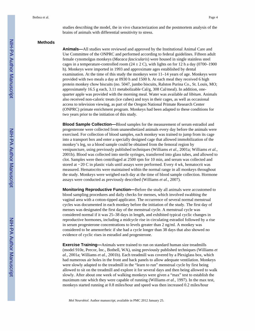
MethodsAnimals—All studies were reviewed and approved by the Institutional Animal Care andUse Committee of the ONPRC and performed according to federal guidelines. Fifteen adultfemale cynomolgus monkeys (Macaca fascicularis) were housed in single stainless steelcages in a temperature-controlled room (24 ± 2 C), with lights on for 12 h a day (0700–1900h). Monkeys were imported in 1993 and approximate ages established by dentalexamination. At the time of this study the monkeys were 11–14 years of age. Monkeys wereprovided with two meals a day at 0930 h and 1500 h. At each meal they received 6 highprotein monkey chow biscuits (no. 5047, jumbo biscuits, Ralston Purina Co., St. Louis, MO;approximately 16.5 g each, 3.11 metabolizable Cal/g, 308 Cal/meal). In addition, one-quarter apple was provided with the morning meal. Water was available ad libitum. Animalsalso received non-caloric treats (ice cubes) and toys in their cages, as well as occasionalaccess to television viewing, as part of the Oregon National Primate Research Center(ONPRC) primate enrichment program. Monkeys had been adapted to these conditions fortwo years prior to the initiation of this study.
Blood Sample Collection—Blood samples for the measurement of serum estradiol andprogesterone were collected from unanesthetized animals every day before the animals wereexercised. For collection of blood samples, each monkey was trained to jump from its cageinto a transport box and enter a specially designed cage that allowed immobilization of themonkey’s leg, so a blood sample could be obtained from the femoral region byvenipuncture, using previously published techniques (Williams et al., 2001a; Williams et al.,2001b). Blood was collected into sterile syringes, transferred into glass tubes, and allowed toclot. Samples were then centrifuged at 2500 rpm for 10 min, and serum was collected andstored at −20 C in plastic vials until assays were performed. Every 4 wk, hematocrit wasmeasured. Hematocrits were maintained within the normal range in all monkeys throughoutthe study. Monkeys were weighed each day at the time of blood sample collection. Hormoneassays were conducted as previously described (Williams et al., 2007).
Monitoring Reproductive Function—Before the study all animals were accustomed toblood sampling procedures and daily checks for menses, which involved swabbing thevaginal area with a cotton-tipped applicator. The occurrence of several normal menstrualcycles was documented in each monkey before the initiation of the study. The first day ofmenses was designated the first day of the menstrual cycle. A menstrual cycle wasconsidered normal if it was 25–38 days in length, and exhibited typical cyclic changes inreproductive hormones, including a midcycle rise in circulating estradiol followed by a risein serum progesterone concentrations to levels greater than 2 ng/ml. A monkey wasconsidered to be amenorrheic if she had a cycle longer than 38 days that also showed noevidence of cyclic rises in estradiol and progesterone.
Exercise Training—Animals were trained to run on standard human size treadmills(model 910e, Precor, Inc., Bothell, WA), using previously published techniques (Williams etal., 2001a; Williams et al., 2001b). Each treadmill was covered by a Plexiglass box, whichhad numerous air holes in the front and back panels to allow adequate ventilation. Monkeyswere slowly adapted to the treadmill in the “learn to run” menstrual cycle by first beingallowed to sit on the treadmill and explore it for several days and then being allowed to walkslowly. After about one week of walking monkeys were given a “max” test to establish themaximum rate which they were capable of running (Williams et al., 1997). In the max test,monkeys started running at 0.8 miles/hour and speed was then increased 0.2 miles/hour
Bethea et al. Page 4
Mol Neurobiol. Author manuscript; available in PMC 2012 January 25.
NIH
-PA Author Manuscript
NIH
-PA Author Manuscript
NIH
-PA Author Manuscript

every two minutes until the monkey failed to be able to keep up with the pace of thetreadmill. Our previous studies showed that monkeys reached maximum heart rate by thetime they reached maximum speed (Williams et al., 1997).
Experimental Design—The experimental model used a combined stress thatencompassed mild psychosocial stress + moderate dieting + moderate exercise. The mildpsychosocial stress involved moving single-caged monkeys to a new housing room, wherethey were surrounded by unfamiliar animals. The moderate diet was a 20% decrease incalorie intake, and the moderate exercise was provided by running monkeys on a motorizedtreadmill at 80% maximum speed (determined for each monkey in the first week of thestudy) for one hour per day, 5 days per week. The initial study involved a five menstrualcycle design (see Figure 1): Cycle 1- a control menstrual cycle in which blood samples werecollected daily to track reproductive hormone secretion, Cycle 2- a learn-to-run cycle inwhich monkeys were accustomed to the treadmill (first sitting on it, and then walking) whileblood sample collection was continued, Cycle 3- stress cycle 1, in which monkeys weremoved to a new room on day 1 of the menstrual cycle, calorie intake was decreased by 20%and monkeys initiated running 5 days a week, Cycle 4-stress cycle 2, in which monkeysmoved to a second new room on day 1 and calorie restriction and running were continued,and Cycle 5- a recovery cycle in which monkeys were moved back to their homeenvironment, food intake was increased back to ad libitum and exercise was terminated. Formonkeys that failed to have a menstrual cycle after initiation of the stress, Cycle 3 wascontinued for 60 days and then the recovery cycle was initiated. For monkeys that failedmense at the end of a second stress cycle, Cycle 4 was continued for 60 days and then therecovery cycle was initiated.
At the end of the initial study, monkeys were maintained in their home cage with ad libitumfood intake and no exercise until they exhibited three normal menstrual cycles. Bloodsamples were collected daily during this time to determine whether animals displayedconsistent peak plasma estradiol concentrations and peak luteal phase progesteroneconcentrations.
ResultsMonkeys were categorized based on their reproductive hormone secretion and menstrualcyclicity during the two stress cycles. About one third of the monkeys continued to havemenstrual cycles throughout the stress, retained normal cyclic patterns of ovarian steroidhormones, showed menses within 38 days for each Stress Cycle and continued to ovulate, asjudged by the presence of an estradiol surge and progesterone secretion in the luteal phase(n=5; called high stress-resilient, HSR; Figure 2). About one third of the monkeys continuedto show cyclic changes in estradiol and progesterone and menses during Stress Cycle 1, andshowed menses within 38 days of initiating this cycle, but then showed a suppression ofcirculating estradiol and progesterone and failed to have a menstrual cycle in Stress Cycle 2(n=6; called medium stress-resilient, MSR; Figure 3). The last one third of the monkeysshowed an immediate suppression of circulating estradiol and progesterone concentrationsand failed to have a menstrual cycle within 60 days of initiating stress exposure (n=4; calledstress-sensitive, SS; Figure 4). Differences between groups for circulating hormone levels,length of the menstrual cycle, calorie intake, weight, and weight loss were assessed by aone-way analysis of variance, followed by a Student Newman Keuls post-hoc test. ABonferroni correction was used to account for multiple comparisons. Differences wereconsidered significant for p≤0.05. All data are reported at mean±SEM.
Prior to stress exposure there were significant differences between the SS and HSR groupsin peak follicular phase estradiol levels and peak luteal phase progesterone levels (Figure 5),
Bethea et al. Page 5
Mol Neurobiol. Author manuscript; available in PMC 2012 January 25.
NIH
-PA Author Manuscript
NIH
-PA Author Manuscript
NIH
-PA Author Manuscript

with SS animals showing lower estradiol (SS: 292±86 pg/ml; HSR: 635±90 pg/ml, p=0.01)and progesterone (SS:15.2±4.8 ng/ml; HSR: 31±4.5 ng/ml, p=0.001). There were nosignificant differences, however, between SS and HSR animals in circulating levels of LH orFSH either at the midcycle surge or during the rest of the cycle in Control Cycle 1. Therewere also no significant differences in length of the menstrual cycle, length of the follicularphase, and initial body weight or body weight loss between the groups (Table 1). In thesecond part of the study, significant differences remained throughout three control menstrualcycles in the peak serum estradiol and progesterone concentrations in the HSR and SSgroups (Table 2).
DiscussionThe results of this study show striking individual differences in the response of thereproductive axis to a moderate level of combined psychosocial and metabolic stress. Aboutone third of the animals showed a rapid and profound suppression of reproductive hormonesecretion (SS) while about one third retained normal menstrual cyclicity throughout twomonths of stress exposure (HSR). The final one third retained menstrual cyclicity during theinitial phases of stress exposure but then lost cyclic secretion of reproductive hormones(MSR). Very interestingly, there was a significant difference in peak follicular phaseestradiol concentrations and peak luteal phase progesterone concentrations between the HSRand SS groups, with the MSR group showing intermediate levels of these hormones, evenbefore stress was initiated (in the Control 1 Cycle). The fact that this difference in peakovarian steroid hormone levels was maintained across three consecutive control menstrualcycles suggests that secretion of ovarian steroid hormones is a stable characteristic ofindividuals with an increased propensity for sensitivity to stress-induced reproductivedysfunction. On the other hand, characteristics such as body weight, weight loss, menstrualcycle length or length of the follicular phase appear to have no relationship to propensity todevelop stress-induced reproductive dysfunction to a moderate, short-term stress exposure.
There would appear to be at least two possible mechanisms that could underlie the linkbetween stress-sensitivity and chronically lower circulating levels of ovarian steroidhormones. One possibility is that stress-sensitive individuals could have less GnRH and LH/FSH stimulation to the ovary resulting from either chronically increased inhibitory input intothe GnRH system or chronically decreased stimulatory input into GnRH neurons. In fact, anumber of neural systems have been identified whose activity is altered by various forms ofstress (e.g., corticotropin-releasing hormone, β-endorphin, neuropeptide Y, andnorepinephrine) and each can in turn, alter the central neural drive to the reproductive axis(McEwen, 2007). However, our failure to detect significant differences between HSR andSS animals in serum LH or FSH concentrations across the cycle does not support thishypothesis. But, we note that in this study only a single daily blood sample was collected.Because gonadotropins are released in a pulsatile fashion it is possible that our failure to finddifferences in serum LH and FSH between HSR and SS groups may reflect the lack ofsensitivity in our sampling method. A more sensitive method for examining differences inpulsatile gonadotropin secretion would be to chronically catheterize monkeys, and collectfrequent blood samples (at 10 minute intervals) to accurately assess pulsatile LH secretion(Cameron and Nosbisch, 1991). We are currently conducting this study in animals that havebeen adapted to a vest and tether for remote sampling.
Alternatively, another mechanism potentially linking differential stress-sensitivity to varyinglevels of circulating ovarian steroid hormones is that low steroid hormone levels may lead toincreased anxiety and sensitivity to stress. Estrogen and progesterone regulate many centralneural systems that mediate anxiety. The following series of studies examined severalpivotal neural systems involved in stress, anxiety and reproductive function. Further studiesare needed to determine if increased sensitivity to stress leads to lower ovarian steroid
Bethea et al. Page 6
Mol Neurobiol. Author manuscript; available in PMC 2012 January 25.
NIH
-PA Author Manuscript
NIH
-PA Author Manuscript
NIH
-PA Author Manuscript

hormone secretion or whether lower ovarian steroid hormone secretion increases sensitivityto stress. It is also possible that there is a circular aspect of this relationship, such thatindividuals with a predisposition for stress sensitivity are more likely to have lower activityof the hypothalamic-pituitary-gonadal axis, which in turn heightens their sensitivity to stressby further decreasing ovarian steroid hormone levels in the brain.
We believe that it is likely that the results of this study, examining sensitivity to a complexpsychosocial and metabolic stress, have direct relevance to a number of forms of stress-induced reproductive dysfunction. Forms of stress-induced reproductive dysfunction that areseen clinically include Functional Hypothalamic Amenorrhea, Anorexia and BulimiaNervosa, and Exercise-Associated Amenorrhea. Although it was initially believed that eachof these syndromes represented a fairly discrete type of stress (i.e., Functional HypothalamicAmenorrhea: a psychosocial stress, Anorexia Nervosa: a nutritional stress, Exercise-Associated Amenorrhea: an exercise stress) there is a growing recognition that each of thesesyndromes involves exposure to a combination of stresses. As discussed in the introductionthere is strong evidence that Functional Hypothalamic Amenorrhea involves bothpsychosocial and metabolic stresses. Reproductive dysfunction occurring in patients withAnorexia Nervosa was considered primarily the result of severe nutritional stress; however,it is well documented that a substantial percentage of anorexic patients do not experiencenormalization of reproductive function with weight restoration (Katz et al., 1978; vanBinsbergen et al., 1990). The propensity to exercise intensively (Penas-Lledo et al., 2002)and the significant psychological stress associated with weight gain in anorexic patients(Troop et al., 1998) are likely contributors to the long-term suppression of the reproductiveaxis. Likewise, in Bulimia Nervosa there is disordered eating, but this is often accompaniedby increased exercising, and these patients experience significant psychological stress(Wolff et al., 2000). Recent studies of Exercise-Associated Amenorrhea indicate that withinclinical populations that show reproductive dysfunction there is often simultaneous calorierestriction (Loucks et al., 1992; Laughlin and Yen, 1996; Warren and Stiehl, 1999). Thus,clinically relevant forms of stress-induced reproductive dysfunction all involve simultaneousexposure to multiple forms of stress.
In summary, this model established that there are individual differences in sensitivity of thereproductive axis to stress-induced impairment, even for a relatively moderate form ofstress. Prior activity of the reproductive axis, in a non-stressed condition can be used toidentify individuals that are stress-sensitive versus stress-resilient, in that peak secretion ofestradiol and progesterone over a menstrual cycle appears to be a fairly stable individualcharacteristic. This finding suggests that the development of strategies for recognizingstress-sensitive individuals and then providing targeted therapy to increase stress-resiliencemay be possible in the future.
The next series of studies questioned whether there are endogenous underlying differencesin central neural systems between stress resilient and stress sensitive monkeys which couldaccount for the differences in steroid hormone secretion and the differences in their ability tomaintain ovulation in the presence of stress. One of the most important neural systemsgoverning an animal’s state of arousal and ability to cope with stress is the serotonin neuralsystem (Siever et al., 1991).
II. Global Differences in the Serotonin Neural SystemIntroduction
The serotonin neural system plays a pivotal role in mood and affective regulation, cognition,satiety and in numerous autonomic functions (Van de Kar, 1991; Jacobs and Azmitia, 1992;Mann et al., 1995). Decreased activity of the central serotonin system is found in individuals
Bethea et al. Page 7
Mol Neurobiol. Author manuscript; available in PMC 2012 January 25.
NIH
-PA Author Manuscript
NIH
-PA Author Manuscript
NIH
-PA Author Manuscript

with increased stress sensitivity and anxiety disorders (Tancer et al., 1994; Ressler andNemeroff, 2000; Bhagwagar et al., 2002). In addition, stress impacts serotonin function in avariety of ways depending on the intensity and duration of the stress (Botchin et al., 1994;Shively et al., 1995; Filipenko et al., 2002). Although many studies have shown individualresponses to stress, we probed the neural function of animals long after the stress wasremoved and the animals were all cycling normally. We hypothesized that monkeys thatshow sensitivity of the reproductive axis to stress may have lower activity of the centralserotonin system.
Thus, the next goal was to determine whether there are differences in endogenous functionof the central serotonergic system in stress sensitive versus stress resistant animals in theabsence of stress. We used fenfluramine administration followed by measurement ofprolactin and cortisol to access global serotonin availability (O’Keane and Dinan, 1991;Newman et al., 1998). Fenfluramine causes an immediate release of serotonin viatransporter reversal and blocks serotonin reuptake thereby causing a rapid elevation ofextracellular serotonin. Fenfluramine-induced release of pituitary hormones thus reflects thelevel of endogenous activity in the serotonergic neurons regulating hypothalamicneuroendocrine systems (Rothman and Baumann, 2002).
MethodsThirteen female cynomolgus macaques described above were used. Two animals were lostdue to clinical reasons. The animals were housed in single cages and monitored daily formenstruation. Upon detection of menstruation, the animals were scheduled for fenfluraminechallenge before day 5 of the follicular phase of their cycle in July 2001 and for thyrotropinreleasing hormone (TRH) and corticotropin releasing hormone (CRF) challenge inSeptember 2001.
On day 1 of a non-stressed menstrual cycle, animals were scheduled for a fenfluraminechallenge before day 5 of the cycle. On the day of the challenge each animal was sedatedwith ketamine (100 mg) in their home cage and transported to a surgical suite. The monkeyswere placed on a temperature-regulated surgical table and connected to vital sign monitorsunder the supervision of the veterinary surgical staff. Fenfluramine challenges and controlchallenges of TRH+CRH were administered under propofol anesthesia as previouslypublished (Bethea et al., 2005a).
A two factor ANOVA was conducted on the data with group and time as the dependentvariables using the Statistix Analytical Software package (Tallahassee, FL). Specific post-hoc comparisons were made by Tukey’s analysis with a Bonferroni correction for multiplecomparisons. The treatment response within a group was analyzed with a nonparametricANOVA (Freidman’s) for repeated measures followed by Dunn’s post-hoc comparison.When indicated, comparisons were made with Student’s or Welch’s t-test as determined byvariances. Differences were considered significant if p < 0.05. Data is presented as mean±SEM.
ResultsAs shown in Figure 6, top panel, prolactin secretion was significantly different between theexperimental groups (two way ANOVA, p < 0.0001). Prolactin levels before fenfluraminechallenge were higher in the stress sensitive group than in the high or medium stressresistant groups (p < 0.01). However, stress-sensitive animals had a lower response tofenfluramine compared to high stress-resistant animals (post hoc test HSR vs. SS, p <0.001). The prolactin response of the medium stress-resistant animals did not differ from theother two groups. Cortisol secretion in response to fenfluramine (Figure 6, bottom panel)
Bethea et al. Page 8
Mol Neurobiol. Author manuscript; available in PMC 2012 January 25.
NIH
-PA Author Manuscript
NIH
-PA Author Manuscript
NIH
-PA Author Manuscript
was also significantly different between the experimental groups (2 way ANOVA, p <0.0001; post hoc test HSR vs. SS, p < 0.001). Stress-sensitive animals had a greater releaseof cortisol compared to high stress-resilient animals, with the medium stress-resistantanimals again showing no significant difference from the other two groups. In contrast,prolactin secretion in response to the thyrotropin releasing hormone (TRH) challenge wasnot suppressed in the stress-sensitive group, and the response to CRH challenge was similarbetween the groups (Bethea et al., 2005a). Thus, a difference in the pituitary stores ofprolactin or ACTH cannot explain the results.
DiscussionSerotonin appears to be a key neurotransmitter in the regulation of mood, including affectivestate and anxiety levels (Siever et al., 1991) Moreover, it has been generally hypothesizedthat a diminished capacity of the serotonergic system may underlie a heightenedsusceptibility to stress as well as vulnerability to depression and/or drug abuse (Graeff et al.,1996; Summers et al., 1998). Decreases in ovarian steroid hormones also lead to decreasesin various aspects of serotonin neural function (Bethea et al., 2002). This data supports thenotion that the serotonin system of stress-sensitive individuals has a lower functionalcapacity than that of stress-resilient individuals even in the absence of stress and it supportsthe long-standing hypothesis that individuals with heightened sensitivity to stress havediminished serotonin function.
Nonetheless, the stress-sensitive group had a higher release of cortisol than the stress-resilient group. This is confounding if cortisol release also reflects the serotonin capacity.There are two potential explanations. One possibility is that in the stress sensitiveindividuals, the CRH neurons driving the hypothalamic-adrenal axis are super-sensitive toserotonin. Earlier work demonstrated that serotonergic denervation increases the functionalneuroendocrine response of the HPA axis to serotonin agonists (Van de Kar et al., 1989).Hence, in the stress-sensitive group, even the lower amount of serotonin that was releasedby fenfluramine may have acted upon super-sensitive CRF neurons. This line of reasoning issupported by a report that different pathways from the mediobasal hypothalamus mediateserotonergic stimulation of prolactin and corticosterone secretion in rodents (Van de Kar etal., 1985a; Bagdy and Makara, 1994). The cellular or molecular mechanism involved in theswitch of axis sensitivity between stress-sensitive and stress-resilient animals could be ofgreat interest. As described below, we found that indeed, CRH is higher in stress sensitiveindividuals in the absence of stress.
Rodent studies also suggest that the effect of fenfluramine on corticosterone is not mediatedby serotonin release (Van de Kar et al., 1985b). If this is true in primates, then perhaps thenon-serotonin mediated effect of fenfluramine on cortisol is greater in stress-sensitive thanin stress-resilient animals. However, the nature of the non-serotonin mechanism remainsunresolved. In a similar manner, fenfluramine challenges in alcoholic patients produce lowerprolactin and higher cortisol secretion than in nonalcoholic controls (Anthenelli et al., 2001;Weijers et al., 2001).
Exactly how stressors are transduced by the brain into perceptions of stress, physiologicalstress responses, and deleterious effects on mood and many other health outcomes is notunderstood. However, this study probed neural function in non-stressed animals that had apreviously documented difference in reproductive function under stress; and we show thatthere are endogenous differences in serotonin capacity even in the non-stressed state. It isattractive to speculate that the lower endogenous serotonin makes the individual moresensitive to stress. Thus, the “stressfulness” of a stimulus may reside more in the individualnervous system than in the stimulus. Our animals were individually housed and not stressedat the time of the fenfluramine challenge, so the basal cortisol secretion was not a variable
Bethea et al. Page 9
Mol Neurobiol. Author manuscript; available in PMC 2012 January 25.
NIH
-PA Author Manuscript
NIH
-PA Author Manuscript
NIH
-PA Author Manuscript
between the groups. Thus, the differences observed in stress sensitivity may have resultedfrom differences in genetic predisposition or early rearing experiences, factors known toinfluence activity of the hypothalamic-pituitary-adrenal axis (Meaney, 2001). A relationshipbetween low socioeconomic status and low serotonergic activity, also measured by theprolactin response to fenfluramine, has been observed in a study of men and women(Matthews et al., 2000).
From the earlier study in which reproductive function was characterized, we know that thestress-sensitive animals have lower peak and lower average estradiol and progesterone levelsacross 3 non-stressed menstrual cycles. Indeed, ovarian steroid production appears to be astable endogenous and individual state. QTL studies in baboons have found a microsatellitepolymorphism that has a significant effect on estrogen (Martin et al., 2001). The corpusluteum is the major source of progesterone and the health and function of the corpus luteumdepends largely on the viability of the ovulatory follicle (Collins et al., 1984a, b). Thus, it isnot surprising that levels of estrogen and progesterone during a non-stressed menstrual cyclecorrelated with each other, and that both reflect the magnitude of sensitivity of thereproductive axis to stress.
In summary, the stress sensitivity of cynomolgus macaques correlates with prolactinsecretion in response to the serotonin releaser, fenfluramine, suggesting that highly stressresistant animals have higher levels of endogenous serotonin than stress sensitive animalseven in the absence of stress. In the next study, we examined gene expression related toserotonin neural function in stress sensitive versus stress resilient individuals.
III. Serotonin Gene ExpressionIntroduction
Serotonin neurotransmission is generally thought of as a combination of synthesis, release,turnover, neural activity and degradation. Pivotal proteins governing these functions aretranslated from mRNAs coding tryptophan hydroxylase 2 (TPH2), the serotonin reuptaketransporter (SERT), the 5HT1A autoreceptor, and the monoamine oxidases A and B (MAO-A, MAO-B).
Therefore, we questioned whether the difference in the functional capacity of the serotoninsystem could be due to differences in the expression of 4 genes (SERT, 5HT1A, MAO-A,MAO-B) within serotonin neurons of the dorsal raphe nucleus or due to differences inserotonin cell number. We also questioned the expression of tryptophan hydroxylase (TPH),but at the time only TPH-1 was known and the gene for TPH-2 had not been discovered(Walther and Bader, 2003; Walther et al., 2003). When TPH-2 was discovered, the sectionsof the dorsal raphe from these animals were depleted.
MethodsAfter completion of all in vivo protocols, the animals were monitored daily formenstruation. Upon detection of menstruation, the animals were scheduled for euthanasiabefore day 5 of the follicular phase of their cycle during November/December 2001.
The monkeys were euthanized according to the procedures recommended by the Panel onEuthanasia of the American Veterinary Association. Each animal was sedated withketamine, given an overdose of pentobarbital (25 mg/kg, i.v.), and exsanguinated byseverance of the descending aorta. A blood sample was obtained at necropsy fordetermination of estrogen and progesterone concentrations at the time of death. The brainwas perfused and 25 μm sections through the midbrain raphe region were obtained aspreviously described (Bethea et al., 2005b). RT-PCR for a polymorphism in the serotonin
Bethea et al. Page 10
Mol Neurobiol. Author manuscript; available in PMC 2012 January 25.
NIH
-PA Author Manuscript
NIH
-PA Author Manuscript
NIH
-PA Author Manuscript

reuptake transporter promoter region (5HTTLPR) was conducted on genomic DNAextracted from whole blood as previously described (Bethea et al., 2005b).
In situ hybridization for SERT, 5HT1A, MAO-A and MAO-B, densitometric analysis of theautoradiograms and analysis of the data was previously published (Bethea et al., 2005b). Sixanatomical levels of the dorsal raphe nucleus were examined in a rostral to caudal directionat 250μ intervals. All statistical analyses were conducted using the Prism Statistic Program(GraphPad, San Diego, CA). A confidence level of p<0.05 was considered significant.
ResultsSpecific signals for SERT, 5HT1A autoreceptor, MAO-A and MAO-B mRNAs weredetected in the dorsal raphe and representative photomicrographs of the autoradiographicsignals for each transcript are shown in (Bethea et al., 2005b).
SERT—Examination of SERT across 6 levels of the dorsal raphe indicated that there was asignificant decrease in expression in the caudal levels. Hence, the overall mean opticaldensity (OD) was calculated and then, mean was obtained for the rostral 4 levels and thecaudal 2 levels. As shown in Figure 7, top panel, the stress-sensitive group exhibited asignificant decrease in the SERT OD in the caudal levels of the dorsal raphe, but not in therostral levels. In the overall mean of all levels SERT expression tended to decrease in theMSR and SS groups, but it did not reach statistical significance. Analysis of positive pixelarea yielded similar results. SERT OD in the dorsal raphe was also significantly correlatedwith serum P concentrations measured during the pre-stress control menstrual cycle, i.e.higher SERT mRNA signal was observed in animals with higher serum P concentrations(Figure 7, bottom panel). These animals also had the highest degree of stress resilience.
5HT1A—There was no difference between the groups in 5HT1A mRNA. Nonetheless, the5HT1A OD in the entire raphe was highly correlated with serum P concentrations measuredduring the pre-stress control menstrual cycle wherein the animals with the greatest 5HT1AOD exhibited the highest serum P concentrations [r2=0.5917; p=0.009].
MAO-A—MAO-A mRNA, which codes for the serotonin degradative enzyme, is a lowabundance mRNA in the dorsal raphe. However, MAO-A mRNA decreased significantlyacross all 6 levels of the dorsal raphe as stress resilience decreased. There was markedly lessoverall MAO-A OD in stress-sensitive and medium-stress resilient animals than in highlystress-resilient animals (Figure 8, top panel). The positive pixel area for MAO-A signal alsodecreased significantly in the stress-sensitive animals. MAO-A OD was also significantlycorrelated with serum P concentrations during the pre-stress control menstrual cycle with thehighest expression of MAO-A observed in the animals with the highest serum Pconcentrations (Figure 8, bottom panel).
MAO-B—MAO-B mRNA codes for the enzyme that preferentially degradescatecholamines and it is expressed abundantly in the dorsal raphe. MAO-B mRNA exhibiteda decrease in both OD and pixel areas in stress sensitive groups, in a manner similar toMAO-A, but the differences were not statistically significant. However, MAO-B OD signalwas also highly correlated with serum P concentrations during the pre-stress controlmenstrual cycle, with the highest expression of MAO-B occurring in animals with thehighest peak of serum P during a normal control cycle [r2=0.36733; p=0.0036]
Three levels of the dorsal raphe were chosen for antigen retrieval, serotoninimmunocytochemistry (ICC) and counting as previously described (Bethea et al., 2005b).After combining the cell number for all levels for each animal and then obtaining the mean
Bethea et al. Page 11
Mol Neurobiol. Author manuscript; available in PMC 2012 January 25.
NIH
-PA Author Manuscript
NIH
-PA Author Manuscript
NIH
-PA Author Manuscript

for each group, there was a significant difference across the groups (ANOVA p = 0.0243).The SS group had significantly fewer cells than the HSR group (p, 0.05, Tukey’s), and theMSR group was in between the HSR and SS group (Figure 9)
RT-PCR analysis of the polymorphic locus 2 in the promoter region of the serotoninreuptake transporter gene, 5HTTLPR, indicated that all of the monkeys in this studycontained only the long allele of this polymorphism. Subsequent reports indicate thatcynomolgus macaques are not polymorphic at this locus.
DiscussionWe followed up our initial fenfluramine responsiveness findings by assessing the level ofexpression for 4 genes in serotonin neurons and then counting the number of serotonin-positive neurons at 3 levels of the DRN. We found that overall expression for this group ofgenes in the dorsal raphe is compromised to a lesser or greater extent in stress-sensitivemonkeys, even in the absence of stress, and that the decrease in gene expression may reflectthe decrease in serotonin cell number in stress-sensitive animals. This study reinforces thenotion that serotonin neural function is compromised in stress-sensitive individuals, andshows, for the first time in primates, that there are fewer serotonin neurons and less overallgene expression, in stress-sensitive individuals. Two mRNAs pivotal for serotonin function(SERT and MAO-A) are significantly lower in stress-sensitive compared to stress-resilientanimals. Two other mRNAs that play a role in serotonin function (5-HT1A and MAO-B)show a decremental trend in the stress-sensitive animals compared to the stress-resilientanimals. More importantly, all 4 of these mRNAs show a significant correlation with serumlevels of progesterone during a non-stressed control cycle, which were significantly lower instress-sensitive animals. None of the variables examined had any correlation to the serumconcentrations of E or P at the time of euthanasia. We have no method for integratingchanges across different genes, but it seems clear that gene expression in the serotoninsystem is compromised in stress-sensitive animals in the absence of stress. Moreover,analysis of these data as a population continuum is more reflective of physiology thandividing them into discrete groups.
Subsequent to the conduct of these experiments, a second isoform of TPH was discoveredcalled TPH-2 that is responsible for the synthesis of brain serotonin. We examined sectionsfrom our animals for TPH mRNA, but at the time only TPH-1 was known (Pecins-Thompson et al., 1996). Of the 251bp in our TPH-1 cDNA, a 230bp stretch is 65%homologous to TPH-2. We cannot rule out the possibility that it was hybridizing to TPH-2under the stringency conditions employed. Due to this uncertainty, we cannot report theresults with confidence and the raphe sections from these animals are depleted. Therefore,TPH2 was recently examined in new groups of animals. TPH2 was significantly lower instress sensitive animals than in highly stress resilient animals (unpublished and data notshown).
There is a body of literature showing that stress can lead to secondary disease includingreproductive dysfunction in sensitive individuals (Xiao et al., 1999; Cameron, 2000;McEwen, 2007). Decreased activity of central serotonin pathways is linked to a number ofstress-sensitive psychiatric disorders including anxiety and depression (Mann et al., 1996;Cleare et al., 1998; Arango et al., 2002; Bhagwagar et al., 2002), bipolar disorder (Sobczaket al., 2002), alcoholism (Weijers et al., 2001), anorexia nervosa (Monteleone et al., 1998),and premenstrual syndrome (Fitzgerald et al., 2003). Both stress and circulating levels ofovarian steroid hormones can influence serotonin neurotransmission (Bethea et al., 2002;Shively et al., 2003). The serotonin neural system also contributes to aspects of cognition,learning and memory (Meltzer et al., 1998; Terry et al., 2008; Thomas and O’Brien, 2008).Hence, it has become an important site for pharmacologic intervention in psychiatric
Bethea et al. Page 12
Mol Neurobiol. Author manuscript; available in PMC 2012 January 25.
NIH
-PA Author Manuscript
NIH
-PA Author Manuscript
NIH
-PA Author Manuscript

disorders, and increasingly in stress-related physiological disorders. Moreover, the same riskfactors that lead to an increased incidence of anxiety disorders also lead to an increasedincidence of other stress-related problems including growth retardation (Montgomery et al.,1997), type 2 diabetes (Russak and Schwartz, 1997), and rheumatoid arthritis (Wallace,1987). Sensitivity to stress-induced reproductive dysfunction may fall in this same category.
The observation that stress-sensitive animals have fewer serotonin neurons was unexpected.We previously reported that there were no differences in serotonin cell number in spayedrhesus macaques with or without one month of hormone therapy (HT) (Bethea, 1994) andhuman studies have not observed differences in serotonin cell number in depressed suicidesversus controls (Underwood et al., 1999). However, other studies found fewer DRN neuronsin diseases such as Alzheimer’s (Aletrino et al., 1992) and alcoholism (Halliday et al.,1993). A long-term experiment is underway in our Japanese macaque troop to test thehypothesis that ovarian hormones protect serotonin neurons from apoptosis.
The data from the stress-sensitive animals do not directly reflect the earlier studies withhormone replacement (Bethea et al., 1999; Bethea et al., 2002) because of the differences inserotonin neuron number. We previously found that estrogen (with or without progesterone)treatment of spayed rhesus macaques decreases SERT, 5HT1A and MAO-A mRNAexpression in the DRN. In this study, we observed the highest expression of SERT, 5HT1Aand MAO-A on autoradiograms in the animals with the greatest number of serotoninneurons and highest serum progesterone levels. We found lower gene expression signals inanimals with fewer neurons. Thus, although estrogen and progesterone regulate serotoningene expression, they not be directly or wholly responsible for the changes in geneexpression in the different stress sensitive groups. Rather, we believe that the lifetimesecretion of ovarian steroids may be preventing cell death or altering the set point offunction in the serotonin system.
We speculate that when animals of similar temperament and serotonin cell number, like thespayed rhesus monkeys, are acutely treated with ovarian hormones, then the mRNAregulation previously observed will be manifested, that is downregulation of SERT, 5HT1Aand MAO-A. In this study, the animals were all euthanized in the early follicular phase of anon-stressed menstrual cycle when estrogen and progesterone levels were similar across thegroups. Retrospectively, the animals with the highest cyclic secretion of progesterone hadthe most serotonin neurons, and they yielded higher gene expression signals onautoradiograms, i.e. elevated SERT, 5HT1A and MAO-A compared to stress sensitiveanimals. Hence, in this model the gene expression detected on the films may be aconsequence of the serotonin cell number or viability and not acute hormone levels.
Neurodegenerative diseases have become a major target for the development ofpharmacotherapies. These diseases share synaptic loss, neuronal atrophy and death ascommon pathological hallmarks. Recent data suggests that depression, mood disorders andother mental illnesses may have a degenerative component and that antidepressantscontribute to neural plasticity and cellular resilience (Kempermann and Kronenberg, 2003;Manji et al., 2003). Estrogen has been shown to be neuroprotective in other brain regions(Yang et al., 2003; Zhao et al., 2004), and so it is possible that ovarian hormones areneuroprotective for serotonin neurons as well. Therefore, we hypothesize that animals withhigher lifetime secretion of ovarian steroids will experience decreased serotonin neuronaldeath and greater cellular resilience. The maintenance of a larger population of serotoninneurons will promote stress-resilience due to greater overall serotonin availability andneurotransmission. It is attractive to speculate further that the lower serotonin cell numberand gene expression makes the individual more sensitive to stress and stress inducedreproductive dysfunction.
Bethea et al. Page 13
Mol Neurobiol. Author manuscript; available in PMC 2012 January 25.
NIH
-PA Author Manuscript
NIH
-PA Author Manuscript
NIH
-PA Author Manuscript

In summary, the number of serotonin neurons and overall expression of pivotal genes inserotonin neurons correlates strongly with the stress sensitivity of the individual and with thepeak of progesterone secretion during non-stressed control cycles. That is, animals with highstress resistance have the highest lifetime secretion of progesterone, the greatest number ofserotonin neurons and the highest expression of serotonin-related genes in the dorsal raphenucleus. These strong associations between progesterone, degree of stress-sensitivity andserotonin warrant further investigation to determine causal relationships within thesesystems.
Next, we questioned whether serotonin receptors and other systems that impact GnRHrelease are altered in stress sensitive individuals. Therefore, we began our exploration of thehypothalamic systems that impact GnRH release and the control of the reproductive axis.First, we examined the serotonin receptors, and then we examined the expression ofGAD67, the enzyme responsible for the synthesis of the major inhibitory neurotransmitter,GABA. Subsequently, we examined CRH and POMC gene expression as two additionalsystems believed to inhibit GnRH.
IV. Hypothalamic Systems: Serotonin Receptors and GABAIntroduction
The hypothalamus receives abundant innervation from the raphe serotonergic system(Azmitia and Segal, 1978; Azmitia and Gannon, 1986) and expresses serotonin 5HT1A, 2Aand 2C receptors (Wright et al., 1995; Gundlah et al., 1999). Hypothalamic serotonin isinvolved in the regulation of GnRH secretion through systems that regulate GnRH neuronsand that are influenced by stress. Among them, gamma-aminobutyric acid (GABA) providesthe major input to GnRH neurons (Robinson, 1995; Sim et al., 2000; Jansen et al., 2003).There is evidence that serotonin inhibits LH secretion in rats through the stimulation ofGABA neurons (Morello et al., 1989). In the monkey, GAD67 (the rate-limiting enzyme inthe synthesis of GABA) expressing neurons localized in the infundibulum co-express theserotonin 5HT2C receptor. Estrogen treatment suppressed both GAD67 (Mirkes and Bethea,2001), 2001) and 5HT2C receptor (Gundlah et al., 1999) gene expression, suggesting theparticipation of these neurons in the control of ovulation in monkeys.
We hypothesized that the lower serotonergic input from the dorsal raphe nucleus to thehypothalamus, or the lower concentrations of estrogen and progesterone in the stress-sensitive animals may affect the expression of 5HT receptors in the hypothalamus, as well asthe expression of GAD67 in GABA neurons that are regulated by serotonin. The sameanimals from which the midbrain raphe was obtained also provided the hypothalami for thefollowing studies.
MethodsIn situ hybridization for 5HT1A, 5HT2A and 5HT2C receptors and GAD67, densitometricanalysis of the autoradiograms and analysis of the data was previously published (Centeno etal., 2007a). Prehybridization, hybridization and wash temperatures were empiricallyoptimized for each probe. The development, sequence and characterization of the monkeyspecific 5HT1A, 5HT2A and 5HT2C receptors and GAD67 cDNAs and riboprobehybridization were published previously (Pecins-Thompson and Bethea, 1998; Gundlah etal., 1999; Mirkes and Bethea, 2001). All statistical analyses were conducted using the PrismStatistic Program (GraphPad, San Diego, CA). A confidence level of p<0.05 was consideredsignificant.
Bethea et al. Page 14
Mol Neurobiol. Author manuscript; available in PMC 2012 January 25.
NIH
-PA Author Manuscript
NIH
-PA Author Manuscript
NIH
-PA Author Manuscript

Results5HT1A mRNA expression in the VMN of HSR, MSR and SS monkeys—Inmacaque hypothalamus, the ventromedial nucleus (VMN) exhibited the densest expressionof 5HT1A receptor mRNA (Gundlah et al., 1999), and also expressed estrogen andprogestin receptor mRNA (Bethea et al., 1996). Therefore, 4 levels of the VMN wereexamined for 5HT1A mRNA expression in the characterized groups. There were nodifferences between groups in either 5HT1A pixel area or optical density (OD) at any levelof the VMN [data published in (Centeno et al., 2007a)].
5HT2A mRNA expression in the PVN of HSR, MSR and SS monkeys—Weshowed that 5HT2A receptor mRNA expression was most prominent in the hypothalamicparaventricular nucleus (PVN) and mammillary nuclei (Gundlah et al., 1999). Since thePVN plays a pivotal role in neuroendocrine regulation, we examined 5HT2A mRNAexpression in 5 levels of the PVN of the characterized groups. Figure 10, left panel, showsthe overall mean positive pixel area and OD of 5HT2A ISH signal in the PVN. The average5HT2A pixel area and OD were significantly higher in the SS animals (p<0.05).
5HT2C mRNA expression in the infundibulum of HSR, MSR and SS monkeys—We showed that the 5HT2C receptor mRNA was densely expressed and regulated byovarian steroid hormone replacement in the infundibular region of macaques (Gundlah etal., 1999). This region contains dense populations of estrogen and progestin receptorexpressing neurons as well (Bethea et al., 1996) and it plays a crucial role in the regulationof reproductive function. Therefore, we examined 5HT2C mRNA expression in 3 levels ofthe infundibular nucleus of the characterized groups. Figure 10, middle panel, shows that SSanimals exhibited significantly higher 5HT2C positive pixel area and OD in the overallaverage of all levels of the infundibulum, compared with HSR animals (p<0.05; Kruskal-Wallis ANOVA, followed by Dunn’s posthoc test).
GAD67 mRNA expression in the infundibulum of HSR, MSR and SS monkeys—We previously showed that GAD67 mRNA expression was also robust in the macaqueinfundibulum and infundibular GABA neurons expressed 5HT2C mRNA (Mirkes andBethea, 2001). Moreover, the infundibular nucleus contains dense populations of estrogenand progestin receptor containing neurons and GAD67 was suppressed by ovarian steroidhormone replacement. Therefore, we examined GAD67 mRNA expression at 3 levels of theinfundibular nucleus of the characterized groups. As illustrated in Figure 10, right panel, SSmonkeys exhibited significantly higher average GAD67 positive pixel area and OD,compared with HSR individuals (p<0.05; Kruskal-Wallis ANOVA followed by Dunn’s posthoc test). There was no significant difference in the levels of GAD67 mRNA in theinfundibulum of MSR monkeys, compared with HSR animals. The same pattern ofexpression was observed in the posterior hypothalamus (PH) (Centeno et al., 2007a). Inaddition, infundibular GAD67 mRNA was positively correlated with 5HT2C mRNA(R=0.644, P=0.019), and negatively correlated with luteal phase serum progesterone(R=0.732, P=0.016).
DiscussionThese experiments show that stress-sensitive monkeys exhibited higher levels of 5HT2Areceptor mRNA in the PVN, higher levels of 5HT2C receptor mRNA in the hypothalamicinfundibular region, as well as higher levels of GAD67 mRNA in the infundibulum and thePH, compared with stress-resilient individuals, but there was no change in the expression of5HT1A receptor mRNA levels in the VMN.
Bethea et al. Page 15
Mol Neurobiol. Author manuscript; available in PMC 2012 January 25.
NIH
-PA Author Manuscript
NIH
-PA Author Manuscript
NIH
-PA Author Manuscript

It is notable that the expression of 5HT1A receptor in the hypothalamic VMN is notdifferent between stress-sensitive, medium stress-resilient and highly stress-resilientmonkeys. The dense expression of 5HT1A in the VMN is consistent with our previousobservations (Gundlah et al., 1999). However, 5HT1A mRNA in the VMN was notregulated by ovarian steroid hormone replacement, in spite of a robust population of steroidhormone receptor expressing neurons. Therefore, neither the difference in ovarian hormoneconcentrations nor the predicted difference in serotonin production between stress-sensitiveand stress-resilient animals impacted the expression of the 5HT1A receptor in the VMN.
The serotonin 5HT1A receptor is coupled to protein Gi, reducing adenylyl cyclase and/orincreasing the opening of K+ channels (Fargin et al., 1991; Albert et al., 1996). Thehypothalamic 5HT1A receptor is involved in the inhibitory action of serotonin on femalerodent sexual behavior. Administration of 5HT1A agonists into the VMN inhibits lordosisbehavior in rats (Aiello-Zaldivar et al., 1992; Uphouse et al., 1992). In addition, serotonin inthe VMN plays a modulatory role in the regulation of feeding behavior (Blundell and Hill,1987; Schwartz et al., 1990). The VMN contains few if any neuroendocrine neurons(neurons that project to the median eminence and regulate anterior pituitary hormonesecretion). Our indicator of stress sensitivity is ovulation, which is controlled byneuroendocrine areas outside of the VMN. Along this line of reasoning, serotonin circuitsthat are not involved in mediating stress to neuroendocrine neurons may not be differentbetween the groups.
The dense concentration of 5HT2A mRNA in the PVN is in agreement with our previousobservations in pigtail macaques (Gundlah et al., 1999). In this study, stress-sensitivemonkeys exhibited higher levels of 5HT2A mRNA in this nucleus, compared with stress-resilient individuals. In contrast, ovarian steroid hormone replacement had no effect on5HT2A expression (Gundlah et al., 1999). Thus, it is attractive to speculate that the decreasein the activity of the serotonin system in stress-sensitive animals, and not the difference inovarian hormone secretion, led to an upregulation of the 5HT2A receptor in the stress-sensitive animals.
The 5HT2A receptor is coupled to a protein Gq, activating a phospholipase C (PLC) thatproduces a mobilization of intracellular calcium and activation of protein kinase C (PKC)and neuronal excitability (Sanders-Bush and Canton, 1995). Activation of PVN 5HT2Areceptors by specific agonists produces an increase in the secretion of hormones related tothe stress system, like ACTH, corticosterone, oxytocin, renin, and prolactin, as well as anactivation of CRH- and oxytocin-expressing neurons (Van de Kar et al., 2001). Increased5HT2A receptor expression has been reported in individuals with reduced serotonergic tone,as is the case for obsessive-compulsive disorder patients (Adams et al., 2005), suicidevictims (Escriba et al., 2004) and in anorexia nervosa (Kaye et al., 2005). In addition, the5HT 2A receptor expression is downregulated after serotonin or antidepressant treatment(Peroutka and Snyder, 1980; Blackshear and Sanders-Bush, 1982; Saucier et al., 1998).Taken together, these reports suggest that a decrease in serotonergic input is related to5HT2A receptor upregulation. Stress-sensitive animals exhibited lower serotonergic activityas well as fewer serotonin cells and lower expression of genes related to serotonin functionin the dorsal raphe nucleus. Therefore, the higher expression of the 5HT2A receptor mRNAobserved in stress-sensitive monkeys in the present study may be a compensatorymechanism for the lower dorsal raphe serotonergic tone to the PVN and it may becontributing, in part, to the increased sensitivity to stress observed in these animals.
The dense localization of the 5HT2C receptor in the infundibulum is consistent with ourprevious observations. Moreover, the 5HT2C receptor was decreased by ovarian steroidhormone replacement in the infundibular nucleus (Gundlah et al., 1999). In this study, we
Bethea et al. Page 16
Mol Neurobiol. Author manuscript; available in PMC 2012 January 25.
NIH
-PA Author Manuscript
NIH
-PA Author Manuscript
NIH
-PA Author Manuscript

found that the 5HT2C receptor was increased in stress-sensitive animals, which also havethe lowest amount of serotonergic tone and the lowest concentrations of estrogen andprogesterone during normal menstrual cycles. Either of these deficits could have led toupregulation of the 5HT2C receptor in the infundibulum, a region with a large concentrationof neurons that express estrogen and progestin receptors (Bethea et al., 1996).
Like the 5HT2A receptor, the 5HT2C receptor is coupled to a protein Gq, activating thePLC and PKC systems and increasing neuronal excitability (Boess and Martin, 1994). It wasrecently reported that depletion of serotonin increases expression of 5HT2C mRNAisoforms encoding receptors with higher sensitivity to serotonin (Gurevich et al., 2002). Inaddition, repeated administration of 5HT2C agonists down regulates 5HT2C receptors(Pranzatelli et al., 1993). These results suggest that serotonin levels affect the expressionand activity of the 5HT2C receptor. Therefore, we speculate that the lower levels ofserotonin in stress-sensitive monkeys, together with the lower levels of estrogen andprogesterone, contribute to the upregulation of 5HT2C receptor observed in the presentstudy.
GAD67 mRNA was robustly expressed in the infundibulum of cynomolgus macaques,which is consistent with our previous report of GAD67 expression in pigtail macaques.Moreover, we previously found that GAD67 mRNA expression was decreased by ovariansteroid hormone replacement and that 5HT2C receptor mRNA colocalizes with GAD67mRNA in infundibular neurons (Mirkes and Bethea, 2001). In the present study, we foundthat stress-sensitive monkeys expressed higher levels of GAD67 mRNA in the infundibularregion, compared with highly stress-resilient animals. Moreover, there was a positivecorrelation between GAD67 and 5HT2C receptor mRNA.
GABA has been proposed as a mediator in the steroid feedback that modulates GnRHsecretion (Sullivan and Moenter, 2005), and it may play a role in some forms ofhypothalamicinfertility, like in the polycystic ovarian syndrome, as well as in infertilityrelated to negative energy balance (Sullivan and Moenter, 2004b, a). Increased GAD67 andGAD65 expression is observed in long-term feed-restricted male rats and is involved in thereduction of LH secretion observed in these animals (Leonhardt et al., 1999). There isevidence that serotonin inhibits LH secretion in rats through the stimulation of GABAneurons (Morello et al., 1989). A body of literature suggests that GABA acts throughGABAA receptors within the vicinity of theGnRH neuron soma to inhibit LH secretion(Herbison et al., 1991; Jarry et al., 1991; Scott and Clarke, 1993; Mitsushima et al., 1994).Furthermore, GnRH neurons express functional GABAA receptors that may inhibit GnRHneuronal excitability (Spergel et al., 1999; Sim et al., 2000; Han et al., 2004).
It is attractive to speculate that the lower levels of ovarian steroids in stress-sensitivemonkeys and the higher levels of 5HT2C may be responsible for the increased levels ofGAD67 observed in stress-sensitive animals. In turn, this could lead to an increase in GABAsynthesis and secretion. GABA may be suppressing GnRH and LH secretion, which wouldaffect estrogen and progesterone secretion by the ovary, as indicated by the negativecorrelation between GAD67 expression and luteal phase progesterone levels obtained in thepresent study.
In summary, we found that in non-stressed conditions, monkeys that were previouslycharacterized as stress-sensitive, exhibited higher expression of 5HT2A receptor mRNA inthe PVN, an area strongly implicated in governance of neuroendocrine stress responses.Stress-sensitive monkeys also exhibited higher 5HT2C receptor and GAD67 mRNAs in theinfundibulum, an area crucial for ovulation. GABA neurons in the infundibulum expresssteroid hormone receptors and may directly respond to the lower levels of estrogen and
Bethea et al. Page 17
Mol Neurobiol. Author manuscript; available in PMC 2012 January 25.
NIH
-PA Author Manuscript
NIH
-PA Author Manuscript
NIH
-PA Author Manuscript

progesterone in stress-sensitive monkeys. Alternatively, the lower serotonin tone in stress-sensitive animals may lead to upregulation of 5HT2A and 2C receptors, upregulation ofGAD67, a decrease in pituitary LH and ultimately lower serum estrogen and progesteronelevels. However, 5HT1A receptor mRNA in the VMN was not different between the stress-characterized groups. The VMN has been implicated in the regulation of sexual behaviorand food intake, but not neuroendocrine function. This further suggests that the serotoninand GABAergic systems may be selectively altered in the stress-sensitive and reproductive-related circuits of stress-sensitive monkeys, and may be participating in altering thesensitivity of the reproductive system to stress in these individuals, although non-stressrelated circuits may be unaffected. Other regulatory neural systems that are activated inconditions of stress and that impinge upon GnRH neurons include corticotropin releasinghormone (CRH) and β-endorphin, derived from pro-opiomelanocortin (POMC).
V. Hypothalamic systems: CRH and POMC (β-endorphin)Introduction
In many stressful situations the hypothalamic-pituitary-adrenal (HPA) axis becomesactivated and CRH, ACTH and cortisol increase. CRH was originally isolated from thehypothalamus (Vale et al., 1981) and its neuroendocrine role in the HPA response to stressis well characterized (Morimoto et al., 1993; Nemeroff, 2004). CRH also plays a role in theintegration of autonomic and behavioral responses to stress. Within the limbic system thereis evidence that the CRH system modulates behavioral traits such as locomotor activity,sleep, addictive behavior and in particular, anxiety related behaviors (Dunn and Berridge,1990; Liebsch et al., 1995). Moreover, CRH neurons and fibers are found in numerouslimbic structures outside of the hypothalamus (Frim et al., 1990; Delville et al., 1992;Keegan et al., 1994). Therefore, we questioned whether CRH expression differed betweenindividuals who show differential sensitivity to stress-induced reproductive dysfunction.
Another neuropeptide involved in the regulation of ovulation is β-endorphin, which isderived from pro-opiomelanocortin (POMC) (Leadem and Kalra, 1985; Petraglia et al.,1986). In rodents, the synthesis of β-endorphin is linked to the transcription of POMCmRNA (Millington et al., 1986). β-endorphin-containing neurons inhibit GnRH neuronalactivity under certain circumstances, in particular during the progestin-dominated lutealphase of the menstrual cycle (Ferin and Van de Wiele, 1984). Stress-sensitive animals ceaseovulation immediately upon exposure to stress, which could potentially involve rapidactivation of POMC neurons and release of β-endorphin. Therefore, we speculated that β-endorphin expression might differ between individuals with sensitivity versus resilience tostress-induced reproductive dysfunction.
In the following study, we examined the expression of CRH in the hypothalamicparaventricular nucleus (PVN), the centrum medianum-subfascicularis complex of thethalamus (CM-Sf) and in the central nucleus of the amygdala; and we examined theexpression of POMC, the precursor mRNA for β-endorphin, in the medial basalhypothalamus of cynomolgus monkeys that had been previously shown to exhibit differentsensitivities to stress-induced reproductive.
MethodsCRH mRNA expression was examined with in situ hybridization (ISH) at 5 levels of thehypothalamic paraventricular nucleus (PVN) and at 5 levels of the centrum-medianum-subfascicularis complex of the thalamus (CM-Sf). CRH protein expression was examinedwith immunocytochemistry (ICC) at 5 levels of the hypothalamic PVN and at 4 levels of thecentral nucleus of the amygdala. POMC mRNA expression was examined with ISH at 6levels of the hypothalamic infundibular nucleus. Protocols for immunocytochemistry, in situ
Bethea et al. Page 18
Mol Neurobiol. Author manuscript; available in PMC 2012 January 25.
NIH
-PA Author Manuscript
NIH
-PA Author Manuscript
NIH
-PA Author Manuscript

hybridization, image analysis with NIH Image and statistical analysis, were previouslypublished (Centeno et al., 2007c).
ResultsRepresentative autoradiographs of CRH mRNA signal in the PVN and CM-Sf complex fromHSR, MSR and SS individuals are shown in Figure 11. The CRH signal is robust in bothareas of the SS animals and declines to nearly undetectable in the HSR animals. As shown inFigure 12, top and middle panels, CRH positive pixel area and OD were significantlyelevated in the caudal 2 levels of the PVN in the MSR and SS animals compared to HSRanimals. There was no difference between groups in the rostral 3 levels, which masked thedifference in the caudal levels in the overall average. Nonetheless, the average of levels 4and 5 demonstrated a significant difference between the groups (p < 0.05). Uponexamination of the PVN in hematoxylin stained sections, it appeared that the PVN waslarger and extended further in a caudal direction in the MSR and SS groups (not shown).Figure 12, bottom panel shows there was a significant positive correlation between theexpression of CRH mRNA in the PVN and 5HT2A mRNA in the PVN (r2=0.4233; p=0.02).CRH mRNA expression in the CM-Sf of the thalamus was also higher in stress sensitiveanimals (Centeno et al., 2007c).
The differences in CRH mRNA expression in the PVN were manifested at the protein levelas well. Figure 13 contains representative montages of the CRH immunostaining at levels 2and 4 in the PVN of HSR, MSR and SS individuals. At matching levels of the PVN, theCRH immunostaining is darker and more widespread in the MSR and SS individuals than inthe HSR individuals. To quantify the immunostaining, Slidebook 4.2 was employed tosegment positive immunostained pixels. As shown in Figure 14, left panel, there was asignificant increase in the overall average CRH positive area/section from all 5 levels inMSR and SS groups compared to the HSR group (p < 0.05). The percent of the regionalvolume occupied by CRH immunostaining is shown in Figure 14, right panel. There was asignificant increase in CRH volume in the MSR and SS groups compared to the HSR group(p < 0.05).
Immunostaining of the amygdala for CRH revealed a dense fiber plexis in the centralnucleus. As shown in Figure 14, bottom panel, there was a significant difference in CRHpositive pixel area between the groups, and the pixel area of CRH fiber staining wassignificantly higher in the SS group than in the MSR or HSR groups (p < 0.05).
There was no difference in POMC mRNA expression in the infundibular nucleus of themediobasal hypothalamus as determined with in situ hybridization.
DiscussionThe CRH system has been strongly implicated in stress-related disorders (Holsboer, 1999).Moreover, CSF CRH is elevated in individuals with major depressive disorder (Dunn andBerridge, 1990; Carpenter et al., 2004) and in individuals who suffer from adult affectivedisorders due to childhood trauma and concurrent stress (Gutman and Nemeroff, 2003; Baoet al., 2005).
These data suggest that ongoing CRH production is higher in SS animals in several areaswithin the central nervous system. Whether this would translate to a greater release of CRHunder stress is unknown. Female rats have higher CRH mRNA expression than males, butthey do not release more CRH under stress (Duncko et al., 2001). Moreover, the areas inwhich CRH was higher are not directly involved in the regulation of the HPA axis. Therostral regions of the PVN contain the neuroendocrine CRH neurons, i.e., the neurons thatproject to the median eminence and control the secretion of ACTH (Swanson and
Bethea et al. Page 19
Mol Neurobiol. Author manuscript; available in PMC 2012 January 25.
NIH
-PA Author Manuscript
NIH
-PA Author Manuscript
NIH
-PA Author Manuscript

Sawchenko, 1983). The neurons that project to other areas of the brain, particularly to themidbrain and periaquaductal gray regions are present in the caudal two-thirds of the PVN(Luiten et al., 1985; Portillo et al., 1998). The CRH produced in the amygdala and CM-Sf ofthe thalamus probably does not directly regulate ACTH either. Rather these neurons play arole in communication within the greater limbic system. Outside of the neuroendocrine CRHsystem, CRH administration produces hypertension, tachycardia and an elevated oxygenconsumption, induces a reduction in food intake, increases grooming behavior, locomotoractivity, vocalization and induces an aroused state, but decreases sexual receptivity (Lenz,1987). CRH can act at sites distant from release via convection through CSF (Bittencourtand Sawchenko, 2000), and CSF CRH levels are elevated in monkeys with a fearfultemperament (Kalin et al., 2000).
Previous studies have shown that CRH and activation of the CRH system inhibits LHsecretion and in turn, ovulation, via CRH-R2 receptors (Rivier et al., 1986; Ferin, 1993;Polkowska and Przekop, 1997; Li et al., 2005). Also, CRH fibers have been shown to bejuxapositioned on GnRH neurons in humans (Dudas and Merchenthaler, 2002). However,stress suppressed reproductive function in CRH knockout mice similarly to wild type mice(Jeong et al., 1999). In addition, CRH from the PVN did not suppress pulsatile LH secretionin lactating rats (Tsukamura et al., 1993) and neuroanatomical studies do not find directconnections between CRH and GnRH neurons in rats (Hahn et al., 2003). We havepreliminary data suggesting that there is little difference in the diurnal release of cortisol orthe cortisol response to acute stress between SS, MSR or HSR monkeys (Herod andCameron, 2007). This information leads to the speculation that the lack of difference inCRH expression in the rostral PVN may underlie the lack of difference in normal diurnalHPA axis activity or the HPA axis activation in response to acute stress in monkeys thatdiffer in responsiveness to stress-induced reproductive dysfunction. Instead, the elevation inCRH expression in the caudal PVN, thalamus and amygdala of SS animals may becommunicating with other brain areas.
Our observation of more CRH neurons in the caudal region of the PVN in SS animals isconsistent with the report that depressed patients exhibit 4 times more CRH neurons thancontrols (Raadsheer et al., 1994). Although careful volumetric studies are needed, it appearsthat the PVN extends further in the caudal direction in the SS animals. This observation issupported by an earlier study reporting a larger PVN in depressed patients (Bao et al., 2005).The caudal PVN CRH neurons may play a role in the regulation of the serotonin system,which is compromised in SS animals.
The amygdala is a pivotal site of extrahypothalamic production of CRH, and CRH fibers,which project to the bed nucleus of the stria terminalis, are prominent in the central nucleusof the amygdala (Bassett and Foote, 1992). The central nucleus plays a crucial role inmediating fear and anxiety in primates (Kalin et al., 2004) and an oral CRH-R1 antagonistdecreases stereotypical mouth movements in a behavioral test of anxiety in macaques (He etal., 2000). Also, infusion of a CRH-R1 antisense oligonucleotide into the central nucleus hasbeen shown to reduce anxiety behaviors in rats (Liebsch et al., 1995). Moreover, theamygdala sends a CRH projection to the dorsal raphe (Peyron et al., 1998), which regulatesserotonin neurons (Summers et al., 2003).
CRH and the urocortins influence several aspects of serotonergic neurotransmission,including the release and synthesis of serotonin as well as the firing rate of serotoninneurons (Oshima et al., 2003; Linthorst, 2005). Knockout of CRH-R1 results in enhancedsynthesis of serotonin, suggesting that CRH decreases serotonin through the R1 receptor(Penalva et al., 2002). It was recently reported that blocking CRH action in the medianraphe nucleus decreased c-fos expression in the central nucleus of the amygdala, further
Bethea et al. Page 20
Mol Neurobiol. Author manuscript; available in PMC 2012 January 25.
NIH
-PA Author Manuscript
NIH
-PA Author Manuscript
NIH
-PA Author Manuscript

emphasizing the reciprocal connectivity of these regions (Funk et al., 2003). Thus, ourobservation that CRH is higher in SS animals than in MSR or HSR animals suggests thatthere would be an increase in CRH release in the raphe nucleus, which could decreaseserotonin function, leading to dysfunction of numerous autonomic systems, includingreproduction.
CRH mRNA was elevated in the CM-Sf of the thalamus of SS animals. This areacorresponds to the center median-parafascicular complex in humans. A body of literaturesuggests that CRH neurons in the CM-Sf complex may participate in processingsomatosensory and visceral information related to stress. (Kaelber and Mitchell, 1975;Kaelber et al., 1975; Fuchs and Siegel, 1984; Hsu et al., 2001; Parent and Parent, 2005).
Another important neuropeptide that inhibits GnRH neurons is β-endorphin (Van Vugt etal., 1984; Ferin, 1987). Moreover, the suppressive effect of CRH on LH secretion isabolished by the opiate antagonist, naloxone, indicating that CRH increasesβ-endorphin,which in turn, inhibits LH secretion (Gindoff and Ferin, 1987). Therefore, we hypothesizedthat SS animals may have elevated expression of POMC mRNA, which codes for theprecursor protein of β-endorphin. However, the data render this hypothesis null. There wasno significant difference in POMC mRNA expression between the SS, MSR and HSRgroups. Nonetheless, we cannot rule out a difference in post-translational processing of thePOMC protein, which could lead to differences in the release of β-endorphin peptide. Also,this observation does not rule out differences in the release of β-endorphin under stress, andwe are currently examining this possibility.
In summary, under non-stressed conditions, animals that have previously exhibitedincreased sensitivity to the suppression of reproductive function during stress havesignificant differences in their brains compared to animals that maintain reproductivefunction during stress. They have fewer serotonin neurons and reduced expression ofserotonin-related genes, they have higher expression of 5HT2A and 5HT2C receptor mRNAin hypothalamic terminal fields, they have higher expression of GAD67 in GABA neuronsadjacent to GnRH neurons, they have higher levels of CRH mRNA and protein in the caudalPVN, higher levels of CRH mRNA in the CM-Sf and a greater density of CRH fibers in thecentral nucleus of the amygdala. However, they show no difference in 5HT1A mRNAexpression in the hypothalamic ventromedial nucleus, which is not a neuroendocrine area,and they show no difference in POMC expression in the arcuate/infundibular region whereGnRH neurons reside in macaques. Our next goal was to examine the GnRH system, thefinal link in the control of reproductive function.
VI. Hypothalamic GnRH neuron functionIntroduction
GnRH is the neuropeptide that controls the secretion of pituitary LH and FSH, which in turnregulate the production of ovarian estrogen and progesterone, and promote ovulation. Theregulation of GnRH is complex, involving both negative and positive feedback throughestrogen. Negative feedback involves estrogenic suppression of GnRH, thus removal of theovaries results in elevated LH. Positive feedback involves the ability of high levels ofestrogen to stimulate GnRH, thus producing the preovulatory surge of LH. However,estrogen may act on GnRH neurons via ERβ and act on upstream neural circuits, whichimpinge on GnRH neurons to achieve the different regulatory components (Herbison, 1998).Since ovulation is the endpoint by which we determined stress sensitivity, and since GnRHis the pivotal neuropeptide governing the HPG axis, we questioned whether the GnRHsystem was also showing suboptimal function in SS monkeys. To do this, we examined the
Bethea et al. Page 21
Mol Neurobiol. Author manuscript; available in PMC 2012 January 25.
NIH
-PA Author Manuscript
NIH
-PA Author Manuscript
NIH
-PA Author Manuscript

expression of GnRH mRNA by in situ hybridization, and the expression of GnRH peptideby immunohistochemistry in the mediobasal hypothalamus of SS and HSR monkeys.
MethodsOnly 3 animals per group had complete representation of the median eminence and wereconsidered for this analysis. GnRH riboprobe preparation, in situ hybridization, GnRHimmunocytochemistry and image analysis were previously described (Centeno et al.,2007b). Student’s t test was used for statistical analysis.
ResultsFigure 15A and B show autoradiograms of GnRH ISH signal in the mediobasalhypothalamus of one representative HSR and one SS monkey, respectively. Because GnRHcells are diffusely located in the mediobasal hypothalamus, the type of discrete signalobserved in the autoradiogram cannot be subjected to densitometry film analysis. Therefore,the sections were exposed with emulsion and individual cells, as well as the number ofgrains per cell expressed as positive pixels, were counted in the dark field images as shownin Figures 15C and D. As illustrated in Figure 16, top and middle panels, SS macaquesexhibited more GnRH neurons compared with HSR animals (p<0.05). In addition, SSanimals exhibited significantly more neurons with 501–1000 positive pixels per cell andwith greater than 2001 positive pixels per cell (*p<0.05).
GnRH-immunoreactive cells were counted in a montage of the arcuate nucleus and themedian eminence using the Marianas Stereology workstation and the Slidebook 4.1software. The number of GnRH positive neurons per cubic mm was calculated and theresults are shown in Figure 16, bottom panel. SS animals exhibited significantly moreGnRH-positive cell bodies than HSR animals. Due to the 250-micron distance between thesections, it is unlikely that a GnRH cell body would be present in two sections.
The median eminence was well represented in 3 SS animals and in 3 HSR animals, witheach animal exhibiting intact median eminence in 2–4 sections. Individual GnRH beadedfibers were prominent in the median eminence and were segmented into positive pixels byNIH Image software. As shown in Figure 17, HSR animals had a significantly greaternumber of pixels representing GnRH fiber immunostaining than did SS animals (p<0.05).
Of note, the average cycle length for the 8 months preceding euthanasia was obtained foreach animal. Each animal had exhibited 6 or more menses in the 8-month period precedingeuthanasia. The three animals in the HSR group exhibited cycle lengths of 31.8±0.6,31.5±4.0 and 28.9±0.2, respectively. The three animals in the SS group exhibited cyclelengths of 38.2±9.3, 34.0±4.02 and 34.4±1.3, respectively (not different between groups).
DiscussionIn our model of hypothalamic amenorrhea, the end point of our stress assessment isovulation, which is dependent upon proper function of the HPG axis. The pivotal peptidecontrolling this axis is GnRH. Therefore, we questioned whether the GnRH system wasaltered in SS versus HSR individuals, either with regard to synthesis of GnRH or secretionof GnRH. We hypothesized that both synthesis and secretion may differ between SS andHSR groups or that synthesis may be unimpaired in SS animals but that GnRH secretioncould be impaired as a result of differences in neural input to GnRH neurons.
In the present study, we found that the mediobasal hypothalamus of SS monkeys during theearly follicular phase of a non-stressed menstrual cycle (before day 5), exhibited anincreased number and density of detectable GnRH cell bodies compared to HSR individuals.
Bethea et al. Page 22
Mol Neurobiol. Author manuscript; available in PMC 2012 January 25.
NIH
-PA Author Manuscript
NIH
-PA Author Manuscript
NIH
-PA Author Manuscript

In addition, the numbers of GnRH neurons with very high GnRH expression weresignificantly increased in SS animals compared to HSR animals. Lack of detection of GnRHmRNA or protein in soma expressing low levels of GnRH may be the cause of fewernumbers of GnRH neurons in the HSR animals. Regardless of whether there are more GnRHneurons or simply more highly expressing GnRH neurons in SS animals, clearly SS animalshave higher overall GnRH mRNA and protein in GnRH cell soma. However, SS monkeysshowed a significantly lower density of GnRH-positive fibers in the median eminencesuggesting that (a) either the peptide accumulated in the cell bodies and was not transporteddown the axons or (b) that SS animals were producing and releasing more GnRH, therebydepleting the GnRH fibers. As discussed below, we have evidence to support the firstexplanation, rather than the second. Moreover, we speculate that with stress, thesedifferences become even more exaggerated.
Earlier studies have reported similar changes in GnRH neurons after stress exposure in ratsand sheep. Intermittent electric shocks suppressed GnRH secretion and ovulation in ewes(Przekop et al., 1988). The hypothalamus of those animals showed a reduction of GnRH-immunoreactive fibers in the median eminence, but an increased density of GnRH–immunoreactive cell bodies in the preoptic area (Przekop et al., 1988). The same result wasobtained after nutritional stress (Polkowska, 1996) or CRF treatment (Polkowska andPrzekop, 1997). Moreover, increased activity of the amygdala reduced the density ofhypothalamic GnRH-immunoreactive fibers in rats (Amado et al., 1993; Friedman et al.,2002). All of these reports suggested that reduced GnRH transport might be responsible forthe higher concentration of the peptide in the cell bodies, but lower density in medianeminence fibers, as we observed in SS monkeys.
There are other important physiological states in which there is discordance betweenhypothalamic GnRH and LH secretion. For example, LH is high in neonatal monkeys andthen becomes suppressed in juvenile monkeys until the onset of puberty. However, there isno difference in hypothalamic GnRH mRNA or protein when neonatal and juvenile male orfemale macaques have been compared, although pituitary and serum LH are markedlyreduced in juveniles (Fraser et al., 1989; Ma et al., 1994; El Majdoubi et al., 2000).
The observed association between lower peak E production and higher GnRH mRNA in theSS monkeys is also consistent with other reports. Estradiol treatment of gonadectomizedmacaques reduced GnRH mRNA expression (El Majdoubi et al., 1998), suggesting that thelower secretion of E in the SS animals may contribute to the higher expression of GnRH.Krajewski et al. reported three subtypes of GnRH neurons in cynomolgus monkeys, andestradiol treatment of ovariectomized cynomolgus monkeys reduced GnRH mRNAexpression in type I GnRH neurons (Krajewski et al., 2003). Higher levels of GnRH geneexpression in type I GnRH neurons were also reported by this same group inpostmenopausal women (Rance and Uswandi, 1996). In the present study, we foundsignificantly more cells with greater than 2000 GnRH pixels/cell in SS animals, comparedwith HSR animals. Based on the size, mRNA content and localization, we speculate that thecells with greater than 2000 GnRH pixels/cell represent the type I GnRH neurons. Thus, thelower levels of E production in the SS monkeys may contribute to the higher levels ofGnRH mRNA expression observed in this study. In turn, dysregulation of transport couldresult in decreased GnRH release; decreased LH and FSH release and compromised ovarianestradiol secretion, thereby establishing a minimally effective HPG axis.
Alternatively, the GnRH neurons of the SS individuals may be producing and secretingmore GnRH which would also be manifested by higher GnRH mRNA and lower GnRHfiber content. We are currently conducting studies of pulsatile LH secretion in monkeys withdifferent sensitivity to stress and our preliminary data indicate that during an 8-hour period
Bethea et al. Page 23
Mol Neurobiol. Author manuscript; available in PMC 2012 January 25.
NIH
-PA Author Manuscript
NIH
-PA Author Manuscript
NIH
-PA Author Manuscript
in the follicular phase, the SS animals have fewer than half the number of LH pulses than dothe HSR animals (Herod and Cameron, 2007). Thus, it is likely that the lower GnRH fiberstaining in SS animals is due to reduced transport and release of GnRH.
Although it has been reported that GnRH neurons express ERβ (Skynner et al., 1999), theeffects of E on GnRH neurons have been mainly attributed to actions via interneurons.GABA has been proposed as a pivotal mediator in the steroid feedback that modulatesGnRH secretion (Leonhardt et al., 1999; Sullivan and Moenter, 2005). A body of evidenceshows thatGABA acts through GABAA receptors within the vicinity of theGnRH neuronsoma to inhibit LH secretion (Herbison et al., 1991; Jarry et al., 1991; Scott and Clarke,1993; Mitsushima et al., 1994) and GnRH neurons express GABAA receptors that mayinhibit GnRH neuronal excitability (Spergel et al., 1999; Han et al., 2004). Our observationof increased GAD67 is consistent with an inhibitory effect of GABA on GnRH processing.
The CRF system is also a potential candidate mediating the stress-related suppression of thereproductive axis. Activity of the hypothalamic-pituitary-adrenal (HPA) axis and cortisol areelevated in many stress conditions that are associated with reproductive dysfunction,including Functional Hypothalamic Amenorrhea (Barbarino et al., 1989; Biller et al., 1990;Berga et al., 1997; Brundu et al., 2006). Antagonism of CRF has been reported to preventreproductive failure in stressed male rats (Rivier et al., 1986). However, other studies havefound no causality between cortisol or CRF and LH secretion in various stress models(Helmreich et al., 1993; Tsukamura et al., 1993; Jeong et al., 1999) and evidence of directsynaptic contact between CRF and GnRH neurons is lacking (Hahn et al., 2003). In studiesunderway, we find little relation between basal cortisol or acute stress-induced cortisol andstress sensitivity status (Herod and Cameron, unpublished data).
In summary, we found that monkeys sensitive to stress-induced reproductive dysfunctionshowed a higher number and density of GnRH cell bodies and a greater number of GnRHneurons with robust GnRH expression, but a lower density of GnRH fibers in the medianeminence, compared with stress-resilient individuals. The mechanisms leading to theseobservations remain to be defined. However, it is possible that the lower preovulatoryestrogen surge leads to greater GnRH expression in the soma, but inhibitoryneurotransmitters decrease action potentials within GnRH neurons, which may be coupled topeptide transport (Chytrova and Johnson, 2004).
VII. SummaryIndividual monkeys who exhibit an immediate cessation of ovulation upon exposure to acombined metabolic and psychosocial stress have altered neural expression of pivotal genesin pathways integrating stress and reproductive function. These alterations are present whenthe individuals are not stressed and probably play a role in the rapid shutdown of thereproductive system when stress is administered. We found that SS animals havecompromised serotonin function. They release less serotonin as indicated by prolactinsecretion when administered the serotonin-releasing agent, fenfluramine. Upon examinationof serotonin related gene expression in the dorsal raphe nucleus, we found that compared toHSR animals, SS animals express significantly lower amounts of TPH2, SERT, and MAO-AmRNAs with a similar trend in 5HT1A and MAO-B. Moreover, the expression of each ofthese transcripts is positively correlated with the peak of progesterone during a control, non-stressed menstrual cycle. That is, the higher the peak of progesterone, the higher theexpression of each transcript. Gene expression for the serotonin 2A and 2C receptors wasincreased in SS animals compared to HSR animals, which is consistent with the presumedlower serotonin afferent input to the PVN and infundibular nucleus. However, 5HT1Areceptor expression in the VMN, a non-neuroendocrine nucleus, was not different between
Bethea et al. Page 24
Mol Neurobiol. Author manuscript; available in PMC 2012 January 25.
NIH
-PA Author Manuscript
NIH
-PA Author Manuscript
NIH
-PA Author Manuscript
sensitivity groups. In addition, GAD67 mRNA expression was significantly higher in theinfundibulum of SS animals suggesting that inhibitory GABAergic output within thisreproductive center is higher in SS animals than HSR animals. Examination of CRH geneand protein expression revealed some remarkable differences between SS and HSR animals.SS animals apparently have a larger PVN that extends further in the caudal direction. In thisPVN extension, CRF neurons are prevalent and there is higher expression of CRH mRNAand protein in the PVN due to this extension. The caudal PVN projects to the amygdala andmidbrain, but not to the median eminence. Indeed, there were more CRH fibers in thecentral nucleus of the amygdala in SS animals compared to HSR animals. Reasoning fromthis information, it appears that SS animals have chronic excess CRH, which may impactserotonergic and amygdalar functions. No difference was observed in POMC mRNAexpression in the infundibulum under non-stress conditions. The GnRH system was the finalcommon pathway leading to cessation of ovulation. Curiously, there were more GnRHneurons detected in the SS animals than in the HSR animals. However, there were fewerGnRH positive fibers in the median eminence of SS animals. Together the data suggest thatGnRH transport is compromised in SS animals, but how this is accomplished requiresfurther investigation. Altogether, this knowledge suggests that there are profounddifferences in the function of neural systems in stress sensitive individuals. They appear tobe primed for robust physiological and neurobiological responses to stress, which in ourmodel, translates to rapid cessation of reproductive function. Studies are underway todetermine gene expression in these systems during stress and to determine if selectiveserotonin inhibitors or CRH antagonists can prevent the stress sensitive response of thereproductive system.
AcknowledgmentsThis research was supported by Eunice Kennedy Shriver NICHD/NIH through cooperative agreement U54 HD18185 as part of the Specialized Cooperative Centers Program in Reproduction and Infertility Research, NIH/MH62677 to CLB and NIH/RR00163 for the operation of ONPRC.
The authors are indebted to the Division of Animal Resources at ONPRC for the expert care of the animals and tothe Department of Pathobiology for performing the necropsies. In addition, we would like to acknowledge Mr. SamFox who conducted the fenfluramine, CRH and TRH challenge experiments; Dr. Nick Lu who perfused the brains,Mr. John Streicher who sectioned the midbrains, Ms. Rachel Sanchez who performed the immunocytochemicalanalysis of the CRH fibers in the amygdala and Ms. Arubala Reddy who assisted with the construction of the CRHcDNA.
Literature CitedAdams KH, Hansen ES, Pinborg LH, Hasselbalch SG, Svarer C, Holm S, Bolwig TG, Knudsen GM.
Patients with obsessive-compulsive disorder have increased 5-HT2A receptor binding in the caudatenuclei. Int J Neuropsychopharmacol. 2005; 8:391–401. [PubMed: 15801987]
Aiello-Zaldivar M, Luine V, Frankfurt M. 5,7-DHT facilitated lordosis: effects of 5-HT agonists.Neuroreport. 1992; 3:542–544. [PubMed: 1391765]
Albert PR, Lembo P, Storring JM, Charest A, Saucier C. The 5-HT1A receptor: signaling,desensitization, and gene transcription. Neuropsychopharmacology. 1996; 14:19–25. [PubMed:8719026]
Aletrino MA, Vogels OJM, Van Domburg PHMF, Ten Donkelaar HJ. Cell loss in the nucleus raphesdorsalis in Alzeheimer’s disease. Neurobiol Aging. 1992; 13:461–468. [PubMed: 1508296]
Amado D, Cavalheiro EA, Bentivoglio M. Epilepsy and hormonal regulation: the patterns of GnRHand galanin immunoreactivity in the hypothalamus of epileptic female rats. Epilepsy Res. 1993;14:149–159. [PubMed: 7681003]
Anthenelli RM, Maxwell RA, Geracoti TDJ, Hauger R. Stress hormone dysregulation at rest and afterserotonergic stimulation among alcohol-dependent men with extended abstinence and controls.Alcoholism: Clinical and Experimental Research. 2001; 25:692–703.
Bethea et al. Page 25
Mol Neurobiol. Author manuscript; available in PMC 2012 January 25.
NIH
-PA Author Manuscript
NIH
-PA Author Manuscript
NIH
-PA Author Manuscript

Arango V, Underwood MD, Mann JJ. Serotonin brain circuits involved in major depression andsuicide. Prog Brain Res. 2002; 136:443–453. [PubMed: 12143401]
Arena B, Maffulli N, Maffulli F, Morleo MA. Reproductive hormones and menstrual changes withexercise in female athletes. Sports Med. 1995; 19:278–287. [PubMed: 7604200]
Azmitia EC, Gannon PJ. The primate serotonergic system: a review of human and animal studies and areport on Macaca fascicularis. Advances in Neurology. 1986; 43:407–468. [PubMed: 2418648]
Azmitia EC, Segal M. An autoradiographic analysis of the differential ascending projections of thedorsal raphe and median raphe nuclei in the rat. Journal of Comparative Neurology. 1978;179:641–668. [PubMed: 565370]
Baer DS, Crumpacker DW. Fertility and offspring survival in mice selected for different sensitivitiesto alcohol. Behav Genet. 1977; 7:95–103. [PubMed: 559488]
Bagdy G, Makara GB. Hypothalamic paraventricular nucleus lesions differentially affect serotonin-1A(5HT-1A) and 5HT2 receptor agonist-induced oxytocin, prolactin and corticosterone responses.Endocrinology. 1994; 143:1127–1131. [PubMed: 8119151]
Bao AM, Hestiantoro A, Van Someren EJ, Swaab DF, Zhou JN. Colocalization of corticotropin-releasing hormone and oestrogen receptor-alpha in the paraventricular nucleus of thehypothalamus in mood disorders. Brain. 2005; 128:1301–1313. [PubMed: 15705605]
Barbarino A, De Marinis L, Folli G, Tofani A, Della Casa S, D’Amico C, Mancini A, Corsello SM,Sambo P, Barini A. Corticotropin-releasing hormone inhibition of gonadotropin secretion duringthe menstrual cycle. Metabolism. 1989; 38:504–506. [PubMed: 2498612]
Bassett JL, Foote SL. Distribution of corticotropin-releasing factor-like immunoreactivity in squirrelmonkey (Saimiri sciureus) amygdala. J Comp Neurol. 1992; 323:91–102. [PubMed: 1430317]
Berga SL, Daniels TL, Giles DE. Women with functional hypothalamic amenorrhea but not otherforms of anovulation display amplified cortisol concentrations. Fertility & Sterility. 1997;67:1024–1030. [PubMed: 9176439]
Berga SL, Girton LG. The psychoneuroendocrinology of functional hypothalamic amenorrhea.Psychiatric Clinics of North America. 1989; 12:105–116. [PubMed: 2565569]
Berga SL, Marcus MD, Loucks TL, Hlastala S, Ringham R, Krohn MA. Recovery of ovarian activityin women with functional hypothalamic amenorrhea who were treated with cognitive behaviortherapy. Fertil Steril. 2003; 80:976–981. [PubMed: 14556820]
Bethea CL. Regulation of progestin receptors in raphe neurons of steroid-treated monkeys.Neuroendocrinology. 1994; 60:50–61. [PubMed: 8090282]
Bethea CL, Brown NA, Kohama SG. Steroid regulation of estrogen and progestin receptor messengerribonucleic acid in monkey hypothalamus and pituitary. Endocrinology. 1996; 137:4372–4383.[PubMed: 8828498]
Bethea CL, Lu NZ, Gundlah C, Streicher JM. Diverse actions of ovarian steroids in the serotoninneural system. Frontiers in Neuroendocrinology. 2002; 23:41–100. [PubMed: 11906203]
Bethea CL, Pau FK, Fox S, Hess DL, Berga SL, Cameron JL. Sensitivity to stress-inducedreproductive dysfunction linked to activity of the serotonin system. Fertil Steril. 2005a; 83:148–155. [PubMed: 15652901]
Bethea CL, Pecins-Thompson M, Schutzer WE, Gundlah C, Lu ZN. Ovarian steroids and serotoninneural function. Molecular Neurobiology. 1999; 18:87–123. [PubMed: 10065876]
Bethea CL, Streicher JM, Mirkes SJ, Sanchez RL, Reddy AP, Cameron JL. Serotonin-related geneexpression in female monkeys with individual sensitivity to stress. Neuroscience. 2005b; 132:151–166. [PubMed: 15780474]
Bhagwagar Z, Whale R, Cowen PJ. State and trait abnormalities in serotonin function in majordepression. British Journal of Psychiatry. 2002; 180:24–28. [PubMed: 11772847]
Biller BMK, Federoff HJ, Koenig JI, Klibanski A. Abnormal cortisol secretion and responses tocorticotropin-releasing hormone in women with hypothalamic amenorrhea. Journal of ClinicalEndocrinology and Metabolism. 1990; 70:311–317. [PubMed: 2153693]
Bittencourt JC, Sawchenko PE. Do centrally administered neuropeptides access cognate receptors?: ananalysis in the central corticotropin-releasing factor system. J Neurosci. 2000; 20:1142–1156.[PubMed: 10648719]
Bethea et al. Page 26
Mol Neurobiol. Author manuscript; available in PMC 2012 January 25.
NIH
-PA Author Manuscript
NIH
-PA Author Manuscript
NIH
-PA Author Manuscript

Blackshear MA, Sanders-Bush E. Serotonin receptor sensitivity after acute and chronic treatment withmianserin. J Pharmacol Exp Ther. 1982; 221:303–308. [PubMed: 7077527]
Blundell JE, Hill AJ. Serotoninergic modulation of the pattern of eating and the profile of hunger-satiety in humans. Int J Obes. 1987; 11(Suppl 3):141–155. [PubMed: 3440687]
Boess FG, Martin IL. Molecular biology of 5-HT receptors. Neuropharmacology. 1994; 33:275–317.[PubMed: 7984267]
Botchin MB, Kaplan JR, Manuck SB, Mann JJ. Neuroendocrine responses to fenfluramine challengeare influenced by exposure to chronic social stress in adult male cynomolgus macaques.Psychoneuroendocrinology. 1994; 19:1–11. [PubMed: 9210207]
Brundu B, Loucks TL, Adler LJ, Cameron JL, Berga SL. Increased cortisol in the cerebrospinal fluidof women with functional hypothalamic amenorrhea. J Clin Endocrinol Metab. 2006; 91:1561–1565. [PubMed: 16464944]
Cameron, JL. Reproductive dysfunction in primates, behaviorally induced. In: Fink, G., editor.Encyclopedia of Stress. New York: Academic Press; 2000. p. 366-372.
Cameron, JL.; Bridges, MW.; Graham, RE.; Bench, L.; Berga, SL.; Matthews, K. Basal heart ratepredicts development of reproductive dysfunction in response to psychological stress. 80th AnnualMeeting of the Endocrine Society; 1998. p. 1-76.
Cameron JL, Nosbisch C. Suppression of pulsatile luteinizing hormone and testosterone secretionduring short term food restriction in the adult male rhesus monkey (Macaca mulatta).Endocrinology. 1991; 128:1532–1540. [PubMed: 1999171]
Carpenter LL, Tyrka AR, McDougle CJ, Malison RT, Owens MJ, Nemeroff CB, Price LH.Cerebrospinal fluid corticotropin-releasing factor and perceived early-life stress in depressedpatients and healthy control subjects. Neuropsychopharmacology. 2004; 29:777–784. [PubMed:14702025]
Centeno ML, Sanchez RL, Cameron JL, Bethea CL. Hypothalamic expression of serotonin 1A, 2A and2C receptors and GAD67 in female cynomolgus monkeys with different sensitivity to stress. BrainResearch. 2007a; 1142:1–12. [PubMed: 17320837]
Centeno ML, Sanchez RL, Cameron JL, CLB. Hypothalamic GnRH expression in female monkeyswith different sensitivity to stress. J Neuroendocrinology. 2007b; 19:1–11.
Centeno ML, Sanchez RL, Reddy AP, Cameron JL, Bethea CL. Corticotropin-releasing hormone andpro-opiomelanocortin gene expression in female monkeys with differences in sensitivity to stress.Neuroendocrinology. 2007c; 86:277–288. [PubMed: 17934253]
Champlin AK. Suppression of oestrus in grouped mice: the effects of various densities and the possiblenature of the stimulus. J Reprod Fertil. 1971; 27:233–241. [PubMed: 5166413]
Chytrova G, Johnson JE. Spontaneous retinal activity modulates BDNF trafficking in the developingchick visual system. Mol Cell Neurosci. 2004; 25:549–557. [PubMed: 15080885]
Clark MS, McDevitt RA, Hoplight BJ, Neumaier JF. Chronic low dose ovine corticotropin releasingfactor or urocortin II into the rostral dorsal raphe alters exploratory behavior and serotonergic geneexpression in specific subregions of the dorsal raphe. Neuroscience. 2007; 146:1888–1905.[PubMed: 17467184]
Cleare AJ, Murray RM, O’Keane V. Assessment of sertonergic function in major depression using d-fenfluramine: relation to clinical variables and antidepressant response. Biological Psychiatry.1998; 44:555–561. [PubMed: 9787879]
Collins RL, Williams RF, Hodgen GD. Endocrine consequences of prolonged ovarianhyperstimulation: hyperprolactinemia, follicular atresia, and premature luteinization. Fertility andSterility. 1984a; 42:436–445. [PubMed: 6432589]
Collins RL, Williams RF, Hodgen GD. Human menopausal gonadotropin/human chorionicgonadotropin-induced ovarian hyperstimulation with transient hyperprolactinemia:Steroidogenesis enhanced during bromocriptine therapy in monkeys. Journal of ClinicalEndocrinology & Metabolism. 1984b; 55:727–733.
De Souza MJ, Miller BE, Loucks AB, Luciano AA, Pescatello LS, Campbell CG, Lasley BL. Highfrequency of luteal phase deficiency and anovulation in recreational women runners: bluntedelevation in follicle-stimulating hormone observed during luteal-follicular transition. J ClinEndocrinol Metab. 1998; 83:4220–4232. [PubMed: 9851755]
Bethea et al. Page 27
Mol Neurobiol. Author manuscript; available in PMC 2012 January 25.
NIH
-PA Author Manuscript
NIH
-PA Author Manuscript
NIH
-PA Author Manuscript

Delville Y, Stires C, Ferris CF. Distribution of corticotropin-releasing hormone immunoreactivity ingolden hamster brain. Brain Res Bull. 1992; 29:681–684. [PubMed: 1422865]
Dobson H, Ghuman S, Prabhakar S, Smith R. A conceptual model of the influence of stress on femalereproduction. Reproduction. 2003; 125:151–163. [PubMed: 12578529]
Dobson H, Smith RF. What is stress, and how does it affect reproduction? Anim Reprod Sci. 2000;60–61:743–752.
Dudas B, Merchenthaler I. Close juxtapositions between luteinizing hormone-releasing hormone-immunoreactive neurons and corticotropin-releasing factor-immunoreactive axons in the humandiencephalon. J Clin Endocrinol Metab. 2002; 87:5778–5784. [PubMed: 12466386]
Duncko R, Kiss A, Skultetyova I, Rusnak M, Jezova D. Corticotropin-releasing hormone mRNAlevels in response to chronic mild stress rise in male but not in female rats while tyrosinehydroxylase mRNA levels decrease in both sexes. Psychoneuroendocrinology. 2001; 26:77–89.[PubMed: 11070336]
Dunn AJ, Berridge CW. Physiological and behavioral responses to corticotropin-releasing factoradministration: is CRF a mediator of anxiety or stress responses? Brain Res Brain Res Rev. 1990;15:71–100. [PubMed: 1980834]
Dunn AJ, Swiergiel AH. The role of corticotropin-releasing factor and noradrenaline in stress-relatedresponses, and the inter-relationships between the two systems. Eur J Pharmacol. 2008; 583:186–193. [PubMed: 18281033]
El Majdoubi M, Sahu A, Plant TM. Effect of estrogen on hypothalamic transforming growth factoralpha and gonadotropin-releasing hormone gene expression in the female rhesus monkey.Neuroendocrinology. 1998; 67:228–235. [PubMed: 9588692]
El Majdoubi M, Sahu A, Plant TM. Changes in hypothalamic gene expression associated with thearrest of pulsatile gonadotropin-releasing hormone release during infancy in the agonadal malerhesus monkey (Macaca mulatta). Endocrinology. 2000; 141:3273–3277. [PubMed: 10965898]
Escriba PV, Ozaita A, Garcia-Sevilla JA. Increased mRNA expression of alpha2A-adrenoceptors,serotonin receptors and mu-opioid receptors in the brains of suicide victims.Neuropsychopharmacology. 2004; 29:1512–1521. [PubMed: 15199368]
Fargin A, Yamamoto K, Cotecchia S, Goldsmith PK, Spiegel AM, Lapetina EG, Caron MG,Lefkowitz RJ. Dual coupling of the cloned 5-HT1A receptor to both adenylyl cyclase andphospholipase C is mediated via the same Gi protein. Cell Signal. 1991; 3:547–557. [PubMed:1786205]
Feng YJ, Shalts E, Xia LN, Rivier J, Rivier C, Vale W, Ferin M. An inhibitory effects ofinterleukin-1a on basal gonadotropin release in the ovariectomized rhesus monkey: reversal by acorticotropin-releasing factor antagonist. Endocrinology. 1991; 128:2077–2082. [PubMed:1900786]
Ferin M. A role for the endogenous opioid peptides in the regulation of gonadotropin secretion in theprimate. Horm Res. 1987; 28:119–125. [PubMed: 3136066]
Ferin M. Neuropeptides, the stress response, and the hypothalamo-pituitary-gonadal axis in the femalerhesus monkey. Ann N Y Acad Sci. 1993; 697:106–116. [PubMed: 8257005]
Ferin M. The antireproductive role of corticotropin releasing hormone and interleukin-1 in the femalerhesus monkey. Ann Endocrinol (Paris). 1995; 56:181–186. [PubMed: 7677402]
Ferin M, Vande Wiele R. Endogenous opioid peptides and the control of the menstrual cycle. Eur JObstet Gynecol Reprod Biol. 1984; 18:365–373. [PubMed: 6098492]
Filipenko ML, Beilina AG, Alekseyenko OV, Dolgov VV, Kudryavtseva NN. Increase in expressionof brain serotonin transporter and monoamine oxidase genes induced by repeated experience ofsocial defeats in male mice. Biochemistry. 2002; 67:451–455. [PubMed: 11996659]
Fitzgerald M, Malone KM, Li S, Harrison WM, McBride PA, Endicott J, Cooper T, Mann JJ. Bluntedserotonin response to fenfluramine challenge in premenstrual dysphoric disorder. AmericanJournal of Psychiatry. 2003; 154:556–558. [PubMed: 9090347]
Fraser MO, Pohl CR, Plant TM. The hypogonadotropic state of the prepubertal male rhesus monkey(Macaca mulatta) is not associated with a decrease in hypothalamic gonadotropin-releasinghormone content. Biol Reprod. 1989; 40:972–980. [PubMed: 2669986]
Bethea et al. Page 28
Mol Neurobiol. Author manuscript; available in PMC 2012 January 25.
NIH
-PA Author Manuscript
NIH
-PA Author Manuscript
NIH
-PA Author Manuscript

Friedman MN, Geula C, Holmes GL, Herzog AG. GnRH-immunoreactive fiber changes withunilateral amygdala-kindled seizures. Epilepsy Res. 2002; 52:73–77. [PubMed: 12458023]
Frim DM, Robinson BG, Pasieka KB, Majzoub JA. Differential regulation of corticotropin-releasinghormone mRNA in rat brain. Am J Physiol. 1990; 258:E686–692. [PubMed: 2333962]
Fuchs SA, Siegel A. Neural pathways mediating hypothalamically elicited flight behavior in the cat.Brain Res. 1984; 306:263–281. [PubMed: 6466976]
Fumagalli F, Molteni R, Racagni G, Riva MA. Stress during development: Impact on neuroplasticityand relevance to psychopathology. Prog Neurobiol. 2007; 81:197–217. [PubMed: 17350153]
Funk D, Li Z, Shaham Y, Le AD. Effect of blockade of corticotropin-releasing factor receptors in themedian raphe nucleus on stress-induced c-fos mRNA in the rat brain. Neuroscience. 2003; 122:1–4. [PubMed: 14596843]
Gindoff PR, Ferin M. Endogenous opioid peptides modulate the effect of corticotropin-releasing factoron gonadotropin release in the primate. Endocrinology. 1987; 121:837–842. [PubMed: 3113919]
Gos T, Becker K, Bock J, Malecki U, Bogerts B, Poeggel G, Braun K. Early neonatal and postweaningsocial emotional deprivation interferes with the maturation of serotonergic and tyrosinehydroxylase-immunoreactive afferent fiber systems in the rodent nucleus accumbens,hippocampus and amygdala. Neuroscience. 2006; 140:811–821. [PubMed: 16632206]
Graeff FG, Guimaraes FS, De Andrade TG, Deakin JF. Role of 5HT in stress, anxiety and depression.Pharmacology, Biochemistry and Behavior. 1996; 54:129–141.
Gundlah C, Pecins-Thompson M, Schutzer WE, Bethea CL. Ovarian steroid effects on serotonin 1A,2A and 2C receptor mRNA in macaque hypothalamus. Molecular Brain Research. 1999; 63:325–339. [PubMed: 9878811]
Gurevich I, Englander MT, Adlersberg M, Siegal NB, Schmauss C. Modulation of serotonin 2Creceptor editing by sustained changes in serotonergic neurotransmission. J Neurosci. 2002;22:10529–10532. [PubMed: 12486144]
Gutman DA, Nemeroff CB. Persistent central nervous system effects of an adverse early environment:clinical and preclinical studies. Physiol Behav. 2003; 79:471–478. [PubMed: 12954441]
Hahn JD, Kalamatianos T, Coen CW. Studies on the neuroanatomical basis for stress-inducedoestrogen-potentiated suppression of reproductive function: evidence against direct corticotropin-releasing hormone projections to the vicinity of luteinizing hormone-releasing hormone cell bodiesin female rats. J Neuroendocrinol. 2003; 15:732–742. [PubMed: 12834433]
Halliday G, Ellis J, Heard R, Caine D, Harper C. Brainstem serotonergic neurons in chronic alcoholicswith and without the memory impairment of Korsakoff’s psychosis. J Neuropathol Exp Neurol.1993; 52:567–579. [PubMed: 8229075]
Han SK, Todman MG, Herbison AE. Endogenous GABA release inhibits the firing of adultgonadotropin-releasing hormone neurons. Endocrinology. 2004; 145:495–499. [PubMed:14617578]
He L, Gilligan PJ, Zaczek R, Fitzgerald LW, McElroy J, Shen HS, Saye JA, Kalin NH, Shelton S,Christ D, Trainor G, Hartig P. 4-(1,3-Dimethoxyprop-2-ylamino)-2,7-dimethyl-8-(2, 4-dichlorophenyl)pyrazolo[1,5-a]-1,3,5-triazine: a potent, orally bioavailable CRF(1) receptorantagonist. J Med Chem. 2000; 43:449–456. [PubMed: 10669572]
Helmreich DL, Mattern LG, Cameron JL. Lack of a role of the hypothalamic-pituitary-adrenal axis inthe fasting-induced suppression of luteinizing hormone secretion in adult male rhesus monkeys(Macaca mulatta). Endocrinology. 1993; 132:2427–2437. [PubMed: 8389280]
Henn FA, Vollmayr B. Stress models of depression: forming genetically vulnerable strains. NeurosciBiobehav Rev. 2005; 29:799–804. [PubMed: 15925700]
Herbison AE. Multimodal influence of estrogen upon gonadotropin-releasing hormone neurons.Endocrine Reviews. 1998; 3:302–330. [PubMed: 9626556]
Herbison AE, Chapman C, Dyer RG. Role of medial preoptic GABA neurones in regulatingluteinising hormone secretion in the ovariectomised rat. Exp Brain Res. 1991; 87:345–352.[PubMed: 1769388]
Herod, S.; Cameron, JL. Female monkeys sensitive to stress-induced reproductive dysfunction havelow central drive to the reproductive axis in nonstressed conditions. Annual Meeting of theEndocrine Society; Toronto, Canada. 2007.
Bethea et al. Page 29
Mol Neurobiol. Author manuscript; available in PMC 2012 January 25.
NIH
-PA Author Manuscript
NIH
-PA Author Manuscript
NIH
-PA Author Manuscript

Holsboer F. The rationale for corticotropin-releasing hormone receptor (CRH-R) antagonists to treatdepression and anxiety. J Psychiatr Res. 1999; 33:181–214. [PubMed: 10367986]
Hsu DT, Lombardo KA, Herringa RJ, Bakshi VP, Roseboom PH, Kalin NH. Corticotropin-releasinghormone messenger RNA distribution and stress-induced activation in the thalamus. Neuroscience.2001; 105:911–921. [PubMed: 11530229]
Jacobs BL, Azmitia EC. Structure and function of the brain serotonin system. Physiolog Rev. 1992;72:165–231.
Jansen HT, Cutter C, Hardy S, Lehman MN, Goodman RL. Seasonal plasticity within thegonadotropin-releasing hormone (GnRH) system of the ewe: changes in identified GnRH inputsand glial association. Endocrinology. 2003; 144:3663–3676. [PubMed: 12865349]
Jarry H, Leonhardt S, Wuttke W. Gamma-aminobutyric acid neurons in the preoptic/anteriorhypothalamic area synchronize the phasic activity of the gonadotropin-releasing hormone pulsegenerator in ovariectomized rats. Neuroendocrinology. 1991; 53:261–267. [PubMed: 2041587]
Jeong KH, Jacobson L, Widmaier EP, Majzoub JA. Normal suppression of the reproductive axisfollowing stress in corticotropin-releasing hormone-deficient mice. Endocrinology. 1999;140:1702–1708. [PubMed: 10098506]
Kaelber WW, Mitchell CL. Alteration in escape responding in the cat. A lesion and degeneration andcomparison following stimulation studies. Brain Behav Evol. 1975; 12:137–150. [PubMed:1212615]
Kaelber WW, Mitchell CL, Yarmat AJ, Afifi AK, Lorens SA. Centrum-medianum--parafascicularislesions and reactivity to noxious and non-noxious stimuli. Exp Neurol. 1975; 46:282–290.[PubMed: 1116503]
Kajantie E. Fetal origins of stress-related adult disease. Ann NY Acad Sci. 2006; 1083:11–27.[PubMed: 17148730]
Kalin NH, Shelton SE, Davidson RJ. Cerebrospinal fluid corticotropin-releasing hormone levels areelevated in monkeys with patterns of brain activity associated with fearful temperament. BiolPsychiatry. 2000; 47:579–585. [PubMed: 10745049]
Kalin NH, Shelton SE, Davidson RJ. The role of the central nucleus of the amygdala in mediating fearand anxiety in the primate. J Neurosci. 2004; 24:5506–5515. [PubMed: 15201323]
Katz JL, Boyar R, Roffwarg H, Hellman L, Weiner H. Weight and circadian luteinizing hormonesecretory pattern in anorexia nervosa. Psychosom Med. 1978; 40:549–567. [PubMed: 740842]
Kaye WH, Bailer UF, Frank GK, Wagner A, Henry SE. Brain imaging of serotonin after recoveryfrom anorexia and bulimia nervosa. Physiol Behav. 2005; 86:15–17. [PubMed: 16102788]
Keegan CE, Herman JP, Karolyi IJ, O’Shea KS, Camper SA, Seasholtz AF. Differential expression ofcorticotropin-releasing hormone in developing mouse embryos and adult brain. Endocrinology.1994; 134:2547–2555. [PubMed: 8194481]
Kempermann G, Kronenberg G. Depressed new neurons-adult hippocampal neurogenesis and acellular plasticity hypothesis of major depression. Biological Psychiatry. 2003; 54:499–503.[PubMed: 12946878]
Krajewski SJ, Abel TW, Voytko ML, Rance NE. Ovarian steroids differentially modulate the geneexpression of gonadotropin-releasing hormone neuronal subtypes in the ovariectomizedcynomolgus monkey. J Clin Endocrinol Metab. 2003; 88:655–662. [PubMed: 12574196]
Laughlin GA, Yen SS. Nutritional and endocrine-metabolic aberrations in amenorrheic athletes. J ClinEndocrinol Metab. 1996; 81:4301–4309. [PubMed: 8954031]
Leadem CA, Kalra SP. Reversal of beta-endorphin-induced blockade of ovulation and luteinizinghormone surge with prostaglandin E2. Endocrinology. 1985; 117:684–689. [PubMed: 3160573]
Lenz HJ. Extrapituitary effects of corticotropin-releasing factor. Hormone and Metabolic Research.1987; 16(Suppl):17–23.
Leonhardt S, Shahab M, Luft H, Wuttke W, Jarry H. Reduction of luteinizing hormone secretioninduced by long-term feed restriction in male rats is associated with increased expression ofGABA-synthesizing enzymes without alterations of GnRH gene expression. J Neuroendocrinol.1999; 11:613–619. [PubMed: 10447799]
Bethea et al. Page 30
Mol Neurobiol. Author manuscript; available in PMC 2012 January 25.
NIH
-PA Author Manuscript
NIH
-PA Author Manuscript
NIH
-PA Author Manuscript

Li XF, Bowe JE, Lightman SL, O’Byrne KT. Role of corticotropin-releasing factor receptor-2 instress-induced suppression of pulsatile lutenizing hormone secretion in the rat. Endocrinology.2005; 146:318–322. [PubMed: 15388651]
Li XF, Edward J, Mitchell JC, Shao B, Bowes JE, Coen CW, Lightman SL, O’Byrne KT. Differentialeffects of repeated restraint stress on pulsatile lutenizing hormone secretion in female Fischer,Lewis and Wistar rats. J Neuroendocrinol. 2004; 16:620–627. [PubMed: 15214865]
Liebsch G, Landgraf R, Gerstberger R, Probst JC, Wotjak CT, Engelmann M, Holsboer F,Montkowski A. Chronic infusion of a CRH1 receptor antisense oligodeoxynucleotide into thecentral nucleus of the amygdala reduced anxiety-related behavior in socially defeated rats. RegulPept. 1995; 59:229–239. [PubMed: 8584759]
Linthorst AC. Interactions between corticotropin-releasing hormone and serotonin: implications for theaetiology and treatment of anxiety disorders. Handb Exp Pharmacol. 2005:181–204. [PubMed:16594259]
Loucks AB, Laughlin GA, Mortola JF, Girton L, Nelson JC, Yen SS. Hypothalamic-pituitary-thyroidalfunction in eumenorrheic and amenorrheic athletes. J Clin Endocrinol Metab. 1992; 75:514–518.[PubMed: 1639953]
Luiten PG, ter Horst GJ, Karst H, Steffens AB. The course of paraventricular hypothalamic efferentsto autonomic structures in medulla and spinal cord. Brain Res. 1985; 329:374–378. [PubMed:3978460]
Ma YJ, Costa ME, Ojeda SR. Developmental expression of the genes encoding transforming growthfactor alpha and its receptor in the hypothalamus of female rhesus macaques.Neuroendocrinology. 1994; 60:346–359. [PubMed: 7545971]
MacLusky NJ, Naftolin F, Leranth C. Immunocytochemical evidence for direct synaptic connectionsbetween corticotrophin-releasing factor (CRF) and gonadotrophin-releasing hormone (GnRH)-containing neurons in the preoptic area of the rat. Brain Res. 1988; 439:391–395. [PubMed:3282601]
Manji HK, Quiroz JA, Sporn J, Payne JL, Denicoff K, NAG, Zarate CA Jr, Charney DS. Enhancingneuronal plasticity and cellular resilience to develop novel, improved therapeutics for difficult-to-treat depression. Biol Psychiatry. 2003; 53:707–742. [PubMed: 12706957]
Mann JJ, Malone KM, Diehl DJ, Perel J, Cooper TB, Mintun MA. Demonstration in vivo of reducedserotonin responsivity in the brain of untreated depressed patients. Am J Psychiatry. 1996;153:174–182. [PubMed: 8561196]
Mann JJ, McBride PA, Malone KM, DeMeo M, Keilp J. Blunted serotonergic responsivity indepressed inpatients. Neuropsychopharmacology. 1995; 13:53–64. [PubMed: 8526971]
Marcus MD, Loucks TL, Berga SL. Psychological correlates of functional hypothalamic amenorrhea.Fertility & Sterility. 2001; 76:310–316. [PubMed: 11476778]
Martin LJ, Blangero J, Rogers J, Mahaney MC, Hixson JE, Carey KD, Morin PA, Comuzzie AG. Aquantitative trait locus influencing estrogen levels maps to a region homologous to humanchromosome 20. Physiological Genomics. 2001; 5:75–80. [PubMed: 11242591]
Matthews KA, Flory JD, Muldoon MF, Manuck SB. Does socioeconomic status relate to centralserotonergic responsivity in healthy adults? Psychosom Med. 2000; 62:231–237. [PubMed:10772403]
McEwen, BS. The End of Stress As We Know It. Joseph Henry Press; Washington, DC: 2002.McEwen BS. Physiology and neurobiology of stress and adaptation: central role of the brain. Physiol
Rev. 2007; 87:873–904. [PubMed: 17615391]McEwen BS. Central effects of stress hormones in health and disease: Understanding the protective
and damaging effects of stress and stress mediators. Eur J Pharmacol. 2008; 583:174–185.[PubMed: 18282566]
Meaney MJ. Maternal care, gene expression, and the transmission of individual differences in stressreactivity across generations. Annu Rev Neurosci. 2001; 24:1161–1192. [PubMed: 11520931]
Meltzer CC, Smith G, DeKosky ST, Pollock BG, Mathis CA, Moore RY, Kupfer DJ, Reynolds CF III.Serotonin in aging, late-life depression, and Alzheimer’s disease: the emerging role of functionalimaging. Neuropsychopharmacology. 1998; 18:407–430. [PubMed: 9571651]
Bethea et al. Page 31
Mol Neurobiol. Author manuscript; available in PMC 2012 January 25.
NIH
-PA Author Manuscript
NIH
-PA Author Manuscript
NIH
-PA Author Manuscript

Millington WR, Blum M, Knight R, Mueller GP, Roberts JL, O’Donohue TL. A diurnal rhythm inproopiomelanocortin messenger ribonucleic acid that varies concomitantly with the content andsecretion of beta-endorphin in the intermediate lobe of the rat pituitary. Endocrinology. 1986;118:829–834. [PubMed: 2935389]
Mirkes SJ, Bethea CL. Oestrogen, progesterone and serotonin converge on GABAergic neurones inthe monkey hypothalamus. Journal Neuroendocrinology. 2001; 13:182–192.
Mitchell JC, Li XF, Breen L, Thalabard JC, O’Byrne KT. The role of the locus coeruleus incorticotropin-releasing hormone and stress-induced suppression of pulsatile luteinizing hormonesecretion in the female rat. Endocrinology. 2005; 146:323–331. [PubMed: 15486230]
Mitsushima D, Hei DL, Terasawa E. gamma-Aminobutyric acid is an inhibitory neurotransmitterrestricting the release of luteinizing hormone-releasing hormone before the onset of puberty. ProcNatl Acad Sci U S A. 1994; 91:395–399. [PubMed: 8278400]
Mo B, Feng N, Renner K, Forster G. Restraint stress increases serotonin release in the central nucleusof the amygdala via activation of corticotropin-releasing factor receptors. Brain Res Bull. 2008;76:493–498. [PubMed: 18534257]
Monteleone P, Brambilla F, Bortolotti F, La Rocca A, Maj M. Prolactin response to d-fenfluramine isblunted in people with anorexia nervosa. British Journal of Psychiatry. 1998; 172:439–442.[PubMed: 9747408]
Montgomery SM, Bartley MJ, Wilkinson RG. Family conflict and slow growth. Archives of Disordersof Childhood. 1997; 77:326–330.
Morello H, Caligaris L, Haymal B, Taleisnik S. Inhibition of proestrous LH surge and ovulation in ratsevoked by stimulation of the medial raphe nucleus involves a GABA-mediated mechanism.Neuroendocrinology. 1989; 50:81–87. [PubMed: 2666873]
Morimoto A, Nakamori T, Morimoto K, Tan N, Murakami N. The central role of corticotropin-releasing factor (CRF-41) in psychological stress in rats. Journal of Physiology. 1993; 460:221–229. [PubMed: 8487193]
Nemeroff CC. Early-Life Adversity, CRF Dysregulation, and Vulnerability to Mood and AnxietyDisorders. Psychopharmacol Bull. 2004; 38:14–20. [PubMed: 17065965]
Newman ME, Shapira B, Lerer B. Evaluation of central serotonergic function in affective and relateddisorders by the fenfluramine challenge test: a critical review. Int J Neuropsychopharmcol. 1998;1:49–69.
O’Keane V, Dinan TG. Prolactin and cortisol responses to d-fenfluramine in major depression:evidence for diminished responsivity of central serotonergic function. American Journal ofPsychiatry. 1991; 148:1009–1015. [PubMed: 1853948]
Oshima A, Flachskamm C, Reul JM, Holsboer F, Linthorst AC. Altered serotonergicneurotransmission but normal hypothalamic-pituitary-adrenocortical axis activity in micechronically treated with the corticotropin-releasing hormone receptor type 1 antagonist NBI30775. Neuropsychopharmacology. 2003; 28:2148–2159. [PubMed: 12915860]
Osterlund MK, Overstreet DH, Hurd YL. The flinders sensitive line rats, a genetic model ofdepression, show abnormal serotonin receptor mRNA expression in the brain that is reversed by17beta-estradiol. Molecular Brain Research. 1999; 74:158–166. [PubMed: 10640686]
Parent M, Parent A. Single-axon tracing and three-dimensional reconstruction of centre median-parafascicular thalamic neurons in primates. J Comp Neurol. 2005; 481:127–144. [PubMed:15558721]
Pecins-Thompson M, Bethea CL. Ovarian steroid regulation of 5HT1A autoreceptor messengerribonucleic acid expression in the dorsal raphe of rhesus macaques. Neuroscience. 1998; 89:267–277. [PubMed: 10051234]
Pecins-Thompson M, Brown NA, Kohama SG, Bethea CL. Ovarian steroid regulation of tryptophanhydroxylase mRNA expression in rhesus macaques. The Journal of Neuroscience. 1996;16:7021–7029. [PubMed: 8824338]
Penalva RG, Flachskamm C, Zimmermann S, Wurst W, Holsboer F, Reul JM, Linthorst AC.Corticotropin-releasing hormone receptor type 1-deficiency enhances hippocampal serotonergicneurotransmission: an in vivo microdialysis study in mutant mice. Neuroscience. 2002; 109:253–266. [PubMed: 11801362]
Bethea et al. Page 32
Mol Neurobiol. Author manuscript; available in PMC 2012 January 25.
NIH
-PA Author Manuscript
NIH
-PA Author Manuscript
NIH
-PA Author Manuscript

Penas-Lledo E, Vaz Leal FJ, Waller G. Excessive exercise in anorexia nervosa and bulimia nervosa:relation to eating characteristics and general psychopathology. Int J Eat Disord. 2002; 31:370–375. [PubMed: 11948642]
Pernar L, Curtis AL, Vale WW, Rivier JE, Valentino RJ. Selective activation of corticotropin-releasingfactor-2 receptors on neurochemically identified neurons in the rat dorsal raphe nucleus revealsdual actions. J Neurosci. 2004; 24:1305–1311. [PubMed: 14960601]
Peroutka SJ, Snyder SH. Long-term antidepressant treatment decreases spiroperidol-labeled serotoninreceptor binding. Science. 1980; 210:88–90. [PubMed: 6251550]
Petraglia F, Di Meo G, De Leo V, Nappi C, Facchinetti F, Genazzani AR. Plasma beta-endorphinlevels in anovulatory states: changes after treatments for the induction of ovulation. Fertil Steril.1986; 45:185–190. [PubMed: 2936625]
Peyron C, Petit JM, Rampon C, Jouvet M, Luppi PH. Forebrain afferents to the rat dorsal raphenucleus demonstrated by retrograde and anterograde tracing methods. Neuroscience. 1998;82:443–468. [PubMed: 9466453]
Polkowska J. Stress and nutritional influences on GnRH and somatostatin neuronal systems in the ewe.Acta Neurobiol Exp (Wars). 1996; 56:797–806. [PubMed: 8917908]
Polkowska J, Przekop F. The effect of corticotropin-releasing factor (CRF) on the gonadotropinhormone releasing hormone (GnRH) hypothalamic neuronal system during preovulatory periodin the ewe. Acta Neurobiol Exp (Wars). 1997; 57:91–99. [PubMed: 9407696]
Portillo F, Carrasco M, Vallo JJ. Separate populations of neurons within the paraventricularhypothalamic nucleus of the rat project to vagal and thoracic autonomic preganglionic levels andexpress c-Fos protein induced by lithium chloride. J Chem Neuroanat. 1998; 14:95–102.[PubMed: 9625354]
Pranzatelli MR, Murthy JN, Tailor PT. Novel regulation of 5-HT1C receptors: down-regulationinduced both by 5-HT1C/2 receptor agonists and antagonists. Eur J Pharmacol. 1993; 244:1–5.[PubMed: 8420788]
Przekop F, Polkowska J, Mateusiak K. The effect of prolonged stress on the hypothalamic luteinizinghormone-releasing hormone (LHRH) in the anoestrous ewe. Exp Clin Endocrinol. 1988; 91:334–340. [PubMed: 3075185]
Raadsheer FC, Hoogendijk WJ, Stam FC, Tilders FJ, Swaab DF. Increased numbers of corticotropin-releasing hormone expressing neurons in the hypothalamic paraventricular nucleus of depressedpatients. Neuroendocrinology. 1994; 60:436–444. [PubMed: 7824085]
Rance NE, Uswandi SV. Gonadotropin-releasing hormone gene expression is increased in the medialbasal hypothalamus of postmenopausal women. J Clin Endocrinol Metab. 1996; 81:3540–3546.[PubMed: 8855798]
Ressler KJ, Nemeroff CB. Role of serotonergic and noradrenergic systems in the pathophysiology ofdepression and anxiety disorders. Depress Anxiety. 2000; 12:2–19. [PubMed: 11098410]
Rivier C, Rivier J, Vale W. Stress-induced inhibition of reproductive functions: role of endogenouscorticotropin-releasing factor. Science. 1986; 231:607–609. [PubMed: 3003907]
Robinson JE. Gamma amino-butyric acid and the control of GnRH secretion in sheep. Journal ofReproduction and Fertility Supplement. 1995; 49:221–230. [PubMed: 7623315]
Rothman RB, Baumann MH. Serotonin releasing agents. Neurochemical, therapeutic and adverseeffects. Pharmacology, Biochemistry and Behavior. 2002; 71:825–836.
Ruggiero DA, Underwood MD, Rice PM, Mann JJ, Arango V. Corticotropic-releasing hormone andserotonin interact in the human brainstem: behavioral implications. Neuroscience. 1999;91:1343–1354. [PubMed: 10391441]
Russak LG, Schwartz GE. Feelings of parental care predict health status in midlife: A 35 year follow-up of the Harvard Mastery of Stress Study. Journal of Behavioral Medicine. 1997; 20:1–11.[PubMed: 9058175]
Sanders-Bush, E.; Canton, H. Serotonin receptors: signal transduction pathways. In: Bloom, FE.;Kupfer, DJ., editors. Psychopharmacology, The Fourth Generation of Progress. New York:Raven Press; 1995. p. 431-442.
Bethea et al. Page 33
Mol Neurobiol. Author manuscript; available in PMC 2012 January 25.
NIH
-PA Author Manuscript
NIH
-PA Author Manuscript
NIH
-PA Author Manuscript
Saucier C, Morris SJ, Albert PR. Endogenous serotonin-2A and -2C receptors in Balb/c-3T3 cellsrevealed in serotonin-free medium: desensitization and down-regulation by serotonin. BiochemPharmacol. 1998; 56:1347–1357. [PubMed: 9825734]
Schwartz DH, Hernandez L, Hoebel BG. Serotonin release in lateral and medial hypothalamus duringfeeding and its anticipation. Brain Res Bull. 1990; 25:797–802. [PubMed: 2289169]
Scott CJ, Clarke IJ. Evidence that changes in the function of the subtypes of the receptors for gamma-amino butyric acid may be involved in the seasonal changes in the negative-feedback effects ofestrogen on gonadotropin-releasing hormone secretion and plasma luteinizing hormone levels inthe ewe. Endocrinology. 1993; 133:2904–2912. [PubMed: 8243318]
Shively CA, Fontenot MB, Kaplan JR. Social status, behavior, and central serotonergic responsivity infemale cynomolgus monkeys. American Journal of Primatology. 1995; 37:333–339.
Shively CA, Mirkes SJ, Lu NZ, Henderson JA, Bethea CL. Soy and social stress affect serotoninneurotransmission in primates. Pharmacogenomics J. 2003; 3:114–121. [PubMed: 12746737]
Siever LJ, Kahn RS, Lawlor BA, Trestman RL, Lawrence TL, Coccaro EF. II. Critical issues indefining the role of serotonin in psychiatric disorders. Pharmacological Reviews. 1991; 43:509–525. [PubMed: 1723206]
Sim JA, Skynner MJ, Pape JR, Herbison AE. Late postnatal reorganization of GABA(A) receptorsignalling in native GnRH neurons. Eur J Neurosci. 2000; 12:3497–3504. [PubMed: 11029619]
Skynner MJ, Sim JA, Herbison AE. Detection of estrogen receptor alpha and beta messengerribonucleic acids in adult gonadotropin-releasing hormone neurons. Endocrinology. 1999;140:5195–5201. [PubMed: 10537149]
Smith RF, Ghuman SP, Evans NP, Karsch FJ, Dobson H. Stress and the control of LH secretion in theewe. Reprod Suppl. 2003; 61:267–282. [PubMed: 14635941]
Sobczak S, Honig A, van Duinen MA, Reidel WJ. Serotonergic dysregulation in bipolar disorders: aliterature review of serotonergic challenge studies. Bipolar Disorders. 2002; 4:347–356.[PubMed: 12519094]
Spergel DJ, Kruth U, Hanley DF, Sprengel R, Seeburg PH. GABA-and glutamate-activated channelsin green fluorescent protein-tagged gonadotropin-releasing hormone neurons in transgenic mice.J Neurosci. 1999; 19:2037–2050. [PubMed: 10066257]
Sullivan SD, Moenter SM. Gamma-aminobutyric acid neurons integrate and rapidly transmitpermissive and inhibitory metabolic cues to gonadotropin-releasing hormone neurons.Endocrinology. 2004a; 145:1194–1202. [PubMed: 14645118]
Sullivan SD, Moenter SM. Prenatal androgens alter GABAergic drive to gonadotropin-releasinghormone neurons: implications for a common fertility disorder. Proc Natl Acad Sci U S A.2004b; 101:7129–7134. [PubMed: 15096602]
Sullivan SD, Moenter SM. GABAergic integration of progesterone and androgen feedback togonadotropin-releasing hormone neurons. Biol Reprod. 2005; 72:33–41. [PubMed: 15342358]
Summers CH, Kampshoff JL, Ronan PJ, Lowry CA, Prestbo AA, Korzan WJ, Renner KJ.Monoaminergic activity in subregions of raphe nuclei elicited by prior stress and theneuropeptide corticotropin-releasing factor. J Neuroendocrinol. 2003; 15:1122–1133. [PubMed:14636174]
Summers CH, Larson ET, Summers TR, Renner KJ, Greenberg N. Regional and temporal separationof serotonergic activity mediating social stress. Neuroscience. 1998; 87:489–496. [PubMed:9740407]
Swanson LW, Sawchenko PE. Hypothalamic integration: organization of the paraventricular andsupraoptic nuclei. Annu Rev Neurosci. 1983; 6:269–324. [PubMed: 6132586]
Tancer ME, Mailman RB, Stein MF, Mason GA, Carson SW, Golden RN. Neuroendocrineresponsivity to monoaminergic system probes in generalized social phobia. Anxiety. 1994;1:216–223. [PubMed: 9160577]
Terry AV Jr, Buccafusco JJ, Wilson C. Cognitive dysfunction in neuropsychiatric disorders: Selectedserotonin receptor subtypes as therapeutic targets. Behav Brain Res. 2008
Thomas AJ, O’Brien JT. Depression and cognition in older adults. Curr Opin Psychiatry. 2008; 21:8–13. [PubMed: 18281834]
Bethea et al. Page 34
Mol Neurobiol. Author manuscript; available in PMC 2012 January 25.
NIH
-PA Author Manuscript
NIH
-PA Author Manuscript
NIH
-PA Author Manuscript

Tilbrook AJ, Turner AI, Clarke IJ. Stress and reproduction: central mechanisms and sex differences innon-rodent species. Stress. 2002; 5:83–100. [PubMed: 12186687]
Troop NA, Holbrey A, Treasure JL. Stress, coping, and crisis support in eating disorders. Int J EatDisord. 1998; 24:157–166. [PubMed: 9697014]
Tsukamura H, Ohkura S, Coen CW, Maeda KI. The paraventricular nucleus and corticotrophin-releasing hormone are not critical in suppressing pulsatile LH secretion in ovariectomizedlactating rats. J Endocrinol. 1993; 137:291–297. [PubMed: 8326255]
Underwood MD, Khaibulina AA, Ellis SP, Moran A, Rice PM, Mann JJ, Arango V. Morphometry ofthe dorsal raphe nucleus serotonergic neurons in suicide victims. Biological Psychiatry. 1999;46:473–483. [PubMed: 10459396]
Uphouse L, Caldarola-Pastuszka M, Montanez S. Intracerebral actions of the 5-HT1A agonists, 8-OH-DPAT and buspirone and of the 5-HT1A partial agonist/antagonist, NAN-190, on female sexualbehavior. Neuropharmacology. 1992; 31:969–981. [PubMed: 1436391]
Vale W, Spiess J, Rivier C, Rivier J. Characterization of a 41-residue ovine hypothalamic peptide thatstimulates secretion of corticotropin and beta-endorphin. Science. 1981; 213:1394–1397.[PubMed: 6267699]
van Binsbergen CJ, Coelingh Bennink HJ, Odink J, Haspels AA, Koppeschaar HP. A comparative andlongitudinal study on endocrine changes related to ovarian function in patients with anorexianervosa. J Clin Endocrinol Metab. 1990; 71:705–711. [PubMed: 2203799]
Van de Kar L. Neuroendocrine pharmacology of serotonergic (5HT) neurons. Animal Review ofPharmacology and Toxicology. 1991; 31:289–320.
Van de Kar LD, Carnes M, Maslowski J, Bonadonna AM, Rittenhouse PA, Kunimoto K, PiechowskiRA, Bethea CL. Neuroendocrine evidence for denervation supersensitivity of serotonin receptors:effects of the 5HT agonist RU 24969 on corticotropin, corticosterone, prolactin and reninsecretion. Journal of Pharmacology and Experimental Therapeutics. 1989; 251:428–433.[PubMed: 2553918]
Van de Kar LD, Javed A, Zhang Y, Serres F, Raap DK, Gray TS. 5-HT2A receptors stimulate ACTH,corticosterone, oxytocin, renin, and prolactin release and activate hypothalamic CRF andoxytocin-expressing cells. J Neurosci. 2001; 21:3572–3579. [PubMed: 11331386]
Van de Kar LD, Karteszi M, Bethea CL, Ganong WF. Serotonergic stimulation of prolactin andcorticosterone secretion is mediated by different pathways from the mediobasal hypothalamus.Neuroendocrinology. 1985a; 41:380–384. [PubMed: 2997650]
Van de Kar LD, Urban JH, Richardson KD, Bethea CL. Pharmacological studies on the serotonergicand nonserotonin-mediated stimulation of prolactin and corticosterone secretion by fenfluramine.Neuroendocrinology. 1985b; 41:283–288. [PubMed: 2931613]
Van Vugt DA, Lam NY, Ferin M. Reduced frequency of pulsatile luteinizing hormone secretion in theluteal phase of the rhesus monkey. Involvement of endogenous opiates. Endocrinology. 1984;115:1095–1101. [PubMed: 6745188]
von Borell E, Dobson H, Prunier A. Stress, behaviour and reproductive performance in female cattleand pigs. Horm Behav. 2007; 52:130–138. [PubMed: 17467708]
Wallace DJ. The role of stress and trauma in rheumatoid arthritis and systemic lupus erythematosus.Seminars in Arthritis and Rheumatology. 1987; 16:153–157.
Walther DJ, Bader M. A unique central tryptophan hydroxylase isoform. Biochem Pharmacol. 2003;66:1673–1680. [PubMed: 14563478]
Walther DJ, Peter JU, Bashammakh S, Hortnagl H, Voits M, Fink H, Bader M. Synthesis of serotoninby a second tryptophan hydroxylase isoform. Science. 2003; 299:76. [PubMed: 12511643]
Warren MP, Stiehl AL. Exercise and female adolescents: effects on the reproductive and skeletalsystems. J Am Med Womens Assoc. 1999; 54:115–120. 138. [PubMed: 10441915]
Warren MP, Voussoughian F, Geer EB, Hyle EP, Adberg CL, Ramos RH. Functional hypothalamicamenorrhea: hypoleptinemia and disordered eating. Journal of Clinical Endocrinology andMetabolism. 1999; 84:873–877. [PubMed: 10084564]
Weijers HG, Wiesbeck GA, Jakob F, Boning J. Neuroendocrine responses to fenfluramine and itsrelationship to personality in alcoholism. J Neural Transm. 2001; 108:1093–1105. [PubMed:11716144]
Bethea et al. Page 35
Mol Neurobiol. Author manuscript; available in PMC 2012 January 25.
NIH
-PA Author Manuscript
NIH
-PA Author Manuscript
NIH
-PA Author Manuscript

Williams NI, Berga SL, Cameron JL. Mild metabolic stress potentiates the suppressive effect ofpsychological stress on reproductive function in female cynomolgus monkeys. Endocrine SocietyAbstracts. 1997:PI-367.
Williams NI, Berga SL, Cameron JL. Synergism between psychosocial and metabolic stressors: impacton reproductive function in cynomolgus monkeys. Am J Physiol Endocrinol Metab. 2007;293:E270–276. [PubMed: 17405827]
Williams NI, Caston-Balderrama AL, Helmreich DL, Parfitt DB, Nosbisch C, Cameron JL.Longitudinal changes in reproductive hormones and menstrual cyclicity in cynomolgus monkeysduring strenuous exercise training: abrupt transition to exercise-induced amenorrhea.Endocrinology. 2001a; 142:2381–2389. [PubMed: 11356685]
Williams NI, Helmreich DL, Parfitt DB, Caston-Balderrama A, Cameron JL. Evidence for a causalrole of low energy availability in the induction of menstrual cycle disturbances during strenuousexercise training. J Clin Endocrinol Metab. 2001b; 86:5184–5193. [PubMed: 11701675]
Wolff GE, Crosby RD, Roberts JA, Wittrock DA. Differences in daily stress, mood, coping, and eatingbehavior in binge eating and nonbinge eating college women. Addict Behav. 2000; 25:205–216.[PubMed: 10795945]
Wright DE, Seroogy KB, Lunfergren KH, Davis BM, Jennes L. Comparative localization of serotonin1A, 1C and 2 receptor subtype mRNA in rat brain. J Comp Neurol. 1995; 351:357–373.[PubMed: 7706547]
Xiao E, Xia-Zhang L, Barth A, Zhu J, Ferin M. Stress and the menstrual cycle: relevance of cyclequality in the short- and long-term response to a 5-day endotoxin challenge during the follicularphase in the rhesus monkey. J Clin Endocrinol Metab. 1998; 83:2454–2460. [PubMed: 9661628]
Xiao E, Xia-Zhang L, Ferin M. Stress and the menstrual cycle: short- and long-term response to a five-day endotoxin challenge during the luteal phase in the rhesus monkey. Journal of ClinicalEndocrinology and Metabolism. 1999; 84:623–626. [PubMed: 10022427]
Xiao E, Xia-Zhang L, Ferin M. Inadequate luteal function is the initial clinical cyclic defect in a 12-day stress model that includes a psychogenic component in the Rhesus monkey. J ClinEndocrinol Metab. 2002; 87:2232–2237. [PubMed: 11994369]
Xiao E, Xia-Zhang L, Thornell D, Shalts E, Ferin M. The luteinizing hormone but not the cortisolresponse to arginine vasopressin is prevented by naloxone and a corticotropin-releasing hormoneantagonist in the ovariectomized rhesus monkey. Neuroendocrinology. 1996; 64:225–232.[PubMed: 8875440]
Xiao EN, Ferin M. The inhibitory action of corticotropin-releasing hormone on gonadotropin secretionin the ovariectomized rhesus monkey is not mediated by adrenocorticotropic hormone. BiolReprod. 1988; 38:763–767. [PubMed: 2840981]
Yang SH, Liu R, Wu SS, Simpkins JW. The use of estrogens and related compounds in the treatmentof damage from cerebral ischemia. Ann N Y Acad Sci. 2003; 1007:101–107. [PubMed:14993044]
Zhao L, Wu TW, Brinton RD. Estrogen receptor subtypes alpha and beta contribute to neuroprotectionand increased Bcl-2 expression in primary hippocampal neurons. Brain Res. 2004; 1010:22–34.[PubMed: 15126114]
Bethea et al. Page 36
Mol Neurobiol. Author manuscript; available in PMC 2012 January 25.
NIH
-PA Author Manuscript
NIH
-PA Author Manuscript
NIH
-PA Author Manuscript

Figure 1.Schematic diagram of experimental design.
Bethea et al. Page 37
Mol Neurobiol. Author manuscript; available in PMC 2012 January 25.
NIH
-PA Author Manuscript
NIH
-PA Author Manuscript
NIH
-PA Author Manuscript

Figure 2.Circulating levels of plasma estradiol (E; open squares) and progesterone (P; closeddiamonds) in a monkey which showed high stress-resilience when exposed to combinedpsychosocial stress + diet + exercise. This monkey continued to display cyclic patterns ofovarian steroid hormones throughout stress exposure.
Bethea et al. Page 38
Mol Neurobiol. Author manuscript; available in PMC 2012 January 25.
NIH
-PA Author Manuscript
NIH
-PA Author Manuscript
NIH
-PA Author Manuscript

Figure 3.Circulating levels of plasma estradiol (E; open squares) and progesterone (P; closeddiamonds) in a monkey which showed medium stress-resilience when exposed to combinedpsychosocial stress + diet + exercise. This monkey continued to display cyclic patterns ofovarian steroid hormones when initially exposed to stress, but then became amenorrheicwith further stress exposure.
Bethea et al. Page 39
Mol Neurobiol. Author manuscript; available in PMC 2012 January 25.
NIH
-PA Author Manuscript
NIH
-PA Author Manuscript
NIH
-PA Author Manuscript
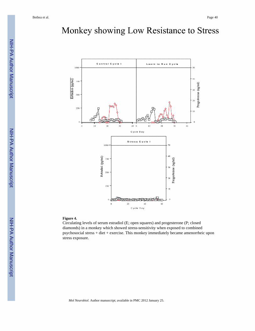
Figure 4.Circulating levels of serum estradiol (E; open squares) and progesterone (P; closeddiamonds) in a monkey which showed stress-sensitivity when exposed to combinedpsychosocial stress + diet + exercise. This monkey immediately became amenorrheic uponstress exposure.
Bethea et al. Page 40
Mol Neurobiol. Author manuscript; available in PMC 2012 January 25.
NIH
-PA Author Manuscript
NIH
-PA Author Manuscript
NIH
-PA Author Manuscript

Figure 5.Peak follicular phase plasma estradiol (E) concentrations (left panel) and peak luteal phaseplasma progesterone (P) concentrations (right panel) in HSR, MSR and SS monkeys duringthe Control 1 menstrual cycle. Asterisks indicate a significant difference between groups,p<0.05.
Bethea et al. Page 41
Mol Neurobiol. Author manuscript; available in PMC 2012 January 25.
NIH
-PA Author Manuscript
NIH
-PA Author Manuscript
NIH
-PA Author Manuscript

Figure 6.Prolactin (top) and cortisol (bottom) responses to an injection of fenfluramine (5 mg/kg, i.v.)in saline while monkeys were maintained under Propofol anesthesia. In the top panel, therewas a significant difference between the groups in the amount of prolactin secreted afterfenfluramine (2 way ANOVA, p < 0.0001), with the HSR group secreting significantly moreprolactin compared to the SS group (Tukey’s post hoc test, p < 0.001). In the bottom panel,the cortisol response to fenfluramine was significantly different between the experimentalgroups (2 way ANOVA, p < 0.0001), with the SS group secreting more cortisol compared tothe HSR group (Tukey’s post hoc test, p < 0.001). Reprinted from Bethea et al., 2005a.
Bethea et al. Page 42
Mol Neurobiol. Author manuscript; available in PMC 2012 January 25.
NIH
-PA Author Manuscript
NIH
-PA Author Manuscript
NIH
-PA Author Manuscript
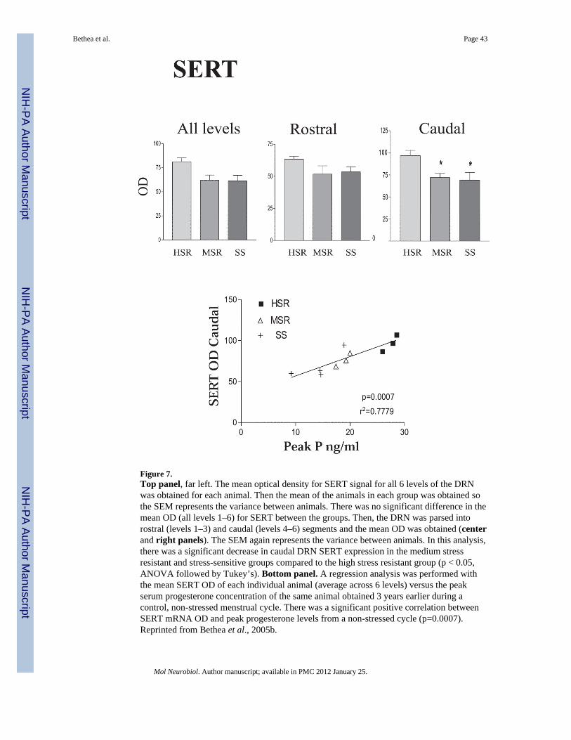
Figure 7.Top panel, far left. The mean optical density for SERT signal for all 6 levels of the DRNwas obtained for each animal. Then the mean of the animals in each group was obtained sothe SEM represents the variance between animals. There was no significant difference in themean OD (all levels 1–6) for SERT between the groups. Then, the DRN was parsed intorostral (levels 1–3) and caudal (levels 4–6) segments and the mean OD was obtained (centerand right panels). The SEM again represents the variance between animals. In this analysis,there was a significant decrease in caudal DRN SERT expression in the medium stressresistant and stress-sensitive groups compared to the high stress resistant group (p < 0.05,ANOVA followed by Tukey’s). Bottom panel. A regression analysis was performed withthe mean SERT OD of each individual animal (average across 6 levels) versus the peakserum progesterone concentration of the same animal obtained 3 years earlier during acontrol, non-stressed menstrual cycle. There was a significant positive correlation betweenSERT mRNA OD and peak progesterone levels from a non-stressed cycle (p=0.0007).Reprinted from Bethea et al., 2005b.
Bethea et al. Page 43
Mol Neurobiol. Author manuscript; available in PMC 2012 January 25.
NIH
-PA Author Manuscript
NIH
-PA Author Manuscript
NIH
-PA Author Manuscript
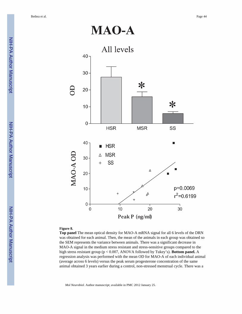
Figure 8.Top panel The mean optical density for MAO-A mRNA signal for all 6 levels of the DRNwas obtained for each animal. Then, the mean of the animals in each group was obtained sothe SEM represents the variance between animals. There was a significant decrease inMAO-A signal in the medium stress resistant and stress-sensitive groups compared to thehigh stress resistant group (p < 0.007, ANOVA followed by Tukey’s). Bottom panel. Aregression analysis was performed with the mean OD for MAO-A of each individual animal(average across 6 levels) versus the peak serum progesterone concentration of the sameanimal obtained 3 years earlier during a control, non-stressed menstrual cycle. There was a
Bethea et al. Page 44
Mol Neurobiol. Author manuscript; available in PMC 2012 January 25.
NIH
-PA Author Manuscript
NIH
-PA Author Manuscript
NIH
-PA Author Manuscript

significant positive correlation between MAO-A mRNA OD and peak progesterone levelsfrom a non-stressed cycle (p=0.0069). Reprinted from Bethea et al., 2005b.
Bethea et al. Page 45
Mol Neurobiol. Author manuscript; available in PMC 2012 January 25.
NIH
-PA Author Manuscript
NIH
-PA Author Manuscript
NIH
-PA Author Manuscript

Figure 9.The number of serotonin-positive neurons on the right side of the dorsal raphe nucleus wasobtained at 3 anatomical levels. The mean of all of the levels was obtained for each animaland then the overall mean was obtained for the group so that the SEM represents thevariance between animals. There were significantly fewer serotonin neurons in the SS group(ANOVA, p < 0.03). * Significantly different from HSR with Tukey’s posthoc pairwisecomparison, p < 0.05. Reprinted from Bethea et al., 2005b.
Bethea et al. Page 46
Mol Neurobiol. Author manuscript; available in PMC 2012 January 25.
NIH
-PA Author Manuscript
NIH
-PA Author Manuscript
NIH
-PA Author Manuscript

Figure 10. 5HT2A, 5HT2C and GAD67 mRNA expression in hypothalamus of HSR, MSR andSS monkeysLeft panel. 5HT2A optical density (OD) and pixel area were obtained at 5 levels of thePVN of HSR, MSR and SS monkeys. The mean of all of the levels was obtained for eachanimal and then the overall mean was obtained for the group so that the SEM represents thevariance between animals. There was significantly more 5HT2A mRNA in the PVN of SSanimals compared to HSR animalsMiddle panel. 5HT2C optical density (OD) and pixel area were obtained at 3 levels of theinfundibulum of HSR, MSR and SS monkeys. The mean of all of the levels was obtained foreach animal and then the overall mean was obtained for the group so that the SEMrepresents the variance between animals. There was significantly more 5HT2C mRNA in theinfundibulum of SS animals compared to HSR animalsRight panel. GAD67 optical density (OD) and pixel area were obtained at 3 levels of theinfundibulum of HSR, MSR and SS monkeys. The mean of all of the levels was obtained foreach animal and then the overall mean was obtained for the group so that the SEMrepresents the variance between animals. There was significantly more GAD67 mRNA inthe infundibulum of SS animals compared to HSR animals. *p<0.05, Kruskal-Wallis one-way ANOVA, Dunn’s posthoc test compared with HSR animals. The number of animalsused in each group is shown below the histograms. Reprinted from Centeno et al., 2007a.
Bethea et al. Page 47
Mol Neurobiol. Author manuscript; available in PMC 2012 January 25.
NIH
-PA Author Manuscript
NIH
-PA Author Manuscript
NIH
-PA Author Manuscript

Figure 11.Autoradiographs of CRH mRNA signal at level 2 of the PVN and CM-Sf of a representativeanimal from each experimental group (HSR, MSR and SS). CRH mRNA expression isbarely detectable in the HSR animal, but it increases markedly in the MSR and SS animals.Reprinted from Centeno et al., 2007c.
Bethea et al. Page 48
Mol Neurobiol. Author manuscript; available in PMC 2012 January 25.
NIH
-PA Author Manuscript
NIH
-PA Author Manuscript
NIH
-PA Author Manuscript

Figure 12.Top and Middle Optical density and positive pixel area of the CRH mRNA signal in thePVN at each of 5 rostral to caudal levels, in levels 4 and 5 combined, and in all levelscombined. There is a significant difference between the groups at caudal levels 4 and 5 andin the average of levels 4 and 5 (p < 0.05, ANOVA in both analyses). CRH mRNAexpression is significantly higher in the MSR and SS groups compared to the HSR group (p< 0.05, SNK). Bottom. The average CRH mRNA expression in the PVN was significantlycorrelated with 5HT2A mRNA expression in the PVN. Reprinted from Centeno et al.,2007c.
Bethea et al. Page 49
Mol Neurobiol. Author manuscript; available in PMC 2012 January 25.
NIH
-PA Author Manuscript
NIH
-PA Author Manuscript
NIH
-PA Author Manuscript

Figure 13.Photomicrographs of CRH immunostaining at level 2 and level 4 of a representative animalfrom each group. The immunostaining is darker and somewhat more extensive in the MSRand SS animals at level 2. At level 4, the PVN has disappeared in this HSR animal, butCRH-positive neurons are still prevalent in the MSR and SS animals. By level 5, the PVNwas absent in all HSR animals. Reprinted from Centeno et al., 2007c.
Bethea et al. Page 50
Mol Neurobiol. Author manuscript; available in PMC 2012 January 25.
NIH
-PA Author Manuscript
NIH
-PA Author Manuscript
NIH
-PA Author Manuscript

Figure 14.Histograms illustrating the quantity of CRH immunostaining in 5 levels of the PVN and in 4levels of the central nucleus of the amygdala of the HSR, MSR and SS groups. Top leftpanel, shows the overall average CRH positive area (μm2)/section in each group. There wasa significant difference between the groups with MSR and SS groups exhibitingsignificantly higher expression of CRH protein than the HSR group Top right panel showsthe volume of CRH immunostaining expressed as a percent of the volume of the region(μm3). There was a significant difference between the groups with MSR and SS groupsexhibiting significantly higher expression of CRH protein than the HSR group. Bottompanel illustrates the density of CRH fibers (positive pixel area covered by fibers) in thecentral nucleus of the amygdala from HSR, MSR and SS groups. There was a significantdifference between the groups with the SS group exhibiting significantly higher CRH fiberdensity than the MSR and HSR groups * different from HSR; + different from MSR;ANOVA followed by Student-Newman-Keuls posthoc pairwise comparison. Reprinted fromCenteno et al., 2007c.
Bethea et al. Page 51
Mol Neurobiol. Author manuscript; available in PMC 2012 January 25.
NIH
-PA Author Manuscript
NIH
-PA Author Manuscript
NIH
-PA Author Manuscript

Figure 15. GnRH mRNA expression in the MBH of high stress-resilient and stress-sensitivemonkeys(A) Autoradiogram of GnRH in situ hybridization signal in the arcuate nucleus and medianeminence of one high stress-resilient (HSR) monkey(B) Autoradiogram of GnRH in situ hybridization signal in the arcuate nucleus and medianeminence of one stress-sensitive (SS) monkey.(C) Dark field image of GnRH ISH signal after exposure with emulsion in the ARC of onehigh stress-resilient (HSR) monkey (scale bar = 10 μm).(D) Dark field image of GnRH in situ hybridization signal after exposure with emulsion inthe arcuate nucleus of one stress-sensitive (SS) monkey (scale bar = 10 μm).Reprinted from Centeno et al., 2006b.
Bethea et al. Page 52
Mol Neurobiol. Author manuscript; available in PMC 2012 January 25.
NIH
-PA Author Manuscript
NIH
-PA Author Manuscript
NIH
-PA Author Manuscript

Figure 16. Analysis of the total number of cells expressing GnRH mRNA in the arcuate nucleusand median eminence of HSR and SS monkeys (n=3/group)Top. The number of neurons expressing GnRH mRNA grains at 3x background wascounted in 6 sections through the arcuate nucleus and median eminence. The histogramillustrates the mean (± SEM) of the total number of neurons that express GnRH mRNA.Middle. The grain over each GnRH neurons were segmented into positive pixels. Thehistogram illustrates the mean (± SEM) of the total number of GnRH neurons exhibitingdifferent numbers of positive pixels over grains per cell.Bottom. Histogram of the mean (± SEM) density of GnRH-immunopositive cell bodies inthe arcuate nucleus and median eminence of HSR and SS monkeys (n=3/group) expressed asthe number of cells per mm3. Six sections at 250μ intervals through the mediobasalhypothalamus were counted and the total number of neurones was obtained for each animal/mm3 (density). The group means were then computed and statistically compared. *p<0.05,Student’s t-test. Reprinted from Centeno et al., 2006b.
Bethea et al. Page 53
Mol Neurobiol. Author manuscript; available in PMC 2012 January 25.
NIH
-PA Author Manuscript
NIH
-PA Author Manuscript
NIH
-PA Author Manuscript

Figure 17.Histogram illustrating the average of the positive pixels in a sample area of GnRH fibres inthe median eminence of HSR and SS monkeys (SS). (n=3/group; *p<0.05, Student’s t-test).Reprinted from Centeno et al., 2006b.
Bethea et al. Page 54
Mol Neurobiol. Author manuscript; available in PMC 2012 January 25.
NIH
-PA Author Manuscript
NIH
-PA Author Manuscript
NIH
-PA Author Manuscript

NIH
-PA Author Manuscript
NIH
-PA Author Manuscript
NIH
-PA Author Manuscript
Bethea et al. Page 55
Table 1
Menstrual cycle and weight parameters by stress-sensitivity category.
Stress-Sensitivity Category Menstrual Cycle Length (days) Follicular Phase Length (days) Weight (kg) Weight Change
HSR 31.5±1.9 16.2±2.3 4.1±0.3 0.34±0.6
MSR 33.9±2.3 16.8±3.6 3.9±0.4 0.3±0.8
SS 30.7±1.6 15.7±4.1 4.2±0.4 0.32±0.7
Mol Neurobiol. Author manuscript; available in PMC 2012 January 25.

NIH
-PA Author Manuscript
NIH
-PA Author Manuscript
NIH
-PA Author Manuscript
Bethea et al. Page 56
Tabl
e 2
Peak
est
radi
ol a
nd p
roge
ster
one
conc
entra
tions
in H
SR a
nd S
S an
imal
s acr
oss t
hree
con
trol m
enst
rual
cyc
les.
Stre
ss-S
ensi
tivity
Cat
egor
yC
ycle
1 E
2 (p
g/m
l)C
ycle
2 E
2 (p
g/m
l)C
ycle
3 E
2 (p
g/m
l)C
ycle
1 P
4 (n
g/m
l)C
ycle
2 P
4 (n
g/m
l)C
ycle
3 P
4 (n
g/m
l)
HSR
734±
91*
603±
98*
622±
54*
25±3
.8*
23±2
.4*
31±5
.6*
SS45
2±56
431±
5936
7±92
14±2
.39.
8±4.
113
.6±3
.3
* indi
cate
s a si
gnifi
cant
diff
eren
ce, p≤0
.05,
com
pare
d to
SS
anim
als i
n th
e sa
me
cycl
e
Mol Neurobiol. Author manuscript; available in PMC 2012 January 25.