Profiling the autoantibody repertoire by serological antigen selection
RESEARCH Open Access
SIV antigen immunization induces transientantigen-specific T cell responses and selectivelyactivates viral replication in draining lymph nodesin retroviral suppressed rhesus macaquesHaitao Hu1, Lucio Gama2, Pyone P Aye3, Janice E Clements2, Peter A Barry4, Andrew A Lackner3 andDrew Weissman1*
Abstract
Background: HIV infection causes a qualitative and quantitative loss of CD4+ T cell immunity. The institution ofanti-retroviral therapy (ART) restores CD4+ T cell responses to many pathogens, but HIV-specific responses remaindeficient. Similarly, therapeutic immunization with HIV antigens of chronically infected, ART treated subjects resultsin poor induction of HIV-specific CD4 responses. In this study, we used a macaque model of ART treatment duringchronic infection to study the virologic consequences of SIV antigen stimulation in lymph nodes early afterimmunization. Rhesus CMV (RhCMV) seropositive, Mamu A*01 positive rhesus macaques were chronically infectedwith SIVmac251 and treated with ART. The immune and viral responses to SIV gag and RhCMV pp65 antigenimmunization in draining lymph nodes and peripheral blood were analyzed. Animals were immunized oncontralateral sides with SIV gag and RhCMV pp65 encoding plasmids, which allowed lymph nodes draining eachantigen to be obtained at the same time from the same animal for direct comparison.
Results: We observed that both SIV and RhCMV immunizations stimulated transient antigen-specific T cellresponses in draining lymph nodes. The RhCMV-specific responses were potent and sustained (50 days post-immunization) in the periphery, while the SIV-specific responses were transient and extinguished quickly. The SIVantigen stimulation selectively induced transient SIV replication in draining lymph nodes.
Conclusions: The data are consistent with a model whereby viral replication in response to SIV antigen stimulationlimits the generation of SIV antigen-specific responses and suggests a potential mechanism for the early loss andpoor HIV-specific CD4+ T cell response observed in HIV-infected individuals.
BackgroundCD4+ T cells play a central role in maintaining effectivecellular and humoral immune responses by providinghelp to CD8+ T cells, B cells and innate effectors. Theprotective role of CD4+ T cell responses in HIV-1 infec-tion has been suggested in previous studies [1,2]. How-ever, HIV-1 infection results in the progressive loss ofCD4+ T cell responses, which is characterized as both adecline in the number of CD4+ T cells and a loss of thefunctional activity of cells with certain antigenic
specificities [3-6]. Although the institution of anti-retro-viral therapy (ART) causes viral suppression and recov-ery of CD4+ T cell response to some commonpathogens, HIV-specific CD4 response remains deficient[7,8]. Similarly, immunization of chronically infected,ART treated patients with HIV antigens does not resultin the generation of significant HIV-specific CD4+ T cellresponses, suggesting that HIV-specific CD4+ T cellsare dysfunctional or preferentially depleted in infectionand fail to recover [9-11]. The mechanisms for the fail-ure of HIV antigen immunization to induce HIV-speci-fic CD4+ response are not fully clear [12].During an immune response, antigen-presenting cells
(APC) activate CD4+ T cells to specific antigen
* Correspondence: [email protected] of Infectious Diseases, University of Pennsylvania, Philadelphia, PA,USAFull list of author information is available at the end of the article
Hu et al. Retrovirology 2011, 8:57http://www.retrovirology.com/content/8/1/57
© 2011 Hu et al; licensee BioMed Central Ltd. This is an Open Access article distributed under the terms of the Creative CommonsAttribution License (http://creativecommons.org/licenses/by/2.0), which permits unrestricted use, distribution, and reproduction inany medium, provided the original work is properly cited.

specificities in lymphoid tissue. However, in HIV-1infection, lymphoid tissue also represents an importantsite for viral replication, and the interaction betweenAPC and CD4+ T cells may enhance viral replication bymultiple mechanisms (reviewed in [13]). It has beenshown that even in the setting of potent regimens ofART, a very low level of viral replication could still bedetected [14-16], which may be derived from DCmediated activation of latent virus in memory CD4+ Tcells, homeostatic regulation of memory populations, orother long-lived reservoirs [17]. Given that HIV-specificCD4+ memory T cells are preferentially infected byHIV, carrying more viral DNA than total memory cells[18], we were interested in determining if activation ofHIV-specific CD4+ T cells results in viral replication inlymphoid tissue.We used a rhesus macaque model of ART treatment
during chronic infection to study the effects of SIV anti-gen stimulation in lymph nodes (LNs) compared to acontrol immunogen on viral replication early afterimmunization. Mamu A*01+ rhesus macaques wereinfected with SIVmac251 and after 4 months treatedwith ART resulting in viral suppression and immunereconstitution. The macaques were also rhesus CMV(RhCMV) seropositive and immunized with an RhCMVimmunogen as a control antigen stimulation. Animalswere immunized on the left side (both arms and legs)with an SIV gag-encoding expression plasmid and onthe right side (both arms and legs) with a RhCMVpp65-encoding expression plasmid, which allowed drain-ing LNs for each antigen to be obtained from the sameanimal at the same time, allowing for a direct compari-son of the effect of SIV and RhCMV antigen stimulationon viral replication.
ResultsInfection and immunization of rhesus macaquesAll animals used in this study (FH40, DD05, and CT64)were Mamu A*01 positive to reduce MHC variation indisease course and T cell responses and were naturallyinfected with RhCMV. The study was designed to infectanimals (1000 TCID50 of SIVmac251 by intravenousinjection) and allow them to reach steady state viralloads (4 months) followed by ART treatment (PMPAand D4T) for 5.5 months. Animals were then immu-nized with plasmids encoding SIV gag or RhCMV pp65in both arms and legs. Two LN biopsies draining eitherSIV gag or RhCMV pp65 injections from the same ani-mal at the same time were obtained on the indicateddays (D3: FH40 Inguinal; D5: DD05 Inguinal, D7: CT64Inguinal; D9: FH40 Axillary; D11: DD05 Axillary, D14:CT64 Axillary) (Figure 1). Immunizations used expres-sion plasmids previously used as vaccines that weredemonstrated to induce potent T cell responses in unin-fected rhesus macaques [19-25]. Serum and PBMCswere obtained every 2 to 3 weeks throughout theexperiment.
ART suppresses viral replication with recovery ofperipheral CD4+ T cell countsSIV infection was established in all three animals withkinetics typical of primary infection in naïve rhesusmacaques (Figure 2A) [26,27]. Introduction of ART 4months post infection, when set point viral loads hadbeen established, efficiently suppressed viral replicationto undetectable levels. One macaque demonstrated occa-sional blips in viral load that returned to undetectablelevels by the subsequent measurement without anychange in therapy (Figure 2A). Absolute CD4+ T cell
Figure 1 Experimental protocol for infection and immunization of rhesus macaques. Mamu A*01 rhesus macaques naturally infected withRhCMV were intravenously inoculated with SIVmac251 followed by ART treatment (DT4 and PMPA) from days 119 post infection through theend of experiment. On days 286 or 290 post infection, monkeys received immunizations with SIV gag encoding DNA i.m. (2 mg per injection), inthe left arm and left leg, and immunizations with RhCMV pp65 encoding DNA i.m. (2 mg per injection) in the right arm and leg. LNs biopsiesdraining either SIV gag or RhCMV pp65 immunization sites from the same animal at the same time were obtained on Day 3 for FH40, Day 5 forDD05, Day 7 for CT64, Day 9 for FH40, Day 11 for DD05, and Day 14 for CT64. LN biopsies from both sides were also collected from all animalson Day 60 post immunization. PBMCs were collected pre-immunization and every 2-3 weeks post immunization.
Hu et al. Retrovirology 2011, 8:57http://www.retrovirology.com/content/8/1/57
Page 2 of 11

counts in the peripheral blood demonstrated stabilizationafter introduction of ART in all animals with sustainedlevels of more than 500 cells/μl (Figure 2B). These find-ings demonstrate that chronic SIV infection was achievedand ART successfully suppressed viral replication leadingto partial recovery of peripheral CD4+ T cell counts.
SIV and RhCMV antigen immunization induces antigen-specific T cell responses in draining LNsAnimals were immunized with SIV gag encoding plas-mid in the arm and leg on one side and RhCMV pp65encoding plasmid on the other side at the same time.LNs, one draining an SIV immunization site and onedraining an RhCMV immunization site, were excisedfrom each animal at 2 time points (axillary and ingu-inal) post immunization. LN biopsies from both sidesfrom all macaques were also collected on day 60-postimmunization. We found that the levels of antigen-specific responses in day 60 LNs were similar in com-paring both antigens in both LNs, suggesting that the
effects of immunization on T cell responses in the LNswere transient and returned to baseline by 60 dayspost immunization (Figure 3 and data not shown).Therefore, this time point was chosen as a baseline forstandardization.First, we assessed immune activation induced by anti-
gen immunizations in draining LNs to determinewhether the two DNA plasmids were immunogenic. LNmononuclear cells were analyzed for IFN-g mRNAexpression, a major effector cytokine for adaptive immu-nity. As shown in Figure 3A, an increase in IFN-gmRNA expression was detected in LNs draining SIV gagand RhCMV pp65 on day 3 post immunization, whichwas followed by a decline on day 5 to the baseline levelsas observed in day 60 LNs (Figure 3A). The result sug-gests that both DNA plasmids are immunogenic in therhesus macaques used in this study, which is consistentwith previous primate studies where the same DNAplasmids were used and shown to be immunogenic inuninfected macaques [19-25].We then evaluated the antigen-specific T cell responses
in LNs by measuring ex vivo cytokine production of LNmononuclear cells. Cells were stimulated with either SIVgag or RhCMV pp65 peptide pools and production of IL-2, TNF-a and IFN-g in T cells was determined by poly-chromatic flow cytometry. Flow cytometry plots for cyto-kine staining are shown (Figure 3B and 3C). We foundthat the LN draining SIV gag on day 3, when the immuneactivation, as measured by IFN-g mRNA, was the highest,demonstrated potent gag-specific T cell responses basedon IL-2, TNF-a and IFN-g production (Figure 3B and3D). In contrast, the day 3 LN draining RhCMV pp65immunization from the same animal, when stimulated bygag peptides, demonstrated no significant response (Fig-ure 3B and 3D). Some multifunctionality of the CD4+ Tcell response was observed with approximately 4%expressing three cytokines and 28% expressing two. Simi-larly, the LNs collected on day 5 post immunization wereevaluated for RhCMV-specific T cell responses by stimu-lating the LN cells with RhCMV-pp65 peptides. A signifi-cant RhCMV-specific response was induced in the LNdraining the RhCMV pp65 compared to the LN drainingthe SIV gag immunization site (Figure 3C and 3E). Drain-ing LN responses to SIV immunization decreased by day5 and from day 7 onwards were similar to the levelsobserved in day 60-post immunization LNs (Figure 3Fand 3G). Taken together, these data suggest that bothSIV and RhCMV immunization induced transient anti-gen-specific T cell responses in draining LNs.
Differential SIV- and RhCMV-specific T cell responses inperipheral bloodBoth antigens were immunogenic and induced antigen-specific T cell responses in draining LNs, we next
Figure 2 Viral loads and peripheral CD4+ T cell counts. Viralloads and CD4 counts were measured every 2 to 3 weeksthroughout the experiment. (A) Viral RNA in plasma was quantifiedby a bDNA signal amplification assay and expressed as viral RNAcopies per ml plasma. ART treatment controlled the viral loads in allthree animals. (B) Peripheral CD4+ T cell counts increased after theinitiation of ART. Macaque blood samples were stained for CD3,CD4, and CD8 and the number of CD3+, CD4+ T lymphocytes weredetermined by flow cytometry and peripheral white blood cellcounts. CD4+ T cell counts are expressed as CD4+ T cells per μlblood.
Hu et al. Retrovirology 2011, 8:57http://www.retrovirology.com/content/8/1/57
Page 3 of 11
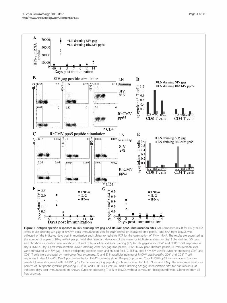
Figure 3 Antigen-specific responses in LNs draining SIV gag and RhCMV pp65 immunization sites. (A) Composite result for IFN-g mRNAlevels in LNs draining SIV gag or RhCMV pp65 immunization sites for each animal on indicated time points. Total RNA from LNMCs wascollected on the indicated days post immunization and subject to real-time PCR for the quantitation of IFN-g mRNA. The results are expressed asthe number of copies of IFN-g mRNA per μg total RNA. Standard deviation of the mean for triplicate analyses for Day 3 LNs draining SIV gagand RhCMV immunization sites are shown. (B and D) Intracellular cytokine staining (ICS) for SIV gag-specific CD4+ and CD8+ T cell responses inday 3 LNMCs. Day 3 post immunization LNMCs draining either SIV-gag (top panels, B) or RhCMV-pp65 (bottom panels, B) immunization siteswere stimulated with SIV gag 15-mer overlapping peptide pools and stained for IL-2, TNF-a, and IFN-g. SIV-specific cytokine-producing CD4+ andCD8+ T cells were analyzed by multi-color flow cytometry. (C and E) Intracellular staining of RhCMV pp65-specific CD4+ and CD8+ T cellresponses in day 5 LNMCs. Day 5 post immunization LNMCs draining either SIV-gag (top panels, C) or RhCMV-pp65 immunizations (bottompanels, C) were stimulated with RhCMV pp65 15-mer overlapping peptide pools and stained for IL-2, TNF-a, and IFN-g. The composite results forpercent of SIV-specific cytokine producing CD8+ (F) and CD4+ (G) T cells in LNMCs draining SIV gag immunization sites for one macaque atindicated days post immunization are shown. Cytokine producing T cells in LNMCs without stimulation (background) were subtracted from allflow analyses.
Hu et al. Retrovirology 2011, 8:57http://www.retrovirology.com/content/8/1/57
Page 4 of 11

analyzed T cell responses induced by immunization inthe peripheral blood. PBMC collected at multiple timepoints post immunization were stimulated with SIV gagor RhCMV pp65 peptide pools and the frequency ofcytokine producing T cells were analyzed by polychro-matic flow cytometery. We expressed the data as thepercent of CD8+ T cells able to produce any cytokine orcombination of cytokines (IL-2, TNF-a and/or IFN-g)after subtracting the levels in unstimulated cells. Theaverage of all animals is shown (Figure 4A). A potentincrease in RhCMV-specific CD8+ T cells was observedat day 3-post immunization in blood with an average of1.4% of cells able to produce a cytokine. The RhCMV-specific response was sustained until day 50 post-immu-nization. In contrast, SIV-specific CD8+ T cell responseswere transient and extinguished quickly in the bloodwith only an increase on day 9 post-immunization
(Figure 4A). Further characterization of the gag andpp65 responses demonstrated that gag-specific cells hadincreased levels of expression of PD1 and containedboth central memory (CD95+, CD28+) and effectormemory (CD95+, CD28-) cells, while the pp65 respond-ing cells were predominantly effector memory pheno-type. All animals demonstrated similar kinetics of gagand pp65 specific responses. PBMCs were stained withthe Mamu A*01 specific gag tetramer, p11C (CTPY-DINQM). All animals demonstrated a decrease in tetra-mer positive CD8+ T cells after the initiation of ART,but levels remained above 1% after 5.5 months of sup-pressive ART (Figure 4B).
SIV antigen immunization induces transient activation ofviral replication in the draining LNWe next determined whether immunization with SIV orCMV antigens induced the activation of viral replicationat early, 3 to 7 days, or late, 9 to 14 days, time points.Total RNAs from LN mononuclear cells (LNMC) drain-ing either SIV gag or RhCMV pp65 immunization siteswere analyzed by real-time PCR for early and late SIVRNA transcripts, including doubly spliced (tat), singly-spliced (vif), and unspliced (gag) RNA [28]. The use ofisolated cells with multiple washes both before and aftercryopreservation removed any extracellular viral RNAthat was present as free or germinal center associatedvirions. All comparisons were made between LNs fromthe same animal obtained at the same time that differedonly in whether they drained an SIV or a RhCMVimmunization site. We first investigated SIV RNAs inday 60 LNMCs and found low levels of all 3 transcripts.Importantly, no difference was observed for each viralRNA species at this time point in comparing LNMCsdraining SIV and RhCMV immunization sites from thesame animal. Therefore, we used these levels of SIVRNA as a baseline for the analysis of the effect of anti-gen stimulation on viral replication for that animal.Copy numbers of SIV RNA were normalized to GAPDHRNA, and the results for each time point followingimmunization are shown as fold change relative to RNAcollected at 60 days post immunization for that animal(Figure 5).Each analysis was performed in the same animal at the
same time point where the only difference was the typeof immunization drained, thereby avoiding the need toconsider confounders present in comparisons betweenanimals. The effects of antigen-specific stimulation onSIV replication were first determined by comparing thelevels of SIV doubly spliced RNA in LNMCs drainingSIV gag and RhCMV pp65 immunization. Doublyspliced RNA copies were significantly increased in theLNMCs draining the SIV gag immunization comparedto those draining the pp65 immunization on days 3, 5,
Figure 4 SIV- and RhCMV-specific T cell responses inperipheral blood. (A) PBMCs before and after immunizations werestimulated with either SIV gag or RhCMV pp65 peptide pools andstained for IL-2, TNF-a, and IFN-g. The percentages of cytokine-producing CD8+ T cells were determined by multi-color flowcytometry. The percent of CD8+ T cells able to make any cytokine isshown. The average of all animals is shown on the indicated dayspost immunization. Error bars are the standard error of the mean, p-values using the student’s t-test comparing the levels beforeimmunization (Day 0) to time points post-immunization are shownas a * indicating a p < 0.05. (B) Measurement of p11c tetramer+,CD8+ T cells throughout the experiment. The results are expressedas percent of tetramer+, CD8+ T cells on indicated days post SIVinfection for each animal.
Hu et al. Retrovirology 2011, 8:57http://www.retrovirology.com/content/8/1/57
Page 5 of 11
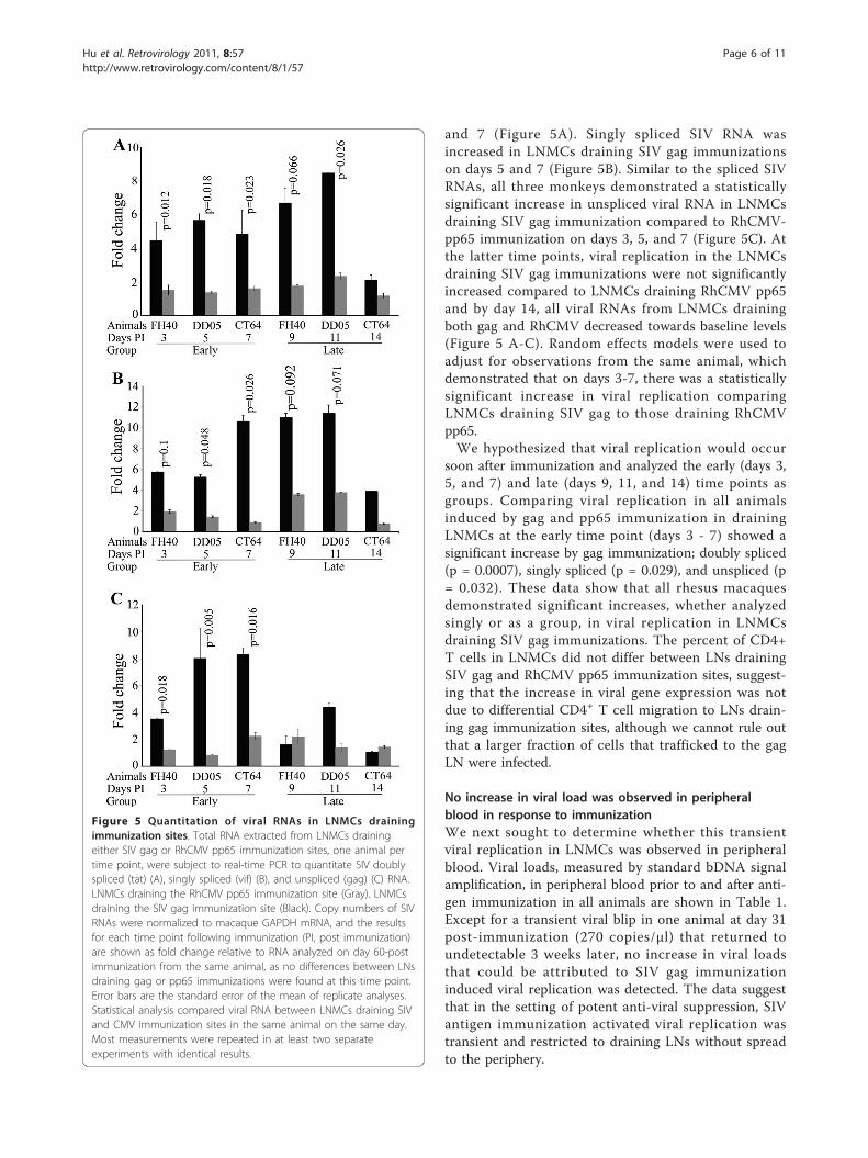
and 7 (Figure 5A). Singly spliced SIV RNA wasincreased in LNMCs draining SIV gag immunizationson days 5 and 7 (Figure 5B). Similar to the spliced SIVRNAs, all three monkeys demonstrated a statisticallysignificant increase in unspliced viral RNA in LNMCsdraining SIV gag immunization compared to RhCMV-pp65 immunization on days 3, 5, and 7 (Figure 5C). Atthe latter time points, viral replication in the LNMCsdraining SIV gag immunizations were not significantlyincreased compared to LNMCs draining RhCMV pp65and by day 14, all viral RNAs from LNMCs drainingboth gag and RhCMV decreased towards baseline levels(Figure 5 A-C). Random effects models were used toadjust for observations from the same animal, whichdemonstrated that on days 3-7, there was a statisticallysignificant increase in viral replication comparingLNMCs draining SIV gag to those draining RhCMVpp65.We hypothesized that viral replication would occur
soon after immunization and analyzed the early (days 3,5, and 7) and late (days 9, 11, and 14) time points asgroups. Comparing viral replication in all animalsinduced by gag and pp65 immunization in drainingLNMCs at the early time point (days 3 - 7) showed asignificant increase by gag immunization; doubly spliced(p = 0.0007), singly spliced (p = 0.029), and unspliced (p= 0.032). These data show that all rhesus macaquesdemonstrated significant increases, whether analyzedsingly or as a group, in viral replication in LNMCsdraining SIV gag immunizations. The percent of CD4+T cells in LNMCs did not differ between LNs drainingSIV gag and RhCMV pp65 immunization sites, suggest-ing that the increase in viral gene expression was notdue to differential CD4+ T cell migration to LNs drain-ing gag immunization sites, although we cannot rule outthat a larger fraction of cells that trafficked to the gagLN were infected.
No increase in viral load was observed in peripheralblood in response to immunizationWe next sought to determine whether this transientviral replication in LNMCs was observed in peripheralblood. Viral loads, measured by standard bDNA signalamplification, in peripheral blood prior to and after anti-gen immunization in all animals are shown in Table 1.Except for a transient viral blip in one animal at day 31post-immunization (270 copies/μl) that returned toundetectable 3 weeks later, no increase in viral loadsthat could be attributed to SIV gag immunizationinduced viral replication was detected. The data suggestthat in the setting of potent anti-viral suppression, SIVantigen immunization activated viral replication wastransient and restricted to draining LNs without spreadto the periphery.
Figure 5 Quantitation of viral RNAs in LNMCs drainingimmunization sites. Total RNA extracted from LNMCs drainingeither SIV gag or RhCMV pp65 immunization sites, one animal pertime point, were subject to real-time PCR to quantitate SIV doublyspliced (tat) (A), singly spliced (vif) (B), and unspliced (gag) (C) RNA.LNMCs draining the RhCMV pp65 immunization site (Gray). LNMCsdraining the SIV gag immunization site (Black). Copy numbers of SIVRNAs were normalized to macaque GAPDH mRNA, and the resultsfor each time point following immunization (PI, post immunization)are shown as fold change relative to RNA analyzed on day 60-postimmunization from the same animal, as no differences between LNsdraining gag or pp65 immunizations were found at this time point.Error bars are the standard error of the mean of replicate analyses.Statistical analysis compared viral RNA between LNMCs draining SIVand CMV immunization sites in the same animal on the same day.Most measurements were repeated in at least two separateexperiments with identical results.
Hu et al. Retrovirology 2011, 8:57http://www.retrovirology.com/content/8/1/57
Page 6 of 11

DiscussionA biphasic destruction of CD4+ T cells is observed inHIV infection with a massive loss of CD4+ T cells dur-ing early infection and a subsequent progressive lossduring the chronic stage of infection [29,30]. Retroviralsuppression by ART results in an increase in peripheralCD4+ T cell counts and functional reconstitution ofCD4+ T cell responses to many common antigens[31,32], but HIV-specific CD4+ T cell responses remaindeficient [33]. We studied ART-treated, chronic SIV andRhCMV infected rhesus macaques and observed; 1) thatboth SIV and RhCMV antigen immunizations couldinduce immune activation and antigen-specific T cellresponses in draining LNs. 2) In peripheral blood, theRhCMV-specific response induced by immunization waspotent and sustained, whereas the SIV-specific responseextinguished quickly. 3) We observed that SIV antigenimmunization transiently induced greater levels of SIVreplication in draining LNs of all animals compared toRhCMV immunization. In this study, the experimentaldesign of immunizing the same animal with both anSIV antigen and a non-SIV antigen on collateral sidesallowed us to directly compare the early immune andviral responses in draining LNs from the same animal atthe same time, making it possible to study the effect ofantigen stimulation in the context of ART-treated,chronic infection with limited animal numbers. All 3animals displayed persistent CMV specific T cellresponses for 50 days and demonstrated a weak transi-ent SIV specific T cell response in peripheral blood.LNMCs from all animals demonstrated elevated levelsof viral replication in response to SIV antigen immuni-zation during the first 7 days after immunization. Thedata do not prove a causal link between the weak SIV Tcell responses and LN viral replication, but are consis-tent with a hypothesis that SIV antigens induce viralreplication that leads to depletion or dysfunction of anti-gen specific cells leading to a reduction in the strengthand longevity of the response.Pathogenic SIVmac251 infection of rhesus macaques
has been well described as one of the preferred experi-mental models for studying HIV pathogenesis [34]. Inthis study, all rhesus macaques inoculated with SIV-mac251 were Mamu A*01 positive to control for an
MHC effect on viral immune responses and disease pro-gression, as well as to aid in the measurement ofimmune responses by a Mamu A*01 restricted SIV gagtetramer. All macaques established primary SIV infec-tion with typical viral replication dynamics [26,27,35],and demonstrated responsiveness to ART with rapidcontrol of viremia (Figure 2A). One animal had occa-sional blips in viral load that returned to undetectablewithout changes in therapy. It has been shown that evenduring the most potent regimens of retroviral suppres-sion, the presence of virus in plasma could be measuredby some ultrasensitive assays [14-16]. We believe theblips in FH40 are similar to those observed during ther-apy in humans, which are not associated with acuteinfection of cells [36] and believe that this is representa-tive of a range of low level virus that can be measuredduring ART [37]. One study identified that HIV-infectedsubjects that developed blips in viral load had higherinstead of lower levels of CD4+ T cell responses to gag[38]. We do not believe that the macaque with blips inviral load is responding differently to ART compared tothe animals without measureable blips.Therapeutic immunization for HIV infection during
ART has been studied in SIV-infected rhesus macaquesand the immunological and virologic consequences havebeen investigated in peripheral blood using DNA immu-nization [39-42], as well as other systems [43-45]. Afterrelease from ART, variable immunologic and virologicbenefits were reported from no control [42], temporalcontrol [43], to long-lasting virologic control [44]. Inthis study, using RhCMV immunization as a non-SIVcontrol in the same animal, we investigated the immu-nologic and virologic consequences of immunizationwith SIV antigen in chronic SIV and RhCMV co-infected, ART treated rhesus macaques focusing on theearly response in draining LNs. The plasmids encodingimmunogens used for immunization were previouslydemonstrated to induce potent immune responses inuninfected macaques [19-25]. The RhCMV pp65 plas-mid had a greater number of immunostimulatory motifs,which would bias towards the null hypothesis. More-over, all the comparisons between LNs at each timepoint were from the same animal collected at the sametime, thus allowing us to use each animal as its owncontrol. Also, our study focused on the local responsesin draining LN, where the antigen-specific T cellresponses and viral replication occur, rather than sys-temic responses in peripheral blood, as done in mostprevious studies. In addition, we chose a range of dayspost immunization covering the early activation ofmemory T cells and generation of effector responses.Our data show that both SIV and RhCMV antigenimmunizations induced transient immune activation andantigen-specific T cell responses in draining LNs.
Table 1 Viral loads in periphery during antigenimmunization
Days PI* 238 290 317 336 344
Animal
CT64 < 125 < 125(D0*) < 125(D27*) < 125 < 125
DD05 < 125 < 125(D0*) < 125(D27*) < 125 < 125
FH40 < 125 < 125(D4*) 270(D31*) < 125 < 125
* Days post immunization.
Hu et al. Retrovirology 2011, 8:57http://www.retrovirology.com/content/8/1/57
Page 7 of 11

Further measurement of these responses in peripheralblood showed that the RhCMV-specific responses weresustained in PBMC with a rapid onset of cytokine pro-ducing CD8+ T cells 3 days post immunization, whichwas maintained at day 50-post immunization, whereasthe immunization induced SIV-specific T cell responseswere transient, appearing only on day 9 post-immuniza-tion, and extinguished quickly in blood. The peripheralSIV- and RhCMV-specific CD8+ T cell responses weresignificantly induced compared to pre-immunizationlevels, supporting that both immunizations inducedimmune responses. Of interest, a study that repeatedlyimmunized macaques with long-standing ART-treatedSIVmac251 infection induced stronger SIV-specific CD4+ and CD8+ T cell responses in blood [46]. This studyused the MVA vector and delivered three immuniza-tions, whereas in our study, only a single gag DNAimmunization was used.HIV-specific CD4+ memory T cells are preferentially
infected by HIV, carrying approximately 2- to 5-foldmore viral DNA than total memory cells [18]. Wehypothesize that during an HIV-specific response, acti-vation of HIV-specific CD4+ T cells, which bear higheramounts of latent virus, results in activation of viralreplication in the LN leading to suppression of CD4+ Tcells and the HIV-specific responses through multiplemechanisms, while a non-HIV antigen-specific responseactivates less viral replication allowing more efficientexpansion of the response.
ConclusionsWe used a naturally RhCMV and experimentally SIV-mac251 co-infected and ART treated rhesus macaquemodel to study the effects of immune stimulation withSIV gag and RhCMV pp65 antigens. The study concen-trated on the early immune and viral responses in drain-ing lymphoid organs. Both antigen immunizations wereable to induce transient immune activation and antigen-specific T cell responses in draining LNs, but the SIVgag immunization also induced significant viral replica-tion. Following immunization, the SIV response extin-guished quickly in peripheral blood, while the RhCMVresponse was sustained. Our data suggest that SIV anti-gens, as part of the normal immune response to thevirus, leads to T cell stimulation that could potentiallylead to viral replication resulting in an impairment inthe generation of virus specific CD4+ T cells. It is possi-ble that this mechanism is responsible for the observa-tion that in progressing HIV-infected subjects, CD4-specific responses to HIV antigens are lost early ininfection and are difficult to restore or induce. Furtherstudies are needed to determine if there is a causal linkbetween SIV or HIV antigen induced viral replicationand impairment of CD4+ T cell responses to the virus.
MethodsEthics statementAll animal experiments were performed in strict accor-dance with the standards of the Association for Assess-ment and Accreditation of Laboratory Animal CareInternational and the “Guide for the Care and Use ofLaboratory Animals” prepared by the National ResearchCouncil. The studies were approved by the University ofPennsylvania and Tulane Institutional Animal Care andUse Committees.
ImmunogensPlasmid DNA expressing the SIV Gag core protein fromSIVmac239 (pSIVgag) was used. It is a Rev-independentexpression vector designed for a high level of proteinexpression, as previously described [19,21-23]. Proteinexpression is under the transcriptional control of theimmediate-early promoter/enhancer of human CMVand the bovine growth hormone polyadenylation signal.Plasmid DNA expressing the RhCMV pp65 protein(pND/pp65-2) was used [20,24,25]. The expression ofRhCMV pp65 uses the same promoter and polyadenyla-tion signal. The GC and CpG content of the plasmidswith inserts are: pSIVgag, 40.5% GC, no CpG-S(GACGTT or AACGTT) motifs and 0.03 potentialCpG-N motifs (CCG, CCGG, CGG) per bp; pND-pp65,51.3% GC, 3 CpG-S motifs and 0.0344 potential CpG-Nmotifs per bp. Neither contained any human optimalTLR9 immunostimulatory motifs (TGTCGTT). DNAwas formulated for injection in 0.15 M citrate bufferand 0.25% bupivicaine at a pH of 6.5.
Infection of rhesus macaques and overview of studyThe study timeline is shown in Figure 1. Three RhCMVseropositive Mamu A*01 positive rhesus macaques wereintravenously infected with SIVmac251 (1000 TCID50)at time zero. Four months after SIV infection, ART wasintroduced (subcutaneous PMPA 20 mg/kg/day (GileadSciences, Inc) and oral D4T 1 mg/kg/day (Bristol-Meyers Squibb)) and continued until the end of theexperiment. The macaques were followed for 5.6months on ART for recovery of peripheral CD4+ T cellcount and viral load suppression. Nine and a halfmonths after infection and 5.6 months after the initia-tion of ART, each monkey received 2 immunizationswith SIV gag encoding DNA [19,21-23] intramuscularly(i.m.) (2 mg/injection), one in the left arm (triceps mus-cle) and one in the left leg (quadriceps muscle) and 2immunizations with RhCMV pp65 encoding plasmid[20] (2 mg/injection) in the right arm and leg. Afterimmunization, draining LN were sampled at two-timepoints for each animal by first removing inguinal LNson each side followed by axillary LN removal from bothsides (Macaque FH40 - D3 and D9, Macaque DD05 -
Hu et al. Retrovirology 2011, 8:57http://www.retrovirology.com/content/8/1/57
Page 8 of 11

D5 and D11, and Macaque CT64 - D7 and D14). Onday 60 post immunization, LNs from both sides of eachanimal, draining SIV gag and RhCMV pp65 immuniza-tions, were collected. Blood was collected every 2-3weeks throughout the experiment. PBMC were isolatedusing Ficoll-diatrizoate gradient centrifugation and ana-lyzed by flow cytometry or cryopreserved in 10%DMSO.
Measurement of peripheral viral load and CD4+ T cellcountsViral RNA in plasma was quantified by a bDNA signalamplification assay (Bayer Inc., version 4.0), specific forSIV, which has a threshold detection limit of 125 copiesper ml of plasma [47].CD4+ T cell counts using whole blood collected in EDTA
were analyzed with anti-CD3 (Clone SP34), anti-CD4(Clone L200) and anti-CD8 (Clone SK1) fluorochrome-labeled monoclonal antibodies (Beckton-Dickinson) andwhite blood cell counts, as previously described [48].
LN biopsyLN biopsy collection and processing were performed aspreviously described [49]. Briefly, LNs were diced intosmall pieces using scalpel blades and then pressedthrough nylon mesh screens and triturated to generatesingle-cell suspensions. The single cell suspensions weredivided into two parts; one was cryopreserved in 10%DMSO and stored in liquid nitrogen and one was frozenas a cell pellet.
In vitro stimulationCryopreserved LNMCs or PBMCs were thawed and re-suspended in complete RPMI 1640 supplemented with10% heat-inactivated serum (HyClone) and L-glutamine(Invitrogen) and rested for two hours at 37°C. Unlessotherwise noted, cells were prepared at 1 × 10⁶ cells/mlfor in vitro stimulation. For characterizing the SIV- andRhCMV-specific T cell responses, LNMCs or PBMCswere incubated with either SIVmac239 gag 15-mer pep-tide pool with 11-amino acid overlap, 2 μg/ml each pep-tide (NIH AIDS Research and Reference ReagentProgram) or rhesus CMV pp65 complete peptide pool(15-mers overlapping by 11 amino acids) [20] at 2 μg/ml for each peptide at 37°C for 6 hours in the presenceof Golgi-Stop (0.7 μg/ml), Golgi-Plug (1 μg/ml), and 1μg/ml of co-stimulatory antibodies anti-CD28 and anti-CD49d (BD Bioscience). Negative control with no sti-mulation and positive control with PMA (50 ng/ml) andIonomycin (500 ng/ml) (Sigma-Aldrich) were used.
Cell staining and analysisAfter 6 hours of stimulation, cells were washed withwashing buffer (PBS with 1% FBS, 0.09% NaN3) and
stained with aqua blue dye (Invitrogen) and pre-titratedamounts of fluorochrome-conjugated surface stainingantibodies (anti-CD4-PerCP Cy5.5, anti-CD8-FITC, anti-CD14-Pac Blue, anti-CD16-Pac Blue, anti-CD95-PE-Cy5,anti-CD20-Pac Blue (eBioscience), and anti-CD28-ECD(Beckman Coulter) and incubated at 4°C for 20 minutes.Cells were then washed and fixed in 250 μl BD Fixa-tion/Permeabilization solution (BD Biosciences) for 20minutes at 4°C. After fixation, cells were permeabilizedwith 1 × BD Perm/Wash buffer and stained with pre-titrated fluorochrome-conjugated antibodies (anti-CD3-APC-Cy7, anti-IL-2-PE, anti-IFN-g-APC, and anti-TNF-a-PE-Cy7 (BD Biosciences) at 4°C for 45 minutes. Cellswere then washed with Perm/Wash buffer and re-sus-pended in 300 μl PBS, 1% FBS.Cells were analyzed on an LSR-II flow cytometer (BD
Biosciences) equipped for the detection of 18 fluores-cence parameters and 200,000 to 500,000 events wereobtained. Flow Jo version 8.8.7. (Tree Star) was used toanalyze the polychromatic flow data with the analyticgating performed as described [50,51].
Tetramer analysesPBMCs were stained with aqua blue; CD14-, CD16-, andCD20-pacific blue; CD3-Cy7-APC; CD8-FITC; and p11C(CTPYDINQM) tetramer-APC (Beckman Coulter) andanalyzed on an LSR II flow cytometer.
Quantitative PCRTotal RNA from LNMCs was isolated using Trizolaccording to manufacturer’s instruction (Invitrogen)and subject to real-time (RT)-PCR on an ABI 7500(Applied Biosystem). Doubly spliced (Tat), singlyspliced (Vif), and unspliced (Gag) SIV RNA and thehousekeeping gene GAPDH were analyzed. The pri-mers and MGB probes for these genes were obtainedfrom Applied Biosystems (Table 2). Changes in theexpression of individual viral RNAs with GAPDH nor-malization were calculated utilizing delta cycle thresh-old (ΔCT) values.Levels of IFN-g mRNA were quantitated by RT-PCR
against a standard curve derived from serial dilutions ofin vitro made transcripts using specific primers andprobe (Table 2). Copies of IFN-g mRNA were expressedas per 1 μg of total RNA.
StatisticsMean, standard error of the mean, and student’s t-testwere performed using Microsoft Excel software. For thecomparison of viral RNA in LNs draining SIV andRhCMV antigen immunization sites on the same dayfrom the same animal, random effects models were usedto adjust for the inherent correction between observa-tions from the same animal. Relative changes were log-
Hu et al. Retrovirology 2011, 8:57http://www.retrovirology.com/content/8/1/57
Page 9 of 11

transformed for analyses in order to meet normalityassumptions. SAS 9.2 was used for the analyses.
Acknowledgements and FundingWe thank Sarah Ratcliffe (University of Pennsylvania) for assistance with thestatistical analyses. These studies were supported by a Pilot grant from theUniversity of Pennsylvania Center for AIDS Research, a Public Health Servicegrant AI-050484 from the National Institute of Allergy and InfectiousDiseases, and by the base operating grant to the Tulane National PrimateResearch Center P51-000164.
Author details1Division of Infectious Diseases, University of Pennsylvania, Philadelphia, PA,USA. 2Department of Molecular and Comparative Pathobiology, JohnsHopkins University School of Medicine, Baltimore, MD, USA. 3Tulane NationalPrimate Research Center, Covington, LA, USA. 4Center for ComparativeMedicine, University of California-Davis, Davis, CA, USA.
Authors’ contributionsHH performed the immunologic and virologic analyses and drafted themanuscript. LG performed immunologic analyses. PPA and AAL performedall animal manipulations and experimentation. JEC and PAB participated inits design and coordination. DW conceived of the study, participated in itsdesign and coordination, and helped to draft the manuscript. All authorsread and approved the final manuscript.
Competing interestsThe authors declare that they have no competing interests.
Received: 12 April 2011 Accepted: 13 July 2011 Published: 13 July 2011
References1. Rosenberg ES, Billingsley JM, Caliendo AM, Boswell SL, Sax PE, Kalams SA,
Walker BD: Vigorous HIV-1-specific CD4+ T cell responses associated withcontrol of viremia. Science 1997, 278:1447-1450.
2. Wilson JDK, Imami N, Watkins A, Gill J, Hay P, Gazzard B, Westby M,Gotch FM: Loss of CD4+ T cell proliferative ability but not loss of humanimmunodeficiency virus type 1 specificity equates with progression todisease. J Infect Dis 2000, 182:792-798.
3. Day CL, Mkhwanazi N, Reddy S, Mncube Z, van der Stok M, Klenerman P,Walker BD: Detection of polyfunctional mycobacterium tuberculosis-specific T cells and association with viral load in HIV-1-infected persons.J Infect Dis 2008, 197:990-999.
4. Lane HC, Depper JM, Greene WC, Whalen G, Waldmann TA, Fauci AS:Qualitative-analysis of immune function in patients with the acquired
immunodeficiency syndrome - evidence for a selective defect insoluble-antigen recognition. New Engl J Med 1985, 313:79-84.
5. Miedema F, Petit AJC, Terpstra FG, Schattenkerk J, Dewolf F, Al BJM,Roos M, Lange JMA, Danner SA, Goudsmit J, Schellekens PTA:Immunological abnormalities in human immunodeficiency virus (HIV)-infected asymptomatic homosexual men - HIV affects the immune-system before CD4+ T-helper cell depletion occurs. J Clin Invest 1988,82:1908-1914.
6. Geldmacher C, Ngwenyama N, Schuetz A, Petrovas C, Reither K,Heeregrave EJ, Casazza JP, Ambrozak DR, Louder M, Ampofo W, Pollakis G,Hill B, Sanga E, Saathoff E, Maboko L, Roederer M, Paxton W, Hoelscher M,Koup RA: Preferential infection and depletion of mycobacteriumtuberculosis-specific CD4 T cells after HIV-1 infection. J Exp Med 2010,207:2869-2881.
7. Cao H, Walker BD: Immunopathogenesis of HIV-1 infection. Clin Dermatol2000, 18:401-410.
8. Elrefaei M, McElroy MD, Preas CP, Hoh R, Deeks S, Martin J, Cao HY: Centralmemory CD4(+) T cell responses in chronic HIV infection are notrestored by antiretroviral therapy. J Immunol 2004, 173:2184-2189.
9. Krowka JF, Stites DP, Jain S, Steimer KS, Georgenascimento C, Gyenes A,Barr PJ, Hollander H, Moss AR, Homsy JM, Levy JA, Abrams DI: Lymphocyteproliferative responses to human immunodeficiency virus-antigens invitro. J Clin Invest 1989, 83:1198-1203.
10. Lederman MM, Connick E, Landay A, Kuritzkes DR, Spritzler J, St Clair M,Kotzin BL, Fox L, Chiozzi H, Leonard JM, Rousseau F, Wade M, Roe JD,Martinez A, Kessler H: Immunologic responses associated with 12 weeksof combination antiretroviral therapy consisting of zidovudine,lamivudine, and ritonavir: Results of aids clinical trials group protocol315. J Infect Dis 1998, 178:70-79.
11. Wahren B, Morfeldtmansson L, Biberfeld G, Moberg L, Sonnerborg A,Ljungman P, Werner A, Kurth R, Gallo R, Bolognesi D: Characteristics of thespecific cell-mediated immune-response in human immunodeficiencyvirus-infection. J Virol 1987, 61:2017-2023.
12. Gudmundsdotter L, Sjodin A, Bostrom AC, Hejdeman B, Theve-Palm R,Alaeus A, Lidman K, Wahren B: Therapeutic immunization for HIV. SpringerSemin Immunopathol 2006, 28:221-230.
13. Wu L, KewalRamani VN: Dendritic-cell interactions with HIV: Infection andviral dissemination. Nature Rev Immunol 2006, 6:859-868.
14. Deeks SG, Wrin T, Liegler T, Hoh R, Hayden M, Barbour JD, Hellmann NS,Petropoulos CJ, McCune JM, Hellerstein MK, Grant RM: Virologic andimmunologic consequences of discontinuing combination antiretroviral-drug therapy in HIV-infected patients with detectable viremia. N Engl JMed 2001, 344:472-480.
15. Persaud D, Siberry GK, Ahonkhai A, Kajdas J, Monie D, Hutton N,Watson DC, Quinn TC, Ray SC, Siliciano RF: Continued production of drug-sensitive human immunodeficiency virus type 1 in children oncombination antiretroviral therapy who have undetectable viral loads. JVirol 2004, 78:968-979.
16. Hermankova M, Ray SC, Ruff C, Powell-Davis M, Ingersoll R, D’Aquila RT,Quinn TC, Siliciano JD, Siliciano RF, Persaud D: HIV-1 drug resistanceprofiles in children and adults with viral load of <50 copies/ml receivingcombination therapy. JAMA 2001, 286:196-207.
17. Pierson T, McArthur J, Siliciano RF: Reservoirs for HIV-1: Mechanisms forviral persistence in the presence of antiviral immune responses andantiretroviral therapy. Annu Rev Immunol 2000, 18:665-708.
18. Douek DC, Brenchley JM, Betts MR, Ambrozak DR, Hill BJ, Okamoto Y,Casazza JP, Kuruppu J, Kuntsman K, Wolinsky S, Grossman Z, Dybul M,Oxenius A, Price DA, Connors M, Koup RA: HIV preferentially infects HIV-specific CD4(+) T cells. Nature 2002, 417:95-98.
19. Boyer JD, Robinson TM, Maciag PC, Peng X, Johnson RS, Pavlakis G,Lewis MG, Shen A, Siliciano R, Brown CR, Weiner D, Paterson Y: DNA primelisteria boost induces a cellular immune response to SIV antigens in therhesus macaque model that is capable of limited suppression of SIV239viral replication. Virology 2005, 333:88-101.
20. Yue Y, Kaur A, Eberhardt MK, Kassis N, Zhou SS, Tarantal AF, Barry PA:Immunogenicity and protective efficacy of DNA vaccines expressingrhesus cytomegalovirus glycoprotein b, phosphoprotein 65-2, and viralinterleukin-10 in rhesus macaques. J Virol 2007, 81:1095-1109.
21. Muthumani K, Bagarazzi M, Conway D, Hwang DS, Manson K, Ciccarelli R,Israel Z, Montefiori DC, Ugen K, Miller N, Kim J, Boyer J, Weiner DB: A Gag-Pol/Env-Rev SIV239 DNA vaccine improves CD4 counts, and reduce viral
Table 2 Primers and probes for real-time PCR
SIV doubly spliced Forward: 5’- AGGCTAATACATCTTCTGCATCAAAC - 3’
Reverse: 5’- GGGTCCTGTTGGGTATGAGTCTA - 3’
Probe: 5’ - CCACCCTCTTATTTCC - 3’
SIV singly spliced Forward: 5’- AGAGGCCTCCGGTTGCA-3’
Reverse: 5’- CCTTCCCCTTTCCACAATAGC-3’
Probe: 5’-ACTGTGGAAGGGACC-3’
SIV unspliced Forward: 5’- TTGCAGCACCCACAACCA-3’
Reverse: 5’-TGATCCTGACGGCTCCCTAA-3’
Probe: 5’- CTCCACAACAAGGACA-3’
IFN-g Forward: 5’- GTGTGGAGACCATCAAGGAAGAC-3’
Reverse: 5’- CGACAGTTCAGCCATCACTTGGAT-3’
Probe: 5’-ACTGACTCGAATGTCCAACGCAAAGC-3’
GAPDH Forward: 5’-GGCATCCTGGGCTACACTGA-3’
Reverse: 5’-AGGAGTGGGTGTCGCTGTTG-3’
Probe: 5’- AGGTGGTCTCCTCTGAC -3’
Hu et al. Retrovirology 2011, 8:57http://www.retrovirology.com/content/8/1/57
Page 10 of 11

loads after pathogenic intrarectal SIVmac251 challenge in rhesusmacaques. Vaccine 2003, 21:629-637.
22. Boyer JD, Robinson TM, Kutzler MA, Parkinson R, Calarota SA, Sidhu MK,Muthumani K, Lewis M, Pavlakis G, Felber B, Weiner D: SIV DNA vaccine co-administered with IL-12 expression plasmid enhances CD8 SIV cellularimmune responses in cynomolgus macaques. J Med Primatol 2005,34:262-270.
23. Schadeck EB, Sidhu M, Egan MA, Chong S-Y, Piacente P, Masood A, Garcia-Hand D, Cappello S, Roopchand V, Megati S, Quiroz J, Boyer JD, Felber B,Pavlakis G, Weiner DB, Eldridge JH, Israel ZR: A dose sparing effect byplasmid encoded IL-12 adjuvant on a SIV gag-plasmid DNA vaccine inrhesus macaques. Vaccine 2006, 24:4677-4687.
24. Abel K, Martinez J, Yue Y, Lacey SF, Wang Z, Strelow L, Dasgupta A, Li Z,Schmidt KA, Oxford KL, Assaf B, Longmate JA, Diamond DJ, Barry PA:Vaccine-induced control of viral shedding following rhesuscytomegalovirus challenge in rhesus macaques. J Virol 2011,85:2878-2890.
25. Abel K, Strelow L, Yue Y, Eberhardt MK, Schmidt KA, Barry PA: Aheterologous DNA prime/protein boost immunization strategy forrhesus cytomegalovirus. Vaccine 2008, 26:6013-6025.
26. Staprans SI, Dailey PJ, Rosenthal A, Horton C, Grant RM, Lerche N,Feinberg MB: Simian immunodeficiency virus disease course is predictedby the extent of virus replication during primary infection. J Virol 1999,73:4829-4839.
27. Harrington PR, Connell MJ, Meeker RB, Johnson PR, Swanstrom R: Dynamicsof simian immunodeficiency virus populations in blood andcerebrospinal fluid over the full course of infection. J Infect Dis 2007,196:1058-1067.
28. Park IW, Steen R, Li Y: Characterization of multiple mRNA species ofsimian immunodeficiency virus from macaques in a CD4+ lymphoid cellline. J Virol 1991, 65:2987-2992.
29. Douek DC, Picker LJ, Koup RA: T cell dynamics in HIV-1 infection. AnnuRev Immunol 2003, 21:265-304.
30. Picker LJ, Watkins DI: HIV pathogenesis: The first cut is the deepest. NatImmunol 2005, 6:430-432.
31. Autran B, Carcelain G, Li TS, Blanc C, Mathez D, Tubiana R, Katlama C,Debre P, Leibowitch J: Positive effects of combined antiretroviral therapyon CD4+ T cell homeostasis and function in advanced HIV disease.Science 1997, 277:112-116.
32. Kelleher AD, Carr A, Zaunders J, Cooper DA: Alterations in the immuneresponse of human immunodeficiency virus (HIV)-infected subjectstreated with an HIV-specific protease inhibitor, ritonavir. J Infect Dis 1996,173:321-329.
33. Rinaldo CR Jr, Liebmann JM, Huang XL, Fan Z, Al-Shboul Q, McMahon DK,Day RD, Riddler SA, Mellors JW: Prolonged suppression of humanimmunodeficiency virus type 1 (HIV-1) viremia in persons with advanceddisease results in enhancement of CD4 T cell reactivity to microbialantigens but not to HIV-1 antigens. J Infect Dis 1999, 179:329-336.
34. Letvin NL: Animal-models for AIDS. Immunol Today 1990, 11:322-326.35. Nowak MA, Lloyd AL, Vasquez GM, Wiltrout TA, Wahl LM, Bischofberger N,
Williams J, Kinter A, Fauci AS, Hirsch VM, Lifson JD: Viral dynamics ofprimary viremia and antiretroviral therapy in simian immunodeficiencyvirus infection. J Virol 1997, 71:7518-7525.
36. Sklar PA, Ward DJ, Baker RK, Wood KC, Gafoor Z, Alzola CF, Moorman AC,Holmberg SD, Investigators at HOS: Prevalence and clinical correlates ofHIV viremia (’blips’) in patients with previous suppression below thelimits of quantification. AIDS 2002, 16:2035-2041.
37. Gandhi RT, Zheng L, Bosch RJ, Chan ES, Margolis DM, Read S, Kallungal B,Palmer S, Medvik K, Lederman MM, Alatrakchi N, Jacobson JM, Wiegand A,Kearney M, Coffin JM, Mellors JW, Eron JJ: AIDS Clinical Trial A5244 Team:The effect of raltegravir intensification on low-level residual viremia inHIV-infected patients on antiretroviral therapy: A randomized controlledtrial. PLoS Med 2010, 7:e1000321.
38. Papasavvas E, Kostman J, Thiel B, Pistilli M, Mackiewicz A, Foulkes A, Gross R,Jordan K, Nixon D, Grant R, Poulin JF, McCune J, Mounzer K, Montaner L:HIV-1-specific CD4+ T cell responses in chronically HIV-1 infectedblippers on antiretroviral therapy in relation to viral replication followingtreatment interruption. J Clin Immunol 2006, 26:40-54.
39. Valentin A, von Gegerfelt A, Rosati M, Miteloudis G, Alicea C, Bergamaschi C,Jalah R, Patel V, Khan AS, Draghia-Akli R, Pavlakis GN, Felber BK: Repeated
DNA therapeutic vaccination of chronically SIV-infected macaquesprovides additional virological benefit. Vaccine 2010, 28:1962-1974.
40. Fuller DH, Rajakumar PA, Wu MS, McMahon CW, Shipley T, Fuller JT,Bazmi A, Trichel AM, Allen TM, Mothe B, Haynes JR, Watkins DI, Murphey-Corb M: DNA immunization in combination with effective antiretroviraldrug therapy controls viral rebound and prevents simian AIDS aftertreatment is discontinued. Virology 2006, 348:200-215.
41. von Gegerfelt AS, Rosati M, Alicea C, Valentin A, Roth P, Bear J, Franchini G,Albert PS, Bischofberger N, Boyer JD, Weiner DB, Markham P, Israel ZR,Eldridge JH, Pavlakis GN, Felber BK: Long-lasting decrease in viremia inmacaques chronically infected with simian immunodeficiency virusSIVmac251 after therapeutic DNA immunization. J Virol 2007,81:1972-1979.
42. zur Megede J, Sanders-Beer B, Silvera P, Golightly D, Bowlsbey A,Hebblewaite D, Sites D, Nieves- Duran L, Srivastava R, Otten GR, Rabussay D,Zhang L, Ulmer JB, Barnett SW, Donnelly JJ: A therapeutic SIV DNAvaccine elicits T-cell immune responses, but no sustained control ofviremia in SIVmac239-infected rhesus macaques. AIDS Res Hum Retrov2008, 24:1103-1116.
43. Hel Z, Venzon D, Poudyal M, Tsai WP, Giuliani L, Woodward R, Chougnet C,Shearer G, Altman JD, Watkins D, Bischofberger N, Abimiku A, Markham P,Tartaglia J, Franchini G: Viremia control following antiretroviral treatmentand therapeutic immunization during primary SIV251 infection ofmacaques. Nat Med 2000, 6:1140-1146.
44. Lu W, Wu XX, Lu YZ, Guo WZ, Andrieu JM: Therapeutic dendritic-cellvaccine for simian AIDS. Nat Med 2003, 9:27-32.
45. De Rose R, Fernandez CS, Smith MZ, Batten CJ, Alcantara S, Peut V,Rollman E, Loh L, Mason RD, Wilson K, Law MG, Handley AJ, Kent SJ:Control of viremia and prevention of AIDS following immunotherapy ofSIV-infected macaques with peptide-pulsed blood. Plos Pathogens 2008,4.
46. Tryniszewska E, Nacsa J, Lewis MG, Silvera P, Montefiori D, Venzon D, Hel Z,Parks RW, Moniuszko M, Tartaglia J, Smith KA, Franchini G: Vaccination ofmacaques with long-standing SIVmac251 infection lowers the viral setpoint after cessation of antiretroviral therapy. J Immunol 2002,169:5347-5357.
47. Veazey RS, Klasse PJ, Ketas TJ, Reeves JD, Piatak M, Kunstman K,Kuhmann SE, Marx PA, Lifson JD, Dufour J, Mefford M, Pandrea I,Wolinsky SM, Doms RW, Demartino JA, Siciliano SJ, Lyons K, Springer MS,Moore JP: Use of a small molecule ccr5 inhibitor in macaques to treatsimian immunodeficiency virus infection or prevent simian-humanimmunodeficiency virus infection. J Exp Med 2003, 198:1551-1562.
48. Connor RI, Montefiori DC, Binley JM, Moore JP, Bonhoeffer S, Gettie A,Fenamore EA, Sheridan KE, Ho DD, Dailey PJ, Marx PA: Temporal analysesof virus replication, immune responses, and efficacy in rhesus macaquesimmunized with a live, attenuated simian immunodeficiency virusvaccine. J Virol 1998, 72:7501-7509.
49. Veazey RS, Tham IC, Mansfield KG, DeMaria M, Forand AE, Shvetz DE,Chalifoux LV, Sehgal PK, Lackner AA: Identifying the target cell in primarysimian immunodeficiency virus (SIV) infection: Highly activated memoryCD4+ T cells are rapidly eliminated in early SIV infection in vivo. J Virol2000, 74:57-64.
50. Betts MR, Nason MC, West SM, De Rosa SC, Migueles SA, Abraham J,Lederman MM, Benito JM, Goepfert PA, Connors M, Roederer M, Koup RA:HIV nonprogressors preferentially maintain highly functional HIV-specificCD8(+) T cells. Blood 2006, 107:4781-4789.
51. Casazza JP, Betts MR, Price DA, Precopio ML, Ruff LE, Brenchley JM, Hill BJ,Roederer M, Douek DC, Koup RA: Acquisition of direct antiviral effectorfunctions by CMV-specific CD4(+) T lymphocytes with cellularmaturation. J Exp Med 2006, 203:2865-2877.
doi:10.1186/1742-4690-8-57Cite this article as: Hu et al.: SIV antigen immunization inducestransient antigen-specific T cell responses and selectively activates viralreplication in draining lymph nodes in retroviral suppressed rhesusmacaques. Retrovirology 2011 8:57.
Hu et al. Retrovirology 2011, 8:57http://www.retrovirology.com/content/8/1/57
Page 11 of 11
Copyright © 2022 FDOKUMEN