SIVISV.BOOK_Layout 1 - Congresso SIV-ISV
344
-
Upload
khangminh22 -
Category
Documents
-
view
1 -
download
0
Transcript of SIVISV.BOOK_Layout 1 - Congresso SIV-ISV


SEDEPiattaforma FAD Nadirex
http://nadirex.dnaconnect.sm
ORGANIZING SECRETARIAT AND PROVIDER NR. 265
Nadirex International S.r.l.Via Riviera, 39 - 27100 Pavia
Tel. +39.0382.525714 Fax. +39.0382.525736
Contact: Gloria [email protected]
mob. +39 347 8589333
Contact: Francesca Granata [email protected]
www.nadirex.comwww.congressosivsiv.com

ORGANIZING COMMITTEE
PRESIDENT
Arnaldo Caruso (Brescia, Italy)
CHAIRS
Guido Antonelli (Rome, Italy)
Franco Buonaguro (Naples, Italy)
Arnaldo Caruso (Brescia, Italy)
Massimiliano Galdiero (Naples, Italy)
Giuseppe Portella (Naples, Italy)
SCIENTIFIC SECRETARIAT
Francesca Caccuri (Brescia, Italy)
Rossana Cavallo (Turin, Italy)
Massimo Clementi (Milan, Italy)
Gianluigi Franci (Salerno, Italy)
Maria Cristina Parolin (Padua, Italy)
Alessandra Pierangeli (Rome, Italy)
Luisa Rubino (Bari, Italy)
Gabriele Vaccari (Rome, Italy)
EXECUTIVE BOARD
Guido Antonelli (Rome, Italy)
Franco Buonaguro (Naples, Italy)
Arnaldo Caruso (Brescia, Italy)
Massimiliano Galdiero (Naples, Italy)
Giuseppe Portella (Naples, Italy)
3

EXECUTIVE BOARD
PRESIDENT
Arnaldo Caruso (Brescia, Italy)
VICE PRESIDENT
Canio Buonavoglia, University of Bari (Bari, Italy)
SECRETARY
Giorgio Gribaudo, University of Turin (Turin, Italy)
TREASURER
Luisa Rubino, National Research Council (Bari, Italy)
ADVISER
Guido Antonelli, University of Rome “La Sapienza” (Rome, Italy)
ADVISORY COUNCIL
Elisabetta Affabris (Rome, Italy)
Fausto Baldanti (Pavia, Italy)
Lawrence Banks (Trieste, Italy)
Roberto Burioni (Milan, Italy)
Arianna Calistri (Padua, Italy)
Rossana Cavallo (Turin, Italy)
Dario Di Luca (Ferrara, Italy)
Francesco Di Serio (Bari, Italy)
Alfredo Garzino-Demo (Padua, Italy)
Antonio Mastino (Messina, Italy)
Maria Cristina Parolin (Padua, Italy)
Mauro Pistello (Pisa, Italy)
Enzo Tramontano (Cagliari, Italy)
Gabriele Vaccari (Rome, Italy)
Maurizio Zazzi (Siena, Italy)
4

5
FACULTY
Elisabetta AFFABRIS (Rome, Italy)
Andrea ANTINORI (Rome, Italy)
Guido ANTONELLI (Rome, Italy)
Fausto BALDANTI (Pavia, Italy)
Nicola BRUNETTI PIERRI (Naples, Italy)
Franco Maria BUONAGURO (Naples, Italy)
Canio BUONAVOGLIA (Bari, Italy)
Francesca CACCURI (Brescia, Italy)
Adriana CALDERARO (Parma, Italy)
Arianna CALISTRI (Padua, Italy)
Thierry Jean CANDRESSE (Bordeaux, France)
Maria Rosaria CAPOBIANCHI (Rome, Italy)
Arnaldo CARUSO (Brescia, Italy)
Cristina CASALONE (Turin, Italy)
Rossana CAVALLO (Turin, Italy)
Massimo CICCOZZI (Rome, Italy)
Massimo CLEMENTI (Milan, Italy)
Maria Grazia CUSI (Siena, Italy)
Francesco DI SERIO (Bari, Italy)
Simona FIORENTINI (Brescia, Italy)
Gianluigi FRANCI (Salerno, Italy)
Massimiliano GALDIERO (Naples, Italy)
Giovanni GIAMMANCO (Palermo, Italy)
Davide GIBELLINI (Verona, Italy)
Giorgio GRIBAUDO (Turin, Italy)
Anthony GRIFFITHS (Boston, USA)
Giuseppe IPPOLITO (Rome, Italy)
Giuseppina LA ROSA (Rome, Italy)
Gianvito LANAVE (Bari, Italy)
Adriano LAZZARIN (Milan, Italy)
Tiziana LAZZAROTTO (Bologna, Italy)
David LEMBO (Turin, Italy)
Fabrizio MAGGI (Varese, Italy)
Paolo MALATESTA (Genoa, Italy)
Nicasio MANCINI (Milan, Italy)
Aldo MANZIN (Cagliari, Italy)
Peggy MARCONI (Ferrara, Italy)
Vito MARTELLA (Bari, Italy)
Antonio MASTINO (Messina, Italy)
Claudio Maria MASTROIANNI (Rome, Italy)
Stefano MENZO (Ancona, Italy)
Anna Teresa PALAMARA (Rome, Italy)
Giorgio PALÙ (Padua, Italy)
Maria Cristina PAROLIN (Padua, Italy)
Carlo Federico PERNO (Rome, Italy)
Mauro PISTELLO (Pisa, Italy)
Massimo PIZZATO (Trento, Italy)
Matteo POROTTO (Naples, Italy)
Giuseppe PORTELLA (Naples, Italy)
Elisabetta RIVA (Rome, Italy)
Roberta RIZZO (Ferrara, Italy)
Luisa RUBINO (Bari, Italy)
Carolina SCAGNOLARI (Rome, Italy)
Guido SCALIA (Catania, Italy)
Guido SILVESTRI (Atlanta, USA)
Valentina SVICHER (Rome, Italy)
Marcello TAVIO (Ancona, Italy)
Maria Lina TORNESELLO (Naples, Italy)
Enzo TRAMONTANO (Cagliari, Italy)
Gabriele VACCARI (Rome, Italy)
Aldo VENUTI (Rome, Italy)

MONDAY, 5th JULY 2021OPENING OF POSTER SESSION AND SPONSOR EXHIBITION
08.00-08.30 Registration of participants (access and connection to web platform)
PLENARY SESSION
08.30-09.00 5th National SIV - ISV Congress - Welcome Address
SESSION 1: COVID-19: EVOLUTION OF A VIRUSChairs: A. Caruso (Brescia, Italy), G. Palù (Padua, Italy), M. Tavio (Ancona, Italy)
09.00-09.20 Lecture 1 - The crisis at the time of coronavirus: genomic evolution ofSARS-CoV-2M. Ciccozzi (Rome, Italy)
09.20-09.40 Lecture 2 - Intra-host evolution of SARS-CoV-2 during persistent infection S. Fiorentini (Brescia, Italy)
09.40-10.00 Lecture 3 - Genetic variability of the SARS-CoV-2 genomes and its implicationsF. Maggi (Varese, Italy)
ORAL COMMUNICATIONS
10.00-10.10 OC1: Early diffusion of SARS-CoV-2 infection in the inner area of the Italian Sardinia Island Presenter: N. Grandi (Cagliari, Italy)
10.10-10.20 OC2: Identification of highly recurrent SARS-CoV-2 haplotypes circulating worldwide and in Veneto regionPresenter: L. Manuto (Padua, Italy)
10.20-10.30 OC3: Study of COVID-19 patients harboring variants of SARS-CoV-2:comparison with a population from the first pandemic wavePresenter: L. Mazzuti (Rome, Italy)
10.30-10.40 OC4: Usefullness of rt-PCR in SARS-CoV-2 variant surveillancePresenter: A. Rizzo (Milan, Italy)
6
SCIENTIFIC PROGRAMME
PLENARY SESSION

7
10.40–10.50 OC5: Genomic characterization of the emergent SARS-CoV-2 lineage in two provinces of Campania (Italy): whole-genome sequencing studyPresenter: M. Scrima (Ariano Irpino, Italy)
10.50-11.00 Discussion
PARALLEL SESSION - ROOM A
SESSION 2: VIRAL DIAGNOSIS Chairs: R. Cavallo (Turin, Italy), S. Menzo (Ancona, Italy)
11.00-11.20 Lecture 4 - A modern, integrated approach to molecular diagnostics ofSARS-CoV-2C.F. Perno (Rome, Italy)
11.20-11.40 Lecture 5 - Immunologic response to SARS-CoV2 infected patients andvaccinated subjects F. Baldanti (Pavia, Italy)
11.40-12.00 Lecture 6 - Viral gastroenteritis: an overview of current diagnostic methods G. Giammanco (Palermo, Italy)
ORAL COMMUNICATIONS
12.00-12.10 OC6: Colorimetric test for fast detection of SARS-CoV-2 in nasal andthroat swabsPresenter: M. Cennamo (Rome, Italy)
12.10-12.20 OC7: Analysis of anti-spike neutralizing antibodies titers and levels of circulating biomarkers in COVID-19 patientsPresenter: F. Frasca (Rome, Italy)
12.20-12.30 OC8: Vaginal self-collected vs cervical clinicians collected samples for cervical cancer screeningPresenter: C. Sani (Florence, Italy)
12.30-12.40 OC9: Exploratory analysis to identify the best antigen and the best immune biomarkers to study SARS-CoV-2 infectionPresenter: S. Najafi Fard (Rome, Italy)
12.40-12.50 OC10: Serosurvey in BNT162B2 vaccine-elicited neutralizing antibodies against authentic SARS-CoV-2 variantsPresenter: A. Zani (Brescia, Italy)
12.50-13.00 Discussion
PARALLEL SESSION - ROOM A

PARALLEL SESSION - ROOM B
SESSION 3: ENVIRONMENTAL AND PLANT VIROLOGY Chairs: F. Di Serio (Bari, Italy), L. Rubino (Bari, Italy)
11.00-11.20 Lecture 7 - Metagenomics of plant-associated viromesT. Candresse (Bordeaux, France)
11.20-11.40 Lecture 8 - Environmental surveillance for early pathogen identification, epidemic trends and genomic variability: a focus on SARS-CoV-2G. La Rosa (Rome, Italy)
ORAL COMMUNICATIONS
11.40-11.50 OC11: A tripartite narna-like mycovirus reveals the existence of a split RNA-dependent RNA polymerase palm domain hosted by two distinctproteinsPresenter: M. Forgia (Turin, Italy)
11.50-12.00 OC12: Transcriptomics to reveal the genetic basis of CMD2 resistance incassavaPresenter: A.V. Carluccio (Bari, Italy)
SPONSORED SYMPOSIUM 1with the unrestricted educational grant of QIAGEN SRLChair: T. Lazzarotto (Bologna, Italy)
12.00-12.20 A changing respiratory infection diagnostic paradigm amidstthe COVID-19 pandemicD. Manissero (London, UK)
12.20-12.30 Discussion
13.00-14.00 Break
8
PARALLEL SESSION - ROOM B

PARALLEL SESSION - ROOM A
SESSION 4: GENETICS, BIOTECHNOLOGY AND BIOINFORMATICSChairs: G. Portella (Naples, Italy), M.C. Parolin (Padua, Italy)
14.00-14.20 Lecture 9 - Network medicine in infectious diseasesG. Ippolito (Rome, Italy)
14.20-14.40 Lecture 10 - CRISPR: diagnosis and treatment of viral infections M. Pistello (Pisa, Italy)
14.40-15.00 Lecture 11 - Clinical applications of viral vectors for gene therapyN. Brunetti Pierri (Naples, Italy)
ORAL COMMUNICATIONS
15.00-15.10 OC13: In silico and in vitro combined approaches identified promising candidates as SARS-CoV-2 and HCoV-OC43 inhibitorsPresenter: I. Arduino (Turin, Italy)
15.10-15.20 OC14: SARS-CoV-2 genomic characterization in Campania, Italy Presenter: A. Grimaldi (Naples, Italy)
15.20-15.30 OC15: Plant-produced VP2-based particles provide protection againstvery virulent Infectious Bursal Disease VirusPresenter: C. Marusic (Rome, Italy)
15.30-15.40 OC16: Identification of the nuclear proteome from all human virusesby a comprehensive analysis of classical nuclear localizationsPresenter: H. Ghassabian (Padua, Italy)
15.40-15.50 OC17: High-throughput platforms to assess neutralizing antibodies and antiviral molecules against SARS-CoV-2Presenter: D. Stelitano (Naples, Italy)
15.50-16.00 Discussion
9
PARALLEL SESSION - ROOM A

PARALLEL SESSION - ROOM B
SESSION 5: FRONTIERS IN GENERAL VIROLOGY 1Chairs: A. Calistri (Padua, Italy), G. Franci (Salerno, Italy)
14.00-14.20 Lecture 12 - Viral respiratory pathogens: direct and indirect mechanisms of lung injuryN. Mancini (Milan, Italy)
ORAL COMMUNICATIONS
14.20-14.30 OC18: Molecular features of the measles fusion complex: infection and spread in the central nervous systemPresenter: F.T. Bovier (Naples, Italy)
14.30-14.40 OC19: Nrf2 and G6PD as key players in modulating cell antioxidant response and influenza virus replicationPresenter: M. De Angelis (Rome, Italy)
14.40-14.50 OC20: Analysis of adaptive immune response to SARS-CoV-2 infectionand the acquired immune response after COVID-19 vaccination with different assaysPresenter: F. Bossi (Trieste, Italy)
14.50-15.00 OC21: The expression of the truncated ACE2 isoforms only is related to the interferon response in airway epithelial cells from young adultsPresenter: G. Oliveto (Rome, Italy)
SPONSORED SYMPOSIUM 2with the unrestricted educational grant of ADAChairs: G. Scalia (Catania, Italy)
15.00-15.20 Active Surveillance of Asymptomatic SARS-CoV- Infection E. Borghi (Milan, Italy)
15.20-15.40 The impact of automation in the COVID-19 pandemic managementMarc Lütgehetmann (Hamburg, Germany)
15.40-16.00 Discussion
10
PARALLEL SESSION - ROOM B

PLENARY SESSION
SESSION 6: VIRUS-HOST INTERACTIONChairs: E. Affabris (Rome, Italy), G. Gribaudo (Turin, Italy)
16.00-16.20 Lecture 13 - The SERINC enigma in viruses and cells M. Pizzato (Trento, Italy)
16.20-16.40 Lecture 14 - ACE2: a key factor in regulating SARS-CoV-2 and influenza virus infectionL. Nencioni (Rome, Italy)
16.40-17.00 Lecture 15 - Identification of a novel gateway for SARS-CoV-2 entry into human endothelial cellsF. Caccuri (Brescia, Italy)
ORAL COMMUNICATIONS
17.00-17.10 OC22: The first high detailed human nuclear proteome and the HPV16 genome interactionPresenter: G. Franci (Salerno, Italy)
17.10-17.20 OC23: Parallel G-quadruplexes recruit the HSV-1 transcription factorICP4 to promote viral transcription in infected human cellsPresenter: I. Frasson (Padua, Italy)
17.20-17.30 OC24: Dissecting lyssavirus-host interaction in the syrian hamster modelPresenter: M. Castellan (Legnaro, Italy)
17.30-17.40 OC25: The US21 viroporin of human cytomegalovirus regulates celladhesion and migrationPresenter: A. Luganini (Turin, Italy)
17.40-17.50 OC26: The innate immunological response mediated by PKR is counteracted by herpes virus tegument proteinsPresenter: R. Pennisi (Messina, Italy)
17.50-18.00 Discussion
18.00-19.00 SIV-ISV GENERAL MEMBER MEETING
18.00-20.00 POSTER SESSION AND SPONSOR EXHIBITION
11
PLENARY SESSION

TUESDAY, 6th JULY 2021
OPENING OF POSTER SESSION AND SPONSOR EXHIBITION
PLENARY SESSION
SESSION 7: VIRAL ONCOLOGYChairs: F.M. Buonaguro (Naples, Italy), P. Marconi (Ferrara, Italy)
09.00-09.20 Lecture 16 - The oncogenic role of HPV in the pathogenesis of orofaryngeal and genital cancersM.L. Tornesello (Naples, Italy)
09.20-09.40 Lecture 17 - Oncolytic viruses for cancer therapy P. Malatesta (Genoa, Italy)
09.40-10.00 Lecture 18 - Therapeutic vaccines for virus-associated tumorsA. Venuti (Rome, Italy)
ORAL COMMUNICATIONS
10.00-10.10 OC27: Immunotherapeutic efficacy of an oncolytic HSV retargeted to prostate specific membrane antigenPresenter: T. Gianni (Bologna, Italy)
10.10-10.20 OC28: dl922-947 adenovirus and G-quadruplex binder combination against breast cancerPresenter: A.M. Malfitano (Naples, Italy)
10.20-10.30 OC29: The cellular deacetylase SIRT1 contributes to p53 curbing by HPV16 and 18 and its targeting inhibits cancer cell proliferationPresenter: I. Lo Cigno (Novara, Italy)
10.30-10.40 OC30: The unprecedented wide interaction of HTLV-1-encoded HBZ protein with the RNA splicing and stability machineries in leukemic cellsPresenter: M. Shallak (Varese, Italy)
10.40-10.50 OC31: Bovine Delta papillomavirus E5 oncoprotein interacts withTRIM25 and hampers antiviral innate immune responsePresenter: S. Roperto (Naples, Italy)
10.50-11.00 Discussion
12
PLENARY SESSION

PARALLEL SESSION - ROOM A
SESSION 8: VETERINARY VIROLOGY Chairs: C. Buonavoglia (Bari, Italy), G. Vaccari (Rome, Italy)
11.00-11.20 Lecture 19 - SARS-CoV-2, a threat to marine mammals: One Health, One Ocean, One VirologyC. Casalone (Turin, Italy)
11.20-11.40 Lecture 20 - Hepadnavirus in cats: the discovery of a human hepatitis B-like virusG. Lanave (Bari, Italy)
ORAL COMMUNICATIONS
11.40-11.50 OC32: Avian reovirus P17 suppresses angiogenesis by promoting DPP4 secretionPresenter: E. Manocha (Brescia, Italy)
11.50-12.00 OC33: SARS-CoV-2 virus dynamic in a mink farm in Italy: lessons learnedPresenter: A. Moreno (Brescia, Italy)
12.00-12.10 OC34: Unrevealed genetic diversity of GII norovirus in the swine population of north east ItalyPresenter: L. Cavicchio (Padua, Italy)
12.10-12.20 OC35: Identification of different cress DNA viruses in lizards and geckosPresenter: P. Capozza (Bari, Italy)
12.20-12.30 OC36: Isolation of HEV-3 strains from swine fecal samples on humanA549 cell line Presenter: G. Ianiro (Rome, Italy)
12.30-13.00 Discussion
13
PARALLEL SESSION - ROOM A

PARALLEL SESSION - ROOM B
SESSION 9: IMMUNITY AND VACCINES Chairs: D. Gibellini (Verona, Italy), M.G. Cusi (Siena, Italy)
11.00-11.20 Lecture 21 - Innate immune response: at the forefront of viral infectionsR. Rizzo (Ferrara, Italy)
11.20-11.40 Lecture 22 - Caspase 8-virus interplay and innate immunity to HSV-1 infectionA. Mastino (Messina, Italy)
ORAL COMMUNICATIONS
11.40-11.50 OC37: Robust and persistent B and T-cell responses after COVID-19in immunocompetent and transplanted patientsPresenter: F. Zavaglio (Pavia, Italy)
11.50-12.00 OC38: SARS-CoV-2 N protein target TRIM-25-mediated RIG-I activation to suppress innate immunityPresenter: G. Gori Savellini (Siena, Italy)
SESSION 10: UPDATES ON HIVChair: A. Manzin (Cagliari, Italy)
12.00-12.30 Lecture 23 - New therapeutic strategies against HIVA. Antinori (Rome, Italy)
ORAL COMMUNICATIONS
12.30-12.40 OC39: Binding to P1(4,5)P2 is indispensable for secretion of B cell clonogenic HIV-1 matrix protein p17 variantsPresenter: A. Bugatti (Brescia, Italy)
12.40-12.50 OC40: Potential of latency reversing agents in NK cell-mediated eradication of the HIV reservoirPresenter: D.A. Covino (Rome, Italy)
12.50-13.00 Discussion
13.00-14.00 Break
14
PARALLEL SESSION - ROOM B

PARALLEL SESSION - ROOM A
SESSION 11: ANTIVIRAL THERAPY Chairs: M. Galdiero (Naples, Italy), E. Tramontano (Cagliari, Italy)
14.00-14.20 Lecture 24 - Oxysterols: a promising class of broad spectrum antiviralsD. Lembo (Turin, Italy)
14.20-14.40 Lecture 25 - Advanced development of filovirus medical countermeasures yields insights into viral pathogenesis A. Griffiths (Boston, USA)
14.40-15.00 Lecture 26 - Intranasal fusion inhibitory lipopeptide prevents direct contact SARS-CoV-2 transmission in ferretsM. Porotto (Naples, Italy)
ORAL COMMUNICATIONS
15.00-15.10 OC41: Antiviral therapeutics from amphibian skin peptidesPresenter: C. Zannella (Naples, Italy)
15.10-15.20 OC42: Identification of inhibitors of SARS-CoV-2 3CLpro enzymatic activity using a small molecule in vitro repurpusing screenPresenter: F. Esposito (Cagliari, Italy)
15.20-15.30 OC43: In vitro investigation of the mechanism of action of two broadlyneutralizing human monoclonal antibodies against rabies virusPresenter: M. Zorzan (Padua, Italy)
15.30-15.40 OC44: Antiviral and antioxidant activities of artemisia annua againstSARS-CoV-2Presenter: M. Baggieri (Rome, Italy)
15.40-15.50 OC45: Antiviral activity against HSV-1 and SARS-CoV-2 of leaf extractderived from vitis viniferaPresenter: F. Dell’Annunziata (Naples, Italy)
15.50-16.00 Discussion
15
PARALLEL SESSION - ROOM A

PARALLEL SESSION - ROOM B
SESSION 12: FRONTIERS IN GENERAL VIROLOGY 2 Chairs: E. Riva (Rome, Italy), V. Martella (Bari, Italy)
14.00-14.20 Lecture 27 - MALDI-ToF mass spectrometry as innovative tool for viruses identificationA. Calderaro (Parma, Italy)
ORAL COMMUNICATIONS
14.20-14.30 OC46: Role of extracellular vesicles in propagating HSV-1 induced brain neurodegenerative damagePresenter: V. Protto (Rome, Italy)
14.30-14.40 OC47: SARS-CoV-2 sensing by TLR3 enhances immune response and hACE2 expressionPresenter: S. Rizzo (Ferrara, Italy)
14.40-14.50 OC48: Molecular dynamics simulations to investigate the antiviral effect of heparin in SARS-CoV-2 spike infectionPresenter: G. Paiardi (Heidelberg, Germany)
14.50-15.00 OC49: Isolation and characterization of monoclonal antibodies specific for SARS-CoV-2 and its major variants useful for developing innovative diagnostic assays and immunotherapyPresenter: S. Mariotti (Rome, Italy)
SPONSORED SYMPOSIUM 3with the unrestricted educational grant of ABBOTT MOLECULARChairs: V. Svicher (Rome, Italy)
15.00-15.20 Innovative approaches to control current and future viral diseasesJ. Dhein (Wiesbaden, Germany)
15.20-15.30 Discussion
PLENARY SESSION
SESSION 13: COVID-19: PATHOGENESIS, PREVENTION AND CONTROLChairs: G. Antonelli (Rome, Italy), M. Clementi (Milan, Italy), A. Lazzarin (Milan, Italy)
16.00-16.20 Lecture 28 - Clinical Characteristics and Treatment of COVID-19 patients: an updateC.M. Mastroianni (Rome, Italy)
16
PARALLEL SESSION - ROOM B
PLENARY SESSION

16.20-16.40 Lecture 29 - SARS-CoV-2 diagnostics: a critical review of the current assaysM.R. Capobianchi (Rome, Italy)
16.40-17.00 Lecture 30 - SARS-CoV-2 and antiviral interferon response: who wins at tug-of-war? C. Scagnolari (Rome, Italy)
ORAL COMMUNICATIONS
17.00-17.10 OC50: Oral microbiome and local immune/inflammatory response in COVID-19 patients: a cross-sectional studyPresenter: M. D’Accolti (Ferrara, Italy)
17.10-17.20 OC51: The pathogenic HERV-W envelope protein is associated to the hyper-inflammation and lymphocytes exhaustion in COVID-19Presenter: S. Grelli (Rome, Italy)
17.20-17.30 OC52: Study of entry checkpoints and kinetics of SARS-CoV-2 variantsin VeroE6 cellsPresenter: P. Quaranta (Pisa, Italy)
17.30-17.40 OC53: A real-life use of remdesivir in patients with coronavirus disease2019: a retrospective case-control studyPresenter: F. Cogliati Dezza (Rome, Italy)
17.40-17.50 OC54: Single-dose BNT 162B2 mRNA COVID-19 vaccine significantlyboosts neutralizing antibody response in health care workers recoveringfrom asymptomatic or mild natural SARS-CoV-2 infectionPresenter: I. Vicenti (Siena, Italy)
17.50-18.00 Discussion
CLOSING LECTUREIntroduction: G. Antonelli (Rome, Italy)
18.00-18.30 Closing Lecture 31 - SARS-CoV-2 and COVID-19: what has been done and what remains to doPresenter: G. Silvestri (Atlanta, USA)
18.30-19.00 LURIA AWARDS - Chair: A. Caruso (Brescia, Italy)
19.00-19.15 Closing Remarks
17

Abstracts

THE VARIANTS QUESTION: WHAT IS THE PROBLEM?
D. Zella1, M. Giovanetti2,3, F. Benedetti1, F. Unali4, S. 5, M. Ciccozzi6
1 Institute of Human Virology and Global Virus Network Center, Department of Biochemistry and MolecularBiology, University of Maryland School of Medicine, Baltimore, MD 21201, USA; 2 Laboratório de Flavivírus, In-stituto Oswaldo Cruz, Fundação Oswaldo Cruz, Rio de Janeiro, Brazil; 3 Laboratório de Genética Celular e Mo-lecular, ICB, Universidade Federal de Minas Gerais, Belo Horizonte, Minas Gerais, Brazil; 4 Area Comunicazionee Brand Management, University Campus Bio-Medico of Rome, Rome, Italy; 5 Unit of Clinical Laboratory Science,University Campus Bio-Medico of Rome, Rome, Italy; 6 Medical Statistic and Molecular Epidemiology Unit, Uni-versity of Biomedical Campus, Rome, Italy
The severe acute respiratory syndrome coronavirus 2 (SARS-CoV-2) originated in Wuhan,China in early December 2019 has rapidly widespread worldwide becoming one of themajor global public health issues of the last centuries. Over the course of the pandemic,due to the advanced whole genome sequencing technologies, an unprecedent number ofgenomes have been generated, providing invaluable insights into the ongoing evolutionand epidemiology of the virus allowing the identification of hundreds of circulating geneticvariants during the pandemic. In recent months variants of SARS-CoV-2 that have moremutations on the Spike protein has brought concern all over the world. These have beencalled ‘variants of concern’ (VOC) as it has been suggested that their genome mutationsmight impact transmission, immune control, and virulence. Tracking the spread of SARS-CoV-2 variants of concern is crucial to inform public health efforts and control the ongoingpandemic. In this review, a concise characterization of the SARS-CoV-2 mutational pat-terns and variants of concern circulating and co-circulating across the world has been pre-sented in order to determine the magnitude of the SARS-CoV-2 threat and to betterunderstand the virus genetic diversity and its dispersion dynamics.
19

INTRA-HOST EVOLUTION OF SARS-COV-2 DURING PERSISTENT INFECTION
S. Fiorentini
Section of Microbiology, University of Brescia, Brescia (Italy)
Duration of SARS-CoV-2 shedding through respiratory secretions is highly variable amongindividuals and is independent from the presence of symptoms. In general, viral transmis-sion posed by post-convalescent COVID-19 patients may be negligible, however these re-sults cannot be applied to special populations such as immunocompromised patients. Infact, accumulating data indicate that, in the absence of adequate immune response, a pro-longed SARS-CoV-2 replication occurs and individuals release infectious viral particles formonths. Immunocompromised individuals, being capable to spread a contagious virus fora long time, are now considered the main reservoir where new SARS-CoV-2 variants couldemerge and spread. Viral persistence is a prerequisite for intra-host virus evolution andparticular attention should be paid to the within-host mutations occurring in immunocom-promised patients. In this population the immunological pressure restricting mutations isstrongly reduced therefore the persistent replication of SARS-CoV-2 leads to the accumu-lation of amino acid (aa) changes, especially within the Spike protein. These mutationsoften occur early during infection and, in the absence of a competent host immunity, aremaintained, stabilized and may become predominant.Due to the fact that within-host evolved infectious viral variants have the potential toescape the vaccine-induced immunity and to potentially expand the viral host range, it isof paramount importance to avoid their spread in the general population.
20

GENETIC VARIABILITY OF THE SARS COV-2 GENOMES AND ITS IMPLICATIONS
F. Maggi
Department of Medicine and Surgery, University of Insubria, Varese
SARS CoV-2 makes, such as natural products of viral replication, fewer mutations thanmost viruses with an RNA genome. However, several mutations have been revealed at asignificant level during the pandemic, some of which able to spread to global dominancein a very short time. These mutations can increase the viral fitness conferring a competitiveadvantage for SARS CoV-2 replication, transmission, and/or escape from immunity. Virusgenomes can acquire one mutation or many and genomes that differ in sequence are ge-nerally called variants. The SARS CoV-2 variants surveillance is largely focused on muta-tions in the spike protein, which is involved in the attachment to the cell receptor and it’sthe most important target of neutralizing antibodies. Thus, the focus of SARS CoV-2 inte-rest is investigating if a variant with changes at specific sites in spike has acquired a de-monstrably different phenotype, replacing other circulating variants because moretransmissible, and/or influencing the rate of reinfections or the efficacy of current vaccinesbecause more able to escape from host antibodies. Variants with increased risk have beenrecently labeled by WHO using letters of the Greek alphabet (i.e. Alpha, Beta, Gamma,etc…), and classified as Variants of Concern (VOC), those with the highest global public he-alth significance, and Variants of Interest (VOI), those with established or suspected phe-notypic implications.
21

IMMUNOLOGIC RESPONSE TO SARS-COV-2 IN INFECTED PATIENTS AND VACCINATED SUBJECTS
F. Baldanti
Dpt. Clinical, Surgical, Diagnostic and Pediatric Sciences, University of Pavia, Molecular Virology Unit, FondazioneIRCCS Policlinico San Matteo, 27100 Pavia, Italy
A strong and persistent humoral and T-cellular response is elicited following SARS-CoV-2infection as well as following vaccination. In particular, total IgM and IgG can be detectedin patients after a couple of weeks from SARS-CoV-2 infection and last for a prolongedtime span. In addition, higher neutralization titers are detected in patients recovering fromsevere than from mild syndromes. Anti-S antibodies, and in particular anti-RBD, dominatethe neutralizing activity against SARS-CoV-2. No significant cross-reactivity with othercoronavirus is detectable. Specific T-cell response to SARS-CoV-2 peptides can also be de-termined. Both responses grossly correlate, but maturation of T-cell immunity take longer.Following vaccination both B-and T-cell response are detectable. While following the firstvaccine dose the level of immunity is suboptimal, it increases to maximum levels followingthe second dose. B- and T-cell responses are higher in individuals vaccinated following na-tural infection, documenting the increased protection of a triple exposure to SARS-CoV-2antigens against the major circulating virus variants.
22

A MODERN, INTEGRATED APPROACH TO MOLECULAR DIAGNOSTICS OF SARS-COV-2
C.F. Perno
Microbiology and Diagnostic Immunology Unit, Bambino Gesù Children's Hospital, IRCCS, Rome, Italy
23

VIRAL GASTROENTERITIS: AN OVERVIEW OF CURRENT DIAGNOSTIC METHODS
G.M. Giammanco, F. Bonura, C. Filizzolo, L. Mangiaracina, G. Sanfilippo,G. Sciortino, S. De Grazia
Department of Health Promotion, Mother and Child Care, Internal Medicine and Medical Specialties (PROMISE),University of Palermo, Palermo
Acute gastroenteritis (AGE) is defined as a diarrheal disease (loose or watery stools at leastthree times in 24-hours) lasting less than 14 days, frequently accompanied by nausea, vo-miting, fever, or abdominal pain. The impact of AGE on public health is underestimated dueto several factors: most cases are mild and the patient does not consult a doctor; a copro-logical examination is not always prescribed; the diagnostic protocols used in the labora-tories are not uniform; the notification is strongly disregarded. Globally, diarrhea isidentified as the second leading cause of death among children under 5 years, causing anestimated 800,000 deaths annually, mostly in developing countries. Although in industria-lized countries deaths from AGE are rarely seen, gastrointestinal diseases remain an im-portant cause of morbidity in younger children resulting in medical and healthcareexpenses, loss of productivity, and other costs to society and families. Rotavirus was re-garded as the most frequent cause of AGE in infants and children before rotavirus vaccineswere introduced in many countries. Norovirus, Campylobacter and non-typhoid Salmonellaeare the most common causes of AGE in adults, but Norovirus alone is responsible of almosthalf of gastroenteritis epidemics. Norovirus infection can be easily transmitted either di-rectly or indirectly with contaminated water and food, generating sporadic cases, epidemicclusters or large outbreaks. Although 90% of viral AGEs are due to group A Rotavirus (RVA)and Norovirus (NoV), also other viruses, such as Sapovirus (SaV), Adenovirus 40/41 (AdV)and Astrovirus (AstV), are being considered as clinically relevant. Historically, the diagnosisof viral AGE has progressively evolved from electron microscopy and culture on cell lines,to viral antigens detection and nucleic acids amplification (PCR or RT-PCR). More recently,several commercial multiplex molecular assays have become available for comprehensive(syndromic) detection of gastrointestinal pathogens, including many enteric viruses. Mul-tiplex molecular assays have the potential to consolidate laboratory workflow reducingthe time to result, improving diagnostic accuracy and allowing to simultaneously detectdifferent pathogens. Widespread adoption of multiplex PCR diagnostic methods couldhelp to adequately define the prevalence of the different enteric viruses and their etiologicrole in AGE in both single and co-infections. However, the high genetic and antigenic va-riability of enteric viruses, due to evolutionary mechanisms such as point mutations accu-mulation, recombination, and cross-species transmission and reassortment, represents anunceasing challenge for both antigenic and biomolecular diagnostic tools.
24

METAGENOMICS OF PLANT-ASSOCIATED VIROMES
T. Candresse
Univ. Bordeaux, INRAE, UMR BFP, F33140 Villenave d’Ornon, France
In the past few years, the rapid development of high-throughput sequencing (HTS) techno-logies has opened a new era of unbiased discovery and genomic characterization of virusesin plant and environmental samples. Protocols for the efficient analysis of a variety of nu-cleic acid templates, including virion-associated nucleic acids (VANA) and double-strandedRNAs (dsRNAs), are now available, together with efficient pipelines for the bioinformaticsanalysis of the huge volumes of sequence data involved. As a consequence, virus metage-nomics have already dramatically challenged the limits of our knowledge of (plant) virusbiodiversity. Whereas few studies have focused so far on plant viral metagenomics in na-tural or in agro-ecosystems, large-scale environmental metagenomics studies have revea-led that phages and eukaryotic viruses are the most abundant biological entities on earthwith an estimated number of 1030-31 species. Even if the study of phytoviromes is still veryrecent, a first glimpse at their structure and diversity and at the functioning of viral popu-lations at the ecosystem level has been achieved, revealing as for other viruses the extre-mely partial and fragmentary nature of our previous knowldege. Yet, the very nature anddiversity of viruses impose methodological limitations or questions on such efforts, in par-ticular when it comes to the reproducible description of viromes, and to the precise esti-mation of their richness and diversity. Similarly, the ability to unambiguously separatebetween the plant-infecting and the plant-associated (i.e. viruses infecting plant-associa-ted hosts but not the plants themselves) components of the virome has proven a very si-gnificant challenge. Taking examples from our recent metagenomics efforts, possiblemethodological approaches will be critically presented, together with some of the recentlydiscovered characteristics of plant-associated viromes. Some standing questions callingfor further research will also be discussed.
25

ENVIRONMENTAL SURVEILLANCE FOR EARLY PATHOGEN IDENTIFICATION, EPIDEMIC TRENDS AND GENOMIC VARIABILITY: A FOCUS ON SARS-COV-2
G. La Rosa
Department of Environment and Health, Istituto Superiore di Sanità, Rome, Italy
Wastewater surveillance offers a practical approach to the identification of viruses excre-ted in feces, in the catchment area of any given wastewater treatment plant. This type ofsurveillance strategy can be useful as an “early warning” system to detect the introduc-tion/re-introduction of SARS-CoV-2 in a community, or to identify increasing/decreasingtrends in the prevalence of infections following public health interventions etc. What ismore, sequence analysis of SARS-CoV-2 found in wastewater opens up the possibility ofdetecting variants circulating in a community, information that is essential for the planningof adequate preventive measures for potentially affected areas.To date, over 50 studies have been published worldwide, reporting the molecular detectionof SARS-CoV-2 in wastewater, with viral concentrations of up to 106 copies per liter. InItaly, the first study on SARS-CoV-2 found positive influent sewage samples collected bet-ween February and April 2020 from wastewater treatment plants in Milan and Rome, afew days after the first notified Italian case of autochthonous SARS-CoV-2. Another study,subsequently performed on archival samples collected between October 2019 and Fe-bruary 2020, demonstrated that SARS-CoV-2 had already been circulating in northernItaly at the end of 2019. Interestingly, it had been circulating in different geographic re-gions simultaneously, which changed our previous understanding of the geographical cir-culation of the virus in Italy. Recently, mutations characteristic of variants of concern20I/501Y.V1 (UK variant) and 20J/501Y.V3 (Brazilian variant) and 20E.EU1 (Spanish va-riant) were detected in sewage samples collected in central Italy, where outbreaks of theUK and Brazilian variants had been documented by public health authorities, illustratingthe potential contribution of wastewater surveillance to explore SARS-CoV-2 diversity. Surveillance of SARS-CoV-2 in wastewater can provide valuable complementary and in-dependent information to public health decision-makers by monitoring the levels of viruscirculating in the population and exploring SARS-CoV-2 diversity. Commission Recommen-dation (EU) 2021/472 of 17 March 2021 on a Common Approach to Establish a SystematicSurveillance of SARS-CoV-2 and its Variants in Wastewaters in the EU supports MemberStates in establishing wastewater surveillance systems across the Union as a complemen-tary data collection and management tool of the COVID-19 pandemic, focusing on theemergence and spread of SARS-CoV-2 variants.
26

NETWORK MEDICINE IN INFECTIOUS DISEASES
G. Ippolito
Scientific Direction, National Institute for Infectious Diseases Lazzaro Spallanzani-IRCCS, Rome, Italy
27

CRISPR IN THE DIAGNOSIS AND TREATMENT OF VIRAL INFECTIONS
M. Pistello
Department of Translational Research University of Pisa and Pisa University Hospital
Aim of the study. Clustered Regularly Interspaced Short Palindromic Repeats (CRISPR)gene editing is an invaluable asset for gene therapy. Since its discovery, the gene therapytrials increased significantly as well as aim and expectancies of the studies. Tweaking nu-cleic acids with CRISPRS now promises to advance the microbiology field further and fa-ster. Researchers have used CRISPR gene-editing technology to modify eukaryotic andprokaryotic species, as a tool for molecular testing, and to develop models to study disease.Most recently, researchers carried out studies aimed to develop faster, more accurateCOVID-19 and other viral screening methods as well as cell and gene therapies, the latterwith the aim to cure infected cells and animals.Methods used. CRISPR coupled with different nucleases can target DNA (e.g. Cas9 andCas 12) or RNA (Cas13), and precisely edit the nucleic acid strands with the help of pro-grammable guide RNAs.Results and conclusions. CRISPR/Cas9 has been used to cure humanized mice from HIVinfection, CRISPR/Cas13a proved effective to halt replication of flu and SARS-CoV-2 vi-ruses in murine lung tissue and diagnostic tests using Cas12 and Cas13 enzymes have al-ready been developed for detection of SARS-CoV-2 and other viruses. Additionally,CRISPR diagnostic tests can be performed using simple reagents and paper-based lateralflow assays, which can potentially reduce laboratory and patient costs significantly.The classification of CRISPR-Cas systems as well as the basis of the CRISPR/Cas mecha-nisms of action will be presented. The application of these systems in medical diagnosticsand treatment with emphasis on the diagnosis and therapy of COVID-19 will be discus-sed.
28

CLINICAL APPLICATIONS OF VIRAL VECTORS FOR GENE THERAPY
N. Brunetti-Pierri1,2
1 Department of Translational Medicine, “Federico II” University, 80131 Naples, Italy and 2 Telethon Institute ofGenetics and Medicine, 80078 Pozzuoli (Naples), Italy
Gene therapy has the potential to provide effective therapies or even a definitive cure forseveral genetic diseases. The delivery of therapeutic genes is largely performed with virus-based vectors that retain the natural capacity of viruses to deliver DNA or RNA to infectedcells but are devoid of harmful viral coding sequences. Among several viral vectors, lenti-viral and adeno-associated virus (AAV) vectors have shown the greatest clinical successin ex vivo and in vivo gene therapy, respectively. Inborn errors of metabolism (IEM) are ge-netic disorders affecting human biochemical pathways that are attractive targets for genetherapy because of their severity, high overall prevalence, lack of effective treatments, andpossibility of early diagnosis through newborn screening. Gene replacement strategies forIEM are aimed either at correcting the liver disease or providing a source for productionand secretion of the lacking enzyme for cross-correction of other tissues. Several precli-nical studies have been conducted in the last years and, for several diseases, gene therapyhas reached the clinical stage, with a growing number of ongoing clinical trials. I will present the status of current applications of viral vectors for gene therapy of inheriteddiseases, highlighting the most recent data from a phase I/II open label, dose escalationgene therapy clinical trial that we are conducting for Mucopolysaccharidosis type VI (MPSVI), a multi-system and severe IEM caused by deficiency of arylsulfatase B (ARSB) (Clini-caltrials.gov Identifier: NCT03173521). In this clinical trial, nine MPS VI subjects of 4 yearsof age have been enrolled to receive an intravenous injection of an AAV2/8 vector expres-sing the ARSB gene under the control of a liver-specific promoter. Although further clinicalfollow up is needed, AAV2/8-mediated, liver-directed gene therapy in MPS VI was foundto be safe and resulted in sustained expression of ARSB with preliminary evidence of di-sease stabilization.
29
VIRAL RESPIRATORY PATHOGENS: DIRECT AND INDIRECT MECHANISMS OF LUNG INJURY
N. Mancini
Laboratorio di Microbiologia e Virologia, Università Vita-Salute San Raffaele, IRCCS Ospedale San Raffaele, Mi-lano
Viral respiratory infections are characterized by a highly diversified plethora of clinicalmanifestations, spanning from mild upper airway involvement to life-threatening lowerrespiratory tract involvement. The underlying pathophysiological mechanisms are a mixof direct viral and indirect “patient-specific” immune-mediated damage. In this communi-cation, I will make a quick overview of some of these mechanisms trying to evidence thethin line existing between a protective immune response, potentially limiting viral repli-cation, and an unbalanced, dysregulated immune activation possibly leading to the mostsevere complication. The comprehension of these mechanisms is increasing and could pavethe way for the development of new tailored immune-based antiviral strategies.
30
THE SERINC ENIGMA IN VIRUSES AND CELLS
M. Pizzato
Department of Cellular, Computational and Integrative Biology, University of Trento, 38123 Trento, Italy
The SERINC family of genes encode multipass transmembrane proteins present in all eu-karyotes, including yeast. During evolution, the family expanded to include five paralogsin higher eukaryotes. While the core cellular function of these proteins remains enigmatic,SERINC5 was found to potently inhibit retrovirus infectivity, and to be counteracted byNef of primate lentiviruses, by glycoGag of gammaretroviruses and by S2 of equine infec-tious anemia virus. As well as inhibiting infectivity, SERINC5 alters also retroviral sensiti-vity to neutralizing antibodies (nAbs). Our studies show that, SERINC3 and SERINC1 also exert a modest activity against HIV-1.In contrast, SERINC4 inhibits the virus potently even at low expression levels. To gain un-derstanding of the molecular activity of these proteins, we investigated their molecularstructure by cryo-EM, to discover an unprecedented bipartite transmembrane topologyand identify subdomains critical for HIV-1 restriction activity. The ability of inhibiting HIV-1 infectivity mirrors the effect of the SERINC mutants and paralogs on HIV-1 susceptibilityto neutralizing antibodies, indicating that inhibition of infectivity is functionally related tothe effect on neutralization and supporting the notion that SERINCs target the envelopeglycoprotein.We also investigated the counteracting abilities of the known retroviral antagonistsagainst the different human paralogs. HIV-1 Nef, MLV glycoGag and EIAV S2 appear to bemaximally active against SERINC5, while their ability to downregulate other human SE-RINC paralogs varies, with S2 being the most versatile towards different SERINCs. Thisindication suggests that the three retroviral antagonists target SERINC proteins with di-verse mechanisms. Supporting this possibility, we identified SERINC regions that are dif-ferentially required for downregulation by the three antagonists. As a result, we coulddesign a SERINC5 gene that is no longer targeted by Nef and is no longer be counteractedby HIV-1.Altogether, our data show that SERINC proteins are unusual restriction factors and pro-vide evidence that they pose a strong selective pressure which has driven the independentemergence of counteracting factors in different retroviral genera.
31

ACE2: A KEY FACTOR IN REGULATING SARS-COV-2 AND INFLUENZA VIRUS INFECTION
M. De Angelis1, D. Amatore2, A. Di Martino3, F. Pacifici4, P. Checconi5, A. Fraternale6, R. De Santis2, M. Magnani6, P. Stefanelli3, D. Della Morte4, F.R. Lista2, L. Nencioni1, A.T. Palamara1,3
1 Dept. Public Health and Infectious Disease, Sapienza University, Rome, Italy; 2 Scientific Department, Army Me-dical Center, Rome, Italy; 3 Dept. Infectious Diseases, Istituto Superiore di Sanità, Rome, Italy; 4 Dept. SystemsMedicine, University Tor Vergata, Rome, Italy; 5 Dept. Human Sciences and Promotion of the Quality of Life, Te-lematic University, Rome, Italy; 6 Dept. Biomolecular Sciences, University Carlo Bo, Urbino (PU), Italy
Multiple host factors, including the number and distribution of receptors for viral entry,the redox balance and the activation of related pathways, play a pivotal role in determiningthe rate of viral yield and the pathogenesis of infections. Among them, the expression ofAngiotensin Converting Enzyme 2 (ACE2) on different cell types is a key factor, not onlyin favouring virus entry, but also in regulating the cell response to infection, thanks to itsability to modulate antioxidant and anti-inflammatory responses. Indeed, ACE2 downre-gulation mediated by respiratory viruses including SARS-CoV-2 and influenza virus, hasbeen associated with tissue damage and ARDS development supporting the ACE2 anti-in-flammatory and antioxidant role. These properties may be mediated by ACE2 regulationof nuclear factor erythroid 2-related factor 2 (Nrf2), an important regulator of the antio-xidant response involved in the expression of genes related to GSH synthesis, ROS elimi-nation and NADPH production. Furthermore, the hyperactivation of ACE2/Ang 1-7/Masaxis has been associated to protection against lung fibrosis by attenuating the ROS pro-duction mediated by the isoform of NADPH oxidase (NOX4), the main source of ROS du-ring influenza virus infection. The generation of ROS is also important for virus/host cellinteraction, since it can oxidize the cysteine residues on the peptidase domain of ACE2and RBD of the Spike protein, keeping them in oxidized form that increases the affinity forACE2 receptor and favors the viral entry.On the basis of these evidence, we aimed at exploring the efficacy of redox-modulatingcompounds, against SARS-CoV-2 and influenza virus infection, by interfering with theACE2 and Spike binding affinity, as well as on virus-modulated ACE2 expression and its re-lated pathways.SARS-CoV-2 was pre-incubated for 1 h at 37° C with different thiol compounds: a GSH-precursor, a conjugate of N-acetylcysteine and s-acetyl-β-mercaptoethylamine, able to in-crease GSH content; its dithiol derivative; or the N-butanoyl GSH derivative (GSH-C4).The mixture was used to infect ACE2-expressing cell lines. In parallel, stilbene derivativesor a mixture of polyphenols, known to regulate ACE2 expression through sirtuin pathways,were added at different steps of SARS-CoV-2 or influenza virus replication. Viral titrationwas performed by TCID50, hemagglutination and plaque assays. ACE2 and its related pa-thways as well as viral proteins expression was analyzed by western blot and in cell westernassays.The results showed that all the reducing agents were able to impair the infectivity of SARS-
32

CoV-2. In particular, the GSH-C4 molecule decreased the viral titer by 40%, while theGSH-precursor and its metabolite showed a higher efficacy in reducing plaque formation(84 and 70% respectively) compared to untreated conditions.The use of polyphenols decreased the expression of influenza virus proteins (Nucleopro-tein and Hemagglutinin) and consequently the viral titre, when they were added after in-fection for the following 24 h. In addition, they were able to counteract the influenzavirus-induced decrease of ACE2, by up-regulating the protein expression at the levels ofmock-infected cells. In the same conditions, the pro-inflammatory cytokine IL-6 levels werestrongly reduced suggesting a potential use of redox-modulating compounds in attenua-ting virus-induced inflammation. In SARS-CoV-2 model the use of mixture of polyphenolsreduced the viral titre of 2 log as well as viral proteins expression, in particular when thecompound was added at all the steps of viral replication. Accordingly, ACE2 expression in-creased in these treated cells.In conclusion, redox-modulating compounds can act by a dual mechanism: preventing thebinding between ACE2 and Spike protein or increasing ACE2 expression in cells alreadyinfected by respiratory viruses and suggest that reducing conditions in host cells may de-crease the severity of infection and its outcome.
33

IDENTIFICATION OF A NOVEL GATEWAY FOR SARS-COV-2 ENTRY INTO HUMAN ENDOTHELIAL CELLS
F. Caccuri
Section of Microbiology, Department of Molecular and Translational Medicine, University of Brescia MedicalSchool, 25123 Brescia, ItalySection of Microbiology, Department of Molecular and Translational Medicine, Uni-versity of Brescia Medical School, 25123 Brescia, Italy
SARS-CoV-2-associated acute respiratory distress syndrome (ARDS) and acute lung injuryare life-threatening manifestations of severe viral infection. The pathogenic mechanismsthat lead to respiratory complications, such as endothelialitis, intussusceptive angiogene-sis, and vascular leakage remain unclear. In this study, by using an immunofluorescenceassay and in situ RNA-hybridization, we demonstrate the capability of SARS-CoV-2 to in-fect human primary lung microvascular endothelial cells (HL-mECs) in the absence of cy-topathic effects and release of infectious particles. Our data point to the role of integrinsin SARS-CoV-2 entry into HL-mECs in the absence of detectable ACE2 expression. Follo-wing infection, HL-mECs were found to release a plethora of pro-inflammatory and pro-angiogenic molecules, as assessed by microarray analyses. This conditionedmicroenvironment stimulated HL-mECs to acquire an angiogenic phenotype. Proteomeanalysis confirmed a remodeling of SARS-CoV-2-infected HL-mECs to inflammatory andangiogenic responses and highlighted the expression of antiviral molecules as annexin A6and MX1. These results support the hypothesis of a direct role of SARS-CoV-2-infectedHL-mECs in sustaining vascular dysfunction during the early phases of infection. The con-struction of virus-host interactomes will be instrumental to identify potential therapeutictargets for COVID-19 aimed to inhibit HL-mEC-sustained inflammation and angiogenesisupon SARS-CoV-2 infection.
34

THE ONCOGENIC ROLE OF HPV IN THE PATHOGENESIS OF OROFARYNGEALAND GENITAL CANCERS
M.L. Tornesello
Molecular Biology and Viral Oncology Unit. Istituto Nazionale Tumori IRCSS Fond. Pascale, Napoli, Italy
The persistent infection with high risk human papillomaviruses (HPVs) represents the ne-cessary cause of nearly all cases of invasive cervical carcinoma and a variable proportionof other anogenital cancers (anus, vagina, vulva and penis) as well as of oropharyngeal can-cer. The identification of HPV DNA combined with markers of viral activity, such as E6*mRNA or p16INK4a expression, provided an accurate assessment of the burden of HPV-related cancers at specific sites. The results showed active HPV (6, 11, 16, 18, 31, 33, 45,52, 58) in 84% cervical, 82.6% anal, 71% vaginal, 27.7% penile, 24.6% vulvar and 22.4%oropharyngeal cancers (de Sanjosè et al., JNCI Cancer Spectrum, 2019). The oncogenic activity of high risk HPVs differs in each anatomical site reflecting the viraltropism, the natural history of infection, the expression of HPV mRNA isoforms and theactivation of different pathways of carcinogenesis. Moreover, cell transformation is su-stained by the accumulation of genetic alterations, including APOBEC‐associated muta-genesis, cancer driver mutations in protein‐coding genes (i.e. PIK3CA, KRAS, and EGFR),as well as a large number of non‐coding RNA alterations and other epigenetic changes. Te-lomerase reverse transcriptase (TERT) overexpression is a hallmarks of many cancers in-cluding HPV-related types and its activation is caused either by HPV E6 transactivationor by activating mutations in the TERT promoter (TERTp) region. We have identified by Sanger sequencing and droplet digital PCR the TERTp mutations,C228T and C250T, in 17% of cervical squamous cell carcinoma (SCC), 54% vulvar and 37%penile cancers as well as in 60% of oral carcinoma. TERTp mutations are more frequent inHPV-negative cancers of different sites. Moreover, expression of TERT gene is significantlyhigher in TERTp mutated compared to not mutated tumors irrespective of HPV16 E6 andE7 levels. Therefore, TERT promoter mutations play a relevant role in oral SCC as well asin cervical SCC, in addition to the already known effect of HPV16 E6 protein on TERT ex-pression. Expression profile of p53-related genes showed a significant de-regulation ofthose associated with cell proliferation and apoptosis, suggesting the activation of non-canonical pathways in cancer cases overexpressing telomerases. In conclusion, HPV-related tumors are characterized by distinctive genetic changes com-pared with HPV‐negative tumors resulting in distinct biological and clinical entities. TheHPV status, cancer driver mutations in coding and non-coding regions of human genomemay define new biomarkers to guide for cancer diagnosis, prognosis and targeted therapyin anogenital and oropharyngeal cancers.
35

ONCOLYTIC VIRUSES FOR CANCER THERAPY
F. Alessandrini 1, F. Piaggio1,2, D. Ceresa2, I. Appolloni1, L.Menotti3, G. Campadelli-Fiume4, P. Malatesta1,2
1 Department of Experimental Medicine, University of Genoa, Genoa, Italy. 2 Ospedale Policlinico San MartinoIRCCS per l'Oncologia, Genoa, Italy. 3 Department of Pharmacy and Biotechnology, University of Bologna, Bolo-gna, Italy. 4 Department of Experimental, Diagnostic and Specialty Medicine, University of Bologna, Bologna,Italy
Oncolytic HSVs (oHSVs) are emerging as one of the efficient therapies to fight malignantneoplasms lacking effective therapeutic alternatives. The high safety profile of the oHSVscurrently in clinical trials or practice was achieved at the expense of virulence. These viru-ses are attenuated to varying degrees. To overcome these limitations without hamper thesafety of the treatment, an appealing alternative to attenuation is virus retargeting againstrelevant tumor-specific receptors. Retarget virus can be used to specifically infect and killtumor cells and to stimulate the immune system to react against consequently released(neo-)antigen. This effect can be potentially enhanced by arming o-HSV with immunosti-mulatory cytokines, as Il-12 or by employing them together immune-checkpoint inhibitors.These approaches may represent an opportunity for high grade gliomas that are both hi-ghly lethal and immune-evasive tumors.Aim of the study: We evaluated for the first time, the effects of an o-HSV retargeted tohuman HER2, fully virulent in its target cells and armed with mIL-12 (R-115) on a murinemodel of glioblastoma induced by overexpression of PDGF-B and engineered to expresshuman-HER2 (mHGGpdgf-Her2). The model faithfully recapitulates key features of thehuman glioblastoma as its peculiar immune-evasivity and leads invariably to the deathmice harboring them. The aim of the study was to determine the safety of the virus and itsperformance, in term of survival time of treated mice, compared to the non-armed version(R-LM113). In addition, we assayed the combination of R-115 with immune-checkpointinhibitors.Methods: Balb/c mices were orthotopically transplanted with mHGGpdgf-Her2 and treatedwith intra-tumor injection of R-115 or R-LM113, 8 or 21 days after transplant. Survivaltime of treated mice were then recorded. Long survivors were rechallenged with an addi-tional transplant of mHGGpdgf-Her2 or the non HER2-expressing mHGGpdgf parental cells.Results and conclusion: The injection of R-115 and R-LM113 in established tumors resul-ted in an almost doubling of survival time, but, only with R-115 leaded to the complete era-dication of hHER2-positive tumor, in about 40% of animals. The treatment with R-115induced the acquisition of a resistance to recurrence from the same neoplasia as well. Ad-ministration of immune-checkpoint inhibitors, however, was not associated with any fur-ther improvement. The treatment with R-115 was effective also in tumors heterogeneousfor hHER2 expression, resulting in a significant improvement in the median survival timeof mice and in about 15% of completely cured animals. Overall, such a high degree of pro-tection was unprecedented. Our study constitutes a proof-of-concept evidence of the be-nefits offered by a fully virulent, retargeted and armed oHSV in the treatment ofglioblastoma and paves the way for clinical translation.
36

THERAPEUTIC VACCINES FOR VIRUS-ASSOCIATED TUMORS
A. Venuti
HPV-Unit IRCCS Regina Elena National Cancer Institute – Rome
Almost one in every five malignancies can be attributed to infectious agents. Among in-fection-related neoplasms, cancers of the stomach, liver and cervix uteri are largely attri-butable to Helicobacter pylori, hepatitis B and C viruses, and human papilloma virus,respectively. Other infectious organisms can also cause cancer; these include the Epstein-Barr virus (nasopharyngeal carcinoma, and different types of lymphoma), Merkel cell Po-lyomavirus (Merkel carcinoma), Human herpes virus-8 (Kaposi's Sarcoma), human T-cellleukemia virus type I (leukaemia, lymphoma), liver flukes (cholangiocarcinoma) and schi-stosomiasis (bladder cancer). Infection with human immunodeficiency virus, althoughstrongly associated with an excess of cancer incidence at many cancer sites, is probablynot carcinogenic per se, but acts mainly via immunodeficiency. Finally, CMV is suspectedto be involved in glioblastoma development. However, associations with new infectiousagents remain yet to be explored. As the large majority of infection linked tumors are as-sociated with viruses, the idea of developing vaccines against these viruses was alogical/scientific consequence. Indeed preventive vaccine were developed and proved ef-ficacious against hepatitis B virus and HPV. However, a vaccine can be also utilized in atherapeutic setting in order to eliminate cells already infected/transformed by viruses.The concept that a vaccine could be useful in the treatment of cancer diseases is a long-held hope coming from the observation that patients with cancer who developed bacterialinfections experienced remission of their malignancies. The earliest mention of cancer-fi-ghting infections dates to a citation from Ebers papyrus (1550 B.C.) attributed to greatEgyptian physician Imhotep (2600 B.C.), who recommended to treat tumors (swellings)with a poultice followed by an incision. In 1896, New York surgeon William Coley locallyinjected streptococcal broth cultures to induce erysipelas in an Italian patient (Mr. Zola)with an inoperable neck sarcoma, obtaining a tumor regression. Virus-associated tumorsoffer the unique opportunity to develop vaccine against true tumor associate antigen(TAA). In particular HPV viral oncoproteins are ideal TAA because they are always expres-sed by tumor cells. A number of technologies were develop as anti-HPV therapeutic vac-cines. Live vector vaccines employing viruses (Ankara modified vaccine virus, TG4001Transgene Inc. France) or bacteria (Listeria monocytogenes, ADXS11-001 Advaxis Inc,Princeton, NJ, USA) are in clinical trials with promising results (NCT03260023 andNCT02853604, respectively). Protein- or peptide-based vaccines have been also evalua-ted and tested in clinical trials with some interesting outcomes for synthetic long peptide-vaccine in early stages of HPV carcinogenesis. More challenging approaches such asvaccines based on dendritic cell (DC), tumor cells or adoptive T-cell therapy (ACT) havebeen developed but they cannot be easily performed and require specialized clinical cen-ters. On the contrary, technologies utilizing DNA or RNA vaccines can be easily performedand are in advanced clinical trials. Our group has developed innovative technologies thatutilize green world to enhance activity of such DNA vaccines. Many other different HPVDNA vaccines have been constructed and proven to be active in pre-clinical models andfew of them are in clinical trials. In particular, VGX3100 (Inovio Pharmaceuticals Inc., USA)
37

is a plasmid DNA-based immunotherapy delivered by electroporation, under investigationfor the treatment of HPV 16 and HPV 18 infection and pre-cancerous lesions of the cervix,vulva, and anus (Phase II/III) (NCT01304524 and NCT03603808). Two studies that arecurrently in phase III (NCT03721978 and NCT03185013) using VGX-3100 against cervi-cal cancer show promising results. Another DNA vaccine with potential clinical use is theGX-188E (Genexine, Inc., Korea) that recently, showed to be highly efficacious in patientswith grade 3 cervical intraepithelial neoplasia (CIN3) (NCT02139267). This vaccine con-sists of shuffled E6 and E7 genes of HPV type 16/18. VGX-3100 has the potential to bethe first approved treatment for HPV infection of the cervix and the first non-surgical tre-atment for precancerous cervical lesions. Finally, very preliminary evidences suggest thatCMV antigens could be utilized to set-up vaccines against glioblastoma.
38
SARS-COV-2, A THREAT TO MARINE MAMMALS: ONE HEALTH, ONE OCEAN,ONE VIROLOGY
C. Casalone
Turin, Italy
Zoonotically transmitted coronaviruses were responsible for three disease outbreakssince 2002, with the “Severe Acute Respiratory Syndrome Coronavirus-2” (SARS-CoV-2)causing the dramatic “Coronavirus Disease-2019” (CoViD-19) pandemic, which affectedpublic health, economy, and society on a global scale. The impacts of the SARS-CoV-2 pan-demic permeate into our environment and wildlife as well; in particular, concern has beenraised about the viral occurrence and persistence in aquatic and marine ecosystems. Thedischarge of untreated wastewaters carrying infectious SARS-CoV-2 into natural watersystems that are home of sea mammals may have dramatic consequences on vulnerablespecies. The efficient transmission of coronaviruses raise questions regarding the contributions ofvirus-receptors interactions. The main receptor of SARS-CoV-2 is Angiotensin ConvertingEnzyme-2 (ACE-2), serving as a functional receptor for the viral spike (S) protein. Thisstudy was aimed, through the comparative analysis of the ACE-2 receptor with the humanone, at assessing the susceptibility to SARS-CoV-2 of the different species of marine mam-mals living in Italian waters . It was determined, by means of immunohistochemistry, theACE-2 receptor localization in the lung tissue from different cetacean species, in order toprovide a preliminary characterization of ACE-2 expression in the marine mammals’ re-spiratory tract. Furthermore, in order to evaluate if and how wastewater management in Italy may leadto susceptible marine mammal populations being exposed to the virus, geo-mapping dataof wastewater plants, associated to the identification of specific stretches of coast moreexposed to extreme weather events, overlapped to marine mammal population data, werecarried out. The results of our research showed the SARS-CoV-2 exposure for marinemammals inhabiting Italian coastal waters. Thus, we highlight the potential hazard of re-verse zoonotic transmission of SARS-CoV-2 infection, along with its impact on marinemammals regularly inhabiting the Mediterranean Sea, whilst also stressing the need of ap-propriate action to prevent further damage to specific vulnerable populations.
39

HEPADNAVIRUS IN CATS: THE DISCOVERY OF A HUMAN HEPATITIS B-LIKE VIRUS
G. Lanave
Department of Veterinary Medicine, University of Bari
Hepadnaviruses are enveloped spherical viruses of 42–50 nm in diameter with an icosa-hedral capsid. The viral genome comprises a circular, partially double-stranded DNA mo-lecule. Hepadnaviruses can be classified into five genera. Orthohepadnavirus infectsmammals, Avihepadnavirus infects birds, Herpetohepadnavirus infects amphibians andreptiles, whilst Metahepadnavirus and Parahepadnavirus infect fish. The prototype spe-cies, hepatitis B virus (HBV), increases the risk of chronic liver diseases, cirrhosis and he-patocellular carcinoma (HCC) in humans. Approximately 257 million people worldwideare HBV carriers, and HBV causes 887,000 deaths annually (https://www.who.int/news-room/fact-sheets/detail/hepatitis-b).In 2018, a feline homolog of HBV was detected in a domestic cat (Felis catus) diagnosedwith multicentric large B-cell lymphoma and co-infected with feline immunodeficiency re-trovirus (FIV) (Aghazadeh et al., 2018). By screening of an age-stratified population thevirus, Domestic Cat Hepadnavirus (DCH), was detected in cats, with a significantly higherprevalence in the sera of animals with a clinical suspicion of infectious disease, suggestinga possible association with FIV and feline leukaemia virus (FeLV) infections (Lanave et al.,2019). Virus load found in majority of the animals with suspected hepatic disease was >104
genome copies per mL, which is above the threshold for the risk of active hepatitis andliver damage from HBV infection in humans (Lanave et al., 2019). A multicentre study inUSA, UK, Australia and New Zealand has revealed an association between DCH infectionand chronic hepatitis and HCC in cats (Pesavento et al., 2019). A Thailandese study iden-tified DCH in cat sera and in necropsied animals, with the highest prevalence in FIV-infec-ted animals (Piewbang C, et al., 2020). Immunohistochemistry in Thai DCH-positive catsrevealed chronic active hepatitis and hepatic fibrosis and cytoplasmic expression of theDCH-viral C protein in hepatocytes, using a commercial serum containing antibodies spe-cific for HBV, thus indicating antigenic cross-reactivity between DCH and HBV.A cross-sectional study in Malaysia revealed the presence of DCH in liver tissue samplesand in blood samples from domestic cats (Anpuanandam et al., 2021). Interestingly, petcats or cats with elevated ALT were significantly more likely to be DCH positive. Cats withpositive DCH detection from liver tissues may not necessarily have viraemia. Overall, the pathogenic potential of DCH in cats is still uncertain, although hepadnavirusesare generally considered hepatotropic viruses. The discovery of novel viruses has been in-creasing in recent years, due to the large adoption of new sequencing technologies. Un-derstanding if these viruses play a role as pathogens poses several challenges and opensinteresting perspectives for the research.
ReferencesAghazadeh M, et al. 2018 doi: 10.3390/v10050269.Lanave G, et al. 2019 doi: 10.1038/s41598-019-47175-8.Pesavento PA, et al. 2019 doi: 10.3390/v11100969.Piewbang C, et al. 2020 doi: 10.1371/journal.pone.0241212. Anpuanandam K et al., 2021 doi: 10.1186/s12917-020-02700-0.
40

INNATE IMMUNE RESPONSE: AT THE FOREFRONT OF VIRAL INFECTIONS
R. Rizzo
Department of Chemical, Pharmaceutical and Agricultural Sciences, University of Ferrara, Italy
The human innate immune response, particularly Natural killer cell response, is highly ro-bust and effective first line of defense against virus invasion. NK cells are a rapidly expan-ding family of innate lymphoid cells and represent 5–20% of all circulating lymphocytes inhumans. The role of NK cells is analogous to that of cytotoxic T cells in the vertebrate adap-tive immune response. NK cells provide rapid responses to virus-infected cell and otherintracellular pathogens and respond to tumor formation. They act with signaling/commu-nication molecules to activate an antiviral response in neighboring cells so that those cellsbecome active against the infection. Previous experimental studies have identified someof the molecular mechanisms for NK cell response. However, the principles underlyinghow NK cells might differ in their ability to counteract the infection is not understood(Eliassen E, et al, Rizzo R, et al. The Interplay between Natural Killer Cells and Human Her-pesvirus-6.Viruses. 2017;9(12):367.). We take a network modeling approach to provide a theoretical framework to identify keyfactors that determine the effectiveness of the NK cell response against virus infection ofa host. In this approach, we consider the virus spread among host cells and the NK cell ac-tivation when infection of cells is mostly well mixed (e.g., in the blood) and when infectionis spatially segregated (e.g., in tissues). We show that in general, NK cell response is con-trolled, when virus infection spreads spatially, by KIR (killer-cell immunoglobuline-like re-ceptor) receptors, which regulate the killing function of NK cells by interacting with majorhistocompatibility (MHC) class I molecule (Rizzo R, et al. KIR2DS2/KIR2DL2/HLA-C1 Ha-plotype Is Associated with Alzheimer's Disease: Implication for the Role of HerpesvirusInfections. J Alzheimers Dis. 2019; 67(4):1379-1389. Rizzo R, et al. Implication of HLA-Cand KIR alleles in human papillomavirus infection and associated cervical lesions. Viral Im-munol. 2014; 27(9):468-70. Caselli E, Rizzo R, et al. High prevalence of HHV8 infectionand specific killer cell immunoglobulin-like receptors allotypes in Sardinian patients withtype 2 diabetes mellitus. J Med Virol. 2014; 86(10):1745-51. Rizzo R, et al. Altered naturalkiller cells' response to herpes virus infection in multiple sclerosis involves KIR2DL2 ex-pression. J Neuroimmunol. 2012;251(1-2):55-64. Rizzo R, et al. J Neuroimmunol. KIR2DL2inhibitory pathway enhances Th17 cytokine secretion by NK cells in response to herpe-svirus infection in multiple sclerosis patients. 2016;294:1-5. Rizzo R, et al. Increase in Pe-ripheral CD3-CD56brightCD16- Natural Killer Cells in Hashimoto's Thyroiditis Associatedwith HHV-6 Infection. Adv Exp Med Biol. 2016;897:113-20. Caselli E, et al, Rizzo R, et al.Virologic and immunologic evidence supporting an association between HHV-6 and Ha-shimoto's thyroiditis. PLoS Pathog. 2012;8(10):e1002951). Pattern recognition receptors (PRRs), the key components of antiviral immunity that de-tect conserved molecular features of viral pathogens and initiate signalling that results inthe expression of antiviral genes, are more effective in tissue infections (Bortolotti D, etal., Rizzo R. SARS-CoV-2 Spike 1 Protein Controls Natural Killer Cell Activation via theHLA-E/NKG2A Pathway. Cells. 2020; 9(9):1975. Bortolotti D, et al., Rizzo R. DNA Sensors'Signaling in NK Cells During HHV-6A, HHV-6B and HHV-7 Infection. Front Microbiol.
41

2020; 11:226. Caselli E…,Rizzo R et al. HHV-6A Infection and Systemic Sclerosis: Clues ofa Possible Association. Microorganisms. 2019;8(1):39. Bortolotti D, et al., Rizzo R. HHV-6A infection of endometrial epithelial cells affects immune profile and trophoblast inva-sion. Am J Reprod Immunol. 2019; 82(4):e13174. Caselli E, et al, Rizzo R. HHV-6A Infectionof Endometrial Epithelial Cells Induces Increased Endometrial NK Cell-Mediated Cytoto-xicity. Front Microbiol. 2017; 8:2525. Rizzo R, et al. HHV-6A/6B Infection of NK Cells Mo-dulates the Expression of miRNAs and Transcription Factors Potentially Associated toImpaired NK Activity. Front Microbiol. 2017;8:2143. Marci R, et al, Rizzo R. Presence ofHHV-6A in Endometrial Epithelial Cells from Women with Primary Unexplained Infertility.PLoS One. 2016;11(7):e0158304).Thus, our work provides an explanation of why the NK cell response can serve an effectiveand robust response in different tissue types to a wide range of viral infections of a host.
42

CASPASE 8/VIRUS INTERPLAY AND INNATE IMMUNITYTO HSV-1 INFECTION
F. Marino-Merlo1, A. Serafino2, C. Borner3, A. Mastino2
1 Dept. of Chemical, Biological, Pharmaceutical, and Environmental Sciences, University of Messina, Messina,Italy; 2 Institute of Translational Pharmacology, CNR, Rome, Italy; 3 Institute of Molecular Medicine and SpemannGraduate School of Biology and Medicine, University of Freiburg, Freiburg, Germany
Aim of the study. Different laboratories demonstrated that regulated cell death (RCD) byapoptosis plays an important role in the progression of viral replication in cells infected byherpes simplex viruses (HSV). Moreover, it is now clear that not all cell types undergo thesame fate in terms of cytopathic effect in response to HSV infection and that the absoluteparadigm “more apoptosis/less viral replication” is a reductive explanation of complex me-chanisms controlling RCD during infection by HSV. Thus, to get further information oncellular proteins controlling both induction of apoptosis and virus replication in cells in-fected by HSV-1, attention of the present study was focused on caspase-8. In fact, datafrom studies of other authors indicated that this caspase was implicated in processes oc-curring during RCD caused by HSV infection, while results of our studies excluded a rolefor death receptors in HSV-1 induced RCD. Methods used. Human monocytic U937 cells and their stable transfectants carrying a do-minant negative murine IκBαwere used as suspension cells prone to RCD in response toHSV-1 infection. Mouse embryonic fibroblasts (MEFs), wt or knock-out/knock-down forcaspase-8, FADD, Bax/Bak, or HEp-2 human epithelial cells were used as adherent cellsfor HSV-1 infection. Vero kidney epithelial cells of African green monkey, were used forvirus production. For all experiments the "F" strain of HSV-1 was used. Caspase-3 and ca-spase-8 activities were measured by fluorogenic assays or by Western blot analysis. Thelater technique was utilized also for Beclin-1 or LC3-I/LC3-II analysis. Immunofluorescenceby optical or confocal microscopy and transmission electron microscopy were used to de-tect HSV-1 infection and RCD- or autophagic-like-related cell features. Cell viability wasdetected by classical assays. Results and conclusions. HSV-1 induced caspase-8 activity in apoptosis-prone infectedcells. HSV-1-induced cell death was hindered in C8-KO-MEFs. Interestingly, caspase-8functionality was required not only for apoptotic RCD induction but also for infectiousvirus release in HSV-1 infected MEFs. However, HSV-1 did not utilize the FADD adaptorprotein for inducing RCD in target cells and recruitment of caspase-8 in response to HSV-1 infection in MEFs occurred downstream of the mitochondrial signalling, thus suggestinga non-canonical role of caspase-8 in modulation of RCD following HSV-1 infection. Consi-dering that caspase-8 was shown to target Beclin-1 in different experimental conditionswe then analysed Beclin-1 expression following HSV-1 infection. In fact, Beclin-1 cleavagewas clearly detected in wt MEFs, but not in caspase-8 KO MEFs infected by HSV-1. Mo-reover, LC3-I/LC3-II conversion in response to HSV-1 infection remarkably occurred onlyin MEFs in which caspase-8 cleavage was not achieved. Actually, experiments carried outby confocal and electron microscopy clearly showed that in caspase-8 KO cells infectionby HSV-1 stimulated a strong perinuclear vesicular response, resembling autophagy, ca-pable of trapping virions in cellular endosomes and thus containing the spread of the virus.
43

In this context, the production of p18 in caspase-8 functional cells would act as a negativeinhibitor of the autophagic-like response, indirectly strengthening the mechanisms ofapoptotic RCD. Finally, pharmacological inhibition of PI3-kinase restored the ability ofHSV-1 to induce apoptotic RCD in C8-KO-MEFs. All together our results provide supportfor a non-canonical role of caspase-8 in HSV-1 infection as the possible regulator for theswitching of autophagic-like and apoptotic response in cells prone to apoptosis followinginfection.
44

NEW THERAPEUTIC STRATEGIES AGAINST HIV
A. Antinori
Clinical and Research Department of Infectious Diseases, National Institute for Infectious Diseases Lazzaro Spallanzani IRCCS, Roma, Italy
The availability of new antiretroviral drugs and new drug classes has strongly modified theapproach to initial and long term strategies in antiretroviral setting. Integrase strand tran-sfer inhibitors (INSTI) are now the new standard for ARV initiation and switching in pa-tients with virologic suppression. The 2nd generation INSTIs, characterized by highpotency and elevated genetic barrier, admit high rapid decay, elevated efficacy, prolongedpersistence and long term suppression with low risk of viral rebound and selecting resi-stance. Two drug regimens are now feasible, combining a 2nd generation INSTI and a re-verse transcriptase inhibitor or a protease inhibitor, both for ART initiation and forswitching during viral suppression. The elevate protection from viral resistance conferredby new high-genetic barrier regimens, allows new initiation strategies, as rapid od same-day treatment initiation, in order to improve retention to care and adherence to ART pro-gram. New insight for ART starting in naïve population come from the emerging frontiersof starting ART in pre-exposure prophylaxis (PrEP) failure, ART initiation in late HIV pre-senter and in advanced HIV disease (AIDS presenters). The availability of new drug class(reverse transcriptase/translocation inhibitors, maturation inhibitors, attachment inhibi-tors, new entry inhibitors, capsid inhibitors), admits new effective approach both in earlythan in the late lines rescue of therapy. Most of the new ARV drugs are developed by na-noformulated, long-acting injectable formulation, and this emerging scenario representsthe main issue and challenge in ARV setting, for optimizing viral suppression and long termcontrol of viral replication and HIV disease.
45

OXYSTEROLS: A PROMISING CLASS OF BROAD SPECTRUM ANTIVIRALS
D. Lembo
Turin, Italy
Oxysterols are a family of cholesterol oxidation derivatives that contain an additional hy-droxyl, epoxide or ketone group in the sterol nucleus and/or a hydroxyl group in the sidechain. The majority of oxysterols in the blood are of endogenous origin, derived from cho-lesterol via either enzymatic or non-enzymatic mechanisms. An increasing number of re-ports demonstrate the role of specific oxysterols as effectors of the host antiviral response.Here we review the recent findings about the broad antiviral activity of oxysterols againstenveloped and non-enveloped human viral pathogens (including SARS-CoV-2), and providean overview of their putative antiviral mechanism(s).
46

ADVANCED DEVELOPMENT OF FILOVIRUS MEDICAL COUNTERMEASURES YIELDS INSIGHTS INTO VIRAL PATHOGENESIS
A. Griffiths
Boston, USA
Ebola virus (EBOV), of the family Filoviridae, is an RNA virus that can cause hemorrhagicfever with high lethality. Defective viral genomes (DVGs) are truncated genomes thathave been observed during multiple RNA virus infections and have been associated withviral persistence and immunostimulatory activity. As DVGs have been detected in cellspersistently infected with EBOV, we hypothesized that DVGs may also accumulate duringviral replication in filovirus-infected hosts. Therefore, we interrogated sequence data fromserum and tissues using a bioinformatics tool in order to identify the presence of DVGs innonhuman primates (NHPs) infected with EBOV, and the related viruses Sudan virus(SUDV) and Marburg virus (MARV). Serum collected during acute EBOV and SUDV infec-tions, but not MARV infection, contained a higher proportion of short trailer sequencecbDVGs than the challenge stock. This indicated an accumulation of these DVGs throu-ghout infection by EBOV and SUDV. Using RT-PCR and deep sequencing, we also confir-med the presence of 5’ cbDVGs in EBOV-infected NHP testes, which is of interest due toEBOV persistence in semen of male survivors of infection. This work suggests that DVGsplay a role in EBOV infection in vivo and further study will lead to a better understandingof EBOV pathogenesis.
47

INTRANASAL FUSION INHIBITORY LIPOPEPTIDE PREVENTS DIRECT CONTACT SARS-COV-2 TRANSMISSION IN FERRETS
M. Porotto
Naples, Italy
Containment of the COVID-19 pandemic requires reducing viral transmission. Severeacute respiratory syndrome coronavirus 2 (SARS-CoV-2) infection is initiated by mem-brane fusion between the viral and host cell membranes, which is mediated by the viralspike protein. We have designed lipopeptide fusion inhibitors that block this critical firststep of infection and, on the basis of in vitro efficacy and in vivo biodistribution, selecteda dimeric form for evaluation in an animal model. Daily intranasal administration to ferretscompletely prevented SARS-CoV-2 direct-contact transmission during 24-hour cohousingwith infected animals, under stringent conditions that resulted in infection of 100% of un-treated animals. These lipopeptides are highly stable and thus may readily translate intosafe and effective intranasal prophylaxis to reduce transmission of SARS-CoV-2.
48

APPLICATION OF MALDI-TOF MS AS INNOVATIVE TOOL IN CLINICAL VIROLOGY
A. Calderaro
Department of Medicine and Surgery, University of Parma, Parma, Italy
Introduction. Virus detection and/or identification is traditionally performed using me-thods based on cell culture, electron microscopy and antigen or nucleic acid detection. Inthis study, Matrix- Assisted Laser Desorption/Ionization Time-of-Flight Mass Spectrome-try (MALDI-TOF MS), commonly used in clinical microbiology, was evaluated as an inno-vative tool to be applied to virus identification by using two different approaches.Materials and Methods. In the first approach, human polioviruses were selected as amodel to evaluate the ability of MALDI-TOF MS to identify specific viral protein as bio-markers of purified virus particles, followed by the serotypes identification. To this aim theSabin reference strains were firstly analysed and, subsequently, the results were corrobo-rated by a blind application of the assay to clinically isolated strains.In the second approach, a protein profiles library was newly create to discriminate bet-ween uninfected and respiratory virus infected cell cultures after a viral proteins enri-chment method. The library was built using different reference strains after an extensivemodification of the MALDI-TOF MS pre- processing, MSP creation, subtyping MSP crea-tion and identification default parameters setting. Results. The very efficient technique adopted to obtain highly purified polioviruses allowedus to discriminate viral protein peaks from uninfected cells peaks and to detect specificpoliovirus protein biomarkers. Moreover, MALDI-TOF MS analysis applied to the threeSabin poliovirus serotypes revealed characteristic peak profiles for each of them, as de-monstrated by statistical analysis, showing three independent clusters for the three sero-types. After a statistical investigation, the VP4 was recognized as a potential biomarkerto identify poliovirus strains at the serotype level. On the bases of VP4 all clinical isolateswere identified at the serotype level.In the second approach, the spectra generated from virus infected cell cultures, analysedin the molecular weight range 2000–20,000 Da, revealed the presence of some differentpeaks not overlap- ping those of uninfected cell cultures for all the reference virus infectedcell cultures, with a frequency of 100% in all the replicates. The parameters for the creationof the Main Spectrum Profile (MSP) for each of the reference virus infected cell cultureswere set on the basis of these peaks and the obtained MSP spectra were used to create anew respiratory viruses library in our Bruker Daltonics database in order to blindly identifyviruses isolated from biological samples aſter a cell culture step. The spectra obtained by58 additional cultured strains correctly matches with the new database demonstratingits reliability.Discussion and Conclusions. In conclusion, this study could be considered a starting pointfor further evolutions of the developed system, since the differences observed comparingthe virus infected cellculture spectra suggest the possibility to apply these method to the identification of otherviruses responsible for respiratory tract infections, as well as to viral agents causing infec-tions of other body sites.
49
CLINICAL CHARACTERISTICS AND TREATMENT OF COVID-19 PATIENTS: AN UPDATE
C.M. Mastroianni
Department of Public Health and Infectious Diseases, Sapienza University, Rome, Italy
The manifestations of COVID-19 are characterized by an heterogeneous spectrum of cli-nical severity ranging from a mild, self-limiting, upper respiratory tract infections with mi-nimal symptoms to a diffuse viral pneumonia causing significant hypoxia with ARDS,multiorgan failure and death. Beside dyspnea, hypoxemia and acute respiratory distress,lymphopenia and cytokine release syndrome are also important clinical features in patientswith severe COVID-19 pneumonia suggesting that homeostasis of the immune systemplays an important role in the development of COVID-19 pneumonia. In addition to lungdisease, clinical features of SARS-CoV-2 include myocardial damage and ischemia-relatedvascular disease, which are associated with a hypercoagulable state predisposing to throm-botic-related complications and eventually death. Most SARS-CoV-2 infected persons arenot hospitalized, and relatively little is known about the progression of symptoms, clinicaloutcomes, and severity predictors among outpatients At present only limited therapeutic options have been approved for the treatment ofCOVID- 19 by regulatory agencies on the basis of randomized controlled trials (RCTs). Mo-reover, clinicians who at present are facing either the first or the beginning of the secondepidemic wave are influenced in their everyday clinical practice, not only by new publishedstudies but also by press releases concerning RCTs.Recently a promising approach in thecontrol of COVID19 is the isolation of human SARS CoV- 2-neutralizing monoclonal anti-bodies (MoAbs) that could be used as therapeutic or prophylactic agents. Monoclonal an-tibodies could be also useful in high risk patients with severe immunodepression who donot respond to vaccine.
50

SARS-COV-2 DIAGNOSTICS: A CRITICAL REVIEW OF THE CURRENT ASSAYS
M.R. Capobianchi
Virology and Biosecurity Laboratories, National Institute for Infectious Diseases "Lazzaro Spallanzani" IRCCS,Rome, Italy
Laboratory tests for SARS-CoV-2 are a fundamental pillar for combating the spread ofthe disease. The available test armamentarium has rapidly evolved and expanded, allowingthe creation of a complex set of algorithms that can be adapted to various situations andrespond to different needs. In particular, the armamentarium of the developed methods makes it possible to addressquestions in the diagnostic, screening and surveillance fields. Obviously, no test is perfectand knowing its performance and clinical accuracy is essential to select the most appro-priate test algorithm. In addition, it is crucial to consider that testing alone, in the absenceof other proven prevention strategies, is not sufficient to prevent community transmission. An extensive arsenal of laboratory tests against SARS-CoV-2 is now available: each testcan be adapted to its ideal context, but none of it, taken individually, can provide an answerto every question. Basically, there are different types of tests that are based on the recognition of either thevirus (detection of viral RNA by molecular methods; detection of viral antigens by eitherclassical or point of care tests) or the immune response (serological tests recognizing an-tibodies against viral antigens).Molecular or antigenic tests have the ultimate goal of identifying the virus, in a differentway and using different types of samples; they do not provide information about the in-fectivity of the virus and could be prone to the influence of viral variability.Serological tests indicate previous exposure to the virus or to viral antigens. The serologicalinformation is of little significance for diagnostic purpose, being late compared to infectionevent. On the contrary, serological tests find renewed interest in the study of vaccine re-sponse. The correct method for measuring protective antibodies is based on the seroneutraliza-tion test; this test requires high biocontainment, and therefore cannot be performed inthe routine setting. To overcome this gap, the last generation serological tests have beendesigned on S protein in the conformation that interacts with the cellular receptor, andmeasure antibodies presumably capable of neutralizing virus infectivity, that can be usedas correlate of protective antibodies. A further class of tests available for the study of vaccine response is based on th detectionof T cell response, which can use multiple viral antigen targets, including a variety of struc-tural and non structural antigens. The immune response to vaccines in immune-competent people is generally very high, vir-tually 100%. Therefore, measurement of the immune response by laboratory techniquesis not indicated in the general population. Instead, the study focused on well-defined ex-perimental settings may provide information on the duration and the intensity of e humoraland cellular response to the various vaccines, even against variants, and therefore repre-sents a tool to inform vaccine prevention strategies.
51

SARS-COV-2 AND ANTIVIRAL INTERFERON RESPONSE: WHO WINS AT TUG-OF-WAR?
C. Scagnolari
Virology Laboratory, Department of Molecular Medicine, Sapienza University, Rome Italy
COVID-19 can result in severe disease characterized by significant immunopathology thatis spurred by an exuberant, yet dysregulated, innate immune response with a poor adaptiveresponse. A limited and delayed interferon I (IFN-I) and IFN-III response has been shownto result in exacerbated proinflammatory cytokine production and in extensive cellular in-filtrates in the respiratory tract, resulting in lung pathology. COVID-19 patients have highlyheterogeneous patterns of IFN-I/III response both in the respiratory tract. In particular,type I/III IFNs, IFN-regulatory factor 7 (IRF7), and IFN stimulated genes (ISGs), appear tobe highly expressed in the oropharyngeal cells of SARS-CoV-2 positive patients comparedto healthy controls but the critically-ill patients that required invasive mechanical venti-lation had a general decrease in expression of IFN/ISG genes. Notably, several SARS-CoV-2 proteins, including open reading frame 3b (ORF3b), ORF6, ORF7, ORF8, and thenucleocapsid (N) protein, can inhibit IFN type I and II (IFN-I and -II) production. However,a number of likely pathogenic autoreactive antibodies including anti IFN neutralizing an-tibodies have been associated with life-threatening SARS-CoV-2 infection. Moreover, atleast 3.5% of patients with life-threatening COVID-19 pneumonia had known or new ge-netic defects at eight of the 13 candidate loci involved in the TLR3- and IRF7-dependentinduction and amplification of type I IFNs. Thus, the lack of IFN responses against SARS-CoV-2 early on during infection in combination with hyperinflammation, makes the pa-tients succumb rapidly to COVID-19. The development of effective therapeutics forpatients with severe COVID-19 depends on our understanding of the pathological ele-ments of this unbalanced innate immune response.
52

COVID-19: WHAT HAS BEEN DONE, AND WHAT REMAINS TO DO
G. Silvestri
Emory University School of Medicine, Yerkes National Primate Research Center, Atlanta, USA
Eighteen months after the beginning of the COVID-19 pandemic and the discovery of itscausative agent, the human beta-coronavirus SARS-CoV-2, much progress has been madein several research areas related to this novel disease. However, much work remains to bedone even in those areas of COVID-19 research that have seen the most significant ad-vances. In this presentation, I will briefly review the state-of-the-art of our knowledge ofthis novel viral infection, with focus on the following areas: (i) the origin of SARS-CoV-2;(ii) the main mechanisms involved in its transmission and the key aspects of COVID-19 pa-thogenesis; (iii) the innate and adaptive immune responses to SARS-CoV-2; (iv) the efficacy(or lack thereof) of the so-called non-pharmacological interventions such as lockdownsand other restrictions to people movement, social distancing, and universal face-masking;(v) the main therapeutic approaches that are effective in the initial stages of COVID-19,including small molecule antiviral and monoclonal antibodies; (vi) the key therapeutic ap-proaches to severe COVID-19; (vii) the characteristics, safety, and efficacy of the mainCOVID-19 vaccines. I will conclude this lecture by providing the audience with a brief per-spective of some specific scenarios that might characterize the future of the pandemic.
53

Poster & Oral Communications

P01Circulation of new SARS-CoV-2 variants in central Italy: characterization by deep -sequencing of the full-lenght spike
M.C. Bellocchi1, R. Scutari1, L. Carioti1, M. Iannetta2, L. Piermatteo1, M. Botticelli1, M. Alkhatib1, S. Tedde 2, L. Duca 1, V. Malagnino 2, A. Crea 2, L. Ansaldo 2, S. D’Anna 1, A. Bertoli1,2, P. Paba2, A. Di Lorenzo2, R. Salpini1, V. Svicher1, L. Sarmati2, M. Andreoni2, F. Ceccherini-Silberstein1
for the PTV-ID-COVID Group
1 University of Rome Tor Vergata, Rome, Italy - 2 University Hospital of Rome Tor Vergata, Rome, Italy
Aim. The spread of SARS-CoV-2 variants of concern (VOC) has led to increased attentionon the spike protein analysis. Our aim was an in-deep characterization of the spike variantscirculating in Central Italy by next generation sequencing (NGS) of the S gene. Methods: Nasopharyngeal-swabs (NS) of SARS-CoV-2 infected individuals were collectedbetween June 2020 to April 2021. NGS full-length S gene was performed using MiSeq plat-form (Illumina-Inc) with home-made protocols. Mutations were defined according to thefrequency prevalence as: major (>99%), minor (2-20%), intermediate (>20-70%). All indivi-duals had NS RT-PCR positive for 3 genes: envelope (E), nucleocapsid (N) and RNA-depen-dent-RNA-polymerase (RdRp) with Cycle-Threshold (Ct) values <35. Mann-Whitney andKruskal-Wallis tests were used for statistical analyses.Results. Full-length spike sequences were characterized in 93 individuals: 63 (67.7%)males, with median(IQR) age of 62 (49-71) years; 80 (86.0%) with Italian ethnicity and 13(14.0%) non-Italian [5(5.4%) African, 2(2.2%) Asiatic, 6(6.4 %) Hispanic]. Seventy-three(78.5%) were hospitalized patients, of whom 62 (84.9%) with reported diagnosis of pneu-monia. Median (IQR) NS Ct of E-N-RpRd was 25(21-28), 24(20-27) and 25(21-28), respec-tively. The median (IQR) reads for each individual sample was 167580 (127213-225050).Overall, 51 (54.8%) patients carried VOC, with the UK B.1.1.7_501Y.V1_20I and BrazilianP.1_501Y.V3_20J, starting from February and March 2021, respectively. Thirty-nine(41.9%) patients were characterized by the UK VOC B.1.1.7 and 11 of them (28.2%) carriedalso one additional major spike mutation. Brazilian VOC P.1 was observed in 12 (12.9%) pa-tients and of these, 4 (33.3%) had one additional major mutation. As expected, 8 individualswith a suspect of transmission cluster, showed an identical pattern of P.1 Brazilian VOC,without any additional major or minor mutations. Interestingly, in April 2021, 2 patients(2.2%) showed the Nigerian-variant B.1.525, one with additional major mutation. Moreover,26 (28.0%) patients showed EU1 variant characterized by A222V+D614G, 69.2% with 2additional major mutations (A262S+P272L); 3 (3.2%) showed the Scottish-variant S:N439Kcharacterized by N439K+D614G, 1 patient (1.1%) showed the Portuguese-variant20A.EU2 characterized by S477N+D614G, all others (10, 10.7%) showed variants withthe D614G.Overall, minor mutations were observed in 45 patients [48.4%, median (IQR) 2 (1-3) mu-tations]; while only 2 patients showed 3 mutations with intermediate prevalence.Stratifying individuals according to type of unknown additional mutations [only major ad-ditional (Ma, N=23), minor +/- major (mMa, N=45), and without additional mutations (Wa,N=25)] we observed that individuals with mMa mutations had higher Ct-values respect to
55

all other groups: [E/N/RdRp Ct median(IQR) mMa: 27(24-29)/26(24-29)/28(25-31) vs Ma:24(21-27)/23(20-26)/25(22-28) vs Wa: 21(17-24)/19(16-25)/21(16-26), all p<0.001]. In-terestingly, days from first COVID19 symptoms to NS sampling was significant longer inhospitalized patients with mMa respect to others [median (IQR) mMa: 9 (7-11) days vs Ma:5 (4-10) days vs Wa: 9 (5-11) days, p=0.02].Finally, regarding patients infected by VOC, significant lower Ct-values were observed inpatients carrying the Brazilian P.1compared to the UK B.1.1.7 [E/N/RdRp Ct median(IQR):16(15-25)/15(13-24)/15(14-25) vs 24(21-27)/24(20-26)/25(22-28), p=0.011/p=0.009/p=0.007, respectively]. Conclusion. We confirm a spread of VOC in our Center, starting from February 2021. Thepresence of minor additional mutations, associated with higher Ct-values, seems to corre-late with a longer duration of infection and lower viral-load, suggesting an evolution withincreased variability regardless of the type of variant and/or VOC.
56

P02Lineage B.1.525 SARS-CoV-2 infection in vaccinated patients
L. Cardillo1, L. Cozzolino1, M. Viscardi1, E. De Carlo2, C. de Martinis1, G. Fusco1
1 Istituto Zooprofilattico Sperimentale del Mezzogiorno - Portici, Naples- Italy - Department of Animal Health-Unit of Virology - 2 Istituto Zooprofilattico Sperimentale del Mezzogiorno - Portici, Naples- Italy - Scientific Di-rector
Aim of the study. During March 2021, nasopharyngeal swabs were performed on elevenregularly vaccinated with Pfizer–BioNTech COVID-19 vaccine patients, belonging to a ho-spital in Napoli province, and submitted to SARS-CoV-2 qRT-PCR.All samples were tested positive and subsequently nested-PCR was conducted in order tobetter clarify the Lineage involved against vaccinated subjects. Methods used. Nested-PCR reactions were obtained by modifying the ThermoFisherScientific’s protocol for sequencing the SARS-CoV-2 S gene(5) that gave rise to 669 bp am-plicons.Tested positive samples, that gave rise to the correct target size, were purified, followedby Sanger sequencing. Finally, the sequencing reactions were applied to a 3500 GeneticAnalyzer capillary electrophoresis system (Applied Biosystems). Results and conclusions. Sequencing results revealed that 3 of 11 patients had a particularvariant G/484K.V3 (Lineage B.1.525), with E484K, del69-70 among other defining muta-tions, belonging from several countries. Regarding the remaining 8 patients, further inve-stigations using the Next-Generation Sequencing (NGS) method are still ongoing.First identified as a geographically dispersed cluster in UK on the 2 February 2021. Thisvariant was designated VUI-21FEB-03 (B.1.525) on 12 February 2021. The earliest sampledate for VUI-21FEB-03 (B.1.525) in England was 15 December 2020. As of 31 March 2021,cases have been reported in 39 countries or territories(4).Other characteristic mutations of this variant are basically Spike A67V, Spike D614G, SpikeE484K, Spike F888L, Spike H69del, Spike Q677H, Spike V70del, Spike Y144del, E L21F, MI82T, N A12G, N T205I, NSP3 T1189I, NSP6 F108del, NSP6 G107del, NSP6 L260F, NSP6S106del, NSP12 P323F.Among all the mentioned mutation E484K being the most concerning as it aids in immuneevasion and drastically causes the efficacy of current vaccines to be reduced by large mar-gins(1). Protective mRNA- and vector-based vaccines encoding the SARS-CoV-2 S proteinhave been recently largely used worldwide and are considered a key point for the contain-ment of the COVID-19 pandemic. The most worrisome variant is the South African orB.1.351 which harbors the above mutations E484K found in our Lineage B.1.525. It is evident that the SARS-CoV-2 variants represent an international health risk, theE484K and N501Y mutations being the two most implicated mutations(2). In fact someauthors suggest that this combined mutation may affect the neutralizing antibody re-sponse. It also seems that the replacement of asparagine (N) with tyrosine (Y) in position501 (N501Y), present in the variants belonging to lineages B.1.1.7, B.1.351 and P.1, doesnot appear to affect in vitro sera neutralization. However, additional substitutions, suchas Spike E484K present in lines B.1.351 and P.1, could allow evasion of neutralizing anti-bodies (3). Lineage B.1.525 contains numerous mutations together with the Spike E484K
57

and this suggests dutifully to investigate the involvement of this mutation in the lack of ef-ficacy of currently available vaccines.It is a crucial key point that targeted vaccines are synthesized to ensure that immunizedindividuals gain effective protection against these upcoming variants.
1. Hoffmann, Markus et al. “SARS-CoV-2 variants B.1.351 and P.1 escape from neutralizing antibodies.”Cell, S0092-8674(21)00367-6. 20 Mar. 2021, doi:10.1016/j.cell.2021.03.036
2. Robinson, Jared et al. “COVID-19 and mutations a threat level assessment.” Nepal journal of epide-miology vol. 11,1 983-987. 31 Mar. 2021, doi:10.3126/nje.v11i1.35659
3. Jangra, Sonia et al. “SARS-CoV-2 spike E484K mutation reduces antibody neutralisation.” The Lancet.Microbe, 10.1016/S2666-5247(21)00068-9. 7 Apr. 2021, doi:10.1016/S2666-5247(21)00068-9
4. SARS-CoV-2 variants of concern and variants under investigation in England Technical briefing 8 1April 2021
5. https://assets.thermofisher.com/TFS-Assets/GSD/brochures/sequencing-sars-cov-2-s-gene-69-70del-protocol.pdf
58

P03 Metatranscriptomic characterization of COVID-19 to identify co-infections,altered upper respiratory tract microbiome and host functional responses
C. Ferravante1,2, E. Alexandrova1, Y. D’Agostino1, J. Lamberti1, O. Strianese3, D. Memoli1,4, T. Rocco4, A. Sellitto1, I. Terenzi1, D. Palumbo1,3, V. Mirici Cappa1, G. Nassa1,3, R. Tarallo1,3, G. Franci1,5, P. Pagliano1,6, M. Cennamo7, G. Portella7, G. Fenza8, A. Saccomanno9, M. Galdiero10,11, F. Rizzo1,3, A. Weisz1,3,4, G. Giurato1,3
1 Department of Medicine, Surgery and Dentistry ‘Scuola Medica Salernitana’ and Laboratory of Molecular Me-dicine and Genomics, University of Salerno, Baronissi (SA), Italy - 2 Department of Veterinary Medicine and AnimalProduction, University of Napoli ‘Federico II’, Napoli, Italy - 3 Genome Research Center for Health, Campus of Me-dicine, Baronissi (SA), Italy - 4 Program of Medical Genomics, AOU ‘SS Giovanni di Dio e Ruggi d’Aragona’ Universityof Salerno, Italy - 5 Program of Clinical Microbiology, AOU ‘SS Giovanni di Dio e Ruggi d’Aragona’ University of Sa-lerno, Italy - 6 Unit of Infectious Diseases, , AOU ‘SS Giovanni di Dio e Ruggi d’Aragona’ University of Salerno, Italy- 7 Dept of Medical Translational Sciences, University of Napoli ‘Federico II’, Napoli (Italy) - 8 Department of Ma-nagement & Innovation Systems, University of Salerno, Fisciano (SA), Italy - 9 Centre for Research in Pure and Ap-plied Mathematics, University of Salerno, Fisciano (SA), Italy - 10 Department of Experimental Medicine, Universityof Campania ‘L. Vanvitelli’, Napoli, Italy - 11 Unit of Virology and Microbiology, AOU University of Campania ‘L.Vanvitelli’, Napoli, Italy
Objectives. The last 16months have been characterized by a global outbreak of respiratoryillnesses caused by the SARS-CoV-2 coronavirus. Due to virus high transmissibility, Coro-navirus Disease 2019 (COVID-19) rapidly become a pandemic. To elucidate the pathoge-nesis of the variable clinical expression of the disease, information concerningco-infections with bacterial or viral pathogens in COVID-19 patients are needed. This in-vestigation, performed mainly on patients from Campania region, focused on three coreelements inferable from metatranscriptomics analysis of RNA form nasopharyngeal swabs:SARS-CoV-2 genome analysis, nasal microbiome composition and host cell responses.Methods. Whole RNA sequencing (metatranscriptomics) was applied to nuclei acids ex-tracted from nasopharyngeal swabs from 91 patients infected by SARS-CoV-2, selectedbased on different criteria, including age and sex, sampling time and clinical information.Samples were gathered between February 2020 and February 2021. Libraries were pre-pared using the Illumina TruSeq stranded total RNA protocol and sequenced on Illuminaplatforms. Sequence reads mapping on human genome were filtered out and analysis fo-cused first on SARS-CoV-2, by genome assembly and variant identification. Unmappedfragments were then aligned to all microbial reference genomes available, to identify thetaxonomic composition and functional pathways of active microbiota. Finally, host tran-scriptome analysis was performed on the human sequence reads. Results and Conclusion. SARS-CoV-2 analysis revealed the presence of 9 different clades(20B, 19A, 20A, 20C, 20D, 20E (EU1), 20A.EU2, 19B, 20I/501Y.V1). Analysis on microbialcomposition of COVID-19 samples showed so far an altered airway microbiome, with co-detection of other human respiratory viruses (including human alphaherpesvirus 1, rhino-virus B, and human orthopneumovirus) in severely ill patients. Notably, the predominantrespiratory microbial taxa were Burkholderia cepacia complex (BCC), Staphylococcus epi-dermidis, or Mycoplasma spp. (including M. hominis and M. orale). Preliminary analysis onhost expressed genes revealed a transcriptional signature of several RNAs associated with
59

immune pathway activation, such as cytokine signaling. The profiling data relative to thehost transcriptome collected here represents a starting point to fully elucidate the invol-vement of SARS-CoV-2 in COVID-19 disease. In this context, the immune-associated hosttranscriptional signatures of COVID-19 could represent a tool for improving COVID-19diagnosis and indicating disease severity.
Work supported by Regione Campania (POR Campania FESR 2014/2020 grants: “Monitoring the spreadand genomic variability of the Covid 19 virus in Campania using NGS technology”, CUP B14I20001980006and “GENOMAeSALUTE”, azione 1.5; CUP: B41C17000080007).
60

P04SARS-CoV-2 complete genome sequencing from the Italian Campania Regionusing a highly automated next generation sequencing system
M. Fiorenza1, M. Cennamo1, E. La Civita1, A. Liotti1, A. Furno1, L. Conte1, S. Di Somma1, F. Napolitano1, L. Vallefuoco1, R. Sorrentino1, E. A. Molinari1, N. Normanno2, D. Terracciano1, G. Portella1
1 Department of Translational Medical Sciences, University of Naples "Federico II", 80131 Naples, Italy - 2 CellBiology and Biotherapy Unit, Istituto Nazionale Tumori-IRCCS-"Fondazione G. Pascale", Naples, Italy
Aim of the study: Since the first complete genome sequencing of SARS-CoV-2 in December2019, more than 550,000 genomes have been submitted into the GISAID database. Se-quencing of the SARS-CoV-2 genome might allow identification of variants with increasedcontagiousness, different clinical patterns and/or different response to vaccines. A highlyautomated next generation sequencing (NGS)-based method might facilitate an active ge-nomic surveillance of the virus. Methods used: RNA was extracted from 27 nasopharyngeal swabs obtained from citizensof the Italian Campania region in March-April 2020 tested positive for SARS-CoV-2. Fol-lowing viral RNA quantification, sequencing was performed using the Ion AmpliSeq SARS-CoV-2 Research Panel on the Genexus Integrated Sequencer, an automated technologyfor library preparation and NGS. The SARS-CoV-2 complete genomes were built using thepipeline SARS-CoV-2 RECoVERY (REconstruction of COronaVirus gEnomes & Rapid ana-lYsis) and analysed by IQ-TREE software.Results and conclusions: The complete genome (100%) of SARS-CoV-2 was successfullyobtained for 21/27 samples. In particular, the complete genome was fully sequenced forall 15 samples with high viral titer (>200 copies/µl), for the two samples with low (<200)copies of the viral genome and for 4/10 samples with a viral load below the limit of detec-tion (LOD) of 20 viral copies. The complete genome sequences classified into the B.1 andB.1.1 SARS-CoV-2 lineages. In comparison to the reference strain Wuhan-Hu-1, 51 totalnucleotide variants were observed with 29 non-synonymous substitutions, 18 synony-mous and 4 reported in untranslated regions (UTRs). Thirteen of the 29 non-synonymousvariants were observed in ORF1ab, 7 in S, 1 in ORF3a, 2 in M and 6 in N genes. The Genexussystem resulted successful for SARS-CoV-2 complete genome sequencing, also in caseswith low viral copies. The use of this highly automated system might facilitate the stan-dardization of SARS-CoV-2 sequencing protocols and make faster the identification ofnovel variants during the pandemic.
61

P05A platform for spatial and temporal monitoring of COVID-19 genetic evolutionin Campania
G. Fenza1, A. Saccomanno2, O. Strianese3, V. Mirici Cappa4, D. Memoli4, C. Ferravante5, E. Alexandrova4, Y. D’Agostino4, J. Lamberti4, G. Pecoraro4, V. Melone4, V. D’Amore4, G. Nassa3,4, R. Tarallo3,4, G. Franci4,6, P. Pagliano4,7, A. Weisz3,4, F. Rizzo3,4, G. Giurato3,4
1 Department of Management & Innovation Systems, University of Salerno, Fisciano (SA), Italy - 2 Centre for Re-search in Pure and Applied Mathematics, University of Salerno, Fisciano (SA), Italy - 3 Genome Research Centerfor Health, Campus of Medicine, Baronissi (SA), Italy - 4 Department of Medicine, Surgery and Dentistry ‘ScuolaMedica Salernitana’ and Laboratory of Molecular Medicine and Genomics, University of Salerno, Baronissi (SA),Italy - 5 Department of Veterinary Medicine and Animal Production, University of Napoli ‘Federico II’, Napoli(Italy) - 6 Program of Clinical Microbiology, AOU ‘SS Giovanni di Dio e Ruggi d’Aragona’ University of Salerno,Italy - 7 Unit of Infectious Diseases, AOU ‘SS Giovanni di Dio e Ruggi d’Aragona’ University of Salerno, Italy
Objective: The novel coronavirus named SARS-CoV-2 (Severe Acute Respiratory Syn-drome Coronavirus 2), responsible for COVID-19 disease, has spread in a worldwide pan-demic since its first appearence in late 2019 in Wuhan (China) end constantly changed itgenome leading to the appearance and diffusion of viral variants with changing biologicaland epidemiological behaviours. The continuous appearance and quick diffusion of mu-tated viruses obliged the decision-makers to make fast and reliable choices about a numberof activities (free circulation of individuals, education, work, etc.). The continuous evolvingsituation and the lack of an overall data hub providing an exhaustive awareness of this phe-nomenon to decision-makers made the decisions hard and often riskful. This work focusedon collection and visualization of epidemiological data concerning presence of COVID-19variants in the 5 provinces of the Campania region. The use of NGS technology allowed toobtain the unique identification of any variant of the viral genome in each sample analyzed.To provide support to decision-making bodies an interactive valuable dashboard was crea-ted which provides the situation awareness with timely data and emerging territorial con-ditions. Methods: We used state of the art paradigm and big data-enabled technologies to build aplatform which adopts opensource solutions to index epidemiological metadata, like ELKstack. We integrated several third-party libraries to annotate and classify input genomicsequence of the virus, such as NextClade and Pangolin (Phylogenetic Assignment ofNamed Global Outbreak LINeages). These modules are used to extract Clade and Lineagecorresponding to the input samples. Additionally, geographically and demographic anno-tations are ingested into the platform whenever available, to enable the monitoring of virusand variant evolutions along the time and space dimensions.Results and conclusions: The results led to creation of TREVOM (Temporal and RegionalEvolution of Virus Omics Monitoring), a platform for monitoring COVID-19 variant sprea-ding in Campania. At present, the largest sets of data it collects are relative to samples col-lected in two distinct periods: February-April 2020 and September 2020-March 2021. Thesystem used a multi-dimensional classification mechanism that allows to filter the searchspace of the collected samples by cross-relating collected samples through practical di-
62

mensions. The final result is an interactive dashboard that can select the data through thegeographic filter (by province and municipality), time slider (date of the collected sample),Clade and Lineage of interest, demographic information (gender, age), types mutationsfound (proteins, nucleotides, and type of mutation). Visualization, temporal and spatialdata aggregation functions have been made available to decision-makers. Mainly threetemporal patterns in reported cases, characterized by the prevalence of specific clades inCampania: 20A, between March and June 2020, 20E (EU1), between September and De-cember 2020 and a rapid raising of the 20C and 19B in January to March 2021.
Work supported by Regione Campania (POR Campania FESR 2014/2020 grants: “Monitoring the spreadand genomic variability of the Covid 19 virus in Campania using NGS technology”, CUP B14I20001980006and “GENOMAeSALUTE”, azione 1.5; CUP: B41C17000080007).
63

P06 - OC 1Early diffusion of SARS-CoV-2 infection in the inner area of the Italian SardiniaIsland
N. Grandi1*, G. Piras2*, M. Monne2, R. Asproni2 , T. Fancello3, M. Fiamma4, G. Mameli4, G. Casu3, I. lo Maglio4, A.D. Palmas2 and E. Tramontano1,5
1 Laboratory of Molecular Virology, Department of Life and Environmental Sciences, University of Cagliari, Ca-gliari, Italy - 2 UOC Ematologia, P.O. “San Francesco”, Azienda Tutela Salute, ASSL Nuoro, Italy - 3 UOC Cardiologia,P.O. “San Francesco”, Azienda Tutela Salute, ASSL Nuoro, Italy - 4 UOC Laboratorio Analisi clinico-chimiche e Mi-crobiologia, P.O. “San Francesco”, Azienda Tutela Salute, ASSL Nuoro, Italy - 5 Istituto di Ricerca Genetica e Bio-medica, Consiglio Nazionale delle Ricerche, Cagliari, Italy - * = equally contributors
Severe acute respiratory syndrome coronavirus 2 (SARS-CoV-2) has been responsible forcoronavirus disease 2019 (COVID-19) pandemic, which started as a severe pneumoniaoutbreak in Wuhan, China, in December 2019. Italy has been the first European countryaffected by SARS-CoV-2 diffusion, registering more than 1.650.000 cases and 57.600 de-aths until the end of 2020. The geographical distribution of SARS-CoV-2 in Italy duringearly phases of 2020 pandemics has not been homogeneous, including severely affectedregions as well as administrative areas being only slightly interested by the infection.Among the latter, Sardinia represented one of the lowest incidence areas, likely due to itsinsular nature. The present study investigated the degree of diversity of SARS- CoV-2 ge-nomes circulating in Sardinia at the beginning of the pandemics, to assess whether the li-mited diffusion of the infection in the island led to a lower diversity in local viral genomeswith respect to the ones circulating in highly exposed parts of the country.To this aim, we performed next generation sequencing of a small number of completeSARS-CoV-2 genomes from clinical samples obtained at “San Francesco” Hospital labora-tory for COVID-19 between the end of March and the end of May 2020. The obtained viralgenomes have then been characterized in terms of structure and phylogeny and comparedto national and international SARS- CoV-2 variants.Overall, results provided a snapshot of the initial phases of the SARS-CoV-2 infection ininner area of the Sardinia island, showing an unexpected genomic diversity and confirmingthe relevance of superspreading events in shaping the early phase of the pandemic. In fact,our analysis revealed a remarkable genetic diversity in local SARS-CoV-2 viral genomes,showing the presence of at least 4 different clusters that can be distinguished by specificamino acid substitutions. Based on epidemiological information, these sequences can belinked to at least 8 different clusters of infection, 4 of which likely originated from importedcases that were mainly represented by professionals returning from scientific congresses.In addition, the presence of amino acid substitutions which were not previously reportedin Italian patients has been observed, asking for further investigations in a wider popula-tion to assess their prevalence and dynamics of emergence during the pandemic.
64

P07Impact of PD-L1 checkpoint in COVID-19 patients
G. Greco1, F. Sabbatino2, V. Conti2, G. Franci2, P. Pagliano2, C. Zannella1, C. Vecchione2, M. Ciccarelli2, A. Filippelli2, S. Pepe2
1Department of Experimental Medicine, University of Campania “Luigi Vanvitelli”, Naples, Italy - 2Departmentof Medicine, Surgery and Dentistry "Scuola Medica Salernitana", University of Salerno, Baronissi, Italy
Aim of study. The outbreak of severe acute respiratory syndrome 2 (SARS-CoV-2) as thefifth pandemic in the 21st has raised major concerns. SARS-CoV-2, the aetiological patho-gen of coronavirus disease 2019 (COVID-19), causes acute respiratory infections and ga-stroenteritis, inducing critical conditions such as sepsis, dyspnoea, hypoxemia, acuterespiratory syndrome, cytokine secretion syndrome, and advanced T-cell lymphopenia hasbeen [1]. During SARS-CoV-2 infection, there is a positive relationship between the de-struction of T lymphocytes and the increased expression of inhibitory immune system mo-lecules on their surface (aDiao, 2020). Indeed, it has been shown that serum levels of theprogrammed cell death ligand (PD-L1 or CD274) become significantly elevated in patientswith COVID-19. Furthermore, in a 2012 study on the PD-1 / PD-L1 axis in acute viral in-fections affecting the lower respiratory tract by Erickson (John J. Erickson), it was shownthat this pathway alters the activity of CD8+ T lymphocytes in the human respiratory sy-stem, and in addition, dendritic cells prevent T lymphocyte activity in acute viral infectionsby expressing high levels of PD-L1 on their surfaces [2]. The aim of this work is to elucidatethe functions of these high levels of PD-L1 and their effects on T lymphocyte dysfunctionand depletion, rendering them ineffective in individuals with COVID-19.Methods used. RNAseq profiles used to assess PD-L1 gene profiles in human cells werecollected by GEO (http://www.ncbi.nlm.nih.gov/geo/, s.d.), accession GSE147507 andGSE148729. In addition, RNAseq profiles of immune cells were collected by the Immuno-logical Genome project (ImmGen, s.d.) using the Skyline RNA-seq tool. Human expressionlevels are the counts of reads aligned to the genome and expressed as counts per million.Values were extracted from supplemental data from GSE147507 and GSE148729. Genecounts were normalised using the EdgeR package, Bioconductor, (Mark D. Robinson D. J.,2010) which considers that most genes are invariant between experiments.Results and conclusions. In order to understand the molecular basis of COVID-19 disease,we characterised PD-L1 transcript levels in a variety of model systems infected with SARS-CoV-2, including in vitro tissue cultures, ex-vivo infections of primary cells, samples derivedfrom COVID-19 patients and immune cells. In infections with low multiplicity of infection(MOI 0.2), exogenous expression of ACE2 allowed SARS-CoV-2 to replicate in cell lines.Differential analysis of these cells showed that PD-L1 levels were similar to the control.Interestingly, a dramatic increase in viral load (MOI 2) was associated with an increase inPD-L1 RNAseq levels in the cells. Furthermore, transcriptional levels of CD274 were abo-lished after ruxolitinib treatment, indicating that IFN-I was involved in its expression. Atthis point we correlated these results with human infections by comparing post-mortemlung samples from COVID-19 positive patients with healthy lung tissue biopsied from unin-fected individuals. The transcriptional profile of these samples demonstrated increasedPD-L1 transcript levels compared to healthy subjects. In addition, to profile PD-L1 expres-
65

sion in immune system cells, data on PD-L1 transcript levels were collected from the Im-munological Genome database of 8 peripheral blood samples. The PD-L1 gene was highlyreduced in interferon-stimulated T4 cells in COVID-19 patients. In contrast, in neutrophilsand T gamma delta cells, CD274 expression levels were dramatically increased. Severalinnate immune cell subsets were depleted in COVID-19 patients, including γδT cells, pla-smacytoid dendritic cells (pDCs), conventional dendritic cells (DCs), CD16+ monocytesand NK cells. In addition, elevated IL-10 levels observed in COVID-19 patients may alsoindicate the role of the PD-1 / PD-L1 axis in the development of acute viral infections andmonocyte rearrangement. Patients with more severe states had higher expressions of PD-L1 on both monocytes and DCs. Thus, PD-1 / PD-L1 pathways play a crucial role in COVID-19 infection and increased expression of PD-L1, in γδ T cells, monocytes and neutrophilsmay serve as a biomarker for the assessment of SARS-CoV-2 infection.
[1] John J. Erickson, P. G. (n.d.). Viral acute lower respiratory infections impair CD8+ T cells through PD-1 . J Clin Invest.
[2] Diao, B. (2020). Reduction and functional exhaustion of T cells in patients with coronavirus disease2019 (COVID-19). Immunol.
66

P08 - OC 2Identification of highly recurrent SARS-CoV-2 haplotypes circulating worldwide and in Veneto region
L. Manuto, M. Bado, G. Mazzotti, A. Crisanti, E. Lavezzo, S. Toppo
University of Padova, Department of Molecular Medicine (DMM)
Various severe acute respiratory syndrome coronavirus 2 (SARS-CoV-2) variants haveemerged since the start of the pandemic, declared by the World Health Organization(WHO) on 11 March2020. The prompt identification of novel viral strains, or genetic mutations promoting viralfitness or immune escape is pivotal to control the spread of the virus, especially as vaccinesare currently administered globally. The vast availability of genomic data related to SARS-CoV-2 facilitates immune surveillance. However, the available datasets are heterogeneousand the contribute of different countries is unbalanced, thus an ad-hoc pipeline is crucialto filter out low quality data and to extrapolate unbiased information from the publiclyavailable datasets. In this study, a pipeline was defined to accurately filter and analyze pu-blicly available SARS-CoV-2 sequencing data and to further identify and monitor over timeand space highly recurrent viral mutations circulating globally and in Veneto region. Ac-cordingly, all SARS-CoV-2 high quality and complete genome sequences available on GI-SAID were downloaded and further processed. Sequences collected in Veneto region wereincluded in the study to compare the global evolution of the virus to the local one. To followviral evolution overtime, the sequences were grouped according to their collection dateand each group was individually analysed to identify genomic positions with a remarkablefrequency of mutation. Due to a strong imbalance in the contribution of the different coun-tries to the GISAID dataset, a normalization method was implemented in the mutation fre-quency calculations, to prevent an over representation of only a restricted group ofnations. As a result, 129 hotspots were identified within the SARS-CoV-2 genome, withORF8 and N gene having the highest mutation rate. An overall accumulation of mutationswith respect to the reference genome from Wuhan was observed, with the emergence andspread of novel variants becoming prevalent worldwide and in Veneto region with a similartrend. Interestingly, the spatiotemporal analysis revealed groups of mutations that increa-sed in frequency over time in different countries but disappeared later on, without beingdefined as a novel variant, while others emerged as low frequency mutations and subse-quently took over as novel viral strains. As it can be easily updated, our pipeline is a usefultool to monitor the global and local emergence of novel viral variants or genetic mutationsacross space and time, based on public and private data.
67

P09Evaluation of SARS-CoV-2 infection on a sample of population of the Campania Region
C. Maiello**, M. Ferrigno*, R. Angrisani**, A. Rainone *, M. A. Sorrentino*, N. Occhiati*, L. Di Maio*, L. Lovriso*, E. Policano*, L. Folliero*, M. Aloisio*, P. Petrosino*, E. Mocerino**, M. D’Orazio*, M. Ametrano**, C. Furfaro*
(*) Azienda Aanitaria Locale Napoli 3 sud Presidio Ospedaliero “Cav. R. Apicella” Pollena Trocchia (NA) Labora-torio Di Patologia Clinica - (**) Azienda Sanitaria Locale Salerno Presidio Ospedaliero Di Agropoli (Sa) LaboratorioDi Patologia Clinica
Introduction. The SARS CoV 2 pandemic, which affected the entire world population, tookthe scientific community by surprise, unleashing countless and useless, if not deleterious,controversies. In addition to catching us unprepared to face the complications of virosis,the new coronavirus has found us ignorant about its gene structure, in part about the modeof transmission, its incubation, the course of the disease it generates and the immune re-sponse of the human body.Epidemiological studies are still underway aimed at the mode of onset and stability of an-tibodies to SARS CoV 2, as well as their neutralizing power. In our facilities we have dealtwith the molecular diagnostics of SARS CoV 2 and the monitoring of the samples that werepositive, as well as the antibody assay as soon as it was available. The RT - PCR real time methods validated at international level provide for the detection,in addition to the internal control, of various genes: the RdRp gene as a stable marker forthe diagnosis of SARS CoV2 and those related to the genes common to the Coronavirus.Materials and methods: Approximately 31.000 rhino-pharyngeal swabs were tested.The tests were performed with PANA 9600 extraction and amplification workstationsusing reagents Tianlong - Viral DNA and RNA Extraction kit and KHB - Diagnostic kit forSARS CoV 2 - Nucleic Acid Real Time PCR.The rhino - pharyngeal swabs, both of patients hospitalized in the ward and of those inquarantine at their home, persistent in area, were taken in charge within a few hours ofcollection and processed within 24 hours.Results.
N° Samples Negative Positive 2nd Positive on a sample taken 10 days after the 1st positive result
31.000 24.746 2.821 of which138 low charge 2.356
Of the 31.000 samples, 1.077 were unsuitable due to the absence of biological material.
Conclusions. From November 2020 to date, a number of results equal to about 31.000have been reported and this observational study will continue in order to evaluate the in-cidence of SARS CoV 2 positive patients, monitor the course and evolution towards thedisease.
68

P10Genomic analysis of SARS-CoV-2 variants in Lombardy: a single-center perspective
A. Mancon1, F. Bracchitta1, L. Rizzuto1, A. Rizzo1, D. Mileto1, S. Giubileo1, A. Romeo1, A. Tamoni1, L. Fiori1, C. Tonielli1, C. Bossi1, S. Grosso1, A. Lombardi1,M.R. Gismondo1, V. Micheli1
1 Laboratory of Clinical Microbiology, Virology and Bioemergencies, ASST Fatebenefratelli Sacco, University Ho-spital L. Sacco, Milan
Aim: SARS-CoV-2 evolved in multiple lineages and variants, that spread around the worldduring the pandemic. The aim of this work was to evaluate the changes in viral strains epi-demiology during the three Italian epidemic waves using whole genome sequencing.Methods: Samples were selected according to RT-PCR Ct values (<30), both clinical andIstituto Superiore di Sanità indications (i.e.: random sampling, travellers, cluster, re-infec-tion, post-vaccine infection and other) and positivity for E484K by RT-PCR. The epidemicwaves were defined as follows: 1st=20/02/2020–30/08/2020, 2nd=01/09/2020–31/01/2021, 3rd=01/02/2021–present. Viral genome was extracted using QIAamp DSPVirus Mini kit and sequenced by means of CleanPlex® SARS-CoV-2 Panel kit (Paragon Ge-nomics, Inc., USA) on MiSEQ instrument (Illumina, Inc., USA); the FASTQ files were pro-cessed on SOPHiA™ platform (SOPHiA GENETICS SA, CH). Viral lineage was attributedusing Pangolin COVID-19 Lineage Assigner software.Results and conclusions: A total of 302 whole genome sequences were obtained fromswabs collected in different Lombard centers (lineages in Table 1). An increase in viral he-terogeneity was evident, probably due to travels and local evolution. In particular, B.1.177viral strain became dominant during the second epidemic wave, while UK variant (B.1.1.7)entered in January, paving the way to extended diffusion in February 2021; low prevalenceof both Brazil (P.1) and South Africa (B.1.351) variants was found, suggesting minor tran-smission success. These data are confirmed by RT-PCR variants detection, which estimatesa current prevalence of 90% for B.1.1.7 in our centre; RT-PCR can not however detect allstrains, especially new ones, thus making sequencing essential for a proper surveillance.
Table 1. Distribution of viral strains in the three epidemic waves
69
FIRST WAVE SECOND WAVE THIRD WAVELINEAGE N (%) LINEAGE N (%) LINEAGE N (%)
B.1 17 (100.0) A.19 1 (1.2) A 1 (0.5)AD.2 3 (3.5) A.23.1 1 (0.5)B.1.1 1 (1.2) B.1 2 (1.0)
B.1.1.7 20 (23.5) B.1.1.318 2 (1.0)B.1.160 3 (3.5) B.1.1.355 3 (1.5)B.1.177 49 (57.6) B.1.1.7 86 (43.0)B.1.221 1 (1.2) B.1.160 1 (0.5)B.1.235 1 (1.2) B.1.177 77 (38.5)B.1.241 3 (3.5) B.1.221 2 (1.0)B.1.258 1 (1.2) B.1.258 3 (1.5)B.1.416 2 (2.4) B.1.351 1 (0.5)
B.1.525 7 (3.5)B.1.575.1 1 (0.5)
C.36 3 (1.5)P.1 10 (5.0)
TOT 17 TOT 85 TOT 200

P11 - OC 3Study of COVID-19 patients harboring variants of SARS-COV-2: comparisonwith a population from the first pandemic wave
L. Mazzuti1, M.A. Zingaropoli2, P. Pasculli2, G.M. Masci3, R. Campagna1, G. Oliveto1, F. Iafrate3, C. Catalano3, P. Ricci3, M.R. Ciardi2, C.M. Mastroianni2, A. Pierangeli1, G. Antonelli1, O. Turriziani1
1 Department of Molecular Medicine, Sapienza University of Rome, Italy - 2 Department of Public Health and Infec-tious Diseases, Sapienza, University of Rome, Italy - 3 Department of Radiological, Oncological and Pathological Scien-ces, Sapienza University of Rome, Italy
Since severe acute respiratory syndrome coronavirus-2 (SARS-CoV-2) virus initially emer-ged in China, it has rapidly spread to other countries worldwide as a global threat untilcausing “the first pandemic of the 21st century” as defined by the WHO on March 11,2020.In this last year across the world, several variants of SARS-CoV-2 emerged. Among these,three new variants have rapidly become dominant, spreading also in Italy and arousingconcerns: B.1.1.7 (UK), B.1.351 (South Africa), and P.1 (Brazil). The recently emerged SARS-CoV-2 variants harbor several mutations at the receptor binding domain in the spike (S)glycoprotein that may alter virus-host cell interactions, contributing to immune escape,increased transmissibility and conferring resistance to inhibitors and antibodies. Aim of the study was to investigate differences in clinical, laboratory and radiological fin-dings in patients with coronavirus disease 2019 (COVID-19) comparing patients harboringa wild-type SARS-CoV-2 and patients with new emerged variants.We included 62 nasopharyngeal swabs from COVID-19 patients admitted to UniversityHospital “Policlinico Umberto I” in Rome. To define the SARS-CoV-2 genotype, S gene wassequenced using Sanger method. Patients were stratified into two groups: wild-type group,patients harboring wild-type SARS-CoV-2 hospitalized from March 2020 since April 2020,and variant group, patients infected with new emerged variants enrolled from March 2021since April 2021. Laboratory and clinical data and imaging findings from chest computedtomography (CT), were also collected and evaluated.Thirty-nine patients (17 females, 22 males) with a median age of 56 (IQR: 50-62) wereincluded in the wild-type group and 23 patients (17 females, 6 males) with a median ageof 46 (IQR: 37-57) were included in the variant group. According to clinical outcome, asignificant high percentage of patients with Acute respiratory distress syndrome in thevariant group compared to the wild-type group was observed (56.5% vs 25.6%, respec-tively; p= 0.0282). At admission, the absolute number of white blood cells (WBC), neutrophils and the neu-trophil/lymphocyte ratio (NLR) were significantly higher in the variant group than in thewild-type group. [WBC: 6.4 (IQR: 5.1-9.4) vs 4.8 (IQR: 3.8-5.6), p=0.0002; neutrophils5.7 (IQR:3.8-7.1) vs 2.8, (IQR: 2.2-3.8), p<0.0001; NLR: 6.3 (IQR: 3.2-14.7) vs 2.4 (IQR:1.6-4.3), p<0.0001). Conversely, a significantly lower number of lymphocytes was found
70

in the variant group compared to the wild-type one [0.9 (IQR:0.5-1.1) vs 1.2 (IQR:0.8-1.7), p=0.0139]. Finally, in the variant group, chest CT score was higher compared to the wild-type one[15 (IQR:11-21) vs 12 (IQR:8-15), p=0.0399]. Otherwise, lower Ct values in the variantgroup was observed compared to the wild-type group [26 (IQR:21-28) vs 30 (IQR:25-32), p=0.0014].Our preliminary data showed higher chest CT score, absolute count of WBC and neutro-phils, and NRL in the variant group compared to the wild-type one suggesting that the highchest CT score observed in variant group may be due to an increased neutrophils mediatedorgan damage.
71

P12SARS-CoV-2 B.1.1.7 reinfection after previous COVID-19
F. Novazzi1, A. Baj1,2, A. Genoni2, PG. Spezia3, A. Colombo1, G. Cassani1, C. Zago1,R. Pasciuta1, R. Capuano2, M. Prestia2, D. Dalla Gasperina 4,2, W. Ageno5,2, P. Severgnini6, F. Dentali4,2, D. Focosi7, F. Maggi1,2
1 Laboratory of Microbiology, ASST Sette Laghi, Varese, Italy - 2 Department of Medicine and Surgery, Universityof Insubria, Varese, Italy - 3 Department of Translational Research, University of Pisa, Pisa, Italy - 4 Unit of InternalMedicine, ASST dei Sette Laghi, Varese, Italy - 5 Department of Emergency, ASST dei Sette Laghi, Varese, Italy -6 Anesthesia and Intensive Care, ASST Sette Laghi, and Biotechnology and Life Sciences Department, Varese,Italy - 7 North-Western Tuscany Blood Bank, Pisa University Hospital, Pisa, Italy
We report here two SARS-COV-2 B.1.1.7 reinfection cases in Lombardy residents.Case 1 was a 56-years old immunocompetent male, admitted to the hospital on January 3,when interstitial pneumonia was diagnosed and treatment with continuous positive airwaypressure ventilation was initiated. Tested positive for SARS-CoV-2 RNA, he was then movedto the intensive care unit, and intubated. After a gradual improvement, NPSs tested negativefor SARS-CoV-2 RNA on Jan 23, Jan 31, and Feb 2, while anti-SARS-CoV-2 IgG was 194AU/ml. On Feb 4, a new NPS tested positive for SARS-CoV-2 RNA, without clinical worsening,and additional NPSs tested negative for SARS-CoV-2 RNA on Feb 5, Feb 6, and Feb 11. Se-quences of the RBD fragment of the spike gene from the Jan 4 sample showed no mutationswhen compared with SARS CoV-2 isolate Wuhan-Hu-1, whereas the Feb 4 strain resultedwith all the barcoding mutations described above (deposited in GenBank as MW599860). Case 2 was a 58-years old immunocompetent male who tested positive for SARS-CoV-2RNA on NPS on Jan 7, 2021. He was moved to the COVID ward for oxygen support, pro-gressing to CPAP ventilation on Jan 21. CPAP was discontinued on Jan 26, and two fol-low-up NPSs on Jan 31, and Feb 2 were negative for SARS-CoV-2 RNA. New NPSs on Feb4 and 6 were positive for SARS-CoV-2 RNA, with anti-RBD IgG >400 AU/ml. On Feb 10the patient was moved to subacute medical unit. At that point, we decided to sequencethe RBD from NPS samples dated Jan 19 and Feb 6: the Jan 19 RBD sequence showed ab-sence of mutation if compared with SARS CoV-2 isolate Wuhan-Hu-1, while the Feb 6strain resulted in B.1.1.7 (deposited in GenBank as MW599954).According to Facebook mobility data ( COVID-19 Mobility Data Network. Available from:https://visualization.covid19mobility.org/?date=2021-02-09&dates=2020-11-09_2021-02-09®ion=WORLD.), in 16 out of 19 countries analyzed, there is at least a 50% chancethe variant was already imported by travelers from the UK by December 7th ( Du Z, WangL, Yang B, Ali ST, Yang B, K. Tsang T, et al. International risk of the new variant COVID-19importations originating in the United Kingdom. 2021:2021.01.09.21249384.), with Italybeing the country with the highest risk. Accordingly, many cases have been reported inLombardy. Theoretical models have estimated the reinfection rate at 0.7%, similar to olderstrains (Graham MS, Sudre CH, May A, Antonelli M, Murray B, Varsavsky T, et al. The effectof SARS-CoV-2 variant B.1.1.7 on symptomatology, re-infection and transmissibility.2021:2021.01.28.21250680), so that many more cases are likely undetected.
72

P13 - OC 4Usefullness of RT-PCR in SARS-CoV-2 variant surveillance
A. Rizzo, F. Bracchitta, G. Gagliardi, L. Rizzuto, A. Mancon, C. Pagani, S.G. Rimoldi,A. Gigantiello, D. Mileto, D. Curreli A. Lombardi, M.R. Gismondo, V. Micheli
Laboratory of Clinical Microbiology, Virology and Bioemergencies, ASST Fatebenefratelli Sacco, L. Sacco University Hospital, Milan
Aim of the study: The recent emergence of SARS-CoV-2 variants worldwide with the po-tential for increased transmission, disease severity and resistance to current vaccine re-quires a real-time strain surveillance. Being the gold standard approach by viral full genomesequencing expensive and time-consuming, RT-PCR could be a good alternative tool to ra-pidly track the major Variant of Concern circulation (VOC). The aim of this study was toevaluate the local circulation of SARS-CoV-2 variants in patients addressing to ASST-Fa-tebenefratelli-Sacco Hospital in Milan. Methods used: A total of 973 molecular nasopharyngeal swabs resulted positive with RT-PCR Ct mean value < 35 was selected in the period 22 February-26 March. For the detec-tion of the SARS-CoV-2 major VOC B.1.1.7, P.1 and B.1.351 two commercial multiplexRT-PCRs specific for crucial aminoacid positions (RIDA GENE SARS-CoV-2 Lineage kit -BioPharm and COVID-19 Variant Catcher - Clonit) were performed on in- and out-patientsfrom 22 to 28 February (n=351) and subsequently only on hospitalized patients until 26March (n=622). The patients were classified according to sex, age (11 groups: 0-1, 2-6, 7-19, 20-29, 30-39, 40-49, 50-59, 60-69, 70-79, 80-89 and ≥90 years old) and clinical settinghospitalisation.Results and conclusions: A total of 851 out 973 samples provided a reliable result, while122 were not evaluable due to low viral load. In details, 69% (589/851) of the cases were
73

classified as B.1.1.7, 4% (34/851) as P.1/B.1.351 and 27% (228/851) harbouring a no VOCvirus. B.1.1.7 and P.1/B.1.351 variants resulted most frequently in the age groups 50-59(19%, 109/589) and 60-69 (21%, 7/34) years old, respectively; according to gender, malesharbouring more frequently P.1/B.1.351 versus females (71%, 24/34). When consideringthe clinical conditions, no significantly differences in VOC distribution between in- andout-patients were found in the subgroup analyzed (89/161 vs 120/190; p=0.16). VOCB.1.1.7 became quickly the dominant strain, spanning from 56% to 96% in just over amonth (chi-square test for trend p<0.0001), confirming the national trend observed in thesame period. In conclusion, RT-PCR assays represent a valid alternative tool in detectionand surveillance for SARS-CoV-2 VOCs, especially in those laboratories where sequencingtechnology is not available. Genomic analysis is, however, essential for a definitive lineageassignment and an accurate characterization of new emerging viral variants.Graph 1. Distribution of B.1.1.7, P.1/B.1.351 and no VOC
74

P14 - OC 5Genomic characterization of the emergent SARS-CoV-2 lineage in two provin-ces of Campania (Italy): whole-genome sequencing study
M. Scrima1,3, A.M. Cossu1,3, E.L. D'Andrea1,2, Y. Abruzzese1, C. Iannarone1, C. Miarelli1, T.M.R. Noviello4, M. Ceccarelli4, A. Fucci1,2 and M. Caraglia1,3,10
1 COVID Lab; Biogem scarl Ariano Irpino, Italy - 2 Protein Factory, Biogem Scarl, Ariano Irpino, Italy - 3 MolecularOncology and Precision Medicine Laboratory, Ariano Irpino, Italy - 4 Bioinformatics Core, BIOGEM s.c.a.r.l, Italy- 5 Department of Precision Medicine, University of Campania Luigi Vanvitelli, Naples, Italy
Aim of the study. Coronavirus disease 2019 (COVID-19) has emerged in December 2019when the first case was reported in Wuhan, China and turned into a pandemic with 27 mil-lion cases. Nevertheless, there is still much to learn about the viral population variation when thevirus is evolving as it continues to spread.In the past few months, we have seen emergence of clinically important mutations thatalter infectivity, severity, or immune susceptibility of SARS-CoV-2.Methods. We have performed a Whole-Genome Sequencing (Ion Torrent System, ThermoFisher Scientific) for 62 SARS-CoV-2 strains obtained from individuals who live in Campa-nia (Italy). We focused our attention on 2 provinces of Campania (Benevento and Avellino) in a timespan of three months from January to March 2021. Results. The sequence data were subjected to phylogenetic analysis using Nextclade, iden-tifying six types of clades: 20A (Lineage B.1.258.14) (n = 6; 9.7%), 20B (Lineage B.1.1.351)(n = 5; 8%), 20A.EU2 (Lineage B.1.160 ) (n=2; 3%), 20E (EU1) (Lineage B.1.177.75) (n=5;8%), 20I/501Y.V1 (B.1.1.7) (n=40; 65%), 20 J/501Y.V3 (Lineage P.1) (n = 4; 6%).The five of lineages B.1.258.14, B.1.1.351, B.1.160, B.1.177.75 and P.1 were identified firstin Benevento and after in Avellino respectively in early and in the second half of March butB.1.1.7 (Variant UK) was identified in two province from early January to end of March.Conclusions. We have identified several missense mutation of Spike gene in all lineageidentified whose co-occurrence not previously observed but we have to investigate therole and the effect of each mutations through model conformational state transition ana-lysis. Prominent mutations in the Spike protein have emerged independently in many globalstrains, possibly driving resurgence of the pandemic when it appeared to be coming undercontrol.In conclusion our initial study have detected five different lineage of SARS-CoV-2 in a smallpart of Campania region and the presence of new virus variants in each lineage whose rolewe must investigate in future studies.
75

P15Genomic monitoring of SARS-CoV-2 variants in Campania
Y. D’Agostino1, V. Mirici Cappa1, E. Alexandrova1, O. Strianese2, T. Rocco3, D. Memoli3, J. Lamberti1, G. Pecoraro1,2, V. Melone1, V. D’Amore1, I. Terenzi1, G. Nassa1,2, R. Tarallo1,2, P. Pagliano1,4, G. Franci1,5, E. Vaccaro6, M. Cennamo7, G. Portella7, G. Fenza8, A. Saccomanno9, M. Galdiero10,11, G. Giurato1,2, A. Weisz1,2,3, F. Rizzo1,2
1 Department of Medicine, Surgery and Dentistry “Scuola Medica Salernitana’ and Laboratory of Molecular Me-dicine and Genomics, University of Salerno, Baronissi (SA), Italy - 2 Genome Research Center for Health, Campusof Medicine, Baronissi (SA), Italy - 3 Program of Medical Genomics, AOU ‘SS Giovanni di Dio e Ruggi d’Aragona’University of Salerno, Italy - 4 Unit of Infectious Diseases, AOU ‘SS Giovanni di Dio e Ruggi d’Aragona’ Universityof Salerno, Italy - 5 Program of Clinical Microbiology, AOU ‘SS Giovanni di Dio e Ruggi d’Aragona’ University ofSalerno, Italy - 6 Unit of Transfusion Medicine, Molecular Biology Section, AOU ‘SS Giovanni di Dio e Ruggi d’Ara-gona’ University of Salerno, Italy - 7 Department of Medical Translational Sciences, University of Napoli ‘FedericoII’, Napoli (Italy) - 8 Department of Management & Innovation Systems, University of Salerno, Fisciano (SA), Italy- 9 Centre for Research in Pure and Applied Mathematics, University of Salerno, Fisciano (SA), Italy - 10 Depar-tment of Experimental Medicine, University of Campania "Luigi Vanvitelli", Napoli, Italy - 11 Unit of Virology andMicrobiology, AOU University of Campania ‘L. Vanvitelli’, Napoli, Italy
Objective: The novel coronavirus named SARS-CoV-2 (Severe Acute Respiratory Syn-drome Coronavirus 2) and responsible for COVID-19 disease, has spread in a worldwidepandemic since its first outbreak in late 2019 in Wuhan (China). The infection in humansis generally associated with a mild to severe respiratory disease, with a mortality rate ofalmost 2%. As many RNA viruses, SARS-CoV-2 evolves rapidly and accumulates geneticdiversity in relatively short periods. Indeed, starting from mid-2020, new viral mutantsand variant strains have been detected, in some cases associated with higher transmissi-bility and severity of symptoms, highlighting the need to monitor the genetic diversity andepidemiology of SARS-CoV-2 throughout the pandemic. Currently, next-generation se-quencing (NGS) is the gold standard method used for the effective unbiased identificationand classification of new and existing variants that are circulating in the population. Methods: We used a targeted amplicon-based NGS panel protocol for high-throughputsequencing of SARS-CoV-2 whole-genome on Illumina platform. Bioinformatics analysiswas performed to assembly the genome, identify InDel and SNV and classify clades/linea-ges. Results and conclusions: Starting from March 2020, we collected mainly in Campania re-gion, about 2.000 RNA samples, extracted from oropharyngeal swab, from COVID-19 po-sitive patients exhibiting differential disease severity and we investigated the phylogeneticevolution of viral mutations. The complete genome sequencing was been obtained for 924 samples (all deposited in theGISAID portal) allowing the identification of viruses belonging to 11 different clades, ac-cording to the Nextstrain nomenclature system, or 55 lineages, according to the Pango sy-stem. Globally we identified 18.555 SNV and 64 InDel, of which 2.302 inside the Spikeprotein. Moreover, we observed in Campania a three waves temporal pattern in reported cases,characterized by the prevalence of specific clades, the 20A between March-June 2020,
76

the 20E (EU1) between September-December 2020, and a rapid raising of the 20I/501Y.V1in January-March 2021, which tend replaced now those previously circulating. Moreover, based on the collected NGS data, we developed a highly sensitive real-time-PCR based assay that allows rapid classification of variants/clades even with a relativelylow limit of detection (LOD), and which could be adaptable in the future to virus evolution.We expect that the extensive application of genomic surveillance, by NGS and/or variant-specific PCR, will provide powerful support for the fight against this public health emer-gency.
Work supported by Regione Campania (POR Campania FESR 2014/2020 grants: “Monitoring the spreadand genomic variability of the Covid 19 virus in Campania using NGS technology”, CUP B14I20001980006and “GENOMAeSALUTE”, azione 1.5; CUP: B41C17000080007).
77

P16Spike is the most recognized antigen in the whole-blood platform in both acuteand convalescent COVID-19 patients
A. Aielloa, S. Najafi Farda, E. Petrucciolia, L. Petronea, V. Vaninia,b, C. Farronia, G. Cuzzia, A. Navarrac, G. Gualanod, S. Mostid, L. Pierellie, E. Nicastrif
and D. Golettia
a Translational Research Unit, National Institute for Infectious Diseases Lazzaro Spallanzani-IRCCS, Rome, Italy- b UOS Professioni Sanitarie Tecniche, National Institute for Infectious Diseases Lazzaro Spallanzani-IRCCS,Rome, Italy - c Clinical Epidemiology Unit, National Institute for Infectious Diseases Lazzaro Spallanzani-IRCCS,Rome, Italy - d Clinical Division of Respiratory Infectious Diseases, National Institute for Infectious Diseases Laz-zaro Spallanzani-IRCCS, Rome, Italy - e UOC Transfusion Medicine and Stem Cell Unit, San Camillo Forlanini Ho-spital, Rome, Italy - f Clinical Division of Infectious Diseases, National Institute for Infectious Diseases LazzaroSpallanzani-IRCCS, Rome, Italy
Aims: To identify the best experimental approach to detect a SARS-CoV-2-specific T cellresponse using a whole-blood platform.Methods: Whole-blood from 56 COVID-19 and 23 “NO-COVID-19” individuals wereovernight stimulated with different concentrations (0.1 or 1 μg/mL) of SARS-CoV-2 Pep-Tivator® Peptide Pools, including spike (pool S), nucleocapsid (pool N), membrane (poolM), and a MegaPool (MP) of these three peptide pools. ELISA was used to analyse IFN-γlevels. Results: IFN-γ-response to every SARS-CoV-2 peptide pool was significantly increased inCOVID-19 patients compared with “NO-COVID-19” individuals. Pool S and MegaPoolwere the most potent immunogenic stimuli (median: 0.51, IQR: 0.14-2.17; and median:1.18, IQR: 0.27-4.72, respectively) compared to pools N and M (median: 0.22, IQR: 0.032-1.26; and median: 0.22, IQR: 0.01-0.71, respectively). Whole-blood test based on pool Sand MegaPool showed a good sensitivity of 77% and a high specificity of 96%. IFN-γ-re-sponse was mediated by both CD4+ and CD8+ T cells, and detected independently of cli-nical parameters in both hospitalized and recovered patients.Conclusions: This easy-to-use assay for detecting SARS-CoV-2-specific T cell responsemay be implemented in clinical laboratories as a powerful diagnostic tool.
78

P17Hematological parameters as indicators of disease severity in COVID-19patients; Pakistan’s experience
M. Ali1,2*, A. Waris1
1 Department of Biotechnology, Quaid-i-Azam University Islamabad, Pakistan-45320 - 2 Pakistan Academy ofSciences, Islamabad, Pakistan
Aim of the study. The progression of COVID-19 could be identified by various factors, in-cluding biochemical and hematological parameters. Methods. Various hematological parameters were examined and evaluated for their as-sociation with the severity and mortality of COVID-19 disease. In this study, a total of 101COVID-19 positive patients were examined, in which 52 were mild, 24 were moderate, 09were severe, and 16 were critical diseased patients. Results and conclusion. A significant association between the disease severity and eleva-tion in blood parameters, including white blood cells, platelets, lymphocytes, and granulo-cytes were observed. The white blood cells, minimal infectious dose, and granulocyteswere significantly elevated (p-value = <0.001), while the platelet count was decreased insevere and critical patients compared to other groups. We also recorded 16 deaths asso-ciated with the critical diseased group. From these findings, it can be concluded that theseparameters could be used as a predictor for the prognosis and severity of COVID-19.
79

P18Symptomatic SARS-CoV-2 infections after full schedule BNT162b2 vaccination in seropositive healthcare workers
A. Baj1, F. Novazzi2, A. Genoni3, F. Drago Ferrante2, S. Taborelli4, B. Pini5, M. Partenope5, M. Valli5, R. Pasciuta2, A. Colombo2, C. Zago2, G. Cassani2, D. Dalla Gasperina1, R. Capuano2, M. Prestia2, P. G. Spezia6, L. Azzi7, D. Focosi8, F. Maggi1,2
1 Department of Medicine and Surgery, University of Insubria, Varese, Italy - 2 Laboratory of Microbiology, ASSTdei Sette Laghi, Varese, Italy - 3 Department of Biotechnology and Life Sciences, University of Insubria, Varese,Italy - 4 Unit of Occupational Medicine, ASST dei Sette Laghi, Varese, Italy - 5 Clinical Pathology Unit, St. AnnaHospital, Como, Italy - 6 Department of Translational Research, University of Pisa, Pisa, Italy - 7 Unit of Oral Me-dicine and Pathology, ASST dei Sette Laghi, Varese, Italy - 8 North-Western Tuscany Blood Bank, Pisa UniversityHospital, Pisa, Italy
In Italy and many EU countries, vaccination campaigns have been started in December2020 with the BNT162b2 mRNA vaccine Comirnaty® (Pfizer/BioNtech), prioritizing he-althcare workers (HCW). The susceptibility of vaccinated HCW to active, albeit asympto-matic, infection is of high interest given the risk of transmitting the virus to frailhospitalized patients. Additionally, the asymptomatic status in the vaccinated HCW coulddelay recognition of the index case, favoring nosocomial outbreaks, with obvious implica-tions for the final reach of herd immunity.We report here 11 cases of SARS-CoV-2 infection in seropositive healthcare workers(HCW) vaccinated with BNT162b2 mRNA vaccine. All of them had successfully mountedanti-Spike serum IgG after full schedule vaccination: while most cases were asymptomaticfor the entire time between the first positive and the first negative NPS, 5 reported feverand arthralgia. Among them, two cases are notable because of positivity in a rectal swab. Given the ECDC and Italian Ministry of Health recommendation to sequence SARS-CoV-2 isolated from vaccinees to detect immune-escaping variants of concern ( European Cen-tre for Disease Prevention and Control. Sequencing of SARS-CoV-2: first update. 18January 2021. ECDC: Stockholm; 2021), we sequenced the entire Spike gene in all 11cases. The clades were characterized using NextClade (https://clades.nextstrain.org/), andshowed high heterogeneity, confirming the synchronous circulation of many differentSARS-CoV-2 strains in the Lombardy region. Given the risk for SARS-CoV-2 introduction from asymptomatic vaccinees, this case seriessuggests the need to continue nasopharyngeal screening program.At least one case of asymptomatic infection in a vaccinated HCW has been recently re-ported (Schiavone M, Gasperetti A, Mitacchione G, Viecca M, Forleo GB. Response to:COVID-19 re-infection. Vaccinated individuals as a potential source of transmission. Eu-ropean Journal of Clinical Investigation. 2021 Mar:e13544), but since the biological sam-ples were limited to NPS, it could not be concluded whether the infection had beencontained on the nasopharynx or not. Our series is the first to demonstrate that in fullyvaccinated HCW, SARS-CoV-2 is not only able to colonize the nasopharynx, but also to in-
80

fect cells in distant tissues and give clinical symptoms. No case of nosocomial transmissionfrom the HCW described in this series to inpatients has been documented to date. Whilewe cannot conclude whether the detected viral RNA was immunocomplexed or not, it iswell known that sterilizing immunity against respiratory viruses largely depends on neu-tralizing IgA levels in secretions. Unfortunately, the intramuscular route of the currentlyapproved vaccines only induces low-level IgA in secretions in a minority of recipients.
81
P19SARS-CoV-2 wastewater surveillance in Sicily
F. Bonuraa, C. Filizzoloa, L. Mangiaracinaa, G. Sanfilippoa, G. Sciortinoa, P. Gervasob,G. Carusoc, D. Mirabiled, G. Ferrerae, V. Ingallinellaf, C. Buonoraf, V. Cammaratag,M. Palermoh, C. Maidaa, D. Pistoiaa, S. De Graziaa, G.M. Giammancoa
a Department of Health Promotion, Mother and Child Care, Internal Medicine and Medical Specialties (PRO-MISE), University of Palermo, Palermo - b UOC Servizio Igiene degli Ambienti di Vita (SIAV), Dipartimento pre-venzione medico, ASP2, Caltanissetta, Italy - c UOC Servizio di Igiene Ambienti di Vita (SIAV), DipartimentoStrutturale di Prevenzione – Area Igiene Sanità Pubblica, ASP5, Messina, Italy - d UOC Igiene degli Ambienti diVita (SIAV), Dipartimento di Prevenzione Medico, ASP6, Palermo, Italy - e Servizio Epidemiologia e Profilassi, Di-partimento di Prevenzione, ASP7, Ragusa, Italy - f UOC Servizio di Igiene Ambienti di Vita (SIAV), ASP8, Siracusa,Italy - g UOC Servizio di Igiene Ambienti di Vita (SIAV), ASP9, Trapani, Italy - h DASOE - Public Health and Envi-ronmental Risk, Sicilian Region, Department of Public Health, Palermo, Italy
Introduction: Wastewater surveillance of the circulation of the novel SARS-CoV-2 virushas been suggested as a tool to support public health decisions. Monitoring environmentalcontamination through the detection of SARS-CoV-2 RNA in wastewater may also help todetermine the potential of environmental transmission in areas highly exposed to SARS-CoV-2. Italy is one of the European countries most affected by the COVID-19 pandemic.To monitor the presence of SARS-CoV-2 genome in wastewater in Italy, in June 2020 a sur-veillance project was proposed (SARI, Sorveglianza Acque Reflue in Italia) adapting thestandard WHO procedure for Poliovirus environmental surveillance, under the technical-scientific coordination of the Istituto Superiore di Sanità (ISS). The SARI project consistsof a network of regional structures were the Subnational Reference Laboratory (SNRL)for Polio surveillance at the PROMISE Department of the University of Palermo was alsoincluded. Materials and methods: From July 2020 to January 2021, a total of 86 wastewater sam-ples were collected from the Sicilian environmental sampling network that includes 6 Wa-stewater Treatment Plants: Piana degli Albanesi (PA), Catania (Pantano D'Arci), Messina(Mili), Siracusa, Augusta (SR) and Trapani; 2 Hotspots for migrants: Rosolini (SR) and Poz-zallo (RG); 2 Reception Centers for Asylum Seekers (CARA): Milo (TP) and Pian del Lago(CL); and 4 touristic areas: Balestrate (PA), Selinunte (TP), San Vito Lo Capo (TP), and Avola(SR). All samples were analysed for virological investigations according to standard pro-tocols approved by the WHO, including clarification and concentration of samples (cen-trifugation and sedimentation in dextran/PEG gradient) and molecular analysis performedby Real Time RT-PCR. All PCR tests were performed in triplicate and a cycle threshold (Ct)value less than 40 cycles was defined as a positive test. All specimens were also tested bynested RT-PCR with primer sets specific for SARS-CoV-2 and positive samples were geno-typed by sequence analyses in the diagnostic region included in ORF-1ab genome portion. Results: Overall, 10.5% (9/86) of the wastewater samples collected in Sicily showed posi-tive results for SARS-CoV-2 RNA by Real Time RT-PCR, with Ct values ranging from 33.88to 38.87, and the temporal distribution of positive samples was correlated to the swabs
82

surveillance results. Among the positive samples, 44.4% came from Catania, 33.3% fromMessina, 8.3% from Siracusa and 20% from the touristic area of Balestrate (PA). NestedRT-PCR showed lower sensitivity, being able to detect only the samples with lower Ct va-lues in RT-PCR. Sequence analysis was performed to define the viral genotype. Discussions and conclusions: Sewage surveillance provides early detection of SARS-CoV-2 circulation, possibly revealing also mild, subclinical, or asymptomatic infections in thepopulation. Biomolecular analyses could be able to define the genotype of a SARS-CoV-2through peculiar aminoacidic substitutions, allowing timely discovery of emerging of newvariants of SARS-CoV-2.
83
P20 Bowel ischemia in otherwise asymptomatic patients: a possible COVID-19 onset
D. Bortolotti1, P. Zamboni2,3, S. Occhionorelli2,3, L. Traina3, L. M. Neri2, R. Gafà2,4,A. Passaro2, R. Rizzo1
1 Department of Chemical and Pharmaceutical Sciences, University of Ferrara, Ferrara, Italy - Via Luigi Borsari, 46- 44121 Ferrara - 2 Department of Translational Medicine, University of Ferrara, Ferrara, Italy - Via Luigi Borsari,46 - 44121 Ferrara - 3 Department of Surgery, University Hospital of Ferrara Arcispedale Sant'Anna, Ferrara, Italy– Via Aldo Moro, 8 Cona – 44124 Ferrara - 4 Oncological and Medical Department, University Hospital of FerraraArcispedale Sant'Anna, Ferrara, Italy – Via Aldo Moro, 8 Cona – 44124 Ferrara
Background: Even if the majority of SARS-CoV-2 infected subjects have no or very mildsymptoms, little is know about the clinical course of COVID-19 in asymptomatic patients.In particular, It seems that asymptomatic patients with SARS-CoV-2 can become hyper-coagulable and develop acute vascular events. Cases description: In this study, three consecutive patients hospitalized during the firstCOVID-19 wave in Italy, presenting severe abdominal symptoms due to extensive ischemiaand necrosis of the bowel, with co-existent thrombosis of abdominal blood vessels. Nonehad the usual manifestations of COVID-19, in particular chest computed tomography (CT)showed no signs of interstitial pneumonia and repeated pharyngeal swabs tested persi-stently negative by RT-PCR. By contrast, blood tests and abdominal CT suggested intesti-nal ischemia, which was confirmed by laparotomy and histology of resected specimens.Patients underwent emergency surgery with intestinal resection. Aim and methods: Immunohistochemical testing for SARS-CoV-2 on resected tissue wereperformed with the aim to elucidate whether asymptomatic patients with SARS-CoV-2can become hypercoagulable and develop acute vascular events.Results: Histological examination of all samples showed extensive intestinal and gallblad-der wall ischemia and necrosis, with thrombosis of intramural blood vessels. Blood vesselendotheliitis and neo-angiogenesis were also observed. Interestingly, all tested sampleswere strongly positive for SARS-CoV-2, as shown in Figure 1a. In particular, we found thehighest extent of viral presence in the gallbladder sample, in terms of H-Score (Figure 1b).This acute and severe intestinal onset has, to our knowledge, not been reported previously,although intestinal infarct may be a complication of interstitial pneumonia in patients te-sting positive for SARS-CoV-2, as supported by our previous results (PMID:33465499).Conclusions: our results reported that the three patients enrolled for this study had beeninfected with SARS-CoV-2 some months before coming to our attention. Their disease wasprobably asymptomatic, but the virus persisted in the gut, as shown by IHC analysis. This evidence suggests that the resected samples of recent emergency cases of intestinalischemia should be tested for SARS-CoV-2, perhaps to reveal considerably more victimsof the pandemic than currently suspected and to better elucidate SARS-CoV-2 pathoge-nesis process.
84

P21Antibody response to SARS-CoV-2 vaccination is extremely vivacious in sub-jects with previous SARS-CoV-2 infection
A. Callegaro1,2, D. Borleri3, M. Arosio, C. Farina2, G. Napolitano1,2, D. Valenti4,5,M. Rizzi5, F. Maggiolo5
1 Biobank - 2 Microbiology and Virology Laboratory - 3 Occupational Medicine Unit - 4 FROM Foundation - 5 Di-vision of Infectious Diseases, ASST Papa Giovanni XXIII, Bergamo, Italy
Aim of the study: The SARS-CoV-2 pandemic calls for rapid actions, now principally orien-ted to a world-wide vaccination campaign. In this study we verified if, in individuals with aprevious SARS-CoV-2 infection, a single dose of mRNA vaccine would be immunologicallyequivalent to a full vaccine schedule in naïve individuals.Methods used: Health care workers with a previous SARS-CoV-2 infection were sampledsoon before the second dose of vaccine and between 7 and 10 days after the second dose,the last sampling time was applied to SARS-CoV-2 naïve individuals, too. Antibodies againstSARS-CoV-2 were measured using Elecsys® Anti-SARS-CoV-2 S immunoassay. The studywas powered for non-inferiority. We used non parametric tests and Pearson correlationtest to perform inferential analysis.Results and conclusion: Out of 184 health care workers, joining the National vaccinationcampaign, 53 were previously diagnosed with COVID-19; 21 had been previously testedpositive (nasal swab or serological test) for SARS-CoV-2 without any symptom of COVID-19 and 110 were naïve individual. After a single vaccine injection, the median titer of spe-cific antibodies in individuals with previous COVID-19 was 30,527 U/ml (IQR19,992-39,288) and in subjects with previous SARS-CoV-2 asymptomatic infection was19,367.5 U/ml (IQR 14,688-31,353) (P = 0.032)(figure panel A). Both results were farabove the median titer in naïve individuals after a full vaccination schedule: 1,974.5 U/ml(IQR 895-3,455) (P < 0.0001)(figure panel A). With the second dose, in previously exposed
85

individuals, median titers raised to 43,073 U/ml (IQR 31,605-61,903) (P < 0.0001)(figurepanel B). Titers were slightly higher in subjects who acquired the infection during the firstwave, observed in March/April 2020, 8-11 months before testing, compared to those withmore recent infection (2-3 months) (P = 0.017)(figure panel C), but titers were not influen-ced by age (P = 0.083). Adverse events after vaccine injection were more frequent afterthe second dose of vaccine (mean 0.95, 95%CI from 0.75 to 1.14 versus mean 1.91, 95%CIfrom 1.63 to 2.19)(P<0.0001) and in exposed compared to naïve (mean 1.63; 95%CI from1.28 to 1.98 versus mean 2.35; 95%CI from 1.87 to 2.82)(P=0.015). In SARS-CoV-2 natu-rally infected individuals a single mRNA vaccine dose seems sufficient to reach immunity.Modifying current dosing schedules would speed-up vaccination campaigns.
86

P22Neutrophil to lymphocyte ratio and clinical severity in COVID-19 patients of 1st and 2nd waves
M. Gelzo1,2, S. Cacciapuoti3, B. Pinchera3, A. De Rosa4, G. Cernera1,2, F. Scialò1,5,M. Mormile3, G. Fabbrocini3, R. Parrella4, I. Gentile3, G. Castaldo1,2
1 CEINGE-Biotecnologie avanzate, scarl, Naples, Italy - 2 Dipartimento di Medicina Molecolare e BiotecnologieMediche, Università di Napoli Federico II, Naples, Italy - 3 Dipartimento di Medicina Clinica e Chirurgia, Universitàdi Napoli Federico II, Naples, Italy - 4 Dipartimento di Malattie Infettive e Emergenze Infettive, Divisione di Ma-lattie Infettive Respiratorie, Ospedale Cotugno, AORN dei Colli, Naples, Italy - 5 Dipartimento di Medicina Tra-slazionale, Università della Campania L. Vanvitelli, Naples, Italy
Aims: We evaluated the neutrophil/lymphocyte ratio (NLR) related to the clinical severityin COVID-19 patients from 1st wave (March-May 2020) and 2nd wave (September-De-cember 2020).Methods: We studied 49 patients from the 1st wave and 168 patients from the 2nd wave.The clinical severity was classified on the basis of the seven ordinal scale made by theWorld Health Organization (WHO)-Research and Development Blueprint expert group[1]. Blood samples were analyzed for cell count and serum interleukin (IL)-6 and myelope-roxidase (MPO) were measured by Luminex (Labospace s.r.l., Biorad, Italy). Results and conclusions: In the patients of the 2nd wave, we found significantly lower levelsof serum IL-6 and MPO, as compared to the 1st wave patients, for each WHO subgroups. Inaddition, among the patients of the 1st wave, the NLR was significantly higher in WHO 5-7(severe patients) as compared to either WHO 3 (mild patients) and WHO 4 patients (mode-rate patients). This trend of NLR ratio was due to both neutrophils and lymphocytes counts.In fact neutrophil number was higher in WHO 5-7 subgroup as compared to WHO 3 andWHO 4 patients, on the contrary the lymphocytes number was significantly lower. Thesefindings are in agreement with the literature highlighting that the NLR has a good predictivevalue on disease severity and mortality in patients with COVID-19 infection [2]. While, in2nd wave patients, none of these parameters showed significant differences related to WHOscore. Although the patients of the 2nd wave were younger than 1st wave patients, we didnot found association between the age and the cytometric parameters. Therefore, the lackof association between NLR and COVID-19 severity in 2nd may depend on the different the-rapies performed in the two waves. In fact, the patients of the Ist wave were hospitalizedand treated after the diagnosis based on symptoms and followed by molecular analysis.While, in most patients from the 2nd wave the diagnosis has been performed when theywere still asymptomatic. Moreover, it could be taken into account that the result of the na-sopharyngeal test was obtained more rapidly in the 2nd wave, and all the patients startedto be treated several days, before hospitalization, by corticosteroids and/or azithromycinthat act on circulating IL-6 and MPO levels as well as on neutrophils and lymphocytes. Ove-rall, this study reveals the lack of NLR predictive value for COVID-19 severity and mortalityin patients of the 2nd wave, probably for the used therapies. Therefore, NLR value may bean useful prognostic marker only in naïve patients, before starting therapies.
[1] WHO Working Group on the Clinical Characterisation and Management of COVID-19 infection. A minimalcommon outcome measure set for COVID-19 clinical research. Lancet Infect. Dis. 2020;20:e192-e197.
[2] Li X et al. Predictive values of neutrophil-to-lymphocyte ratio on disease severity and mortality in COVID-19 patients: a systematic review and meta-analysis. Crit. Care 2020;24:647.
87

P23 - OC 53A real-life use of remdesivir in patients with coronavirus disease 2019: a retrospective case-control study
F. Cogliati Dezza, A. Oliva, V. Mauro, F.E. Romani, R. Aronica, F. Cancelli, C.M. Mastroianni
Department of Public Health and Infectious Diseases, Sapienza University of Rome
Background. Remdesivir (REM), a nucleotide analogue drug that inhibits viral RNA poly-merases, has shown potent antiviral activity in vitro and efficacy in animal models ofCOVID-19; nevertheless, clinical trials have shown conflicting data on its effectiveness.Aims of the study were to evaluate the impact of remdesivir on Intensive Care Unit (ICU)admission and in-hospital mortality.Methods. We conducted a retrospective, single-center, case-control (1:1) study includinghospitalized patients with confirmed SARS-COV2 infection. Cases were patients treatedwith REM for 5 days, controls were patients not receiving REM. REM was used in patientswith radiologic evidence of pneumonia and receiving oxygen support. Cases and controlswere matched for age, sex, duration of symptoms (days) and severity of infection at admis-sion (expressed by PaO2/FiO2). Univariable and multivariable analyses were performedto explore the effect of REM on ICU admission and in-hospital mortality.Results. A total of 192 patients (96 cases and 96 controls) were included in the study, 134males and 58 females, 13.5% were admitted to ICU with a mortality of 14.1%. Overall, me-dian (IQR) age was 64 years (56-72), duration of symptoms was 6 days (3-8) andPaO2/FiO2 at admission was 279 (211-337). Patients receiving REM had a lower rate ofICU admission (6% vs 20%, p: 0.003), whereas no difference between cases and controlswere observed as for mortality rate (p: 0.14). At multivariable analysis only high CRP (p:0.006), severity of infection (p: 0.015), and haematological malignancies (p: 0.011) wereindependently associated with ICU admission. Presence of haematological malignancies(p: 0.001), lower duration of symptoms (p: 0.012), higher severity of infection (p: 0.001)and low lymphocytes count (p: 0.036) at admission were independently associated within-hospital mortality.Conclusions. Our real-life study showed that therapy with REM did not have an effect oneither in-hospital mortality or ICU admission. Additional studies are warranted to evaluatethe efficacy of REM in COVID-19 patients.
88

P24Is SARS-CoV-2 spreading among undocumented migrants? A real life experience
A. Colpani1, B. Zauli1, A. De Vito1, C. Fanelli1, V. Fiore1, G. Madeddu1, A. Donisi2
1 Unit of Infectious Disease, Department of Medical. Surgery, and Experimental Sciences, University of Sassari,Sassari - 2 Migration Health Unit, Primary Health Care Department, AUSL Piacenza, Piacenza.
Aim of the study: Since 2019, the world is experiencing the spread of a new coronavirusnamed SARS-CoV-2. Several measures have been put in place to curb the spreading of co-ronavirus disease (COVID-19). Migrants can be particularly vulnerable due to crowded li-ving spaces, precarious hygiene conditions, informal jobs, difficult access to correcteducation in native language, and no access to primary care. In Piacenza, undocumentedmigrants were excluded from “test and trace” strategy, especially during the first wave ofthe pandemic. We aimed to evaluate the virus spreading among a migrant population previously excludedby community surveillance programs.Methods: We conducted a retrospective study, collecting data about people withoutSARS-CoV-2 related symptoms who attended the outpatient clinic for undocumented mi-grants from 1st November 2020 to 31st March 2021. Patients who performed a nasopha-ryngeal swab and serologic test to evaluate the presence of antibody anti-SARS-CoV-2were enrolled. We included only patients aged 18 years old and above. We collected dataabout sex, age, and country of origin. People with either a positive PCR test or serologywere defined as SARS-CoV-2 positive.Results and Conclusion: We enrolled 202 patients, 11 (5.4%) women, with a median ageof 26.9 (IQR 24.4-32.2) years. Twenty-nine (14.4%) patients tested positive for SARS-CoV-2 at the nasopharyngeal swab during the study period. Of these, 17 had positive or lowpositive results for IgG and two had positive results for both IgG and IgM testing. Besides,
89

27 (13.4%) participants resulted positive to serological testing but negative to PCR testing.Two of them had a previous positive PCR test. The median age of SARS-CoV-2 positive (n=56) was significantly higher than in negativepeople [29.9 (IQR 25.6-35.4) vs. 26.6 (IQR 24.3-31.5)]; p-value=0.008]. Nationality is summarized in Figure 1. Regarding SARS-CoV-2 positive people, the mostfrequent nationality was Bangladeshi, with 25 (46.3%) people (p-value<0.001). The highestpercentage of positive was found among the same nationality (51% of Bangladeshi testedpositive).Our data show an high virus spreading among the migrant population living in Piacenza.In particular, 13.4% were positive only to serology suggesting an undetected previous in-fection and 14.4% with a positive PCR could have been undiagnosed if not screened in ouroutpatient clinic. The patients found positive were put in isolation until negative to the na-sopharyngeal swab and also educated through proper mediation about the most effectivepreventive measures. Our data underline the significantly higher prevalence of SARS-CoV-2 infection in the undocumented migrant population in respect of the general populationof Piacenza province in the same period (14.4% vs. 5.9%, p-value <0.001). The extention ofsurveillance programs to the whole population, thus including undocumented people, iscrucial in order to curb the spreading of the virus.
90

P25 - OC 50Oral microbiome and local immune/inflammatory response in COVID-19patients: a cross-sectional study
M. D’Accolti1, I. Soffritti1, C. Fabbri2, A. Passaro3, R. Manfredini4, G. Zuliani3, M. Franchi2, C. Contini5, E. Caselli1
1 Section of Microbiology, Department of Chemical, Pharmaceutical and Agricultural Sciences, CIAS researchCenter and LTTA, University of Ferrara, Ferrara, Italy - 2 Section of Dentistry, Department of Biomedical and Spe-cialty Surgical Sciences, University of Ferrara, Ferrara Italy - 3 Internal Medicine Unit, Department of Transla-tional Medicine, University of Ferrara, Ferrara, Italy - 4 Medical Clinic Unit, Department of Medical Sciences,University of Ferrara, Ferrara, Italy - 5 Section of Infectious Diseases and Dermatology, Department of MedicalSciences, University of Ferrara, Ferrara, Italy
Background: The new human coronavirus SARS-CoV-2, responsible for the developmentof COVID-19 disease, has become a global pandemic emergency. Like other respiratoryviruses, the primary site of the entrance is represented by the oropharynx, and the localmicrobiome environment may influence its capability to infect and induce the disease. Theaim of the present study was therefore to characterize the oral microbiome in a cohort ofCOVID-19 patients with different symptom levels, to evidence the eventual associationbetween virus-induced disease and the microbial environment of the oral cavity. Moreover,the inflammation and local immune response were also assessed in parallel.Methods: Overall, 75 oral rinse samples were collected from 39 COVID-19 subjects and36 controls recruited in the study. Each specimen was reassessed by digital droplet PCRto measure the load of SARS-CoV-2 at the time of withdrawal. The profile of the oral mi-crobiome was analyzed by Whole Genome Sequencing (WGS), allowing evidencing alsothe non-bacterial components (mycome and virome) of the oral microbiome. In parallel,the local immune response (secretory IgA) and inflammatory cytokine release (IL-6, IL-17,TNFα, and GM-CSF) were assessed by specific ELISA assays.Results: WGS results showed significant alpha-diversity decrease in the oral microbiomeof COVID-19 patients compared with matched controls, associated with symptom seve-rity, and oral dysbiosis was associated with the increased local concentration of inflamma-tory cytokines and decreased mucosal secretory IgA response. Bacterial genera associatedwith poor oral hygiene and periodontitis were increased in COVID-19 subjects (Prevotella,Capnocytophaga, Porphyromonas, Abiotrophia, Aggregatibacter), with Enterococcus and Ente-robacter spp. exclusively detectable in COVID-19 patients. In addition, also mycetes (Can-dida, Saccharomyces), and viruses (EBV, HSV-1) were significantly increased, withAspergillus, Nakaseomyces, and Malassezia genera exclusively detectable in COVID-19 pa-tients.Conclusions: The oral microbiome may be important in defining the individual susceptibi-lity to SARS-CoV-2 infection and the subsequent development of symptomatic COVID-19. In particular, poor oral hygiene might facilitate inflammation and a worse course ofCOVID-19 disease. Instead, sIgA presence associated with mild symptoms may be consi-dered as an important marker in monitoring therapy and vaccine development.
91

P26Demographic and clinical characteristics of COVID-19 patients: AORN Sant’Anna e San Sebastiano of Caserta experience
M.T. Della Rocca1,2, P. Iuliano3,4, V. Panetta1, A. Durante1, F. Simeone3, G. Di Caprio3, R. Greco1 and P. Maggi3
1 U.O.S.D. Microbiology, AORN Sant’Anna e San Sebastiano Caserta, Via F.Palasciano, 81100 Caserta,Italy - 2 Sec-tion of Microbiology and Virology, University Hospital "Luigi Vanvitelli", Via Costantinopoli, 80138 Naples, Italy- 3 Department of Infectious Diseases, AORN Sant’Anna e San Sebastiano Caserta, Via F.Palasciano, 81100 Ca-serta,Italy - 4 Department of Mental and Physical Health and Preventive Medicine, University of Campania "LuigiVanvitelli," Naples, Italy
Introduction/Aim of the study. SARS-CoV-2 (Severe Acute Respiratory Syndrome Coro-navirus 2), is a viral strain of the SARS-related coronavirus species, belonging to the genusBetacoronavirus (Coronaviridae family), first discovered around the end of 2019 for the firsttime in Wuhan, China causing a syndrome called COVID-19. On January 30, 2020, theWHO declared SARS-CoV- 2 infection as a global health emergency. There have currentlybeen around 137 million confirmed cases globally, of which 2.97 million deaths as of04/15/2021. The situation in Campania has about 360,000 confirmed cases. The purposeof this study was to report the demographic and clinical-laboratory characteristics of pa-tients hospitalized from March to April 2020 at AORN of Sant ‘Anna and San Sebastianoof Caserta. However, the study reported the first data of Viral RNA SARS-CoV-2 detectionin other districts (urine, faces, tears) collected in the same patient’s court followed in post-discharge visit. The relationships between clinical manifestations, organ involvement andthe presence of virus in different districts can improve the virus detection rate and reducethe spread of the disease.Methods. The study was conducted between March-April 2020 on patients with confir-med SARS-COV-2 infections enrolled in our hospital. From May to July 2020, in post-di-smissed control patients SARS-COV-2 infection was monitored in pharyngeal, tears, facesand urine specimens. Demographic information, epidemiological and clinical characteri-stics (including medical history, underlying comorbidities, symptoms and signs), laboratoryfindings and Chest RX scan results was obtained. The RNA Real Time PCR assay was con-ducted in accordance to the protocol established by WHO. Statistical analysis was con-ducted with IBM SPSS software; categorical variables were express as the cunts andpercentage. Resulta and conclusions. A total of 70 confirmed patients were enrolled at Infection Di-sease, Intensive Care, Pulmonary departments in the period of March-April 2020. Fifty-one patients were male (72,9%) and 19 (27,1%) females. The average age of the patientswas 63 years. Comorbidities with higher rate were hypertension (45,7%), ischemic heartdisease (25,7%) and also diabetes mellitus (24,3%). The most common patient’s symptomswere fever (82,9%), dispnea (82,9%) and cough (62,9%). Other symptoms included diarrhea(14,3%), sickness (5,7%) and chest pain (2,9%). Our data showed, among all the inpatients,a percentage of 11,4% of co-infections. In this study also reported the laboratory and ra-diologic findings on admission. As showed in several study the median value of D-Dimero,Protein C Reactive, Interleukin-6 and Lactate Dehydrogenase showed significant altera-
92
tions reference to the normal range. We also checked Chest RX scan that highlight pulmo-nary involvement. There were 29 patients with Bilateral Interstitial Pneumonia (41,4%);13 with Bilateral Alveolar Pneumonia (18,5%) finally 5 (7,1%) with both of pattern. A num-ber of 29 (41,4%) patients were identified as uncomplicated cases followed by 24,32% mildcases and 34,3% severe cases. In post demised control patients, a rate of 4,3% were testedpositive to faces samples followed by tears samples (2,0%). This study reported demogra-phical and clinical characteristics of COVID-19 patients enrolled at our hospital. In orderto better understand the disease, we also evaluated the presence of SARS-COV-2 virus indifferent specimens in patients after demission. The first data obtained encourage futurestudies to better understand the presence of SARS-COV-2 in different specimens and mo-nitoring the clinical course of COVID-19.
93

P27Preliminary evidence of blunted humoral response to SARS-CoV-2 mRNA vaccine in Multiple Sclerosis patients treated with Ocrelizumab
G. Donnarumma1, A. Gallo2, R. Capuano2, A. Bisecco2, E. Grimaldi1, M. Conte2, A. d’Ambrosio2, G. Tedeschi2, M. Galdiero1
1 Department of Experimental Medicine, University of Campania "Luigi Vanvitelli", Naples, Italy - 2 Departmentof Advanced Medical and Surgical Sciences, University of Campania "Luigi Vanvitelli", Naples, Italy
Objectives. Several concerns regard the immunogenicity of SARS-CoV-2 vaccines in peo-ple with multiple sclerosis (pwMS), since the majority of them is treated with immunomo-dulating/immunosuppressive disease modifying therapies. Here we report the first dataon the humoral response to mRNA SARS-CoV-2 vaccine in a case series of 4 pwMS treatedwith ocrelizumab (OCR) as compared to a group of healthy subjects (HS). Methods. We collected serum samples at 0, 14, 21 days after the first dose and 7 days afterthe second dose of NT162b2-mRNA-COVID-19 vaccine from 55 health-care workers and4 relapsing pwMS on OCR, with no history of COVID-19 infection. Sera were tested usingthe LIAISON®SARS-CoV-2 TrimericS-IgG assay (DiaSorin-S.p.A.) for the detection of IgGantibodies to SARS-CoV-2 spike protein. The anti-spike IgG titers were expressed in Bin-ding Antibody Units (BAU), an international standard unit. Results. At baseline all subjects were negative for anti-spike IgG. Seven days after the se-cond dose of vaccine all HS mounted a significant humoral response (geometric mean2010.4 BAU/mL C.I. 95% 1512.7-2672) while the 4 pwMS showed a lower response (range<4.81-175 BAU/mL)Discussion. Humoral response to NT162b2-mRNA-vaccine in pwMS treated with OCRwas clearly blunted. Further data are urgently needed to confirm and expand these preli-minary results and to develop strategies to optimize the response to SARSCoV-2 vaccinesin pwMS on OCR.
94

P28 - OC 7Analysis of anti-spike neutralizing antibodies titers and levels of circulatingbiomarkers in COVID-19 patients
F. Frasca1, M. Scordio1, G. Oliveto1, A. Viscido1, L. Sorrentino1, L. Santinelli2, C. Pinacchio2, G. Ceccarelli2, E. Gentilini2, E. Nelson Cavallari2, A. Gaeta2, G. d’Ettorre2, A. Pierangeli1, G. Antonelli1,3, C. Scagnolari1
1 Virology Laboratory, Department of Molecular Medicine and - 2 Department of Public Health and InfectiousDiseases, Sapienza University, Rome, Italy - 3 Microbiology and Virology Unit, Sapienza University Hospital “Po-liclinico Umberto I”, Rome, Italy
Introduction: Human plasma biomarkers, such us lactate dehydrogenase (LDH), D-dimerand C-reactive protein (CRP) have been well recognized as indicators of pathophysiologicalchanges and inflammatory status caused by SARS-CoV-2 infection. Also, individuals thatbecome infected with SARS-CoV-2 produce neutralizing antibodies against this virus. Ho-wever, the neutralizing antibody titer could be highly variable. Since a possible associationbetween blood proteins and SARS-CoV-2 neutralizing antibodies is still poorly understood,the purpose of this preliminary study was to investigate the relationship between anti-spike-Nab titers and levels of circulating biomarkers of COVID-19 severity. Methods: Plasma samples were collected, during the second SARS-CoV-2 wave from 67male patients with symptomatic SARS-CoV-2 infection hospitalized at the Policlinico Um-berto I Hospital in Rome. Clinical data were available for 48 out of 67 SARS-CoV-2 positivepatients analyzed. Neutralization activity of heat-inactivated plasma was assessed onVeroE6 cells using a replication-competent VSV-pseudovirus espressing the SARS-CoV-2spike protein. Two-fold serial dilutions of patients plasma were pre-incubated with 102TCID50/100 uL and then added to cell monolayers in 96-well plates; CPE was scored at48 hours post-infection. Statistical analysis was performed using SPSS v.20.0.Results: Results obtained in the VSV-SARS-CoV-2 neutralization assay showed that28.35% (n=19) of the COVID-19 patients were negative to neutralizing antibodies and35.82% (n=24) of the patients produced antibodies with anti-spike-neutralizing activitybelow the dilution 1:160. By contrast, 35.82% (n=24) showed a high titer of neutralizingantibodies (i.e. greater than 1:160). No significance differences were observed among thetwo subgroups concerning the days elapsed from the onset of the SARS-CoV-2 infectionto the collection and analysis of plasma samples. Of note, in the subgroup of SARS-CoV-2patients with a lower titer of anti-spike-neutralizing antibodies, LDH levels were positivelycorrelated with D-dimer (p=0.021; r=0.632) and CRP (p=0.003; r=0.714). These relation-ships were not found in the subgroup of COVID-19 patients with a higher neutralizing an-tibody response against SARS-COV-2.Conclusions: Our results showed that SARS-CoV-2 patients with a neutralizing antibodytiter below 1:160 expressed high levels of LDH, D-dimer and CRP, suggesting that the ana-lysis of both anti-spike-neutralizing antibodies and plasma biomarkers might be helpful tobetter delineate the prognosis in patients with COVID-19.
95

P29Absolute quantitation of IgG response to COVID-19 mRNA vaccine, a longitudinal study
A. Fucci1, S. Giacobbe1, E.L. D’Andrea1, G. Vitale2, G. Icardi3, M. Scrima4, M. Caraglia4,5 & M.L. Nolli1
1 Laboratory of Protein Factory, Biogem scarl, Ariano Irpino (AV) - 2 Istituto Auxologico Italiano IRCCS, Laboratoryof Geriatric and Oncologic Neuroendocrinology Research, Cusano Milanino, MI, Italy and Department of ClinicalSciences and Community Health (DISCCO), University of Milan - 3 Planning and Prevention Unit, I.R.C.C.S. SanMartino Hospital and Department of Health Sciences, University of Genoa, Genoa, Italy - 4 Laboratory of Mole-cular Oncology and Precision Medicine, Biogem scarl, Ariano Irpino (AV) - 5 Department of Precision Medicine,University of Campania "L. Vanvitelli", Naples
Aim of the study. Rapid vaccine-induced immunity is a key global strategy to controlCOVID-19 pandemics, also for spread of new variants. The monitoring of the amounts ofserum IgG and IgM after the vaccination through a quantitative assay should be a way tocheck the success of the vaccination strategies. Methods. We performed a longitudinal nested case-control analysis on 80 healthcare wor-kers vaccinated with BNT162b2 mRNA COVID-19 vaccine (Pfizer-BioNTech, Mainz, Ger-many) between January and February 2021. Fifteen of them had a previouslaboratory-confirmed mild or asymptomatic SARS-CoV-2 infection. All participants weretested before the first immunization dose (baseline), and then 10, 20, 30, 40 and 60 dayslater, for a total of 6 time points. IgG and IgM were titrated by using a novel quantitativeserological assay developed in Biogem (COVID-19 QuantiGEM IgM and IgG ELISA kit, Bio-gem), which allows the absolute quantitation of IgG and IgM anti-SARS-CoV2. IgG weredetected in the baseline sample of ten participants, who had a recent COVID-19 diagnosis(less than 6 months); five subjects with a remote infection (March 2020) where insteadIgG negative. Results and conclusions. Ten days after the first dose, antibodies were detectable in allsamples who had a previous COVID-19 diagnosis, even if seronegative at baseline, with amedian concentration of 336 ug/mL (CI95% 2.24 – 591.21). Three participants with no hi-story of SARS-CoV-2 infection, had antibody levels matching those of participants with re-cent COVID-19 infection, suggesting an undiagnosed infection. At day 20, seroconversionwas observed in 70% of cases, with concentrations ranging from 0.49 to 360 ug/mL (Me-dian= 1.75 ug/mL). At day 30, all participants mounted a strong humoral response, with amedian concentration of IgG of 206 ug/mL (N = 65, CI95% 102.2 – 424.8). These levels arehigher than those measured in convalescent serum samples (N = 108, Median = 18 ug/mL,CI95%, 4.4 – 19.7). In subjects with a previous COVID-19 infection, the increase was about10 times greater (Median= 2080 ug/mL).Tests performed at subsequent times indicated a gradual reduction of IgG concentration,which decreased from 84.4 ug/mL at 40 days, to 18 ug/mL at 60 post-immunization. Overall, the data obtained from the longitudinal analysis, allowed the preliminary outliningof the humoral response to the vaccine, which reaches the peak 30 days after the admini-stration of the first dose and coincides with the start of the protection window. Subse-quently the concentration of IgG gradually decreases. How this diminution relates to the
96
protection against the virus (or reduction thereof) is one of the key points to be clarified.The results obtained from subjects with a previous COVID-19 diagnosis, suggest that im-mune response is promptly boosted by vaccine administration, and that viral infectioncould be analogous to immune priming. The rapid and strong increase of antibodies obser-ved in patients where virus-induced IgG were yet undetectable, support that additionalimmunity measures are involved in SARS-CoV2 protection. The identification of immunecorrelate of protection for SARS-CoV-2 vaccines in humans, as well as the evaluation ofthe persistence of the antibody response are pivotal for defining effective immunizationprograms and will need further investigation.
97

P30Seroprevalence of SARS-CoV-2 assessed by four chemiluminescence immunoassays and one immunocromatography test for SARS-CoV-2
A. Furno1, M. Cennamo1, E. La Civita1, A. Liotti1, F. Russo1, G. Carbone1, S. Di Somma1, F. Napolitano1, F. Aloe1, T. M. Sorbo2, P. Romano3, D. Iorio4, V. Bruno4, P. Filauri4, F. Alfarano4, E. Coluccia4, P. Cerino, D. Terracciano, G. Portella
1 Department of Translational Medical Sciences, University of Naples "Federico II", 80131 Naples, Italy - 2 Divisionof Nephrology, Department of Translational Medical Sciences, School of Medicine, University of Campania "LuigiVanvitelli", Naples, Italy - 3 Department of Surgical Sciences and Nephrology; University Federico II of Naples,Naples, Italy - 4 Department of Precision Medicine, University of Campania "Luigi Vanvitelli", Via L. De Crecchio,80138 Naples, Italy - 5 Centro di Referenza Nazionale per l'Analisi e Studio di Correlazione tra Ambiente, Animalee Uomo, Istituto Zooprofilattico Sperimentale del Mezzogiorno, Portici 80055, Italy
Aim of the study: The onset of the new SARS-CoV-2 coronavirus encouraged the deve-lopment of new serologic tests that could be additional and complementary to Real TimeRT-PCR based assays. In such a context, the study of performances of available tests is ur-gently needed, as their use has just been initiated for seroprevalence assessment. The aimof this study was to compare four chemiluminescence immunoassays and one immuno-cromatography test for SARS-Cov-2 antibodies for the evaluation of the degree of diffu-sion of SARS-CoV-2 infection in Salerno Province (Campania Region, Italy).Methods used: 3185 specimens from citizens were tested for anti-SARS-CoV-2 antibodiesas part of a screening program. Four automated immunoassays (Abbott and Liaison SARS-CoV-2 CLIA IgG and Roche and Siemens SARS-CoV-2 CLIA IgM/IgG/IgA assays) and onelateral flow immunoassay (LFIA Technogenetics IgG-IgM COVID-19) were used. Results and conclusions: Seroprevalence in the entire cohort was 2.41%, 2.10%, 1.82%and 1.85% according to the Liaison IgG, Abbott IgG, Siemens and Roche total Ig tests, re-spectively. When we explored the agreement among the rapid tests and the serologic as-says, we reported good agreement for Abbott, Siemens, and Roche (Cohen’s Kappacoefficient 0.69, 0.67 and 0.67, respectively), whereas we found moderate agreement forLiaison (Cohen’s Kappa coefficient 0.58). Our study showed that Abbott and Liaison SARS-CoV-2 CLIA IgG, Roche and Siemens SARS-CoV-2 CLIA IgM/IgG/IgA assays and LFIA Te-chnogenetics IgG-IgM COVID-19 have good agreement in seroprevalence assessment. Inaddition, our findings indicate that the prevalence of IgG and total Ig antibodies againstSARS-CoV-2 at the time of the study was as low as around 3%, likely explaining the ampli-tude of the current second wave.
98

P31SARS-CoV-2 mRNA detection in the cerebrospinal fluid of a COVID-19-relatedacute encephalitis
V. Gentili1, F. Sganga2, R. Rizzo1, D. De Plano3, M. Polastri3, E. Fallica4, G.P.I. Caio5,R. De Giorgio5, R. Cultrera6
1 Microbiology, Dept. of Translational Medicine, University of Ferrara - Ferrara (Italy) - 2 Geriatric Unit, Dept. ofMedicine, "S. Anna" University Hospital - Ferrara (Italy) - 3 Internal Medicine Unit, Dept. of Medicine, "S. Anna"University Hospital - Ferrara (Italy) - 4 Neurology Unit, "S. Anna" University Hospital - Ferrara (Italy) - 5 InternalMedicine, Allergology and Gastroenterology, Dept. of Translational Medicine, University of Ferrara - Ferrara(Italy) - 6 Infectious Diseases, Dept. of Translational Medicine, University of Ferrara - Ferrara (Italy)
Background: COVID-19-related neurological disease is poorly understood. Previous re-ports almost exclusively focused on clinical data describing post-COVID encephalitis wi-thout establishing any actual relationship between central nervous system (CNS)manifestations and viral detection. Case description: A 70-year-old male was admitted in Emergency Department of St. AnnaHospital, Ferrara, Italy, for high fever, chills and lipothymia after an episode of diarrhea oc-curred two days before. On admission, the patient had respiratory rate 22 breaths/min, C-reactive protein 9.5 mg/ml and lymphopenia (900 cells/µl). Arterial blood gas analysisshowed pH 7.5, PaCO2 28 mm Hg, PaO2 60 mm Hg. RT-PCR assay on nasopharyngealswab was positive for SARS-CoV-2 infection. A chest X-Ray revealed consolidations andground-glass opacities in the lungs. Neurological examination showed acute confusion andagitation alternating with drowsiness and lethargy (Glasgow Coma Scale = 7). T2-weightedbrain MRI images showed a limbic encephalitis more evident on the left side. Electroen-cephalography (EEG) was compatible with diffuse non-specific encephalic pain; this pat-tern was confirmed 14 days later. Cerebrospinal fluid (CSF) analysis was normal and testednegative for bacteria, fungi and viruses. HIV antibodies and blood cultures were negative.qRT-PCR showed 871.3±7.13 copies/ml of SARS-CoV-2 RNA in CSF. A 4-day supernatantof VeroE6 cell culture infected with 1 ml CSF showed 1000.5±9.43 SARS-CoV-2 RNA co-pies/ml. A treatment with hydroxychloroquine 400 mg (the first day), 200 mg (the other 4days), remdesivir 200 mg (the first day), then 100 mg/day, methylprednisolone 1g/day andhuman intravenous immunoglobulin 0.4 g/Kg/day for 5 days was established. One weekafter treatment discontinuation, the SARS-CoV-2 RNA detection resulted negative on CSF. Discussion: The diagnosis of COVID-19-related encephalitis can be extremely challenging,as the definition of viral encephalitis largely depends on virus isolation from CSF. Evidenceof a direct viral insult to the CNS is lacking. To our knowledge, this is one of the first accu-rate detection of SARS-CoV-2 dissemination in the CSF. Absence of SARS-CoV-2 RNA de-tection in the CSF and a significant neurological improvement were observed aftertreatment. Our data provide novel evidence to understand the SARS-CoV-2-related neu-rological manifestations, a rapidly evolving area in the field of COVID-19 wide clinical spec-trum.
99

P32 - OC 51The pathogenic HERV-W envelope protein is associated to the hyper-inflammation and lymphocytes exhaustion in COVID-19
C. Matteucci1, A. Minutolo1, V. Petrone1, M. Fanelli1, M. Iannetta2,3, V. Malagnino2,3, M. Zordan2,3, P. Vitale2,3, S. Bernardini1, E. Garaci4, P. Di Francesco1, P. Sinibaldi Vallebona1,5, H. Perron6,7, L. Sarmati2,3, M. Andreoni2,3, E. Balestrieri1, S. Grelli1,8
1 Department of Experimental Medicine, University of Rome Tor Vergata, Rome, 00133, Italy - 2 Department ofSystems Medicine, University of Rome Tor Vergata, Rome, 00133, Italy - 3 Infectious Diseases Clinic, Policlinicof Tor Vergata, Rome, 00133, Italy - 4 IRCCS San Raffaele Pisana, Rome 00163, Italy - 5 National Research Council,Rome, 00133, Italy - 6 University of Lyon, Lyon, 69007, France; Institute of Translational Pharmacology - 7 Ge-neuro – Innovation, Lyon-France 69008 - 8 Virology Unit, Policlinic of Tor Vergata, Rome, 00133, Italy
Aim: The identification of early biomarkers for predicting coronavirus disease 2019(COVID-19) progression and new therapeutic intervention are needed. Recent findingsdemonstrated that the Human Endogenous Retrovirus-W Envelope (HERV-W ENV) is ac-tivated in response to infectious agents leading to various immune-pathological effects.The present study aimed to evaluated HERV-W involvement during severe acute respira-tory syndrome coronavirus 2 (SARS-CoV-2) infection. Methods: HERV-W ENV expression was evaluated in swab and in blood samples of COVID-19 patients and of Healthy Donors (HDs) by RT Real Time PCR and flow cytometry. Patientshave been characterized for immunophenotype, biochemical and inflammatory markers,molecular and protein cytokine expression, clinical signs and disease progression. To addressthe contribution of SARS-CoV-2 on the activation of HERV-W ENV, in vitro experiments wereperformed stimulating Peripheral Blood Mononuclear Cells (PBMCs) from HDs with SARS-CoV-2 Spike protein and monitoring the induction of HERV-W ENV and IL6. Results and Conclusions: HERV-W ENV was found to be highly expressed already in SARS-CoV-2 positive versus negative swabs and correlated with the expression of several cyto-kines (IL-1β, IL-6, IL-10, INF-α, INF-β, MCP-1) and N gene of SARS-CoV-2. In blood, HERV-WENV mRNA and protein were found highly expressed in COVID-19 patients but not in he-althy donors (HDs). Notably, the detection of HERV-W ENV protein was most significantlyassociated with lymphocytes, in particular in CD3+ T cells. HERV-W ENV protein expres-sion correlated with the T cell differentiation, exhaustion, and senescence markers: in par-ticular, CD3+CD8+ T HERV-W ENV expression was associated with a decrease in naïve(CD45RA+CCR7+) and central memory (CD45RA-CCR7+) cells and positively correlatedwith effector memory (CD45RA-CCR7+) and terminal effector memory (CD45RA+ CCR7+)cells. Furthermore, HERV-W ENV positively correlated with CD3+CD4+PD1+ andCD3+CD8+CD57+ T cells. Moreover, the percentage of HERV-W ENV-positive cells in CD4+
T cells significantly correlated with coagulopathy markers and with COVID-19 severity.Most importantly, ENV expression in CD4+ T lymphocytes at sampling reflected the respi-ratory outcome during the global course of hospitalization. The in vitro experiment alsoconfirmed a specific induction by SARS-CoV-2 Spike protein to early activate HERV-WENV. The data demonstrated a close association between the expression of HERV-W ENVand several immunological and clinical parameters related to the severity of COVID-19disease, supporting the role of HERV-W ENV as contributing factor in the developmentand progression of COVID-19 and suggesting a new potential therapeutic target.
100

P33Profiling of oral microbiota and cytolines in COVID-19 patients
V. Iebba1, N. Zanotta2, G. Campisciano2, V. Zerbato3, S. Di Bella1, C. Cason2, R. Luzzati1, M. Confalonieri1,4, A.T. Palamara5, M.Comar1,2
1 Department of Medical, Surgical and Health Sciences, University of Trieste, Piazzale Europa, 1, 34127 Trieste,Italy - 2 Laboratory of Advanced Microbiology Diagnosis and Translational Research, Institute for Maternal andChild Health-IRCCS Burlo-Garofolo, Trieste, Italy - 3 Infectious Diseases Department, Udine University, 33100Udine, Italy - 4 Pulmonology Department, University Hospital of Cattinara, 34149 Trieste, Italy - 5 IRCCS San Raf-faele Pisana, 00166 Rome, Italy; Department of Public Health and Infectious Diseases, Sapienza University ofRome, Laboratory Affiliated to Institute Pasteur Italia- Cenci Bolognetti Foundation, 00185 Rome, Italy
SARS-CoV-2 presence has been recently demonstrated in the sputum or saliva, suggestinghow the shedding of viral RNA outlasts the end of symptoms. Recent data from transcrip-tome analysis show that oral cavity mucosa harbors high levels of ACE2 and TMPRSS2, hi-ghlighting its role as a double-edged sword for SARS-CoV-2 body entrance or interpersonaltransmission. Here we studied the oral microbiota structure and inflammatory profile ofnaive severe COVID-19 patients by 16S rRNA V2 automated targeted sequencing and ma-gnetic bead-based multiplex immunoassays, respectively. A significant diminution in spe-cies richness was observed in COVID-19 patients, along with a marked difference inbeta-diversity. Species such as Prevotella salivae and Veillonella infantium were distinctivefor COVID-19 patients, while Neisseria perflava and Rothia mucilaginosa were predominantin controls. Interestingly, these two groups of oral species oppositely clustered within thebacterial network, defining two distinct Species Interacting Group (SIGs). COVID-19-re-lated pro-inflammatory cytokines were found in both oral and serum samples, along witha specific bacterial consortium able to counteract them. We introduced a new parameter,named CytoCOV, able to predict COVID-19 susceptibility for an unknown subject at 71%of power with an AUC equal to 0.995. This pilot study evidenced a distinctive oral micro-biota composition in COVID-19 subjects, with a definite structural network in relation tosecreted cytokines. Our results would be usable as a theranostic approach against COVID-19, using bacterial consortia as biomarkers or to reduce local inflammation.
101

P34 Anti-SARS-CoV-2 vaccination strategy and immunoresponse in healthcareworkers of the INT - IRCCS "Fondazione Pascale" Cancer Center (Naples, Italy)
M.A. Isgrò1, D. Rea1, L. Di Capua1, G. Trillò2, L. Russo1, G. Botti3, L. Miscio3, A.A.M. Bianchi3, F.M. Buonaguro3, E. Cavalcanti1
1 Division of Laboratory Medicine, Istituto Nazionale Tumori – IRCCS Fondazione Pascale, Napoli, Italia - 2 Spe-cialization School in Clinical Pathology and Clinical Biochemistry, School of Medicine and Surgery, Universitàdegli Studi di Napoli Federico II, Napoli, Italia - 3 Istituto Nazionale Tumori – IRCCS Fondazione Pascale, Napoli,Italia
Background: Coronavirus disease 2019 (COVID-19), the first pandemic of the 21st cen-tury, has been characterized by the speed of global transmission and the rapidity of thedevelopment of diagnostic reagents and vaccines. By December 2020, less than 12 monthsafter the identification of the novel Severe Acute Respiratory Syndrome Coronavirus 2(SARS-CoV-2), the FDA-authorized and EMA-approved mRNA anti-spike vaccine (namedBNT162b2 / Pfizer) was implemented in Italy for mass vaccination starting from healthworkers. Aim: In the present study, differences in the SARS-CoV-2 humoral immune response aftervaccine administration were evaluated between two cohorts of healthcare professionalsat the INT Tumor Center - IRCCS "Fondazione Pascale" (Naples, Italy): previously exposedand not exposed to SARS-CoV-2 subjects.Methods: We determined specific anti-RBD (receptor-binding domain) titers against tri-meric spike glycoprotein (S) of SARS-CoV-2 by Roche Elecsys Anti-SARS-CoV-2 S immu-noassay in serum samples of 35 healthcare workers with a previous documented historyof SARS-CoV-2 infection and 158 healthcare workers without, after 1 and 2 doses of vac-cine, respectively. Moreover, geometric mean titers and relative fold changes (FC) werecalculated.Results: Both previously exposed and not exposed to SARS-CoV-2 subjects developed si-gnificant immune responses to SARS-CoV-2 after the administration of 1 and 2 doses ofvaccine, respectively. Anti-S antibody responses to the first dose of vaccine were signifi-cantly higher in previously SARS-CoV-2-exposed subjects in comparison to titers of notexposed subjects after the first as well as the second dose of vaccine. Fold changes for sub-jects previously exposed to SARS-CoV-2 were very modest, given the high basal antibodytiter, as well as the upper limit of 2500.00 BAU/mL imposed by the Roche method. Con-versely, for naïve subjects, mean fold change following the first dose was low (x = 1.6), rea-ching 3.8 FC in 72 subjects (45.6%) following the second dose.Conclusions: The results showed that, as early as the first dose, SARS-CoV-2-exposed in-dividuals developed a remarkable and statistically significant immune response in compa-rison to those who did not contract the virus previously, suggesting the possibility ofadministering only one dose in previously SARS-CoV-2-exposed subjects in order to pre-vent a risk of vaccine-associated disease enhancement (VADE). FC for not exposed sub-jects, after the second dose, were 3.8 in > 45.0% of vaccinees, and ≤ 3.1 in 19.0%, the lattershowing a potential susceptibility to further SARS-CoV-2 infection.
102

P35COVID-19 vaccine MRNABNT162B2 elicitis human antibody response in milkof breastfeeding
E. La Civita1, M. Cennamo1, A. Liotti1, A. Furno1, L. Conte1, C. Grillo1, C. Gnasso1,C. Cozzolino1, S. Di Somma1, F. Napolitano1, L. Sarno2, M. Guida2, D. Terracciano1,G. Portella1
1 Department of Translational Medical Sciences, University of Naples "Federico II", 80131 Naples, Italy - 2 De-partment of Neurosciences, Reproductive Science and Dentistry, School of Medicine, University of Naples Fe-derico II, Naples, Italy
Aim of the study: To demonstrate the release of SARS Cov-2 Spike (S) antibodies inhuman milk samples obtained by patients who have been vaccinated withmRNABNT162b2 vaccine.Methods used: Milk and serum samples were collected in 10 volunteer women 20 daysafter the first dose and 7 seven days after the second dose of mRNABNT162b2 vaccine.Anti SARS-CoV-2 S antibody were measured by the Elecsys® Anti-SARS-CoV-2 S ECLIAassay (Roche Diagnostics AG, Rotkreuz, Switzerland), a quantitative electrochemilumine-scence immunometric method.Results and conclusions: At first sample, anti SARS Cov-2 S antibodies were detected inall serum samples (103,9±54,9 U/mL) and only in two (40%) milk samples with a low con-centration (1,2±0,3 U/mL). At the second sample, collected 7 days after the second dose,anti SARS Cov-2 S antibodies were detected in all serum samples (3875,7±3504,6 UI/mL)and in all milk samples (41,5±47,5 UI/mL). No correlation was found between level ofserum and milk antibodies; milk antibodies/serum antibodies ratio was on average 2%(range: 0,2 - 8,4%). We demonstrated a release of anti SARS-Cov-S antibodies in breastmilk of women vaccinated with mRNABNT162b2. Vaccinating breastfeeding women couldbe a strategy to protect their infants by COVID-19 infection.
103

P36 Screening workers to prevent SARS-Cov-2 infection in the workplace: results of a year of investigation in the Marche Nord Companies
C. Orlandi1, D. Betti1, B. Borghi2, A. Baroni2, M. Papalini2, M. Magnani1, A. Casabianca1
1 Department of Biomolecular Sciences, University of Urbino "Carlo Bo” - 2 Confindustria Pesaro Urbino
Aim of the study: During the first months of the SARS-CoV-2 pandemic, the Covid-Lab setup at the University of Urbino was authorized (on 8th May 2020) to carry out molecularSARS-CoV-2 diagnostic tests for COVID-19 by the regional reference laboratory (VirologyUnit, AOU Ospedali Riuniti, Ancona, Italy). Meanwhile the Confindustria Pesaro Urbinoassociation, as part of the procedures for implementation of the internal anti-contagionsecurity protocols, signed an agreement with the Covid-Lab to support its companies spe-cifically about the containment of the spread of the virus in the workplace and the healthof its workers. Serological tests, identifying exposure to the SARS-CoV-2 by detecting antibodies directedagainst the virus, are useful in epidemiological evaluation of viral circulation, but they donot replace the molecular method for viral RNA detection that is the only conclusively dia-gnostic test. A fast and cost-effective procedure involves a double step: a positive serological rapid test(performed in the workplace, always taken on a voluntary basis) for the presence of IgMand/or IgG or both antibodies, is followed by a nasopharyngeal swab processed and ana-lysed at the Covid-Lab on the same day as the rapid test. To ensure an optimal safety path,rapid tests are repeated periodically (every 2/4 weeks).Methods: The detection of IgG and IgM Ab was performed using a rapid immunochroma-tographic test marketed by Diatheva, CE-IVD certified. The test provides the result in just15 min with the aid of a simple lancet device used to generate a blood drop at the fingertip.Total RNA from nasopharyngeal swabs was extracted using Total RNA Purification Kit(Norgen). Three sets of primers and probes detect the RNA-dependent RNA polymerase(RdRp) specific gene and envelope gene (E) of the SARS-CoV-2, and the internal control(human RNase P) to evaluate RNA extraction and the presence of PCR inhibitors (COVID-19 PCR DIATHEVA Detection kit, Diatheva, CE-IVD certified). The one-step real-time RT-PCR multiplex reactions were carried out in a 7500 real-time PCR system (AppliedBiosystems) according to the manufacturer's specifications. Results were considered validonly when the cycle threshold (Ct) value of the internal control was ≤ 40. The results wereconsidered positive when the Ct values of RdRp target gene were ≤ 45, negative when >45. Results: Between May 2020 and April 2021, over 10,000 rapid serological tests had beencarried out on workers of 33 companies associated with Confindustria Pesaro Urbino, andin 5% of cases IgG or IgM were found (512). Only for workers testing positive to antibodiesit was necessary to perform the nasopharyngeal swabs and the virus RNA analysis. All the512 swabs gave a valid result regarding internal control amplification (RNAse P Ct ≤ 40,mean±SD 29.81±3.88) with 102 positive results (20%) for SARS-CoV-2 with a Ct value ≤45 (mean±SD 23.62±7.59). Overall, only 1% of samples resulted positive for viral RNA
104

(102/10,000). In the months May-September 2020, were analysed 107 swabs and the po-sitivity rate was 0%. In the months October-December 2020 the positivity rate was 19%(64/329) and in the months January-15 April 2021 50% (38/76). Based on AMCLI indica-tions (Associazione Microbiologi Clinici Italiani, Prot. 001-2021: Indicazioni operativeAMCLI su quesiti frequenti relativi alla diagnosi molecolare di infezione da SARS-CoV-2)we have evaluated the number of “very low positive” samples with RdRp Ct value ≥ 35. Weobserved 57 samples above (89%) and 7 (11%) below (these data are updated to Decem-ber. 2020).Conclusion: The University of Urbino set up a rapid-response diagnostic centre for Coro-navirus (Covid-Lab) that guaranteed the result within 24 hours (generally <6 h) allowingthe company to activate the optimal safety path to ensure the health and safety of workersin the workplace. Our observations during this first year of activity, highlight that in theworkplace, the infection does not seem to spread if precautionary measures are followedand only 1% (1 worker out of 100) tested positive for the SARS-CoV-2 virus.
105

P37Coinfection of tubercolosis and COVID-19 limits the ability to in vitro respondto SARS-Cov-2
L. Petrone1, E. Petruccioli1, V. Vanini1, G. Cuzzi1, G. Gualano2, P. Vittozzi2, E. Nicastri3, G. Maffongelli3, A. Grifoni4, A. Sette4, G. Ippolito5, G.B. Migliori6, F. Palmieri2, D. Goletti1
1 Translational Research Unit, National Institute for Infectious Diseases Lazzaro Spallanzani-IRCCS, Rome, Italy- 2 Respiratory Infectious Diseases Unit, National Institute for Infectious Diseases Lazzaro Spallanzani-IRCCS,Rome, Italy - 3 Clinical Division of Infectious Diseases, National Institute for Infectious Diseases Lazzaro Spal-lanzani-IRCCS, Rome, Italy - 4 Center for Infectious Disease and Vaccine Research, La Jolla Institute for Immu-nology (LJI), La Jolla, CA 92037, USA - 5 Scientific Direction, National Institute for Infectious Diseases LazzaroSpallanzani-IRCCS, Rome, Italy - 6 Servizio di Epidemiologia Clinica delle Malattie Respiratorie, Istituti CliniciScientifici Maugeri IRCCS, Tradate, Italy
Aim of the study. The interaction of COVID-19 and tuberculosis (TB) are still poor cha-racterized. Here we evaluated the immune response specific for M. tuberculosis (Mtb) andSARS-CoV-2 using a whole-blood-based assay-platform in COVID-19 patients either withTB or latent TB infection (LTBI).Methods. We evaluated IFN-γ level in plasma from whole-blood stimulated with Mtb an-tigens in the Quantiferon-Plus format or with peptides derived from SARS-CoV-2 spikeprotein, Wuhan-Hu-1 isolate (CD4-S).Results. We consecutively enrolled 63 COVID-19, 10 TB-COVID-19 and 11 LTBI-COVID-19 patients. IFN-γ response to Mtb-antigens was significantly associated to TB status and therefore itwas higher in TB-COVID-19 and LTBI-COVID-19 patients compared to COVID-19 patients(p≤0.0007). Positive responses against CD4-S were found in 35/63 COVID-19 patients, 7/11 LTBI-COVID-19 and only 2/10 TB-COVID-19 patients. Interestingly, the responders in the TB-COVID-19 group were less compared to COVID-19 and LTBI-COVID-19 groups (p=0.037and 0.044, respectively). Moreover, TB-COVID-19 patients showed the lowest quantita-tive IFN-γ response to CD4-S compared to COVID-19-patients (p=0.0336) and LTBI-COVID-19 patients (p=0.0178). Conclusions. Our data demonstrate that COVID-19 patients either TB or LTBI have a lowability to build an immune response to SARS-CoV-2 while retaining the ability to respondto Mtb-specific antigens.
106

P38 - OC 52Study of entry checkpoints and kinetics of SARS-CoV-2 variants in veroe6 cells
P. Quaranta1,3, B. Storti2, C. Di Primio3, N. Clementi4,5, P.G. Spezia1, V. Carnicelli6,G. Lottini1, E. Paolini7, G. Freer1, M. Lai1, M. Costa3, F. Beltram2,8, A. Diaspro9,10, M. Pistello1,9, R. Zucchi6, P. Bianchini9, G. Signore8, R. Bizzarri2,6
1 Retrovirus Center, Department of Translational Research and New Technologies in Medicine and Surgery, Uni-versity of Pisa, SS 12 Dell’Abetone e del Brennero 2 56127 Pisa, Italy - 2 NEST, Scuola Normale Superiore andIstituto Nanoscienze-CNR, Piazza San Silvestro 12, 56127 Pisa, Italy - 3 Istituto di Neuroscienze - CNR, Via Mo-ruzzi 1, 56124 Pisa, Italy - 4 Laboratory of Medical Microbiology and Virology, University “Vita-Salute” San Raf-faele, Via Olgettina, 58, 20132, Milan, Italy - 5 Laboratory of Medical Microbiology and Virology, IRCCS SanRaffaele Hospital, Milan, Italy - 6 Department of Surgical, Medical and Molecular Pathology, and Critical CareMedicine, University of Pisa, Via Roma 65, 56126 Pisa, Italy - 7 Department of Mathematics, University of Pisa,Largo Bruno Pontecorvo 5, 56127 Pisa, Italy - 8 Fondazione Pisana per la Scienza, via F. Giovannini 13, 56017San Giuliano Terme (PI), Italy - 9 Pisa University Hospital, Via Pietro Trivella, 56126, Pisa, Italy - 10 Nanoscopy,CHT, Istituto Italiano di Tecnologia, Via E. Melen 83, 16152 Genoa Italy
Aim of the study. In December 2019, a new Coronavirus named SARS-COV2 appeared inWuhan (China), and rapidly spread worldwide causing a pandemic with devastating socialconsequences. Currently, SARS-CoV2 infected 136 million of people in the world and cau-sed the death of 2.94 million of them. One year after the outbreak of the pandemic, wehave several vaccines available that hold promise for a rapid end of it, nevertheless theymay be ineffective against the numerous emerging variants, as consequence the develop-ment of new drugs and of a therapy against COVID-19 is needed. In this context, elucida-tion of structure-property relationships that modulate virus-cell host checkpoints, suchas entry, replication, and egress, is crucial to assess the role of genome mutation on virusinfectivity furthermore the mechanistic knowledge of virus entry in cells is relevant fordeveloping drugs tailored to prevent infection (Seyedpour S, et al. J Cell Physiol 2021; XiaS, et al. Cell Res 2020). To this aim, we developed a multi-scale microscopy imaging toolboxto address some major issues related to SARS-CoV-2 interactions with host cells. Methods used: Our approach harnesses both conventional and super-resolution fluore-scence microscopy (Airyscan, STORM, and STED); through these techniques we investi-gated the entry phase of SARS-CoV-2 variants in VeroE6 cells. We also use real time PCRtechnique to compare the kinetics of replication of B.1 and B1.1.7 SARS CoV-2 strains. TheB.1 is characterized by D614G, the dominant mutation in SARS-CoV-2 lineages that havebeen circulating worldwide since spring 2020; while the B.1.1.7 SARS-CoV-2 variant (VolzE, et al. Cell 2021) has become the dominant lineage in UK (Cyranoski D. Nature 2021). Results and conclusions: Our results suggest that the variant of concern B.1.1.7, currentlyon the rise in several countries by a clear transmission advantage (Volz E, et al. Cell 2021),outcompetes its ancestor B.1 in terms of a much faster kinetics of entry. Both strains usedthe late pathway mechanism of entry and showed similar fraction of pre-cleaved S protein,accordingly, the faster entry of B.1.1.7 could be directly related to the N501Y mutation inthe S protein, which is known to strengthen the binding of Spike RBD with ACE2. Remar-kably, we also directly observed the significant role of clathrin as mediator of late entryendocytosis, as already suggested for other CoVs and from pseudovirus-based infectionmodels. In conclusion, a fluorescence microscopy-based approach is a valuable tool to eva-luate the entry kinetic of SARS-CoV-2 and its variants.
107

P39 - OC 47SARS-CoV-2 sensing by TLR3 enhances immune response and hACE2expression
S. Rizzo1, D. Bortolotti1, V. Gentili1, G. Schiuma, F. Caccuri2, A. Caruso2, R. Rizzo1
1 Department of Chemical, Pharmaceutical and Agricultural Sciences, University of Ferrara (Italy) - 2 Departmentof Microbiology and Virology, “Spedali Civili,” Brescia - Italy
Background: SARS-CoV-2 is the causative agent of COVID-19, a disease associated to apeculiar clinical case history, characterized by an inefficient immune system response andhigh levels of inflammatory cytokines, known as “cytokine storm”. To achieve an efficientcontrol of the infection, it is crucial that the host immune response is balanced, in order toavoid both excessive inflammation that could damage the host system as observed inCOVID-19 patient lungs, and immune system low activation, that could facilitate viralspread.Methods: In this study we used Calu-3/MRC-5 multicellular spheroids as an in vitro lungmodel. The choice to use MTCSs lung model was supported by the evidence that both epi-thelial and fibroblast components could participate in the inflammatory response observedduring lung damage, contributing to both cytokine storm and antiviral response. Aim: Our aim was to investigate how RNA sensors might be involved in SARS-CoV-2 in-fection.Results: We observed that SARS-CoV-2 infection induced the expression of TLR3 andTLR7 RNA sensors, resulting in the up-modulation of pro-inflammatory cytokines, inter-feron (IFN) type 1, type 2 and lambda III. TLR3 regulates mainly the cytokine secretion viaNF-kappaB and IRF3, while TLR7 controls mainly IFNs expression via NF-kappaB. The IFNType 1 enhanced JAK/STAT pathway, that conversely induced human angiotensin-conver-ting enzyme 2 (hACE2) expression. Discussion: We propose that TLR3 and TLR7 RNA sensing of SARS-CoV-2 in lung environ-ment is a key driver of inflammation and viral entry, and that the specific inhibition of innateimmune pathways may beneficially mitigate inflammation-associated COVID-19. In par-ticular, the induction of JAK/STAT pathway might have a possible implication in the dere-gulated immune response observed in severe COVID-19 patients supporting it as a targetfor a more functional therapy.
108
P40Cytokine signature and prediction models in the waves of COVID-19 pandemics
S. Romano1,2, S. Cabaro1,2, V. D’Esposito1,2, M. F. Di Tolla1,2, T. Migliaccio1,2, A. Riva1,2, L. Atripaldi3, U. Atripaldi3, T. Di Matola3, D. Terracciano1, M. Cennamo1,M. Sansone4, F. Beguinot1,2, G. Portella1, P. Formisano1,2
1 Department of Translational Science, University of Naples “Federico II”, Napoli - 2 URT “Genomic of Diabetes”,Institute of Experimental Endocrinology and Oncology (IEOS), National Research Council (CNR), Napoli - 3
Azienda Ospedaliera dei Colli, Napoli - 4 Department of Electrical Engineering and Information Technology, Uni-versity of Naples “Federico II”
The coronavirus disease 2019 (COVID-19) is an emerging infectious disease caused bySARS-CoV-2, the novel coronavirus responsible for the onset of severe acute respiratorysyndrome and associated with the current worldwide outbreak. Patients with severeCOVID-19 develop a dysregulated release of cytokines, also known as “cytokine storm”,associated with disease state and severity. In Europe, at least two waves of infections havebeen observed. In this study, we have defined cytokine patterns in individuals affected byCOVID-19 in the two pandemic waves.A panel of 27 molecules has been analyzed in serum samples from patients with moderateand severe/critical symptoms of COVID-19, compared with healthy donors. In COVID-19patients ospedalized during the first wave, a wide variety of cytokine was increased (IL-1β, IL-1ra, IL-2, IL-4, IL-5, IL-6, IL-7, IL-8, IL-10, IL-12 (p70), IL-15, IL-17, FGF-b, G-CSF, GM-CSF,IFN-γ, IP-10, MCP-1, MIP-1α, PDGF, TNF-α, and VEGF). Linear Discriminant Analysis(LDA) algorithm allowed to classify the three groups based on cytokine patterns. Classifi-cation and Regression Tree (CART) analysis further defined accurately an algorithm basedon IL-6 concentration to discriminate controls and COVID-19 patients, whilst IL-8 well de-fined disease severity. During the second wave of pandemics, an increasing trend only forIL-1β, IL-1ra, IL-2, IL-6, IL-8, IL-10, GM-CSF, IFN-γ, and IP-10 was observed. CART analysisconfirmed the role of IL-6 as better predictor of infection to discriminate controls and mo-derate COVID-19 patients. In conclusion, the identification of the role of single cytokines in SARS-CoV-2 infectionmay allow to help envision novel therapeutic options and will pave the way to set up inno-vative diagnostic tools.
109
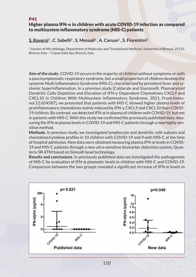
P41Higher plasma IFN-α in children with acute COVID-19 infection as comparedto multisystem inflammatory syndrome (MIS-C) patients
S. Roversi1 , C. Sabelli2 , S. Messali1 , A. Caruso1 , S. Fiorentini1
1 Section of Microbiology, Department of Molecular and Translational Medicine, University of Brescia, 25123,Brescia, Italy - 2 Copan Italia Spa, Brescia, Italy
Aim of the study. COVID-19 occurs in the majority of children without symptoms or witha paucisymptomatic respiratory syndrome, but a small proportion of children develop thesystemic Multi Inflammatory Syndrome (MIS-C), characterized by persistent fever and sy-stemic hyperinflammation. In a previous study [Caldarale and Giacomelli. PlasmacytoidDendritic Cells Depletion and Elevation of IFN-γ Dependent Chemokines CXCL9 andCXCL10 in Children With Multisystem Inflammatory Syndrome. 2021, Front.Immu-nol.12:654587], we presented that patients with MIS-C showed higher plasma levels ofproinflammatory chemokines mainly induced by IFN-γCXCL9 and CXCL10 than COVID-19 children. By contrast, we detected IFN-α in plasma of children with COVID-19, but notin patients with MIS-C. With this study we confirmed the previously published data, mea-suring the IFN-αplasma levels in COVID-19 and MIS-C patients through a new highly sen-sitive method.Methods. In previous study, we investigated lymphocyte and dendritic cells subsets andchemokine/cytokine profiles in 10 children with COVID-19 and 9 with MIS-C at the timeof hospital admission. New data were obtained measuring plasma IFN-α levels in COVID-19 and MIS-C patients through a new ultra-sensitive biomarker detection system, Quan-terix SR-XTM based on Simoa® bead technology.Results and conclusions. In previously published data we investigated the pathogenesisof MIS-C by evaluation of IFN-α plasmatic levels in children with MIS-C and COVID-19.Comparison between the two groups revealed a significant increase of IFN-α levels in
110

COVID-19 children as compared to MIS- C (p=0.021). In contrast, IFN-γwas undetectablein the plasma of both groups. Because plasmacytoid dendritic cells (pDCs) constitute themain source of IFN-α in blood, low levels of IFN-α are associated with low pDC levels inMIS-C subjects. Analysis of mDC and pDC counts revealed a profound depletion of pDCsin patients with MIS-C (p=0.01), as compared to children with COVID-19. Measure-ment of plasma IFN-α levels with a more sensitive method shows a reduction in plasmaticIFN-α levels in MIS-C than COVID-19 patients (p=0.048) and allowed us to confirm thedata previously published: COVID-19 patients present a higher plasma IFN-α levels thanMIS-C patients.
111

P42High IFN-α levels in ruxolitinib-treated Aicardi-Goutieres patient duringSARS-CoV-2 infection: a case report
R. Scaduto1,2, S. Roversi1,2, J. Galli3, M. Cattalini1, R.M. Ferraro4, M. Cortesi1, S. Giliani4, E. Fazzi3, A. Caruso2, R. Badolato1
1 Unit of Pediatrics, Dept of Clinical and Experimental Sciences University of Brescia and Angelo Nocivelli Insti-tute of Molecular Medicine, University of Brescia, ASST Spedali Civili Brescia - 2 Section of Microbiology, Deptof Molecular and Translational Medicine, University of Brescia - 3 Child Neurology and Psichiatry Unit, Universityof Brescia and ASST Spedali Civili di Brescia, Brescia - 4 A. Nocivelli Institute for Molecular Medicine, Universityof Brescia - ASST Spedali Civili di Brescia, Brescia, Italy
We report a case of a five-year-old female affected by Aicardi–Goutières syndrome (AGS)with late- onset of symptoms who was under treatment with ruxolitinib, admitted at ourclinic for COVID-19 infection. The clinical course was regular without any significant com-plication. During SARS-CoV-2 infection there was a significant increase in interferon-α(IFN-α) serum levels compared to basal levels, followed by normalization after SARS-CoV-2 test negativity.Introduction. AGS is a rare monogenic leukodystrophy with pediatric onset. It belongs toa group of conditions called type I interferonopathies, and it is characterized by psycho-motor disturbances, cerebral white matter abnormalities with basal ganglia calcification,cerebral atrophy, chronic cerebrospinal fluid (CSF) lymphocytosis, and elevated levels ofIFN-α. The diagnosis of AGS is usually established in the first year of live in patients withcharacteristic clinical findings and typical abnormalities on cranial radiological imagingand/or by the identification of biallelic pathogenic variant.Clinical Case. We herein report the case of a 5-year-old female affected by AGS, diagnosedwith compound heterozygous mutations in ADAR gene, because of previous Aicardi–Gou-tières syndrome diagnosed in her brother with the same mutations in ADAR gene. She wasinitially treated with intravenous immunoglobulins infusion and subsequently with ruxo-litinib. In January 2021 she developed fever and cough initially treated with amoxicillin.She underwent nasopharyngeal swab with analysis of SARS- CoV-2 by reverse transcrip-tase polymerase chain reaction, which resulted positive. In consideration of the underlyingpathology, she was admitted at our Department. At admission she was in good general cli-nical conditions. Blood pressure and oxygen saturation were normal. Laboratory findingsshowed normal white cell count, Erythrocyte Sedimentation Rate (ERS), and C-ReactiveProtein (CPR). Chest- X-ray showed only a minimal bronchovascular accentuation. Duringthe hospital stay she continued her domiciliary treatment with ruxolitinib, acyclovir andsubcutaneous immunoglobulins. Her clinical course was regular, without fever, with nor-mal respiratory dynamic, and normal saturation levels without oxygen supplementation.Pre-admission IFN-α levels were normal (one month before COVID-19 infection). Duringthe course of the disease IFN-α was significantly increased (Interferon Score 296.01).IFN-α levels returned normal one month after recovery from COVID-19. Recently, apreliminary study has shown that the increase in blood IFN-α levels in COVID-19 patientswere directly associated with improvement in disease severity and greater survival. So wehypothesized that increasing IFN-α serum levels in this patient may be an adequate re-sponse during COVID-19 infection and may have provided a benign clinical course.
112

P43Effects of remdesivir on negativation time in SARS-CoV-2 disease: an openlabel cohort clinical trial - preliminary data
F.S. Serino1, M. Manara1, L. Di Donato1, C. Guarente1, L. Brollo2
1 Department of Infectious Diseases, Hospital of Jesolo, AULSS 4 Veneto Orientale, Jesolo, (Italy) - 2 Chief of theDepartment of Infectious Diseases, Hospital of Jesolo, AULSS 4 Veneto Orientale, Jesolo, (Italy)
Background: Though all the factors (including the timing) that contribute to the progres-sion of patients to severe COVID-19 are not yet fully understood, different panels suggestthe potential benefit of early intervention with an antiviral (Remdesivir) in hospitalizedpatients with COVID-19 Severe Pneumonia. Daily experience on the use of Remdesivir inthis context leads us to hypothesize that the earlier is the treatment (within the 10 daysrequired by the guidelines) the greater will be the probability of a favorable trend in termsof mortality and need for supplemental oxygen.Materials and methods: A total of 24 patients (16 M and 8 F) of different age groups (35%between 30-50 years; 50% between 50-70 years; 15% older than 70 years) hospitalizedfor COVID-19 Pneumonia and following the Remdesivir prescribing criteria according toAIFA, were enrolled. They were divided according to the timing of drug administration wi-thin 5 days (54%) or within 10 days (46%) of the first Nasopharyngeal Swab (NPS) with aPositive output. The NPSs were processed and analyzed with a PCR technique by the LabDepartment of the Covid Hospital of Jesolo. Possible presence of one or more comorbi-dities (e.g. arterial hypertension, COPD, diabetes) were also considered as risk factors fordisease progression (43% had no comorbidities; 45% had 1 comorbidities and 12% had 2or more comorbidities).Results: Clinical improvement and the time (in days) needed for the first negative NPS(from the first positive NPS and from the first given dose of Remdesivir) were used as pa-rameters. We observed (Table 1) that: 76,0% (of the patients who had received the drugwithin 5 days were negative within 3 weeks, most of them between the 2nd and 3rd week(69,2%); 72,7% of those who had received the drug within 10 days were negative within
113
Table 1. Number of negative patients across time

3weeks, only 45,5% between the 2nd and 3rd week. Negative patients at 2-3weeks treatedwithin 5 days vs treated within 10 days P = 0,0538. The remainder were negative for 30days or more.Conclusions: Although within the limits of sample size, patient heterogeneity and themethods used (lack of a comparison population), the advantage of early treatment withRemdesivir (within 5 days from the first positive NPS) could indeed help patients to havea better outcome and a shorter negativization time. It should also be noted that the pre-sence of one or more comorbidities did not significantly affect the negativization time:datain Table 2 show comparable time to patients without comorbidities. These data representa preliminary notice, our investigation is still ongoing. Further clinical trials could be sup-portive in the near future.
114
Table 2. Number of negative patients across time, calculated since the first given Rem-desivir dose and compared with the number of comorbidities

P44Evaluation of a fully closed real time PCR platform for the detection of SARS-CoV-2 in nasopharyngeal swabs: a pilot study
L. Stanziola1, M. Cennamo1, E.A. Molinari1, A. Liotti1, E. La Civita1, M. Fiorenza1, C. De Luca2, L. Vallefuoco1, R. Sorrentino1, T.M. Sorbo3, P. Romano4, E. Montella2,G. Troncone2, D. Terracciano1, G. Portella1
1 Department of Translational Medical Sciences, University of Naples "Federico II", 80131 Naples, Italy - 2 PublicHealth, University of Naples Federico II, Naples, Italy - 3 Division of Nephrology, Department of TranslationalMedical Sciences, School of Medicine, University of Campania "Luigi Vanvitelli", Naples, Italy - 4 Department ofSurgical Sciences Submission and Nephrology; University Federico II of Naples, Naples
Aim of the study: To date, reverse transcriptase PCR (RT-PCR) on nasopharyngeal swabsis the 'gold standard' approach for the diagnosis of COVID-19. The need to develop easyto use, rapid, robust and with minimal hands-on time approaches are warranted. In thissetting, the Idylla SARS-CoV-2 Test may be a valuable option. The aim of our study is toevaluate the analytical and clinical performance of this assay on previously tested SARS-CoV-2 people by conventional RT-PCR based approach in different settings, including ini-tial diagnosis and clinical follow-up. Methods used: To evaluate the sensitivity and specificity of the Idylla SARS-CoV-2 Test,we retrieved 55 nasopharyngeal swabs, previously analysed by a fully validated assay, fromsymptomatic patients or from people who have been in close contact with COVID-19 po-sitive cases. Discordant or high discrepant cases were further analysed by a third techni-que. In addition, a second subset of 14 nasopharyngeal swab samples with uncertainresults (cycle threshold between 37 and 40), by using the fully validated assay, from pa-tients with viral infection beyond day 21, were retrieved. Results and conclusions: Overall, Idylla showed a sensitivity of 93.9% and a specificity of100.0%. In addition, in the additional 14 nasopharyngeal swab samples, only five (35.7%)featured a positive result by the Idylla SARS-CoV-2 Test. We demonstrated that the IdyllaSARS-CoV-2 Test may represent a valid, fast, highly sensitive and specific RT-PCR test forthe identification of SARS-CoV-2 infection.
115

P45 - OC 54Single-dose BNT 162B2 mRNA COVID-19 vaccine significantly boosts neutralizing antibody response in health care workers recovering from asymptomatic or mild natural SARS-CoV-2 infection
I. Vicenti1, F. Gatti2, R. Scaggiante3, A. Boccuto1, D. Zago2, M. Basso2, F. Dragoni1,M. Zazzi1, S.G. Parisi2
1 Department of Medical Biotechnologies, University of Siena, Italy - 2 Department of Molecular Medicine Uni-versity of Padova, Italy - 3 Belluno Hospital, Belluno, Italy
Aim of the study. To investigate the role of post-infection vaccination with BNT162b2mRNA COVID-19 vaccine in a cohort of Italian healthcare workers (HCWs), recoveredfrom asymptomatic or mild natural SARS-CoV-2 infection. Methods. Two-fold serial dilutions of heat-inactivated sera were incubated with 100TCID50 of SARS-CoV-2 virus (lineage B.1) at 37°C for 1 h in 96-well plates. Then, 10,000pre-seeded Vero E6 cells per well (ATCC CRL-1586) were treated with serum-virus mix-tures and incubated at 37°C. After 72 h, cell viability was determined through the com-mercial kit CellTiter-Glo® 2.0 Cell Viability Assay (Promega). The NtAb titer was definedas the reciprocal value of the sample dilution that showed a 50% protection of virus-indu-ced cytopathic effect (ID50). Each run included an uninfected control, an infected controland a known SARS-CoV-2 neutralizing serum. Antibodies with ID50 titers ≥10 were defi-ned as SARS-CoV-2 positive and neutralizing. Statistical analyses were performed usingIBM SPSS Statistics, version 20.Results. A total of 57 Italian HCWs were enrolled, including 41 SARS-CoV-2 infected in-dividuals (median age 45 [34-51] years, male 34.1%) recovering from asymptomatic infec-tion or mild disease and 16 uninfected controls (median age 49 years [34-59], male 31.2%).Sera from previously infected HCWs were collected on the day of first-dose vaccination(T1_inf) and 21±4 days thereafter (T2_inf), whereas sera samples from uninfected controlswere collected on the day of first-dose vaccination (T1_uninf, all confirmed to be negative),20±4 days thereafter (T2_uninf) and then further 20±4 days following the second-dosevaccination (T3_uninf). Previously infected HCW were vaccinated after a median (IQR) of313 (285-322) days since diagnosis. NtAb titres increased significantly at T2_inf with re-spect to T1_inf (median values 1544 [732-2232] vs. 26 [10-88]; p < 0001) and a significantcorrelation between days from diagnosis and NtAb levels at T2_inf (Pearson R2 = 0.395; p= 0.011) but not at T1_inf (Pearson R2 = 0.119; p = 0.502) was found. Similarly, in the pre-viously uninfected control group, there was a significant increase in NtAb titres at T3_uninfwith respect to T2_uninf (median values 183 [111-301] vs. 5 [5-15]; p <0001). Notably,T2_inf NtAb levels (1544 [732-2232]) were significantly higher than uninfected NtAb ti-tres, when measured both at T2_uninf (p <0.0001 for both analyses).Conclusion. The need to vaccinate those who have recovered from natural infection is stillunder debate. In this work, we demonstrated that a single-dose vaccination of people withmild or asymptomatic previous infection is sufficient to significantly boost SARS-CoV-2immunity.
116

P46Monitoring anti SARS-CoV-2 antibody response after BNT162b2 vaccine
G. Anichini, C. Gandolfo, C. Terrosi, G. Gori Savellini, M.G. Cusi
Virology Unit, Department of Medical Biotechnologies, Siena University Hospital, Siena, Italy
Aim: We have designed a prospective study with the aims to explore and monitor the an-tibody response against SARS-CoV-2 virus induced by the COMIRNATY mRNA vaccine ina sample of 178 vaccinated volunteers. Here, we report the interim analysis of the study, including serological results ten andthirty days, and three months (in process) after the second mRNA BNT162b2 vaccineboost among healthcare personnel at S. Maria alle Scotte University Hospital in Siena. Mo-reover, vaccinated sera of 60 subjects were also tested for the presence of neutralizingantibodies against the virus ten days after receiving the second dose of vaccine. Resultswere then compared with neutralizing titers obtained from 38 previously infected subjectsten days after receiving one dose of vaccine.Methods: The humoral immune response of all healthcare workers was evaluated by che-miluminescent (CMIA) and, among them, 60 were also tested by live virus-based neutrali-zation assay. Both IgG and neutralizing titers were also analyzed according to thesymptoms developed after the second vaccine administration.Results: Our results showed a significant decrease of circulating IgG levels over time bet-ween ten and thirty days after vaccination, despite neutralizing titers remained quite sta-ble. Moreover, both IgG and neutralizing titers showed a robust correlation with the gradeof clinical symptoms reported after the second dose administration. Results after threemonths will be processed.Moreover, we observed a significantly lower neutralizing antibody response in naïve pa-tients after complete vaccination compared with those previously infected by SARS-CoV-2 after receiving only a single dose1.These findings provide evidence that after the administration of a single dose of vaccine,humoral response against SARS-CoV-2 in subjects with a past history of SARS-CoV-2 in-fection is greater than the one produced by naïve subjects after receiving two doses ofvaccine.
Reference:1. Anichini G, Terrosi C, Gandolfo C et al. SARS-CoV-2 Antibody Response in Persons with Past Natural
Infection. N Engl J Med. 2021 Apr 14. doi: 10.1056/NEJMc2103825.
117

P47Time course of neutralizing antibody in healthcare workers with mild or asymptomatic COVID-19 infection
A. Boccuto1, F. Gatti2, R. Scaggiante3, E. Moldolo3, D. Zago2, M. Basso2, F. Dragoni1,N. Bartolini1, I. Vicenti1, M. Zazzi1, S. G. Parisi2
1 Department of Medical Biotechnologies, University of Siena, Italy - 2 Department of Molecular Medicine Uni-versity of Padova, Italy - 3 Belluno Hospital, Belluno, Italy
Aim of the study. Development of neutralizing antibody (NtAb) is crucial for protectionfrom SARS-CoV-2 reinfection. The aim of the study was to analyze the time course of NtAbtiters in a cohort of 104 non-hospitalized healthcare workers (HCW) with mild or asym-ptomatic SARS- CoV-2 infection diagnosed after symptoms onset or for contact tracing.Methods. Two-fold serial dilutions of heat-inactivated sera were incubated with 100TCID50 of SARS-CoV-2 virus (lineage B.1) at 37°C for 1 h in 96-well plates. Then, 10,000pre-seeded Vero E6 cells per well (ATCC CRL-1586) were treated with serum-virus mix-tures and incubated at 37°C. After 72h, cell viability was determined through the com-mercial kit Cell-titer Glo 2.0 (Promega). The NtAb titer was defined as the reciprocal valueof the sample dilution that showed a 50% protection of virus-induced cytopathic effect(ID50). Antibodies with ID50 titers ?5 were defined as SARS-CoV-2 positive and neutrali-zing. Statistical analyses were performed using MedCalc® Statistical Software version19.7 (MedCalc Software Ltd). Results. One hundred and four patients (34 males, 70 females) were included in the study.The median time from diagnosis to the first NtAb titration (T1) was 65 (39-86) days. Ove-rall, the median (IQR) NtAb titre in 86 patients was 24.9 ID50 (14.0-68.1) while the remai-ning 18 (17.3%) patients had a negative result. Sixty-seven HCW (24 males, 43 females)had two NtAb measurements (T1 and T2). In this group the median interval between T1and T2 was 140 (120-189) days and the median interval between diagnosis and T2 was223 (172-262) days. No correlation between T2-T1 interval and NtAb titer at T2 (rho=-0.131, p=0.302) and between T2-diagnosis interval and NtAb titer at T2 (rho=0.0194,p=0.8771) was observed. Five (7.5%) and 13 (19.4%) HCW had no detectable NtAb titreat T1 and T2, respectively (p = 0.043). In this group, 4 individuals were negative both at T1and at T2 (median T2-T1 interval was 52 [175-191] days) and 1 was negative only at T1but positive at T2 (17 ID50). Nine of the 62 HCW positive at T1 had undetectable NtAb atT2 after a median interval of 184 (166-194) days: at T1, their median ID50 (9.9, [6.7-14.7])was lower than the ID50 (28.0 [14.6-90.5]) of the 53 HCW who were positive both at T1and at T2 (p=0.0034). Overall, the median NtAb titre decreased significantly from T1 toT2 (24 [11.7-66.0] vs. 17 [7-44.2], p=0.009) and this result was confirmed in the 53 persi-stently positive HCW (28 [14.6-90.5] vs. 27 [12.8-57.1], p=0.037). Conclusion. The strengths of this study include the homogeneity of the population enrolledand the use of authentic virus neutralization with a SARS-CoV-2 isolate circulating in Italyat HCW enrolment. Following mild or asymptomatic infection, most individuals respondwith neutralizing antibody to SARS-CoV-2. In our study, NtAb levels decreased over time,however serum neutralizing activity was retained after a median of 7 months from SARS-CoV-2 diagnosis in 79% of cases. An NtAb cut-off indicative of protection from infection,as well the duration over the time of NtAb titers, remain to be established.
118

P48 - OC 20Analysis of adaptive immune response to SARS-CoV-2 infection and the acquired immune response after COVID-19 vaccination with different assays
F. Bossi1, A. Vendramin1, S. Molon1, E. Ricci2, V. Lucente2, E. De Marchi, G. Spirito2,M. Marino3, L. Grumiro3, M. Gasparin1, K. Bortolozzo1, L. Pasini2, V. Sambri3
1 Eurospital SpA, Trieste - 2 Casa di Cura Giovanni XXIII – Monastier (TV) - Unità Operativa Microbiologia delLaboratorio Unico di Pievesestina dell'Ausl Romagna
Aim of the study. The etiologic agent of COVID-19 is SARS-CoV-2, an RNA virus thatconsists of four kinds of structural proteins: spike glycoproteins (S1 and S2), envelopeprotein (E), membrane protein (M), and nucleocapsid protein (N). The diagnosis ofCOVID-19 is primarily made by detecting viral RNA using RT-qPCR, which is the goldstandard test, or by detecting antigens of SARS-CoV-2. Serological tests, on the otherhand, are useful to evaluate both adaptative immune response to SARS-CoV-2 infectionand acquired immune response after vaccine administration, thereby representing avalid tool for the evaluation of the course of the disease and the immunological protec-tion. This research study aims at investigating both adaptative and acquired immune re-sponse by means of different assays. Method used. The detection of SARS-CoV-2 IgG antibodies was performed on serum orplasma samples collected from: i) healthy donors (n=20); ii) subjects after 1-4 months re-covery from SARSCoV- 2 infection (n=10); iii) subjects before and after PfizermRNABNT162b2 (Comirnaty) vaccine administration at different time points (i.e., 20-21days after the first dose and 8-10 days after second one) (n=17); and iv) subjects at 16 daysafter the second dose of Pfizer Comirnaty (n=100). The assays used were: i) SARS-CoV-2IgG (Eurospital SpA) and SARS-CoV-2 RBD IgG (Eurospital SpA), ELISA tests for the de-tection of IgG antibodies against a nucleocapside and spike proteins or RBD region of S1subunit of SARS-CoV-2, respectively; ii) ACCESS SARS-CoV-2 IgG II (Beckman Coulter Inc.,Ref. C69057), a CLIA test for the detection of IgG antibodies against receptor binding do-main (RBD) of the S1 protein. The efficacy of SARS-CoV-2 IgG neutralizing antibodies wastested on samples collected recovered COVID-19 subjects (n=25) with a Plaque ReductionNeutralization Test (PRNT), which quantifies the titre of neutralizing antibodies, and com-pared to SARS-CoV-2 RBD IgG (Eurospital SpA). Tests were performed following manu-facturer’s instructions for use. Results and conclusions. Samples from recovered COVID-19 subjects analysed with PRNTtest showed a different efficacy of neutralising antibodies in preventing the virus fromcreating plaques, which positively correlated with the level of RBD IgG antibodies detectedwith SARS-CoV-2 RBD IgG test, thereby giving rise to a stratification of the results. Inagreement with these data, samples from vaccinated subjects revealed an antibody re-sponse to RBD region after the first dose, which were higher after the second one. Fromthe analysis with SARS-CoV-2 RBD IgG, which totally agreed with ACCESS SARS-CoV-2IgG II test, the 90% of samples collected from individuals 16 days after receiving the seconddose of vaccine displayed a great amount of RBD IgG antibodies (Index > 0.8) which cor-related with a high PNRT titre, according to the stratification of the results observed onrecovered COVID-19 subjects. However, the remaining 10% of samples displayed a nega-
119

tive result (Index 0.8), but higher that the Index calculated on healthy donors (e.g., > 0.5).However, in these samples IgG antibodies against total N and S proteins were detectedwith SARS-CoV-2 IgG test. Overall these data point out a strong correlation betweenSARS- CoV-2 RBD IgG and PRNT tests results, showing a stratification of the efficacy ofneutralizing antibodies. Moreover, it emerged that patients who recovered from COVID-19 produce an immune response against SARS- CoV-2 that is higher for N protein detectingantibodies, instead of RBD region.
120

P49 Antibody response to COVID-19 vaccination in healthcare workers of Microbiology Unit of "S. Maria della Misericordia" Hospital
S. Bozza1,2, A. Graziani 2, A. Mencacci1,2, B. Camilloni1,2
1 Microbiology Unit of S. Maria della Misericordia Hospital, Perugia - 2 Department of Medicine and Surgery, Uni-versity of Perugia
Aim: SARS CoV2 coronavirus disease (COVID-19) is an infectious disease caused by a re-cently discovered Coronavirus (SARS-CoV-2), a new strain that had not previously beenidentified in humans. The presence of IgG antibodies directed against SARS-CoV-2 is in-dicative of an immune response to the infection; however, it is not yet well known whetherthe presence of these antibodies provides lasting protective immunity and how long afterinfection or vaccination, they remain detectable. The transmembrane glycoprotein S ofthe virus forms 'trimers' that protrude from the viral surface, and is the main target ofneutralizing antibodies and, therefore, fundamental in the design of vaccines [Walls AC,et al. Structure, Function, and Antigenicity of the SARS-CoV-2 Spike Glycoprotein. Cell183(6);2020]. The recent availability of m-RNA vaccines (Pfizer BionTech & Moderna)and Adenovirus vector vaccines (AstraZeneca & Janssen J&J), has started in Italy a massvaccination campaign. The first to be vaccinated were the healthcare workers (HCWs) ofthe COVID units. The aim of our study was to analyze the kinetics of the vaccine-inducedantibody response in HCW working in the Microbiology Unit of S. Maria della Misericor-dia Hospital (Perugia).Methods used: This study involved 40 HCWs who received the first dose of Pfizer/BioN-Tech (95%) or Moderna (5%) vaccine between December 27, 2020 and January 31, 2021.Sera samples were drawn before vaccination (T0), 21 days after the first vaccine dose (T1),7 days (T2) and 30 days after the second vaccine dose (T3). The sera were analyzed by LIAI-SON® SARS-CoV-2 TrimericS IgG test, an indirect chemiluminescence immunoassay(CLIA). The results were expressed in BAU/mL (Binding Antibody Units/mL) calculatedwith respect to the first international standard WHO 20/136 [First WHO International Stan-dard for anti-SARS-CoV-2 Immunoglobulin (Human). NIBSC code: 20/136. Instructions for Use(Version 2.0, 17/12/2020)]. Considered a cutoff = 33.8 BAU/mL, the presence of high LIAI-SON® values (ie ≥ 520 BAU/mL) was considered a sign of a good antibody response be-cause it has been shown that this value overlaps with high neutralizing antibodyconcentration (ie ≥ 1:80). Results and Conclusions: Most participants were female (77.5%) and the mean age was45 years (range 25-68). Before the vaccination, no HCW had antibodies to SARS-CoV-2.TrimericS IgG geometric mean titers (GMTs) increased rapidly after the first vaccination,with seroconversion in all participants by day 21. Four people (20%) had an antibody value≥ 520 BAU/mL. The effect of the second vaccine dose, evaluated at 7 and 30 days aftervaccination, showed a significant increase in the GMT values (p<0.01) and in the numberof subjects with antibodies ≥ 520 BAU/mL (97.5% at day 7 and 95.1% at day 30). Compa-
121

ring the results obtained at T2 and T3, 22 HCWs (55.0%) show a decrease in the antibodyconcentration with at least a halving of the values. In the remaining 22% the amount ofTrimericS IgG is stable or higher. In conclusion, the vaccines induced anti-SARS-CoV-2 im-mune response in all HCWs. During the study period, characterized by a high circulationof P.1 variant in Umbria and in particular within the Perugia hospital, no cases of COVID-19 infection were recorded in the 40 vaccinated HCWs. To assess the durability of the an-tibody response, the analysis of a further sera sample (3 months after the second vaccinedose) is planned.
122

P50Neutralization of SARS-CoV-2 variants by convalescent and post-vaccineserum
A. Cara1, A. Rosellini1, P. Villa1, C. Dolfa1, R. Fonnesu1, F. Maggi2, M.L. Vatteroni1,P. Mazzetti1, M.Pistello1
1 Department of Translational Research and New Technologies in Medicine and Surgery, OU Virology, AziendaOspedaliero-Universitaria Pisana, , Pisa, Italy - 2 Department of Medicine and Surgery, University of Insubria,Varese, Italy
Aim of the study. The pandemic caused by SARS-CoV-2 is a public health emergency of in-ternational concern.Neutralizing antibodies (NAbs) against SARS-CoV-2 have been considered therapeuticagents for the treatment of coronavirus disease 2019 (COVID-19) so far. Nevertheless,the emergence and rapid spreading of viral variants worldwide may potentially limit NAbstherapy. This study aims to evaluate the neutralizing activity of serum collected from vac-cinated (Pfizer) and convalescent donors against different variants of SARS-CoV-2.Methods used. Serum neutralization test was performed in microtiter plates and takes 3days to complete and was based on the cytopathic effect (CPE). Different clinical isolatesof SARS-CoV 2 were used to detect the neutralization of the virus: the original and theBrasilian, English, Nigerian, and Sud-African variants. Results and conclusions. The results indicate that NAbs titers against wild-type SARS-CoV-2 are generally higher in plasma from vaccinated subjects compared to convalescents.The neutralization power of both vaccinated and convalescent plasmas against Brasilian,English, Nigerian, and Sud-African SARS-CoV-2 variants is also discussed.
123

P51Dynamics of SARS-CoV-2 neutralizing antibody response among infected healthcare workers
S. Chandel1, G. Griffante1, V. Caneparo2, D. Ferrante1, S. Bortoluzzi, D. Cappello1,V. Bettio, C. Aleni, L. Scotti1, C. Airoldi1, S. Esposito, D. Cappello1, G. Gazzola, M. De Andrea2, C. Borgogna1, F. Faggiano1, M. Gariglio1
*UNIRSA group - 1 Department of Translational Medicine, University of Eastern Piedmont, Novara 28100, Italy- 2 CAAD Center for Translational Research on Autoimmune and Allergic Disease, University of Eastern Piedmont,Novara 28100, Italy
Rational. Understanding the nature of immunity following mild/asymptomatic infection withSARS-CoV-2 is crucial to controlling the pandemic . It is well known that neutralizing antibodyis a very strong correlate of protection, and data on the extent of the humoral response ge-nerated during SARS-CoV-2 infection among patients with different disease severity, its ef-fectiveness in terms of neutralization of the infection, and its longevity, are needed. Materials and methods. Study design. A cohort study of subjects (n=100 ≥18 years of age)with a PCR-documented infection by SARS-CoV-2, mostly healthcare workers (HCW) ofthe University-Hospital in Novara (66%), proceeded with a 10-month follow-up after re-covery (n=71). Among these, 36 subjects received mRNA-based vaccination during thefollow up period. Laboratory analysis: Sera from this study cohort were tested for the pre-sence of anti-SARS-CoV-2 RBD and Spike (S) IgG antibodies using Kantaro QuantitativeSARS-CoV-2 IgG Antibody RUO kit (REF# DSR200, New York, USA). Serum samples of thepositive patients were then tested for the presence of neutralizing antibodies (Nab) usinga SARS-CoV-2 S protein-pseudotyped vesicular stomatitis virus (VSV) vector-based neu-tralization assay (kindly provided by Sean Whelan, USA). Statistical analysis: descriptiveanalysis of the immune response in the study group is being studied with longitudinal mo-dels of multilevel analysis at T1 (approximately 3 to 5 weeks after healing considering T0),and T2 (9 to 11 months after T1). Results and conclusion. The infection clinical presentation was symptomatic in 84% of casesof which 10% were hospitalized, but none in intensive care unit (ICU). At T1, we found that7% (n=7) were negative for anti-SARS-CoV-2 Abs by ELISA, while 93% (n=93) were foundpositive for both anti-SARS-CoV-2 RBD and S Abs. With regard to the neutralizing activity,a significant correlation was found between high anti-Spike Ab titers and high neutralizingtiters (NT) while low Ab titers correlated with low NT. Consistent with previous reports,those patients who were hospitalized, therefore with a more severe outcome, showed higherNT titers when compared to those who developed mild disease or remained asymptomatic.On a follow-up at T2, 71 subjects were tested for the presence of anti-SARS-CoV-2 antibo-dies by ELISA, while we are currently performing the neutralization assays. Among the vac-cinated subjects, 35 out of 36 showed increased anti-S Ab titers when compared to the T1dosage. Among non-vaccinated subjects, those with high anti-S Ab levels (n=12, 34%) at T1still displayed high anti-S Ab titers at T2. As expected, those with low Ab titers (n=15, 42%)showed very low anti-S Ab titers at T2. A decrease in anti-S Ab levels was seen among 8 (22%)of non-vaccinated subjects from T1 to T2, while an increase was seen in one individual fromT1 to T2 (3%). Collectively, our findings demonstrate the high specificity of our assays, whichmay help understanding the quality or duration of the antibody response upon SARS-CoV-2 infection and determining the effectiveness of potential vaccines.
124

P52The IgA immune response in COVID-19 patients and vaccines: a neglected but critical aspect of SARS-CoV-2 infection
F. De Marco1, L. de Latouliere1, L. Sorrentino1,5, C. Mandoj2, L. Conti2, M. De Rienzo3, M.L. Foddai3, C. Castilletti6, M.R. Capobianchi6, G. Ciliberto4
1 Department of Research, Technological Innovation & Advanced Diagnostics - 2 Department Clinical Pathologyand Cancer Biobank - 3 Immunohematology and Transfusional Medicine Unit - 4 Scientific Director, the “ReginaElena” National Cancer Institute - IRCCS, Rome, Italy - 5 Dept of molecular medicine - Laboratory of Virology“Sapienza” Rome University - Italy - 6 Virology and Biosecurity Laboratories, National Institute for Infectious Di-seases "Lazzaro Spallanzani" IRCCS, Rome, Italy
Aim of the study: In the effort to counter-act the current COVID-19 pandemic a great at-tention has been posed on the study of the humoral immune response to the SARS-CoV-2infection. Despite a rather large amount of data have been rapidly generated a number offundamental issues remain poorly known, namely: the kinetics of Ab response, their valueas predictors of clinical evolution and outcome and the duration of sterilizing immunity.Within this scenario comparatively few works have been dedicated to the role of the IgAthis Isotype is expected to play a crucial role in any kind of air transmitted viral infection. Methods used: A total of 99 patients with confirmed SARS-CoV-2 infection and 275 vac-cinees were enrolled. Their serial serum samples were collected at different times sinceclinical symptoms onset or first vaccine inoculation. Total SARS-CoV-2 Anti S + Anti N spe-cific IgM and IgG were assayed by the SNIBE-MAGLUMI (CLIA) assay. Their RBD/ACE2 bin-ding inhibiting titres were measured both by the COVID-19 Spike-ACE2 binding assay kit(RayBiotech Inc) and by the SARS-CoV-2 Inhibitor Screening Kit (Adipogen). On a selectednumber of cases the direct Viral Infection Inhibiting Titre was also evaluated. The specificAnti S IgA titres were evaluated by the RecombivirusTM Human Anti-SARS-CoV-2 Virus(COVID-19) Spike 1 IgA ELISA Kit (Alpha Diagnostic International). Data analyses and gra-phics were performed with the PRISM-6 software (GraphPad Inc). Results: The RBD/ACE2 Binding assay kits is simple and convenient and can be used to ge-nerate quantitative results. Their use in the clinical setting might provide a swift, robustand convenient way to assess the neutralizing activity of sera without the limitations andthe safety concerns linked to the labour intensive, time consuming method for direct viralinfection Inhibition assay. At any time RBD/ACE2 binding inhibition titre does appear tocorrelated with the total Anti COVID Ab titres. Anti S1 specific serum IgA are easily de-tected in sera by commercial assay kits. The IgA titres are generally higher than that ofIgM, appear to persist quite longer and to correlate with the neutralizing titre both in con-valescent patients and in healthy vaccinees. IgA appear as a promising adjunct to the toolsfor COVID patients’ evaluation. Both anti S IgA and anti RBD titres are sharply higher inBNT162b2 vaccine recipients than in convalescent patients.
125

P53Mucosal humoral response in BNT162B2 COVID-19 vaccinated subjects
L. Azzi1, M. Shallak2, D. Dalla Gasperina3, F. Maggi4, R.S. Accolla2, G. Forlani2
1 Unit of Oral Medicine and Pathology, ASST dei Sette Laghi, Department of Medicine and Surgery, University ofInsubria, Varese, Italy - 2 Laboratories of General Pathology and Immunology “Giovanna Tosi”, Department ofMedicine and Surgery, University of Insubria, Varese, Italy - 3 Unit of Infectious and Tropical Diseases, ASST deiSette Laghi, Department of Medicine and Surgery, University of Insubria, Varese, Italy - 4 Laboratory of ClinicalMicrobiology, ASST dei Sette Laghi, Department of Medicine and Surgery, University of Insubria, Varese, Italy
Aim of the Study: Mucosal immunity, including secretory IgA (sIgA), plays an importantrole in early defenses against respiratory pathogens, such as SARS-CoV-2 infection. MostRecent reports have focussed on the efficacy of BNT162b2 vaccine in the induction ofvirus specific IgG-neutralizing/sterilizing antibodies. However, cases of reinfected healthcare vaccinated workers are increasingly frequent, suggesting that the specific immuneresponse induced by the vaccination may not be sufficient to fully protect from mucosalviral infection. In order to evaluate the mucosal immune response generated after vacci-nation we analyzed the titer and the neutralization capacity of both IgG and IgA in seraand saliva of BNT162b2 vaccinated subjects. Methods: The titer and the neutralizing capacity of SARS-CoV-2-specific IgG and IgA in seraand saliva of sixty healthcare worker volunteers of ASST dei Sette Laghi, who received twoinjection of BNT162b2 mRNA vaccina, were analyzed by using a quantitative ELISA assayspecific for SARS-CoV-2 spike protein subunit S1and a viral neutralization assay, respectively.Interestingly 6% of the enrolled subjects was previously exposed to virus. Samples were col-lected at the day of the first dose (T0), two weeks after the first injection (T1) and two weeksafter the second injection (T2). The CLIA serological assay on serum was used as a standarddiagnostic reference for the evaluation of IgG neutralizing antibodies in serum.Results and discussion: 88% of recruited subjects developed IgG neutralizing antibodiesin serum after the first dose, and 100% showed serological positivity by CLIA after the se-cond dose. The amount of total IgG in serum positively correlated with the level of neu-tralizing IgG antibodies detected by CLIA. In saliva we could measure a significant amountof IgG only after the second administration of the vaccine. Interestingly in the majority ofthe previously exposed subjects we observed high IgG levels both in serum and saliva afterthe first dose, reinforcing the idea that in this group a single vaccination is sufficient to pro-tect from the infection. As far as the IgA, we were able to detect a significant amount ofserum IgA in 60% and 95% of vaccinated subjects after the first and second injection, re-spectively. In saliva the IgA were present in 40% and 50% of subjects after the first andthe second administration of the vaccine, respectively. The presence of IgA in saliva afterthe first vaccination was independent from previous exposure to the virus. After the se-cond vaccination only 25% of vaccinated people presented neutralizing antibodies in sa-liva, being included in this group the majority of individuals previously exposed to theinfection. Taken together, these results indicate that although half of vaccinated peopledisplay specific IgA antibodies in oral mucosa, these antibodies are relatively poor at neu-tralizing the virus highlighting the weak efficacy of the BNT162b2 mRNA vaccine to induceIgA-driven mucosal immunity.
126

P54BNT162b2 efficacy: results from healthcare workers vaccination. The renaissance study
O.M. Gagliardi1, A. Pani2, V. Cento2, F. Di Ruscio3, R. Gibilisco3, A. Zampiero4, D. Campisi4, A. Rimondini1, M. Senatore1, P.A. Schenardi1, S. Agliardi1, T. Conti1,G. Gazzaniga1, A. Schianchi1, F. Scaglione2-4
1 Postgraduate school of Clinical Pharmacology and Toxicology - 2 Department of Oncology and Hemato-Onco-logy, Università degli Studi di Milano, Milan, Italy - 3 Postgraduate school of Microbiology and Virology, Universitàdegli Studi di Milano, Milan, Italy - 4 Chemical-Clinical and Microbiological Analyses, ASST Grande Ospedale Me-tropolitano Niguarda, Milan, Italy
Aim of the study. The BNT162b2 vaccine demonstrated 95% of efficacy at preventing se-vere COVID-19 versus placebo. However, the approval process required a short time bynecessity, and we lack data on special populations (1). In addition, it is still unknown whe-ther BNT162b2 reduces transmission of SARS CoV-2. To define immunogenicity ofBNT162b2 and his efficacy to reducing transmission of SARS CoV-2, we evaluated anti-body response in the first vaccinated individuals in Italy: the healthcare workers (HWs). Methods. As a measure of immunogenicity of BNT162b2 we evaluated anti-Trimeric Spikeglycoprotein IgG titer after 14 days from the second dose of BNT162b2 using a quantita-tive chemiluminescent assay (Diasorin). We excluded HWs with an incomplete vaccinationcourse or a previous molecular, serological or antigenic test resulting positive for SARSCoV2 infection. In addition, to rule out prior exposure to SARS-CoV2 we also screenedHWs for anti-Nucleocapsid (N) antibodies, through an electrochemiluminescence immu-noassay (Roche). To determine the efficacy of BNT162b2 to reducing transmission of SARSCoV2 we compared the pre-vaccine and the post-vaccine trends of infected HWs until27th March 2021.Results and conclusions. 5,023 HWs receiving the first dose of vaccine were included inthe study; 144 participants for incomplete vaccination and 871 of them for known priorexposure to SARS-CoV2 were excluded. 163 of HWs with no previous history of labora-tory-confirmed COVID-19 resulted positive to anti-N (95% CI: 5.4%-7.4%). The final po-pulation consisted of 2569 HWs; of these, only 4 had a negative antibody response (0.16%prevalence; 95% CI: 0.04%-0.40%) %). Notably, these non-responders were assuming im-munosuppressant drugs, especially mycophenolate; 23 participants that referred to be im-munocompromised had a measurable antibody response. 3,4% of participants had titersbelow 1000 BAU/mL (n/N=87/2569, 3.4%), 9,8% from 1000 to 1500 BAU/mL(n/N=251/2569), 19,2% from 1500 to 2000 (n/N=494/2569), and 67,5% above 2000(n/N=1733/2569). The female gender was associated with a significantly higher probabi-lity of having an anti-Spike titer above 800 than male gender (64.5% [1044/1618] vs. 58.3%[410/703]; p=0.005). Of note, this correlation was found also adjusting for age groups (OR[95%CI]: 1.275 [1.062-1.531]; p=0.009). Before the beginning of vaccinations in Italy theincidence of SARS CoV-2 infections within our HWs was steadily above 20%. Up to 27thMarch 2021, we found only one individual with a positive SARS CoV2 RT-PCR result withinthe vaccinated HWs, even if poorly symptomatic. Additionally, 6 vaccinated individualswith a weak-positive SARS-CoV-2 RT-PCR resulted negative to digital PCR analysis. None
127

of our vaccinated HWs developed a moderate or a severe form of COVID-19 (100%). Con-cluding, in our study in vaccinated healthcare workers BNT162b2 vaccine revealed highinduction of immunogenicity and a strong efficacy to reducing transmission of COVID-19.
Bibliography
1) Polack FP, Thomas SJ, Kitchin N, Absalon J, Gurtman A, Lockhart S, Perez JL, Pérez Marc G, MoreiraED, Zerbini C, Bailey R, Swanson KA, Roychoudhury S, Koury K, Li P, Kalina WV, Cooper D, Frenck RWJr, Hammitt LL, Türeci Ö, Nell H, Schaefer A, Ünal S, Tresnan DB, Mather S, Dormitzer PR, Şahin U, Jan-sen KU, Gruber WC; C4591001 Clinical Trial Group. Safety and Efficacy of the BNT162b2 mRNACovid-19 Vaccine. N Engl J Med. 2020 Dec 31;383(27):2603-2615. doi: 10.1056/NEJMoa2034577.
Epub 2020 Dec 10. PMID: 33301246; PMCID: PMC7745181.
128

P55 - OC 38SARS-CoV-2 N protein target TRIM-25-mediated RIG-I activation to suppressinnate immunity
G. Gori Savellini, G. Anichini, C. Gandolfo, M.G. Cusi
Department of Medical Biotechnologies, University of Siena, Siena, Italy
Aim of the study. During the infection by the novel SARS-CoV-2 virus, a weak productionof INF-β, along with an exacerbated release of pro-inflammatory cytokines, has been re-ported. SARS-CoV-2 encodes several proteins able to counteract the host immune system,which is believed to be one of the most important features contributing, at least in part, tothe viral pathogenesis and development of a severe clinical picture (Blanco-Melo et al.,2020; Hadjadj et al., 2020; Yuen et al., 2020). Previous reports demonstrated that SARS-CoV-2 N protein efficiently suppresses INF-β production by interacting with RIG-I (Oh etal., 2021). In the present study, we better characterized the mechanism by which the SARS-CoV-2 N counteracts INF-β secretion. In particular, we focused on the involvement of theN protein on TRIM25 activity and its effects on RIG-I activation.Methods. Plasmids. The N SARS-CoV-2 coding gene (nt 28218-29477) and the HA-taggedTRIM25 SPRY domain were cloned by standard procedure in pcDNA4HisMax-C plasmid(Life Technologies) in frame with the 6xHis tag and in pEF-Bos, respectively. Other plasmidsused in the experiments were kindly provided by colleagues or purchased. Immunopreci-pitation. Cell lysates from co-transfected cells were used for immunoprecipitation withanti-HA magnetic beads. For RIG-I ubiquitination detection, cell samples were lysed in 1%SDS by boiling and then diluted to 0.1% SDS final concentration with RIPA buffer. Proteinswere captured with anti-FLAG M2 magnetic beads. Proteins were resolved by SDS-PAGEand probed by immunoblotting with selected antibodies. Luciferase reporter assay. Lenti-X 293T cells were transfected with indicated plasmids. Thirty-six hours post-transfection,cells were stimulated with poly(I:C) and collected after additional 12h. Luciferase activitieswere measured using the Dual Luciferase reporter assay reagent (Promega), according tothe manufacturer's instructions. Results are given as mean values of several experiments± standard deviations (SD).Results and conclusions. In the present study, we demonstrated that the SARS-CoV-2 Nprotein significantly impaired the RIG-I signaling pathway leading to IFN-βproduction. Byluciferase reporter assay, a marked decrease in both IFN-β- or NF-kB-promoter activationwas noted when the N protein was co-expressed along with RIG-I or MDA-5. TRIM25, anE3 ubiquitin ligase involved in full RIG-I activation, was previously described to be targetedby the nucleoprotein of the SARS-CoV virus. For this reason, we next investigated the in-terplay between SARS-CoV-2 N protein and TRIM25. Interestingly, by reporter gene assay,we observed that TRIM25-mediated RIG-I activation was significantly reduced when theN protein was ectopically expressed. On the contrary, no activity was observed with re-spect to Riplet, another important E3 ligase active on RIG-I. Taken together, these data sug-gested a direct and specific involvement of the N protein in the TRIM25-RIG-I signalingcascade. Furthermore, as demonstrated by co-immunoprecipitation experiments, we re-ported a pronounced affinity of the N protein for TRIM25, in particular for its SPRY do-main, harboring the RNA-binding activity necessary for its self-activation. Moreover, the
129

SARS-CoV-2 N protein was found associated to a multiprotein complex containing bothRIG-I and TRIM25. Thus, we hypnotized that the N protein intercalates between RIG-I andTRIM25, spatially limiting RIG-I ubiquitination and activation by TRIM25. Indeed, a reduc-tion in K63-moiety poly-ubiquitination of RIG-I was observed when the N protein was co-expressed. Here, we described new findings regarding the interplay between SARS-CoV-2and the IFN system, filling some gaps for a better knowledge of the molecular mechanismsassociated to the COVID-19 disease.
130

P56Humoral response assessment in BNT162b2 vaccinated healthcare workers:an assay comparison
D. Mileto1, M. Cutrera1, G. Gagliardi1, A. Rizzo1, A. Gigantiello1, A. Mancon1, M. Bianchi1, F. De Poli1, G. Giacomel1, I. Burgo2, M. Longo1, S. Grosso1, V. Micheli1,G. Rizzardini3, M.R. Gismondo1, and A. Lombardi1
1 Laboratory of Clinical Microbiology, Virology and Bioemergencies, ASST Fatebenefratelli Sacco, L. Sacco Uni-versity Hospital, Milan - 2 Hematology and Transfusion Medicine, ASST Fatebenefratelli Sacco, L. Sacco UniversityHospital, Milan - 3 Division of Infectious Diseases, ASST Fatebenefratelli Sacco, L. Sacco University Hospital,Milan
Aim of the study: On 27th December 2020 the vaccination with mRNA BNT162b2 (Co-mirnaty) started in Italy. We evaluated the antibody development after first and seconddose according to the vaccination schedule in healthcare workers naïve at SARS-CoV-2 in-fection comparing four chemiluminescence immunoassays (CLIA) and plaque reductionneutralization test (PRNT) results.Methods used: Serum samples of 37 enrolled subjects, median age 40 (range 24 - 65) years,were collected for each following time point: T0, 1 day before I vaccination dose; T1 andT2, 10 and 20 days after I dose respectively; T3, T4 and T5, 10, 20 and 30 days after II doserespectively. Each time point was analyzed using different CLIA assays: LIAISON® SARS-
131
Fig. 1. Anti-SARS-CoV-2 response measured by CLIA assays and neutralizing antibodies titre anti-SARS-CoV-2 measured by PRNT at baseline (T0), 10 days after I dose (T1), 20 days after I dose (T2), 10 days afterII dose (T3), 20 days after II dose (T4) and 30 days after II dose (T5).

CoV-2 S1/S2 IgG (DiaSorin Inc.) to measure antibodies against the SARS-CoV-2 nativeS1/S2 proteins, while iFlash-2019-nCoV NAb (Shenzhen YHLO Biotech Co), LIAISON®SARS-CoV-2 TrimericS IgG (DiaSorin Inc) and Elecsys Anti-SARS-CoV-2 S (Roche Diagno-stics Rotkreuz, Switzerland) to quantify the specific RBD-binding antibodies. Furthermore,the neutralizing antibody serum titre was determined by PRNT for all samples at each timepoint using the European strain (D614G) and the T5 collected serum of 10 subjects weretested against the SARS-CoV-2 UK strain (501Y.V1, lineage B.1.1.7) too.Results and conclusion: All enrolled subjects developed antibodies 10 days after II doseaccording to all methods: the CLIA assays showed the highest level of antibody to S1/S2and RBD at T3 while the highest level of neutralizing antibody titre is reached at T4 accor-ding to PRNT (median 1:320; IQR 1:160-1:320) (Fig.1).Regarding the neutralization titers against B.1.1.7, 2/10 (20%) of tested serum showed aslight decline: 1:320 for EU vs 1:160 for UK. Further evaluations on antibody titres andvaccine efficacy against SARS-CoV-2 variants will be conducted by our ongoing study.
132

P57Vaccination against COVID-19 prevents disease severity and infection in a hospital in Northern Italy
C. Belgiovine1, I. Dambrosio2, F. Frattolillo2, S. Sancini2, A. Porcella2, N. Nicodemo2, A. Garegnani2, R. Lacavalla2, C. Vidello2, M. Monari1
1 Humanitas Clinical and Research Center-IRCCS, Via Manzoni 56, 20089 Rozzano Milan, Italy - 2 Mater DominiHumanitas, Via Gerenzano 2 Castellanza, Varese
Background: Recently COVID-19 vaccines based on mRNA has been approved, and theschedule for these vaccines require a first jab followed by recall immunization. The Italianimmunization program identified health care workers as a priority. There is still no certainevidence about their efficacy in containing the infection, but only on their ability to avoidsever COVID-19 disease. Aim: this study has a dual purpose: both to evaluate the efficacy of this vaccination in pre-venting COVID infection and disease severity in vaccinated health care workers of an Ita-lian Hospital (Humanitas Mater Domini, Castellanza-Varese).Methods: All samples are tested routinely for sourvelliance or in case of sympthoms. Themolecular method for the detection is: SARS-COV2: Alliplex, SARS CoV-2, Seegene.Results: In our hospital, 479 health care workers, between 1-01.21 to 8.2.21, were vacci-nated, 94 of them had already contracted SARS-CoV2. During the screening of health-careworkers, between 11.1.21 to 11.4.21, of the 385 who had never contracted the virus, 9 in-fected subjects were found.
Table1: result of infection pre and post vaccination.
Nr. Infection Re-infection pauci-of helth Past 1° dose 2° dose post post symptomatic asyntomatic
care infection vaccine vaccine vaccination vaccinationworkers
1 x x x x 15 x x 3 23 x x x 3
The mean duration of positivity was 16.5 days for these who received a single dose of thevaccine and 10 days for those who completed the vaccine schedule. No infected subjectsshowed any symptoms. Discussion: The study highlights how the vaccine is able to prevent the infection of the disease(99% efficacy), and also severe symptoms of COVID-19, regardless of the number of dosesmade by health-care professionals. In our observation, between 9 infection post vaccination6 are asymptomatic and only 3 paucisymptomatic (Table 1). So all health workers could returnto work in 14 days despite in case of infection without vaccination where the average time ofabsence was estimated between 21 to 45 days with some days of hospitalization. Further stu-dies could be made on the infectious capacity of the virus in vaccinated subjects, trying to in-vestigate the differences between those vaccinated with a single dose or with both.Conclusion. The vaccine shows 99% effectiveness in preventing infection and 100% in pre-venting COVID-19 disease in Mater Domini hospital.
133

P58Impaired priming of SARS-CoV-2 specific naïve CD8+ T cells in older subjects
E. Gallerani1, D. Proietto1, B. Dallan1, M. Campagnaro1, S. Pacifico2, V. Albanese2,E. Marzola2, P. Marconi1, A. Caputo1, V. Appay3, R. Gavioli1 and F. Nicoli1
1 Laboratory of Biochemistry, Immunology and Microbiology (BIM), Department of Chemical, Pharmaceuticaland Agricultural Sciences, University of Ferrara, 44121 Ferrara, Italy - 2 Department of Chemical, Pharmaceuticaland Agricultural Sciences, University of Ferrara, 44121 Ferrara, Italy - 3 CNRS UMR 5164, ImmunoConcEpT, Uni-versité de Bordeaux, 33000 Bordeaux, France
Advanced age is associated with severe symptoms and death upon SARS-CoV-2 infection.Virus-specific CD8+ T-cell responses have shown to be protective toward critical COVID-19 manifestations, suggesting that suboptimal cellular immunity may contribute to theage-pattern of the disease. The induction of a CD8+ T-cell response against an emergingpathogen like SARS-CoV-2 relies on the activation of naïve T cells. To investigate whetherthe primary CD8+ T-cell response against this virus is defective in advanced age, we usedan in vitro approach to prime SARS-CoV-2-specific naïve CD8+ T cells from healthy, unex-posed donors of different age groups. Compared to younger adults, older individuals di-splay a poor SARS-CoV-2-specific T-cell priming capacity in terms of both magnitude andquality of the response. In addition, older subjects recognize a lower number of epitopes.Our results implicate that immune aging is associated with altered primary SARS-CoV-2-specific CD8+ T-cell responses.
134
P59Neutralizing antibodies responses against SARS-CoV-2 in sardinian cohort group
G. Sanna1, A. Marongiu1, 2, G. Franci3, M. Galdiero4, C. Piras1, G. Pala1, V. Palmas1,R. Littera5, M. Vacca5, R. Jammoul5, A. Perra6, F. Sedda6, M. Campagna7, F. Pes7,D. Firinu7, M. Erbi7, M. Secci7, F. Meloni7, L. Chessa7, A. Manzin1
1 Department of Biomedical Sciences, University of Cagliari, Cittadella Universitaria, 09042, Monserrato, Cagliari,Italy; Giuseppina Sanna; email: [email protected] - 2 Department of Biomedical Sciences, University of Sassari,Sassari 07100, Italy - 3 Department of Medicine, Surgery and Dentistry "Scuola Medica Salernitana", Universityof Salerno, 84081 Baronissi, Italy - 4 Department of Experimental Medicine, University of Campania Luigi Van-vitelli, 80138 Naples, Italy - 5Department of Internal Medical Sciences, Medical Genetics, "R. Binaghi" Hospital,University of Cagliari, Italy - 6 Department of Biomedical Sciences, Unit of Oncology and Molecular Pathology,University of Cagliari, Cittadella Universitaria, 09042, Monserrato, Cagliari, Italy - 7 Department of MedicalSciences and Public Health, University of Cagliari, Cittadella Universitaria, 09042, Monserrato, Cagliari, Italy
Severe acute respiratory syndrome-related coronavirus 2 (SARS-CoV-2), the etiologicalagent of COVID-19, determined over 132 million cases of infections and almost 3 milliondeaths worldwide.The pandemic has called for science, technology and innovation to provide solutions, anddue to an incredible scientific and financial global effort several prophylactic and thera-peutic apparatus, such as monoclonal antibodies, and vaccines have been developed in lessthan one year to fight this emergence. After SARS-CoV-2 infection, serum neutralizing an-tibodies are produced by B cells, and studies on virus-neutralizing antibodies kinetics arepivotal. The process of protective immunity, and the duration of this kind of protectionagainst COVID-19, remain to be clarified. Several authors have described antibodies re-sponses in COVID-19 patients, but the pending question is the duration of these responses.Some of them report rapid declining of antibodies, while others detected the lost of neu-tralizing antibodies by around 30-60 days after the beginning of illness. All this data haveimportant implication for the duration of protective immunity from reinfection, and thesuccess of vaccination in prevention from reinfection and disease.We tested 100 sera from 3 groups of individuals, some of them providing multiple sequen-tial sera (1- healthy, no previous CoV2 infected, vaccinated, 2- healthy, previous CoV2 in-fected, vaccinated, 3- healed, (previous CoV2 infected), not vaccinated) to assess thekinetics of antibodies neutralizing activity. We found that SARS-CoV-2 infection elicits mo-derate neutralizing antibody activity in most individuals; nor age and gender appear to in-fluence antibodies responses, neither Body Mass Index (BMI) is correlated with antibodiesproduction. BNT162b2 vaccine, when administered in two doses, results able to inducehigh antibodies titers endowed with potent neutralizing activity against bare SARS-CoV-2 in in vitro neutralizing assay. The cohort group is increasing in order to implement ourdatabase.
135

P60 - OC 10Serosurvey in BNT162B2 vaccine-elicited neutralizing antibodies against authentic SARS-CoV-2 variants
A. Zani, F. Caccuri, S. Messali, C. Bonfanti, A. Caruso
Section of Microbiology, Department of Molecular and Translational Medicine, University of Brescia, 25123 Bre-scia, Italy
Introduction: The ongoing outbreak of coronavirus disease (COVID-19) caused by the se-vere acute respiratory syndrome coronavirus 2 (SARS-CoV-2) and its accompanying mor-bidity, mortality and destabilizing socioeconomic effects have made the development oftherapeutics and vaccines an urgent global health priority. Because the spike protein ofSARS-CoV-2 is critical for viral entry, it has been targeted for vaccine development andtherapeutic antibody interventions. Italy launched its immunization campaign at the endof December 2020 with the inoculation of the first SARS-CoV-2 spike-based vaccine ap-proved in the Country, the BNT162b2 from Pfizer. Since the beginning of vaccine deve-lopment, different SARS-CoV-2 variants characterized by multiple mutations in their spikehave arisen, raising concern over vaccine efficacy.Aim of the study: An important issue for spike-based vaccines is whether the authenticvirus can escape vaccine-elicited neutralizing antibodies. Here, using human sera from re-cipients of the BNT162b2 mRNA vaccine, we report the impact on antibody neutralizationof a panel of authentic SARS-CoV-2 variants including United Kingdom (UK-IT-BS, B.1.1.7lineage), South African (SA- IT-BS, B.1.351 lineage), Brazilian (BR-IT-VA, P.1 lineage) andNigerian (NI-IT-BS, B.1.525 lineage) isolates.Material and Methods: We collected and tested a panel of human sera obtained between10 and 20 days after the administration of the second dose of BNT162b2 from 37 volun-teers with no history of natural SARS-CoV-2 infection. The presence of antibodies againstthe spike glycoprotein receptor binding domain (RBD) was attested by Electro-chemilu-minescence immunoassay (ECLIA). All sera collected contained antibodies with values ran-ging from 265.7 to >5000 UI/ml. Each serum was then tested for neutralization of the wildtype (WT-IT-BS, B.1 lineage), UK-IT-BS, SA-IT-BS, BR-IT- VA and NI-IT-BS authentic viruses.Neutralization was performed by cytopathic effect (CPE)-based assay, using viruses soonafter having confirmed their identity by next generation whole-genome sequencing.Results: All the serum samples efficiently neutralized WT-IT-BS and all the viral variants.In particular, as compared with neutralization of WT-IT-BS, neutralization of UK-IT-BS andNI-IT-BS was significantly higher, and neutralization of SA-IT-BS and BR-IT-VA was robustbut significantly lower. Interestingly, UK-IT-BS and NI-IT-BS variants bearing a single mu-tation in the RBD (N501Y or E484K) were robustly neutralized by vaccine-elicited anti-bodies. At the same time, two close mutations in the RBD of SA-IT-BS and BR-IT-VA (E484K+ N501Y) challenged antibody neutralization, possibly due to perturbation of antigen re-cognition.Conclusions: Considering the capability of the BNT162b2 to elicit potent T-cell immunityagainst multiple variants, vaccine-mediated protection must be validated by data on clini-cal effectiveness collected in regions where SARS-CoV-2 variants are common.
136

P61 - OC 37 Robust and persistente B and T-cell responses after COVID-19in immunocompetent and transplanted patients
F. Zavaglio1, V. Frangipane2, M. Morosini2, E. Gabanti1, P. Zelini3, M. Gregorini4, T. Rampino4, A. Asti4, E. Seminari5, A. Di Matteo5, C. Pellegrini6, F. Meloni2, D. Lilleri1, F. Baldanti1,7
1 Unit of Microbiology and Virology, IRCCS Policlinico San Matteo Foundation, Pavia, Italy - 2 Research Laboratoryof Lung Diseases, Section of Cell Biology, IRCCS Policlinico San Matteo Foundation, Pavia, Italy - 3 Obstetrics andGynecology, IRCCS Policlinico San Matteo Foundation, Pavia, Italy - 4 Unit of Nephrology, Dialysis and Transplan-tation, IRCCS. Policlinico San Matteo Foundation, Pavia, Italy - 5 Infectious Diseases Clinic, University of Paviaand IRCCS Policlinico San Matteo Foundation, Pavia, Italy - 6 Cardiac Surgery, Department of Intensive Medicine,IRCCS Policlinico San Matteo Foundation, Pavia, Italy - 7 Department of Clinical, Surgical, Diagnostic and PediatricSciences, University of Pavia, Pavia, Italy
Aim: Antibody and T-cell responses were evaluated in SARS-CoV-2-infected immunocom-petent and transplanted patients with pneumonia or mild symptoms.Methods: The SARS-CoV-2-specific immune response was analysed in 44 immunocompe-tent (IC) and 17 solid-organ transplanted (TX) patients with pneumonia (IC n=18 and TXn=12) or mild symptoms (IC n=26 and TX n=5). Anti-Spike and Nucleocapsid IgG and IgAantibodies were determined by ELISA. Antigen-specific T cell response was determinedby stimulation of peripheral blood mononuclear cells with peptide pools of the Spike, En-velope, Membrane and Nucleocapsid proteins. Antigen-specific “effector” T cells were de-termined with a 20 hours Activation Induced Marker (AIM) assay. “Memory” T cells weredetermined with a 7-day lymphoproliferative response (LPR) assay.Results: Peak antibody response was detected within the first two months after infectionat similar levels in IC and TX patients. Anti-Spike IgG and IgA persisted for at least one yearafter infection, while anti-Nucleocapsid IgG declined earlier. Patients with pneumonia de-veloped higher antibody levels than patients with mild symptoms. Similarly, both effectorand memory T-cell responses were detected within the first two months after infection atcomparable levels in IC and TX patients, and were higher in patients with pneumonia.SARS-CoV-2-specific T cells were detected mainly in CD4+ and TFH than in the CD8+ T-cell subset. Levels of SARS-CoV-2-specific effector T cells were significantly lower 6-12months after infection than in the acute phase, while the LPR of memory T cells did notchange significantly over time. The Spike, Membrane and Nucleocapsid proteins elicitedthe major CD4+ and CD8+ T-cell responses, whereas the T-cell response to Envelope pro-tein was negligible. Among effector CD4+ T-cells, the dominant specificity was directed to-wards the Nucleocapsid protein, whereas the Spike protein induced the major LPR inmemory CD4+ T and TFH cells.Conclusion: After SARS-CoV-2 infection, the antibody and T cell responses develop rapidlyand persist over time in both immunocompetent and transplanted patients.
137

P62SARS-CoV-2 diagnosis: implementation of a qPCR kit on the automated platform Panther Fusion system
P. Bottino, E. Zanotto, C. Balloco, S. Varetto, S. Alizzi, F. Sidoti, C. Costa, R. Cavallo
Microbiology and Virology U., A.O.U. "Città della Salute e della Scienza di Torino"
Aim of the study. The ongoing SARS-CoV-2 pandemic has called for the use of massiveqPCR tests in order to detect positive cases and to trace contacts with the aim to stop com-munity transmission. However, many commercial kits are available in two separate stages:extraction and qPCR. For these reasons, fully automated platforms provide an importantsupport to increase the laboratory testing capability and to reduce the turnaround time.The Panther Fusion® system (Hologic Inc.) is a high throughput, fully automated, sample-to-result in vitro diagnostic (IVD) system capable of performing various IVD tests usingreal-time PCR. Furthermore, the platform has an Open Access™ functionality that allowsusers to perform real-time PCR laboratory-developed tests (LDT) alongside IVD tests. Wetook advantage of Open Access functionality to automatize a commercial qPCR kit (SARS-CoV-2 ELITe MGB® Kit, Elitech) in order to increase the laboratory testing capability. Methods. The study was performed on 51 nasopharyngeal swabs collected for diagnosisof SARS-CoV-2 infection. Samples were simultaneously tested with SARS-CoV-2 ELITeMGB® Kit adapted on Open Access System (LDT-OA) and the standard of care (SoC) con-sisting in extraction with DSP Midi Kit on QIAsymphony platform (Qiagen®) and amplifi-cation with the qPCR kit aforementioned on ABI 7500 Fast Dx instrument (AppliedBiosystems™). Both methods were also compared with a referring test based on TMA te-chnology (Aptima® SARS-CoV-2 Assay, Hologic Inc.). Agreement between the two me-thods (LDT-OA and SoC) and sensitivity and specificity towards TMA were evaluated.Results. A total of 38 samples resulted positive for LDT-OA and SoC, 1 sample was detec-ted by LDT-OA and 11 samples tested negative for both methods. When compared withTMA, LDT-OA was able to detected as positive 39 samples and SoC 38. Agreement bet-ween LDT-OA and SoC was 97% for positive samples and 100% for negative ones. Sensi-tivity and specificity of both methods in comparison with TMA are resumed in Table 1.
Table 1. Sensitivity and specificity of methods in comparison with TMA
LDT-OA vs TMA SoC vs TMA
Sensitivity 98% 95%
Specificity 100% 100%
The Cohen's kappa coefficient between SoC and TMA was 0.88, while for LDT-OA andTMA was slightly better (K Value= 0.94).Difference analysis of the cycle threshold (ΔCt) between LDT-OA and SoC were respecti-vely -0.28±0.94 for ORF8 gene and +0.31±1.06 for RdRp gene.
138

Conclusions. Herein, we demonstrated the successful implementation of a qPCR kit onthe automated platform Panther Fusion® system with excellent performances in terms ofsensitivity and specificity. This approach provides important advantages: a fully automatedworkflow with minimal hands-on time and a Turnaround Time of 2 hours and 30 minutesfor each sample against more than 4 hours with the standard of care procedure. Further-more, the platform is provided with a random access, thus avoiding the necessity to workwith defined samples batch, allowing for fast and reliable diagnostics in clinical routine.
139

P63 - OC 6Colorimetric test for fast detection of SARS-CoV-2 in nasal and throat swabs
M. Cennamo1, B. Della Ventura2, A. Minopoli2, R. Campanile2, E. La Civita1, A. Liotti1, A. Furno1, L. Stanziola1, M. Fiorenza1, S. Di Somma1, F. Napolitano1, A. Russo1, M. Pagano1, R. Velotta2, D. Terracciano1, G. Portella1
1 Department of Translational Medical Sciences, University of Naples "Federico II", 80131 Naples, Italy - 2 De-partment of Physics "E. Pancini", University of Naples "Federico II", Via Cintia 26, 80126, Naples, Italy
Aim of the study: Mass testing is fundamental to face the pandemic caused by the coro-navirus SARS-CoV-2 discovered at the end of 2019. To this aim, it is necessary to establishreliable, fast and cheap tools to detect viral particles (virion) in biological material so toidentify the people capable to spread the infection.Method used: We demonstrate that a colorimetric biosensor based on gold nanoparticleinteraction induced by SARS-CoV-2 lends itself as an outstanding tool for detecting virionsin nasal and throat swabs. The extinction spectrum of a colloidal solution of gold nanopar-ticles functionalized with antibodies targeting three surface proteins of SARS-CoV-2(spike, envelope and membrane) is redshifted in few minutes when mixed to a solution con-taining the virion. Results and conclusions: The optical density of the mixed solution measured at 560 nmwas compared to the threshold cycle (Ct) of a Real Time-PCR (gold standard for detectingthe presence of viruses) finding that the colorimetric method is able to detect very lowviral load with a detection limit approaching that of RT-PCR. Since the method is sensitiveto the infecting particle (virion) rather than to its RNA, the achievements reported hereopen new perspective not only in the context of the current and possible future pandemics,but also in microbiology as the biosensor proves itself to be a powerful though simple toolfor measuring the virion concentration.
140

P64Detection of SARS-CoV-2 RNA on plastic glasses and bottles. A possible approach for non-invasive surveillance of communities?
F. Di Maria1, T. Bonato2, A. Pivato3, R. Piazza2, P. Mancini4, G. Bonanno Ferraro4,C. Veneri4, M. Iaconelli4, E. Beccaloni4, F. Scaini4, L. Bonadonna4, T. Vicenza5, G. La Rosa4, E. Suffredini5
1 LAR Laboratory – Dipartimento di Ingegneria – University of Perugia, Via G. Duranti 93, 06125 Perugia, Italy -2 Department of Environmental Sciences, Informatics and Statistics (DAIS), Ca’ Foscari University of Venice, ViaTorino 155, 30172 Venezia-Mestre, Venice, I - 3 Department of Civil, Environmental and Architectural Enginee-ring (ICEA), University of Padua, Italy - 4 Department of Environment and Health, Istituto Superiore di Sanità,Rome, Italy - 5 Department of Food Safety, Nutrition and Veterinary Public Health, Istituto Superiore di Sanità,Rome, Italy
The diagnosis of Severe Acute Respiratory Syndrome Coronavirus 2 (SARS-CoV-2) infec-tion relies on the detection of viral RNA by real-time reverse transcription polymerasechain reaction (rRT-PCR) performed with respiratory specimens, especially nasopharyn-geal swabs. However, this procedure requires specialized and trained medical personnel,as well as centralized laboratory facilities for the analysis. For these reasons, several stu-dies have suggested the use of other body fluids, including saliva, for the detection of SARS-CoV-2. In this study we explored an alternative approach to investigate SARS-CoV-2presence in small communities (such as office communities, schools, canteen goers, malls,residential social and health facilities, sport centers, etc.) through indirect sampling of sa-liva deposited on single-use glasses and plastic (PET) bottles.Twenty different collection points were selected based on their specific profile in genera-ting this kind of waste, as e.g. those in which coffee and/or drink vending machines operate.The collection points were distributed on one Italian area (570 square Km, 650,000 inha-bitants), and sampling took place between 1 January 2021 and 20 February 2021, a periodroughly corresponding in Italy to the third COVID-19 pandemic peak. Sterile foam swabs,specifically selected for viral recovery form surfaces and moisted with sterile saline solu-tion, were used to wipe rims and finishes of plastic glasses and bottles, respectively. Overall, 20 swab samples were analyzed, each representing between 50 and 100 glassesand plastic bottles. Recovery of biological material from the surfaces was confirmed byanalysis of human RNaseP sequence (12 samples) and inhibition of PCR was ruled out byanalysis of an external amplification control. Three swabs tested positive for SARS CoV-2by a published real-time RT-(q)PCR targeting the nsp14 region and viral load ranged from4.8×103 to 4.0×10⁶ genome copies/swab. Results were confirmed by conventional RT-ne-sted-PCR amplification of a portion of the spike protein followed by sequencing. Mutationstypical of some variants circulating in Italy were detected in all the positive samples(A222V and P521S). Recent studies showed that detecting SARS-CoV-2 in wastewater can be an efficient andlow-cost solution for tracking COVID-19 outbreaks (the so-called Wastewater-based epi-demiology) but limited to specific communities. Similarly, information can also be obtained
141

by the analysis of surfaces - in this case, plastic glasses and bottles - that comes in contactwith saliva. Consequently, if applied in specific sites (e.g., airports, highway rest areas), this analyticalcontrol could also be used for monitoring and/or tracing the movements of SARS-CoV-2and similar infective disease cross different areas and regions. This peculiarity indicatesthat this new approach, that we named “solid waste-based-epidemiology (SWBE)”, may re-present an effective support for non-invasive and dynamic health surveillance aims.
142

P65Post vaccinale evaluation of the increase in IgG anti SARS-CoV-2 in a sampleof health care workers of the ASL Napoli 3 Sud
M. Ferrigno, C. Maiello, A. Rainone, M. A. Sorrentino, L. Riccardi, N. Occhiati, L. Di Maio, L. Lovriso, E. Policano, L. Folliero, V. Rodriguez, G. Pagliara, A. Cimino,M. Aloisio, P. Petrosino, F. Maiello, M. D’Orazio, C. Furfaro
Azienda Sanitaria Locale Napoli 3 Sud Presidio Ospedaliero “Cav. R. Apicella” Pollena Trocchia (Na) - LaboratorioDi Patologia Clinica
Introduction. The introduction in laboratory diagnostics of quantitative tests for the de-tection of anti-SARS CoV 2 IgG and IgM antibodies has significantly changed the use ofthe qualitative ones previously adopted. If before the advent of vaccines the dosage ofthese antibodies was aimed at identifying recent and / or previous infections, currently,the test, and particularly the detection of IgG antibodies, and their quantification, is usefulabove all in the evaluation of the post vaccine immune response. The aim of our study was to evaluate the production / increase of anti SARS CoV 2 IgG se-condary to vaccine administration.Materials and methods. A representative sample of Health care workers of our ASL sub-jected to anti SARS CoV 2 vaccination was examined and, after the prescribed period, thequantitative test for the detection of IgG and IgM was performed. Prior to the advent ofvaccination campaign, in order to contain the infection, the same test was performed pe-riodically for healthcare workers, first qualitative and subsequently quantitative test. Itshould be noted that, prior to vaccine administration, the protocol provided the executionof a rhino-pharyngeal swab for the diagnosis of SARS CoV 2 infection, when IgM was de-tected.The tests were carried out on Abbott Architect ci 8200 instrumentation using the AbbottSARS-CoV-2 IgG , SARS-CoV-2 IgG II Quant and SARS-CoV-2 IgM kitsResults
Anti SARS CoV 2 IgG absent Anti SARS CoV 2 IgG present pre vaccination post vaccination
568 568
Anti SARS CoV 2 IgG present (low titre) IgG anti SARS CoV 2 present (high titre) pre vaccination (infection or contact) post vaccination
91 91
Anti SARS CoV 2 IgG present post vaccination without pre vaccination IgG dosage 2.525
Conclusions. The examination of the results highlights the importance of the vaccinationcampaign carried out early and wisely in our ASL as early as December 2020.The antibody titer will be checked periodically over time.
143

P66SARS-CoV-2 serological diagnosis: comparison of Liaison® vs Abbott® chemiluminescent assay and Roche® electrochemiluminescent assay
R. Gibilisco1, F. Di Ruscio1, A. Zampiero2, A. Nava2, D. Fanti2, C. Vismara2, A. Pani3,V. Cento3, F. Scaglione2,3, D. Campisi2
1 Residency in Microbiology and Virology, Università degli Studi di Milano, Milan, Italy - 2 Clinical Chemistry andMicrobiology, ASST Grande Ospedale Metropolitano Niguarda, Milan, Italy - 3 Department of Oncology and He-mato-Oncology, Università degli Studi di Milano, Milan, Italy
Aim of the study: Highly sensitive and specific SARS-CoV-2 serological assays are neededto guide clinical decision-making, the vaccination efficacy, epidemiological studies. Ouraim was to compare the analytical performance of 2 different chemiluminescent and 1electro-chemiluminescence serological assays.Methods used: Chemiluminescent assay Abbott (S1-RBD protein-based) and electro-che-miluminescent assay Roche (S protein-based) were compared with chemiluminescentassay Diasorin (TrimericS spike protein-based).LIAISON® SARS-Cov-2 TrimericS IgG uses immunoassay technology in chemiluminescence(CLIA), a quantitative assay for detection of SARS-CoV-2 anti-Trimerics spike glycoproteinantibodies. Alinity i SARS-CoV-2 IgG II Quant is a qualitative and quantitative microparti-cles-capture chemiluminescent immunological assay (CMIA) used for determination of an-tibodies directed against the receptor-binding domain (RBD) of the S1 subunit.Elecsys Anti-SARS-CoV-2 (including IgM) on platform Cobas is a qualitative and semi-quantitative electro-chemiluminescent immunoassay intended for antibodies detectionagainst SARS-CoV-2 spike (S) protein receptor binding domain (RBD).Serological tests were performed on anonymised residual serum samples withdrawn from153 patients: 23/153 were COVID-19 patients whith a PCR-positive , 130/153 wereCOVID-19 patients with a PCR-negative or without PCR. Cohen’s kappa coefficient wasused to measure the agreement between Diasorin vs Abbott, between Diasorin vs Rocheand between Abbott vs Roche.Results: Using Alinity i SARS-CoV-2 IgG II Quant VS LIAISON® SARS-Cov-2 TrimericSIgG among the 153 samples, sensitivity was 100% (95%CI:97%-100%), specificity was100% (95%CI: 92%-100%). Liaison declareed analytical Sensitivity (days post PCR) was98.7% (≥15 days), declared Specificity was 99.5% (95% CI: 99.0% - 99.7%). Alinity i’s SARS-CoV-2 IgG II Quant declared sensitivity was 99.37% (95%CI:96.50%-99.97%), declaredspecificity was 99.60% (95%CI:99.15%-99.76%). Using Elecsys Anti-SARS-CoV-2 VS LIAI-SON® SARS-Cov-2 TrimericS IgG among the 153 samples, sensitivity was 100%(95%CI:97%-100%), specificity was 100% (95%CI: 85%-99%). Elecsys® Anti-SARS-CoV-2 declared sensitivity was 98.8 % (95% CI: 98.1 – 99.3 %) and declared specificity was 100% (95 % CI: 99.7 – 100 %). Using Elecsys Anti-SARS-CoV-2 vs Alinity i SARS-CoV-2 IgG IIQuant among the 153 samples, sensitivity was 100% (95%CI:97%-100%), specificity was100% (95%CI: 85%-99%).Results and conclusions: The agreement between Diasorin and Abbott chemiluminescentassay is “Excellent” (κ-values 0.80-1.00), between Diasorin and Roche is “Excellent” (κ-va-lues 0.81-1.00), between Abbott and Roche assays is “Excellent” (κ-values 0.81-1.00).Theese compared qualitative and quantitative essays show high sensitivity and specificity.
144

P67Parvovirus B19: role in the differential diagnosis of measles and rubella
S. Gioacchini, M. Baggieri, A. Marchi, P. Bucci, M. Kojouri, E. D'Ugo, F. Magurano
Dipartimento di Malattie Infettive, Istituto Superiore di Sanità, Rome, Italy
Aim of the study. In the context of measles and rubella elimination strategy (plan), it is cru-cial that all countries confirm every suspected measles and rubella case in the communityfor an effective surveillance of these diseases (World Health Organization (WHO). Stra-tegic plan for measles and congenital rubella infection in the European Region of WHO2003).Considering that near to the eradication and measles and rubella cases are not expected,the differential diagnosis assumes a key role. Since the etiology of double negative casesis still unknown, we focused on Human Parvovirus B19 (B19V), responsible for exanthemicdisease and rash illness that can be confused with measles and rubella infection (NicolayN. et al., 2009; Toshev A. et al., 2014). In Italy, samples from measles and rubella suspected cases are tested in the National Re-ference Laboratory for measles and rubella (NRL) at the National Institute of Health andin laboratories belonging to the network MoRoNet (Magurano F. et al., 2015). The aim ofour retrospective study is to evaluate the role of B19V as causative agent of rash illness inmeasles and rubella negative cases. Methods. From April 2008 to December 2019, urine, oral fluid and dried blood sampleswere collected from patients with rash/fever were tested and sent to the NRL/MoRoNetlabs to confirm measles or rubella infection. Samples from a total of 157 double negativecases/patients were further investigated for B19V by molecular methods. Overall, 223samples from these 157 patients, were tested by Real-Time PCR. Those samples with a po-sitive result were further amplified by PCR for genotyping. Then, genetically characteri-zation was performed. Results. A total of 14 samples (7 blood, 3 urine, 4 oral fluid samples) resulted B19V positi-ves and 7 sequences were obtained. Phylogenetic analysis showed that all the sequencesbelonged to genotype 1, the most prevalent B19V genotype in the world. For the first timeB19V has been identified in sample of oral fluid, even if no data had been available in lite-rature about the use of this sample. Conclusions. As several countries already do, the NRL has introduced in Italy the possibilityto investigate B19V on double negative cases as routinary diagnosis in support of measlesand rubella surveillance. Our results showed that the B19V differential diagnosis, in caseswhere measles and rubella can be involved, is an accurate tool not only for case manage-ment but also for public health control activities (Goncalves G. et al., 2005; Toshev A. etal., 2014; Cubel R. et al., 2017). Furthermore, oral fluid can be used as an alternative samplefor molecular diagnosis of B19V even if more studies are needed to establish how this sam-ple is valid.
145

P68Detection of uncommon G3P[4] group a rotavirus strains in symptomatic children in Italy, during 2019
G. Ianiro1, A. Chiereghin2, L. Gabrielli3, T. Lazzarotto3, M. Monini1
1 Department of Food Safety, Nutrition and Veterinary Public Health, Istituto Superiore di Sanità, Rome, Italy - 2
Section of Microbiology, Department of Specialized, Experimental, and Diagnostic Medicine, University of Bo-logna, Italy and Department of Public Health, Local Health Authority of Bologna, Italy - 3 Microbiology Unit, De-partment of Specialized, Experimental, and Diagnostic Medicine, IRCCS St. Orsola Polyclinic, University ofBologna, Bologna, Italy
Background: Group A rotaviruses (RVAs) are a major cause of dehydrating acute gastro-enteritis (AGE) in children <5 years of age worldwide, with high mortality rate mainly indeveloping countries. The RVA genome is composed of 11 double-stranded RNA segments,encoding 6 structural viral (VP) and 5 or 6 nonstructural (NSP) proteins. The nucleotidesequence differences of genes encoding the VP7 and VP4 proteins define the G- and P-genotypes, with a binary classification system. Since 2008, a more complete genotypingsystem has been developed aiming to classify all the genomic segments and to highlightpossible reassortment events in the circulating strains.Methods: During the 2019 RotaNet-Italy surveillance, 4 stool specimens from childrenhospitalized with AGE symptoms at the IRCCS St. Orsola Polyclinic of Bologna and testedpositive for RVA with a commercial chemiluminescent immunoassay, were collectedand sent to the Istituto Superiore di Sanità of Rome for nucleotide sequencing and ge-notyping.Results: After RT nested PCR and agarose gel genotyping, the 4 RVA strain revealed theuncommon G3P[4] genotype, and were then subjected to nucleotide sequencing of the 11genomic segments in order to define the complete viral genomic backbone. Nucleotide se-quencing showed for all strains a complete genomic constellation 2 (DS-1-like) in combi-nation with the G3P[4] genotype. Phylogenetic analyses posed the evidence that theoccurrence of multiple reassortment events originated the detected strains.Conclusions: This study reports the complete genotyping and phylogenetic analysis of anuncommon rotavirus A strain presenting a genotype never detected before in Italy, andable to cause acute dehydrating gastroenteritis in four patients. The genome characteri-zation highlighted possible reassortment events between G3P[8] and G2P[4] strains pre-viously circulating in Italy. Also, the genomic backbone could be generated by additionalintra genotype reassortment events. The monitoring of the circulation of RVA genotypesis essential to predict the possible spreading of novel epidemic strains in the pediatric po-pulation.
146

P69 Enhanced liver fibrosis (ELF) score and fibroscan: a comparison between twononinvasive methods in patient with chronic liver disease HCV-related
E.A. Molinari1, L. Stanziola1, L. Vallefuoco1, R. Sorrentino1, S. Di Somma1, F. Napolitano1, A. Furno1, A. Russo, M. Pagano, V. Cossiga2, G. Pontillo2, M. Guarino2, A. Fiorentino2, D. Terracciano1, F. Morisco2, G. Portella1
1 Department of Translational Medical Sciences, University of Naples "Federico II", 80131 Naples, Italy - 2 De-partment of Clinical Medicine and Surgery, Gastroenterology Unit, University of Napoli "Federico II", Napoli,Italy
Aim of the study: Liver fibrosis is an important predictor of clinical outcome, as overallsurvival and also liver-related morbidities and mortality in patient with chronic HCV in-fection. Liver biopsy is still considered the gold standard to evaluate liver fibrosis. Nonin-vasive methods are also available, such as Fibroscan. Recently ELF score has beendeveloped. ELF score is an algorithm based on tissue inhibitor of metalloproteinases 1(TIMP-1), amino-terminal propeptide of type III procollagen (PIIINP) and hyaluronic acid(HA) serum levels. The aim of this study is to compare ELF score with Fibroscan in patientswith chronic liver disease HCV-related treated with direct acting antiviral agents (DAAs).Methods used: We enrolled 119 patients with chronic liver disease HCV-related (66 male,53 females; mean age of 64.7 ± 9.6 years). All patients were treated with DAAs and mostof them (94.1%) achieved a sustained viral response (SVR). Serum samples were obtainedbefore DAA treatment and 24 weeks after the end of treatment (SVR24). ELF score wasdetermined using an ADVIA Centaur XPT (Siemens Healthcare USA) as recommended bymanufacturer. Results and conclusions: At baseline, Pearson coefficient between ELF score and Fibro-scan showed a moderate and direct significant correlation (r=0.35. p<0.001), the weightedkappa coefficient indicated a fair agreement (WK= 0.28, 95% C.I.: 0.16 to 0.4) betweenELF score and Fibroscan values with a tendency of the former to overestimate the severityof fibrosis compared to the latter. In particular 20 patients (16.8%), graded as moderateby Fibroscan (9.5-12 kPa), were classified as severe fibrosis by ELF. Same trend was obser-ved when comparing the two parameters at SVR24; the correlation coefficient was stati-stically significant (r=0.31, p=0.002) and weighted kappa revealed an even loweragreement between ELF and Fibroscan (WK= 0.13, 95% C.I.: -0.08 to 0.35). Of note 39(37.5%) patients classified as severe fibrosis by ELF were either mild (n=25) or moderate(n=14) according to Fibroscan (7-12 kPa). Our findings suggest that ELF score shows agood correlation with Fibroscan and could be used in patients with HCV related chronicliver disease candidate to DAA treatment and in subsequent follow-up. This finding is ofparticular interest in relation to the simplified approach to HCV therapy, with the objectiveof total eradication of the population.
147

P70Two years surveillance of Norovirus and Rotavirus acute gastroenteritis in healthcare associated infections in Italy
M. Monini1, I. Di Bartolo1, G. Ianiro1, A. Di Pierro2, G. Giammanco3, S. De Grazia3,F. Bonura3, G.M. Rossolini4,5, S. Pollini4,5, V. Rabensteiner6, E. Pagani2
and the NosoVirNet group
1 Department of Food Safety, Nutrition and Veterinary Public Health, Istituto Superiore di Sanità, Rome, Italy - 2
Laboratory of Microbiology and Virology, Azienda Sanitaria dell’Alto Adige, Bolzano, Italy - 3 Dipartimento diScienze per la Promozione della Salute e Materno Infantile "G. D'Alessandro", Università di Palermo, Palermo,Italy - 4 Department of experimental and Clinical Medicine, University of Florence, Florence, Italy - 5 Microbiologyand Virology Unit, Florence Careggi University Hospital, Florence, Italy - 6) Provincia Autonoma di Bolzano
Nosocomial or healthcare associated infections (HCAIs) are a major concern for public he-alth and can be acquired during hospitalization or in long-term care facilities or less fre-quently during ambulatory care. Infections appearing 48 hours after hospitalization orwithin 24 hours after having received health care are considered HCAI. Hospital-acquiredacute gastroenteritis (AGE) is frequently cause by Clostridium but several studies havedemonstrated that rotavirus A (RVA) and norovirus (NoV) are also common causes of no-socomial AGE.Surveillance of gastroenteritis in healthcare settings is lacking and the occurrence of RVAand NoV is probably underestimated. To overcome this gap, the “Surveillance of norovirusand rotavirus acute gastroenteritis in healthcare associated infections” was conceived inthe framework of the CCM program 2018 with the financial support of the Italian Ministryof Health. From march 2018 to march 2020, a surveillance program was established tocollect data on NoV and RVA HCAIs. The national network comprises 11 hospitals/localhealth authority/department of the Italian National Sanitary System (SSN) and located ineight regions distributed along the North, Centre and South of Italy. Fecal Samples werecollected and viruses typed by molecular methods. A total of 541 fecal samples were collected and analyzed for the presence of enteric viru-ses. Over two years of surveillance, four outbreaks occurred in four different long termcare facilities (LTCF) for elderly caused by NoV infections, as confirmed by nucleotide se-quencing of NoV strains involved. Overall, 137/541 samples (25.3%) resulted positives forRVA, and 117/541 (21.6%) for NoV GII.Among samples resulted negative for RVA and NoV, 18/541 were positives for AdV (3.3%),9/541 for Sav (1.7%) and 2/541 for AstV (0.4%).Concerning NoV, 54 sequences were obtained on the capsid region (ORF2) and 14 on thepolymerase region (ORF1) and 10 recombinant genotypes were identified. Among the 137samples resulted positive for RVA, the predominant genotype detected was G12P[8](45/137 cases, 32.8%), followed by G1P[8] (26/137, 19.%).During the surveillance a lack of awareness of viral infections in HCAI was observed. Be-sides this, the occurrence of these viral infections has been observed rarely in some ho-spitals. Occurrence of GE outbreaks in LTCF could be even more frequent and deserves
148

attention because infections caused by NoV can spread fast and can be fatal in elderly. Pre-liminary data obtained during this surveillance provide insight into the burden of heal-thcare-associated AGE in order to be helpful for future public health action.
NosoVirNetGroup:Farina Claudio, Martinelli Monica; Calderaro Adriana; Aprea Giuseppe; Binda Sandro, Pellegrinelli Laura,Campanini Giulia, Baldanti Fausto, Turriziani Ombretta, Sciandra Ilaria, Hallgass Maria Elena, GiannottaAlberto, Coltella Luana, Concato Carlo, Esposito Susanna, Camilloni Barbara, Tamara Brunelli, CastigliaPaolo, Chiereghin Angela, Lazzarotto Tiziana, Gabrielli Liliana, Contarini Marianatalia, Defrancesco Maria,
Zoppelletto Maira, Valentini Diletta.
149

P71A case of CMV pancreatitis in an immunocompetent host
A. Morea¹, A. Forniti², C. Caroselli², S. Verdenelli², F. Amadori³, L.R. Suardi²
¹ Infectious Diseases Unit, Azienda Usl Toscana nord ovest. Livorno, Italy - ² Infectious Diseases Unit, AziendaOspedaliera Universitaria Pisana. University of Pisa, Italy - ³ Infectious Diseases Unit, Azienda Usl Toscana nordovest Massa, Italy
An immunocompetent 43-year-old man with no medical history was admitted to the in-ternal medicine ward of the University Hospital in Pisa for fever of unknown origin (FUO).A chest and abdomen computed tomography (CT) scan was performed, reporting diffusebilateral lymphoadenomegaly and splenomegaly. Among the numerous tests he also per-formed a Cytomegalovirus (CMV) and Epstein-Barr virus (EBV) serology and viraemia as-says. CMV serology documented a positive immunoglobulin M (IgM) and a weak positiveimmunoglobulin G (IgG). EBV serology was also positive for IgM and IgG, however onlyCMV viraemia was positive with 7350 copies/ml viral load. HIV status proved negative;lymphocytes subpopulations and Ig classes levels tested normal. Peripheric blood smearshowed lymphocytosis with a prevalence of granulated lymphocytes with no atypical fin-dings. During the hospital stay there was a progressive rise in amylases and lipases mean-while, the patient reported a sense of epigastric tenderness and dyspepsia. Anesophagogastroscopy was then performed revealing the presence of antral gastric ulcers,which were biopsied reporting nonspecific inflammation findings. Proton pump inhibitortherapy was started however due to lack of clinical improvement a second abdominal CTwas performed and documented an inflammatory process affecting the pancreas. CMVserology was repeated during hospitalization in several moments, documenting a progres-sive reduction in IgM and a progressive increase in IgG; also IgG avidity initially tested lowthen increased thus confirming the hypothesis of a primary CMV infection. Clinicians initially opted for an observational monitoring with intravenous hydration; ho-wever, amylases and lipases did not spontaneously decrease and since pancreatic pseudo-cysts or any obstructive cause were excluded by magnetic resonance (MR), antiviraltreatment with ganciclovir 5 mg/kg twice daily was initiated. In the following days therewas finally a reduction in pancreatic enzymes and clinical improvement, so the patient wasdischarged after 14 days of intravenous therapy, with continuation of antiviral therapywith oral valganciclovir.Like other cases described in scientific literature1, CMV pancreatitis was also accompaniedby gastric ulcers in this one. These clinical scenarios are more frequent in immunocompro-mised patients but can also occur in healthy subjects especially in CMV primary infectionshospitalized for FUO2 and often requiring antiviral treatment3.
References1. Saeed, M. I. et al. Cytomegalovirus pancreatitis in an immunocompetent patient. IDCases 22, e00932
(2020).2. Nolan, N. et al. Primary cytomegalovirus infection in immunocompetent adults in the United States –
A case series. IDCases 10, 123–126 (2017).3. Nangle, S., Mitra, S., Roskos, S. & Havlichek, D. Cytomegalovirus infection in immunocompetent adults:
Is observation still the best strategy? IDCases 14, e00442 (2018).
150

P72Assessment the feasibility of pool testing for SARS-CoV-2 infection screening
I. Paganini1, C. Sani1, E. Burroni1, S. Bisanzi1, G. Pompeo1, F. Cellai1, J. Viti1, A. Antonelli2,3, C. Chilleri2,3, M. Coppi2,3, M. Baccini4, F. Mealli4, G. M. Rossolini2,3,F. Carozzi5
1 Regional Laboratory of Cancer Prevention; ISPRO, Florence, Italy - 2 Microbiology and Virology Unit, CareggiUniversity Hospital, Florence - 3 Dep. of Experimental and Clinical Medicine, University of Florence - 4 Dep. ofStatistics, Computer Science, Applications, University of Florence - 5 University of Florence, contract professorfor Master degree in molecular and applied biology
Background and Aim of the study. SARS-CoV-2 pandemic represented a huge challengefor national health systems worldwide.Molecular testing to identify infected patients and avoid new infections is a crucialtool in containing the pandemic, so the high demand of tests may result in the lack of rea-gent supplies. Recently, rapid antigen detection (RAD) tests for qualitative determinationof SARS-CoV-2 antigen, easy interpreted within 30 minutes, were introduced. However,RAD in most cases presents a lower sensitivity compared to Real Time PCR (RT-PCR) andmay produce more false negative results, causing negative consequences for pandemicspreading. Pooling nasopharyngeal swabs (NPS) seems to be a promising strategy, savingtime and resources but it could reduce the sensitivity of the RT-PCR and exacerbate samplemanagement in terms of automation and tracing. In this study, we evaluated the feasibilityof pool testing for SARS-CoV-2 infection diagnosis, even in low viral load samples.Methods. NPS were firstly tested singularly using an automated system for RNA extractionfollowed by RT-PCR assay targeting four viral genes (E, RdRP/S and N). 349 negative and60 positive samples were selected for pool preparation, anonymized after results registra-tion and stored at 4°C. Only positive samples showing a Ct value of at least 28 for eachgene were chosen for pool preparation; Ct values of positive samples selected were onaverage about 35 for the four genes analysed. Pools were prepared with an automated in-strument (Opentrons OT-2, Biomedical Service), mixing 4, 6 or 20 NPS, including one, twoor none positive samples and processed under the same conditions of the singular test.The instrument software allows to trace samples included in each pool. Sensitivity wasestimated as the proportion of pools resulting positive, among those including at least onespecimen classified as positive by singular test.Results and Conclusions. A total of 80 pools (35 pools of 4 samples, 37 pools of 6 samplesand 8 pools of 20 samples) were processed. The ΔCt between the single test and the poolswere on average, for 4-samples pools 1.51, 1.27 and 2.32 higher for E, RdRP/S and N gene,respectively, and 1.3, 1.52 and 2.58 for 6-samples pools, approximately consistent withthe dilution factor; ΔCt for pools of 20 samples did not shown a correlation with the dilu-tion factor, but they are numerically too low to obtain reliable data. The overall sensitivitywas 86% (90% CI =[78-91]), with all the 8 pools with more than one positive samples withlow viral load resulting positive. The overall sensitivity in pools with only one positive sam-ple was 84% (90% CI =[76-90]). The sensitivity of 4-sample pools with 1 positive was 82%
151

(90% CI =[67-90]), and in 6-sample pools with 1 positive was 87% (90% CI =[74-94]). Con-sidering only pools including positive samples showed Ct value <34, the sensitivity reached98% (90% CI =[90-99]) (100% and 92% for pools of 6 and 4 samples, respectively).Our study demonstrated an overall good sensitivity of pool testing with low viral load NPS,increasing sample traceability thank to automated preparation. Pool testing needs time,specific instrument and trained personnel, but could be fully automated with dedicatedinstruments and the sensitivity still remains higher, allowing good results in terms of falsenegative rate due to low viral load. Finally, a reduction in prevalence and in symptomaticcases is expected after vaccination campaign, making the theoretically ideal contest forthe use of pool testing in large screening.
152

P73 - OC 9 Exploratory analysis to identify the best antigen and the best immune biomarkers to study SARS-CoV-2 infection
E. Petruccioli1, S. Najafi Fard1, A. Navarra2, L. Petrone1, V. Vanini 1,3, G. Cuzzi1, G. Gualano4, L. Pierelli5, A. Bertoletti6, E. Nicastri4, F. Palmieri4, G. Ippolito7, D. Goletti1
1 Translational Research Unit, National Institute for Infectious Diseases Lazzaro Spallanzani - IRCCS - Rome,(Italy) - 2 Clinical Epidemiology Unit, National Institute for Infectious Disease Lazzaro Spallanzani - IRCCS – Rome,(Italy) - 3 UOS Professioni Sanitarie Tecniche National Institute for Infectious Diseases Lazzaro Spallanzani –IRCCS - Rome, (Italy) - 4 Clinical Division of Infectious Diseases, National Institute for Infectious Diseases LazzaroSpallanzani - IRCCS – Rome, (Italy) - 5 UOC Transfusion Medicine and Stem Cell Unit, San Camillo Forlanini Ho-spital - Rome, (Italy) - 6 Programme in Emerging Infectious Diseases, Duke-National University of Singapore Me-dical School - Singapore (Singapore) - 7 Scientific Direction, National Institute for Infectious Disease "LazzaroSpallanzani"- IRCCS – Rome, (Italy)
Background: recent studies proposed the whole-blood based IFN-γ-release assay to studythe antigen-specific SARS-CoV-2 response. Since the early prediction of disease progres-sion could help to assess the optimal treatment strategies, an integrated knowledge of T-cell and antibody response lays the foundation to develop biomarkers monitoring theCOVID-19. Whole-blood-platform tests based on the immune response detection toSARS-CoV2 peptides is a new approach to discriminate COVID-19-patients from uninfec-ted-individuals and to evaluate the immunogenicity of vaccine candidates, monitoring theimmune response in vaccine trial and supporting the serological diagnostics results. Here,we aimed to identify in the whole-blood-platform the best immunogenic viral antigen andthe best immune biomarker to identify COVID-19-patients.Methods: whole-blood was overnight-stimulated with SARS-CoV-2 peptide pools of nu-cleoprotein- (NP) Membrane-, ORF 3a- and Spike-protein. We evaluated: IL-1β, IL-1Ra, IL-2, IL-4, IL-5, IL-6, IL-7, IL-8, IL-9, IL-10, IL-12p70, IL-13, IL- 15, IL-17A, eotaxin, FGF, G-CSF,GM-CSF, IFN-γ, IP-10, MCP-1, MIP-1α, MIP-1β, PDGF, RANTES, TNF-α, VEGF. By a sparsepartial least squares discriminant analysis we identified the most important soluble factorsdiscriminating COVID-19- from NO-COVID-19-individuals. Results: we identified a COVID-19 signature based on six immune factors: IFN-γ inducedby Spike, IP-10 induced by Spike, IL-2 by Spike; RANTES by NP; IP-10 by NP and IL-2 byORF3a. We demonstrated that the test based on IP-10 induced by Spike had the highestAUC (0.85, p< 0.0001) and that the clinical characteristics of the COVID-19-patients didnot affect IP-10 production. Finally, we validated the use of IP-10 as biomarker for SARS-CoV2 infection in 2 additional COVID-19-patients cohorts. Conclusions: evaluating patients with acute COVID-19 and recovered patients, we set-upa whole-blood assay identifying the best antigen to induce a T-cell response and the bestbiomarkers for SARS-CoV-2 infection and we focused on IP-10, already described as a po-tential biomarker for other infectious disease such as tuberculosis and HCV. An additionalapplication of this test is the evaluation of immune response in SARS-CoV-2 vaccine trials:the IP-10 detection may define the immunogenicity of a Spike-based vaccine, whereas theimmune response to the virus may be evaluated detecting other soluble factors inducedby other viral-antigens.
153
P74Development and evaluation of two serological ELISA assays based on SARS-CoV-2 N protein
G. Pezzoni1, S. Grazioli1, R. Savoldi3 , A. Bregoli1, L. Capucci1, D. Lelli1, E. Foglia Alessandro1, M.B. Boniotti1, B. Colitti2, S. Rosati2, E. Brocchi1
1 Istituto Zooprofilattico Sperimentale della Lombardia e dell’Emilia Romagna, Brescia, Italy - 2 Università degliStudi di Torino, Dipartimento di Scienze Veterinarie, Grugliasco, Torino, Italy - 3 Istituto Clinico Città di Brescia,Brescia, Italy
Aim of the study: Coronavirus Disease 2019 (COVID-19), which is caused by the novelSARS-CoV-2, continues to spread worldwide more than one year after its discovery. Sofar, a lot of studies about its diffusion in the human population have been conducted, andmany serological assays have been developed for use in the human field. However, thereis a need for diagnostic tools to study SARS-CoV-2 distribution in animals. The most im-munogenic proteins of SARS-CoV-2 are the Nucleocapsid protein (Np) and the Spike pro-tein, both are viral structural proteins and the latter is most externally exposed sincelocated in the viral envelope.This study describes the development and evaluation of two serological ELISA assaysbased on SARS-CoV-2 Np and designed with two different principles, namely a species andisotype independent double antigen (DA) ELISA and a human IgG specific assay (IgGELISA).Methods: SARS-CoV-2 Np was expressed in E. coli, purified by Immobilized Metal AffinityChromatography and conjugated with horseradish peroxidase (HRPO). For the DA assay,unconjugated Np was coated onto the ELISA microplates and then incubated with testsera. After washing, the Np-HRPO was added and, after incubation followed by washing,a colorimetric reaction was developed. For the second assay, immunoglobulins purifiedfrom an anti-human-IgG polyclonal serum were coated onto the ELISA microplates, thentest sera and Np-HRPO were simultaneously incubated; after washing the colorimetricreaction was developed. For both assays the results were expressed as a percentage of re-activity versus the positive control included in each plate.Four groups of human sera were collected and tested: 379 sera from blood donors, sam-pled in the period 2015-2016 and thus considered SARS-CoV-2 negative; 24 sera from in-dividuals sampled one to three months after a positive result to the SARS-CoV-2 molecularassay; 70 sera collected in May 2020 from hospitalized patients, positive for SARS-CoV-2according to the molecular test; 85 sera collected in June 2020 from healthcare workerspreviously tested with a commercial serological test able to detect human IgG againstSARS-CoV-2 Spike protein (Liaison-DiaSorin).Results and conclusions: Cut-off values were set for the two assays, mainly based on theresults obtained by testing the 379 SARS-CoV-2 negative sera, of which more than 99%resulted below the established thresholds. Both tests consistently detected as positive allthe 24 sera collected from one to three months after SARS-CoV-2 positive result by mo-lecular test. A total of 56 and 46 out of 70 sera from hospitalized patients were identifiedas positive by DA assay and IgG ELISA respectively. Thanks to its peculiar design the DAELISA detects all immunoglobulin isotypes, thus it resulted able to discover an earlier hu-
154

moral response than the IgG ELISA, succeeding to detect the IgM, while sampling was plau-sibly too early even for the detection of IgM in patients who scored negative.When the 85sera from healthcare workers were tested, DA and IgG ELISA showed a concordance of92% and 89% respectively with the commercial ELISA assay, both tests missed four posi-tive sera, of which three scored as borderline with the commercial test. Overall, the resultsobtained highlight that both assays can correctly differentiate SARS-CoV-2 human positiveand negative sera. Furthermore, ELISAs based on Np can also be useful to differentiate in-fected from vaccinated individuals when vaccines based on the Spike protein are used. TheDA ELISA, which is species independent, is a promising tool for the evaluation of the dif-fusion of SARS-CoV-2 in different animal species. Studies heading towards this objectiveare currently ongoing.
155

P75Respiratory infections pre and post SARS-CORONAVIRUS 2 era
L. Piccioni, S. Ranno, L. Coltella, G. Linardos, G. Pizzichemi, S. Chiavelli, C. Concato, C.F. Perno
Microbiology and Diagnostic Immunology Unit, Bambino Gesù Children's Hospital, IRCCS, Rome, Italy
Aim of the study. Respiratory infections remain the most common cause of infant and childmorbidity and mortality worldwide. Among respiratory virus, Respiratory Syncytial virusis the main cause of hospitalization in children followed by Adenovirus, Rhinovirus and In-fluenza A virus (Gaunt ER et al. “Disease burden of the most commonly detected respira-tory viruses in hospitalized patients calculated using the disability adjusted life year (DALY)model.” J Clin Virol. 2011;52(3):215-221.)The aim of our study is to analyze the trend of viral respiratory infections in the BambinoGesù Children’s Hospital (Rome, Italy) before and after COVID-19 pandemic in Italy. Methods. We retrospectively analyzed results obtained from nasopharyngeal swabs be-longing to patients under 18 years of age, admitted to our hospital in the last three years.Samples were processed for respiratory virus detection by multiplex real-time PCR.Results and conclusions. A total of 9100 patients ca. was screened for suspected virus re-spiratory infections in the last three years (2018-2021) at the Bambino Gesù Children’sHospital. Data collected corroborated that the main cause of respiratory infections in chil-dren are due to Rhinovirus and Respiratory Syncytial virus. The first graph shows the num-ber of respiratory infections compared to the external medium temperature of the cityof Rome: as the temperature increases, the percentage of infections decreases.The graph underlines a decrease from March 2020 compared to the previous season. On11 March 2020, World Health Organization declared COVID-19 as a pandemic and Italiangovernment imposed several public health measures to prevent the transmission.
156

From the onset of the COVID-19 pandemic in our hospital around 35,700 paediatric pa-tients were screened for SARS CoV2. The second graph shows the trend of SARS-CoV-2infections in comparison to the other respiratory virus infections during the last year. Inconclusion, data highlight that safety procedures undertaken for pandemic containmenthad as results both the reduction of SARS-CoV-2 transmission and of the number of re-spiratory infections compared to previous years.
157

P76KI and WU polyomavirus in respiratory samples of SARS-CoV-2 infected patients
C. Prezioso1,2, U. Moens3, G. Oliveto4,5,6, F. Frasca4,6, A. Viscido4,5, G. Brazzini1, F. Piacentini1, M. Scordio4,6, G. Guerrizio4,5, D.M. Rodio4,5, A. Pierangeli4,6, O. Turriziani4,5, G. d’Ettorre1, G. Antonelli4,5,6, C. Scagnolari4,6, V. Pietropaolo1,5
1 Department of Public Health and Infectious Diseases, “Sapienza” University of Rome, Italy - 2 IRCSS San RaffaelePisana, Microbiology of Chronic Neuro-degenerative Pathologies, Rome, Italy - 3 Department of Medical Biology,Faculty of Health Sciences, University of Tromsø-The Arctic University of Norway, 9037 Tromsø, Norway - 4 La-boratory of Microbiology and Virology, Department of Molecular Medicine, “Sapienza” University of Rome, Italy- 5 Microbiology and Virology Unit, “Sapienza” University Hospital "Policlinico Umberto I", Rome, Italy - 6 IstitutoPasteur Italia, Rome, Italy
Severe Acute Respiratory Syndrome Coronavirus‐2 (SARS‐CoV‐2) has been declared a glo-bal pandemic. Our goal was to determine whether coinfections with respiratory polyoma-viruses, such as Karolinska Institutet polyomavirus (KIPyV) and Washington Universitypolyomavirus (WUPyV) occur in a significant subset of SARS‐CoV‐2 infected patients. Oropharyngeal swabs from 150 individuals, 112 symptomatic COVID-19 patients and 38healthcare workers not infected by SARS-CoV-2, were collected during the period fromMarch 2020 through May 2020 and tested for KIPyV and WUPyV DNA presence. Of the112 SARS‐CoV‐2 positive patients, 27 (24.1%) were co-infected with KIPyV, 5 (4.5%) werepositive for WUPyV, and 3 (2.7%) were infected simultaneously by KIPyV and WUPyV.Neither KIPyV nor WUPyV DNA was detected in the oropharyngeal samples of the 38 he-althcare workers. A correlation was found in patients concurrently infected with SARS-CoV-2 and KIPyV,but not WUPyV. These results suggest that KIPyV and WUPyV may behave as opportuni-stic respiratory pathogens. Additional studies are needed to establish whether KIPyV andWUPyV, simultaneously detected in SARS‐CoV‐2 patients, could potentially drive viral in-terference or influence disease outcome.
158

P77Detection of cytomegalovirus with molecular biology technique in transplanted and/or immunodepressed subjects in a representative sampleof the Campania region
A. Rainone*, C. Maiello**, M. Ferrigno*, M.A. Sorrentino*, R. Angrisani**, L. Di Maio*, N. Occhiati*, L. Lovriso*, E. Policano*, F. Maiello**, L. Folliero*, M. Aloisio *, P. Petrosino*, M. Ametrano**, M. D’Orazio*, C. Furfaro*
Azienda Sanitaria Locale Napoli 3 Sud, Presidio Ospedaliero “Cav. R. Apicella”, Pollena Trocchia (NA) - * Labora-torio di Patologia Clinica, Direttore: Dott. M. D’Orazio, Azienda Sanitaria Locale Salerno - Presidio Ospedalierodi Agropoli (SA) - ** Laboratorio di Patologia Clinica, Direttore Dott. M. Ametrano
Introduction. Cytomegalovirus (CMV) is a globally widespread virus belonging to the Her-pesvirus family.Man is the only reservoir for Cytomegalovirus infection, whose transmission occurs fromperson to person through blood and biological fluids. Currently, the causes of mortality, asa complication of liver transplantation, see opportunistic infections secondary to immu-nosuppression at the top. Among these, Cytomegalovirus (CMV) is the most frequent in-fection in solid organ transplant patients. CMV infection is currently considered a complexphenomenon, concerning humoral and cellular immunity mechanisms, and innate andadaptive responses.Methods. Our study contemplated the quantitative assay of CMVDNA in whole blood ofapproximately three hundred and fifty (350) patients, of which 90% were liver transplantsand 10% were on immunosuppressive therapy. Cobas X 480 - Roche instrument was usedfor nucleic acid extraction, while Cobas Z 480 - Roche instrument was used for its ampli-fication, with dedicated diagnostics Cobas CMV - Roche and Cobas Control Kit CMV -Roche.Results. The results obtained showed the absence of viral replication in about 84% of pa-tients, viral replication not quantizable, but not absent, in about 3% and viral replicationin about 13%.Conclusions. CMV is a potential threat to the hepatotransplant and / or immunosuppres-sed patient, but the effective preventive strategies that can be implemented today havereduced the possible complications related to the infection.A further study is underway for the evaluation of Cytomegalovirus infection in pregnantwomen who show the presence of anti-CMV IgM on serological examination.
159

P78Comparison of three commercial diagnostic tests for antibody anti SARS-CoV-2 spike protein detection
S. Ranno1, G. Linardos1, L. Piccioni1, L. Coltella1, M. Agosta1, E. Galeno1, S. Gobbi1,M. D’Agostini2, F. Antonelli4 , R. Veralli4, S. Angeletti3, O. Porzio2, C.F. Perno1, E. Riva3
1 Microbiology and Diagnostic Immunology Unit, Bambino Gesù Children's Hospital, IRCCS, Rome, Italy - 2 ClinicalLaboratory, Bambino Gesù Children's Hospital, IRCCS, Rome, Italy - 3 Unit of Clinical Laboratory Science, Uni-versity Campus Bio-Medico of Rome, Italy - 4 Unit of Virology, University Campus Bio-Medico of Rome, Italy
Aim of the study. A lot of serological diagnostic tests have been promptly developed in re-sponse to the SARS-CoV2 pandemic with the aim to evaluate both natural or post vaccineprotection. This study purpose was to compare three different commercial assays: Elec-sys-anti SARS-CoV-2 S (Roche Diagnostics) Architect SARS-CoV-2 IgG anti-S II (Abbott);Liaison SARS-CoV2 TrimericS IgG (DiaSorin).Methods. A total of 100 serum samples have been selected on the basis of patients clinicalhistory: 17 negative subjects; 40 positive subjects with SARS-CoV2 RNA molecular detec-tion in naso-pharingeal swab and 43 vaccinated subjects. For all samples a qualitative de-tection of antibodies direct against the nucleocapsid (N) protein of SARS CoV 2 withElecsys anti SARS-CoV-2 (Roche Diagnostic) and a quantitative detection direct againstthe Spike (S) protein with the three compared assays was performed. In table 1 are sum-marized the test characteristics.
Table 1: Assays characteristics. Electro-chemiluminescence immunoassay (ECLIA); Che-miluminescence immunoassay (CLIA); Chemiluminescence Microparticle Immunoas-say (CMIA); Receptor Binding Protein (RBD); Binding Arbitrary Units (BAU)
anti-N (Roche) Anti-S (Roche) TrimericS Anti-S (Abbott)(DiaSorin)
Technology ECLIA ECLIA CLIA CMIA
Protein Target Nucleocapsid RBD Trimeric S RBD
Antibodies IgA, IgM, IgG IgA, IgM, IgG IgG IgG
Instrument Cobas e801 Cobas e801 Liaison XL Architect
Cut-off ≥1 Index ≥ 0.80 BAU/mL ≥ 33.8 BAU/mL ≥7.1 BAU/mL
Results and conclusions. Overall, positivity to anti-S antibody was revealed in 77/100 sam-ples by Anti-S (Roche), 67/100 samples by TrimericS (DiaSorin) and 75/100 samples byAnti-S (Abbott).The results for anti-N antibody confirm clinical infection diagnosis: 40/100 positive sam-ples.
160

On the basis of clinical data and of antibody isotypes detected by the test we have discri-minated true positives and true negatives results, and we have calculated sensitivity, spe-cificity, positive predictive value and negative predictive value of each: 100%,100%, 100%,100% (anti-S Roche); 85%,88%, 96%, 67% (TrimericS DiaSorin); 99%, 96%, 99%, 96 % (anti-S Abbott).Most of the discordant results are very close to the cut off declared by manufacturers andin some cases the quality of sample (i.e. high concentration of hemoglobin) may have in-fluenced antibody detection.In conclusion, in our experience all the assays have proved suitable for the detection of thespecific anti-S antibody response.
161

P79Evaluation of analytical results obtained with different elution proceduresfrom simulated nasal matrix collected with Copan Nasopharyngeal Floqswabs
S. Roversi1, C. Sabelli2, S. Messali1, A. Caruso1, S. Fiorentini1
1 Section of Microbiology, Department of Molecular and Translational Medicine, University of Brescia, 25123,Brescia, Italy - 2 Copan Italia Spa, Brescia, Italy
Rationale and Aim of the study. Molecular testing for SARS-CoV-2, the etiologic agent ofCOVID-19, preferentially utilize nasopharyngeal sampling with flocked swabs. This sam-pling method is presumed to have the highest diagnostic yield, as evidenced by its use asthe reference method by Food and Drug Administration. Many analytical systems requirethe direct allocation of primary collection tubes and, therefore, is often necessary to throwaway the swab before sample processing. This manipulation may impede fastening of highthroughput procedures and can be an important contamination source. It would be the-refore important to eliminate the swab soon after collection, avoiding to leave it withinthe collection tube.FLOQSwabs® developed by Copan are designed for a rapid spontaneous release of thecollected material into the collection tube filled with universal transport medium (UTM®,Copan Italia). Aim of this study was to identify the shortest swirling time that allow thecomplete release of the virus in the transport medium.Methods. A simulated nasal matrix produced by Copan was spiked with a known amountof SARS-CoV-2. Swab was immersed in the SARS-CoV-2-positive matrix then swirled inUTM® for 5’, 10’, 15’ or 20’. A total of 10 replicates for each time points were tested. Com-parison with the standard method was obtained evaluating 94 replicates.For each sample, 200 μl of UTM® were extracted using the DSP Virus/Pathogen Kit (QIA-GEN) and the Qiasymphony instrument (QIAGEN). One-step RT-PCR has been performedon CFX Real time system (Biorad) using the Allplex SARS-CoV-2 Assay (Seegene) as specificreagent.
162

Results and conclusions. The results obtained using different swirling times were compa-rable and have shown no differences in virus release. Standard deviations among Ct valueswere within acceptable limits (≤15%) so we can conclude that optimal release was alreadyobtained in 5’ (Figure, left panel). Moreover, no sensitivity differences were observed when5’-long swirling was compared with data obtained with the standard collection procedure(Figure, right panel).Results make feasible sampling protocols where FLOQSwabs® are eliminated at the timeof nasopharyngeal sample collection.
163

P80Analysis and validation of a rapid one-step RT-PCR assay for specific detectionof SARS-CoV-2 on nasopharyngeal swabs
S. Fontana1, S. Roversi2, S. Messali2, R. Negrini3, G. Flamminio1, A. Caruso2, S. Fiorentini2,3
1 ASST Spedali Civili Brescia, Presidio Ospedaliero di Montichiari, Laboratorio di Analisi - 2 University of Brescia,Department of Molecular and Translational Medicine, Section of Microbiology - 3 ASST Spedali Civili Brescia,Presidio Ospedaliero di Gardone Val Trompia, Laboratorio di Analisi
Rationale and aim of the study. COVID-19 global pandemic is an unprecedented healthemergency, calling an urgent need of reliable diagnostic tools. Admission from emergencydepartment to the COVID-free zone of the hospital is, in fact, dependent from SARS-CoV-2 testing results. In this context, antigenic tests are inadequate because their lack of sen-sitivity, therefore RT-PCR assays remain essential to avoid high incidence of false negativeresults. On the other side, a short turn around time (TAT) allows to rapidly differentiateadmissions, thus optimizing emergency workflow. In this study we evaluated the sensitivityand specificity of the rapid point-of-care RT-PCR assay VitaPCR™ (Credo Diagnostics Bio-medical, Singapore) whose sample’s analysis time is around 20 min. Performances werecompared both to another extraction-free system and a standard diagnostic test.Methods. From August 2020 to April 2021, 1190 patients presenting to the 2 spoke Emer-gency Departments of the Spedali Civili Hospital were included in the study. Each patientbenefited from two nasopharyngeal swab samplings. One for the two swab was evaluatedon VitaPCR™ SARS-CoV-2. Results were systematically compared to those obtained usingRT-PCR Simplexa™ COVID-19 Direct (Diasorin) and to a standard diagnostic system thatuse MGISP-960 system. For the latter platform RNA extraction was performed with SARS-CoV-2 MGIEasy Nucleic Acid Extraction Kit (EuroClone) and amplification was made withSARS-CoV-2 ELITe MGB® KIT (ELITechGroup). VitaPCR™ has single position with a 30-min TAT; Diasorin has 8 positions with a 110-min TAT and MGISP-960 plus Elite MGB®amplification is a high throughput method with a 4 hours TAT.Results and conclusions. We have analyzed 1190 patients, 46% of them (N=553) wereprocessed at Montichiari site (Site A) and 54% (N=637) were evaluated at Gardone ValTrompia hospital (site B). Site A have run VitaPCR™ in comparison with the MGISP-960plus Elite MGB® amplification standard assay whereas results from site B were comparedwith data obtained from RT-PCR Simplexa™ COVID-19 Direct (Diasorin).Among the 1190 analyzed samples, 258 were positive and 898 negative using both assays,while 34 were discordant: 27 resulted as negative and 7 as positive samples. Interestingly,more than 80% of discordant samples showed high Ct values (greater than 33), attestingfor the presence of low viral load. Therefore, the lack of presumptive positive samples fallsinto a category of patients with a presumable low infectivity.The great value of a very short delay in supporting clinicians taking decision for emergencypatients, together with acceptable performances, make this method eligible for smallemergency department needs.
164

P81 - OC 8Vaginal self-collected vs cervical clinicians collected samples for cervical cancer screening
C. Sani1, G.F antacci1, E. Burroni1, S. Bisanzi1, G. Pompeo1, F. Cellai1, I. Paganini1,A. Iossa2, C. Di Pierro2, C.B. Visioli3, C. Nicolai4, A. Lombardi4, F. Carozzi5
1 Regional Laboratory of Cancer Prevention; ISPRO, Florence, Italy - 2 Screening and secondary prevention,ISPRO, Florence, Italy - 3 Clinical Epidemiology, ISPRO, Florence, Italy - 4 Tuscany North-West Local Health Unit- 5 Università degli Studi di Firenze - Professore a contratto.- Corso di Laurea Magistrale Biologia Molecolare edApplicata
Aim of the study. Substantial reduction in worldwide cervical cancer mortality is going tobe realized through two crucial strategies: an effective HPV vaccination campaign and theimplementation of cervical cancer screening programs. A key feature of a self-collectedHPV testing strategy is the move of the primary screening activities from the clinic to thecommunity with the efforts to increase the affordability and availability of HPV DNA tests.The aim of this study, conducted in ISPRO Florence, was to evaluate whether self-samplingcan increase attendance of non responder-women, to guarantee a comparable clinical sen-sitivity with the HPV test performed on clinician samples; to evaluate the performance ofmolecular triage in a screening setting; and to investigate the acceptability of self-collec-tion respect of clinician-collection of the samples. The study is founded by Regional Insti-tute of Cancer (public health institution). Methods. Women aged 34–64 years, residents in Florence, Massa Carrara and Viareggiowho have not responded to the previous screening invitation, were eligible for the selfsampling project.A total of 8800 women were randomly assigned to one of the following arms:• control arm (4400 women) with a standard invitation to perform HPV test (clinical usual
care); • two intervention arms (2200 women for each arm): a group received at home the “dry”
self-sampler device FLOQSwab® (Copan); another group received at home the “wet”self-sampler device FLOQSwab® (Copan) and 1 ml of preservation solutions mSWAB® (Copan).
We used an intent-to-treat analysis to compare invitation compliance between the controland intervention arms. Women in the intervention arms received a satisfaction question-naire. In the control arm, only HPV positive women with positive triage cytology were sentto colposcopy, according to the standard protocol. In the intervention arms, HPV positivewomen were sent directly to colposcopy and a new cervical sample were taken to performPap test Triage, genotyping, methylation and to repeat HPV test.Results and conclusions. Compliance to the invitation within 90 days, according to inten-tion-to-treat analysis, was 18.1%, 13.5% and 13.9% for control, “Dry” and “Wet” arms, re-spectively. The relative compliance was: 0.75; [95% Confidence Interval (95%CI):0.66-0.85] and 0.77 (95%CI: 0.68-0.87) for the dry and wet devices, respectively, therefore,25% and 23% lower than standard recall, respectively. The analytical sensitivity and re-producibility of self collected vs conventional samples were comparable. The HPV positi-vity rate was: 10.7%, 11.7% and 8.6% for “Dry”, “Wet” and control arms, respectively.
165

Concordance (K Cohen) between self collected and clinicians samples collected during col-poscopy was 0.705. The colposcopy referral rate was 8.7% for self collected samples, 6.3%for clinicians samples (HPV HR positive) and 2.4% for the standard protocol. No significantdifferences were found in genotyping and methylation data between self and clinicianssamples, probably due to the low number of CIN 2+ lesions in study population. A total of560 satisfaction questionnaires were completed: 90.9% found that the self-collection sy-stem was very easy/easy to use; 97.6% found easy to do and understood the instructions;and 92.6% have no pain, discomfort or bleeding related to the self collection. Finally, 81.1%of women preferred self-collection method rather than cervical clinician collection.Our study did not confirm greater adherence to self sampling than the standard invitation.The reasons are still to be investigated. Although, the results confirmed the potential ofHPV self–collection testing demonstrating its acceptability and a good agreement bet-ween self and clinician-sampled HPV test. This strategy assumes particular relevance inthe Covid-19 era, guaranteeing adherence to screening but limiting patients' access topoints of care.
166

P82In-house validation of a real-time RT-qPCR and a droplet digital RT-PCR assaygor SARS-CoV-2 detection in nasopharyngeal swabs
B. Pierri1,2, Y.T.R. Proroga3, F. Capuano3, P. Cerino1, A. Mancusi3, S. Girardi3, L. Vassallo1, M. Tafuro1, G. La Rosa4, F. Beikpour5,6, S. Di Pasquale6, T. Vicenza6, L. Cozzi6, E. Suffredini6
1 Centro di Referenza Nazionale per l'Analisi e Studio di Correlazione tra Ambiente, Animale e Uomo, IstitutoZooprofilattico Sperimentale del Mezzogiorno, Portici (NA), Italy - 2 Department of Medicine, Surgery and Den-tistry ‘Scuola Medica Salernitana’, University of Salerno, Baronissi (SA), Italy - 3 Department of Food Microbiology,Istituto Zooprofilattico Sperimentale del Mezzogiorno, Portici (NA), Italy - 4 Department of Environment andHealth, Istituto Superiore di Sanità, Rome, Italy - 5 Department of Veterinary Medicine, University of Bari ‘AldoMoro’, Bari, Italy - 6 Department of Food Safety, Nutrition and Veterinary Public Health, Istituto Superiore di Sa-nità, Rome, Italy
Severe Acute Respiratory Syndrome Coronavirus 2 (SARS-CoV-2) emerged in December2019 in China, and subsequently spread worldwide, till the recognition of COVID-19 as apandemic by WHO. In a previous study a real-time RT-qPCR targeting the nsp14 of SARS-CoV-2 genome region (3′-to-5′ exoribonuclease, ExoN) was developed for the detectionand quantitation of this virus in wastewaters [1]. Aim of the present study was the asses-sment of the performance of this RT-qPCR assay and of a droplet digital PCR (ddPCR)based on the same primers/probe combination for the diagnostic of SARS-CoV-2 in naso-pharyngeal swabs.The clinical specimens were taken with the COPAN UTM-RT system within the GEN-COVID study protocol. A total of 258 nasopharyngeal swabs collected between Apriland July 2020 in Campania Region were selected for the study, covering the variabilityof the Ct values obtained with the reference diagnostic RT-PCR assay (Real Quality RQ-2019-nCoV kit, AbAnalitica). RNA was extracted using a magnetic bead-based protocol(GeneQuality X120 Pathogen kit) according to manufacturer’s instructions. RNA wasanalyzed with the aforementioned reference assay (targeting the RdRp gene of SARS-CoV-2 and gene E of Sarbecovirus), with the developed real-time RT-qPCR and with thecorresponding ddPCR, the latter performed on a Bio-Rad QX200 system using the One-Step RT-ddPCR Supermix (BioRad). Conflicting results were further investigated byanalyzing the samples with two additional protocols (the CDC protocol for N1 and N2[2], and a RT-nested-PCR for the spike region [3] followed by sequencing). The perfor-mance characteristics, i.e. sensitivity (Se), specificity (Sp) and accuracy (Ac), were cal-culated for the methods. Agreement of results between the reference diagnostic assay and the two assays basedon nsp14 was achieved on 226 samples (156 positive and 70 negative); further 8 sampleswere positive only using the reference assay and the nsp14 real-time RT-qPCR. Conside-ring the identifications obtained with the additional protocols on the 24 conflicting results,the calculated performance characteristics (and their 95%C.I.) were: Se 100.0% (97.4-100.0), Sp 98.7% (92.1-100.0), and Ac 99.6% (97.5-100.0) for the real-time RT-qPCR nsp14
167

assay, Se 92.4% (87.4-95.6), Sp 100.0% (94.2-100.0), and Ac 94.7% (91.1-97.0) for theddRT-PCR assay.The results of the study support the use of the nsp14 real-time RT-qPCR and ddPCR assaysfor the detection of SARS-CoV-2 in nasopharyngeal swabs.
[1] La Rosa et al., 2021 https://doi.org/10.1016/j.scitotenv.2020.141711 [2] CDC https://www.fda.gov/media/134922/download
[3] Shirato et al., 2020 https://doi.org/10.7883/yoken.JJID.2020.061
168

P83Prevalence analysis of respiratory viruses during SARS-CoV-2 pandemic in a hospital in north-east Italy
D. Treggiari, C. Piubelli, E. Pomari, F. Formenti, R. Silva, and F. Perandin
Department of Infectious, Tropical Disease and Microbiology, IRCCS Sacro Cuore Don Calabria Hospital, Negrardi Valpolicella, Verona, Italy
Aim of the study: SARS-CoV-2 virus spread in Italy and the rest of the northern hemi-sphere during the influenza epidemic season 2019/2020 and persisted even during theseason 2020/2021. Considering only symptoms, SARS-CoV-2 infection cannot be distin-guished from other respiratory viruses, therefore the differential diagnostic analysis wasmandatory (i.e. molecular diagnostic tests). Here, we aimed to describe retrospectivelythe prevalence of influenza (INF-A/B) and respiratory syncytial (RSV-A/B) viruses obser-ved in our hospital during the COVID-19 pandemic 2020/2021 season and to compareour data with national and global virus surveillance data. Methods used: This study is an observational retrospective analysis of INF-A/B and RSV-A/B molecular diagnostic test collected from hospitalized patients and outpatients refer-ring to IRCCS Sacro Cuore Don Calabria Hospital (Negrar di Valpolicella, Verona, Italy) fornasopharyngeal swab sampling between November 2019 and March 2021. The specimenswere screened for respiratory viruses using GeneXpert Xpress Flu/RSV multiplex (Ce-pheid) during season 2019/2020, whereas Bosphore SARS-CoV-2/Flu/RSV kit (Anatoliageneworks) during season 2020/2021. Descriptive statistics was generated to define thestudy population and p<0.05 was defined as the level of significance.Results and conclusions: Out of 6618 respiratory specimens, 182 (2.7%) came from season2019/2020, whereas 6436 (97.2%) from season 2020/2021. According to age, we obser-ved the highest proportion of population for 2019/2020 season in patients aged 0–10years (27.2% female, 33.6% male, p<0.001) and in aged 60-80 years (41.5% female, 36.9%male, p=0.10). For 2020/2021 season, the highest proportion of population was in patientsaged 30-80 years (84.0% female, 84.2% male, p<0.01). The most common ethnicity wasItalian, represented by 94% patients. In the course of the season 2020/2021, none of thesamples tested resulted positive to both INF-A/B and RSV-A/B regardless patient age, sexor season week number. For comparison, we retrieved non-SARS-CoV-2 respiratory viru-ses data collected in our site during 2019/2020 season, from week 46/2019 to week14/2020. Overall, 19 out of 182 nasopharyngeal swabs (10.4%) were found to be positivefor at least one INF-A or B virus, whereas 27 out of 182 swabs (14.8%) were found to bepositive for RSV-A/B. The percentage of positive samples for INF-A/B was highest in week8/2020 (3.3%) and lowest in week 10/2020 (0.5%). The highest prevalence of INF-A/B in-fection was observed in the age 0-4 years (4.9%) and the lowest rates of infection were inpatients aged 18-65 (1.0%) years. It should be noted that in week 10/2020 Italy startednationwide lockdown measure and school closure (9th March 2020). Therefore a sharpdecrease of positive cases was observed after the lockdown (week 10) in which the circu-
169

lation of INF-A/B detected was significantly different compared to previous weeks(p<0.05). Analyzing prevalence of RSV-A/B for the same periods, we did not observe a morerelevant prevalence of RSV in overall population compare to INF-A/B. However, betweenthe two viruses, RSV-A/B was the most prevalent virus detected in the 0-4 years cohortpopulation (p<0.05). In addition, we observed a decrease in RSV-A/B positive cases afterweek 10 due to lockdown implementation.Our findings, clearly demonstrated the extremely low prevalence of INF-A/B and RSV-A/Bamong hospitalized patients and outpatients referring to our hospital. This was consistentwith national and global data which also reported the lowest ever INF-A/B prevalence re-corded by the surveillance network (InfluNet and FluNet). Mitigation measures and travelrestriction may have played an important role in temporarily stopping circulation of respi-ratory viruses.
170

P84Assessment of HIV-1 DNA quantification methods in the italian HIV DNA network
I. Vicenti1, F. Dragoni1, A. Giannini1, F. Saladini1, O. Turriziani2, D. Di Carlo2, S. Belmonti3, S. Di Giambenedetto3,13, F. Lombardi3, F. Ceccherini-Silberstein4, R. Scutari4, C. Alteri5, M. Antonello5, A. Lai6, A. Bergna6, S. Racca7, A. Ardemagni7,A. Casabianca8, C. Orlandi8, I. Bon9, A. Bertoldi9, P. Bagnarelli10, L. Di Sante10, I. Abbate11, G. Rozera11, M.R, Capobianchi11, S. Paolucci12, F. Giardina12, M. Zazzi1
1 Department of Medical Biotechnologies, University of Siena - 2 Department of Molecular Medicine, SapienzaUniversity of Rome - 3 Department of Safety and Bioethics, Infectious Disease Section, Catholic University of Sa-cred Heart, Rome; 4 Department of Experimental Medicine, University of Rome Tor Vergata, Rome - 5 Departmentof Oncology and Hemato-oncology, University of Milan, Milan - 6 Department of Biomedical and Clinical SciencesL. Sacco, University of Milan, Milan - 7 Laboratory of Microbiology and Virology, IRCCS San Raffaele ScientificInstitute, Milan - 8 Department of Biomolecular Sciences, University of Urbino "Carlo Bo", Urbino, PU - 9 Micro-biology Section, Department of Experimental, Diagnostic and Specialty Medicine, School of Medicine, Universityof Bologna - 10 Virology Unit, Department of Biomedical Sciences and Public Health, Polytechnic University ofMarche, Ancona - 11 Virology and Biosafety Laboratories Unit, National Institute for Infectious Diseases IRCCSL. Spallanzani, Rome - 12 Molecular Virology Unit, Microbiology and Virology Department, Fondazione IRCCSPoliclinico San Matteo, Pavia - 13 Fondazione Policlinico Universitario A. Gemelli IRCCS, Rome Italia, UOC Infec-tious Disease
Aim of the study. Total cell-associated HIV-1 DNA is considered a surrogate marker of theHIV-1 reservoir, however, certified systems for its quantification are not available. The“Italian HIV DNA network” was launched to create a collaborative network to validateHIV-1 DNA quantification methods in use at University and Hospital labs, including ho-mebrew systems and commercial kits not yet marked for in vitro diagnostic use. Materials and Methods. As participants to the first external quality assurance program,12 labs received from the Coordinator Lab a comprehensive panel of HIV-1 DNA stan-dards, reconstructed blood samples (RBS) and DNA extracts from different HIV-1 subty-pes. Detailed instructions were provided to each Lab, concerning the stocks manipulationand the number of dilutions and replicates to test. Results obtained by each lab were ana-lyzed at Coordinator Lab. Reference values for the RBS and subtypes were defined as themedian value of the measurements provided by all the participating labs. Data are reportedas mean copies per test or copies per million cells ± SD or median (IQR) values, as indicated.Statistical analysis was performed with SPSS version 20.0. Results and conclusions. Of the 12 labs, 4 performed homebrew real-time (rt) PCR, 2 dif-ferent commercial rtPCR kit, 3 digital droplet (dd) PCR, and 3 both homebrew rtPCR andddPCR. Based on Probit analysis, performed on 4 replicates for each 10-fold dilution series(5 to 50000 nominal standard copies), the global 95% hit rate was 3.5 ± 1.3. The R2 valuefor linear regression was 0.993 ± 0.020. The ratio between nominal and measured valueswas 1.5 ± 1.4, 1.6 ± 1.2, 1.7 ± 1.2 and 2.0 ± 1.4 for the 2560, 320, 40 and 5-copy standardsand 1.4 ± 1.0, 1.4 ± 1.0, 1.5 ± 1.4 and 2.2 ± 2.6 for the 16949, 1801, 605 and 133-copy/mil-lion RBS, respectively. The coefficient of variation (CV) was 14.7 ± 6.5, 15.7 ± 7.4, 24.2 ±
171

9.7, 48.3 ± 18.7 for the 2560, 320, 40 and 5-copy standards and 31.6 ± 22.9 and 38.8 ±22.7, 61.8 ± 38.3 and 65.7 ± 28.6 for the 16949, 1801, 605 and 133-copy/million RBS, re-spectively. A significantly higher CV was scored for rtPCR vs. ddPCR with the 40-copy(25.9 ± 9.8 vs. 17.3 ± 10.1, P=0.026) and 5-copy standard (55.5 ± 33.2 vs. 37.4 ± 28.0,P=0.088) but not with RBSs. The HIV-negative clinical sample was scored as positive by 3labs (16/54 replicates), 2 using ddPCR and one using a commercial rtPCR, with a meanvalue of 1149 ± 1243 HIV-1 DNA copies per million cells. Importantly, HIV-1 subtypeswere frequently missed (CRF01_AE by three lab) or underestimated by >1 log (subtypesA, C, D, F by one lab; CRF01_AE by one lab; CRF02_AG by one lab). The overall performancewas excellent with HIV-1 DNA standards and good with the RBS, however the detectionand quantification of different HIV-1 subtypes needs to be improved.
172

P85Association between HSV-1 and Parkinson's disease
S. Agostini1, R. Mancuso1, L.A. Citterio1, A.S. Costa1, M. Meloni2, M. Clerici1
1 Laboratory of Molecular Medicine and Biotech, IRCCS Fondazione Don Gnocchi – ONLUS - 2 Centro Diagno-stico e Riabilitativo per la Malattia di Parkinson e i Disordini del Movimento, IRCCS Fondazione Don Gnocchi –ONLUS - 3 Department of Pathophysiology and Transplantation, University of Milan
Parkinson’s disease (PD) is a progressive nervous system disorder that affects movement.The most common symptoms of the disease are resting tremors, unstable posture, brady-kinesia, rigidity, and non-motor symptoms, as dysphagia. PD is characterized by the loss ofneurons in the substantia nigra, in association with the accumulation of misfolded and ag-gregated forms of α-Synuclein protein. The etiology of PD is still unknown, but epidemio-logical studies suggest a strong association between genetics and environment in the onsetand development of the disease. Regarding environment factors, HSV-1 is one of the moresuspected to have a role in the disease. Paired Immunoglobulin-like type 2 receptor alpha(PILRA) is a cell surface inhibitory receptor expressed on innate immune cells down-regu-lating inflammation. The PILRA rs1859788 A/G single nucleotide polymorphism (SNP) cau-ses the 78 Glyicin (G) substitution, coded by G allele, with Arginine (R), coded by the Aalleles. This SNP is an AD risk locus. PILRA plays a key role for HSV-1 infection, as the virusbinds this cellular protein to infect cells, and, in presence of R78 the viral capacity to infectmacrophage is lower in comparison with G78, suggesting that R78/R78 people are less su-sceptible to infection and, even in case of infection, are able to block HSV-1 recurrence andreactivation. Recently we found that the rs1859788 polymorphism of PILRA gene is asso-ciated with the HSV-1 specific IgG antibodies in Alzheimer’s disease (AD), suggesting thatHSV-1 infection and the host genetic pattern is fundamental on the role of HSV-1 in AD.The aim of the present work is to verify whether HSV-1 and HSV1-related host genes areassociated with PD. The HSV-1, CMV and HHV-6 antibody titers were measured in serumof 51 PD patients and 73 HC by ELISA. For the same subjects the genotype distribution ofPILRA rs1859788 polymorphism was analyzed. The HSV-1 seropositivity was higher in PD(98 %) than HC (92 %), whereas for the other two herpesviruses it was the opposite (PD:88 %; HC: 98 % for CMV and PD: 56 % and HC: 67 % for HHV-6), but without statistical dif-ference for any virus. Regarding the antibody titers, the HSV-1 levels were significantly hi-gher in PD compared to HC (p=0.05). No significantly difference was observed regardingthe antibody titers of CMV and HHV-6 between the two groups. Regarding the PILRA ge-notyping, the rs1859788 polymorphism was not differentially distributed between PD andHC, but a trend of significativity (p=0.06) was found regarding the minor allele A (AA+AG),resulted more frequent in PD (67.4 %) compared to HC (50 %). Moreover, splitting the studypopulation in relation with gender, if no differences were observed among female, the fre-quency was significant different in male PD compared to male HC (p=0.05), with, again, theminor allele A (AA+AG) statically more frequent in male PD (64.7 %) compared to male HC(37.1 %) (p=0.036). No relation was found between HSV-1 IgG and PILRA genotype. In con-clusion, results herein confirm the possible involvement of HSV-1 in the Parkinson’s disease,and indicate an intriguing interaction between PILRA gene and the pathology, in relation ei-ther with HSV-1 infection or with the inflammation typical of the disorder.
173

P86Overexpression of endogenous retroviruses in children with celiac disease
P.A. Tovo1, A. Opramolla2, A. Pizzol2, G. Calosso2, V. Daprà3, I. Galliano3, C. Calvi3,M. Pinon2, F. Cisarò2, C. Rigazio2, P.L. Calvo2, M. Bergallo1,3*
1 Department of Public Health and Pediatric Sciences, University of Turin, Turin, Italy - 2 Pediatric Gastroente-rology Unit, Regina Margherita Children's Hospital, Azienda Ospedaliera-Universitaria Citta della Salute e dellaScienza, Turin, Italy - 3 Pediatric Laboratory, Department of Public Health and Pediatric Sciences, University ofTurin, Turin, Italy
Background: Human endogenous retroviruses (HERVs) represent 8% of our genome. Al-though no longer infectious they can regulate transcription of adjacent cellular genes,produce retroviral RNAs and encode viral proteins that can modulate both innate andadaptive immune responses. Based on this, HERVs have been studied and proposed ascontributing factors in various autoimmune disorders. Celiac disease (CD) is consideredan autoimmune disease, but HERV expression has not been studied in celiac patients. Aim: To assess the transcription levels of pol genes of HERV-H, -K, and -W and of theirTRIM28 repressor in WBCs from celiac children and age-matched control subjects. Methods: A PCR real time Taqman amplification assay was used to evaluate HERV andTRIM28 transcripts with normalization of the results to glyceraldehyde-3-phosphate de-hydrogenase. Results: The RNA levels of pol genes of the three HERV families were significantly higherin WBCs from 38 celiac patients than from 51 control subjects. TRIM28 transcription wascomparable between the two study populations.Conclusions: Present results show, for the first time, that pol genes of HERV-H, -K, and -W are overexpressed in patients with CD. Given their proinflammatory and autoimmuneproperties this suggests that HERVs may contribute to the development of CD in suscep-tible individuals.
Key words: Celiac disease, human endogenous retroviruses, autoimmune diseases, chil-dren.
174

P87 - OC 18 Molecular features of the measles fusion complex: infection and spread in thecentral nervous system
F.T. Bovier1,2,3, B. Corneo4, G. Franci5, M. Galdiero6, N.A.P. Lieberman7, A.L. Greninger7, M. Porotto1,2,3
1 Department of Pediatrics, Columbia University Vagelos College of Physicians and Surgeons, New York, NY,USA - 2 Center for Host–Pathogen Interaction, Columbia University Vagelos College of Physicians and Surge-ons,New York, NY, USA - 3 Department of Experimental Medicine,University of Campania “Luigi Vanvitelli,” Ca-serta, Italy - 4 Stem Cell Core Facility, Columbia University Vagelos College of Physicians and Surgeons, NewYork, USA - 5 Department of Medicine, Surgery and Dentistry "Scuola Medica Salernitana",Salerno, Italy - 6 De-partment of Experimental Medicine, University of Campania "Luigi Vanvitelli", Naples, Italy - 7 Department ofLaboratory Medicine, University of Washington Medical Center, Seattle, WA, USA
Aim of the study. Measles virus (MeV) infection causes an acute systemic disease, asso-ciated in some instances with central nervous system (CNS) infection leading to lethal neu-rological disease. A clinical isolate of MeV bearing a single amino acid change -L454W- inthe envelope viral fusion protein (F) was identified in the brain of two patients who diedof MeV CNS infection. We hypothesized that this mutation conferred an advantage overwild-type virus in the CNS, contributing to brain dissemination in these patients. Methods. We tested our hypothesis in human induced pluripotent stem cell-(hiPSC)-de-rived brain organoids with the combination of transcriptome analysis.Results and conclusions. In human brain organoids derived from three separate hiPSC,the virus bearing the F L454W spread more efficiently than wild type in this model. Theincreased spread is associated with an enhanced innate immunity response. Next-gene-ration sequencing reveals a positive selection of F L454W in these models of CNS infection.Our data suggest that this single mutation in MeV F renders the virus more neuropatho-genic, allowing it to propagate in the face of the innate immune response.
175

P88 -OC 39Binding to PI(4,5)P2 is indispensable for secretion of B cell clonogenic HIV-1matrix protein P17 variants
A. Bugattia, F. Caccuria, F. Filippinia, C. Ravellib, A. Carusoa
a Section of Microbiology, Department of Molecular and Translational Medicine, University of Brescia medicalSchool, Brescia, Italy - b Section of Experimental Oncology and Immunology, Department of Molecular and Tran-slational Medicine, University of Brescia medical School, Brescia, Italy
Introduction: HIV-1 matrix protein p17 is released from infected cells and can be detectedin blood at nanomolar concentrations in the blood of patients even under successful com-bined antiretroviral therapy (cART). Extracellularly, p17 deregulates the biological activityof different immune cells. Secretion of p17 is dependent on the interaction of Gag precur-sor polyprotein (Pr55Gag) with phosphatidylinositol-(4,5)-bisphosphate (PI(4,5)P2) and itssubsequent cleavage from Pr55Gag by cellular aspartyl proteases. Recently, we demon-strated that p17 variants (vp17s) derived from non-Hodgkin lymphoma (NHL) tissues ofHIV-1+ patients possess B cell growth-promoting activity by activating the Akt signalingpathway. Aim of the study: Due to the partial unfolding state of vp17s we asked whether interactionof Pr55Gag carrying vp17s with PI(4,5)P2 occurs, leading to Pr55Gag cleavage by cellularor viral proteases and secretion as biologically active proteins. Methods used: Jurkat T-cells, were transiently nucleofected with pNL4-3 plasmids expres-sing two vp17s, namely NHL-a101 and NHL-a102, characterized by different amino acidinsertions at the C-terminal region. Vp17s secretion was then quantified by cellular ELISAassay. To determine if vp17s are secreted in their biologically active form, we performed asingle cell cloning assay on Raji B-cells and a wound healing assay on endothelial cells. Cel-lular localization of vp17s was performed by an indirect immunofluorescence assay andconfocal microscopy analysis, while a SPR assay was used to evaluate the binding of vp17sto heparan sulfate proteoglycans (HSPGs). Results: In this study, we show that both vp17s are secreted from cells either in the ab-sence or in the presence of active HIV-1 protease. Secretion of biologically active vp17salso occurred in HIV-1-infected Jurkat T-cells during the active phase of viral replication.Binding to PI(4,5)P2 was indispensable for the subsequent cleavage of p17 and vp17s fromthe precursor Pr55Gag polyprotein and their secretion. We also demonstrated that HSPGsexpressed at the cell surface were involved in tethering of different viral matrix proteins.Conclusions: This finding stimulates at investigating whether p17 tethered on the surfaceof HIV-1 reservoirs may represent a likely target for immune-mediated killing.
176

P89Role of Immediate Early Proteins ICP0 and ICP27 in Apoptotic pathway
M. Buratto1, M. Sicurella1, A. Stanchina1, A. Caproni1, F. Salvatori1, M. Pappadà1,R. Fontana1,2, R. Manservigi2, P. Marconi1
1 Department of Chemical, Pharmaceutical and Agricultural Sciences (DOCPAS), University of Ferrara - 2 Depar-tment of Life Sciences and Biotechnology, University of Ferrara, Italy
Herpes simplex virus (HSV) type 1 and type 2 are human neurotropic, host-adapted pa-thogens whose lifestyle is based on a long-term dual interaction with the infected hostthat can establish both lytic and latent infections. These viruses establish latent infectionsin sensory ganglia; such infections can undergo reactivations that can be either asympto-matic or symptomatic. In this case, cold sores, keratitis, blepharitis, meningitis, encephalitis,genital infections or systemic and severe conditions in immune compromised patients canbe observed. Dissemination is very common in human communities owing to latent infec-tion, periodic reactivation, and asymptomatic virus shedding. HSV has evolved in the man-ner to modulate apoptosis in different cell types with anti- apoptotic genes to promote thegeneration of new viral progenies or with pro-apoptotic genes to promote cell death tofavor viral release and shedding. The aim of our research was to evaluate the modulation of the expression of genes and re-spective proteins involved in apoptotic pathway after HSV1 infection. Nevertheless westudied the possible role of HSV1 immediate early genes ICP0 and ICP27.We analyzed gene and protein expression in two cell strains: HRPE (Human Retinal Pig-ment Epithelial cells) and SHSY5Y (bone marrow cell line, derived from neuroblastoma,used as model for neuronal function and differentiation). All cells were infected with wildtype HSV1 (strain F), HSV1 ICP0- and ICP27- (two mutants, deleted for ICP0 or ICP27genes, respectively) and no infected cells were used as controls. We retrotranscribed totalmRNA at 4 and 8 hours post infection (including non-infected cells) and performed relativeqPCR using TaqMan™ Array Human Apoptosis Panel Assay, a 384 wells plate with 93 apop-tosis related genes and 3 housekeeping genes spotted. The+ data were confirmed by loo-king at the protein expression by Western blot.Results indicate that in HRPE infected cells, BIRC1, BIRC2, BIRC7 and BIRC8 genes wereover expressed 8h after infection, while BIRC2, CFLAR, NFKB1 and NFKB2 genes wereupregulated in SHSY5Y cells 4 hours after infection. Interestingly all upregulated HRPEgenes belong to intrinsic apoptotic pathway, and are involved in preventing the Apopto-some to activate caspase 9, or blocking the activity of caspase 9 itself. The results on geneexpression were also confirmed by the increase of the respective proteins. Moreover, manyupregulated genes in SHSY5Y cells belongs to extrinsic apoptotic pathway, and are invol-ved in promoting cells survival or possibly preventing caspase 8 activity. Therefore, in theSHSY5Y cell line infected with the mutants lacking ICP0 or ICP27 we observed the cutand, consequently, the activation of pro caspase 8, while this was not seen in non-infectedor infected with the wt virus, confirming the role of these very early proteins in blockingthe extrinsic apoptotic pathway during the early stages of infection.
177

P90 - OC 24Dissecting lyssavirus-host interaction in the Syrian Hamster model
M. Castellan*, M. Zorzan*, R. Mura#, L. Brandao#, A. Pastori*, S. Leopardi*, S. Crovella§, P. De Benedictis*
* Istituto Zooprofilattico Sperimentale delle Venezie, Legnaro, Italy - # Federal University of Pernambuco (UFPE),Brazil - § Department of Biological and Environmental Science – Qatar University, Doha, Qatar
Aim of the study. Understanding the molecular links between a virus and the host immunesystem represents a key point for the prevention and treatment of infectious diseases. Ra-bies virus (RABV) is the prototype of the Lyssavirus genus, which accounts for more than17 identified species, mostly adapted to specific bat ecological niches. Lyssaviruses are ge-nerally transmitted through saliva, following a biting event, and are expected to trigger a100% fatal neurological disease that can be spread from reservoir species (like bats) toother mammals, including humans1. Of note, what is known about lyssaviruses has beendeduced from RABV that, differently from the other mostly unknown members of thegenus, is widely widespread among mammals. The Syrian hamster (Mesocricetus auratus)has been employed as an animal model to study human and animal diseases caused byemergent pathogens. Although not extensively characterized, it has peculiar features re-lated to the immune system, which makes it more similar to humans rather than to thewell-established mouse model2. Currently, host response against lyssaviruses has only par-tially been unveiled, exploiting the mouse as animal model and the intracerebral way aspreferred route of viral challenge3. The available data regarding lyssaviruses behaviour inthe infected host are associated to RABV, assuming all lyssaviruses perform the same. The-refore, the main purposes of this study were: (i) to further characterize the Syrian hamsteras an animal model to investigate the host-pathogen interaction and (ii) to investigate andcompare the immune response elicited by different lyssavirus species (i.e. RABV vs diver-gent lyssaviruses).Methods. Eight-week old Syrian hamster females were infected by intramuscular routewith Duvenhage lyssavirus (DUVV – a bat associated lyssavirus circulating in Africa andresponsible for rare human infections) and a dog-associated RABV from Western Africa,often spilling over to humans and other accidental hosts; clinical score was registered inthe three weeks after infection. At the onset of symptoms, animals were sacrificed andbrain immediately collected. Half of the brain was formalin fixed for immunofluorescenceexamination, while the remaining half was stored for RNA-seq analysis. The obtained RNA-seq reads were aligned on the partially annotated Syrian hamster genome publicly avaial-ble4 to identify differentially expressed genes between infected and control animals.Real-time PCR and immunofluorescence were performed to validate RNA-seq results. Results and conclusions. The transcriptomic analysis revealed activation of the classicalpathways related to lyssaviruses infection3 both in RABV and DUVV-infected animals, un-derlying a comparable behaviour. We observed the activation of the type-I IFN responseand a clear upregulation of inflammosome components and activators of the pyroptosis,
178

with the key effector Gasdermin-D being up regulated in infected animals. Moreover, theobtained RNA-seq data were also employed to improve the Syrian hamster genome anno-tation. To validate the transcriptomic results in such a poorly characterized animal model,we also developed ad hoc protocols to identify immune markers in the Syrian hamstermodel. In conclusion, we describe a mammalian system of lyssavirus infection, closely si-mulating the natural disease progression and we set up experimental procedures thatcould be used as standard protocols for future studies on other emergent viral pathogens.
1. Banyard, A. C. & Fooks, A. R. Rabies and Other Lyssaviruses (Poxviridae). Encycl. Virol. 2, 738–746(2021).
2. Miao, J., Chard, L. S., Wang, Z. & Wang, Y. Syrian Hamster as an Animal Model for the Study onInfectious Diseases. Front. Immunol. 10, 1–12 (2019).3. Koraka, P. et al. Analysis of mouse brain transcriptome after experimental Duvenhage virus infection
shows activation of innate immune response and pyroptotic cell death pathway. Front. Microbiol. 9,1–14 (2018).
4. McCann, K. E., Sinkiewicz, D. M., Norvelle, A. & Huhman, K. L. De novo assembly, annotation, and cha-racterization of the whole brain transcriptome of male and female Syrian hamsters. Sci. Rep. 7, 22–25(2017).
179

P91A novel nanobret protease sensor to sense HIV-1 PR activity in living cells
M. Centazzo, H. Ghassabian, G. Alvisi
Department of Molecular Medicine, University of Padova, Padova Italy
HIV-1 is one of the most relevant viral pathogens of our times, and after more than 40years since its discovery it is still impossible to clear the infection from affected individuals.Despite the essential role of viral protease in virion maturation and viral polyprotein pro-cessing, a few aspects of its activity remain overlooked. To this end we developed a newNanoBRET sensor, which allows to monitor HIV-1 protease activity in real time in livingcells. In addition, such novel is endowed with a threefold increase in signal to noise ratio aas compared to a traditional BRET based sensor employing Renilla luciferase and YFP. Ex-pression of wild type but not catalytically inactive protease resulted in a significant de-crease of the BRET ratio, and allowed validation of the inhibitory activity of severalwell-known protease inhibitors. The possible applications of this system are numerous,and include (i) validation of different putative HIV-1 cleavage sites in cells to better cha-racterize host protein targeted by the protease; (ii) real time-live cells kinetic analysis ofincoming protease activity in HIV-1 virion upon infection; (iii) screening and identificationof new antiviral protease inhibitors. Additionally, the design of this molecular probe makesit possible to rapidly exchange the protease-cleavable linker, thus allowing a great flexibilityand adaptability to other viral proteases. As a proof of principle, we developed differentprobes sensitive to both the SARS-CoV-2 proteases, putting this kind of design on the fo-refront of current research in virology.
180

P92 Chronic HCV infection is associated with overexpression of human endogenous retroviruses that persists after drug-induced viral clearance
V. Daprà3, P.A. Tovo1*, S. Garazzino2, C. Alliaudi1,3, C. Calvi1,3, P. Montanari1,3, I. Galliano1,3, M. Bergallo1,3*
1 Department of Pediatric Sciences and Public Health, University of Turin, Piazza Polonia 94, Turin, Italy - 2 De-partment of Pediatrics, Infectious Diseases Unit, Regina Margherita Children's Hospital, Piazza Polonia 94, Turin,Italy - 3 Pediatric Laboratory, Department of Pediatric Sciences and Public Health, University of Turin, Turin, Italy
Aim of the study: Chronic HCV infection is associated with several hepatic and extra-he-patic complications, including cancers and autoimmune disorders, whose frequency is re-duced but not abolished after drug-induced viral clearance. The causes of thesecomplications and of their persistence are ill-defined. Human endogenous retroviruses(HERVs) are remnants of ancestral infections and constitute 8% of the human genome.Most HERV elements are inactive, but some are transcribed. HERV overexpressions areassociated with many cancers and autoimmune diseases with putative pathogenetic role.Several viral infections induce HERV activation, but there are no studies in HCV infectedsubjects. Methods: We assessed, through a PCR real time amplification assay, the transcription le-vels of pol genes of HERV-H, -K, and -W and of their repressor TRIM28 in WBCs of verti-cally infected children both before and after direct acting antivirals (DAA) therapy. Results: The results documented significantly higher expressions of HERV-H-pol andHERV-K-pol, not of HERV-W-pol, in HCV infected subjects as compared to age-matchedcontrols. HERV RNA levels remained unchanged after DAA-driven viral clearance. No si-gnificant variations in transcription levels of TRIM28 were observed in infected subjects. Conclusions: Our findings demonstrate HERV-H-pol and HERV-K-pol overexpressions insubjects with chronic HCV infection, without variations after positive response to DAA,suggesting that this might justify their predisposition to cancers and autoimmune disor-ders that persists after DAA-induced resolution of viremia.
Keywords: Hepatitis C virus infection, human endogenous retroviruses, viral clearance,cancers, autoimmune diseases.
181

P93 - OC 19Nrf2 and G6PD as key players in modulating cell antioxidant response and influenza virus replication
M. De Angelisa, D. Amatoreb, P. Checconic , G. De Chiarad, A.T. Palamaraa, L. Nencionia
a Department of Public Health and Infectious Diseases, Sapienza University of Rome, Laboratory affiliated to Isti-tuto Pasteur Italia-Fondazione Cenci Bolognetti, P.le Aldo Moro 5, 00185, Rome, Italy - b Scientific Department,Army Medical Center, Rome, Italy - c Department of Human Sciences and Promotion of the Quality of Life, IRCCSSan Raffaele Pisana, San Raffaele Roma Open University, Via di Val Cannuta 247, 00166, Rome, Italy - d Instituteof Translational Pharmacology, National Research Council, Rome, Italy
Respiratory viruses, like influenza virus, use different strategies to manipulate host cellmachinery to their advantage, including the modulation of intracellular redox state, mainlycharacterized by an increase of Reactive Oxygen Species and a decrease of intracellularglutathione (GSH) content. One of the key players for intracellular redox homeostasis isNuclear factor erythroid 2–related factor 2 (Nrf2), known to be activated under a stressfulcondition to transcribe antioxidant genes. Glucose-6- phosphate dehydrogenase (G6PD)is an enzyme involved in glucose metabolism and GSH regeneration, whose transcriptionis under Nrf2 control. G6PD is also regulated at the activity level by SIRT2, a NADPH-de-pendent deacetylase that acts at the lysine regulatory site.Different viruses, including human coronavirus HCoV-229E, decrease G6PD expressionto favour their replication. Our preliminary results also showed that the expression andthe activity of G6PD were decreased during influenza virus infection, but the mechanismsunderlying this down- modulation, its dependence by Nrf2 activity and whether thesepathways contribute to virus- induced oxidative stress and viral replication remain to beclarified.Our study was then aimed at evaluating the contribution of G6PD in influenza virus me-diated modulation of the antioxidant response, as well as the potential crosstalk with Nrf2and SIRT2. The role of these pathways in influenza virus replication has been also investi-gated.Methods: Human epithelial lung cells (A549) and Madine Darby canine kidney cells(MDCK) were infected with influenza A/Puerto Rico/8/34 H1N1 virus. The expression ofG6PD, Nrf2 and SIRT2 proteins and mRNA levels were evaluated by western blot and real-time PCR. Viral titer was measured by using TCID50 assay in G6PD-silenced A549 cells.GSH level and G6PD activity were evaluated by using colorimetric assays.Results and Conclusions: G6PD expression and activity were significantly decreased fol-lowing influenza virus infection and their down-regulation correlated with the reductionof Nrf2 and SIRT2 expression, respectively. The decreased expression of SIRT2 in infectedcells caused the increased acetylation of G6PD, likely contributing to the loss of its enzy-matic activity. This hypothesis was confirmed by the use of a selective inhibitor of SIRT2deacetylase that caused a further decrease in the G6PD activity in comparison with un-treated cells. Furthermore, the use of a SIRT2 activator rescued G6PD activity, GSH con-
182

tent and Nrf2 expression and induced, at the same time, a decrease in the influenza virusreplication. The treatment with specific antioxidant molecules was also able to impair thereplication of influenza virus and restore the intracellular redox balance by increasing theexpression of antioxidant factors and the production of GSH. Finally, it was investigatedwhether SARS-CoV-2 infection determines a modulation of the antioxidant response si-milar to that by influenza virus. It was found a reduction in the expression of the same an-tioxidant factors thus suggesting similar antioxidant mechanisms involved in regulatingSARS-CoV-2 replication.Overall these data identify a novel mechanism used by influenza virus to tip the intracel-lular redox balance towards increased oxidative conditions that favour different steps ofits replicative cycle. Furthermore, the use of molecules able to restore the activity of spe-cific redox-related pathways may open the way to novel cell-targeted strategies againstrespiratory virus infections.
183
P94 - OC 22The first high detailed Human nuclear proteome and the HPV16 genome interaction
G. Franci1, A. Chianese2, F. Dal Piaz1, A. Nebbioso3, G. Donadio1, G. Greco2, M.V. Morone2, G. Sanna4, S. Amitrano5, L. Altucci3, M. Galdiero2
1 Department of Medicine, Surgery and Dentistry "Scuola Medica Salernitana", University of Salerno, 84081 Ba-ronissi, Italy - 2 Department of Experimental Medicine, University of Campania Luigi Vanvitelli, 80138 Naples,Italy - Department of Precision Medicine, University of Campania Luigi Vanvitelli, 80138 Naples, Italy - 4 Depar-tment of Biomedical Sciences, University of Cagliari, Cittadella Universitaria Monserrato (CA), 09042 Monser-rato, Italy - 5Fondazione “Bartolo Longo III millennio” Via Lepanto, 153, 80045 Pompei (NA), Italy
Aim of studies: Human Papilloma Virus (HPV) is a member of the Papillomaviridae familythat infects actively proliferating cells such as epithelial cells, and its replication cycle isassociated with the differentiation of the host cell [1]. It can cause asymptomatic infectionsor clinical manifestations, ranging from mild (such as warts or benign skin lesions) to severe(such as anal, oropharyngeal and cervical cancer). To date, over 200 different HPV geno-types have been identified, and it has been shown that HPV infections are responsible for5% of human cancers; in particular, HPV genotypes 16 and 18 are responsible for 70% ofcervical cancers in the world. Currently there are 3 vaccines against HPV approved by theFood and Drug Administration (FDA), but effective treatments are not available. The aimof this project is to identify all human proteins interacting with the HPV16 genome in orderto stratify patients and search new potential therapies through a new approach. In orderto obtain that we setup an innovative method for long DNA regions modified with bioti-nylated oligos. We call this innovative and high performant method: Long Regions of DNAPull-down (LDP). Methods: Firstly, hybrid oligos were designed using Primer3 software for all HPV16 se-quence genome, in which part of them were specific to each sequence, and the other partcorresponded to unique sequences not present in the human genome and HPV. pHPV-16plasmid in E. coli bacterium (ATCC® 45113™) was amplified, extracted and used as tem-plate for the first step of amplification through PCR pull-down. The 500 bp amplifiers wereused as templates in the second PCR step, to obtain fragments with 5' biotinylated ends.SiHa cell lines (Elabscience EP-CL-0210), containing ~3 integrated HPV16 copies per cell,were propagated and nuclear extraction was performed. Subsequently, DNA pull-downwas carried out: biotinylated dsDNA fragment interacted with streptavidin Sepharosebeads and the nuclear cell extract was added to this complex. In this way the specific inte-racting proteins can be isolated and at the end the eluate proteins will be processed bymass spectrometry. Results and Conclusion: About 4350 proteins have been identified by mass spectrometryanalysis, and among them 310 were unique. Gene ontology (GO) was carried out by ana-lysing the biological processes, the molecular functions and the cellular components inwhich the identified interactors participate. A major part of them is involved in DNA re-plication, transcription and translation. Some interactors are already known and take partin the HPV associated diseases, but the rest are unknown. For example, the Chromodo-main-helicase-DNA-binding protein 4 (CHD4), component of the histone deacetylase
184

NuRD complex, participates in the chromatin remodelling by deacetylating histones [2].Also, Histone deacetylase 1 and 2 (HDAC 1 and HDAC2) have been identified and as re-ported in literature, it interacted with HPV E7 protein. However, since the integration me-chanism of the virus in the host cell is unknown, the important feature could be providedby the two proteins identified with our method with specific functions in the genome in-tegration. Further studies will be conducted to find new potential and efficacy therapiesto fight HPV16 infection, progression and pathogenic.
[1] EM. de Villiers, C. Fauquet, TR. Broker, Hu. Bernard, H. zur Hausen. Classification of papillomaviruses.Virology. 2004 Jun 20;324(1):17-27. [2] A Brehm, S J Nielsen, E A Miska, D J McCance, J L Reid, A JBannister, T Kouzarides. The E7 oncoprotein associates with Mi2 and histone deacetylase activity topromote cell growth. EMBO J. 1999 May 4;18(9):2449-58.[3] P. Rosendo-Chalma, V. Antonio-Vejar,GD. Bigoni-Ordóñez, CC.r Patiño-Morales, A. Cano-García, A. García-Carrancá. CDH1 and SNAI1 areregulated by E7 from human papillomavirus types 16 and 18. Int J Oncol. 2020 Jul;57(1):301-313.
185

P95 - OC 23Parallel G-quadruplexes recruit the HSV-1 transcription factor ICP4 to promote viral transcription in infected human cells
I. Frasson, M. Nadai, S.N. Richter
Department of Molecular Medicine, University of Padova, Padova Italy
Background: Human Alphaherpesviruses are characterized by a lytic cycle in epithelial cellsand by a latent state in the nervous system (Everett, 2014). Human herpes simplex (HSV)is the a prominent member of the family and its infection in humans has historically beenassociated with mild oral sores and to much more severe syndromes (Saleh et al., 2021).Indeed, HSV is the leading cause of infectious blindness and encephalitis in humans (Duarteet al., 2019; Toma et al., 2008). Genital herpes and neonatal encephalopathy linked to HSVinfection do cause medical concern (James and Kimberlin, 2015; Sauerbrei, 2016). Antiviraltreatments, facing worrisome resistance traits, cure the symptoms but not the infection(Taylor and Gerriets, 2021). Thus, there is an imperative need to identify innovative anti-viral targets. Elucidation of the viral/cellular proteins that control viral gene expression ininfected cells may provide new essential targets.Non B-forms of DNA, called non-canonical DNA secondary structures, have gained muchattention as unique regulators of many biological processes; they include G-quadruplexes(G4s), i-motifs, cruciform, triplexes and others (Bochman et al., 2012). G4s have becomeone of the most exciting nucleic acid secondary structure, formed in guanine-rich DNA andRNA sequences. G4s can readily form under physiologically relevant conditions and havebeen proved to finely regulate important biological processes, such as transcription, repli-cation and genome maintenance (Spiegel et al., 2020). Their pivotal role in the regulationof viral cycles of important human pathogens such as HIV-1, HCV, Zika and Ebola has beendescribed. G4s do also represent feasible antiviral targets (Ruggiero and Richter, 2018).We have demonstrated that G4s are abundantly embedded in the HSV-1 genome (Artusiet al., 2016, 2015), with high enrichment at promoter regions. Indeed, G4s are present andpossibly regulate transcription in all immediate early genes in infected cells (Frasson et al.,2019).Aim of the study: Here we aimed at identifying and characterizing the viral and cellularproteins that, by specifically recognizing the G4 structures embedded in the viral genome,regulate HSV-1 viral cycle.Methods: Human U-2 OS cells were infected with HSV-1 virus, and nuclear extracts at 8and 16 h.p.i were obtained. Nuclear extracts were used in pull down assays coupled to LC-MS analysis using established viral and cellular G4s as baits. Results were confirmed bycross-linking assays. CD spectroscopy and FRET analysis revealed protein effect on DNAsecondary structures, while PLA assays and immune-FISH fluorescent microscopy confir-med protein-G4 binding in infected cells.Results and conclusions: By cross-linking/pull-down assay we identified ICP4, the majorHSV-1 transcription factor, as the protein that most efficiently interacts with viral G4s du-
186

ring infection. ICP4 was proved to specifically bind and unfold a particular G4 structure,known as parallel G4. ICP4 unfolding pattern was recorded not only on the viral G4s butalso on the cellular ones, like those regulating the expression of c-myc and VEGF oncoge-nes. ICP4 activity on the G4 structures present in HSV-1 immediate early gene promotersboosted transcription in vitro and in infected cells. We showed that this mechanism wasalso exploited by ICP4 to promote its own transcription. Proximity ligation assay allowedvisualization of G4-protein interaction at the single selected G4 in cells. Moreover, G4 li-gands inhibited ICP4 binding to G4s and reverted its activity on its own promoter.
In conclusion, our results indicate the existence of a well-defined G4-viral protein networkthat regulates the productive HSV-1 cycle. They also point to G4s as elements that recruittranscription factors to activate transcription in cells.
187

P96 - OC 16Identification of the nuclear proteome from all human viruses by a comprehensive analysis of classical nuclear localizations
H. Ghassabian, G. Cassioli, M. Allegro, M. Centazzo, N. Ambkari, G. Palù, E. Lavezzo, G. Alvisi
Department of Molecular Medicine, University of Padova, Padova Italy
We defined the nuclear proteome of all human viruses, discriminating between viral pro-teins translocated in an IMPα/β dependent or independent process by combining a bioin-formatics analysis with extensive functional characterization of viral cNLSs. Our studyrepresents an unprecedented opportunity.to compare how viruses differently interact withthe host cell nuclear transport machinery, with important implications for the developmentof broad-range host targeted antivirals. In depth functional validation of putative cNLSsidentified led to the discovery of more than 20 novel viral cNLS. Among them, two extre-mely well conserved cNLS in orthologues of Vaccinia Virus proteins A19 and N2. Both pro-teins localized in the cell nucleus via energy and IMPα/β-dependent process, and theirnuclear import could be abolished by site specific mutagenesis of the cNLSs. A19 and N2mutant derivatives impaired in nuclear transport were tested for their ability to interferewith IRF3 activation upon poly (I:C) stimulation, suggesting a role for nuclear transportduring Poxviridae members life cycle. Furthermore, we also report the first characteriza-tion of the nuclear import process of Human Polyomaviruses (HPyVs) Large T antigens (LT)as well as of the cNLS involved. Although LT from all 14 HPyVs bear a functional cNLS, thelatter are extremely heterogenous, both in terms of activity and structural organization.Importantly, cNLS activity mirrored the levels of nuclear accumulation of full length pro-teins, with lowest activity associated to HPyV7. Surprisingly, while most HPyVs bear oneor more monopartite cNLS, four of them bear a bipartite cNLS. Clearly, such structural dif-ferences suggest an important role in conferring binding abilities to specific IMPα isoformswith potential implication for viral tropism determination.
188

P97 HIV-1 NefSF2 protein increases vesicles production in THP-1 human monocytic cells differentiated with PMA
F. Giannessi1, A. Aiello1, Z.A Percario1, K. Fecchi2, M. Carollo3, M. Sargiacomo2
and E. Affabris1
1 Dept.of Science, University Roma Tre, Rome, Italy - 2 Global Health Center, Istituto Superiore di Sanità, Rome,Italy - 3 Core Facilities Cytometry Unit, Istituto Superiore di Sanità, Rome, Italy
Extracellular vesicles (EVs) are lipid bilayer-enclosed entities containing proteins and nu-cleic acids that mediate intercellular communication both in physiological and pathologicalconditions (Schorey et al., 2016). EVs resemble enveloped viruses in both structural andfunctional aspects. In full analogy with viral biogenesis, some of these vesicles are genera-ted inside cells and once released into the extracellular milieu, are called “exosomes” (size30-100nm), whereas others bud from the plasma membrane and are generally referred toas “microvesicles” (size >100nm) (Wurdinger et al., 2012). The protein Nef of Human Im-munodeficiency Virus (HIV) is a virulence factor that acts as an adaptor molecule insidethe infected cells. It regulates viral production, induces immunoevasion and importantlycan be transferred to uninfected cells through exosomes (Percario et al., 2015). In thisstudy we examined if the recombinant myristoylated HIV-1 NefSF2 was able to induce theproduction of vesicles in THP-1 cells differentiated with PMA. To this aim, we used com-mercially available BODIPY®-C16 fatty acid to label the cells, that once incorporated, pro-duces fluorescent exosomes and microvesicles that we examined and quantified asreported by Sargiacomo and colleagues (Coscia et al., 2016). The supernatant of the trea-ted and untreated cells was harvested after 20 hours of treatment. Two categories of EVswere isolated through several ultracentrifugation session: small-EVs (Exo) which includedexosomes, and medium/large-EVs (MV) which included microvesicles (Théry et al., 2018).We observed that treatment of cells with myrNefSF2 increases about twice the productionof small-EVs but not that of medium/large-EVs. In addition, Nef is incorporated into thesmall-EVs fraction. The analysis of the vesicular production induced by cell treatment withNef mutants suggests that some of its conserved domains are involved in small-EVs pro-duction. Also, we have verified that small-EVs produced by cells can be transferred and in-fluence cell activation when used to treat cells of same cell line. Further studies are inprogress to analyze the pro-inflammatory effects induced by the small-EVs containing myr-NefSF2.
189

P98Human cytomegalovirus-induced host protein citrullination is crucial for viral replication
G. Griffante1, F. Gugliesi1, S. Pasquero1, V. Dell’Oste1, M. Biolatti1, A. J. Salinger2,3,S. Mondal2, P.R. Thompson2, E. Weerapana3, R. J. Lebbink4, J. A. Soppe4, T. Stamminger5, V. Girault6, A. Pichlmair6, G. Oroszlán7, D. M. Coen7, M. De Andrea1,8 and S. Landolfo1*
1 Department of Public Health and Pediatric Sciences, University of Turin, Turin 10126, Italy - 2 Department ofBiochemistry and Molecular Pharmacology, UMass Medical School, Worcester, Massachusetts 01605, UnitedStates - Department of Chemistry, Boston College, Chestnut Hill, Massachusetts 02467, United States - 4 De-partment of Medical Microbiology, University Medical Center Utrecht, Utrecht 3584 CX, The Netherlands - 5 In-stitute of Virology, Ulm University Medical Center, Ulm 89081, Germany - 6 Institute of Virology, TechnicalUniversity of Munich, Munich 81675, Germany - 7 Department of Biological Chemistry & Molecular Pharmaco-logy, Blavatnik Institute, Harvard Medical School, Boston, Massachusetts 02115, United States - 8 CAAD Centerfor Translational Research on Autoimmune and Allergic Disease, University of Eastern Piedmont, Novara 28100,Italy
Citrullination is the conversion of arginine-to-citrulline by protein arginine deiminases(PADs), whose dysregulation is implicated in the pathogenesis of various types of cancersand autoimmune diseases. Consistent with the ability of human cytomegalovirus (HCMV)to induce post-translational modifications of cellular proteins to gain a survival advantage,we show that HCMV infection of primary human fibroblasts triggers PAD-mediated ci-trullination of several host proteins, and that this activity promotes viral fitness. Citrulli-nome analysis reveals significant changes in deimination levels of both cellular and viralproteins, with interferon (IFN)-inducible protein IFIT1 being among the most heavily dei-minated one. As genetic depletion of IFIT1 strongly enhances HCMV growth, and in vitroIFIT1 citrullination impairs its ability to bind to 5’-ppp-RNA, we propose that viral-inducedIFIT1 citrullination is a novel mechanism of HCMV evasion from host antiviral resistance.Overall, our findings point to a crucial role of citrullination in subverting cellular responsesto viral infection.
190

P99The envelope protein of Usutu virus attenuates West Nile virus virulence in immunocompetent mice
L. Jurisic1, G. Zaccaria2, D. Malatesta3, G. Di Teodoro4, A. Conte5, A. Lorusso6
1 Istituto Zooprofilattico Sperimentale dell’Abruzzo e Molise (IZSAM), Teramo, Italy; Faculty of Veterinary Me-dicine, University of Teramo, Teramo-Italy - 2.3.4.5 Istituto Zooprofilattico Sperimentale dell’Abruzzo e Molise(IZSAM), Teramo, Italy - 6 Istituto Zooprofilattico 1 / 3 Submission Sperimentale dell’Abruzzo e Molise (IZSAM),Teramo, Italy
West Nile virus (WNV) and Usutu virus (USUV) are the two most widespread mosquito-borne flaviviruses in Europe with significant public health impact causing severe neuroin-vasive disease in humans. Although USUV is an emerging pathogen, little is known aboutits pathogenesis, biologic features, virulence factors and host spectrum. Moreover, despitethe existence of several reverse genetics studies on flaviviruses, only one study involvedUSUV.Here we first compared the kinetics of infections of three wild type (wt) WNV and oneUSUV isolates at different doses in adult immunocompetent mice. While all WNV isolatesshowed the same severe neurovirulence properties, USUV did not cause clinical signs inmice. Then, we engineered WNV and USUV genome by reverse genetics. While recombi-nant wild type WNV (r-wt WNV) was successfully rescued, despite several attempts, r-wtUSUV was not rescued. In turn, a recombinant virus carrying the 5’-UTR of WNV withinthe USUV genome backbone (r-USUV5’UTR WNV) was rescued and when administeredto mice, it did not cause signs or disease as wt USUV, suggesting that 5’UTR of a markedneurotropic parental WNV was not per se a virulence factor. Interestingly, a chimeric viruscarrying the E protein of USUV in the WNV genome backbone(r-WNVE USUV) showed an attenuated profile in mice compared to wt WNV but signifi-cantly more virulent than wt USUV. Therefore, we demonstrated that the E protein ofUSUV was likely responsible for the observed attenuated profile in mice after administe-ring with r-WNVE USUV. Moreover, except when is tested against serum samples origina-ting from a live WNV infection, r-WNVE USUV showed the same antigenic profile of wtUSUV, confirming that this protein is the major immunodominant protein of USUV.
191

P100 - OC 25The US21 viroporin of human cytomegalovirus regulates cell adhesion and migration
A. Luganini1, V. Serra1, S. M. Bhat1, G. Scarpellino2, L. Munaron2, A. Fiorio Pla2, G. Gribaudo1
1 Laboratory of Microbiology and Virology - 2 Laboratory of Molecular Angiogenesis, Department of Life Sciencesand Systems Biology, University of Turin, Turin, Italy
Background. During coevolution with its host, the Human Cytomegalovirus (HCMV) hasinvested a large part of its protein coding potential to ensure the dysregulation of the ma-jority of cellular homeostatic circuits. Among the viral genes involved, the US12 gene fa-mily includes a set of 10 contiguous tandemly arranged genes (US12 to US21) andconstitutes about 5% of HCMV’s genetic content. The identification of putative seven-transmembrane hydrophobic domains in each of the US12 ORFs predicts a common struc-tural framework that associates these proteins with cellular membranes. However, despitethe confirmed evolutionary importance of the US12 proteins to HCMV biology, only a fewfunctions in the regulation of virus cell tropism, virion composition, and immunoevasionhave been associated with the family to date. Previously, we characterized the function ofthe US21 protein as a HCMV homolog of cellular TMBIM proteins and an ER-residentvirus-encoded calcium-permeable channel able to dysregulate intracellular Ca2+ homeo-stasis and that inhibit apoptosis (Luganini et al., PNAS, 115, E12370, 2018). Aim of the study. Given the role of Ca2+ in controlling cell adhesion and motility, in thisstudy we investigated whether the pUS21-mediated Ca2+ release from intracellular storesmight influence these important cytobiological responses.Resuts and conclusions. Using engineered human cell lines to express pUS21 in an indu-cible manner, that allows to investigate cell responses without non-specific toxic effectsdue to a prolonged pUS21 expression, we observed a significant increase in the migrationrate of cells expressing pUS21wt, while its mutation in the critical D201 residue that definethe pUS21’s Ca2+ channel leaking function, affected the ability to stimulate cell motility,thus suggesting an involvement of its ability to reduce the Ca2+ content in intracellularstores. To test this hypothesis, we performed migration assays in the presence of an inhi-bitor of calpain 2, a Ca2+ -activated cysteine protease, that regulates the disassembly ofcellular focal adhesions and promotes cell motility. The addition of the calpain 2 inhibitorabrogated the pUS21-mediated increase of migration in cells expressing this HCMV pro-tein. As a further confirmation of a calpain 2 involvement, its biochemical activity was mea-sured in cell expressing either pUS21wt or its mutated forms in the two critical amino acidresidues that define the TMBIM architecture of pUS21, D178 and D201. An increase ofcalpain 2 activity was observed in extracts from cells expressing pUS21wt or pUS21-D178N, while the expression of pUS21-D201N was unable to stimulate calpain 2 activity,thus confirming an involvement of pUS21 Ca2+ channel function in the control of calpain2 activation. Moreover, the functional relationship between pUS21 and calpain 2 was fur-
192

ther suggested by the observation that talin 1, a known calpain proteolytic substrate, in-teract with pUS21 as observed by mass spectrometry and co-immunoprecipitation.Together, these findings highlight a novel role of the pUS21 viroporin of HCMV as a regu-lator of focal adhesion dynamics, cell adhesion, and migration as a consequence of the dy-sregulation that pUS21 exerts on the localized Ca2+ dependent activation of calpain 2 andthe subsequent talin activity at newly forming protrusions.
193

P101Nicotine upregulates ACE2 expression and increases competence for SARS-CoV-2 in human pneumocytes
L. Macera1,2, A. Rosellini2, P. Russo3, C. Tomino4, P.G. Spezia1, M. Pistello1,2, F. Maggi5
1 Department of Translational Research, Retrovirus Center and Virology Section, University of Pisa, Pisa, Italy -2 Virology Division, Pisa University Hospital, Pisa, Italy - 3 Clinical and Molecular Epidemiology, IRCSS San RaffaelePisana, Rome, Italy - 4 Scientific Direction, IRCSS San Raffaele Pisana, Rome, Italy - 5 Department of Medicineand Surgery, University of Insubria, Varese, Italy
Aim of the study. The coronavirus disease 2019 (COVID-19) pandemic has a variable de-gree of severity according to underlying comorbidities and life-style. Several studies havereported an association between cigarette smoking and increased severity of COVID-19(Gulsen et al., “The effect of smoking on COVID-19 symptom severity: Systematic reviewand meta-analysis”, 2020, in press; Dorjee et al., “Epidemiological risk factors associatedwith death and severe disease in patients suffering from COVID-19: a comprehensive sy-stematic review and meta analysis”, 2020, preprint). The exact mechanism of action is no-wadays largely unclear. We investigated whether angiotensin-converting enzyme 2 (ACE2)is overexpressed in pneumocytes after exposure to nicotine (Wang et al., “Structural andfunctional basis of SARS-CoV-2 entry by using human ACE2”, 2020), and if this leads inturn to increased SARS-CoV-2 replication and cytopathic effect.Methods. Nicotine was assayed using in vitro experiments for its ability to stimulate ACE2-expression in human pulmonary adenocarcinoma A549 epithelial cells. We exposed lowACE2-expressing A549 cells to nicotine and assessed ACE2 expression at different timepoints. Furtherly, we used the nicotine-exposed cells in plaque reduction neutralisationtest (PRNT).Results and Conclusions. Nicotine exposure induces rapid and long-lasting increases ingene and protein expression of the severe acute respiratory syndrome coronavirus 2(SARS-CoV-2) receptor ACE2, which in turn translates into increased competence forSARS-CoV-2 replication and cytopathic effect.These findings show that nicotine makes worse SARS-CoV-2 pulmonary infection and haveimplications for public health policies.
194

P102Role of the HSV-1 US11 protein in the pathway of apoptotic response in monocytic cells
M. Musarra-Pizzo1 , R. Pennisi1,2 , M.T. Sciortino1
1 Department of Chemical Biological Pharmaceutical and Environmental Sciences, University of Messina, Mes-sina, 98166, Italy - 2 Shenzhen International Institute for Biomedical Research, 1301 Guanguang Rd. 3F Building1-B, Silver Star Hi-tech Park Longhua District, Shenzhen, Guangdong, 518116, China
In response to viral infection, host cells undergoing apoptosis to ensure elimination of in-fected cells and limit the release of progeny virus. Among other, wild-type herpes simplexvirus type 1 (HSV-1) modulate the apoptosis pathways to subvert cellular antiviral de-fences and promote viral propagation (Yu et al., 2017). The viral protein Us11, a true lategene product, acts as an inhibitor of PKR thus preventing the shutdown of protein tran-slation and therefore interfering with PKR mediated host cell responses, including theFADD/caspase 8 death-signalling pathway (Khoo et al., 2002; Cassady and Gross, 2002;Balachandran et al., 2000). Therefore, we used an HSV-1 strain F Us11/Us12-null virus(R3630) in order to learn about the impact of Us11 deletion on apoptosis response in mo-nocytes cells, which show appreciable apoptosis response following infection (Mastino etal., 1997; Ito et al., 1997; Bosnjak et al., 2005; Iannello et al., 2011; Kather et al., 2010). Tothis purpose, we infected a human monocytic leukaemia cell line (THP-1) in vitro with thewild type HSV-1 and the R3630 viruses and we analyse the apoptotic marker by westernblot. We found that the cleaved forms of caspase 3, caspase-8 and PARP were detected inboth HSV-1 wild type and R3630 infected cells at later time point post infection. However,a different cleavage pattern of caspase-8 was observed in absence of Us11/Us12 proteins.Transfection experiments confirmed that the overexpression of Us11, but not Us12, wasresponsible for the specific cleavage pattern of caspase-8, observed during the infectionprocess. Consistently, we found that the Us11-mediated cleavage of caspase-8 was abro-gated when the accumulation of γ2 proteins was inhibited by phosphonoacetic acid (PAA)treatment. However, the cleavage mediated by the Us11 protein was not prevented by thetreatment with the caspase-8 inhibitor z-IETD-fmk, which is known to block the cano-nical caspase-8 cleavage. This evidence indicates that an alternative cleavage of caspase8 was observed during infection and requires viral DNA synthesis and accumulation of γ2Us11 protein. Hence, our results demonstrated that the Us11 viral protein specifically mo-dulates the cleavage of caspase-8 during the infection of monocytic cells. However, furtherinvestigations are required to better investigate this alternative cleavage and its implica-tions for viral replication, cell death, and immune evasion.
195

P103 - OC 26The innate immunological response mediated by PKR is counteracted by herpes virus tegument proteins
R. Pennisi1,2 , M. Musarra-Pizzo2 , G. Zhou1 , M. T. Sciortino2
1 Shenzhen International Institute for Biomedical Research, 1301 Guanguang Rd. 3F Building 1-B, Silver Star Hi-tech Park Longhua District, Shenzhen, Guangdong, 518116, China - 2 Department of Chemical, Biological, Phar-maceutical and Environmental Sciences, University of Messina, Viale SS. Annunziata, 98168 Messina, Italy
PKR is the best-characterized component of the human innate immunity which establishesand maintains an antiviral state stopping the proteins synthesis process. The present studystemmed from the observation that HSV-1 encodes for VHS, Us3 and UL13 tegument pro-teins with additional enzymatic activity useful in the immune escape mechanism. VHS ispackaged to the virus as a tegument protein and released into the cytoplasm of infectedcells, where it works as an endoribonuclease provoking the degradation of mRNAs (Taddeoet al. 2006; Sciortino M. T. et al. 2013). Otherwise, Us3 and UL13 are tegument proteinswith kinase activity involved in the viral transport and maturation and in the regulation ofhost and viral molecular pathways (Gershburg, S. et al. 2015). A combined approach of viralplasmids transfection and mutant viruses infection was employed to investigate the roleof VHS, Us3 and UL13 in the regulation of PKR at the transcriptional and translationallevel. Besides, the cycloheximide (CHX) and actinomycin D (Act.D) treatment were usedas inhibitors of protein synthesis to indirectly simulate in vitro accumulation/ depletion ofmRNAs potentially targeted by VHS and activators of PKR. Lastly, Phosphonoacetic acid(PAA) inhibitor was used to block the viral DNA polymerase and abrogate the γ2 gene ac-cumulation during viral infection. The results report that: i) VHS RNase activity affectsPKR activation. Indeed, VHS mutant virus (R2621), defective in the RNAse activity, accu-mulates phospho- PKR in several cell types compared to total abrogation by HSV-1 wt. Si-milarly, the transient transfection with the VHS mutant plasmid, encoding the catalyticallyinactive form of VHS protein, accumulates phospho-PKR compared to the wild-type VHS;ii) the overload of viral mRNAs and the abrogation of γ2 genes induce phospho-PKR ac-cumulation suggesting that, during HSV-1 replication both mechanisms are involved in thephospho-PKR modulation; iii) Us3 and UL13 control PKR activation. Indeed, the infectionwith Us3 mutant virus (R7041) and UL13 mutant virus (R7356) provokes the accumulationof phospho-PKR similarly to R2621 infection. Otherwise, the Us3 and UL13 overexpres-sion, by plasmid transfection, blocks PKR activation. iv) the accumulation of VHS and Us3transcripts and proteins correlates with PKR depletion at the transcriptional and transla-tional level. In this contest, it was studied the reciprocal regulation between VHS and Us3protein and it was find that Us3 kinase activity is responsible for the VHS phosphorylation.A shifted high molecular weight VHS band was detected in western blot analysis followingUs3 transfection. Moreover, multiple transfections experiments using VHS/Us3 and VHSmutant/Us3 plasmids showed that VHS wild-type but not VHS mutant protein limits theaccumulation of Us3 protein. This finding suggests that the RNase activity of VHS affects
196

US3. Therefore, this study proposes a new immunological escape mechanism used by HSV-1. Since that the activation of PKR needs double-stranded RNA binding, it is possible toassume that HSV-1 uses the RNase activity of VHS to steal RNAs useful for PKR activationtogether with the enrollment of Us3 and UL13 to limit PKR (Pennisi R. et al 2020). Furtherstudies are in progress to clarify the reciprocal interplay between Us3 and VHS. Collecti-vely, our findings remark the PKR role as a biological sensor in the virus-host interactionand the HSV-1 capability to deceive innate immune response by viral protein-protein mo-dulation.
197

P104 - OC 21The expression of the truncated ACE2 isoforms only is related to the interferonresponse in airway epithelial cells from young adults
G. Oliveto1, A. Viscido1, F. Frasca1, M. Scordio1, L. Sorrentino1, A.M. Zicari2, A. Salvatori2, F. Salvatori2, L. Matera2, A. Febbo2, R. Nenna2, F. Midulla2, C. Scagnolari1, G. Antonelli1, A. Pierangeli1
1 Virology Laboratory, Department of Molecular Medicine, and 2 Department of Pediatrics and Infantile Neurop-sychiatry “Sapienza” University
Introduction: SARS-CoV-2 entry into respiratory tract cells starts with the binding to itsmain receptor, the angiotensin-converting enzyme 2 (ACE2). ACE2 has been proposed tobe an interferon (IFN)-stimulated gene (ISG) in epithelial tissues; however, more recentstudies demonstrated the existence of a truncated ACE2 isoform (dACE2) of unknownfunctions, not able to serve as SARS-CoV-2 entry receptor. Moreover, the authors obser-ved that IFNs and viruses induced dACE2, but not full-length ACE2, in several, but not all,cell types. Our previous study also supported this acquisition in the A549 and Calu3 celllines but data in SARS-CoV-2 infected-patients are not yet available. Hence, we measuredthe expression of both ACE2 isoforms in respiratory mucosal cells from SARS-CoV-2 in-fected, and not-infected individuals in relation to the activation of type I and III IFN-sti-mulated genes (ISG). Methods: from young adults attending Policlinico Umberto I University Hospital for SARS-CoV-2 molecular/serological testing, residual nasopharyngeal swabs and blood sampleswere collected. Anti-SARS-CoV-2 IgG antibodies were measured in all individuals resultednegative to SARS-CoV-2 molecular tests, by a semi-quantitative ELISA. Patient data (i.e.smoke, atopic asthma) were collected and anonymized. Quantitative expression of genescoding for the full-length ACE2 isoform (f-lACE2) and dACE2 using specific primers andprobes [Scagnolari et al. Cytokine. 2021] and the ISG15 and ISG56 were measured in na-sopharyngeal swabs from enrolled individuals.Results: 63 young adults (18-30 years) were enrolled: 36/63 (57.1%) resulted never-in-fected (negative to SARS-CoV-2 molecular and serological tests), 14/63 (22.2%) were ne-gative to SARS-CoV-2 when swabs were collected but positive to anti-SARS-CoV-2antibodies (past-infection) and 13/63 (20.6%) were positive to SARS-CoV-2 RNA (present-infection, all had no or little respiratory symptoms). In 19 samples (of which 17 were ne-gative to SARS-CoV-2), neither ACE2 isoforms were detectable and in three, only flACE2expression was measured at low levels; in the remaining samples both isoforms were de-tectable so that their expression levels and the ratio between flACE2 and dACE2 was cal-culated. Overall, participants ever SARS-CoV-2 infected had a significantly lower ratiobetween flACE2 and dACE2 expression (p=0.041) with respect to those never infected.Moreover, individuals presently positive to SARS-CoV-2 and those with a past infection,had a significantly higher (Kruskal-Wallis test, p=0.015) dACE2 expression with respectto the never-infected group. Interestingly, both ISGs were significantly more expressed in
198

the group with a dACE2 level higher than flACE2 (i.e. ratio flACE2/dACE2 < 1), indicatinga positive relation of dACE2 levels with the ISGs.Conclusions: we evidence in the upper respiratory mucosal cells, an ISG-type induction ofdACE2, but not of the flACE2, in young adults during or after a mild SARS-CoV-2 infection.The induction by IFNs of the truncated ACE2 isoform only would not enhance the risk offurther progression of SARS‐CoV‐2 in the respiratory tract.
199

P105Nrf2 expression differs in bronchiolitis caused by RSV-A, RSV-B or HRV
A. Viscido1*, W. Toscanelli2*, G. Oliveto1, R. Nenna3, M. De Angelis2, F. Midulla3,G. Antonelli1, A.T. Palamara2, A. Pierangeli1, C. Scagnolari1* and L. Nencioni2*
1 Virology Laboratory, Department of Molecular Medicine, 2 Department of Public Health and Infectious Diseases,and 3Department of Pediatrics and Infantile Neuropsychiatry, “Sapienza” University of Rome - *equally contri-buted to the work
Introduction. Respiratory Syncytial Virus (RSV) and Rhinovirus (HRV) are the first and se-cond most common cause of bronchiolitis, an illness characterized by acute inflammationand respiratory distress in infants. The first line of defense against respiratory pathogensis the immune response occurring in the naso-pharyngeal mucosa. The antiviral innate im-mune response is largely dependent on the proper activation of type I and III Interferons(IFN) pathways. In turn, the immune response is modulated by oxidative stress and inflam-matory processes in which a crucial role is played by the Nuclear Factor Erythroid 2-Rela-ted Factor 2 (Nrf2) activation, able to reduce inflammation and to interfere with the IFNresponse. As far as these interactions are concerned, most previous data were obtainedfrom in vitro primary cell cultures and in the BALs of severe cases of RSV and HRV infec-tions. Accordingly, the aim of this study was to correlate the activation of several genes ofthe type I and III IFN pathways with the expression of Nrf2 and some of its antioxidant re-lated-genes, including Glucose-6-phosphate dehydrogenase (G6PD), in naso-pharyngealcells from bronchiolitis cases. Methods. From prospectively enrolled, previously healthy, term infants hospitalized forbronchiolitis in 2016-17 and 2017-18 epidemic seasons, cases positive to RSV and HRVwere selected. Quantitative expression of genes coding for IFNs beta and lambda 1-3, forthe ISGs MXA, ISG15 and ISG56 (markers of type I and III IFN activation), and of Nrf2 andG6PD were measured in nasopharyngeal washings from full-term infants hospitalized withbronchiolitis caused by RSV-A (N=33), RSV-B (N=28) and HRV (N=21).Results. Significantly higher levels of the ISGs, but not of the IFN-coding genes, were foundin RSV than in HRV patients, whereas Nrf2 levels, but not of G6PD, were decreased in theRSV with respect to the HRV group. Interestingly, when analyzing separately RSV-A and -B cases, the latter group showed values intermediate between RSV-A and HRV (Jon-ckheere-Terpstra Test, p<0.05 for the comparison of any of the ISGs and of Nrf2 valuesamong the three ordered groups). Conclusions. The elevated level of the IFN-stimulated genes observed in infants infectedwith RSV-A, may be associated to a more severe bronchiolitis course, confirming our pre-vious findings. On the other hand, we showed for the first time in the upper respiratorytract, that Nrf2 is lowered in RSV-more severe cases, whereas Nrf2 activation may be ef-fective in controlling bronchiolitis severity during HRV infections.
200

P106Human Endogenous Retroviruses (HERVs) and Mammalian Apparent LTRs Retrotransposons (MaLRs) are dynamically modulated in different stages of immunity
M.P. Pisano, N. Grandi and E. Tramontano
Laboratory of Molecular Virology, Department of Life and Environmental Sciences, University of Cagliari, Cagliari,Italy
Human Endogenous retroviruses (HERVs) and Mammalian Apparent LTRs Retrotranspo-sons (MaLRs) are remnants of ancient retroviral infections that represent a large fractionof our genome. The HERV and MaLR transcriptional activity is regulated in developmentalstages, adult tissues, and pathological conditions. In this work, we used a bioinformatic ap-proach based on RNA-sequencing (RNA-seq) to study the expression and modulation ofHERVs and MaLR in a scenario of activation of the immune response. We analyzed tran-scriptome data from subjects before and after the administration of an inactivated vaccineto investigate the HERV and MaLR expression and differential expression in response tothe administration of the vaccine. Specifically, we described the HERV transcriptome inPBMCs and identified HERV and MaLR loci differentially expressed after the 2nd, 3rd and4th inactivated vaccine administrations. We found that the HERV and MaLR expressionincreased in response to the vaccine and that the activation of several individual HERVand MaLR loci is indicative of each vaccine administration and correlated to different genesand immune-related pathways.
201

P107 - OC 46Role of extracellular vesicles in propagating HSV-1 induced brain neurodegenerative damage
V. Protto1, M. Miteva1, V. Ardovini1, M.E. Marcocci1, A.T. Palamara1,2, G. De Chiara3
1 Department of Public Health and Infectious Diseases, Sapienza University of Rome, Laboratory affiliated toIstituto Pasteur Italia-Fondazione Cenci Bolognetti, Rome, Italy - 2 San Raffaele Pisana, IRCCS, Telematic Uni-versity, Rome, Italy - 3 Institute of Translational Pharmacology, CNR, Rome, Italy
Introduction and Aim: Extracellular vesicles (EVs) are involved in cell-to-cell communica-tion, spreading of intracellular proteins and miRNA, including neurotoxic proteins such asamyloid beta (Abeta) and Tau, key players in Alzheimer’s disease (AD). A growing body ofevidence supports the role of Herpes Simplex Virus-1 (HSV-1) infection in AD pathogene-sis and we recently showed the accumulation of Tau-related brain damages in an in vivomodel of multiple HSV-1 reactivations in mice. Recent studies suggest that viruses couldexploit EVs to pass from cell to cell along with harmful proteins, such as the phosphorylatedand aggregated forms of Tau. These can be transmitted among neurons and enter insidecells acting as seeds for the aggregation of the endogenous protein, thus propagating Tau-dependent damages in the brain. Hence, the aim of our study was to investigate whetherHSV-1 infection in the brain could promote virus diffusion and Tau spreading among neu-ronal cells via EVs.Methods used: EVs were purified from supernatants of human neuroblastoma and primarycultures of rat neurons following 24 hours (h) of HSV-1- or Mock-infection and analysedin western blot (WB). Alternatively, purified EVs were treated with neutralizing anti-HSV-1 antibodies and/or UV to block virus infection and incubated on uninfected neurons for24h-48h. Cell lysates were then analysed in WB for phosphorylated Tau (pTau) contentand compared to untreated cells. Brains were isolated from HSV-1- and Mock-infected BALB/c mice undergone cycles ofhyperthermia to induce virus reactivations within their life. Brain exosomes were isolatedfrom the dissociated tissues through several steps of differential centrifugations and ul-tracentrifugation of a sucrose gradient. EVs were then analysed in WB to characterize theirprotein content.Results and Conclusions: Results showed that EVs isolated from HSV-1-infected cells(HSV-EVs) contained both viral proteins and increased levels of pTau. To evaluate whetherEVs contain infective virions, control cells were layered with HSV-EVs and treated withanti HSV-1 antibodies to neutralize free virus. The analysis of viral titre in the supernatant24h p.i. showed the occurrence HSV-1 productive infection, indicating that EVs are exploi-ted for viral spreading. Moreover, in cells layered with HSV-EVs that were previously treated with UV to inactivateintra- or extra-vesicles viral particles, we found higher levels of pTau, with respect to cellsincubated with EVs isolated from control cells. These results indicate that EVs derived
202

from infected cells could carry neurotoxic proteins and promote Tau phosphorylation andaggregation in uninfected neurons. Finally, we characterized for their content EVs isolated from the brains of mice that wereHSV-1- or Mock-infected. Preliminary results show that the EVs enriched fractions con-tains several forms of pTau whose levels are modulated in HSV-1-infected mice comparedwith matched controls. Overall, these data indicate that HSV-1 can promote pTau propa-gation, as well as viral spreading among neurons via EVs, and support the hypothesis thatrepeated HSV-1 reactivations may concur to neurodegeneration.
203

P108HPV infection: the role of vaginal microbiota
B. Santella1, E. Serretiello1, V. Folliero1, M.T. Schettino2, P. De Franciscis2, N. Colacurci2, A. Schiattarella2, M. Galdiero1, G. Franci3
1 Department of Experimental Medicine, University of Campania “Luigi Vanvitelli”, Naples, Italy - 2 Departmentof Woman, Child and General and Specialized Surgery, University of Campania "Luigi Vanvitelli", 80138 Naples,Italy - 3 Department of Medicine, Surgery and Dentistry "Scuola Medica Salernitana", University of Salerno, 84081Baronissi, Italy
Aim of study: Human papillomaviruses (HPV) are naked dsDNA viruses belonging to thePapillomaviridae family. The World Health Organization (WHO) estimates that the preva-lence of HPV infection is between 9 and 13 % of the world population and only in the Uni-ted States (USA), more than 6.2 million are positive every year. There are more than 100types of HPV, among them two serotypes (16 and 18) are related to 70% of cervical cancersand pre-cancerous cervical lesions. The vaginal microbiota could play a considerable rolein HPV infection and the genesis of cervical tumors caused by HPV. Moreover, bacteriaare strongly associated with vaginal inflammation and oncogenic mutations in human cells.We aim to investigate if HPV infection could influence the bacterial microbiota composi-tion in the uterine cervix. Methods used: A total of 31 women were enrolled in the study. The vaginal swabs hadbeen collected as mucosal scrapings with a brush. The HPV-DNA was extracted withQIAamp DNA Microbiome. The V3-V4-V6 region of the 16S rDNA gene was amplified byPolymerase Chain Reaction (PCR) followed by sequencing with MiSeq Illumina. Results and conclusions: The main phylum identified in the vaginal microbiota were Fir-micutes, Bacteroidetes, Proteobacteria and Actinobacteria. The Firmicutes occupied most ofthe commensal vaginal flora, with a lower percentage in HPV positive women. The phylumof Actinobacteria, Proteobacteria and Bacteroides was more represented in HPV positive pa-tients. Lactobacilli represented the dominant genus, with a high percentage of L. iners, L.jensenii and L. crispatus as species. Gardnerella vaginalis, Enterococcus spp., Staphylococcusspp., Proteus spp. and Atopobium were the most represented in HPV positive patients. Analtered vaginal microbiota might play a functional role in HPV cervical infection, progres-sion and clearance. The relationship between infection and microbiota could spur the de-velopment of new probiotic-based therapies to fight infection. However, further studiesare needed to clarify the role of the vaginal microbiota in HPV infection.
204

P109Kinetic of IL-35 and IgA antibodies in COVID-19 patients
A. Quirino1, N. Marascio1, C. Peronace1, G.S. Barreca1, L. Gallo1, A. Giancotti1, A.G. Lamberti1, M. Mazzitelli2, E.M. Trecarichi2, C. Torti2, E. Garofalo3, A. Bruni3,F. Longhini3, F. Divenuto1, G. Pavia1, G.G.M. Scarlata1, G. Matera1, M.C. Liberto1
and Microbiology COVID-19 Group** E. Colosimo, L. Martino, C. Zangari, E. La Ratta, C. Di Cello, N. Raso, R. Sinopoli,V. Tancrè, E. Gambardella, V. Esposito, C. Maida, F. Mongiardo1 Department of Health Sciences, Unit of Microbiology, "Magna Graecia" University, Viale Europa, 88100 Catan-zaro, Italy - 2 Department of Medical and Surgical Sciences, Unit of Infectious and Tropical Diseases, "MagnaGraecia" University, Viale Europa, 88100 Catanzaro, Italy - 3 Department of Medical and Surgical Sciences, In-tensive Care Unit, "Magna Graecia" University, Viale Europa, 88100 Catanzaro, Italy
Introduction and aim of the study: SARS-CoV-2-specific antibodies (Abs) used in diagno-stic routine are IgM and IgG, while IgA is less considered in the diagnosis of COVID-19(Huang Z. et al., 2020, doi: 10.1111/cei.13500). Both specific IgM and IgA antibodies areproduced in the early response with their peaks observed 7 days from illness onset; whilespecific IgG antibodies develop from 10 to 18 days after and are presumably long-lastingprotective antibodies (Nuccetelli M. et al., 2020, doi: 10.1080/08820139.2020.1823407).There are few data regarding the long-term kinetics of IgA antibodies during symptomaticCOVID-19, as well as during convalescence. Interleukin (IL)-35, is a Breg cytokine thatplays an important regulatory role in inflammatory diseases (Zhang J. et al., 2019, doi:10.1111/imm.13044). The increase of IL-35 could determine the reduction of antibodiesin the late stages of infection. There are no published data regarding the inhibition of an-tibodies release associated with IL-35 during COVID-19 disease. The aims of our studywere to evaluate the kinetic of anti-SARS-CoV-2 IgA antibodies and IL-35 at different timepoints (T0 0-4 days; T1 7-10 days; T2 15-20 days; T3 25-40 days; T4 45-60 days), inCOVID-19 patients. Also a group of healthy control subjects was enrolled. Materials and Methods: We enrolled a total of 58 patients admitted to “Mater Domini”University Hospital of Catanzaro from March to December 2020. We selected sera from37 COVID-19 patients confirmed by rRT-PCR. Also a group of 21 healthy control subjectswith a negative SARS-COV-2 rRT-PCR was included. The kinetic of IgA-Abs and IL-35 wastested using Anti-SARS-CoV-2 IgA ELISA Kit (Euroimmun®) and Human Interleukin 35(IL-35) ELISA Kit (Cusabio®), respectively.Results and conclusions: The Anti-SARS-CoV-2 IgA levels of COVID-19 patients were si-gnificantly higher than the control subjects. IgA levels significantly increased starting fromday 4 and peaked between 7-20 days; afterward such IgA significantly decreased withoutachieving undetectable levels after 60 days. The IgA durability, as well as the early onsetmay be very interesting besides the diagnostic purposes, also for pathogenic implications,because IgA may defend the mucosal barriers against viruses (Paces J. et al., 2020, doi:10.33549/physiolres.934492). IL-35 levels were significantly higher than the healthy con-trol subjects. The IL-35 levels started to increase during the first 4 days, peaked within 15-20 days from symptoms onset and remained at substantially high levels after 45-60 days,suggesting that IL-35 could be used as a new COVID-19 early marker. Also such cytokinemight play a role in reducing durability of SARS-CoV-2 specific antibodies.
205

P110Late onset intra-uterine growth restriction: role of HHV-6 infection
G. Schiuma1, D. Bortolotti1, V. Gentili1, S. Rizzo1, E. Santi2, C. Taliento2, A. Vitagliano2, S. Beltrami1, G. Lanza3, R. Gafà3, P. Greco2, R. Rizzo1
1 Department of Chemical and Pharmaceutical Sciences, University of Ferrara (Italy) - 2 Department of MedicalSciences, Obstetric and Gynecological Clinic, University of Ferrara (Italy) - 3 Department of Translational and forRomagna Medicine, University of Ferrara (Italy)
Background: Late onset intra-uterine growth restriction (IUGR) refers to an impairedgrowth and development of the fetus and/or its, characterized by placental morphologicalabnormalities that affect fetus’ supply nutrients supply.Human Leukocyte Antigen-G (HLA-G) is physiologically expressed during pregnancy, butin normal placenta decreased during the last weeks of gestation induce childbirth. Severalviruses involved in congenital infection, such as herpesviruses, exploit HLA-G expressionas an immune-escape mechanism. To date, despite different congenital herpetic infectionshave been associated with late IUGR, no direct implication of HHV-6 infection, has beenreported. Aim and Methods: We evaluated HLA-G expression and HHV-6 infection in 11 placentasfrom late onset IUGR newborns and 11 placentas from uncomplicated pregnancies by hi-stopathological and immunohistochemistry analysis. Results: We found higher levels of HLA-G expression and HHV-6 presence in IUGR pla-centas compared with physiological placentas and a co-localization of HLA-G expressionand HHV-presence in IUGR samples, associated with maternal and newborn clinical cha-racteristic.Conclusions: We report HLA-G expression alteration in IUGR placentas in presence ofHHV-6, suggesting the direct involvement of HHV-6 in HLA-G deregulation that affectsvessels remodeling and prevents the correct pregnancy carry-over observed in IUGR. Al-though these findings demonstrate HHV-6 association with IUGR, further studies are re-quired.
206

P111HSV-1 as a risk factor in the pathogenesis of Alzheimer's disease: expressionof nuclear tau in different cell lines and in mouse model neurons
M. Sicurella1, D. Cecchi1, M. Buratto1, A. Caproni1, F. Salvatori1, M. Pappadà1, R. Fontana1,2, M.C. Tomasini3, R. Manservigi2, P. Marconi1
1 Department of Chemical, Pharmaceutical and Agricultural Sciences (DOCPAS), University of Ferrara - 2 Depar-tment of Life Sciences and Biotechnology, University of Ferrara, Italy - 3 Department of Biomedical and Specia-lized Surgical Sciences, Physiology division, University of Ferrara, Italy
Initial observations of Herpes simplex virus (HSV1) type 1 DNA in Alzheimer’s disease(AD) brains, suggested a possible involvement of the virus in the pathogenesis of AD. Themain pathological hallmarks of the AD are: chronic inflammation, calcium dyshomeostasisand the presence of the aberrant highly insoluble protein aggregates neurofibrillary tan-gles (NFT) and senile plaques (SP), respectively composed by the amyloid-β peptide (Aβ)and the hyperphosphorylated protein tau (P-tau). Previous studies have suggested a linkbetween HSV1 and abnormally phosphorylated tau in the development of AD. This workis focused on the possible involvement of HSV1 in perturb protein tau physiology, payingparticular attention to nuclear tau since several viral functions such as transcription, DNAreplication, assembly of new capsids and packaging of DNA occur in the host cell nucleus.Therefore, a possible interference of HSV-1 with nuclear tau stems from different obser-vations such as: i) the virus induces oxidative stress, a type of injury to which neurons re-spond by recruiting tau in the nucleus; ii) HSV-1 induces chromatin relaxation, bypromoting histone acetylation, and tau was found to bind DNA in a similar manner to hi-stones.In order to endorse the hypothesis of a possible correlation between HSV1 and nucleartau, Western blot and Immunofluorescence analysis were conducted using Tau-1 antibodythat recognizes Tau when serine residues (195-202) are dephosphorylated. Despite seve-ral groups showed that HSV1 leads to an increase in the hyperphosphorylated form of Tau,we demonstrated that HSV-1 infection induces the accumulation of Tau-1 positive iso-forms in the nucleus of mitotic SH-SY5Y and HRPE cells and in the axons of differentiatedneuronal cells. In particular, in SH-SY5Y and HRPE cell lines, HSV-1 rapidly affected nu-cleolar organization promoting the distribution of Tau-1 in the whole nucleus, suggestingthat HSV-1 infection interfere with the localization of Tau and its association with cellularDNA. Subsequently, the temporal profile of Tau expression in primary cultures of cerebralcortex neurons from the triple-transgenic mouse model of AD (3xTg-AD) was determinedfollowing HSV-1 infection. Primary HSV-1 infected neurons showed a marked increase ofTau-1 positive isoforms that appeared clustered in dots and dispersed along the axons.These results indicate that Tau levels, regardless of its cell-dependent localization, increaseand accumulate in the presence of viral infections, suggesting that HSV-1 may contributeat the outbreak of the pathological phenotype in neuronal cells.
207

P112Modulation of mirnome by HCMV and HHV-6 infection in human dermal fibroblasts: possible significance in systemic sclerosis
I. Soffritti1, M. D’Accolti1, G. Ravegnini2, C. Arcangeletti3 and E. Caselli1
1 Department of Chemical, Pharmaceutical and Agricultural Sciences and LTTA, University of Ferrara, 44121Ferrara, Italy - 2 Department of Pharmacy and Biotechnology, University of Bologna, 40126 Bologna, Italy - 3 De-partment of Medicine and Surgery, University of Parma, 43126 Parma, Italy
Background: Human cytomegalovirus (HCMV) and Human herpesvirus 6 (HHV-6) havebeen reportedly suggested as triggers of the onset and/or progression of many autoim-mune diseases. Among them, we recently reported a possible involvement of both virusesin systemic sclerosis (SSc), a severe autoimmune disorder characterized by vasculopathyand multi-organ fibrosis. The etiology and pathogenesis of SSc are still largely unknownbut virological and immunological observations support a role for these beta-herpesviru-ses. We recently observed a direct impact of HCMV and HHV-6 infection on the expressionof cell factors associated with fibrosis at the cell microenvironment level. Since in SSc pa-tients miRNA expression has been found deregulated at the tissue or blood level, here weaimed to investigate the impact of HCMV and HHV-6 infection on the miRNome of in vitroinfected primary human dermal fibroblasts, which represent one of the main SSc targetcells.Methods: Human primary dermal fibroblasts were infected in vitro with cell-free inoculaof HCMV and HHV-6, and at different times post infection (0, 4, 7, 10, and 14 d.p.i.) werecollected to extract RNA. The analysis was performed by Taqman arrays detecting andquantifying 754 miRNAs.Results: The miRNome analysis showed that both viruses significantly modulated miRNAexpression in infected cells, with effects evident at both early and late times p.i.. PCA ana-lysis showed a significantly different clusterization of miRNA at all time tested. Up to 106miRNAs were up-regulated and 170 down-modulated by HCMV infection; HHV-6 infec-tion up-regulated the expression of up to 117 miRNA and down-modulated 112 miRNAs.Several altered miRNAs belong to those already recognized for their key function in fibro-sis; several other miRNAs appear potentially involved in the process leading to cell functionimpairment and apoptosis.Conclusion: HCMV and HHV-6 infection profoundly remodulate cell miRNome in humandermal fibroblasts, and the correlation between these in vitro results with in vivo obser-vations is strongly suggestive of a role of HCMV and HHV-6 in the multistep pathogenesisof fibrosis in SSc.
Keywords: miRNome; HCMV; HHV-6; systemic sclerosis; fibrosis
208

P113 - OC 40Potential of latency reversing agents in NK cell-mediated eradication of the HIV reservoir
D.A. Covino, M.G. Desimio, M. Doria
Primary Immunodeficiency Unit, Bambino Gesù Children’s Hospital, IRCCS, Rome, Italy
Aim of the study: The antiretroviral therapy (ART) effectively reduces HIV viral loads toundetectable levels, but viral persistence in latently infected CD4+ T cells makes actualdrug regimens unable to cure infection. ‘Shock and kill’ is an actively pursued strategy to-wards HIV eradication that implies administration of latency reversing agents (LRAs) as-sociated with the elimination of cells harbouring reactivated HIV by host immuneresponses or viral-induced apoptosys. However, concluded LRA-based trials have failedto reduce the HIV reservoir size of ART patients and clearly demonstrated the need forinterventions improving both latency reversal and immune-mediated killing. Here we se-arched for 2-LRA combinations that could enhance NK-cell mediated clearance of latentHIV reservoir via the potentiation of the NKG2D/NKG2D ligands (NKG2DL) axis. The ra-tional behind this approach stands on the reported ability of various LRAs to induce bothexpression of latent HIV and NKG2DLs, thus potentially enhancing the virus-mediatedNKG2DL induction and increasing the susceptibility of T cells that exit viral latency to re-cognition and killing by NK cells via their activating NKG2D receptor.Methods: Using experimental T cell models of latency, we screened 2-LRA combinationsfor their capacity to reactivate HIV and up-regulate NKG2DLs while preserving cell via-bility. LRA combinations consisted in one leading candidate histone deacetylase inhibitor(HDACi) (Vorinostat, VOR; Romidepsin, ROM; Panobinostat, PAN) associated with a PKCagonist (Prostratin, PRO), proteasome inhibitor (Bortezomib, BOR) or TLR-7 agonist (GS-9620). Selected combinations were then tested for their impact on NK cell viability, phe-notype, and function. The effects of LRA combinations were evaluated in NK-cell mediatedkilling of autologous CD4+ T cells that exit HIV latency using an experimental system inwhich both effector and target cells were equally exposed to the drugs.Results and conclusion: In the J1.1 latently infected T-cell line, HIV reactivation and ex-pression of NKG2Ls were induced at higher levels by combinations of VOR or ROM withPRO and of any HDACi with BOR as compared with single drug treatments; on the otherhand, GS-9620 impaired the capacity of each HDACi to induce HIV and NKG2DL expres-sion. In primary cells, BOR was highly toxic for T and NK cells at a clinically relevant dose,whereas PRO, which by itself increased the viability of CD4+ T and NK cells, attenuatedthe toxic effects of VOR or ROM in CD4+ T cells as well as the toxicity of ROM and PAN inNK cells. In addition, while VOR and PAN had deleterious effects on the NK-cell phenotype,the NKG2D receptor was up-modulated by ROM and, to a higher level, by the ROM/PROcombination. By using a primary CD4+ T cell-based model of latency, we demonstratedthat HDACis did not reactivate HIV in terms of Gag p24 protein expression and partly an-tagonized the latency reversal activity of PRO when used in combination; nevertheless,
209

ROM/PRO resulted in a significantly higher HIV reactivation and further NKG2DL up-mo-dulation as compared to ROM alone. Finally, we showed that ROM did not stimulate NK-cell mediated suppression of T cells that exit viral latency, whereas PRO and, even better,ROM/PRO increased suppression; importantly, by pre-incubating NK cells with anti-NKG2D blocking antibody, we were able to demonstrate that ROM/PRO augmented thefunction of NKG2D in NK cell-mediated suppression of p24+ target cells as compared withsingle drug treatments.Overall, this study disclosed limitations of current LRA candidates and emphasizes the im-portance of considering the effects on NK cells of potential interventions to eliminate thelatent HIV reservoir. Moreover, our results provided evidence that the pathways mediatingNK cell recognition and killing of T cells harboring reactivated HIV may be specifically mo-dulated by LRAs.
210

P114Rhein and oncolytic herpes simplex virus in HCC treatment
A. Ambrosino1, D. Stelitano1, F. Dell’Annunziata1, F. Caruso1, B.M. Nastri1, A. Chianese1, B. Santella1, G. Franci2, M. Galdiero1
1 Department of Experimental Medicine, University of Campania “Luigi Vanvitelli”, 80138, Napoli, Italy - 2 De-partment of Medicine, Surgery and Dentistry "Scuola Medica Salernitana", University of Salerno, 84081 Baronissi,Italy
Aim of the study. Hepatocellular carcinoma (HCC) is a global health burden representingthe fourth cause of death among tumors worldwide [1]. Actually the only approved drugsfor liver cancer treatments are sorafenib, regorafenib and lenvatinib [2]. These drugs oftenresult in an unsuccessful outcome in advanced liver cancers, so new approaches occur. Thecombination of oncolytic viruses and epigenetic drugs has been proposed as a valid solu-tion for different types of cancers. Rhein (4,5-dihydroxyanthraquinone-2-carboxylic acid),a natural compound obtained from rhubarb, has been suggested as a valid instrument forHCC therapy [3]. We investigated the potential of the combination between an oncolyticHerpes simplex virus type 1 (oHSV-1) and rhein in HCC treatment. oHSV-1 is an enginee-red virus modified to infect and to replicate only in cancer cells.Methods used. Huh-7 and HepG2 cell lines were treated with different concentrations ofrhein and oHSV-1 in a post-treatment assay for 24, 48 and 72 hours. Cell viability was eva-luated through MTT assay. Apoptosis and the cell cycle arrest was evaluated after thecombinatorial treatment. In particular, apoptosis was analyzed through the evaluation ofthe nuclear fragmentation, and chromatin condensation by DAPI staining, the expressionof caspase 3, 9 and 8 by Western blotting analysis. Cell cycle arrest and cell death wereevaluated by FACS and Annexin V analysis.Results and conclusions. Rhein and oHSV-1 combinatorial therapy triggered apoptosis ofcancer cells. We observed an increased nuclear fragmentation and chromatin condensa-tion compared to the non-combinatorial treatment. This stronger effect is remarkable al-ready after 24 hours. Together these data confirm the role of rhein as a pro-apoptoticcompound in HCC cell lines. Moreover, this effect is enhanced when rhein is combinedwith oHSV-1 in post-treatment. These results suggest that our combinatorial approachmay represent a valid solution to face the problem of resistance to treatments in advancedliver cancer and so give hopes to the future research.
References[1] Sia D. et al. Liver Cancer Cell of Origin, Molecular Class, and Effects on Patient Prognosis. Gastroen-
terology. 2017 Mar;152(4):745-761.[2] Kudo, Masatoshi et al. Lenvatinib versus sorafenib in first-line treatment of patients with unresectable
hepatocellular carcinoma: a randomised phase 3 non-inferiority trialThe Lancet, Volume 391, Issue 10126, 1163 – 1173
[3] Wang A, Jiang H, Liu Y, et al. Rhein induces liver cancer cells apoptosis via activating ROS-dependentJNK/Jun/caspase-3 signaling pathway. J Cancer. 2020;11(2):500-507.
211

P115 Increased susceptibility to skin carcinogenesis and UVB-induced damage in immunodeficient human paillomavirus (HPV)-8 transgenic mice
C. Borgogna1, L. Martuscelli1, C. Olivero2, G. Patel2, B. Akguel3, R. Boldorini4, M. De Andrea5,6, and M. Gariglio1
1 Virology Unit, Department of Translational Medicine, Novara Medical School, Italy - 2 European Cancer StemCell Research Institute, School of Biosciences, Cardiff University, UK - 3 Institute of Virology, University of Co-logne, Medical Faculty and University Hospital Cologne, Germany - 4 Pathology Unit, Department of Health Scien-ces, Novara Medical School, Italy - 5 Virology Unit, Department of Public Health and Pediatric Sciences, TurinMedical School, Italy - 6 CAAD-Center for Translational Research on Autoimmune and Allergic Disease, NovaraMedical School, Italy
A large body of experimental and epidemiological evidence indicates that infection by skin-tropic papillomavirus belonging to the β-genus (β-HPV genotypes whose prototype isHPV8) contribute to increased skin cancer risk, especially in the immunocompromised set-ting, e.g. organ transplant recipients (OTR). Despite these findings, a causal role of theseviruses has been difficult to verify because of their ubiquitous prevalence in the generalpopulation, their absence in some cancers, and the lack of experimental animal models. Toprovide mechanistic insights into β-HPV-induced skin carcinogenesis in the immunosup-pressed setting, a mouse model that recapitulates the events occurring in organ transplantrecipient (OTR) was generated by crossing the β-HPV8 transgenic mice (K14-HPV8, thatexpress the entire early region of the HPV8 genome in a skin specific manner) with Rag2deficient mice (Rag2-/-) that lack functional B and T lymphocytes. Our findings provide proofof concepts that immunosuppression accelerates HPV8-induced skin carcinogenesis. In-deed, skin cancer development in the Rag2-/-: K14-HPV8 has been more aggressive in termsof incidence profile, lesion extension, and progression to overt cancer when compared tothe immunocompetent counterpart, Rag2+/+:K14-HPV8. As β-HPV-induced skin canceroccurs mainly in sun-exposed areas of the body with the contribution of UVB-induced DNAdamage, we used our mouse model to demonstrate a causal link between HPV8 infection,immunosuppression, UVB exposure, and skin cancerogenesis. We show that exposure toa single low dose of UVB is sufficient to induce skin hyperplasia and inflammation in a re-markable short period of time in Rag2-/-:K14-HPV8 while nothing relevant is happening inRag2+/+:K14-HPV8. UVB-induced DNA damage was still evident when the mice were sa-crificed at 30 days post UVB-exposure as documented by immunohistochemical stainingwith the DNA-damage marker γH2AX. A statistically significant accumulation of γH2AX-positive cells was observed when UVB-irradiated Rag2-/-:K14-HPV8 were compared toRag2+/+:K14-HPV8. Histologically, the skin of UVB-irradiated Rag2-/-:K14-HPV8 was thic-ker, hyperplastic, and showed increased accumulation of mast cells in the dermis whencompared to Rag2+/+:K14-HPV8. Next, Bio-Plex immunoarrays have been used to comparethe relative levels of 23 cytokines in skin extracts from Rag2-/-:K14-HPV8 vs Rag2+/+:K14-HPV8 mice that revealed an abundance of MCP-1, CXCL-1, IL-6, G-CSF, GM-CSF in the im-munosuppressed mice when compared to the normocompetent counterpart.
212

P116 Involvement of microRNAs and extracellular vesicles in human papillomavirus-induced carcinogenesis
M.V. Chiantore1, R.M. Mongiovì1, M. Iuliano2, G. Mangino2, L. Capriotti2, M. Tommasino3, P. Di Bonito1, G. Romeo1,2
1 Department of Infectious Diseases, Istituto Superiore di Sanità, Rome, Italy - Department of Medico-SurgicalSciences and Biotechnologies, Sapienza University of Rome, Italy - 3 Infections and Cancer Biology Group, Inter-national Agency for Research on Cancer, Lyon, France
Background: Human Papillomaviruses (HPVs) of beta genus infect cutaneous keratinocy-tes. Emerging biological evidence indicates that some β-HPV genotypes share similar bio-logical, transforming properties with certain anogenital high-risk (HR)-HPVs of alphagenus. In fact, the β3-species HPV49 and 76 have been shown to induce immortalizationof primary keratinocytes with similar efficiency to HPV16.Aim of the study: This study aims to investigate whether different HPV types (alpha andbeta) can perturb cellular processes through the deregulation of microRNA (miRNA) ex-pression profile. Moreover, since the extracellular vesicles (EVs) may contribute to the re-programming of the cancer microenvironment, it has been tested if HPV expressing cellscould support cellular transformation by EV delivery of HPV oncogenes as well as signifi-cant miRNAs.Methods: TaqMan Array Human MicroRNA Card assay was used to detect the miRNA ex-pression profiles on keratinocytes transduced with E6/E7 from HPV16, -49 and -76 (K16,K49 and K76 cells) and in the EVs isolated by differential centrifugation (i.e. 500 x g per10’, 2000 x g per 10’, 100000 x g per 60’) from the supernatant of two of these cell types(EV_K16, EV_K49). The expression of selected oncogenic miRNAs and the E6/E7 viral on-cogenes were validated by Real Time RT-PCR, while Western Blot analyses were perfor-med to study the microRNA target expression.Results: The expression of E6 and E7 from the different HPV types studied affects the ex-pression of specific miRNAs compared to control keratinocytes. In particular, 35 and 28miRNAs were shared among K16 and K49 or K76 cells, respectively, while 19 microRNAswere found to be deregulated in all the three cell types. The analysis of the expression ofmiRNAs in the EVs was first performed in EV_K16 compared to EVs silenced for E6/E7 (si-lenced EV_K16). Twenty-six miRNAs appear to be upregulated in EV_K16 while 6 are dow-nregulated. Moreover, 18 miRNAs are exclusively expressed in EV_K16 whereas 16 insilenced EV_K16. A selection of deregulated miRNAs (miR-17, -21, -429, -518b, -590-5p),validated by RT-qPCR, showed an upregulated expression in EV_K16. Subsequently,miRNA expression was analyzed in EV_K49 compared to EVs silenced for E6/E7 (silencedEV_K49). MiR-17, -19a, -21, -31, -34a, -99a and -590-5p were upregulated in EV_K49 com-pared to silenced EV_K49. These specific HPV-related miRNAs show a similar series of tar-gets. Interestingly, Cyclin D1 is a target of all of the miRNAs analyzed in EV_K49, whereasp21 and CDK4/CDK6 are targets of three of these miRNAs. The expression of E6 and E7 mRNAs into EV_K16 and EV_K49 has been also investigated.HPV16 E6 and E7 mRNA is detected in EV_K16 whereas more experiments are neededto confirm the oncogenes expression in EV_K49.
213

Conclusions: The results showed that the expression of E6 and E7 from different HPVs inkeratinocytes affects the expression of specific miRNAs. This highlights the plasticity ofthe virus/host interaction of different genotypes during a persistent HPV infection. Mo-reover, the observation of the presence of specific miRNAs as well as of viral oncogenes inEVs released by HPV-positive cells suggests that the EV delivery may potentiate the virusoncogenic effects in the tumor cell microenvironment.
214

P117Virotherapy as a novel tool for reshaping tumor microenvironment in aggressive breast cancer
S. Di Somma, F. Napolitano, G. Castellano, I. Cimmino, M. Cennamo, M. Fiorenza,A.M. Malfitano, P. Formisano, G. Portella
Dipartimento di Scienze Mediche Traslazionali, Università degli Studi di Napoli Federico II
Aim of the study. Oncolytic virotherapy based on the use of oncolytic viruses (OVs) hasreceived significant attention since the approval of an engineered Herpes Simplex VirusTalimogene Laherparepvec (T-VEC) in 2015 by FDA and EMA. Our group has extensivelyevaluated the anticancer activity of the oncolytic adenovirus dl922-947 in different cancermodels. Our hypothesis is that the oncolytic adenovirus dl922-947 might be an efficientdirect anticancer agent in aggressive breast cancer. Moreover, dl922-947 represents alsoa tool to dissect the complex interactions within BC tumor microenvironment (TME), theinteraction of OVs with TME is quite unexplored and needs to be elucidated to unveil theefficacy of virotherapy, since TME as well cancer cells influence therapeutic response andpatient prognosis. Methods used. We first assessed the cytotoxic effect of dl922-947 on two BC cell linesMDA-MB231 (ER-/triple negative, aggressive) and MCF7 (ER+, less aggressive) 72 hourspost infection trough SRB assays. We have evaluated apoptosis/necrosis by AnnexinV/Propidium Iodide staining followed by flow cytometry analysis. Immunogenic cell death(ICD) has been evaluated testing the release of immunomodulatory molecules such as ade-nosine triphosphate (ATP) by a luminescence assay, high mobility group box 1 (HMGB1)intracellular content and/or surface exposure of calreticulin (CALR) by flow cytometry. Fi-nally, we have assessed dl922-947 modulation of CCL5 and IL-6 secretion, cytokines highlyreleased in aggressive BC, in MDA-MB231 and MCF7 infected cells.Results and conclusions. dl922-947 infection exhibits cytotoxic effects in aggressive BCcell lines (MDA-MB231, IC 50 3 pfu/cell), inducing apoptosis and regulating main hallmarksof ICD. dl922-947 affects the release of ATP and HMGB1 expression which could enhancethe recruitment and activation of dendritic cells, and CALR exposure which could promotephagocytosis. Breast TME is partly composed of macrophages that markedly infiltrate thetumor and present a pro-tumorigenic (M2) phenotype usually associated with poor pro-gnosis. OVs infection and modulation of multiple factors secreted by tumor cells may shiftM2 activated macrophage toward M1 macrophages, contributing to stimulation of anti-tumor immunity. We will assess whether dl922-947 affects BC-TME cells crosstalk, focu-sing on the BC cells-macrophages interaction. Our results suggest that dl922-947 mayaffect immune cells recruitment by regulation of CCL5 and IL-6 secretion. We will evaluateother cytokines and chemokines highly expressed in triple negative breast cancer (TNBC)and playing important role in homing and migration of immune cells such as GM-CSF, M-CSF, IL-6, TGFβ, TNF-α, IL-8. Moreover, we are setting up differentiation protocols of THP-1 cells, an acute monocytic leukemia cell line, largely used as a human model of
215

monocytes/macrophages, that will help us to define the effects of dl922-947 on the cros-stalk between BC cells and macrophages. The study of OVs effects in BC microenviron-ment could unveil its therapeutic efficacy providing a complete evaluation of BC-TMEcrosstalk. Moreover, it could reveal unknown mechanisms responsible for acquisition ofdrug resistance, and help to predict therapy response.
216

P118Dual Cytoplasmic and nuclear localization of HTLV-1-encoded HBZ protein is a unique feature of adult cell leukemia
G. Forlani1, M. Shallak1, A. Tedeschi1, I. Cavallari2, A. Marais3, O. Hermine3, R.S. Accolla1
1 Laboratories of General Pathology and Immunology “Giovanna Tosi”, Department of Medicine and Surgery,University of Insubria, Varese, Italy - 2 Istituto Oncologico Veneto IOV - IRCCS, Padua, Italy - 3 Department ofHematology, Necker-Enfants Malades, University Hospital, Assistance Publique Hopitaux de Paris, Paris Descar-tes University, Paris, France
Adult T-cell leukemia-lymphoma (ATL), is a highly malignant T-cell neoplasm caused byhuman T-cell leukemia virus type 1 (HTLV-1), characterized by a poor prognosis. Two viralproteins, Tax- 1 and HBZ play important roles in the pathogenesis of ATL. While Tax-1 canbe found in both cytoplasm and nucleus of HTLV-1 infected patients, HBZ is exclusivelylocalized in the cytoplasm of HTLV-1 asymptomatic carriers and patients with chronic neu-rologic disease HAM/TSP, and only in the nucleus of ATL cell lines, suggesting that the nu-clear localization of HBZ can be a hallmark of neoplastic transformation. To clarify thiscrucial point, here we investigated in detail the pattern of HBZ expression in ATL patients.We made use of our monoclonal antibody 4D4-F3, that at present is a uniquely reportedreagent, among the few described, able to detect endogenous HBZ by immunofluore-scence and confocal microscopy in cells from asymptomatic carriers, HAM/TSP and ATLpatients. We found that HBZ localizes both in the cytoplasm and in the nucleus of cells ofATL patients irrespective of their clinical status, with a strong preference for the cytopla-smic localization. Also Tax-1 localized in both compartments. As HBZ is exclusively locali-zed in the cytoplasm in asymptomatic carriers and in non-neoplastic pathologies, thisfinding shows that neoplastic transformation consequent to HTLV-1 infection is accom-panied and associated with the capacity of HBZ to translocate to the nucleus, which sug-gests a role of cytoplasmic-to-nuclear translocation in HTLV-1-mediated oncogenesis
217

P119 - OC 27Immunotherapeutic effectiveness of retargeted HSV oncolytic virus againstthe prostate membrane antigen
T. Gianni1, A. Vannini1, D. Bressanin1, F. Parenti 2, A. Zaghini 2
and G. Campadelli-Fiume1
1 Department of Experimental, Diagnostic and Specialty Medicine, University of Bologna, 40126 Bologna, Italy- 2 Department of Veterinary Medical Sciences, University of Bologna, 40064 Bologna, Italy
The FDA and EMA approval of the oncolytic herpes simplex virus (oHSV) OncovexGM-CSF drove renewed interest in oncolytic virotherapy. By definition, oncolytic viruses (OVs)infect and kill tumor cells and spare healthy tissues. Most frequently, the cancer-selectiveOVs generated by deletion of virulence genes grow to high yields in tumor cells, which aredefective in mounting an antiviral innate response, and replicate very poorly in the cellswhich sustain the natural infection. Inasmuch as OVs elicit an antitumor immune response,they serve as immunotherapeutic agents, and can be combined with additional immuno-modulatory molecules, including immune checkpoints inhibitors. Our laboratory has pur-sued the generation of cancer-specific rather than cancer-selective oHSVs by retargetingHSV tropism to cancer-specific receptors selected from the family of Tumor AssociatedAntigens. This strategy fully preserves virulence in the targeted cancer cells, prevents off-tumor and off-target infections, and results in high immunotherapeutic efficacy and im-proved safety profile. Prostate cancers account for about 20% of all cancers diagnosed in men over the age of50. PSMA (prostate specific membrane antigen) is expressed at high frequency and at highlevel in prostate cancer cells, and at low level in non-cancerous prostate cells. These pro-perties make PSMA an interesting candidate receptor for retargeted oHSVs. The oHSV R-405 is retargeted to PSMA, through the insertion in gD of a single-chain variable-fragmentantibody to PSMA, and is detargeted from the HSV natural receptors HVEM and nectin-1. R-405 is armed with murine IL-12 to increase its immunotherapeutic profile. In vitro,R-405 specifically infected PSMA-positive cells and failed to infect PSMA-negative cellsthat express the HSV natural receptors. When administered intratumorally, R-405 fullyinhibited the growth of LLC-1-PSMA tumours implanted in immunocompetent mice, andconferred long-term protection against PSMA-positive as well as PSMA-negative untrea-ted distant tumors.
218

P120Human papillomavirus (HPV) testing of vaginal self-collected samples: evaluation of different resuspension media
C. Giubbi1, M. Martinelli1, I. Triva1, R. Musumeci1, F. Perdoni1, G.E. Calcagno1, S. Canali1, R. Fruscio1,2, F. Landoni1,2 , S. Castriciano3, C. Cocuzza1
1 University of Milano-Bicocca, Monza, Italy - 2 San Gerardo Hospital, ASST Monza, Monza, Italy - 3 Copan Italia,Brescia, Italy
Aim of the study. Human Papillomavirus (HPV) testing in cervical cancer screening offersthe possibility to introduce self-sampling in screening programs to improve coverage rates.ThinPrep (Hologic) medium has been widely used to suspend cervical and vaginal self-sam-ples for HPV testing. However, this medium is toxic and flammable, requiring special han-dling and additional costs for the transport. This study aims to evaluate MSwab™ andeNat™ (Copan) as alternative resuspension media for HPV detection in self-collected va-ginal samples.Methods used. Two self-collected vaginal swabs (FLOQSwab®, Copan) and a physicianadministered cervical sample (L-shaped Endocervical/Esocervical FLOQSwab®, Copan)were collected from 20 women referred for colposcopy at San Gerardo Hospital (Monza,Italy) because of an abnormal cervical cytology. The self-collected vaginal samples weretransported dry to the laboratory; from 10 women swabs were resuspended in 5 ml ofThinPrep and 5 ml of MSwab™ respectively and from the remaining 10 women in 5 ml ofThinPrep and 5 ml of eNat™.All samples were extracted using Microlab Nimbus starting from 200 ul of sample and HPVdetection performed with AnyplexII HPV28 (Segeene). Vaginal samples were tested onthe day of the collection, after one (T1) and two (T2) weeks stored at room temperatureto evaluate the stability of different media. Concordance in HPV detection between sam-ple types was determined using Cohen's Kappa value (k).Results and conclusions. A good concordance in the results of HPV test was observed bet-ween cervical and vaginal self- samples resuspended in MSwab™ and eNat™; with an ex-cellent agreement (k=1.000) between vaginal self-samples suspended in ThinPrep ascompared to both MSwab™ and eNat™ even after one- and two-weeks storage at roomtemperature. These preliminary data suggest that eNat™ and MSwab™ may be a valid al-ternative to ThinPrep for the resuspension of vaginal samples for HPV testing. Furtherstudies on a larger number of samples are necessary to confirm these findings and to assessthe stability of viral nucleic acids in these media.
219
P121 - OC 29The cellular deacetylase SIRT1 contributes to P53 curbing by HPV16 and 18 and its targeting inhibits cancer cell proliferation
I. Lo Cigno1, F. Calati1, C. Girone1, G. Loi2, M. Krengli3, M. Niebler4, F. Rösl4, A. Venuti5, M. Gariglio1
1 University of Piemonte Orientale, Medical School, Department of Translational Medicine, Molecular VirologyUnit, Novara, Italy - 2 Medical Physics, University Hospital Maggiore della Carità, Novara, Italy - 3 Division of Ra-diotherapy, University Hospital Maggiore della Carità, Novara, Italy; Department of Translational Medicine, Uni-versity of Piemonte Orientale, Novara, Italy - 4 Division of Viral Transformation Mechanisms, German CancerResearch Center (DKFZ), Heidelberg, Germany - 5 HPV Unit, UOSD Tumor Immunology and Immunotherapy,IRCCS Regina Elena National Cancer Institute, Rome, Italy
Human papillomavirus (HPV)-associated cancers of the genital and head&neck (HN) regionrepresent ~5% of all cancers worldwide and are set to remain a major health concern forthe foreseeable future, thereby requiring novel effective therapeutic solutions. Currently,the treatment for these tumors involves radiotherapy, chemotherapy, or surgery, all withdevastating effects on the targeted anatomical sites. Thus, alternative antiviral therapieswith fewer side effects are urgently needed to improve patient outcomes. Sirtuins are anevolutionarily conserved family of NAD+- dependent deacetylases and ADP-ribosyltran-sferases playing important roles in a broad range of biological activities. SIRT1, the mainNAD+-dependent deacetylase in mammalian cells, functions through deacetylation of itssubstrates, which include histone substrates and non-histone targets, such as p53. It iswell known also that SIRT1 is a crucial regulator of HPV replication in cervical keratinocy-tes, it is involved in E1-E2-mediated viral DNA replication, and it is strongly upregulatedin HPV+ cells in an E7-dependent fashion as well as in cervical intraepithelial neoplasia(CIN), with an increasing trend from low-to high-grade CIN1 to 3. By contrast, much lessis known about SIRT1 expression in head and neck cancer (HNC). Here, we have charac-terized the mechanistic details and function of a novel signaling pathway involving the cel-lular deacetylase SIRT1 in HPV-induced carcinogenesis and assessed whether targetingof this pathway is an effective anticancer strategy against HPV-associated cancer. Twocervical carcinoma derived cell lines, HeLa and CaSki— harboring integrated HPV18 andHPV16 genome, respectively—and NOKE6E7 cells, a newly established model of HPV-in-duced HN carcinogenesis have been used. SIRT1 inhibition by either the pharmacologicalinhibitor EX527 or gene silencing restored functional p53 in HPV+ cells. Much to our sur-prise, we also found that SIRT1 inhibition downregulates E6 and E7 expression levels sug-gesting that SIRT1 may be targeted to not only rescue p53 activity but also revert themalignant phenotype. Consistently, SIRT1 inhibition by EX527 treatment alone promotedcell death of HPV+ cell lines and increased their sensitivity to sublethal doses of doxoru-bicin (DX). Likewise, SIRT1 inhibition also increased sensitivity to cisplatin or radiothe-rapy-induced apoptosis.In addition, the impact of SIRT1 inhibition on HPV16-driven tumorigenesis has been alsoassessed using an in vivo tumorigenicity assay based on dorsal subcutaneous (s.c.) injection
220

of syngeneic C3 cells, which harbor an integrated HPV16 genome, in C57BL/6J mice. Upons.c. injection, mice were treated or not with EX527, and tumor growth was measured over-time by means of a Vernier caliper. Remarkably, EX527 treatment led to a significant re-duction of tumor growth when compared to vehicle-treated animals while rescue of p53expression was also observed by immunohistochemistry. Altogether, our findings uncoveran essential role of SIRT1 in HPV-driven carcinogenesis and provide evidence that inhibi-tion of such process can sensitize transformed cells to genotoxic agents or ionizing radia-tion.
221

P122 - OC 28dl922-947 adenovirus and G-quadruplex binder combination againstbreast cancer
A.M. Malfitano1, F. Napolitano1, S. Di Somma1, J. Amato2, B. Pagano2, A. Randazzo2, G. Portella1
1 Department of Translational Medical Sciences, University of Naples Federico II, 80131 Naples, Italy - 2 Depar-tment of Pharmacy, University of Naples Federico II, 80131, Naples, Italy
Aim of the study: G-quadruplex (G4) are secondary structures characterized by the for-mation of G-tetrads, planar cyclic arrays of four guanine bases. G4 motifs are observed atthe telomeres and in the promoter regions of oncogenes. Stabilization of G4 motifs withspecific ligands induces DNA damage along with induction of cancer cell death. Small mo-lecules targeting G4 are the focus of clinical investigation as anticancer agents. We hypo-thesized that DNA destabilization induced by G4 ligands, might favor or potentiate theanticancer activity of other agents that targeting DNA may better elicit their antineoplasticactivity or cooperate with G4 ligands to counteract cancer cell growth and diffusion. Tothis aim, we used oncolytic viruses (OVs), in particular, the adenovirus dl922-947 able toreplicate and exert a direct lytic activity in cancer cells. We approached combinatory stu-dies using Braco-19 and Pyridostatin as G4 ligands in combination with dl922-947 to ad-dress potential enhanced anti-tumor effects in breast cancer. Methods used: We used breast cancer MDA-MB-231 and MCF-7 cell lines to evaluate theanticancer effects of G4 ligands, dl922-947 and/or their combination. We assessed cyto-toxicity induced by sulforhodamine B assay. Cell cycle progression and G4 structure for-mation were evaluated by flow cytometry. Viral entry using a non-replicating reporteradenovirus transducing GFP (AdGFP) was also assessed by flow cytometry after 24 and48 hours post infection. dl922-947 DNA amplification was quantified by Real-Time PCR.SA-β-gal staining was used to assess cell senescence evaluated by light microscope.Results and conclusions: G4 ligands and dl922-947 efficiently inhibited breast cell proli-feration. G4 binders induced G4 motifs in breast cancer cells, in particular, in MCF-7 cells,G4 motif distribution was detected in the S and G2/M phases for both molecules, additio-nally, Braco-19 induced G4 motifs also in the subG0/G1 phase. The combination G4 bin-ders-dl922-947 increased viral entry in both the cell lines, such effect is particularlyinteresting in MCF-7 cells that we found not susceptible to adenovirus entrance. Additio-nally, dl922-947 efficiently replicated in these cells, as revealed by the increase of viral co-pies in the intracellular fraction with respect to the extracellular release. Enhancedcytotoxicity was observed using the combination dl922-947-pyridostatin with respect tothe single agents in MDA-MB231 cells, other combinations: dl922-947/pyridostatin,dl922-947/braco-19 (except in MCF-7 cells) increased cytotoxicity with respect to thevirus used as single agent. These effects were associated with variation of the cell cycleprogression with increase in the subG0/G1 phase using G4 binders and dl922-947 in com-bination. Indeed, all the agents used singularly or in combination enhanced cell senescence.Noteworthy, we observed that dl922-947 induced G4 structure formation and the com-bination with pyridostatin potentiated this effect. In conclusion, our results suggest thatthe combination G4 binders /virotherpy might represent a novel therapeutic approachagainst breast cancer.
222

P123U94 inhibits tumorigenesis of prostate cancer cells lines
F. Giordano1, F. Caccuri2, M.L. Panno3, S. Marsico4
1 Dipartimento di Farmacia e Scienze della Salute e della Nutrizione, Università della Calabria, Arcavacata diRende (CS), 2 Dipartimento di Medicina Molecolare e Traslazionale, Università di Brescia, Brescia - 3 Dipartimentodi Farmacia e Scienze della Salute e della Nutrizione, Università della Calabria, Arcavacata di Rende (CS), 4 Di-partimento di Farmacia e Scienze della Salute e della Nutrizione, Università della Calabria, Arcavacata di Rende(CS)
Advanced-stage prostate cancer progression and insensitivity to androgen ablation is amajor problem in clinical therapy because the prognosis remains poor and no effectivespecific targeted therapy is readily available. Recently, we demonstrated the ability of U94,the latency gene of human herpes virus 6 (HHV-6), to exert its anticancer activity on threedifferent triple- negative breast cancer cell lines by inhibiting DNA damage repair genes,cell cycle and eventually leading to cell death following activation of the intrinsic apoptotic.Interestingly, we found that U94 acted synergistically with DNA-damaging drugs. The roleof U94 in favouring a less aggressive phenotype in cancer cells was confirmed on cervicalcancer cells (HeLa), whereas the antitumor capability of U94 was further demonstratedin glioma cell lines U251 and U87. Furthermore, U94 suppressed transformation mediatedby the oncogene H-ras in NIH 3T3 and tumorigenesis of prostate cancer PC3 cell line. Inthe current study, we demonstrate that U94 gene expression obtained by an HSV-1-basedamplicon in prostate cancer cell lines (LnCap and PC3), is able to exert anticancer activityby inhibiting cell proliferation (by MTT assay), cell cycle (by flow cytometry) and leadingto cell death following activation of the apoptotic pathway (by western blotting analysis).The induction of apoptosis by viral protein signifies a key target for cancer therapy. Virusesrepresent a cancer cause and, why not, a solution.
223

P124Intra-tumor delivery of HPV therapeutic vaccines in an oral orthotopicpreclinical model
F. Paolini 1,2, S. Falcucci 1,2, R. Covello3, S. Massa4, G. Coppola5, C. Bonomo3, V. Balzano2, R. Franconi6, A. Venuti 1,2
1 HPV-Unit - 2 UOSD Tumor Immunology and Immunotherapy - 3 Department of Pathology, IRCCS Regina ElenaNational Cancer Institute, Rome Italy - 4 Division of Biotechnology and Agroindustry - Department of Radiolo-gical, Oncological and Pathological Sciences, Sapienza University of Rome, Rome, Italy - 6 Division of Health Pro-tection Technology, Department for Sustainability, ENEA, Rome, Italy
Aim of the study. High-risk Human Papillomaviruses (HPVs) are considered the causalagent of ano-genital cancers, and of a subset of head-neck cancers. Persistent/latent oralHPV infection, especially HPV16 infection, is associated with development of oropharyn-geal squamous cell carcinoma (OPSCC) [Wu R et al, Oral Oncol. 2018]. In the last decades,a significant increase in the incidence of HPV-associated OPSCC has been observed in se-veral western countries. Noteworthy, the presence of HPV in OPSCC is related with a bet-ter response to therapy and a better prognosis. Nevertheless, it should be noted this typeof cancer doesn't have a true effective therapy which is burdened with a slew of comorbi-dities and high recurrence rates. New approaches such as immunotherapy appear to bevalid alternative treatments. Prophylactic/therapeutic vaccines as well as genetic (DNA-based) vaccination are long lasting ideas that can be applied also for preventing and trea-ting HPV-associated OPSCC. Our previous studies showed that a chimeric DNA vaccineformulation, the signal sequence of the Polygalacturonase-inhibiting protein (PGIPss) fromPhaseolus vulgaris, fusion with L2 and E7 genes of HPV16 (Italian/European patent) wasable to enhance humoral responses to HPV antigens [Massa S et al, Hum Vaccin Immuno-ther. 2017; Franconi R et al, Cancers (Basel). 2020].Goal of this study was to explore the possibility to enhance the activity of this chimericDNA vaccine formulation by introducing an intra-tumor delivery of following HPV DNAvaccines: PGIPss-L2-E7 and E7-SAP (Italian and International Patent) where E7 gene ofHPV16 was fused to mutated Saporin (SAP) from Saponaria officinalis. [Massa et al, Hum.Vaccines. 2011]. This last formulation was supposed to induce apoptotic effects that inturn may stimulate immune responses. Methods. Vaccination was performed in a well-established pre-clinical animal model ofHPV-associated oral cancer, AT-84 E7-Luc cells [Venuti et al, Cancer Immunol Immunother.2015]. The following therapeutic vaccination schedule was applied by inoculating AT-84Luc E7 cells in oral pavement of C3H mice (150x103 cell/inoculum). When tumors reacheda palpable volume (about 1 mm in the longest diameter), mice were vaccinated via intra-muscular plus electroporation (EP) and intra-dermal routes using 50 µg and 10 µg ofPGIPss-L2-E7 DNA vaccine, respectively. After 7 days, mice were boosted with the samedose/scheduling. In addition, an intra-tumor injection of PGIPss-L2-E7 (50µg) or E7-SAP(50µg) DNA vaccines plus EP was made. Three days after last vaccination, mice were eu-thanized and tumor, serum and splenocytes collected for histological and immunologicalanalyses, respectively. Results. Systemic administration of PGIPss-L2-E7 DNA vaccine was able to decrease
224

tumor growth. Additional intra-tumor delivery of DNA vaccines improved tumor growthcontrol only with PGIPss-L2-E7 vaccine whereas E7-SAP did not modify the responseagainst tumor.Histology showed presence of apoptosis in tumors injected with PGIPss-L2-E7 and sur-prisingly no apoptosis in E7-SAP treated tumors.Humoral and cell-mediated immune responses upon these treatments were detected.Strong IgG response against L2 capsid protein (cross-reacting antigen for protectionagainst different HPV) confirmed the possible double activity (preventive and therapeutic)of the chimeric formulation.Conclusions. Additional intra-tumor treatment showed that PGIPss-L2-E7 vaccine wasable to improve immune responses whereas E7-SAP did not. This effect can be ascribedto the presence of apoptosis within the tumor and/or to the introduction of a new antigen(L2) inside tumor, rendering it more immunogenic. This last scenario would open new per-spectives of therapy by introducing neo-antigens inside tumors.
225

P125Association between polymorphism in the enhancer gene and hepatitis Cvirus-induced hepatocellular cancer risk
E. Serretiello1, G. Greco1, L. Rinaldi2, F. Pinto1, M. Manfedini1, F. Caruso1, G. Franci3, M. Galdiero1
1 Department of Experimental Medicine, University of Campania “Luigi Vanvitelli”, Naples, Italy - 2 Departmentof Advanced Medical and Surgical Sciences, University of Campania “L. Vanvitelli”, Naples, Italy - 3 Departmentof Medicine, Surgery and Dentistry "Scuola Medica Salernitana", University of Salerno, 84081 Baronissi, Italy
Aim of study. Hepatocellular carcinoma (HCC) is an important complication of cirrhosisrelated to the hepatitis C virus (HCV) and is reported with an average incidence of 1-4%per year [1]. Furthermore, HCV proteins exert a direct carcinogenic effect by deregulatinghost cell cycle checkpoints and increasing immune-mediated oxidative stress, which in turnleads to a higher frequency of DNA mutations in liver cells [2]. As reported in the literature,HCV infection induces epigenetic changes that can increase the risk of HCC. These chan-ges persist as an epigenetic signature even after treatment with direct acting antivirals(DAAs) [3]. Although there are surveillance programs for HCC, these are not effective be-cause many cancers are diagnosed in the late stages and the patient is already in a terminalstate. The purpose of our work is to analyze patients who develop HCC, even if eradicatedby the virus after effective antiviral therapies, and to evaluate the relationship betweenpolymorphism in the enhancer gene and risk of developing HCC.Methods used. The pipelline consists of four main phases. First of all, the GWAS SNPs re-lated to HCV- induced HCC were downloaded from the GRASP2 database, then we asso-ciated the LD SNPs related to HCC by querying the SNiPRA3 database. At this point, wehave identified the enhancer regions in three HCC cell lines (Hep G2, SNU387, Li-7) thanksto the HACER4 database. After that, we mapped the SNPs on the enhancer thus obtainingthe SNP enhancers characteristic of HCV-associated HCC and, finally, we evaluated ourresults by querying the rVarBase5 database.Results and conclusions. The 64 SNPs related to HCV-associated HCC were used to querythe SNiPA database to obtain 612 SNP-LDs, for a total of 676 SNPs associated with HCV-induced HCC. Subsequently, having mapped the SNPs to the enhancer regions, we obtai-ned 32 SNP enhancers associated with HCV-induced HCC. Then, by interrogating therVarBase5 database, we analyzed these 32 SNP enhancers and we discovered that only 4of these had evidence of SNP enhancers and 3 of these were involved in distal transcrip-tional regulation. In conclusion we can say that the SNPs associated with HCV-inducedHCC are different, and SNP chr17: 29141558 was significantly associated with the risk ofdeveloping HCV-induced HCC. This demonstrates that the TRIL gene plays an importantrole in the risk of HCV-related HCC. These data will be confirmed by the analysis on bloodsamples from patients who developed HCC after HCV infection. This study is currentlyunderway in our research laboratory.
226

References[1] Axley, P.; Ahmed, Z.; Ravi, S.; Singal, A.K. Hepatitis C Virus and Hepatocellular Carcinoma: A Narrative
Review. J. Clin. Transl. Hepatol. 2018, 6, 79–84. [CrossRef] [PubMed] [2] Mitchell, J.; Lemon, S.M.; McGivern, D.R. How do persistent infections with hepatitis C virus cause liver
cancer? Curr. Opin. Virol. 2015, 14, 101–108. [CrossRef] [3] Rinaldi L, Nevola R, Franci G, Perrella A, Corvino G, Marrone A, Berretta M, Morone MV, Galdiero M,
Giordano M, Adinolfi LE, Sasso FC. Risk of Hepatocellular Carcinoma after HCV Clearance by Direct-Acting Antivirals Treatment Predictive Factors and Role of Epigenetics. Cancer (Basel) 2020.
227

P126 - OC 30The unprecedented wide interaction of HTLV-1-encoded HBZ protein with the RNA splicing and stability machineries in leukemic cells
M. Shallak1, I. Iacobucci2, M. Monti2, T. Alberio3, M. Fasano3, F. Mortreux4, R.S. Accolla1, G. Forlani1
1 Laboratories of General Pathology and Immunology "Giovanna Tosi", Department of Medicine and Surgery,University of Insubria, 21100 Varese, Italy - 2 Department of Chemical Sciences, University Federico II of Naples,Strada Comunale Cinthia, 26, 80126 Naples, Italy; CEINGE Advanced Biotechnologies, Via G. Salvatore 486,80145 Naples, Italy - 3 Laboratory of Biochemistry and Functional Proteomics, Department of Science and HighTechnology, University of Insubria, Busto Arsizio, VA, Italy - 4 Laboratory of Biology and Modelling of the Cell,Univ Lyon, ENS de Lyon, Univ Claude Bernard, CNRS UMR 5239, INSERM U1210, 46 Allée d'Italie Site JacquesMonod, 69007, Lyon, France
Aim of the study: Adult T-cell leukemia/lymphoma (ATL) is a rare T-cell lymphoproliferativeneoplasm caused by the human T-cell leukemia virus type 1 (HTLV-1) and characterizedby poor prognosis. The detailed mechanism behind ATL occurrence remains unresolved.Two viral regulatory proteins, Tax-1 and HTLV-1 bZIP factor (HBZ), encoded by the senseand antisense viral transcripts, respectively, play important roles in HTLV-1 infectivity byaltering key pathways of cell homeostasis. However, an organic picture of the host factorsinteracting with the viral proteins is still lacking. Toward this goal, here we investigatedthe HBZ interactome on the patient derived leukemic cell line ATL-2s.Methods: Enrichment of endogenous HBZ from the nuclear fractions of ATL-2s cells wascarried out by immunoprecipitation with the 4D4-F3 anti HBZ mAb, followed by massspectrometry. The DEAD-Box RNA helicases DDX5-p68 and DDX17-p72 and their inte-raction with HBZ were studied by immunofluorescence followed by confocal microscopyanalysis and by coimmunoprecipitation assay.Results and Conclusion: The interactome was mostly distributed into three nodules ofinteractions corresponding to protein families mainly involved in mRNA splicing, non-sense-mediated nRNA decay (NMD) and JAK-STAT signaling pathway. We focused ourattention on DDX5 and DD17, two RNA helicases recently shown to be involved in alter-native splicing induced by their recruitment on cellular genes after the NF-kB activationby HTLV-1 Tax-1.By immunofluorescence and confocal microscopy, we found a partial co-localization bet-ween HBZ and DDX5 or DDX17. Furthermore, by co-immunoprecipitation assay we coulddemonstrate a direct interaction of DDX5 and DDX17 with HBZ in ATL-2s and in HBZ-transduced Jurkat T cells (Jurkat-HBZ). The wide interaction of HBZ with molecules involved in RNA splicing and stability and par-ticularly DDX5 and DDX17, often altered in cancer, strongly suggests a more complex anddiffuse role of the endogenous viral oncogene in the establishment of the oncogenic phe-notype.
228

P127Role of HPV infection and tert promoter mutation in vulvar squamous cell carcinoma
N. Starita1, A. Cerasuolo1, V. Gigantino2, G. Aquino2, L. Dassi1, S. Greggi 3, C. Scaffa3, N.S. Losito2, F.M. Buonaguro1, M.L. Tornesello1
1 Molecular Biology and Viral Oncology Unit, Istituto Nazionale Tumori IRCCS Fond Pascale, Napoli, Italy - 2 Pa-thology Unit, Istituto Nazionale Tumori IRCCS Fond Pascale, Napoli, Italy - 3 Gynecology Oncology Unit, IstitutoNazionale Tumori IRCCS Fond Pascale, Napoli, Italy - 4 Cancer Immunoregulation Unit, Istituto Nazionale TumoriIRCCS Fond Pascale, Napoli, Italy
Aim: Human papillomavirus (HPV)-related tumors show distinct clinical features and dif-ferent mutational signatures in coding and non-coding regions of the human genome com-pared to HPV-negative neoplasia. High-risk HPVs, especially genotype 16, are the mainrisk factor for lower genital tract tumors, including a significant fraction of vulvar squa-mous cell carcinoma (VSCC). In the present study we analyzed the distribution of HPV ge-notypes, the HPV16 E6 and E7 viral genes expression as well as mutations in TERTpromoter (TERTp) and TP53 genes of VSCC, in order to identify significant molecularevents associated or not with HPV-driven carcinogenesis. Methods: A total of 52 VSCC cases, including fresh frozen (n=17) and archived formalinfixed paraffin embedded (FFPE) (n=35) samples, were analyzed for HPV DNA as well assomatic mutations in TERTp and TP53 gene by PCR and nucleotide sequencing analysis.TERTp mutations allele frequencies were quantified by droplet digital PCR (ddPCR). Theexpression of TERT, HPV16 E6 and E7 genes was analyzed by Real-time PCR. Results: HPV DNA was detected in 36.5% of VSCC with HPV16 representing the 73.7%of all infections. Recurrent mutations at nucleotide positions -124 G>A and -146 G>A up-stream TERT ATG start site were identified in 42.3% of VSCC by standard PCR/Sanger se-quencing and in 53.8% of VSCC by ddPCR with mutant allele frequencies ranging from 5%to 70%. Nucleotide changes in TP53 exon 7 were found in 19.2% of VSCC. The TERTp andTP53 exon 7 mutation frequencies were higher in HPV negative (60.6% and 24.2%, re-spectively) than HPV positive cases (42.1% and 10.5%, respectively). The TERT mRNA le-vels were 2-fold higher in TERTp mutated versus not-mutated VSCC, irrespective ofHPV16 E6 and E7 expression. Conclusions: TERTp mutations are highly frequent in VSCC. The mutation frequency is tosome extent lower in HPV-related VSCC than virus negative tumours. TERTp mutationsare associated with increased telomerase expression irrespective of HPV16 E6 and E7 le-vels.
229

P128Antibody response to peptide microarray of HCV proteome identifies specific epitopes with diagnostic potential for disease progression in HCVinfected subjects
A.L. Tornesello1, U. Reimer2, P. Holenya2, T. Knaute2, F. Izzo3, L. Buonaguro1, M.L. Tornesello1 and F.M. Buonaguro1
1 Molecular Biology and Viral Oncology Unit, Istituto Nazionale Tumori – IRCCS Fondazione Pascale, Napoli, Italy- 2 JPT Peptide Technologies GmbH - Volmerstrasse 5 - 12489 Berlin, Germany - 3 Hepatobiliary Surgery, IstitutoNazionale Tumori – IRCCS Fondazione Pascale, Napoli, Italy
Aims: Hepatitis C virus (HCV) infection is a progressive disease that can cause chronic ac-tive hepatitis, cirrhosis, hepatocellular carcinoma (HCC), and lymphoproliferative disor-ders. Currently, non invasive diagnosis of liver disease is mainly based on biochemicalassays testing for increased levels of transaminases and alpha-fetoprotein, which are in-dicative of liver damage and inflammation but not specific for liver neoplasia or lympho-proliferative disorders. The main objective of this study was to identify new biomarkersspecific for HCV-related neoplasia among chronically HCV infected subjects by HCV-based peptide array analysis.Method: A retrospective cohort of 71 HCV-positive subjects, at different disease stageswere included in this study. Epitope mapping of IgG antibodies, which have been producedagainst HCV proteins in the serum of patients diagnosed with HCC (n=48), mixed cryoglo-bulinemia (MC, n=10) and with HCV chronic infection (n=13), was performed by probinga microarray composed of 5952 overlapping peptides which cover the whole proteome ofeight viral genotypes and viral subtypes (1a,1b, 2, 3, 4, 5, 6, 7; coverage 80-94%). Results: Sera of HCC and MC patients showed a significant stronger immune responseagainst C, E2, NS3, NS4A, NS4B, NS5A and p7 proteins compared to HCV positive subjects.The antibody responses against E2 (aa 155-169 and aa 218-232), NS3 (aa 44-58 and 348-362), NS4A (aa 25-39), NS4B (aa 17-31), NS5A (aa 19-33, aa 29-43, aa 149-163 and aa196-210) and p7 (aa29-43) peptides was statistically significant higher in HCC or MC pa-tients compared with chronically infected HCV infection subjects. Conclusion: Our results suggest that immune response against specific HCV protein do-mains may represent useful biomarkers of disease progression among HCV-positive pa-tients and suggest that peptide microarrays are good tools for the screening ofimmunotherapy targets in preclinical HCV research.
230

P129Oncolytic adenovirus in combination therapeutic approaches with epigenetic modulators
C. Zannella1, A.M. Malfitano2, A. Ambrosino1, B. Santella1, S. Di Somma2, F. Napolitano2, A. Chianese1, G. Franci3, M. Galdiero1, G. Portella2
1 Department of Experimental Medicine, University of Campania “Luigi Vanvitelli”, 80138 Naples, Italy - 2 De-partment of Translational Medical Sciences, University of Naples Federico II, 80131 Naples, Italy - 3 Departmentof Medicine, Surgery and Dentistry "Scuola Medica Salernitana", University of Salerno, 84081 Baronissi, Italy
Aim of the study: According to the World Cancer Report, cancer rates are increasing by50% with 15 million new cases in 2020. Cancer research is taking great steps and is con-stantly looking for new strategies for the treatment of lesions effective therapies, such asanaplastic thyroid carcinoma (ATC). Among these, oncolytic virotherapy represents a the-rapeutic option with a wide range of approaches and applications [1]. Oncolytic viruses(OVs) are natural or engineered viruses characterized by the unique characteristics of pre-ferentially infecting, replicating and lysing cancer cells, as well as activating the immuneresponse. Considerable interest is arousing the combinatorial use of oncolytic viruses andepigenetic modulators to enhance the immune response and allow viral entry, replicationand spread between proximal cells. Methods used: Two different ATC cell lines (8505-c and BHT-101) were treated with theoncolytic adenovirus dl922-947 alone and in combination with different epigenetic drugs,i.e. Suberoylanilide hydroxamic acid (SAHA) and toxoflavin (PKF118-310), and the cyto-toxicity was analysed via sulforhodamine B assay. Furthermore, protein extracts were col-lected and Western Blot analysis was performed.Results and conclusions: The combination strategy gave promising results, showing a con-siderable increase of cytotoxicity. The best data were obtained with the combination con-stituted by PKF and dl922-947 especially on 8505-c cell line at both lower and higherplaque-forming units (PFU). The level of H3K27ac were strongly reduced when cells wereinfected and treated with PKF compared to the virus alone, and, on the contrary, the levelsof H3K9me2 and H3K9me3 showed a high increase in the combinatorial treatment re-spect to cells infected with dl922-947. All together, these results demonstrated that on-colytic adenovirus in combination with epigenetic modulators improves viral performancein ATC cells, and it is influenced by a fine epigenetic regulation.
1. Malfitano AM, Di Somma S, Iannuzzi CA, Pentimalli F, Portella G. Virotherapy: From single agents tocombinatorial treatments. Biochem Pharmacol. 2020 Jul;177:113986.
231

P130Deep next-generation sequencing (NGS) data analysis of SARS-CoV-2: detetion of defective viral genomes
G. Campisi, S. Messali, C. Arnaldo
Department of Molecular and Translational Medicine, Section of Microbiology, University of Brescia, Brescia,Italy
Introduction: RNA viruses, during viral replication, generate truncated forms of viral ge-nomes, also called defective viral genomes (DVGs). DVGs possess the minimum nucleotidesequence that interferes with the innate immune host response and are highly relevant toviral pathogenesis. Aim of the study: In this study, we evaluated if SARS-CoV-2 is able to generate defectiveviral genomes, particularly copy-back (DVGs), that are the primary initiators of the antiviralimmune response during host infection.Materials and Methods: Total RNA was extracted from nasopharyngeal swabs of two po-sitive SARS-CoV-2 patients, one collected from a symptomatic patient at the early epide-mic phase (March 2020) and the other collected from an asyntomatic patient at the lateepidemic wave (May 2020). Randomly amplified cDNA was generated using Sequence-in-dependent Single-Primer Amplifcation (SISPA). PCR- product were purified and quantifiedusing the Qubit DNA HS Assay Kit (Termo Fisher Scientifc), then genomic libraries wereprepared using Nextera DNA Flex kit (Illumina, San Diego, CA). Sequencing was performedusing an Illumina MiniSeq® platform (Illumina) generating 2x150 bp paired-end reads. DI-Tector, an open-source python script, was used to analyze row reads of genome sequencesfor the identification of DVGs using default parameters.Results: DI-Tector output gave a multitude of SARS-CoV-2 DVG structures over the wholegenomes, mainly in the non-structural region. All the results were evaluated according tothe mean reads coverage, viral mapping region, and the percentage of DVGs types. Weselected several significant DVGs that computationally have been detected. Our analysiswas focused particularly on helicase domain (NSP13) 3’-copy-back 17141-17195 DVGs,which was the most representative for both samples. The presence of several defectiveviral genomes in this region were confirmed by digital droplets PCR. It is worth noting thatsignificant difference in the amount of 3’-copy-back 17141-17195 DVGs types in the twotested samples was observed.Conclusion: Our data show that SARS-CoV-2 is able to generate several defective viral ge-nomes. This evidence may led to speculate increased amounts of 3’-copy-back 17141-17195 DVGs, could be related to suppression of clinical symptoms. Therefore 3’-copy-back17141-17195 DVGs may have immunomodulatory effects and sustain milder forms ofcovid-19.
232

P131 - OC 14Genomic characterization of SARS-CoV-2 variants in Campania, Italy
A. Grimaldi1, P. Annunziata1, F. Panariello1, T. Giuliano1, B. Pierri2, P. Cerino2, A. Ballabio1 and D. Cacchiarelli2
1 Telethon Institute of Genetics and Medicine (TIGEM), Pozzuoli, Naples, Italy - 2 Istituto Zooprofilattico Speri-mentale del mezzogiorno (IZSM), Portici, Naples, Italy
SARS-CoV-2, the etiological agent of COVID-19 pandemic, is a positive-sense, single-stran-ded RNA virus belonging to the genus Betacoronavirus 1. SARS-CoV-2 spreading aroundthe globe in last two years promoted the natural selection of viral variants harbouring be-neficial mutations 2. The substitution of aspratate-614 with a glycine in the Spike protein(Spike D614G) is emblematic from this point of view. While not identified until February2020, viruses carrying this mutation represent today 99% of all SARS-CoV-2 samples 3. In-deed, the D614G mutation guarantees a competitive advantage over the wild-type geno-type, both in terms of replication efficiency and of infectivity 3. Similarly, the B.1.1.7 virallineage, characterized by a set of 17 mutations, first appeared in November 2020 in En-gland and in March 2021 it was responsible for the 97% of English COVID-19 cases 4,5.These data demonstrate that the SARS-CoV-2 genome is under active natural selectionand they remark the importance of performing genomic surveillance both at the interna-tional and national level. While being one of the first countries hit by the pandemic, tillDecember 2020 Italy missed an efficient program to profile viral genetic diversity, espe-cially in the south of the country. To fill this gap, we developed a genomic framework tocharacterize SARS-CoV-2 variants in Campania, the most populous region in the south ofItaly. Taking advantage of a network of hospitals and laboratories providing clinical speci-mens, we were able to sequence and publish on GISAID 5 more than 6000 samples, allo-wing Italy to become the 7th country depositing the highest number of sequences inEurope. Our data revealed the dynamics of COVID-19 pandemic in Campania, showingthat the three main peaks of infections correspond to the arise of three specific lineages.Furthermore, we were able to identify, as soon as it appeared, a new viral lineage (designa-ted as B.1.177.88) specifically located in Campania and carrying the Spike E484K substi-tution. Such mutation is known to decrease viral sensibility to both monoclonal antibodiesand human serum upon BNT162b2 vaccine treatment6. Our approach, in conclusion, hi-ghlights the power of genomic surveillance for the detection and characterization of newviral variants, an extremely important activity to face SARS-CoV-2 rapid evolution, espe-cially as new selective stimuli, such as vaccines and antibodies therapies, are being intro-duced in the environment.
1. Zhu N, Zhang D, Wang W, et al. A Novel Coronavirus from Patients with Pneumonia in China, 2019. NEngl J Med. 2020;382(8):727-733. doi:10.1056/nejmoa2001017
2. Rambaut A, Holmes EC, O’Toole Á, et al. A dynamic nomenclature proposal for SARS-CoV-2 lineagesto assist genomic epidemiology. Nat Microbiol. 2020;5(11):1403-1407. doi:10.1038/s41564-020-0770-5
233

3. Baric RS. Emergence of a Highly Fit SARS-CoV-2 Variant. N Engl J Med. 2020;383(27):2684-2686.doi:10.1056/nejmcibr2032888
4. Rambaut A, Loman N, Pybus O, et al. Preliminary genomic characterisation of an emergent SARS-CoV-2 lineage in the UK defined by a novel set of spike mutations. Virological. 2020.
5. Elbe S, Buckland-Merrett G. Data, disease and diplomacy: GISAID’s innovative contribution to globalhealth. Glob Challenges. 2017;1(1):33-46. doi:10.1002/gch2.1018
6. Chen RE, Zhang X, Case JB, et al. Resistance of SARS-CoV-2 variants to neutralization by monoclonaland serum-derived polyclonal antibodies. Nat Med. March 2021. doi:10.1038/s41591-021-01294-w
234

P132 - OC 49Isolation and characterization of monoclonal antibodies specific for SARS-CoV-2 and its major variants useful for developing innovative diagnostic assays and immunotherapy
S. Mariotti, A. Capocefalo, M.V. Chiantore, A. Iacobino, R. Teloni, A. Marchi, M. Baggieri, P. Bucci, M.F. Pirillo, A. Gallinaro, A. Di Virgilio, M. Sgarbanti, S. Sandini, C. Acchioni, F. Grasso, F. Tosini, M. Borghi, M.L. De Angelis, D. Negri, F. Magurano, A. Cara, P. Di Bonito and R. Nisini
Istituto Superiore di Sanità, Roma
Aim of the study. The emergence of the new pathogen SARS-CoV-2 determined a rapidneed of monoclonal antibodies (moAb) for viral detection in biological liquids as a rapidtool to identify infected individuals to be treated or quarantined. Isolation of carriers is apivotal public health measure for outbreak control until herd immunity is achieved throughmass vaccination programs. Numerous assays are now commercially available, howeverthe rapidly spreading of SARS-CoV-2 variants worldwide causes a continuing need for newreagents for diagnosis and immunotherapy. Since the first months of the new emergency,a working group at ISS has developed a strategy to produce SARS-CoV-2 specific moAb,choosing SARS-CoV-2-Spike (S) and its Receptor Binding Domain (RBD) proteins as targetsand using novel immunization procedures. Methods. Balb/c mice were primed with the recombinant SARS-CoV-2 (Wuhan isolate)furin site-mutated ectodomain (ECDm) of S glycoprotein with a trimerization domain pro-duced in a mammalian cell system. Mice were boosted twice with in-house produced re-combinant RBD or with the same ECDm. The splenocytes from animals showing high titersof specific IgG were fused with mouse myeloma cells, following the standard procedureand specific hybridomas were cloned by limiting dilutions. Different recombinant S do-mains corresponding to RBD, S1 and S2 were generated in mammalian cells and used to-gether with the furin site-mutated ECDm to select clones producing S-specific moAb byELISA. Specific purified moAb were characterized by 1) western blot using full S wild-typeectodomain and several S domains produced in eukaryotic and prokaryotic systems to eva-luate the ability of moAb to detect linear, conformational, glycosylated or not-glycosylatedproteins and to provisional map the recognized epitopes; 2) western blot using SARS-CoV-2 in the supernatant of infected VERO cells to assess the capacity of moAb to recognizethe virus and 3) flow cytometry analysis using HEK-293T cells expressing the full-length Sof Whuan isolate or UK-B.1.1.7 and SA-B.1.351 variants (plasmids for the expression of Svariants were kindly provided by Paul McKay, Imperial College, London) to show the ca-pacity of moAb to bind the viral proteins of SARS-COV-2 and its variants in their nativeconformation. Finally, the viral neutralization capacity of moAb was investigated by bothS-pseudotyped lentiviral vectors (Dispinsieri S. et al., Nat Commun 2021. In press) or theSARS-CoV-2 Italian isolate BetaCov/Italy/CDG1/2020jEPI ISL 412973j2020-02-20 (Ma-gurano et al. 2021). Results. The immunization procedure was highly efficient and a huge panel of differentSARS-CoV-2-specific moAb were isolated, purified and characterized. We obtained moAb
235

recognizing different epitopes along the sequence of S protein including diverse glycosy-lated or non-glycosylated sites. The majority of isolated moAb were able to bind SARS-CoV-2 Whuan isolates as well as the UK-B.1.1.7 and SA-B.1.351 SARS-CoV-2 variants. Themost efficient moAb of different isotype were selected to develop sandwich assay for an-tigenic tests. Three moAb specific for the RBD protein were shown to bind UK-B.1.1.7 andSA-B.1.351 SARS-CoV-2 variants as efficiently as the Whuan isolate. RBD specific moAbinhibited the SARS-CoV-2 Italian isolate infection of VERO cells at high titers. Experimentsare in progress to test the neutralization activity of the three moAb in the neutralizationtest using lentiviral vectors pseudotyped with S from UK-B.1.1.7 and SA-B.1.351 variants. Conclusions. The isolated moAb represent important additional tools for the diagnosis ofSARS-CoV-2 infection. Cooperation with innovative biotech companies is ongoing to de-velop immunoassay to detect SARS-CoV-2 antigens and its more common variants. Studiesare ongoing to evaluate the role of the RBD-specific neutralizing moAb as possible newtools for the COVID-19 immunotherapy
236

P133 - OC 15Plant-produced VP2-based particles provide protection against very virulentInfectious Bursal Disease Virus
C. Marusic1, A. Bortolami2, M. Donini1, C. Lico1, C. Terregino2, S. Baschieri1
1 Laboratory of Biotechnology, ENEA Casaccia Research Center, Rome, Italy - 2 Specialized Virology and Experi-mental Research Department Istituto Zooprofilattico Sperimentale delle Venezie, Legnaro, Italy
Aim of the work. Infectious Bursal Disease Virus (IBDV), the etiological agent of Gumborodisease, causes mortality and immunosuppression in chickens and major losses to poultryindustry worldwide. The IBDV major capsid protein VP2 is considered the best candidatefor the production of novel subunit vaccines. This structural protein contains the majorconformational epitopes responsible for the induction of IBDV neutralizing antibodies inchickens and was demonstrated to be able to form supramolecular structures in yeast andinsect cells. The aim of this study was to express an engineered version of the VP2 protein(His-pVP2) in order to verify its ability to self-assemble into virus-like particles in plantsand to protect animals from a very virulent IBDV strain.Methods. The His-pVP2 synthetic gene was cloned in a pBI-based plant expression vectorand the resulting construct was used to transform LBA4404 A. tumefaciens.Transient production of the recombinant pVP2 was obtained by vacuum agro-infiltratingthe plants at the 6-7 leaf stage. The plant tissues were collected at different time pointsand crude plant extracts were analysed by ELISA and Western blot. The assembly of His-pVP2 into supramolecular structures in plant tissues was demonstrated by transmissionelectron microscopy (TEM). VP2-based particles were used to immunise SPF chicks of 8days of age. After three intramuscular injections the animals were challenged via the oculo-nasal route with a suspension of a Moroccan vvIBDV strain. Anti-IBDV antibody titerswere determined using a commercial kit. Results and conclusions. An engineered version of the pVP2 derived from a vvIBDV fieldisolate, fused to a polyhistidine-tag (His-pVP2) was transiently expressed in N. bentha-miana. The expressed His-pVP2 was demonstrated to assemble in plant cells not only intoVLP (T=13) but also into a range of different supramolecular structures such as SVP witha diameter ranging from ∼15 nm to 20 nm (T=1 shell), particles with a larger diameter(∼35-50 nm), possibly corresponding to T=7, and tubular structures of variable lengths(in the range of 100-400 nm) and ∼22 nm in diameter.The recombinant VP2-based particles when used for the intramuscular immunization ofspecific-pathogen-free chicks resulted able to induce the production of anti-IBDV specificantibodies at titers comparable to those registered in the control group immunized withan inactivated commercial vaccine. Moreover, all the immunized birds survived to the chal-lenge with a Moroccan very virulent IBDV strain with no major histomorphological alte-rations of the Bursa of Fabricius, similarly to what obtained with the commercialinactivated vaccine.
237

In conclusion in this work we demonstrate the formation of VP2-based supramolecularstructures in plant cells that when administered to chikens are able to activate immuneresponses conferring 100% of protection.
– Marusic C, et al. The expression in plants of an engineered VP2 protein of Infectious Bursal DiseaseVirus induces formation of structurally heterogeneous particles that protect from a very virulent viral
strain. PLoS One 2021; 16(2):e0247134. doi: 10.1371/journal.pone.0247134.
238

P134SARS-CoV-2 genome reconstruction: a method for obtaining accurate datafrom low viral load clinical samples and a simple targeted methd for variantsidentification
C. Piubelli1, L. Marcolungo2, C. Beltrami2, C. Degli Esposti2, G. Lopatriello2, A. Mori1, E. Pomari1, M. Deiana1, S. Scarso1, Z. Bisoffi1,3, V. Grosso2, E. Cosentino2,S. Maestri2, D. Lavezzari2, B. Iadarola2, M. Paterno2, E. Segala2, B. Giovannone2,M. Gallinaro2, M. Rossato2,4 and M. Delledonne2,4
1 Department of Infectious, Tropical Diseases and Microbiology, IRCCS Sacro Cuore Don Calabria Hospital, Ne-grar di Valpolicella, 37024 Verona, Italy - 2 Department of Biotechnology, University of Verona, Strada le Grazie15, 37134 Verona, Italy - 3 Department of Diagnostics and Public Health, University of Verona, 37134 Verona,Italy - 4 Genartis srl, via IV Novembre 24, 37126 Verona, Italy
Aim of the study: This work focused on two goals:1- to develop an amplicon-based viral genome sequencing workflow (Accurate SARS-CoV-2 genome Reconstruction, ACoRE) for the complete and accurate reconstruction of SARS-CoV-2 sequences from clinical samples, including suboptimal ones that would usually beexcluded even if unique and irreplaceable. 2- to develop a cost/time-effective sequencing-based workflow (STrain-Amplicon-Seq,STArS) applicable in the clinical routine for both SARS-CoV-2 diagnostics and genotyping,starting from the well-established ARTIC protocol coupled to nanopore sequencing.Methods used: for ACoRE protocol, total RNA was extracted from nasopharyngeal swabscollected from 172 COVID-19 patients diagnosed at the Department of Infectious, Tropi-cal Diseases and Microbiology of the IRCCS Sacro Cuore Don Calabria Hospital. RNA sam-ples were subjected to reverse transcription producing 2 or 3 cDNAs per samples andsubjected to amplification using the ARTIC nCoV-2019 V3 panel. The number of amplifi-cation cycles was adapted according to the Ct obtained by SARS-CoV-2-specific real-timePCR (CDC protocol). Full length or fragmented amplicon sequencing was obtained usingKAPA Hyper prep kit and the MiSeq platform (Illumina) or Illumina DNA Prep kit and No-vaseq 6000 device (Illumina) respectively. A pipeline for data filtering, reference genomealignment and variant calling was developed. For STArS protocol A set of 10 ampliconswas selected from the ARTIC tiling panel, to cover all the main biologically relevant geneticvariant located on the Spike gene, a minimal set of variants to uniquely identify the currentcirculating strains, genomic sites usually amplified by RT-qPCR method to identify SARS-CoV-2 presence. PCR-amplified clinical samples (both positive and negative for SARS-CoV-2 presence) were pooled with a serially diluted exogenous amplicon at knownconcentration and sequenced on a MinION device. The study was approved by the com-petent Ethical Committee for Clinical Research of Verona and Rovigo Provinces (Prot N°39528/2020).Results and conclusions: The ACoRE protocol was optimized to improve flexibility and thecombination of technical replicates was established as the central strategy to achieve ac-curate analysis of low-titer/suboptimal samples. We demonstrated the utility of the ap-proach by achieving complete genome reconstruction and the identification offalse-positive variants in >170 clinical samples, with Ct values in the range 15–40, thus
239

avoiding the generation of inaccurate and/or incomplete sequences. Most importantly,ACoRE was crucial to identify the correct viral strain responsible of a relapse case, whichwould be otherwise mis-classified as a re-infection due to missing or incorrect variant iden-tification by a standard workflow. STArS method allowed to effectively genotype strain-specific variants. Thanks to the reduced turnaround time and costs, the proposed approachwill simplify the clinical application of sequencing for viral genotyping, thus helping in con-trasting the global pandemic.
240

P135Validation of monocytes as carrier cells for intravascular delivery of oncolytic HSV-1
A. Reale1, A. Vitiello1, M. Cadamuro1, L. Krutzke2, S. Kochanek2, G. Palù1, A. Calistri1
1 Department of Molecular Medicine, University of Padova, Via A.Gabelli, 63 35121 Padova (Italy) - 2 Departmentof Gene Therapy, University of Ulm, Helmholtzstrasse 8/1, 89081 Ulm (Germany)
Oncolytic viruses (OVs) are therapeutics which can combine cancer cell-killing activity, im-munotherapy and gene therapy, if the viral genome is modified to encode therapeuticgenes. Oncolytic Herpes Simplex Virus type 1 (oHSV1) talimogene laherparepvec was ap-proved for the treatment of unresectable melanoma in the USA and the EU, while OV re-search is focusing on increasing effectiveness both in melanoma and other solid tumors.The most employed delivery method is intratumoral administration, both in preclinical andclinical studies. In fact, following intravenous injection, the host’s immune system can clearthe attenuated viruses before they can reach the target. A strategy to achieve systemicdelivery involves using carrier cells which can be infected ex vivo, shielding the OV fromthe immune system. Human monocytes can be infected by HSV-1 but are relatively resi-stant to infection. At the same time, in many solid tumors, circulating monocytes are thesource of tumor-associated macrophages (TAMs), and are actively recruited by cancercells.Here we propose an approach which to our knowledge was never attempted before, i.e.the use of monocytes as carrier cells for oHSV1. We aim to demonstrate that human mo-nocytic cells are susceptible to oncolytic HSV1 infection, can migrate in vitro towards can-cer cells and transfer the virus. We also aim to assess the migration of uninfected andinfected monocytic cells towards human tumor cells in a biologically more relevant setting,such as in chicken embryos with human head and neck cancer cells (UMSC11B cells) gro-wing on the chorioallantoic membrane (CAM). Experiments in ovo were performed in col-laboration with Dr. Lea Krutzke at the Department of Gene Therapy of the University ofUlm (Germany).Human monocytic THP-1 cells were susceptible to infection in vitro with an EGFP-expres-sing oHSV1 with a backbone similar to talimogene, and could transfer infection to humanbreast cancer MDA-MB-231 cells both in coculture and 5µm-pore-transwell migration ex-periments. After infection, THP-1 cells were washed three times with phosphate buffersaline (PBS) solution, and supernatant from the final washing was titrated to ensure thatno infectious extracellular virions were present. Primary human monocytes were also su-sceptible when infected with a higher viral load. Both MDA-MB-231 and UMSC11B cellssupported productive oHSV1 infection in vitro. All cells were infected for 1 hour at 37°Cin serum-free medium. The multiplicity of infection (MOI) varied according to differentcell types, ranging from 0.01 plaque forming units (PFU)/cell for UMSC11B cells to 1-3PFU/cell for monocytic cell lines, to 10 PFU/cell for primary monocytes. Following intravascular injection in chicken embryos of up to 5x10⁵ uninfected THP-1cells resuspended in PBS solution, no significant toxicity could be observed over severaldays, while human CD14+ human cells were detected by immunohistochemistry and im-
241

munofluorescence in sections of UMSC11B tumors on the CAM. A similar experiment isbeing performed with EGFP-oHSV1-infected THP-1 cells to evaluate biodistribution andtransmission of infection to UMSC11B cells in ovo. Our results show that monocytes arepromising carrier cells for oHSV1, deserving further investigation in an immunocompetentanimal model. As a preliminary step in this direction, we infected a murine monocytic cellline (J774A.1 Balb/c cells) which was also susceptible to oHSV-1 in vitro.
242

P136 - OC 17High-throughput platforms to assess neutralizing antibodies and antiviral molecules against SARS-CoV-2
D. Stelitano1,2,3, C. Mathieu4, F.T. Bovier1,2,3, G. Franci5, G. Portella6, M. Cennamo6,B. Horvat4, M. Galdiero1, M. Porotto1,2,3
1 Department of Experimental Medicine, University of Campania “Luigi Vanvitelli”, 80138Napoli, Italy - 2 Depar-tment of Pediatrics, Columbia University Irving Medical Center, NY, NY, USA - 3 Center for Host–Pathogen In-teraction, Columbia University Irving Medical Center, NY, NY, USA - 4 CIRI, International Center for InfectiologyResearch, INSERM U1111, University of Lyon, University Claude Bernard Lyon 1, CNRS, UMR5308, Ecole Nor-male Supérieure de Lyon, France - 5 Department of Medicine, Surgery, Dentistry, University of Salerno "ScuolaMedica Salernitana", 84081, Salerno, Italy - 6 Department of Translational Medical Sciences, University of NaplesFederico II, 80131 Naples, Italy
Aim of the study. The current SARS-CoV-2 virus pandemic has caused more than132,000,000 confirmed cases and 2,800,000 deaths worldwide. Flexible and rapid plat-forms for evaluating the neutralizing activity of the human serum to monitor populationimmunity after infection or vaccination is an absolute priority. Moreover, the identificationof antiviral agents effective against SARS-CoV-2 is urgently required. Methods. We developed a cell-based platform to screen neutralizing antibodies (nAb) andantiviral intervention under low containment (BSL2). A key element of this platform iscDNA encoding the spike protein (S) to mimic multiple rounds of infection. SARS-CoV-2spike protein is the main target of nAb. Our high-throughput assay miming various cyclesof infection allows the identification of neutralizing antibodies and antiviral molecules tar-geting the SARS-CoV-2 S protein. Additionally, we developed a cell-based ELISA to assessthe potential neutralization activity of sera in BSL1 conditions. Results and conclusions. We tested sera from patients with disease from moderate to se-vere for the presence of nAb. We evaluated the SARS-CoV-2 S binding of the different serathrough our cell-based ELISA. We assessed the capability of these antibodies to recognizethe spike proteins of other beta-coronaviruses (e.g., SARS-CoV-1 and MERS), showing thatour assay allows the detection of cross-reactivity against their spike proteins. The resultsobtained through our platforms statistically correlated with the results wiht live SARS-CoV-2 virus, confirming the validity of our assays. This platform is seamlessly adaptableto the variants of concern that are constantly emerging during the COVID-19 pandemic,providing a real-time tool to evaluate neutralization activity of convalescent and immuni-zed sera.
243

P137Optimization of a protocol for the production of pseudotyped vectors with SARS-CoV-2 spike glycoprotein
G. Lottinia,b, M. Sidotia, E. Plicantia, M. Laia, P. Quarantaa, M. Pistelloa, G. Freera
a Retrovirus Centre, Department of Translational Research, University of Pisa, Pisa, Italy - b Department of Me-dical Biotechnologies, University of Siena, Siena, Italy
COVID-19 is a severe respiratory disease caused by SARS-CoV2, a positive-sense single-stranded RNA beta-coronavirus. Because of its high pathogenicity and transmissibility, thevirus is classified as a biosafety level 3 (BSL3) agent and cannot be manipulated in BSL2settings. To overcome this problem, but also to study viral entry or to set up drug screeningand neutralization tests, it is possible to produce lentiviral vectors pseudotyped with SARS-CoV2 spike (S) glycoprotein. Pseudotyped lentiviral vectors are vector particles that havea replication-defective core of a lentivirus and the envelope protein of another virus ontheir surface. In this work, we produced vector particles with an HIV structural backbone, bearing theSARS-CoV2 S glycoprotein and encoding only for a reporter gene. The aim of this work is the optimization of a protocol to produce pseudotyped lentiviralvectors with SARS-CoV2 S glycoprotein. We started from a codon-optimised Spike glyco-protein without the 19 aminoacids coding for the ER retention signal.We started from testing different transfection protocols, with different plasmid amountsand reporter genes and different plasmid combination, to find the most performing one.Initially, we tried to use a reporter plasmid coding for the GFP and evaluated, the transfec-tion efficiency of different protocols by FACS. Then we performed transduction assays onHuH7 cell line, with pseudotyped lentiviral vectors produced in Hek 293T cell line, eva-luating transduction efficiency of different vectors produced. Once we found the best protocol, we started to improve the transduction method. Weused polybrene (10 μg/ml), fresh vector and performed spinoculation for 1h, 1000 x g, at35°C. We also produced Hek 293T stably expressing ACE-2 receptor. We found that tran-sfection efficiency was not directly proportional to transduction efficiency. Therefore, wechose the protocol that showed the best transduction efficiency on HuH7 cells. Then wefound that the polybrene and the spinoculation improved the process of transduction by50 and 20 %, respectively.Pseudotyped vectors with SARS-CoV 2 S did not work as well as VSV-G pseudotyped vec-tors, produced as controls. Moreover, when we transduced HuH7 cells, we obtained bigsyncytia that FACS was unable to acquire, so we produced a cell line stably expressingACE-2 successfully transduced with vectors. Moreover, we also tried to produce vectors coding for the luciferase reporter gene, and wefound that this assay was more sensitive and could detect vectors also in small quantities.In conclusion, we found that to quantify pseudotyped lentiviral vectors with SARS-CoV2 Sglycoprotein, the transduction protocol is important; it must include spinoculation and theuse of fresh vectors. Transduction of the right cell line expressing high levels of ACE-2 proteinis also fundamental. Moreover, the choice of the right gene reporter is important to have agood titration of particles and to use them in serological and entry inhibition assays.
244

P138Quinolinonyl Non-Diketo Acid Derivatives as Inhibitors of the HIV-1 Ribonuclease H Function of Reverse Transcriptase
A. Messore1, A. Corona2, V.N. Madia1, F. Saccoliti3, V. Tudino1, A. De Leo1, D. Ialongo1, L. Scipione1, D. De Vita1, G. Amendola4, E. Novellino5, S. Cosconati4,M. Metifiot6, M.L. Andreola6, F. Esposito2, N. Grandi2, E. Tramontano2, R. Costi1,R. Di Santo1
1 University of Rome La Sapienza, Chimica e Tecnologie del Farmaco, Roma - 2 Universita degli Studi di Cagliari,Life and Environmental Sciences, Cagliari - 3 Italian Institute of Technology, D3 PharmaChemistry - 4 Universityof Campania Luigi Vanvitelli, Dipartimento di Scienze e Tecnologie Ambientali Biologiche e Farmaceutiche - 5
University of Naples Federico II, Pharmaceutical and Toxicological Chemistry, Napoli - 6 Laboratoire MFP, UMR-5234, Bordeaux
Novel anti-HIV agents are still needed to overcome resistance issues, in particular inhibi-tors acting against novel viral targets. The ribonuclease H (RNase H) function of the re-verse transcriptase (RT) represents a validated and promising target and no inhibitorreached the clinical pipeline yet. Here we present rationally designed non-diketo-acid se-lective RNase H inhibitors (RHI) based on quinolinone scaffold starting from former dualintegrase (IN)/RNase H quinolinonyl diketoacids. Several derivatives were synthesized and tested against RNase H and 1viral repli-cation and found active at micromolar concentration. Docking studies within RNase H ca-talytic site, coupled with site-directed mutagenesis, and Mg2+ titration experiments,demonstrated that our compounds coordinate the Mg cofactor andinteract with amino acids of the RNase H domain that are highly conserved among naïveand treatment-experienced patients. In general, the new inhibitors influenced also the po-lymerase activity of RT but were selective against RNase H. Hence providing importantinsight for the development of the scaffold.
245

P139Cysteamine and its oxidised form cystamine reduces SARS-CoV-2 productionin culture and cytokines release from immune cells
T. Alonzi*, A. Aiello*, S. Najafi Fard*, L. Petrone*, M.R. Capobianchi§, M. Piacentini*#, D. Goletti*
* Translational Research Unit, National Institute for Infectious Diseases "L. Spallanzani" IRCCS, Rome - § Labo-ratory of Virology, National Institute for Infectious Diseases "L. Spallanzani", IRCCS, Rome - # Laboratory of Elec-tron Microscopy, National Institute for Infectious Diseases "L. Spallanzani", IRCCS, Rome. Department of Biology,University of Rome "Tor Vergata", Rome
Background. The aminothiol cysteamine, derived from coenzyme A degradation in mam-malian cells, and its disulfide product of oxidation, cystamine have been demonstrated tohave anti-infective properties targeting viruses, bacteria, and even the malarial parasite.To determine if cysteamine and cystamine exert an antiviral action against SARS-CoV-2,the causative agent of COVID-19, we tested their efficacy in a cell-based assay, while theirimmunomodulatory properties were tested on both whole-blood and peripheral bloodmononuclear cell (PBMC).Methods. Cysteamine and cystamine antiviral effect was evaluated using the VERO-E6cells-based cytopathic effect (CPE) inhibition assay. The immunomodulatory effects ofthese compounds were assessed using the whole-blood followed by multiplex cytokineassay or PBMC by flow cytometry. Results. We found that in SARS-CoV-2-infected Vero-E6 cells cysteamine and cystaminedecrease both the virus-induced cytopathic effects, with EC50 values of 180±53 and 80±39µM respectively, and the in vitro viral production. We also found that cysteamine and cystamine significantly decreased the magnitude ofcytokine production induced by a broad stimulus such as Staphylococcal Enterotoxin Bantigen (SEB). Moreover, we found a reduced TNFα and IL-2 production by SEB-stimulatedCD4+ cells.Conclusions. Overall our findings suggest that cysteamine, an already human applied drug,and cystamine exert an anti-viral effect against SARS-CoV-2 and have immunomodulatoryeffect in vitro, thus providing a rationale to test these compounds as novel therapy forCOVID-19.
246

P140 - OC 13 In silico and in vitro combined approaches identified promising candidates as SARS-CoV-2 and HCoV-OC43 inhibitors
I. Arduino1, M. Donalisio1, M. Milani2, E. Mastrangelo2, F. Boni2, A. Marcello3, R. M. Bonotto3, E. Schneider4 , D. Lembo1
1 Department of Clinical and Biological Sciences, Laboratory of Molecular Virology and Antiviral Research, Uni-versity of Turin, Orbassano, Turin, Italy - 2 CNR-IBF, Istituto di Biofisica, Milano, Italy; Dipartimento di Bioscienze,Università di Milano, Milan, Italy - 3 International Centre for Genetic Engineering and Biotechnology, Laboratoryof Molecular Virology, Padriciano, Trieste, Italy - 4 High Throughput Screening Facility of the International Centrefor Genetic Engineering and Biotechnology, Padriciano, Trieste, Italy
Aim of the study: Coronaviruses are a large family of viruses that already challenged hu-manity in at least two other previous outbreaks and are likely to be a constant threat forthe future. The current pandemic of the novel coronavirus SARS-CoV-2 urged the needfor broad-spectrum antiviral drugs as the first line of treatment. In particular, drug repo-sitioning represents an effective drug discovery strategy from existing drugs that couldshorten the time and reduce the cost compared to de novo drug discovery. In this context,the aim of this study was to identify effective compounds with anti- betacoronavirus acti-vity to tackle the current and also future pandemics.Methods used: In this work we developed a pipeline based on in silico docking of knowndrugs on SARS-CoV-1/2 RNA-dependent RNA polymerase (RdRp) combined with in vitroantiviral assays on both SARS-CoV-2 and the common cold human coronavirus HCoV-OC43. We analyzed in silico the public database of approved/investigational drugs (Drug-Bank library), targeting a wide region around the active site of SARS-CoV-1/2 RdRp. Areasonable number of compounds (together with suramin, known to inhibit several RNAviruses) was selected for further investigation, taking into account commercial availabilityand solubility properties of compounds. The selected compounds were subjected to an ad-ditional in silico analysis targeting the active site of CoV main protease. Next, we assessedin vitro the antiviral activity of these compounds against two pathogenic betacoronavirusstrains, SARS-CoV-2 and HCoV-OC43, by means of antiviral assays and viral yield reduc-tion assays.Results and conclusions: We identified certain drugs with activity against both betacoro-navirus strains at a similar inhibitory concentration, while other drugs exhibited inhibitoryactivity only against a specific strain. In particular, the antipsychotic drug lurasidone andthe antiviral drug elbasvir showed promising activity in the low micromolar range againstboth SARS-CoV-2 and HCoV-OC43 with good selectivity indexes. Thus, our approach al-lowed the identification of lead candidate drugs for further in vitro and clinical investigationto contain the present outbreak. Furthermore, it could contribute to the identification ofbroad-spectrum anti-CoV inhibitors/therapies that would allow for a rapid and effectivereaction to future epidemics. Moreover, treatment of CoV infections with drugs that couldinhibit different viral targets, as predicted for lurasidone and elbasvir, would be an effectiveway to lower chances of the emergence of drug resistant viral strains.
247

P141 - OC 44 Antiviral and antioxidant activities of Artemisia annua against SARS-CoV-2
M. Baggieri1, A. Marchi1, P. Bucci1, M. Kojouri1, S. Gioacchini1, D. Nuzzo2, P. Picone2, A. Pinto3, S. Dallavalle3, F. Ubaldi, F. Magurano1
1 Dipartimento di Malattie Infettive, Istituto Superiore di Sanità, Rome, Italy - 2 Consiglio Nazionale delle Ricerche,Palermo, Italy - 3 Dipartimento di Scienze per gli Alimenti, la Nutrizione e l'Ambiente, University di Milano, Italy
Aim of the study. The COVID-19 pandemic caused by severe acute respiratory coronavirus2 (SARS-CoV-2) has taken a heavy toll on public health and global economy. Unfortunately,there are currently no specific and effective antiviral drugs available to treat the large num-ber of infected patients. Artemisinin isolated from the plant Artemisia annua, sweet wormwood, is a sesquiterpenelactone with an endoperoxide bridge and has been widely used as antimalarial drug. It sho-wed multiple pharmacological activities, including anticancer, antiviral, and immune mo-dulation.The antiviral broad-spectrum potential of artemisinins has been demonstrated by severalstudies on the inhibition of several DNA and RNA viruses, including SARS-CoV (Shi You Liet al., 2005; Efferth T. et al., 2008; Flobinus A. et al., 2014). Our study focused on testing invitro the antioxidant and antiviral activities of crude extracts of Artemisia annua againstSARS-CoV-2.Methods. Crude plant extract was obtained from harvested leaves soaked in 96% ethanoland incubated in the dark at room temperature. After filtration, alcohol was removed fromthe mother tincture by liquid nitrogen, to preserve thermo labile components, obtaining aproduct of 227mg. The essential oil was resuspended with 30% DMSO to obtain a concentration of 100mg/mland serial dilutions (from 10 mg/ml) were prepared with sterile PBS. The dilutions weretested to assess the cytotoxic potential on Vero E6 cells by cytotoxicity test assay (XTTCell Viability Assays, Roche).The antiviral potential was assessed by plaque reduction neutralization test (PRNT80) onVero E6 cells. The presence of SARS-CoV-2–infected cells was indicated by the formationof viral plaques. Moreover, the capacity of the Artemisia extract to reduce ROS generationinduced by TBH was analyzed by DCFH-DA assay. Results. According to results from XTT, plant extract showed no cytotoxic effect. The in-fectivity in PFU of SARS-CoV-2 challenge virus in the PRNT80 was reduced as direct resultof the plant extract capable of neutralizing infectious virus. The dilutions used to treat thevirus, from 1mg/ml to 1ul/ml, were effective more than 80%. To analyze whether the Arte-misia extracts were able to protect the A549 cell, we treated the cells with the oxidativeagent tert-Butyl hydroperoxide (TBH) 500 μM in combination with increasing concentra-tions (25, 50, 100 and 200 μg/ml) of the Artemisia extract after 24 hours.Conclusions. Our study demonstrates the antiviral activity of Artemisia annua extract invitro confirming previous results on Sars-CoV-2 infections in vitro (Kerry G. et al., 2020;Ruiyuan Cao et al., 2020). Moreover, the plant extract plays a significant role as antioxidantagent.
248

References1. Efferth, T., Romero, M. R., et al. The antiviral activities of artemisinin and artesunate. Clin. Infect. Dis.
2008; 47, 804−811;2. Flobinus, A., Taudon, N., Desbordes, M., et al. Stability and antiviral activity against human cytomega-
lovirus of artemisinin derivatives. J. Antimicrob. Chemother. 2014; 69, 34−40;3. Shi You Li, Cong Chen, Hai Qing Zhang, et al. Identification of natural compounds with antiviral activi-
ties against SARS-associated coronavirus. Antiviral Res. 2005;67(1):18-23;4. K. Gilmore, Y. Zhou, Santseharay R., et al. In vitro efficacy of Artemisinin-based treatments against
SARS-CoV-2. doi: https://doi.org/10.1101/2020.10.05.326637; 5. Ruiyuan Cao, Hengrui Hu, Yufeng Li, et al. Anti-SARS-CoV-2 Potential of Artemisinins In Vitro. ACS
Infect. Dis. 2020, 6, 9, 2524–2531.
249

P142Inhibitory effect of ophthalmic solutions against SARS-CoV-2
A. Chianese1, F. Petrillo1, M. De Bernardo2, C. Zannella1, MV. Morone1, M. Galdiero1, G. Boccia2, N. Rosa2, G. Franci2
1 Department of Experimental Medicine, section of Virology and Microbiology,University of Campania “LuigiVanvitelli”, Naples, Italy - 2 Department of Medicine, Surgery and Dentistry "Scuola Medica Salernitana", Univer-sity of Salerno, 84081 Baronissi, Italy
Aim of the study. SARS-CoV-2 (Severe Acute Respiratory Syndrome Coronavirus 2) be-longs to the Coronaviride family and represents the etiological agent of COVID-19. InMarch 2020, a state of a global pandemic was declared, due to the marked spread of thevirus and the significant increase in cases [1]. Transmission of the virus can occur by director indirect contact, through air droplets, and affect oral and ocular areas [2]. To cope withthis situation, it is important to increase preventive measures and reduce viral spread [3].In this work we will focus on trying to reduce the ocular infection and trasmission, testingthe antiviral activity against SARS-CoV-2 of some already marketed eye drops (Iodim, Ozo-drop, DROpsept, and Septavis). Methods used. SARS-CoV-2 (strain VR PV10734) was propagated on Vero cells, epithelialkidney cells of Cercopithecus aethiops (ATCC CCL-81). Co-treatment and virus pre-treat-ment assays were performed. Different volumes of compounds (12.5, 25, 50, 100 μL) inthe presence of the virus (103 PFU/cell) for different time points (15 sec, 30 sec, 1 min, 5min, 10 min, 15 min, 30 min, 1 h, and 2 h) were evaluated. Also, molecular tests were per-formed to evaluate the antiviral potential of eye drops through Real-time PCR.Results and conlusions. The obtained data indicate that only 3 of 4 ophthalmic solutions(Iodim, Ozodrop, and DROpsept) exhibited a high inhibitory activity against SARS-CoV-2at different volumes and times tested. Also, molecular tests confirmed data obtained invitro. Indeed, eye drops inhibited the expression of the nucleocapsid protein (N) and redu-ced the expression of spike protein (S). Following the significant inhibitory effect ofophthalmic solution against the virus, it is possible to reach the conclusion that these so-lutions could represent a preventive resource for ocular infections.
References[1] Avinash S Bidra, Jesse S Pelletier, Jonna B Westover, Samantha Frank, Seth M Brown, Belachew Tes-
sema. Rapid In-Vitro Inactivation of Severe Acute Respiratory Syndrome Coronavirus 2 (SARS-CoV-2) Using Povidone-Iodine Oral Antiseptic Rinse. Journal of Prosthodontics. 2020. Jul;29(6):529-533
[2] Daniel Melo de Oliveira Campos, Umberto Laino Fulco, Claudio Bruno Silva de Oliveira, Jonas IvanNobre Oliveira. SARS-CoV-2 virus infection: Targets and antiviral pharmacological strategies.Journalof evidence-based medicine.2020. Nov;13(4):255-260
[3] Luigi Cirrincione, Fulvio Plescia, Caterina Ledda, Venerando Rapisarda, Daniela Martorana, RalucaEmilia Moldovan, Kelly Theodoridou and Emanuele Cannizzaro. COVID-19 Pandemic: Prevention and
Protection Measures to Be Adopted at the Workplace. Sustainability 2020, 12(9), 3603.
250

P143The cholesterol metabolite 27-hydroxycholesterol inhibits SARS-CoV-2 and is markedly decreased in COVID-19 patients
A. Civra1, A. Marcello2, R. Milan Bonotto2, M. Costantino1, L. Nascimento Alves2,S. Rajasekharan2, C. Giacobone3, C. Caccia4, R. Cavalli5, M. Adami6, P. Brambilla3,V. Leoni3, G. Poli7, D. Lembo1
1 Department of Clinical and Biological Sciences; Laboratory of Molecular Virology and Antiviral Research; Uni-versity of Turin; San Luigi Hospital; Orbassano, Turin; Italy - 2 Laboratory of Molecular Virology; InternationalCentre for Genetic Engineering and Biotechnology (ICGEB); Trieste; Italy - 3 Laboratory of Clinical Chemistry;Hospitals of Desio and Monza, ASST-Monza and Department of Medicine and Surgery, University of Milano-Bi-cocca; Monza; Italy - 4 Unit of Medical Genetics and Neurogenetics; Fondazione IRCCS, Istituto NeurologicoCarlo Besta; Milan; Italy - 5 Department of Drug Science and Technology; University of Turin; Turin; Italy - 6 De-partment of Pharmacological and Biomolecular Sciences; University of Milan; Milan; Italy - 7 Department of Cli-nical and Biological Sciences; Unit of General Pathology and Physiopathology; University of Turin; San LuigiHospital; Orbassano, Turin; Italy
Aim of the study. There is an urgent need to identify antivirals against the human corona-virus (HCoV) SARS-CoV-2 in the current COVID-19 pandemic and to contain future similaremergencies early on. Specific side-chain cholesterol oxidation products of the oxysterolsfamily have been shown to inhibit a large variety of both enveloped and non-envelopedhuman viral pathogens. In this study we focused on investigating the anti-CoV potentialof the oxysterol 27-hydroxycholesterol (27OHC), a cholesterol oxidation product of en-zymatic origin, constitutively present in several human biologic fluids, and capable of in-hibiting or even preventing the infection of both enveloped and non-enveloped viralpathogens.Materials and Methods. We tested the in vitro antiviral activity of 27OHC against a clinicalisolate of SARS-CoV-2 and a second member of beta-CoV subfamily and common cold etio-logical agent, OC43, by plaque reduction assay. Importantly, we integrated these in vitrodata with relevant clinical evidences, by assessing the oxysterols hematic profile in 67 con-secutive COVID-19 adult patients. Only SARS-CoV-2 cases confirmed through real-timereverse-transcriptase–polymerase-chain-reaction (RT-PCR) assays of nasal and pharyn-geal swabs were included in the analysis. Results and conclusions. The results of antiviral assays demonstrate the in vitro inhibitoryactivity of the redox active oxysterol 27OHC against SARS-CoV-2 and against HCoV-OC43without significant cytotoxicity. Interestingly, physiological serum levels of 27OHC inSARS-CoV-2 positive subjects were significantly decreased compared to the matched con-trol group, reaching a marked 50% reduction in severe COVID-19 cases. Moreover, no cor-relation was observed between 24-hydroxycholesterol and 25-hydroxycholesterol serumlevels and the severity of the disease. Opposite to that of 27OHC was the behavior of tworecognized markers of redox imbalance, i.e. 7-ketocholesterol and 7β-hydroxycholesterol,whose serum levels were significantly increased especially in severe COVID-19 patients.These results show that low blood levels of 27OHC are associated with the more severeforms of COVID-19, and suggest that the exogenous administration of 27OHC may repre-sent in the near future an antiviral strategy in the worsening of diseases caused by presentand emerging CoVs.
251

P144Lactoferrin as potential supplementary nutraceutical agent in COVID-19patients: in vitro and in vivo preliminary evidences
M.P. Conte1, E. Campione2, L. Rosa1, C. Lanna2, C. Del Vecchio3, F. Iacovelli4, M. Falconi4, L. Bianchi2, P. Valenti1
1 Department of Public Health and Infectious Diseases, University of Rome “La Sapienza”, Rome, Italy - 2 Derma-tology Unit, Tor Vergata University Hospital, Department of Systems Medicine, Rome, Italy - 3 Department ofMolecular Medicine, University of Padova, Padua, Italy - 4 Department of Biology, Structural BioinformaticsGroup, University of Rome “Tor Vergata”, Rome, Italy
Aim of the study: Lactoferrin (Lf), a multifunctional cationic glycoprotein, secreted by exo-crine glands and neutrophils, possesses pleiotropic activities. The Lf is able to chelate twoferric ions per molecule influencing both bacterial and viral replication and reactive oxygenspecies formation. The binding of Lf to the anionic surface compounds of host cells and/orof bacteria or viruses, thanks to its cationic feature, is associated to the host protectionagainst bacterial and viral adhesion and entry. Moreover, Lf is able to access into host cellsand to translocate into the nucleus restoring iron and inflammatory homeostasis disorders.Therefore, based on these evidences, we investigated the role of Lf in in vitro counteractingSARS-CoV-2, in in silico putative binding with viral structure(s) and in COVID-19 patientsas unique or supplementary treatment.Methods used: The in vitro experiments were carried out in different cell line models (VeroE6 and Caco-2 cells) infected with SARS-CoV-2 strain.Lf, used at different concentration (100 or 500μg/ml), was pre-incubated with cell mono-layers or virus prior of the infection or added at the moment of infection or in post-infec-tion phase in order to verify its protective, neutralizing and therapeutic activity againstSARS-CoV-2, respectively. Furthermore, the SARS-CoV-2 Spike trimer structure was usedto perform a protein-protein molecular docking analysis to verify a putative direct inte-raction between the two glycoproteins: Spike and Lf.Regarding the clinical trial, a total of 92 mild-to-moderate (67/92) and asymptomatic(25/92) COVID-19 patients were recruited and divided in 3 groups according to the ad-ministered regimen. Thirty-two patients, 14 hospitalized and 18 in home-based isolationreceived oral and intranasal liposomal bovine Lf (bLf) as unique treatment; 32 hospitalizedpatients were treated with standard of care treatment and 28, in home-based isolation,did not take any medication. Furthermore, 32 healthy subjects, COVID-19 negative, wereadded as a control group for ancillary analysis.Results and conclusions: The Lf antiviral activity varies according to concentration(100 or 500μg/ml), multiplicity of infection (0.1/0.01) and cell type (Vero E6/Caco-2 cells).Our experimental results indicate that Lf, in a dose-dependent manner, exerts its antiviralactivity either by direct binding to the SARS-CoV-2 particles or host cells obscuring viralreceptors.Moreover, the results obtained through the molecular docking strongly support the hypo-thesis of a direct recognition between the Lf and the Spike glycoproteins.Regarding the clinical trial, bLf-treated COVID-19 patients obtained an earlier and signi-ficant (p <0.0001) median rRT-PCR SARS-COV-2 RNA negative conversion compared to
252

the standard of care- treated and non-treated COVID-19 patients (14.25 vs 27.13 vs 32.61days, respectively).In addition, bLf-treated COVID-19 patients showed significant fast clinical symptoms re-covery than standard of care-treated and non-treated COVID-19 patients. Of note, in bLf-treated patients, a significant decrease of serum ferritin and IL-6 levels, parameterscharacterizing inflammatory processes, were observed. Serum D-dimers levels was alsofound significantly decreased following bLf supplement. No adverse events were reported.Overall, these in vitro and in vivo observations led us to speculate a potential and safe uni-que or supplementary treatment of bLf in the management of mild-to-moderate and asym-ptomatic COVID-19 patients.
253

P145Antiviral effect of a cationic antimicrobial peptide against murine norovirusand hepatitis A virus
G. Palmieri1, Y.T.R. Proroga2, T. Vicenza3, M. Balestrieri1, M. Gogliettino1, B. Agrillo1,4, A. Mancusi2, S. Di Pasquale3, E. Suffredini3, L. Cozzi3
1 Institute of Biosciences and BioResources, National Research Council (IBBR‐CNR), Naples, Italy - 2 Departmentof Food Microbiology, Istituto Zooprofilattico Sperimentale del Mezzogiorno, Portici (NA), Italy - 3 Department ofFood Safety, Nutrition and Veterinary Public Health, Istituto Superiore di Sanità, National Reference Laboratoryfor Foodborne Viruses, Rome, Italy - 4 Materias s.r.l., Naples, Italy
Antimicrobial peptides (AMPs) are amphiphilic agents with a broad-spectrum antimicro-bial activity, accomplished by mechanically damaging the integrity of bacterial membranes[1]. The AMPs have been extensively used as substitutes of antibiotics for bacterial infec-tions, but recently their use has been extended to antiviral therapy [2]. In order to investi-gate the antiviral potential of these peptides, this study evaluated the effect of a cationicantimicrobial peptide belonging to the IDR (innate defense regulator) family, named pAVP(potential-AntiViral-Peptide) against Hepatitis A Virus (HAV) and murine norovirus (MNV-1), the latter used as a surrogate for human norovirus.Preliminary results were obtained on pAVP cytotoxicity on cell lines permissive for HAVand MNV-1, namely Frp3 and RAW 264.7. pAVP solutions with concentrations rangingfrom 10 μM to 80 μM were assayed and no cytotoxic effect was observed. The pAVP solutions 40 μM and 80 μM were selected for antiviral tests, treating viral su-spensions of HAV and MNV-1 at two different titrations (4.6×10⁶ TCID50/ml and its 100-fold dilution for HAV, and 3.2×10⁴ TCID50/ml and its 100-fold dilution for MNV-1). Briefly,the pAVP-virus solutions were incubated with viral stocks for 1 hour at room temperatureand residual viral infectivity was evaluated by titration based on cytopathic effect. Untrea-ted HAV and MNV-1 suspensions and 40 μM and 80 μM pAVP solutions, incubated at sameconditions, were used as positive and negative controls, respectively.The results showed for HAV and MNV-1 a decrease of viral infectivity between 0.66 and1.66 log. In detail, in presence of pAVP 40 μM, the two HAV suspensions decreased by 0.66and 1.33 log (higher concentration and 100-fold dilution, respectively) and MNV-1 viralstocks decreased by 1.00 and 0.84 log. As regard to the antiviral activity of pAVP 80 μM,the two HAV suspensions decreased by 1.66 and 1.33 log, while for MNV-1 a reduction of1.00 log was obtained regardless of the viral concentration.This study highlights that the peptide pAVP exerts a partial inhibitory effect on MNV-1and HAV infectivity. Further studies are necessary to optimise the peptide effect with alook at its potential future use, to control contamination by foodborne viruses.
Acknowledgments: Progetto di Ricerca Corrente IZSME 06/20 RC “Dalle superfici agli alimenti: nuovesoluzioni per alimenti più sicuri (NewSan)”
References[1] Hancock, R.E.W., and H.G. Sahl. Antimicrobial and host-defence peptides as novel anti-infective the-
rapeutic strategies. Nature Biotechnology 24, 1551-1557 (2006) doi:10.1038/nbt1267[2] Agarwal, G., Gabrani, R. Antiviral Peptides: Identification and Validation. Int J Pept Res Ther 27, 149–
168 (2021). https://doi.org/10.1007/s10989-020-10072-0
254

P146Antiviral activity of hydrolates from Citrus limon, Thymus vulgaris and Thymusserpyllum against norovirus surrogates
L. Cozzi1, T. Vicenza1, R. Battistini2, C. Masotti2, E. Suffredini1, S. Di Pasquale1, C. Ercolini2, L. Serracca2
1 Department of Food Safety, Nutrition and Veterinary Public Health, Istituto Superiore di Sanità, National Re-ference Laboratory for Foodborne Viruses, Rome, Italy - 2 Istituto Zooprofilattico Sperimentale del Piemonte,Liguria e Valle d’Aosta, La Spezia, Italy
Hydrolates are natural compounds obtained as co-products of essential oils extraction, fromthe steam-distillation or hydro-distillation of fresh medicinal plants [1]. Due to their antimi-crobial, antiviral and antifungal features, they are suggested to be suitable for food safetyapplications [2]. In this study we assessed the potential antiviral effect of Citrus limon, Thymusvulgaris and Thymus serpyllum hydrolates on murine norovirus (MNV-1), a surrogate forhuman noroviruses that can be cultivated in vitro on RAW 264.7 cell line. Times of exposure and concentrations of hydrolates were evaluated. Preliminary assays de-monstrated that Citrus limon, Thymus vulgaris and Thymus serpyllum hydrolate solutions at 1%and 2% concentrations do not have a cytotoxic effect on RAW 264.7. Therefore, to evaluatethe effect of these hydrolates on viral infectivity, a MNV-1 suspension (2.14×10⁵ TCID50/ml)was incubated with 1% and 2% v/v of each hydrolate in serum-free culture medium. Treatedvirus aliquots were stored at -80°C immediately (t=0) and after 24 h of incubation at 20±2°C(t=24). An untreated MNV-1 suspension, and 1% and 2% hydrolate solutions kept at the sameconditions were used as positive and negative controls, respectively. The decrease of viralinfectivity was calculated by comparison of the TCID50/ml of the untreated virus with thatof the viral suspension treated with the three different hydrolates. The results showed that Thymus vulgaris and Thymus serpyllum hydrolates caused an imme-diate (t=0) decrease of the MNV-1 infectivity of almost 2 log both at 1% and 2% concentra-tions. In detail, both hydrolates achieved a 1.83 log reduction at 1% and 1.67 reduction at 2%.Prolongation of the incubation time (t=24) provided further reduction, but displayed limitedefficacy considering the natural loss of infectivity of untreated virus. As regard to Citrus limon,this hydrolate showed an immediate (t=0) reduction of viral infectivity of 1 log and 1.33 logat 1% and 2% concentration, respectively, followed by a further reduction of approximately1 log after 24h of incubation. These results showed that Thymus vulgaris and Thymus serpyllumexert their antiviral effect almost immediately and a longer exposure time does not providea significant increase of their antiviral activity, regardless of the concentration of use. In conclusion, the results obtained are relevant for the use of the hydrolates in the field offood technology and food safety, as they support their efficacy at low concentrations andwith the use of short contact times.
Acknowledgments: Progetto di Ricerca Corrente IZS PLV 13/19 RC “Nuove frontiere per la depurazionedelle ostriche: un approccio integrato tra scienza, tecnologia e tradizione”
References[1] D'Amato S, Serio A, Chaves López C, Paparella A. Hydrosols: Biological activity and potential as anti-
microbials for food applications, Food Control, 86: 126-137, 2018.[2] Kaewprom K, Chen YH, Lin CF. Antiviral activity of Thymus vulgaris and Nepeta cataria hydrosols
against porcine reproductive and respiratory syndrome virus. Thai J Vet Med, 47(1): 25-33, 2017.
255

P147 Peptidodendrimer designed on the viral spike protein inhibits SARS-CoV-2 infectivity
A. De Filippis1, C. Zannella1, V. Folliero1 D. Stelitano1, V. Fiore1, G. Franci2, M. Galdiero1
1 Department of Experimental Medicine, University of Campania Luigi Vanvitelli, Naples, Italy - 2 Department ofMedicine, Surgery and Dentistry “Scuola Medica Salernitana”, University of Salerno, Baronissi, Italy
Aim of study. The Coronavirus disease 2019 (COVID-19) appeared in Wuhan City (China)on December 2019 and it spreads worldwide with great speed. Everyday the number ofdeaths grows drastically, reaching 3 millions deaths globally and over 137 millions reportedcases since the start of the outbreak. The etiological agent of COVID-19 was identified ina novel coronavirus called Severe acute respiratory syndrome-associated coronavirus 2(SARS-CoV-2). It belongs to the large family of Coronaviridae, consisting of enveloped RNAviruses with a broad host range and the property of causing zoonotic diseases, such asSARS and Middle East respiratory syndrome (MERS). The infections lead to mild to severe respiratory illness and even death. It was widely re-ported that the surface spike (S) protein is essential for the coronavirus binding and entryof host cells. It is a class I fusion protein, characterized by two subunits: S1 binds to thehuman receptor angiotensinconverting enzyme 2 (hACE2), meanwhile the S2 subunit takespart in the viral and target cell membrane fusion. The S2 subunit contains several main ele-ments, including a hydrophobic fusion peptide, a pair of heptad repeat (HR) helices and apre-transmembrane domain. The S protein undergoes different conformational changes:the fusion peptide inserts into the target cell membrane and the two HRs rearrange toform a six-helices bundle (6-HB) leading to the fusion between the viral envelope and cel-lular membrane. Methods used. Starting from the entire aminoacidic sequence of the viral S protein (NCBIYP_009724390.1), a peptide designed on the C-terminal HR (HGINASVVNIQKEIDRLNE-VAKNLNESL-OH) was synthetized by solid-phase methodology and purified by reversed-phase high performance liquid chromatography. Then a solution of peptide was added todendrimer, the mixture was concentrated and purified by size-exclusion chromatography.The anti-SARS-CoV-2 activity and the cytotoxicity of the peptidodendrimer were evalua-ted through viral plaque reduction and MTT assays, respectively. Results and Conclusions: The peptidodendrimer was examinated for its ability to inhibitSARS-CoV-2 plaque formation on Vero cells, at peptide concentrations of ∼30 µM. To va-lidate that the observed antiviral activity was not a consequence of cellular cytotoxicity, aMTT assay was performed. Vero cells treated with ∼30 µM of peptidodendrimer showedno difference in absorbance as compared to untreated cells. Peptides derived from the HRhelices of the class I viral fusion proteins have been demonstrated to possess a strong an-tiviral activity, also against SARS and MERS viruses. The addition of dendrimer allows thepeptide to be targeted close to the spike C-terminal HR preventing the formation of the6-HB and, as a result, the fusion virus-cell membranes. These findings allow for the iden-tification of possible therapeutic peptides for the treatment of SARS-CoV-2 infections.
256

P148 - OC 45 Antiviral activity against HSV-1 and SARS-CoV-2 of leaf extract derived from Vitis vinifera
F. Dell’Annunziata1, F. Sarno2, A. Nebbioso2, L. Altucci2, C. Buonocore3, D. de Pascale4, G, Vitale4, R. Giugliano1, A. Chianese1, G. Franci1, M. Galdiero1
1 Department of Experimental Medicine, University of Campania “Luigi Vanvitelli”, Naples, Italy - 2 Departmentof Precision Medicine, University of Campania “Luigi Vanvitelli, Naples, Italy - 3 Institute of Biochemistry and CellBiology (IBBC), National Research Council (CNR) Naples, Italy - 4 Department of Marine Biotechnology StazioneZoologica “Anton Dohrn”, Naples, Italy
Aim of the study. Vitis vinifera represent a species widely studied to produce moleculeswith powerful biological activity. In addition to wine, also winery bioproducts, such as grapepomace, skins, and seeds are rich in molecules with high anti- bacterial, viral and fungal ac-tivity. However, to date the knowledge of vine leaves biological properties is not comple-tely clarified. In this scenario, the present study evaluated phenolic composition andantiviral activity of Vitis vinifera leaf extract against two human viruses, the Herpes simplexvirus type 1 (HSV-1) and the pandemic and currently widespread Severe acute respiratorysyndrome Coronavirus 2 (SARS-CoV-2).Methods used. Green healthy leaves were harvested in April 2019 in Portici (Naples, Italy)from a clone of V. vinifera. Growth conditions are normal open field conditions of the Cam-pania (Italy) region. Polyphenols were extracted using a solution of 1.5 ml of 75% (v/v) me-thanol / 0.05% (v/v) trifluoroacetic acid (TFA). The extract was assessed by LC-MS analysis.The cytotoxicity of extracts (65-500 μg/mL) was evaluated through the metabolic activityof viable cells via 3-(4,5-dimethylthiazol-2-yl)-2,5-diphenyl-2H tetrazolium bromide (MTT)assay on HCT-116, A549, H9c2, MCF7, HepG2 and Hacat after 48 hours of treatment. Theantiviral activity against HSV-1 and SARS-CoV-2 via plaque assay and RT Real-time PCR.Relative target Ct (the threshold cycle) values of UL54, UL52 and UL27 (for HSV-1) and Sprotein (for SARS-CoV-2) was normalized to GAPDH as housekeeping gene.Results and conclusions. About 40 phenolic compounds were identified in the phenolicvine extract by HPLC-MS / MS analysis. Most of them were derivatives of quercetin, othersincluded derivatives of luteolin, kaempferol, apigenin, isoramnetin, myricetin, chrysoeriol,biocanin, isookanin, and scutellarin. The leaf extract inhibited replication of both HSV-1and SARS-CoV-2 in the early stages of infection by directly blocking proteins on the viralsurface, at a very low concentration of 10 µg/mL. These evidences highlight the powerfulantiviral activity of natural extracts and lay the foundations for potential antiviral drug de-sign and vaccine development derived from natural compounds.
257

P149 - OC 42Identification of inhibitors of SARS-CoV-2 3CLpro enzymatic activity using a small molecule in vitro repurpusing screen
F. Esposito1, M. Kuzikov2, E. Costanzi3, J. Reinshagen2, L. Vangeel4, M. Wolf2, B. Ellinger2, C. Claussen2, G. Geisslinger2,11, A. Corona1, D. Iaconis5, C. Talarico5,C. Manelfi5, R. Cannalire6, G. Rossetti7, J. Gossen7, S. Albani7, F. Musiani8 , K. Herzog9, Y. Ye10, B. Giabbai3, N. Demitri3, D. Jochmans4, S. D. Jonghe4, J. Rymenants4, V. Summa6, A. R. Beccari5, P. Leyssen4, P. Storici3, J. Neyts4, P. Gribbon2, A. Zaliani2 and E. Tramontano1
1 Dipartimento di Scienze della vita e dell’ambiente, Cittadella Universitaria di Monserrato, SS-554, Monserrato,Cagliari, Italy -2 Fraunhofer Institute for Translational Medicine and Pharmacology (ITMP) and Fraunhofer Clusterof Excellence for Immune mediated diseases (CIMD), Schnackenburgallee 114, 22525 Hamburg, and TheodorStern Kai 7, 60590 Frankfurt, Germany - 3 ElettraSincrotrone Trieste S.C.p.A., SS 14 - km 163, 5 in AREA SciencePark 34149 Basovizza, Trieste, Italy - 4 KU Leuven, Department of Microbiology, Immunology and Transplanta-tion, Rega Institute for Medical Research, Laboratory of Virology and Chemotherapy, Herestraat 49 - box 1043,3000- Leuven, Belgium - 5 Dompé Farmaceutici SpA, via Campo di Pile, 67100 L’Aquila, Italy - 6 Department ofPharmacy, University of Naples Federico II, Via D. Montesano, 49, 80131 Naples, Italy - 7 Institute of Neuro-science and Medicine (INM-9)/Institute for Advanced Simulation (IAS-5) and Jülich Supercomputing Centre (JSC)Forschungszentrum Jülich D-52425 Jülich, Germany - 8 Laboratory of Bioinorganic Chemistry, Department ofPharmacy and Biotechnology, University of Bologna, Bologna, Italy - 9 EU-OPENSCREEN ERIC, Robert-Rössle-Straße 10, 13125 Berlin, Germany - 10 University of Chinese Academy of Sciences, Beijing, 100049, China - 11 In-stitute of Clinical Pharmacology, Goethe-University, Theodor Stern Kai 7, 60590 Frankfurt, Germany
Compound repurposing is an important strategy for the identification of effective treat-ment options against SARS-CoV-2 infection and COVID-19 disease. In this regard, SARS-CoV-2 main protease (3CL-Pro), also termed M-Pro, is an attractive drug target as it playsa central role in viral replication by processing the viral polyproteins pp1a and pp1ab atmultiple distinct cleavage sites.We here report the results of a repurposing program involving 8.7 K compounds containingmarketed drugs, clinical and preclinical candidates, and small molecules regarded as safein humans.We confirmed previously reported inhibitors of 3CL-Pro and have identified 62 additionalcompounds with IC50 values below 1 µM and profiled their selectivity toward chymotryp-sin and 3CL-Pro from the Middle East respiratory syndrome virus. A subset of eight inhi-bitors showed anticytopathic effect in a Vero-E6 cell line, and the compoundsthioguanosine and MG-132 were analyzed for their predicted binding characteristics toSARS-CoV-2 3CL-Pro. The X-ray crystal structure of the complex of myricetin and SARS-Cov-2 3CL-Pro was solved at a resolution of 1.77 Å, showing that myricetin is covalentlybound to the catalytic Cys145 and therefore inhibiting its enzymatic activity.
258

P150Evaluation of phenolic profile and antimicrobial activity of grapevine wastefrom typical cultivars of Campania
V. Folliero1, A. Morana2, G. Squillaci2, F. La Cara2, F. De Caro3, V. Carbone4, P. Minasi4, F. Dell’Annunziata1, G. Franci3, M. Galdiero1
1 Department of Experimental Medicine, University of Campania “Luigi Vanvitelli”, Naples, Italy - 2 Research In-stitute on Terrestrial Ecosystems (IRET), National Research Council of Italy, (CNR) - 3 Department of Medicine,Surgery and Dentistry "Scuola Medica Salernitana", University of Salerno, 84081 Baronissi, Italy - 4 Proteomicand Biomolecular Mass Spectrometry Center, Institute of Food Sciences (ISA), National Research Council of Italy(CNR)
Aim of the study. The present study aimed to determine the phenolic composition and an-tiviral activity of grape cane extracts from three typical cultivars of Campania.Methods used. Aqueous extracts at different pH (1÷13) were obtained from Aglianico,Fiano, and Greco grape canes. The phenolic contents were measured by the Folin-Ciocal-teu method. The grape cane extracts at different pH were analyzed by Reverse-PhaseHigh-Performance Liquid Chromatography Ultraviolet (RP-HPLC-UV) and HPLC-Electro-spray Ionization Multistage Ion Trap Mass Spectrometry (HPLC-ESI-ITMSn). The cytoto-xicity of extracts (1-1000 μg/mL) was evaluated through the metabolic activity of viablecells via 3-(4,5-dimethylthiazol-2-yl)-2,5-diphenyl-2H-tetrazolium bromide (MTT) assayon Vero cells after 24 hours of treatment. The antiviral activity against herpes simplexvirus type 1 (HSV-1) and herpes simplex virus type 2 (HSV-2) via co-treatment, virus pre-treatment, cell pre-treatment and post-treatment assays were evaluated.Results and conclusions. The findings showed that pH 13 produced extracts with higherpolyphenolic contents, recording a yield greater than twice as much as the respective ex-tracts prepared at pH 1. Greco grape cane extracts contained the highest amount of phe-nolic compounds at each pH, ranging from 42.7 ± 0.4 to 104.3 ± 3.0 mg of Gallic AcidEquivalent / g of dry extract for pH 1 and 13, respectively. Seventy-five compounds weredetected in the extracts via HPLC-MS of which six were first described in grape canes. Cy-totoxicity data showed that none of the extracts at pH 7.00 were (was?) toxic to cell mo-nolayers after 24 hours at maximum concentration. Conversely, a significant reduction inviability with extracts at pH 1 was observed, less marked with extracts at pH 13.00. All ex-tracts showed a strong virucidal activity against HSV-1 and HSV-2. Greco grape cane ex-tracts at pH 7 exhibited high antiviral activity with 100% inhibition of virus plaques at 10µg/mL. At pH 13, Greco extract was the most active by showing an IC50 at 0.9 μg/mL. Theextracts showed slightly different antiviral activity against HSV-2. All neutral and alkalineextracts exhibited a strong antiviral effect against HSV-1 and HSV-2. These data suggestedsuggest a potential application of grape cane extracts as new natural antiviral preparationsand emphasize the importance of investigating the biological effects of agricultural waste.
259

P151Drug repurposing: palmitoylethanolamide in the treatment of SARS-CoV-2
R. Fonnesu, V. La Rocca, E. Iacono, G. Lottini, M. Sidoti, E. Plicanti, G. Freer, M. Pistello, M. Lai
Centro Retrovirus. Dipartimento di Ricerca Traslazionale e delle Nuove Tecnologie in Medicina e Chirurgia, Uni-versità di Pisa, Pisa
Introduction: Drug repurposing is a strategy for identifying new uses for approved or in-vestigational drugs that are outside the scope of the original medical indication. This stra-tegy allows saving money and time, shortening preclinical tests and clinical trials [1].The global SARS CoV-2 pandemic offered a new goal: finding effective drugs to fight orprevent this infection since no specific drugs have been identified yet [2].Molecular docking high throughput analysis has identified Palmitoylethanolamide (PEA)as a potential ligand for Spike protein of SARS CoV-2. This bind could inhibit the attack ofthe virus to ACE2 and its entrance into cells.PEA is an endogenous amide, largely present in various living organisms, his productionoccurs constitutively but is diminished following various pro-inflammatory stimuli.Micronized PEA preparations are commonly used as analgesic, anti-inflammatory, and neu-roprotective mediator [3].This study aims to test PEA efficacy to inhibit SARS CoV-2 entry into cells and characterizea possible antiviral role of this molecule.Materials and Methods: A clinical strain of SARS CoV-2 was incubated at different timeswith different concentrations of PEA, and used for the infection of susceptible HuH-7 cells.Viral infections were assessed using qRT-PCR performed on supernatants and immuno-cytochemistry performed on infected cells, using high content confocal microscopy.Results: Preliminary data show 2-fold reduction in viral infection rate when SARS CoV-2virions are exposed to PEA prior infection. Interestingly, infectivity reduction increaseswith the time of incubation of SARS CoV-2 virions with PEA.Discussion: Early clinical trials with PEA suggested that the compound reduced the inci-dence of acute respiratory infections [3]. Our data are still ongoing, but promising results united with the historical use of PEA, makethis molecule a good candidate to fend off SARS CoV-2 infection.
1. S. Pushpakom et al. “Drug repurposing: progress, challenges and recommendations”. Nature ReviewsDrug Discovery. Oct 2018. doi:10.1038/nrd.2018.168
2 R. Ghanbari. “Existing antiviral options against SARS-CoV-2 replication in COVID-19 patients”. FU-TURE MICROBIOLOGY. Jan 2021. doi:10.2217/fmb-2020-0120
3. L. Rankin “The Basal Pharmacology of Palmitoylethanolamide”. Int. J. Mol. Sci. Oct 2020.doi:10.3390/ijms21217942
260

P152Antiviral activity of human milk and derived extracellular vesicles againstknown and emerging viruses
R. Francese1, M. Donalisio1, A. Civra1, P. Tonetto2, S. Sottemano2, A. Coscia2, S. Cirrincione3, C. Lamberti3, L. Cavallarin3, G.E. Moro4, E. Bertino2, D. Lembo1
1 Department of Clinical and Biological Sciences, Laboratory of Molecular Virology and Antiviral Research, Uni-versity of Turin, Orbassano (TO), Italy - 2 Department of Public Health and Pediatrics, Neonatal Intensive CareUnit, University of Turin, Turin, Italy - 3 National Research Council - Istituto di Scienze delle Produzioni Alimentari,Grugliasco (TO), Italy - 4 Italian Association of Human Milk Banks (AIBLUD), Milan, Italy
Aim of the study: Human milk (HM) is a complex biofluid that has evolved over millenniato nourish infants, support their growth and to protect them from numerous diseases. Itcontains macro- and micro-nutrients and a myriad of biologically active components, in-cluding antimicrobial factors, which have a profound role in infant survival and health. Inparticular, HM antiviral factors are numerous and multi-functional and science is just be-ginning to clarify their function. The general aim of this study is to evaluate the specific an-tiviral activity of HM against viral pathogens common in newborns and children (i.e.cytomegalovirus –CMV-, respiratory syncytial virus –RSV-, and rotavirus –HRoV-) andagainst emerging flaviviruses (i.e. zika virus –ZIKV- and usutu virus –USUV-). Furthermore,the study is addressed to investigate the role and contribution of specific HM components,i.e. the extracellular vesicles (EVs), to the HM overall protective activity.Methods used: Different samples of colostra were collected from mothers admitted toSant’Anna Hospital of Turin for delivery. Colostra were centrifuged at high speed to obtainthe aqueous fractions and subsequently EVs were purified and characterized by means ofNanosight, TEM and Western blot analyses. Both colostra and EVs were tested in vitroagainst HCMV, RSV, HRoV, ZIKV and USUV and the effective concentrations inhibiting the50% of infection (EC50s) were calculated and compared between them. The anti-ZIKVand anti-USUV activity was also evaluated at different stages of HM maturation, by testingtransitional and mature milk samples in vitro. Furthermore, we explored the mechanism ofaction of EVs against the three pediatric clinically relevant viruses, CMV, HRoV and RSV.Specific antiviral assays were performed to identify the step of viral replication inhibitedby EVs and shaving experiments of these vesicles and proteomic analysis of the shavedpeptides were carried out to investigate the role of EV surface proteins in impairing viralinfection.Results and conclusions: We demonstrated that human colostrum was endowed with asignificant antiviral activity against both pediatric clinically relevant viruses and emergingflaviviruses. HM protection against ZIKV and USUV was maintained through lactation,with no significant difference in the activity of colostrum, transitional and mature milk. Weshowed that EVs substantially contributed to the overall protective activity of HM againstall the viruses on study. Furthermore, our results revealed that EVs were able to impairthe early steps of HCMV, RSV and HRoV cell infection, with EVs surface proteins playingan important role in mediating this activity. This study discloses the intrinsic antiviral ac-tivity of HM against known and emerging viruses and contributes to unravelling novel me-chanism underlying the functional role of HM as protective and therapeutic agent in infant.
261

P153Therapeutic efficacy of hyperimmune immunoglobulins isolated from the plasma of convalescent COVID-19 subjects
C. Fusco1, C. Zannella2, G. Franci3, M. Galdiero3, A. Leonardi1, I. Gargiulo4, R. Parrella4, G. Matarese1,5
1 Dipartimento di Medicina Molecolare e Biotecnologie Mediche, Università degli Studi di Napoli “Federico II”,Naples, Italy - 2 Dipartimento di Medicina Sperimentale, Università degli Studi della Campania “Luigi Vanvitelli”,Naples, Italy - 3 Dipartimento di Medicina Chirurgia ed Odontoiatria “Scuola Medica Salernitana”, Baronissi, Italy- 4 Dipartimento delle Malattie Infettive e Urgenze Infettivologiche, Azienda Ospedaliera Specialistica dei Colli– Ospedale Cotugno, Naples, Italy - 5 Istituto di Endocrinologia e Oncologia Sperimentale, Consiglio NazionaleDelle Ricerche (IEOS-CNR), Naples, Italy
The COVID-19 pandemic has now become a major global health threat. Originating inWuhan, China, in December 2019 it has spread with great speed and aggression, causingmore than 3 million deaths to date according to the World Health Organization (WHO),as well as numerous problems in the national and regional health system. Although manytherapies are available today for hospitalized patients with SARS-CoV-2 infection in me-dium-critical conditions, currently there is no therapy that has proven to be certainly ef-fective in the treatment of SARS-CoV-2 infection. The aim of our study was to evaluatethe therapeutic efficacy of the administration of highly purified hyperimmune immuno-globulins from convalescent donors in COVID-19 patients, with a moderate to severe pul-monary involvement. Hyperimmune immunoglobulins are a biomedical preparationconsisting of an enriched anti SARS-CoV-2 gamma-globulin fraction isolated from the pla-sma of subjects recently recovered with acclaimed infection. Therapy with hyperimmuneimmunoglobulins is characterized by the induction of a “passive immunization" thanks tothe infusion of highly purified IgGs. Therefore, we firstly generated a database of potentialdonors recovered from both severe and mild/asymptomatic forms of COVID-19 infection,enrolled and screened by the Azienda Ospedaliera dei Colli, with a specific antibody titlefor SARS-CoV-2, measured by chemiluminescence method. Next, we analyzed the abilityof the anti SARS-CoV-2 antibodies, present in the collected sera, to bind the virus and pre-vent its ability to infect cells at different dilutions (1:10; 1:40; 1: 160; 1: 640), through anin vitro neutralization test. Preliminary results show that in a cohort of 100 convalescentsubjects, 30% of the subjects with a low antibody title (< 60 AU / ml) have a high capacityto neutralize the SARS-CoV-2 virus in vitro. On the contrary, 10% of subjects with a highantibody title (> 60 AU / ml) showed a low neutralizing capacity. Our preliminary data sug-gest that a low antibody title not always corresponds to a low neutralization efficacy ofthe virus, as well as a high antibody title not always corresponds to a strong ability to neu-tralize the virus, a parameter that should be considered as a fundamental factor during hy-perimmune immunoglobulin isolation.
262

P154Comparison of immunocytometric pattern in COVID-19 patients of 1st and 2nd waves
G. Scalia1, M. Raia1, M. Gelzo1,2, S. Cacciapuoti3, A. De Rosa4, B. Pinchera3, R. Scotto3, G. Fabbrocini3, I. Gentile3, R. Parrella4, G. Castaldo1,2
1 CEINGE-Biotecnologie avanzate, scarl, Naples, Italy - 2 Dipartimento di Medicina Molecolare e BiotecnologieMediche, Università di Napoli Federico II, Naples, Italy - 3 Dipartimento di Medicina Clinica e Chirurgia, Universitàdi Napoli Federico II, Naples, Italy. 4Dipartimento di malattie infettive e emergenze infettive, Divisione di malattieinfettive respiratorie, Ospedale Cotugno, AORN dei Colli, Naples, Italy
Aims: This study describes the immunocytometric pattern of COVID-19 patients from the2nd wave (September 2020-January 2021; n = 170) in comparison with those of the 1stwave (March-May 2020; n = 35) [1].Methods: We studied COVID-19 patients at hospital admission. The clinical severity ofenrolled patients was classified on the basis of the seven ordinal scale made by the WorldHealth Organization (WHO)-Research and Development Blueprint expert group [2]. Wegrouped the patients as follows: WHO 3, not requiring supplemental oxygen; WHO 4, re-quiring supplemental oxygen; and WHO 5-7, requiring non-invasive or invasive mechanicalventilation or death. We analyzed a large panel of lymphocyte subpopulations by cyto-fluorimetry. In addition, serum interleukin (IL)-6 was measured by ELISA assay.Results and conclusions: In comparison with 1st wave, the patients of 2nd wave showed:i) significantly lower levels of IL-6 for each WHO stage; ii) a decreasing trend of total lym-phocytes, T cells, T helper and T suppressor lymphocytes in WHO 3 subgroup, while theseparameters were higher for WHO 4 and WHO 5-7 subgroups; iii) a higher number of Blymphocytes, naïve lymphocytes, and total activated lymphocytes in WHO 4 and WHO 5-7 subgroups. Interestingly, we also observed that these parameters showed an increasingtrend related to the clinical severity (WHO score) in the 2nd wave, as opposed to the 1stwave which showed a decreasing trend; iv) a decreasing trend of T activated, TH1 and TH1activated lymphocytes. Our findings reveal that the 2nd wave patients with severe diseasehad a better immunocytometric pattern compared to the 1st wave. Instead, 2nd wave pa-tients with a mild disease (WHO 3) resulted more immunosuppressed than 1st wave. Thesedifferences on immunocytometric parameters may be due to some factors. In particular,the differences between therapeutic approaches for the patients of the two waves couldbe taken into account. In fact, the most of 2nd wave patients were treated by steroids, azi-thromycin [3,4] and/or hydroxychloroquine before hospitalization admission, while thepatients of the 1st wave were hospitalized before starting therapies. Further studies arenecessary to better define the therapies in COVID-19 patients. Immunocytometry maycontribute to predict the disease severity and to select a personalized treatment.
[1] Cacciapuoti S. et al. Immunocytometric analysis of COVID patients: A contribution to personalizedtherapy? Life Sci. 2020;261:118355.
[2] WHO Working Group on the Clinical Characterisation and Management of COVID-19 infection. A mi-nimal common outcome measure set for COVID-19 clinical research. Lancet Infect. Dis. 2020;20:e192-e197.
[3] Elenkov IJ. Glucocorticoids and the Th1/Th2 balance. Ann. N. Y. Acad. Sci. 2004;1024:138-146.[4] Ansari AW et al. Azithromycin differentially alters TCR-activated Helper T cell subset phenotype and
effector function. Front. Immunol. 2020;11:556579.
263

P155New antiviral glycolipidis from a marine Rhodococcus sp.
R. Giugliano1, F. Palma Esposito2, C. Buonocore3, D. de Pascale2, BM. Nastri1, V. Folliero1, G. Franci4, M. Galdiero1
1 Department of Experimental Medicine, section of Virology and Microbiology, University of Campania “LuigiVanvitelli”, Naples, Italy - 2 Department of Marine Biotechnology, Stazione Zoologica Anton Dohrn, Naples Italy- 3 Institute of Biochemistry and Cell Biology (IBBC), National Research Council (CNR), Naples, Italy - 4 Depar-tment of Medicine, Surgery and Dentistry "Scuola Medica Salernitana", University of Salerno, Baronissi, Italy
Aim of study. To counter the spread of enveloped viruses, such as SARS-CoV-2, it is worthinterrupting the chain of infection using molecules with detergent-like action, such as bio-surfactants. Among them, glycolipids, that are structurally constituted by one or more fattyacid chains linked to one or more sugars, are the most studied and well-characterized. Ac-tinobacteria belonging to the Rhodococcus genus are among main producers of trehaloselipids, a class of biosurfactant in which the sugar moiety is represented by the trehalosedisaccharide, and other glycolipids.This study aimed to evaluate the antiviral activity of a trehalose and trihexose lipids mix-ture produced by a novel marine Rhodococcus sp. “IR2” towards enveloped viruses belon-ging to the Coronaviridae and Herpesviridae families. In particular these glycolipids weretested against Human Coronavirus 229E (HCoV-229E), belonging to the Alphacoronavirusgenus, the Human Coronavirus OC43 (HCoV-OC43), belonging to the genus Betacorona-virus, and Herpes simplex virus type 1 (HSV-1).Methods used. Strain IR2 was isolated from Mediterranean marine sediments and it wasidentified by 16S and whole genome analysis. This bacterium was cultivated in 22 differentgrowth media, following the One Strain MAny Compounds (OSMAC) approach. The cellfree supernatants were collected and extracted by ethyl acetate, then the organic solventwas evaporated, and the obtained extracts were investigated through Liquid Chromato-graphy coupled to High Resolution Mass Spectrometry (LC-HRMS). The extract containingthe glycolipids was fractionated by Solid Phase Extraction (SPE) and the obtained fractionsinvestigated for the antiviral activity towards HCoV-229E and HSV-1 by plaque assay, andtowards HCoV-OC43 by cell viability assay.Results and conclusions. IR2 strain was identified as a novel species of the Rhodococcusgenus. Through the OSMAC approach, the culture medium containing glycerol showed tobe the only one that led to the production of biosurfactants among 22 tested conditions.In particular, these molecules were identified as trehalose and trihexose lipids by LC-HRMSand most of them were never reported previously. The extract containing this new mixturewas fractionated by SPE at 50%, 90%, and 100% of methanol in water, and each fractionwas subjected to antiviral assay. The fraction 90% MeOH showed best results, in fact, itwas able to inactivate the enveloped viruses, showing a complete viral inhibition at 7.5 and30 μg/ml for HSV-1 and HCoV-229E, respectively. Finally, when cells were infected withHCoV-OC43, only 20% of cytopathic effect was observed in presence of 60 μg/ml of gly-colipids. In conclusion, the isolated Rhodococcus strain is a promising bacterium because itproduces a new biosurfactant mixture with antiviral activity. Considering that the antiviralaction of biosurfactants is not specific, but directed against the envelope and its proteins,these molecules can target a broad spectrum of viruses. In particular, biosurfactants couldplay a key role in the fight against SARS-CoV-2 pandemic.
264

P156Combating actions of green 2D-materials against herpes simplex virus type 1
A. Mali1, P. More1, F. Foglia1, C.A. Lanza1, R. Giugliano1, A. Ambrosino1, M. Singh2,C. Altucci2, B. Santella1, A. De Filippis1, M. Galdiero1
1 Department of Experimental Medicine, University of Campania “Luigi Vanvitelli”, 80138 Naples, Italy - 2 Labo-ratory of Bio-Nano-Photonics, Department of Physics "Ettore Pancini", University of Naples "Federico II", Naples,80126, Italy
Aim of the study. Bi-dimensional (2D) nanomaterials constitute an interesting topic dueto the their biological activity, such as their bactericide and antiviral actions. In detail, westudied the interaction between 2D nanoflakes, i.e. MoS2 and graphene oxide (GO), andHerpes simplex virus type-1 (HSV-1). HSV-1 is the causative agent of the common coldsores but is also associated with lesions affecting the genital areas. Very rarely HSV-1 canspread to the ocular area, causing herpetic keratitis, and also to the central nervous systemand thus can determine neurological sequelae, including cognitive dysfunction and lear-ning difficulties.Methods used. Firstly MoS2 and GO nanoflakes cytotoxicity was investigated via 3-(4,5-dimethylthiazol-2-yl)-2,5-diphenyltetrazolium bromide (MTT); on the other side, their an-tiviral abilities was evaluated through plaque assays (co-treatment, cell pre-treatment,virus pre-treatment and post-treatment assays) and Transmission Electron Microscopy(TEM).Results and conclusions. The experimental results are really novel and somewhat surpri-sing, highlighting a stronger antiviral action of GO as compared to MoS2. In particular, GOshowed a strong antiviral activity in the virus pre-treatment, that only detects the directNSs-virus interaction, and co-treatment experiments; on the other side, MoS2 only indu-ced some antiviral action in virus the pre-treatment experiment. No antiviral effect wasnoted in either cell pre- and post-treatment case for both nanomaterials. We interpretedthis as caused by the presence of specific glycoproteins on the cell membrane that havehigh affinity with the oxygen functionalized groups on the GO NSs surfaces. All togetherthese data indicate the importance of green 2D-materials as innovative antiviral com-pounds.
265

P157The cranberry extract Oximacro® prevents Hazara virus infection by inhibitingthe attachment to target cells
M. Mirandola1, M.V. Salvati1, C. Rodigari1, A. Mirazimi 2,3, M.E. Maffei4, G. Gribaudo4 and C. Salata1
1 Department of Molecular Medicine, University of Padova, 35121 Padova, Italy - 2 Department of LaboratoryMedicine, Karolinska Institutet, 17177 Stockholm, Sweden - 3 National Veterinary Institute, 75189 Uppsala,Sweden - 4 Department of Life Sciences and Systems Biology, University of Turin, 10123 Turin, Italy
Hazara virus (HAZV) belongs to the Nairoviridae family and is included in the same sero-group of the Crimean-Congo haemorrhagic fever virus (CCHFV). CCHFV is the most wi-despread tick-borne arbovirus responsible for a serious haemorrhagic disease for whichspecific and effective treatment or preventive system are missing. Bioactive compoundsderived from several natural products may provide a natural source of broad-spectrumantiviral agents and, in addition, provided by a good tolerability and minimal side effects.Previous in vitro studies showed that Oximacro®, a cranberry (V. macrocarpon Ait.) extractwith a high polyphenol content, inhibits the replication of herpes simplex and influenza vi-ruses by hampering their attachment to target cells. Given the broad-spectrum antimicro-bial activity of polyphenols and the urgency to develop therapies for the treatment ofCCHF, we investigated the antiviral activity of Oximacro® against the Hazara virus(HAZV), a surrogate nairovirus model of CCHFV that can be handled in Level 2 BiosecurityLaboratories (BSL-2). The results of time-of-addition experiments indicated that Oxima-cro® exerts an antiviral activity against HAZV by targeting early stages of the viral repli-cation cycle, such as the initial adsorption to target cells. Although the details of themolecular mechanism of action remain to be clarified, Oximacro® exerts a virucidal effectthrough a direct interaction with HAZV particles that leads to the subsequent impairmentof virus attachment to cell-surface receptors. As a whole, the evidence obtained suggestOximacro® as a valuable candidate to be considered for the development of therapeuticstrategies for CCHF infections.
Keywords: Hazara virus; Crimean-Congo haemorrhagic fever virus; Vaccinum macrocar-pon ex-tract; Oximacro®; A-type proanthocyanidins; virucidal and antiviral activities.
266

P158Evaluation of antiviral effect of silver nanoparticles from sour cherry leave extract
P. More1, S. Shinde1, A. Mali1, A. Ambrosino1, A. Santangelo1, A. Zaino1, C.A. Lanza1, F. Foglia1, S.A. Maiella1, G. Franci2, M. Galdiero1
1 Department of Experimental Medicine, section of Virology and Microbiology, University of Campania “LuigiVanvitelli”, Naples, Italy - 2 Department of Medicine, Surgery and Dentistry “Scuola Medica Salernitana” Univer-sity of Salerno, Baronissi (SA), Italy
Aim of study. Last decade a variety of viruses, such as influenza, hepatitis, herpes simplexvirus (HSV), and human immunodeficiency virus (HIV) are becoming life threatening andthere is no cure for this viral infection, we can reduced the effect, but complete cure in notavailable. For the reason alternative treatment for those viral infection is necessary. Silvernanoparticles are promising alternative for antiviral systems as they bring a significant im-provement in antiviral activity through effective adsorption of them on the viral surfaces.In this study we are focusing on formation of green synthesis silver nanoparticles with thehelp of sour cherry leaves and will evaluate antiviral activity against HSV-1 and Canine di-stemper virus (CDV).Methods used. Finely powdered cherry leaves were boiled in distilled water at 80 °C forabout 20 min. The supernatant was filtered using Whatman filter paper No.1 to removethe particulate matter. To the extracted solution of sour cherry leaves, AgNO3 was addedand a brown colour change indicated the formation of silver nanoparticles. Characteriza-tion of silver nanoparticles were carried out by different methods like UV-VIS Spectro-scopic Analysis, Dynamic light scattering (DLS), Zeta potential, Fourier-transform infraredspectroscopy (FTIR), and transmission electron microscope (TEM).Results and conclusions. Absorption spectra of aqueous precursors and synthesizedAgNPs were recorded with UV-VIS spectrometer. The chemical compositions of plant ex-tract, and the synthesized silver nanoparticles were studied using FTIR. Sizes of nanopar-ticles were measured with the help of DLS and Zeta potential and it gives idea aboutpotential stability of colloidal system. To detect size and shape of nanoparticles, TEM wasused. To check antiviral activity of AgNps against HSV-1 and CDV pre- and co-treatmentassays were performed and cell viability was determined by MTT assay. AgNPs were activein inhibiting viral infection both in pre- and co-treatment assays against two different vi-ruses, i.e. HSV-1 and CDV. We did not observe a decrease in cell viability in MTT assay. Wecan conclude that the green synthesis of silver nanoparticles from sour cherry leaves wascapable of controlling viruses infectivity most likely by blocking interaction of the viruswith the cell, which might be dependent on the size and zeta potential of the AgNPs.
267

P159SmCo nanoparticles produced for microbiological applications
M.V. Morone1, G. Corvino1, E. Serretiello1, S. Shinde1, R. Giugliano1, F. Foglia1, G. Greco1, M. Galdiero1, A. Morone2
1 Department of Experimental Medicine, University of Campania “Luigi Vanvitelli”, 80138 Naples, Italy - 2 NationalResearch Council - Institute of Structure of Matter U.O. di Tito Scalo, Industrial Zone of Tito Scalo, Italy
Aim of the study. Samarium-cobalt (SmCo) is a permanent magnetic material having pe-culiar characteristics. Magnetic SmCo nanoparticles (SmCo Nps) can be used in differentapplication fields such as sensors and biosensors for environmental and biological measu-rements and medical application, but little is known about their antimicrobial properties(1). Methods used. SmCo Nps were produced using liquid phase Pulsed laser ablation techni-que (PLD) (2) and they were resuspended in Phosphate Buffer Saline (PBS). They wereanalyzed for their potential microbiological applications to cause the death of bacteria andviruses (3, 4). First of all, we evaluated SmCo Nps toxicity through 3-(4,5-dimethylthiazol-2-yl)-2,5-diphenyltetrazolium bromide (MTT) assay on Cellosaurus cell line Vero 76 after24 hours. Results and conclusions. Nanoparticles resulted non toxic at any concentration tested.Our preliminary data are very encouraging: SmCo Nps in fact were active on Gram-positivebacteria to the highest concentrations, but they did not show any activity on Gram-nega-tive bacteria, most likely due to the differences that they have in the structural composi-tion. Furthermore, we also evaluated nanoparticles’ activity against Herpes Simples VirusTypes 1 (HSV-1). SmCo Nps antiviral activity was assessed in co-exposure with the virus,inhibiting the viral replication during the early phases of infection at the highest concen-trations. At the final, these preliminary data appear very interesting, emphasizing the in-novative properties and applications of nanoparticles to act as antibacterial and antiviralagents. Our future aim is to extend the analysis towards microorganisms resistant to thecommon therapies, as well as verify SmCo Nps’s activity against the pandemic virus re-sponsible of Coronavirus Disease 2019 (COVID-19).
Bibliography
1) J. Kudr, Y. Haddad , L. Richtera, Z. Heger, M. Cernak , V. Adam and O. Zitka. Magnetic Nanoparticles:From Design and Synthesis to Real World Applications, Nanomaterials, 2017.
2) Della Ventura B., Gelzo M., Dello Russo A., Edmondo B., Castaldo G., Gentile F.,Velotta R.. Novel andSensitive Immunosensors Based on Metal-Enhanced Fluorescence by Nanostructured Surface:Appli-cation to Human IgG Detection in Urine (2018) 25° International Symposium On Matastable, Amor-phous and Nanostructures Materials, Roma, 2/06/2018, 06/06/2018
3) D. V. Quy, N.M. Hieu, P Tra, N. H. Nam,N. H. Hai, N. T. Son, P. T. Nghia, N. T. V. Anh, T. T. Hong, and NH Luong. Synthesis of Silica-Coated Magnetic Nanoparticles and Application in the Detection of Pa-thogenic Viruses, Nanomaterials, 2013.
4) C. Xu, O. U. Akakuru, J. Zheng and A. Wu Applications of Iron Oxide-Based Magnetic Nanoparticles inthe Diagnosis and Treatment of Bacterial Infections, frontiers in Bioengineering and Biotechnology,2019.
268

P160A new therapeutic approach in HSV-1 infection: synergic effect of YY1 and HDACs
B.M. Nastri, C. Zannella, M.V. Morone, G. Greco, C. Del Vecchio, C. Russo, C. De Biasio, M. Galdiero
Department of Experimental Medicine, section Virology and Microbiology, University of Campania “Luigi Van-vitelli”, Naples, Italy
Aim of the study. Yin Yang 1 (YY1) is a ubiquitously distributed mediator implicated in se-veral processes such as cell growth, differentiation, tumor development and apoptosis.The origin of its name derives from its capability to activate or inhibit gene expression de-pending on the cofactors that it enrolls. In fact, YY1 is able to recognizes a consensus se-quence in promoter gene region characterized by not less than one core binding elementsas CCAT and ACAT. In addition, it is involved in the regulation of some epigenetic modifi-cations as histone acetylation, deacetylation and methylation thanks to the interactionwith some factors as p300, HDACs, Ezh2, Ezh1 and PRMT1. YY1 represents a regulationfactor in HSV-1 gene expression through the transactivation of different viral β and γgenes. HSV-1 is a dsDNA virus belonging to Herpesviridae family. This virus behaves as acommon human pathogen that can establish productive lytic infection in epithelial cellsand latent infection in the nervous system.Methods. Vero cells (ATCC CCL-81) were pre-treated with YY1 inhibitor, called NPI-0052(Sigma SML1916), combined with several HDAC and Ezh2inhibitors (as valproic acid, tri-chostatin A, entinostat, vorinostat and GSK126). Then, HSV-1, carrying an EGFP genefused to the HSV-1 tegument protein VP22, was used to infect the cell monolayer. Via pla-que assay and fluorescence microscopy it has been possible to monitor the course of in-fection and via Annexin V assay it was evaluated the putative apoptosis effect. A Real-TimePCR, after a Chromatin immunoprecipitation (ChIP), was carried out using β and γ pri-mers.Results and conclusions. The obtained data highlight how HSV-1 infection is regulated sy-nergistically by YY1 inhibitor together with HDAC and Ezh2 inhibitors. Definitely, thereis a reduction in HSV-1 infection after the treatment of NPI-0052 in combination with se-veral epigenetic drugs and no apoptosis was detected after the treatment. This encouragethe idea that the reduction in HSV-1 infection was due only to the antiviral activity of thedifferent combinations. ChIP analysis helps to sustain the results highlighting that NPI-0052, together with the epigenetic drugs, prevent the recognition and the modulation ofHSV-1 genome.During the past two decades antivirals have been used for the treatment of herpesvirusinfections, especially acyclovir, penciclovir, and their prodrugs. However, recently, antiviralresistance has rapidly emerged making necessary the research of new antiviral drugs. Thesynergistic action between YY1 inhibitor NPI-0052 and several epigenetic drugs poten-tially represent a new and efficient therapeutic approach in the prevention and healingfrom HSV infection.
269

P161 - OC 48Molecular dynamics simulations to investigate the antiviral effect of heparinin SARS-CoV-2 spike infection
G. Paiardi1,3, S. Richter1, M. Rusnati3, R.C. Wade1,2
1 Molecular and Cellular Modeling Group, Heidelberg Institute for Theoretical Studies (HITS), Heidelberg, Ger-many - 2 Zentrum für Molekulare Biologie (ZMBH), DKFZ-ZMBH Alliance and Interdisciplinary Center for Scien-tific Computing (IWR), Heidelberg University, Germany - 3 Experimental Oncology and Immunology, Departmentof Molecular and Translational Medicine, Brescia, Italy
Heparin is a linear anionic polysaccharide chain administered intravenously to COVID-19patients [1] and via aerosol for the treatment of other lung diseases [2]. Experimental dataindicate that heparin acts as an antiviral agent against SARS-CoV-2 by binding the viralspike glycoprotein, which is involved in both attachment and fusion to the human host cells,through the interaction with the host cell angiotensin-converting enzyme 2 (ACE2) recep-tor and heparan sulfate proteoglycans, co-receptors indispensable for SARS-CoV-2 infec-tion that may influence the host susceptibility [2,3].The aim of this study is to investigate the antiviral effect of heparin using molecular dyna-mics (MD) simulations as a “computational microscope” that allows a real-time visualiza-tion at an atomistic level of the interaction mechanism.For this purpose, we modelled the prefusion configuration of the homotrimeric spike gly-coproteinin both active (open) and inactive (closed) states in the presence of zero, one or three linearpolyanionic chains of heparin, performing several replica microsecond simulations of eachsystem. Our models reveal long basic groove patches on the spike head that can accom-modate linear anionic polysaccharide chains. Some N-glycans of the spike are also involvedin these interactions. We identify direct and allosteric mechanisms by which heparin andHSPGs can affect the spike- host cell interaction. Our results provide a basis for under-standing the role of HSPGs in SARS- CoV-2 pathogenesis and for the rational optimizationof heparin derivatives for new antiviral therapies.
References
[1] Hippensteel JA, LaRiviere WB, Colbert JF, Langouet-Astrie CJ, Schmidt EP. Heparin as a therapy forCOVID-19: current evidence and future possibilities. Am J Physiol Lung Cell Mol Physiol. 319, L211-L217 (2020).
[2] Yildiz-Peko A, Ozsoy Y. Inhaled Heparin: Therapeutic Efficacy and Recent Formulations. J Aerosol MedPulm Drug Deliv. 30, 143-156 (2017).
[3] Clausen TM, Sandoval DR, Spliid CB, Pihl J, Perrett HR, Painter CD, et al. SARS-CoV-2 Infection De-pends on Cellular Heparan Sulfate and ACE2. Cell. 183, 1043-1057 (2020).
[4] Kim SY, Jin W, Sood A, Montgomery DW, Grant OC, Fuster MM, et al. Characterization of heparin andsevere acute respiratory syndrome-related coronavirus 2 (SARS-CoV-2) spike glycoprotein binding
interactions. Antiviral Res. 181, 104873 (2020)
270

P162In-vitro evaluation of the immunomodulatory effects of baricitinib: implication for COVID-19 therapy
L. Petronea, E. Petrucciolia, T. Alonzia, V. Vaninia, G. Cuzzia, S. Najafi Farda, C. Castillettib, F. Palmieric, G. Gualanoc, P. Vittozzic, E. Nicastrid, L. Lepored, A. Grifonie, A. Antinorif, A. Vergorif, G. Ippolitog, F. Cantinih, and D. Golettia
a Translational Research Unit, National Institute for Infectious Diseases Lazzaro Spallanzani-IRCCS, Rome, Italy- b Laboratory of Virology, National Institute for Infectious Diseases Lazzaro Spallanzani-IRCCS, Rome, Italy - c
Respiratory Infectious Diseases Unit, National Institute for Infectious Diseases Lazzaro Spallanzani-IRCCS,Rome, Italy - d Clinical Division of Infectious Diseases, National Institute for Infectious Diseases Lazzaro Spal-lanzani-IRCCS, Rome, Italy - e Center for Infectious Disease and Vaccine Research, La Jolla Institute for Immu-nology (LJI), La Jolla, CA 92037, USA - f HIV/AIDS Department, National Institute for Infectious Diseases LazzaroSpallanzani-IRCCS, Rome, Italy - g Scientific Direction, National Institute for Infectious Disease "Lazzaro Spal-lanzani"-IRCCS, Rome, Italy - h Rheumatology department, Azienda USL Toscana Centro, Hospital of Prato, Italy
Aim of the study: Baricitinib seems a promising therapy for COVID-19. To fully-investigateits effects, we in-vitro evaluated the impact of baricitinib on the SARS-CoV-2-specific-re-sponse using the whole-blood platform.Methods: We evaluated baricitinib effect on the IFN-γ-release and on a panel of solublefactors by multiplex-technology after stimulating whole-blood from 39 COVID-19 patientswith SARS-CoV-2 antigens. Staphylococcal Enterotoxin B (SEB) antigen was used as a po-sitive control. Results: In-vitro exogenous addition of baricitinib significantly decreased IFN-γ responseto spike- (median: 0.21, IQR: 0.01-1; spike+baricitinib 1000 nM median: 0.05, IQR: 0-0.18;p<0.0001) and to the remainder-antigens (median: 0.08 IQR: 0-0.55; remainder-anti-gens+baricitinib 1000 nM median: 0.03, IQR: 0-0.14; p=0.0013). Moreover, baricitinib si-gnificantly decreased SEB-induced response (median: 12.52, IQR: 9.7-15.2;SEB+baricitinib 1000 nM median: 8, IQR: 1.44-12.16; p<0.0001). Baricitinib did modulateother soluble factors besides IFN-γ, significantly decreasing the spike-specific-responsemediated by IL-17, IL-1β, IL-6, TNF-α, IL-4, IL-13, IL-1ra, IL-10, GM-CSF, FGF, IP-10, MCP-1,MIP-1β (p≤0.0156). The baricitinib-decreased SARS-CoV-2-specific-response was obser-ved mainly in mild/moderate COVID-19 and in those with lymphocyte count ≥1x10³/μl. Conclusions: Exogenous addition of baricitinib decreases the in-vitro SARS-CoV-2-specificresponse in COVID-19 patients using a whole-blood platform. These results are the firstto show the effects of this therapy on the immune-specific viral response.
271
P163Antiviral activity of silver nanoparticles from Campania Region red grape cultivars (aglianico, fiano and greco)
S. Shinde1, A. De Filippis1, P. More1, A. Schettino1, R. Manente1, G. Giordano1, M. Galdiero1, G. Franci2
1 Department of Experimental Medicine, Section of Virology and Microbiology, University of Campania “LuigiVanvitelli”, Naples, Italy - 2 Department of Medicine, Surgery and Dentistry “Scuola Medica Salernitana”, Univer-sity of Salerno, Baronissi (SA), Italy
Aim of study. Herpes simplex virus 1 (HSV-1) is a neurotropic virus that after primary in-fection of epithelial cells becomes latent in neurons of the peripheral nervous system andcan be periodically reactivated resulting in recurrent clinical or subclinical episodes throu-ghout life. Canine distemper virus (CDV) is considered as a highly contagious and an acu-tely febrile disease in dogs. In the prevention of epidemic and pandemic emerging andneglected viral infections, alternative antiviral agents are need. In the last 20 years, nano-techonology have emerged as potential alternative antimicrobial agents and for drug de-livery, both in vitro that in vivo models.The interaction between silver nanoparticles and viruses is attracting great interest dueto the potential antiviral activity of these particles. In this work, we demonstrate that sta-ble, non-cytotoxic silver nanoparticles synthesized from bark extracts of Aglianico, Fiano,Greco (vine verities of Campania region). We show that production of silver nanoparticlesfrom different vine verities is feasible and capable of reducing viral infectivity of HerpesSimplex Virus (HSV) and Canine Distemper Virus (CDV), probably by blocking interactionof the virus with the cell.Methods used. The bark extracts of Aglianico, Fiano, Greco were challenged with 1mMsilver nitrate solution and colour changes from colourless to brown is due to the formationof silver nanoparticles and the excitation of surface plasmons. Different techniques hasbeen used to characterize silver nanoparticle i.e. UV–VIS Spectroscopic Analysis, dynamiclight scattering (DLS), Zeta potential Analysis, Fourier-transform infrared spectroscopy(FTIR) and transmission electron microscope (TEM). To evaluate cytotoxicity of nanopar-ticles MTT assay was performed based on the reduction of the yellowish MTT to the inso-luble and dark blue formazan by viable and metabolically active cells. Further, to testwhether AgNPs can affect HSV and CDV infectivity in vitro, we used a co-treatment assayin which the different AgNPs of interest and viruses were concomitantly added to the cellculture in order to have both of them present during and after viral adsorption. For pre-treatment, virus was pretreated with the different AgNPs for 1 hour before infection. Theextent of HSV and CDV replication was assessed by plaque titration after addition of car-boxymethyl cellulose.Results and conclusions. From the present study it was observed that AgNPs were capableof controlling viral infectivity, which might be dependent on characteristics of the AgNPs. Itwas observed that AgNPs produced by Aglianico, Fiano, Greco have a size of 57nm, 61 nmand 58 nm, respectively. The FTIR analysis showed presence of protein capping agent whenAgNPs scanned 400-4000 cm-1. Nanoparticles have considerable antiviral activity againstHSV and CDV and were less cytotoxic to Vero cells. The synthesized silver nanoparticles mayhave important advantage over conventional antibiotics to which the viruses got resistance.
272

P164Effectiveness of polyphenols in improving virucidal properties of surgical masks
M. Verani1, I. Federigi1, E. Bramanti2, B. Campanella2, F. Cicogna2, S. Coiai2, E. Passaglia2, B. Casini3, B. Tuvo3, A. Carducci1
1 Laboratory of Hygiene and Environmental Virology, Department of Biology, University of Pisa, Via S. Zeno35/39, 56127, Pisa, Italy - 2 Institute of Chemistry of Organometallic Compounds, National Research Council,via Giuseppe Moruzzi 1, Pisa, 56124, Italy - 3 Department of Translational Research and New Technologies inMedicine and Surgery, University of Pisa, Via San Zeno 35/39, 56127 Pisa, Italy
Aims of the study. The use of face masks has been suggested as important mitigation stra-tegies against respiratory virus spread, like influenza viruses and recently SARS CoV2. Ho-wever, the real effectiveness of these devices is still in discussion, underlining the necessityto improve filtering ability with the use of alternative materials for their production. Re-garding this point, the antiviral properties of polyphenolic compounds suggest that theirapplication on the external surface of disposable masks could increase viral inactivatingproperties, reduce cross-contamination of operator's hands and extend their lifetime. Theaim of the study was the evaluation of virucidal activity of surgical masks containing poly-phenol molecules extracted from olive leaves and dry clove buds towards two respiratorytransmitted viruses: Human Adenovirus 5 (HAdV5) and Human Coronavirus 229E(HCoV229E).Methods. Tests were performed according to UNI EN ISO 18184:2019, briefly, externalsurface of sterilized surgical masks coupons were soaked with polyphenol extracts and,after drying, known concentration of viruses was distributed on these. The coupons werethen eluted and viral titers were estimated by coltural method. Each combination of ex-tracts and viruses were tested in triplicate and negative controls were added. Results and conclusion. The obtained data confirmed a virucidal effect of the soakedmasks comparing to control due to polyphenols. In particular, the tests on HAdV5 revealeda similar abatement percentage for olive leaves and dry clove buds, of 91.5% and 95.6%,corresponding to 1.07 and 1.36 Log reduction, respectively. While HCoV229E resultedmore susceptible to olive leaves extracts action, with an abatement of 89.1% and 0.96 logreduction, than dry clove buds (32.2% - 0.16). Comparing the two viruses, our experimentaldata underlined a more resistance of HCoV229E than HAdV5. The work confirmed thepossible role of natural compounds as antiviral molecules to use for improving the barriereffect of masks, further experiments can better clarify the sensibility of different type ofrespiratory viruses to polyphenol activity.
273
P165 - OC 41Antiviral therapeutics from amphibian skin peptides
C. Zannella1, G. Franci2, P. Grieco3, A. Mali1, MT. Della Rocca1, M. Galdiero1
1 Department of Experimental Medicine, section of Virology and Microbiology, University of Campania “LuigiVanvitelli”, Naples, Italy - 2 Department of Medicine, Surgery and Dentistry "Scuola Medica Salernitana", Univer-sity of Salerno, 84081 Baronissi, Italy - 3 Department of Pharmacy, University of Naples “Federico II”, Italy
Aim of the study. In the era of the new COVID-19 pandemic that has locked the world forits serious economic, social and deadly implications, it is mandatory the search for newmolecules and therapies able to fight infectious viral diseases. Antimicrobial peptides(AMPs) constitute an emerging class of therapeutic agents in a number of different fields:they are used as antibacterial, antiviral, antifungal, antiparasitic, antioxidant and anti-tumor peptides [1,2]. They are small peptides, of no more than 100 amino acids, with alarge presence of positively charged and hydrophobic residues. In particular, amphibianskin secretions represent a rich arsenal of peptides capable of defending against differentmicroorganisms. Starting from megainin, discovered in 1987, hundreds of AMPs have beenfound by amphibians [3,4]. Temporins, isolated from Rana temporaria, belong to a family ofshort (8–17 amino acids), hydrophobic, C-terminally α-amidated peptides with well knownantibacterial and antifungal properties. They adopt an α-helical conformation in hydro-phobic environments and have the ability to perturb the integrity of target cell membra-nes.Methods used. Peptides have been synthesized using solid-phase Fmoc chemistry method,followed by purification by reversed phase HPLC. The cytotoxic activity was determinedvia 3-(4,5-dimethylthiazol-2-yl)-2,5-diphenyltetrazolium bromide (MTT) assay. The anti-viral activity was evaluated against different viruses, including the Herpes Simplex Virustype 1 (HSV-1) and Severe Acute Respiratory Syndrome Coronavirus-2 (SARS-CoV-2)through both plaque assays, molecular test and Transmission electron microscopy (TEM)analysis. Results and conlusions. When peptides were preincubated with viruses, significant anti-viral activity was observed demonstrating that they were able to disrupt the viral envelope,as observed also by TEM. Temporins influenced the extracellular phases of viral lifecycle,probably by blocking the viral attachment and entry steps. Our results show novel possibleapplications of amphibian skin peptides in the field of antivirals. Further studies will be fo-cused on their specific mechanism of action in order to understand the viral target onwhich the peptides act.
[1] Rončević T. et al., Antimicrobial Peptides as Anti-Infective Agents in Pre-Post-Antibiotic Era?, Int J MolSci. 2019 Nov 14;20(22):5713.
[2] Merlino F. et al., Glycine-replaced derivatives of [Pro 3,DLeu 9]TL, a temporin L analogue: Evaluationof antimicrobial, cytotoxic and hemolytic activities. Eur J Med Chem. 2017 Oct 20;139:750-761
[3] Patocka J. et al., Antimicrobial Peptides: Amphibian Host Defense Peptides. Curr Med Chem.2019;26(32):5924-5946
[4] Hirano M. et al., Development of Antimicrobial Stapled Peptides Based on Magainin 2 Sequence. Mo-
lecules. 2021 Jan 16;26(2):444
274

P166 - OC 43 In vitro investigation of the mechanism of action of two broadly neutralizinghuman monoclonal antibodies against rabies virus
M. Zorzan*, M. Castellan*, B. Zecchin*, A. Minola#, F. Benigni#, D. Corti#§, P. De Benedictis*
* Istituto Zooprofilattico Sperimentale delle Venezie, Padua, Italy - # Humabs BioMed SA, a subsidiary of Vir Bio-technology, Bellinzona, Switzerland - § Vir Biotechnology, San Francisco, CA, USA
Aim of the study. Rabies virus (RABV), family Rhabdoviridae, genus Lyssavirus, is the etio-logical agent of rabies and is found ubiquitously worldwide in different animal reservoirs.Despite being almost 100% fatal following the onset of symptoms in non-vaccinated indi-viduals, the disease can be prevented by mass vaccination of animal reservoirs and properpre- or post-exposure prophylaxis in humans. However, currently available antibodies donot provide full protection against all non-RABV lyssavirus strains. In the present study,the mechanism of action of two potent and broadly neutralizing human monoclonal anti-bodies (mAbs, RVC58 and RVC20 1), was in vitro investigated.Methods. Human neuroblastoma SH-SY5Y cells and Challenge Virus Standard (CVS-11)rabies virus strain were used as in vitro model of rabies infection. AlamarBlue® HS cell via-bility assays were performed to evaluate cell viability/metabolism. Fiji-ImageJ softwarewas used for analysis of immunofluorescence images acquired by confocal microscope.Commercial kits (Promega) were used for evaluation of effector functions activated bymAbs.Results and conclusions. Interactions between infected cells and rabies virus were inve-stigated from different points of view. First of all, cell cytotoxicity assays indicated that onday 4 post-infection and following 72 hour mAbs treatment, both untreated uninfectedand mAbs treated infected SH-SY5Y cells had a significantly increased cell proliferationcompared to untreated infected cells, suggesting that mAbs treatment allows cells to main-tain a higher level of metabolic activity. Secondly, experiments based on pre-incubation ofrabies virus with mAbs prior to cell infection indicated that RVC58 and RVC20 can neu-tralize extracellular virus and/or inhibit a very early phase of viral lifecycle preceding theformation of inclusion bodies (i.e. virus endocytosis, fusion of the viral envelope with en-dosomal membranes). Thirdly, mAbs treatment of previously infected cells significantly in-hibited virus spreading in cell culture, suggesting a potential inhibitory effect of RVC58and RVC20 also on late phases of viral lifecycle (i.e. virus assembly, budding). In this regard,immunofluorescence analysis indicated that both mAbs co-localized with a plasma mem-brane marker in CVS-11 infected cells, thus binding viral Glycoprotein (G) spanning thecell membrane, with no evidence of cell internalization. In 3D view rendering analysis ofconfocal images viral Ribonucleoprotein typically appeared as a compact complex belowor embedded in the cell membrane of infected cells, in line with the expected "pull" effectof G from the outside of the plasma membrane based on current model of rhabdovirus as-sembly and budding 2. Conversely, some Ribonucleoprotein complexes in RVC20 treatedinfected cells apparently lost their compact features to assume a basket-like conformation,clearly in contrast with the aforementioned model. As RVC20 is demonstrated to preventfusion of viral envelope with endosomal membranes by locking G prefusion state 3, a similar
275
mechanism can be hypothesized for RVC20 inhibition of viral budding based on the knowninvolvement of G membrane ectodomain, containing RVC20 target site, in both processesof membrane fusion and nipple evagination during viral budding. Besides virus neutraliza-tion, activation of effector functions by RVC58 and RVC20 through their Fc portion wasalso investigated to gain more insight into viral clearance mechanisms. Both mAbs, but nottheir Fab forms (lacking Fc portion), specifically induced complement-dependent cytoto-xicity in a dose-dependent manner in a cell-based assay. In addition, RVC20 efficiently ac-tivated in vitro antibody-dependent cell cytotoxicity (ADCC) as well as antibody-dependent cell phagocytosis (ADCP), whereas RVC58 induced only mild ADCC and noADCP. In conclusion, this study indicates that RVC58 and RVC20 can act through multiplemechanisms of action, including inhibition of several phases of RABV lifecycle and activa-tion of different effector functions, further strengthening their potential value for an effi-cient rabies post-exposure prophylaxis.
1. De Benedictis P, Minola A, Rota Nodari E, et al. EMBO Mol Med. 2016;8(4):407–421.doi:10.15252/emmm.201505986
2. Jayakar HR, Jeetendra E, Whitt MA. Virus Res. 2004;106(2):117–132. doi:10.1016/j.viru-sres.2004.08.009
3. Hellert J, Buchrieser J, Larrous F, et al. Nat Commun. 2020;11(1):596. Published 2020 Jan 30.
doi:10.1038/s41467-020-14398-7
276

P167 - OC 35Identification of different cress DNA viruses in lizards and geckos
P. Capozza1, G. Lanave1, G. Diakoudi1, F. Pellegrini1, R. Cardone1, V. I. Vasinioti1,N. Decaro1, A. Alberti2, J. A. Mendoza-Roldan1, K. Bányai3, D. Otranto1, C. Buonavoglia1, V. Martella1
1 Department of Veterinary Medicine, University of Bari. - 2 Department of Veterinary Medicine, University ofSassari - 3 Institute for Veterinary Medical Research, Centre for Agricultural Research, Budapest, Hungary
Circovirus (family Circoviridae, CVs) are non-enveloped, spherical viruses. The viruseshave small, covalently closed, circular, single-stranded DNA (ssDNA) genomes and repre-sent the smallest known autonomously replicating, capsid-encoding animal pathogens(ICTV-https://talk.ictvonline.org/ictv-reports/ictv_online_report/ssdna-viruses/w/circo-viridae/659/genus-circovirus). CVs cause fatal diseases in birds and pigs, and they havebeen identified in the stools, blood and cerebrospinal fluid (CSF) of humans, although thepathogenic role, if any, is unclear (1, 2). Squamata reptiles, in particular geckos (i.e., Tarentola spp., Hemidactylus spp.) and lacertidlizards (i.e., Podarcis spp.), have become synanthropic in urban and peri-urban areas of theMediterranean basin (3).In order to better understand the ecology and potential zoonotic role of CVs and CV-likeviruses, in this study we extended the research of CVs in biological samples of squamates.Faecal samples from lizards and geckos, collected in different Italian regions (Apulia, Ba-silicata, Calabria and Sicily), were screened using a degenerate PCR protocol and directsequencing. Genome sequences were generated after enrichment of circular DNA withrolling cycle amplification (RCA) and inverse PCR. We identified CRESS DNA viruses in about 31.7% (33/104) samples, including avian-like(N=3), dog (n=4), bat-like (n=1), rodent-like (n=4) and insect-like (n=2) viruses, all which li-kely reflected dietary or environmental contamination. Rep sequences of at least two types of human-like cycloviruses (CyCV) were identifiedconsistently, regardless of geographic location, namely TN9-like (n=10) and TN12-like(n=6).A third human-like CyCV, TN25-like, was detected in a unique sample. A potential recom-binant CyCV (TN9xTN25) strain was also recognized.The complete genome of TN9-, TN12- and TN25-like CyCVs, of a rodent-like CV, of an in-sect-like CyCV and of a bat-like CyCV were generated.CRESS DNA viruses were not detected in sera of volunteers (n=100) living in an islandwhere sampling in squamates revealed human-like CyCV sequences. Whether the CRESS DNA viruses identified in squamates are only contaminant viruses ofthe intestinal content or they can replicate, to some extent. in these animals remains un-clear. Also, since these animals are synanthropic, and since CyCVs have been identified re-peatedly in humans with and without clinical signs, squamates could play a role insustaining CyCV circulation and increasing CyCV pressure in the environment.
277

References1. Li L., et al. Multiple diverse circoviruses infect farm animals and are commonly found in human and chim-
panzee feces. J Virol. 2010 2;84(4):1674-82.2. Smits S., et al. Novel cyclovirus in human cerebrospinal fluid, Malawi, 2010-2011. Emerg Infect Dis.
2013;19(9):1511–3. 3. Todd B., et al. The global status of reptiles and causes of their decline. Ecotoxicology of Amphibians and
Reptiles, second ed. Taylor and Francis, Hoboken, pp. 47–67, 2010.
278

P168 - OC 34Unrevealed genetic diversity of GII norovirus in the swine population of North East Italy
L. Cavicchio1, L. Tassoni1, A. Laconi2 3, G. Cunial4, L. Gagliazzo4, A. Milani2, M. Campalto1, G. Di Martino4, M. Forzan5, I. Monne2, M. S. Beato6
1 Diagnostic Virology Laboratory, Department of Animal Health, Istituto Zooprofilattico Sperimentaledelle Venezie (IZSVe), Viale dell’Università 10, 35020, Legnaro, Padua, Italy - 2 EU, OIE/FAO and Na-tional Reference Laboratory for Avian Influenza and Newcastle Disease, Istituto Zooprofilattico Spe-rimentale delle Venezie (IZSVe), Viale dell’Università 10, 35020, Legnaro, Padua, Italy - 3 EpidemiologyDepartment, Istituto Zooprofilattico Sperimentale Delle Venezie (IZSVe), Viale dell’Università 10,35020, Legnaro, Padua, Italy - 4 Department of Veterinary Virology, University of Pisa, Viale dellePiagge 2, 56124, Pisa, Italy - 5 Department of Comparative Biomedicine and Food Science, Universityof Padua, Legnaro, Padua, Italy
Noroviruses (NoVs) are one of the major causative agents of non-bacterial gastroenteritisin humans worldwide. NoVs belong to the Caliciviridae family, which includes small non-enveloped viruses with single stranded positive RNA genome. NoVs are classified into tengenogroups (G) based on the variation of the major capsid protein (VP1), and are furtherdivided into 49 genotypes. Due to the recombination events that occur between ORF1and ORF2, a dual nomenclature based on VP1 and RNA-dependent-RNA-polymerase(RdRp) was recently proposed. Based on the RdRp sequence, NoVs are classified into eightP (polymerase)-groups and further divided into 60 P-types. In swine, the main genogroupand P-group identified are GII and GII.P; which can infect humans. To date, only one caseof GIIP.11 have been identified in swine in Italy while the circulation of other P-types iscurrently unknown. Human NoV infections are caused by GI, GII, and GIV genotypes, witha higher frequency of the GII.4. Animal enteric caliciviruses genetically related to humanNoVs have been detected in pigs and cows. Since the first report of GII NoVs in pigs in theUSA, other countries in Europe and Latin America have reported the presence of this ge-nogroup in diseased and healthy pigs. The detection of NoVs in swine fecal samples, retailand imported raw meat samples, has raised public health concerns about the zoonotic po-tential of porcine NoVs and the role of swine in the epidemiology of this infection. However,the contribution of swine NoVs to human infections remains unclear and to be elucidated.In the present study we investigated the presence and distribution of NoVs in healthy pigsof the Veneto region. Between January 2018-2019, 225 swine faecal samples were col-lected from 74 swine through on-farm monitoring. Faeces homogenate supernatant wasused for viral RNA extraction. The eluted RNA was used for a one-step RT-PCR with p290-p110 primer pair, targeting a 300 bp fragment of the RdRp gene, conserved in the Calici-viridae family. A primer walking approach was used to amplify and Sanger sequence alarger fragment of the RdRp and VP1 gene of the NoV positive samples. The evolutionaryrelationships of the identified NoV were reconstructed by phylogenetic analysis using ntsequences. Presence of unique aa mutations of the RdRp sequences was investigated andthe nt sequence entropy was calculated to evaluate the genetic variability between theGII.11 P NoVs identified.NoV circulation was particularly high in older pigs. The phylogenetic analysis showed theco-circulation of NoVs belonging to two different P-types: GII.P11 and GII.P18, here de-
279

scribed for the first time in Italy, presenting an extensive genetic diversity, never describedbefore worldwide. Distinct NoV genetic subgroups and unique amino acid mutations wereidentified for each P-type for the first time. This study demonstrated the co-circulation ofdiverse swine NoVs subgroups in Italy, raising questions on the origin of such diversity andsuggesting that continuous monitoring of swine NoVs is needed to track the emergenceof potentially zoonotic viruses by recombination events.
280

P169Unexpected genetic diversity of two novel swine MRVS in Italy
L. Cavicchio1, L. Tassoni1, G. Zamperin2, M. Campalto1, M. Carrino1, F. Tonon3, S. Leopardi2, P. De Benedictis2, M. S. Beato1
1 Diagnostic Virology Laboratory, Department of Animal Health, Istituto Zooprofilattico Sperimentale delle Ve-nezie (IZSVe), Viale dell’Università 10, Legnaro, 35020 Padua, Italy - 2 OIE Collaborating Centre for Diseases atthe Animal/Human Interface, Istituto Zooprofilattico Sperimentale delle Venezie (IZSVe), Viale dell’Università10, Legnaro, 35020 Padua, Italy - 3 Suivet, Via Ernesto Che Guevara 55, 42123 Reggio Emilia, Italy
Mammalian Orthoreovirus (MRV) are segmented dsRNA viruses in the family Reoviridae.MRVs are divided into at least three serotypes: MRV1, MRV2 and MRV3. Reassortantstrains are frequently detected because of their segmented genome. They are capable ofinfecting all types of mammals; in particular, they have been detected in pigs, bats and hu-mans, being associated with both asymptomatic and symptomatic infections. However, inrecent years, MRVs have often been described as being the only pathogen in human hostscausing severe enteritis, acute respiratory infections and encephalitis. Recently, reportson human MRVs have increased worldwide, describing human MRVs as reassortant strainsinvolving strains detected in the animal reservoirs. In Europe, swine MRV (swMRV) wasfirst isolated in Austria in 1998 and subsequently reported more than fifteen years laterin Italy. The role of MRV in diarrheal manifestation in pigs remains uncertain and seemsto be in relation to the age of infected animals; however, evidence suggests that MRV mayalso contribute to the severity of gastrointestinal manifestation. In the present study, wecharacterized two novel reassortant swine MRV identified in one same Italian farm overtwo years. In February 2016, six fecal samples were collected from fattening pigs during an acute epi-sode of gastroenteritis reported by a field veterinarian in a farm located in the Treviso pro-vince. The IZSVe researchers visited the same farm two years later, where eight faecessamples were collected from healthy pigs. Faeces homogenate were processed for viralRNA extraction. The eluted RNA was used for MRV detection by a one-step RT-PCR tar-geting a L1 conserved region. For MRV characterization, one-step RT-PCRs targeting theS1 gene of serotype 2 (S1.2) and 3 (S1.3) were applied. Virus isolation was attempted forall samples collected in 2016 and 2018, using African green monkey kidney (Vero). Nega-tive staining Electron Microscopy (EM), RT-PCR targeting the L1 of the MRVs and typingRT-PCRs targeting the S1.2 and S1.3 genes were carried out on cell cultures presentingcytopatic effect (CPE). Next generation sequencing (NGS) was carried out on two viral iso-lates, one from 2016 and one from 2018 and, since genome termini of some swine MRVsegments was poorly sequenced in NGS, they were Sanger sequenced with a primer wal-king approach. The evolutionary relationships between the two identified swine MRVswere reconstructed by phylogenetic analysis using nt sequences of the ten gene segments.The homology modelling of the S1 protein of the identified swine MRVs was carried outto identify the position of aa mutations on a three-dimensional structure of the S1 protein.The two viruses shared the same genetic backbone but showed evidence of reassortmentin the S1, S4, M2 segments and were therefore classified into two serotypes: MRV3 de-tected in 2016 and MRV2 detected in 2018. A genetic relation of both MRVs to pig, bat
281

and human MRVs and other unknown sources was identified. A considerable genetic di-versity was observed in the Italian MRV3 and MRV2 compared to other available swineMRVs. The S1 protein presented unique amino acid signatures in both swine MRVs, withunexpected frequencies for MRV2. The remaining genes formed distinct and novel geneticgroups that revealed a geographically related evolution of swine MRVs in Italy. This is thefirst report of the complete molecular characterization of novel reassortant swine MRVsin Italy and Europe, which suggests a greater genetic diversity of swine MRVs never iden-tified before. Further monitoring in swine, bats and humans are necessary to identify thezoonotic potential of MRVs.
282

P170Tick-borne-encephalitis virus: seroprevalence in blood donors and in sentinelanimals in the autonomous province of Bolzano
P. Ceschi1, S. Spertini2, C. Holzner2, S. Gamper2, E. Bernini2, R. Vaccaro2, G. Venturi+, L. Busani3, E. Suffredini4, T. Vicenza4, G. Capelli5, M. Toson5, M. Bregoli6, C. Piffer1, G. Morosetti1
1 Veterinary Services, Local Health Unit of South Tyrol, Bolzano, Italy - 2 Hygiene and Public Health Services,Local Health Unit of South Tyrol, Bolzano, Italy - 3 Istituto Superiore di Sanità, Department of Infectious Diseases,Rome, Italy - 4 Istituto Superiore di Sanità. Department of Food Safety, Nutrition and Veterinary public Health,Rome, Italy - 5 Italian health authority and research organization for animal health and food safety, Padua, Italy- 6 Italian health authority and research organization for animal health and food safety, Udine, Italy
Aim of the study. Tick-borne-encephalitis virus (TBEV) is a positive-sense single strandedRNA virus belonging to the Flavivirus genus, whose main transmission routes are tick bites(Ixodes species) and the consumption of nonpasteurized milk. TBE cases were first confir-med in the Autonomous Province of Bolzano in 2000 and an increasing number of caseshas been reported during the last decade. TBE infection may also occur in domestic animalsand livestock. In these cases, the disease is often asymptomatic and some species can beconsidered as indicators for risk of infection in humans. As comprehensive data on TBEcirculation among both the human and animal populations in the Province of Bolzano werelacking, the aim of this study was to evaluate TBE seroprevalence in blood donors and insentinel animals as dogs and horses. Furthermore, circulation of TBEV in dairy producinganimals (goats) was investigated.Methods. A total of 600 samples from blood donors, and respectively 475 samples fromdogs and 178 samples from horses (representative of local animals populations) were col-lected in 2018 and tested for anti-TBEV antibodies. Moreover, 123 raw bulk goat milk sam-ples, collected between spring and autumn 2018 from 23 farms, were also tested foranti-TBEV antibodies, as well as for presence of viral RNA. Detection of anti-TBEV antibo-dies was undertaken with commercially available kits; RNA detection was performed byscreening with a previosly published real-time RT-qPCR, followed by confimation with RT-nested-PCR. Appropriate quality controls were included in the analysis for data validation. Results. Overall, 1,1% of blood donors, 3,3% of dogs and 3,4% of horses tested positive foranti-TBEV. Antibodies were not detected instead in bulk goat milk samples and viral RNApresence could not be confirmed in samples testing positive by the screening assay.Conclusions. This study indicates circulation of TBEV in the Autonomous Province of Bol-zano, both in the human population and in sentinel animals as dogs and horses. Furtherstudies are required to assess relative relevance of foodborne, occupational and leisureexposure in TBE transmission in the Autonomous Province of Bolzano .
283

P171Evaluation of infectivity of hepatitis E virus in fermented dried salami
D. De Medici1, M.N. Losio2, S. Di Pasquale1, E. Cosciani Cunico2, L. Cozzi1, P. Monastero2, T. Vicenza1, A. Di Sandro3, R. Lena3, E. Dalzini2, E. Suffredini1, E. Pavoni4
1 Department of Food Safety, Nutrition and Veterinary Public Health, Istituto Superiore di Sanità, National Re-ference Laboratory for Foodborne Viruses, Rome, Italy - 2 National Reference Centre for Emerging Risks in FoodSafety (CRESA), Istituto Zooprofilattico Sperimentale della Lombardia e dell’Emilia Romagna, Brescia, Italy - 3
Italian Ministry of Health, Directorate General for Food hygiene and safety and Nutrition, Rome, Italy - 4 Depar-tment of Food Safety, Istituto Zooprofilattico Sperimentale della Lombardia e dell’Emilia Romagna, Brescia, Italy
Hepatitis E virus (HEV) is an increasingly recognized zoonotic pathogen, transmitted tohumans through contact with infected animals or through consumption of contaminatedfood. The aim of this study was the evaluation of the retention of HEV infectivity in expe-rimentally contaminated fermented salami. A HEV viral stock (strain 47832c, titer ≈103 TCID50/mL and 4.1×10⁷ genome copies/mL)was used to contaminate salami batter (minced pork meat, spices, salt, additives, etc.) pre-pared according to an industrial recipe. The HEV-spiked mixture was then used to preparetwelve salami; salami batter without HEV contamination was used to prepare an equiva-lent number of negative controls. During the ripening the salami were maintained in a cli-matic chamber for 38 days. Two HEV-spiked and two negative control samples were takenat different time (0, 5, 10, 20, 25 and 38 days) and samples were analyzed as follows: a) bya standardized RT-(q)PCR to quantify viral RNA; b) by a ‘viability PCR assay’ (EMA RT-(q)PCR) to estimate number of viral particles retaining capsid and structural integrity; c)by an integrated cell culture-PCR method (replication on cell monolayers confirmed byreal-time RT-PCR) to assess infectious virus. Sample extractions were performed using: i)a Trizol-Chloroform treatment for the detection by real-time RT(q)PCR; ii) a proteinase K– chloroform – PEG digestion/concentration protocol, followed by ethidium bromide mo-noazide treatment (300 μM) and photoactivation for the detection by EMA RT-(q)PCR; iii)homogenization with cell culture medium, centrifugation and filtration for infectivity assayon A549/D3 cell line.Results showed a limited decrease of the associated RT-(q)PCR signal for HEV during thecuring process, from 4.8 log genome copies (g.c.)/g to values between 4.1 and 4.3 log g.c./gafter 20 days of seasoning. The number of structurally intact virions (EMA RT-(q)PCR), onthe other hand, decreased from 3.2 log particles/g at the beginning of the process to 1.7log after 10 days of seasoning, with a slight increase in the subsequent days. Finally, HEVdisplayed a total loss of infectivity between the 20th and the 25th day of curing, with nogrowth detectable on cell lines (qualitative assay).In conclusion, this study showed a decrease in both infectivity and integrity of HEV viralparticles during the curing process. These data will be useful for modelling of HEV infecti-vity, as well as for risk assessment purposes.
Acknowledgement: Agreement Ministry of Health – Istituto Superiore di Sanità “Study of the infectivityof enteric viruses during the preparation process of pork based products”.
284

P172Identification of a canine homolog of human hepatitis B virus
G. Diakoudi1, P. Capozza1, G. Lanave1, F. Pellegrini1, B. Di Martino2, G. Elia1, M. Camero1, M. Tempesta, V.R. Barrs3, K. Banyai4, C. Catella1, S. Lucente1, C. Buonavoglia1, V. Martella1
1 Department of Veterinary Medicine, University of Bari, Valenzano (Bari), Italy - 2 Faculty of Veterinary Medicine,University of Teramo, Teramo, Italy - 3 City University of Hong Kong, Kowloon Tong, Hong Kong - 4 Institute forVeterinary Medical Research, Centre for Agricultural Research, Budapest, Hungary
Hepadnaviruses (family Hepadnaviridae) are DNA viruses with a partially double-strandedcircular genome that infect a variety of animal species. The prototype species, hepatitis Bvirus (HBV), is a major public health problem causing chronic liver infections in humansand increased risk of hepatocellular carcinoma (1). In 2018, a novel hepadnavirus, similarto HBV, was identified in a domestic cat (domestic cat hepadnavirus, DCH) in Australia (2).Since canine and feline viromes partially overlap, we hypothesized that DCH-like virusesmight be also harbored in dogs. We screened 619 sera from dogs collected between 2018 and 2019, using quantitativePCR (qPCR) designed on the basis of the sequence of DCH. The complete sequence of acanine hepadnavirus strain (domestic dog hepadnavirus, DDH) was generated, after enri-ching the DNA with rolling circle amplification. Sequence analysis was performed usingthe Geneious v. 10.2.4 software packages. We detected DDH DNA in 40/619 (6.5 %) of the canine samples, although in most samplesDDH DNA was at low titer (< 103 copies). In this subset, 12/40 (30.0%) of the animals werealso positive for leishmania, 15/40 (37.5%) had hepatic impairment based on clinical signsand/or biochemical analysis. The complete genome of 3.2 kb in length of DDH was gene-rated. Upon sequence analysis, the virus displayed 98% nucleotide identity to a DDH strainITA/2018/165-83 (3), and 96.9% nucleotide identity to the Australian feline referencestrain Sydney2016 (2), respectively.The findings of this study confirm the presence of hepadnaviruses in dogs. The data repor-ted a possible correlation between the presence of the virus and pathological conditionsthat might cause immunosuppression, a pattern similar to what has been already reportedfor cats and humans (2, 3). Further investigation to better understand the impact of DDHon canine health, as well as its correlation to immunosuppressive infectious diseases, isneeded.
References1. Seeger, C. and Mason, W. S. Hepadnaviruses, (ed. Knipe, D. M.) 2185–221. (Fields virology. 7th ed. Phi-
ladelphia, PA: Lippincott Williams & Wilkins; 2013)2. Aghazadeh et al. A novel hepadnavirus identified in an immunocompromised domestic cat in Australia.
Viruses, 10(5):269, (2018).
3. Lanave et al. Identification of hepadnavirus in the sera of cats. Scientific Reports, 9: 10668 (2019).
285

P173Pilot investigation of astroviruses in squamates
G. Diakoudi1, P. Capozza1, F. Pellegrini1, V. I. Vasinioti1, G. Lanave1, C. Catella1, M. Camero1, A. Parisi2, L. Capozzi2, J. A. Mendoza-Roldan1, D. Otranto1, C. Buonavoglia1, V. Martella1
1 Department of Veterinary Medicine, University of Bari, Valenzano (Bari), Italy - 2 Istituto Zooprofilattico Spe-rimentale di Puglia e Basilicata, Sezione di Putignano, Bari, Italy
Astroviruses (family Astroviridae) are small, non-enveloped, positive sense, single-strandedRNA viruses that are associated with acute gastroenteritis in children worldwide. Sincetheir first description in children with diarrhea, astroviruses have been identified in severalanimal species causing a wide variety of diseases like hepatitis in birds and encephalitis inhuman and animal hosts (1, 2). In this study, we investigated the circulation of astrovirusesin synanthropic squamates, mainly geckos and lacertid lizards, found in urban and peri-urban areas in Italy. We collected a total of 100 fecal samples from lizards and geckos found in different Italianregions (Apulia, Basilicata, Calabria and Sicily). Using a degenerate RT-PCR protocol anddirect sequencing we identified astrovirus RNA in 11% (11/100) of the samples. The viruswas also detected in the brain of a gecko (RI111). Using a 3’RACE RT-PCR protocol, we ge-nerated information for the complete capsid (ORF2) and partial RdRp genomic (ORF1b)regions for 6 of the 11 positive samples. Sequence and phylogenetic analysis was conduc-ted using the Geneious v. 10.2.4 software packages. On sequence analysis, the astrovirus strains identified in this study were up to 48.2% iden-tical at the nucleotide level to other astroviruses identified in squamates (1). The Italianstrains shared 41.8-97.7% nucleotide and 29.3-99.1% amino acid identity to each other inthe ORF2 gene. Upon phylogenetic analysis of the ORF2 genomic region, the Italian strainsformed three distinct clusters highlighting the variability of the circulating astroviruses.Interestingly, similar astrovirus strains were identified in different geographic locations, apattern suggestive of species-specificity or dietary exposure, rather than a mere environ-mental exposure. Astroviruses have long been considered to infect in a species-specific manner. However,lately they have been shown to emerge through cross-species transmission and recombi-nation events (3), highlighting the potential threat of animal-to-animal or animal-to-humancirculation. Since squamates are synanthropic and often part of the diet of feral and do-mestic cats, they might play a role as carriers, maintaining the circulation of some patho-gens in the ecosystem.
References1. Shi et al. The evolutionary history of vertebrate RNA viruses. Nature, 556:197–202, 2018.2. Johnson et al. Astrovirus Pathogenesis. Viruses, 9(1):22, 2017.3. De Benedictis et al. Astrovirus infections in humans and animals - molecular biology, genetic diversity,
and interspecies transmissions. Infection, Genetics and Evolution, 11(7):1529-1544 (2011).
286

P174Dioxin modifies the expression of nucleocapsid protein during canine coronavirus infection
C. Cerracchio1, F.P. Nocera1, M. Ambrosio1, S. Lambiase2, L. De Martino1
and F. Fiorito1
1 University of Naples Federico II, Department of Veterinary Medicine and Animal Production, Naples - 2 IstitutoZooprofilattico del Mezzogiorno, Portici, Naples
Aim of the study: Current evidence suggests that environmental contaminants may in-crease the health risk assessment of infectious diseases. 2,3,7,8-tetrachlorodibenzo-p-dioxin (TCDD), known as dioxin, is a widespread environ-mental contaminant that can suppress humoral and cell-mediated immunity, making hu-mans and animals more susceptible to infectious agents. In vivo and in vitro findingsindicate that dioxin might interfere with the replication of both human and animal viruses,such as influenza A viruses, coxsackie virus B3, cytomegalovirus, herpes simplex II and bo-vine herpesvirus 1 (Fiorito et al., 2017). Moreover, it has been reported a remarkable cor-relation between the level of environmental pollutants with the rate of mortality inCOVID-19 pandemic, due to severe acute respiratory syndrome-coronavirus-2 in someEuropean countries, including Italy (Bornstein et al., 2020).Canine coronavirus (CCoV) may cause self-limiting enteric infections, accompanied by highmorbidity and low mortality in dogs. Recently, it has been described that emerging pan-tropic strains of CCoV could cause hypervirulent and multi-systemic fatal disease, in con-trast to classical enteric coronavirus infections (Buonavoglia et al., 2006; Timurkan et al.,2021).The nucleocapsid protein (NP) is a structural protein of CoVs responsible for various fun-ctions, including binding the viral RNA genome, packing viral genome RNA into ribonu-cleoproteins, and compressing it into a compact virion core. In addition, NP is generallymore stable than CoV spike protein, which has a higher mutation rate (Tseng et al., 2021).Methods used: To elucidate the effects of dioxin in enteric CCoV infection, herein, follo-wing infection for 48 h with the reference CCoV strain S/378 in in a canine fibrosarcomacell line (A-72) cell line, in the presence of very low concentrations of TCDD (0.01, 1 and100 pg/mL), in vitro bio-screen, immunofluorescence (IF) staining, cytomorphological andvirus yield analyses, were performed. Results and conclusions: During CCoV infection, cell viability was analyzed by using Try-pan Blue exclusion test while cells were attached to wells. Our results confirmed that du-ring infection CCoV decreased total cells number (Ruggieri et al., 2007; De Martino et al.,2010). In the presence of TCDD, a considerable reduction of cell viability was found duringinfection, whereas dioxin enhanced cell viability in uninfected cells. For virus productionassay, viral cytopathic effect was assessed. We observed morphological changes, due tosyncytia development, which appeared as clusters in infected cells (Ruggieri et al., 2007;De Martino et al., 2010). These features were markedly increased by TCDD, that inducedthe formation of super-clusters. IF assay indicated that NP was localized in the nucleus as
287

well as in the cytoplasm of infected cells. These cells showed nuclei weakly counterstainedwith DAPI. Dioxin noticeably induced NP expression in all compartments of infected cells,whose nuclei appeared strongly labeled with DAPI. Taken together, our preliminary investigations suggest that dioxin exposure may interferewith regulation of CCoV infection. Indeed, TCDD intensified the expression of NP, detectedin more viable super-clusters cells. Future investigations are necessary to clarify the mechanism by which an environmentalcontaminant, as TCDD, is involved in CCoV infection.
288

P175 - OC 36Isolation of HEV-3 strains from swine fecal samples on human A549 cell line
G. Ianiro1, M. Monini1, L. De Sabato1, M.G. Ammendolia1, F. Ostanello2, I. Di Bartolo1
1 Istituto Superiore di Sanità, Rome, Italy - 2 University of Bologna, Ozzano dell’Emilia, Bologna, Italy
Background: Hepatitis E virus is an emerging virus, recently recognized as zoonotic. Thedisease is considered emerging and, in humans, the infection can be asymptomatic or cau-ses a disease generally self-limiting that can become chronic in immunocompromised. Thezoonotic genotypes, named HEV-3 and HEV-4, infect pigs and wild boar that are the mainreservoirs. A few data are available on virus replication and mechanism of infection, dueto lack of an efficient system of cultivation. This study aimed to cultivate different strainsof HEV-3 detected in Italian pigs and to develop a reproducible protocol of isolation ofwild-type HEV strains on cell cultures.Methods: fecal suspensions from HEV-positive pig samples were obtained in Tris-HCl 0,1M (10% w/v) and used for the cell inoculum. Cell lines A549, ST100 and PK15 were growthin MEM (10% FBS + 2.5 g/mL amphotericin B) at 34.5 °C and 5% CO2 for 3 days beforebeing infected.Afterwards, cell monolayers were inoculated at MOI 0.1 (starting tire of inocula ≥105 ge-nome copies GC/ml).The supernatant was refreshed with MEM every 3-4 days. The infec-ted cell monolayers were split regularly, 1:2 at the same growth conditions. The HEV GCsof supernatant were quantified by quantitative real-time RT-PCR and viral particleswere visualised by transmission electron microscopy (TEM).Results: three wild-type HEV-3 strains derived from fecal samples collected from Italianpigs were isolated on A549 cell monolayers. No growth was observed with HEV-3 strainfrom positive wild boar liver. Neither ST100 nor PK15 cell lines were permissive for thegrowth of the same isolated strains. At 60 days post infection, the isolates, namely2BN5_IT20, 3AC47_IT21 19M2_IT21, reached titres comprises between 105-106 HEVRNA GC/ml. Infected cells were successfully split as confirmed by virus production in thesupernatant for at least 14days post-split. Electron microscopic observations of superna-tants showed assembled structured viral particles with size ranging from 28 to 33 nm.Conclusions: the protocol developed for human HEV virus propagation was also succes-sfully used for swine HEV-3 cultivation from fecal samples. The protocol is troublesomebut ensures reproducible cultivation of wild type viruses. Further studies will be performedto ameliorate the protocol and to produce the virus for future study focusing on the repli-cation cycle, immune responses induced by the virus and its resistance to temperature andother treatments.
289

P176 - OC 32 Avian reovirus P17 suppresses angiogenesis by promoting DPP4 secretion
E. Manocha1, A. Bugatti1, M. Belleri2, A. Zani1, F. Caccuri1, M. Presta2, A. Caruso1*
1 Section of Microbiology, Department of Molecular and Translational Medicine, University of Brescia, 25123Brescia, Italy - 2 Section of Experimental Oncology and Immunology, Department of Molecular and TranslationalMedicine, University of Brescia, 25123 Brescia, Italy
Aim: Avian reovirus p17 (ARV p17) is a non-structural protein displaying no sequence si-milarity with other known proteins. It was shown to activate autophagy, interfere withgene transcription and induce a significant tumor cell growth inhibition in vitro and in vivo.In this study we show the potent antiangiogenic properties of ARV p17 in human endo-thelial cells. Methods: The anti-migratory and anti-angiogenic activity of the viral protein was investi-gated by wound healing, 2-D and 3-D Matrigel, and spheroid assay. Aortic ring assay andCAM assay were performed to demonstrate the same ex vivo and in vivo, respectively.Results: The viral protein was found to significantly inhibit human endothelial cell (EC) mi-gration, capillary-like structure and new vessel formation. ARV p17 was not only able tosuppress the EC physiological angiogenesis but also rendered ECs insensitive to two dif-ferent potent proangiogenic inducers, such as VEGF-A and FGF-2. ARV p17 was found toexert its antiangiogenic activity by upregulating transcription and release of the well-known tumor suppressor molecule dipeptidyl peptidase 4 (DPP4). Conclusion: The ability of ARV p17 to impact on angiogenesis is completely new and hi-ghlights the “two compartments” activity of the viral protein that is expected to hamperthe tumor parenchymal/stromal cross-talk. The complex antitumor activities of ARV p17open the way to a new promising field of research aimed to develop new therapeutic ap-proaches for treating tumor and cancer metastasis.
Keywords: ARV p17; angiogenesis; DPP4; VEGF-A; FGF-2; antiangiogenic activity
290

P177 - OC 33SARS-CoV-2 virus dynamic in a mink farm in Italy: lessons learned
A. Moreno1, D. Lelli1, T. Trogu1, A. Lavazza1, I. Barbieri1, M.B. Boniotti1, C. Salogni1,S. Giovannini1, G. Alborali1, S. Bellini1, M. Boldini1, M. Farioli2, L. Ruocco3, O. Bessi3, A. Maroni Ponti3, G. Belli4, A. Margutti4, M. Giorgi4
1 IZSLER - 2 Regione Lombardia - 3 Direzione Generale Sanità animale e Farmaci veterinari, Ministero della Salute- 4 ATS Valpadana
Since the first report in December 2019, SARS-CoV2 (Sars2) has affected more than 128million people, causing over 2.8 million deaths worldwide. Animals (dogs, cats, felids) couldalso be naturally infected with Sars2 from humans, but there is no evidence that they caninfect humans. Farmed minks were also found infected following exposure from affectedhumans in Canada, Denmark, France, Greece, Italy, Lithuania, Netherlands, Spain, Swedenand USA, but they can also act as reservoir of Sars2, transmitting the virus between themand posing a risk for virus spill-over to humans (1). This study describes the Sars2 virus dy-namic in the only virus-positive mink farm diagnosed in Italy. According with the note11120 of Ministry of Health (14/05/20), Sars2 surveillance was performed in a farm of28000 minks in Cremona province for 4.5 months starting from the end of July 2020 whena farm worker tested positive for Sars2 PCR. The first clinical examination revealed no cli-nical signs and no increase of mortality. In late August, a slight increase in mortality anddiarrhea but no respiratory signs were observed in some animals. A total of 1879 samples,593 oropharyngeal (OR) and 535 rectal (R) swabs from dead animals and 251 OR and 500R swabs from live animals, were collected. All animals were then sacrificed in December2020 following the approval of the OM 21/11/20, which introduced Sars2 infection as oneof the notifiable diseases for which stamping out is compulsory. At that time, 74 OR and Rswabs and sera were collected from sacrificed animals. The sera were collected for rese-arch purposes only, since official protocols at that time did not include serological tests.Anatomopathological and histological examinations and bacteriological cultures were per-formed on dead animals. Real-time RT-PCR for Sars2 targeting the E gene was performedon all samples. Partial sequencing of S gene (1400bp) of both the mink and human positivesamples detected respectively in November and in August was performed using Sangerprotocol. Antibody detection against N (Ab-N) and S (Ab-S) Sars2 proteins was performedusing a double-antigen ELISA and indirect ELISA for Ab-N, and a surrogate neutralizationassay (SNA) and the virus neutralization assay (VNT) for Ab-S. Necroscopic investigationshowed catarrhal enteritis, hepatic steatosis, and splenomegaly in some animals. Histologyevidenced pulmonary edema and plasma cells infiltrate in the spleen. Samples, analyzedfollowing the slight increase of mortality, revealed a septicemic S. equi as well as S. enteritidisvar. enteritidis from feces. Initial PCR testing for Sars2 of 20 animals that died in early Au-gust showed an OR swab positive near the cutoff value. One week later, both OR and Rswabs from another dead animal resulted low positive. All other samples were negativeuntil the end of October, when one R swab was low positive. Phylogenetic analysis of themink and human sequences showed that they belonged to two different clades. The geno-mic features of the S protein were: i) absence of the Y453F mutation (mt) in both sequen-ces; ii) presence of the N501T mt only in the human sequence; iii) presence of the S477N
291

mt in the mink sample. Serological analysis detected Ab-S in 100% of animals by SNA andVNT but no Ab-N.The high Sars2 seroprevalence confirmed virus circulation in the mink farm without spe-cific clinical sings or increase of mortality even with a very low number of PCR-positivesamples. Thus, the course of infection may be asymptomatic and mortality due to othercauses may make the Sars2 diagnosis difficult. From the data here gathered, it is possibleto assume that a syndromic surveillance associated with genome detection may not besufficient to make diagnosis in asymptomatic animals. The serology may be useful for bet-ter investigating virus circulation in the farm. However, Ab-N are likely produced for ashort time at very low titers or below detection levels, especially in asymptomatic animals,and therefore serologic testing should be aimed to detect Ab-S. The use of SNA serologicaltests that can be performed in BSL2 is a viable option over VNT tests using live virusesthat must be performed in BSL3. A correlation between human and mink sequences wasnot demonstrated and therefore taking all facts together it could be hypothesized thatSars2 could had been introduced unnoticed into the farm earlier than the start of the sur-veillance period.
1- EFSA Journal 2021;19(3):6459, DOI: https://doi.org/10.2903/j.efsa.2021.6459
292
P178Dynamic evolution of H1 swine viruses: what can we expect in the near future?
A. Moreno1, G. Alborali1 , L. Baioni2, S. Faccini1 , A. Luppi2, A. Prosperi2, C. Rosignoli1, C. Salogni1, S. Canziani1, S. Salvato1, T. Trogu1, C. Chiapponi2
1 IZSLER - 2 OIE Reference Center for Swine Influenza, IZSLER, Parma
Swine influenza is a highly contagious viral infection of pigs caused by influenza A viruses(SwIAVs) belonging to three different subtypes H1N1, H1N2, and H3N2. Its evolution isdifferent and more complex than that of human AVIs due to the geographical separationof pig populations, multiple introductions of viruses and continuous circulation in pig farms.Three lineages (Lin) and multiple genotypes (with internal genes of different origin) ofSwIAVs, have emerged and some become established in swine. Of these, SwIAV H1 havebeen found to be the most variable viruses in Europe. A new classification system, basedon genetic characteristics, subdivides H1 swIAVs in 3 Lin 1A, 1B and 1C and more than 28clades (1). Lin1A includes classical swine and 2009pdm-H1N1 viruses derived from thehuman 1918pdmH1N1 virus. Human seasonal Lin1B contains swIAVs with an H1 derivedfrom human seasonal IAVs. Eurasian avian Lin1C contains swIAVs that originated fromavian IAVs, which emerged in Europe in 1979. In this study, we analyzed the H1 SwIAVsisolated in Italy in 2019-2020 with particular reference to the new H1 clades. IAV surveil-lance was based on investigation of pigs showing respiratory disease by genome detectionusing M gene RT-PCR, virus isolation, multiplex PCR typing and whole genome sequencing.Molecular analysis of the HA protein was made comparing H1 SwIAV sequences using theH3numberig (A/Aichi/2/68 H3N2).A total of 3946 samples were tested for IAV genome with 568 positives (14.4%). Of these,341 SwIAVs were typed by PCR. Most of them were H1 swIAVs (82,4% in 2019 and 92,9%in 2020) while only a few were typed as H3N2. Regarding H1A viruses, a new 2009pdmH1HA sub-clade (1A.3.3.2) has recently been detected in our country characterized by verylittle or no cross-reactions with human seasonal vaccines including A/Cal/04/09. Thirty-eight aa mutations, some located in the receptor binding site (RBS) and in the antigenicsites (Ca1, Cb and Sa), were observed compared to A/Cal/04/09 (2). Within H1B viruses,the Italian clade 1B.1.2.2, first detected in 2003, has been successfully adapted to the Ita-lian pigs and continues to be the predominant human-like H1 clade. It is characterized bytwo aa deletions (del) in the 130 loop of RBS. A very interesting situation were observedin recent avian-like H1C viruses with the detection also in Italy of the two new Europeanclades, provisionally named 1C.2.4 and 1C.2.5, which showed lower cross-reactivity toother contemporary swine 1C strains (2). Three aa del patterns, all including one del at pos133, were identified in 1C.2.4 strains, characterized by this del alone or with others at 125or 158.The current situation is characterized by the circulation of a large number of genotypesbut with a predominance of H1 viruses of Lin 1B and 1C. H1A pdm viruses continue to cir-culate in pig farms but with a lower incidence than other H1 viruses, however a new geneticand antigenic clade has been detected and it appears that it may stabilize in the swine po-pulation in the future. In the Lin1B, a great diversity of genotypes is circulating in Italianpigs but the 1B.2.2 clade, which appeared in 2003, seems to be the one with a greater sta-
293

bility and selective advantage over all others. The Lin1C, which seemed to be more stablein the previous years, is now showing a great variability with the report of two new cladeswith interesting antigenic and genetic characteristics. Many of the deletions were in the130 loop, which together with the 220 loop and 190 helix form the structural elementsthat define the RBS edges. Since pigs and humans share the same pattern of IAV receptorsin their respiratory tract, inter-species transmission of IAV between pigs and humans canoccur in both directions. Therefore, effective prevention and control of swIAV may notonly be a benefit for pig health but also for human health. Vaccination is the main tool forcontrolling IAV in pigs and therefore surveillance of the antigenic variants circulating isessential to better understand the efficacy of vaccination.
1- Anderson et al., 2016 mSphere, 1(6), e00275-16.
2- OFFLU Swine Influenza Report, 2020; http://www.offlu.net/
294

P179 - OC 31Bovine Delta papillomavirus E5 oncoprotein interacts with TRIM25 and hampers antiviral innate immune response
F. De Falco1, A. Cutarelli2, P. Cerino2, V. Uleri1, S. Roperto1*
1 Dipartimento di Medicina Veterinaria e Produzioni Animali, Università degli Studi di Napoli Federico II, Napoli,Italia - 2 Istituto Zooprofilattico Sperimentale del Mezzogiorno, Portici, Napoli, Italia
The recognition of microbial nucleic acids is a major mechanism by which the immune sy-stem detects pathogens. Pattern recognition receptors (PRRs) are responsible for sensingthe presence of pathogens, including viruses, since they recognize conserved features ofmicrobes known as pathogen-associated molecular patterns (PAMPs). Retinoic acid-indu-cible gene I (RIG-I)-like receptors (RLRs) are one of the four different classes of PRRs. TheRLR family is composed of retinoic acid-inducible gene I (RIG-I), melanoma differentiation-associated gene 5 (MDA5), and laboratory of genetics and physiology 2 (LGP2). RLRs lo-calize to the cytosol, and their expression is greatly increased after virus infection. Theactivation of RIG-I and MDA5 is regulated by multiple ubiquitin E3 ligases of the tripartitemotif (TRIM) proteins such as TRIM containing 25 (TRIM25) and Riplet. Downstream si-gnaling pathway is mediated by mitochondrial antiviral signaling adaptor (MAVS) leadingto phosphorylation of IKK complex and TBK1 which are responsible for the activation oftranscription factors such as NF-kB and IRFs ultimately resulting in producing interferontype I and III. RLRs play a major role in triggering and modulating antiviral immunity by de-tecting exogenous viral RNAs. RLRs recognize also DNA ligands from DNA viruses as wellas those derived from bacteria. Bovine papillomaviruses (BPVs), a heterogeneous groupof species-specific viruses distributed worldwide, comprise 29 types assigned to five ge-nera. Bovine Delta papillomavirus (δPVs) show their transforming activity through the E5protein, a highly conserved oncoprotein, believed to be the major δPV oncoprotein. In thisstudy, we showed, for the first time, in a spontaneous model of bovine papillomavirus di-sease that the interaction between E5 oncoprotein and E3 ubiquitin ligase TRIM25 candownregulate multiple downstream effectors of the host antiviral response pathway me-diated by the RLRs.
295

P180Preliminary results from an environmental investigation for orthohepevirus C(HEV-C) in Italy
V. Sarchese1, P. Fruci1, F. Di Profio1, A. Palombieri1, G. Lanave2, G. La Rosa3, E. Suffredini4, F. Marsilio1, V. Martella2, B. Di Martino1
1 Faculty of Veterinary Medicine, Università degli Studi di Teramo, Italy - 2 Department of Veterinary Medicine,Università Aldo Moro di Bari, Valenzano, Italy - 3 Department of Environment and Health, Istituto Superiore diSanità, Rome, Italy - 4 Department of Food Safety, Nutrition and Veterinary Public Health, Istituto Superiore diSanità, Rome, Italy
Background. HEV is a quasi-enveloped, icosahedral, single-stranded positive-sense RNAvirus classified in the family Hepeviridae, genus Orthohepevirus, which comprises 4 species,Orthohepevirus A to D. HEV-A is a recognised agent of human acute hepatitis worldwideand in developed countries the infection has mostly a zoonotic origin. In addition to HEV-A, recent studies have documented zoonotic infection of humans with Orthohepevirus spe-cies C (HEV-C) presumably derived from rats (HEV-C1). HEV-C caused persistent hepatitisin a liver transplant recipient in Hong Kong and induced severe acute hepatitis in an im-munocompetent individual from Canada. Furthermore, based on comprehensive clinicaland epidemiological analyses, additional seven human infections by HEV-C have been iden-tified in Hong Kong between January 2018 and October 2019. In order to collect infor-mation on the circulation of HEV-C in Italy, a pilot study was initiated by assessinguntreated sewage samples collected from wastewater treatment plants (WWTPs) locatedin a geographical area considered at high risk for HEV infection (Abruzzo Region). Material and methods. Fifty-six influent sewage specimens were collected during 2016-2017 in four WWTPs distributed over the province of Teramo (Abruzzo Region), by swab-bing different surface points of the separation grids used for primary wastewatertreatment. Total RNA was extracted by TRIzol LS procedure and analysed by heminestedRT-PCR using broadly reactive primers targeting a region of 338-bp of the viral RNA-de-pendent RNA polymerase (RdRp) complex, highly conserved among members of the familyHepeviridae. Furthermore, all the specimens were screened with specific primer sets forHEV-C, designed to amplify a fragment of 314-bp of the ORF2 gene. Results. Overall, HEV-C RNA was detected in 7.1% (4/56) sewage samples. Three speci-mens tested positive with broad-range PCR primers for Hepeviridae, whilst one sample wasdetected using specific primers for HEV-C ORF2. By BLAST and FASTA analyses the am-plicons displayed the highest nucleotide identity (85.3-92.7%) to HEV-C strains previouslydetected in rats in Spain, Greece, Austria and China. Upon phylogenetic analyses based ei-ther on the RdRp or ORF2, all strains segregated into the species HEV-C genotype C1,along with other HEV strains of rodents.Conclusions. The results obtained in this study provide firm evidence for the circulationof HEV-C in Italy. There is concern in global and national health bodies that HEV is anunder-recognised pathogen with an overall notification rate increasingly over the last fewyears. Meanwhile, evidence indicates that HEV-C of rodent origin is an additional possiblecause of viral hepatitis in humans. Accordingly, we cannot exclude that the high prevalencefound in this study may be also subsequent to human viral excretion. It will be crucial toimprove epidemiological surveillance with the inclusion of HEV-C in the diagnostic algo-rithms of human hepatitis.
296

P181Occurrence and trend of human viruses in wild bivalves growing in Venicechannels during the COVID-19 pandemic
M. Sigovini1, D. Tagliapietra1, I. Guarneri1, G. La Rosa2, F. Beikpour3,4, T. Vicenza3,S. Di Pasquale3, M. Losardo5, L. Cozzi3, E. Suffredini3
1 National Research Council of Italy - Marine Sciences Institute (CNR - ISMAR), Venice, Italy - 2 Department ofEnvironment and Health, Istituto Superiore di Sanità, Rome, Italy - 3 Department of Food Safety, Nutrition andVeterinary Public Health, Istituto Superiore di Sanità, Rome, Italy - 4 Department of Veterinary Medicine, Uni-versity of Bari ‘Aldo Moro’, Bari, Italy - 5 President office, Istituto Superiore di Sanità, Rome, Italy
Bivalve shellfish are filter-feeding animals that, as a part of their nutrition process, mayaccumulate biological and chemical contaminants present in their growing waters. There-fore, they are often used as sentinel organisms for the indirect monitoring of human pa-thogens. The historical city of Venice and smaller inhabited islands are not provided witha centralized sewage system, with a large number of sewage discharges flowing withouttreatment directly into the canals. Aim of the present study was to assess occurrence andtrends of human viruses in bivalve shellfish of the Venice lagoon during the first COVID-19 lockdown and of the subsequent progressive reopening of the activities.Wild bivalve shellfish (oysters, Crassostrea gigas) were collected monthly between Apriland December 2020 in 8 sites over the network of urban canals and channels of Venice la-goon. For detection of human viruses, digestive tissue (DT) was prepared according to ISO15216 and nucleic acids were extracted using the MiniMag system (bioMerieux). The re-maining body tissues were also extracted by a modified PEG-glycine precipitation protocol[1]. Samples were analyzed by real-time RT-qPCR for Norovirus (NoV GI and GII), hepatitisA and E viruses (HAV, HEV) Aichi virus (AiV), Astrovirus (AV), and Severe Acute RespiratorySyndrome Coronavirus 2 (SARS-CoV-2) with protocols reported elsewhere [2, 3, 4]. SARS-CoV-2 analysis was conducted on both nucleic acids extracted from DT and from the otherresidual body tissues. Appropriate controls for viral recovery and PCR inhibition asses-sment were included in the analysis. Furthermore, samples were tested for Pepper MildMottle Virus (PMMV), a plant virus transient of the human intestine, candidate as a viralindicator of fecal contamination for waters [5].Fecal contamination of the waters surrounding the sampling points was confirmed byPMMV. NoV were the most frequently detected viruses (GI 81.9% and GII 84.7% of sam-ples), followed by AiV (27.8%) and AV (15.3%). Fluctuations of NoV concentrations weredetected in August and September, depending on the sampling point. HAV and HEV weredetected twice and once, respectively, and the occurrence of HAV was associated to twosamples taken in the same point a month apart, possibly as a consequence of a small HAVcluster in the surrounding area. SARS-CoV-2 was never detected in the tested samples,despite the evidence of its diffusion in the area during the observed period (first and se-cond wave of the COVID-19 pandemic). This may be due to a lower bioaccumulation ofthis respiratory virus in bivalve shellfish or to a lower endurance in the aquatic environ-ment compared to enteric viruses.
297

These results show a significant diffusion of several enteric viruses in the human popula-tion living on the urban areas of Venice lagoon and highlight the relevance of environmen-tal surveillance to understand viral circulation dynamics.
References[1] Amoroso et al., 2021 https://doi.org/10.1007/s12560-021-09470-4[2] ISO 15216-1:2017[3] Suffredini et al., 2020 https://doi.org/10.1016/j.foodcont.2019.106828[4] La Rosa et al., 2021 https://doi.org/10.1016/j.scitotenv.2020.141711
[5] Bonanno Ferraro et al., 2021 https://doi.org/10.1007/s12560-020-09458-6
298

P182Chronic wasting disease: present situation and surveillance of the Italian National Reference Center for Animal Encephalopathies
L. Tripodi*, G. Ru*, F. Lazzara***, L. Florio*, D. Meloni*, M. Mazza*, E. Bozzetta*, M.G. Perrotta**, M. Caramelli*, C. Casalone*, B. Iulini*
* Istituto Zooprofilattico Sperimentale del Piemonte, Liguria e Valle d'Aosta, Italy - **Ministero della Salute, Italy- *** S.C. Sanità Animale Dip. Prevenzione, ASL 3 Genovese, Italy
Chronic Wasting disease (CWD) is a prion disease that affects cervids and belonged to theTransmissible Spongiform Encephalopathies (TSEs). TSE are neurodegenerative diseasesthat affect humans and animals. In cervids, CWD is particularly contagious and eradicationin endemic areas is very difficult, therefore it’s becoming a serious problem for their con-servation or their breeding. While the number of cases of bovine spongiform encephalo-pathy (BSE) has been steadily declining, CWD, a relatively new arrival in Europe in cervids(EFSA, 2019), has confronted public agencies and the general public with the managementof an emerging public health risk. Given the presence of a large population of cervids inItaly and the growing concern about the spread of the infection in Europe, the Italian Mi-nistry of Health has established a surveillance plan to evaluate the risk of CWD at nationallevel. Sampling is carried out by the local health authority and entails sampling of the brain-stem (obex) and medial retropharyngeal lymph nodes of wild and farmed cervids founddead or in emaciated condition. The samples are sent to the local Istituto Zooprofilattico(public health institute) and then to the National Reference Center for Animal Encepha-lopathies (CEA). From 2017 to 2019 a total of 1005 samples (736 roe deer, 216 deer, 37fallow deer, and 16 reindeer) were analyzed at CEA with a rapid screening test and all re-sulted negative for CWD. A total of 113 brains belonging to the 1005 samples were sub-jected to histopathological examination to investigate the main patterns ofneuropathological lesions and possible correlated pathogens. Brain lesions were detectedin 20 animals, 10 were unsuitable for examination due to autolysis or tissue freezing, while83 were without lesions. Neuropathological examination revealed in most cases encepha-litis or nonsuppurative meningoencephalitis (84%). The results deriving from the applica-tion of the surveillance plan in Italy demonstrated the absence of cases of CWD, howeverour work has allowed us to investigate the possible causes of death of the animals exami-ned. Our study underscored the advantage of brain sampling during necropsy of wild ani-mals, as it can yield scienti?c information relevant for animal health. Furthermore, throughour analysis we demonstrated that the examination of the brain is essential to detect neu-ropathological lesions in animals whose clinical status and symptoms are unknown.
LITERATURE CITED – EFSA Panel on Biological Hazards (BIOHAZ), Koutsoumanis K., Ana Allende A., Alvarez-Ordonez A.,
Bolton D.,– Bover-Cid S., Chemaly M., Davies R., De Cesare A., Herman L., Hilbert F., Lindqvist R., Nauta M., Peixe
L., Ru G., Skandamis P., Suffredini E., Andreoletti O., Benestad S.L., Comoy E., Nonno R., da Silva FelicioT., Ortiz- Pelaez A. and Simmons M..Update on chronic wasting disease (CWD) III. EFSA Journal
2019;17(11):5863 doi:10.2903/j.efsa.2019.5863.
299

P183Comparison of the egeneup and the minimag nuclisens semi-automatic systems for viral nucleic acids extraction from food matrices
T. Vicenza, L. Cozzi, E. Suffredini, S. Di Pasquale
Department of Food Safety, Nutrition and Veterinary Public Health, Istituto Superiore di Sanità, National Refe-rence Laboratory for Foodborne Viruses, Rome, Italy
Foodborne transmission of enteric viruses like Hepatitis A virus (HAV) and Norovirus(NoV) is an important public health issue. The method ISO 15216 allows harmonized de-tection of HAV and NoV in food matrices like bivalve shellfish, soft fruits, leafy vegetablesand bottled water. An interlaboratory validation of the method was undertaken in 2017with the use of the platform available at the moment (the MiniMag system, bioMerieux),but the use of alternative nucleic extraction systems is allowed, following demonstrationof equivalence. The aim of this study was the assessment of the performance of an alter-native semi-automatic nucleic acids extraction system, the eGeneUP platform (also sup-plied by bioMerieux), and its comparison with the MiniMag system.Four different matrices (mussels, strawberry, lettuce and still water) were treated accor-ding to ISO 15216 to prepare concentrates suitable for nucleic acid extraction. The con-centrate of each matrix was experimentally contaminated with 5 different inoculum levelsof HAV (strain HM175), NoVGI (GI.3) and NoVGII (GII.4 2006b). For each inoculum level,RNA extraction was performed in triplicate with the two semi-automatic bioMerieux plat-forms. All samples and all viral targets were then analysed by real-time RT-(q)PCR assayas described in ISO 15216-1:2017 annexes.Both extraction systems allowed detection of the viral targets up to the last inoculum level.Overall, the eGeneUP extraction provided positive results for HAV, NoVGI and NoVGII in57/60 (95.0%), 54/60 (90.0%) and 59/60 (98.3%) of the replicates, respectively. The Mini-Mag platform, instead, achieved detection of HAV in 54/60 (90.0%), NoVGI in 49/60(81.7%) and NoVGII in 58/60 (96.7%) of the analysis. Therefore, considering the three viraltargets together, eGeneUP extraction outperformed MiniMag extraction (170 vs. 161 po-sitive detection; 98.3% compared to 89.4%). With regard to the different kinds of matricesincluded in the study, inhibition was acceptable for all the tested samples, but significantdifferences were associated to the inhibition registered on leafy vegetables (lettuce) sam-ples, in which high values were recorded for both MiniMag and eGeneUP. Furthermore,higher LOD50 values were also calculated for leafy vegetables extracted with MiniMag.This study show that extraction systems may perform differently depending on food ma-trices and that, therefore, systematic analysis of performance should be undertaken whenreplacing platforms. The obtained results support the use of the eGeneUP system in theapplication of ISO 15216 under accreditation.
300

P184Hepatitis E virus: retention of infectivity in presence of additives used for the production of cured meats
D. De Medici1, T. Vicenza1, A. Di Sandro2, R. Lena2, L. Cozzi1, S. Di Pasquale1, E. Suffredini1
1 Department of Food Safety, Nutrition and Veterinary Public Health, Istituto Superiore di Sanità, National Re-ference Laboratory for Foodborne Viruses, Rome, Italy - 2 Italian Ministry of Health, Directorate General forFood hygiene and safety and Nutrition, Rome, Italy
Foodborne viruses are recognised as a priority for consumer safety by the Competent Au-thorities and the food industry. Hepatitis E virus (HEV) is a zoonotic disease transmittedthrough pork meat and cured pork products consumption. Additives are commonly usedduring the preparation of food to prevent spoilage and extend shelf-life, while ensuringcontrol of pathogenic microorganisms. The aim of the study was to evaluate the persi-stence of viral infectivity, capsid integrity and viral RNA in presence of additives commonlyadopted in the production of cured meat products (salami, sausages, etc).HEV (strain 47832c, titer ≈103 TCID50/mL and 4.1×10⁷ genome copies/mL) was added towater suspensions of the different additives as follows: i) NaCl (1.0% to 3.0% with 0.5%increments); ii) NaNO2 (100, 150, and 250 mg/ L); iii) NaNO3 (80, 150 and 250 mg/L); iv)aqueous solutions pH 4.5 to 6.5 with 0.5 pH unit increments to simulate the pH changesthat occur during the seasoning process of cured meats. All samples were incubated at14°C±1°C for 0, 10, 35 and 60 days (and additionally for 3 and 45 days for pH solutions).For each condition and incubation time the following parameters were evaluated: a) viralinfectivity using an integrated cell culture-PCR method (viral infection on A549/D3 cellmonolayers confirmed by real-time RT-PCR; b) capsid and structural integrity of viral par-ticles using a ‘viability PCR’ assay (ethidium bromide monoazide treatment 300 μM follo-wed by photoactivation and real-time RT-(q)PCR; c) concentration of viral RNA by astandardized RT-(q)PCR.Viral infectivity of HEV displayed a significant reduction (≥1 log) from the 10th day of in-cubation regardless of the type or concentration of additive considered (NaCl, NaNO2,and NaNO3), as well as for all pH values. In detail, viral infectivity remained unaltered untilthe 3rd day at pH values between 4.5 and 6.5, then losing infectivity in the subsequentdays. As regards to NaCl, NaNO2, and NaNO3, HEV displayed loss of infectivity in pre-sence of the highest salt concentrations already at day 0, and a further decrease to unde-tectable levels was recorded with longer incubation times. Significantly, a reduction of HEVinfectivity was observer also for the untreated virus (positive control), though it occurredfrom the 35th day. The analyzes carried out to determine the persistence of viral RNA andthe integrity of the viral particles, showed a progressive decrease in the PCR starting fromthe 35th day of observation, with the ‘viability PCR’ method consistently providing lowervalues compared to conventional real-time RT-(q)PCR.Overall, the combination of the results suggests that, while a progressive, non-specific de-terioration of HEV occurs over time, additives and pH exert a reduction of infectivity, pro-
301

bably through an action on viral receptors rather than on RNA or capsid integrity. Further-more, while it is not possible to provide information on the functionality of the viral recep-tors through molecular methods, the values obtained with the ‘viability PCR’ assayconsistently show lower values than those obtained by real-time RT-(q)PCR, and is there-fore preferable for the purposes of an assessment of the risk associated with the detectionof HEV.
Acknowledgement: Agreement Ministry of Health – Istituto Superiore di Sanità “Study of the infectivityof enteric viruses during the preparation process of pork based products”.
302

P185 Identification of a bacteriophage in the phytoplasma insect vector Euscelidius variegatus
S. Abbà1, M. Rossi1, S. Ottati1,2, G. Martino1, M. Turina1, L. Galetto1, C. Marzachì1,M. Vallino1
1 Istituto per la Protezione Sostenibile delle Piante, CNR, Torino, Italia - 2 DISAFA, Università degli Studi di Torino,Grugliasco, Torino, Italia
Plant pathogen transmission by insect vectors represent a global economic issue dueto their negative impact on crop production. Insect vectors, like all other organisms, areassociated with a dynamic and interactive microbial community, called microbiome, whichincludes not only living members (microbiota), but also elements considered to be non-li-ving organisms (viruses, plasmids, prions, viroids, and free DNA) and the whole spectrumof molecules produced by those microbes. Insects are colonized by a diverse microbialcommunity, such as bacteria, protozoa, fungi and viruses, which can influence their phy-siology, ecology and evolution as well as their competence as vectors. Insect–virus asso-ciations, in particular, are not limited to insects that act as vectors of viral pathogens.Viruses, in fact, may also be pathogenic to their insect host, cause a covert infection or in-fect members of the insect microbiome, as in the case of bacteriophages, i.e. viruses thatinfect bacteria. The environmental impact of bacteriophages in various ecosystems hasbeen attracting considerable interest for the past ten years, but only recently their role inthe dynamics of microbial communities has been investigated. However, only a few phageshave been described so far in insect microbiomes (e.g. the phage APSE-1 of the pea aphidendosymbiont Hamiltonella defensa, or phage WO of Wolbachia). In this work, metatran-scriptomics was integrated with classical microbiological and microscopy techniques toidentify a DNA tailed-phage and its bacterial host within the microbiome of the phytopla-sma insect vector Euscelidius variegatus Kirschbaum (Hemiptera, Cicadellidae). Phytopla-smas are plant pathogenic bacteria transmitted by insects, which can cause severe lossesin agriculture. E. variegatus is used as a laboratory vector of the phytoplasma causingFlavescence dorée, a quarantine pest that threatens viticulture in Europe, because of thedifficulties in rearing its monovoltine natural vector Scaphoideus titanus. During the elec-tron microscope (TEM) observation of a partial viral purification obtained from E. varie-gatus, we serendipitously observed some bacteriophage-like particles with head-and-tailmorphology typical of the viral family of Siphoviridae. In order to identify the observed viralparticles, we re- analyzed two RNA-seq libraries that were originally constructed wi-thout any prior phage enrichment step to explore E. variegatus transcriptome. Stringentselection of only phage hallmark genes resulted in the identification of three transcriptscoding for three putative major capsid proteins. PCR amplifications allowed us to reliablyassociate one of these sequences to the observed phage, which was thereafter named Eu-scelidius variegatus phage 1 (EVP-1). EVP-1 host was isolated in axenic culture and identi-fied as the bacterial endosymbiont of E. variegatus (BEV), which was recently renamed as
303

Candidatus Symbiopectobacterium sp. This bacterial isolate proved to harbour multipleprophages that become active on axenic media; in fact, in addition to siphovirus-likeparticles, TEM observations of the colony also revealed the presence of phages with po-dovirus- and myovirus-like morphology, suggesting that different environments, namelythe insect microbiome and axenic cultures, can trigger different mechanisms that finelyregulate the within-host interactions among phages. Unraveling the microbiome of insectvectors and understanding the complex relationships within its components may help tounveil possible microbe influences on pathogen transmission, and it is a crucial step towardinnovative sustainable strategies for disease management in agriculture.
304

P186 Identification of possible new viruses in bumble bees by next-generation sequencing
R.Abou Kubaa1, A.K. Alabdullah2, A. Giampetruzzi1, R. Addante3, M. Saponari1
1 CNR, Italian National Research Council, Institute for Sustainable Plant Protection, Bari-Italy - 2 Crop GeneticsDepartment, John Innes Centre, Colney, Norwich NR4 7UH, UK - 3 Department of Soil, Plant and Food Sciences,University of Bari Aldo Moro, Bari, Italy
The past decade has witnessed an increasing interest in studying bumble bee (Bombus spp.)populations due to their role in agricultural food production and natural ecosystems. Somestudies showed that some diseases have been transmitted from commercially reared bum-ble bees to wild populations and led to noticeable population reduction in some species.Other studies demonstrated that RNA viruses can be transmitted to bumble bees by sha-ring diverse foraging sites with honey bees (Apis mellifera L.). A recent study showed thatbumble bee infection in the wild does not occur by spillover from honey bees. High throu-ghput sequencing (HTS) technologies can provide comprehensive insights into the bee vi-rome, leading to a better understanding of the bee-pathogen interactions, including theviral diseases and their impacts. In the present study, we used HTS technologies to cha-racterize the RNA virome from a pool of five forage bumble bees collected from gardenspaces. Bumble bees were homogenized in TRIzol reagent (Thermo Fisher), followed byisopropanol precipitation. The total RNAs were purified by RNeasy Plant Mini Kit (QIA-GEN) and treatment with DNAse (Promega). PolyA enrichment of total RNAs, IlluminaTruSeq RNA library construction, followed by 2 × 100 NovaSeq sequencing were outsour-ced to Macrogen Inc. (Seoul, Korea). The obtained reads (36,919,326 reads with 44.531%GC contents) were checked for their quality by FastQC and assembled using metaSPAdesversion 3.9.0 with the “only-assembler” parameter and multiple kmers (-k, 71, 81, and 91).A total of 55831 contigs were assembled and blasted against the NCBI viral sequencesusing Blast+ package (v. 2.9.0; Blastx with E-value < 1e-10). Bioinformatic analyses showedthat 4.8% (2663 contigs) were of non-host origin, out of which 31 contigs showed signifi-cant similarity to viral sequences. The results demonstrated the occurrence of a numberof viruses belonging to different genera, such as Partitiviridae, Secoviridae, Riboviridae, He-peviridae, Dicistroviridae and other belonging to unclassified Riboviria realm. Most of the vi-ruses found are common in invertebrates; others are common on vertebrates and/orplants. Furthermore, further investigations by RT-PCR confirmed the presence of some vi-ruses infecting honey bees, like Black queen cell virus (Triatovirus) and also, the presence ofMayfield virus 1-like (unclassified Riboviria). The use of HTS technologies allowed disco-vering viruses that may pose threats to the bumble bees worldwide.
Keywords: Bumble bees, Virus, HTS, NGS, RT-PCR
305

P187Plant viruses go global: the european virus archive
G.P. Accotto1, M. Ciuffo1, R. Pierro1, L. Rubino2, A.M. Vaira1, C. Prat3, J.L. Romette3
1 CNR, Institute for Sustainable Plant Protection, 10135 Torino, Italy - 2 CNR, Institute for Sustainable Plant Pro-tection, 70126 Bari, Italy - 3 Unité des Virus Emergents (UVE), Aix-Marseille University-IRD 190-INSERM 1207-IRBA IHU Infection, 13005 Marseille, France
The current virus pandemic has brought to the general attention the need of sharing re-sources for the development of antiviral strategies. In this framework, key players are col-lections of biological resources and derived products, which can provide differentlaboratories and Institutions with virological material of utmost importance.The European virus Archive - Global (EVA-GLOBAL; https://www.european-virus-archive.com/) is a non profit organisation including a global network with expertise in vi-rology, consisting of 38 partner laboratories both based at EU member state and non-EUinstitutions (Romette et al., Antiviral Res 2018;158:127–134; Coutard et al., BiopreservBiobank 2020;18:561-569). This project has received funding from the European Union'sHorizon 2020 research and innovation programme under grant agreement No. 871029.The aim of the project is to collect, amplify, characterize, standardize, authenticate, anddistribute viruses and derived products.The main strengths of the network rely on the synergic collaboration between laborato-ries, the quality of the offer in terms of viruses and derived products, the availability ofdiagnostic material and protocols. New tools, techniques and concepts are continuouslydeveloped in the frame of all possible virological branches. An on-line catalogue, includingproducts and services, is available to the scientific community. Starting in 2020, plant viruses have been included in EVAg catalogue, and synergic actionsare being taken amongst the most important European plant virology institutions to esta-blish a wide collection of viruses and isolates, as well as derived products and associatedservices. Quality standards are being envisaged and adapted to plant viruses, in order tomaintain the high profile of the EVA-GLOBAL products. This will support the advancementof knowledge in plant virology. Moreover, the project will represent an essential contribu-tion to the preservation of virus biodiversity.
306

P188 - OC 12Transcriptomics to reveal the genetic basis of CMD2 resistance in cassava
A.V. Carluccio1,2*, R.O. Adeoti1, I. Rabbi1, M. Gedil1, A. Gisel1,3 and L. Stavolone1,2*
1 Bioscience Centre, International Institute of Tropical Agriculture, Ibadan, Nigeria - 2 Institute for SustainablePlant Protection, CNR, Bari, Italy - 3 Institute for Biomedical Technologies, CNR, Bari, Italy
Cassava mosaic disease (CMD) is the most severe and widespread cassava (Manihot escu-lenta) viral disease and the most important factor limiting cassava production in Sub-Sa-haran Africa, causing huge economic losses. After cereals, cassava is the most importantsource of calories worldwide, and more than 800 million people use it as a source of foodand income generation in Africa, Asia, and Latin AmericaLeaf symptoms of CMD in cassava vary from mild mosaic to more severe symptoms of leafcurl, leaf distortion, yellowing, and plant stunting depending on the infected cassava cul-tivar, the virus genotype(s) (mixed infections are common), and the climatic conditions. CMD is caused by several circular ssDNA cassava mosaic geminivirus species belongingto the genus Begomovirus in the family Geminiviridae and is transmitted by the polyphagouscryptic whitefly Bemisia tabaci. Cultivated cassava species are protected from CMD by a polygenic resistance introgressedfrom the wild species Manihot glaziovii, a dominant resistance conferred by a single QTLon chromosome 12, named CMD2. However, the gene(s) involved in regulation of CMDresistance have not yet been uncovered. To investigate in detail the gene(s) involved in CMD2 resistance, genome wide RNA se-quencing analysis was performed focusing on chromosome 12, and in particular on theCMD2 locus. This locus contains 406 annotated transcripts, that were exposed to a diffe-rential expression analysis between two susceptible (TMEB117 and TMS4 (2)1425) andtwo resistant (TMS961089A and TMS011412) lines. Our comparative analyses revealedthe existence of a unique gene within the CMD2 locus differentially regulated betweensusceptible and resistant lines. To confirm that this gene is indeed involved in CMD2 mediated resistance to CMD, quan-titative real time PCR was performed on samples selected within progeny lines of a map-ping population obtained from one susceptible (TMEB117) and one resistant(TMS961089A) line. In addition, the expression of the gene of interest was also investiga-ted in several different genotypes with opposite CMD resistance property. Taken togetherthese results confirmed the involvement of the gene in CMD2-mediated resistance in cas-sava and support the hypothesis that its overexpression is triggered upon virus infectionto favor virus spread and onset of the infection. The identification of a susceptibility genefor CMD, opens new possibilities not only for a marker-assisted selection but also for abiotechnological and gene editing approach both needed for cassava improvement.
307

P189A complex virome associated to tospovirus-transmitting thrips species offersnew approaches to contain the damage they cause
M. Chiapello1, L. Bosco2, M. Ciuffo1, S. Ottati1, 2, L. Tavella2 and M. Turina1*
1 Institute for Sustainable Plant Protection, CNR, Strada delle Cacce 73, 10135 Torino, Italy - 2 Dipartimento diScienze Agrarie, Forestali e Alimentari, University of Torino, Largo P. Braccini 2, 10095 Grugliasco, Torino, Italy
Aim of the study. Franklinella occidentalis (western flower thrips, or WFT) and Thrips tabaci(onion thrips, or OT) are insect species that greatly impact horticultural crops through theirefficient transmission of tomato spotted wilt virus and iris yellow spot virus. The holobiontis the complex of organisms constituting a specific ecological unit. Insect holobionts includethe insect species and the microorganisms associated to them. Particularly interesting aresome mutualistic interactions between the insects and the microorganisms they host. In-terfering with such obligatory mutualistic interactions can result in new approaches tocontain their populations or the damage they cause transmitting viruses of medical, vete-rinary, or agricultural importance. Persistent insect viruses are a neglected component ofthe insect holobiont. Purpose of this work is to characterize the virome associated to thetwo model systems for tospovirus-transmitting thrips species. Methods used. We collected adults of these thrips species from different host plants in12 sites in various regions in Italy. Total RNAseq from high-throughput sequencing (HTS)was used to assemble the virome following a bioinformatic pipeline we developed and thenwe assigned putative viral contigs to each thrips sample by qRT-PCR. A thorough analysisof ORFan contigs was carried out to investigate their possible viral origin. Small RNA ana-lysis was also performed by NGS on a specific population of WFT from Piedmont (NWItaly).Results and conclusions. Excluding plant and fungal viruses, we were able to identify 64viral segments, corresponding to 42 viruses: 15 were assigned to WFT, 17 to OT and 9 vi-ruses could not be assigned to any species based on our stringent criteria for host asso-ciation. All but one of these viruses are putative representative of new species and someviruses are the type member of new higher-ranking taxa. These additions to the viral phy-logeny suggest new evolutionary trajectories specific for thrips. Most of the Baltimore’sclasses of RNA viruses were present (positive- and minus- strand and dsRNA viruses), butonly one DNA virus was identified in our collection. Endogen virus elements were alsoidentified and the small RNA population they originate was characterized. Repeated sam-pling in a subset of locations in 2019 and 2020 and further virus characterization in a sub-set of four populations maintained in laboratory through rearing on a controlleddietallowed us to provide evidence of a locally persistent thrips core virome that characterizeseach population. A web-based application was also developed to explore the results of theresearch.
308

P190Looking for novel species demarcatin criteria for viroid classification
M. Chiumenti1, B. Navarro1, T. Candresse2, R. Flores3, and F. Di Serio1
1 Istituto per la Protezione Sostenibile delle Piante, Consiglio Nazionale delle Ricerche, Via Amendola 122/D,Bari 70126, Italy - 2 Univ. Bordeaux, INRAE, UMR BFP, Villenave d’Ornon Cedex, CS20032 33882, France - 3 In-stituto de Biologıa Molecular y Celular de Plantas, Consejo Superior de Investigaciones Cientıficas– UniversidadPolitecnica de Valencia, Valencia 46022, Spain
Viroids are small, circular, non-coding RNAs that infect plants. Based on the rules establi-shed by the International Committee on Taxonomy of Viruses (ICTV), more than thirty vi-roid species have been classified in two families and eight genera. However, at least 13viroids are still waiting for a conclusive classification. To classify a viroid in a new species,the species demarcation criteria currently adopted by ICTV ask for less than 90 per centsequence identity on the overall genome and evidence of biological divergence comparedto the closest related viroids. When a viroid infects latently its natural or experimentalhosts, and/or infects woody plants and/or has a narrow host range, biological studies aredifficult, delaying its conclusive classification. Several new viroids have been discoveredin the last few years by high-throughput sequencing technologies, which are expected toidentify many other viroids in the next future. Therefore, to avoid the proliferation of un-classified viroids, the need of new reliable species demarcation criteria mainly based onmolecular signatures of viroids, is urgent. To this aim, we have recently tested for viroidsa sequence-based approach built on pairwise-sequence identity matrices that were usedto identify a threshold identity score (TIS) for each viroid genus. We showed that the cur-rent classification would not be modified if TISs are used as the main species demarcationcriterion. Moreover, the application of pairwise identity matrices and TISs allows the im-mediate classification of the unclassified viroids, allocating them to a known or to a newspecies. Therefore, by this criterion the necessity of additional biological data is limited tofew ambiguous situations. The pairwise identity score approach has the indisputable ad-vantages that the proposed TISs for species demarcation are not arbitrarily established,the request for biological evidence is not mandatory, and the current viroid classificationscheme is not modified. The complete study supporting this proposal has been recentlypublished (Chiumenti et al., 2021, Virus Evolution 7: veab001. Doi: 10.1093/ve/veab001).We regret very much that Prof. Ricardo Flores, who actively contributed to this study, pas-sed away and could not read this abstract. We dedicate this work to Ricardo who has beenan eminent scientist and a generous person. He deeply investigated viroids strongly con-tributing to the current knowledge on these infectious agents. He will be greatly missed
309

P191TSWV-targeted VIGS in plants and thrips as possible virus control strategy:preliminary results
S. Daghino1, S. Ottati1,2, G. Forestello1,2, M. Ciuffo1, L. Tavella2, M. Turina1
1 Institute for Sustainable Plant Protection, CNR, Strada delle Cacce 73, 10135 Torino, Italy - 2 Dipartimento diScienze Agrarie, Forestali e Alimentari, University of Torino, Largo P. Braccini 2, 10095 Grugliasco, Torino, Italy
Aims of the study. Tomato spotted wilt virus (TSWV) is one of the most important plantviruses, infecting more than 1000 plant species over 80 families and posing major threatsto global food security. TSWV is transmitted by thrips in a persistent-propagative manner.TSWV control strategies are based on integrated disease management approaches (e.g.use of insecticides, scheduling the planting to avoid the thrips peaks) and on the use ofplant varieties carrying resistance genes. All these strategies have drawbacks, such as theenvironmental damages caused by chemicals and the raising of pesticide-resistant vectorspecies or resistance-breaking viral strains. A promising alternative strategy of insect pestcontrol, currently under exploration by biotechnology industries, is the exploitation ofRNA interference (RNAi)-mediated natural defenses, with the aim of specifically suppres-sing the expression of genes essential for the growth, development, or reproduction of thetarget specie, without adversely affecting non-target species (Christiaens et al., 2020,Front. Plant Sci. 11: 451). The RNAi molecular machinery naturally targets invading pa-thogens, including viruses, that, on the other hand, can silence the insect defenses, in aconstant arm race. RNAi can be achieved by virus-induced gene silencing (VIGS), based onviruses engineered to produce the target dsRNA during virus replication. VIGS is an esta-blished silencing-strategy in plants, and suitable plasmids are available (Dommes et al.,2019, J. Exp. Bot. 70 (1): 757–770). The aim of this work was to test whether TSWV canbe silenced by VIGS in model plant species as well as in thrips after feeding on replicativedsRNAs produced in plant cells.Methods used. We engineered VIGS plasmids based on tobacco rattle virus vector to ac-cumulate TSWV-derived short dsRNA (test) and Tomato Brown Rugose Fruit Virus(ToBRFV)-derived short dsRNA (negative control). The plasmids were expressed in planta(D. stramonium and N. benthamiana) by agroinfiltration together with a plasmid expressingTRV RNA1; plants were later challenged either mechanically with TSWV to directly ob-serve the infection, or used for the oviposition of thrips, later fed with TSWV and testedfor their transmission rate.Results and conclusions. We could observe a clear and specific silencing of the TSVW in-fection in N. benthamiana by the specific TRV-TSWV VIGS-vector, both locally and syste-mically, resulting in effective control of TSWV. In D. stramonium we could observe a delayof the infection in the test plants with respect to the control, but not in complete preven-tion of TSWV infection. Preliminary data suggest that preliminary feeding of the thripswith test D. stramonium leaves agroinfiltrated with TRV-TSWV VIGS vector reduced theirability to transmit TSWV to healthy plants to the 40%. Further experimental approachesare needed to support our phenotypical evaluation, e.g. the molecular quantification ofboth the VIGS-vector and TSWV in plants.Our results suggest the possibility of using the RNAi technology mediated by plant-virusvectors, with the aim to reduce the spread of viruses from infected to healthy plants.
310

P192 - OC 11A tripartite narna-like mycovirus reveals the existence of a split RNA-dependent RNA polymerase palm domain hosted by two distinct proteins
M. Forgia1, M. Daghino1, S. Perotto2, S. Turina1
1 IPSP-CNR, Strada delle Cacce 73 10135, Torino, TO - 2 Dipartimento di Scienze della Vita e Biologia dei Sistemi,Università di Torino, Viale Mattioli 25, 10125, Torino, TO
Aim of the study. Narna-like viruses are commonly found infecting fungi. Fungal narnavi-ruses are characterized by a monopartite genome encoding for a single ORF hosting thetypical Palm- domain present in RNA-dependent-RNA-polymerases. Recently, a group ofmycoviruses have been reported showing a monopartite genome with the predicted RdRpcarrying only A and B motifs from the Palm-domain and lacking the typical C motif withconserved GDD residues and involved in the catalytic site of the RdRP. Our studies on themetatranscriptome obtained from a collection of ericoid mycorrhizal fungi revealed onenarna-like virus segment lacking the palm-domain C motif (EricV1) together with a viralcontig encoding for an ORF with little homology to fungal narna-like RdRp (EricV2). Bylooking at the conserved ends and after in vivo characterization, we observed that the twocontigs were part of the same viral genome (called Oidiodendron maius splipalmivirus1=OmSV1), and analyses on the predicted protein from EricV2 showed the presence ofthe C motif that was missing in the EricV1 segment. A recent study confirmed our evidenceshowing other examples of fungal narna-like split palm-domain viruses (here called spli-palmiviruses). Aim of this study was to complete the molecular characterization of OmSV1and to attempt at assembling a full length cDNA infectious clone. Methods. A metatranscriptomic approach was used to study the virome associated to acollection of different mycorrhizal fungi. After in silico characterization of the viruses in-fecting the fungal collection, we performed qRT-PCR analysis, RACE and northern blotanalysis to obtain the full-length sequence of the viral genome. Viral genome was then clo-ned with the aim to obtain an infectious clone. At the same time, attempt to cure the viruswas made using single conidia isolation, axenic culture on antiviral molecules and hyphaltipping. Resulta and conclusions. Our study reveals the existence of a completely new organizationof the viral replication module for RNA viruses. The positive sense RNA virus related tonarnaviruses that we called OmSV1 hosts the RdRp palm-domain on two different proteinscarrying half of the conserved motifs on each of them. Our study also reveals a third RNAfragment associated to the virus that encodes for a protein of unknown function. Currentwork is focused on attempts to cure the virus or produce an infectious clone in order togenerate isogenic isolates that would be useful to study the phenotype associated to thevirus which infects the mycorrhizal fungus Oidiodendron maius. Our study reports the firstevidence of a division between two different proteins of the subdomains present in theRdRp Palm-domain, raising questions about the mechanics of viral replication and of theevolution of this enzyme likely at the origin of life as we know it.
311

P193Genetic diversity of norovirus genogroups I and II in urban sewage through a next generation sequencing-based approach: four years monitoring
G. Bonanno Ferraro1, P. Mancini1, C. Veneri1, M. Iaconelli1, E. Suffredini2, D. Brandtner3, G. La Rosa1
1 Department of Environment and Health, Istituto Superiore di Sanità, Rome, Italy - 2 Department of Food Safety,Nutrition and Veterinary Public Health, Istituto Superiore di Sanità, Rome, Italy - 3 Independent researcher, RomeItaly
Human Norovirus (NoVs) belong to the Caliciviridae viral family. It is responsible of fo-odborne and waterborne gastroenteritis worldwide in humans of all ages. Norovirusesare genetically highly heterogeneous including 10 genogroups (GI-GX) and 48 capsidgenotypes. Viruses from the GI, GII, and GIV genogroups are known to infect humans. Norovirus canbe transmitted by the fecal-oral pathway through contaminated environments, food,water, and person-to-person contact. Among GII NoVs, despite the broad genetic diversity,GII.4 has been identified as the predominant genotype responsible for 65-80% of NoV in-fections worldwide. New GII.4 variants have emerged every 2–4 years since 2002; in ad-dition, non-GII.4 epidemic variants emerged recently have drawn extensive attention,especially the GII.17 Kawasaki 2014 in 2014/2015. The aim of this study was to provide data on genetic diversity of GI and GII NoVs along afour year period, testing urban sewage by nested PCR coupled with a next generation(NGS) sequencing-based approach. Between 2017 and 2020 a total of 200 sewage samples were collected from five waste-water treatment plants in Rome: 35 in 2017, 60 in 2018, 45 in 2019 and 60 in 2020. Sewage concentration was performed by the two phase polyethylene glycol (PEG), dextranseparation method, according to the WHO guidelines for environmental poliovirus sur-veillance. Viral nucleic acids were extracted using the NucliSENS MiniMag semi-automatedextraction system (BioMerieux). All samples were analysed by RT-nested PCRs targetingthe region C of the capsid gene, followed by NGS for a deeper analysis of the genotypescirculating in the study period. The amplicons from positive samples were pooled per yearand further analyzed by NGS carried out on MiSeq II sequencer (Illumina). Subsequentbioinformatic analysis was carried on with a similarity base approach, using a joined readsmethod with high base calling reliability. Similarity threshold for genotypes and variant as-signation was guided by a cross similarity study applied to reference sequences.NGS revealed 6 different GI capsid genotypes (GI.1, GI.2, GI.3, GI.4, GI.5, and GI.7) and 9GII genotypes (GII.2, GII.3, GII.4, GII.5, GII.6, GII.7, GII.12, GII.13, and GII.17) circulatingduring the study period. With regard to genogroup I, the most abundant and steady geno-types found in wastewaters were GI.1, GI.2 and GI.4. Instead, GI.3 and GI.5 were found tobe less abundant and sporadic. GI.7 presence was minor and discontinued. For genogroupII, the first two years of the study period was characterized by a prevalence of GII.2 and
312

GII.4, which decreased with the appearance of GII.5, GII.6, GII.7 and GII.12 in 2019, theyear showing the widest viral diversity. Across the years, GII.3, GII.13 and GII.17 were al-ways present, and the GII.4 Sydney was the prevalent variant.Our results show a significant circulation of a wide variety of NoV genotypes in raw sewageand the prevalence of the GII.4 Sydney variant along time. Wastewater surveillance, cou-pled with NGS, is a powerful tool to study the genetic diversity and to identify new geno-types or variants responsible of human infections.
313

P194SARS-CoV-2 variants of concern (UK and Brazilian variant) detected in urbanwastwaters in central Italy
G. La Rosa1, P. Mancini1, G. Bonanno Ferraro1, C. Veneri1, M. Iaconelli1, L. Lucentini1, L. Bonadonna1, S. Brusaferro2, D. Brandtner3, A. Fasanella4, L. Pace4,A. Parisi4, D. Galante4, E. Suffredini5
1 Department of Environment and Health, Istituto Superiore di Sanità, Rome, Italy - 2 Office of the President, Isti-tuto Superiore di Sanità, Rome, Italy - 3 Independent Researcher, Rome, Italy - 4 Experimental ZooprophylacticInstitute of the Apulia and Basilicata Regions, Foggia, Italy - 5 Department of Food Safety, Nutrition and Veteri-nary Public Health, Istituto Superiore di Sanità, Rome, Italy
New SARS-CoV-2 mutations are constantly emerging, raising concerns of increased tran-smissibility, virulence or escape from host immune response. Several new variants emer-ged in the fall of 2020, most notably:i. In the United Kingdom (UK), a new variant of SARS-CoV-2 (known as 20I/501Y.V1, VOC
202012/01, or B.1.1.7) emerged with a large number of mutations. ii. In South Africa, another variant of SARS-CoV-2 (known as 20H/501Y.V2 or B.1.351)
emerged independently of B.1.1.7iii. In Brazil, a variant of SARS-CoV-2 (known as P.1 or 20J/501Y.V3) emerged that was
first was identified in four travellers from Brazil, who were tested during routine scree-ning at an airport in Tokyo, Japan.
In this study, a long nested RT-PCR assay (~1500 bps) was developed to detect multiplekey spike protein mutations distinctive of the three Variants of Concern (VoCs)20I/501Y.V1, 20H/501Y.V2, and 20J/501Y.V3, as well as of the 20E.EU1 variant (Spain),the CAL.20C recently identified in California, and the mink-associated variant. To extendvariant detection to challenging matrices such as sewage, where long fragments are diffi-cult to amplify, two short nested RT-PCR assays (~300 bps) were also designed, targetinginternal portions of the region spanned by the long nested assay.After validation on clinical samples (nasopharyngeal swabs), the three assays were testedon 34 urban wastewater samples: 20 urban sewage samples collected from WTPs in Romein 2020, positive for SARS-CoV-2, and 14 sewage samples collected in January and Fe-bruary 2021 in areas in Central Italy, where circulation of VoCs had been reported. None of these samples collected in Rome displayed mutations indicative of VoCs. On theother hand, 6 of the samples collected in outbreak locations (43%) showed mutations in-dicative of the UK and Brazilian variant or other variants (Spanish). To our knowledge, thisis the first evidence of sequences harbouring key mutations of 20I/501Y.V1 and20J/501Y.V3 in urban wastewaters, highlighting the potential contribution of wastewatersurveillance to explore SARS-CoV-2 diversity.Combination of the developed protocol with next generation sequencing (NGS) may pro-vide additional depth to the analytical approach, overcoming the limitations of Sanger se-quencing, which underestimates less common sequences that may be present in complexmatrices like wastewater.
314

In conclusion, we demonstrated the potential of this approach, based on nested amplifi-cation followed by conventional Sanger sequencing, to allow the rapid identification ofspecific VoCs in the catchment of wastewater treatment plants, to quickly identify areaswhere the intensification of clinical surveillance and/or targeted preventive interventionare required. The approach proposed here can be applied to better understand SARS-CoV-2 variant diversity, geographic distribution and impact worldwide.
315
P195Emerging viruses in raw and treated wastewaters, surface waters, and drinkingwaters
P. Mancini1, M. Iaconelli1, C. Veneri1, G. Bonanno Ferraro1, L. Meucci2, F. Bersani2,G. La Rosa1
1 Istituto Superiore di Sanità, Department of Environment and Health, Rome, Italy - 2 Centro Ricerche SMAT,Turin, Italy
Enteric viruses are among the commonest waterborne pathogens, causing both sporadicand outbreak-related illness. The occurrence of enteric viruses (norovirus, adenovirus, en-terovirus, hepatitis A and E Viruses) was investigated in the water supply network servingthe city of Turin and surrounding municipalities (Piedmont). Emerging or potentially emer-ging viruses (saffold virus, salivirus, aichivirus, bocavirus, sapovirus) and the non-patho-genic pepper mild mottle virus, recently proposed as a potential indicator of human fecalpollution, were also studied.A total of 69 water samples were collected and analysed in 2019-2020: 19 raw and 19treated sewage samples, and 31 surface, ground water, and drinking water samples. 24-hour composite wastewater samples were collected from two water treatment plants atthe inlet, before treatments. The other waters samples were collected in selected points:8 samples from the Po river, 4 samples collected at the outlet of a lagoon where part of theriver water flows in, to let sediments separate, 7 samples of groundwater collected fromtwo wells, and 12 drinking water samples. Sewage samples were concentrated by the two-phase polyethylene glycol separation method according to the WHO Guidelines for Envi-ronmental Poliovirus Surveillance. The other water samples were filtrated on site usingelectropositive charged cartridges and eluted within 24h as described in USEPA Method1615. After genome extraction, samples were tested for eleven different viruses by specificnested RT-PCR protocols followed by amplicon sequencing.All untreated wastewaters were contaminated with at least one viral family. Multiple virusfamilies and genera were frequently detected. The most prevalent viruses found were sa-livirus and bocavirus (18/19 each, 95%), followed by norovirus GI (17/19, 89%), aichivirusand sapovirus (15/19 each, 79%); adenovirus (12/19, 63%), enterovirus (10/19, 53%), no-rovirus GII (6/19, 32%), Hepatitis E virus (3/19, 16 %), hepatitis A virus and saffold virus(1/19 each, 5%). Pepper mild mottle virus was detected in 100% of untreated wastewatersamples. Treated wastewater samples were found negative for all the viral pathogens aswell as for pepper mild mottle virus.As expected, surface water samples (Po river and riverside artificial lagoon) showed higherpercentages of viral contamination in comparison to groundwaters. The frequency of po-sitives was as follows: adenovirus and norovirus GI+GII (9/12 each, 75%); enterovirus(7/12, 58%); bocavirus Aichivirus, sapovirus, salivirus and hepatitis E virus were only spo-radically observed (1/12 each, 8%). Fifty percent (6/12) of the samples tested positive forthe non-pathogenic pepper mild mottle virus. Most river and lagoon water samples sho-
316

wed multiple contamination (up to 7 positives in the same sample). Only three groundwater samples revealed single contamination for norovirus (2/7, 28%) and enterovirus(1/7, 14%). All the tested drinking water samples were found uncontaminated. The resultsof this short term study indicates that the advanced water supply network and wastewatertreatment facilities operating in the area efficiently remove all viral pathogens after tre-atment, offering thus an excellent barrier for human health safety and environmental pro-tection of water ecosystems.
317

P196One-year monitoring of SARS-CoV-2 in urban sewage in Rome, Italy
G. La Rosa2, G. Bonanno Ferraro1, P. Mancini1, C. Veneri1, M. Iaconelli1, L. Lucentini1, L. Bonadonna1, E. Suffredini2
1 Department of Environment and Health, Istituto Superiore di Sanità, Rome, Italy - 2 Department of Food Safety,Nutrition and Veterinary Public Health, Istituto Superiore di Sanità, Rome, Italy
Severe Acute Respiratory Syndrome Coronavirus 2 (SARS-CoV-2) wastewater-based epi-demiology (WBE) has been applied as a complementary tool for studying trends of COVID-19 cases and for early warning of the occurrence of infected populations. The EuropeanCommission asks the Member States to put in place wastewater surveillance systems andensure that relevant data on the spread of the virus and its variants is promptly providedto the competent health authorities (Recommendation on monitoring COVID-19 and itsvariants in wastewaters in the EU, March 2021). In Italy, a national project is ongoing tostudy SARS-CoV-2 occurrence and concentration in urban wastewaters throughout thecountry. The present study shows results of one year monitoring of SARS-COV-2 in urbanwastewaters collected in Rome, Italy.A total of 301 sewage samples (250 mL, 24 hours composite raw sample) were collectedbetween 1st April 2020 and 31 March 2021 in four major wastewater treatment plants(WTPs) of Rome. Samples were stored at −20 °C, and dispatched frozen to the lab for ana-lysis. Before sample concentration, a viral inactivation procedure (56 °C for 30 min) wasundertaken as a safety measure. Sample concentration was performed using the two-phase (PEG-dextran) separation method recommended by the WHO Guidelines for envi-ronmental surveillance of poliovirus circulation (WHO, 2003), with slight modifications.Nucleic acids were extracted with the MiniMag platform (bioMerieux) and SARS-CoV-2detection and quantitation was performed using a real-time RT-qPCR targeting theORF1ab (nsp14 region) of SARS-CoV-2 genome. Trend analysis was assessed using a 14-day rolling average for both the single WTP and for the whole city (i.e., considering theoverall results of the four WTPs).A total of 218 samples (72.4%) provided positive results for SARS-CoV-2, concentrationranging from below the quantification limit of 5.9×10³ genome copies (g.c.)/L of sewageto 1.6×10⁶ g.c./L. Positive results were equally distributed among the four WTPs (no sta-tistically significant difference was detected: chi-quare test = 0.1395, p-value = 0.987). Asregard to trends of SARS-CoV-2 concentrations, a first peak of concentration in sewagecould be detected in correspondence of the first wave of COVID-19 pandemic (spring2020), and was followed by a steady decrease of viral concentration between July and Au-gust 2020. Since September, an inversion of the trend was observed and an increase ofSARS-CoV-2 concentration in sewage was recorded up to December 2020 (second waveof the pandemic). After that, a slight decrease of the signal was observed but viral concen-trations remained substantially unaltered between the second half of January and March2021, possibly in relation to high viral circulation in this period, following the emergence
318

of SARS-CoV-2 variants (UK and Brazilian) in Italy and in the city area. WBE is an effective approach for monitoring SARS-CoV-2 temporal and geographicaltrends and a helpful complementary surveillance tool to guide public health response. Stu-dies are ongoing for normalization of data by considering different variables (e.g. dilutioneffects due to climatic conditions, WTP flow rate etc), to improve correlation/matchingbetween epidemiological data and SARS-COV-2 data in wastewater, to use the pilot studyin implementing a national WBE system. This type of approach may also be applicable forsurveillance of other viruses and pathogens in wastewaters.
319

P197Fast, simple and cost-effective concentration method for SARS-CoV-2 detection in wastewaters
G. La Rosa1, G. Bonanno Ferraro1, P. Mancini1, C. Veneri1, M. Iaconelli1, E. Nicosia2,S. Rosatto3, G. Allaria3, E. Iervasi4, I. Tomesani3, L. Dondero3, E. Grasselli3, E. Poznanski6, A. Stenico6, A.M. Prast6, M. Rossi6, E. Suffredini7
1 Department of Environment and Health, Istituto Superiore di Sanità, Rome, Italy - 2 ARPAL-Regione Liguria,Italy - 3 DISTAV-University of Genoa, Italy - 5 DIMES-University of Genoa, Italy - 6 Biological laboratory, Agencyfor Environment and Climate Protection of the Autonomous Province of Bolzano, Italy - 7 Department of FoodSafety, Nutrition and Veterinary Public Health, Istituto Superiore di Sanità, Rome, Italy
The coronavirus disease 2019 (COVID-19) caused by Severe Acute Respiratory SyndromeCoronavirus 2 (SARS-CoV-2) was declared a pandemic by the WHO on March 11, 2020.The wastewater-based epidemiology (WBE) constitutes a useful tool to forecast pandemictrend. For this reason, the EU published (17th March 2021) a recommendation to all Mem-ber States asking to adopt precise strategies aimed to monitor SARS-CoV-2 and its variantsin wastewaters. The WBE constitutes a challenge, considering that RNA is extracted froma matrix with a particular composition because of the many sources of RNases and thegreat variability due to geography, population and treatment processes. The most criticalstep of this procedure consists in concentration process, allowing to reduce the wastewa-ter volume for the following RNA extraction step. To this aim, many methods have beenconsidered and published, nevertheless, given the high variability of the starting material,the validity of each method should be tested in the specific wastewater’s context. Withinthe national project coordinated by Istituto Superiore di Sanità “Sorveglianza di SARS-CoV-2 in reflui urbani” (SARI) two different protocols for concentrating wastewaters werecompared in three different territorial laboratories.Seventy-five wastewater samples, collected in three different Italian regions between Oc-tober 2020 and March 2021, were concentrated using: i) a two-phase (PEG-dextran) bi-phasic separation based on the WHO Guidelines for environmental surveillance ofpoliovirus circulation [1], performed on 250 mL; ii) a PEG precipitation based on high speedcentrifugation, described by Wu and colleagues [2], with slight modifications, performedon 40 mL of sewage. Nucleic acids were extracted with the MiniMag NucliSens (bioMe-rieux) and analyzed by a real-time RT-PCR targeting the ORF1ab nsp14 region of SARS-CoV-2 genome [3].Agreement of results between the two concentration methods was obtained in 43 samples(9 negative and 34 positive results). In the remaining tests, SARS-CoV-2 was detected ei-ther only by the biphasic separation method (3 samples) or with the modified Wu method(29 samples). The difference between the two methods was statistically significant (McNe-mar’s test p value <0.0001). Among the 34 samples providing positive results, 25 displayedhigher Ct values in samples prepared with the biphasic concentration and the average Ctdifference between the biphasic and the Wu protocol was 2.14 cycles. Moreover, compa-red to the two-phase separation, the PEG precipitation by centrifugation is faster, simpler
320
to perform, and less expensive. The results of this study support the use of the modifiedPEG precipitation protocol with centrifugation for the concentration of wastewater sam-ples for SARS-CoV-2 detection.
[1] WHO, 2003. Guidelines for environmental surveillance of poliovirus circulation. [2] Wu et al., 2020. doi:10.1128/mSystems.00614-20
[3] La Rosa, 2021. https://doi.org/10.1016/j.scitotenv.2020.141711
321

P198 Monitoring enteric viruses, somatic coliphages, protozoa and campylobacter, in surface waters (Tiber river) for drinking water production in Rome
C. Veneri1, P. Mancini1, G. Bonanno Ferraro1, M. Iaconelli1, R. Briancesco1, S. Della Libera1, R. Paradiso1, A.M. Coccia1, L. Bonadonna1, C. Ottaviano2, M. Arizzi2, M. Segatori2, C. Ceci3, A. Zitelli3, G. La Rosa1
1 Istituto Superiore di Sanità, Department of Environment and Health, Rome, Italy - 2 ACEA Elabori SpA, Engi-neering laboratories research innovation SpA - 3 ACEA-ATO-2 Spa
Rome’s drinking water supply comes mostly from springs and wells. The water system, fedby the new Grottarossa plant for the treatment of water from the Tiber, has been develo-ped to supply the drinking water network of the central areas of Rome only in case of emer-gency, by serving 350,000 inhabitants. The advanced treatment provided by the plantconsists of different steps: uptake, flocculation and disinfection in mixing thank, sedimen-tation, filtration with sand filter and then activated carbon filters and finally post disinfec-tion.Laboratories of the water utilities routinely carry out analyses for the traditional bacterialindicators in water supplies, but other infectious agents such as viruses, parasitic and freeliving protozoa and particular pathogenic bacteria are not included in the drinking-waterroutine monitoring. The Water Safety Plan (WSP) is an approach to prevent and reducerisks associated with the drinking water service, assessing dangerous events along the en-tire water supply chain including from collection to distribution. As part of the WSP im-plemented in 2019 focused on the water system supplied by the new Grottarossa plant, astudy was performed to investigate the occurrence of enteric viruses: norovirus (NoV),adenovirus (HAdV), enterovirus (EV) in the Tiber River, and viral removal after treatment.Contemporaneously, the following parameters were investigated by culture-based me-thods: somatic coliphages (UNI EN ISO10705-2:2001 and BS ISO 10705-3:2003), Cam-pylobacter (ISO 179995:2019) and free-living amoebas (culture and microscopicexamination) while the parasitic protozoa Giardia and Cryptosporidium were detected byan immunofluorescence microscopic method. Thirteen river waters samples at the Tiber River, and 13 drinking water effluents from thenew Grottarossa drinking water plant, near the storage facility, were collected during2018-2020. Samples were filtered on site by Nanoceram electropositive cartridge andconcentrated according to USEPA Method 1615. After extraction of nucleic acid using ma-gnetic silica beads, viruses were detected by nested-PCR and by real-time PCR. Both Genogroup I (GI) and genogroup II (GII) NoV were detected in river waters: 3 sampleswere positive for GI (capsid type GI.1), and 3 for GII (types GII.1, GII.4, and GII.8). HAdVwere detected in 8 river water samples (61%), characterized as type 41 and type 12. En-teroviruses, Giardia and Cryptosporidium were detected in river waters respectively 69%,54% and 39%. Enteric viruses, Giardia and Cryptosporidium were not detected in treateddrinking water samples. Similarly, somatic coliphages were detected only in river watersamples (100%) in concentration ranging from 40 to 1500 PFU/100 mL. Free-living amoe-bas were detected in all river waters and only in one treated water sample; a chlorine dio-xide disinfection system failure was reported in conjunction with these findings. Both raw
322

and treated waters tested negative for the presence of Campylobacter. Rivers are important for drinking water supply worldwide. However, they are often im-pacted by pathogen discharges via wastewater treatment plants, combined sewer over-flows and surrounding urbanized and countryside areas. This study shows that althoughviruses and other pathogens may be present in even high concentrations in rivers, theirpresence can indeed be reduced, or even eliminated, with more advanced treatment pro-cesses at municipal drinking water treatment plants. Data obtained will also be useful forthe elaboration of a WSP of the investigated water system to intercept critical issues andprevent them in order to safeguard the health of users.
323

P199Minimal mutations of citrus tristeza virus P23 cause substantial changes in the phenotypic expression in specific citrus hosts
G. Licciardello1, R. Ferraro2, A.F. Catara3
1 Council Agricultural Research and Economics (CREA) - Research Centre for Olive, Citrus and Tree fruit, 87036Rende (CS), Italy - 2 Agrobiotech Soc. Coop. z.i. Blocco Palma I, Stradale Lancia 57, 95121 Catania, Italy - 3 For-merly, Department of Phytosanitary Science and Technologies, University of Catania, 95123 Catania, Italy
Citrus tristeza virus (CTV; genus Closterovirus, family Closteroviridae) is a flexuous filamen-tous virus transmitted in a semi-persistent manner by a few aphid species. Its +ssRNA ge-nome (approximately 19.5 kb in length), includes twelve open reading frames (ORFs)flanked by 5’- and 3’- untranslated regions (UTRs). CTV infects citrus, causing decline oftrees grafted on sour orange (SO) rootstock, seedling yellow (SY) of lemon and grapefruit,and stem pitting (SP) in grapefruit and sweet orange. Unexpectedly, SO trees on their ownroots overcome the SY when naturally infected with CTV isolates. These isolates then in-duce decline on trees grafted on sour orange, and field growing sweet-on-sour aged treesshowing signs of decline eventually recover after intensive pruning. To understand thisphenomenon, we have studied the genetic structure of CTV SY and non-SY isolates in Sicilyby sequencing some field isolates by high throughput sequencing (HTS) associated withmolecular assays and extensive bioindexing. Our results show that some non-SY variantsof the virus are naturally spread in citrus orchards. Others, closely related to the severevariant of the virus, protect the trees from a subsequent infection, as result of interactionsbetween the genetic traits of the protecting and challenging viruses (Folimonova et al.,2020). Sequence comparisons revealed that the non-SY isolates differ from the prevalentSY isolate SG29 (severe) in terms of only eight non-silent point mutations. These were di-stributed on five ORFs (Orf1a, p33, p25, p18 and p23), and within one intergenic regionbetween p33 and p6. Of these, four amino acid changes shared the same position. Theseincluded a single change from isoleucine to threonine at position 1589 of the Orf1A, twochanges from alanine to valine and from glycine to alanine at positions 242 and 299, re-spectively, of the p33 and a single change from asparagine to serine at position 54 of thep23, reported as one of the three polymorphic regions that differentiate between mild andsevere CTV isolates (Sambade et al., 2003; Ferraro et al., 2017). With regard to the repla-cement of two amino acids in p33 associated in the observed transition from SY of SG29with the recovery stage of the mutated isolates, p33 has a major role in CTV-T36 infectionof sour orange plants (Folimonova, 2012). It will thus be interesting to see whether the ex-pected minimal changes play a role either in escaping cross protection from the parent ge-nome or allowing the recovered SY isolates to effectively replicate in sour orange tissues.In conclusion, these results highlight that the substitution of Ser54 with Asn54 in the 50-54 region of the asymptomatic Sicilian is typical of mild recovered SY isolates and causessubstantial changes in the phenotypic expression in specific hosts (Sambade et al, 2003;Ruiz-Ruiz et al, 2019). Overall, the recovery of some citrus genotypes, infected with CTV-SY, results from a genetic adaptation of the virus to overcome host resistance. In addition,the SY phenotype is determined by specific changes in p23, either directly or associatedwith specific host factors. Further studies are needed to clarify the role of p33, which af-fects the viral pathogenicity of CTV-T36 (Sun and Folimonova 2019).
324

P200Isolation of new phages and biocontrol of plant bacterial diseases
G. Martino, M. Vallino, M. Turina, M. Ciuffo
Istituto per la Protezione Sostenibile delle Piante - Consiglio Nazionale delle Ricerche, Strada delle Cacce 7310135, Torino, TO
Aim of the study. Phytopathogenic bacteria outbreaks are generally complicated by lackof effective bactericides and resistance development. The genus Pseudomonas includes theagents of the bacterial kiwifruit vine disease (Pseudomonas syringae pv. actinidia, Psa) andof the halo blight of bean (Pseudomonas syringae pv. phaseolicola, Pph). Psa can infect bothActinidia deliciosa and A. chinensis and is broadly spread in Italy and New Zealand, the twomajor production areas. Pph is prevalent worldwide. The use of antibiotics like streptomy-cin as pesticides is forbidden in Europe, where the only approach allowed routinely againstbacterial diseases is copper treatment in high quantities. A possible alternative controlmethod is the use of bacteriophages, which are viruses that infect specific bacterial targetscausing their lysis. Phages have the advantages to be very specific and to leave no residueson plants. Phage cocktails allow a broader host-range and possible synergy between ac-tions of different phages. Purpose of this work is to develop phage-based new biocontrolstrategies to fight effectively Psa and Pph on kiwifruit and bean. Method used. Bacteriophages were isolated from soil near infected bean and kiwifruitplants. Phages were preliminarily isolated and identified through double agar assays. Theparticles were observed through electron microscopy and the genetic material was se-quenced through Illumina Miniseq. The obtained genomes were assembled and annotatedthrough PATRIC platform, Artemis and a manual curation through Blastp. Phage detectionin plants after treatment were performed with titration with double agar assay and RT-PCR amplification with specific primers.Results and conclusions. We have already isolated bacterial strains from beans and kiwi-fruit mainly from Piedmont (Italy). At the same time, we have collected 13 lytic phage iso-lates active against Pph and 7 lytic phage isolates against Psa. A morphological observationof the plaques and of the capsid structures coupled with a molecular characterization re-vealed that we have isolated one novel species of Pph phage, phiB1_1(Autographiviridae)and four species of Psa phages, two of which are novel to literature, phiK7A1 (Myoviridae)and phiK7B1 (Siphoviridae). The annotation of these three phages did not reveal the pre-sence of genes associated to lysogeny, bacterial virulence or toxins, so they are eligible forpossible biocontrol strategies.Isolated phages were tested for stability to abiotic stresses, as temperature, pH and UV-C exposition. For treatment of the halo blight of bean we are testing the application of thephage phiB1_1 via irrigation. Regarding the kiwifruit bacterial canker, in addition to irri-gation we are testing a trunk infusion delivery approach to reach the bacteria in the xylem.For both patho-systems a delivery system through non-pathogenic carrier bacteria is alsounder evaluation. We are performing tests with phage phiK7B1 and not pathogenic strainsof Pseudomonas syringae pv. syringae, to assess the difference in persistence of the phageon the leaves surface, with or without carrier bacteria.This work will be useful for an estimation of a future commercialization of phage-basedproducts/techniques against Psa and Pph, and more generally, plant bacterial diseases.
This research work has been developed as a result of the European H2020 project VIROPLANT.
325

P201Viroid-like RNAs containing hammerhead ribozymes associated with fig trees
B. Navarro1, A. Olmedo-Velarde2, J.S. Hu2, M.J. Melzer2 and F. Di Serio1
1 Istituto per la Protezione Sostenibile delle Piante, Consiglio Nazionale delle Ricerche, Bari, Italy - 2 Plant andEnvironmental Protection Sciences, University of Hawai‘i at Mānoa, Honolulu, HI, United States
Viroids and viroid-like satellite RNAs are infectious non-coding RNAs reported fromplants. On the one hand from a biological point of view, they differ from each other: whileinfectivity of viroid-like RNAs depend on a co-infecting helper virus, viroids are autono-mous in this respect. On the other hand, some viroids and viroid-like satellite RNAs sharemany structural features, including the presence in both RNA polarity strands of hammer-head ribozymes involved in replication. Analysis of high-throughput sequencing (HTS) datarevealed the possible presence of small circular RNAs, with typical features of viroids andviroid-like RNAs, in fig trees grown in the Hawaiian Islands. Cloning and sequencing of thecorresponding amplified cDNAs confirmed the existence of at least one of these RNAs andshowed that it is circular, has a size of 357-360 nucleotides, contains a hammerhead ribo-zyme (HRz) in each polarity strand, and has very low sequence identity with previously re-ported sequences in databases. The tentative name fig hammerhead viroid-like RNA(FHVd-LR) has been proposed for this RNA associated with fig trees. Circular forms weredetected by Northern-blot hybridization assays that also showed that one of the twostrands, designed as (+) polarity, accumulated at higher level in vivo. The RNA self-cleavingactivity during transcription, mediated by the hammerhead ribozymes contained in bothpolarity strands of FHVd-LR, was confirmed in vitro. Sequence variability of FHVd-LR wasalso studied, showing that this viroid-like RNA has the typical features of quasi-species.Although FHVd-LR has been detected by RT-PCR in RNA preparations from several figtrees grown in Hawaii, bioassays aimed to confirm its autonomous infectivity failed so far.Therefore, the possibility that FHVd-LR would be a viroid-like satellite RNA cannot be di-smissed. At the same time whether fig or an organism associated with fig is the actual hostof FHVd-LR needs further investigation.
326

P202SARS-CoV-2 and Coronavirus 229E detection in atmospheric particulate matter
A. Pivato1, G. Formenton2, F. Di Maria3, T. Baldovin4, I. Amoruso4, T. Bonato5, P. Mancini6, G. Bonanno Ferraro6, C. Veneri6, M. Iaconelli6, L. Bonadonna6, T. Vicenza7, E. Suffredini7, G. La Rosa6
1 Department of Civil, Environmental and Architectural Engineering (ICEA), University of Padua, Italy - 2 Envi-ronmental Agency of Veneto Region (ARPAV), Mestre (VE), Italy - 3 LAR5 Laboratory – Dipartimento di Ingegneria– University of Perugia, Perugia, Italy - 4 Department of Cardiac, Thoracic, Vascular Sciences and Public Health,Hygiene and Public Health Unit, University of Padua, Italy - 5 Società Estense Servizi Ambientali (S.E.S.A. S.p.A.),Este (PD), Italy - 6 Department of Environment and Health, Istituto Superiore di Sanità, Rome, Italy - 7 Departmentof Food Safety, Nutrition and Veterinary Public Health, Istituto Superiore di Sanità, Rome, Italy
Severe Acute Respiratory Syndrome Coronavirus 2 (SARS-CoV-2) is a respiratory virustransmitted through droplets emission. Sneezing, coughing, speaking, etc. contributesto the emission of the virus from infected subjects, and disseminates the viral particleswith the developed bioaerosol. The analysis of airborne particles has been recently sug-gested as another useful tool for detecting the presence of SARS-CoV-2 in given areas(Anand et al., 2021). Based on this evidence, the present study aims to investigate SARS-CoV-2 and Coronavirus 229E occurrence in atmospheric PM. A total of 38 air filter sam-ples were included in the study. Thirty-two were taken in compliance with the standardEN 12341:2014 whereas 6 were taken with a high volumetric rate sampler (i.e. 500l/min) (Techora/Echo PM Hi Vol) using Teflon filters with 3 µm porosity hydrophobicPTFE, 142 mm.Sampling was performed in the province of Venice (Italy) between 21.02.2020 and08.03.2020 (n=16, 7 on 47 mm quartz fiber filters for PM10 and 9 on 47 mm quartz fiberfilters for PM2.5) and between 27.10.2020 and 25.11.2020 (n=22, 6 on Teflon filters and16 on quartz fiber filters for PM2.5). To assess viral recovery, filters were spiked with100 μl of a process control virus (Mengovirus) and left to dry. Filters were then transfer-red to an extraction tube containing 2 ml of lysis buffer (30 min incubation on an orbitalshaker followed by a brief centrifugation) and RNA was extracted with the NucliSENSminiMAG semi-automated extraction system (bioMerieux), followed by purification withthe OneStep PCR Inhibitor Removal Kit (Zymo Research). Samples were then tested withpreviously developed real-time RT-PCR for the alfa-Coronavirus 229E and for SARS-CoV-2 (ORF1ab, nsp14 region). A total of 14 samples were positive for Coronavirus229E, 11 of which collected between October and November 2020 (11 positives on 22samples, 50%) and 3 collected in February and March 2020 (3 positives on 16 samples,19%). Seven samples were positive for SARS-CoV-2 genome, concentration ranging from4.9×102 to 2.7×10³ genome copies/filter with an estimated concentration in air of about10 genome copies/m3. SARS-CoV-2 detection was prevalently associated to high volumerate samplers using Teflon filters collected in November 2020 (4 positive samples). These findings suggested that the analysis of outdoor particulate matter, if properlyperformed, can represent another possible tool for the surveillance of communities. Fi-nally, it is also worth to mention that the SARS-CoV-2 concentration in outdoor atmo-
327

sphere, when detected, is very low indicating low potential probability of contributingto the spreading of the infection; the survival of SARS-CoV-2 in outdoor conditions andon PM is limited.
Reference– Anand, U., Adelodun, B., Pivato, A., Suresh, S., Indari, O., Jakhmola, S., Chandra, H., Kumar, P., Tripathi,
V., Di Maria, F., 2021. A review of the presence of SARS-CoV-2 RNA in wastewater and airborne par-
ticulates and its use for virus spreading surveillance. Environ. Res. 196, 110929.
328

P203The International Committee on Taxonomy of viruses (ICTV) adopts a binomialnomenclature for virus species
L. Rubino1, S. Sabanadzovic2, F. M. Zerbini3
1 Istituto per la Protezione Sostenibile delle Piante, Consiglio Nazionale delle Ricerche, Bari, Italy - 2 Departmentof Biochemistry, Molecular Biology, Entomology and Plant Pathology, Mississippi State University, MississippiState, Mississippi, USA - 3 Departamento de Fitopatologia, Instituto de Biotecnologia Aplicada à Agropecuária,Universidade Federal de Viçosa, Viçosa, Minas Gerais, Brazil
The International Committee on Taxonomy of Viruses (ICTV), formerly the InternationalCommittee on Nomenclature of Viruses, founded in 1966, is responsible for developingand maintaining an internationally agreed system of hierarchical classification of virusesand naming of taxa. The advancements in virus taxonomy are made available to the scien-tific community through a website (https://talk.ictvonline.org) and published reports/pa-pers. Over the years the ICTV has adapted its taxonomic framework to reflect current kno-wledge on the evolution of global virosphere. In particular, the ICTV now allows the clas-sification of viruses known only from genomic data. Taxonomic ranks have been expandedbeyond orders, paving the way to comprehensive studies on evolutionary connections ofviruses. A “megataxonomy” approach was established, unifying all currently classified RNAviruses in the realm Riboviria, whereas ssDNA and dsDNA viruses were afterwards classi-fied into the realms Monodnaviria, Varidanaviria and Duplodnaviria.The expansion of taxonomic ranks brings virus taxonomy closer to other biological taxo-nomies. Species naming in all these taxonomies follow a Latinized binomial format (i.e., bi-nomial nomenclature) first introduced by Carl Linnaeus in 1753, consisting of two italicizedwords indicating the genus (“genus name”) and the species (“specific name/species epi-thet”), respectively. Typical examples of binomial species names are Arabidopsis thaliana,Saccharomyces cerevisiae, Homo sapiens, and Escherichia coli. In contrast, viral species namesdid not follow a uniform format, except for a requirement to be italicized and to have thefirst letter of the first word capitalized. The ICTV recognized the need for a standardizednomenclature and, after years of internal discussions, in 2020 launched a call for public di-scussion and feedback on adopting a uniform binomial momenclature.Following open debates, a binomial nomenclature for virus species has been ratified bythe ICTV in the 2021 vote. Thus, a virus species name will consist of two (and only two)italicized words, the first one being the genus name and the second one consisting of a"free-form" species epithet. In this framework, Linnaean-style, Latinized virus species arepermitted, but not mandated. The species names can be composed using the standardLatin-script English alphabet containing 26 letters and/or Arabic numbers.Importantly, this change in nomenclature will apply to virus species only. Common virusnames (for example human immunodeficiency virus 1, tobacco mosaic virus, grapevinevirus A, etc) will not be affected and will remain unchanged.
329

P204The interplay between carnation italian ringspot virus p36 replicase proteinand mitochondria alters the mitochondrial function in yeast cells
L. Rubino1, G. Petrosillo2, D. Marzulli2, A. Antonacci1, A. De Stradis1, S. Giannattasio2
1 CNR, Istituto per la Protezione Sostenibile delle Piante, UOS Bari - 2 CNR, Istituto di Biomembrane, Bioener-getica e Biotecnologie Molecolari, Bari
Positive-strand RNA [(+)RNA] viruses, which are agents of important diseases in humans,animals and plants, depend on the host endomembrane system for their replication.(+)RNA virus invariably replicate in association with specific host cell membranes, whichare extensively rearranged to form partially enclosed vesicular enclaves, constituting thevirus replication site. Carnation Italian ringspot virus (CIRV) genomic (+)RNA replicationoccurs on the mitochondrial outer membrane which is induced to proliferate and invagi-nates to produce numerous vesicles between the inner and outer membrane. CIRV p36protein is required for targeting and anchoring the virus replication complex to the mito-chondrial outer membrane in plant and when ectopically expressed in Saccharomyces ce-revisiae. In yeast cells, CIRV p36 is able to increase necrotic cell death and concomitantlydecrease regulated cell death in response to acetic acid. p36-mitochondria interaction was analyzed in S. cerevisiae cells expressing CIRV p36 underthe control of the inducible GAL1 promoter. p36-expressing cell growth on a non-fermen-table carbon source was negatively affected, compared to that of control cells, indicatingthat CIRV p36 ectopic expression decreased respiratory yeast cell growth. Accordingly,the viral protein effect was more striking at temperatures favoring p36 expression. Con-focal and electron microscopy analyses demonstrated that p36 expression in yeast cellsaltered the mitochondrial network. Measuring several parameters of mitochondrial fun-ction showed that heterologous expression of p36 decreased oxygen consumption in yeastcells due to respiratory chain complex impairment. Basal respiration rate and CCCP-sti-mulated maximal respiration rate were dramatically reduced in p36-expressing yeast cellsas compared with control cells. Similar results were obtained in ADP-stimulated state 3respiration measured in freshly isolated mitochondria, using succinate as a substrate. Asignificant reduction of the activity of complexes II + III and IV was observed in yeast. Immunoblot analysis of either whole cell lysates or cell membrane-enriched fractions fromp36-expressing or control cells showed that mitochondrial proteins are not affected byp36 overexpression, since the level of marker proteins of mitochondrial matrix, inner andouter membrane was not changed by p36 expression. These data suggest that p36 altersmitochondrial function, without affecting mitochondrial biogenesis in yeast.
330

P205Survey for citrus tristeza virus in the region of Apulia, Southern Italy
L. Manco1, P. Pollastro1, M.R. Silletti1, D. Angione1, D. Valenzano1, A. Percoco2,V.N. Fornarelli2, V. Cavalieri1, F. Palmisano1, M. Saponari3, G. Loconsole3
1 Centro di Ricerca, Sperimentazione e Formazione in Agricoltura “Basile Caramia” - 2 Regione Puglia, Area Poli-tiche per lo Sviluppo Rurale, Osservatorio Fitosanitario Regionale - 3 Institute for Sustainable Plant Protection,National Research Concil, Italy
Citrus Tristeza Virus (CTV) is one the most important viruses infecting citrus. Althoughthe negative impact of CTV-induced diseases is mitigated by the use virus-tolerant/resi-stant rootstocks, the spread of severe strains of the virus is of major concern for all Medi-terranean citrus-growing areas. As recently, published by the European Food SafetyAgency, although the virus is widespread in several EU/Mediterranean Countries wherecitrus is present, strains associated with severe field syndrome or strains overcoming theresistance of the tolerant/resistant rootstock (the so called resistant-breaking strains) arerare or never reported in the Mediterranean basin. As such, these strains have been cate-gorized as “non- EU” strains, for which the conditions foreseen in the EU Regulation2031/2016 apply (i.e. mandatory monitoring programs and eradication measures). In Apu-lia region, citrus are one of the major crops, represented by a wide panel of species and se-veral local cultivars. In 2020, in compliance with the new EU Regulation an extensivemonitoring program has been implemented, targeting the virus and its quarantine insectvector (the aphid Toxoptera citricidus). Surveys in the field included visual inspections andsampling using the scheme designed by Gottwald and Hughes (2000). Sampling was per-formed in spring and autumn by collecting 4 twigs from each tree. First screening tests in-cluded serological tests using broad-spectrum antisera, namely ELISA “Enzyme-LinkedImmunosorbent Assay” and DTBIA “Direct Tissue ImmunoBlot Assay”. A total of 400 hec-tares were inspected and more than 7000 samples collected and tested in ELISA or DTBIA,resulting in approx.. 650 positive samples. Among this panel of CTV-positive samples, sam-ples representing different species/orchards/age of the trees, were re- tested by real timePCR to assess the presence of non-EU strains. Briefly, a strain specific real time PCR assaywas used to detect isolates of the virus genetically related to strains harbouring VT, T3,T68 (commonly associated to biologically severe strains) and RB genotypes (associated toresistant-breaking strains) (Yokomi et al., 2010). None of the real time PCR tests yieldedpositive reactions, indicating that the infections detected in the monitored citrus- growingarea were not associated to strains categorized as non-EU strains, and more specificallyto strains potentially inducing severe syndromes in biological tests (i.e. stem pitting, see-dling yellow, etc). Nonetheless, the occurrence of the virus in this area where sour orange,in the highly susceptible combination with sweet orange, is still the predominant rootstock,raise concern for the old orchards, where phenomena of tree decline can be observed. Nospecimens of T. citricidus were identified on any of the surveyed orchards where aphid’sinfestations were recorded. Gottwald T.R., Hughes G., 2000. A new survey method for ci-trus tristeza virus disease assessment. Proc. of the 14th Conf. of the Int. Org. of Citrus Vi-rologists IOCV: 77–87. Yokomi R. K., Saponari M., and P. J. Sieburth P. J. (2010). RapidDifferentiation and Identification of Potential Severe Strains of Citrus tristeza virus byReal- Time Reverse Transcription-Polymerase Chain Reaction Assays. Phytopathology,100 (4) : .319-327.
331

P206Effect of onion yellow dwarf virus infection on the composition of volatile organic fraction in "Rossa di Tropea" onion
A. Taglienti1, F. Araniti2, A. Piscopo3, A. Tiberini1
1 Research Centre for Plant Protection and Certification, Council for Agricultural Research and Economics, ViaC.G. Bertero 22, 00156 Rome, Italy - 2 Department of Agricultural and Environmental Sciences (DISAA), Univer-sity of Milan, Via Celoria, 2, 20133 Milan, Italy - 3 Department of Agricultural Science, University ‘Mediterranea’of Reggio Calabria, loc. Feo di Vito, 89122 Reggio Calabria, Italy
'Rossa di Tropea' onion is a particular onion cultivar cropped in Calabria region (SouthernItaly), characterized by pink/red coloured bulbs, worldwide known for their organolepticfeatures as mild to sweet flavour, representing one of the Italian most important vegetablecrops and granted with Protected Designation of Origin (PDO) and Protected Geographi-cal Indication (PGI) trademarks. OYDV (genus Potyvirus, family Potyviridae, ssRNA (+)) hasbeen reported to be the most widely spread virus in onion and Allium spp., also found in'Rossa di Tropea' and causing detrimental effects on crop yield and bulb quality. This virusrepresents the most limiting biotic stressor in onion, inducing severe symptoms as yello-wing, dwarfing and stem twirling. Several studies address the effect of virus interferenceon host cellular metabolism and biosynthetic pathways; in particular, volatile organic com-pounds (VOCs) contribute to overall sensory characteristics of food products, often de-termining their acceptability. Hence, the impact of virus infection on the presence of suchcompounds may compromise bulb’s sensory quality. This work aims at investigating theeffect of OYDV infection on the organoleptic properties in onion, mainly focusing on thedetermination of VOCs profile and its evolution during postharvest storage, and on sen-sory analysis.Plant material (i.e., onion bulbs) was obtained from an experimental trial involving 180plants organized in 6 randomized blocks, 3 healthy and 3 OYDV-infected, respectively.Samples were collected at two analytical time points, mirroring the 'Rossa di Tropea'onion bulb production steps: at harvesting time (t0), i.e., at the complete developmentof plants and bulbs, and after storage (t2), i.e., 3 months after harvesting, when the bulbsreach the marketable stage. Each time point was represented by collecting three bio-logical replicates. The VOCs produced by healthy and OYDV-infected bulbs were chemically characterizedusing a HS/SPME GC-MS method; relative quantitation of these compounds was also ac-complished by evaluating the relative percentage for each peak area over the total ionchromatogram (TIC) area. Statistical analysis, both univariate and multivariate, was appliedfor highlighting significant differences among groups, observing clustering of samplesbased on phytosanitary status and sampling time, and identifying the most responsiblecompounds for such classification. Sensory analysis by means of a panel test was also per-formed on samples at t2 in order to compare the perceived attributes of healthy and in-fected bulbs and to correlate them to the VOCs profile.Seven out of 11 quantified VOCs were observed to undergo regulation in infected bulbswith respect to healthy control at t0 or t2; some were significantly underrepresented,while others were upregulated upon infection. The multivariate models based on Partial
332

Least Squares Discriminant Analysis (PLS-DA) and Hierarchical Clustering Analysis (HCA)showed a degree of separation between groups, according to phytosanitary status andsampling time; particularly remarkable differences between healthy and infected sampleswere observed at t2. The sensory analysis indicated some descriptors to be significantlydifferent between healthy and infected samples; interestingly, among the general attribu-tes of acceptability, texture rating was remarkably higher in healthy onions than infectedones. This can be correlated with some effect of infection to the vegetable tissues, sinceviral infection is know to induce structural alteration starting from the cell wall.
333

P207Biological and molecylar characterization of cryphonectria ambivirus 1, type member of a recently characterized orfan RNA virus clade with unprecedented genome organization
M. Funmilayo Akinyuwa1, M. Forgia3, D. Bulgari2, E. Gobbi2, M. Turina3
1 University of Padua, Via 8 Febbraio, 2 - 35122 Padova (PD) - 2 Agri-food and Environmental Microbiology Plat-form (PiMiAA), Department of Molecular and Translational Medicine, University of Brescia, Brescia 25123, Italy- 3 IPSP-CNR, Strada delle Cacce 73 10135, Torino, TO
Aim of the study. Ambivirus are a group of viruses that we have recently characterized in-fecting a number of fungal species, among which Cryphonectria parasitica, a model systemfor mycovirus-host interactions and the causal agent of chestnut blight. Ambivirus havean unprecedented genome organization: a circa 5 kb fragment encoding for two ORFs inambisense orientation (ORF-A and ORF-B). ORF-A is more conserved among different am-bivirus species and has some (but not all) of the conserved domains generally associatedto an RNA dependent RNA polymerase (RdRP). Both plus and minus strand accumulateabundantly during replication, and in some species, a dimer of the genome is often the mostabundant RNA species that accumulates in vivo. The virus does not seem to be encapsi-dated. Cryphonectria ambivirus 1 (CpAV1) is among the first ambivirus we have isolated.Aim of this study is the evaluation of the molecular and biological effects of ambivirus in-fection on C. parasitica life cycle and on its ability to cause disease on chestnut. Methods. In order to evaluate the biological effect of the virus on his host, a first step wasobtaining isogenic virus-infected and virus free strains: for this purpose, monoconidial iso-lates were obtained and screened for virus presence by qRT-PCR. We then proceeded toanalyse possible biological effects on a number of different growth conditions and abioticstress: high temperature (35°C), low temperature (15°), high salt (3%), conidia production,stromatal pustules on wood, virulence on apples and chestnut cuttings. The saprophyticqualities of ambivirus-infected, and ambivirus-free C. parasitica on digestate substrate arealso under evaluation. We also wanted to determine the full-length sequence performing5’ and 3’ RACE on the infected isolate. RNAseq for DGE analysis are also being performed.Results and conclusions. So far, we can not reveal any specific phenotype strictly associa-ted to the presence-absence of the virus, with the exception of some effect on the lesionsize caused on apples. Surprisingly RACE revealed that 5’ and 3’ UTR have a circa 250ntlong terminal repeat in the same orientation: this feature is unprecedented among RNAviruses. The ongoing molecular and biological characterization will rely on sRNA and DGEcharacterization and attempts at assembling an infectious cDNA clone.
This research work has been developed as a result of mobility stay funded by the Erasmus+ - KA1 ErasmusMundus Joint Master Degree Programme of the European Commission under the PLANT HEALTH Project.
334

Authors Index

A
S. Abbà, 303I. Abbate, 171R.Abou Kubaa, 305Y. Abruzzese, 75C. Acchioni, 235R.S. Accolla, 126, 217, 228G.P. Accotto, 306M. Adami, 251R. Addante, 305R.O. Adeoti, 307E. Affabris, 189W. Ageno, 72S. Agliardi, 73, 127M. Agosta, 160S. Agostini, 160, 173B. Agrillo, 254B. Akguel, 212C. Airoldi, 124A.K. Alabdullah, 305V. Albanese, 134S. Albani, 258T. Alberio, 228A. Alberti, 277G. Alborali, 291, 293C. Aleni, 124F. Alessandrini, 36A. Aiello, 78, 189, 247E. Alexandrova, 59, 62, 76F. Alfarano, 98M. Alkhatib, 55M. Ali, 79S. Alizzi, 138G. Allaria, 320M. Allegro, 188C. Alliaudi, 181F. Aloe, 98M. Aloisio, 68, 143, 159T. Alonzi, 246, 271C. Alteri, 171C. Altucci, 265L. Altucci, 184, 257G. Alvisi, 180, 188F. Amadori, 150J. Amato, 222D. Amatore, 32, 182N. Ambkari, 188A. Ambrosino, 211, 231, 265, 267M. Ambrosio, 287G. Amendola, 245M. Ametrano, 68, 159S. Amitrano, 184M.G. Ammendolia, 289I. Amoruso, 327M.L. Andreola, 245M. Andreoni, 55, 100S. Angeletti, 160D. Angione, 331R. Angrisani, 68, 159
G. Anichini, 117, 129P. Annunziata, 233L. Ansaldo, 55A. Antinori, 45, 271A. Antonacci, 330A. Antonelli, 151F. Antonelli, 160G. Antonelli, 70, 95, 158, 198, 200M. Antonello, 171I. Appolloni, 36V. Appay, 134G. Aquino, 229F. Araniti, 55C. Arcangeletti, 208A. Ardemagni, 171V. Ardovini, 202I. Arduino, 247M. Arizzi, 322C. Arnaldo, 232R. Aronica, 88M. Arosio, 85R. Asproni, 64A. Asti, 137L. Atripaldi, 109U. Atripaldi, 109L. Azzi, 80, 126
B
M. Baccini, 151M. Bado, 67R. Badolato, 112M. Baggieri, 145, 248P. Bagnarelli, 171L. Baioni, 293A. Baj, 72, 80F. Baldanti, 22, 137T. Baldovin, 327E. Balestrieri, 100M. Balestrieri, 254A. Ballabio, 233C. Balloco, 138V. Balzano, 224K. Banyai, 285I. Barbieri, 291A. Baroni, 104G.S. Barreca, 205V.R. Barrs, 285N. Bartolini, 118S. Baschieri, 237M. Basso, 116, 118R. Battistini, 255M. S. Beato, 279, 281E. Beccaloni, 141A.R. Beccari, 258F. Beguinot, 109F. Beikpour, 167, 297C. Belgiovine, 133M. Belleri, 290G. Belli, 291
S. Bellini, 291M.C. Bellocchi, 55S. Belmonti, 171F. Beltram, 107C. Beltrami, 239S. Beltrami, 206F. Benedetti, 19F. Benigni, 275M. Bergallo, 174, 181A. Bergna, 171S. Bernardini, 100E. Bernini, 283F. Bersani, 317E. Bertino, 261A. Bertoldi, 171A. Bertoletti, 153A. Bertoli, 55O. Bessi, 291D. Betti, 104V. Bettio, 124M. Bhat, 192A.A.M. Bianchi, 102L. Bianchi, 252M. Bianchi, 102, 131P. Bianchini, 107M. Biolatti, 190S. Bisanzi, 151, 165A. Bisecco, 94Z. Bisoffi, 239R. Bizzarri, 107G. Boccia, 250A. Boccuto, 116, 118M. Boldini, 291R. Boldorini, 212I. Bon, 171L. Bonadonna, 141, 314, 318, 322,
327G. Bonanno Ferraro, 141, 312,
314, 316, 318, 320, 322, 327T. Bonato, 141, 327C. Bonfanti, 136F. Boni, 247M.B. Boniotti, 154, 291C. Bonomo, 224R. M. Bonotto, 247, 251F. Bonura, 24, 82, 148B. Borghi, 104M. Borghi, 235C. Borgogna, 124, 212D. Borleri, 85C. Borner, 43A. Bortolami, 237D. Bortolotti, 84, 108, 206K. Bortolozzo, 119S. Bortoluzzi, 124L. Bosco, 308C. Bossi, 69F. Bossi, 119G. Botti, 102M. Botticelli, 55
336

P. Bottino, 138F.T. Bovier, 175, 243S. Bozza, 121E. Bozzetta, 299F. Bracchitta, 69, 73D. Brandtner, 312, 314E. Bramanti, 273P. Brambilla, 251L. Brandao, 178G. Brazzini, 158A. Bregoli, 154M. Bregoli, 283D. Bressanin, 218R. Briancesco, 322E. Brocchi, 154L. Brollo, 113N. Brunetti-Pierri, 29A. Bruni, 205V. Bruno, 98P. Bucci, 145, 235, 248A. Bugatti, 176, 290D. Bulgari, 334S. Brusaferro, 314F.M. Buonaguro, 102, 229, 230L. Buonaguro, 230C. Buonavoglia, 277, 285, 286C. Buonocore, 257, 264C. Buonora, 82M. Buratto, 177, 207I. Burgo, 131E. Burroni, 151, 165L. Busani, 283
C
S. Cabaro, 109D. Cacchiarelli, 233C. Caccia, 251S. Cacciapuoti, 87, 263F. Caccuri, 34, 108, 136, 176, 223,
290M. Cadamuro, 241G.P.I. Caio, 99F. Calati, 220G.E. Calcagno, 219A. Calderaro, 49A. Calistri, 241A. Callegaro, 85G. Calosso, 174C. Calvi, 174, 181P.L. Calvo, 174V. Cammarata, 82M. Camero, 285, 286B. Camilloni, 121G. Campadelli-Fiume, 36, 218M. Campagna, 135R. Campagna, 70M. Campagnaro, 134M. Campalto, 279, 281B. Campanella, 273
R. Campanile, 140E. Campione, 252G. Campisciano, 101D. Campisi, 127, 144G. Campisi, 232S. Canali, 219F. Cancelli, 88T. Candresse, 25, 309V. Caneparo, 124R. Cannalire, 258F. Cantini, 271S. Canziani, 293G. Capelli, 283D. Cappello, 124M.R. Capobianchi, 51, 125, 171,
246A. Capocefalo, 235P. Capozza, 277, 285, 286L. Capozzi, 286L. Capriotti, 213A. Caproni, 177, 207F. Capuano, 167R. Capuano, 72, 80, 94L. Capucci, 154A. Caputo, 134A. Cara, 123, 235M. Caraglia, 75, 96M. Caramelli, 299G. Carbone, 98V. Carbone, 259L. Cardillo, 57R. Cardone, 277A. Carducci, 273L. Carioti, 55A.V. Carluccio, 307V. Carnicelli, 107M. Carollo, 189C. Caroselli, 150F. Carozzi, 151, 165M. Carrino, 281A. Caruso, 108, 110, 112, 136, 162,
164, 176, 290F. Caruso, 211, 226G. Caruso, 82A. Casabianca, 104, 171C. Casalone, 39, 299E. Caselli, 91, 208B. Casini, 273C. Cason, 101G. Cassani, 72, 80G. Cassioli, 188G. Castaldo, 87, 263M. Castellan, 178, 275G. Castellano, 215C. Castilletti, 125, 271S. Castriciano, 219G. Casu, 64C. Catalano, 70A.F. Catara, 324M. Cattalini, 112
C. Catella, 285, 286E. Cavalcanti, 102V. Cavalieri, 331I. Cavallari, 217L. Cavallarin, 261R. Cavalli, 258R. Cavallo, 138L. Cavicchio, 279, 281G. Ceccarelli, 95M. Ceccarelli, 75F. Ceccherini-Silberstein, 55, 171D. Cecchi, 207C. Ceci, 322D. Ceresa, 36F. Cellai, 151, 165M. Cennamo, 59, 61, 76, 98, 103,
109, 115, 140, 215, 243M. Centazzo, 180, 188V. Cento, 127, 144A. Cerasuolo, 229P. Cerino, 98, 167, 233, 295G. Cernera, 87C. Cerracchio, 287P. Ceschi, 283S. Chandel, 124P. Checconi, 32, 182L. Chessa, 135A. Chianese, 184, 211, 231, 250,
257M.V. Chiantore, 213, 235M. Chiapello, 308C. Chiapponi, 293S. Chiavelli, 156A. Chiereghin, 146C. Chilleri, 151M. Chiumenti, 309M.R. Ciardi, 70M. Ciccarelli, 65M. Ciccozzi, 19F. Cicogna, 273G. Ciliberto, 125A. Cimino, 143I. Cimmino, 215S. Cirrincione, 261F. Cisarò, 174L.A. Citterio, 173M. Ciuffo, 306, 308, 310, 325A. Civra, 251, 261C. Claussen, 258N. Clementi, 107M. Clerici, 173A.M. Coccia, 322C. Cocuzza, 219D. M. Coen, 190F. Cogliati Dezza, 88S. Coiai, 273N. Colacurci, 204B. Colitti, 154A. Colombo, 72, 80E. Colosimo, 205
337

A. Colpani, 89L. Coltella, 156, 160E. Coluccia, 98M. Comar, 101C. Concato, 156M. Confalonieri, 101A. Conte, 191L. Conte, 61, 103M. Conte, 94M.P. Conte, 252L. Conti, 125T. Conti, 127V. Conti, 65C. Contini, 91M. Coppi, 151G. Coppola, 224B. Corneo, 175A. Corona, 245, 258M. Cortesi, 112D. Corti, 275G. Corvino, 268A. Coscia, 261E. Cosciani Cunico, 284S. Cosconati, 245E. Cosentino, 239V. Cossiga, 147A.M. Cossu, 75A.S. Costa, 173C. Costa, 138M. Costantino, 251E. Costanzi, 258R. Costi, 245M. Costa, 107R. Covello, 224D.A. Covino, 209L. Cozzi, 167, 254, 255, 284, 297,
300, 301C. Cozzolino, 103L. Cozzolino, 57A. Crea, 55A. Crisanti, 67S. Crovella, 178R. Cultrera, 99G. Cunial, 279D. Curreli, 73M.G. Cusi, 117, 129A. Cutarelli, 295M. Cutrera, 131G. Cuzzi, 78, 106, 271
D
M. D’Accolti, 91, 208M. Daghino, 311S. Daghino, 310M. D’Agostini, 160Y. D’Agostino, 59, 62, 76D. Dalla Gasperina, 72, 80, 126B. Dallan, 134S. Dallavalle, 248
F. Dal Piaz, 184E. Dalzini, 284A. d’Ambrosio, 94I. Dambrosio, 133V. D’Amore, 62, 76E.L. D'Andrea, 75, 96S. D’Anna, 55V. Daprà, 174, 181L. Dassi, 229M. De Andrea, 124, 190, 212M. De Angelis,32, 182, 200M.L. De Angelis, 235P. De Benedictis, 178, 275, 281M. De Bernardo, 250C. De Biasio, 269E. De Carlo, 57G. De Chiara, 182, 202F. De Caro, 259N. Decaro, 277F. De Falco, 295A. De Filippis, 256, 265, 272P. De Franciscis, 204R. De Giorgio, 99C. Degli Esposti, 239S. De Grazia, 24, 82, 148M. Deiana, 239L. de Latouliere, 125A. De Leo, 245S. Della Libera, 322D. Della Morte, 32F. Dell’Annunziata, 211, 257, 259M. Delledonne, 239V. Dell’Oste, 190M.T. Della Rocca, 92, 274B. Della Ventura, 140C. De Luca, 115C. Del Vecchio, 252, 269E. De Marchi, 119F. De Marco, 125C. de Martinis, 57L. De Martino, 287D. De Medici, 284, 301N. Demitri, 258F. Dentali, 72D. de Pascale, 257, 264D. De Plano, 99F. De Poli, 131M. De Rienzo, 125A. De Rosa, 87, 263L. De Sabato, 289R. De Santis, 32V. D’Esposito, 109A. De Stradis, 330M.G. Desimio, 209G. d’Ettorre, 95, 158D. De Vita, 245A. De Vito, 89G. Diakoudi, 277, 285, 286A. Diaspro, 107I. Di Bartolo, 148, 289
S. Di Bella, 101P. Di Bonito, 213, 235G. Di Caprio, 92L. Di Capua, 102D. Di Carlo, 171C. Di Cello, 205L. Di Donato, 113P. Di Francesco, 100A. Di Lorenzo, 55L. Di Maio, 68, 143, 159F. Di Maria, 141, 327A. Di Martino, 32B. Di Martino, 285, 296G. Di Martino, 279T. Di Matola, 109A. Di Matteo, 137S. Di Pasquale, 167, 254, 255, 284,
297, 300, 301A. Di Pierro, 148C. Di Pierro, 165C. Di Primio, 107F. Di Ruscio, 127, 144, 219R. Di Santo, 245F. Di Serio, 309, 326F. Di Profio, 296A. Di Sandro, 284, 301L. Di Sante, 171S. Di Somma, 61, 98, 103, 140, 147,
215, 222, 231G. Di Teodoro, 191M. F. Di Tolla, 109F. Divenuto, 205A. Di Virgilio, 235C. Dolfa, 123G. Donadio, 184M. Donalisio, 247, 261L. Dondero, 320M. Donini, 237A. Donisi, 89G. Donnarumma, 94M. D’Orazio, 68, 143, 159M. Doria, 209F. Drago Ferrante, 80F. Dragoni, 116, 118, 171L. Duca, 55E. D'Ugo, 145A. Durante, 92
E
G. Elia, 285B. Ellinger, 258M. Erbi, 135C. Ercolini, 255F. Esposito, 245, 258S. Esposito, 124V. Esposito, 205
338

F
C. Fabbri, 91G. Fabbrocini, 87, 263S. Faccini, 293F. Faggiano, 124M. Falconi, 252S. Falcucci, 224E. Fallica, 99T. Fancello, 64C. Fanelli, 89M. Fanelli, 100D. Fanti, 144M. Farioli, 291C. Farina, 85C. Farroni, 78A. Fasanella, 314M. Fasano, 228E. Fazzi, 112A. Febbo, 198K. Fecchi, 189I. Federigi, 273G. Fenza, 59, 62, 76D. Ferrante, 80, 124R. Ferraro, 324R.M. Ferraro, 112C. Ferravante, 59, 62G. Ferrera, 82M. Ferrigno, 68, 143, 159M. Fiamma, 64P. Filauri, 98A. Filippelli, 65F. Filippini, 176C. Filizzolo, 24, 82V. Fiore, 89, 256S. Fiorentini, 20, 110, 162, 164A. Fiorentino, 147M. Fiorenza, 61, 115, 140, 215L. Fiori, 69A. Fiorio Pla, 192F. Fiorito, 287D. Firinu, 135G. Flamminio, 164R. Flores, 309L. Florio, 299D. Focosi, 72, 80M.L. Foddai, 125F. Foglia, 265, 267, 268E. Foglia Alessandro, 154L. Folliero, 68, 143, 159V. Folliero, 204, 256, 259, 264R. Fonnesu, 123, 260R. Fontana, 177, 207S. Fontana, 164G. Forestello, 310M. Forgia, 311, 334G. Forlani, 126, 217, 228F. Formenti, 169G. Formenton, 327P. Formisano, 109, 215
V.N. Fornarelli, 331A. Forniti, 150M. Forzan, 279R. Francese, 261M. Franchi, 91G. Franci, 59, 62, 65, 76, 135, 175,
184, 204, 211, 226, 231, 243,250, 256, 257, 259, 262, 264,267, 272, 274
R. Franconi, 224V. Frangipane, 137F. Frasca, 95, 158, 198I. Frasson, 186A. Fraternale, 32F. Frattolillo, 133G. Freer, 107, 244, 260P. Fruci, 296R. Fruscio, 219A. Fucci, 75, 96M. Funmilayo Akinyuwa, 334C. Furfaro, 68, 143, 159A. Furno, 61, 98, 103, 140, 147C. Fusco, 262G. Fusco, 57
G
E. Gabanti, 137L. Gabrielli, 146A. Gaeta, 95R. Gafà, 84, 206G. Gagliardi, 73, 131O.M. Gagliardi, 127L. Gagliazzo, 279D. Galante, 314M. Galdiero, 59, 76, 94, 135, 175,
184, 204, 211, 226, 231, 243,250, 256, 257, 259, 262, 264,265, 267, 268, 269, 272, 274
E. Galeno, 160E. Gallerani, 134L. Galetto, 303J. Galli, 112I. Galliano, 174, 181A. Gallinaro,235M. Gallinaro, 239A. Gallo, 94L. Gallo, 205E. Gambardella, 205S. Gamper, 283C. Gandolfo, 117, 129E. Garaci, 100S. Garazzino, 181A. Garegnani, 133I. Gargiulo, 262M. Gariglio, 124, 212, 220E. Garofalo, 205M. Gasparin, 119F. Gatti, 116, 118R. Gavioli, 134
G. Gazzaniga, 127G. Gazzola, 124M. Gedil, 307G. Geisslinger, 258M. Gelzo, 87, 263A. Genoni, 72, 80I. Gentile, 87, 263V. Gentili, 99, 108, 206E. Gentilini, 95P. Gervaso, 82H. Ghassabian, 180, 188B. Giabbai, 258S. Giacobbe, 96C. Giacobone, 251G. Giacomel, 131Di Giambenedetto, 171G.M. Giammanco, 24, 82, 148A. Giampetruzzi, 305S. Giannattasio, 330A. Giancotti, 205F. Giannessi, 189T. Gianni, 218A. Giannini, 171F. Giardina, 171R. Gibilisco, 127, 144A. Gigantiello, 73, 131V. Gigantino, 229S. Giliani, 112S. Gioacchini, 145, 248F. Giordano, 223G. Giordano, 272M. Giorgi, 291M. Giovanetti, 19S. Giovannini, 291B. Giovannone, 239S. Girardi, 167V. Girault, 190C. Girone, 220A. Gisel, 307M.R. Gismondo, 69, 73, 131C. Giubbi, 219S. Giubileo, 69R. Giugliano, 257, 264, 265, 268T. Giuliano, 233G. Giurato, 59, 62, 76E. Gobbi, 334S. Gobbi, 160M. Gogliettino, 254D. Goletti, 78, 106, 153, 246, 271G. Gori Savellini, 117, 129J. Gossen, 258C. Gnasso, 103N. Grandi, 64, 201, 245E. Grasselli, 320F. Grasso, 235A. Graziani , 121S. Grazioli, 154G. Greco, 65, 184, 226, 268, 269P. Greco, 206R. Greco, 92
339

S. Greggi, 229M. Gregorini, 137S. Grelli, 100A.L. Greninger, 175G. Gribaudo, 192, 266P. Gribbon, 258P. Grieco, 274G. Griffante, 124, 190A. Griffiths , 47A. Grifoni, 106, 271C. Grillo, 103A. Grimaldi, 233E. Grimaldi, 94S. Grosso, 69, 131V. Grosso, 239L. Grumiro, 119G. Gualano, 78, 106, 153, 271C. Guarente, 113M. Guarino, 147I. Guarneri, 297G. Guerrizio, 158F. Gugliesi, 190M. Guida, 103
H
O. Hermine, 217K. Herzog, 258P. Holenya, 230C. Holzner, 283B. Horvat, 243J.S. Hu, 326
I
A. Iacobino, 235I. Iacobucci, 228M. Iaconelli, 141, 312, 314, 316,
318, 320, 322, 327D. Iaconis, 258E. Iacono, 260F. Iacovelli, 252B. Iadarola, 239F. Iafrate, 70D. Ialongo, 245G. Ianiro, 146, 148C. Iannarone, 75M. Iannetta, 55, 100G. Icardi, 96V. Iebba, 101E. Iervasi, 320V. Ingallinella, 82D. Iorio, 98A. Iossa, 165G. Ippolito, 27, 106, 153, 271M.A. Isgrò, 102M. IulianoP. Iuliano, 92B. Iulini, 299F. Izzo, 230
J
R. Jammoul, 135D. Jochmans, 258D. Jonghe, 258L. Jurisic, 191
K
T. Knaute, 230S. Kochanek, 241M. Kojouri, 145, 248M. Krengli, 220L. Krutzke, 241M. Kuzikov, 258
L
F. La Cara, 259R. Lacavalla, 133E. La Civita, 61, 98, 103, 115, 140A. Laconi, 279A. Lai, 171M. Lai, 107, 244, 260C. Lamberti, 261A.G. Lamberti, 205J. Lamberti, 59, 62, 76S. Lambiase, 287G. Lanave, 40, 277, 285, 286, 296C.A. Lanza, 265, 267S. Landolfo, 190F. Landoni, 219C. Lanna, 252G. Lanza, 206E. La Ratta, 205V. La Rocca, 260G. La Rosa, 26, 141, 167, 296, 297,
312, 314, 316, 318, 320, 322,327
A. Lavazza, 291D. Lavezzari, 239E. Lavezzo, 67, 188F. Lazzara, 299T. Lazzarotto, 146R. J. Lebbink, 190D. Lelli, 154, 291D. Lembo, 46, 247, 251, 261R. Lena, 284, 301A. Leonardi, 262V. Leoni, 251S. Leopardi, 178, 281L. Lepore, 271P. Leyssen, 258M.C. Liberto, 205G. Licciardello, 324C. Lico, 237N.A.P. Lieberman, 175D. Lilleri, 137G. Linardos, 156, 160A. Liotti, 61, 98, 103, 115, 140
F.R. Lista, 32R. Littera, 135I. Lo Cigno, 220G. Loconsole, 331G. Loi, 220I. lo Maglio, 64A. Lombardi, 69, 73, 131, 165F. Lombardi, 171F. Longhini, 205M. Longo, 131G. Lopatriello, 239A. Lorusso, 191M. Losardo, 297M.N. Losio, 284N.S. Losito, 229G. Lottini, 107, 244L. Lovriso, 68, 143, 159S. Lucente, 285V. Lucente, 119L. Lucentini, 314, 318A. Luganini, 192A. Luppi, 293R. Luzzati, 101
M
L. Macera, 194G. Madeddu, 89V.N. Madia, 245S. Maestri, 239M.E. Maffei, 266G. Maffongelli, 106F. Maggi , 21, 72, 80, 123, 126, 194P. Maggi, 92F. Maggiolo, 85M. Magnani, 32, 104F. Magurano, 145, 235, 248C. Maida, 82, 205S.A. Maiella, 267C. Maiello, 68, 143, 159F. Maiello, 143, 159V. Malagnino, 55, 100D. Malatesta, 191P. Malatesta, 36A.M. Malfitano, 215, 222, 231A. Mali, 265, 267, 274G. Mameli, 64M. Manara, 113N. Mancini, 30P. Mancini, 141, 312, 314, 316,
318, 320, 322, 327L. Manco, 331A. Mancon, 69, 73, 131A. Mancusi, 167, 254R. Mancuso, 173C. Mandoj, 125C. Manelfi, 258R. Manente, 272M. Manfedini, 226R. Manfredini, 91
340

L. Mangiaracina, 24, 82G. Mangino, 213E. Manocha, 290R. Manservigi, 177, 207L. Manuto, 67A. Manzin, 135A. Marais, 217N. Marascio, 205A. Marcello, 247, 251A. Marchi,145, 235, 248M.E. Marcocci, 202L. Marcolungo, 239P. Marconi, 134, 177, 207A. Margutti, 291M. Marino, 119F. Marino-Merlo, 43S. Mariotti, 235A. Marongiu, 135A. Maroni Ponti, 291S. Marsico, 223F. Marsilio, 296V. Martella, 277, 285, 286, 296M. Martinelli, 219G. Martino, 303, 325L. Martino, 205L. Martuscelli, 212C. Marusic, 237C. Marzachì, 303E. Marzola, 134D. Marzulli, 330G.M. Masci, 70C. Masotti, 255S. Massa, 224A. Mastino, 43E. Mastrangelo, 247C.M. Mastroianni, 50, 70, 88G. Matarese, 262G. Matera, 205L. Matera, 198C. Mathieu, 243C. Matteucci, 100V. Mauro, 88M. Mazza, 299P. Mazzetti, 123M. Mazzitelli, 205G. Mazzotti, 67L. Mazzuti, 70F. Mealli, 151V. Melone, 62, 76D. Meloni, 299F. Meloni, 135, 137M. Meloni, 173M.J. Melzer, 326D. Memoli, 59, 62, 76A. Mencacci, 121A. Mendoza-Roldan, 277, 286L. Menotti, 36S. Messali, 110, 136, 162, 164, 232A. Messore, 245M. Metifio, 245
L. Meucci, 316C. Miarelli, 75V. Micheli, 69, 73, 131F. Midulla, 198, 200T. Migliaccio, 109G.B. Migliori, 106R. Milan Bonotto, 247, 251A. Milani, 279M. Milani, 247D. Mileto, 69, 73, 131P. Minasi, 259A. Minola, 275A. Minopoli, 140A. Minutolo, 100D. Mirabile, 82M. Mirandola, 266A. Mirazimi, 266V. Mirici Cappa, 59, 62, 76L. Miscio, 102M. Miteva, 202E. Mocerino, 68U. Moens, 158E. Moldolo, 118E. A. Molinari, 61, 115, 147S. Molon, 119M. Monari, 133P. Monastero, 284F. Mongiardo, 205R.M. Mongiovì, 213S. Mondal, 190M. Monini, 146, 148, 289I. Monne, 279M. Monne, 64P. Montanari, 181E. Montella, 115M. Monti, 228A. Morana, 259P. More, 265, 267, 272A. Morea, 150A. Moreno, 291, 293A. Mori, 239F. Morisco, 147M. Mormile, 87G.E. Moro, 261A. Morone, 268M.V. Morone, 184, 268, 269G. Morosetti, 283M. Morosini, 137F. Mortreux, 228S. Mosti, 78L. Munaron, 192R. Mura, 178M. Musarra-Pizzo, 195, 196F. Musiani, 258R. Musumeci, 219
N
M. Nadai, 186S. Najafi Fard, 78, 153, 246, 271F. Napolitano, 61, 98, 103, 140,
147, 215, 222, 231G. Napolitano, 85L. Nascimento Alves, 251G. Nassa, 59, 62, 76B.M. Nastri, 211, 264, 269A. Nava, 144A. Navarra, 78, 153B. Navarro, 309, 326A. Nebbioso, 184, 257D. Negri, 235R. Negrini, 164E. Nelson Cavallari, 95L. Nencioni, 32, 182, 200R. Nenna, 198, 200L.M. Neri, 84J. Neyts, 258E. Nicastri, 78, 106, 153, 271N. Nicodemo, 133C. Nicolai, 165F. Nicoli, 134E. Nicosia, 320M. Niebler, 220R. Nisini, 235F.P. Nocera, 287M.L. Nolli, 96N. Normanno, 61F. Novazzi, 72, 80E. Novellino, 245T.M.R. Noviello, 75D. Nuzzo, 248
O
N. Occhiati, 68, 143, 159S. Occhionorelli, 84A. Oliva, 88C. Olivero, 212G. Oliveto, 70, 95, 158, 198, 200A. Olmedo-Velarde, 326A. Opramolla, 174C. Orlandi, 104, 171G. Oroszlán, 190F. Ostanello, 289D. Otranto, 277, 286S. Ottati, 303, 308, 310C. Ottaviano, 322
P
P. Paba, 55L. Pace, 314F. Pacifici, 32S. Pacifico, 134C. Pagani, 73E. Pagani, 148
341

I. Paganini, 151, 165B. Pagano, 222M. Pagano, 140, 147P. Pagliano, 59, 62, 65, 76G. Pagliara, 143G. Paiardi, 270G. Pala, 135A.T. Palamara, 32, 101, 182, 200,
202M. Palermo, 82F. Palma Esposito, 264A.D. Palmas, 64V. Palmas, 135F. Palmieri, 106, 153, 271G. Palmieri, 254F. Palmisano, 331A. Palombieri, 296G. Palù, 188, 241D. Palumbo, 59F. Panariello, 233V. Panetta, 92A. Pani, 127, 144M.L. Panno, 223E. Paolini, 107F. Paolini, 224S. Paolucci, 171M. Papalini, 104M. Pappadà, 177, 207R. Paradiso, 322F. Parenti, 218A. Parisi, 286, 314S.G. Parisi, 116R. Parrella, 87, 262, 263M. Partenope, 80R. Pasciuta, 72, 80P. Pasculli, 70L. Pasini, 119S. Pasquero, 190E. Passaglia, 273A. Passaro, 84, 91A. Pastori, 178G. Patel, 212M. Paterno, 239G. Pavia, 205E. Pavoni, 284G. Pecoraro, 62, 76C. Pellegrini, 137F. Pellegrini, 277, 285, 286R. Pennisi, 195, 196S. Pepe, 65F. Perandin, 169Z.A Percario, 189A. Percoco, 331F. Perdoni, 219C.F. Perno, 23, 156, 160C. Peronace, 205S. Perotto, 311A. Perra, 135H. Perron, 100M.G. Perrotta, 299
F. Pes, 135F. Petrillo, 250L. Petrone, 78, 106, 153, 246, 271V. Petrone, 100G. Petrosillo, 330P. Petrosino, 68, 143, 159E. Petruccioli, 78, 106, 153, 271G. Pezzoni, 154F. Piacentini, 158M. Piacentini, 246F. Piaggio, 36R. Piazza, 141L. Piccioni, 156, 160A. Pichlmair, 190P. Picone, 248A. Pierangeli, 70, 95, 158, 198, 200L. Pierelli, 78, 153L. Piermatteo, 55B. Pierri, 167, 233R. Pierro, 306V. Pietropaolo, 158C. Piffer, 283B. Pinchera, 87, 263B. Pini, 80M. Pinon, 174A. Pinto, 248F. Pinto, 226G. Piras, 64, 135M.F. Pirillo, 235M.P. Pisano, 201A. Piscopo, 332M. Pistello, 28, 107, 123, 194, 244,
260D. Pistoia, 82A. Pivato, 141, 327C. Piubelli, 169, 239M. Pizzato, 31G. Pizzichemi, 156A. Pizzol, 174E. Plicanti, 244M. Polastri, 99G. Poli, 251E. Policano, 68, 143, 159P. Pollastro, 331S. Pollini, 148E. Pomari, 169, 239G. Pompeo, 151, 165G. Pontillo, 147A. Porcella, 133M. Porotto, 48, 175, 243G. Portella, 59, 61, 76, 98, 103,
109, 115, 140, 147, 215, 222,231, 243
O. Porzio, 160E. Poznanski, 320A.M. Prast, 320C. Prat, 306M. Presta, 290M. Prestia, 72, 80C. Prezioso, 158
D. Proietto, 134Y.T.R. Proroga, 167, 254A. Prosperi, 293V. Protto, 202
Q
P. Quaranta, 107, 244A. Quirino, 205
R
I. Rabbi, 307V. Rabensteiner, 148S. Racca, 171S. Rajasekharan, 251M. Raia, 263A. Rainone, 68, 143, 159T. Rampino, 137A. Randazzo, 222S. Ranno, 156, 160N. Raso, 205G. Ravegnini, 208C. Ravelli, 176D. Rea, 102A. Reale, 241U. Reimer, 230J. Reinshagen, 258L. Riccardi, 143E. Ricci, 119P. Ricci, 70S.N. Richter, 186, 270C. Rigazio, 174S.G. Rimoldi, 73A. Rimondini, 127L. Rinaldi, 226A. Riva, 109E. Riva, 160G. Rizzardini, 131M. Rizzi, 85A. Rizzo, 69, 73, 131F. Rizzo, 59, 62, 76R. Rizzo, 41, 84, 99, 108, 206S. Rizzo, 108, 206L. Rizzuto, 69, 73T. Rocco, 59, 76C. Rodigari, 266D.M. Rodio, 158V. Rodriguez, 143F.E. Romani,88P. Romano, 98, 115S. Romano, 109A. Romeo, 69G. Romeo, 213J.L. Romette, 306S. Roperto, 295L. Rosa, 252N. Rosa, 250S. Rosati, 154S. Rosatto, 320
342

A. Rosellini, 123, 194C. Rosignoli, 293F. Rösl, 220M. Rossato, 239G. Rossetti, 258M. Rossi, 303, 320G.M. Rossolini, 148, 151S. Roversi, 110, 112, 162, 164G. Rozera, 171G. Ru, 299L. Ruocco, 291L. Rubino, 306, 329, 330M. Rusnati, 270A. Russo, 140, 147C. Russo, 269F. Russo, 98L. Russo, 102P. Russo, 194J. Rymenants, 258
S
S. Sabanadzovic, 329F. Sabbatino, 65C. Sabelli, 110, 162F. Saccoliti, 245A. Saccomanno, 59, 62, 76F. Saladini, 171C. Salata, 266A. J. Salinger, 190C. Salogni, 291, 293R. Salpini, 55M.V. Salvati, 266S. Salvato, 293A. Salvatori, 198F. Salvatori, 177, 198, 207V. Sambri, 119S. Sancini, 133S. Sandini, 235G. Sanfilippo, 24, 82C. Sani, 151, 165G. Sanna, 135, 184M. Sansone, 109A. Santangelo, 267B. Santella, 204, 211, 231, 265E. Santi, 206M. Saponari, 305, 331V. Sarchese, 296M. Sargiacomo, 189L. Sarmati, 55, 100F. Sarno, 257L. Sarno, 103R. Savoldi, 154R. Scaduto, 112C. Scaffa, 229R. Scaggiante, 116, 118F. Scaglione, 127, 144C. Scagnolari, 52, 95, 158, 198, 200F. Scaini, 141G. Scalia, 263
G.G.M. Scarlata, 205G. Scarpellino, 192S. Scarso, 239P.A. Schenardi, 127A. Schettino, 272M.T. Schettino, 204A. Schianchi, 127A. Schiattarella, 204G. Schiuma, 108, 206E. Schneider, 247F. Scialò, 87G. Sciortino, 24, 82M.T. Sciortino, 195L. Scipione, 245M. Scordio, 95, 158, 198L. Scotti, 124R. Scotto, 263M. Scrima, 75, 96R. Scutari, 55, 171M. Secci, 135F. Sedda, 135E. Segala, 239M. Segatori, 322A. Sellitto, 59E. Seminari, 137M. Senatore, 127A. Serafino, 43F.S. Serino, 113V. Serra, 192L. Serracca, 255E. Serretiello, 204, 226, 268A. Sette, 106P. Severgnini, 72F. Sganga, 99M. Sgarbanti, 235M. Shallak, 126, 217, 228S. Shinde, 267, 268, 272M. Sicurella, 177, 207F. Sidoti, 138, 244G. Signore, 107M. Sigovini, 297M.R. Silletti, 331R. Silva, 169G. Silvestri, 53F. Simeone, 92M. Singh, 265P. Sinibaldi Vallebona, 100R. Sinopoli, 205I. Soffritti, 91, 208J. A. Soppe, 190T.M. Sorbo, 98, 115L. Sorrentino, 95, 125, 198M. A. Sorrentino, 68, 143, 159R. Sorrentino, 61, 115, 147S. Sottemano, 261S. Spertini, 283P.G. Spezia, 72, 80, 107, 194G. Spirito, 119G. Squillaci, 259T. Stamminger, 190
A. Stanchina, 177L. Stanziola, 115, 140, 147N. Starita, 229L. Stavolone, 307P. Stefanelli, 32D. Stelitano, 211, 243, 256A. Stenico, 320P. Storici, 258B. Storti, 107O. Strianese, 59, 62, 76L.R. Suardi, 150E. Suffredini, 141, 167, 254, 255,
283, 284, 296, 297, 300, 301,312, 314, 318, 320, 327
V. Summa, 258V. Svicher, 55
T
S. Taborelli, 80M. Tafuro, 167D. Tagliapietra, 297A. Taglienti, 332C. Talarico, 258C. Taliento, 206A. Tamoni, 69V. Tancrè, 205R. Tarallo, 59, 62, 76L. Tassoni, 279, 281L. Tavella, 308, 310S. Tedde, 55A. Tedeschi, 217G. Tedeschi, 94R. Teloni, 235M. Tempesta, 285I. Terenzi, 59, 76D. Terracciano, 61, 98, 103, 109,
115, 140, 147C. Terregino, 237C. Terrosi, 117P.R. Thompson, 190A. Tiberini, 332M.C. Tomasini, 207I. Tomesani, 320C. Tomino, 194M. Tommasino, 213P. Tonetto, 261C. Tonielli, 69F. Tonon, 281S. Toppo, 67A.L. Tornesello, 230M.L. Tornesello, 35, 229, 230C. Torti, 205W. Toscanelli, 200F. Tosini, 235M. Toson, 283P.A. Tovo, 174, 181L. Traina, 84E. Tramontano, 64, 201, 245, 258E.M. Trecarichi, 205
343

D. Treggiari, 169G. Trillò, 102L. Tripodi, 299I. Triva, 219T. Trogu, 291, 293G. Troncone, 115V. Tudino, 245M. Turina, 303, 308, 310, 325, 334S. Turina, 311O. Turriziani, 70, 158, 171B. Tuvo, 273
U
F. Ubaldi, 248V. Uleri, 295F. Unali, 19
V
M. Vacca, 135E. Vaccaro, 76R. Vaccaro, 283A.M. Vaira, 306D. Valenti, 85P. Valenti, 252D. Valenzano, 331L. Vallefuoco, 61, 115, 147M. Valli, 80M. Vallino, 303, 325L. Vangeel, 258V. Vanini, 78, 106, 153, 271A. Vannini, 218S. Varetto, 138V. I. Vasinioti, 277, 286L. Vassallo, 167M.L. Vatteroni, 123C. Vecchione, 65
R. Velotta, 140A. Vendramin, 119C. Veneri, 141, 312, 314, 316, 318,
320, 322, 327G. Venturi, 283R. Veralli, 160M. Verani, 273S. Verdenelli, 150A. Vergori, 271A. Venuti, 37, 220, 224I. Vicenti, 116, 118, 171T. Vicenza, 141, 167, 254, 255,
283, 284, 297, 300, 301, 327C. Vidello, 133P. Villa, 123M. Viscardi, 57A. Viscido, 95, 158, 198, 200C.B. Visioli, 165C. Vismara, 144A. Vitagliano, 206G. Vitale, 96, 257P. Vitale, 100J. Viti, 151A. Vitiello, 241P. Vittozzi, 106, 271
W
R.C. Wade, 270A. Waris, 79E. Weerapana, 190A. Weisz, 59, 62, 76M. Wolf, 258
Y
Y. Ye, 258
Z
G. Zaccaria, 191A. Zaghini, 218C. Zago, 72, 80D. Zago, 116, 118A. Zaino, 267A. Zaliani, 258P. Zamboni, 84G. Zamperin, 281A. Zampiero, 127, 144C. Zangari, 205A. Zani, 136, 290C. Zannella, 65, 231, 250, 256,
262, 269, 274N. Zanotta, 101E. Zanotto, 138B. Zauli, 89F. Zavaglio, 137M. Zazzi, 116, 118, 171F. M. Zerbini, 329B. Zecchin, 275P. Zelini, 137D. Zella, 19V. Zerbato, 101G. Zhou, 196A.M. Zicari, 198M.A. Zingaropoli, 70A. Zitelli, 322M. Zordan, 100M. Zorzan, 178, 275R. Zucchi, 107G. Zuliani, 91
344