Engineering Metabolism of Chimeric Antigen Receptor (CAR ...
18
Cancers 2021, 13, 1123. https://doi.org/10.3390/cancers13051123 www.mdpi.com/journal/cancers Review Engineering Metabolism of Chimeric Antigen Receptor (CAR) Cells for Developing Efficient Immunotherapies Joslyn L. Mangal 1 , Jamie L. Handlos 2 , Arezoo Esrafili 2 , Sahil Inamdar 2 , Sidnee Mcmillian 2 , Mamta Wankhede 2 , Riccardo Gottardi 3,4 and Abhinav P. Acharya 1,2,5,6, * 1 Biological Design Graduate Program, School for Biological and Health Systems Engineering, Arizona State University, Tempe, AZ 85281, USA; [email protected] 2 Department of Chemical Engineering, School for the Engineering of Matter, Transport, and Energy, Arizona State University, Tempe, AZ 85281, USA; [email protected] (J.L.H.); [email protected] (A.E.); [email protected] (S.I.); [email protected] (S.M.); [email protected] (M.W.) 3 Department of Pediatrics, Division of Pulmonary Medicine, Perelman School of Medicine, University of Pennsylvania, Philadelphia, PA 19104, USA; [email protected] 4 Fondazione Ri.MED, 90133 Palermo, Italy 5 Department of Materials Science and Engineering, School for the Engineering of Matter, Transport, and Energy, Arizona State University, Tempe, AZ 85281, USA 6 Biodesign Center for Immunotherapy, Vaccines and Virotherapy, Tempe, AZ 85281, USA * Correspondence: [email protected] Simple Summary: This review paper here describes the recent progress that has been made in chi‐ meric antigen receptor (CAR) ‐based therapies for treatment of tumors and the role of metabolism in the tumor microenvironment in relation to these therapies. Moreover, this manuscript also dis‐ cusses role of different CAR‐based cells for treatment of solid tumors, which is a major challenge in the CAR immunotherapy field. Abstract: Chimeric antigen receptor (CAR) T cell‐based therapies have shown tremendous advance‐ ment in clinical and pre‐clinical studies for the treatment of hematological malignancies, such as the refractory of pre‐B cell acute lymphoblastic leukemia (B‐ALL), chronic lymphocytic leukemia (CLL), and large B cell lymphoma (LBCL). However, CAR T cell therapy for solid tumors has not been successful clinically. Although, some research efforts, such as combining CARs with immune check‐ point inhibitor‐based therapy, have been used to expand the application of CAR T cells for the treat‐ ment of solid tumors. Importantly, further understanding of the coordination of nutrient and energy supplies needed for CAR T cell expansion and function, especially in the tumor microenvironment (TME), is greatly needed. In addition to CAR T cells, there is great interest in utilizing other types of CAR immune cells, such as CAR NK and CAR macrophages that can infiltrate solid tumors. However, the metabolic competition in the TME between cancer cells and immune cells remains a challenge. Bioengineering technologies, such as metabolic engineering, can make a substantial con‐ tribution when developing CAR cells to have an ability to overcome nutrient‐paucity in the solid TME. This review introduces technologies that have been used to generate metabolically fit CAR‐ immune cells as a treatment for hematological malignancies and solid tumors, and briefly discusses the challenges to treat solid tumors with CAR‐immune cells. Keywords: CAR macrophage; CAR T cell; immunotherapy; solid tumors; immunometabolism; tu‐ mor microenvironment 1. Introduction Adoptive cell transfer (ACT) strategies including tumor‐infiltration lymphocytes (TILs), T cell receptor (TCR) engineered T cells, and CAR T cells have been highly effica‐ cious cancer immunotherapies in clinic. CAR T cells are a type of cell‐based therapy where Citation: Mangal, J.L.; Handlos, J.L.; Esrafili, A.; Inamdar, S.; Mcmillian, S.; Wankhede, M.; Gottardi, R.L.; Acharya, A.P. Engineering Metabolism of Chimeric Antigen Receptor (CAR) Cells for Developing Efficient Immunotherapies. Cancers 2021, 13, 1123. https://doi.org/ 10.3390/cancers13051123 Academic Editor: Vita Golubovskaya Received: 11 December 2020 Accepted: 3 March 2021 Published: 5 March 2021 Publisher’s Note: MDPI stays neu‐ tral with regard to jurisdictional claims in published maps and insti‐ tutional affiliations. Copyright: © 2021 by the authors. Li‐ censee MDPI, Basel, Switzerland. This article is an open access article distributed under the terms and con‐ ditions of the Creative Commons At‐ tribution (CC BY) license (http://crea‐ tivecommons.org/licenses/by/4.0/).
-
Upload
khangminh22 -
Category
Documents
-
view
0 -
download
0
Transcript of Engineering Metabolism of Chimeric Antigen Receptor (CAR ...
Cancers 2021, 13, 1123. https://doi.org/10.3390/cancers13051123 www.mdpi.com/journal/cancers
Review
Engineering Metabolism of Chimeric Antigen Receptor (CAR)
Cells for Developing Efficient Immunotherapies
Joslyn L. Mangal 1, Jamie L. Handlos 2, Arezoo Esrafili 2, Sahil Inamdar 2, Sidnee Mcmillian 2, Mamta Wankhede 2,
Riccardo Gottardi 3,4 and Abhinav P. Acharya 1,2,5,6,*
1 Biological Design Graduate Program, School for Biological and Health Systems Engineering, Arizona State
University, Tempe, AZ 85281, USA; [email protected] 2 Department of Chemical Engineering, School for the Engineering of Matter, Transport, and Energy, Arizona
State University, Tempe, AZ 85281, USA; [email protected] (J.L.H.); [email protected] (A.E.);
[email protected] (S.I.); [email protected] (S.M.); [email protected] (M.W.) 3 Department of Pediatrics, Division of Pulmonary Medicine, Perelman School of Medicine, University of
Pennsylvania, Philadelphia, PA 19104, USA; [email protected] 4 Fondazione Ri.MED, 90133 Palermo, Italy 5 Department of Materials Science and Engineering, School for the Engineering of Matter, Transport, and
Energy, Arizona State University, Tempe, AZ 85281, USA 6 Biodesign Center for Immunotherapy, Vaccines and Virotherapy, Tempe, AZ 85281, USA
* Correspondence: [email protected]
Simple Summary: This review paper here describes the recent progress that has been made in chi‐
meric antigen receptor (CAR) ‐based therapies for treatment of tumors and the role of metabolism
in the tumor microenvironment in relation to these therapies. Moreover, this manuscript also dis‐
cusses role of different CAR‐based cells for treatment of solid tumors, which is a major challenge in
the CAR immunotherapy field.
Abstract: Chimeric antigen receptor (CAR) T cell‐based therapies have shown tremendous advance‐
ment in clinical and pre‐clinical studies for the treatment of hematological malignancies, such as the
refractory of pre‐B cell acute lymphoblastic leukemia (B‐ALL), chronic lymphocytic leukemia (CLL),
and large B cell lymphoma (LBCL). However, CAR T cell therapy for solid tumors has not been
successful clinically. Although, some research efforts, such as combining CARs with immune check‐
point inhibitor‐based therapy, have been used to expand the application of CAR T cells for the treat‐
ment of solid tumors. Importantly, further understanding of the coordination of nutrient and energy
supplies needed for CAR T cell expansion and function, especially in the tumor microenvironment
(TME), is greatly needed. In addition to CAR T cells, there is great interest in utilizing other types
of CAR immune cells, such as CAR NK and CAR macrophages that can infiltrate solid tumors.
However, the metabolic competition in the TME between cancer cells and immune cells remains a
challenge. Bioengineering technologies, such as metabolic engineering, can make a substantial con‐
tribution when developing CAR cells to have an ability to overcome nutrient‐paucity in the solid
TME. This review introduces technologies that have been used to generate metabolically fit CAR‐
immune cells as a treatment for hematological malignancies and solid tumors, and briefly discusses
the challenges to treat solid tumors with CAR‐immune cells.
Keywords: CAR macrophage; CAR T cell; immunotherapy; solid tumors; immunometabolism; tu‐
mor microenvironment
1. Introduction
Adoptive cell transfer (ACT) strategies including tumor‐infiltration lymphocytes
(TILs), T cell receptor (TCR) engineered T cells, and CAR T cells have been highly effica‐
cious cancer immunotherapies in clinic. CAR T cells are a type of cell‐based therapy where
Citation: Mangal, J.L.; Handlos, J.L.;
Esrafili, A.; Inamdar, S.; Mcmillian,
S.; Wankhede, M.; Gottardi, R.L.;
Acharya, A.P. Engineering
Metabolism of Chimeric Antigen
Receptor (CAR) Cells for Developing
Efficient Immunotherapies. Cancers
2021, 13, 1123. https://doi.org/
10.3390/cancers13051123
Academic Editor: Vita
Golubovskaya
Received: 11 December 2020
Accepted: 3 March 2021
Published: 5 March 2021
Publisher’s Note: MDPI stays neu‐
tral with regard to jurisdictional
claims in published maps and insti‐
tutional affiliations.
Copyright: © 2021 by the authors. Li‐
censee MDPI, Basel, Switzerland.
This article is an open access article
distributed under the terms and con‐
ditions of the Creative Commons At‐
tribution (CC BY) license (http://crea‐
tivecommons.org/licenses/by/4.0/).
Cancers 2021, 13, 1123 2 of 18
autologous T lymphocytes are genetically engineered to express the binding site of spe‐
cific antibodies for the ability to target tumor‐associated antigens (TAAs) [1]. There have
been three generations of CAR T cells to date. In the first generation of CARs, the T‐cell
signaling domain was fused with an intracellular portion of a TCR CD3ζ subunit [2].
However, their poor performance in vivo, due to lack of co‐stimulation, led to the devel‐
opment of second‐generation CAR T cells where two types of T‐cell signaling domains, a
co‐stimulatory domain, either CD28 or 4‐1BB, and a T‐cell activation domain were incor‐
porated into the construct. Both of these generations expressed a single chain variable
fragment (scFv) against CD19, which is expressed at a high‐level on B cell malignancies.
However, the second generation of CARs was more efficacious in showing antitumor ef‐
fects in patients. The later generation of CAR T cells incorporated two co‐stimulatory do‐
mains derived from different co‐stimulatory domains [2], for purposes of enhancing the
activation and proliferation of these cells upon interaction with their target antigen [3].
There have been more than 370 clinical trials on CAR T cells to date worldwide (clinical‐
trials.gov) [4] and, although there are risks associated with CAR T cells, such as neurotox‐
icity and cytokine release syndrome (CRS), CAR T cells are the first case of cellular gene
therapy to be commercially approved by the U.S. FDA. Figure 1 shows the general meth‐
odology of generating CAR expressing cells. Specifically, as the first step, leukocytes are
extracted from the patient’s blood or donor’s body, and T cells are purified. Next, these T
cells are genetically modified to express CAR using lentivirus (KymriahTM) or retrovirus
(YescartaTM). After T cells are differentiated into their CD4 or CD8 T cells subsets, activa‐
tion of the T cells is needed. CD8+ T cells can be activated with cytokines, such as IL‐2 [5].
Interestingly, in addition to activation and proliferation of CAR CD8+ T cells, IL‐2 has also
been used clinically as a monotherapy to induce cancer regression in patients [6]. Im‐
portantly, the CARs are encoded with viral vectors, which integrate into the genome of
the patient T cells, thus allowing them to bind directly to TAA, such as CD19, independent
of HLA. However, the efficacy of CAR‐T cell therapy is challenged by the nutrient de‐
pleted and immunosuppressive TME ensued by tumor cells. The high metabolic demand
required for tumor cell proliferation and metastasis, as well as the increased ability for
tumor cells to internalize nutrients, leaves the TME nutrient depleted [7,8]. Thereby starv‐
ing effector T cells and preventing their anti‐tumor cytotoxic effects [8]. This increased
uptake of nutrients by tumor cells can lead to an increased accumulation of metabolic by‐
products, such as lactic acid and CO2, in the TME which in turn has been found to prevent
effector T cell activity [9,10]. Additionally, tumor cells can evade the immune system via
immunosuppressive mediators, such as immunosuppressive enzyme (Indoleamine‐2,3‐
dioxygenase (IDO) or arginase) [6,11] or cytokine (Interleukin 10 (IL‐10) or Transforming
growth factor beta (TGFβ) [12,13] production for the induction of T cell suppression or
tolerance. This here demonstrates the need to engineer highly resilient and metabolically
fit CAR‐T cells with capabilities of enduring the nutrient depleted and immunosuppres‐
sive TME.

Cancers 2021, 13, 1123 3 of 18
Figure 1. Treatment of patients with CAR immune cells. In the first step, immune cells are isolated
from the patient’s blood by leukapheresis. These autologous immune cells are then manufactured
off‐site and genetically modified to target and kill antigen carrying cancer cells. The treatment is
initiated by intravenous infusion of CAR immune cells into the patient.
In addition to CAR‐T cells, bioengineering technologies have enabled great progress
in developing other immune cell types such as CAR‐NK cells, CAR‐macrophages (CAR‐
M), and CAR‐γδ T cells, which can provide effective responses in persistent solid tumors
[14,15]. The following sections will discuss several bioengineering strategies that have led
to the development of effective CAR T cell therapies, as well as the metabolic demand of
anti‐tumor CAR immune cells and an introduction to different types of non‐T cell‐based
CAR therapies.
2. Challenges of CAR T Cell Therapy
Although, CAR T cells provide tremendous advantages in killing cancer cells, they
also have drawbacks and mechanisms of resistance related to off‐targeting effects and an‐
tigen loss of cancer cells. Antigen loss in certain cancers is likely contributed to antigen
escape or lineage switch [16]. Antigen escape, which is occurs when there is a loss or
downregulation of the target antigen, may take place when a patient relapses with a phe‐
notypically similar cancer but lacks the expression of the previously targeted antigen [17].
For example, CAR T therapy can successfully kill one type of cancerous cell, but the pa‐
tient may relapse if the tumor reforms with a different population of cancerous cells
[18,19]. Monitoring CAR T cell efficacy for antigen loss may be essential for relapse pre‐
diction and prevention. In contrast, lineage switch can occur when a patient develops a
genetically similar tumor with differences in phenotypic expressions [17].
In addition to antigen escape and lineage switch, unforeseen toxicities are another
common limitation associated with CAR T cell therapies. The toxicity associated with T
cell therapies may be related to incorrect dosages, off targeting effects, and incorrect tim‐
ing of T cell activity. Specifically, CAR T cells can target healthy B cells in non‐tumor tis‐
sues, and this can lead to “on‐target, off‐tumor” toxic responses. Additional mechanisms
of resistance to CAR T cell therapy is the inability to harvest enough autologous T cells
for CAR engineering, the inability to generate effective CAR technologies from patients
who have previously been exposed to chemotherapy, and the inherent tumor heterogene‐
ity being an obstacle in recognizing the most optimal target [20–22] Therefore, in order to
Cancers 2021, 13, 1123 4 of 18
overcome the drawbacks of CAR T cell therapies, further research needs to be done to
identify multiple tumor‐specific antigens, signaling domains, and optimizing and devel‐
opment of safe, reliable CARs, based on the specific type of tumor.
Notably, some of these challenges have been addressed pre‐clinically using recent
strategies of suicide genes, inhibitory CAR, dual‐antigen receptors, or the use of exoge‐
nous molecules to help control CAR T cell function [23]. The implementation of these
strategies have led to the development of more effective CAR T cell therapies [23]. Despite
the deficiencies associated with CAR T cells, studies have clearly shown that CAR‐based
T‐cell therapies can control tumor progression in patients that do not respond to conven‐
tional treatments [24,25].
Furthermore, three additional important parameters that should be considered when
engineering CAR in T cells include (i) identifying the most relevant T cell subset to induce
the most robust antitumor response, (ii) finding the best ex vivo T cell processing proce‐
dure to ensure that the most fit T cells are generated, and (iii) determining whether or not
additional T cell engineering is required for the most optimal in vivo performance [26].
Each of these aspects require additional study for the further development of effective
CAR cells that have a higher capability in targeting and killing cancerous cells within het‐
erogeneous tumor complexes. Moreover, additional research on the manufacturing pro‐
cess of CAR cells can also decrease costs and increase the number of centers that specialize
in engineering CAR constructs.
3. CAR T Cell Immunotherapy for Solid Tumors
Despite extensive efforts in pre‐clinical studies, CAR T cell therapy has not been suc‐
cessful in treating solid tumors in clinic. There are several limitations to current CAR T
cell technologies that need to be addressed in order to have a more efficacious construct
when treating solid tumors. Namely, one of the limitations being the physical nature of
solid tumors itself. The solid feature of the tumor creates a physical barrier, in turn pre‐
venting CAR T cells from successfully infiltrating the tumor. Consequently, this affects
the CAR T cells’ ability to locate the ideal target antigen as compared to hematological
diffused tumors [27]. Moreover, as observed in human tumor cultures, in order to access
tumor sites and exert antitumor effects, CAR T cells must be able to degrade heparan sul‐
phate proteoglycans (HSPGs) by releasing specific enzymes, such as heparanase (HPSE)
in the TME to reach their target [28,29]. Notably, in solid tumors, chemokine‐receptor mis‐
match, cancer associated fibroblasts (CAFs), physical barriers represented by the extracel‐
lular matrix (ECM) and stroma, and abnormal vasculature at tumor sites are also some of
the limitations to CAR T cell infiltration [27]. In contrast to hematological cancers, where
CAR T cells can circulate the bloodstream to eventually reach the targeted cancer cells
without having to overcome physical barriers. Additionally, solid tumors promote infil‐
trating myeloid cells to produce immunosuppressive signals and molecules within the
TME for the inhibition of T effector cell infiltration and activity [30]. Interestingly, a strat‐
egy of photothermal therapy has been shown to promote direct tumor cell killing, partial
disruption of the ECM, and enhanced tumor infiltration and activation of CAR T cells in
mice bearing human melanoma tumors [31]. Additionally, clinical studies have shown
that CAR T cell infiltration within solid tumors can be enhanced when targeting a tissue‐
specific antigen, such as prostate‐specific membrane antigen (PSMA), which can be found
on malignant prostate cells [32]. However, the selective targeting of conventional CAR T
cells is reliant on identifying specific TAAs of interest. Therefore, universal CAR‐T cells
have become a popular area of study and can promote the selective targeting of various
antigens without prior TAA identification. For instance, given that CD16‐CAR T cells are
capable of identifying the FC‐region of monoclonal antibodies, the combinatorial delivery
of CD16‐CAR T cells and monoclonal antibodies can promote the selective targeting of
multiple antigens, and in turn avert the antigen loss, downregulation or mutation limita‐
tion that is associated with conventional CAR cell therapy [33–36].

Cancers 2021, 13, 1123 5 of 18
In addition to trafficking and infiltration, multiple challenges in the hostile solid TME
can affect the efficacy and function of CAR T cells. For example, nutritional depletion,
acidic pH, oxidative stress, and hypoxia that are rendered by the metabolism of tumor
cells, can also inhibit CAR T cell function [32,37]. Something that is also important to note
when generating effective CAR T cells is to consider the reduction in memory and effector
T cell activity in the TME due to (1) the clonal deletion of self/tumor‐specific T cells leads
to a decreased number in tumor‐specific TCRs, (2) poor activation of innate immune cells
and antigen‐presenting cells (APCs) in the TME, and (3) formation of an overall immuno‐
suppressive TME [38]. Interestingly, these challenges have inspired the development in
CAR T cell‐based treatments that partially overcome each of these three obstacles. How‐
ever, the efficacy of CAR T cell therapy is influenced by multiple challenges generated by
stromal cells, such as cancer associated fibroblast, and suppressive immune cells, tumor
associated macrophages, tumor associated neutrophils, and Tregs. Other factors, such as
immunosuppressive cells, the presence of inhibitory soluble factors, and cytokines are
also responsible for hindering the ability of CAR T cells to target the solid tumors effec‐
tively.
The following sections discuss a few of these issues that are involved in reducing the
efficiency of CAR T cells within solid tumors and the TME, as well as how metabolism
plays a role in CAR T cell efficacy.
3.1. Impact of TME on T Cell Metabolism
Over the past few decades, the role of immune cell metabolism is being recognized
as a major hurdle in limiting the function and efficacy of antitumor T cells for cancer ther‐
apy. The metabolic pathways within immune cells, in particular T cells, is known to con‐
trol T cell activation, proliferation, differentiation, migration and function [39]. Therefore,
recent efforts identifying that metabolites within the TME can alter T cell function is vital
information for the future development of more stable and effective CAR technologies
[40]. Additionally, hypoxia associated with the TME in solid tumors is one of the chal‐
lenges that has been shown to decrease T‐bet expression in TILs and reduce lymphocyte’s
activation [41], and generation of high level of reactive oxygen species (ROS) by tumor
cells can cause oxidative stress in mouse melanoma models. Therefore, such a TME can
inhibit T cell immune responses, such as activation, proliferation, differentiation and
apoptosis [42]. Interestingly, engineered CAR T cells have been generated to secrete an
antioxidant enzyme, catalase (CAT), to reduce hydrogen peroxide to water and oxygen.
Thus, these CAT‐CAR T cells can maintain their anti‐tumor function and were shown to
have a reduced oxidative state with decreased levels in ROS accumulation in solid human
tumor samples [32]. Moreover, since the metabolism of memory T cells relies on oxygen,
hypoxic conditions are a major challenge for these cell types in the TME. Additionally,
low oxygen concentrations can limit oxidative phosphorylation [27]. Studies have shown
that increased levels of hypoxia can lead to an upregulation of PD‐L1 and HIF‐1a in mye‐
loid‐derived suppressor cells (MDSCs) to ultimately lead to T cell exhaustion and Treg
generation [43].
Therefore, understanding the metabolic transition of T cells in the TME, and a change
in the cellular metabolic reprogramming of cancer cells due to oncogenic mutations can
lead to a better understanding of the issues related to the metabolic state in TME [44].
Glycolysis plays a crucial role in the differentiation and expansion of effector T cells. Upon
encountering an antigen (such as lymphoma specific CD20) T cells undergo changes in
their metabolic activity for their differentiation into effector cell subsets. Indeed, naïve T
cells rely on oxidative phosphorylation (OXPHOS) and fatty acid oxidation (FAO) to meet
energy demands. However, activated effector T cells rely on aerobic glycolysis to facilitate
faster proliferation [44–46]. On the other hand, glycolysis is also a preferred metabolic
program of cancer cells. An increased reliance on glycolysis over OXPHOS, known as
Warburg effect, generates energy in the form of adenosine triphosphate (ATP) and lactate
[44], under hypoxic conditions during the early avascular phase of tumor development
Cancers 2021, 13, 1123 6 of 18
[47]. Therefore, glucose availability in the TME is decreased due to the increased uptake
by tumor cells, in turn, leading to lowered AKT and mTOR signaling in T cells, which are
vital for a greater reliance on anabolic metabolism of T cells and their function. This then
leads to a downregulation of glucose transporter (Glut1) and prevention of effector T cell
activation and function [27,46]. Consequently, this process further diminishes an effector
T cells ability to have an increased reliance on glycolytic metabolism. More recently, it has
been shown that a reduction in glucose availability leads to a decrease of phosphoenolpy‐
ruvate, a glycolysis metabolite, which is necessary to sustain TCR signaling and antitumor
T‐cell effector activity [48]. Glucose depravation can also lead to an increased expression
of programmed cell death protein 1 (PD‐1) on T cells [49], however the inhibition of pro‐
grammed cell death ligand 1 (PD‐L1) on solid tumor cells, to prevent tumor‐mediated T
cell death, can drive tumor cells to rely more on OXPHOS. This in vivo data, within a
sarcoma mouse model, suggested that this increased tumor reliance on OXPHOS may
lead to an increase in glucose availability in the TME for effector T cell function and sur‐
vival [49,50].
Interestingly, lactate, as a major byproduct of aerobic glycolysis is generated in large
amounts in the TME and can hinder cytotoxic T lymphocyte activity and disturb T‐cell
metabolism [30,45,51]. Increased extracellular levels of lactate has shown to decrease the
intracellular pH of T cells and inhibit T cell glycolysis, via direct inhibition of hexokinase
2 (HK) and 6‐phosphofructo‐2‐kinase (PFK) [52,53]. Blocking acidification prior to anti‐
PD‐1 or anti‐CTLA‐4 may lead to efficient anti‐tumor responses [52]. Generation of lactate,
and factors like vascular endothelial growth factor (VEGF), IDO, Prostaglandin E2 (PGE2),
and adenosine are active players that contribute to the suppression of T cell immune re‐
sponses within the TME. Moreover, low level of amino acids such as cysteine, arginine,
tryptophan, and lysine within the TME can cause malfunctions in protein translation or
can induce autophagy responses in effector T cells as well [37,54]. Low levels of arginine
can alter T cell responsiveness due to the decreased expression of the CD3ζ chain. How‐
ever, providing T cells with arginine has demonstrated an increase in pro‐inflammatory
cytokine production and an increase antitumor T cell responses in vitro [55,56]. Therefore,
supplementing CAR‐T cells with amino acids, such as cysteine or arginine, may lead to
an increase in antitumor CAR T cell activity.
Notably, metabolic adaptation of cancer cells extends beyond ATP production. For
example, cellular metabolism of several tumors can be modified by loss of tumor suppres‐
sors, such as P53, or activation of oncoproteins, such as phosphoinositide 3‐kinase (PI3K).
In fact, balance between energy production and macromolecular biosynthesis and redox
status are key requirements of metabolic adaptation of tumor cells [44,47]. These factors
lead to the immunosuppressive TME and low immunogenicity of cancer cells, which are
ultimately responsible for restricting the therapeutic efficacy of CAR T cells in solid tu‐
mors. Thus, TME metabolism and immunometabolism is an active area of research to sub‐
stantially improve clinical outcomes of CAR T immunotherapy for treating solid tumors.
For example, to improve cell‐based cancer immunotherapy, research has been performed
on immune cell metabolism (e.g., T cells, dendritic cells,[57] macrophages) to understand
how it is affected by the TME, and how it can be manipulated specifically in adoptive
transfer therapies like CAR T immunotherapy [58–61].
Interestingly, studies suggest that cancer cells outcompete T cells for glucose in vivo
in cancerous mouse models, therefore preventing the cytokine production that is required
for T cells to mount a cellular response against the tumor (Figure 2A) [50,62]. Although
further studies are required to understand if this phenomenon is also consistent in human
studies. However, it is observed that checkpoint inhibitor therapies (e.g., anti‐CTLA‐4)
combined with other therapies are effective, and it is known that these checkpoint inhibi‐
tors accelerate glycolysis in TILs [63,64]. Therefore, this suggests that the ineffectiveness
of antitumor T cells may be due to them being deprived of glucose in the TME. Notably,
metabolic pathways diverge and converge at many different levels, and therefore, cells
have to choose the most optimal path to achieve their metabolic goals to further determine

Cancers 2021, 13, 1123 7 of 18
their fate and function [65]. Overall, different metabolic pathway choices affect the func‐
tion and fate of immune cells. Thus, metabolic commitment to a pathway is influenced by
both signaling pathways and substrate availability in the microenvironment. These con‐
cepts have been applied to CAR T cell therapies for making these cells more effective in
killing cancer cells in the solid TME (Figure 2B). For instance, inhibition of IDO because
of increased tryptophan has shown promise for greater success in cancer treatment [66].
Similarly, checkpoint blockade therapy (anti‐PD‐1, anti‐PD‐L1, anti‐CTLA‐4) corrects nu‐
trient restriction experienced by T cells in a progressing tumor by upregulating CD28 me‐
diated glycolysis (Figure 2C) [50]. These elegant studies clearly demonstrate that meta‐
bolic regulation affects both the function of T cells and their response to low nutrient mi‐
croenvironments [67]. These data also suggest that T cell function and cellular metabolism
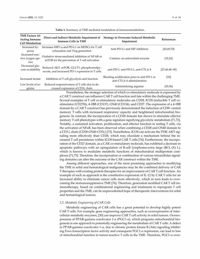
can be modified to treat different types of tumors in vivo [68]. Table 1 demonstrates how
the TME modulates immunometabolism and potential strategies to overcome the induced
metabolic impairments.

Cancers 2021, 13, 1123 8 of 18
Figure 2. Metabolically fit chimeric antigen receptor (CAR) cells need to be generated for effective
CAR immunotherapy. (A) When CAR immune cells reach their target, due to the paucity of nutri‐
ents, these cells can become exhausted. This prevents the CAR immune cells from functioning and
allows for tumor growth. (B) Metabolically fit CAR immune cells can be generated by modifying
the metabolic pathways that endow these immune cells to out‐compete cancer cells for nutrients
and thus remain active even in the TME for the eradication of cancer cells. (C) Metabolic pathways
that are disrupted and can be modified to generate metabolically fit CAR‐immune cells.
Cancers 2021, 13, 1123 9 of 18
Table 1. Summary of TME mediated modulation of immunometabolism.
TME Factors Af‐
fecting Immune
Cell Metabolism
Direct and Indirect Metabolic Impairment of
Immune Cells in TME
Strategy to Overcome Induced Metabolic
Impairment References
Increased hy‐
poxia
Increases HIF1‐a and PD‐L1 on MDSCs for T cell
exhaustion and Treg generation Anti‐PD‐L1 and HIF inhibitors [43,69,70]
Increased reac‐
tive oxygen spe‐
cies
Oxidative stress‐mediated inhibition of NF‐kB or
mTOR for the prevention of T cell activation Catalase, an antioxidant enzyme [32,42]
Decreased glu‐
cose availability
Reduced AKT, mTOR, GLUT1, phosphoenolpy‐
ruvate, and increased PD‐1 expression in T cells anti‐PD‐1, anti‐PD‐L1, anti‐CTLA‐4 [27,46,48–50]
Increased lactate Inhibition of T cell glycolysis and function Blocking acidification prior to anti‐PD‐1 or
anti‐CTLA‐4 administration
[52]
Low levels of ar‐
ginine
Reduced responsiveness of T cells due to de‐
creased expression of CD3ζ chain Administering arginine [55,56]
Nonetheless, the strategic selection of which co‐stimulatory molecule is expressed by
a CAR T construct can influence CAR T cell function and fate within the challenging TME.
Several examples of T cell co‐stimulatory molecules are CD28, ICOS (inducible T cell co‐
stimulator (CD278)), 4‐1BB (CD137), OX40 (CD134), and CD27. The expression of a 4‐1BB
domain by a CAR T construct has previously demonstrated the induction of CD8+ central
memory T cells with increased respiratory capacity and heightened mitochondrial bio‐
genesis. In contrast, the incorporation of a CD28 domain has shown to stimulate effector
memory T cell phenotypes with a gene signature signifying glycolytic metabolism [71,72].
Notably, a sustained activation, proliferation, and effector function in resting T cells, via
the activation of NFκB, has been observed when combining a CD28 and OX40 domain to
a CD3 ζ chain (CD28‐OX40‐CD3ζ) [73]. Nonetheless, ICOS can activate the PI3K‐AKT sig‐
naling more effectively than CD28, which may elucidate a mechanism behind the in‐
creased T cell persistence within ICOS‐based CAR T cells [74]. Furthermore, the incorpo‐
ration of the CD27 domain, as a CAR co‐stimulatory molecule, has exhibited a decrease in
apoptotic pathways with an upregulation of B‐cell lymphoma‐extra large (BCL‐(X) L),
which is known to modulate metabolic functions of mitochondrial multiprotein com‐
plexes [71,75]. Therefore, the incorporation or combination of various intracellular signal‐
ing domains can alter the outcome of the CAR construct within the TME.
Among different approaches, one of the most promising approaches to modifying
the TME in solid and hematological malignancies may be the combined delivery of CAR
T therapies with existing protein therapies for an improvement of CAR T cell function. An
example of such as approach is the constitutive expression of IL‐12 by CAR T cells for an
increased ability to eliminate cancer cells more effectively, which in turn leads to over‐
coming the immunosuppressive TME [76]. Therefore, generation modified CAR T cell im‐
munotherapy, based on combinatorial engineering and treatments to reprogram T cell
properties and the TME, can be unprecedented hope of therapeutic interventions for solid
and hematological tumors.
3.2. Metabolic Engineering of CAR Cells
Metabolic engineering of CAR cells has a great potential to develop highly potent
CAR T cells. For example, gene engineering approaches, such as overexpression of inter‐
cellular metabolic enzymes, [30] can improve CAR T cell activity in solid tumors. Overex‐
pression of PPAR‐gamma coactivator 1‐α (PGC1‐α), which programs mitochondrial bio‐
genesis is one approach to potentially engineering the metabolism of CAR T cells. A defect
in PPAR‐gamma coactivator 1‐α, due to chronic protein kinase B (Akt) signaling inhibit‐
ing Foxo transcription factor activity and consequent PGC1‐α repression, can lead to loss
of mitochondrial function in tumor‐reactive T cells in the TME. Therefore, PGC1‐α over‐

Cancers 2021, 13, 1123 10 of 18
expressing T cells significantly increases mitochondrial mass, resulting in greater meta‐
bolic efficiency of T cells in the TME [77]. However, studies have not yet implemented this
approach in CAR T cell therapies, indicating that further studies are required. Interest‐
ingly, another study showed that co‐inhibitory factor gene editing, such as the combina‐
tion of PD‐1 blockade in CAR T cells [78] can also enhance CAR T cell function. Specifi‐
cally, cancer cells often upregulate ligands, such as PD‐L1, that bind to inhibitory recep‐
tors on T cells and limit the capacity of CAR T cells to combat solid tumors. Using clus‐
tered regularly interspaced short palindromic repeats (CRISPR)/Cas9 system to knockout
PD‐1 has been shown to augment the function of CAR T cells in vitro and in vivo. Indeed,
CRISPR/Cas9 system can disrupt PD‐1 gene locus in human primary T cells, which leads
to reduction of PD‐1HI population. Notably, this reduction does not have a significant ef‐
fect on CAR T cell proliferation. Besides the boosting of CAR T cell cytokine production,
a combination of CAR T cells with CRISPR/Cas9‐mediated PD‐1 genome can enhance the
ability of CAR T cells to recognize antigens and target antigen‐expressing tumors [78].
Furthermore, the inhibition of PD‐1 in T cells, is shown to lead to metabolic changes where
T cells transition from glycolysis toward the Krebs cycle with an increased rate of FAO.
This alteration in T cell metabolism using PD‐1 inhibitors demonstrated an increase in T
cell survival, function, and terminal differentiation by relying on a fat‐based metabolism
and in turn mimicking functions similar to memory T cells. PD‐1 ligation has also been
shown to enhance the PPAR/PPARγ PGC1‐α axis when administering bezafibrate (a pan‐
PPAR agonist) for the prevention of T cell death and for the initiation of a long‐lived T cell
phenotype under PD‐1 blockade [79–81]. Therefore, utilizing PD‐1 therapies in conjunc‐
tion with CAR T cell therapies may be a feasible approach to altering the metabolic profile
of T cells as a strategy to maintain CAR T cell function in a nutrient depleted environment.
Adoptive cell therapy, such as TIL therapy, has also shown success and is a current
clinical approach to treating cancer. In comparison to CAR‐T cell therapy where circulat‐
ing T cells from the blood are extracted and engineered to bind to specific proteins ex‐
pressed by cancer cells, TILs are found and extracted from the tumor. TILs that recognize
the tumor cells are then expanded and infused back into the patient. Although TIL therapy
does not require engineering of T cells, TIL therapy is a more invasive approach and re‐
quires identifying TIL‐rich tumor samples which may not exist or may be challenging to
acquire [82]. Additionally, TILs have dysregulated metabolism due to the nature of the
TME, which has shown to increase exhaustion and deplete effector T cell function [83].
Therefore, acquiring metabolically stable T cells from the periphery for CAR therapy may
be an advantage in comparison to expanding metabolically dysregulated TILs for the
treatment of cancer.
4. Other Potent CAR Immune Cells
The idea of generating metabolically fit immune cells can also be extended to other
CAR immune cells such as NK cells, macrophages, and dendritic cells [84–88]. In fact,
recent research is beginning to explore specific metabolic enzymes in these immune cell
types, which can be manipulated to make these cells more metabolically fit. The metabolic
reprogramming of macrophages and dendritic cells [89] has led to the discovery of meta‐
bolic processes, such as glycolysis, the Krebs cycle, and fatty acid metabolism, having sig‐
nificant effects on their cellular function [90]. For example, macrophages undergo meta‐
bolic reprogramming in response to environmental cues, danger signals, and cytokines.
Macrophage function is also affected by certain metabolites such as succinate and citrate
[90]. Overall, the immune system can regulate metabolic pathways to change cell function
and fate, thus modulating these pathways in immune cells could generate a metabolically
fit CAR‐based immunotherapy.
Cancers 2021, 13, 1123 11 of 18
4.1. NK Cell CAR Therapies
CAR NK cells have shown promising results for tumor suppression in pre‐clinical
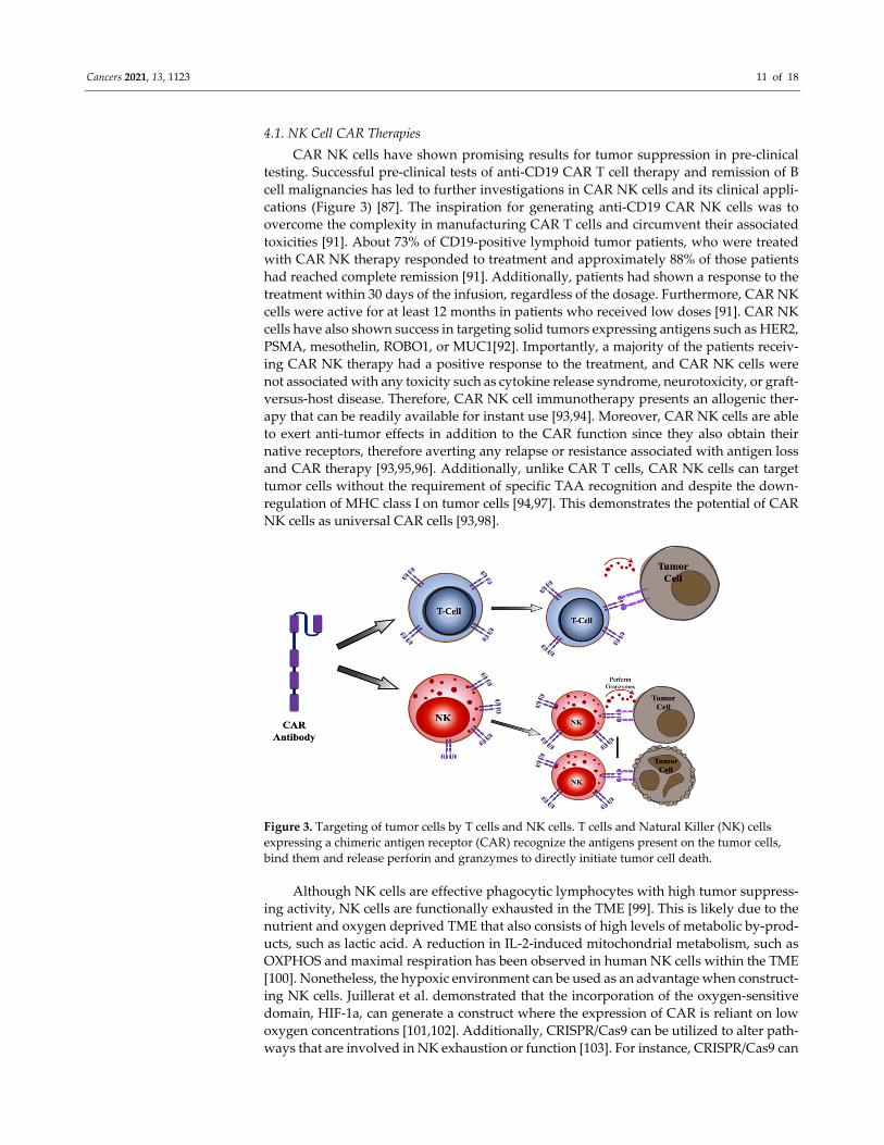
testing. Successful pre‐clinical tests of anti‐CD19 CAR T cell therapy and remission of B
cell malignancies has led to further investigations in CAR NK cells and its clinical appli‐
cations (Figure 3) [87]. The inspiration for generating anti‐CD19 CAR NK cells was to
overcome the complexity in manufacturing CAR T cells and circumvent their associated
toxicities [91]. About 73% of CD19‐positive lymphoid tumor patients, who were treated
with CAR NK therapy responded to treatment and approximately 88% of those patients
had reached complete remission [91]. Additionally, patients had shown a response to the
treatment within 30 days of the infusion, regardless of the dosage. Furthermore, CAR NK
cells were active for at least 12 months in patients who received low doses [91]. CAR NK
cells have also shown success in targeting solid tumors expressing antigens such as HER2,
PSMA, mesothelin, ROBO1, or MUC1[92]. Importantly, a majority of the patients receiv‐
ing CAR NK therapy had a positive response to the treatment, and CAR NK cells were
not associated with any toxicity such as cytokine release syndrome, neurotoxicity, or graft‐
versus‐host disease. Therefore, CAR NK cell immunotherapy presents an allogenic ther‐
apy that can be readily available for instant use [93,94]. Moreover, CAR NK cells are able
to exert anti‐tumor effects in addition to the CAR function since they also obtain their
native receptors, therefore averting any relapse or resistance associated with antigen loss
and CAR therapy [93,95,96]. Additionally, unlike CAR T cells, CAR NK cells can target
tumor cells without the requirement of specific TAA recognition and despite the down‐
regulation of MHC class I on tumor cells [94,97]. This demonstrates the potential of CAR
NK cells as universal CAR cells [93,98].
Figure 3. Targeting of tumor cells by T cells and NK cells. T cells and Natural Killer (NK) cells
expressing a chimeric antigen receptor (CAR) recognize the antigens present on the tumor cells,
bind them and release perforin and granzymes to directly initiate tumor cell death.
Although NK cells are effective phagocytic lymphocytes with high tumor suppress‐
ing activity, NK cells are functionally exhausted in the TME [99]. This is likely due to the
nutrient and oxygen deprived TME that also consists of high levels of metabolic by‐prod‐
ucts, such as lactic acid. A reduction in IL‐2‐induced mitochondrial metabolism, such as
OXPHOS and maximal respiration has been observed in human NK cells within the TME
[100]. Nonetheless, the hypoxic environment can be used as an advantage when construct‐
ing NK cells. Juillerat et al. demonstrated that the incorporation of the oxygen‐sensitive
domain, HIF‐1a, can generate a construct where the expression of CAR is reliant on low
oxygen concentrations [101,102]. Additionally, CRISPR/Cas9 can be utilized to alter path‐
ways that are involved in NK exhaustion or function [103]. For instance, CRISPR/Cas9 can
Cancers 2021, 13, 1123 12 of 18
successfully express the NKG2D ligand, major histocompatibility complex class I poly‐
peptide‐related sequence A (MICA), which may promote NK‐mediated anti‐tumor effects
[104]. It is also important to note that tumor cells in the TME evade NK cell function via
TGF‐b, metabolic disturbances, and checkpoints among many other immunosuppressive
mechanisms [96]. However, NK cells with a chimeric receptor consisting of the activating
receptor, NKG2D, along with the cytotoxic ζ‐chain of a TCR can overcome the immuno‐
suppressive TME while promoting inflammatory responses within the TME [97,105]. Fur‐
thermore, transduction of NK cells with activating cytokines, such as IL‐2, IL‐12, IL‐15, IL‐
18, and IL‐21, can promote the proliferation and function of NK cells [106]. For example,
NK cells co‐expressing CAR and IL‐15 were more potent than unmodified NK cells with
and had shown to increase proliferative rates and selective cell‐killing activity in breast
carcinoma [107]. Furthermore, CAR NK persistence and function can be achieved when
engineering memory‐like NK cells with CAR [108].
Although CAR NKs have shown some success in recent clinical trials, there are still
challenges associated with this treatment option. For example, CAR NK cells have low ex
vivo expansion as well as low transduction efficiency and lifespan, which limits their use
and warrants further research [94,109]. However, electroporation pulse codes and buffer
optimization for protein uptake can improve NK transduction rates [103]. Nonetheless,
CAR NK cells may have several advantages over CAR T cells including safer clinical uses,
more advanced mechanisms of cancer cell recognition, and an increased abundance of NK
cells in clinical samples for the generation of CAR NK cells [109].
4.2. CAR Macrophages
Due to the success of CAR T cell and CAR NK cell therapies in the clinic, further
research has been carried out to engineer other potent CAR immune cells in pre‐clinical
animal models. Among those, a promising cell type, that is gaining traction in CAR‐based
immunotherapy, is CAR macrophages. Engineering CAR macrophages is a relatively new
avenue for CAR research which attempts to overcome some of the previously mentioned
limitations associated with CAR T therapies.
Macrophages are known to naturally traffic into solid tumors and may result in a
targeted cancer cell treatment that leaves healthy cells unaffected. Interestingly, a family
of engineered chimeric antigen receptors for phagocytosis (CAR‐Ps) has recently been
generated and might direct macrophages towards the desired cancerous cells targets [88].
CAR‐P macrophages have shown specificity toward targets as they have been shown to
recognize and attack beads coated with the CD19 protein [88]. Furthermore, CAR‐P mac‐
rophages have been able to phagocytose cancer cells and debris in vitro as well.
In another interesting development, human epidermal growth factor receptor 2
(HER2) targeting‐CAR‐Ms were developed with the capacity to phagocytose HER2 anti‐
gen expressing ovarian cancer cells [14]. Moreover, it was found that a one‐time combined
treatment of CAR‐Ms and T cells decreased tumor burden in a xenograft mouse models.
Interestingly, the infusion of CAR‐Ms in mice converted the M2 (immunosuppressive
macrophage phenotype) type of macrophages in the tumor to M1 (pro‐inflammatory mac‐
rophage phenotype) [14] and induced antigen specific T cell responses against the tumors.
Interestingly, in 2018, Carisma Therapeutics was successful in raising funds for develop‐
ing CAR macrophage immunotherapies. In addition to CAR‐macrophages, the precursor
of macrophages, monocytes can also have antitumor activity. The advantage of using
monocytes as oppose to macrophages can be that it reduces the time between retrieval
and infusion from seven days to one day.
Overall, CAR monocytes/macrophages are a promising avenue of CAR cell‐based
immunotherapy and has the potential to overcome the shortcomings of CAR T cell‐based
immunotherapies, especially in targeting solid tumors.

Cancers 2021, 13, 1123 13 of 18
5. Summary
In summary, cellular metabolism plays a crucial role in the immune response and
based on different stages of immune cell phenotype (naive, effector, memory, regulatory
T cells; M1; M2) and their activation state, the metabolic properties of these cells will
change. Additionally, other factors, such as nutrients, cytokines, and growth factors, can
affect effector T cell metabolism. Moreover, the high glycolytic metabolism of tumor cells
creates a microenvironment that is low in vital nutrients, in turn making it highly hypoxic
and acidic, which then leads to the metabolic inhibition of immune cells, poor inflamma‐
tory cell trafficking to the tumor, the production of immunosuppressive cytokines, and
expression of co‐inhibitory ligands. These suppressive influences render significant chal‐
lenges for CAR cell therapies. Moreover, although several strategies have been tested to
tackle solid tumor barriers, such as the use of alternative cytoplasmic activation domains
and the use of CRISPR‐Cas9 as gene‐editing techniques, these need to be validated in
clinic. Notably, combination therapy with checkpoint inhibitors and armed CARs has
been used to improve the function of CAR T cells in solid tumors and are being tested in
clinical trials. Importantly, new strategies are required to improve the metabolic fitness of
CAR T cells within the TME and strategies are also required to improve the safety of CAR
cells, particularly as they move into clinic. Indeed, CAR cells can be optimally designed
based on the metabolic properties of the tumor being targeted, and cultured to promote a
less differentiated, long‐lived phenotype that can efficiently self‐renew and differentiate
in vivo into potent effector cells. Next‐generation CAR cell immunotherapy based on com‐
binatorial engineering and treatments to reprogram immune cell properties and the TME
offer unprecedented hope of therapeutic interventions for solid tumors.
6. Future Directions
Generating highly pure and metabolically fit CAR immune cells is a major challenge,
and precision genetic modulation of metabolic pathways may improve efficacy toward
treating solid tumors. Moreover, pharmaceutical modification of CAR immune cells can
also be utilized to modify these energy metabolic pathways to drive the activation of CAR
immune cells, specifically for the treatment of solid tumors. These strategies may pave the
way to more efficient CAR therapies against solid tumors.
A major issue that needs to be addressed for CAR therapy is the costs associated with
manufacturing. Engineering strategies of non‐viral vectors, developing protocols for in‐
hospital CAR therapy generation and generating CAR expression in non‐T cells are some
of the approaches that may lead to a decrease in these costs. Furthermore, a significant
investment in engineering principles and omics approaches are needed to improve cellu‐
lar manufacturing and the quality control and assurance of CAR therapies. The next stages
in developing CAR immune cells will require marrying the fields of engineering and gene
therapy for increasing the efficacy of treatment of solid tumors with low toxicity.
Author Contributions: Conceptualization, A.P.A, R.G. and M.W.; methodology, J.L.M, A.E., J.L.H,
S.I., S.M.;. writing—original draft preparation, J.L.M, A.E., J.L.H, S.I., S.M.; writing—review and
editing, A.P.A., R.G., and M.W. All authors have read and agreed to the published version of the
manuscript.
Funding: This research received no external funding.
Institutional Review Board Statement: Not applicable.
Informed Consent Statement: Not applicable.
Data Availability Statement: Not applicable.
Acknowledgments: This work was supported in part by Ri.MED Foundation (Italy), the Children’s
Hospital of Philadelphia Research Institute, and the Frontier Program in Airway Disorders of the
Children’s Hospital of Philadelphia to RLG. This work was also supported by start‐up funds from
Arizona State University to APA.

Cancers 2021, 13, 1123 14 of 18
Conflicts of Interest: The authors declare no conflict of interest.
Abbreviations:
ACT—Adoptive cell transfer
AKT—protein kinase B
APCs—Antigen presenting cells
ATP—Adenosine triphosphate
B‐ALL—B cell acute lymphoblastic leukemia
Bcl‐X(L)—B‐cell lymphoma‐extra large
CAFs—Cancer‐associated fibroblasts
CAR—Chimeric antigen receptor
CAR‐M—Chimeric antigen receptor macrophage
CAR‐Ps—Chimeric antigen receptors for phagocytosis
CAT—Catalase
CCR—Chimeric costimulatory receptor
CLL—Chronic lymphocytic leukemia
CRISPR—Clustered Regularly Interspaced Short Palindromic Repeats
CRS—Cytokine release syndrome
ECM—Extracellular matrix
ETBR—Endothelin B receptor
FAO—Fatty acid oxidation
GLUT1—Glucose transporter 1
HER2—Human epidermal growth factor receptor 2
HK—Hexokinase 2
HPSE—Heparinase
HSPG—Heparan sulphate proteoglycans
ICOS—Inducible T cell co‐stimulator
IDO—Indoleamine‐2,3‐dioxygenase
IL‐10—Interleukin 10
LBCL—Large B cell lymphoma
MDSC—Myeloid‐derived suppressor cells
MICA—Major histocompatibility complex class I polypeptide‐related sequence A
NK—Natural killer
OXPHOS—Oxidative phosphorylation
PD‐1—Programmed cell death protein 1
PD‐L1—Programmed cell death ligand 1
PFK‐6—phosphofructo‐2‐kinase
PGC1‐α—PPAR‐gamma coactivator 1‐α
PGE2—Prostaglandin E2
PI3K—Phosphoinositide 3‐kinase
PSMA—Prostate‐specific membrane
ROS—Reactive oxygen species
ScFv—Single chain variable fragment
TAA—Tumor‐associated antigens
TCR—T cell receptor
TGFβ—Transforming growth factor beta
TILs—Tumor‐infiltration lymphocytes
TME—Tumor microenvironment
VEGF—Vascular endothelial growth factor
References
1. Batlevi, C.L.; Matsuki, E.; Brentjens, R.J.; Younes, A. Novel immunotherapies in lymphoid malignancies. Nat. Rev. Clin. Oncol.
2016, 13, 25–40.
2. Brudno, J.N.; Kochenderfer, J.N. Chimeric antigen receptor T‐cell therapies for lymphoma. Nat. Rev. Clin. Oncol. 2018, 15, 31–
46.
3. June, C.H.; O’Connor, R.S.; Kawalekar, O.U.; Ghassemi, S.; Milone, M.C. CAR T cell immunotherapy for human cancer. Science
2018, 359, 1361–1365.
Cancers 2021, 13, 1123 15 of 18
4. Available online: https://clinicaltrials.gov/ct2/results/details?cond=CAR+T+cells (accessed on 29 June 2020)
5. Zhang, C.; Liu, J.; Zhong, J.F.; Zhang, X. Engineering CAR‐T cells. Biomark. Res. 2017, 5, 22.
6. Hinrichs, C.S.; Spolski, R.; Paulos, C.M.; Gattinoni, L.; Kerstann, K.W.; Palmer, D.C.; Klebanoff, C.A.; Rosenberg, S.A.; Leonard,
W.J.; Restifo, N.P. IL‐2 and IL‐21 confer opposing differentiation programs to CD8+ T cells for adoptive immunotherapy. Blood
2008, 111, 5326–5333.
7. Pavlova, N.N.; Thompson, C.B. The Emerging Hallmarks of Cancer Metabolism. Cell Metab. 2016, 23, 27–47.
8. Finicle, B.T.; Jayashankar, V.; Edinger, A.L. Nutrient scavenging in cancer. Nat. Rev. Cancer 2018, 18, 619–633.
9. Dietl, K.; Renner, K.; Dettmer, K.; Timischl, B.; Eberhart, K.; Dorn, C.; Hellerbrand, C.; Kastenberger, M.; Kunz‐Schughart, L.A.;
Oefner, P.J.; et al. Lactic Acid and Acidification Inhibit TNF Secretion and Glycolysis of Human Monocytes. J. Immunol. 2009,
184, 1200–1209.
10. Fischer, K.; Hoffmann, P.; Voelkl, S.; Meidenbauer, N.; Ammer, J.; Edinger, M.;Gottfried, E.; Schwarz, S.; Rothe, G.; Hoves, S.;
et al. Inhibitory effect of tumor cell‐derived lactic acid on human T cells. Blood 2007, 109, 3812–3819.
11. Muller, A.J.; Prendergast, G.C. Marrying immunotherapy with chemotherapy: Why say IDO? Cancer Res. 2005, doi:10.1158/0008‐
5472.CAN‐05‐2213.
12. Itakura, E.; Huang, R.R.; Wen, D.R.; Paul, E.; Wünsch, P.H.; Cochran, A.J. IL‐10 expression by primary tumor cells correlates
with melanoma progression from radial to vertical growth phase and development of metastatic competence. Mod. Pathol. 2011,
24, 801‐809.
13. Massagué, J. TGFβ in Cancer. Cell 2008, 134, 215–230.
14. Klichinsky, M.; Ruella, M.; Shestova, O.; Lu, X.M.; Best, A.; Zeeman, M.; Schmierer, M.; Gabrusiewicz, K.; Anderson, N.R.; Petty
N.E.; et al. Human chimeric antigen receptor macrophages for cancer immunotherapy. Nat. Biotechnol. 2020, 38, 947–953.
15. Patel, S.; Burga, R.A.; Powell, A.B.; Chorvinsky, E.A.; Hoq, N.; McCormack, S.E.; Van Pelt, S.N.; Hanley P.J.; Cruz, C.R. Beyond
CAR T Cells: Other Cell‐Based Immunotherapeutic Strategies Against Cancer. Front. Oncol. 2019, doi:10.3389/fonc.2019.00196.
16. Majzner, R.G.; Heitzeneder, S.; Mackall, C.L. Harnessing the Immunotherapy Revolution for the Treatment of Childhood Can‐
cers. Cancer Cell. 2017, 31, 476–485.
17. Majzner, R.G.; Mackall, C.L. Tumor antigen escape from car t‐cell therapy. Cancer Discov. 2018, 8, 1219–1226.
18. Zah, E.; Lin, M.‐Y.; Silva‐Benedict, A.; Jensen, M.C.; Chen, Y.Y. T Cells Expressing CD19/CD20 Bispecific Chimeric Antigen
Receptors Prevent Antigen Escape by Malignant B Cells. Cancer Immunol. Res. 2016, 4, 498–508.
19. Sadelain, M. CAR therapy: the CD19 paradigm. J. Clin. Invest. 2015, 125, 3392–3400.
20. Das, R.K.; Storm, J.; Barrett, D.M. Abstract 1631: T cell dysfunction in pediatric cancer patients at diagnosis and after chemo‐
therapy can limit chimeric antigen receptor potential. Cancer Res. 2018, doi:10.1158/1538‐7445.AM2018‐1631.
21. Heinmöller, P.; Gross, C.; Beyser, K.; Schmidtgen, C.; Maass, G.; Pedrocchi, M.; Rüschoff, J. HER2 Status in Non‐Small Cell Lung
Cancer: Results from Patient Screening for Enrollment to a Phase II Study of Herceptin. Clin Cancer Res. 2003, 9, 5238–5243.
22. Shah, N.N.; Fry, T.J. Mechanisms of resistance to CAR T cell therapy. Nat. Rev. Clin. Oncol. 2019, 16, 1–385.
23. Zhang, E.; Xu, H. A new insight in chimeric antigen receptor‐engineered T cells for cancer immunotherapy. J. Hematol. Oncol.
2017, 10, 1.
24. Frey, N.V.; Porter, D.L. CAR T‐cells merge into the fast lane of cancer care. Am. J. Hematol. 2016, 91, 146–150.
25. Jena, B.; Moyes, J.S.; Huls, H.; Cooper, L.J.N. Driving CAR‐Based T‐Cell Therapy to Success. Curr. Hematol. Malig Rep. 2014, 9,
50–56.
26. Gilham, D.E.; Debets, R.; Pule, M.; Hawkins, R.E.; Abken, H. CAR–T cells and solid tumors: tuning T cells to challenge an
inveterate foe. Trends Mol. Med. 2012, 18, 377–384.
27. Martinez, M.; Moon, E.K. CAR T cells for solid tumors: New strategies for finding, infiltrating, and surviving in the tumor
microenvironment. Front. Immunol. 2019, 10, 1–21.
28. Caruana, I.; Savoldo, B.; Hoyos, V.; Weber, G.; Liu, H.; Kim, E.S.; Ittmann, M.M., Marchetti, D.; Dotti, G. Heparanase promotes
tumor infiltration and antitumor activity of CAR‐redirected T lymphocytes. Nat. Med. 2015, 21, 524–529.
29. D’Aloia, M.M.; Zizzari, I.G.; Sacchetti, B.; Pierelli, L.; Alimandi, M. CAR‐T cells: The long and winding road to solid tumors
review‐article. Cell Death Dis. 2018, doi:10.1038/s41419‐018‐0278‐6.
30. Irving, M.; de Silly, R.V.; Scholten, K.; Dilek, N.; Coukos, G. Engineering chimeric antigen receptor T‐cells for racing in solid
tumors: Don’t forget the fuel. Front Immunol. 2017, 8, 1–19.
31. Chen, Q.; Hu, Q.; Dukhovlinova, E.; Chen, G.; Ahn, S.; Wang, C.; Ogunnaike, E.A.; Ligler, F.S.; Dotti, G.; Gu, Z. Photothermal
Therapy Promotes Tumor Infiltration and Antitumor Activity of CAR T Cells. Adv Mater. 2019, 31, 1–7.
32. Yong, C.S.M.; Dardalhon, V.; Devaud, C.; Taylor, N.; Darcy, P.K.; Kershaw, M.H. CAR T‐cell therapy of solid tumors. Immunol.
Cell Biol. 2017, 95, 356–363.
33. Rataj, F.; Jacobi, S.J.; Stoiber, S.; Asang, F.; Ogonek, J.; Tokarew, N.; Cadilha, B.L.; Puijenbroek, E.V.; Heise, C.; Duewell, P.; et al.
High‐affinity CD16‐polymorphism and Fc‐engineered antibodies enable activity of CD16‐chimeric antigen receptor‐modified
T cells for cancer therapy. Br. J. Cancer. 2019, 120, 79–87.
34. Clémenceau, B.; Congy‐Jolivet, N.; Gallot, G.; Vivien, R.; Gaschet, J.; Thibault, G.; Vié, H. Antibody‐dependent cellular cytotox‐
icity (ADCC) is mediated by genetically modified antigen‐specific human T lymphocytes. Blood 2006, 107, 4669–4677.
35. Kudo, K.; Imai, C.; Lorenzini, P.; Kamiya, T.; Kono, K.; Davidoff, A.M.; Chng, W.J.; Campana, D. T lymphocytes expressing a
CD16 signaling receptor exert antibody‐dependent cancer cell killing. Cancer Res. 2014, 74, 93–103.

Cancers 2021, 13, 1123 16 of 18
36. Ochi, F.; Fujiwara, H.; Tanimoto, K.; Asai, H.; Miyazaki, Y.; Okamoto, S.; Mineno, J.; Kuzushima, K.; Shiku, H.; Barrett, J.; et al.
Gene‐modified human α/β‐T cells expressing a chimeric CD16‐CD3ζ receptor as adoptively transferable effector cells for anti‐
cancer monoclonal antibody therapy. Cancer Immunol. Res. 2014, doi:0.1158/2326‐6066.CIR‐13‐0099‐T
37. Newick, K.; O’Brien, S.; Moon, E.; Albelda, S.M. CAR T Cell Therapy for Solid Tumors. Annu. Rev. Med. 2017, 68, 139–152.
38. Baitsch, L.; Fuertes‐Marraco, S.A.; Legat, A.; Meyer, C.; Speiser, D.E. The three main stumbling blocks for anticancer T cells.
Trends Immunol. 2012, 33, 364–372.
39. Almeida, L.; Lochner, M.; Berod, L.; Sparwasser, T. Metabolic pathways in T cell activation and lineage differentiation. Semin.
Immunol. 2016, 28, 514–524.
40. Kouidhi, S.; Elgaaied, A.B.; Chouaib, S. Impact of metabolism on T‐cell differentiation and function and cross talk with tumor
microenvironment. Front. Immunol. 2017, 8, 270.
41. Zhang, Y.; Kurupati, R.; Liu, L.; Zhou, X.Y.; Zhang, G.; Hudaihed, A.; Filisio, F.; Giles‐Davis, W.; Xu, X.; Karakousis, G.C.; et al.
Enhancing CD8+ T Cell Fatty Acid Catabolism within a Metabolically Challenging Tumor Microenvironment Increases the
Efficacy of Melanoma Immunotherapy. Cancer Cell. 2017, 32, 377–391.
42. Chen, X.; Song, M.; Zhang, B.; Zhang, Y. Reactive Oxygen Species Regulate T Cell Immune Response in the Tumor Microenvi‐
ronment. Oxid. Med. Cell. Longev. 2016, doi:10.1155/2016/1580967
43. Lim, A.R.; Rathmell, W.K.; Rathmell, J.C. The tumor microenvironment as a metabolic barrier to effector T cells and immuno‐
therapy. Elife. 2020, 9, doi:10.7554/elife.55185.
44. Herbel, C.; Patsoukis, N.; Bardhan, K.; Seth, P.; Weaver, J.D.; Boussiotis, V.A. Clinical significance of T cell metabolic repro‐
gramming in cancer. Clin Transl Med. 2016, 5, 29.
45. Ho, P.C.; Liu, P.S. Metabolic communication in tumors: A new layer of immunoregulation for immune evasion. J. Immunother.
Cancer 2016, 4, 1–9.
46. Patsoukis, N.; Weaver, J.D.; Strauss, L.; Herbel, C.; Seth, P.; Boussiotis, V.A. Immunometabolic regulations mediated by coin‐
hibitory receptors and their impact on T cell immune responses. Front Immunol. 2017, 8, 1–19.
47. Cairns, R.A.; Harris, I.S.; Mak, T.W. Regulation of cancer cell metabolism. Nat. Rev. Cancer. 2011, 11, 85–95.
48. Ho, P.C.; Bihuniak, J.D.; MacIntyre, A.N.; Staron, M.; Liu, X.; Amezquita, R.; Tsui, Y.; Cui, G., Micevic, G., Perales, J.C.; et al.
Phosphoenolpyruvate Is a Metabolic Checkpoint of Anti‐tumor T Cell Responses. Cell 2015, 162, 1217–1228.
49. Chang, C.‐H.; Curtis, J.D.; Maggi, L.B.; Faubert, B.; Villarino, A.V.; O’Sullivan, D.; Huang, S.C.‐C.; Van Der Windt, G.J.; Blagih,
J.; Qiu, J.; et al. Posttranscriptional Control of T Cell Effector Function by Aerobic Glycolysis. Cell 2013, 153, 1239–1251,
doi:10.1016/j.cell.2013.05.016.
50. Chang, C.‐H.; Qiu, J.; O’Sullivan, D.; Buck, M.D.; Noguchi, T.; Curtis, J.D.; Chen, Q.; Gindin, M.; Gubin, M.M.; Van Der Windt,
G.J.; et al. Metabolic Competition in the Tumor Microenvironment Is a Driver of Cancer Progression. Cell 2015, 162, 1229–1241,
doi:10.1016/j.cell.2015.08.016.
51. Hope, H.C.; Salmond, R.J. Targeting the tumor microenvironment and T cell metabolism for effective cancer immunotherapy.
Eur. J. Immunol. 2019, 49, 1147–1152, doi:10.1002/eji.201848058
52. Brand, A.; Singer, K.; Koehl, G.E.; Kolitzus, M.; Schoenhammer, G.; Thiel, A.; Matos, C.; Bruss, C.; Klobuch, S.; Peter, K.; et al.
LDHA‐Associated Lactic Acid Production Blunts Tumor Immunosurveillance by T and NK Cells. Cell Metab. 2016, 24, 657–671,
doi:10.1016/j.cmet.2016.08.011.
53. Haas, R.; Smith, J.; Rocher‐Ros, V.; Nadkarni, S.; Montero‐Melendez, T.; D’Acquisto, F.; Bland, E.J.; Bombardieri, M., Pitzalis,
C.; Perretti, M.; et al. Lactate regulates metabolic and proinflammatory circuits in control of T cell migration and effector func‐
tions. PLoS Biol. 2015, doi:10.1371/journal.pbio.1002202.
54. Srivastava, M.K.; Sinha, P.; Clements, V.K.; Rodriguez, P.; Ostrand‐Rosenberg, S. Myeloid‐Derived Suppressor Cells Inhibit T‐
Cell Activation by Depleting Cystine and Cysteine. Cancer Res. 2010, 70, 68–77, doi:10.1158/0008‐5472.can‐09‐2587.
55. Lind DS. Arginine and cancer. J. Nutr. 2004, doi:10.1093/jn/134.10.2837S.
56. Geiger, R.; Rieckmann, J.C.; Wolf, T.; Basso, C.; Feng, Y.; Fuhrer, T.; Kogadeeva, M.; Picotti, P.; Meissner, F.; Mann, M.; et al. L‐
Arginine Modulates T Cell Metabolism and Enhances Survival and Anti‐tumor Activity. Cell 2016, 167, 829–842.e13,
doi:10.1016/j.cell.2016.09.031.
57. Acharya, A.P.; Sinha, M.; Ratay, M.L.; Ding, X.; Balmert, S.C.; Workman, C.J.; Wang, Y.; Vignali, D.A.A.; Little, S.R. Localized
Multi‐Component Delivery Platform Generates Local and Systemic Anti‐Tumor Immunity. Adv. Funct. Mater. 2017, 27,
doi:10.1002/adfm.201604366.
58. Pearce, E.L.; Poffenberger, M.C.; Chang, C.‐H.; Jones, R.G. Fueling Immunity: Insights into Metabolism and Lymphocyte Func‐
tion. Science 2013, 342, 1242454, doi:10.1126/science.1242454.
59. Frauwirth, K.A.; Riley, J.L.; Harris, M.H.; Parry, R.V.; Rathmell, J.C.; Plas, D.R.; Elstrom, R.L.; June, C.H.; Thompson, C.B. The
CD28 Signaling Pathway Regulates Glucose Metabolism. Immunity 2002, 16, 769–777, doi:10.1016/s1074‐7613(02)00323‐0.
60. Kolev, M.; Dimeloe, S.; Le Friec, G.; Navarini, A.; Arbore, G.; Povoleri, G.A.; Fischer, M.; Belle, R.; Loeliger, J.; Develioglu, L.; et
al. Complement Regulates Nutrient Influx and Metabolic Reprogramming during Th1 Cell Responses. Immunity 2015, 42, 1033–
1047.
61. Delgoffe, G.M.; Pollizzi, K.N.; Waickman, A.T.; Heikamp, E.; Meyers, D.J.; Horton, M.R.; Xiao, B.; Worley, P.F.; Powell, J.D. The
kinase mTOR regulates the differentiation of helper T cells through the selective activation of signaling by mTORC1 and
mTORC. Nat. Immunol. 2011, 12, 295–303.
Cancers 2021, 13, 1123 17 of 18
62. Cham, C.M.; Driessens, G.; O’Keefe, J.P.; Gajewski, T.F. Glucose deprivation inhibits multiple key gene expression events and
effector functions in CD8+ T cells. Eur. J. Immunol. 2008, 38, 2438–2450, doi:10.1002/eji.200838289.
63. Hamid, O.; Robert, C.; Daud, A.; Hodi, F.S.; Hwu, W.‐J.; Kefford, R.; Wolchok, J.D.; Hersey, P.; Joseph, R.W.; Weber, J.S.; et al.
Safety and Tumor Responses with Lambrolizumab (Anti–PD‐1) in Melanoma. N. Engl. J. Med. 2013, 369, 134–144,
doi:10.1056/nejmoa1305133.
64. Wolchok, J.D.; Kluger, H.; Callahan, M.K.; Postow, M.A.; Rizvi, N.A.; Lesokhin, A.M.; Segal, N.H.; Ariyan, C.E.; Gordon, R.‐A.;
Reed, K.; et al. Nivolumab plus Ipilimumab in Advanced Melanoma. N. Engl. J. Med. 2013, 369, 122–133,
doi:10.1056/nejmoa1302369.
65. Pearce, E.L.; Pearce, E.J. Metabolic Pathways in Immune Cell Activation and Quiescence. Immunity Cell Press 2013, 38, 633–643.
66. Liu, X.; Shin, N.; Koblish, H.K.; Yang, G.; Wang, Q.; Wang, K.; Leffet, L.; Hansbury, M.J.; Thomas, B.; Rupar, M.; et al. Selective
inhibition of IDO1 effectively regulates mediators of antitumor immunity. Blood 2010, 115, 3520–3530, doi:10.1182/blood‐2009‐
09‐246124.
67. Buck, M.D.; O’Sullivan, D.; Pearce, E.L. T cell metabolism drives immunity. J. Exp. Med. 2015, 212, 1345–1360,
doi:10.1084/jem.20151159.
68. O’Sullivan, D.; Pearce, E.L.; Targeting T cell metabolism for therapy. Trends Immunol. 2015, 36, 71–80.
69. Noman, M.Z.; DeSantis, G.; Janji, B.; Hasmim, M.; Karray, S.; Dessen, P.; Bronte, V.; Chouaib, S. PD‐L1 is a novel direct target
of HIF‐1α, and its blockade under hypoxia enhanced MDSC‐mediated T cell activation. J. Exp. Med. 2014, 211, 781–790,
doi:10.1084/jem.20131916.
70. Fallah, J.; Rini, B.I. HIF Inhibitors: Status of Current Clinical Development. Curr. Oncol. Rep. 2019, doi:10.1007/s11912‐019‐0752‐
z.
71. Weinkove, R.; George, P.; Dasyam, N.; McLellan, A.D. Selecting costimulatory domains for chimeric antigen receptors: func‐
tional and clinical considerations. Clin. Transl. Immunol. 2019, 8, e1049, doi:10.1002/cti2.1049.
72. Kawalekar, O.U.; O’Connor, R.S.; Fraietta, J.A.; Guo, L.; McGettigan, S.E.; Posey, A.D.; Patel, P.R.; Guedan, S., Scholler, J.; Keith,
B.; et al. Distinct Signaling of Coreceptors Regulates Specific Metabolism Pathways and Impacts Memory Development in CAR
T Cells. Immunity 2016, 44, 380–390.
73. Pulè, M.A.; Straathof, K.C.; Dotti, G.; Heslop, H.E.; Rooney, C.M.; Brenner, M.K. A chimeric T cell antigen receptor that aug‐
ments cytokine release and supports clonal expansion of primary human T cells. Mol. Ther. 2005, 12, 933–941.
74. Guedan, S.; Posey, A.D.; Shaw, C.; Wing, A.; Da, T.; Patel, P.R.; Mcgettigan, S.E.; Casado‐Medrano, V.; Kawalekar, O.U.; Uribe‐
Herranz, M.; et al. Enhancing CAR T cell persistence through ICOS and 4‐1BB costimulation. JCI Insight 2018, 3,
doi:10.1172/jci.insight.96976.
75. Michels, J.; Keep, O.; Senovilla, L.; Lissa, D.; Castedo, M.; Kroemer, G.; Galluzzi, L. Functions of BCL‐XL at the interface between
cell death and metabolism. Int. J. Cell Biol. 2013, doi:10.1155/2013/705294.
76. Abate‐Daga, D.; Davila, M.L. CAR models: next‐generation CAR modifications for enhanced T‐cell function. Mol. Ther. Onco‐
lytics 2016, 3, 16014.
77. Scharping, N.E.; Menk, A.V.; Moreci, R.S.; Whetstone, R.D.; Dadey, R.E.; Watkins, S.C.; Ferris, R.L.; Delgoffe, G.M. The Tumor
Microenvironment Represses T Cell Mitochondrial Biogenesis to Drive Intratumoral T Cell Metabolic Insufficiency and Dys‐
function. Immun. 2016, 45, 374–388, doi:10.1016/j.immuni.2016.07.009
78. Hu, W.; Zi, Z.; Jin, Y.; Li, G.; Shao, K.; Cai, Q.; Ma, X.; Wei, F. CRISPR/Cas9‐mediated PD‐1 disruption enhances human meso‐
thelin‐targeted CAR T cell effector functions. Cancer Immunol. Immunother. 2019, 68, 365–377, doi:10.1007/s00262‐018‐2281‐2.
79. Kumar, A.; Chamoto, K. Immune metabolism in PD‐1 blockade‐based cancer immunotherapy. Int. Immunol. 2021, 33, 17–26,
doi:10.1093/intimm/dxaa046.
80. Patsoukis, N.; Bardhan, K.; Chatterjee, P.; Sari, D.; Liu, B.; Bell, L.N.; Karoly, E.D.; Freeman, G.J.; Petkova, V.; Seth, P.; et al. PD‐
1 alters T‐cell metabolic reprogramming by inhibiting glycolysis and promoting lipolysis and fatty acid oxidation. Nat. Commun.
2015, 6, 6692, doi:10.1038/ncomms7692.
81. Wan, H.; Xu, B.; Zhu, N.; Ren, B. PGC‐1α activator–induced fatty acid oxidation in tumor‐infiltrating CTLs enhances effects of
PD‐1 blockade therapy in lung cancer. Tumori 2020, doi:10.1177/0300891619868287.
82. Yee, C. Adoptive T cell therapy: points to consider. Curr. Opin. Immunol. 2018, 51, 197–203, doi:10.1016/j.coi.2018.04.007.
83. Le Bourgeois, T.; Strauss, L.; Aksoylar, H.I.; Daneshmandi, S.; Seth, P.; Patsoukis, N.; Boussiotis, V.A. Targeting T cell metabo‐
lism for improvement of cancer immunotherapy. Front. Oncol. 2018, doi:10.3389/fonc.2018.00237.
84. Acharya, A.P.; Dolgova, N.V.; Clare‐Salzler, M.J.; Keselowsky, B.G. Adhesive substrate‐modulation of adaptive immune re‐
sponses. Biomaterials 2008, 29, 4736–4750, doi:10.1016/j.biomaterials.2008.08.040.
85. Acharya, A.P.; Carstens, M.R.; Lewis, J.S.; Dolgova, N.; Xia, C.Q.; Clare‐Salzler, M.J.; Keselowsky, B.G. A cell‐based microarray
to investigate combinatorial effects of microparticle‐encapsulated adjuvants on dendritic cell activation. J. Mater. Chem. 2016, 4,
1672–1685.
86. Acharya, A.P.; Clare‐Salzler, M.J.; Keselowsky, B.G. A high‐throughput microparticle microarray platform for dendritic cell‐
targeting vaccines. Biomaterials 2009, 30, 4168–4177, doi:10.1016/j.biomaterials.2009.04.032.
87. Liu, E.; Tong, Y.; Dotti, G.; Shaim, H.; Savoldo, B.; Mukherjee, M.; Orange, J.; Wan, X.; Lu, X.; Reynolds, A.; et al. Cord blood
NK cells engineered to express IL‐15 and a CD19‐targeted CAR show long‐term persistence and potent antitumor activity. Leuk.
2018, 32, 520–531, doi:10.1038/leu.2017.226.

Cancers 2021, 13, 1123 18 of 18
88. A Morrissey, M.; Williamson, A.P.; Steinbach, A.M.; Roberts, E.W.; Kern, N.; Headley, M.B.; Vale, R.D. Chimeric antigen recep‐
tors that trigger phagocytosis. eLife 2018, 7, e36688, doi:10.7554/elife.36688.
89. Mangal, J.L.; Inamdar, S.; Yang, Y.; Dutta, S.; Wankhede, M.; Shi, X.; Gu, H.; Green, M.D.; Rege, K.; Curtis, M.; et al. Metabolite
releasing polymers control dendritic cell function by modulating their energy metabolism. J. Mater. Chem. B 2020, 8, 5195–5203,
doi:10.1039/d0tb00790k.
90. O’Neill, L.A.J.; Pearce, E.J. Immunometabolism governs dendritic cell and macrophage function. J. Exp. Med. 2016, 213, 15–23.
91. Liu, E.; Marin, D.; Banerjee, P.; Macapinlac, H.A.; Thompson, P.; Basar, R.; Kerbauy, L.N.; Overman, B.; Thall, P.; Kaplan, M.; et
al. Use of CAR‐Transduced Natural Killer Cells in CD19‐Positive Lymphoid Tumors. New Engl. J. Med. 2020, 382, 545–553,
doi:10.1056/nejmoa1910607.
92. Xie, G.; Dong, H.; Liang, Y.; Ham, J.D.; Rizwan, R.; Chen, J. CAR‐NK cells: A promising cellular immunotherapy for cancer.
EBioMedicine 2020, 59, 102975, doi:10.1016/j.ebiom.2020.102975.
93. Rezvani, K.; Rouce, R.; Liu, E.; Shpall, E. Engineering Natural Killer Cells for Cancer Immunotherapy. Mol. Ther. 2017, 25, 1769–
1781, doi:10.1016/j.ymthe.2017.06.012.
94. Basar, R.; Daher, M.; Rezvani, K. Next‐generation cell therapies: The emerging role of CAR‐NK cells. Blood Adv. 2020,
doi:10.1182/hematology.2020002547.
95. Sotillo, E.; Barrett, D.M.; Black, K.L.; Bagashev, A.A.; Oldridge, D.; Wu, G.; Sussman, R.T.; LaNauze, C.; Ruella, M.; Gazzara,
M.R.; et al. Convergence of Acquired Mutations and Alternative Splicing of CD19 Enables Resistance to CART‐19 Immunother‐
apy. Cancer Discov. 2015, 5, 1282–1295, doi:10.1158/2159‐8290.cd‐15‐1020.
96. Yilmaz, A.; Cui, H.; Caligiuri, M.A.; Yu, J. Chimeric antigen receptor‐engineered natural killer cells for cancer immunotherapy.
J. Hematol. Oncol. 2020, 13, 1–22, doi:10.1186/s13045‐020‐00998‐9.
97. Lin, C.‐Y.; Gobius, I.; Souza‐Fonseca‐Guimaraes, F. Natural killer cell engineering – a new hope for cancer immunotherapy.
Semin. Hematol. 2020, 57, 194–200, doi:10.1053/j.seminhematol.2020.10.002.
98. Caratelli, S.; Arriga, R.; Sconocchia, T.; Ottaviani, A.; Lanzilli, G.; Pastore, D.; Cenciarelli, C.; Venditti, A.; Del Principe, M.I.;
Lauro, D.; et al. In vitro elimination of epidermal growth factor receptor‐overexpressing cancer cells by CD32A‐chimeric recep‐
tor T cells in combination with cetuximab or panitumumab. Int. J. Cancer 2020, 146, 236–247, doi:10.1002/ijc.32663.
99. Afolabi, L.O.; Adeshakin, A.O.; Sani, M.M.; Bi, J.; Wan, X. Genetic reprogramming for NK cell cancer immunotherapy with
CRISPR/Cas9. Immunology 2019, 158, 63–69.
100. Terrén, I.; Orrantia, A.; Vitallé, J.; Zenarruzabeitia, O.; Borrego, F. NK Cell Metabolism and Tumor Microenvironment. Front.
Immunol. 2019, 10, doi:10.3389/fimmu.2019.02278.
101. Juillerat, A.; Marechal, A.; Filhol, J.M.; Valogne, Y.; Valton, J.; Duclert, A.; Duchateau, P.; Poirot, L. An oxygen sensitive self‐
decision making engineered CAR T‐cell. Sci. Rep. 2017, 7, srep39833, doi:10.1038/srep39833.
102. Navin, I.; Lam, M.T.; Parihar, R. Design and Implementation of NK Cell‐Based Immunotherapy to Overcome the Solid Tumor
Microenvironment. Cancers 2020, 12, 3871, doi:10.3390/cancers12123871.
103. Rautela, J.; Surgenor, E.; Huntington, N.D. Efficient genome editing of human natural killer cells by CRISPR RNP. bioRxiv 2018,
doi:10.1101/406934.
104. Sekiba, K.; Yamagami, M.; Otsuka, M.; Suzuki, T.; Kishikawa, T.; Ishibashi, R.; Ohno, M.; Sato, M.; Koike, K. Transcriptional
activation of the MICA gene with an engineered CRISPR‐Cas9 system. Biochem. Biophys. Res. Commun. 2017, 486, 521–525,
doi:10.1016/j.bbrc.2017.03.076.
105. Parihar, R.; Rivas, C.H.; Huynh, M.; Omer, B.; Lapteva, N.; Metelitsa, L.S.; Gottschalk, S.M.; Rooney, C.M. NK Cells Expressing
a Chimeric Activating Receptor Eliminate MDSCs and Rescue Impaired CAR‐T Cell Activity against Solid Tumors. Cancer Im‐
munol. Res. 2019, 7, 363–375, doi:10.1158/2326‐6066.cir‐18‐0572.
106. Fang, F.; Xiao, W.; Tian, Z. NK cell‐based immunotherapy for cancer. Semin. Immunol. 2017, 31, 37–54,
doi:10.1016/j.smim.2017.07.009.
107. Sahm, C.; Schönfeld, K.; Wels, W.S. Expression of IL‐15 in NK cells results in rapid enrichment and selective cytotoxicity of
gene‐modiWed eVectors that carry a tumor‐speciWc antigen receptor. Cancer Immunol. Immunother. 2012, 61, 1451–1461.
108. Gang, M.; Marin, N.D.; Wong, P.; Neal, C.C.; Marsala, L.; Foster, M.; Schappe, T.; Meng, W.; Tran, J.; Schaettler, M.; et al. CAR‐
modified memory‐like NK cells exhibit potent responses to NK‐resistant lymphomas. Blood 2020, 136, 2308–2318.
109. Hu, Y.; Tian, Z.‐G.; Zhang, C. Chimeric antigen receptor (CAR)‐transduced natural killer cells in tumor immunotherapy. Acta
Pharmacol. Sin. 2017, 39, 167–176, doi:10.1038/aps.2017.125.