EGFRvIII-Specific Chimeric Antigen Receptor T Cells Migrate to and Kill Tumor Deposits Infiltrating...
9
EGFRvIII-Specific Chimeric Antigen Receptor T Cells Migrate to and Kill Tumor Deposits Infiltrating the Brain Parenchyma in an Invasive Xenograft Model of Glioblastoma Hongsheng Miao 1 , Bryan D. Choi 1,2 , Carter M. Suryadevara 1 , Luis Sanchez-Perez 1 , Shicheng Yang 1 , Gabriel De Leon 1,3 , Elias J. Sayour 1,2 , Roger McLendon 2,4 , James E. Herndon II 5 , Patrick Healy 5 , Gary E. Archer 1,2,4 , Darell D. Bigner 1,2,4 , Laura A. Johnson 1,4¤ , John H. Sampson 1,2,4 * 1 Duke Brain Tumor Immunotherapy Program, Division of Neurosurgery, Department of Surgery, Duke University Medical Center, Durham, North Carolina, United States of America, 2 Department of Pathology, Duke University Medical Center, Durham, North Carolina, United States of America, 3 Department of Molecular Cancer Biology, Duke University Medical Center, Durham, North Carolina, United States of America, 4 The Preston Robert Tisch Brain Tumor Center, Duke University Medical Center, Durham, North Carolina, United States of America, 5 Department of Biostatistics and Bioinformatics, Duke University Medical Center, Durham, North Carolina, United States of America Abstract Glioblastoma (GBM) is the most common primary malignant brain tumor in adults and is uniformly lethal. T-cell-based immunotherapy offers a promising platform for treatment given its potential to specifically target tumor tissue while sparing the normal brain. However, the diffuse and infiltrative nature of these tumors in the brain parenchyma may pose an exceptional hurdle to successful immunotherapy in patients. Areas of invasive tumor are thought to reside behind an intact blood brain barrier, isolating them from effective immunosurveillance and thereby predisposing the development of "immunologically silent" tumor peninsulas. Therefore, it remains unclear if adoptively transferred T cells can migrate to and mediate regression in areas of invasive GBM. One barrier has been the lack of a preclinical mouse model that accurately recapitulates the growth patterns of human GBM in vivo. Here, we demonstrate that D-270 MG xenografts exhibit the classical features of GBM and produce the diffuse and invasive tumors seen in patients. Using this model, we designed experiments to assess whether T cells expressing third-generation chimeric antigen receptors (CARs) targeting the tumor- specific mutation of the epidermal growth factor receptor, EGFRvIII, would localize to and treat invasive intracerebral GBM. EGFRvIII-targeted CAR (EGFRvIII + CAR) T cells demonstrated in vitro EGFRvIII antigen-specific recognition and reactivity to the D-270 MG cell line, which naturally expresses EGFRvIII. Moreover, when administered systemically, EGFRvIII + CAR T cells localized to areas of invasive tumor, suppressed tumor growth, and enhanced survival of mice with established intracranial D-270 MG tumors. Together, these data demonstrate that systemically administered T cells are capable of migrating to the invasive edges of GBM to mediate antitumor efficacy and tumor regression. Citation: Miao H, Choi BD, Suryadevara CM, Sanchez-Perez L, Yang S, et al. (2014) EGFRvIII-Specific Chimeric Antigen Receptor T Cells Migrate to and Kill Tumor Deposits Infiltrating the Brain Parenchyma in an Invasive Xenograft Model of Glioblastoma. PLoS ONE 9(4): e94281. doi:10.1371/journal.pone.0094281 Editor: Maria G. Castro, University of Michigan School of Medicine, United States of America Received December 17, 2013; Accepted March 14, 2014; Published April 10, 2014 Copyright: ß 2014 Miao et al. This is an open-access article distributed under the terms of the Creative Commons Attribution License, which permits unrestricted use, distribution, and reproduction in any medium, provided the original author and source are credited. Funding: This work was supported by grants from the National Institutes of Health: 1R01CA177476-01 (J.H. Sampson), 5R01-CA135272-05 (J.H. Sampson), 5P50- NS020023- 30 (D.D. Bigner and J.H. Sampson), 3R25-NS065731-03S1 (J.H. Sampson), 1F30CA177152-01 (B.D. Choi). Additional support was provided by the Pediatric Brain Tumor Foundation (D.D. Bigner and J.H. Sampson), Ben and Catherine Ivy Foundation (J.H. Sampson), Voices Against Brain Cancer (L.A. Johnson), American Brain Tumor Association (L.A. Johnson), and Duke Cancer Institute (J.H. Sampson and B.D. Choi). The funders had no role in study design, data collection and analysis, decision to publish, or preparation of the manuscript. Competing Interests: The authors have declared that no competing interests exist. * E-mail: [email protected] ¤ Current address: Translational Research Program, Abramson Family Cancer Research Institute, Perelman School of Medicine, University of Pennsylvania, Philadelphia, Pennsylvania, United States of America Introduction Glioblastoma (GBM) is the most common form of primary malignant brain tumor in adults and remains one of the most deadly neoplasms. Despite multimodal therapy including maximal surgical resection, radiation, and temozolomide (TMZ), the median overall survival is less than 15 months [1]. Moreover, these therapies are non-specific and are ultimately limited by toxicity to normal tissues [2]. In contrast, immunotherapy promises an exquisitely precise approach, and substantial evidence suggests that T cells can eradicate large, well-established tumors in mice and humans [3–7]. Chimeric antigen receptors (CARs) represent an emerging technology that combines the variable region of an antibody with T-cell signaling moieties, and can be genetically expressed in T cells to mediate potent, antigen-specific activation. CAR T cells carry the potential to eradicate neoplasms by recognizing tumor cells regardless of major histocompatibility complex (MHC) presentation of target antigen or MHC downregulation in tumors, factors which allow tumor-escape from treatment with ex vivo PLOS ONE | www.plosone.org 1 April 2014 | Volume 9 | Issue 4 | e94281
-
Upload
independent -
Category
Documents
-
view
1 -
download
0
Transcript of EGFRvIII-Specific Chimeric Antigen Receptor T Cells Migrate to and Kill Tumor Deposits Infiltrating...

EGFRvIII-Specific Chimeric Antigen Receptor T CellsMigrate to and Kill Tumor Deposits Infiltrating the BrainParenchyma in an Invasive Xenograft Model ofGlioblastomaHongsheng Miao1, Bryan D. Choi1,2, Carter M. Suryadevara1, Luis Sanchez-Perez1, Shicheng Yang1,
Gabriel De Leon1,3, Elias J. Sayour1,2, Roger McLendon2,4, James E. Herndon II5, Patrick Healy5,
Gary E. Archer1,2,4, Darell D. Bigner1,2,4, Laura A. Johnson1,4¤, John H. Sampson1,2,4*
1 Duke Brain Tumor Immunotherapy Program, Division of Neurosurgery, Department of Surgery, Duke University Medical Center, Durham, North Carolina, United States of
America, 2 Department of Pathology, Duke University Medical Center, Durham, North Carolina, United States of America, 3 Department of Molecular Cancer Biology, Duke
University Medical Center, Durham, North Carolina, United States of America, 4 The Preston Robert Tisch Brain Tumor Center, Duke University Medical Center, Durham,
North Carolina, United States of America, 5 Department of Biostatistics and Bioinformatics, Duke University Medical Center, Durham, North Carolina, United States of
America
Abstract
Glioblastoma (GBM) is the most common primary malignant brain tumor in adults and is uniformly lethal. T-cell-basedimmunotherapy offers a promising platform for treatment given its potential to specifically target tumor tissue whilesparing the normal brain. However, the diffuse and infiltrative nature of these tumors in the brain parenchyma may pose anexceptional hurdle to successful immunotherapy in patients. Areas of invasive tumor are thought to reside behind an intactblood brain barrier, isolating them from effective immunosurveillance and thereby predisposing the development of"immunologically silent" tumor peninsulas. Therefore, it remains unclear if adoptively transferred T cells can migrate to andmediate regression in areas of invasive GBM. One barrier has been the lack of a preclinical mouse model that accuratelyrecapitulates the growth patterns of human GBM in vivo. Here, we demonstrate that D-270 MG xenografts exhibit theclassical features of GBM and produce the diffuse and invasive tumors seen in patients. Using this model, we designedexperiments to assess whether T cells expressing third-generation chimeric antigen receptors (CARs) targeting the tumor-specific mutation of the epidermal growth factor receptor, EGFRvIII, would localize to and treat invasive intracerebral GBM.EGFRvIII-targeted CAR (EGFRvIII+ CAR) T cells demonstrated in vitro EGFRvIII antigen-specific recognition and reactivity tothe D-270 MG cell line, which naturally expresses EGFRvIII. Moreover, when administered systemically, EGFRvIII+ CAR T cellslocalized to areas of invasive tumor, suppressed tumor growth, and enhanced survival of mice with established intracranialD-270 MG tumors. Together, these data demonstrate that systemically administered T cells are capable of migrating to theinvasive edges of GBM to mediate antitumor efficacy and tumor regression.
Citation: Miao H, Choi BD, Suryadevara CM, Sanchez-Perez L, Yang S, et al. (2014) EGFRvIII-Specific Chimeric Antigen Receptor T Cells Migrate to and Kill TumorDeposits Infiltrating the Brain Parenchyma in an Invasive Xenograft Model of Glioblastoma. PLoS ONE 9(4): e94281. doi:10.1371/journal.pone.0094281
Editor: Maria G. Castro, University of Michigan School of Medicine, United States of America
Received December 17, 2013; Accepted March 14, 2014; Published April 10, 2014
Copyright: � 2014 Miao et al. This is an open-access article distributed under the terms of the Creative Commons Attribution License, which permitsunrestricted use, distribution, and reproduction in any medium, provided the original author and source are credited.
Funding: This work was supported by grants from the National Institutes of Health: 1R01CA177476-01 (J.H. Sampson), 5R01-CA135272-05 (J.H. Sampson), 5P50-NS020023- 30 (D.D. Bigner and J.H. Sampson), 3R25-NS065731-03S1 (J.H. Sampson), 1F30CA177152-01 (B.D. Choi). Additional support was provided by thePediatric Brain Tumor Foundation (D.D. Bigner and J.H. Sampson), Ben and Catherine Ivy Foundation (J.H. Sampson), Voices Against Brain Cancer (L.A. Johnson),American Brain Tumor Association (L.A. Johnson), and Duke Cancer Institute (J.H. Sampson and B.D. Choi). The funders had no role in study design, data collectionand analysis, decision to publish, or preparation of the manuscript.
Competing Interests: The authors have declared that no competing interests exist.
* E-mail: [email protected]
¤ Current address: Translational Research Program, Abramson Family Cancer Research Institute, Perelman School of Medicine, University of Pennsylvania,Philadelphia, Pennsylvania, United States of America
Introduction
Glioblastoma (GBM) is the most common form of primary
malignant brain tumor in adults and remains one of the most
deadly neoplasms. Despite multimodal therapy including maximal
surgical resection, radiation, and temozolomide (TMZ), the
median overall survival is less than 15 months [1]. Moreover,
these therapies are non-specific and are ultimately limited by
toxicity to normal tissues [2]. In contrast, immunotherapy
promises an exquisitely precise approach, and substantial evidence
suggests that T cells can eradicate large, well-established tumors in
mice and humans [3–7].
Chimeric antigen receptors (CARs) represent an emerging
technology that combines the variable region of an antibody with
T-cell signaling moieties, and can be genetically expressed in T
cells to mediate potent, antigen-specific activation. CAR T cells
carry the potential to eradicate neoplasms by recognizing tumor
cells regardless of major histocompatibility complex (MHC)
presentation of target antigen or MHC downregulation in tumors,
factors which allow tumor-escape from treatment with ex vivo
PLOS ONE | www.plosone.org 1 April 2014 | Volume 9 | Issue 4 | e94281

expanded tumor-infiltrating lymphocytes (TILs) [8] and T-cell
receptor (TCR) gene therapy [9,10]. Clinical trials utilizing CARs
in other tumor systems including renal cell carcinoma [11],
indolent B-cell and mantle cell lymphoma [12], neuroblastoma
[13], acute lymphoblastic leukemia [14], and chronic lymphoid
leukemia [15] have verified their remarkable potential. However,
severe adverse events, including patient deaths, have occurred
from administration of CAR T cells when directed against tumor
antigens simultaneously expressed on normal tissues [16,17].
The tumor-specific variant of the epidermal growth factor
receptor, EGFRvIII, is a type III in-frame deletion mutant of the
wild-type receptor that is exclusively expressed on the cell surface
of GBMs and other neoplasms but is absent on normal tissues [18–
20]. Unlike previous CARs, an EGFRvIII-specific construct
carries the potential to eliminate tumor cells without damaging
normal tissue due to the tumor specificity of its target antigen.
Thus, as a tumor-specific CAR, EGFRvIII-targeted CARs
(EGFRvIII+ CARs) should be able to employ the previously
demonstrated potency of CAR T cells both precisely and safely
against tumor when implemented into the clinic.
Despite their promise, the utility of CAR therapy against brain
tumors has been questioned due to the concept of central nervous
system (CNS) immune privilege. This dogma has since been
challenged, as T cells are now known to infiltrate CNS
parenchyma in the context of neuropathology and neuroinflam-
mation where the blood brain barrier (BBB) is known to be
disrupted [21,22]. GBM in particular has been implicated in BBB
dysfunction through its modulation of the local brain microenvi-
ronment, owing in part to both the inevitable disruption of natural
brain architecture by bulky tumor masses and their inherent
pathologic characteristics that increase the permeability of
microvessels, thereby compromising BBB integrity [23]. While it
is reasonable to suspect that T cells and chemotherapeutic agents
may gain entry to tumor cores through these regions of increased
permeability, the long-term therapeutic benefits of this rationale
have been marred by the fact that GBM is predisposed to the
development of highly invasive neoplastic peninsulas that are
removed from main tumor masses, residing within normal brain
areas that are protected by regions of intact BBB [24–26]. This
may explain the failure of therapeutic regimens that depend on
BBB permeability for targeted treatment delivery, where main
tumor cores are discriminately subjected to therapy while invasive
tumor cells are able to evade clinical intervention and tumor
recurrence becomes inevitable [27].
It recent years, preclinical evaluations of GBM therapy have
correlated only poorly with their clinical counterpart [28], and it
has been increasingly difficult to reconcile this apparent discrep-
ancy in efficacy. One explanation may be the lack of a suitable
preclinical GBM model in which tumor engraftment adequately
mimics the invasive features and physiological growth patterns
found in the clinical scenario. Human tumor xenografts are often
criticized due to their production of large, well-circumscribed,
non-invasive intracranial masses (e.g. U87MG [29–31]), charac-
teristics that impede their use as an adequate platform for
evaluating novel therapies against GBM. Thus, identifying a
preclinical model that accurately recapitulates human GBM is a
critical first step to evaluating the efficacy of novel therapies
designed to target highly invasive gliomas arising de novo in the
brain.
To address these issues in the current study, we developed a
model system to examine the efficacy of CAR T-cell therapy using
a brain tumor explant that more precisely reflects clinically
relevant growth patterns of GBM. D-270 MG is a tumor line
derived directly from a patient’s primary GBM by direct
orthotopic transplant and is known to naturally express EGFRvIII
[32]. In order to monitor the in vivo efficacy of EGFRvIII+ CAR T
cells against D-270 MG intracranial xenografts, we produced a cell
line that co-expresses firefly luciferase (FLuc) and GFP, D-
270MGFLuc/GFP, which retains the pathological features of human
GBM and displays homogeneous levels of EGFRvIII expression.
Here, we present data to demonstrate that D-270MGFLuc/GFP
xenografts display the invasive nature and hallmark characteristics
of human GBM, making it an ideal preclinical model for the
evaluation of this CAR-based strategy. We show that EGFRvIII+
CAR T cells are capable of recognizing D-270MGFLuc/GFP cells in
an antigen-specific manner in vitro and are capable of migrating
into the invasive edges of intracerebral D-270MGFLuc/GFP tumors
in NOD.Cg-Prkdcscid Il2rgtm1Wjl/SzJ (NSG) mice. Importantly, our
results indicate that treatment of D-270MGFLuc/GFP tumors with
EGFRvIII+ CAR T cells significantly inhibited tumor growth and
prolonged survival in NSG mice. Taken together, these observa-
tions in our novel model system demonstrate that adoptively
transferred EGFRvIII+ CAR T cells can readily traffic in vivo to the
invasive edges of GBM to mediate antigen-specific tumor
regression.
Results
D-270MGFLuc/GFP xenograft is highly invasive in NSG miceIn order to evaluate the antitumor efficacy of systemically
delivered EGFRvIII+ CAR T cells against invasive intracerebral
GBM tumors, we utilized the D-270MGFLuc/GFP xenograft, which
was isolated directly from a patient’s primary GBM tumor and has
been previously validated to naturally express EGFRvIII [20,32].
We first sought to histologically evaluate and compare the
characteristic growth patterns of D-270MGFLuc/GFP tumor with
U87MG.DEGFR, a xenograft derived from a previously described
human glioma cell subline [33,34] that is among the most
frequently used models in preclinical studies of GBM.
U87MG.DEGFR is an EGFRvIII+ stably transfected subline of
the parental human malignant astrocytoma cell line U87MG,
which does not naturally express EGFRvIII. NSG mice received
intracerebral tumor implants and were sacrificed after seven days.
Brains were formalin-fixed, paraffin-embedded, and 5 mm sections
were stained with hematoxylin and eosin (H&E). NSG mice with
no tumors were used as a control (Fig. 1a). Here, we show that
U87MG.DEGFR xenografts produce tumors with well-defined
boundaries that can be clearly delineated from normal brain
(Fig. 1b, c). In stark contrast, D-270MGFLuc/GFP tumors exhibit
expanding borders with small perivascular streams of cells
radiating from central tumor masses, and even subarachnoid
infiltration of tumor cells (Fig. 1d, e, f). We found that D-
270MGFLuc/GFP xenografts reveal an invasive nodular prolifera-
tion of malignant cells with reduced eosinophilic cytoplasm and
large nuclei with prominent nucleoli. We also observed focal areas
of necrosis and frequent mitotic activity in tumors. Altogether,
these data demonstrate that the in vivo phenotype of D-
270MGFLuc/GFP xenografts is consistent with the classic features
of human GBM. Moreover, the in vivo growth patterns of this
model recapitulate the diffuse and infiltrative nature of tumors
found in patients, making D-270MGFLuc/GFP an exemplary model
for evaluating this CAR-based platform.
T cells expressing EGFRvIII+ CARs recognizeD-270MGFLuc/GFP tumors that naturally express EGFRvIII
Effective T-cell recognition and antitumor activity requires
antigen-specific receptor expression and engagement of tumori-
genic antigens. Therefore, we sought to determine if EGFRvIII+
EGFRvIII-Specific CAR T Cells Kill Invasive GBM
PLOS ONE | www.plosone.org 2 April 2014 | Volume 9 | Issue 4 | e94281

CAR T cells would recognize D-270MGFLuc/GFP tumor cells,
given their natural expression of EGFRvIII. Towards this end, we
obtained human peripheral blood lymphocytes (PBLs) from
patients and transduced them with a previously-described
retrovirus encoding a third-generation EGFRvIII+ CAR contain-
ing the humanized 139 anti-human EGFRvIII single-chain
variable fragment in tandem with the hCD28-41BB-CD3æ chain
signaling domain [35]. Following transduction, we determined
surface expression by flow cytometry, and T cells were found to
efficiently express the EGFRvIII+ CAR construct on their cell
surface (Fig. 2a). In order to assess antigen-specificity of
EGFRvIII+ CAR T cells, we first quantitatively assessed levels of
EGFRvIII expression in D-270MGFLuc/GFP, U87MG.DEGFR
(EGFRvIII+), and U87MG (EGFRvIII-) control tumor cells. We
found similar levels of EGFRvIII expression between D-
270MGFLuc/GFP and U87MG.DEGFR tumor cells (Fig. 2b).
Next, antigen-specific reactivity against EGFRvIII was determined
in co-culture assays; while EGFRvIII+ CAR T cells did not secrete
observable levels of the type 1 cytokine interferon-gamma (IFN-c)
in response to the U87MG (EGFRvIII-) tumor cell line, they did
produce IFN-c in the presence of the U87MG.DEGFR (EGFR-
vIII+) cell line as measured by intracellular staining (ICS) (Fig. 2c;P = 0.0004; two-way ANOVA). Untransduced T cells alone
showed no IFN-c production versus either target. Importantly,
recognition of the D-270MGFLuc/GFP cell line was observed only
by EGFRvIII+ CAR T cells and not by untransduced T cells
(Fig. 2d; P = 0.002; one-way ANOVA). These data corroborate
the specificity of EGFRvIII+ CAR T cells and demonstrate their
ability to elicit antitumor activity upon interaction with D-
270MGFLuc/GFP tumors.
EGFRvIII+ CAR T cells effectively migrate to invasive GBMtumors
Effective therapy in a clinical setting requires that systemically
administered T cells effectively migrate to and encounter tumor
cells in vivo. Unlike cancers of the periphery, GBM tumors rarely
metastasize outside of the brain, instead shedding neoplastic cells
that migrate away from main tumor cores and develop into highly
invasive peninsulas residing within the normal brain, hiding within
regions of intact BBB [24–26]. Although T cells are able to access
bulky GBM lesions through dysfunction of the local BBB, it is
unknown if they can effectively migrate into the invading tumor
deposits that may reside behind an intact BBB. Therefore, we
sought to evaluate the capacity of EGFRvIII+ CAR T cells to
localize to D-270MGFLuc/GFP intracerebral tumors, which closely
mirror the invasive architecture of human GBM (Fig. 1d–f). To
examine this, human donor PBLs were transduced with the
external Gaussian luciferase (extGLuc) retrovirus or underwent a
dual transduction with both the extGLuc and EGFRvIII+ CAR
retroviruses for use in bioluminescence imaging (BLI) analysis
[36]. Following transduction, T cells were cultured in vitro prior to
systemic infusion into NSG mice bearing established orthotopic D-
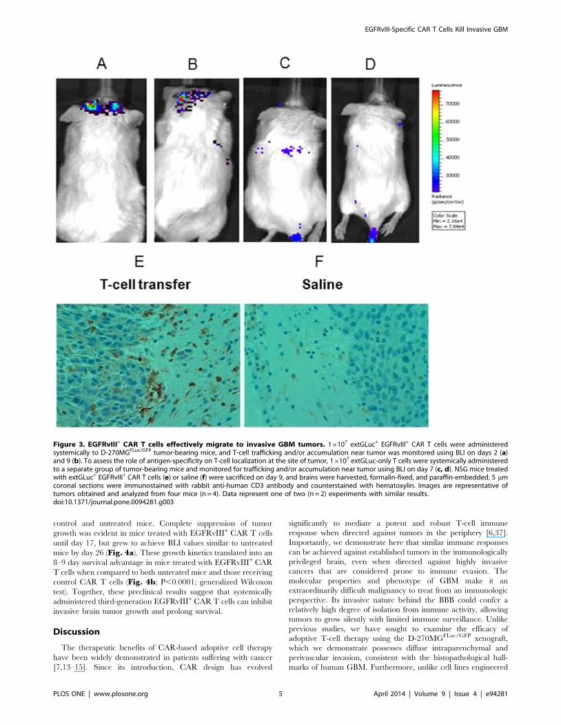
270MGFLuc/GFP tumors. Imaging analysis two (Fig. 3a) and nine
(Fig. 3b) days after T-cell injection revealed extGLuc signals in
the brain area, demonstrating localization of EGFRvIII+ CAR T
cells to the tumor site. We also sought to evaluate whether
EGFRvIII-specificity was necessary for efficient T-cell localization
to tumor, and to do this, we compared the trafficking patterns of
extGluc+ EGFRvIII+ T cells (Fig. 3a,b) with extGluc+ T cells
(Fig. 3c,d) in NSG mice bearing D-270MGFLuc/GFP tumors. We
found that extGluc-only T cells (EGFRvIII-) failed to efficiently
Figure 1. D-270MGFLuc/GFP xenograft is highly invasive in NSG mice. NSG mice received intracerebral tumor implants (D-270MGFLuc/GFP/16104 cells or U87MG.DEGFR/16104 cells) and were sacrificed after seven days. Histological analysis by H&E staining of (a) non-tumor bearing brain,(b, c) established U87MG.DEGFR and (d, e, f) D-270MGFLuc/GFP intracerebral malignant gliomas in NSG mice is shown. Figures delineate tumor vs.normal brain and demonstrate perivascular (PV) and subarachnoid (SA) infiltration. Images are representative of tumors obtained and analyzed fromsix mice (n = 6). x20 magnification.doi:10.1371/journal.pone.0094281.g001
EGFRvIII-Specific CAR T Cells Kill Invasive GBM
PLOS ONE | www.plosone.org 3 April 2014 | Volume 9 | Issue 4 | e94281

migrate across the BBB, whereas extGluc+ EGFRvIII+ CAR T
cells rapidly localized in the brain. This suggests a necessity of
EGFRvIII+ CAR expression for brain-trafficking or accumulation
in NSG mice bearing D-270MGFLuc/GFP tumors. A separate
group of mice were also sacrificed nine days after T-cell injection
and tumor tissue was submitted for immunohistochemical analysis.
Figure 3E shows that systemically administered EGFRvIII+ CAR
T cells successfully migrated to the invasive edges of intracerebral
tumor, particularly in areas of peninsula formation at the leading
edge of tumor invasion. NSG mice receiving saline were used as a
control (Fig. 3F). These results demonstrate that, in our model of
invasive GBM, adoptively transferred EGFRvIII+ CAR T cells
have the capacity to traffic to invasive areas of tumor thought to
reside beyond the BBB.
In vivo systemic delivery of EGFRvIII+ CAR T cells delaystumor growth and prolongs survival
We sought to determine the therapeutic effect of systemically
administered EGFRvIII+ CAR T cells against invasive intracere-
bral gliomas in vivo. Utilizing the NSG mouse model, D-
270MGFLuc/GFP xenografts were implanted intracerebrally and
allowed to engraft for three days prior to intravenous infusion with
EGFRvIII+ CAR T cells. D-270MGFLuc/GFP cells were monitored
using BLI every three days. No significant difference was observed
between groups of mice that were either left untreated or infused
with non-specific control CAR T cells. However, there was a
significant delay in tumor growth in EGFRvIII+ CAR T-cell
treatment groups compared to untreated and control CAR T cells,
as detected by serial BLI recordings of tumor-cell photon emissions
(Fig. 4a; P,0.0001; mixed model). Tumors were not visible in
any group until day 11, at which point tumors began growing in
Figure 2. T cells expressing EGFRvIII+ CARs recognize D-270MGFLuc/GFP tumors that naturally express EGFRvIII. (a) Cells were stainedfor EGFRvIII+ CARs to detect cell-surface expression using EGFRvIII multimer-PE. Negative staining controls were conducted by staining untransducedT cells from the same donor. EGFRvIII CAR+ T cells were gated on CD3+ (left), CD3+ CD4+ (middle), and CD3+ CD8+ (right). (b) Expression levels ofEGFRvIII were quantified in D-270MGFLuc/GFP and U87MG.DEGFR tumor cells using qRT PCR. U87MG (EGFRvIII-) tumor cells were used as a control. (c)In order to assess EGFRvIII specificity, untransduced T cells or EGFRvIII+ CAR T cells were co-cultured with U87MG or U87MG.DEGFR tumor cells.Quantification of cells positive for ICS of IFN-c+ is shown. The effect of T-cell transduction on frequency of IFN-c+ cells significantly differs betweenU87MG and U87MG.DEGFR tumor cells (P = 0.0004; two-way ANOVA). (c) To evaluate D-270MGFLuc/GFP tumor cell recognition, untransduced T cells orEGFRvIII+ CAR T cells were co-cultured with D-270MGFLuc/GFP. Quantification of cells positive for ICS of IFN-c+ is shown. The effect of T-celltransduction on frequency of IFN-c+ cells significantly differs between untransduced T cells and EGFRvIII+ CAR T cells (P = 0.002; one-way ANOVA).EGFRvIII+ CAR T cells were also cultured alone (no target) as a control, and quantification of cells positive for ICS of IFN-c+ was negligible (data notshown). Data represent one of two (n = 2) experiments with similar results.doi:10.1371/journal.pone.0094281.g002
EGFRvIII-Specific CAR T Cells Kill Invasive GBM
PLOS ONE | www.plosone.org 4 April 2014 | Volume 9 | Issue 4 | e94281
control and untreated mice. Complete suppression of tumor
growth was evident in mice treated with EGFRvIII+ CAR T cells
until day 17, but grew to achieve BLI values similar to untreated
mice by day 26 (Fig. 4a). These growth kinetics translated into an
8–9 day survival advantage in mice treated with EGFRvIII+ CAR
T cells when compared to both untreated mice and those receiving
control CAR T cells (Fig. 4b; P,0.0001; generalized Wilcoxon
test). Together, these preclinical results suggest that systemically
administered third-generation EGFRvIII+ CAR T cells can inhibit
invasive brain tumor growth and prolong survival.
Discussion
The therapeutic benefits of CAR-based adoptive cell therapy
have been widely demonstrated in patients suffering with cancer
[7,13–15]. Since its introduction, CAR design has evolved
significantly to mediate a potent and robust T-cell immune
response when directed against tumors in the periphery [6,37].
Importantly, we demonstrate here that similar immune responses
can be achieved against established tumors in the immunologically
privileged brain, even when directed against highly invasive
cancers that are considered prone to immune evasion. The
molecular properties and phenotype of GBM make it an
extraordinarily difficult malignancy to treat from an immunologic
perspective. Its invasive nature behind the BBB could confer a
relatively high degree of isolation from immune activity, allowing
tumors to grow silently with limited immune surveillance. Unlike
previous studies, we have sought to examine the efficacy of
adoptive T-cell therapy using the D-270MGFLuc/GFP xenograft,
which we demonstrate possesses diffuse intraparenchymal and
perivascular invasion, consistent with the histopathological hall-
marks of human GBM. Furthermore, unlike cell lines engineered
Figure 3. EGFRvIII+ CAR T cells effectively migrate to invasive GBM tumors. 16107 extGLuc+ EGFRvIII+ CAR T cells were administeredsystemically to D-270MGFLuc/GFP tumor-bearing mice, and T-cell trafficking and/or accumulation near tumor was monitored using BLI on days 2 (a)and 9 (b). To assess the role of antigen-specificity on T-cell localization at the site of tumor, 16107 extGLuc-only T cells were systemically administeredto a separate group of tumor-bearing mice and monitored for trafficking and/or accumulation near tumor using BLI on day 7 (c, d). NSG mice treatedwith extGLuc+ EGFRvIII+ CAR T cells (e) or saline (f) were sacrificed on day 9, and brains were harvested, formalin-fixed, and paraffin-embedded. 5 mmcoronal sections were immunostained with rabbit anti-human CD3 antibody and counterstained with hematoxylin. Images are representative oftumors obtained and analyzed from four mice (n = 4). Data represent one of two (n = 2) experiments with similar results.doi:10.1371/journal.pone.0094281.g003
EGFRvIII-Specific CAR T Cells Kill Invasive GBM
PLOS ONE | www.plosone.org 5 April 2014 | Volume 9 | Issue 4 | e94281

to express EGFRvIII, such as U87MG.DEGFR, D-270 MG was
isolated from a primary tumor that naturally expressed EGFRvIII
and has maintained expression ex vivo and in vivo. This lends greater
credence to its more accurate recapitulation of the clinical
scenario.
We demonstrate here that EGFRvIII+ CAR T cells recognize
tumors naturally expressing EGFRvIII, such as D-270MGFLuc/GFP.
We show that these EGFRvIII+ CAR T cells recognize tumor cells
in an antigen-specific manner in vitro as measured by ICS (Fig. 2c,d). It is important to note that we measured a 10–15% frequency of
IFN-c+ cells in our in vitro ICS assays, which is less than our recorded
frequency of CD3+ CAR+ T cells. This has been a consistent and
expected result under the culture, transduction, and assay protocols
described here. One explanation may be the varied differentiation
states of CD3+ CAR+ T cells, since different stages of activation can
alter the incubation time required for T-cell secretion of IFN-c. We
have found that longer incubation times (greater than the 18 h
described here) and altered tumor : T cell ratios yield frequencies.
50%, and we suspect that this is due to the inclusion of more cells
occupying a greater spectrum of activation. However, we chose the
assay conditions described here since incubation times.18 h
decrease cell viability, and 18 h incubations have yielded consistent
results to date [38].
Our data show that systemically delivered EGFRvIII+ CAR T
cells have the capacity to migrate to invasive tumor deposits within
the CNS. T-cell migration across endothelium requires molecular
cues provided by chemokine-chemokine receptor interaction and
engagement of adhesion molecules, which are thought to be
independent of TCR engagement [39,40]. The cross-reactivity of
murine adhesion molecules and chemokines with human T cells
and their chemokine receptors is known to be limited [41], and
although this could have negatively impacted T-cell localization,
we instead observed a substantial influx of T cells into the invasive
tumor. We are currently evaluating the contribution of tumor-
derived human chemokines and EGFRvIII-specificity on CAR T
cells to further elucidate the steps required for effective T-cell
migration across the BBB in the invasive areas of GBM.
Importantly, we show here that systemically administered
EGFRvIII+ CAR T cells have the capacity to inhibit tumor
growth and prolong survival of mice with established D-
270MGFLuc/GFP tumors (Fig. 4a, b). Despite the evidence of an
effective, antigen-specific immune response, it is important to note
that brain tumors ultimately continued to grow and caused death
even in mice receiving EGFRvIII+ CAR T cells (Fig. 4b).
Normalized BLI values from these treated mice eventually reached
comparable values to untreated and CAR control groups,
indicating tumor growth after a period of dormancy likely
mediated by antitumor T-cell activity (Fig. 4a). One possible
explanation is the loss or functional loss of EGFRvIII+ CAR T
cells in this model. To test the antitumor efficacy of EGFRvIII+
CAR T cells in vivo, we chose an NSG mouse model, which has the
advantage of evaluating promising preclinical therapies in an
animal system using human tumor tissue. However, one major
drawback of this approach is the fact that human T cells often
have a limited lifespan and functional half-life in the murine
background. For this reason, we sought to monitor T-cell
persistence over time in vivo by BLI analysis. However, we
unexpectedly found the administration of coelenterazine to be
toxic at the manufacturer’s recommended dosage, and as such,
terminated BLI measurements after day 9 (Fig. 3a, b). Our
studies demonstrate that, despite this potential for CAR T cell loss,
EGFRvIII+ CAR T cells were able to persist long enough to
migrate to and mediate antitumor activity against invasive
intracranial tumors. We are currently evaluating host conditioning
regimens to support enhanced and long-term human T-cell
survival and function in NSG mice, since these factors could, in
theory, potentiate antitumor efficacy.
A second possible explanation for the eventual recurrence of
tumor in our model is the concept of antigen-loss, wherein
therapeutic pressure selects for tumor cells that do not express the
target antigen. This explanation would be consistent with two
recent clinical studies where recurrence in patients treated with
CARs [42] or a vaccine targeting a single antigen [43] was
characterized by outgrowth of antigen-loss variants. As such, given
the theoretical limitations of targeting single tumor antigens, future
efforts will likely focus on the identification of additional GBM-
specific targets and multimodal therapies designed to target several
antigens simultaneously through CAR-mediated or alternative
Figure 4. In vivo systemic delivery of EGFRvIII+ CAR T cells delays tumor growth and prolongs survival. NSG mice were implanted with16104 D-270MGFLuc/GFP tumor cells intracranially, randomized into three groups (n = 6-8), and monitored for tumor growth and survival. 5.0 - 106106
T cells were administered intravenously 3 days after tumor implantation. (a) Normalized BLI values associated with longitudinal monitoring of tumorgrowth for untreated, control CAR T-cell, and EGFRvIII+ CAR T-cell groups are shown (value = mean 6SD). The pattern of change in log BLI valuessignificantly differs between the three treatment groups (P,0.0001; mixed model). (b) The survival of animals treated with EGFRvIII+ CAR T cells wassignificantly prolonged (P,0.0001; generalized Wilcoxon test) when compared to other treatment groups. Data represent one of two (n = 2)experiments with similar results.doi:10.1371/journal.pone.0094281.g004
EGFRvIII-Specific CAR T Cells Kill Invasive GBM
PLOS ONE | www.plosone.org 6 April 2014 | Volume 9 | Issue 4 | e94281
T-cell-based approaches. Additional areas of further investigation
may include determining factors involved in eliciting broader
endogenous immune responses through mechanisms such as
epitope spreading, which has emerged as a critical factor during
clinical trials of immunotherapy for melanoma [44].
In the current study, we have demonstrated that systemically
administered and tumor-specific EGFRvIII+ CAR T cells
effectively migrate to areas of tumor invasion and mediate efficacy
in a murine model of invasive human glioma. This work
contributes to the rapidly growing literature supporting the utility
of adoptively transferred CAR T cells targeting tumor-specific
antigens as a potent treatment modality for invasive brain tumors.
Materials and Methods
Human GBM cell lines and xenograftsWe utilized the previously described human glioma cell line,
U87MG [29–31], which does not express EGFRvIII, and subline
U87MG.DEGFR [33,34], which was stably transfected to express
EGFRvIII. We also utilized the D-270 MG cell line, which was
propagated directly as a xenograft from a primary human GBM
harvested from a patient and has previously been shown to
naturally express EGFRvIII [20,32]. Briefly, mechanically minced
tumor tissue was enzymatically dissociated into single cells using
the Papain Dissociation System. After wash, tissue was homoge-
nized and passed through a 75 mm cell strainer, re-suspended with
freezing medium containing 90% fetal bovine serum (FBS) and
10% dimethyl sulfoxide and frozen in individual vials using
standard procedures. For further experimentation, cells were
thawed at 37uC, washed, and counted with trypan blue per
standard practice.
AnimalsNSG mice were obtained (Charles River Laboratories,
Wilmington, MA) and bred under standard conditions at Duke
University Medical Center (DUMC). Mice were kept and utilized
under the accordance of protocols approved by the Duke
University Institutional Animal Care and Use Committee
(IACUC). All mice used in this study were healthy females
between 6–8 weeks of age weighing 0.020–0.025 kg and were
randomized to experimental or control groups. Mice were
routinely monitored for health (every 2 days) and qualified for
euthanasia if they demonstrated an inability to ambulate to food
and water (i.e. in lateral recumbency and unable to right itself), or
if they were unable to move forward two steps when prompted
gently by touching a finger to the hind area. Moribund mice were
humanely euthanized when they met these endpoints using CO2
asphyxiation followed by decapitation as approved by our Duke
University IACUC protocol. In accordance with this protocol,
mice did not receive any analgesics or anesthetics.
Human PBLsHuman PBLs used in this study were obtained from normal
volunteers at Duke University Medical Center. The use of PBLs
was approved under protocol 0009043 by the Duke University
School of Medicine Institutional Review Board (irb.mc.duke.edu).
Approved protocols conform to the Declaration of Helsinki
protocols. All patients signed a written informed consent. PBLs
were cultured in AIM-V medium (Life Technologies, Grand
Island, NY) supplemented with 10% human AB serum (Valley
Biomedical Inc., Winchester, VA), 50 units/mL penicillin, 50 mg/
mL streptomycin (Life Technologies, Grand Island, CA) and
300 IU/mL interleukin-2 (IL-2) and maintained at 37uC with 5%
CO2.
EGFRvIII+ CARs and extGLuc retroviral vectortransduction
The extGLuc retroviral vectors were supplied by Renier
Brentjens of Memorial Sloan-Kettering Cancer Center, New
York, NY. The EGFRvIII+ CAR and extGLuc retroviral vectors
were utilized to generate EGFRvIII+ CAR T cells and exGluc+
EGFRvIII+ CAR T cells. The transduction procedures have
previously been described [36,45,46]. Briefly, peripheral blood
mononuclear cells (PBMCs) from healthy donors and GBM
patients (post-resection, prior to treatment) were thawed and
cultured in AIM-V medium supplemented with 5% human AB
serum, plus antibiotics, 300 IU/mL IL-2, and 50 ng/mL OKT-3.
After 48 hours, T cells (0.256106/mL) were transduced with a
retroviral supernatant containing either the extGLuc, EGFRvIII+
CAR, or both vectors spun onto RetroNectin (Takara Bio Inc,
Japan) coated non-tissue culture treated 6-well plates twice on two
consecutive days as described by the manufacturer. Transduced
cells were allowed to expand in AIM-V medium as above, without
OKT-3.
Rapid Expansion ProtocolTransduced PBLs (or untransduced control PBLs from same
donor) were expanded in vitro using rapid expansion protocol
(REP) [47]. Briefly, T cells were cultured in complete AIM-V
medium plus 10% human AB serum, 300 IU/mL IL-2, and
50 ng/mL OKT-3 in the presence of 100x excess 5000 rads
irradiated allogeneic PBMC feeder cells, and allowed to expand
10–14 days.
Lentiviral transduction of D-270 MG withfirefly-luciferase-GFP gene
The D-270 MG tumor cell line was transduced with a lentiviral
vector encoding the firefly luciferase (FLuc) and EGFP genes
linked by 2A peptide driven by an internal murine stem cell virus
(MSCV) promoter. Briefly, lentiviral vectors were generated by
transient transfection of HEK 293T cells with a four-plasmid
system [48]. Six hours post-transfection, plates were washed twice
with phosphate-buffered saline (PBS) and 20 mL fresh medium
was added. The supernatant was collected 30–48 hours post-
transfection and cell debris was removed by centrifugation at
6000 g for 10 minutes, followed by filtration on 0.45 mm
polyvinylidene fluoride filters. The lentiviral supernatant was kept
at 280uC. D-270 MG cells were then quickly thawed at 37uC,
washed, counted with trypan blue, and re-suspended in the
lentiviral supernatant and zinc medium with 10% FBS and
incubated at 37uC with 5% CO2 overnight. D-270MGFLuc/GFP
tumor cells were cell sorted based on GFP expression. Expression
of FLuc was confirmed by data acquisition using the IVIS 100 in
vivo BLI system (Caliper Life Sciences, Hopkinton, MA) coupled
with Living Image software (PerkinElmer, Waltham, MA).
EGFRvIII expression levels by D-270MGFLuc/GFP and
U87MG.DEGFR tumor cells were measured by qRT PCR as
previously described [49].
Cell surface CAR expression and ICS of transduced T cellsCells were stained for EGFRvIII+ CARs to detect cell-surface
expression using an EGFRvIII multimer-PE as previously
described [50]. Negative staining controls were conducted by
staining untransduced cells from the same donor. ICS was
performed by co-culturing T cells with tumors 1:1 over 18 hours
with the BD GolgiPlug protein transport inhibitor containing
brefeldin A (BD Sciences, San Jose, CA) in RPMI-1640 medium
plus 10% FBS. Following co-culture, cells were submitted to
EGFRvIII-Specific CAR T Cells Kill Invasive GBM
PLOS ONE | www.plosone.org 7 April 2014 | Volume 9 | Issue 4 | e94281
surface staining for CD3, CD8 and intracellular IFN-c stain using
the BD Cytofix/Cytoperm method (BD Biosciences, San Jose,
CA).
Monitoring tumor growth and T-cell trafficking using BLITo monitor tumor growth, 24 NSG mice underwent intracra-
nial implantation of 16104 D-270MGFLuc/GFP tumor cells in 5 ml
PBS and methocell mixer using stereotactic coordinates 2 mm
lateral and 4 mm intraparenchymal from the bregma on
experimental day 0. Tumor growth was analyzed every three to
five days for 26 days by BLI as previously described [51] until the
study was terminated. Briefly, BLI was performed by injecting
mice intraperitoneally with 150 mg D-luciferin/kg (Xenogen,
Hopkinton, MA) 10 minutes prior to imaging and photon emission
(photons s21 cm22 sr21) was recorded. To monitor T-cell
trafficking, T cells were transduced with the extGLuc or co-
transduced with the EGFRvIII+ CAR and extGLuc retroviral
vectors as described above (see EGFRvIII+ CARs and extGLuc
retroviral vector transduction). Mice receiving T cells were injected with
5.0 - 106106 cells intravenously via the tail vein and imaged on
days 2 and 9 after infusion. Briefly, 250 mg coelenterazine
(NanoLight Technology, Pinetop, AZ) was injected IV in mice
receiving either saline or EGFRvIII+ extGLuc+ CAR T cells and
imaged within 90 seconds by measuring BLI. We obtained image
data sets and measurement of signal intensity by using the IVIS
100 in vivo BLI system and through region of interest analysis using
Living Image Software with normalized images displayed on each
data set according to color intensity. Mean BLI values for control
and treatment groups were calculated and plotted according to the
corresponding day of imaging.
H&E Staining and ImmunohistochemistryIn order to evaluate tumor architecture and growth patterns, NSG
mice received intracerebral tumor implants (D-270MGFLuc/GFP/
16104 cells or U87MG.DEGFR/16104 cells) and were sacrificed
after seven days. Brains were harvested, formalin-fixed and paraffin-
embedded. 5 mm sections were stained with H&E. To evaluate
T-cell migration to the invasive edge of tumor, we utilized two
experimental groups, which included four tumor-bearing mice
treated with saline or extGLuc+ EGFRvIII+ CAR 16107 T cells.
Mice were euthanized on day 9. Brains were harvested, formalin-
fixed and paraffin-embedded. 5 mm coronal sections were immu-
nostained with rabbit anti-human CD3 antibody (Thermo Lab
Vision, Clone RM9107S) as recommended by the manufacturer at
a 1:100 dilution and counterstained with hematoxylin.
Statistical methodsStatistical differences in group percentage cytotoxicity and
bioluminescence were evaluated by either a one-way analysis of
variance (ANOVA) model or a two-way ANOVA model with
interaction. The Kaplan Meier estimator was used to generate
survival curves, and differences between survival curves were
calculated using a generalized Wilcoxon test. Patterns of change in
normalized BLI values on the log scale over time were evaluated
using a mixed model that included main effects for time and
treatment group along with a time interaction with treatment
group. This model accounted for within animal correlation of
measurements by using a 1st degree autoregressive covariance
structure.
Acknowledgments
We extend our gratitude to Steven A. Rosenberg and Richard A. Morgan
of the Surgery Branch at the National Cancer Institute for providing us
with the EGFRvIII+ CAR construct and Renier J. Brentjens of Memorial
Sloan-Kettering Cancer Center for providing us with the extGluc
retrovirus used in this study. We also thank Alina Boesteanu for providing
histology images of U87MG.DEGFR xenograft implants and David
Snyder for his contribution to animal care.
Author Contributions
Conceived and designed the experiments: HM BDC CMS LSP GA DB
LAJ JHS. Performed the experiments: HM LSP SY GD EJS. Analyzed the
data: RM JEH PH. Contributed reagents/materials/analysis tools: LSP
LAJ JHS. Wrote the paper: HM BDC LSP CMS LAJ.
References
1. Stupp R, Mason WP, van den Bent MJ, Weller M, Fisher B, et al. (2005)Radiotherapy plus concomitant and adjuvant temozolomide for glioblastoma.
N Engl J Med 352: 987–996.
2. Imperato JP, Paleologos NA, Vick NA (1990) Effects of treatment on long-term
survivors with malignant astrocytomas. Ann Neurol 28: 818–822.
3. Johnson LA, Morgan RA, Dudley ME, Cassard L, Yang JC, et al. (2009) Gene
therapy with human and mouse T-cell receptors mediates cancer regression andtargets normal tissues expressing cognate antigen. Blood 114: 535–546.
4. Hong JJ, Rosenberg SA, Dudley ME, Yang JC, White DE, et al. (2010)
Successful treatment of melanoma brain metastases with adoptive cell therapy.
Clin Cancer Res 16: 4892–4898.
5. Robbins PF, Morgan RA, Feldman SA, Yang JC, Sherry RM, et al. (2011)Tumor regression in patients with metastatic synovial cell sarcoma and
melanoma using genetically engineered lymphocytes reactive with NY-ESO-1.
J Clin Oncol 29: 917–924.
6. Rosenberg SA (2011) Cell transfer immunotherapy for metastatic solid cancer—what clinicians need to know. Nat Rev Clin Oncol 8: 577–585.
7. Kochenderfer JN, Dudley ME, Feldman SA, Wilson WH, Spaner DE, et al.(2012) B-cell depletion and remissions of malignancy along with cytokine-
associated toxicity in a clinical trial of anti-CD19 chimeric-antigen-receptor-transduced T cells. Blood 119: 2709–2720.
8. Rosenberg SA, Yang JC, Robbins PF, Wunderlich JR, Hwu P, et al. (2003) Celltransfer therapy for cancer: lessons from sequential treatments of a patient with
metastatic melanoma. J Immunother 26: 385–393.
9. Zitvogel L, Tesniere A, Kroemer G (2006) Cancer despite immunosurveillance:
immunoselection and immunosubversion. Nat Rev Immunol 6: 715–727.
10. Kalos M, June CH (2013) Adoptive T cell transfer for cancer immunotherapy in
the era of synthetic biology. Immunity 39: 49–60.
11. Lamers CH, Sleijfer S, Vulto AG, Kruit WH, Kliffen M, et al. (2006) Treatmentof metastatic renal cell carcinoma with autologous T-lymphocytes genetically
retargeted against carbonic anhydrase IX: first clinical experience. J Clin Oncol
24: e20–22.
12. Till BG, Jensen MC, Wang J, Qian X, Gopal AK, et al. (2012) CD20-specificadoptive immunotherapy for lymphoma using a chimeric antigen receptor with
both CD28 and 4-1BB domains: pilot clinical trial results. Blood 119: 3940–
3950.
13. Pule MA, Savoldo B, Myers GD, Rossig C, Russell HV, et al. (2008) Virus-
specific T cells engineered to coexpress tumor-specific receptors: persistence andantitumor activity in individuals with neuroblastoma. Nat Med 14: 1264–1270.
14. Brentjens RJ, Davila ML, Riviere I, Park J, Wang X, et al. (2013) CD19-targetedT cells rapidly induce molecular remissions in adults with chemotherapy-
refractory acute lymphoblastic leukemia. Sci Transl Med 5: 177ra138.
15. Porter DL, Levine BL, Kalos M, Bagg A, June CH (2011) Chimeric antigen
receptor-modified T cells in chronic lymphoid leukemia. N Engl J Med 365:725–733.
16. Brentjens R, Yeh R, Bernal Y, Riviere I, Sadelain M (2010) Treatment ofchronic lymphocytic leukemia with genetically targeted autologous T cells: case
report of an unforeseen adverse event in a phase I clinical trial. Mol Ther 18:
666–668.
17. Morgan RA, Yang JC, Kitano M, Dudley ME, Laurencot CM, et al. (2010)
Case report of a serious adverse event following the administration of T cellstransduced with a chimeric antigen receptor recognizing ERBB2. Mol Ther 18:
843–851.
18. Wikstrand CJ, Hale LP, Batra SK, Hill ML, Humphrey PA, et al. (1995)
Monoclonal antibodies against EGFRvIII are tumor specific and react withbreast and lung carcinomas and malignant gliomas. Cancer Res 55: 3140–3148.
19. Heimberger AB, Hlatky R, Suki D, Yang D, Weinberg J, et al. (2005) Prognosticeffect of epidermal growth factor receptor and EGFRvIII in glioblastoma
multiforme patients. Clin Cancer Res 11: 1462–1466.
20. Wong AJ, Ruppert JM, Bigner SH, Grzeschik CH, Humphrey PA, et al. (1992)
Structural alterations of the epidermal growth factor receptor gene in human
gliomas. Proc Natl Acad Sci U S A 89: 2965–2969.
21. Engelhardt B (2006) Molecular mechanisms involved in T cell migration across
the blood-brain barrier. Journal of Neural Transmission 113: 477–485.
EGFRvIII-Specific CAR T Cells Kill Invasive GBM
PLOS ONE | www.plosone.org 8 April 2014 | Volume 9 | Issue 4 | e94281

22. Banks WA, Erickson MA (2010) The blood-brain barrier and immune function
and dysfunction. Neurobiology of Disease 37: 26–32.23. Rascher G, Fischmann A, Kroger S, Duffner F, Grote EH, et al. (2002)
Extracellular matrix and the blood-brain barrier in glioblastoma multiforme:
spatial segregation of tenascin and agrin. Acta Neuropathol 104: 85–91.24. Ajay D, Sanchez-Perez L, Choi BD, De Leon G, Sampson JH (2012)
Immunotherapy with tumor vaccines for the treatment of malignant gliomas.Curr Drug Discov Technol 9: 237–255.
25. Claes A, Idema AJ, Wesseling P (2007) Diffuse glioma growth: a guerilla war.
Acta Neuropathol 114: 443–458.26. Giese A, Westphal M (1996) Glioma invasion in the central nervous system.
Neurosurgery 39: 235–250; discussion 250–232.27. Agarwal S, Manchanda P, Vogelbaum MA, Ohlfest JR, Elmquist WF (2013)
Function of the blood-brain barrier and restriction of drug delivery to invasiveglioma cells: findings in an orthotopic rat xenograft model of glioma. Drug
Metab Dispos 41: 33–39.
28. Vauleon E, Avril T, Collet B, Mosser J, Quillien V, et al. (2010) Overview ofCellular Immunotherapy for Patients with Glioblastoma. Clinical and
Developmental Immunology 2010.29. Hashizume R, Ozawa T, Dinca EB, Banerjee A, Prados MD, et al. (2010) A
human brainstem glioma xenograft model enabled for bioluminescence imaging.
J Neurooncol 96: 151–159.30. Miura FK, Alves MJ, Rocha MC, da Silva R, Oba-Shinjo SM, et al. (2010)
Xenograft transplantation of human malignant astrocytoma cells into immuno-deficient rats: an experimental model of glioblastoma. Clinics (Sao Paulo) 65:
305–309.31. Candolfi M, Curtin JF, Nichols WS, Muhammad AG, King GD, et al. (2007)
Intracranial glioblastoma models in preclinical neuro-oncology: neuropatholog-
ical characterization and tumor progression. J Neurooncol 85: 133–148.32. Bigner SH, Humphrey PA, Wong AJ, Vogelstein B, Mark J, et al. (1990)
Characterization of the epidermal growth factor receptor in human glioma celllines and xenografts. Cancer Res 50: 8017–8022.
33. Nishikawa R, Ji XD, Harmon RC, Lazar CS, Gill GN, et al. (1994) A mutant
epidermal growth factor receptor common in human glioma confers enhancedtumorigenicity. Proc Natl Acad Sci U S A 91: 7727–7731.
34. Lal A, Glazer CA, Martinson HM, Friedman HS, Archer GE, et al. (2002)Mutant epidermal growth factor receptor up-regulates molecular effectors of
tumor invasion. Cancer Res 62: 3335–3339.35. Morgan RA, Johnson LA, Davis JL, Zheng Z, Woolard KD, et al. (2012)
Recognition of glioma stem cells by genetically modified T cells targeting
EGFRvIII and development of adoptive cell therapy for glioma. Hum GeneTher 23: 1043–1053.
36. Santos EB, Yeh R, Lee J, Nikhamin Y, Punzalan B, et al. (2009) Sensitive in vivoimaging of T cells using a membrane-bound Gaussia princeps luciferase. Nat
Med 15: 338–344.
37. Sadelain M, Brentjens R, Riviere I (2013) The basic principles of chimericantigen receptor design. Cancer Discov 3: 388–398.
38. Choi BD, Suryadevara CM, Gedeon PC, Herndon Ii JE, Sanchez-Perez L, et al.
(2014) Intracerebral delivery of a third generation EGFRvIII-specific chimeric
antigen receptor is efficacious against human glioma. J Clin Neurosci 21: 189–
190.
39. Engelhardt B, Ransohoff RM (2012) Capture, crawl, cross: the T cell code to
breach the blood-brain barriers. Trends Immunol 33: 579–589.
40. Ransohoff RM (2009) Chemokines and chemokine receptors: standing at the
crossroads of immunobiology and neurobiology. Immunity 31: 711–721.
41. Mestas J, Hughes CC (2004) Of mice and not men: differences between mouse
and human immunology. J Immunol 172: 2731–2738.
42. Grupp SA, Kalos M, Barrett D, Aplenc R, Porter DL, et al. (2013) Chimeric
antigen receptor-modified T cells for acute lymphoid leukemia. N Engl J Med
368: 1509–1518.
43. Sampson JH, Heimberger AB, Archer GE, Aldape KD, Friedman AH, et al.
(2010) Immunologic escape after prolonged progression-free survival with
epidermal growth factor receptor variant III peptide vaccination in patients with
newly diagnosed glioblastoma. J Clin Oncol 28: 4722–4729.
44. Butterfield LH, Ribas A, Dissette VB, Amarnani SN, Vu HT, et al. (2003)
Determinant spreading associated with clinical response in dendritic cell-based
immunotherapy for malignant melanoma. Clin Cancer Res 9: 998–1008.
45. Hughes MS, Yu YY, Dudley ME, Zheng Z, Robbins PF, et al. (2005) Transfer of
a TCR gene derived from a patient with a marked antitumor response conveys
highly active T-cell effector functions. Hum Gene Ther 16: 457–472.
46. Morgan RA, Dudley ME, Yu YY, Zheng Z, Robbins PF, et al. (2003) High
efficiency TCR gene transfer into primary human lymphocytes affords avid
recognition of melanoma tumor antigen glycoprotein 100 and does not alter the
recognition of autologous melanoma antigens. J Immunol 171: 3287–3295.
47. Riddell SR, Greenberg PD (1990) The use of anti-CD3 and anti-CD28
monoclonal antibodies to clone and expand human antigen-specific T cells.
J Immunol Methods 128: 189–201.
48. Yang S, Cohen CJ, Peng PD, Zhao Y, Cassard L, et al. (2008) Development of
optimal bicistronic lentiviral vectors facilitates high-level TCR gene expression
and robust tumor cell recognition. Gene Ther 15: 1411–1423.
49. Yoshimoto K, Dang J, Zhu S, Nathanson D, Huang T, et al. (2008)
Development of a real-time RT-PCR assay for detecting EGFRvIII in
glioblastoma samples. Clin Cancer Res 14: 488–493.
50. Sampson JH, Choi BD, Sanchez-Perez L, Suryadevara CM, Snyder DJ, et al.
(2013) EGFRvIII mCAR-modified T cell therapy cures mice with established
intracerebral glioma and generates host immunity against tumor-antigen loss.
Clin Cancer Res.
51. Szentirmai O, Baker CH, Lin N, Szucs S, Takahashi M, et al. (2006)
Noninvasive bioluminescence imaging of luciferase expressing intracranial U87
xenografts: correlation with magnetic resonance imaging determined tumor
volume and longitudinal use in assessing tumor growth and antiangiogenic
treatment effect. Neurosurgery 58: 365–372; discussion 365–372.
EGFRvIII-Specific CAR T Cells Kill Invasive GBM
PLOS ONE | www.plosone.org 9 April 2014 | Volume 9 | Issue 4 | e94281