Immune Response Regulation by Leishmania Secreted and Nonsecreted Antigens
Upload
sitmrewariCategory
view
3download
0
Green Fluorescent Protein Bone Marrow Cells ExpressHematopoietic and Neural Antigens in Culture and Migrate Withinthe Neonatal Rat Brain
J. E. Hudson1,2,*, N. Chen1,2, S. Song1,3,4, P. Walczak1,2, P. Jendelová5, E. Sykova5, A. E.Willing1,2, S. Saporta1,2, P. Bickford1,2,4, J. Sanchez-Ramos1,3,4, and T. Zigova1,21Center of Excellence for Aging and Brain Repair, University of South Florida, Tampa, Florida2Department of Neurosurgery, University of South Florida, Tampa, Florida3Department of Neurology, University of South Florida, Tampa, Florida4James A. Haley VA Hospital, Tampa, Florida5Center for Cell Therapy and Tissue Repair, Charles University, Institute of Experimental MedicineASCR, Prague, Czech Republic
AbstractFinding a reliable source of alternative neural stem cells for treatment of various diseases and injuriesaffecting the central nervous system is a challenge. Numerous studies have shown that hematopoieticand nonhematopoietic progenitors derived from bone marrow (BM) under specific conditions areable to differentiate into cells of all three germ layers. Recently, it was reported that cultured,unfractionated (whole) adult BM cells form nestin-positive spheres that can later initiate neuraldifferentiation (Kabos et al., 2002). The identity of the sub-population of BM cells that contributesto neural differentiation remains unknown. We therefore analyzed the hematopoietic and neuralfeatures of cultured, unfractionated BM cells derived from a transgenic mouse that expresses greenfluorescent protein (GFP) in all tissues. We also transplanted the BM cells into the subventricularzone (SVZ), a region known to support postnatal neuro-genesis. After injection of BM cells into theneurogenic SVZ in neonatal rats, we found surviving GFP+ BM cells close to the injection site andin various brain regions, including corpus callosum and subcortical white matter. Many of the graftedcells were detected within the rostral migratory stream (RMS), moving toward the olfactory bulb(OB), and some cells reached the subependymal zone of the OB. Our in vitro experiments revealedthat murine GFP+ BM cells retained their proliferation and differentiation potential andpredominantly preserved their hematopoietic identity (CD45, CD90, CD133), although a fewexpressed neural antigens (nestin, glial fibrillary acdiic protein, TuJ1).
Keywordsbone marrow; green mouse; grafting; subventricular zone; developing rat brain
The identification of nonfetal cells capable of neuronal differentiation has great potential fornumerous cellular therapies. Bone marrow (BM) contains therapeutically useful stem/
© 2004 Wiley-Liss, Inc.*Correspondence to: Jennifer Hudson, Department of Neurosurgery, College of Medicine, University of South Florida, 12901 Bruce B.Downs Blvd., Tampa, FL 33612. E-mail: E-mail: [email protected].
NIH Public AccessAuthor ManuscriptJ Neurosci Res. Author manuscript; available in PMC 2009 August 4.
Published in final edited form as:J Neurosci Res. 2004 April 15; 76(2): 255–264. doi:10.1002/jnr.20043.
NIH
-PA Author Manuscript
NIH
-PA Author Manuscript
NIH
-PA Author Manuscript
progenitor cells and may be considered a possible alternative source of cells for neural graftingin the treatment of neurological diseases.
Several investigators have published reports on hematopoietic and nonhematopoietic stem cellsderived from adult BM. Under certain, specific conditions, the nonhematopoietic BM cellsdifferentiated into cells expressing neuronal and glial antigens (Azizi et al., 1998; Sanchez-Ramos et al., 2000; Woodbury et al., 2000, 2002) and also into myogenic progenitors (Ferrariet al., 1998). Multipotentiality was also noticed in unfractionated BM-derived cells. Intransplantation studies, these cells were shown to express neural markers in the brain (Eglitisand Mezey, 1997; Mezey et al., 2000, 2003; Brazelton et al., 2000; Priller et al., 2001, Cortiet al., 2002a; Hess et al., 2002) and spinal cord (Corti et al., 2002b) and also to differentiateinto heart (Orlic et al., 2001) and liver (Petersen et al., 1999) cells.
In in vitro experiments under conditions commonly used for differentiating neural stem cells,whole BM was induced to form cellular spheres indistinguishable from neural stem cellneurospheres. These BM-derived spheres expressed neurogenin 1, a transcription factor foundduring specific stages of neural development (Kabos et al., 2002). After grafting into theneurogenically active hippocampus of adult rat, some of the transplanted BM cells integratedand tested positive for the neuronal marker NeuN. Thus, these whole BM-derived stem/progenitor cells can be differentiated in vitro by chemicals and growth factors or in vivo, in asuitable microenvironment.
In this study, we focused on the subventricular zone (SVZ), a life-long neurogenic region thatprovides developmentally important cues, such as epidermal growth factor (EGF), fibroblastgrowth factor-2 (FGF2), sonic hedgehog, cytokines, neurotrophic factors, bone morphogenicproteins (BMPs), and noggin (Reynolds and Weiss, 1992; Morshead et al., 1994; Palmer et al.,1995; Seroogy et al., 1995; Gross et al., 1996; Michaelson et al., 1996; Gritti et al., 1999; Limet al., 2000; Sobeih and Corfas, 2002; Marshall et al., 2003). These signals are able to determinethe cell’s phenotypic and positional fate and to maintain a migratory state by providingguidance cues to motile cells. Our own previous studies demonstrated that the SVZ and itsnatural extension, the RMS, can support the survival and migration of various grafted celltypes, from neural (Zigova et al., 1996, 2000; Yang et al., 2000) and nonneural (Zigova et al.,2002) sources. We used neonatal (0–2 days old) rats, because we expected these cues to bestronger in the younger, developing brain.
In the current study, we injected unfractionated BM cells that express green fluorescent protein(GFP) (Okabe et al., 1997) into the anterior part of the SVZ to determine whether progenitorcells from a different dermal origin would be able to survive, take distinct migratory pathways,and eventually adopt neural phenotypes after exposure to this young, highly neurogenicenvironment. At the same time, we plated GFP+ BM cells into media used for neuronal culturesto examine the fate of these cells by immunocytochemistry and to compare morphological andphenotypic features with the grafted cells.
MATERIALS AND METHODSPreparation of BM- and SVZ-Derived Progenitors
BM cells were harvested from transgenic “green mice” that constitutively express GFP(C57BL/6-TgN; Jackson Laboratory, Bar Harbor, ME) according to the procedure describedpreviously by Song and Sanchez-Ramos (2002). Briefly, after sacrifice of the animal, the femurand tibia were removed and flushed out with phosphate-buffered saline with 0.5% bovine serumalbumin (pH 7.2). The marrow was collected and spun down, the supernatant discarded, andthe resulting cell pellet suspended in Dulbecco’s modified Eagle’s medium (DMEM;Invitrogen, La Jolla, CA) with 10% fetal bovine serum (Invitrogen) and gentamicin (50 µl/ml;
Hudson et al. Page 2
J Neurosci Res. Author manuscript; available in PMC 2009 August 4.
NIH
-PA Author Manuscript
NIH
-PA Author Manuscript
NIH
-PA Author Manuscript

Sigma, St. Louis, MO) in a 15-ml conical tube (Falcon, Oxnard, CA) and centrifuged (400 g/7 min). The supernatant was removed, and the cells were resuspended in 1 ml of the above-described fresh medium. Their viability was assessed (85–92%) by using a rapid stainingprocedure with 0.4% trypan blue (Invitrogen). For culture, cells were plated at a density of100,000 cells/cm2 either on poly-L-lysine-coated (10 µg/ml; Sigma) eight-well chamber slides(Lab-Tek) or in 75-ml flasks (Corning, Corning, NY) and incubated at 37°C in 5% CO2. Theslides and flasks were incubated for up to 20 days with media changes every 3 days. Fortransplantation, BM-GFP cells were adjusted to 30,000 cells/µl.
SVZ progenitors were also prepared from the transgenic GFP mice. The SVZ wasmicrodissected from the brain (Zigova et al., 1996; Zigova and Newman, 2002; Laywell et al.,2002) and collected into incubation medium [10 ml of Hank’s balanced salt solution (HBSS;Invitrogen) containing 0.1% trypsin (Sigma) and 0.01 DNase (Sigma)] for 20 min at 37°C.After washing, the tissue was mechanically dissociated by gentle trituration, transferred intoDMEM containing 10% fetal bovine serum, and spun down. The supernatant was removedand the resulting cell pellet resuspended into 1 ml of fresh medium for estimation of cellviability (~90%) and for transplantation (30,000 cells/µl).
Cell cultures from murine BM were fixed with 4% paraformaldehyde after 1–20 days in vitro(DIV) for 10 min at room temperature and stored in 0.1 M phosphate buffer (pH 7.4) at 4°Cuntil immunocytochemistry for hematopoietic and neural markers was performed. To assessthe proliferative capacity of BM cells, the cultures were exposed to the S-phase marker 5-bromo-2′-deoxyuridine (BrdU; Sigma; 10 µM) for 2 hr before fixation.
Transplantation of Cell Suspension Into the Neonatal SVZTimed-pregnant rats (Sprague-Dawley) were purchased from Harlan, Inc. (Indianapolis, IN).After birth, neonatal 1-day-old rats from separate litters were anesthetized by hypothermiaaccording to the procedure described previously (Zigova et al., 1996, 2002b). Briefly, the headof the neonate pup was placed in a contoured Styrofoam mold, and an incision was madethrough the skin overlying the sagittal suture. A small hole was created in the skull on the righthemisphere (1 mm right from the midline, 2 mm anterior to Bregma), and a Hamilton syringewas lowered (2 mm deep to the pial surface) through the opening in the skull and 2 µl of cellsuspension slowly delivered into the SVZ at a rate of 1 µl/min for 2–3 min (BM-GFP, n = 16;SVZ-GFP, n = 5). After transplantation, the skin was repositioned and sealed with surgicalglue (Nexaband Liquid; Vet Prod Labs). The pups were placed under a heat lamp for recoverybefore returning them to their mother. This entire study followed the USF IACUC guidelinesand the Principles of Laboratory Animal Care.
Cell and Tissue Processing and ImmunocytochemistryThe purpose of our tissue culture assay was twofold: to establish the basic characteristics(phenotype and proliferative potential) of unfractionated, freshly prepared, and cultured BM-GFP cells and to work out double-labeling procedures for cell identification and furtherphenotypic characterization. These results were crucial for troubleshooting double- and triple-labeling procedures related to identification of transplanted cells, particularly in those caseswhen the green signal in the brain sections was not strong enough and the signal enhancementwas followed by additional immunoprocedures.
At various times after implantation (2–70 days), the animals were anesthetized by etherinhalation and perfused transcardially with 4% paraformaldehyde in 0.1 M phosphate buffer(pH 7.4). The brains were postfixed for 24 hr, immersed in cryopreservative (20% sucrose in0.1 M PBS) overnight at 4°C, embedded in tissue freezing compound (TBS), and cryosectionedin the sagittal plane at 20 µm thickness.
Hudson et al. Page 3
J Neurosci Res. Author manuscript; available in PMC 2009 August 4.
NIH
-PA Author Manuscript
NIH
-PA Author Manuscript
NIH
-PA Author Manuscript
For immunohistochemistry, the sections were thawed, rinsed thrice with PBS, and incubatedin blocking solution (0.1 M PBS containing 10% goat serum and 0.01% Triton X-100) for 1hr at room temperature, followed by incubation at 4°C overnight with the primary antibody.To enhance the GFP signal, a rabbit polyclonal anti-GFP antibody (Molecular Probes, Eugene,OR; 1:500) was employed. Rabbit polyclonal antifibronectin (Sigma; 1:400) to detect thepresence of extracellular matrix molecules in BM cells and the rat monoclonal panleukocytemarker CD45 (Serotec; 1:100) to detect hematopoietic cells were utilized. The following mousemonoclonal antibodies were used: CD90.2 (Thy-1.2; Pharmingen, San Diego, CA; 1:20) forT lymphocytes and CD133 (Miltenyi Biotec; 1:35) to recognize hematopoietic stem/progenitorcells. The microglia were identified by the rat monoclonal OX42 (1:100; Serotec, Bicester,United Kingdom), whereas mouse anti-nestin (Pharmingen; 1:100), mouse anti-TuJ1(Covance; 1:1,000), 1:500), and rabbit anti-glial fibrillary acidic protein (GFAP; Dako,Carpenteria, CA; 1:500) were used to recognize neural antigens. Controls in which the primaryantibody was eliminated resulted in no staining.
For BrdU staining, cells were pretreated with 2 N HCl at 60°C and washed with borate buffer(pH 8.5) at room temperature, blocked for 1 hr, then incubated with rat anti-BrdU (Accurate,Westbury, NY; 1:500). To estimate the number of fluorescently labeled BrdU cells, the numberof BrdU-positive and the total number of cells were counted after randomly choosing 20 fields.In addition, some slides were immunolabeled with sheep polyclonal Ki67 antibody (Chemicon,Temecula, CA; 1:100). Apoptosis was detected by using the terminal deoxynucleotidyltransferase (tdt) labeling technique (TACS apoptosis detection kit; R&D Systems,Minneapolis, MN). Positive and negative controls were carried out in parallel.
After being rinsed with PBS three times, the sections or cultured cells were incubated withspecies-appropriate fluorescent secondary antibodies conjugated to Alexa Fluor 594(Molecular Probes; 1:700), Alexa Fluor 488 (Molecular Probes; 1:600), or Alexa Fluor 350(Molecular Probes; 1:450) for 2 hr at room temperature. After being rinsed with PBS, the slideswere coverslipped with either 95% glycerol or Vectashield mounting medium. Cell nuclei werevisualized with DAPI (2 mg/ml; Roche). Cresyl violet-stained brain sections were used foridentification of the injection site and for morphological evaluation of the brain. All culturesand brain sections were examined using the Olympus BX60 and IX71 microscopes. Selectedslides were analyzed by confocal microscope (Nikon Inverted Diaphot), and images wereprocessed using the Oncor Image software.
RESULTSMorphology, Proliferative Capacity, and Phenotype of Green BM Cells
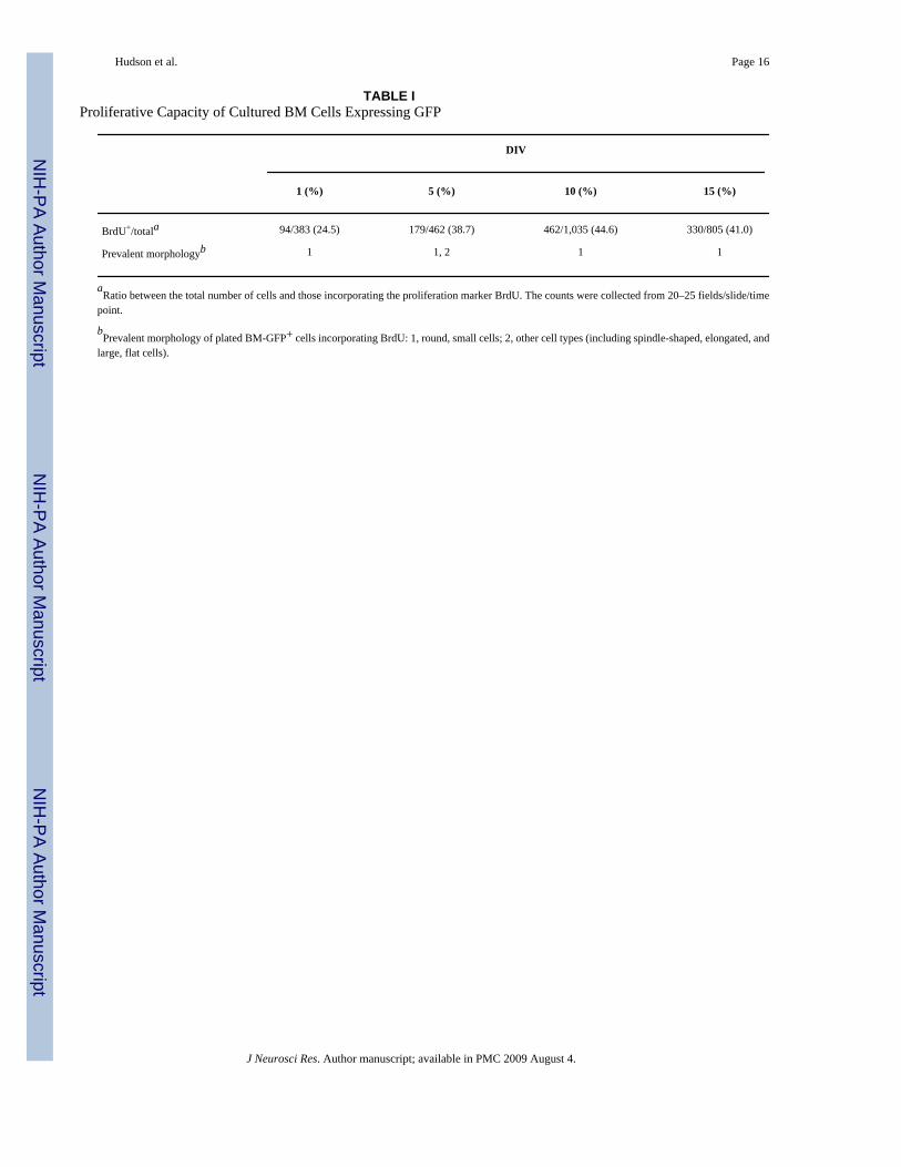
Shortly after plating (1–5 DIV), BM GFP cells began to develop into a heterogeneouspopulation of cells with very distinct cellular morphologies (Fig. 1A, parts A, B). Thediversified morphology became even more evident with longer culture periods (Fig. 1A, partsC, D). We frequently observed, in addition to small, round, spindle-shaped, and elongated cellswith two or more processes, large, flat, stromal-like cells (Fig. 1A, parts B–D). When we pulsedBM cultures with the cell proliferation marker BrdU, we found nuclear labeling in all studiedintervals and in all cell types (Fig. 1A, part E). Cell proliferation was also confirmed by thesecond proliferation marker, Ki67 (Fig. 1A, part E, inset F). At the same time, the low incidenceof apoptotic cells (Fig. 1A, part E, inset G) suggested that BrdU incorporation reflects theproliferative capacity of cultured BM cells (for at least the 3 weeks for which they had beenin culture; Table I) and not their near-death stage.
The expression of fibronectin, a marker typical for BM cells (Bentley and Tralka, 1983), wasfound mainly in round and stromal-like cells (Fig. 1B, part 1) at 5, 10, and 15 DIV. At 5 DIV,
Hudson et al. Page 4
J Neurosci Res. Author manuscript; available in PMC 2009 August 4.
NIH
-PA Author Manuscript
NIH
-PA Author Manuscript
NIH
-PA Author Manuscript

8.1% of the population showed fibronectin positivity that increased to 26.4% at 10 DIV andremained at this level at 15 DIV (28.0%).
The pan-leukocyte marker CD45 was observed in all cell types (85%) except for the largestromal cells (Fig. 1B, part B). The majority of BM cells retained the expression of this markerthroughout the culture time points studied (5–20 DIV). The rare occurrence of the T-cell markerCD90 (1.2–1.8%) and CD133 (>1%), presumable markers of stem/progenitor cells, was alsoobserved (Fig. 1B, parts C–F). The cells expressing these antigens were usually round andsmall.
We also wanted to determine whether the culture environment could induce the BM cells toexpress neural phenotypes and whether these phenotypes could be confirmed by distinctneuronal and glial morphologies. We noticed, in 5- and 13-day-old cultures, immunopositivityfor nestin, a marker for neural progenitor cells (Lendahl et al., 1990). The immunolabeling forthis antigen was strictly related to round, small cells or to cells with few processes (Fig. 1C,parts A, B). The presence of cells expressing glial fibrillary acidic protein (GFAP), an antigentypical for mature glial cells (Dahl et al., 1985), was very rare, and, interestingly, morphologyof GFAP-positive BM cells did not match the classical description or appearance of glial cells(Fig. 1C, part C). The early neuronal marker TuJ1 was detected in some GFP+ cells (Fig. 1C,parts D–F). Morphologically, the GFP/TuJ1-colabeled cells were either small, round cells orcells with stromal-like appearance. In general, the number of cells expressing GFAP (~4.2%)was higher than the number revealing immunopositivity for IIIβ-tubulin (~2.6%) at days 5, 8,and 13.
Transplantation of Murine SVZ-GFP+ Progenitors Into the SVZ of the Neonatal RatThis experiment was performed as a control experiment for the BM transplantation. Knowingfrom our previous studies that homotopically grafted progenitors (rat SVZ progenitors into therat SVZ) behave like endogenous SVZ-derived cells (Zigova et al., 1996), we based our currentcontrol experiments on those findings. In this experiment, we transplanted SVZ cells fromtransgenic GFP mice into rat SVZ. This provided the advantage of easy identification of thegrafted cells by their green fluorescence, which remained detectable by enhancedimunolabeling even throughout the longest postgrafting period.
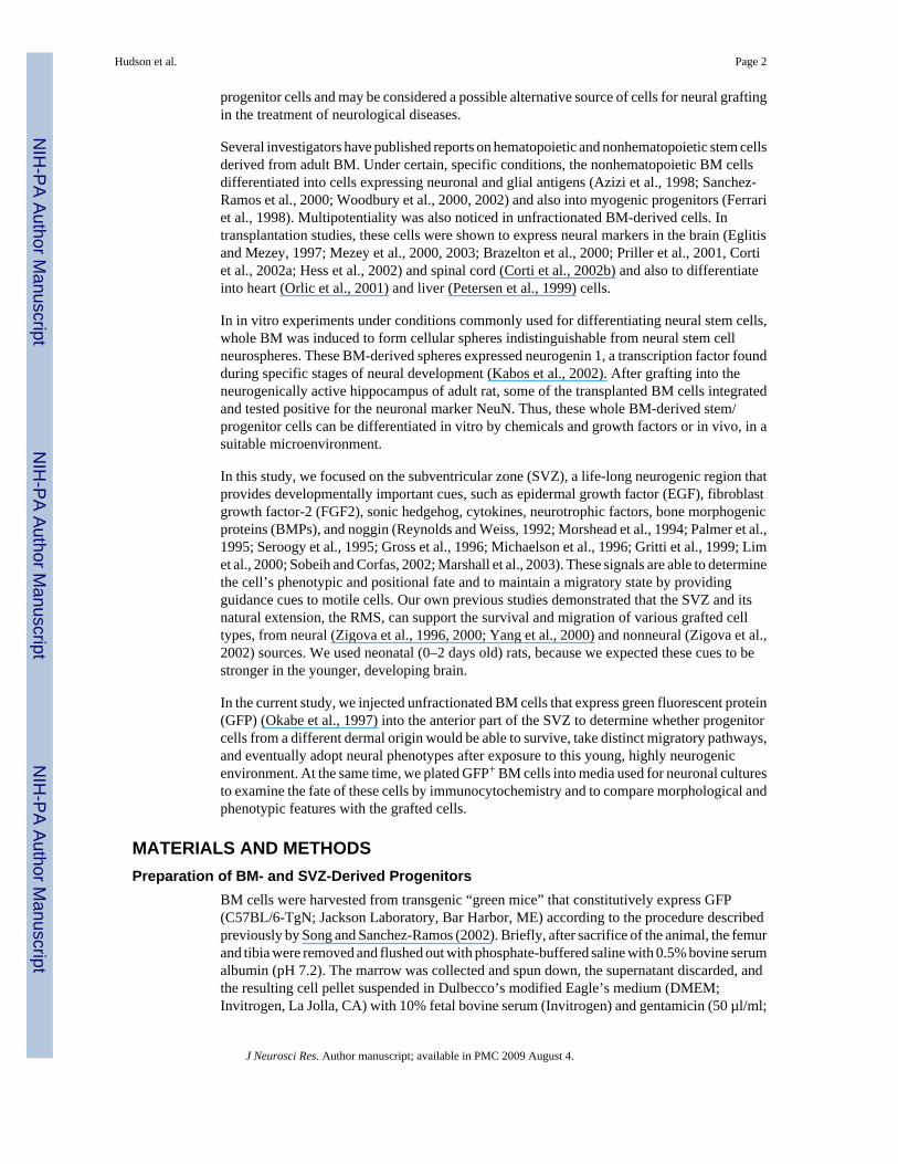
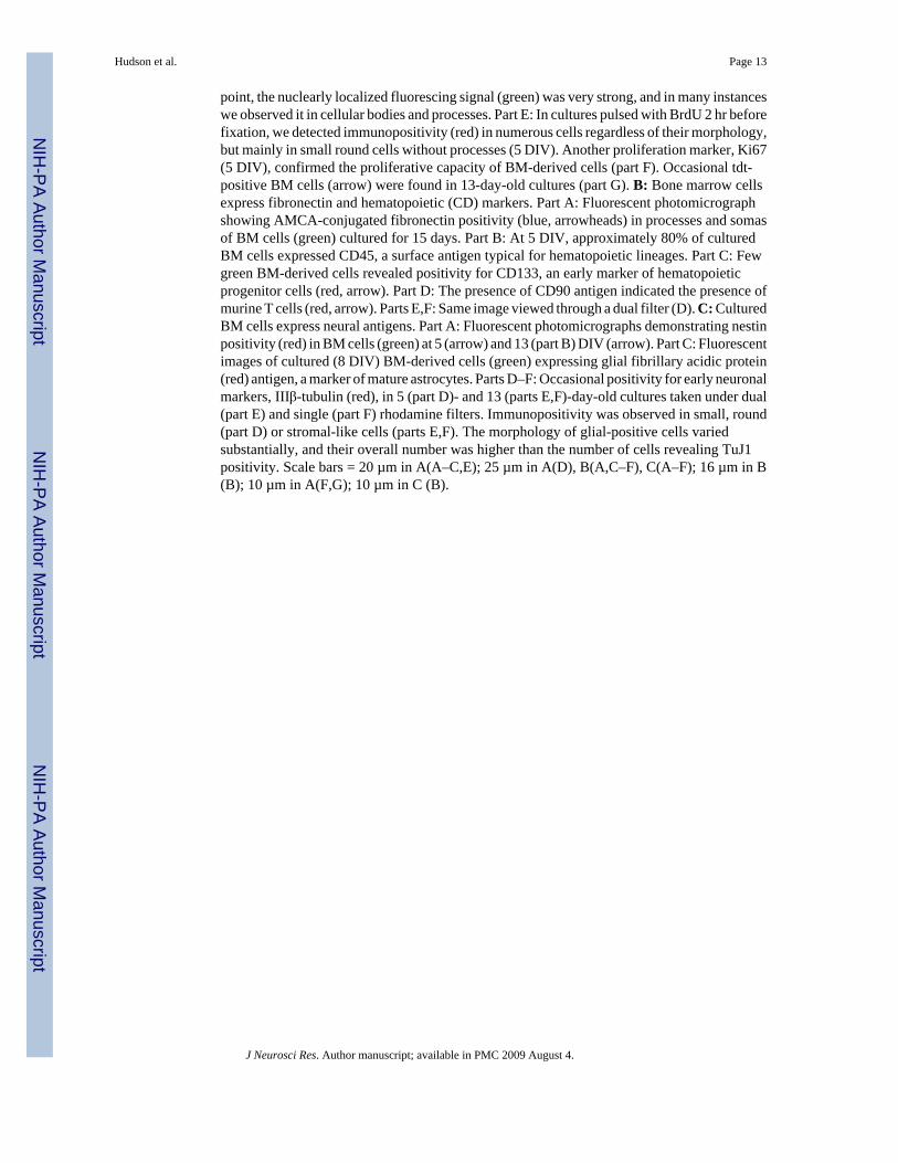
Depending on the survival time (Table II), transplanted murine SVZ progenitors were foundaround the injection site in the anterior part of the SVZ and along the full extent of the RMS(Fig. 2A, part A). Within 1 week, many of these cells reached the subependymal layer of theolfactory bulb (OB) and mingled with endogenous interneurons (Fig. 2A, part B). Two weeksafter grafting, many of the green cells began to send out processes and morphologicallyresemble OB cells (Fig. 2A, part B, and inset). At the same time, these cells acquired a neuron-specific class IIIβ-tubulin immunoreactivity. These findings confirm the ability of murineSVZ-derived progenitors to follow the same migratory route as endogenous analogs and tosettle in the OB while expressing neuronal cell type-specific antigen commonly found in thisregion (Zigova et al., 1998).
Distribution and Phenotype of GFP+ BM Cells After Transplantation Into the SVZ of theNeonatal Rat
To test the potential of the whole BM for neural differentiation in vivo, we transplanted freshlyprepared cells into the anterior part of the SVZ of neonatal (1-day-old) rat pups. It is believedthat this neurogenically active region, rich in neurotrophic factors, may support neuraldifferentiation of those cells in the BM that are not fully committed to hematopoietic fate.
Hudson et al. Page 5
J Neurosci Res. Author manuscript; available in PMC 2009 August 4.
NIH
-PA Author Manuscript
NIH
-PA Author Manuscript
NIH
-PA Author Manuscript
After transplantation, animals survived from 2 days up to 10 weeks (Table II). In those animalswith shorter survival (2 days), the GFP+ BM cells were located only close to the injection site(Fig. 2B, part A). With increased survival time after grafting (1–3 weeks), the cells were foundin corpus callosum and cortex and posteriorly in the subcortical white matter (Fig. 2B, partsC–E). At the same time, many BM cells were observed within the vertical or horizontal limbof the RMS, and some of them resembled the morphology of migrating neuroblasts (Fig. 2B,part B). Few single cells reached the subependymal zone of the OB. Morphologically, thesecells were small, usually with one or two relatively short processes, similar, in some cases, tothe OB granule cells (Fig. 2B, parts F, G). In one instance, we found a clump of BM cells withvery short and thick processes, in the core of the OB (Fig. 2C, parts A–C). Phenotypically,these BM cells either as single cells or in clump formation displayed very low positivity forhematopoietic (CD45) antigens in vivo and did not reveal immunoreactivity to any of the neural(nestin, TuJ1, GFAP) antigens.
DISCUSSIONIn this study, we demonstrated that GFP+ murine BM cells transplanted into the neonatal SVZwere able to migrate from the injection site in the anterior part of the SVZ along the RMS andreach the OB. Some green donor cells were found dorsally by the SVZ–white matter interface.In almost every case, we noted cells confined to the injection site. The green fluorescence signalallowed easy detection of donor cells even in the longest survival periods after grafting (2–70days). In some instances, the signal was converted with an antibody against GFP, confirmingthat the cell fluorescence resulted from the expression of the marker gene. Phenotypically,these BM cells in the most remote locations from the injection site (OB and subcortical whitematter) did not show glial or neuronal immunoreactivity, and CD45 expression was very weak.In cultures (1–15 days), which served as comparisons of cellular morphology and phenotypefor grafted BM cells, we found positivity for CD markers (CD45, CD90, CD133) andoccasionally for common neural antigens (nestin, GFAP, TuJ1); however, none of these cellsdisplayed morphology characteristic of neurons or mature glia.
Early Postnatal SVZ Supports Migration of Endogenous Progenitors Originating From SVZPrevious studies suggested two distinct migratory patterns of SVZ-derived progenitor cells:radial (coronal) in the posterior part and tangential (rostrocaudal) in the anterior part of theforebrain. These migration pathways were also closely associated with either neuronal(neurogenic pathways) or glial (gliogenic pathways) phenotypes. However, the long-distancemigration of progenitor cells from the SVZ toward the OB differs in neonatal and adult rodentbrain. Luskin (1993) described the tangential migration of dividing neuronal progenitorsoriginating from the anterior aspect of the early postnatal SVZ, giving rise to OB interneurons.Doetsch and Alvarez-Buylla (1996) noted in juvenile and adult animals a network of chainsof neuronal progenitors throughout the lateral wall of the lateral ventricle rising from the fullrostrocaudal extent of the SVZ. Retroviral injection studies examining the migratory patternsof neural progenitors within and out of the SVZ (Suzuki and Goldman, 2003) confirmed that,depending on the injection placement, neonatal SVZ progenitors migrated along the entirerostrocaudal extent of the SVZ, including the caudal tip of the SVZ at the hippocampal borderand out of the RMS toward the OB or along the SVZ adjacent to the striatum.
Neonatal Brain Supports Migration of Neural and Nonneural Cell TypesTransplantation studies designed to examine the migratory properties of various cell typeswithin the SVZ showed that cells derived from the anterior SVZ are able to resume migratorybehavior and spatiotemporal distribution within the RMS and OB similar to that of theirendogenous analogs (Zigova et al., 1996). In the current study, we used GFP+ mouse cells totrace their migratory fate after implantation into the anterior part of the SVZ in neonatal rats.
Hudson et al. Page 6
J Neurosci Res. Author manuscript; available in PMC 2009 August 4.
NIH
-PA Author Manuscript
NIH
-PA Author Manuscript
NIH
-PA Author Manuscript

Regardless of the different species and age of the host and donor (adult mouse SVZ progenitorswere grafted into the neonatal rat SVZ), the GFP+ SVZ-derived cells were found in thesubependymal OB of the rat within 3 weeks after transplantation. Easy identification of green-fluorescing GFP+ cells allowed for their detection in distant locations from the injectionplacement. In one case, we found cells in the subcortical white matter (Fig. 2B, parts C–E)posterior to the injection site. One explanation would be that injection of cells was placed moreposterior than in the rest of animals, so some injected cells first migrated to the posterior SVZand then commenced the radial migratory movement toward the overlying subcortical whitematter. Another possible explanation would be that these cells migrated along myelinated(Fulton et al., 1992) and unmyelinated (Golden et al., 1997) axons in the developing subcorticalwhite matter pathways, such as the superior longitudinal or the arcuate fasciculi. This, however,has to be elucidated.
Previously we have also tested the migratory properties of other cells, known as hNT neurons(Zigova et al., 2000). These human, neuronally committed cells were also able to move withinthe RMS, and a few of them even reached the granule cell layer of the OB, similarly topostnatally generated OB interneurons. Most of them, however, stayed close to the injectionsite in the SVZ. This reduction in number of cells reaching the OB after tangential movementalong the RMS and radial migration through the OB granule cell layer was attributed to thehuman origin of these cells as well as to different stages of their neuronal commitment at thetime of grafting.
Transplantation of predifferentiated human umbilical cord blood cells into the anterior part ofthe SVZ revealed that these originally nonneural cells had migrated only through the very initialpart of the RMS and never reached the OB. Many of them were trapped in the corticalparenchyma, in the corpus callosum, or around cortical blood vessels (Zigova et al., 2002b).Here, the freshly prepared whole bone marrow-derived cells were found within the entire extentof the RMS and also in the OB, indicating that these two cell sources, both derived from amesodermal germ layer, have different abilities to read the cues within the RMS and OB. It ispossible that dissimilarities in the migratory behavior of human and mouse donor cells reflectthe species incompatibility and the differences in the development of progenitor cells.Extensive migratory behavior of BM cells derived from marrow stromal cells wasdemonstrated by Kopen at al. (1999). These investigators engrafted murine marrow stromalcells into the lateral ventricle of the neonatal mice and followed their migratory patterns andphenotypic fate over the 12-day period. Engrafted cells were detected in many brain regions,including the islands of Calleja, OB, cerebellum, and brainstem.
Migratory Properties, Phenotype, and Proliferation of BM-Derived CellsThere is now evidence that BM cells can engraft into the brain parenchyma regardless of theroute by which they were administered (systemic vs. intraparenchymal). However, differences,such as final phenotype of transplanted cells, distribution, and proliferative capacity, werenoted in various experimental models. Systemic BM transplantation with GFP-expressing cellsfrom transgenic mice resulted in migration of cells into peripheral tissues (Ono et al., 2003)and brain parenchyma (Ono et al., 1999, 2003; Corti et al., 2002a; Hess et al., 2002). In ourstudy, the BM cells were delivered into a small target area within the neurogenic SVZ, theregion that is known to be a part of the natural migratory pathway of progenitor glial andneuronal cells. After placing BM cells into this region, we found them in two distinct places(RMS/OB and subcortical white matter), which were relatively remote from the injection site,so this could not be explained as a passive distribution or artifact caused by a needle or volumeor pressure developed during the cell delivery. This observation also argues for two possiblephenotypes of transplanted cells, neuronal within the subependymal zone of the OB and glialin the subcortical white matter. In our study, we did not confirm the colocalization of the GFP
Hudson et al. Page 7
J Neurosci Res. Author manuscript; available in PMC 2009 August 4.
NIH
-PA Author Manuscript
NIH
-PA Author Manuscript
NIH
-PA Author Manuscript
signal either with GFAP or with TuJ1, even though some cultured BM cells revealed it (Fig.1C). The most probable explanation for the absence of neural antigens in grafted cells may bethe short survival time (2 weeks), insufficient for the cells to start any differentiation. Otherauthors usually report immunopositivity for neural antigens in grafted BM progenitors afterextended periods (Mezey et al., 2000, 2003; Brazelton et al., 2000; Priller et al., 2001).
Ono et al. (2003) investigated the cytochemical differences in cells that had migrated into thebrain and other tissues after 7 days with antibodies against CD34 (hematopoietic stem cellmarker), Mac-1 (C3bi receptor marker), and ER-MP12 (murine early-stage myeloid precursormarker). They found that clusters of BM-derived cells in the liver and other organs were mainlyCD34−, Mac-1+/−, ER-MP12−, whereas those in the brain were CD34−, Mac-1+, ER-MP12+,suggesting that clusters in the brain were in a different state of differentiation from those inother tissues. In our studies, however, the cluster of BM cells within the OB (14 dayspostgrafting) did not show colocalized OX42 (marker of microglia) antibody reactivity. Thismay be explained again by the shortness of survival, or perhaps other microglia-recognizingantibodies such as F4/80 and ER-MP12 have to be employed.
Another interesting finding reported by other authors was the presence of clusters of BM cellsin the brain parenchyma, suggesting that these cells are actively proliferating while reachingtheir final destination (Ono et al., 2003). In our study, we noted the cluster of BM-derived cellswithin the OB, probably also indicating the proliferative capacity of specific subpopulation ofBM cells. Incorporation of BrdU in cultured (1–15 DIV) BM cells supports this finding. Herewe can speculate that migratory behavior associated with proliferative capacity of progenitorcells can reflect two scenarios. The first possibility is that some of these cells were deliveredto the ventricle and were moved with the cerebrospinal fluid toward the OB ventricular system.This would explain the facts that some early hematopoietic progenitors started to proliferateand that the clump was trapped in the subventricular zone of the OB. Ono et al. (2003) reportedthe presence of similar clumps of cells within the brain parenchyma after systemicadministration of the same cell source. The second explanation would be based on the parallelobservation of endogenous neural progenitors from SVZ/RMS/OB region, for which migrationof proliferating neuroblasts has been previously described (Menezes et al., 1995). The secondpossibility would also suggest (indirectly) that hematopoietic and neural cells might havecommon characteristics or that immature cells within those two cell sources have anoverlapping set of cytochemical properties (receptors, proteins, etc.) that allow them to behavesimilarly in the specific brain environment.
Further studies are warranted to understand better the migratory and phenotypic potential ofprogenitors originating from hematopoietic sources such as BM and human umbilical cordblood and also to explain the differences between theses cell sources with respect to recipientage. This will allow us to determine effective therapeutic strategies for treatment ofneurodegenerative diseases occurring predominantly in older individuals.
ACKNOWLEDGMENTSThis paper is dedicated to my beloved mentor, Tanja Zigova, PhD. It is her vision and devotion to science, heruncompromising work ethics, patience and personal guidance that are reflected in this paper (JEH). The authors thankMarcia McCall for excellent editorial assistance, Sepideh Rouhani for technical assistance, and Ed Haller for confocalmicroscopy. This work was partially supported by NIA grant RO1 AG20927-01 (T.Z.), the Helen E. Ellis EndowmentChair (J.S.-R.), and a J.A. Haley VA Hospital merit review grant (J.S.-R.).
Hudson et al. Page 8
J Neurosci Res. Author manuscript; available in PMC 2009 August 4.
NIH
-PA Author Manuscript
NIH
-PA Author Manuscript
NIH
-PA Author Manuscript

REFERENCESAzizi SA, Stokes D, Augelli BJ, DiGirolamo C, Prockop DJ. Engraftment and migration of human bone
marrow stromal cells implanted in the brains of albino rats—similarities to astrocyte grafts. Proc NatlAcad Sci USA 1998;95:3908–3913. [PubMed: 9520466]
Bentley SA, Tralka TS. Fibronectin-mediated attachment of hematopoietic cells to stromal elements incontinuous bone marrow culture. Exp Hematol 1983;11:129–138. [PubMed: 6339260]
Brazelton TR, Rossi FM, Keshet GI, Blau HM. From marrow to brain: expression of neuronal phenotypesin adult mice. Science 2000;290:1775–1779. [PubMed: 11099418]
Corti S, Locatelli F, Strazzer S, Salani S, Del Bo R, Soligo D, Bossolasco P, Bresolin N, Scarlato G,Comi GP. Modulated generation of neuronal cells from bone marrow by expansion and mobilizationof circulating stem cells with in vivo cytokine treatment. Exp Neurol 2002a;177:443–452. [PubMed:12429190]
Corti S, Locatelli F, Donadoni C, Strazzer S, Salani S, Del Bo R, Caccialanza M, Bresolin N, ScarlatoG, Comi GP. Neuroectodermal and microglial differentiation of bone marrow cells in the mouse spinalcord and sensory ganglia. J Neurosci Res 2002b;70:721–733. [PubMed: 12444594]
Dahl D, Crosby CJ, Sethi JS, Bignami A. Glial fibrillary acidic (GFA) protein in vertebrates:immunofluorescence and immunoblotting study with monoclonal and polyclonal antibodies. J CompNeurol 1985;239:75–88. [PubMed: 3900154]
Doetsch F, Alvarez-Buylla A. Network of tangential pathways for neuronal migration in adult mammalianbrain. Proc Natl Acad Sci USA 1996;93:14895–14900. [PubMed: 8962152]
Eglitis MA, Mezey E. Hematopoietic cells differentiate into both microglia and macroglia in the brainsof adult mice. Proc Natl Acad Sci USA 1997;94:4080–4085. [PubMed: 9108108]
Ferrari G, Cusella-De Angelis G, Coletta M, Paolucci E, Stornaiuolo A, Cossu G, Mavilio F. Muscleregeneration by bone marrow-derived myogenic progenitors. Science 1998;279:1528–1530.[PubMed: 9488650]
Fulton BP, Burne JF, Raff MC. Visualization of O-2A progenitor cells in developing and adult rat opticnerve by quisqualate-stimulated cobalt uptake. J Neurosci 1992;12:4816–4833. [PubMed: 1281496]
Golden JA, Zitz JC, McFadden K, Cepko CL. Cell migration in the developing chick diencephalon.Development 1997;124:3525–3533. [PubMed: 9342045]
Gritti A, Frolichsthal-Schoeller P, Galli R, Parati EA, Cova L, Pagano SF, Bjornson CR, Vescovi AL.Epidermal and fibroblast growth factors behave as mitogenic regulators for a single multipotent stemcell-like population from the subventricular region of the adult mouse forebrain. J Neurosci1999;19:3287–3297. [PubMed: 10212288]
Gross RE, Mehler MF, Mabie PC, Zang Z, Santschi L, Kessler JA. Bone morphogenetic proteins promoteastroglial lineage commitment by mammalian subventricular zone progenitor cells. Neuron1996;17:595–606. [PubMed: 8893018]
Hess DC, Hill WD, Martin-Studdard A, Carroll J, Brailer J, Carothers J. Bone marrow as a source ofendothelial cells and NeuN-expressing cells after stroke. Stroke 2002;33:1362–1368. [PubMed:11988616]
Kabos P, Ehtesham M, Kabosova A, Black KL, Yu JS. Generation of neural progenitor cells from wholeadult bone marrow. Exp Neurol 2002;178:288–293. [PubMed: 12504887]
Kopen GC, Prockop DJ, Phinney DG. Marrow stromal cells migrate throughout forebrain and cerebellum,and they differentiate into astrocytes after injection into neonatal mouse brains. Proc Natl Acad SciUSA 1999;96:10711–10716. [PubMed: 10485891]
Laywell ED, Kukekov VG, Suslov O, Zheng T, Steindler DA. Production and analysis of neurospheresfrom acutely dissociated and postmortem CNS specimens. Methods Mol Biol 2002;198:15–27.[PubMed: 11951618]
Lendahl U, Zimmerman LB, McKay RD. CNS stem cells express a new class of intermediate filamentprotein. Cell 1990;60:585–595. [PubMed: 1689217]
Lim DA, Tramontin AD, Trevejo JM, Herrera DG, Garcia-Verdugo JM, Alvarez-Buylla A. Nogginantagonizes BMP signaling to create a niche for adult neurogenesis. Neuron 2000;28:713–726.[PubMed: 11163261]
Hudson et al. Page 9
J Neurosci Res. Author manuscript; available in PMC 2009 August 4.
NIH
-PA Author Manuscript
NIH
-PA Author Manuscript
NIH
-PA Author Manuscript
Luskin MB. Restricted proliferation and migration of postnatally generated neurons derived from theforebrain subventricular zone. Neuron 1993;11:173–189. [PubMed: 8338665]
Marshall CA, Suzuki SO, Goldman JE. Gliogenic and neurogenic progenitors of the subventricular zone:who are they, where did they come from, and where are they going? Glia 2003;43:52–61. [PubMed:12761867]
Menezes JR, Smith CM, Nelson KC, Luskin MB. The division of neuronal progenitor cells duringmigration in the neonatal mammalian forebrain. Mol Cell Neurosci 1995;6:496–508. [PubMed:8742267]
Mezey E, Chandross KJ, Harta G, Maki RA, McKercher SR. Turning blood into brain: cells bearingneuronal antigens generated in vivo from bone marrow. Science 2000;290:1779–1782. [PubMed:11099419]
Mezey E, Key S, Vogelsang G, Szalayova I, Lange GD, Crain B. Transplanted bone marrow generatesnew neurons in human brains. Proc Natl Acad Sci USA 2003;100:1364–1369. [PubMed: 12538864]
Michaelson MD, Mehler MF, Xu H, Gross RE, Kessler JA. Interleukin-7 is trophic for embryonic neuronsand is expressed in developing brain. Dev Biol 1996;179:251–263. [PubMed: 8873768]
Morshead CM, Reynolds BA, Craig CG, McBurney MW, Staines WA, Morassutti D, Weiss S, van derKooy D. Neural stem cells in the adult mammalian forebrain: a relatively quiescent subpopulationof subependymal cells. Neuron 1994;13:1071–1082. [PubMed: 7946346]
Okabe M, Ikawa M, Kominami K, Nakanishi T, Nishimune Y. “Green mice” as a source of ubiquitousgreen cells. FEBS Lett 1997;407:313–319. [PubMed: 9175875]
Ono K, Takii T, Onozaki K, Ikawa M, Okabe M, Sawada M. Migration of exogenous immaturehematopoietic cells into adult mouse brain parenchyma under GFP-expressing bone marrow chimera.Biochem Biophys Res Commun 1999;262:610–614. [PubMed: 10471372]
Ono K, Yoshihara K, Suzuki H, Tanaka KF, Takii T, Onozaki K, Sawada M. Preservation ofhematopoietic properties in transplanted bone marrow cells in the brain. J Neurosci Res 2003;72:503–507. [PubMed: 12704811]
Orlic D, Kajstura J, Chimenti S, Jakoniuk I, Anderson SM, Li B, Pickel J, McKay R, Nadal-Ginard B,Bodine DM, Leri A, Anversa P. Bone marrow cells regenerate infarcted myocardium. Nature2001;410:701–705. [PubMed: 11287958]
Palmer TD, Ray J, Gage FH. FGF-2-responsive neuronal progenitors reside in proliferative and quiescentregions of the adult rodent brain. Mol Cell Neurosci 1995;6:474–486. [PubMed: 8581317]
Petersen BE, Bowen WC, Patrene KD, Mars WM, Sullivan AK, Murase N, Boggs SS, Greenberger JS,Goff JP. Bone marrow as a potential source of hepatic oval cells. Science 1999;284:1168–1170.[PubMed: 10325227]
Priller J, Persons DA, Klett FF, Kempermann G, Kreutzberg GW, Dirnagl U. Neogenesis of cerebellarPurkinje neurons from gene-marked bone marrow cells in vivo. J Cell Biol 2001;155:733–738.[PubMed: 11724815]
Reynolds BA, Weiss S. Generation of neurons and astrocytes from isolated cells of the adult mammaliancentral nervous system. Science 1992;255:1707–1710. [PubMed: 1553558]
Sanchez-Ramos J, Song S, Cardozo-Pelaez F, Hazzi C, Stedeford T, Willing A, Freeman TB, Saporta S,Janssen W, Patel N, Cooper DR, Sanberg PR. Adult bone marrow stromal cells differentiate intoneural cells in vitro. Exp Neurol 2000;164:247–256. [PubMed: 10915564]
Seroogy KB, Gall CM, Lee DC, Kornblum HI. Proliferative zones of postnatal rat brain express epidermalgrowth factor receptor mRNA. Brain Res 1995;670:157–164. [PubMed: 7719717]
Sobeih MM, Corfas G. Extracellular factors that regulate neuronal migration in the central nervoussystem. Int J Dev Neurosci 2002;20:349–357. [PubMed: 12175873]
Song S, Sanchez-Ramos J. Preparation of neural progenitors from bone marrow and umbilical cord blood.Methods Mol Biol 2002;198:79–88. [PubMed: 11951643]
Suzuki SO, Goldman JE. Multiple cell populations in the early postnatal subventricular zone take distinctmigratory pathways: a dynamic study of glial and neuronal progenitor migration. J Neurosci2003;23:4240–4250. [PubMed: 12764112]
Woodbury D, Schwarz EJ, Prockop DJ, Black IB. Adult rat and human bone marrow stromal cellsdifferentiate into neurons. J Neurosci Res 2000;61:364–370. [PubMed: 10931522]
Hudson et al. Page 10
J Neurosci Res. Author manuscript; available in PMC 2009 August 4.
NIH
-PA Author Manuscript
NIH
-PA Author Manuscript
NIH
-PA Author Manuscript

Woodbury D, Reynolds K, Black IB. Adult bone marrow stromal stem cells express germline, ectodermal,endodermal, and mesodermal genes prior to neurogenesis. J Neurosci Res 2002;69:908–917.[PubMed: 12205683]
Yang H, Mujtaba T, Venkatraman G, Wu YY, Rao MS, Luskin MB. Region-specific differentiation ofneural tube-derived neuronal restricted progenitor cells after heterotopic transplantation. Proc NatlAcad Sci USA 2000;97:13366–13371. [PubMed: 11087876]
Zigova T, Betarbet R, Soteres BJ, Brock S, Bakay RA, Luskin MB. A comparison of the patterns ofmigration and the destinations of homotopically transplanted neonatal subventricular zone cells andheterotopically transplanted telencephalic ventricular zone cells. Dev Biol 1996;173:459–474.[PubMed: 8606005]
Zigova T, Pencea V, Wiegand SJ, Luskin MB. Intraventricular administration of BDNF increases thenumber of newly generated neurons in the adult olfactory bulb. Mol Cell Neurosci 1998;11:234–245.[PubMed: 9675054]
Zigova T, Pencea V, Sanberg PR, Luskin MB. The properties of hNT cells following transplantation intothe subventricular zone of the neonatal forebrain. Exp Neurol 2000;163:31–38. [PubMed: 10785441]
Zigova T, Newman MB. Transplantation into neonatal rat brain as a tool to study properties of stem cells.Methods Mol Biol 2002a;198:341–356. [PubMed: 11951637]
Zigova T, Song S, Willing AE, Hudson JE, Newman MB, Saporta S, Sanchez-Ramos J, Sanberg PR.Human umbilical cord blood cells express neural antigens after transplantation into the developingrat brain. Cell Transplant 2002b;11:265–274. [PubMed: 12075992]
Hudson et al. Page 11
J Neurosci Res. Author manuscript; available in PMC 2009 August 4.
NIH
-PA Author Manuscript
NIH
-PA Author Manuscript
NIH
-PA Author Manuscript
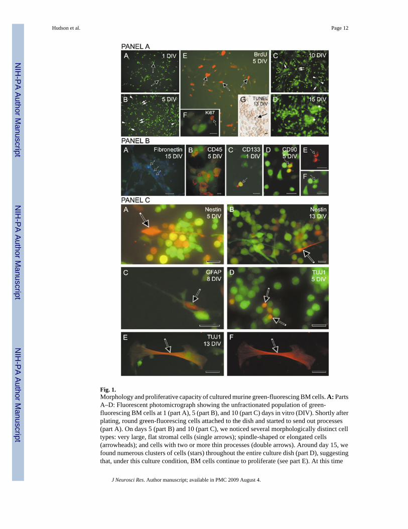
Fig. 1.Morphology and proliferative capacity of cultured murine green-fluorescing BM cells. A: PartsA–D: Fluorescent photomicrograph showing the unfractionated population of green-fluorescing BM cells at 1 (part A), 5 (part B), and 10 (part C) days in vitro (DIV). Shortly afterplating, round green-fluorescing cells attached to the dish and started to send out processes(part A). On days 5 (part B) and 10 (part C), we noticed several morphologically distinct celltypes: very large, flat stromal cells (single arrows); spindle-shaped or elongated cells(arrowheads); and cells with two or more thin processes (double arrows). Around day 15, wefound numerous clusters of cells (stars) throughout the entire culture dish (part D), suggestingthat, under this culture condition, BM cells continue to proliferate (see part E). At this time
Hudson et al. Page 12
J Neurosci Res. Author manuscript; available in PMC 2009 August 4.
NIH
-PA Author Manuscript
NIH
-PA Author Manuscript
NIH
-PA Author Manuscript
point, the nuclearly localized fluorescing signal (green) was very strong, and in many instanceswe observed it in cellular bodies and processes. Part E: In cultures pulsed with BrdU 2 hr beforefixation, we detected immunopositivity (red) in numerous cells regardless of their morphology,but mainly in small round cells without processes (5 DIV). Another proliferation marker, Ki67(5 DIV), confirmed the proliferative capacity of BM-derived cells (part F). Occasional tdt-positive BM cells (arrow) were found in 13-day-old cultures (part G). B: Bone marrow cellsexpress fibronectin and hematopoietic (CD) markers. Part A: Fluorescent photomicrographshowing AMCA-conjugated fibronectin positivity (blue, arrowheads) in processes and somasof BM cells (green) cultured for 15 days. Part B: At 5 DIV, approximately 80% of culturedBM cells expressed CD45, a surface antigen typical for hematopoietic lineages. Part C: Fewgreen BM-derived cells revealed positivity for CD133, an early marker of hematopoieticprogenitor cells (red, arrow). Part D: The presence of CD90 antigen indicated the presence ofmurine T cells (red, arrow). Parts E,F: Same image viewed through a dual filter (D). C: CulturedBM cells express neural antigens. Part A: Fluorescent photomicrographs demonstrating nestinpositivity (red) in BM cells (green) at 5 (arrow) and 13 (part B) DIV (arrow). Part C: Fluorescentimages of cultured (8 DIV) BM-derived cells (green) expressing glial fibrillary acidic protein(red) antigen, a marker of mature astrocytes. Parts D–F: Occasional positivity for early neuronalmarkers, IIIβ-tubulin (red), in 5 (part D)- and 13 (parts E,F)-day-old cultures taken under dual(part E) and single (part F) rhodamine filters. Immunopositivity was observed in small, round(part D) or stromal-like cells (parts E,F). The morphology of glial-positive cells variedsubstantially, and their overall number was higher than the number of cells revealing TuJ1positivity. Scale bars = 20 µm in A(A–C,E); 25 µm in A(D), B(A,C–F), C(A–F); 16 µm in B(B); 10 µm in A(F,G); 10 µm in C (B).
Hudson et al. Page 13
J Neurosci Res. Author manuscript; available in PMC 2009 August 4.
NIH
-PA Author Manuscript
NIH
-PA Author Manuscript
NIH
-PA Author Manuscript

Fig. 2.A: After implantation into the neonatal SVZ, precursors isolated from green mouse SVZfollowed the same migratory route, resided in the OB, and expressed neuronal antigens as theirendogenous counterparts. The animals (n = 5) survived 14–56 days after surgery (see TableII). Part A: After injection, green-fluorescing SVZ cells were found in the RMS (marked bydashed lines) leading to the OB. Part B: Note many green SVZ-derived cells within thesubependyma of the OB (arrows). Inset shows one of the numerous GFP-SVZ cells (green)reaching the OB, colabeled with antibody against IIIβ-tubulin (red, 14 days after grafting).B: BM cells isolated from green mouse were found in the SVZ, the RMS, and the OB. Thesecells did not express neural markers after reaching the OB. The animals (n = 10) survived for2–70 days after grafting (see Table II). Part A: Cresyl violet-stained parasagittal section throughthe host brain showing the injection site (encircled area) targeting the SVZ. The confined,
Hudson et al. Page 14
J Neurosci Res. Author manuscript; available in PMC 2009 August 4.
NIH
-PA Author Manuscript
NIH
-PA Author Manuscript
NIH
-PA Author Manuscript

darkly stained BM cells were easily recognized within the brain parenchyma (70 days survivalafter grafting). Part B: Several GFP-positive BM-derived cells were found within the delineated(dashed lines) migratory route. The arrow points to the cell, which is magnified in the inset.Part C: Brightfield image of a cresyl violet-stained section through the neonatal rat brainshowing the area in the posterior subcortical white matter overlying the hippocampus(arrowheads). We detected within this region several BM-derived green-fluorescing cells(arrows), shown in part D. Part E: Higher magnification allows detailed observation of severalcells with green fluorescent signal (arrows). Part F: Fluorescent photomicrograph of the singleGFP-positive BM-derived cell (arrow) within the OB subependyma (14 days after grafting)observed under the dual (rhodamine and FITC) filter. Part G: The same section was stainedfor glial fibrillary acidic protein (GFAP), a marker of astrocytic glia. The cell indicated by thearrow is GFAP negative (rhodamine filter). C: Confocal images of the transplanted BM cellsisolated from GFP mouse. These cells were implanted into the anterior part of the SVZ of a 1-day-old rat pup. Eight days later, the cells reached the subependymal layer of the OB. On thisparticular section, the green signal was converted through the rhodamine-conjugated anti-GFPantibody to obtain stronger red signal. DAPI counterstain confirmed the singleness of BM-derived nuclei surrounded by GFP immunopositivity. Parts A–C: Fluorescentphotomicrographs showing the cluster of BM-derived cells (red) in the OB subependyma takenthrough a 20-µm-thick section. The arrow points to a single cell that was followed in threeconsecutive images, 2 µm apart. Scale bars = 20 µm in A(A), B(B), in C(A) for C(A–C); 10µm in A(B), in B(F) for B(F,G); 200 µm in B(A,C,D); 50 µm in B(E).
Hudson et al. Page 15
J Neurosci Res. Author manuscript; available in PMC 2009 August 4.
NIH
-PA Author Manuscript
NIH
-PA Author Manuscript
NIH
-PA Author Manuscript
NIH
-PA Author Manuscript
NIH
-PA Author Manuscript
NIH
-PA Author Manuscript
Hudson et al. Page 16
TABLE IProliferative Capacity of Cultured BM Cells Expressing GFP
DIV
1 (%) 5 (%) 10 (%) 15 (%)
BrdU+/totala 94/383 (24.5) 179/462 (38.7) 462/1,035 (44.6) 330/805 (41.0)
Prevalent morphologyb 1 1, 2 1 1
aRatio between the total number of cells and those incorporating the proliferation marker BrdU. The counts were collected from 20–25 fields/slide/time
point.
bPrevalent morphology of plated BM-GFP+ cells incorporating BrdU: 1, round, small cells; 2, other cell types (including spindle-shaped, elongated, and
large, flat cells).
J Neurosci Res. Author manuscript; available in PMC 2009 August 4.

NIH
-PA Author Manuscript
NIH
-PA Author Manuscript
NIH
-PA Author Manuscript
Hudson et al. Page 17TA
BLE
IISp
atio
tem
pora
l Dis
tribu
tion
of G
rafte
d M
urin
e G
FP+
Cel
ls F
ollo
win
g Th
eir T
rans
plan
tatio
n In
to th
e N
eona
tal S
VZ*
Fina
l pos
ition
of G
FP+ c
ells
Rat
Age
of
Surv
ival
No.
host
(day
s)SV
ZR
MS
OB
CC
CT
SWM
LV
Hom
otop
ic S
VZa
gra
fts1
P114
++
++
−−
−
2P1
21+
++
+−
−−
3P1
21−
++
−+
−−
4P1
31−
++
−+
−+
5P1
56+
++
−+
−−
Bon
e m
arro
w g
rafts
1P2
2+
+−
++
−−
2P2
8−
−+
++
−−
3P1
14+
−+
+−
+−
4P2
21+
−−
++
−−
5P1
30−
−−
+−
−−
6P2
35−
+−
++
−−
7P2
43+
−−
++
−−
8P1
56−
+−
+−
−+
9P1
70+
++
+−
−−
10P1
70+
++
+−
−+
* Sum
mar
y of
the
dist
ribut
ion
of S
VZ-
GFP
+ an
d B
M-G
FP+
cells
with
in th
e br
ain
regi
ons a
t var
ious
pos
tinje
ctio
n su
rviv
al ti
mes
. Am
ong
22 a
nim
als t
hat r
ecei
ved
the
trans
plan
t of G
FP+
cells
, 15
wer
eus
ed fo
r ana
lysi
s; in
4 a
nim
als t
he p
roce
ssin
g w
as n
ot sa
tisfa
ctor
y, a
nd in
3 a
nim
als t
he tr
ansp
lant
was
not
foun
d, so
they
wer
e ex
clud
ed fr
om to
tal c
ount
s and
fina
l ana
lysi
s. R
epre
sent
ativ
e im
ages
wer
eta
ken
from
sect
ions
pre
pare
d fr
om ra
ts 1
and
2 fo
r SV
Z an
d ra
ts 2
and
3 fo
r BM
tran
spla
nts.
CC
, cor
pus c
allo
sum
; CT,
cor
tex;
LV
, lat
eral
ven
tricl
e; O
B, o
lfact
ory
bulb
; RM
S, ro
stra
l mig
rato
ry st
ream
;SW
M, s
ubco
rtica
l whi
te m
atte
r; SV
Z, su
bven
tricu
lar z
one;
+, p
rese
nce
of c
ells
; −, a
bsen
ce o
f cel
ls.
J Neurosci Res. Author manuscript; available in PMC 2009 August 4.
Copyright © 2022 FDOKUMEN