Isolation, Maintenance and Expansion of Adult Hematopoietic ...
33
Citation: Mayer, I.M.; Hoelbl-Kovacic, A.; Sexl, V.; Doma, E. Isolation, Maintenance and Expansion of Adult Hematopoietic Stem/Progenitor Cells and Leukemic Stem Cells. Cancers 2022, 14, 1723. https://doi.org/10.3390/ cancers14071723 Academic Editor: Alice Giustacchini Received: 28 February 2022 Accepted: 25 March 2022 Published: 28 March 2022 Publisher’s Note: MDPI stays neutral with regard to jurisdictional claims in published maps and institutional affil- iations. Copyright: © 2022 by the authors. Licensee MDPI, Basel, Switzerland. This article is an open access article distributed under the terms and conditions of the Creative Commons Attribution (CC BY) license (https:// creativecommons.org/licenses/by/ 4.0/). cancers Review Isolation, Maintenance and Expansion of Adult Hematopoietic Stem/Progenitor Cells and Leukemic Stem Cells Isabella Maria Mayer, Andrea Hoelbl-Kovacic, Veronika Sexl * and Eszter Doma Institute of Pharmacology and Toxicology, University of Veterinary Medicine Vienna, 1210 Vienna, Austria; [email protected] (I.M.M.); [email protected] (A.H.-K.); [email protected] (E.D.) * Correspondence: [email protected]; Tel.: +43-125-077-2910 Simple Summary: Transplantation of adult hematopoietic stem cells is an important therapeutic tool to help patients suffering from diverse hematological disorders. All types of blood cells can develop from a single hematopoietic stem cell underlining their enormous potential. Intense efforts are ongoing to generate “engraftable” human hematopoietic stem cells to treat hematopoietic diseases and to understand the molecular machinery driving them. Leukemic stem cells represent a low frequency subpopulation of leukemia cells that possess stem cell properties. They can instigate, maintain, and serially propagate leukemia in vivo, while they retain the capacity to differentiate into committed progenitors. Leukemic stem cells are unaffected by many therapeutic strategies and represent the major cause of relapse. We here describe all methods to maintain and expand murine and human hematopoietic cells in culture and describe their specific advantages. These methods are also employed to understand the biology of leukemic stem cells and to identify novel therapeutic strategies. Abstract: Hematopoietic stem cells (HSCs) are rare, self-renewing cells that perch on top of the hematopoietic tree. The HSCs ensure the constant supply of mature blood cells in a tightly regulated process producing peripheral blood cells. Intense efforts are ongoing to optimize HSC engraftment as therapeutic strategy to treat patients suffering from hematopoietic diseases. Preclinical research paves the way by developing methods to maintain, manipulate and expand HSCs ex vivo to understand their regulation and molecular make-up. The generation of a sufficient number of transplantable HSCs is the Holy Grail for clinical therapy. Leukemia stem cells (LSCs) are characterized by their acquired stem cell characteristics and are responsible for disease initiation, progression, and relapse. We summarize efforts, that have been undertaken to increase the number of long-term (LT)-HSCs and to prevent differentiation towards committed progenitors in ex vivo culture. We provide an overview and compare methods currently available to isolate, maintain and enrich HSC subsets, progenitors and LSCs and discuss their individual advantages and drawbacks. Keywords: hematopoietic stem cells; leukemic stem cells; ex vivo culture; self-renewal; dormancy; maintenance 1. The Adult HSC—A Rare, Self-Renewing Cell Bone marrow (BM) cell transplantation studies and ex vivo colony formation assays identified the presence of HSCs in the 1950s and 1960s [1–3]. Hematopoietic stem cells were functionally defined by their ability to serially engraft transplanted recipients and to replenish all myelolymphoid lineages, through their ability to self-renew and differenti- ate [4]. These transplantation studies revealed the heterogeneity of the HSC compartment, as HSC subclones were able to repopulate lethally irradiated mice ranged from short term (weeks to months, referred as short term (ST-) HSCs) to long term (more than 6 months, referred as LT-HSCs) [4,5]. Long term HSCs are predominantly in a quiescent/dormant Cancers 2022, 14, 1723. https://doi.org/10.3390/cancers14071723 https://www.mdpi.com/journal/cancers
-
Upload
khangminh22 -
Category
Documents
-
view
2 -
download
0
Transcript of Isolation, Maintenance and Expansion of Adult Hematopoietic ...

�����������������
Citation: Mayer, I.M.;
Hoelbl-Kovacic, A.; Sexl, V.; Doma, E.
Isolation, Maintenance and
Expansion of Adult Hematopoietic
Stem/Progenitor Cells and Leukemic
Stem Cells. Cancers 2022, 14, 1723.
https://doi.org/10.3390/
cancers14071723
Academic Editor: Alice Giustacchini
Received: 28 February 2022
Accepted: 25 March 2022
Published: 28 March 2022
Publisher’s Note: MDPI stays neutral
with regard to jurisdictional claims in
published maps and institutional affil-
iations.
Copyright: © 2022 by the authors.
Licensee MDPI, Basel, Switzerland.
This article is an open access article
distributed under the terms and
conditions of the Creative Commons
Attribution (CC BY) license (https://
creativecommons.org/licenses/by/
4.0/).
cancers
Review
Isolation, Maintenance and Expansion of Adult HematopoieticStem/Progenitor Cells and Leukemic Stem CellsIsabella Maria Mayer, Andrea Hoelbl-Kovacic, Veronika Sexl * and Eszter Doma
Institute of Pharmacology and Toxicology, University of Veterinary Medicine Vienna, 1210 Vienna, Austria;[email protected] (I.M.M.); [email protected] (A.H.-K.);[email protected] (E.D.)* Correspondence: [email protected]; Tel.: +43-125-077-2910
Simple Summary: Transplantation of adult hematopoietic stem cells is an important therapeutictool to help patients suffering from diverse hematological disorders. All types of blood cells candevelop from a single hematopoietic stem cell underlining their enormous potential. Intense effortsare ongoing to generate “engraftable” human hematopoietic stem cells to treat hematopoietic diseasesand to understand the molecular machinery driving them. Leukemic stem cells represent a lowfrequency subpopulation of leukemia cells that possess stem cell properties. They can instigate,maintain, and serially propagate leukemia in vivo, while they retain the capacity to differentiateinto committed progenitors. Leukemic stem cells are unaffected by many therapeutic strategiesand represent the major cause of relapse. We here describe all methods to maintain and expandmurine and human hematopoietic cells in culture and describe their specific advantages. Thesemethods are also employed to understand the biology of leukemic stem cells and to identify noveltherapeutic strategies.
Abstract: Hematopoietic stem cells (HSCs) are rare, self-renewing cells that perch on top of thehematopoietic tree. The HSCs ensure the constant supply of mature blood cells in a tightly regulatedprocess producing peripheral blood cells. Intense efforts are ongoing to optimize HSC engraftment astherapeutic strategy to treat patients suffering from hematopoietic diseases. Preclinical research pavesthe way by developing methods to maintain, manipulate and expand HSCs ex vivo to understandtheir regulation and molecular make-up. The generation of a sufficient number of transplantableHSCs is the Holy Grail for clinical therapy. Leukemia stem cells (LSCs) are characterized by theiracquired stem cell characteristics and are responsible for disease initiation, progression, and relapse.We summarize efforts, that have been undertaken to increase the number of long-term (LT)-HSCs andto prevent differentiation towards committed progenitors in ex vivo culture. We provide an overviewand compare methods currently available to isolate, maintain and enrich HSC subsets, progenitorsand LSCs and discuss their individual advantages and drawbacks.
Keywords: hematopoietic stem cells; leukemic stem cells; ex vivo culture; self-renewal; dormancy;maintenance
1. The Adult HSC—A Rare, Self-Renewing Cell
Bone marrow (BM) cell transplantation studies and ex vivo colony formation assaysidentified the presence of HSCs in the 1950s and 1960s [1–3]. Hematopoietic stem cellswere functionally defined by their ability to serially engraft transplanted recipients and toreplenish all myelolymphoid lineages, through their ability to self-renew and differenti-ate [4]. These transplantation studies revealed the heterogeneity of the HSC compartment,as HSC subclones were able to repopulate lethally irradiated mice ranged from short term(weeks to months, referred as short term (ST-) HSCs) to long term (more than 6 months,referred as LT-HSCs) [4,5]. Long term HSCs are predominantly in a quiescent/dormant
Cancers 2022, 14, 1723. https://doi.org/10.3390/cancers14071723 https://www.mdpi.com/journal/cancers
Cancers 2022, 14, 1723 2 of 33
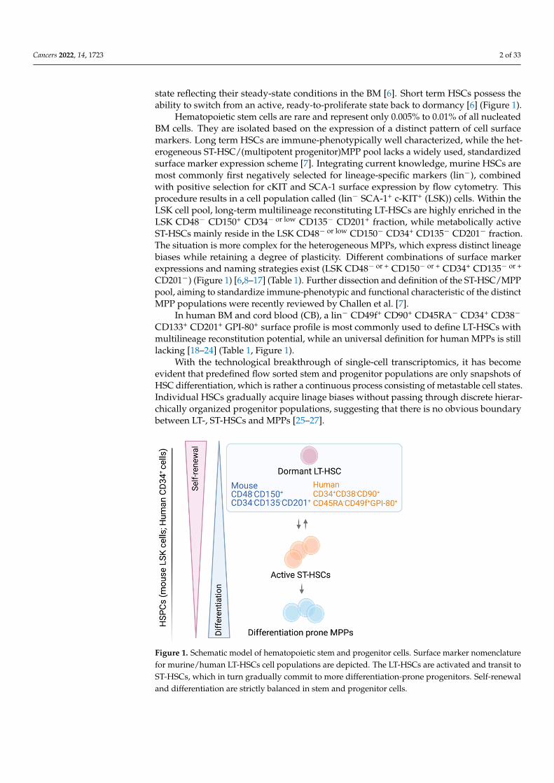
state reflecting their steady-state conditions in the BM [6]. Short term HSCs possess theability to switch from an active, ready-to-proliferate state back to dormancy [6] (Figure 1).
Hematopoietic stem cells are rare and represent only 0.005% to 0.01% of all nucleatedBM cells. They are isolated based on the expression of a distinct pattern of cell surfacemarkers. Long term HSCs are immune-phenotypically well characterized, while the het-erogeneous ST-HSC/(multipotent progenitor)MPP pool lacks a widely used, standardizedsurface marker expression scheme [7]. Integrating current knowledge, murine HSCs aremost commonly first negatively selected for lineage-specific markers (lin−), combinedwith positive selection for cKIT and SCA-1 surface expression by flow cytometry. Thisprocedure results in a cell population called (lin− SCA-1+ c-KIT+ (LSK)) cells. Within theLSK cell pool, long-term multilineage reconstituting LT-HSCs are highly enriched in theLSK CD48− CD150+ CD34− or low CD135− CD201+ fraction, while metabolically activeST-HSCs mainly reside in the LSK CD48− or low CD150− CD34+ CD135− CD201− fraction.The situation is more complex for the heterogeneous MPPs, which express distinct lineagebiases while retaining a degree of plasticity. Different combinations of surface markerexpressions and naming strategies exist (LSK CD48− or + CD150− or + CD34+ CD135− or +
CD201−) (Figure 1) [6,8–17] (Table 1). Further dissection and definition of the ST-HSC/MPPpool, aiming to standardize immune-phenotypic and functional characteristic of the distinctMPP populations were recently reviewed by Challen et al. [7].
In human BM and cord blood (CB), a lin− CD49f+ CD90+ CD45RA− CD34+ CD38−
CD133+ CD201+ GPI-80+ surface profile is most commonly used to define LT-HSCs withmultilineage reconstitution potential, while an universal definition for human MPPs is stilllacking [18–24] (Table 1, Figure 1).
With the technological breakthrough of single-cell transcriptomics, it has becomeevident that predefined flow sorted stem and progenitor populations are only snapshots ofHSC differentiation, which is rather a continuous process consisting of metastable cell states.Individual HSCs gradually acquire linage biases without passing through discrete hierar-chically organized progenitor populations, suggesting that there is no obvious boundarybetween LT-, ST-HSCs and MPPs [25–27].
Cancers 2022, 14, 1723 3 of 35
Figure 1. Schematic model of hematopoietic stem and progenitor cells. Surface marker nomencla-ture for murine/human LT-HSCs cell populations are depicted. The LT-HSCs are activated and transit to ST-HSCs, which in turn gradually commit to more differentiation-prone progenitors. Self-renewal and differentiation are strictly balanced in stem and progenitor cells.
2. Leukemic Stem Cells (LSCs)—Villain to Its Heathy Counterpart Certain leukemias are hierarchically organized with LSCs being on the top, analo-
gous to the hematopoietic tree. Like HSCs, LSCs possess typical stem cell characteristics including the ability for dormancy or self-renewal [45]. The LSCs are responsible for the initiation, progression and relapse of acute myeloid leukemia (AML) or chronic myeloid leukemia (CML) [45–47]. Their disease-initiating capacity term them also ‘leukemia-initi-ating cells’ (LICs). In AML and CML, LSCs can switch back to a quiescent state to evade chemotherapeutics [46,48–50]. This leaves LSCs frequently unaffected by therapeutic strategies and may cause relapse [51–53].
In AML, in vivo leukemia initiation studies are the gold-standard functional assays to define LSCs. Leukemia development directly correlates with the number of AML-LSCs in the primary sample and predicts clinical outcome [54–56]. This functional criterion is not shared by CML-LSCs, as a vast majority of primary samples from chronic phase (CP)-CML patients do not engraft in immunocompromised mice. This may be related to the lower mutational burden in CP-CML, compared to blast phase (BP)-CML, which mirrors an acute leukemia [57–59].
The LSCs predominantly belong to the CD34+ CD38− compartment [46,47,60,61]. The availability of severely immunocompromised mouse strains unveiled the potential of leu-kemia initiation of AML-LSCs. The LICs are not restricted to the CD34+ CD38− cells but are also found in the CD34− fractions [62,63]. Transcriptome analysis and studies of the differ-entiation capacities of CD34+ and CD34− AML LSCs revealed that progenitor as well as mature cells may serve as the origin of LSCs, through an acquired ability to self-renew [64–67]. In this respect, the more advanced stage of BP-CML mirrors an AML, in which LSC activity is found within CD34+ and CD34- populations [64,68,69]. In contrast, LSCs in CP-CML arise from cells with high inherent self-renewal, such as CD34+ CD38− HSCs, as the driver mutation (BCR–ABL1) impairs self-renewal [70]. Besides CD34 and CD38, a growing list of AML and CML LSC-selective cell surface markers were identified, ena-bling classification of LSCs (Table 2).
Figure 1. Schematic model of hematopoietic stem and progenitor cells. Surface marker nomenclaturefor murine/human LT-HSCs cell populations are depicted. The LT-HSCs are activated and transit toST-HSCs, which in turn gradually commit to more differentiation-prone progenitors. Self-renewaland differentiation are strictly balanced in stem and progenitor cells.
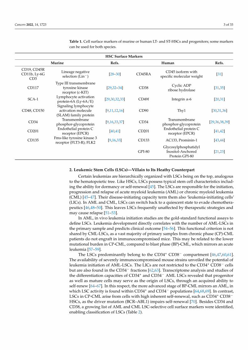
Cancers 2022, 14, 1723 3 of 33
Table 1. Cell surface markers of murine or human LT- and ST-HSCs and progenitors; some markerscan be used for both species.
HSC Surface Markers
Murine Refs. Human Refs.
CD19, CD45RCD11b, Ly-6G
CD3
Lineage negativeselection (Lin−) [28–30] CD45RA CD45 isoform with
specific molecular weight [31]
CD117Type III transmembrane
tyrosine kinasereceptor (c-KIT)
[29,32–34] CD38 Cyclic ADPribose hydrolase [31,35]
SCA-1 Lymphocyte activationprotein-6A (Ly-6A/E) [29,30,32,33] CD49f Integrin α-6 [20,31]
CD48, CD150Signaling lymphocyte
activation molecule(SLAM) family protein
[9,11,12,16] CD90 Thy1 [30,31,36]
CD34 Transmembranephosphor-glycoprotein [9,16,33,37] CD34 Transmembrane
phosphor-glycoprotein [29,36,38,39]
CD201 Endothelial protein Creceptor (EPCR) [40,41] CD201 Endothelial protein C
receptor (EPCR) [41,42]
CD135 Fms-like tyrosine kinase 3receptor (FLT3-R); FLK2 [9,16,33] CD133 AC133, Prominin-1 [43,44]
GPI-80Glycosylphosphatidyl
Inositol-AnchoredProtein GPI-80
[21,23]
2. Leukemic Stem Cells (LSCs)—Villain to Its Heathy Counterpart
Certain leukemias are hierarchically organized with LSCs being on the top, analogousto the hematopoietic tree. Like HSCs, LSCs possess typical stem cell characteristics includ-ing the ability for dormancy or self-renewal [45]. The LSCs are responsible for the initiation,progression and relapse of acute myeloid leukemia (AML) or chronic myeloid leukemia(CML) [45–47]. Their disease-initiating capacity term them also ‘leukemia-initiating cells’(LICs). In AML and CML, LSCs can switch back to a quiescent state to evade chemothera-peutics [46,48–50]. This leaves LSCs frequently unaffected by therapeutic strategies andmay cause relapse [51–53].
In AML, in vivo leukemia initiation studies are the gold-standard functional assays todefine LSCs. Leukemia development directly correlates with the number of AML-LSCs inthe primary sample and predicts clinical outcome [54–56]. This functional criterion is notshared by CML-LSCs, as a vast majority of primary samples from chronic phase (CP)-CMLpatients do not engraft in immunocompromised mice. This may be related to the lowermutational burden in CP-CML, compared to blast phase (BP)-CML, which mirrors an acuteleukemia [57–59].
The LSCs predominantly belong to the CD34+ CD38− compartment [46,47,60,61].The availability of severely immunocompromised mouse strains unveiled the potential ofleukemia initiation of AML-LSCs. The LICs are not restricted to the CD34+ CD38− cellsbut are also found in the CD34− fractions [62,63]. Transcriptome analysis and studies ofthe differentiation capacities of CD34+ and CD34− AML LSCs revealed that progenitoras well as mature cells may serve as the origin of LSCs, through an acquired ability toself-renew [64–67]. In this respect, the more advanced stage of BP-CML mirrors an AML, inwhich LSC activity is found within CD34+ and CD34− populations [64,68,69]. In contrast,LSCs in CP-CML arise from cells with high inherent self-renewal, such as CD34+ CD38−
HSCs, as the driver mutation (BCR–ABL1) impairs self-renewal [70]. Besides CD34 andCD38, a growing list of AML and CML LSC-selective cell surface markers were identified,enabling classification of LSCs (Table 2).

Cancers 2022, 14, 1723 4 of 33
Table 2. LSC-specific cell surface markers present both in AML and CML cells.
Human LSC Surface Markers
AML and CMLMarkers Alternative Name Refs. AML and
CML Markers Alternative Name Refs.
CD25 IL-2Rα chain [71–73] CD33 Siglec-3 [72,74–76]
CD45RACD45, also known as protein
tyrosine phosphatase,receptor (PTPRC)
[77,78] CD93C-type lectin-likedomain (CTLD)
containing glycoprotein[79–81]
CD9Motility Related
Protein-1 (MRP-1)Tetraspanin-29 (TSPAN29)
[72,82,83] CD123 Interleukin 3 receptorsubunit α (IL-3R α) [72]
CD371C-type lectin domain family12 member A (CLEC12A);
CLL-1 antigen[72,84] IL1RAP Interleukin 1 receptor
accessory protein [85,86]
CD69
C-Type Lectin Domain Family2, Member C (CLEC2C)
Activation InducerMolecule (AIM)
[87–89] CD36Thrombospondin ReceptorPlatelet Collagen ReceptorGlycoprotein IIIb (GP3B)
[88,89]
CD43 Leukosialin [75] CD44Pgp-1, multidrug
resistance protein 1(MDR1)
[75]
CD45 Leukocyte commonantigen (LCA) [75] CD157 Bone marrow stromal cell
antigen 1 (BST1) [75]
3. BM Niche of LT/ST-HSCs and LSCs
The LT/ST-HSCs reside in specialized niches that in the cavities of the trabecularregions in long bones. Multiple cellular types, soluble factors and components of theextracellular matrix form these niches [90,91]. The endosteal niche, located at the endostealsurface of the bone is characterized by bone regenerating osteoblasts (OBs) [92]. The OBcells regulate LT-HSCs quiescence by producing CXC motif chemokine 12 ligand (CXCL12),transforming growth factor β (TGFβ) and angiopoitin-1 (ANG-1) [90]. The arteriolarniche is located aside vascular structures and brings LT/ST-HSCs in close proximity toendothelial cells (ECs) and mesenchymal stromal/stem cells (MSCs) [93]. The ECs regulatedormancy and self-renewal via cell-cell contacts (e.g., via E-selectin) and by expression ofstem cell factor (SCF), CXCL12 and Notch ligands [11,90,94–96]. The MSCs play a domi-nant part and are subdivided into Leptin+, CXCL12 abundant reticular (CAR) or Nestin+
cells, which provide soluble factors for HSC maintenance including SCF, thrombopoietin(TPO), CXCL12, fibroblast growth factor 2 (FGF-2) and WNT ligands [90,92,94,97–99]. Theperipheral sympathetic nerves, ensheathed by non-myelinating Schwann cells (NMSCs),constitute another component of the arteriolar niche. Circadian adrenergic signals fromnerve terminals regulate the production of CXCL12 in Nestin+ MSCs. Approximately 20%of HSCs are in direct contact with NMSCs, which are maintaining LT-HSC quiescenceby producing TGF-β [100]. When activated, ST-HSCs relocate to the Leptin+ MSCs con-taining perisinusoidal area [100]. Adipocytes, pericytes, fibroblasts, macrophages andmegakaryocytes are also part of the BM niche and modulate functions of HSCs [90].
The LT/ST-HSCs and niche cells can interact through juxtacrine signaling (or contact-dependent signaling) via N-cadherin, vascular cell adhesion molecule 1 (VCAM1)/verylate antigen-4 (VLA-4), cKIT/membrane bound SCF or NOTCH receptor/ligand [101–103].Niche cells provide extracellular matrix (ECM) proteins including glycoproteins (e.g., fi-bronectin), glycosaminoglycans (e.g., hyaluronic acid), collagen IV and matrix remodelingenzymes. The LT/ST-HSCs bind ECM proteins mainly via integrins, which triggers intra-cellular signals. Of note, the extent of signaling may be modulated by the “stiffness” of thesurrounding via mechanotransduction. Stiffness, composition and location of the niche arecrucial to modulate HSC behavior [104–107]. Besides signaling through integrins, ECM em-

Cancers 2022, 14, 1723 5 of 33
beds LT/ST-HSCs in their niche and provides a reservoir for soluble factors. These factorsand cell-matrix interactions impact the balance between stemness and differentiation [97].
In AML and CML, the BM is tightly packed with malignant hematopoietic cellscausing disturbed niche structures and hematopoiesis [108,109]. The leukemic blastsoccupy and rearrange the BM niches to establish self-protective niches. This process isregulated by stromal-secreted chemokines, CXCL12 and the CXCR4 receptor and createsa “reduced” version of the conventional BM niche [110]. It impairs normal homeostasisand promotes disease progression [45,109,111]. In this protective environment LICs/LSCsare less amenable to chemotherapeutics [45,73,108,112]. Attempts to mimic the BM nichein vitro by 2-3 D techniques try to reduce the transitional gap between in vitro and in vivoresearch [113,114].
4. Essential Factors for HSC Quiescence and Self-Renewal—Cell Cycle Componentsas Mediators
The LT/ST-HSCs are capable of self-renewal or differentiation. Under homeostaticconditions, most LT-HSCs exist in the G0 phase of the cell cycle and are considered “qui-escent”. Signals from the BM preserve the quiescent state and protect from cell damageand exhaustion [115]. Quiescent HSCs are fundamental for transplantation and providethe potential for long-term engraftment [116].
Symmetric cell division results in two identical daughter cells, that either keep stemcell properties or differentiate. During asymmetric cell division, one daughter cell preservesstem cell characteristics, while the other one undergoes differentiation. During asymmetricdivision, cell fate determinants are unequally distributed, like tyrosine-protein kinase recep-tor 2 (TIE2) or NOTCH1. Similarly, active mitochondria, lysosomes and autophagosomesare unevenly shared. This procedure gives rise to a metabolically active, differentiated andshort-lived progenitor cell and a metabolically less active, undifferentiated and long-livedHSC progeny [117–120].
Cell cycle regulators are critical factors in maintaining HSC quiescence and re-inducingproliferation. The cyclin C/cyclin dependent kinase 3 (CDK3) complex regulates G0 phase,while G1 is controlled by cyclin D/CDK4 and CDK6 complexes [121,122]. The balance ofCDKs and cyclin dependent kinase inhibitors (CDKIs) control the transition from G0 to G1.
The G1 -specific CDKIs comprise of two families. The CIP/KIP family consists ofp21 (p21CIP, CDKN1A), p27 (p27KIP, CDKN1B) and p57 (p57KIP2, CDKN1C). The secondCDKI family includes four INK4 members: p15, p16, p18 and p19 (CDKN2A-D). The INK4family members bind to CDK4/CDK6 and inhibit their kinase activities by interferingwith their association with D-type cyclins. In contrast, the CIP/KIP family members bindand inhibits both cyclin and CDK subunits [121,123]. In the absence of CDK4/6 kinaseactivity, retinoblastoma (RB) proteins (RB, p107, p130) remain under-phosphorylated. Thus,they bind to E2F transcription factors and prevent them from being active. Entry into Sphase and cell cycle progression are inhibited [121]. Among all CDKIs, p57 has the highestexpression in LT-HSCs. Members p57 and p27 maintain HSC quiescence by preventingnuclear translocation of HSC70/cyclin D1 and consequent activation of CDK4/6 [124].From the INK4 family, p15 and p18 have been found to negatively regulate HSC ex vivoexpansion [125–127]. During genotoxic stress states and aging, p19 preserves HSCs in aquiescent state protecting them from apoptosis [128].
Pathways triggered by extracellular signals frequently converge on cell cycle regu-lators (cyclins, CDKs, CDKIs, transcription factors, microRNA, etc.) [115,129,130]. TheSCF/c-KIT, TPO/c-MPL and NOTCH ligands/NOTCH1-4 signaling support HSC sur-vival, self-renewal and regeneration [131–134]. The CXCL12/CXCR4, TGF-β/TGFβR andANG-1/TIE1/2 signaling is essential to maintain HSCs in a quiescent state [100,135–137](Figure 2).

Cancers 2022, 14, 1723 6 of 33Cancers 2022, 14, 1723 6 of 35
Figure 2. Overview of murine/human LT/ST-HSCs expansion possibilities and limitations. Self-renewal in LT/ST-HSCs is strictly regulated by multiple factors. The most important receptors and their corresponding ligands are listed. Purified LT/ST-HSCs can be cultured by various methods summarized in the box. Adult LT-HSCs reside in the BM niche which is absent upon cultivation, presenting the greatest limitation and challenge.
Stimulation of c-KIT and MPL activates signal transducers and activators of tran-scription (STAT), mitogen activated protein kinase (MAPK) and phosphoinositide 3-ki-nases (PI3K)/Akt pathways to enhance HSC survival and expansion ex vivo [138–145]. The TPO drives the expression of two negative cell-cycle regulators (p57, p19) and the transcription factor homeobox B4 (HOXB4), a potent promoter of LT-HSC self-renewal [146,147].
Activation by STAT5 typically drives cell survival and proliferation but may also me-diate quiescence through driving the expression of TIE2, p21 and p57 [148,149]. The hy-poxia-inducible factor 2 alpha (HIF2α) is a direct STAT5 target gene, which upregulates c-MYC, VEGF and glucose metabolism. Under hypoxia, STAT5 can impose a long-term pro-liferative advantage on the CD34+/CD38− HSC population, but not on progenitors [150]. Thus, depending on niche signals, STAT5 links self-renewal to a quiescence-typical meta-bolic profile and cell cycle arrest.
NOTCH signaling promotes LT-HSC self-renewal and inhibits differentiation
[95,151]. Unlike secreted niche factors, NOTCH signaling is a juxtacrine communication pathway between NOTCH ligands (Dll1, Dll4, Jagged1, or Jagged2) and NOTCH recep-tors (NOTCH1-4) expressing cells. Ligand-bound NOTCH receptor is cleaved, releasing the NOTCH intracellular domain, which translocates to the nucleus and alters gene tran-scription. NOTCH signaling provides direct transcriptional downregulation of p57 and upregulation of several genes that are important for HSC activation, including HES1, GATA2, cMYC [152–155].
Many important signaling pathways, such as WNT/beta-catenin, MAPK, PI3K/AKT/GSK-3 and JAK/STAT regulate expression of c-MYC. As a transcription factor, c-MYC antagonizes p21 and p27 activity by inducing the expression of D-type cyclins, thus enabling the formation of cyclin D-CDK4/6 complexes [156,157]. In fact, WNT signal-ing has been mostly characterized in dividing cells. However, LT-HSC appear to have the
Figure 2. Overview of murine/human LT/ST-HSCs expansion possibilities and limitations. Self-renewal in LT/ST-HSCs is strictly regulated by multiple factors. The most important receptors andtheir corresponding ligands are listed. Purified LT/ST-HSCs can be cultured by various methodssummarized in the box. Adult LT-HSCs reside in the BM niche which is absent upon cultivation,presenting the greatest limitation and challenge.
Stimulation of c-KIT and MPL activates signal transducers and activators of transcrip-tion (STAT), mitogen activated protein kinase (MAPK) and phosphoinositide 3-kinases(PI3K)/Akt pathways to enhance HSC survival and expansion ex vivo [138–145]. The TPOdrives the expression of two negative cell-cycle regulators (p57, p19) and the transcriptionfactor homeobox B4 (HOXB4), a potent promoter of LT-HSC self-renewal [146,147].
Activation by STAT5 typically drives cell survival and proliferation but may also medi-ate quiescence through driving the expression of TIE2, p21 and p57 [148,149]. The hypoxia-inducible factor 2 alpha (HIF2α) is a direct STAT5 target gene, which upregulates c-MYC,VEGF and glucose metabolism. Under hypoxia, STAT5 can impose a long-term proliferativeadvantage on the CD34+/CD38− HSC population, but not on progenitors [150]. Thus,depending on niche signals, STAT5 links self-renewal to a quiescence-typical metabolicprofile and cell cycle arrest.
NOTCH signaling promotes LT-HSC self-renewal and inhibits differentiation [95,151].Unlike secreted niche factors, NOTCH signaling is a juxtacrine communication pathway be-tween NOTCH ligands (Dll1, Dll4, Jagged1, or Jagged2) and NOTCH receptors (NOTCH1-4)expressing cells. Ligand-bound NOTCH receptor is cleaved, releasing the NOTCH intra-cellular domain, which translocates to the nucleus and alters gene transcription. NOTCHsignaling provides direct transcriptional downregulation of p57 and upregulation of severalgenes that are important for HSC activation, including HES1, GATA2, cMYC [152–155].
Many important signaling pathways, such as WNT/beta-catenin, MAPK, PI3K/AKT/GSK-3 and JAK/STAT regulate expression of c-MYC. As a transcription factor, c-MYCantagonizes p21 and p27 activity by inducing the expression of D-type cyclins, thus enablingthe formation of cyclin D-CDK4/6 complexes [156,157]. In fact, WNT signaling has beenmostly characterized in dividing cells. However, LT-HSC appear to have the highestpercentage of WNT activity, whereas MPPs have the lowest. This indicates that WNTsignaling enforces quiescence, probably by upregulation of p21 [158,159].

Cancers 2022, 14, 1723 7 of 33
The CXCL12 (also termed stromal cell-derived factor 1, SDF1) has pleiotropic effects.It is not only a chemoattractant for HSC homing, but also a regulator of HSC quiescence asit upregulates p57 and limits generation of reactive oxygen species (ROS) and genotoxicstress [136,160]. Similarly, TGF-β potently inhibits HSC proliferation, regulates quiescence,and protects HSCs from excessive differentiation signals. It does so by downregulatingcyclin D2 and upregulating p15, p21, p27 and p57 [125,137,161–165]. The P53 is highlyexpressed in LT-HSCs. It regulates quiescence by inducing p21 expression and driving theexpression of the transcriptional repressors GfI-1 and Necdin [166,167]. Necdin directlyinhibits E2F1, while GFI-1 decreases the expression of inhibitor of DNA binding anddifferentiation-2 (ID2), an inhibitor of RB and repressor of CDKI p21 and p27 [168–170].Upon activation, p53 is repressed by the ETS family transcription factor, MEF/ELF4,enabling entry into the cell cycle [171].
Among other transcriptional regulators, MEF/ELF4 and CDK6 regulate the exit fromdormancy, while PBX-1 and EVI-1 maintain LT-HSC self-renewal [171–175].
Abnormal activation of HSC signaling pathways induce cell cycling, exhaustion ordevelopment of leukemia [176]. Hematopoietic challenges such as inflammation, BMtransplantation or oncogenic transformation also trigger activation and proliferation ofLT/ST-HSCs [177,178].
5. Signaling and Metabolic Changes in LSCs
Malignant transformation is caused by genetic or epigenetic alterations due to heredi-tary or environmental conditions. The fusion protein BCR–ABL1, a constitutively activetyrosine kinase, is the unique hallmark and main driver of CML leukemic cells and LSCs, asit is present in >90% of the patients [179]. Both BP-CML and AML are genetically more com-plex and heterogeneous with subgroups showing individual mutations. Early mutationsimprove the potential to self-renew and may impair differentiation leading to heteroge-neously expanded clones of pre-leukemic HSCs in patients [180–182]. Late co-expressingmutations occur in signaling pathways (e.g., FLT3), promote proliferation and enhanceblock of differentiation [183,184]. The molecular defects underlying AML are complexwith at least 24 different genetically defined subtypes [185]. Expression of AML-associatedmutations and fusion genes involve transcription factors or epigenetic regulators, suchas DNMT3A, IDH1/2 and TET2 mutations or MLL-, NUP98- and AML1-fusions to namefew. Co-expression of mutations in tyrosine kinases and other signaling mediators, suchas FLT3- N/K-RAS-, KIT- mutations support proliferation leading to a more aggressivedisease progression [186].
Common mRNA and epigenetic signatures are found in AML LSCs irrespective of theoncogenic driver or immunophenotype. Ng and colleagues identified a panel of 17 genes(called LSC17), which are highly expressed in LSCs (relative to the bulk of AML cells). HighLSC17 expression reflects stemness properties of LSCs and resistance to standard AMLtherapy. The LSC signature genes include GPR56, AKR1C3, CD34, EMP1, SMIM24, SOCS2,CPXM1, CDK6, KIAA0125, DPYSL3, MMRN1, LAPTM4B, ARHGAP22, NYNRIN, ZBTB46,DNMT3B and are predictive and/or prognostic biomarkers [187,188].
Sachs et al. demonstrated that transcriptional profiles of self-renewal and proliferationare distinct in AML LSCs. LSC-specific self-renewal signature (CD69, S100A4, MYB, ADA,MRI1, CKS2) and proliferation genes (H2AFZ, BCL2A1D, CD36) were identified basedon high expression in AML LSCs relative to normal hematopoietic stem/progenitor cells(HSPC)s. Using cell surface markers, CD69 and CD36 allowed the isolation of differentsubsets of LSCs. The CD69high LSCs were capable of self-renewal and poorly proliferative,whereas the CD36high LSCs did not inflict leukemia and were highly proliferative [89].These genes were not found as a signature in normal HSPCs and may represent a unifyingfeature for the identification of LSCs [62,89,187,189].
The HSCs rely on glycolysis in the hypoxic BM microenvironment, rather than ox-idative phosphorylation (OXPHOS) [190]. In contrast, AML and CML LSCs have highermitochondrial mass and an increased oxygen consumption rate with a greater dependency

Cancers 2022, 14, 1723 8 of 33
on mitochondrial function and OXPHOS [191]. Mitochondrial respiration generates highlevels of ROS in bulk CML and AML blasts relative to LT/ST-HSCs [192,193]. High ROSlevels can induce oxidative DNA damage, high mutational burden and genomic insta-bility, which may impair stem cell function [194–197]. Quiescent AML LSCs generallyhave low ROS levels compared to cycling LSCs and bulk AML cells [198]. These cellsmay revert to glycolysis or using mitophagy to reduce their dependency on mitochondrialrespiration [199,200]. Quiescent low-ROS AML LSCs frequently overexpress BCL-2 andare dependent on amino acid uptake and OXPHOS [198,200]. Besides mitochondrial respi-ration and ROS amounts, levels of glucose, amino acid and free fatty acid are altered inLSCs [191,198,201]. Therefore, ex vivo expansion of LSCs requires specific conditions toallow biomarker discovery, drug development, identification of resistance mechanisms andcombination treatments.
6. Major Challenges Culturing LT-HSCs
Maintaining and expanding LT/ST-HSCs ex vivo are required for curative transplan-tation therapies and to allow the study of molecular mechanisms [202,203]. The long-termgoal is to further optimize application methods for clinical hematopoietic stem cell trans-plantation (HSCT).
The effects of cell culture stress on HSCs induces several changes including loss ofpolarization, accumulation of reactive oxygen species, endoplasmic reticulum stress, geno-toxic stress, replicative stress, disturbed protein homeostasis and ultimately loss of HSCfunctions [204–211]. As a result of these cell intrinsic changes occurring during ex vivoculture, the number of LT/ST-HSCs declines over time accompanied by an increase ofmyeloid potential [206,207,211]. To reduce environmental stress and preserve stem cellfunctions, enhanced proteostasis, a dynamic maintenance of proteome integrity, is particu-larly important in cultured HSCs [207]. Defining culture conditions favoring expansion ofLT/ST-HSCs while maintaining their fitness, still represent a major hurdle.
7. Maintaining Quiescent LT-HSCs
The BM has limited oxygen supply; the most quiescent LT-HSCs reside in a “hy-poxic niche” where blood perfusion and oxygen tension are low [212–215]. In vitrohypoxic cultures with 1–3% oxygen enhance LT/ST-HSC expansion and subsequentengraftment [216–219]. Under normoxia (20% O2), the maintenance of self-renewal re-quires the presence of high cytokine concentrations. Still, normoxia favors differentiationover self-renewal [220].
Kobayashi et al. defined a minimal set of factors that mimic the physiological condi-tions in the BM microenvironment and maintain LT-HSCs in a quiescent, but still engraftablestate for 1 month. Murine LT-HSCs need low concentrations of cytokines (3 ng/mL SCF,0.1 ng/mL TPO), hypoxia (1% O2) and 4% bovine serum albumin (BSA). Supplementingfatty-acids in an albumin-bound form is crucial to avoid intrinsic fatty acid synthesis,which is triggered by hypoxia and low cytokine concentrations [220]. Intrinsic fatty acidsynthesis would interfere with HSC survival [221]. Under low cytokine, hypoxia, 4% BSAconditions, differentiation is suppressed and more than 60% of cells remain functionalCD150+ CD48− LT-HSCs. A gradual decrease of HSC markers including CD150 and EVI1was only observed after a month of culture. Human LT-HSCs were also maintained undercomparable hypoxic conditions in 4% BSA with low cytokines, supplemented with fattyacids and cholesterol. ~90% of the cells exhibited a CD34+ CD38− marker phenotype and40% a CD90+ CD45RA− phenotype, reflecting minimal differentiation [220].
Retinoic acid (RA) signaling is high in dormant LT-HSCs compared to ST-HSCs andMPPs [9]. Retinoic acid is produced by two sequential oxidation steps from dietary vitaminA (retinol). The biological active derivative ATRA signals through the retinoic acid receptor(RAR) and the retinoid X receptor (RXR) families [222,223]. The ATRA restrains c-MYCexpression, and inhibition of MYC activity partially mimics the preservation of dormancyby ATRA. Treatment with ATRA, retinol or MYC inhibitor retains LT-HSC quiescence ex

Cancers 2022, 14, 1723 9 of 33
vivo in serum free, cytokine supplemented (SCF, TPO, FLT3L) media by downregulatingG2M checkpoints, E2F targets, ROS species and c-MYC targets compared to untreatedcells [224].
Recently, purified single mouse (CD45+ EPCR+ CD48− CD150+ SCA-1high) and hu-man (CD34+ CD38− CD90+ CD45RA− CD19− CD49f+) LT-HSCs were maintained in ahibernated (hibHSC), non-proliferative state under minimal cytokine conditions (onlyinterleukin-11 (IL-11)) over a 7 days period [225–227]. Large proportions of hibHSCssurvived without dividing and retained their functional properties, as determined bysingle-cell transplantation. Stress response pathways together with glycolysis, fatty acidbiosynthesis, cAMP and mTOR signaling pathways were upregulated in hibHSCs, mostpresumably because of nutrient withdrawal and limited cytokine availability [225]. Thedevelopment of a “quiescent LT-HSC ex vivo system” will open an avenue to study steady-state LT-HSC properties and effects of targeted manipulation.
8. Expansion Techniques to Retain LT/ST-HSC Phenotype Ex Vivo8.1. 2D Methods8.1.1. Suspension Culture of Murine LT/ST-HSCs
Ex vivo culture approaches try to mimic physiological conditions in the BM andprovide growth factors and cytokines to maintain quiescence or induce proliferation. Earlyattempts did not include any supporting BM cells but relied on media supplements likecytokines and growth factors (Figure 2).
Later, in the 1990s, 5-fluorouracil (5-FU) treated mice were used as a source for collect-ing and culturing LT/ST-HSCs. They were kept in 20% fetal calf serum (FCS) (or withoutserum), 1% BSA, SCF, FLT3/FLK-2 ligand (FLT3L) and IL-11 containing media [228]. Fre-quently, IL-11 is replaced by IL-6 or IL-12 [229]. Further supplements included ITS-X(insulin, transferrin, selenium, ethanolamine) or low-density lipoproteins [229–232]. Underthese conditions, the LT/ST-HSCs maintained their ability to reconstitute lethally irradiatedrecipient mice for up to three weeks but lost it upon addition of IL-3 and/or IL-1 to theculture medium [228,232–234]. In contrast, addition of TPO to mouse BM cells enhancedthe number of LT/ST-HSCs and increased the efficiency of BM reconstitution [235].
The advanced knowledge about HSC self-renewal and its regulation by niche compo-nents allowed for the development of novel ex vivo expansion approaches using differentcombinations of supplements [236–238]. The BM niche cells (e.g., ECs) stimulate self-renewal of LT/ST-HSCs by inducing NOTCH signaling [151,239]. Using the engineeredNOTCH ligand Delta1ext-IgG, mouse LSK cells were successfully cultured in 20% FBS, SCF,FLT3L, IL-6 and IL-11 supplemented media up to 42 days investigated [240,241]. Theimmobilized Delta1ext-IgG (composed of the extracellular domain of Delta1 joined to a Fcpart of human immunoglobulin G1) accelerates the expansion of LSKs and the rate of T-cellreconstitution after transplantation [241,242].
Ieyashu et al. showed that interleukin-1α (IL-1α) and hemopexin (HPX) in serum-free,but BSA-containing medium supports the maintenance of LT/ST-HSCs [231]. The heme-binding plasma glycoprotein HPX is expressed on NMSCs in the BM niche and preventsheme-mediated oxidative stress and dampens intracellular ROS levels [243]. The use ofSCF and TPO together with IL-1α and HPX provides a highly reproducible ex vivo mouseLT/ST-HSC expansion culture system [231].
One of the most frequently used media to enrich LT/ST-HSCs was developed by thegroup of Lodish. It includes serum-free medium with SCF, TPO, FGF-1, IGF-2, and hep-arin, resulting in an 8-fold increase of mouse LT-HSCs in three weeks of culture [145,230].Addition of angiopoietin-like proteins (ANGPTLs: ANGPTL2, ANGPTL3) to the mix-ture of SCF, TPO, IGF-2 and FGF-1 revealed a roughly 50-fold increase in numbers ofrepopulating mouse LT-HSCs [230,244]. Although previous protocols used similar mediareagents, Lodish’s specific combinations favored proliferation of LT-HSCs over ST-HSCsand prevented them from being outcompeted during long-term culturing.

Cancers 2022, 14, 1723 10 of 33
Another critical feature for successful maintenance of LT/ST-HSCs ex vivo is theuse of appropriate BSA, which is a component of most protocols containing fetal bovineserum (FBS). The quality and composition of FBS varies between batches, which leads todifferences in culture conditions and affects self-renewal [231,245]. In general, differencesin serum and BSA concentrations modulate the bio-availability of cytokines and growthfactors based on binding to serum albumin [246]. To standardize culture conditions, BSAwas replaced by polyvinyl alcohol (PVA), a synthetic amphiphilic polymer, which stabilizescytokines. In the presence of PVA, 100 ng/mL TPO and 10 ng/mL SCF are consideredoptimal for murine LT/ST-HSC culture [226]. This represents a significant improvementand enables long term culture (1- to 2-month) with an up to 900-fold expansion of functionallin− c-KIT+ SCA-1+ CD150+ CD34− LT-HSCs [145,226–228,232,247,248].
8.1.2. Suspension Culture of Human LT/ST-HSCs
Human cord blood CB-derived hematopoietic stem cells (CB-HSCs) are an importantsource for HSC transplantations. Their numbers are low in vivo, which requires expandingCB-HSCs ex vivo while preserving their stemness properties for effective application intransplantation and gene therapy. Several promising protocols for serum-free cultivationof human LT/ST-HSCs using combinations of cytokines or small molecules have beendescribed (Figure 2).
Commonly used cytokines for expansion of CD34+ HSCs include SCF, TPO, FLT3L,granulocyte colony-stimulating factor (G-CSF), IL-6 IL-3 and FGF-1 [249–251]. Using acytokine cocktail (SCF, TPO, FLT3L and FGF-1) supplemented with insulin-like growthfactor-binding protein 1/2 (IGFBP1/2) and ANGPTL5, increased the number of humanCD34+ CD38− CD90+ CD133+ CB stem cells. These cells repopulate NOD-SCID mice witha ∼20-fold higher efficiency than non-cultured HSCs [249,251].
Developmental regulators such as NOTCH ligand Delta-1, pleiotrophin, StemReginin-1, UM171, resveratrol, nicotinamide and valproic acid (VPA) were reported to furtherenhance CD34+ HSPCs expansion over 50 fold [250,252].
The FDA approved small molecular weight compounds StemReginin 1 (SR-1) orUM171 are used in addition to cytokines (SCF, TPO, FLT3L and IL-6) to expand humanCD34+ HSCs ex vivo [253–255]. The SR-1 antagonizes the aryl hydrocarbon receptor(AHR), which regulates hematopoiesis through regulation of HES-1, c-MYC, C/EBP, PU.1,β-catenin and CXCR4 [256]. The Ahr knockout mice have increased numbers of HSCs with ahigher proliferative rate and accumulation of plasmacytoid dendritic cells (pDCs) [256,257].In line, antagonizing AHR via SR-1 ex vivo enhanced the frequency of CD34+ HSCs andinduced differentiation of myeloid mDCs and pDCs [255]. In a clinical trial SR–1 hasinduced a 330-fold expansion of CD34+ cells and resulted in fast engraftment of neutrophilsand platelets in patients. Neutrophil recovery response has been viewed as a surrogatemarker of host immunity [258]. The AHR is also antagonized by Resveratrol, a naturallyoccurring polyphenol. Resveratrol binds receptors involved in HSC activity includingAHR and integrin αvβ3 [259,260]. Addition of Resveratrol to a cytokine-containing (SCF,TPO, FLT3-L, IL-6) medium represents a robust ex vivo method for the expansion offunctional CD34+ CB HSCs. The UM171 also inhibits LSD1 (H3K4me1/2 demethylase)and HDAC1/2 (e.g., H3K27ac deacetylase), leading to the re-establishment of H3K4me2and H3K27ac epigenetic marks, which normally rapidly decrease in human LT/ST-HSCsex vivo [261–263]. Of note, when UM171 is combined with SR-1 and cytokines (SCF, TPO,FLT3L, IL-6) the efficiency of expansion is further increased [263].
As a suppressor of SIRT1 deacytelase, nicotinamide inhibits differentiation and en-hances expansion of CD34+ CB-HSCs [264]. The SIRT1 deacetylates and thereby deacti-vates p53 protein [265]. Proof for SIRT1´s ability to maintain stemness potential comesfrom a phase I clinical trial. The surrogate marker, median neutrophil recovery rate, wassignificantly increased in individuals, who had received nicotinamide treated CD34+ CB-HSCs [266].
Cancers 2022, 14, 1723 11 of 33
A screen of CD34+ CB-HSCs identified histone deacetylase inhibitor (HDACI) VPAas a promising candidate for LT-HSC expansion. Adding VPA to SCF, TPO, FLT3L andIL-3 in serum free media enhanced expressions of CD90, c-KIT, CXCR4 and integrin α6(CD49f), increased activation of p53 and reduced ROS levels. The VPA induced HSCexpansion by reprogramming CD34+ CD90− cells to CD34+ CD90+ HSCs, accompaniedby increased proliferation. The VPA-expanded peripheral blood (PB) cells and BM HSCsestablished unbiased multilineage human hematopoietic-cell chimerism in NSG mice at16 weeks post-transplantation [267,268].
The major side effect of allogeneic transplantations is the development of graft-versus-host disease (GvHD). Compared to BM HSCs, the transplantation of CB-derived CD34+ cellswith up to two human leukocyte antigen mismatches revealed a lower risk of GvHD [269].Due to the low numbers of CB-CD34+ cells from a single donor cord, the hematopoieticand immunological recovery of the recipients may be delayed, causing higher infectionrates and transplant-related mortality. In the clinics, SR-1, Notch-ligand, UM171 andnicotinamide-based methods have been associated with improved neutrophil recoveryearly after transplantation, reducing side effects of HSCT [258,266,270]. In spite of majordevelopments in ex vivo expansion of CB-HSCs, the efficiency of their long-term engraft-ment is still inadequate [271]. To overcome this limitation, double CB transplantations,with nonexpanded CB-HSCs of a second donor, were used to engraft. In this case, a higherrisk of GvHD was reported [272]. Thus, enhancing the availability of functional CD34+
CB-HSCs would give an excellent opportunity to improve therapeutic applications.
8.1.3. Suspension Culture of Human LSCs
Several patient-derived AML and CML cell lines are available. They are easy toculture but acquire multiple cytogenetic aberrations upon prolonged culture. This aspectneeds to be considered when comparing experiments from different laboratories thatmay significantly differ [273]. In contrast to primary AML cells, which are geneticallyand functionally heterogenous (consisting of LSCs and differentiated cells), cell lines arehomogeneous, favoring proliferation of the most aggressive clones [274–277]. Culturesystems for primary AML samples that preserve clonal heterogeneity are required to mimicthe situation in patients.
To selectively culture CD34+ HSPCs from CML/AML samples, a feeder cell-free andserum-free liquid culture system containing FLT3L, SCF, IL-3, IL-6, and TPO has been estab-lished. The outcome is highly patient-dependent and shows a great variability [276,278].
The AML specimens share the common feature of high AHR activation in vitro, whichprovokes differentiation. The AHR agonist SR-1 and other small molecular weight com-pounds (UM171 and its derivative UM729), counteract differentiation. No stromal co-culture is required but multiple cytokines are necessary, like BIT (BSA, insulin, transferrin),SCF, FLT3L, IL-3 and G-CSF [279,280]. It is currently unclear how the pyrimido-indoleUM729 enhances CD34+ cell expansion which is AHR independent [280].
Inhibiting GSK-3 and mTORC1 also maintains self-renewing capacity of hematopoieticcells from healthy donors or AML patients [281]. The GSK3 inhibitors (GSK3i) preventβ-catenin degradation and activate WNT target genes, which is essential for long-termHSC self-renewal. The activation of mTORC1 drives HSC proliferation and differentiation,leading to HSC exhaustion [281]. As GSK3-inhibition activates mTORC1 a combinedinhibition of GSK3i and mTORC1 is required to maintain self-renewing abilities [282].
8.2. 2.5D Methods8.2.1. Co-Culturing LT/ST-HSCs
The balance between quiescence and activation of LT/ST-HSCs is tightly regulatedby the BM microenvironment [90]. The absence of a BM niche leads to a gradual loss ofthe LT-HSC status and induces differentiation towards lineage-committed progenitors.Co-culture options were developed to mimic the BM microenvironment (reviewed in [283],Figure 2).

Cancers 2022, 14, 1723 12 of 33
“2.5D co-culture” methods use supporting stromal or endothelial cells to simulatethe BM niche. LT/ST-HSCs or LSCs grow on top of adherent cells, either in direct contact(contact culture) or separated by filters (trans-well culture). Trans-well cultures circum-vent the need to separate HSC cells from the supporting cells, but are less efficient thancultures that allow for direct stroma contact [284,285]. The MSCs are most commonlyused as they express high levels of HSC-supporting factors and significantly improveengraftment [286,287].
Primary MSCs downregulate niche factors (Scf, Cxcl12 and Vcam1) upon cultureand their ability to maintain HSCs declines over time. Nakahara et al. identified fivetranscription factors (Klf7, Ostf1, Xbp1, Irf3 and Irf7) that restored HSC niche function incultured MSCs. Overexpression of these factors revitalized MSCs (rMSCs). The rMSCsexpanded cells showed a seven-fold higher efficiency in expansion of functional LT-HSCsin a setting where lineage-depleted mouse BM cells or CD34+ CBs were co-cultured in thepresence of SCF and TPO [288].
Aside from MSCs, ECs serve as a supporting cell layer. The BM-derived ECs se-crete self-renewal supporting angiocrine growth factors, such as VEGF-A and NOTCHligands [289]. Long-term maintenance of primary ECs involves loss of their angiogenicproperties. To preserve them and initiate immortalization, E4orf1 has been introduced.E4orf1 is an adenoviral E4 gene product which confers long-term survival through tonicphosphorylation of AKT [290]. Immortalization of ECs by E4orf1 allows long-term cul-tures and efficiently supports mouse LT-HSCs when in direct contact in serum-free, SCFsupplemented co-culture [289]. The support is mainly provided by the initiation of Notchsignaling. In analogy, human fetal liver (FL) sinusoidal ECs engineered and immortal-ized by E4orf1 (hFLSECs-E4orf1) are used for long-term culture of CD34+ CB cells. ThehFLSECs-E4orf1 cells also provide activation of NOTCH signaling, mimic the vascularniche and prevent LT-HSC exhaustion [291].
8.2.2. Co-Culturing LSCs
Primary AML cells differentiate and/or undergo apoptosis in culture indicating thatmost AML cells depend on signals from the microenvironment [280,292–295]. Co-culturewith BM stromal cells expands AML LICs, but frequently selects for a specific subpopu-lation [296–298]. Immortalized mouse BM mesenchymal or endothelial stromal cell lines,such as MS-5, FBMD-1, OP9, HS-5, HS-27 or HUVEC are used as supporting cells forprimary AML cultures [114,296,297,299]. The mesenchymal nestin+ MS-5 murine BM stro-mal cell line efficiently maintains functional, chemo-resistant human LSCs ex vivo over3 weeks [297,298]. The MS5 cells secrete CXCL12, ANGPT1, MCP-1 and HGF; MS5/LICcocultures were further supplemented with IL-3, G-CSF and TPO and kept at 3% O2,which provides a niche-like milieu [300]. The hypoxia signaling pathway triggers LICmaintenance in vivo [301].
Primary MSCs isolated from AML patients have an impaired ability to support nor-mal HSPCs, but an enhanced ability to maintain LSCs compared to MSCs from healthydonors [292,302,303]. Leukemic BM niche MSCs have different morphology and growthrate, altered osteogenic or adipogenic differentiation capacity and changed methylation sig-natures [292,302–307]. These co-culture conditions are labor-intensive and require carefulstandardization. Among the critical factors is the cell density of the feeder cells, which mustbe in a cell cycle arrested state. Passage restrictions of the feeder cells are required to avoidstromal cell line exhaustion and ensure comparable experimental conditions [297,308].
8.3. 3D Methods8.3.1. 3D Culture of LT/ST-HSCs
The 3D culture methods attempt to mimic the spatial structure of the BM microen-vironment by providing cell-to-cell or cell-to-biomimetic matrix contacts (Figure 2). Cellspheroids or aggregates are grown on a matrix, or in a scaffold-free suspension. Commonlyused scaffold/matrix materials include natural polymers, such as alginate, Matrigel™

Cancers 2022, 14, 1723 13 of 33
(basement membrane matrix), agarose and bacterial nanocellulose, hyaluronic acid orsynthetic-based polymer materials [296,309,310]. Polyethylene glycol (PEG), PVA, poly(lactic-co-glycolic acid) (PLGA), poly (lactic acid) (PLA) and poly (ε-caprolactone) (PCL)are common materials used to form synthetic scaffolds hydrogels [311].
To generate scaffold-free spheroids, different cell types (e.g., MSCs and LT-/ST-HSCs)are cultured on hanging drops, microplates, non-adhesive surfaces or via a forced floatingmethod (magnetic levitation or agitation-based approaches) to initiate aggregation andto avoid attachment to a culture dish [309,312–316]. Spheroids have some advantagescompared to 2.5D cultures; (i) higher levels of hematopoietic niche factors provided byMSCs, (ii) the maintenance of the cell shape and (iii) improved signaling by cell-cell contactsincreased ex vivo LT-/ST-HSC expansion compared to 2D and 2.5D methods [312,317].
The LT-/ST-HSCs cells can also be encapsulated into a natural or synthetic polymersolution that is cross-linked to form a hydrogel. Hydrogels are biocompatible, retain largeamounts of water and provide excellent permeability [318,319]. Hydrogels are mixedwith ECM proteins such as fibronectin, collagen, laminin or glycosaminoglycans to allowattachment of LT-/ST-HSCs [309,318]. Cytokines, small molecules or other factors can beincorporated depending on the aim of the research project [320].
Compared to a PEG scaffold, zwitterionic hydrogels are super-hydrophilic and aremore resistant to non-specific protein binding [319,321]. Bai et al. set up a zwitterionichydrogel system to ex vivo culture BM and CB LT-/ST-HSCs, using a metalloproteinase-cleavable zwitterionic peptide to reversibly crosslink the gels. The LT-/ST-HSCs-secretedmetalloproteinases gradually cleave peptide crosslinker, allowing cells to actively shapetheir environment [320]. This improved cell migration, cell-cell contacts and stemness ofthe HSCs; and proved to be an efficient method to expand functional CD34+ LT-HSCs basedon reduced differentiation, diminished reactive oxygen species (ROS) production and alow metabolic rate [319–321].
A disadvantage of encapsulation is the homogenous matrix, unlike the porous sponge-like structure of the BM. Although cells have full contact to the hydrogel/ECM matrix,a direct cell-cell contact may be missing. To circumvent this problem, porous scaffoldswere generated (e.g., by salt leaching, freeze-drying or 3D printing), where MSCs andCD34+ CB-HSCs can form close cell-cell contacts, improving ex vivo expansion [322–324].Alternatively, Wharton’s jelly, can be employed as a 3D matrix. Wharton’s jelly is a mucoidconnective tissue surrounding the umbilical cord vessels to confer a mechanical protectionin the womb. Decellularized Wharton’s jelly matrix (DWJM) serves as ECM scaffold and ismixed with primary human MSCs to maintain ex vivo CD34+ CB-HSCs cells. The DWJMshares many components of the BM ECM including collagens, fibronectin, tenascin-C,hyaluronic acid and numerous sulfated glycosaminoglycans possessing many uniquebiochemical characteristics [291,325,326].
Currently, self-sustained 3D applications like small-scale microfluidic devices andlarge-scale bioreactors are developed. Bioreactor is an engineered system that allowsculturing large amounts of CD34+ CB-HSCs through automated control over mediumsupply, waste removal and agitation [327]. Microfluidic devices mimic the vascular nicheby continuously supplying nutrients and oxygen, while generating gradients (e.g., oxygen,calcium ions, cytokines and small molecules) [328,329]. Bone marrow-on-a-chip is an evenmore complex microfluidic system, where the colonization of the 3D matrix by niche cellstakes place in the BM in vivo [330,331].
8.3.2. 3D Culture of LSCs
In an effort to model the BM microenvironment for ex vivo leukemia studies, stiffand porous 3D scaffolds have been used [332–337]. Leukemia research is inclined to findstrategies to disrupt the protective effect of the niche cells to improve therapeutic strategies.Bray et al. co-seeded ECs and MSCs with primary AML cells in matrix metalloproteinase–sensitive PEG-heparin hydrogels, supplemented with growth factors (VEGF, FGF-2, stromal

Cancers 2022, 14, 1723 14 of 33
cell-derived factor 1 (SCD1)). The protective effect of leukemic-vascular interactions in-creased chemoresistance of cancer cells in 3D compared to 2D [334].
The first bioreactor system was recently developed to maintain malignant CD34+ cellsfrom AML and myeloproliferative neoplasm (MPN) patients for up to 3 weeks. The systemcontained human osteoblastic BM niches, engineered by MSCs in 3D porous scaffoldsunder perfusion flow [113].
The 3D methods represent the future to expand LT/ST-HSCs in large quantitiesfor transplantations, but are less suitable for individual research-purposes, as they aretechnically challenging and labor-intensive. The use of 3D techniques is limited when itcomes to high-throughput multi-well experiments, such as drug-screening, but representsthe best choice for any validation experiments.
9. Immortalized Hematopoietic Stem/Progenitor Cell Lines
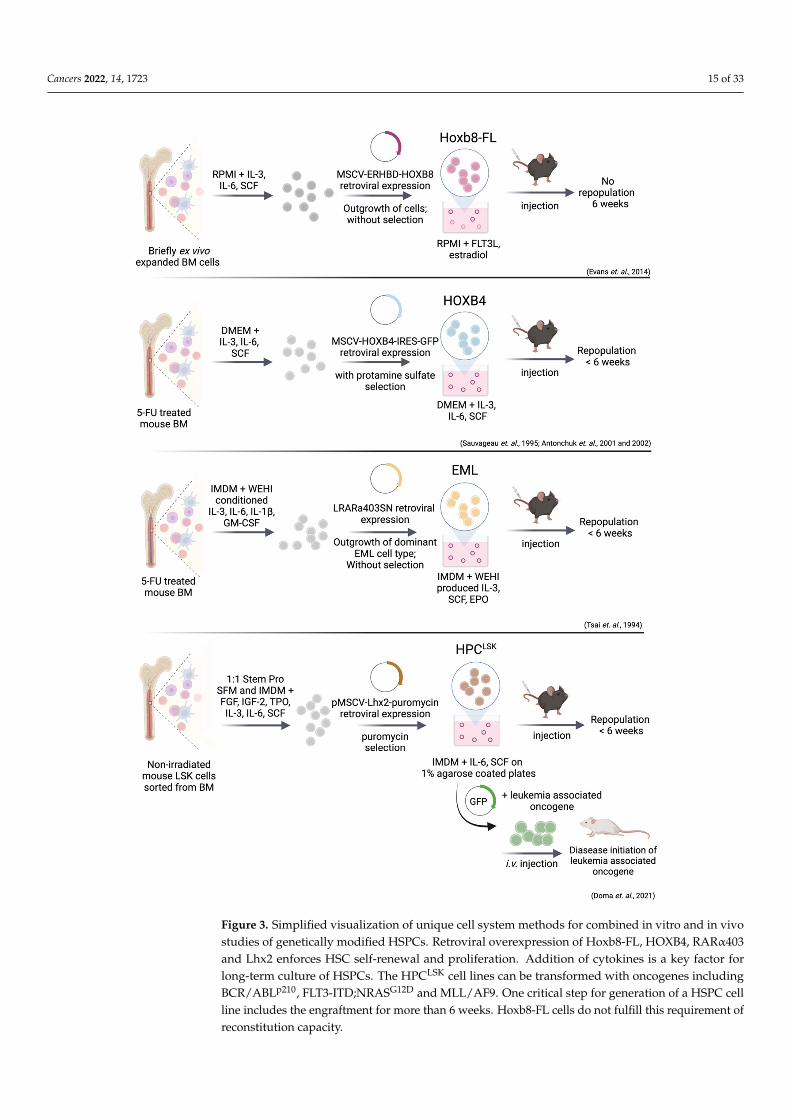
Genetic manipulation of self-renewal pathways by transgene overexpression mayprovide a suitable method for HSPC expansion and the establishment of cell lines. Suc-cessful HSPC immortalization was achieved by overexpression of embryonic develop-mental genes including HOXB, RARα and Lhx2 to enforce cell renewal and arrest celldifferentiation [338–340] (Figure 3).
Bulk- or LSK-sorted mouse BM cells were isolated and cultured in serum, growthfactor and cytokine supplemented media. Dividing cells were immortalized throughretroviral expression of HOXB4 [146,341], HOXB8 [342], truncated RARα, (RARα403) [343]or Lhx2 [339] and resulted in rapid and extensive ex vivo expansion of HSPC populationsfor more than 9 weeks (Figure 3). Exogenous growth factors are still required to induceproliferation. This process raised long-term expanded, progenitor cell lines that remainedmultipotent as demonstrated by their ability to fully repopulate lympho-myeloid lineagesin primary and secondary recipients. All transplanted mice remained healthy and withoutmanifestations of hematopoietic disorders after one year of observation [146,339,341–343].
9.1. Immortalization via HoxB8 and HoxB4
The HOX proteins are a family of evolutionary conserved transcription factors. Inmammals, 39 HOX genes are organized into four distinct clusters: HOXA, HOXB, HOXCand HOXD [344]. A considerable amount of data link HOXA and HOXB genes to cellrenewal and the arrest of cell differentiation [338,345]. These functions were exploitedexperimentally to establish stably growing, homogenous hematopoietic progenitor celllines through retroviral expression of HOX genes (HOXB4 and HOXB8). Expression confersgrowth advantage but does not elicit leukemic potential [146,341,342].
Downregulation of Prdm16 might be involved in preventing leukemia in HOXB4overexpressing LT/ST-HSCs transplanted mice [346]. The transcription regulator PRDM16is associated with AML, causes oncogenic fate conversion from megakaryocyte-erythroidprogenitors (MEPs) to LSCs, by interacting with super enhancers and activating myeloidmaster regulators, including PU.1 [347]. The PRDM16 was markedly repressed by HOXB4,but upregulated by HOXA9 and HOXA10 [346]. This may explain why HOXB4 andprobably also HOXB8 lacks the leukemogenic potential seen with other oncogenic HOXfactors such as HOXA9 and HOXA10.
Cancers 2022, 14, 1723 15 of 33Cancers 2022, 14, 1723 15 of 35
Figure 3. Simplified visualization of unique cell system methods for combined in vitro and in vivo studies of genetically modified HSPCs. Retroviral overexpression of Hoxb8-FL, HOXB4, RARα403 and Lhx2 enforces HSC self-renewal and proliferation. Addition of cytokines is a key factor for long-term culture of HSPCs. The HPCLSK cell lines can be transformed with oncogenes including BCR/ABLp210, FLT3-ITD;NRASG12D and MLL/AF9. One critical step for generation of a HSPC cell line
Figure 3. Simplified visualization of unique cell system methods for combined in vitro and in vivostudies of genetically modified HSPCs. Retroviral overexpression of Hoxb8-FL, HOXB4, RARα403and Lhx2 enforces HSC self-renewal and proliferation. Addition of cytokines is a key factor forlong-term culture of HSPCs. The HPCLSK cell lines can be transformed with oncogenes includingBCR/ABLp210, FLT3-ITD;NRASG12D and MLL/AF9. One critical step for generation of a HSPC cellline includes the engraftment for more than 6 weeks. Hoxb8-FL cells do not fulfill this requirement ofreconstitution capacity.

Cancers 2022, 14, 1723 16 of 33
In Hoxb8–FL cell lines, the hormone binding domain of the estrogen receptor (Erhbd)is fused to the coding sequence of Hoxb8, leading to the activation of Hox genes by estrogen.Estrogen withdrawal provokes differentiation into dendritic cells (DCs). Besides estrogen,growth and survival of these cells strictly depends on FLT3L [342] (Figure 3). Hoxb8–FLcells have both myeloid and lymphoid potential but lack any megakaryocyte and ery-throid capacities and closely resemble MPP4 cells (lin− SCA-1+ cKIT+ CD48+ CD150−
CD34+ CD135+) [6,342,348]. One month after transplantation, Hoxb8–FL cells are no longerdetectable in the BM, but are still present in spleen, peripheral blood, and thymus. Thisobservation suggests a compromised capacity to self-renew. The Hoxb8–FL–derived T–cellsreached merely about 10–30% of physiological T–cell numbers in the thymus, but wereabsent in the periphery [342]. Within the four HSPC lines discussed here, Hoxb8–FL shoesthe least multipotency.
The Hox factor HOXB4 is a key regulator of LT-HSCs self-renewal but its enforcedexpression allows differentiation when transplanted [341,349]. The factor HOXB4 drivesproliferation by upregulating AP-1 complexes with subsequent enhanced cyclin D1 lev-els [350]. Combination of overexpression of HOXB4 with deletion of the HOX cofactor Pbx1(pre-B-cell leukemia transcription factor 1) or expression of NUP98-HOXB4 fusion proteinfurther enhances ex vivo expansion of LT-/ST-HSCs [351]. Both approaches (HOXB4/Pbx1KO and NUP98-HOXB4) reconstituted myeloid and lymphoid populations in vivo withoutinducing leukemia [351–354]. Recent findings indicate that HOXB4 may also reprograminduced pluripotent stem cells (iPSCs) cells into long-term repopulating HSCs, openingnew avenues for human therapeutic possibilities [355,356].
9.2. EML (Erythroid, Myeloid, and Lymphocytic) Cell Line
The EML cells originally emerged as an in vitro model to study self-renewal andlineage commitment. The EML cells express a truncated, dominant-negative form of thehuman RAR (RARα403) [343,357] (Figure 3). The RARα403 outcompetes the endogenousRAR in the formation of biologically active RAR/RXR complexes, leading to c-MYCupregulation and proliferation. As mentioned above, ectopic RA signaling maintains LT/ST-HSC quiescence, while inhibition leads to stem and progenitor proliferation [224,343].
The EML cells generate large numbers of B-lymphoid and erythroid progenitors atthe expense of progenitors for the neutrophil and macrophage lineages. This may beoverridden by a combination of IL-3 and high concentration of RA, which increases thenumber of myeloid progenitors [343]. One advantage of this cell system is the ability forin vitro T-cell differentiation (upon co-culture with murine OP9-DL1 stromal cells in thepresence of SCF, IL-7, and FLT3). OP9-DL1 cells express delta-like-1, a NOTCH ligandwhich drives human and murine HSPCs into T-cells in vitro [358,359].
The main limitation of EML cells cultivated with SCF is their functional heterogeneitycontaining CD34/SCA-1low and high populations. Each subpopulation expresses a distinctpattern of HSPC markers and transcription factors, different multilineage differentiation po-tential and proliferation kinetics [357]. CD34/SCA-1high EML cells exhibit low GATA1 andhigh PU.1 level, which predisposes them to the myeloid lineage. In contrast, CD34/SCA-1low, GATA1 high, PU.1 low cells are erythroid-prone [360]. Besides GATA1, other erythroidgenes including α- and β-hemoglobin, Epor, Eraf (erythroid associated factor) and mast cellproteases are expressed at high levels in the CD34/SCA-1low EML cells [361]. The EMLcells serve as a model for cell intrinsic and extrinsic pathways that regulate plasticity amongmultipotent hematopoietic cells.
Ectopic expression of HOXB4 in EML cells allows HSC self-renewal through up-regulation of stemness-related genes, such as Laptm4b, Gp49a, Sox4, and CD34. HOXB4downregulates erythroid and B cell lineage-specific genes to keep cells in a primitivestate [362]. This cell system extends our knowledge on molecular and functional propertiesof HSPCs but lacks the ability of long-term cultivation without differentiation.

Cancers 2022, 14, 1723 17 of 33
9.3. Immortalization via Lhx2—HPCLSK Cell Lines
Retroviral transduction of the LIM-homeodomain (LIM-HD) transcription factorLhx2 was used to generate multipotent HPCLSK cell lines [339,363,364] (Figure 3). TheLHX2 has a critical role in hematopoiesis and Lhx2-null embryos die in utero with severeanemia [365,366]. The critical role of LHX2 in hematopoiesis was underlined by forcedexpression in embryonic stem (ES) cells, which resulted in the outgrowth of multipotentSCF-dependent progenitor cells [363]. The LHX2 upregulates hematopoietic self-renewalgenes and inhibits differentiation of mouse ES derived LSK cells [367].
The SCF/IL-6 dependent HPCLSK cells are kept on agarose-coated plates to preventadherence-induced myeloid differentiation. The HPCLSK cells efficiently home to the BM,blood, spleen, and thymus and can differentiate into myeloid and lymphoid lineagesin vitro and in vivo. Numbers of HPCLSKs -derived myeloid and lymphoid progenitors inthe BM and differentiated blood cells are comparable to BM-injected mice [339]. Transcrip-tome analysis of HPCLSKs showed a ST-HSC/MPP1 (lin− SCA-1+ cKIT+ CD48− CD150−
CD34+ CD135−) signature, which corresponds to the earliest proliferating stem/progenitorcells despite expression of CD48 and CD150 [6,339,348]. The absence of any cell feederlayer or extensive amounts of cytokines makes them a robust and low-cost model systemthat guarantees long-term multipotency.
9.4. Leukemic Stem Cell Lines Using the HPCLSK System
The HPCLSK cells can be genetically modified e.g., by retroviral transduction to gener-ate LSCs lines harboring hematopoietic stem cell oncogenes including BCR/ABL, MLL-AF9or FLT3-ITD;NRASG12D [141,339,368,369] (Figure 3). The SCF/IL-6 dependency can beovercome by HPCLSK BCR/ABLp210+ cell lines, which grow cytokine-independently.
Injection of transformed HPCLSK-LSCs (BCR/ABLp210+, MLL-AF9, FLT3/NRASG12D)lines in immunocompromised mice induced myeloid leukemia. All diseased animals dis-played elevated WBC counts, blast-like cells in the blood and suffered from splenomegaly [339].The HPCLSK BCR/ABL lines demonstrated similar transcriptional and phospho-signalingsignatures compared to BCR/ABL CML patients [368]. Another advantage of the HPCLSK
system is the ability to rapidly generate cells lines from transgenic mouse models. TheHPCLSKs lines have been generated from Cdk6−/−, Stat5a−/− and Stat5b−/− transgenicmice [141,339]. When HPCLSK BCR/ABLp210+ Cdk6−/− cells were transplanted, loss ofCDK6 was associated with a reduced incidence of leukemia, mimicking the effects ofpublished primary BM transplantation assays [175,339]. These data verified the potentialof the HPCLSK system to generate diverse leukemia models.
Recently, a unique role for STAT5B in driving self-renewal of HSCs/LSCs was de-scribed, additionally using the HPCLSK system to underline the in vivo/ex vivo results.The factor STAT5B, but not STAT5A, is predominantly present in the nucleus of HPCLSK
cells stimulated with cytokines (TPO, EPO, GM-CSF) or transformed cells [141]. Theseassays underline the broad application of the HPCLSK cell lines for functional assays thatrequire high cell numbers. These findings support the relevance of HPCLSK cell system thatrepresents a unique tool to compare LSCs to non-transformed HPCLSK cells in vitro andin vivo, which can be adapted for high-scale preclinical compound screening.
10. Conclusions
We here summarize the current knowledge on methods for ex vivo cultivating primaryLT/ST-HSCs, LSCs, and generating in vitro HSPCs cell lines. In general, enhanced self-renewal ability comes at the expense of differentiation. Self-renewal is orchestrated byBM niche signals, including cytokines, cell-ECM, and cell-cell interactions while LSCs areadditionally affected by the aberrant expression of oncogenic drivers.
Several approaches are available to promote self-renewal including co-cultivation on astromal feeder layer or embedding in a BM-mimicking matrix, in the presence of cytokines.Alternatively, self-renewal may also be maintained via genetic modifications.

Cancers 2022, 14, 1723 18 of 33
Murine and human primary HSCs cultures serve distinct purposes. Human LT/ST-HSCexpansion aims primarily at improving conditions for transplantation settings in personalizedmedicine. Most preclinical studies use murine LT/ST-HSCs and ex vivo expanded primaryLSCs as they are a versatile system to address research questions. Murine systems areinstrumental in understanding the pathways regulating HSC quiescence and allow newmedical perspectives through drug screening and biomarker discovery approaches.
Despite advances, culturing, isolating, and maintaining primary stem cells are stillchallenging. It is a labor-intensive process resulting in low numbers of cells, which preventsthe conduction of high throughput techniques. Murine HPCLSK cells and HPCLSKs derivedLSCs provide a solution, as they can be expanded indefinitely and facilitate basic andmechanistic studies. The HPCLSK lines thus represent a quick, reliable, and reproducibletool to study hematopoietic malignancies and large-scale drug sensitivity/resistant assays.
Despite encouraging results and novel in vitro/ex vivo assays, strategies to selectivelytarget quiescent LSCs, the key drivers of relapse, are still elusive. Their identificationwill pave the way towards the development of new treatment strategies for AML andCML patients.
Author Contributions: All authors made substantial, direct, and intellectual contributions to thework. I.M.M. and E.D. reviewed the literature and wrote the manuscript. A.H.-K. corrected andedited the final version. V.S. corrected, edited, and approved the final version. All authors have readand agreed to the published version of the manuscript.
Funding: This work was supported by the European Research Council under the European Union’sHorizon 2020 research and innovation program grant agreement no. 694354.
Acknowledgments: The authors thank Sebastian Kollmann for continuous discussion. Graphicswere created with BioRender.com (24 March 2022). Open Access Funding by the University ofVeterinary Medicine Vienna.
Conflicts of Interest: The authors declare no conflict of interest.
References1. Becker, A.J.; McCulloch, E.A.; Till, J.E. Cytological Demonstration of the Clonal Nature of Spleen Colonies Derived from
Transplanted Mouse Marrow Cells. Nature 1963, 197, 452–454. [CrossRef]2. Cole, L.J.; Fishler, M.C.; Bond, V.P.S. Subcellular Fractionation of Mouse Spleen Radiation Protection Activity. Proc. Natl. Acad. Sci.
USA 1953, 39, 759–772. [CrossRef]3. Till, J.E.; McCulloch, E.A. A Direct Measurement of the Radiation Sensitivity of Normal Mouse Bone Marrow Cells. Radiat. Res.
1961, 14, 213–222. [CrossRef]4. Morrison, S.J.; Weissman, I.L. The Long-Term Repopulating Subset of Hematopoietic Stem Cells Is Deterministic and Isolatable
by Phenotype. Immunity 1994, 1, 661–673. [CrossRef]5. Bartelmez, S.H.; Andrews, R.G.; Bernstein, I.D. Uncovering the Heterogeneity of Hematopoietic Repopulating Cells. Exp. Hematol.
1991, 19, 861–862.6. Pietras, E.M.; Reynaud, D.; Kang, Y.-A.; Carlin, D.; Calero-Nieto, F.J.; Leavitt, A.D.; Stuart, J.M.; Göttgens, B.; Passegué, E.
Functionally Distinct Subsets of Lineage-Biased Multipotent Progenitors Control Blood Production in Normal and RegenerativeConditions. Cell Stem Cell 2015, 17, 35–46. [CrossRef]
7. Challen, G.A.; Pietras, E.M.; Wallscheid, N.C.; Signer, R.A.J. Simplified Murine Multipotent Progenitor Isolation Scheme:Establishing a Consensus Approach for Multipotent Progenitor Identification. Exp. Hematol. 2021, 104, 55–63. [CrossRef]
8. Busch, K.; Klapproth, K.; Barile, M.; Flossdorf, M.; Holland-Letz, T.; Schlenner, S.M.; Reth, M.; Höfer, T.; Rodewald, H.-R.Fundamental Properties of Unperturbed Haematopoiesis from Stem Cells In Vivo. Nature 2015, 518, 542–546. [CrossRef]
9. Cabezas-Wallscheid, N.; Klimmeck, D.; Hansson, J.; Lipka, D.B.; Reyes, A.; Wang, Q.; Weichenhan, D.; Lier, A.; von Paleske,L.; Renders, S.; et al. Identification of Regulatory Networks in HSCs and Their Immediate Progeny via Integrated Proteome,Transcriptome, and DNA Methylome Analysis. Cell Stem Cell 2014, 15, 507–522. [CrossRef]
10. Kent, D.G.; Copley, M.R.; Benz, C.; Wöhrer, S.; Dykstra, B.J.; Ma, E.; Cheyne, J.; Zhao, Y.; Bowie, M.B.; Zhao, Y.; et al. ProspectiveIsolation and Molecular Characterization of Hematopoietic Stem Cells with Durable Self-Renewal Potential. Blood 2009, 113,6342–6350. [CrossRef]
11. Kiel, M.J.; Yilmaz, Ö.H.; Iwashita, T.; Yilmaz, O.H.; Terhorst, C.; Morrison, S.J. SLAM Family Receptors Distinguish HematopoieticStem and Progenitor Cells and Reveal Endothelial Niches for Stem Cells. Cell 2005, 121, 1109–1121. [CrossRef]
12. Morita, Y.; Ema, H.; Nakauchi, H. Heterogeneity and Hierarchy within the Most Primitive Hematopoietic Stem Cell Compartment.J. Exp. Med. 2010, 207, 1173–1182. [CrossRef]

Cancers 2022, 14, 1723 19 of 33
13. Oguro, H.; Ding, L.; Morrison, S.J. SLAM Family Markers Resolve Functionally Distinct Subpopulations of Hematopoietic StemCells and Multipotent Progenitors. Cell Stem Cell 2013, 13, 102–116. [CrossRef]
14. Rabe, J.L.; Hernandez, G.; Chavez, J.S.; Mills, T.S.; Nerlov, C.; Pietras, E.M. CD34 and EPCR Coordinately Enrich FunctionalMurine Hematopoietic Stem Cells under Normal and Inflammatory Conditions. Exp. Hematol. 2020, 81, 1–15.e6. [CrossRef]
15. Sawai, C.M.; Babovic, S.; Upadhaya, S.; Knapp, D.J.H.F.; Lavin, Y.; Lau, C.M.; Goloborodko, A.; Feng, J.; Fujisaki, J.; Ding, L.; et al.Hematopoietic Stem Cells Are the Major Source of Multilineage Hematopoiesis in Adult Animals. Immunity 2016, 45, 597–609.[CrossRef]
16. Wilson, A.; Laurenti, E.; Oser, G.; van der Wath, R.C.; Blanco-Bose, W.; Jaworski, M.; Offner, S.; Dunant, C.F.; Eshkind, L.;Bockamp, E.; et al. Hematopoietic Stem Cells Reversibly Switch from Dormancy to Self-Renewal during Homeostasis and Repair.Cell 2008, 135, 1118–1129. [CrossRef]
17. Yamamoto, R.; Morita, Y.; Ooehara, J.; Hamanaka, S.; Onodera, M.; Rudolph, K.L.; Ema, H.; Nakauchi, H. Clonal Analysis UnveilsSelf-Renewing Lineage-Restricted Progenitors Generated Directly from Hematopoietic Stem Cells. Cell 2013, 154, 1112–1126.[CrossRef]
18. Fares, I.; Chagraoui, J.; Lehnertz, B.; MacRae, T.; Mayotte, N.; Tomellini, E.; Aubert, L.; Roux, P.P.; Sauvageau, G. EPCR ExpressionMarks UM171-Expanded CD34+ Cord Blood Stem Cells. Blood 2017, 129, 3344–3351. [CrossRef]
19. Gordon, P.R.; Leimig, T.; Babarin-Dorner, A.; Houston, J.; Holladay, M.; Mueller, I.; Geiger, T.; Handgretinger, R. Large-ScaleIsolation of CD133+ Progenitor Cells from G-CSF Mobilized Peripheral Blood Stem Cells. Bone Marrow Transplant. 2003, 31, 17–22.[CrossRef]
20. Notta, F.; Doulatov, S.; Laurenti, E.; Poeppl, A.; Jurisica, I.; Dick, J.E. Isolation of Single Human Hematopoietic Stem Cells Capableof Long-Term Multilineage Engraftment. Science 2011, 333, 218–221. [CrossRef]
21. Prashad, S.L.; Calvanese, V.; Yao, C.Y.; Kaiser, J.; Wang, Y.; Sasidharan, R.; Crooks, G.; Magnusson, M.; Mikkola, H.K.A. GPI-80Defines Self-Renewal Ability in Hematopoietic Stem Cells during Human Development. Cell Stem Cell 2015, 16, 80–87. [CrossRef][PubMed]
22. Radtke, S.; Pande, D.; Cui, M.; Perez, A.M.; Chan, Y.-Y.; Enstrom, M.; Schmuck, S.; Berger, A.; Eunson, T.; Adair, J.E.; et al.Purification of Human CD34+CD90+ HSCs Reduces Target Cell Population and Improves Lentiviral Transduction for GeneTherapy. Mol. Ther.-Methods Clin. Dev. 2020, 18, 679–691. [CrossRef] [PubMed]
23. Sumide, K.; Matsuoka, Y.; Kawamura, H.; Nakatsuka, R.; Fujioka, T.; Asano, H.; Takihara, Y.; Sonoda, Y. A Revised Road Mapfor the Commitment of Human Cord Blood CD34-Negative Hematopoietic Stem Cells. Nat. Commun. 2018, 9, 2202. [CrossRef][PubMed]
24. Zonari, E.; Desantis, G.; Petrillo, C.; Boccalatte, F.E.; Lidonnici, M.R.; Kajaste-Rudnitski, A.; Aiuti, A.; Ferrari, G.; Naldini, L.;Gentner, B. Efficient Ex Vivo Engineering and Expansion of Highly Purified Human Hematopoietic Stem and Progenitor CellPopulations for Gene Therapy. Stem Cell Rep. 2017, 8, 977–990. [CrossRef]
25. Dussiau, C.; Boussaroque, A.; Gaillard, M.; Bravetti, C.; Zaroili, L.; Knosp, C.; Friedrich, C.; Asquier, P.; Willems, L.; Quint, L.; et al.Hematopoietic Differentiation Is Characterized by a Transient Peak of Entropy at a Single-Cell Level. BMC Biol. 2022, 20, 60.[CrossRef]
26. Laurenti, E.; Göttgens, B. From Haematopoietic Stem Cells to Complex Differentiation Landscapes. Nature 2018, 553, 418–426.[CrossRef]
27. Velten, L.; Haas, S.F.; Raffel, S.; Blaszkiewicz, S.; Islam, S.; Hennig, B.P.; Hirche, C.; Lutz, C.; Buss, E.C.; Nowak, D.; et al. HumanHaematopoietic Stem Cell Lineage Commitment Is a Continuous Process. Nat. Cell Biol. 2017, 19, 271–281. [CrossRef]
28. Morrison, S.; Lagasse, E.; Weissman, I. Demonstration That Thy(Lo) Subsets of Mouse Bone Marrow That Express High Levels ofLineage Markers Are Not Significant Hematopoietic Progenitors. Blood 1994, 83, 3480–3490. [CrossRef]
29. Osawa, M.; Hanada, K.; Hamada, H.; Nakauchi, H. Long-Term Lymphohematopoietic Reconstitution by a Single CD34-Low/Negative Hematopoietic Stem Cell. Science 1996, 273, 242–245. [CrossRef]
30. Spangrude, G.J.; Heimfeld, S.; Weissman, I.L. Purification and Characterization of Mouse Hematopoietic Stem Cells. Science 1988,241, 58–62. [CrossRef]
31. Majeti, R.; Park, C.Y.; Weissman, I.L. Identification of a Hierarchy of Multipotent Hematopoietic Progenitors in Human CordBlood. Cell Stem Cell 2007, 1, 635–645. [CrossRef] [PubMed]
32. Okada, S.; Nakauchi, H.; Nagayoshi, K.; Nishikawa, S.; Miura, Y.; Suda, T. In Vivo and in Vitro Stem Cell Function of C-Kit- andSca-1-Positive Murine Hematopoietic Cells. Blood 1992, 80, 3044–3050. [CrossRef] [PubMed]
33. Yang, L.; Bryder, D.; Adolfsson, J.; Nygren, J.; Månsson, R.; Sigvardsson, M.; Jacobsen, S.E.W. Identification of Lin–Sca1+kit+CD34+Flt3– Short-Term Hematopoietic Stem Cells Capable of Rapidly Reconstituting and Rescuing MyeloablatedTransplant Recipients. Blood 2005, 105, 2717–2723. [CrossRef] [PubMed]
34. Kent, D.; Copley, M.; Benz, C.; Dykstra, B.; Bowie, M.; Eaves, C. Regulation of Hematopoietic Stem Cells by the Steel Factor/KITSignaling Pathway. Clin. Cancer Res. 2008, 14, 1926–1930. [CrossRef]
35. Deaglio, S.; Mehta, K.; Malavasi, F. Human CD38: A (r)Evolutionary Story of Enzymes and Receptors. Leuk. Res. 2001, 25, 1–12.[CrossRef]
36. Baum, C.M.; Weissman, I.L.; Tsukamoto, A.S.; Buckle, A.M.; Peault, B. Isolation of a Candidate Human Hematopoietic Stem-CellPopulation. Proc. Natl. Acad. Sci. USA 1992, 89, 2804–2808. [CrossRef]

Cancers 2022, 14, 1723 20 of 33
37. Matsubara, A.; Iwama, A.; Yamazaki, S.; Furuta, C.; Hirasawa, R.; Morita, Y.; Osawa, M.; Motohashi, T.; Eto, K.; Ema, H.; et al.Endomucin, a CD34-like Sialomucin, Marks Hematopoietic Stem Cells throughout Development. J. Exp. Med. 2005, 202,1483–1492. [CrossRef]
38. Civin, C.I.; Strauss, L.C.; Brovall, C.; Fackler, M.J.; Schwartz, J.F.; Shaper, J.H. Antigenic Analysis of Hematopoiesis. III. AHematopoietic Progenitor Cell Surface Antigen Defined by a Monoclonal Antibody Raised against KG-1a Cells. J. Immunol. 1984,133, 157–165.
39. Tjrannfjord, G.E.; Steen, R.; Egeland, T. Characterization of CD34+ Peripheral Blood Cells from Healthy Adults Mobilized byRecombinant Human Granulocyte Colony-Stimulating Factor. Blood 1994, 84, 2795–2801. [CrossRef]
40. Balazs, A.B.; Fabian, A.J.; Esmon, C.T.; Mulligan, R.C. Endothelial Protein C Receptor (CD201) Explicitly Identifies HematopoieticStem Cells in Murine Bone Marrow. Blood 2006, 107, 2317–2321. [CrossRef]
41. Fukudome, K.; Esmon, C.T. Molecular Cloning and Expression of Murine and Bovine Endothelial Cell Protein C/ActivatedProtein C Receptor (EPCR). J. Biol. Chem. 1995, 270, 5571–5577. [CrossRef]
42. Martin, G.H.; Park, C.Y. EPCR: A Novel Marker of Cultured Cord Blood HSCs. Blood 2017, 129, 3279–3280. [CrossRef]43. Gallacher, L.; Murdoch, B.; Wu, D.M.; Karanu, F.N.; Keeney, M.; Bhatia, M. Isolation and Characterization of Human CD34−Lin−
and CD34+Lin− Hematopoietic Stem Cells Using Cell Surface Markers AC133 and CD7. Blood 2000, 95, 2813–2820. [CrossRef]44. Shmelkov, S.V.; St.Clair, R.; Lyden, D.; Rafii, S. AC133/CD133/Prominin-1. Int. J. Biochem. Cell Biol. 2005, 37, 715–719. [CrossRef]45. Vetrie, D.; Helgason, G.V.; Copland, M. The Leukaemia Stem Cell: Similarities, Differences and Clinical Prospects in CML and
AML. Nat. Rev. Cancer 2020, 20, 158–173. [CrossRef]46. Holyoake, T.; Jiang, X.; Eaves, C.; Eaves, A. Isolation of a Highly Quiescent Subpopulation of Primitive Leukemic Cells in Chronic
Myeloid Leukemia. Blood 1999, 94, 2056–2064. [CrossRef]47. Lapidot, T.; Sirard, C.; Vormoor, J.; Murdoch, B.; Hoang, T.; Caceres-Cortes, J.; Minden, M.; Paterson, B.; Caligiuri, M.A.; Dick, J.E.
A Cell Initiating Human Acute Myeloid Leukaemia after Transplantation into SCID Mice. Nature 1994, 367, 645–648. [CrossRef]48. Guan, Y.; Gerhard, B.; Hogge, D.E. Detection, Isolation, and Stimulation of Quiescent Primitive Leukemic Progenitor Cells from
Patients with Acute Myeloid Leukemia (AML). Blood 2003, 101, 3142–3149. [CrossRef]49. Schwartz, D.M.; Farley, T.K.; Richoz, N.; Yao, C.; Shih, H.-Y.; Petermann, F.; Zhang, Y.; Sun, H.-W.; Hayes, E.; Mikami, Y.; et al.
Retinoic Acid Receptor Alpha Represses a Th9 Transcriptional and Epigenomic Program to Reduce Allergic Pathology. Immunity2019, 50, 106–120.e10. [CrossRef]
50. Zhang, B.; Li, L.; Ho, Y.; Li, M.; Marcucci, G.; Tong, W.; Bhatia, R. Heterogeneity of Leukemia-Initiating Capacity of ChronicMyelogenous Leukemia Stem Cells. J. Clin. Investig. 2016, 126, 975–991. [CrossRef]
51. Holyoake, T.L.; Vetrie, D. The Chronic Myeloid Leukemia Stem Cell: Stemming the Tide of Persistence. Blood 2017, 129, 1595–1606.[CrossRef] [PubMed]
52. Pollyea, D.A.; Jordan, C.T. Therapeutic Targeting of Acute Myeloid Leukemia Stem Cells. Blood 2017, 129, 1627–1635. [CrossRef][PubMed]
53. Thomas, D.; Majeti, R. Biology and Relevance of Human Acute Myeloid Leukemia Stem Cells. Blood 2017, 129, 1577–1585.[CrossRef] [PubMed]
54. Griessinger, E.; Vargaftig, J.; Horswell, S.; Taussig, D.C.; Gribben, J.; Bonnet, D. Acute Myeloid Leukemia Xenograft SuccessPrediction: Saving Time. Exp. Hematol. 2018, 59, 66–71.e4. [CrossRef]
55. Pearce, D.J.; Taussig, D.; Zibara, K.; Smith, L.-L.; Ridler, C.M.; Preudhomme, C.; Young, B.D.; Rohatiner, A.Z.; Lister, T.A.;Bonnet, D. AML Engraftment in the NOD/SCID Assay Reflects the Outcome of AML: Implications for Our Understanding of theHeterogeneity of AML. Blood 2006, 107, 1166–1173. [CrossRef]
56. Ran, D.; Schubert, M.; Pietsch, L.; Taubert, I.; Wuchter, P.; Eckstein, V.; Bruckner, T.; Zoeller, M.; Ho, A.D. Aldehyde DehydrogenaseActivity among Primary Leukemia Cells Is Associated with Stem Cell Features and Correlates with Adverse Clinical Outcomes.Exp. Hematol. 2009, 37, 1423–1434. [CrossRef]
57. Gerber, J.M.; Qin, L.; Kowalski, J.; Smith, B.D.; Griffin, C.A.; Vala, M.S.; Collector, M.I.; Perkins, B.; Zahurak, M.; Matsui, W.; et al.Characterization of Chronic Myeloid Leukemia Stem Cells. Am. J. Hematol. 2011, 86, 31–37. [CrossRef]
58. Hehlmann, R. How I Treat CML Blast Crisis. Blood 2012, 120, 737–747. [CrossRef]59. Tanizaki, R.; Nomura, Y.; Miyata, Y.; Minami, Y.; Abe, A.; Hanamura, A.; Sawa, M.; Murata, M.; Kiyoi, H.; Matsushita, T.; et al.
Irrespective of CD34 Expression, Lineage-Committed Cell Fraction Reconstitutes and Re-Establishes Transformed PhiladelphiaChromosome-Positive Leukemia in NOD/SCID/IL-2Rγc−/− Mice. Cancer Sci. 2010, 101, 631–638. [CrossRef]
60. Bonnet, D.; Dick, J.E. Human Acute Myeloid Leukemia Is Organized as a Hierarchy That Originates from a Primitive Hematopoi-etic Cell. Nat. Med. 1997, 3, 730–737. [CrossRef]
61. Hope, K.J.; Jin, L.; Dick, J.E. Acute Myeloid Leukemia Originates from a Hierarchy of Leukemic Stem Cell Classes That Differ inSelf-Renewal Capacity. Nat. Immunol. 2004, 5, 738–743. [CrossRef] [PubMed]
62. Eppert, K.; Takenaka, K.; Lechman, E.R.; Waldron, L.; Nilsson, B.; van Galen, P.; Metzeler, K.H.; Poeppl, A.; Ling, V.; Beyene,J.; et al. Stem Cell Gene Expression Programs Influence Clinical Outcome in Human Leukemia. Nat. Med. 2011, 17, 1086–1093.[CrossRef] [PubMed]
63. Taussig, D.C.; Vargaftig, J.; Miraki-Moud, F.; Griessinger, E.; Sharrock, K.; Luke, T.; Lillington, D.; Oakervee, H.; Cavenagh, J.;Agrawal, S.G.; et al. Leukemia-Initiating Cells from Some Acute Myeloid Leukemia Patients with Mutated NucleophosminReside in the CD34(−) Fraction. Blood 2010, 115, 1976–1984. [CrossRef] [PubMed]

Cancers 2022, 14, 1723 21 of 33
64. Goardon, N.; Marchi, E.; Atzberger, A.; Quek, L.; Schuh, A.; Soneji, S.; Woll, P.; Mead, A.; Alford, K.A.; Rout, R.; et al. Coexistenceof LMPP-like and GMP-like Leukemia Stem Cells in Acute Myeloid Leukemia. Cancer Cell 2011, 19, 138–152. [CrossRef]
65. Kirstetter, P.; Schuster, M.B.; Bereshchenko, O.; Moore, S.; Dvinge, H.; Kurz, E.; Theilgaard-Mönch, K.; Månsson, R.; Pedersen, T.Å.;Pabst, T.; et al. Modeling of C/EBPαMutant Acute Myeloid Leukemia Reveals a Common Expression Signature of CommittedMyeloid Leukemia-Initiating Cells. Cancer Cell 2008, 13, 299–310. [CrossRef]
66. Somervaille, T.C.P.; Matheny, C.J.; Spencer, G.J.; Iwasaki, M.; Rinn, J.L.; Witten, D.M.; Chang, H.Y.; Shurtleff, S.A.; Downing, J.R.;Cleary, M.L. Hierarchical Maintenance of MLL Myeloid Leukemia Stem Cells Employs a Transcriptional Program Shared withEmbryonic Rather Than Adult Stem Cells. Cell Stem Cell 2009, 4, 129–140. [CrossRef]
67. Somervaille, T.C.P.; Cleary, M.L. Identification and Characterization of Leukemia Stem Cells in Murine MLL-AF9 Acute MyeloidLeukemia. Cancer Cell 2006, 10, 257–268. [CrossRef]
68. Ishikawa, F.; Yoshida, S.; Saito, Y.; Hijikata, A.; Kitamura, H.; Tanaka, S.; Nakamura, R.; Tanaka, T.; Tomiyama, H.; Saito, N.;et al. Chemotherapy-Resistant Human AML Stem Cells Home to and Engraft within the Bone-Marrow Endosteal Region. Nat.Biotechnol. 2007, 25, 1315–1321. [CrossRef]
69. Quek, L.; Otto, G.W.; Garnett, C.; Lhermitte, L.; Karamitros, D.; Stoilova, B.; Lau, I.-J.; Doondeea, J.; Usukhbayar, B.;Kennedy, A.; et al. Genetically Distinct Leukemic Stem Cells in Human CD34− Acute Myeloid Leukemia Are Arrested at aHemopoietic Precursor-like Stage. J. Exp. Med. 2016, 213, 1513–1535. [CrossRef]
70. Huntly, B.J.P.; Shigematsu, H.; Deguchi, K.; Lee, B.H.; Mizuno, S.; Duclos, N.; Rowan, R.; Amaral, S.; Curley, D.; Williams, I.R.; et al.MOZ-TIF2, but Not BCR-ABL, Confers Properties of Leukemic Stem Cells to Committed Murine Hematopoietic Progenitors.Cancer Cell 2004, 6, 587–596. [CrossRef]
71. Sadovnik, I.; Hoelbl-Kovacic, A.; Herrmann, H.; Eisenwort, G.; Cerny-Reiterer, S.; Warsch, W.; Hoermann, G.; Greiner, G.; Blatt,K.; Peter, B.; et al. Identification of CD25 as STAT5-Dependent Growth Regulator of Leukemic Stem Cells in Ph+ CML. Clin.Cancer Res. 2016, 22, 2051–2061. [CrossRef] [PubMed]
72. Sadovnik, I.; Herrmann, H.; Eisenwort, G.; Blatt, K.; Hoermann, G.; Mueller, N.; Sperr, W.R.; Valent, P. Expression of CD25 onLeukemic Stem Cells in BCR-ABL1 + CML: Potential Diagnostic Value and Functional Implications. Exp. Hematol. 2017, 51, 17–24.[CrossRef] [PubMed]
73. Saito, Y.; Kitamura, H.; Hijikata, A.; Tomizawa-Murasawa, M.; Tanaka, S.; Takagi, S.; Uchida, N.; Suzuki, N.; Sone, A.; Najima, Y.;et al. Identification of Therapeutic Targets for Quiescent, Chemotherapy-Resistant Human Leukemia Stem Cells. Sci. Transl. Med.2010, 2, 17ra9. [CrossRef] [PubMed]
74. Herrmann, H.; Cerny-Reiterer, S.; Gleixner, K.V.; Blatt, K.; Herndlhofer, S.; Rabitsch, W.; Jager, E.; Mitterbauer-Hohendanner, G.;Streubel, B.; Selzer, E.; et al. CD34+/CD38- Stem Cells in Chronic Myeloid Leukemia Express Siglec-3 (CD33) and Are Responsiveto the CD33-Targeting Drug Gemtuzumab/Ozogamicin. Haematologica 2012, 97, 219–226. [CrossRef]
75. Herrmann, H.; Sadovnik, I.; Eisenwort, G.; Rülicke, T.; Blatt, K.; Herndlhofer, S.; Willmann, M.; Stefanzl, G.; Baumgartner, S.;Greiner, G.; et al. Delineation of Target Expression Profiles in CD34+/CD38− and CD34+/CD38+ Stem and Progenitor Cells inAML and CML. Blood Adv. 2020, 4, 5118–5132. [CrossRef]
76. Jilani, I.; Estey, E.; Huh, Y.; Joe, Y.; Manshouri, T.; Yared, M.; Giles, F.; Kantarjian, H.; Cortes, J.; Thomas, D.; et al. Differences inCD33 Intensity Between Various Myeloid Neoplasms. Am. J. Clin. Pathol. 2002, 118, 560–566. [CrossRef]
77. Kersten, B.; Valkering, M.; Wouters, R.; van Amerongen, R.; Hanekamp, D.; Kwidama, Z.; Valk, P.; Ossenkoppele, G.; Zeijlemaker,W.; Kaspers, G.; et al. CD45RA, a Specific Marker for Leukaemia Stem Cell Sub-Populations in Acute Myeloid Leukaemia. Br. J.Haematol. 2016, 173, 219–235. [CrossRef]
78. Wisniewski, D.; Affer, M.; Willshire, J.; Clarkson, B. Further Phenotypic Characterization of the Primitive Lineage−CD34+CD38−CD90+CD45RA− Hematopoietic Stem Cell/Progenitor Cell Sub-Population Isolated from Cord Blood, MobilizedPeripheral Blood and Patients with Chronic Myelogenous Leukemia. Blood Cancer J. 2011, 1, e36. [CrossRef]
79. Greenlee-Wacker, M.C.; Galvan, M.D.; Bohlson, S.S. CD93: Recent Advances and Implications in Disease. Curr. Drug Targets 2012,13, 411–420. [CrossRef]
80. Inoue, A.; Kobayashi, C.I.; Shinohara, H.; Miyamoto, K.; Yamauchi, N.; Yuda, J.; Akao, Y.; Minami, Y. Chronic Myeloid LeukemiaStem Cells and Molecular Target Therapies for Overcoming Resistance and Disease Persistence. Int. J. Hematol. 2018, 108, 365–370.[CrossRef]
81. Kinstrie, R.; Horne, G.A.; Morrison, H.; Irvine, D.; Munje, C.; Castañeda, E.G.; Moka, H.A.; Dunn, K.; Cassels, J.E.; Parry, N.; et al.CD93 Is Expressed on Chronic Myeloid Leukemia Stem Cells and Identifies a Quiescent Population Which Persists after TyrosineKinase Inhibitor Therapy. Leukemia 2020, 34, 1613–1625. [CrossRef] [PubMed]
82. Charrin, S.; Manié, S.; Billard, M.; Ashman, L.; Gerlier, D.; Boucheix, C.; Rubinstein, E. Multiple Levels of Interactions within theTetraspanin Web. Biochem. Biophys. Res. Commun. 2003, 304, 107–112. [CrossRef]
83. Touzet, L.; Dumezy, F.; Roumier, C.; Berthon, C.; Bories, C.; Quesnel, B.; Preudhomme, C.; Boyer, T. CD9 in Acute MyeloidLeukemia: Prognostic Role and Usefulness to Target Leukemic Stem Cells. Cancer Med. 2019, 8, 1279–1288. [CrossRef] [PubMed]
84. van Rhenen, A.; van Dongen, G.A.M.S.; Kelder, A.; Rombouts, E.J.; Feller, N.; Moshaver, B.; Walsum, M.S.; Zweegman, S.;Ossenkoppele, G.J.; Jan Schuurhuis, G. The Novel AML Stem Cell–Associated Antigen CLL-1 Aids in Discrimination betweenNormal and Leukemic Stem Cells. Blood 2007, 110, 2659–2666. [CrossRef]
85. Askmyr, M.; Ågerstam, H.; Hansen, N.; Gordon, S.; Arvanitakis, A.; Rissler, M.; Juliusson, G.; Richter, J.; Järås, M.; Fioretos, T.Selective Killing of Candidate AML Stem Cells by Antibody Targeting of IL1RAP. Blood 2013, 121, 3709–3713. [CrossRef]

Cancers 2022, 14, 1723 22 of 33
86. Jaras, M.; Johnels, P.; Hansen, N.; Agerstam, H.; Tsapogas, P.; Rissler, M.; Lassen, C.; Olofsson, T.; Bjerrum, O.W.; Richter, J.; et al.Isolation and Killing of Candidate Chronic Myeloid Leukemia Stem Cells by Antibody Targeting of IL-1 Receptor AccessoryProtein. Proc. Natl. Acad. Sci. USA 2010, 107, 16280–16285. [CrossRef]
87. Huang, S.; Liu, Y.; Chen, Y.; Yeh, Y.; Huang, H. CD69 Partially Inhibits Apoptosis and Erythroid Differentiation via CD24, andTheir Knockdown Increase Imatinib Sensitivity in BCR-ABL-positive Cells. J. Cell. Physiol. 2018, 233, 7467–7479. [CrossRef]
88. Landberg, N.; von Palffy, S.; Askmyr, M.; Lilljebjörn, H.; Sandén, C.; Rissler, M.; Mustjoki, S.; Hjorth-Hansen, H.; Richter, J.;Ågerstam, H.; et al. CD36 Defines Primitive Chronic Myeloid Leukemia Cells Less Responsive to Imatinib but Vulnerable toAntibody-Based Therapeutic Targeting. Haematologica 2018, 103, 447–455. [CrossRef]
89. Sachs, K.; Sarver, A.L.; Noble-Orcutt, K.E.; LaRue, R.S.; Antony, M.L.; Chang, D.; Lee, Y.; Navis, C.M.; Hillesheim, A.L.;Nykaza, I.R.; et al. Single-Cell Gene Expression Analyses Reveal Distinct Self-Renewing and Proliferating Subsets in the LeukemiaStem Cell Compartment in Acute Myeloid Leukemia. Cancer Res. 2020, 80, 458–470. [CrossRef]
90. O’Reilly, E.; Zeinabad, H.A.; Szegezdi, E. Hematopoietic versus Leukemic Stem Cell Quiescence: Challenges and TherapeuticOpportunities. Blood Rev. 2021, 50, 100850. [CrossRef]
91. Zhao, M.; Li, L. Dissecting the Bone Marrow HSC Niches. Cell Res. 2016, 26, 975–976. [CrossRef] [PubMed]92. Crane, G.M.; Jeffery, E.; Morrison, S.J. Adult Haematopoietic Stem Cell Niches. Nat. Rev. Immunol. 2017, 17, 573–590. [CrossRef]93. Kunisaki, Y.; Bruns, I.; Scheiermann, C.; Ahmed, J.; Pinho, S.; Zhang, D.; Mizoguchi, T.; Wei, Q.; Lucas, D.; Ito, K.; et al. Arteriolar
Niches Maintain Haematopoietic Stem Cell Quiescence. Nature 2013, 502, 637–643. [CrossRef] [PubMed]94. Acar, M.; Kocherlakota, K.S.; Murphy, M.M.; Peyer, J.G.; Oguro, H.; Inra, C.N.; Jaiyeola, C.; Zhao, Z.; Luby-Phelps, K.; Morrison,
S.J. Deep Imaging of Bone Marrow Shows Non-Dividing Stem Cells Are Mainly Perisinusoidal. Nature 2015, 526, 126–130.[CrossRef]
95. Poulos, M.G.; Guo, P.; Kofler, N.M.; Pinho, S.; Gutkin, M.C.; Tikhonova, A.; Aifantis, I.; Frenette, P.S.; Kitajewski, J.; Rafii, S.; et al.Endothelial Jagged-1 Is Necessary for Homeostatic and Regenerative Hematopoiesis. Cell Rep. 2013, 4, 1022–1034. [CrossRef][PubMed]
96. Winkler, I.G.; Barbier, V.; Nowlan, B.; Jacobsen, R.N.; Forristal, C.E.; Patton, J.T.; Magnani, J.L.; Lévesque, J.-P. Vascular NicheE-Selectin Regulates Hematopoietic Stem Cell Dormancy, Self Renewal and Chemoresistance. Nat. Med. 2012, 18, 1651–1657.[CrossRef]
97. Galán-Díez, M.; Kousteni, S. The Osteoblastic Niche in Hematopoiesis and Hematological Myeloid Malignancies. Curr. Mol. Biol.Rep. 2017, 3, 53–62. [CrossRef] [PubMed]
98. Itkin, T.; Ludin, A.; Gradus, B.; Gur-Cohen, S.; Kalinkovich, A.; Schajnovitz, A.; Ovadya, Y.; Kollet, O.; Canaani, J.; Shezen, E.; et al.FGF-2 Expands Murine Hematopoietic Stem and Progenitor Cells via Proliferation of Stromal Cells, c-Kit Activation, and CXCL12down-Regulation. Blood 2012, 120, 1843–1855. [CrossRef]
99. Takam Kamga, P.; Bazzoni, R.; Dal Collo, G.; Cassaro, A.; Tanasi, I.; Russignan, A.; Tecchio, C.; Krampera, M. The Role of Notchand Wnt Signaling in MSC Communication in Normal and Leukemic Bone Marrow Niche. Front. Cell Dev. Biol. 2021, 8, 599276.[CrossRef]
100. Yamazaki, S.; Ema, H.; Karlsson, G.; Yamaguchi, T.; Miyoshi, H.; Shioda, S.; Taketo, M.M.; Karlsson, S.; Iwama, A.; Nakauchi,H. Nonmyelinating Schwann Cells Maintain Hematopoietic Stem Cell Hibernation in the Bone Marrow Niche. Cell 2011, 147,1146–1158. [CrossRef]
101. Arai, F.; Hosokawa, K.; Toyama, H.; Matsumoto, Y.; Suda, T. Role of N-Cadherin in the Regulation of Hematopoietic Stem Cells inthe Bone Marrow Niche: Function of N-Cadherin in the Regulation of HSPCs. Ann. N. Y. Acad. Sci. 2012, 1266, 72–77. [CrossRef][PubMed]
102. Hao, J.; Zhou, H.; Nemes, K.; Yen, D.; Zhao, W.; Bramlett, C.; Wang, B.; Lu, R.; Shen, K. Membrane-Bound SCF and VCAM-1Synergistically Regulate the Morphology of Hematopoietic Stem Cells. J. Cell Biol. 2021, 220, e202010118. [CrossRef] [PubMed]
103. Schroeder, M.A.; DiPersio, J.F. Mobilization of Hematopoietic Stem and Leukemia Cells. J. Leukoc. Biol. 2012, 91, 47–57. [CrossRef][PubMed]
104. Coulombel, L.; Auffray, I.; Gaugler, M.-H.; Rosemblatt, M. Expression and Function of Integrins on Hematopoietic ProgenitorCells. Acta Haematol. 1997, 97, 13–21. [CrossRef]
105. Lee-Thedieck, C.; Spatz, J.P. Biophysical Regulation of Hematopoietic Stem Cells. Biomater. Sci. 2014, 2, 1548–1561. [CrossRef]106. Martino, F.; Perestrelo, A.R.; Vinarský, V.; Pagliari, S.; Forte, G. Cellular Mechanotransduction: From Tension to Function. Front.
Physiol. 2018, 9, 824. [CrossRef]107. Alonso, J.L.; Goldmann, W.H. Cellular mechanotransduction. AIMS Biophys. 2016, 3, 50–62. [CrossRef]108. Riether, C.; Schürch, C.M.; Ochsenbein, A.F. Regulation of Hematopoietic and Leukemic Stem Cells by the Immune System. Cell
Death Differ. 2015, 22, 187–198. [CrossRef]109. Schepers, K.; Pietras, E.M.; Reynaud, D.; Flach, J.; Binnewies, M.; Garg, T.; Wagers, A.J.; Hsiao, E.C.; Passegué, E. Myeloprolifer-
ative Neoplasia Remodels the Endosteal Bone Marrow Niche into a Self-Reinforcing Leukemic Niche. Cell Stem Cell 2013, 13,285–299. [CrossRef]
110. Sipkins, D.A.; Wei, X.; Wu, J.W.; Runnels, J.M.; Côté, D.; Means, T.K.; Luster, A.D.; Scadden, D.T.; Lin, C.P. In Vivo Imaging ofSpecialized Bone Marrow Endothelial Microdomains for Tumour Engraftment. Nature 2005, 435, 969–973. [CrossRef]
111. Brenner, A.K.; Nepstad, I.; Bruserud, Ø. Mesenchymal Stem Cells Support Survival and Proliferation of Primary Human AcuteMyeloid Leukemia Cells through Heterogeneous Molecular Mechanisms. Front. Immunol. 2017, 8, 106. [CrossRef] [PubMed]

Cancers 2022, 14, 1723 23 of 33
112. Jacamo, R.; Chen, Y.; Wang, Z.; Ma, W.; Zhang, M.; Spaeth, E.L.; Wang, Y.; Battula, V.L.; Mak, P.Y.; Schallmoser, K.; et al. ReciprocalLeukemia-Stroma VCAM-1/VLA-4-Dependent Activation of NF-KB Mediates Chemoresistance. Blood 2014, 123, 2691–2702.[CrossRef] [PubMed]
113. García-García, A.; Klein, T.; Born, G.; Hilpert, M.; Scherberich, A.; Lengerke, C.; Skoda, R.C.; Bourgine, P.E.; Martin, I. CulturingPatient-Derived Malignant Hematopoietic Stem Cells in Engineered and Fully Humanized 3D Niches. Proc. Natl. Acad. Sci. USA2021, 118, e2114227118. [CrossRef]
114. Ito, S.; Barrett, A.J.; Dutra, A.; Pak, E.; Miner, S.; Keyvanfar, K.; Hensel, N.F.; Rezvani, K.; Muranski, P.; Liu, P.; et al. Long TermMaintenance of Myeloid Leukemic Stem Cells Cultured with Unrelated Human Mesenchymal Stromal Cells. Stem Cell Res. 2015,14, 95–104. [CrossRef] [PubMed]
115. Wang, Z.; Ema, H. Mechanisms of Self-Renewal in Hematopoietic Stem Cells. Int. J. Hematol. 2016, 103, 498–509. [CrossRef]116. Gothot, A.; van der Loo, J.C.M.; Clapp, D.W.; Srour, E.F. Cell Cycle-Related Changes in Repopulating Capacity of Human
Mobilized Peripheral Blood CD34+ Cells in Non-Obese Diabetic/Severe Combined Immune-Deficient Mice. Blood 1998, 92,2641–2649. [CrossRef] [PubMed]
117. Florian, M.C.; Dörr, K.; Niebel, A.; Daria, D.; Schrezenmeier, H.; Rojewski, M.; Filippi, M.-D.; Hasenberg, A.; Gunzer, M.;Scharffetter-Kochanek, K.; et al. Cdc42 Activity Regulates Hematopoietic Stem Cell Aging and Rejuvenation. Cell Stem Cell 2012,10, 520–530. [CrossRef]
118. Ito, K.; Turcotte, R.; Cui, J.; Zimmerman, S.E.; Pinho, S.; Mizoguchi, T.; Arai, F.; Runnels, J.M.; Alt, C.; Teruya-Feldstein, J.; et al.Self-Renewal of a Purified Tie2+ Hematopoietic Stem Cell Population Relies on Mitochondrial Clearance. Science 2016, 354,1156–1160. [CrossRef]
119. Loeffler, D.; Wehling, A.; Schneiter, F.; Zhang, Y.; Müller-Bötticher, N.; Hoppe, P.S.; Hilsenbeck, O.; Kokkaliaris, K.D.; Endele, M.;Schroeder, T. Asymmetric Lysosome Inheritance Predicts Activation of Haematopoietic Stem Cells. Nature 2019, 573, 426–429.[CrossRef]
120. Ting, S.B.; Deneault, E.; Hope, K.; Cellot, S.; Chagraoui, J.; Mayotte, N.; Dorn, J.F.; Laverdure, J.-P.; Harvey, M.; Hawkins, E.D.; et al.Asymmetric Segregation and Self-Renewal of Hematopoietic Stem and Progenitor Cells with Endocytic Ap2a2. Blood 2012, 119,2510–2522. [CrossRef]
121. Nebenfuehr, S.; Kollmann, K.; Sexl, V. The Role of CDK6 in Cancer. Int. J. Cancer 2020, 147, 2988–2995. [CrossRef] [PubMed]122. Ren, S.; Rollins, B.J. Cyclin C/Cdk3 Promotes Rb-Dependent G0 Exit. Cell 2004, 117, 239–251. [CrossRef]123. Besson, A.; Dowdy, S.F.; Roberts, J.M. CDK Inhibitors: Cell Cycle Regulators and Beyond. Dev. Cell 2008, 14, 159–169. [CrossRef]
[PubMed]124. Zou, P.; Yoshihara, H.; Hosokawa, K.; Tai, I.; Shinmyozu, K.; Tsukahara, F.; Maru, Y.; Nakayama, K.; Nakayama, K.I.; Suda, T.
P57Kip2 and P27Kip1 Cooperate to Maintain Hematopoietic Stem Cell Quiescence through Interactions with Hsc70. Cell StemCell 2011, 9, 247–261. [CrossRef] [PubMed]
125. Dao, M.A.; Hwa, J.; Nolta, J.A. Molecular Mechanism of Transforming Growth Factor Beta-mediated Cell-cycle Modulation inPrimary Human CD34(+) Progenitors. Blood 2002, 99, 499–506. [CrossRef] [PubMed]
126. Gao, Y.; Yang, P.; Shen, H.; Yu, H.; Song, X.; Zhang, L.; Zhang, P.; Cheng, H.; Xie, Z.; Hao, S.; et al. Small-Molecule InhibitorsTargeting INK4 Protein P18INK4C Enhance Ex Vivo Expansion of Haematopoietic Stem Cells. Nat. Commun. 2015, 6, 6328.[CrossRef]
127. Yuan, Y.; Shen, H.; Franklin, D.S.; Scadden, D.T.; Cheng, T. In Vivo Self-Renewing Divisions of Haematopoietic Stem Cells AreIncreased in the Absence of the Early G1-Phase Inhibitor, P18INK4C. Nat. Cell Biol. 2004, 6, 436–442. [CrossRef]
128. Hilpert, M.; Legrand, C.; Bluteau, D.; Balayn, N.; Betems, A.; Bluteau, O.; Villeval, J.-L.; Louache, F.; Gonin, P.; Debili, N.; et al.P19INK4d Controls Hematopoietic Stem Cells in a Cell-Autonomous Manner during Genotoxic Stress and through the Microen-vironment during Aging. Stem Cell Rep. 2014, 3, 1085–1102. [CrossRef]
129. Cheung, T.H.; Rando, T.A. Molecular Regulation of Stem Cell Quiescence. Nat. Rev. Mol. Cell Biol. 2013, 14, 329–340. [CrossRef]130. Hao, S.; Chen, C.; Cheng, T. Cell Cycle Regulation of Hematopoietic Stem or Progenitor Cells. Int. J. Hematol. 2016, 103, 487–497.
[CrossRef]131. Bowie, M.B.; Kent, D.G.; Copley, M.R.; Eaves, C.J. Steel Factor Responsiveness Regulates the High Self-Renewal Phenotype of
Fetal Hematopoietic Stem Cells. Blood 2007, 109, 5043–5048. [CrossRef]132. Fox, N.; Priestley, G.; Papayannopoulou, T.; Kaushansky, K. Thrombopoietin Expands Hematopoietic Stem Cells after Transplan-
tation. J. Clin. Investig. 2002, 110, 389–394. [CrossRef]133. Kunisato, A.; Chiba, S.; Nakagami-Yamaguchi, E.; Kumano, K.; Saito, T.; Masuda, S.; Yamaguchi, T.; Osawa, M.; Kageyama, R.;
Nakauchi, H.; et al. HES-1 Preserves Purified Hematopoietic Stem Cells Ex Vivo and Accumulates Side Population Cells in Vivo.Blood 2003, 101, 1777–1783. [CrossRef]
134. Stier, S.; Cheng, T.; Dombkowski, D.; Carlesso, N.; Scadden, D.T. Notch1 Activation Increases Hematopoietic Stem Cell Self-Renewal in Vivo and Favors Lymphoid over Myeloid Lineage Outcome. Blood 2002, 99, 2369–2378. [CrossRef]
135. Arai, F.; Hirao, A.; Ohmura, M.; Sato, H.; Matsuoka, S.; Takubo, K.; Ito, K.; Koh, G.Y.; Suda, T. Tie2/Angiopoietin-1 SignalingRegulates Hematopoietic Stem Cell Quiescence in the Bone Marrow Niche. Cell 2004, 118, 149–161. [CrossRef]
136. Nie, Y.; Han, Y.-C.; Zou, Y.-R. CXCR4 Is Required for the Quiescence of Primitive Hematopoietic Cells. J. Exp. Med. 2008, 205,777–783. [CrossRef]

Cancers 2022, 14, 1723 24 of 33
137. Yamazaki, S.; Iwama, A.; Takayanagi, S.; Eto, K.; Ema, H.; Nakauchi, H. TGF-β as a Candidate Bone Marrow Niche Signal toInduce Hematopoietic Stem Cell Hibernation. Blood 2009, 113, 1250–1256. [CrossRef]
138. Buza-Vidas, N.; Antonchuk, J.; Qian, H.; Månsson, R.; Luc, S.; Zandi, S.; Anderson, K.; Takaki, S.; Nygren, J.M.; Jensen, C.T.; et al.Cytokines Regulate Postnatal Hematopoietic Stem Cell Expansion: Opposing Roles of Thrombopoietin and LNK. Genes Dev.2006, 20, 2018–2023. [CrossRef]
139. Geddis, A.E.; Fox, N.E.; Kaushansky, K. Phosphatidylinositol 3-Kinase Is Necessary but Not Sufficient for Thrombopoietin-Induced Proliferation in Engineered Mpl-Bearing Cell Lines as Well as in Primary Megakaryocytic Progenitors. J. Biol. Chem.2001, 276, 34473–34479. [CrossRef]
140. Kimura, S.; Roberts, A.W.; Metcalf, D.; Alexander, W.S. Hematopoietic Stem Cell Deficiencies in Mice Lacking C-Mpl, the Receptorfor Thrombopoietin. Proc. Natl. Acad. Sci. USA 1998, 95, 1195–1200. [CrossRef]
141. Kollmann, S.; Grausenburger, R.; Klampfl, T.; Prchal-Murphy, M.; Bastl, K.; Pisa, H.; Knab, V.M.; Brandstoetter, T.; Doma, E.;Sperr, W.R.; et al. A STAT5B–CD9 Axis Determines Self-Renewal in Hematopoietic and Leukemic Stem Cells. Blood 2021, 138,2347–2359. [CrossRef] [PubMed]
142. Rojnuckarin, P.; Drachman, J.G.; Kaushansky, K. Thrombopoietin-Induced Activation of the Mitogen-Activated Protein Kinase(MAPK) Pathway in Normal Megakaryocytes: Role in Endomitosis. Blood 1999, 94, 1273–1282. [CrossRef] [PubMed]
143. Seita, J.; Weissman, I.L. Hematopoietic Stem Cell: Self-Renewal versus Differentiation: Hematopoietic Stem Cell. Wiley Interdiscip.Rev. Syst. Biol. Med. 2010, 2, 640–653. [CrossRef] [PubMed]
144. Yoshihara, H.; Arai, F.; Hosokawa, K.; Hagiwara, T.; Takubo, K.; Nakamura, Y.; Gomei, Y.; Iwasaki, H.; Matsuoka, S.; Miyamoto, K.;et al. Thrombopoietin/MPL Signaling Regulates Hematopoietic Stem Cell Quiescence and Interaction with the OsteoblasticNiche. Cell Stem Cell 2007, 1, 685–697. [CrossRef] [PubMed]
145. Zhang, C.C.; Lodish, H.F. Cytokines Regulating Hematopoietic Stem Cell Function. Curr. Opin. Hematol. 2008, 15, 307–311.[CrossRef] [PubMed]
146. Antonchuk, J.; Sauvageau, G.; Humphries, R.K. HOXB4 Overexpression Mediates Very Rapid Stem Cell Regeneration andCompetitive Hematopoietic Repopulation. Exp. Hematol. 2001, 29, 1125–1134. [CrossRef]
147. Qian, H.; Buza-Vidas, N.; Hyland, C.D.; Jensen, C.T.; Antonchuk, J.; Månsson, R.; Thoren, L.A.; Ekblom, M.; Alexander, W.S.;Jacobsen, S.E.W. Critical Role of Thrombopoietin in Maintaining Adult Quiescent Hematopoietic Stem Cells. Cell Stem Cell 2007,1, 671–684. [CrossRef]
148. Maurer, B.; Kollmann, S.; Pickem, J.; Hoelbl-Kovacic, A.; Sexl, V. STAT5A and STAT5B—Twins with Different Personalities inHematopoiesis and Leukemia. Cancers 2019, 11, 1726. [CrossRef]
149. Wang, Z.; Li, G.; Tse, W.; Bunting, K.D. Conditional Deletion of STAT5 in Adult Mouse Hematopoietic Stem Cells Causes Loss ofQuiescence and Permits Efficient Nonablative Stem Cell Replacement. Blood 2009, 113, 4856–4865. [CrossRef]
150. Fatrai, S.; Wierenga, A.T.J.; Daenen, S.M.G.J.; Vellenga, E.; Schuringa, J.J. Identification of HIF2α as an Important STAT5 TargetGene in Human Hematopoietic Stem Cells. Blood 2011, 117, 3320–3330. [CrossRef]
151. Lampreia, F.P.; Carmelo, J.G.; Anjos-Afonso, F. Notch Signaling in the Regulation of Hematopoietic Stem Cell. Curr. Stem Cell Rep.2017, 3, 202–209. [CrossRef] [PubMed]
152. Giovannini, C.; Gramantieri, L.; Minguzzi, M.; Fornari, F.; Chieco, P.; Grazi, G.L.; Bolondi, L. CDKN1C/P57 Is Regulated bythe Notch Target Gene Hes1 and Induces Senescence in Human Hepatocellular Carcinoma. Am. J. Pathol. 2012, 181, 413–422.[CrossRef] [PubMed]
153. Guiu, J.; Shimizu, R.; D’Altri, T.; Fraser, S.T.; Hatakeyama, J.; Bresnick, E.H.; Kageyama, R.; Dzierzak, E.; Yamamoto, M.; Espinosa,L.; et al. Hes Repressors Are Essential Regulators of Hematopoietic Stem Cell Development Downstream of Notch Signaling.J. Exp. Med. 2013, 210, 71–84. [CrossRef]
154. Palomero, T.; Lim, W.K.; Odom, D.T.; Sulis, M.L.; Real, P.J.; Margolin, A.; Barnes, K.C.; O’Neil, J.; Neuberg, D.; Weng, A.P.; et al.NOTCH1 Directly Regulates C-MYC and Activates a Feed-Forward-Loop Transcriptional Network Promoting Leukemic CellGrowth. Proc. Natl. Acad. Sci. USA 2006, 103, 18261–18266. [CrossRef]
155. de Pooter, R.F.; Schmitt, T.M.; de la Pompa, J.L.; Fujiwara, Y.; Orkin, S.H.; Zúñiga-Pflücker, J.C. Notch Signaling Requires GATA-2to Inhibit Myelopoiesis from Embryonic Stem Cells and Primary Hemopoietic Progenitors. J. Immunol. 2006, 176, 5267–5275.[CrossRef]
156. Perez-Roger, I. Cyclins D1 and D2 Mediate Myc-Induced Proliferation via Sequestration of P27Kip1 and P21Cip1. EMBO J. 1999,18, 5310–5320. [CrossRef]
157. Zhang, S.; Li, Y.; Wu, Y.; Shi, K.; Bing, L.; Hao, J. Wnt/β-Catenin Signaling Pathway Upregulates c-Myc Expression to PromoteCell Proliferation of P19 Teratocarcinoma Cells. Anat. Rec. Adv. Integr. Anat. Evol. Biol. 2012, 295, 2104–2113. [CrossRef]
158. Fleming, H.E.; Janzen, V.; Lo Celso, C.; Guo, J.; Leahy, K.M.; Kronenberg, H.M.; Scadden, D.T. Wnt Signaling in the NicheEnforces Hematopoietic Stem Cell Quiescence and Is Necessary to Preserve Self-Renewal In Vivo. Cell Stem Cell 2008, 2, 274–283.[CrossRef]
159. Luis, T.C.; Naber, B.A.E.; Roozen, P.P.C.; Brugman, M.H.; de Haas, E.F.E.; Ghazvini, M.; Fibbe, W.E.; van Dongen, J.J.M.; Fodde, R.;Staal, F.J.T. Canonical Wnt Signaling Regulates Hematopoiesis in a Dosage-Dependent Fashion. Cell Stem Cell 2011, 9, 345–356.[CrossRef]

Cancers 2022, 14, 1723 25 of 33
160. Zhang, Y.; Dépond, M.; He, L.; Foudi, A.; Kwarteng, E.O.; Lauret, E.; Plo, I.; Desterke, C.; Dessen, P.; Fujii, N.; et al.CXCR4/CXCL12 Axis Counteracts Hematopoietic Stem Cell Exhaustion through Selective Protection against Oxidative Stress.Sci. Rep. 2016, 6, 37827. [CrossRef]
161. Blank, U.; Karlsson, S. TGF-b Signaling in the Control of Hematopoietic Stem Cells. Blood 2015, 125, 3542–3550. [CrossRef][PubMed]
162. Dao, M.A.; Taylor, N.; Nolta, J.A. Reduction in Levels of the Cyclin-Dependent Kinase Inhibitor P27kip-1 Coupled withTransforming Growth Factor Neutralization Induces Cell-Cycle Entry and Increases Retroviral Transduction of Primitive HumanHematopoietic Cells. Proc. Natl. Acad. Sci. USA 1998, 95, 13006–13011. [CrossRef] [PubMed]
163. Ducos, K.; Panterne, B.; Fortunel, N.; Hatzfeld, A.; Monier, M.-N.; Hatzfeld, J. p21(cip1) mRNA is Controlled by EndogenousTransforming Growth Factor-Beta1 in Quiescent Human Hematopoietic Stem/Progenitor Cells. J. Cell. Physiol. 2000, 184, 80–85.[CrossRef]
164. Vaidya, A.; Kale, V. Hematopoietic Stem Cells, Their Niche, and the Concept of Co-Culture Systems: A Critical Review. J. StemCells 2015, 19, 13–31.
165. Wang, X.; Dong, F.; Zhang, S.; Yang, W.; Yu, W.; Wang, Z.; Zhang, S.; Wang, J.; Ma, S.; Wu, P.; et al. TGF-B1 Negatively Regulatesthe Number and Function of Hematopoietic Stem Cells. Stem Cell Rep. 2018, 11, 274–287. [CrossRef]
166. Asai, T.; Liu, Y.; Di Giandomenico, S.; Bae, N.; Ndiaye-Lobry, D.; Deblasio, A.; Menendez, S.; Antipin, Y.; Reva, B.; Wevrick, R.; et al.Necdin, a P53 Target Gene, Regulates the Quiescence and Response to Genotoxic Stress of Hematopoietic Stem/Progenitor Cells.Blood 2012, 120, 1601–1612. [CrossRef] [PubMed]
167. Liu, Y.; Elf, S.E.; Miyata, Y.; Sashida, G.; Liu, Y.; Huang, G.; Di Giandomenico, S.; Lee, J.M.; Deblasio, A.; Menendez, S.; et al. P53Regulates Hematopoietic Stem Cell Quiescence. Cell Stem Cell 2009, 4, 37–48. [CrossRef]
168. Iavarone, A.; Garg, P.; Lasorella, A.; Hsu, J.; Israel, M.A. The Helix-Loop-Helix Rotein Id-2 Enhances Cell ProliferPion and Bindsto the Retinobl-Asto a Protein. Genes Dev. 1994, 16, 1270–1284. [CrossRef]
169. Mori, S. Lactation Defect in Mice Lacking the Helix-Loop-Helix Inhibitor Id2. EMBO J. 2000, 19, 5772–5781. [CrossRef]170. Taniura, H.; Matsumoto, K.; Yoshikawa, K. Physical and Functional Interactions of Neuronal Growth Suppressor Necdin with
P53. J. Biol. Chem. 1999, 274, 16242–16248. [CrossRef]171. Lacorazza, H.D.; Yamada, T.; Liu, Y.; Miyata, Y.; Sivina, M.; Nunes, J.; Nimer, S.D. The Transcription Factor MEF/ELF4 Regulates
the Quiescence of Primitive Hematopoietic Cells. Cancer Cell 2006, 9, 175–187. [CrossRef] [PubMed]172. Ficara, F.; Murphy, M.J.; Lin, M.; Cleary, M.L. Pbx1 Regulates Self-Renewal of Long-Term Hematopoietic Stem Cells by Maintaining
Their Quiescence. Cell Stem Cell 2008, 2, 484–496. [CrossRef] [PubMed]173. Goyama, S.; Nitta, E.; Yoshino, T.; Kako, S.; Watanabe-Okochi, N.; Shimabe, M.; Imai, Y.; Takahashi, K.; Kurokawa, M. EVI-1
Interacts with Histone Methyltransferases SUV39H1 and G9a for Transcriptional Repression and Bone Marrow Immortalization.Leukemia 2010, 24, 81–88. [CrossRef] [PubMed]
174. Laurenti, E.; Frelin, C.; Xie, S.; Ferrari, R.; Dunant, C.F.; Zandi, S.; Neumann, A.; Plumb, I.; Doulatov, S.; Chen, J.; et al. CDK6Levels Regulate Quiescence Exit in Human Hematopoietic Stem Cells. Cell Stem Cell 2015, 16, 302–313. [CrossRef]
175. Scheicher, R.; Hoelbl-Kovacic, A.; Bellutti, F.; Tigan, A.-S.; Prchal-Murphy, M.; Heller, G.; Schneckenleithner, C.; Salazar-Roa, M.;Zöchbauer-Müller, S.; Zuber, J.; et al. CDK6 as a Key Regulator of Hematopoietic and Leukemic Stem Cell Activation. Blood 2015,125, 90–101. [CrossRef] [PubMed]
176. Wilson, A.; Laurenti, E.; Trumpp, A. Balancing Dormant and Self-Renewing Hematopoietic Stem Cells. Curr. Opin. Genet. Dev.2009, 19, 461–468. [CrossRef]
177. Takizawa, H.; Regoes, R.R.; Boddupalli, C.S.; Bonhoeffer, S.; Manz, M.G. Dynamic Variation in Cycling of Hematopoietic StemCells in Steady State and Inflammation. J. Exp. Med. 2011, 208, 273–284. [CrossRef]
178. Trumpp, A.; Essers, M.; Wilson, A. Awakening Dormant Haematopoietic Stem Cells. Nat. Rev. Immunol. 2010, 10, 201–209.[CrossRef]
179. Cortes, J. Natural History and Staging of Chronic Myelogenous Leukemia. Hematol. Oncol. Clin. N. Am. 2004, 18, 569–584.[CrossRef]
180. Corces, M.R.; Buenrostro, J.D.; Wu, B.; Greenside, P.G.; Chan, S.M.; Koenig, J.L.; Snyder, M.P.; Pritchard, J.K.; Kundaje, A.;Greenleaf, W.J.; et al. Lineage-Specific and Single-Cell Chromatin Accessibility Charts Human Hematopoiesis and LeukemiaEvolution. Nat. Genet. 2016, 48, 1193–1203. [CrossRef]
181. Jan, M.; Snyder, T.M.; Corces-Zimmerman, M.R.; Vyas, P.; Weissman, I.L.; Quake, S.R.; Majeti, R. Clonal Evolution of PreleukemicHematopoietic Stem Cells Precedes Human Acute Myeloid Leukemia. Sci. Transl. Med. 2012, 4, 149ra118. [CrossRef]
182. Lu, C.; Ward, P.S.; Kapoor, G.S.; Rohle, D.; Turcan, S.; Abdel-Wahab, O.; Edwards, C.R.; Khanin, R.; Figueroa, M.E.; Melnick, A.;et al. IDH Mutation Impairs Histone Demethylation and Results in a Block to Cell Differentiation. Nature 2012, 483, 474–478.[CrossRef] [PubMed]
183. Gilliland, D.G.; Griffin, J.D. The Roles of FLT3 in Hematopoiesis and Leukemia. Blood 2002, 100, 1532–1542. [CrossRef] [PubMed]184. Zheng, R.; Small, D. Mutant FLT3 Signaling Contributes to a Block in Myeloid Differentiation. Leuk. Lymphoma 2005, 46, 1679–1687.
[CrossRef] [PubMed]185. Papaemmanuil, E.; Gerstung, M.; Bullinger, L.; Gaidzik, V.I.; Paschka, P.; Roberts, N.D.; Potter, N.E.; Heuser, M.; Thol, F.;
Bolli, N.; et al. Genomic Classification and Prognosis in Acute Myeloid Leukemia. N. Engl. J. Med. 2016, 374, 2209–2221.[CrossRef] [PubMed]

Cancers 2022, 14, 1723 26 of 33
186. Fisher, J.N.; Kalleda, N.; Stavropoulou, V.; Schwaller, J. The Impact of the Cellular Origin in Acute Myeloid Leukemia: Learningfrom Mouse Models. HemaSphere 2019, 3, e152. [CrossRef]
187. Ng, S.W.K.; Mitchell, A.; Kennedy, J.A.; Chen, W.C.; McLeod, J.; Ibrahimova, N.; Arruda, A.; Popescu, A.; Gupta, V.;Schimmer, A.D.; et al. A 17-Gene Stemness Score for Rapid Determination of Risk in Acute Leukaemia. Nature 2016, 540,433–437. [CrossRef]
188. Duployez, N.; Marceau-Renaut, A.; Villenet, C.; Petit, A.; Rousseau, A.; Ng, S.W.K.; Paquet, A.; Gonzales, F.; Barthélémy, A.;Leprêtre, F.; et al. The Stem Cell-Associated Gene Expression Signature Allows Risk Stratification in Pediatric Acute MyeloidLeukemia. Leukemia 2019, 33, 348–357. [CrossRef]
189. Li, S.; Garrett-Bakelman, F.E.; Chung, S.S.; Sanders, M.A.; Hricik, T.; Rapaport, F.; Patel, J.; Dillon, R.; Vijay, P.; Brown, A.L.; et al.Distinct Evolution and Dynamics of Epigenetic and Genetic Heterogeneity in Acute Myeloid Leukemia. Nat. Med. 2016, 22,792–799. [CrossRef]
190. Simsek, T.; Kocabas, F.; Zheng, J.; DeBerardinis, R.J.; Mahmoud, A.I.; Olson, E.N.; Schneider, J.W.; Zhang, C.C.; Sadek, H.A. TheDistinct Metabolic Profile of Hematopoietic Stem Cells Reflects Their Location in a Hypoxic Niche. Cell Stem Cell 2010, 7, 380–390.[CrossRef]
191. Kuntz, E.M.; Baquero, P.; Michie, A.M.; Dunn, K.; Tardito, S.; Holyoake, T.L.; Helgason, G.V.; Gottlieb, E. Targeting MitochondrialOxidative Phosphorylation Eradicates Therapy-Resistant Chronic Myeloid Leukemia Stem Cells. Nat. Med. 2017, 23, 1234–1240.[CrossRef] [PubMed]
192. Nieborowska-Skorska, M.; Kopinski, P.K.; Ray, R.; Hoser, G.; Ngaba, D.; Flis, S.; Cramer, K.; Reddy, M.M.; Koptyra, M.; Penserga,T.; et al. Rac2-MRC-CIII–Generated ROS Cause Genomic Instability in Chronic Myeloid Leukemia Stem Cells and PrimitiveProgenitors. Blood 2012, 119, 4253–4263. [CrossRef] [PubMed]
193. Sallmyr, A.; Fan, J.; Datta, K.; Kim, K.-T.; Grosu, D.; Shapiro, P.; Small, D.; Rassool, F. Internal Tandem Duplication of FLT3(FLT3/ITD) Induces Increased ROS Production, DNA Damage, and Misrepair: Implications for Poor Prognosis in AML. Blood2008, 111, 3173–3182. [CrossRef] [PubMed]
194. Adane, B.; Ye, H.; Khan, N.; Pei, S.; Minhajuddin, M.; Stevens, B.M.; Jones, C.L.; D’Alessandro, A.; Reisz, J.A.; Zaberezhnyy,V.; et al. The Hematopoietic Oxidase NOX2 Regulates Self-Renewal of Leukemic Stem Cells. Cell Rep. 2019, 27, 238–254.e6.[CrossRef]
195. Grossmann, V.; Kohlmann, A.; Zenger, M.; Schindela, S.; Eder, C.; Weissmann, S.; Schnittger, S.; Kern, W.; Müller, M.C.; Hochhaus,A.; et al. A Deep-Sequencing Study of Chronic Myeloid Leukemia Patients in Blast Crisis (BC-CML) Detects Mutations in 76.9%of Cases. Leukemia 2011, 25, 557–560. [CrossRef] [PubMed]
196. Tubbs, A.; Nussenzweig, A. Endogenous DNA Damage as a Source of Genomic Instability in Cancer. Cell 2017, 168, 644–656.[CrossRef]
197. Rebechi, M.T.; Pratz, K.W. Genomic Instability Is a Principle Pathologic Feature of FLT3 ITD Kinase Activity in Acute MyeloidLeukemia Leading to Clonal Evolution and Disease Progression. Leuk. Lymphoma 2017, 58, 2040–2050. [CrossRef]
198. Lagadinou, E.D.; Sach, A.; Callahan, K.; Rossi, R.M.; Neering, S.J.; Minhajuddin, M.; Ashton, J.M.; Pei, S.; Grose, V.;O’Dwyer, K.M.; et al. BCL-2 Inhibition Targets Oxidative Phosphorylation and Selectively Eradicates Quiescent HumanLeukemia Stem Cells. Cell Stem Cell 2013, 12, 329–341. [CrossRef]
199. Hao, X.; Gu, H.; Chen, C.; Huang, D.; Zhao, Y.; Xie, L.; Zou, Y.; Shu, H.S.; Zhang, Y.; He, X.; et al. Metabolic Imaging Reveals aUnique Preference of Symmetric Cell Division and Homing of Leukemia-Initiating Cells in an Endosteal Niche. Cell Metab. 2019,29, 950–965.e6. [CrossRef]
200. Pei, S.; Minhajuddin, M.; Adane, B.; Khan, N.; Stevens, B.M.; Mack, S.C.; Lai, S.; Rich, J.N.; Inguva, A.; Shannon, K.M.; et al.AMPK/FIS1-Mediated Mitophagy Is Required for Self-Renewal of Human AML Stem Cells. Cell Stem Cell 2018, 23, 86–100.e6.[CrossRef]
201. Hattori, A.; Tsunoda, M.; Konuma, T.; Kobayashi, M.; Nagy, T.; Glushka, J.; Tayyari, F.; McSkimming, D.; Kannan, N.; Tojo, A.; et al.Cancer Progression by Reprogrammed BCAA Metabolism in Myeloid Leukaemia. Nature 2017, 545, 500–504. [CrossRef]
202. Bazinet, A.; Popradi, G. A General Practitioner’s Guide to Hematopoietic Stem-Cell Transplantation. Curr. Oncol. 2019, 26,187–191. [CrossRef] [PubMed]
203. Jenq, R.R.; van den Brink, M.R.M. Allogeneic Haematopoietic Stem Cell Transplantation: Individualized Stem Cell and ImmuneTherapy of Cancer. Nat. Rev. Cancer 2010, 10, 213–221. [CrossRef] [PubMed]
204. van Galen, P.; Kreso, A.; Mbong, N.; Kent, D.G.; Fitzmaurice, T.; Chambers, J.E.; Xie, S.; Laurenti, E.; Hermans, K.; Eppert, K.; et al.The Unfolded Protein Response Governs Integrity of the Haematopoietic Stem-Cell Pool during Stress. Nature 2014, 510, 268–272.[CrossRef] [PubMed]
205. Karigane, D.; Kobayashi, H.; Morikawa, T.; Ootomo, Y.; Sakai, M.; Nagamatsu, G.; Kubota, Y.; Goda, N.; Matsumoto, M.;Nishimura, E.K.; et al. P38α Activates Purine Metabolism to Initiate Hematopoietic Stem/Progenitor Cell Cycling in Response toStress. Cell Stem Cell 2016, 19, 192–204. [CrossRef]
206. Koide, S.; Sigurdsson, V.; Radulovic, V.; Saito, K.; Zheng, Z.; Lang, S.; Soneji, S.; Iwama, A.; Miharada, K. CD244 ExpressionRepresents Functional Decline of Murine Hematopoietic Stem Cells after in Vitro Culture. iScience 2022, 25, 103603. [CrossRef]
207. Kruta, M.; Sunshine, M.J.; Chua, B.A.; Fu, Y.; Chawla, A.; Dillingham, C.H.; Hidalgo San Jose, L.; De Jong, B.; Zhou, F.J.; Signer,R.A.J. Hsf1 Promotes Hematopoietic Stem Cell Fitness and Proteostasis in Response to Ex Vivo Culture Stress and Aging. CellStem Cell 2021, 28, 1950–1965.e6. [CrossRef]

Cancers 2022, 14, 1723 27 of 33
208. Ludin, A.; Itkin, T.; Gur-Cohen, S.; Mildner, A.; Shezen, E.; Golan, K.; Kollet, O.; Kalinkovich, A.; Porat, Z.; D’Uva, G.; et al.Monocytes-Macrophages That Express α-Smooth Muscle Actin Preserve Primitive Hematopoietic Cells in the Bone Marrow. Nat.Immunol. 2012, 13, 1072–1082. [CrossRef]
209. Miharada, K.; Sigurdsson, V.; Karlsson, S. Dppa5 Improves Hematopoietic Stem Cell Activity by Reducing Endoplasmic ReticulumStress. Cell Rep. 2014, 7, 1381–1392. [CrossRef]
210. Walter, D.; Lier, A.; Geiselhart, A.; Thalheimer, F.B.; Huntscha, S.; Sobotta, M.C.; Moehrle, B.; Brocks, D.; Bayindir, I.;Kaschutnig, P.; et al. Exit from Dormancy Provokes DNA-Damage-Induced Attrition in Haematopoietic Stem Cells. Nature2015, 520, 549–552. [CrossRef]
211. Xiao, N.; Jani, K.; Morgan, K.; Okabe, R.; Cullen, D.E.; Jesneck, J.L.; Raffel, G.D. Hematopoietic Stem Cells Lacking Ott1 DisplayAspects Associated with Aging and Are Unable to Maintain Quiescence during Proliferative Stress. Blood 2012, 119, 4898–4907.[CrossRef] [PubMed]
212. Christodoulou, C.; Spencer, J.A.; Yeh, S.-C.A.; Turcotte, R.; Kokkaliaris, K.D.; Panero, R.; Ramos, A.; Guo, G.; Seyedhassantehrani, N.;Esipova, T.V.; et al. Live-Animal Imaging of Native Haematopoietic Stem and Progenitor Cells. Nature 2020, 578, 278–283.[CrossRef] [PubMed]
213. Kubota, Y.; Takubo, K.; Suda, T. Bone Marrow Long Label-Retaining Cells Reside in the Sinusoidal Hypoxic Niche. Biochem.Biophys. Res. Commun. 2008, 366, 335–339. [CrossRef]
214. Parmar, K.; Mauch, P.; Vergilio, J.-A.; Sackstein, R.; Down, J.D. Distribution of Hematopoietic Stem Cells in the Bone MarrowAccording to Regional Hypoxia. Proc. Natl. Acad. Sci. USA 2007, 104, 5431–5436. [CrossRef]
215. Winkler, I.G.; Barbier, V.; Wadley, R.; Zannettino, A.C.W.; Williams, S.; Lévesque, J.-P. Positioning of Bone Marrow Hematopoieticand Stromal Cells Relative to Blood Flow in Vivo: Serially Reconstituting Hematopoietic Stem Cells Reside in Distinct NonperfusedNiches. Blood 2010, 116, 375–385. [CrossRef]
216. Broxmeyer, H.E.; O’Leary, H.A.; Huang, X.; Mantel, C. The Importance of Hypoxia and Extra Physiologic Oxygen Shock/Stressfor Collection and Processing of Stem and Progenitor Cells to Understand True Physiology/Pathology of These Cells Ex Vivo.Curr. Opin. Hematol. 2015, 22, 273–278. [CrossRef]
217. Cipolleschi, M.; Dello Sbarba, P.; Olivotto, M. The Role of Hypoxia in the Maintenance of Hematopoietic Stem Cells. Blood 1993,82, 2031–2037. [CrossRef] [PubMed]
218. Danet, G.H.; Pan, Y.; Luongo, J.L.; Bonnet, D.A.; Simon, M.C. Expansion of Human SCID-Repopulating Cells under HypoxicConditions. J. Clin. Investig. 2003, 112, 126–135. [CrossRef]
219. Ivanovic, Z.; Sbarba, P.D.; Trimoreau, F.; Faucher, J.-L.; Praloran, V. Primitive Human HPCs Are Better Maintained and Expandedin Vitro at 1 Percent Oxygen than at 20 Percent. Transfusion 2000, 40, 1482–1488. [CrossRef]
220. Kobayashi, H.; Morikawa, T.; Okinaga, A.; Hamano, F.; Hashidate-Yoshida, T.; Watanuki, S.; Hishikawa, D.; Shindou, H.; Arai, F.;Kabe, Y.; et al. Environmental Optimization Enables Maintenance of Quiescent Hematopoietic Stem Cells Ex Vivo. Cell Rep. 2019,28, 145–158.e9. [CrossRef]
221. Pernes, G.; Flynn, M.C.; Lancaster, G.I.; Murphy, A.J. Fat for Fuel: Lipid Metabolism in Haematopoiesis. Clin. Transl. Immunol.2019, 8, e1098. [CrossRef]
222. Purton, L.E. Roles of Retinoids and Retinoic Acid Receptors in the Regulation of Hematopoietic Stem Cell Self-Renewal andDifferentiation. PPAR Res. 2007, 2007, 087934. [CrossRef] [PubMed]
223. Tang, X.-H.; Gudas, L.J. Retinoids, Retinoic Acid Receptors, and Cancer. Annu. Rev. Pathol. Mech. Dis. 2011, 6, 345–364. [CrossRef]224. Cabezas-Wallscheid, N.; Buettner, F.; Sommerkamp, P.; Klimmeck, D.; Ladel, L.; Thalheimer, F.B.; Pastor-Flores, D.; Roma, L.P.;
Renders, S.; Zeisberger, P.; et al. Vitamin A-Retinoic Acid Signaling Regulates Hematopoietic Stem Cell Dormancy. Cell 2017, 169,807–823.e19. [CrossRef] [PubMed]
225. Oedekoven, C.A.; Belmonte, M.; Bode, D.; Hamey, F.K.; Shepherd, M.S.; Che, J.L.C.; Boyd, G.; McDonald, C.; Belluschi, S.;Diamanti, E.; et al. Hematopoietic Stem Cells Retain Functional Potential and Molecular Identity in Hibernation Cultures. StemCell Rep. 2021, 16, 1614–1628. [CrossRef] [PubMed]
226. Wilkinson, A.C.; Ishida, R.; Kikuchi, M.; Sudo, K.; Morita, M.; Crisostomo, R.V.; Yamamoto, R.; Loh, K.M.; Nakamura, Y.;Watanabe, M.; et al. Long-Term Ex Vivo Haematopoietic-Stem-Cell Expansion Allows Nonconditioned Transplantation. Nature2019, 571, 117–121. [CrossRef]
227. Wilkinson, A.C.; Ishida, R.; Nakauchi, H.; Yamazaki, S. Long-Term Ex Vivo Expansion of Mouse Hematopoietic Stem Cells. Nat.Protoc. 2020, 15, 628–648. [CrossRef] [PubMed]
228. Yonemura, Y.; Ku, H.; Lyman, S.D.; Ogawa, M. In Vitro Expansion of Hematopoietic Progenitors and Maintenance of Stem Cells:Comparison Between FLT3/FLK-2 Ligand and KIT Ligand. Blood 1997, 89, 1915–1921. [CrossRef]
229. Zhang, S.; Morita, M.; Wang, Z.; Ooehara, J.; Zhang, S.; Xie, M.; Bai, H.; Yu, W.; Wang, X.; Dong, F.; et al. Interleukin-12 Supportsin Vitro Self-Renewal of Long-Term Hematopoietic Stem Cells. Blood Sci. 2019, 1, 92–101. [CrossRef]
230. Huynh, H.; Iizuka, S.; Kaba, M.; Kirak, O.; Zheng, J.; Lodish, H.F.; Zhang, C.C. Insulin-Like Growth Factor-Binding Protein2 Secreted by a Tumorigenic Cell Line Supports Ex Vivo Expansion of Mouse Hematopoietic Stem Cells. Stem Cells 2008, 26,1628–1635. [CrossRef]
231. Ieyasu, A.; Ishida, R.; Kimura, T.; Morita, M.; Wilkinson, A.C.; Sudo, K.; Nishimura, T.; Ohehara, J.; Tajima, Y.; Lai, C.-Y.; et al. AnAll-Recombinant Protein-Based Culture System Specifically Identifies Hematopoietic Stem Cell Maintenance Factors. Stem CellRep. 2017, 8, 500–508. [CrossRef] [PubMed]

Cancers 2022, 14, 1723 28 of 33
232. Miller, C.L.; Eaves, C.J. Expansion in Vitro of Adult Murine Hematopoietic Stem Cells with Transplantable Lympho-MyeloidReconstituting Ability. Proc. Natl. Acad. Sci. USA 1997, 94, 13648–13653. [CrossRef] [PubMed]
233. Yonemura, Y.; Ku, H.; Hirayama, F.; Souza, L.M.; Ogawa, M. Interleukin 3 or Interleukin 1 Abrogates the Reconstituting Ability ofHematopoietic Stem Cells. Proc. Natl. Acad. Sci. USA 1996, 93, 4040–4044. [CrossRef] [PubMed]
234. Ogawa, M.; Yonemura, Y.; Ku, H. In Vitro Expansion of Hematopoietic Stem Cells. Stem Cells 1997, 15, 7–12. [CrossRef]235. Yagi, M.; Ritchie, K.A.; Sitnicka, E.; Storey, C.; Roth, G.J.; Bartelmez, S. Sustained Ex Vivo Expansion of Hematopoietic Stem Cells
Mediated by Thrombopoietin. Proc. Natl. Acad. Sci. USA 1999, 96, 8126–8131. [CrossRef]236. Du, J.; He, H.; Li, Z.; He, J.; Bai, Z.; Liu, B.; Lan, Y. Integrative Transcriptomic Analysis of Developing Hematopoietic Stem Cells in
Human and Mouse at Single-Cell Resolution. Biochem. Biophys. Res. Commun. 2021, 558, 161–167. [CrossRef]237. Eaves, C.J. Hematopoietic Stem Cells: Concepts, Definitions, and the New Reality. Blood 2015, 125, 2605–2613. [CrossRef]238. Göttgens, B. Regulatory Network Control of Blood Stem Cells. Blood 2015, 125, 2614–2620. [CrossRef]239. Radtke, F.; Wilson, A.; Stark, G.; Bauer, M.; van Meerwijk, J.; MacDonald, H.R.; Aguet, M. Deficient T Cell Fate Specification in
Mice with an Induced Inactivation of Notch1. Immunity 1999, 10, 547–558. [CrossRef]240. Dallas, M.H.; Varnum-Finney, B.; Delaney, C.; Kato, K.; Bernstein, I.D. Density of the Notch Ligand Delta1 Determines Generation
of B and T Cell Precursors from Hematopoietic Stem Cells. J. Exp. Med. 2005, 201, 1361–1366. [CrossRef]241. Varnum-Finney, B.; Brashem-Stein, C.; Bernstein, I.D. Combined Effects of Notch Signaling and Cytokines Induce a Multiple Log
Increase in Precursors with Lymphoid and Myeloid Reconstituting Ability. Blood 2003, 101, 1784–1789. [CrossRef] [PubMed]242. Dallas, M.H.; Varnum-Finney, B.; Martin, P.J.; Bernstein, I.D. Enhanced T-Cell Reconstitution by Hematopoietic Progenitors
Expanded Ex Vivo Using the Notch Ligand Delta. Blood 2007, 109, 3579–3587. [CrossRef] [PubMed]243. Tolosano, E.; Altruda, F. Hemopexin: Structure, Function, and Regulation. DNA Cell Biol. 2002, 21, 297–306. [CrossRef] [PubMed]244. Farahbakhshian, E.; Verstegen, M.M.; Visser, T.P.; Kheradmandkia, S.; Geerts, D.; Arshad, S.; Riaz, N.; Grosveld, F.; van Til, N.P.;
Meijerink, J.P.P. Angiopoietin-Like Protein 3 Promotes Preservation of Stemness during Ex Vivo Expansion of Murine Hematopoi-etic Stem Cells. PLoS ONE 2014, 9, e105642. [CrossRef]
245. Stein, E.M.; DiNardo, C.D.; Pollyea, D.A.; Fathi, A.T.; Roboz, G.J.; Altman, J.K.; Stone, R.M.; DeAngelo, D.J.; Levine, R.L.;Flinn, I.W.; et al. Enasidenib in Mutant IDH2 Relapsed or Refractory Acute Myeloid Leukemia. Blood 2017, 130, 722–731.[CrossRef]
246. Kragh-Hansen, U. Molecular Aspects of Ligand Binding to Serum Albumin. Pharmacol. Rev. 1981, 33, 17–53.247. Nishimura, T.; Hsu, I.; Martinez-Krams, D.C.; Nakauchi, Y.; Majeti, R.; Yamazaki, S.; Nakauchi, H.; Wilkinson, A.C. Use of
Polyvinyl Alcohol for Chimeric Antigen Receptor T-Cell Expansion. Exp. Hematol. 2019, 80, 16–20. [CrossRef]248. Zhang, C.C.; Lodish, H.F. Insulin-like Growth Factor 2 Expressed in a Novel Fetal Liver Cell Population Is a Growth Factor for
Hematopoietic Stem Cells. Blood 2004, 103, 2513–2521. [CrossRef]249. Fan, X.; Gay, F.P.; Lim, F.W.; Ang, J.M.; Chu, P.P.; Bari, S.; Hwang, W.Y. Low-Dose Insulin-like Growth Factor Binding Proteins 1
and 2 and Angiopoietin-like Protein 3 Coordinately Stimulate Ex Vivo Expansion of Human Umbilical Cord Blood HematopoieticStem Cells as Assayed in NOD/SCID Gamma Null Mice. Stem Cell Res. Ther. 2014, 5, 71. [CrossRef]
250. Ribeiro-Filho, A.C.; Levy, D.; Ruiz, J.L.M.; da Cunha Mantovani, M.; Bydlowski, S.P. Traditional and Advanced Cell Cultures inHematopoietic Stem Cell Studies. Cells 2019, 8, 1628. [CrossRef]
251. Zhang, C.C.; Kaba, M.; Iizuka, S.; Huynh, H.; Lodish, H.F. Angiopoietin-like 5 and IGFBP2 Stimulate Ex Vivo Expansion ofHuman Cord Blood Hematopoietic Stem Cells as Assayed by NOD/SCID Transplantation. Blood 2008, 111, 3415–3423. [CrossRef][PubMed]
252. Ghafouri-Fard, S.; Niazi, V.; Taheri, M.; Basiri, A. Effect of Small Molecule on Ex Vivo Expansion of Cord Blood HematopoieticStem Cells: A Concise Review. Front. Cell Dev. Biol. 2021, 9, 649115. [CrossRef] [PubMed]
253. Boitano, A.E.; Wang, J.; Romeo, R.; Bouchez, L.C.; Parker, A.E.; Sutton, S.E.; Walker, J.R.; Flaveny, C.A.; Perdew, G.H.; Denison,M.S.; et al. Aryl Hydrocarbon Receptor Antagonists Promote the Expansion of Human Hematopoietic Stem Cells. Science 2010,329, 1345–1348. [CrossRef] [PubMed]
254. Shpall, E.J.; Rezvani, K. Cord Blood Expansion Has Arrived. Blood 2021, 138, 1381–1382. [CrossRef]255. Thordardottir, S.; Hangalapura, B.N.; Hutten, T.; Cossu, M.; Spanholtz, J.; Schaap, N.; Radstake, T.R.D.J.; van der Voort, R.;
Dolstra, H. The Aryl Hydrocarbon Receptor Antagonist StemRegenin 1 Promotes Human Plasmacytoid and Myeloid DendriticCell Development from CD34+ Hematopoietic Progenitor Cells. Stem Cells Dev. 2014, 23, 955–967. [CrossRef]
256. Singh, K.P.; Casado, F.L.; Opanashuk, L.A.; Gasiewicz, T.A. The Aryl Hydrocarbon Receptor Has a Normal Function in theRegulation of Hematopoietic and Other Stem/Progenitor Cell Populations. Biochem. Pharmacol. 2009, 77, 577–587. [CrossRef]
257. Liu, H.; Ramachandran, I.; Gabrilovich, D.I. Regulation of Plasmacytoid Dendritic Cell Development in Mice by Aryl HydrocarbonReceptor. Immunol. Cell Biol. 2014, 92, 200–203. [CrossRef]
258. Wagner, J.E.; Brunstein, C.G.; Boitano, A.E.; DeFor, T.E.; McKenna, D.; Sumstad, D.; Blazar, B.R.; Tolar, J.; Le, C.; Jones, J.; et al.Phase I/II Trial of StemRegenin-1 Expanded Umbilical Cord Blood Hematopoietic Stem Cells Supports Testing as a Stand-AloneGraft. Cell Stem Cell 2016, 18, 144–155. [CrossRef]
259. Casper, R.F.; Quesne, M.; Rogers, I.M.; Shirota, T.; Jolivet, A.; Milgrom, E.; Savouret, J.-F. Resveratrol Has Antagonist Activity onthe Aryl Hydrocarbon Receptor: Implications for Prevention of Dioxin Toxicity. Mol. Pharmacol. 1999, 56, 784–790.
260. Lin, H.-Y.; Lansing, L.; Merillon, J.-M.; Davis, F.B.; Tang, H.-Y.; Shih, A.; Vitrac, X.; Krisa, S.; Keating, T.; Cao, H.J.; et al. IntegrinAVβ3 Contains a Receptor Site for Resveratrol. FASEB J. 2006, 20, 1742–1744. [CrossRef]

Cancers 2022, 14, 1723 29 of 33
261. Chagraoui, J.; Girard, S.; Spinella, J.-F.; Simon, L.; Bonneil, E.; Mayotte, N.; MacRae, T.; Coulombe-Huntington, J.; Bertomeu, T.;Moison, C.; et al. UM171 Preserves Epigenetic Marks That Are Reduced in Ex Vivo Culture of Human HSCs via Potentiation ofthe CLR3-KBTBD4 Complex. Cell Stem Cell 2021, 28, 48–62.e6. [CrossRef] [PubMed]
262. Cohen, S.; Roy, J.; Lachance, S.; Delisle, J.-S.; Marinier, A.; Busque, L.; Roy, D.-C.; Barabé, F.; Ahmad, I.; Bambace, N.; et al.Hematopoietic Stem Cell Transplantation Using Single UM171-Expanded Cord Blood: A Single-Arm, Phase 1–2 Safety andFeasibility Study. Lancet Haematol. 2020, 7, e134–e145. [CrossRef]
263. Fares, I.; Chagraoui, J.; Gareau, Y.; Gingras, S.; Ruel, R.; Mayotte, N.; Csaszar, E.; Knapp, D.J.H.F.; Miller, P.; Ngom, M.; et al.Pyrimidoindole Derivatives Are Agonists of Human Hematopoietic Stem Cell Self-Renewal. Science 2014, 345, 1509–1512.[CrossRef] [PubMed]
264. Peled, T.; Shoham, H.; Aschengrau, D.; Yackoubov, D.; Frei, G.; Rosenheimer, G.N.; Lerrer, B.; Cohen, H.Y.; Nagler, A.;Fibach, E.; et al. Nicotinamide, a SIRT1 Inhibitor, Inhibits Differentiation and Facilitates Expansion of Hematopoietic ProgenitorCells with Enhanced Bone Marrow Homing and Engraftment. Exp. Hematol. 2012, 40, 342–355.e1. [CrossRef]
265. Peck, B.; Chen, C.-Y.; Ho, K.-K.; Di Fruscia, P.; Myatt, S.S.; Coombes, R.C.; Fuchter, M.J.; Hsiao, C.-D.; Lam, E.W.-F. SIRT InhibitorsInduce Cell Death and P53 Acetylation through Targeting Both SIRT1 and SIRT2. Mol. Cancer Ther. 2010, 9, 844–855. [CrossRef]
266. Horwitz, M.E.; Chao, N.J.; Rizzieri, D.A.; Long, G.D.; Sullivan, K.M.; Gasparetto, C.; Chute, J.P.; Morris, A.; McDonald, C.;Waters-Pick, B.; et al. Umbilical Cord Blood Expansion with Nicotinamide Provides Long-Term Multilineage Engraftment. J. Clin.Investig. 2014, 124, 3121–3128. [CrossRef]
267. Papa, L.; Zimran, E.; Djedaini, M.; Ge, Y.; Ozbek, U.; Sebra, R.; Sealfon, S.C.; Hoffman, R. Ex Vivo Human HSC ExpansionRequires Coordination of Cellular Reprogramming with Mitochondrial Remodeling and P53 Activation. Blood Adv. 2018, 2,2766–2779. [CrossRef]
268. Zimran, E.; Papa, L.; Djedaini, M.; Patel, A.; Iancu-Rubin, C.; Hoffman, R. Expansion and Preservation of the Functional Activityof Adult Hematopoietic Stem Cells Cultured Ex Vivo with a Histone Deacetylase Inhibitor. Stem Cells Transl. Med. 2020, 9, 531–542.[CrossRef]
269. Rocha, V.; Gluckman, E. Clinical Use of Umbilical Cord Blood Hematopoietic Stem Cells. Biol. Blood Marrow Transplant. 2006, 12,34–41. [CrossRef]
270. Delaney, C.; Heimfeld, S.; Brashem-Stein, C.; Voorhies, H.; Manger, R.L.; Bernstein, I.D. Notch-Mediated Expansion of HumanCord Blood Progenitor Cells Capable of Rapid Myeloid Reconstitution. Nat. Med. 2010, 16, 232–236. [CrossRef]
271. Papa, L.; Djedaini, M.; Hoffman, R. Ex Vivo HSC Expansion Challenges the Paradigm of Unidirectional Human Hematopoiesis.Ann. N. Y. Acad. Sci. 2020, 1466, 39–50. [CrossRef] [PubMed]
272. MacMillan, M.L.; Weisdorf, D.J.; Brunstein, C.G.; Cao, Q.; DeFor, T.E.; Verneris, M.R.; Blazar, B.R.; Wagner, J.E. Acute Graft-versus-Host Disease after Unrelated Donor Umbilical Cord Blood Transplantation: Analysis of Risk Factors. Blood 2009, 113, 2410–2415.[CrossRef] [PubMed]
273. Kasai, F.; Hirayama, N.; Ozawa, M.; Iemura, M.; Kohara, A. Changes of Heterogeneous Cell Populations in the Ishikawa CellLine during Long-Term Culture: Proposal for an in Vitro Clonal Evolution Model of Tumor Cells. Genomics 2016, 107, 259–266.[CrossRef] [PubMed]
274. Dexter, T.M.; Moore, M.A.; Sheridan, A.P. Maintenance of Hemopoietic Stem Cells and Production of Differentiated Progeny inAllogeneic and Semiallogeneic Bone Marrow Chimeras in Vitro. J. Exp. Med. 1977, 145, 1612–1616. [CrossRef]
275. Klco, J.M.; Spencer, D.H.; Miller, C.A.; Griffith, M.; Lamprecht, T.L.; O’Laughlin, M.; Fronick, C.; Magrini, V.; Demeter, R.T.;Fulton, R.S.; et al. Functional Heterogeneity of Genetically Defined Subclones in Acute Myeloid Leukemia. Cancer Cell 2014, 25,379–392. [CrossRef]
276. Miles, L.A.; Bowman, R.L.; Merlinsky, T.R.; Csete, I.S.; Ooi, A.T.; Durruthy-Durruthy, R.; Bowman, M.; Famulare, C.; Patel, M.A.;Mendez, P.; et al. Single-Cell Mutation Analysis of Clonal Evolution in Myeloid Malignancies. Nature 2020, 587, 477–482.[CrossRef]
277. Zeijlemaker, W.; Gratama, J.W.; Schuurhuis, G.J. Tumor Heterogeneity Makes AML a “Moving Target” for Detection of ResidualDisease: Phenotype Instability and MRD in AML. Cytom. B Clin. Cytom. 2014, 86, 3–14. [CrossRef]
278. Brenner, A.K.; Aasebø, E.; Hernandez-Valladares, M.; Selheim, F.; Berven, F.; Grønningsæter, I.-S.; Bartaula-Brevik, S.; Bruserud,Ø. The Capacity of Long-Term in Vitro Proliferation of Acute Myeloid Leukemia Cells Supported Only by Exogenous CytokinesIs Associated with a Patient Subset with Adverse Outcome. Cancers 2019, 11, 73. [CrossRef]
279. Lin, H.; Damen, J.E.; Walasek, M.A.; Szilvassy, S.J.; Turhan, A.G.; Louis, S.A.; Eaves, A.C.; Wognum, A.W. Feeder-Free andSerum-Free in Vitro Assay for Measuring the Effect of Drugs on Acute and Chronic Myeloid Leukemia Stem/Progenitor Cells.Exp. Hematol. 2020, 90, 52–64.e11. [CrossRef]
280. Pabst, C.; Krosl, J.; Fares, I.; Boucher, G.; Ruel, R.; Marinier, A.; Lemieux, S.; Hébert, J.; Sauvageau, G. Identification of SmallMolecules That Support Human Leukemia Stem Cell Activity Ex Vivo. Nat. Methods 2014, 11, 436–442. [CrossRef]
281. Huang, J.; Nguyen-McCarty, M.; Hexner, E.O.; Danet-Desnoyers, G.; Klein, P.S. Maintenance of Hematopoietic Stem Cells throughRegulation of Wnt and MTOR Pathways. Nat. Med. 2012, 18, 1778–1785. [CrossRef]
282. Bhavanasi, D.; Wen, K.W.; Liu, X.; Vergez, F.; Danet-Desnoyers, G.; Carroll, M.; Huang, J.; Klein, P.S. Signaling Mechanisms ThatRegulate Ex Vivo Survival of Human Acute Myeloid Leukemia Initiating Cells. Blood Cancer J. 2017, 7, 636. [CrossRef]
283. Wilkinson, A.C.; Igarashi, K.J.; Nakauchi, H. Haematopoietic Stem Cell Self-Renewal in Vivo and Ex Vivo. Nat. Rev. Genet. 2020,21, 541–554. [CrossRef]

Cancers 2022, 14, 1723 30 of 33
284. Breems, D.A.; Blokland, E.A.W.; Siebel, K.E.; Mayen, A.E.M.; Engels, L.J.A.; Ploemacher, R.E. Stroma-Contact Prevents Loss ofHematopoietic Stem Cell Quality During Ex Vivo Expansion of CD34+ Mobilized Peripheral Blood Stem Cells. Blood 1998, 91,111–117. [CrossRef]
285. Robinson, S.N.; Ng, J.; Niu, T.; Yang, H.; McMannis, J.D.; Karandish, S.; Kaur, I.; Fu, P.; Del Angel, M.; Messinger, R.; et al.Superior Ex Vivo Cord Blood Expansion Following Co-Culture with Bone Marrow-Derived Mesenchymal Stem Cells. BoneMarrow Transplant. 2006, 37, 359–366. [CrossRef]
286. de Lima, M.; McNiece, I.; Robinson, S.N.; Munsell, M.; Eapen, M.; Horowitz, M.; Alousi, A.; Saliba, R.; McMannis, J.D.; Kaur, I.;et al. Cord-Blood Engraftment with Ex Vivo Mesenchymal-Cell Coculture. N. Engl. J. Med. 2012, 367, 2305–2315. [CrossRef]
287. Méndez-Ferrer, S.; Michurina, T.V.; Ferraro, F.; Mazloom, A.R.; MacArthur, B.D.; Lira, S.A.; Scadden, D.T.; Ma’Ayan, A.;Enikolopov, G.N.; Frenette, P.S. Mesenchymal and Haematopoietic Stem Cells Form a Unique Bone Marrow Niche. Nature 2010,466, 829–834. [CrossRef]
288. Nakahara, F.; Borger, D.K.; Wei, Q.; Pinho, S.; Maryanovich, M.; Zahalka, A.H.; Suzuki, M.; Cruz, C.D.; Wang, Z.; Xu, C.; et al.Engineering a Haematopoietic Stem Cell Niche by Revitalizing Mesenchymal Stromal Cells. Nat. Cell Biol. 2019, 21, 560–567.[CrossRef]
289. Butler, J.M.; Nolan, D.J.; Vertes, E.L.; Varnum-Finney, B.; Kobayashi, H.; Hooper, A.T.; Seandel, M.; Shido, K.; White, I.A.;Kobayashi, M.; et al. Endothelial Cells Are Essential for the Self-Renewal and Repopulation of Notch-Dependent HematopoieticStem Cells. Cell Stem Cell 2010, 6, 251–264. [CrossRef]
290. Seandel, M.; Butler, J.M.; Kobayashi, H.; Hooper, A.T.; White, I.A.; Zhang, F.; Vertes, E.L.; Kobayashi, M.; Zhang, Y.; Shmelkov,S.V.; et al. Generation of a Functional and Durable Vascular Niche by the Adenoviral E4ORF1 Gene. Proc. Natl. Acad. Sci. USA2008, 105, 19288–19293. [CrossRef]
291. Li, H.; Pei, H.; Xie, X.; Wang, S.; Jia, Y.; Zhang, B.; Fan, Z.; Liu, Y.; Bai, Y.; Han, Y.; et al. Liver Sinusoidal Endothelial Cells Promotethe Expansion of Human Cord Blood Hematopoietic Stem and Progenitor Cells. Int. J. Mol. Sci. 2019, 20, 1985. [CrossRef]
292. Azadniv, M.; Myers, J.R.; McMurray, H.R.; Guo, N.; Rock, P.; Coppage, M.L.; Ashton, J.; Becker, M.W.; Calvi, L.M.; Liesveld, J.L.Bone Marrow Mesenchymal Stromal Cells from Acute Myelogenous Leukemia Patients Demonstrate Adipogenic DifferentiationPropensity with Implications for Leukemia Cell Support. Leukemia 2020, 34, 391–403. [CrossRef]
293. Bruserud, Ø.; Gjertsen, B.T.; von Volkman, H.L. In Vitro Culture of Human Acute Myelogenous Leukemia (AML) Cells inSerum-Free Media: Studies of Native AML Blasts and AML Cell Lines. J. Hematother. Stem Cell Res. 2000, 9, 923–932. [CrossRef][PubMed]
294. Garrido, S.M.; Appelbaum, F.R.; Willman, C.L.; Banker, D.E. Acute Myeloid Leukemia Cells Are Protected from Spontaneousand Drug-Induced Apoptosis by Direct Contact with a Human Bone Marrow Stromal Cell Line (HS-5). Exp. Hematol. 2001, 29,448–457. [CrossRef]
295. Mayani, H.; Flores-Figueroa, E.; Chávez-González, A. In Vitro Biology of Human Myeloid Leukemia. Leuk. Res. 2009, 33, 624–637.[CrossRef] [PubMed]
296. Cucchi, D.G.J.; Groen, R.W.J.; Janssen, J.J.W.M.; Cloos, J. Ex Vivo Cultures and Drug Testing of Primary Acute Myeloid LeukemiaSamples: Current Techniques and Implications for Experimental Design and Outcome. Drug Resist. Updat. 2020, 53, 100730.[CrossRef]
297. van Gosliga, D.; Schepers, H.; Rizo, A. Establishing Long-Term Cultures with Self-Renewing Acute Myeloid LeukemiaStem/Progenitor Cells. Exp. Hematol. 2007, 35, 1538–1549. [CrossRef]
298. Griessinger, E.; Anjos-Afonso, F.; Pizzitola, I.; Rouault-Pierre, K.; Vargaftig, J.; Taussig, D.; Gribben, J.; Lassailly, F.; Bonnet, D. ANiche-Like Culture System Allowing the Maintenance of Primary Human Acute Myeloid Leukemia-Initiating Cells: A New Toolto Decipher Their Chemoresistance and Self-Renewal Mechanisms. Stem Cells Transl. Med. 2014, 3, 520–529. [CrossRef]
299. Rombouts, W.; Broyl, A.; Martens, A.; Slater, R.; Ploemacher, R. Human Acute Myeloid Leukemia Cells with Internal TandemDuplications in the Flt3 Gene Show Reduced Proliferative Ability in Stroma Supported Long-Term Cultures. Leukemia 1999, 13,1071–1078. [CrossRef]
300. Zhou, B.; Tsaknakis, G.; Coldwell, K.E.; Khoo, C.P.; Roubelakis, M.G.; Chang, C.-H.; Pepperell, E.; Watt, S.M. A Novel Function forthe Haemopoietic Supportive Murine Bone Marrow MS-5 Mesenchymal Stromal Cell Line in Promoting Human Vasculogenesisand Angiogenesis. Br. J. Haematol. 2012, 157, 299–311. [CrossRef]
301. Wang, Y.; Liu, Y.; Malek, S.N.; Zheng, P.; Liu, Y. Targeting HIF1α Eliminates Cancer Stem Cells in Hematological Malignancies.Cell Stem Cell 2011, 8, 399–411. [CrossRef]
302. Chandran, P.; Le, Y.; Li, Y.; Sabloff, M.; Mehic, J.; Rosu-Myles, M.; Allan, D.S. Mesenchymal Stromal Cells from Patients with AcuteMyeloid Leukemia Have Altered Capacity to Expand Differentiated Hematopoietic Progenitors. Leuk. Res. 2015, 39, 486–493.[CrossRef] [PubMed]
303. Geyh, S.; Rodríguez-Paredes, M.; Jäger, P.; Khandanpour, C.; Cadeddu, R.-P.; Gutekunst, J.; Wilk, C.M.; Fenk, R.; Zilkens, C.;Hermsen, D.; et al. Functional Inhibition of Mesenchymal Stromal Cells in Acute Myeloid Leukemia. Leukemia 2016, 30, 683–691.[CrossRef]
304. Battula, V.L.; Le, P.M.; Sun, J.C.; Nguyen, K.; Yuan, B.; Zhou, X.; Sonnylal, S.; McQueen, T.; Ruvolo, V.; Michel, K.A.; et al.AML-Induced Osteogenic Differentiation in Mesenchymal Stromal Cells Supports Leukemia Growth. JCI Insight 2017, 2, e90036.[CrossRef] [PubMed]

Cancers 2022, 14, 1723 31 of 33
305. Binato, R.; de Almeida Oliveira, N.C.; Du Rocher, B.; Abdelhay, E. The Molecular Signature of AML Mesenchymal Stromal CellsReveals Candidate Genes Related to the Leukemogenic Process. Cancer Lett. 2015, 369, 134–143. [CrossRef]
306. Desbourdes, L.; Javary, J.; Charbonnier, T.; Ishac, N.; Bourgeais, J.; Iltis, A.; Chomel, J.-C.; Turhan, A.; Guilloton, F.; Tarte, K.; et al.Alteration Analysis of Bone Marrow Mesenchymal Stromal Cells from De Novo Acute Myeloid Leukemia Patients at Diagnosis.Stem Cells Dev. 2017, 26, 709–722. [CrossRef]
307. Le, Y.; Fraineau, S.; Chandran, P.; Sabloff, M.; Brand, M.; Lavoie, J.R.; Gagne, R.; Rosu-Myles, M.; Yauk, C.L.; Richardson, R.B.; et al.Adipogenic Mesenchymal Stromal Cells from Bone Marrow and Their Hematopoietic Supportive Role: Towards Understandingthe Permissive Marrow Microenvironment in Acute Myeloid Leukemia. Stem Cell Rev. Rep. 2016, 12, 235–244. [CrossRef]
308. Chang-Liu, C.-M.; Woloschak, G.E. Effect of Passagenumber on Cellular Response to DNA-Damaging Agents: Cell Survival andGene Expression. Cancer Lett. 1995, 10, 77–86. [CrossRef]
309. Raic, A.; Naolou, T.; Mohra, A.; Chatterjee, C.; Lee-Thedieck, C. 3D Models of the Bone Marrow in Health and Disease: Yesterday,Today, and Tomorrow. MRS Commun. 2019, 9, 37–52. [CrossRef]
310. Salamanna, F.; Contartese, D.; Maglio, M.; Fini, M. A Systematic Review on in Vitro 3D Bone Metastases Models: A New Horizonto Recapitulate the Native Clinical Scenario? Oncotarget 2016, 7, 44803–44820. [CrossRef]
311. Xu, Y.; Chen, C.; Hellwarth, P.B.; Bao, X. Biomaterials for stem cell engineering and biomanufacturing. Bioact. Mater. 2019, 4,366–379. [CrossRef] [PubMed]
312. Cook, M.M.; Futrega, K.; Osiecki, M.; Kabiri, M.; Kul, B.; Rice, A.; Atkinson, K.; Brooke, G.; Doran, M. Micromarrows—Three-Dimensional Coculture of Hematopoietic Stem Cells and Mesenchymal Stromal Cells. Tissue Eng. Part C Methods 2012, 18, 319–328.[CrossRef] [PubMed]
313. Edmondson, R.; Broglie, J.J.; Adcock, A.F.; Yang, L. Three-Dimensional Cell Culture Systems and Their Applications in DrugDiscovery and Cell-Based Biosensors. Assay Drug Dev. Technol. 2014, 12, 207–218. [CrossRef] [PubMed]
314. Białkowska, K.; Komorowski, P.; Bryszewska, M.; Miłowska, K. Spheroids as a Type of Three-Dimensional Cell Cultures-Examplesof Methods of Preparation and the Most Important Application. Int. J. Mol. Sci. 2020, 21, 6225. [CrossRef]
315. Ham, S.L.; Joshi, R.; Thakuri, P.S.; Tavana, H. Liquid-Based Three-Dimensional Tumor Models for Cancer Research and DrugDiscovery. Exp. Biol. Med. 2016, 241, 939–954. [CrossRef]
316. Lewis, E.E.L.; Wheadon, H.; Lewis, N.; Yang, J.; Mullin, M.; Hursthouse, A.; Stirling, D.; Dalby, M.J.; Berry, C.C. A Quiescent,Regeneration-Responsive Tissue Engineered Mesenchymal Stem Cell Bone Marrow Niche Model via Magnetic Levitation. ACSNano 2016, 10, 8346–8354. [CrossRef]
317. Costa, E.C.; de Melo-Diogo, D.; Moreira, A.F.; Carvalho, M.P.; Correia, I.J. Spheroids Formation on Non-Adhesive Surfaces byLiquid Overlay Technique: Considerations and Practical Approaches. Biotechnol. J. 2018, 13, 1700417. [CrossRef]
318. Lee-Thedieck, C.; Spatz, J.P. Artificial Niches: Biomimetic Materials for Hematopoietic Stem Cell Culture. Macromol. RapidCommun. 2012, 33, 1432–1438. [CrossRef]
319. Liu, S.; Tang, J.; Ji, F.; Lin, W.; Chen, S. Recent Advances in Zwitterionic Hydrogels: Preparation, Property, and BiomedicalApplication. Gels 2022, 8, 46. [CrossRef]
320. Bai, T.; Li, J.; Sinclair, A.; Imren, S.; Merriam, F.; Sun, F.; O’Kelly, M.B.; Nourigat, C.; Jain, P.; Delrow, J.J.; et al. Expansion ofPrimitive Human Hematopoietic Stem Cells by Culture in a Zwitterionic Hydrogel. Nat. Med. 2019, 25, 1566–1575. [CrossRef]
321. Jiang, S.; Cao, Z. Ultralow-Fouling, Functionalizable, and Hydrolyzable Zwitterionic Materials and Their Derivatives for BiologicalApplications. Adv. Mater. 2010, 22, 920–932. [CrossRef] [PubMed]
322. Eggermont, L.J.; Rogers, Z.J.; Colombani, T.; Memic, A.; Bencherif, S.A. Injectable Cryogels for Biomedical Applications. TrendsBiotechnol. 2020, 38, 418–431. [CrossRef] [PubMed]
323. Raic, A.; Rödling, L.; Kalbacher, H.; Lee-Thedieck, C. Biomimetic Macroporous PEG Hydrogels as 3D Scaffolds for the Multiplica-tion of Human Hematopoietic Stem and Progenitor Cells. Biomaterials 2014, 35, 929–940. [CrossRef] [PubMed]
324. Ventura Ferreira, M.S.; Jahnen-Dechent, W.; Labude, N.; Bovi, M.; Hieronymus, T.; Zenke, M.; Schneider, R.K.; Neurs, S.Cord Blood-Hematopoietic Stem Cell Expansion in 3D Fibrin Scaffolds with Stromal Support. Biomaterials 2012, 33, 6987–6997.[CrossRef] [PubMed]
325. Franc, S.; Rousseau, J.C.; Garrone, R.; van der Rest, M.; Moradi-Améli, M. Microfibrillar Composition of Umbilical Cord Matrix:Characterization of Fibrillin, Collagen VI and Intact Collagen, V. Placenta 1998, 19, 95–104. [CrossRef]
326. Jadalannagari, S.; Converse, G.; McFall, C.; Buse, E.; Filla, M.; Villar, M.T.; Artigues, A.; Mellot, A.J.; Wang, J.; Detamore, M.S.;et al. Decellularized Wharton’s Jelly from Human Umbilical Cord as a Novel 3D Scaffolding Material for Tissue EngineeringApplications. PLoS ONE 2017, 12, e0172098. [CrossRef]
327. Rödling, L.; Schwedhelm, I.; Kraus, S.; Bieback, K.; Hansmann, J.; Lee-Thedieck, C. 3D Models of the Hematopoietic Stem CellNiche under Steady-State and Active Conditions. Sci. Rep. 2017, 7, 4625. [CrossRef]
328. Bessy, T.; Itkin, T.; Passaro, D. Bioengineering the Bone Marrow Vascular Niche. Front. Cell Dev. Biol. 2021, 9, 645496. [CrossRef]329. Kotha, S.S.; Hayes, B.J.; Phong, K.T.; Redd, M.A.; Bomsztyk, K.; Ramakrishnan, A.; Torok-Storb, B.; Zheng, Y. Engineering a
Multicellular Vascular Niche to Model Hematopoietic Cell Trafficking. Stem Cell Res. Ther. 2018, 9, 77. [CrossRef]330. Reinisch, A.; Hernandez, D.C.; Schallmoser, K.; Majeti, R. Generation and Use of a Humanized Bone-Marrow-Ossicle Niche for
Hematopoietic Xenotransplantation into Mice. Nat. Protoc. 2017, 12, 2169–2188. [CrossRef]331. Torisawa, Y.; Spina, C.S.; Mammoto, T.; Mammoto, A.; Weaver, J.C.; Tat, T.; Collins, J.J.; Ingber, D.E. Bone Marrow–on–a–Chip
Replicates Hematopoietic Niche Physiology in Vitro. Nat. Methods 2014, 11, 663–669. [CrossRef] [PubMed]

Cancers 2022, 14, 1723 32 of 33
332. Aljitawi, O.S.; Li, D.; Xiao, Y.; Zhang, D.; Ramachandran, K.; Stehno-Bittel, L.; Van Veldhuizen, P.; Lin, T.L.; Kambhampati, S.;Garimella, R. A Novel Three-Dimensional Stromal-Based Model for in Vitro Chemotherapy Sensitivity Testing of Leukemia Cells.Leuk. Lymphoma 2014, 55, 378–391. [CrossRef] [PubMed]
333. Blanco, T.M.; Mantalaris, A.; Bismarck, A.; Panoskaltsis, N. The Development of a Three-Dimensional Scaffold for Ex VivoBiomimicry of Human Acute Myeloid Leukaemia. Biomaterials 2010, 31, 2243–2251. [CrossRef]
334. Bray, L.J.; Binner, M.; Körner, Y.; von Bonin, M.; Bornhäuser, M.; Werner, C. A Three-Dimensional Ex Vivo Tri-Culture ModelMimics Cell-Cell Interactions between Acute Myeloid Leukemia and the Vascular Niche. Haematologica 2017, 102, 1215–1226.[CrossRef] [PubMed]
335. Leisten, I.; Kramann, R.; Ventura Ferreira, M.S.; Bovi, M.; Neuss, S.; Ziegler, P.; Wagner, W.; Knüchel, R.; Schneider, R.K. 3DCo-Culture of Hematopoietic Stem and Progenitor Cells and Mesenchymal Stem Cells in Collagen Scaffolds as a Model of theHematopoietic Niche. Biomaterials 2012, 33, 1736–1747. [CrossRef]
336. Tun, T.; Miyoshi, H.; Aung, T.; Takahashi, S.; Shimizu, R.; Kuroha, T.; Yamamoto, M.; Ohshima, N. Effect of Growth Factors onEx Vivo Bone Marrow Cell Expansion Using Three-Dimensional Matrix Support. Artif. Organs 2002, 26, 333–339. [CrossRef][PubMed]
337. Velliou, E.G.; Dos Santos, S.B.; Papathanasiou, M.M.; Fuentes-Gari, M.; Misener, R.; Panoskaltsis, N.; Pistikopoulos, E.N.;Mantalaris, A. Towards Unravelling the Kinetics of an Acute Myeloid Leukaemia Model System under Oxidative and StarvationStress: A Comparison between Two- and Three-Dimensional Cultures. Bioprocess Biosyst. Eng. 2015, 38, 1589–1600. [CrossRef]
338. Argiropoulos, B.; Humphries, R.K. Hox Genes in Hematopoiesis and Leukemogenesis. Oncogene 2007, 26, 6766–6776. [CrossRef]339. Doma, E.; Mayer, I.M.; Brandstoetter, T.; Maurer, B.; Grausenburger, R.; Menzl, I.; Zojer, M.; Hoelbl-Kovacic, A.; Carlsson, L.;
Heller, G.; et al. A Robust Approach for the Generation of Functional Hematopoietic Progenitor Cell Lines to Model LeukemicTransformation. Blood Adv. 2021, 5, 39–53. [CrossRef]
340. Geoffroy, M.-C.; Esnault, C.; de Thé, H. Retinoids in Hematology: A Timely Revival? Blood 2021, 137, 2429–2437. [CrossRef]341. Sauvageau, G. Overexpression of HOXB4 in Hematopoietic Cells Causes the Selective Expansion of more Primitive Populations
in Vitro and Vivo. Genes Dev. 1995, 14, 1753–1765. [CrossRef] [PubMed]342. Redecke, V.; Wu, R.; Zhou, J.; Finkelstein, D.; Chaturvedi, V.; High, A.A.; Häcker, H. Hematopoietic Progenitor Cell Lines with
Myeloid and Lymphoid Potential. Nat. Methods 2013, 10, 795–803. [CrossRef] [PubMed]343. Tsai, S.; Sitnicka, E.; Collins, S. Lymphohematopoietic Progenitors Immortalized by a Retroviral Vector Harboring a Dominant-
Negative Retinoic Acid Receptor Can Recapitulate Lymphoid, Myelold, and Erythrold Development. Genes Dev. 1994, 12,2831–2841. [CrossRef]
344. Lappin, T.R.; Grier, D.G.; Thompson, A.; Halliday, H.L. Hox Genes: Seductive Science, Mysterious Mechanisms. Ulst. Med. J.2006, 9, 23–31.
345. Abuhantash, M.; Collins, E.M.; Thompson, A. Role of the HOXA Cluster in HSC Emergence and Blood Cancer. Biochem. Soc.Trans. 2021, 49, 1817–1827. [CrossRef]
346. Yu, H.; Neale, G.; Zhang, H.; Lee, H.M.; Ma, Z.; Zhou, S.; Forget, B.G.; Sorrentino, B.P. Downregulation of Prdm16 MRNA Isa Specific Antileukemic Mechanism during HOXB4-Mediated HSC Expansion in Vivo. Blood 2014, 124, 1737–1747. [CrossRef][PubMed]
347. Hu, T.; Morita, K.; Hill, M.C.; Jiang, Y.; Kitano, A.; Saito, Y.; Wang, F.; Mao, X.; Hoegenauer, K.A.; Morishita, K.; et al. PRDM16sTransforms Megakaryocyte-Erythroid Progenitors into Myeloid Leukemia–Initiating Cells. Blood 2019, 134, 614–625. [CrossRef]
348. Cheng, H.; Zheng, Z.; Cheng, T. New Paradigms on Hematopoietic Stem Cell Differentiation. Protein Cell 2020, 11, 34–44.[CrossRef]
349. Antonchuk, J.; Sauvageau, G.; Humphries, R.K. HOXB4-Induced Expansion of Adult Hematopoietic Stem Cells Ex Vivo. Cell2002, 109, 39–45. [CrossRef]
350. Krosl, J.; Sauvageau, G. AP-1 Complex Is Effector of Hox-Induced Cellular Proliferation and Transformation. Oncogene 2000, 8,5134–5141. [CrossRef]
351. Ohta, H.; Sekulovic, S.; Bakovic, S.; Eaves, C.J.; Pineault, N.; Gasparetto, M.; Smith, C.; Sauvageau, G.; Humphries, R.K. Near-Maximal Expansions of Hematopoietic Stem Cells in Culture Using NUP98-HOX Fusions. Exp. Hematol. 2007, 35, 817–830.[CrossRef]
352. Beslu, N.; Krosl, J.; Laurin, M.; Mayotte, N.; Humphries, K.R.; Sauvageau, G. Molecular Interactions Involved in HOXB4-InducedActivation of HSC Self-Renewal. Blood 2004, 104, 2307–2314. [CrossRef] [PubMed]
353. Cellot, S.; Krosl, J.; Chagraoui, J.; Meloche, S.; Humphries, R.K.; Sauvageau, G. Sustained in Vitro Trigger of Self-RenewalDivisions in Hoxb4hiPbx1lo Hematopoietic Stem Cells. Exp. Hematol. 2007, 35, 802.e1–802.e19. [CrossRef]
354. Pineault, N.; Abramovich, C.; Ohta, H.; Humphries, R.K. Differential and Common Leukemogenic Potentials of MultipleNUP98-Hox Fusion Proteins Alone or with Meis1. Mol. Cell. Biol. 2004, 24, 1907–1917. [CrossRef] [PubMed]
355. Hwang, Y.; Broxmeyer, H.E.; Lee, M.R. Generating Autologous Hematopoietic Cells from Human-Induced Pluripotent Stem Cellsthrough Ectopic Expression of Transcription Factors. Curr. Opin. Hematol. 2017, 24, 283–288. [CrossRef]
356. Izawa, K.; Yamazaki, S.; Becker, H.J.; Bhadury, J.; Kakegawa, T.; Sakaguchi, M.; Tojo, A. Activated HoxB4-Induced HematopoieticStem Cells from Murine Pluripotent Stem Cells via Long-Term Programming. Exp. Hematol. 2020, 89, 68–79.e7. [CrossRef][PubMed]

Cancers 2022, 14, 1723 33 of 33
357. Weston, W.; Zayas, J.; Perez, R.; George, J.; Jurecic, R. Dynamic Equilibrium of Heterogeneous and Interconvertible MultipotentHematopoietic Cell Subsets. Sci. Rep. 2015, 4, 5199. [CrossRef]
358. Dai, B.; Wang, P. In Vitro Differentiation of Adult Bone Marrow Progenitors into Antigen-Specific CD4 Helper T Cells UsingEngineered Stromal Cells Expressing a Notch Ligand and a Major Histocompatibility Complex Class II Protein. Stem Cells Dev.2009, 18, 235–245. [CrossRef]
359. Kutlesa, S.; Zayas, J.; Valle, A.; Levy, R.B.; Jurecic, R. T-Cell Differentiation of Multipotent Hematopoietic Cell Line EML in theOP9-DL1 Coculture System. Exp. Hematol. 2009, 37, 909–923. [CrossRef]
360. Pina, C.; Fugazza, C.; Tipping, A.J.; Brown, J.; Soneji, S.; Teles, J.; Peterson, C.; Enver, T. Inferring Rules of Lineage Commitment inHaematopoiesis. Nat. Cell Biol. 2012, 14, 287–294. [CrossRef]
361. Ye, Z.-J.; Kluger, Y.; Lian, Z.; Weissman, S.M. Two Types of Precursor Cells in a Multipotential Hematopoietic Cell Line. Proc. Natl.Acad. Sci. USA 2005, 102, 18461–18466. [CrossRef] [PubMed]
362. Lee, H.M.; Zhang, H.; Schulz, V.; Tuck, D.P.; Forget, B.G. Downstream Targets of HOXB4 in a Cell Line Model of PrimitiveHematopoietic Progenitor Cells. Blood 2010, 116, 720–730. [CrossRef] [PubMed]
363. Pinto do O, P.; Kolterud, Å.; Carlsson, L. Expression of the LIM-Homeobox Gene LH2 Generates Immortalized Steel Factor-Dependent Multipotent Hematopoietic Precursors. EMBO J. 1998, 17, 5744–5756. [CrossRef] [PubMed]
364. do Pinto, Ó.P.; Richter, K.; Carlsson, L. Hematopoietic Progenitor/Stem Cells Immortalized ByLhx2 Generate FunctionalHematopoietic Cells in Vivo. Blood 2002, 99, 3939–3946. [CrossRef] [PubMed]
365. Hobert, O.; Westphal, H. Functions of LIM-Homeobox Genes. Trends Genet. 2000, 16, 75–83. [CrossRef]366. Porter, F.D.; Drago, J.; Xu, Y.; Cheema, S.S.; Wassif, C.; Huang, S.-P.; Lee, E.; Grinberg, A.; Massalas, J.S.; Bodine, D.; et al. Lhx2, a
LIM Homeobox Gene, Is Required for Eye, Forebrain, and Definitive Erythrocyte Development. Development 1997, 124, 2935–2944.[CrossRef]
367. Kitajima, K.; Kawaguchi, M.; Iacovino, M.; Kyba, M.; Hara, T. Molecular Functions of the LIM-Homeobox Transcription FactorLhx2 in Hematopoietic Progenitor Cells Derived from Mouse Embryonic Stem Cells. Stem Cells 2013, 31, 2680–2689. [CrossRef]
368. Adnan-Awad, S.; Kim, D.; Hohtari, H.; Javarappa, K.K.; Brandstoetter, T.; Mayer, I.; Potdar, S.; Heckman, C.A.; Kytölä, S.; Porkka,K.; et al. Characterization of P190-Bcr-Abl Chronic Myeloid Leukemia Reveals Specific Signaling Pathways and TherapeuticTargets. Leukemia 2021, 35, 1964–1975. [CrossRef]
369. Schmalzbauer, B.S.; Thondanpallil, T.; Heller, G.; Schirripa, A.; Sperl, C.M.; Mayer, I.M.; Knab, V.M.; Nebenfuehr, S.; Zojer, M.;Mueller, A.C.; et al. CDK6 Degradation Is Counteracted by p16INK4A and p18INK4C in AML. Cancers 2022, 14, 1554. [CrossRef]