Effects of bone marrow-derived mononuclear cells on airway and lung parenchyma remodeling in a...
11
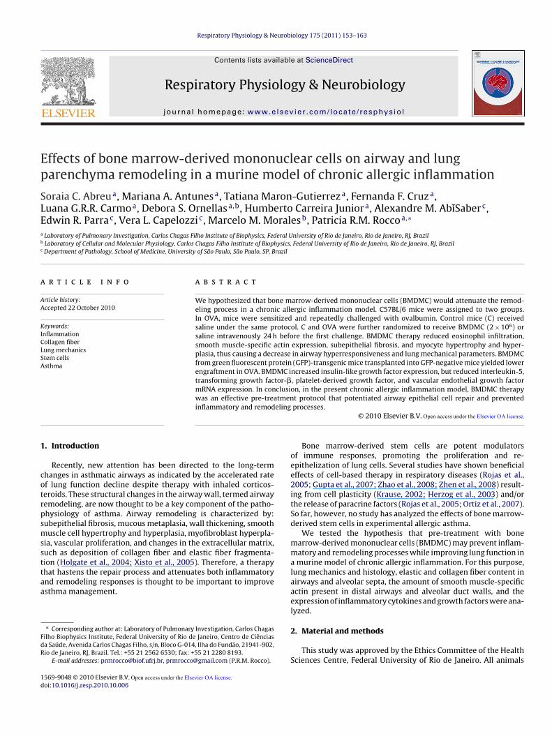
Respiratory Physiology & Neurobiology 175 (2011) 153–163 Contents lists available at ScienceDirect Respiratory Physiology & Neurobiology journal homepage: www.elsevier.com/locate/resphysiol Effects of bone marrow-derived mononuclear cells on airway and lung parenchyma remodeling in a murine model of chronic allergic inflammation Soraia C. Abreu a , Mariana A. Antunes a , Tatiana Maron-Gutierrez a , Fernanda F. Cruz a , Luana G.R.R. Carmo a , Debora S. Ornellas a,b , Humberto Carreira Junior a , Alexandre M. Abˇ ıSaber c , Edwin R. Parra c , Vera L. Capelozzi c , Marcelo M. Morales b , Patricia R.M. Rocco a,∗ a Laboratory of Pulmonary Investigation, Carlos Chagas Filho Institute of Biophysics, Federal University of Rio de Janeiro, Rio de Janeiro, RJ, Brazil b Laboratory of Cellular and Molecular Physiology, Carlos Chagas Filho Institute of Biophysics, Federal University of Rio de Janeiro, Rio de Janeiro, RJ, Brazil c Department of Pathology, School of Medicine, University of São Paulo, São Paulo, SP, Brazil article info Article history: Accepted 22 October 2010 Keywords: Inflammation Collagen fiber Lung mechanics Stem cells Asthma abstract We hypothesized that bone marrow-derived mononuclear cells (BMDMC) would attenuate the remod- eling process in a chronic allergic inflammation model. C57BL/6 mice were assigned to two groups. In OVA, mice were sensitized and repeatedly challenged with ovalbumin. Control mice (C) received saline under the same protocol. C and OVA were further randomized to receive BMDMC (2 × 10 6 ) or saline intravenously 24 h before the first challenge. BMDMC therapy reduced eosinophil infiltration, smooth muscle-specific actin expression, subepithelial fibrosis, and myocyte hypertrophy and hyper- plasia, thus causing a decrease in airway hyperresponsiveness and lung mechanical parameters. BMDMC from green fluorescent protein (GFP)-transgenic mice transplanted into GFP-negative mice yielded lower engraftment in OVA. BMDMC increased insulin-like growth factor expression, but reduced interleukin-5, transforming growth factor-, platelet-derived growth factor, and vascular endothelial growth factor mRNA expression. In conclusion, in the present chronic allergic inflammation model, BMDMC therapy was an effective pre-treatment protocol that potentiated airway epithelial cell repair and prevented inflammatory and remodeling processes. © 2010 Elsevier B.V. 1. Introduction Recently, new attention has been directed to the long-term changes in asthmatic airways as indicated by the accelerated rate of lung function decline despite therapy with inhaled corticos- teroids. These structural changes in the airway wall, termed airway remodeling, are now thought to be a key component of the patho- physiology of asthma. Airway remodeling is characterized by: subepithelial fibrosis, mucous metaplasia, wall thickening, smooth muscle cell hypertrophy and hyperplasia, myofibroblast hyperpla- sia, vascular proliferation, and changes in the extracellular matrix, such as deposition of collagen fiber and elastic fiber fragmenta- tion (Holgate et al., 2004; Xisto et al., 2005). Therefore, a therapy that hastens the repair process and attenuates both inflammatory and remodeling responses is thought to be important to improve asthma management. ∗ Corresponding author at: Laboratory of Pulmonary Investigation, Carlos Chagas Filho Biophysics Institute, Federal University of Rio de Janeiro, Centro de Ciências da Saúde, Avenida Carlos Chagas Filho, s/n, Bloco G-014, Ilha do Fundão, 21941-902, Rio de Janeiro, RJ, Brazil. Tel.: +55 21 2562 6530; fax: +55 21 2280 8193. E-mail addresses: [email protected], [email protected] (P.R.M. Rocco). Bone marrow-derived stem cells are potent modulators of immune responses, promoting the proliferation and re- epithelization of lung cells. Several studies have shown beneficial effects of cell-based therapy in respiratory diseases (Rojas et al., 2005; Gupta et al., 2007; Zhao et al., 2008; Zhen et al., 2008) result- ing from cell plasticity (Krause, 2002; Herzog et al., 2003) and/or the release of paracrine factors (Rojas et al., 2005; Ortiz et al., 2007). So far, however, no study has analyzed the effects of bone marrow- derived stem cells in experimental allergic asthma. We tested the hypothesis that pre-treatment with bone marrow-derived mononuclear cells (BMDMC) may prevent inflam- matory and remodeling processes while improving lung function in a murine model of chronic allergic inflammation. For this purpose, lung mechanics and histology, elastic and collagen fiber content in airways and alveolar septa, the amount of smooth muscle-specific actin present in distal airways and alveolar duct walls, and the expression of inflammatory cytokines and growth factors were ana- lyzed. 2. Material and methods This study was approved by the Ethics Committee of the Health Sciences Centre, Federal University of Rio de Janeiro. All animals 1569-9048 © 2010 Elsevier B.V. doi:10.1016/j.resp.2010.10.006 Open access under the Elsevier OA license. Open access under the Elsevier OA license.
-
Upload
independent -
Category
Documents
-
view
1 -
download
0
Transcript of Effects of bone marrow-derived mononuclear cells on airway and lung parenchyma remodeling in a...
Ep
SLEa
b
c
a
AA
KICLSA
1
cotrpsmssttaa
FdR
1d
Respiratory Physiology & Neurobiology 175 (2011) 153–163
Contents lists available at ScienceDirect
Respiratory Physiology & Neurobiology
journa l homepage: www.e lsev ier .com/ locate / resphys io l
ffects of bone marrow-derived mononuclear cells on airway and lungarenchyma remodeling in a murine model of chronic allergic inflammation
oraia C. Abreua, Mariana A. Antunesa, Tatiana Maron-Gutierreza, Fernanda F. Cruza,uana G.R.R. Carmoa, Debora S. Ornellasa,b, Humberto Carreira Juniora, Alexandre M. AbıSaberc,dwin R. Parrac, Vera L. Capelozzi c, Marcelo M. Moralesb, Patricia R.M. Roccoa,∗
Laboratory of Pulmonary Investigation, Carlos Chagas Filho Institute of Biophysics, Federal University of Rio de Janeiro, Rio de Janeiro, RJ, BrazilLaboratory of Cellular and Molecular Physiology, Carlos Chagas Filho Institute of Biophysics, Federal University of Rio de Janeiro, Rio de Janeiro, RJ, BrazilDepartment of Pathology, School of Medicine, University of São Paulo, São Paulo, SP, Brazil
r t i c l e i n f o
rticle history:ccepted 22 October 2010
eywords:nflammationollagen fiberung mechanics
a b s t r a c t
We hypothesized that bone marrow-derived mononuclear cells (BMDMC) would attenuate the remod-eling process in a chronic allergic inflammation model. C57BL/6 mice were assigned to two groups.In OVA, mice were sensitized and repeatedly challenged with ovalbumin. Control mice (C) receivedsaline under the same protocol. C and OVA were further randomized to receive BMDMC (2 × 10⁶) orsaline intravenously 24 h before the first challenge. BMDMC therapy reduced eosinophil infiltration,smooth muscle-specific actin expression, subepithelial fibrosis, and myocyte hypertrophy and hyper-
tem cellssthma
plasia, thus causing a decrease in airway hyperresponsiveness and lung mechanical parameters. BMDMCfrom green fluorescent protein (GFP)-transgenic mice transplanted into GFP-negative mice yielded lowerengraftment in OVA. BMDMC increased insulin-like growth factor expression, but reduced interleukin-5,transforming growth factor-�, platelet-derived growth factor, and vascular endothelial growth factormRNA expression. In conclusion, in the present chronic allergic inflammation model, BMDMC therapy
tmeneling
was an effective pre-treainflammatory and remod
. Introduction
Recently, new attention has been directed to the long-termhanges in asthmatic airways as indicated by the accelerated ratef lung function decline despite therapy with inhaled corticos-eroids. These structural changes in the airway wall, termed airwayemodeling, are now thought to be a key component of the patho-hysiology of asthma. Airway remodeling is characterized by:ubepithelial fibrosis, mucous metaplasia, wall thickening, smoothuscle cell hypertrophy and hyperplasia, myofibroblast hyperpla-
ia, vascular proliferation, and changes in the extracellular matrix,uch as deposition of collagen fiber and elastic fiber fragmenta-
ion (Holgate et al., 2004; Xisto et al., 2005). Therefore, a therapyhat hastens the repair process and attenuates both inflammatorynd remodeling responses is thought to be important to improvesthma management.∗ Corresponding author at: Laboratory of Pulmonary Investigation, Carlos Chagasilho Biophysics Institute, Federal University of Rio de Janeiro, Centro de Ciênciasa Saúde, Avenida Carlos Chagas Filho, s/n, Bloco G-014, Ilha do Fundão, 21941-902,io de Janeiro, RJ, Brazil. Tel.: +55 21 2562 6530; fax: +55 21 2280 8193.
E-mail addresses: [email protected], [email protected] (P.R.M. Rocco).
569-9048 © 2010 Elsevier B.V. oi:10.1016/j.resp.2010.10.006
Open access under the Elsevier OA license.
t protocol that potentiated airway epithelial cell repair and preventedprocesses.
© 2010 Elsevier B.V.
Bone marrow-derived stem cells are potent modulatorsof immune responses, promoting the proliferation and re-epithelization of lung cells. Several studies have shown beneficialeffects of cell-based therapy in respiratory diseases (Rojas et al.,2005; Gupta et al., 2007; Zhao et al., 2008; Zhen et al., 2008) result-ing from cell plasticity (Krause, 2002; Herzog et al., 2003) and/orthe release of paracrine factors (Rojas et al., 2005; Ortiz et al., 2007).So far, however, no study has analyzed the effects of bone marrow-derived stem cells in experimental allergic asthma.
We tested the hypothesis that pre-treatment with bonemarrow-derived mononuclear cells (BMDMC) may prevent inflam-matory and remodeling processes while improving lung function ina murine model of chronic allergic inflammation. For this purpose,lung mechanics and histology, elastic and collagen fiber content inairways and alveolar septa, the amount of smooth muscle-specificactin present in distal airways and alveolar duct walls, and theexpression of inflammatory cytokines and growth factors were ana-lyzed.
Open access under the Elsevier OA license.
2. Material and methods
This study was approved by the Ethics Committee of the HealthSciences Centre, Federal University of Rio de Janeiro. All animals
1 logy &
rrRm
2m
nfCeatDGaiCmibawtipCpCU
2
i2C
FS
54 S.C. Abreu et al. / Respiratory Physio
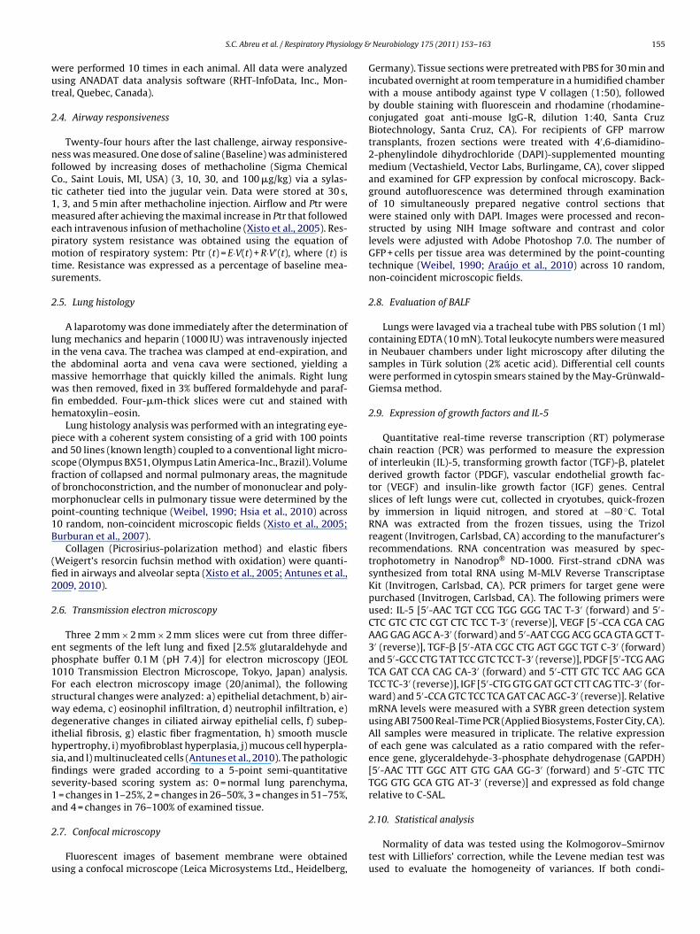
eceived humane care in compliance with the “Principles of Labo-atory Animal Care” formulated by the National Society for Medicalesearch and the “Guide for the Care and Use of Laboratory Ani-als” prepared by the National Academy of Sciences, USA.
.1. Extraction and characterization of bone marrow-derivedononuclear cells
Bone marrow cells were extracted from male C57BL/6 (20–25 g,= 13) mice and administered on the day of collection. BMDMC
rom GFP + male mice (20–22 g, n = 3) were administered to 1657BL/6 female mice to evaluate the degree of pulmonary GFP + cellngraftment. Briefly, under anesthesia with ketamine (25 mg/kg)nd xylazine (2 mg/kg) iv, bone marrow cells were aspirated fromhe femur and tibia by flushing the bone marrow cavity withulbecco’s modified Eagle’s medium (DMEM) (Life Technologies,rand Island, NY, USA). After a homogeneous cell suspension waschieved, cells were centrifuged (400 × g for 10 min), re-suspendedn DMEM and added to Ficoll-Hypaque (Histopaque 1083, Sigmahemical Co., St. Louis, MO, USA), and again centrifuged and supple-ented with phosphate-buffered saline (PBS). Cells were counted
n a Neubauer chamber with Trypan Blue for evaluation of via-ility. For the administration of saline or BMDMC, mice werenesthetized with sevoflurane, the jugular vein of each mouseas dissected, and cells were slowly injected. A small aliquot of
he mononuclear cells was used for immunophenotipic character-zation of the injected cell population. Cell characterization waserformed by flow cytometry using antibodies CD45 (leukocyte),D34 (hematopoietic precursors), CD3, CD8, and CD4 (T lym-hocyte), CD14 (monocytes and macrophages), CD11b, CD29 andD45- (non-hematopoietic precursors), all from BD Biosciences,SA.
.2. Animal preparation and experimental protocol
Eighty female C57BL/6 mice (20–25 g) were used. Lung mechan-cs and histology as well as molecular biology were analyzed in4 female C57BL/6 mice (n = 6/group). Of the remaining 56 female57BL/6 mice, 20 (n = 5/group) were used to compute airway hyper-
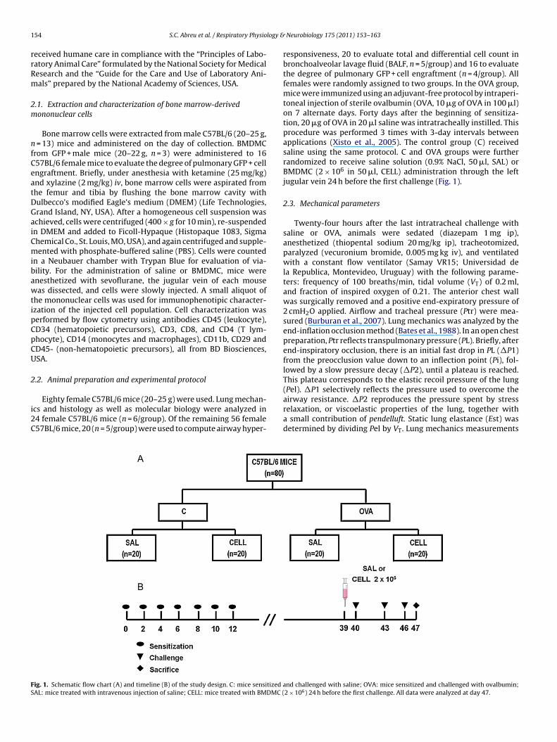
ig. 1. Schematic flow chart (A) and timeline (B) of the study design. C: mice sensitizedAL: mice treated with intravenous injection of saline; CELL: mice treated with BMDMC (
Neurobiology 175 (2011) 153–163
responsiveness, 20 to evaluate total and differential cell count inbronchoalveolar lavage fluid (BALF, n = 5/group) and 16 to evaluatethe degree of pulmonary GFP + cell engraftment (n = 4/group). Allfemales were randomly assigned to two groups. In the OVA group,mice were immunized using an adjuvant-free protocol by intraperi-toneal injection of sterile ovalbumin (OVA, 10 �g of OVA in 100 �l)on 7 alternate days. Forty days after the beginning of sensitiza-tion, 20 �g of OVA in 20 �l saline was intratracheally instilled. Thisprocedure was performed 3 times with 3-day intervals betweenapplications (Xisto et al., 2005). The control group (C) receivedsaline using the same protocol. C and OVA groups were furtherrandomized to receive saline solution (0.9% NaCl, 50 �l, SAL) orBMDMC (2 × 10⁶ in 50 �l, CELL) administration through the leftjugular vein 24 h before the first challenge (Fig. 1).
2.3. Mechanical parameters
Twenty-four hours after the last intratracheal challenge withsaline or OVA, animals were sedated (diazepam 1 mg ip),anesthetized (thiopental sodium 20 mg/kg ip), tracheotomized,paralyzed (vecuronium bromide, 0.005 mg kg iv), and ventilatedwith a constant flow ventilator (Samay VR15; Universidad dela Republica, Montevideo, Uruguay) with the following parame-ters: frequency of 100 breaths/min, tidal volume (VT) of 0.2 ml,and fraction of inspired oxygen of 0.21. The anterior chest wallwas surgically removed and a positive end-expiratory pressure of2 cmH2O applied. Airflow and tracheal pressure (Ptr) were mea-sured (Burburan et al., 2007). Lung mechanics was analyzed by theend-inflation occlusion method (Bates et al., 1988). In an open chestpreparation, Ptr reflects transpulmonary pressure (PL). Briefly, afterend-inspiratory occlusion, there is an initial fast drop in PL (�P1)from the preocclusion value down to an inflection point (Pi), fol-lowed by a slow pressure decay (�P2), until a plateau is reached.This plateau corresponds to the elastic recoil pressure of the lung
(Pel). �P1 selectively reflects the pressure used to overcome theairway resistance. �P2 reproduces the pressure spent by stressrelaxation, or viscoelastic properties of the lung, together witha small contribution of pendelluft. Static lung elastance (Est) wasdetermined by dividing Pel by VT. Lung mechanics measurementsand challenged with saline; OVA: mice sensitized and challenged with ovalbumin;2 × 10⁶) 24 h before the first challenge. All data were analyzed at day 47.
logy &
wut
2
nfCt1mepmts
2
litmwfih
pasfomp1B
(fi2
2
ep1Fswdihsfis1a
2
u
S.C. Abreu et al. / Respiratory Physio
ere performed 10 times in each animal. All data were analyzedsing ANADAT data analysis software (RHT-InfoData, Inc., Mon-real, Quebec, Canada).
.4. Airway responsiveness
Twenty-four hours after the last challenge, airway responsive-ess was measured. One dose of saline (Baseline) was administered
ollowed by increasing doses of methacholine (Sigma Chemicalo., Saint Louis, MI, USA) (3, 10, 30, and 100 �g/kg) via a sylas-ic catheter tied into the jugular vein. Data were stored at 30 s,, 3, and 5 min after methacholine injection. Airflow and Ptr wereeasured after achieving the maximal increase in Ptr that followed
ach intravenous infusion of methacholine (Xisto et al., 2005). Res-iratory system resistance was obtained using the equation ofotion of respiratory system: Ptr (t) = E·V(t) + R·V′(t), where (t) is
ime. Resistance was expressed as a percentage of baseline mea-urements.
.5. Lung histology
A laparotomy was done immediately after the determination ofung mechanics and heparin (1000 IU) was intravenously injectedn the vena cava. The trachea was clamped at end-expiration, andhe abdominal aorta and vena cava were sectioned, yielding a
assive hemorrhage that quickly killed the animals. Right lungas then removed, fixed in 3% buffered formaldehyde and paraf-n embedded. Four-�m-thick slices were cut and stained withematoxylin–eosin.
Lung histology analysis was performed with an integrating eye-iece with a coherent system consisting of a grid with 100 pointsnd 50 lines (known length) coupled to a conventional light micro-cope (Olympus BX51, Olympus Latin America-Inc., Brazil). Volumeraction of collapsed and normal pulmonary areas, the magnitudef bronchoconstriction, and the number of mononuclear and poly-orphonuclear cells in pulmonary tissue were determined by the
oint-counting technique (Weibel, 1990; Hsia et al., 2010) across0 random, non-coincident microscopic fields (Xisto et al., 2005;urburan et al., 2007).
Collagen (Picrosirius-polarization method) and elastic fibersWeigert’s resorcin fuchsin method with oxidation) were quanti-ed in airways and alveolar septa (Xisto et al., 2005; Antunes et al.,009, 2010).
.6. Transmission electron microscopy
Three 2 mm × 2 mm × 2 mm slices were cut from three differ-nt segments of the left lung and fixed [2.5% glutaraldehyde andhosphate buffer 0.1 M (pH 7.4)] for electron microscopy (JEOL010 Transmission Electron Microscope, Tokyo, Japan) analysis.or each electron microscopy image (20/animal), the followingtructural changes were analyzed: a) epithelial detachment, b) air-ay edema, c) eosinophil infiltration, d) neutrophil infiltration, e)egenerative changes in ciliated airway epithelial cells, f) subep-
thelial fibrosis, g) elastic fiber fragmentation, h) smooth muscleypertrophy, i) myofibroblast hyperplasia, j) mucous cell hyperpla-ia, and l) multinucleated cells (Antunes et al., 2010). The pathologicndings were graded according to a 5-point semi-quantitativeeverity-based scoring system as: 0 = normal lung parenchyma,= changes in 1–25%, 2 = changes in 26–50%, 3 = changes in 51–75%,nd 4 = changes in 76–100% of examined tissue.
.7. Confocal microscopy
Fluorescent images of basement membrane were obtainedsing a confocal microscope (Leica Microsystems Ltd., Heidelberg,
Neurobiology 175 (2011) 153–163 155
Germany). Tissue sections were pretreated with PBS for 30 min andincubated overnight at room temperature in a humidified chamberwith a mouse antibody against type V collagen (1:50), followedby double staining with fluorescein and rhodamine (rhodamine-conjugated goat anti-mouse IgG-R, dilution 1:40, Santa CruzBiotechnology, Santa Cruz, CA). For recipients of GFP marrowtransplants, frozen sections were treated with 4′,6-diamidino-2-phenylindole dihydrochloride (DAPI)-supplemented mountingmedium (Vectashield, Vector Labs, Burlingame, CA), cover slippedand examined for GFP expression by confocal microscopy. Back-ground autofluorescence was determined through examinationof 10 simultaneously prepared negative control sections thatwere stained only with DAPI. Images were processed and recon-structed by using NIH Image software and contrast and colorlevels were adjusted with Adobe Photoshop 7.0. The number ofGFP + cells per tissue area was determined by the point-countingtechnique (Weibel, 1990; Araújo et al., 2010) across 10 random,non-coincident microscopic fields.
2.8. Evaluation of BALF
Lungs were lavaged via a tracheal tube with PBS solution (1 ml)containing EDTA (10 mN). Total leukocyte numbers were measuredin Neubauer chambers under light microscopy after diluting thesamples in Türk solution (2% acetic acid). Differential cell countswere performed in cytospin smears stained by the May-Grünwald-Giemsa method.
2.9. Expression of growth factors and IL-5
Quantitative real-time reverse transcription (RT) polymerasechain reaction (PCR) was performed to measure the expressionof interleukin (IL)-5, transforming growth factor (TGF)-�, plateletderived growth factor (PDGF), vascular endothelial growth fac-tor (VEGF) and insulin-like growth factor (IGF) genes. Centralslices of left lungs were cut, collected in cryotubes, quick-frozenby immersion in liquid nitrogen, and stored at −80 ◦C. TotalRNA was extracted from the frozen tissues, using the Trizolreagent (Invitrogen, Carlsbad, CA) according to the manufacturer’srecommendations. RNA concentration was measured by spec-trophotometry in Nanodrop® ND-1000. First-strand cDNA wassynthesized from total RNA using M-MLV Reverse TranscriptaseKit (Invitrogen, Carlsbad, CA). PCR primers for target gene werepurchased (Invitrogen, Carlsbad, CA). The following primers wereused: IL-5 [5′-AAC TGT CCG TGG GGG TAC T-3′ (forward) and 5′-CTC GTC CTC CGT CTC TCC T-3′ (reverse)], VEGF [5′-CCA CGA CAGAAG GAG AGC A-3′ (forward) and 5′-AAT CGG ACG GCA GTA GCT T-3′ (reverse)], TGF-� [5′-ATA CGC CTG AGT GGC TGT C-3′ (forward)and 5′-GCC CTG TAT TCC GTC TCC T-3′ (reverse)], PDGF [5′-TCG AAGTCA GAT CCA CAG CA-3′ (forward) and 5′-CTT GTC TCC AAG GCATCC TC-3′ (reverse)], IGF [5′-CTG GTG GAT GCT CTT CAG TTC-3′ (for-ward) and 5′-CCA GTC TCC TCA GAT CAC AGC-3′ (reverse)]. RelativemRNA levels were measured with a SYBR green detection systemusing ABI 7500 Real-Time PCR (Applied Biosystems, Foster City, CA).All samples were measured in triplicate. The relative expressionof each gene was calculated as a ratio compared with the refer-ence gene, glyceraldehyde-3-phosphate dehydrogenase (GAPDH)[5′-AAC TTT GGC ATT GTG GAA GG-3′ (forward) and 5′-GTC TTCTGG GTG GCA GTG AT-3′ (reverse)] and expressed as fold changerelative to C-SAL.
2.10. Statistical analysis
Normality of data was tested using the Kolmogorov–Smirnovtest with Lilliefors’ correction, while the Levene median test wasused to evaluate the homogeneity of variances. If both condi-
1 logy &
twNramSRp
3
blTl(lC((
tartO
Fso
56 S.C. Abreu et al. / Respiratory Physio
ions were satisfied, two-way ANOVA followed by Tukey’s testhen required was used to compare differences among the groups.onparametric data were analyzed using two-way ANOVA on
anks followed by Tukey’s test. Parametric data were expresseds means ± SEM, while non-parametric data were expressed asedians (interquartile range). All tests were performed using the
igmaStat 3.1 statistical software package (Jandel Corporation, Sanaphael, CA, USA), and statistical significance was established as< 0.05.
. Results
The pool of intravenously injected BMDMC was characterizedy flow cytometry showing the following subpopulations: total
ymphocyte (lower SSC, CD45+/CD11b−/ CD29−/ CD34− = 29.7%),lymphocyte (lower SSC/ CD45+/ CD3+/ CD34− = 5.4%), T helper
ymphocyte (CD3+/ CD4+/ CD8− = 2.4%), T cytotoxic lymphocyteCD3+/CD4−/ CD8+ = 2.3%), monocytes (CD45+/ CD29+/ CD11b+ow/ CD34−/CD3− = 4.9%), neutrophils (CD45+/ CD11b+/ CD34−/D29−/ CD14−/ CD34−/ CD3−), hematopoietic progenitorsCD34+/CD45+ = 0.3%), and other progenitors cells (CD45− = 3.8%)data presented in Fig. S.1 in the Supplementary Material).
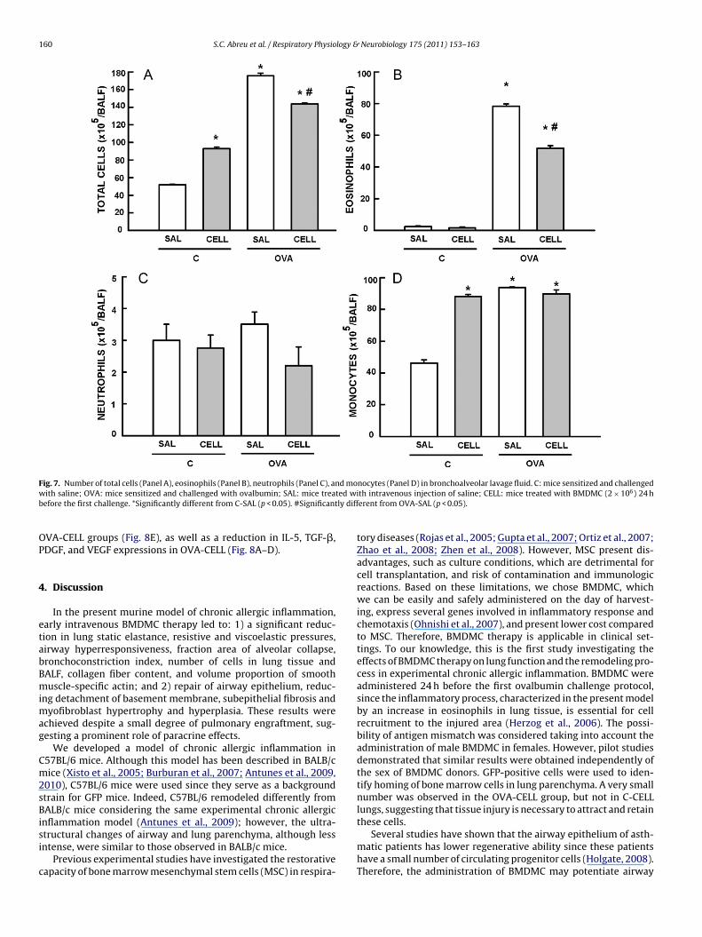
Est (33%), �P1 (15%), and �P2 (49%) were higher in the OVA-SAL
han in the C-SAL group. BMDMC significantly inhibited Est, �P1,nd �P2 alterations (Fig. 2A, B). Furthermore, the increase in airwayesistance evoked by methacholine was significantly augmentedhe OVA-SAL group; however, these changes were minimized inVA animals receiving BMDMC (Fig. 2C).ig. 2. Lung static elastance (Est) (Panel A); resistive (�P1, gray bar), viscoelastic (�P2, wponsiveness (Panel C). C: mice sensitized and challenged with saline; OVA: mice sensitif saline (white bars); CELL: mice treated with BMDMC (2 × 10⁶) 24 h before the first cha
Neurobiology 175 (2011) 153–163
In the OVA-SAL group, morphometric examination of lungsdemonstrated a significant increase in the fraction area of alveo-lar collapse, the number of mononuclear and polymorphonuclearcells, collagen fiber content in airways and alveolar septa and inthe volume proportion of smooth muscle-specific actin in terminalbronchioles and alveolar ducts (Table 1). In Fig. 3, a reduction in thecentral airway diameter and inflammatory cell infiltration can beobserved. BMDMC therapy minimized the fraction area of alveolarcollapse and polymorphonuclear cells, and the volume proportionof smooth muscle-specific actin in alveolar ducts, but preventedchanges in airway contraction, amount of collagen fiber in airwayand alveolar septa, and in volume proportion of smooth musclespecific actin in terminal bronchiole. Elastic fiber content in airwayand alveolar septa was similar in all groups (Table 1). Additionally,central airway diameter was observed to be less constricted andpresented a reduction in inflammation in OVA-CELL group (Fig. 3).
Fig. 4 depicts typical electron microscopy findings of airway andlung parenchyma in each group. OVA-SAL animals showed airwayepithelial cell detachment from the basement membrane, degen-erative changes in ciliated airway epithelial cells, eosinophil andneutrophil infiltration, myofibroblast and mucous cell hyperplasia,subepithelial fibrosis, and smooth muscle hypertrophy (Table 2).BMDMC therapy attenuated all these ultrastructural changes and
promoted the appearance of undifferentiated cells, without adefined phenotype, and multinucleated cells (Table 2).Confocal microscopy showed basement membrane ruptureassociated with increased inflammatory process in the OVA-SALgroup, which was prevented with the use of BMDMC (Fig. 5).
hite bar) and total pressures (�Ptot = �P1 + �P2) (Panel B); and airway hyperre-zed and challenged with ovalbumin; SAL: mice treated with intravenous injectionllenge. *Significantly different from C-SAL (p < 0.05).

S.C. Abreu et al. / Respiratory Physiology & Neurobiology 175 (2011) 153–163 157
Table 1Lung morphometry.
C-SAL C-CELL OVA-SAL OVA-CELL
Normal (%) 99.0 ± 0.72 98.0 ± 2.60 68.2 ± 11.1* 88.1 ± 5.91 *#Collapse (%) 1.00 ± 0.72 1.80 ± 2.69 31.8 ± 11.1* 11.8 ± 5.91*#MN (%) 33.13 ± 2.90 29.35 ± 3.20 51.2 ± 3.06* 29.03 ± 1.61PMN (%) 0.30 ± 0.32 3.00 ± 1.06* 7.20 ± 0.96* 3.64 ± 1.63*#Contraction index 2.28 ± 0.32 2.25 ± 0.32 3.02 ± 0.51* 2.39 ± 0.31Collagen fiber (�m2/�m) Airways 1.04 ± 0.05 1.02 ± 0.05 3.30 ± 0.40* 1.06 ± 0.15
Alveolar septa 0.014 ± 0.03 0.014 ± 0.03 0.070 ± 0.007* 0.011 ± 0.002Elastic fiber (�m2/�m) Airways 1.47 ± 0.17 1.35 ± 0.28 1.40 ± 0.15 1.39 ± 0.20
Alveolar septa 0.26 ± 0.04 0.25 ± 0.05 0.27 ± 0.02 0.26 ± 0.01Actin (%) Terminal bronchiole 3.11 ± 0.36 2.98 ± 0.36 6.70 ± 0.80* 4.08 ± 0.42
Alveolar duct 0.23 ± 0.17 1.05 ± 0.19 2.08 ± 0.17* 1.87 ± 0.21*#
The fraction of area of normal and collapsed alveoli, mononuclear (MN) and polymorphonuclear (PMN) cells, contraction index, collagen and elastic fibers in airways andalveolar septa, volume proportion of smooth muscle-specific actin in terminal bronchiole and alveolar duct. All values were computed in ten random, non-coincident fieldsper mice. C: mice sensitized and challenged with saline; OVA: mice sensitized and challenged with ovalbumin; SAL: mice treated with intravenous injection of saline; CELL:mice treated with BMDMC (2 × 10⁶) 24 h before the first challenge. *Significantly different from C-SAL (p < 0.05). #Significantly different from OVA-SAL (p < 0.05).
F(i
TS
P3wd
ig. 3. Representative photomicrographs of airways stained with hematoxylin–eosin. Nblack arrows) in the OVA-SAL group. C: Mice sensitized and challenged with saline; OVA: mnjection of saline; CELL: mice treated with BMDMC (2 × 10⁶) 24 h before the first challen
able 2emi-quantitative analysis of electron microscopy.
C-SAL
Epithelial detachment 0 (0–0)Airway edema 0 (0–0)Eosinophil infiltration 0 (0–0)Neutrophil infiltration 0 (0–0)Degeneration of ciliated airway epithelial cells 0 (0–0)Subepithelial fibrosis 0 (0–0)Elastic fiber fragmentation 0 (0–0)Smooth muscle hypertrophy 0 (0–0)Myofibroblast hyperplasia 0 (0–0)Mucous cells hyperplasia 0 (0–0)Multinucleated cells 0 (0–0)
athologic findings were graded according to a 5-point semi-quantitative severity-base= 51–75%, and 4 = 76–100% of the examined tissue. C: mice sensitized and challenged wiith intravenous injection of saline; CELL: mice treated with BMDMC (2 × 10⁶) 24 h befifferent from C-CELL (p < 0.05). #Significantly different from OVA-SAL (p < 0.05).
ote the reduction in central airway diameter as well as increased cell infiltrationice sensitized and challenged with ovalbumin; SAL: mice treated with intravenous
ge. Original magnification: ×400. Bars = 100 �m.
C-CELL OVA-SAL OVA-CELL
0 (0–0) 2 (2–3)* 1 (0–1)*#0 (0–0) 3 (2–4)* 1 (0–2)*#0 (0–0) 3 (2–4)* 1 (1–2)*#
1 (0–1)* 2 (1–3)* 1 (0–2)*#0 (0–0) 3 (3–4)* 1 (1–2)*#0 (0–0) 3 (3–4)* 1 (1–2)*#0 (0–0) 2 (2–3)* 0 (0–1)0 (0–0) 3 (2–4)* 1 (1–2)*#0 (0–0) 3 (3–4)* 1 (1–2)*#0 (0–0) 3 (2–4)* 1 (0–2)*#
1 (0–1)* 0 (0–0) 2 (1–3)*§
d scoring system: 0 = normal lung parenchyma, 1 = changes in 1–25%, 2 = 26–50%,th saline; OVA: mice sensitized and challenged with ovalbumin; SAL: mice treatedore the first challenge. *Significantly different from C-SAL (p < 0.05). §Significantly

158 S.C. Abreu et al. / Respiratory Physiology & Neurobiology 175 (2011) 153–163
F ma (ra clearc erentp neutro
CcBC
ig. 4. Electron microscopy of terminal bronchiole (left panels) and lung parenchynd challenged with ovalbumin; SAL: saline; CELL: bone marrow-derived mononuells [Muc(m)] ciliated cells (Cil), alveolar ducts (AD), alveolar lumen (Alv), undiffneumocytes type II (PII*), capillary (Cap), apoptosis (arrows), MF (myofibroblast),
A very small number of GFP + cells was detected in the OVA-ELL group (<1%), vs. none in C-CELL (Fig. 6). The number of totalells and eosinophils in BALF was higher in OVA-SAL (Fig. 7A, B).MDMC yielded an increase in the number of total cells in the C-ELL group resulting from an increased monocyte count (Fig. 7A, C).
ight panels). C: mice sensitized and challenged with saline; OVA: mice sensitizedcell therapy. Basal membrane (BM), mucous cells (Muc), multinucleated mucous
iated cells (Cund), alveolar septa (Sep), pneumocytes type II (PII), multinucleatedphils (N).
In the OVA-CELL group, the number of total cells and eosinophilswas lower than in OVA-SAL.
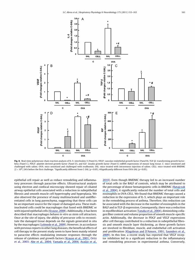
IL-5, TGF-�, PDGF, VEGF, and IGF mRNA expressions in lung tis-sue were higher in OVA-SAL (Fig. 8A–E). BMDMC administrationpromoted an increase in IGF mRNA expression in the C-CELL and

S.C. Abreu et al. / Respiratory Physiology & Neurobiology 175 (2011) 153–163 159
Fig. 5. Representative photomicrographs of airways and lung parenchyma using confocal microscopy. Note, in green, rupture of basement membrane (arrows) in the OVA-SAL group. Basement membrane (BM), epithelial cells (Epi), terminal bronchiole (Br), alveolar duct (AD), vessel (Ves), and type II pneumocyte (PII). C: Mice sensitized andchallenged with saline; OVA: mice sensitized and challenged with ovalbumin; SAL: mice treated with intravenous injection of saline; CELL: mice treated with BMDMC(2 × 10⁶) 24 h before the first challenge. Original magnification: ×400. Bars = 100 �m. (For interpretation of the references to color in this figure legend, the reader is referredto the web version of the article.)
Fig. 6. Confocal microscopy of lung parenchyma in C and OVA C57BL/6 female mice treated from BMDMC from GFP male mice. C: mice sensitized and challenged withsaline; OVA: mice sensitized and challenged with ovalbumin; CELL: mice treated with BMDMC (2 × 10⁶) 24 h before the first challenge. Note that few GFP + cells (green) werepresent in the lung parenchyma. Original magnification: ×200. (For interpretation of the references to color in this figure legend, the reader is referred to the web version ofthe article.)
160 S.C. Abreu et al. / Respiratory Physiology & Neurobiology 175 (2011) 153–163
F d mow ted wib ly diff
OP
4
etabBmimag
Cm2sBisi
c
ig. 7. Number of total cells (Panel A), eosinophils (Panel B), neutrophils (Panel C), anith saline; OVA: mice sensitized and challenged with ovalbumin; SAL: mice trea
efore the first challenge. *Significantly different from C-SAL (p < 0.05). #Significant
VA-CELL groups (Fig. 8E), as well as a reduction in IL-5, TGF-�,DGF, and VEGF expressions in OVA-CELL (Fig. 8A–D).
. Discussion
In the present murine model of chronic allergic inflammation,arly intravenous BMDMC therapy led to: 1) a significant reduc-ion in lung static elastance, resistive and viscoelastic pressures,irway hyperresponsiveness, fraction area of alveolar collapse,ronchoconstriction index, number of cells in lung tissue andALF, collagen fiber content, and volume proportion of smoothuscle-specific actin; and 2) repair of airway epithelium, reduc-
ng detachment of basement membrane, subepithelial fibrosis andyofibroblast hypertrophy and hyperplasia. These results were
chieved despite a small degree of pulmonary engraftment, sug-esting a prominent role of paracrine effects.
We developed a model of chronic allergic inflammation in57BL/6 mice. Although this model has been described in BALB/cice (Xisto et al., 2005; Burburan et al., 2007; Antunes et al., 2009,
010), C57BL/6 mice were used since they serve as a backgroundtrain for GFP mice. Indeed, C57BL/6 remodeled differently fromALB/c mice considering the same experimental chronic allergic
nflammation model (Antunes et al., 2009); however, the ultra-tructural changes of airway and lung parenchyma, although lessntense, were similar to those observed in BALB/c mice.
Previous experimental studies have investigated the restorativeapacity of bone marrow mesenchymal stem cells (MSC) in respira-
nocytes (Panel D) in bronchoalveolar lavage fluid. C: mice sensitized and challengedth intravenous injection of saline; CELL: mice treated with BMDMC (2 × 10⁶) 24 herent from OVA-SAL (p < 0.05).
tory diseases (Rojas et al., 2005; Gupta et al., 2007; Ortiz et al., 2007;Zhao et al., 2008; Zhen et al., 2008). However, MSC present dis-advantages, such as culture conditions, which are detrimental forcell transplantation, and risk of contamination and immunologicreactions. Based on these limitations, we chose BMDMC, whichwe can be easily and safely administered on the day of harvest-ing, express several genes involved in inflammatory response andchemotaxis (Ohnishi et al., 2007), and present lower cost comparedto MSC. Therefore, BMDMC therapy is applicable in clinical set-tings. To our knowledge, this is the first study investigating theeffects of BMDMC therapy on lung function and the remodeling pro-cess in experimental chronic allergic inflammation. BMDMC wereadministered 24 h before the first ovalbumin challenge protocol,since the inflammatory process, characterized in the present modelby an increase in eosinophils in lung tissue, is essential for cellrecruitment to the injured area (Herzog et al., 2006). The possi-bility of antigen mismatch was considered taking into account theadministration of male BMDMC in females. However, pilot studiesdemonstrated that similar results were obtained independently ofthe sex of BMDMC donors. GFP-positive cells were used to iden-tify homing of bone marrow cells in lung parenchyma. A very smallnumber was observed in the OVA-CELL group, but not in C-CELLlungs, suggesting that tissue injury is necessary to attract and retain
these cells.Several studies have shown that the airway epithelium of asth-matic patients has lower regenerative ability since these patientshave a small number of circulating progenitor cells (Holgate, 2008).Therefore, the administration of BMDMC may potentiate airway
S.C. Abreu et al. / Respiratory Physiology & Neurobiology 175 (2011) 153–163 161
Fig. 8. Real-time polymerase chain reaction analysis of IL-5: interleukin-5 (Panel A), VEGF: vascular endothelial growth factor (Panel B), TGF-�: transforming growth factor-b rowthc L: mi( ). #Sig
etuafiaebiwdOtbwctre
eta (Panel C), PDGF: platelet derived growth factor (Panel D), and IGF: insulin ghallenged with saline; OVA: mice sensitized and challenged with ovalbumin; SA2 × 106) 24 h before the first challenge. *Significantly different from C-SAL (p < 0.05
pithelial cell repair as well as reduce remodeling and inflamma-ory processes through paracrine effects. Ultrastructural analysissing electron and confocal microscopy showed repair of ciliatedirway epithelial cells associated with a reduction in subepithelialbrosis and smooth muscle cell hypertrophy and hyperplasia. Welso observed the presence of many multinucleated and undiffer-ntiated cells in lung parenchyma, suggesting that these cells cane an important source for the repair of damaged area. These mult-
nucleated cells could be macrophages that fused with BMDMC orith injured epithelial cells (Krause, 2008). Additionally, it has beenescribed that macrophages behave in vitro as stem cell attractors.nce at the site of injury, the ability of precursor cells to reconsti-
ute the damaged tissue depends on the signals generated in situy the macrophages (Lolmede et al., 2009). However, in accordance
ith previous reports in other lung diseases, the beneficial effects ofell therapy in the present study seem to have been mainly relatedo paracrine effects modulating immune responses through theelease of cytokines and growth factors (Krause et al., 2001; Ortizt al., 2003; Abe et al., 2004; Yamada et al., 2004; Araújo et al.,
factor (Panel E) mRNA expressions of mouse lung tissue. C: mice sensitized andce treated with intravenous injection of saline; CELL: mice treated with BMDMCnificantly different from OVA-SAL (p < 0.05).
2010). Even though BMDMC therapy led to an increased numberof total cells in the BALF of controls, which may be attributed tothe percentage of donor hematopoietic cells in BMDMC (Ratajczaket al., 2004), it significantly reduced the number of total cells andeosinophils in OVA-CELL. We found that BMDMC therapy caused areduction in the expression of IL-5, which plays an important rolein the remodeling process of asthma. Therefore, this reduction canbe associated with the decrease in the number of eosinophils in theBALF and in TGF-� expression. Consequently, there was a reductionin myofibroblast activation (Tanaka et al., 2004), diminishing colla-gen fiber content and volume proportion of smooth muscle-specificactin. Additionally, the decrease in PDGF and VEGF expressionsafter cell therapy contributed to a reduction in subepithelial fibro-sis and smooth muscle layer thickening, as these growth factors
are involved in fibroblast, muscle, and endothelial cell activationand proliferation (Klagsbrun and D’Amore, 1991; Saunders et al.,2009). In this line, a recent study has reported that VEGF recep-tor inhibition led to a significant reduction in the inflammatoryand remodeling processes in experimental asthma. Conversely,
1 logy &
Bc(eain
bwBsfifiSetoieni
iacTeet
A
BmtPNbm
PTSmR
A
t
R
A
A
A
A
62 S.C. Abreu et al. / Respiratory Physio
MDMC yielded an increase in IGF expression, which may be asso-iated with the effect of these cells on the re-epithelization processYamashita et al., 2005) previously observed in experimental mod-ls of idiopathic fibrosis. Thus, the paracrine effects of BMDMCcted on airway and lung parenchyma remodeling resulting in anmprovement in lung mechanics and reducing airway responsive-ess.
Our study has some limitations: 1) BMDMC were injected 24 hefore the first ovalbumin challenge, before the remodeling processas established. Thus, more studies should be performed to analyzeMDMC effects after the last challenge; 2) despite the widespreadpeculation that MSC constitute a unique cell type distinct frombroblasts (Horwitz et al., 2007), some investigators have usedbroblast therapy as control (Mei et al., 2007; Xu et al., 2007).ince the use of fibroblasts did not result in beneficial effects (Sunt al., 2009), in the present study we administrated saline as con-rol instead of fibroblasts; 3) we cannot ascertain whether the rolef cytokines and growth factors is related to engraftment. To clar-fy this issue, specific gene deficient animals should be used; and 4)ven though the amount of GFP was quantified in lung tissue, we didot analyze whether these engrafted cells are transdifferentiated
nto any type of lung cell.In conclusion, in the present murine model of chronic allergic
nflammation, bone marrow-derived mononuclear cell therapy wasn effective pre-treatment protocol, potentiating airway epithelialell repair and reducing inflammatory and remodeling processes.hese results were achieved despite a small degree of pulmonaryngraftment, suggesting a role of paracrine effects. Additionalxperiments are necessary to analyze the effects of BMDMC afterhe remodeling process has been established.
cknowledgements
The authors would like to express their gratitude to Mr. Andreenedito da Silva for animal care, Mrs. Jaqueline Lima do Nasci-ento, Luis Felipe Prota, Cynthia Samary, and Johnatas Dutra for
heir skilful technical assistance during the experiments, Mr. Brunoaredes for his help with flow cytometry analysis; Mrs. Ana Luciaeves da Silva for her help with microscopy; and Mrs. Moira Eliza-eth Schöttler and Claudia Buchweitz for assistance in editing theanuscript.Supported by: This study was supported by Centers of Excellence
rogram (PRONEX-FAPERJ), Brazilian Council for Scientific andechnological Development (CNPq), Rio de Janeiro State Researchupporting Foundation (FAPERJ), Coordination for the Improve-ent of Higher Education Personnel (CAPES), and São Paulo State
esearch Supporting Foundation (FAPESP).
ppendix A. Supplementary data
Supplementary data associated with this article can be found, inhe online version, at doi:10.1016/j.resp.2010.10.006.
eferences
be, S., Boyer, C., Liu, X., Wen, F.Q., Kobayashi, T., Fang, Q., Wang, X., Hashimoto, M.,Sharp, J.G., Rennard, S.I., 2004. Cells derived from the circulation contribute tothe repair of lung injury. Am. J. Respir. Crit. Care Med. 170, 1158–1163.
ntunes, M.A., Abreu, S.C., Damaceno-Rodrigues, N.R., Parra, E.R., Capelozzi, V.L.,Pinart, M., Romero, P.V., Silva, P.M., Martins, M.A., Rocco, P.R., 2009. Differentstrains of mice present distinct lung tissue mechanics and extracellular matrixcomposition in a model of chronic allergic asthma. Respir. Physiol. Neurobiol.165, 202–207.
ntunes, M.A., Abreu, S.C., Silva, A.L., Parra-Cuentas, E.R., Ab’saber, A.M., Capelozzi,V.L., Ferreira, T.P., Martins, M.A., Silva, P.M., Rocco, P.R., 2010. Gender-specificlung remodeling and inflammation changes in experimental chronic allergicasthma. J. Appl. Physiol. (July), 15.
raújo, I.M., Abreu, S.C., Maron-Gutierrez, T., Cruz, F., Fujisaki, L., Carreira, H., Ornel-las, F., Ornellas, D., Vieira-de-Abreu, A., Castro-Faria-Neto, H.C., Ab’Saber, A.,
Neurobiology 175 (2011) 153–163
Teodoro, W.R., Diaz, B.L., Peres da Costa, C., Capelozzi, V.L., Pelosi, P., Morales,M.M., Rocco, P.R., 2010. Bone marrow-derived mononuclear cell therapy inexperimental pulmonary and extrapulmonary acute lung injury. Crit. Care Med.38, 1733–1741.
Bates, J.H., Ludwig, M.S., Sly, P.D., Brown, K., Martin, J.G., Fredberg, J.J., 1988. Inter-rupter resistance elucidated by alveolar pressure measurement in open-chestnormal dogs. J. Appl. Physiol. 65, 408–414.
Burburan, S.M., Xisto, D.G., Ferreira, H.C., Riva, D., Carvalho, G.M., Zin, W.A., Rocco,P.R.M., 2007. Lung mechanics and histology during sevoflurane anesthesia in amodel of chronic allergic asthma. Anesth. Analg. 104, 631–637.
Gupta, N., Su, X., Popov, B., Lee, J.W., Serikov, V., Matthay, M.A., 2007. Intrapulmonarydelivery of bone marrow-derived mesenchymal stem cells improves survivaland attenuates endotoxin-induced acute lung injury in mice. J. Immunol. 179,1855–1863.
Herzog, E.L., Chai, L., Krause, D.S., 2003. Plasticity of marrow-derived stem cells.Blood 102, 3483–3493.
Herzog, E.L., Van Arnam, J., Hu, B., Krause, D.S., 2006. Threshold of lung injuryrequired for the appearance of marrow-derived lung epithelia. Stem Cells 24,1986–1992.
Holgate, S.T., 2008. Pathogenesis of asthma. Clin. Exp. Allergy 38, 872–897.Holgate, S.T., Holloway, J., Wilson, S., Bucchieri, F., Puddicombe, S., Davies, D.E., 2004.
Epithelial-mesenchymal communication in the pathogenesis of chronic asthma.Proc. Am. Thorac. Soc. 1, 93–98.
Horwitz, E.M., Andreef, M., Frassoni, F., 2007. Mesenchymal stromal cells. Biol. BloodMarrow Transplant. 13S1, 53–57.
Hsia, C.C., Hyde, D.M., Ochs, M., Weibel, E.R., 2010. An official research policy state-ment of the American Thoracic Society/European Respiratory Society: standardsfor quantitative assessment of lung structure. Am. J. Respir. Crit. Care Med. 181,394–418.
Klagsbrun, M., D’Amore, P.A., 1991. Regulators of angiogenesis. Annu. Rev. Physiol.53, 217–239.
Krause, D.S., 2002. Plasticity of marrow-derived stem cells. Gene Ther. 9, 754–758.
Krause, D.S., 2008. Bone marrow-derived cells and stem cells in lung repair. Proc.Am. Thorac. Soc. 5, 323–327.
Krause, D.S., Theise, N.D., Collector, M.I., Henegariu, O., Hwang, S., Gardner, R.,Neutzel, S., Sharkis, S.J., 2001. Multi-organ, multi-lineage engraftment by a singlebone marrow derived stem cell. Cell 105, 369–377.
Lolmede, K., Campana, L., Vezzoli, M., Bosurgi, L., Tonlorenzi, R., Clementi, E., Bianchi,M.E., Cossu, G., Manfredi, A.A., Brunelli, S., Rovere-Querini, P., 2009. Inflamma-tory and alternatively activated human macrophages attract vessel-associatedstem cells, relying on separate HMGB1- and MMP-9-dependent pathways. J.Leukoc. Biol. 85, 779–787.
Mei, S.H., McCarter, S.D., Deng, Y., Parker, C.H., Liles, W.C., Stewart, D.J., 2007. Pre-vention of LPS-induced acute lung injury in mice by mesenchymal stem cellsoverexpressing angiopoietin 1. PLoS Med. 4, e269.
Ohnishi, S., Yasuda, T., Kitamura, S., Nagaya, N., 2007. Effect of hypoxia on geneexpression of bone marrow-derived mesenchymal stem cells and mononuclearcells. Stem Cells 25, 1166–1177.
Ortiz, L.A., Dutreil, M., Fattman, C., Pandey, A.C., Torres, G., Go, K., Phinney, D.G., 2007.Interleukin 1 receptor antagonist mediates the antiinflammatory and antifi-brotic effect of mesenchymal stem cells during lung injury. Proc. Natl. Acad.Sci. U.S.A. 104, 11002–11007.
Ortiz, L.A., Gambelli, F., McBride, C., Gaupp, D., Baddoo, M., Kaminski, N., Phinney,D.G., 2003. Mesenchymal stem cell engraftment in lung is enhanced in responseto bleomycin exposure and ameliorates its fibrotic effects. Proc. Natl. Acad. Sci.U.S.A. 100, 8407–8411.
Ratajczak, M.Z., Kucia, M., Majka, M., Reca, R., Ratajczak, J., 2004. Heterogeneouspopulations of bone marrow stem cells—are we spotting on the same cells fromthe different angles? Folia Histochem. Cytobiol. 42, 139–146.
Rojas, M., Xu, J., Woods, C.R., Mora, A.L., Spears, W., Roman, J., Brigham, K.L., 2005.Bone marrow-derived mesenchymal stem cells in repair of the injured lung. Am.J. Respir. Cell Mol. Biol. 33, 145–152.
Saunders, R., Siddiqui, S., Kaur, D., Doe, C., Sutcliffe, A., Hollins, F., Bradding, P., Ward-law, A., Brightling, C.E., 2009. Fibrocyte localization to the airway smooth muscleis a feature of asthma. J. Allergy Clin. Immunol. 123, 376–384.
Sun, C.K., Chang, L.T., Sheu, J.J., Chiang, C.H., Lee, F.Y., Wu, C.J., Chua, S., Fu, M., Yip, H.K.,2009. Bone marrow-derived mononuclear cell therapy alleviates left ventricularremodeling and improves heart function in rat-dilated cardiomyopathy. Crit.Care Med. 37, 1197–1205.
Tanaka, H., Komai, M., Nagao, K., Ishizaki, M., Kajiwara, D., Takatsu, K., Delespesse,G., Nagai, H., 2004. Role of interleukin-5 and eosinophils in allergen-induced airway remodeling in mice. Am. J. Respir. Cell. Mol. Biol. 31,62–68.
Weibel, E.R., 1990. Morphometry: stereological theory and practical methods. In:Gil, J. (Ed.), Models of Lung Disease-Microscopy and Structural Methods. MarcelDekker, New York, pp. 199–247.
Xisto, D.G., Farias, L.L., Ferreira, H.C., Picanco, M.R., Amitrano, D., Lapa e Silva, J.R.,Negri, E.M., Mauad, T., Carnielli, D., Silva, L.F.F., Capelozzi, V.L., Faffe, D.S., Zin,W.A., Rocco, P.R.M., 2005. Lung parenchyma remodeling in a murine model
of chronic allergic inflammation. Am. J. Respir. Crit. Care Med. 171, 829–837.Xu, J., Woods, C.R., Mora, A.L., Joodi, R., Brigham, K.L., Iyer, S., Rojas, M., 2007.Prevention of endotoxin-induced systemic response by bone marrow-derivedmesenchymal stem cells in mice. Am. J. Physiol. Lung Cell. Mol. Physiol. 293,L131–141.

logy &
Y
Y
S.C. Abreu et al. / Respiratory Physio
amada, M., Kubo, H., Kobayashi, S., Ishizawa, K., Numasaki, M., Ueda, S., Suzuki,
T., Sasaki, H., 2004. Bone marrow-derived progenitor cells are important forlung repair after lipopolysaccharide-induced lung injury. J. Immunol. 172,1266–1272.amashita, N., Tashimo, H., Ishida, H., Matsuo, Y., Arai, H., Nagase, H., Adachi, T.,Ohta, K., 2005. Role of insulin-like growth factor-I in allergen-induced airwayinflammation and remodeling. Cell. Immunol. 235, 85–91.
Neurobiology 175 (2011) 153–163 163
Zhao, F., Zhang, Y.F., Liu, Y.G., Zhou, J.J., Li, Z.K., Wu, C.G., Qi, H.W., 2008.
Therapeutic effects of bone marrow-derived mesenchymal stem cells engraft-ment on bleomycin induced lung injury in rats. Transplant. Proc. 40, 1700–1705.Zhen, G., Liu, H., Gu, N., Zhang, H., Xu, Y., Zhang, Z., 2008. Mesenchymal stem cellstransplantation protects against rat pulmonary emphysema. Front. Biosci. 13,3415–3422.