Toxicological effects in rats chronically fed low dietary levels of fumonisin B 1
13
Toxicology 161 (2001) 39–51 Toxicological effects in rats chronically fed low dietary levels of fumonisin B 1 W.C.A. Gelderblom a, *, S. Lebepe-Mazur a , P.W. Snijman a , S. Abel a , S. Swanevelder b , N.P.J. Kriek c , W.F.O. Marasas a a Programme on Mycotoxins and Experimental Carcinogenesis, P.O. Box 19070, Tygerberg 7505, South Africa b Biostatistics, Medical Research Council, P.O. Box 19070, Tygerberg 7505, South Africa c Department of Pathology, Faculty of Veterinary Science, Uni6ersity of Pretoria, Onderstepoort 0110, South Africa Received 25 September 2000; received in revised form 9 November 2000; accepted 6 December 2000 Abstract The toxicity of low dietary levels of fumonisin B 1 (FB 1 ), i.e. 1, 10 and 25 mg FB 1 /kg diet, were monitored in rats over a period of 24 months. No effects on the body weight gain and feed intake profiles were noticed, while the relative liver weight was significantly (P B0.05) reduced in the FB 1 -treated rats. Mild toxic effects, including single cell necrosis (apoptosis), proliferation of bile duct epithelial cells (DEC), and early signs of fibrosis, bile duct hyperplasia and in one case, adenofibrosis, were noticed in the liver of the rats fed the highest (25 mg/FB 1 /kg diet) dietary level. A significant (P B0.05) increase in the level of oxidative damage was also noticed in the liver of the rats of high dosage dietary group. The toxic effects were less severe in the 10 mg FB 1 /kg dietary group, whilst only a few ground glass foci were observed in the 1 mg FB 1 /kg dietary group. Hepatocyte nodules, staining positively for glutathione-S-transferase (placental form, PGST), were observed macroscopically in the 25 mg FB 1 /kg treated group and to a lesser extent in the 10 mg FB 1 /kg treated rats. The most prominent toxic lesions by FB 1 (10 and 25 mg FB 1 /kg dietary groups) in the kidneys were restricted to the tubular epithelium manifesting as granular cast, necrosis, apoptosis, calcification and the presence of regenerative foci in the proximal convoluted tubules. The existence of a cytotoxic/proliferative threshold with respect to cancer induction by FB 1 in rat liver became apparent, with a dietary level of B10-mg FB 1 /kg diet as a no effect threshold for the induction of hepatocyte nodules. © 2001 Elsevier Science Ireland Ltd. All rights reserved. Keywords: Toxicity of fumonisin B 1 in rats www.elsevier.com/locate/toxicol 1. Introduction Chronic feeding studies in male BD IX rats over 26 months showed that 66% of the rats developed liver cancer at a dietary level of 50 mg fumonisin B 1 (FB 1 )/kg diet (Gelderblom et al., * Corresponding author. E-mail address: [email protected] (W.C.A. Gelderblom). 0300-483X/01/$ - see front matter © 2001 Elsevier Science Ireland Ltd. All rights reserved. PII:S0300-483X(00)00459-5
Transcript of Toxicological effects in rats chronically fed low dietary levels of fumonisin B 1

Toxicology 161 (2001) 39–51
Toxicological effects in rats chronically fed low dietarylevels of fumonisin B1
W.C.A. Gelderblom a,*, S. Lebepe-Mazur a, P.W. Snijman a, S. Abel a,S. Swanevelder b, N.P.J. Kriek c, W.F.O. Marasas a
a Programme on Mycotoxins and Experimental Carcinogenesis, P.O. Box 19070, Tygerberg 7505, South Africab Biostatistics, Medical Research Council, P.O. Box 19070, Tygerberg 7505, South Africa
c Department of Pathology, Faculty of Veterinary Science, Uni6ersity of Pretoria, Onderstepoort 0110, South Africa
Received 25 September 2000; received in revised form 9 November 2000; accepted 6 December 2000
Abstract
The toxicity of low dietary levels of fumonisin B1 (FB1), i.e. 1, 10 and 25 mg FB1/kg diet, were monitored in ratsover a period of 24 months. No effects on the body weight gain and feed intake profiles were noticed, while therelative liver weight was significantly (PB0.05) reduced in the FB1-treated rats. Mild toxic effects, including singlecell necrosis (apoptosis), proliferation of bile duct epithelial cells (DEC), and early signs of fibrosis, bile ducthyperplasia and in one case, adenofibrosis, were noticed in the liver of the rats fed the highest (25 mg/FB1/kg diet)dietary level. A significant (PB0.05) increase in the level of oxidative damage was also noticed in the liver of the ratsof high dosage dietary group. The toxic effects were less severe in the 10 mg FB1/kg dietary group, whilst only a fewground glass foci were observed in the 1 mg FB1/kg dietary group. Hepatocyte nodules, staining positively forglutathione-S-transferase (placental form, PGST), were observed macroscopically in the 25 mg FB1/kg treated groupand to a lesser extent in the 10 mg FB1/kg treated rats. The most prominent toxic lesions by FB1 (10 and 25 mgFB1/kg dietary groups) in the kidneys were restricted to the tubular epithelium manifesting as granular cast, necrosis,apoptosis, calcification and the presence of regenerative foci in the proximal convoluted tubules. The existence of acytotoxic/proliferative threshold with respect to cancer induction by FB1 in rat liver became apparent, with a dietarylevel of B10-mg FB1/kg diet as a no effect threshold for the induction of hepatocyte nodules. © 2001 ElsevierScience Ireland Ltd. All rights reserved.
Keywords: Toxicity of fumonisin B1 in rats
www.elsevier.com/locate/toxicol
1. Introduction
Chronic feeding studies in male BD IX ratsover 26 months showed that 66% of the ratsdeveloped liver cancer at a dietary level of 50 mgfumonisin B1 (FB1)/kg diet (Gelderblom et al.,
* Corresponding author.E-mail address: [email protected] (W.C.A.
Gelderblom).
0300-483X/01/$ - see front matter © 2001 Elsevier Science Ireland Ltd. All rights reserved.
PII: S 0300 -483X(00 )00459 -5

W.C.A. Gelderblom et al. / Toxicology 161 (2001) 39–5140
1991). The cancer developed from severely cir-rhotic livers, while cholangiofibrosis also ap-peared to be a common lesion in the majority ofthe livers of the rats. Very few hepatotoxic effectswere noticed in the liver of male and femaleFischer rats, even upto a dietary level of 150 mgFB1/kg fed over a period of 2 years (NTP TR 496,1999). These long-term, as well as short-term feed-ing studies in rats (Voss et al., 1993, 1995;Gelderblom et al., 1996a) indicated that the liverand kidneys are the major target organs affectedby FB1.
Toxicological and carcinogenic studies to assessthe biological effects of FB1 in vivo in rats(Gelderblom et al., 1992) and in vitro utilisingprimary hepatocytes (Gelderblom et al., 1992;Norred et al., 1992; Gelderblom et al., 1993)failed to detect any genotoxic effects. In spite ofthese findings, it was shown that FB1 closelymimics the effects of the genotoxic hepatocarcino-gens with respect to their ability to induce cancerinitiation and promotion in the liver (Gelderblomet al., 1992, 1994, 1996b). Studies utilising a short-term, cancer initiation/promotion model in the ratliver, indicated that, with FB1, a concurrent hepa-totoxic effect is required for the induction ofcancer initiation (Gelderblom et al., 1994). Cancerpromotion, however, can be induced at dietarylevels of FB1 that were less hepatotoxic(Gelderblom et al., 1996b). It is not knownwhether cancer will develop in the liver of rats inthe absence of chronic hepatotoxic effects.
The present study further investigates the toxi-cological effects of low dietary levels of FB1 inmale BD IX rats as part of an ongoing process toestablish risk assessment parameters of fumon-isins for humans.
2. Materials and methods
2.1. FB1 standard
FB1 was purified as described elsewhere (Ca-wood et al., 1991) to a purity of between 92 and95% as verified by HPLC (Shephard et al., 1990).Contaminants consisted of the mono-methyl es-ters (1–2%) of FB1 and H2O (1–2%). The mono-
methyl esters also exhibited similar biologicalactivity with respect to cancer initiation in ratliver (Gelderblom et al., 1993). The stock sampleof the mycotoxin was kept as a dried powder inan airtight container at 4°C.
2.2. Animals and diets
Inbred BD IX rats, after having reached a bodyweight of approximately 100 g, were randomlydivided into three treatment groups of 20 ratseach. They were caged individually in a controlledenvironment facility with a 12-h artificial lightcycle, an environmental temperature varying from23 to 25°C and humidity of 50%.
The rats were fed a semi-synthetic diet (VanRensburg et al., 1985) earlier used in long-termcarcinogenesis studies in rats (Gelderblom et al.,1991). This corn based diet consisted of siftedcorn meal (750 g/kg diet) and a vitamin andmineral supplement compiled to be marginallydeficient in vitamins and lipotropes (Gelderblomet al., 1991). Aflatoxin (Thiel et al., 1986) andfumonisin (Shephard et al., 1990) levels in thecorn meal supplement were analysed on a regularbasis. Depending on the matrices analysed, thedetection limits of these methods were 0.1–0.5and 10–50 mg/kg for aflatoxin B1 and FB1,respectively.
The experimental diets were prepared by dis-solving the calculated amount of FB1 intomethanol (ca 50 ml) and mixing it into a sub-sam-ple (200 g) of the diet that was then allowed todry overnight in a extraction cabinet. Onlymethanol was added to the control diet. Thesub-samples were mixed into the correspondingstock diets (6-kg) and stored under N2 at 4°C forperiods of not longer than 4 weeks.
2.3. Treatments
Water and food were available ad libitum. Thedifferent groups of the experimental rats were feddiets containing, respectively 1, 10 and 25 mgFB1/kg. All the rats were weighed once weeklyand feed intake was measured daily over a periodof 14 days after 2, 8 and 14 months, respectively.The 14 month FB1 intake of the rats fed the

W.C.A. Gelderblom et al. / Toxicology 161 (2001) 39–51 41
control, 1, 10 and 25 mg FB1/kg diets were usedas the average FB1 intake to calculate the cumula-tive FB1 intakes over the 24 month period. From18 months after commencement of the experi-ment, rats in poor condition were euthanised fornecropsy purposes. All surviving rats were killedafter 24 months by intraperitoneally administeringan overdose of sodium pentobarbital and sub-jected to a necropsy.
2.4. Clinical pathological analyses
Serum samples were prepared from blood col-lected from the abdominal aorta of rats aftereuthanisation. These samples were analysed foralanine aminotransferase (ALT), aspartate amino-transferase (AST), glutamine aminotransferase(GLT), alkaline phosphatase (ALP), and totalcholesterol by Technicon autoanalyses. Low- andhigh-density lipoproteins (LDL and HDL) andtriacylglycerol (TAG) were determined by meth-ods described earlier (Fincham et al., 1992).
2.5. Necropsy
Routine necropsies were performed on all therats. At necropsy, the body and liver weight of therats were determined and the organs observedmacroscopically for the presence of any abnor-malities. Specimens collected from the major lobesof the liver were preserved in 10% buffered forma-lin for histopathology and histochemical stainingto determine the presence of GSTP and gammaglutamyltranspeptidase (GGT) foci. The remain-der of the liver was frozen at −80°C in saline,while the rest of the organs were preserved in 10%buffered formalin for histological examination.
2.6. Histopathological and histochemical analyses
GSTP staining (Tatematsu et al., 1988) wasperformed using the avidin–biotin–peroxidasecomplex (ABC) and affinity-purified biotin-la-belled goat anti-rabbit IgG serum (Vector Labo-ratories, Burlingame, CA). The paraffinwax-embedded sections of the acetone-preservedmaterial were routinely processed throughpetroleum benzene and a graded alcohol series
before being stained with the reagents in the ABCkit. Rabbit GSTP-antiserum (kindly donated byDr E Farber, Department of Pathology, Univer-sity of Toronto) was used at a dilution of 1:800.The sections were counter-stained with Carazzi’shaematoxylin to provide blue-stained nuclei, whilethe GSTP+ cells were characterised by the pres-ence of a reddish-brown pigment. Negative andpositive controls for the stain were also includedto test for the specificity of anti-GSTP antibodybinding. The procedures outlined by Ogawa et al.(1980) were followed for the GGT staining. Thepresence of GGT+ and GSTP+ altered lesionswere detected by light microscopy (10–20X mag-nification). The GSTP+ foci were further cate-gorised into lesions containing B5 cells; 5–20cells and \20 stained cells per focus. Tissuesections of all the preserved organs were routinelyprocessed for histopathological evaluation andstained with haematoxylin and eosin.
2.7. Determination of thiobarbituric acid reacti6ematerial (TBARS)
The liver homogenate (1 ml) (0.1–2.0 mgprotein/ml phosphate buffer containing 0.1 MEDTA) was mixed with 2 ml of a cold 10%trichloroacetic acid (TCA) solution containing0.01% butylated hydroxytoluene (BHT). Follow-ing centrifugation (3000 rpm), 2 ml of the super-natant was added to 2 ml of a 0.67% TBAsolution in a test tube and immersed in boilingwater for 10 min. The mixture was then allowedto cool and the absorbance measured at 532 nm(Esterbauer and Cheeseman, 1990). Lipid peroxi-dation was expressed as nmole malondialdehyde(MDA) equivalents per mg protein using a molarextinction coefficient of 1.56×10⁵ M/cm at 532nm for MDA (Buege and Aust, 1978). Determina-tion of MDA as described above is known not tobe specific as various substances such as sucrose,metal ions and whole tissue homogenates mayalso react with TBA or influence the assay proce-dure. The term thiobarbituric acid-reacting sub-stances (TBARS) are, therefore, used to describethe reaction product in the assay (Esterbauer andCheeseman, 1990). Non-specific lipid peroxidationwas prevented by the incorporation of EDTA in

W.C.A. Gelderblom et al. / Toxicology 161 (2001) 39–5142
the buffers and BHT in the reaction solutions forthe TBARS assay.
2.8. Statistical analyses
Data were subjected to analysis of variance(ANOVA) using the Statistical Analysis System(SAS Institute Inc., Cary, NC). Differences be-tween mean were determined by the Tukey Stu-dentised Range method. The differences betweenthe feed intake profiles at months 2, 8 and 14(Table 1) were analysed by the parametric paired-difference t-test. The Kruskal-Wallis test was usedto determine whether the median of the lesions inthe different organs, the liver and kidneys differedsignificantly between the four treatment groups.
3. Results
3.1. Feed intake and body weight
Fumonisin analyses of commercial corn meal
samples that were used in the diet revealed meanlevels (FB1+FB2) of approximately 0.22 mg/kg(ranging between 0 and 0.6 mg FB/kg). Based onthese data the control diet contained levels ofapproximately 0.16 mg FB/kg. No aflatoxin con-tamination was detected. The survival rate of theanimals, prior to termination that started between18 and 24 months after commencement was in theorder of 80%. Rats terminated before the end ofthe experiment, (prior to 24 months) sufferedfrom mild to severe interstitial pneumonia.
There were no significant differences betweenthe body weights of the FB1-treated rats and thecontrol group at 2, 8 and 14 months (Table 1).The total body weight gain for the duration (24months) of the experiment (Table 4) also did notdiffer significantly (P\0.05) between the differenttreatment groups. However, the relative liverweight (g/100 g body weight) of the rats fed theFB1-containing diets was significantly lower(either PB0.01 or PB0.05) than the controlgroup at 24 months. The feed intake (g feed/100 gbody weight) profiles were significantly (PB
Table 1Body weight, intake of feed and FB1, and the cumulative amount of FB1 ingested at 2, 8 and 14 months, respectively, aftercommencement of the experimenta
Cumulative (mg FB1/100Treatment (mg FB1 intake (mg/100 g BWFeed intake (g/100 g BWMean body weightg BW)b(BW) (g) per day)FB1/kg diet) per day)
2 months0.0008c5.2690.88a217926 0.0590.02Ctrl (n=20)
1 (n=20) 4.9890.43a223923 0.00590.001 0.290.1218922 5.0390.84a 0.0690.01 2.890.610 (n=20)
25 (n=20) 5.4890.70a209918 0.1490.02 7.891.1
8 months4.1690.67B335938a 0.1790.04Ctrl (n=20) 0.0007c
340947a1 (n=18) 4.2390.45B 0.00590.001 1.190.3345938a10 (n=20) 10.192.64.0290.82B 0.0490.01
25 (n=19) 26.095.20.1090.014.1190.51B347928a
14 months419933a 3.0190.46CCtrl (n=18) 0.0005c 0.2190.05
1 (n=16) 437963a 3.0890.47C 0.00390.001 1.490.0513.790.50.0390.013.3990.37C413935a10 (n=17)
387955 3.4790.43D 0.0890.0125 (n=18) 36.594.6
a Values are expressed as the mean9S.D. of the various values obtained from the rats remaining in each group at each timeperiod. The means followed by the same letter do not differ significantly. If the letters and cases differ, then PB0.01; if the letterdiffers then PB0.05.
b Values represent the cumulative FB1 intake from the start of the experiment.c Values calculated from the FB1 content of commercial maize meal used in compiling the diet (see Section 2). BW; body weight.

W.C.A. Gelderblom et al. / Toxicology 161 (2001) 39–51 43
Table 2Clinical pathological data of rats euthanised 24 months after commencement of the experimenta
LDL cholesterol Total cholesterolTreatment (mg Plasma TAG HDL Cholesterol % HDL(mmol/l)(mmol/l) (mmol/l)(mmol/l)FB1/kg diet)
Plasma lipid profiles:1.84a (0.40)Control 3.37a (1.46)1.03a (0.07) 0.73a (0.16) 42.18a (4.89)
1 1.76ab (0.34)1.04a (0.08) 3.94a (1.91) 0.63ab (0.22) 40.35a (6.64)1.45b (0.38) 4.26a (2.13)1.02a (0.05) 0.48b (0.20)10 35.48a (7.52)
1.12a (0.22)25 1.93a (0.28) 3.52a (0.92) 0.76a (0.15) 39.34a (4.06)
Serum parameters:AST (U/l) ALT (U/l)Creatinine (mmol/l) GLT (U/l) ALP (U/l)
43.67a (5.92)Control 103.13a (21.68) 29.86a (3.76) 3.56a (1.24) 119.63a (18.45)120.44a (16.18)1 31.00a (17.00)48.36ab (5.70) 4.09a (0.94) 125.50a (26.73)168.36B (23.42) 43.36a (14.82)52.56b (6.95) 3.63a (0.92)10 120.75a (18.4)189.00B (14.80)25 42.80a (12.25)54.13b (8.44) 3.25a (1.28) 120.63a (29.11)
a Values are expressed as the mean9S.D. (in parenthesis) of nine or 10 animal per group. Means followed by the same letter donot differ statistically (P\0.05). If the letters differ, then PB0.05 and when letters and case differ, then PB0.01.
0.001) reduced in the older rats during the timecourse of the experiment (Table 1). The cumula-tive FB1 intake of the different groups was calcu-lated at 2, 8 and 14 months using the daily FB1
intakes monitored at that specific period. The14-month FB1 intake profiles of the rats fed thecontrol, 1, 10 and 25 mg FB1/kg diets were usedas the average FB1 intake to calculate cumulativeFB1 intakes over the 24 month period. The feedwastage during the 14-day dietary intake experi-ments was 0.3690.21%.
3.2. Clinical pathological analyses
After 24 months the total cholesterol and theHDL cholesterol were significantly (PB0.05)lower in the rats fed the 1 and 10 mg FB1/kg dietsthan in the controls and the high dose group(Table 2). There were no significant difference(P\0.05) between the control and the 25 mgFB1/kg diet groups. FB1 treatment did not alterthe levels of TAG, LDL and the % HDL. SerumGLT and ALP were not altered, while AST wassignificantly (PB0.01) and ALT markedly (butnot significantly) increased in a dose responsemanner in the 10 and 25 mg/kg FB1-treated
groups. Creatinine levels were significantly ele-vated above the controls in the rats fed 10 and 25mg FB1/kg diets (PB0.05) and markedly (but notsignificantly, PB0.1) in the 1 mg FB1/kgtreatment.
3.3. Enzyme altered foci and/or nodules
GGT+ foci, with irregular outlines, were notreadily detected and only a few liver sections fromthe highest dose of FB1 with an average (S.D.) of0.61190.442 foci/cm2. The foci contained morethan 10 cells with a diameter ranging from 435 to630 mm (data not shown). No foci were detectedin either the controls or the rats receiving 1 and10 mg FB1/kg diet. The number of GSTP+ posi-tive lesions (B5 cells), minifoci (5–20 cells) andfoci and nodules (\20 cells) did not differ signifi-cantly between the control, 1 and 10 mg FB1/kgdiet groups although more rats contained foci intheir livers in the 10 mg FB1/kg group. Thenumber of mini foci (5–20 cells/focus) and foci(\20 cells/focus) were significantly (PB0.01)higher in the group fed the 25 mg FB1/kg diet(Fig. 1). All the rats (14/14) treated with the 25mg FB1/kg diets exhibited foci in their livers while

W.C.A. Gelderblom et al. / Toxicology 161 (2001) 39–5144
Fig. 1. The distribution of GSTP+ single cells, mini foci andfoci in liver tissue sections of rats treated with three differentdietary levels (1, 10 and 25 mg FB1/kg) over a period of 24months. Values are the mean with the number of rats withlesions in the liver given in parenthesis.
3.5. Gross and histopathological changes
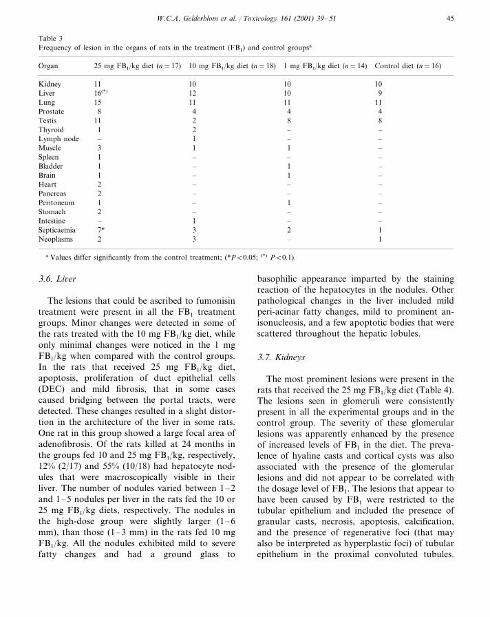
The number of lesions and the extent of thechanges observed at necropsy and histopathologi-cally differed according to the dosage of FB1
consumed in the diet over the duration of theexperiment. The lesions detected in the individualrats reflected changes of three basic categories —those associated with exposure to FB1 in the diet,those associated with secondary acute bacterialinfections (septicaemia and abscessation), andage-associated changes (Table 3). Except for themarginally significant (PB0.1) increase in thelesions in the liver, no differences in the frequencyof lesions could be observed in other organs thatcan be ascribed to the FB1-dietary treatment.Those seen in the other organs were either age-as-sociated changes or the consequences of acutebacterial infections. The prevalence of organchanges caused by secondary bacterial infections(abscesses in organs such as the lungs, prostate,and spleen, as well as peritonitis and meningitis)was noticeably higher in the rats fed the highdosage (25 mg/kg diet) of FB1. No difference inthe prevalence of these infections was detectedbetween the lower dosage groups and the controlanimals. Interstitial pneumonia occurred in alarge proportion of the rats of all groups andresulted in the development of chronic bronchitisthat in certain cases progressed to bronchiectasia.These pneumonic changes also seemed to enhancethe development of secondary bacterial infections.Age-associated lesions that occurred in rats of thevarious treatment groups and in the control ratsincluded prostatic hyperplasia and prostatitis, cal-cification and atrophy of the testes, and the occur-rence of various neoplasms. The neoplasms seenwere a gastric carcinoma and squamous cell car-cinoma of undetermined location in the 25 mgFB1/kg dietary group, lymphoma, small intestinalcarcinoma and a sub-cutaneous fibrosarcoma inthe 10 mg FB1/kg dietary group, and a subcuta-neous fibrosarcoma in the control group. No neo-plasms were detected in the 1 mg FB1/kg dietarygroup.
The lesions seen in the liver and kidneys of theanimals affected reflected the consequence of ex-posure of these animals to dietary FB1 (Table 4).
only 5/14, 4/12, 8/11 rats had altered hepatocytefoci in the control 1, 10 mg FB1/kg treatedgroups, respectively.
3.4. TBARS generation
The FB1-treated rats showed no significantlyincrease in the production of TBARS atdietary levels of 1 and 10 mg dietary levels. Asignificant (PB0.05) increase was noticed in theliver of the rats fed the 25 mg FB1/kg dietary level(Fig. 2).
Fig. 2. Dose-response of TBARS generation in vivo in rat liverhomogenates as a function of dietary FB1 levels. Data are themean of triplicate determinations. *PB0.05.
W.C.A. Gelderblom et al. / Toxicology 161 (2001) 39–51 45
Table 3Frequency of lesion in the organs of rats in the treatment (FB1) and control groupsa
10 mg FB1/kg diet (n=18) 1 mg FB1/kg diet (n=14)Organ Control diet (n=16)25 mg FB1/kg diet (n=17)
10Kidney 1011 1012 1016(*) 9Liver11 11Lung 1115
4 48 4Prostate2Testis 811 82 –1 –Thyroid1 –Lymph node ––1 13 –Muscle– –Spleen –1– 11 –Bladder– 1Brain –1– –2 –Heart
2Pancreas – – –– 11 –Peritoneum– –Stomach –21 –– –Intestine3 2 1Septicaemia 7*3 –2 1Neoplasms
a Values differ significantly from the control treatment; (*PB0.05; (*) PB0.1).
3.6. Li6er
The lesions that could be ascribed to fumonisintreatment were present in all the FB1 treatmentgroups. Minor changes were detected in some ofthe rats treated with the 10 mg FB1/kg diet, whileonly minimal changes were noticed in the 1 mgFB1/kg when compared with the control groups.In the rats that received 25 mg FB1/kg diet,apoptosis, proliferation of duct epithelial cells(DEC) and mild fibrosis, that in some casescaused bridging between the portal tracts, weredetected. These changes resulted in a slight distor-tion in the architecture of the liver in some rats.One rat in this group showed a large focal area ofadenofibrosis. Of the rats killed at 24 months inthe groups fed 10 and 25 mg FB1/kg, respectively,12% (2/17) and 55% (10/18) had hepatocyte nod-ules that were macroscopically visible in theirliver. The number of nodules varied between 1–2and 1–5 nodules per liver in the rats fed the 10 or25 mg FB1/kg diets, respectively. The nodules inthe high-dose group were slightly larger (1–6mm), than those (1–3 mm) in the rats fed 10 mgFB1/kg. All the nodules exhibited mild to severefatty changes and had a ground glass to
basophilic appearance imparted by the stainingreaction of the hepatocytes in the nodules. Otherpathological changes in the liver included mildperi-acinar fatty changes, mild to prominent an-isonucleosis, and a few apoptotic bodies that werescattered throughout the hepatic lobules.
3.7. Kidneys
The most prominent lesions were present in therats that received the 25 mg FB1/kg diet (Table 4).The lesions seen in glomeruli were consistentlypresent in all the experimental groups and in thecontrol group. The severity of these glomerularlesions was apparently enhanced by the presenceof increased levels of FB1 in the diet. The preva-lence of hyaline casts and cortical cysts was alsoassociated with the presence of the glomerularlesions and did not appear to be correlated withthe dosage level of FB1. The lesions that appear tohave been caused by FB1 were restricted to thetubular epithelium and included the presence ofgranular casts, necrosis, apoptosis, calcification,and the presence of regenerative foci (that mayalso be interpreted as hyperplastic foci) of tubularepithelium in the proximal convoluted tubules.

W.C.A. Gelderblom et al. / Toxicology 161 (2001) 39–5146
Some of the changes associated with septicaemia(such as degenerative changes in the tubular ep-ithelium, acute tubular necrosis and the presenceof granular casts) would not differ from thosecaused by the toxicological effects of FB1. Thepresence of these changes may influence interpre-tation of the significance of some of the moreacute lesions present in the kidneys.
4. Discussion
The consequences of the intake of low dietarylevels of FB1 in the present study reflected theability of the compound to induce discernibletoxic effects at dosages of, as low as, 0.03 mg/kgbody weight per day (1 mg FB1/kg diet) in thediet over a period of 18–24 months. No hepato-
Table 4Frequency of histopathology lesions observed in the liver and kidneys of rats in the FB1 treatment and control groupsa
Parameter Treatments (mg FB1/kg diet)
25 10 1 Control
16/20Survival rate 17/20 18/20 14/20387.4 (41.4)Body weight gain 390.5 (54.0) 399.2 (39.5) 407 (48.1)
2.5 (0.2)2.2 (0.1)**2.2 (0.3)**Liver weight (%) 2.3 (0.3)*Li6er histology
13* 8*Anisokaryosis 2 –Ground glass foci –4*3(*)7*
21 12Cell swelling10649Fatty changes
9* 0 0Hepatocyte nodules 0Basophilic foci
3 –Bile duct hyperplasia – –Apoptosis 1232Portal fibrosis –––5*
– ––2OVC hyperplasia5* – – –Lobular distortion
–Bridging fibrosis –1 ––Adenofibrosis 1 – –
Kidney histology7 – –Granular casts –
36Hyaline casts 5 3Cortical cysts 6 2 15
89810Periglomerular infiltrateCapsular cell proliferation 5544
– 113Hyaline droplets in glomerulus5 1 3 –Peri-glomerular scarring
PCTs–Fatty changes 5* – –
2* – – 1Apoptosis3(*) 2(*)Cell swelling – –
DCTs4* 3(*)Necrosis of epithelium – –4* – – –Calcinosis3(*) –Foci of hyperplasia (regeneration) – –
Embolic abscesses 1 – – ––Acute tubular necrosis 2 ––14 1Infarction (recent and healed) 3
––1–Pyelonephritis
a Values differ significantly from the control treatment, **PB0.01, *PB0.05; (*)0.05BPB0.1. PCTs, proximal convoluted tubuli;DCTs, distal convoluted tubuli; OVC, oval cells.

W.C.A. Gelderblom et al. / Toxicology 161 (2001) 39–51 47
cellular or cholangiocellular carcinomas devel-oped at the higher dosage levels used (10 and 25FB1/kg). The neoplasms detected in the rats inthis study are considered to be incidental age-as-sociated lesions as they occurred in various organsand also in the control group of animals. Therewas no obvious dose-response relationship be-tween the neoplasms seen in the various groupsand the level of FB1 ingested. Septicemia was alsosignificantly increased in highest dosage group.Whether this effect could be related to the adverseeffects of FB1 on the immune system as describedby Martinova et al. (1995) is not known atpresent.
The lesions induced by FB1 in rats in thepresent experiment were similar but far less severethan those seen in short- and long-term experi-ments in which higher dosages were used(Gelderblom et al., 1996a). The clinical pathologydata correlated with the presence and extent ofthe lesions seen histopathologically in the liverand kidney. The changes that occurred in the liverof the FB1-treated rats, such as fibrosis, prolifera-tion of bile duct (oval) epithelial cells, lipid accu-mulation and hepatocyte nodules have beendescribed in detail elsewhere (Gelderblom et al.,1988, 1991, 1992, 1993, 1994). In this study, theperi-acinar fatty changes and cell swelling alsooccurred in the control rats and these changesappear to be a consequence of the diet rather thanthe ingestion of FB1. The histological lesionscaused by FB1 in the kidneys, also reflected by anincrease in serum creatinine levels were present inthe rats fed the 10 and 25 mg FB1/kg diet. Thesefindings are in agreement with those reported instudies, where renal lesions were detected in maleand female Sprague–Dawley rats fed 15 and 50mg FB1/kg, respectively, for 4 weeks (Voss et al.,1993, 1995). The changes that can be ascribed toFB1 in this study are those of chronic, low-graderenal toxicity in which the lesions are limited tothe proximal and distal tubules. Many of thelesions seen in the kidneys of the rats in this studywere those of an endemic, age-associatedglomerulopathy. The severity of these age-associ-ated lesions that appeared in terms of severity, toreflect a dose-response effect, did appear to beenhanced by exposure to the FB1 in the diet. A
focal hyperplastic (or regenerative response) wasseen in only three of the rats at a dosage level of25 mg FB1/kg in the diet. In some instances,interpretation of induced lesions was hampered bythe changes that could be ascribed as incidental asthey were caused by embolic abscessation, acutetubular necrosis, pyelonephritis and infarctiondue to septicemia and disseminated abscessationin a number of rats in the advanced stage of theexperiment.
With respect to the lowest observed effects, itwould appear that under the present experimentalconditions, a level of 1 mg FB1/kg diet stillcaused minimal toxic changes (histological) in theliver and kidneys of BD IX rats fed a mar-ginally deficient diet over a period of 2 years. Theclinical pathology data also reflects sig-nificant to marginal effects in serum creatinineand AST. The only significant change obtained inthe liver with 1 mg FB1/kg dietary levelwas the induction of ground glass foci.The low dietary levels of FB1 used in the presentstudy did not induce significant (P\0.05) feedrefusal and a reduction in body weightgain as was observed in short-term studiesusing high dosage levels of the toxin (Gelderblomet al., 1994). However, the relative liver weightwas significantly decreased (Table 4), probablydue to the inhibitory effect of FB1 on hepatocytecell proliferation (Gelderblom et al., 1994, 1996b)and the induction of apoptosis (Lemmer et al.,1999).
The current study further supports the hypothe-sis that a relationship exists between FB1-inducedhepatotoxicity and hepatocarcinogenicity. In theshort-term cancer initiating promoting assays, di-etary levels of 100 mg FB1/kg and less, which onlyinduce mild toxic effects failed to initiate cancer(Gelderblom et al., 1994). Cancer promotion oc-curs at lower dietary levels (up to 50 mg FB1/kgdiet) in the absence of marked toxicity(Gelderblom et al., 1996b). These data suggestthat a threshold exists for cancer initiation andpromotion. This threshold may be a function ofthe level of FB1, the severity of the lesions in-duced and the duration of exposure. The hepato-cyte nodules noticed in the rats that received 25

W.C.A. Gelderblom et al. / Toxicology 161 (2001) 39–5148
Fig. 3. Diagram illustrates the kinetics of the induction of preneoplastic and neoplastic lesions in the liver of male BD IX rats asa function of time and different dietary levels of FB1. A dietary level of \50 mg FB1/kg (Gelderblom et al., 1991) induceshepatocyte nodules after 6 months (T=6 months) while HCC are induced between 18 and 24 months (T=18–24 months). Dietarylevels B25 mg FB1/kg induces mild toxic effects and hepatocyte nodules between 18 and 24 months (T=18–24 months).
mg FB1/kg (9/17) in the present experiment, indi-cate that the mild hepatotoxic effects induced byFB1 is associated with their development. It canbe argued that the presence of only mild toxiceffects caused ingestion of low levels of FB1, delaythe progression of these lesions to cancer. This isalso in agreement with the threshold effect withrespect to cancer promoting potential of the fu-monisins (Gelderblom et al., 1996b). A cytotoxic/proliferative threshold, therefore, seems to existwith respect to induction of hepatocyte nodulesand their progression into cancer. This conclusionis in agreement with the mechanism proposed forcancer induction by non-genotoxic carcinogens byCohen and Ellwein (1990).
The induction of hepatocyte nodules and/orfoci in the present study was similar with respectto GSTP and GGT staining and the histologicalappearance of the changes when compared withthe earlier studies in which these lesions developedinto neoplasms. The chronic hepatotoxicitycaused by the higher levels of FB1 used in the
earlier study (Gelderblom et al., 1991) is likely tohave played a critical role in the induction andprogression of these early lesions into neoplasia.The mild hepatotoxicity induced in the presentstudy resulted in less regenerative proliferationand oxidative stress than would have been thecase at higher dosages. This would suggest thatthe kinetics of cancer induction and the subse-quent development of neoplasms occur at a farslower rate when only mild hepatotoxic effects arepresent (Fig. 3).
Short-term studies in rats showed that the fu-monisins are slow cancer initiators (Gelderblom etal., 1994). Cancer promotion, on the other hand,is effected at relative low dietary levels in theabsence of excessive hepatotoxicity (Gelderblomet al., 1996b). The role of ‘spontaneously’ initiatedcells has been considered to play a role whenanimals are chronically exposed to the non-genotoxic carcinogens such as nafenopin and phe-nobarbital (Schulte-Hermann et al., 1981; Wardand Henneman, 1990; Kraupp-Grasl et al., 1991).

W.C.A. Gelderblom et al. / Toxicology 161 (2001) 39–51 49
The association of these pre-existing putative ini-tiated cells in normal animals and their subse-quent development into cancer appears to beunlikely as there is very little association betweenenzyme-altered foci and the development of theultimate cancer, especially those that occur inolder animals (Ghoshal and Farber, 1993). Recentstudies suggested that the fumonisins (Gelderblomet al., 1992, 1994) and the peroxisome prolifera-tors, cipofibrate (Rao and Reddy, 1991) and clofi-brate (Nagai et al., 1993) exhibit cancer-initiatingpotential. This implies that the role of ‘sponta-neous’ initiated cells in fumonisin-induced hepato-carcinogenesis or the induction of hepatocytenodules in the present study cannot be provenbeyond doubt. This becomes evident in recentstudies, which indicate that oxidative damage in-duced by FB1 in vitro and in vivo in the liver islikely to be involved in the cancer initiating activ-ity of the compound (Abel and Gelderblom, 1998;Sahu et al., 1998; Yin et al., 1998). The highdosage FB1-treated rats also exhibited enhancedlipid peroxidation in the liver in the present study.
The presence of the early preneoplastic lesions(GSTP+ foci and/or nodules) needs to be dis-cussed in relation to the carcinogenic properties ofthe fumonisins and the determination of risk as-sessment parameters. These lesions are generallyconsidered as the site from which cancer developsin the liver of rats (Farber, 1992). The ability ofcarcinogens to induce these early, ‘potentially’preneoplastic lesions has been frequently used toassess the carcinogenic potential of naturally oc-curring compounds, which are important in riskdetermination (Tsuda and Farber, 1980). A modelfor estimating the risk of carcinogen exposure inhumans was developed by incorporating informa-tion on both intermediate lesions, such as alteredhepatocyte foci, as well as malignant tumours(Moolgavkar and Luebeck, 1995) into the assess-ment. Such risk assessment parameters have beencalculated (Gelderblom et al., 1996a) for the car-cinogenic potential of FB1 based on the dataobtained from long-term studies in BD IX rats(present study, Gelderblom et al., 1991). The di-etary level of 25 mg FB1/kg was considered to bethe No Observed Effect Level (NOEL) for cancerinduction of the fumonisins as a diet of 50 mg
FB1/kg resulted in 66% of the rats developingcancer under similar experimental conditions. Thecorresponding Tolerable Daily Intake (TDI) valuefor cancer induction in humans, using a safetyfactor of 1000 was 0.8 mg FB1/kg body weight perday (Gelderblom et al., 1996a). Based on thecurrent data, the Lowest Observed Effect Level(LOEL) in male BD IX rats with respect to theinduction of GSTP/GGT+ nodules and/or foci, isbetween 10 and 25 mg FB1/kg or a FB1 intake ofbetween 0.3 and 0.8 mg FB1/kg body weight perday. The NOEL for the induction of hepatocytenodules in male BD IX rats is B10 mg FB1/kgdiet (0.3 mg FB1/kg body weight). The NOEL ofFB1 for hepato- and nephrotoxicity in male BDIX rats appear to be in the region of 1 mg FB1/kgdiet or an intake of 0.03 mg FB1/kg body weightper day.
The interpretation of these risk assessmentparameters are complicated, as accurate FB in-take profiles have not been established to deter-mine Probable Daily Intakes (PDI) in humans. Inaddition, FB-intake depends not only on the levelof corn contamination but also on the pattern ofconsumption of corn by the population at risk(Gelderblom et al., 1996a). Other importantparameters include the pharmacokinetics, bio-chemical mode of action, threshold effects, as wellas the dependence on cell proliferation and di-etary deficiencies (Butterworth and Goldsworthy,1991; Gelderblom et al., 1991). These factors haveto be considered against the background that FB1
is carcinogenic to rats only at dosages that causeovert hepatotoxic lesions that may culminate incirrhosis. As the fumonisins co-occur with afla-toxin B1 (Chamberlain et al., 1993; Chu and Li,1994), the synergistic interaction between thesecarcinogens, as well as other carcinogenic stimulisuch as a chronic hepatitis B virus infection haveto be critically evaluated to assess the risk of thefumonisins to humans. This is of particular inter-est as the ingestion of the fumonisins is implicatedas a possible risk factor in the development ofprimary liver cancer in endemic high liver cancerareas in China (Ueno et al., 1997).
Based on the present findings, importantparameters such as the modulation of cell prolif-

W.C.A. Gelderblom et al. / Toxicology 161 (2001) 39–5150
eration and dietary deficiencies, which could en-hance the progression of early pre-malignant le-sions into neoplasia need to be considered infuture studies to establish relevant risk assessmentparameters for the fumonisins in humans.
References
Abel, S., Gelderblom, W.C.A., 1998. Oxidative damage andfumonisin B1-induced toxicity in primary rat hepatocytesand rat liver in vivo. Toxicology 131, 121–131.
Buege, J.A., Aust, S.D., 1978. Microsomal lipid peroxidation.Methods Enzymol. 52, 302–310.
Butterworth, B.E., Goldsworthy, T.L., 1991. The role of cellproliferation in multistage carcinogenesis. Proc. Soc. Exp.Biol. Med. 683–687.
Cawood, M.E., Gelderblom, W.C.A., Vleggaar, R., Berhend,Y., Thiel, P.G., Marasas, W.F.O., 1991. Isolation of thefumonisin mycotoxins: a quantitative approach. J. Agric.Food Chem. 39, 1958–1962.
Chamberlain, W.J., Bacon, C.W., Norred, W.P., Voss, K.A.,1993. Levels of fumonisin B1 in maize naturally contami-nated with aflatoxins. Food Chem. Toxicol. 31, 995–998.
Cohen, S.M., Ellwein, L.B., 1990. Cell proliferation in carcino-genesis. Science 249, 1007–1011.
Chu, F.S., Li, G.Y., 1994. Simultaneous occurrence of fumon-isin B1 and other mycotoxins in moldy maize collectedfrom the People’s Republic of China in regions with highincidences of esophageal cancer. Appl. Environ. Microbiol.60, 847–852.
Esterbauer, H., Cheeseman, K.H., 1990. Determination ofaldehydic lipid peroxidation products: malonaldehyde and4-hydroxynonenal. Methods Enzymol. 186, 407–421.
Farber, E., 1992. On cells of origin of liver cell cancer. In:Sirica, A.E. (Eds.), The role of cell types of hepatocarcino-genesis. CRC Press, Boca raton, Ann Arbor, London,Tokyo, pp. 1–27.
Fincham, J.E., Marasas, W.F.O., Taljaard, et al., 1992.Artherogenic effect in a non-human primate of Fusariummoniliforme cultures added to a carbohydrate diet.Atherosclerosis 94, 13–25.
Gelderblom, W.C.A., Jaskiewicz, K., Marasas, W.C.A., Thiel,P.G., Horak, R., Vleggaar, R., Kriek, N.P.J., 1988. Fu-monisins — novel mycotoxins with cancer promoting ac-tivity produced by Fusarium moniliforme. Appl. Environ.Microbiol. 54, 1806–1811.
Gelderblom, W.C.A., Kriek, N.P.J., Marasas, W.F.O., Thiel,P.G., 1991. Toxicity and carcinogenicity of the Fusariummoniliforme metabolite, fumonisin B1, in rats. Carcinogen-esis 12, 1247–1251.
Gelderblom, W.C.A., Semple, E., Marasas, W.F.O., Farber,E., 1992. The cancer initiating potential of the fumonisin Bmycotoxins produced by Fusarium moniliforme. Carcino-genesis 13, 433–437.
Gelderblom, W.C.A., Cawood, M.E., Snyman, S.D., Vleggaar,R., Marasas, W.F.O., 1993. Structure-activity relationshipsof fumonisins in short-term carcinogenesis and cytotoxicityassays. Food Chem. Toxicol. 31, 407–414.
Gelderblom, W.C.A., Cawood, M.E., Snyman, S.D., Marasas,W.F.O., 1994. Fumonisin B1 dosimetry in relation to can-cer initiation in rat liver. Carcinogenesis 105, 209–214.
Gelderblom, W.C.A., Snyman, S.D., Abel, S., et al., 1996a.Hepatotoxicity and carcinogenicity of the fumonisins inrats: a review regarding mechanistic implications for estab-lishing risk in humans. In: Jackson, L.S., De Vries, J.W.,Bullerman, L.B. (Eds.), Fumonisins in Food. Plenum, NewYork, NY, pp. 279–296.
Gelderblom, W.C.A., Snyman, S.D., Lebepe-Mazur, S., Vander Westhuizen, L., Kriek, N.P.J., Marasas, W.F.O.,1996b. The cancer promoting potential of fumonisin B1 inrat liver using diethylnitrosamine as cancer initiator. Can-cer Lett. 109, 101–108.
Ghoshal, A.K., Farber, E., 1993. Choline deficiency, lipotropedeficiency and the development of liver disease includingliver cancer; a new perspective. Lab. Invest. 68, 255–260.
Kraupp-Grasl, B., Huber, W., Taper, H., Schulte-Hermann,R., 1991. Increased susceptibility of aged rats to hepatocar-cinogegenesis by the peroxisome proliferator nafenopinand the possible involvement of altered liver foci occurringspontaneously. Cancer Res. 51, 666–671.
Lemmer, E.R., De la, M., Hall, P., Omori, N., et al., 1999.Histopathology and gene expression changes in rat liverduring feeding of fumonisin B1, a carcinogenic mycotoxinproduced by Fusarium moniliforme. Carcinogenesis 20,817–824.
Martinova, E.A., Merrill, A.H., Jr, 1995. Fumonisin B1 alterssphingolipid metabolism and immune funstion in BALB/cmice. Mycopatholgia 130, 163–170.
Moolgavkar, S.H., Luebeck, E.G., 1995. Incorporating cellproliferation kinetics into models for cancer risk assess-ment. Toxicology 102, 141–147.
Nagai, M.K., Armstrong, D., Farber, E., 1993. Induction ofresistant hepatocytes by clofibrate, a non-genotoxic car-cinogen. Proc. Am. Assoc. Cancer Res. 34, 164.
Norred, W.P., Plattner, R.D., Vesonder, R.F., Bacon, C.W.,Voss, K.A., 1992. Effects of selected secondary metabolitesof Fusarium moniliforme on unscheduled synthesis of DNAby primary rat hepatocytes. Food Chem. Toxicol. 30,233–237.
NIH Publication No. 99-3955, 1999. NTP technical report onthe toxicology and carcinogenesis studies of fumonisin B1
(CAS No. 116355-83-0) in F344/N rats and B6C3F1 mice(feed studies). Peer reviewed 21 May 1999.
Ogawa, K., Solt, D., Farber, E., 1980. Phenotypic diversity asan early property of putative preneoplastic cells in livercarcinogenesis. Cancer Res. 40, 725–733.
Rao, S.M., Reddy, J.K., 1991. An overview of peroxisomeproliferator-induced hepatocarcinogenesis. Environ.Health Perspect. 93, 205–209.
Sahu, S.C., Eppley, R.M., Page, S.W., Gray, G.C., Barton,C.N., O’Donnell, M.W., 1998. Peroxidation of membrane

W.C.A. Gelderblom et al. / Toxicology 161 (2001) 39–51 51
lipids and oxidative DNA damage by fumonisin B1 inisolated rat liver nuclei. Cancer Lett. 125, 117–121.
Schulte-Hermann, R., Ohde, G., Schuppler, J., Timmermann-Trosiener, I., 1981. Enhanced proliferation of putativepreneoplastic single cell rat liver following treatment withthe tumor promoters phenobarbital, hexachlorocyclohex-ane, steroid compounds and nafenopin. Cancer Res. 41,2556–2562.
Shephard, G.S., Sydenham, E.W., Thiel, P.G., Gelderblom,W.C.A., 1990. Quantitative determination of fumonisinsB1 and B2 by high-performance liquid chromatographywith fluorescence detection. J. Liq. Chromatogr. 13, 2077–2087.
Tatematsu, M., Aoki, T., Kagawa, M., Mera, Y., Ito, N.,1988. Reciprocal relationship between development of glu-tathione S-transferase positive foci and proliferation ofsurrounding hepatocytes in rats. Carcinogenesis 9, 221–225.
Thiel, P.G., Stockenstrom, S., Gathercole, P.S., 1986. Afla-toxin analysis by reverse phase HPLC using post-columnderivatization for enhancement of fluorescence. J. Liq.Chromatogr. 9, 103–112.
Tsuda, H., Farber, E., 1980. Resistant hepatocytes as earlychanges in liver induced by polycyclic aromatic hydrocar-bons. Int. J. Cancer 25, 137–139.
Ueno, Y., Iijima, K., Wang, S.D., Sugira, Y., Sekijima, M.,Tanaka, T., Chen, C., Yu, S.Z., 1997. Fumonisins as apossible contributing risk factor for primary liver cancer: a3-year study of corn harvested in Haimen, China, byHPLC and ELISA. Food Chem. Toxicol. 35, 1143–1150.
Van Rensburg, S.J., Hall, J.M., Du Bruyn, D.B., 1985. Effectsof various dietary staples on esophageal carcinogenesisinduced in rats by subcutaneously administered N-nitro-somethylbenzylamine. J. Natl. Cancer Inst. 75, 561–566.
Voss, K.A., Chamberlain, W.J., Bacon, C.W., Norred, W.P.,1993. A preliminary investigation on renal and hepatictoxicity in rats fed purified fumonisin B1. Nat. Toxins 1,222–228.
Voss, K.A., Chamberlain, W.J., Bacon, C.W., Riley, R.T.,Norred, W.P., 1995. Subchronic toxicity of fumonisin B1
to male and female rats. Food Addit. Contam. 12, 473–478.
Ward, J.M., Henneman, J.R., 1990. Naturally-occurring age-dependent gluthathione S-transferase P immunoreactivehepatocytes in ageing female F344 rat liver as potentialpromotable targets for non-genotoxic carcinogens. CancerLett. 52, 187–195.
Yin, J.-J, Smith, M.J., Eppley, R.M., Page, S.W., Sphon, J.A.,1998. Effects of fumonisin B1 on lipid peroxidation inmembranes. Biochim. Biophys. Acta 1371, 134–142.
.