Familial collapsing glomerulopathy: Clinical, pathological and immunogenetic features
Upload
khangminh22Category
view
3download
0
Ghent University Faculty of Medicine and Health Sciences
Department of Forensic Medicine
Investigation of fatalities related to the use of 3,4-methylenedioxymethamphetamine (MDMA, “Ecstasy”) and analogues:
anatomo-pathological and thanato-toxicological approach
Thesis submitted as partial fulfilment of the requirements for the degree of Doctor in Medical Sciences 2002 Els A. DE LETTER
PROMOTOR: Prof. dr. Michel HA. PIETTE
Ghent University
Faculty of Medicine and Health Sciences Department of Forensic Medicine
Investigation of fatalities related to the use of 3,4-methylenedioxymethamphetamine (MDMA, “Ecstasy”) and analogues:
anatomo-pathological and thanato-toxicological approach
Onderzoek van fatale gevallen gerelateerd aan het gebruik van 3,4-methyleendioxymethamfetamine (MDMA, “Ecstasy”) en analogen:
anatomo-pathologische en thanato-toxicologische benadering Thesis submitted as partial fulfilment of the requirements for the degree of Doctor in Medical Sciences 2002 Els A. DE LETTER
PROMOTOR: Prof. dr. Michel HA. PIETTE

“ This is the very ecstasy of love, Whose violent property fordoes itself
And leads the will to desperate undertakings As oft as any passion under heaven That does afflict our natures."
Hamlet, Act II, Scene I, 116
Voor ons ma, onze pa, en nonkel Staf

Cover : Immunoreactive Purkinje cells in an MDMA overdose victim (case 00/112) after staining with a monoclonal antibody recognizing MDMA and its closely related compounds.

Table of contents
Table of contents ________________________________________________________________________________
1
TABLE OF CONTENTS
Introduction and aims 13 PART ONE Review of the medico-legal literature and survey of
amphetamine-related fatalities at the Department of Forensic Medicine (Ghent University)
Chapter 1 Review of the medico-legal literature:
focus on 3,4-methylenedioxymethamphetamine (MDMA) 27
I Pharmacology of MDMA: human and animal experimental data 28
I.1 Pharmacokinetics of MDMA in humans 28 I.2 Pharmacodynamics of MDMA 33
I.2.1 Animal experimental data 33 I.2.1.1 Cardiovascular effects 33 I.2.1.2 Hepatotoxicity 34 I.2.1.3 Cerebral effects and neurotoxicity 34 I.2.2 Desired effects in humans 43 I.2.3 Human toxicity 44
I.2.3.1 Symptoms 45 I.2.3.1.1 Cardiovascular effects 45
I.2.3.1.2 Hepatotoxicity 45 I.2.3.1.3 Central nervous system effects 46 I.2.3.1.4 Uro-genital effects 47 I.2.3.1.5 Various symptoms 48
I.2.3.1.6 Multiple organ failure 48 I.2.3.2 Correlation between blood levels and clinical effects 49
II Epidemiological data on the use of MDMA 50
II.1 Europe 51
II.1.1 Belgium 51 II.1.2 Other European countries 51
II.2 USA and the rest of the world 52
Table of contents ________________________________________________________________________________
2
III Medico-legal implications of MDMA 52
III.1 Accidents 52 III.2 (Attempted) suicide 54 III.3 Criminal offences 54
IV Thanatological findings 55
IV.1 Anatomo-pathological data in MDMA-related fatalities
IV.1.1 General findings 55 IV.1.2 Findings pointing to (ab)use of amphetamine 57
and derivatives IV.2 Thanato-toxicological data in MDMA-related fatalities 62
IV.2.1 Animal experimental data 62 IV.2.2 Survey of MDMA-related human fatalities 63
IV.3 Discussion of the human thanatological literature data 73
Chapter 2 Survey of amphetamine-related fatalities at the 99 Department of Forensic Medicine, Ghent University, between January 1976 and April 2002
I Introduction 99
II Case studies 101
III Toxicological data 112
III.1 Drug assays 112 III.2 Results 112 IV Summary of the cases 116
V Discussion 119
Table of contents ________________________________________________________________________________
3
PART TWO Experiments in rabbits
Chapter 3 Is vitreous humour useful for the interpretation of 3,4- 127 methylenedioxymethamphetamine (MDMA) blood levels ?
Experimental approach with rabbits
Els A. De Letter, Peter De Paepe, Karine M. Clauwaert, Frans M. Belpaire, Willy E. Lambert , Jan F. Van Bocxlaer , Michel H.A. Piette Based on: Int J Legal Med 2000;114:29-35
Chapter 4 Post-mortem redistribution of 3,4-methylenedioxy- 145
methamphetamine (MDMA, “ecstasy”) in the rabbit Part one: Experimental approach after intravenous infusion. Els A. De Letter, Karine M. Clauwaert, Frans M. Belpaire, Willy E. Lambert ,
Jan F. Van Bocxlaer , Michel H.A. Piette Int J Legal Med 2002;116:216-224
Chapter 5 Post-mortem redistribution of 3,4-methylenedioxy- 165 methamphetamine (MDMA, “ecstasy”) in the rabbit
Part two: Post-mortem infusion in trachea or stomach.
Els A. De Letter, Frans M. Belpaire, Karine M. Clauwaert, Willy E. Lambert , Jan F. Van Bocxlaer , Michel H.A. Piette
Int J Legal Med 2002;116:225-232
Table of contents ________________________________________________________________________________
4
PART THREE Case analyses in current forensic practice
Chapter 6 Thanato-toxicological approach 183
I MDMA AND ITS METABOLITE MDA I.1 Distribution study of 3,4-methylenedioxymethamphetamine 183
and 3,4-methylenedioxyamphetamine in a fatal overdose
Els A. De Letter, Karine M. Clauwaert, Willy E. Lambert , Jan F. Van Bocxlaer , André P. De Leenheer, Michel H.A. Piette
J Anal Toxicol 2002;26:113-118
I.2 Distribution study of the amphetamine derivative MDMA 196 and its metabolite 3,4-methylenedioxyamphetamine (MDA) in two overdoses
II OTHER AMPHETAMINE DERIVATIVES RECENTLY 203 OBSERVED IN A FEW CASES IN BELGIUM
II.1 One fatal and seven nonfatal cases of 4-methyl- 203
thioamphetamine (4-MTA) intoxication: clinico-pathological findings
Els A. De Letter, Vera A.E. Coopman, Jan A.C.M. Cordonnier, Michel H.A. Piette
Based on: Int J Legal Med 2001;114:352-356
II.2 Fatal 4-MTA intoxication: development of a liquid 216 chromatographic – tandem mass spectrometric assay
for multiple matrices
T. Decaestecker, E. De Letter, K. Clauwaert, M.P. Bouche, W. Lambert, J. Van Bocxlaer, M. Piette, E. Van den Eeckhout, C. Van Peteghem, A. De Leenheer
Based on: J Anal Toxicol 2001;25:705-710.
Table of contents ________________________________________________________________________________
5
II.3 Fatality due to combined use of the designer drugs MDMA 230 and PMA: a distribution study
Riet Dams, Els A. De Letter, Kjell A. Mortier, Jan A.C.M. Cordonnier, W.E. Lambert, M.H.A. Piette, S. Van Calenbergh and A.P. De Leenheer Accepted for publication in J Anal Toxicol
Chapter 7 Immunohistochemical approach
Immunohistochemical demonstration of the amphetamine derivatives 245 3,4-methylenedioxymethamphetamine (MDMA) and 3,4-methylene- dioxyamphetamine (MDA) in human post-mortem brain tissues and the pituitary gland
Els A. De Letter, Marc F.A. Espeel, Marijke E.C. Craeymeersch, Willy E. Lambert, Karine M. Clauwaert, Riet Dams, Kjell A. Mortier, Michel H.A. Piette
Accepted for publication in Int J Legal Med Summary and conclusions 265 Résumé et conclusions 273 Samenvatting en conclusies 283 Dankwoord 293

Introduction and aims
Introduction and aims ________________________________________________________________________________
1
Introduction and aims
The detection of toxic substances, in particular of illicit drugs, plays an important role in the forensic inquiry. It is important to point out whether or not a person was under the influence at the very moment of an accident or criminal offence.
Whereas the blood or plasma level of a substance often correlates with recent cerebral effects in a living person, this is not necessarily applicable to the dead due to interfering thanato-chemical processes. Problems include post-mortem degradation, redistribution and sometimes even post-mortem production of a substance. When drug instability is important, falsely decreased levels may be measured or the drug can become undetectable. On the other hand, post-mortem redistribution and/or neo-formation may result in falsely elevated concentrations. The competition between drug instability and redistribution should be taken into account when considering a specific concentration as being therapeutic, toxic or lethal. These post-mortem phenomena have been investigated for several compounds such as ethanol, cocaine, benzodiazepines, barbiturates and antidepressant medication. For ethanol, bacterial post-mortem production has been proven (1), whereas for cocaine, instability is prominent (2). In addition, the interpretation of cocaine levels may be difficult due to competing post-mortem processes, namely tissue release on the one hand, and chemical and enzymatic degradation of the substance on the other (3,4). Some benzodiazepines - and nitrobenzodiazepines in particular - are chemically and metabolically very unstable (5). Post-mortem decrease of anticonvulsant serum concentrations, especially for phenobarbital and phenytoin, has been described and therefore interpretation with respect to “subtherapeutic” serum levels or noncompliance should be interpreted with caution (6). Post-mortem redistribution into cardiac blood has also been substantiated, for example for barbiturates (7), amitriptyline (8-12) and procainamide (13). To a certain extent, the interference of post-mortem phenomena can be avoided by sampling blood as soon as possible after death from an isolated peripheral vein such as the femoral vein (14). In addition, since the vitreous humour is to a minor extent influenced by autolytic processes - due to its well-isolated position –, this specimen can be interesting for toxicological investigation. Moreover, the vitreous fluid is convenient (e.g. simple to sample and not affected by hemolysis). Vitreous humour levels have been studied for various substances such as alcohol (15), morphine (16), and cocaine (17). In humans, quantification of 3,4-methylenedioxymethamphetamine (MDMA, “ecstasy”) in the vitreous humour has only been performed in a few cases (18,19). Abuse of amphetamine derivatives such as MDMA and 3,4-methylenedioxy-amphetamine (MDA) is an important public issue and fatalities are not infrequent in current forensic practice. For the amphetamine derivatives and MDMA in particular, very little data from fatalities are available in the literature. Since amphetamine-related fatalities, including those of MDMA users, are on the increase in medico-legal practice (e.g. 20-22), fundamental research on these post-mortem phenomena is required. Though MDMA appears to be stable in vitro (23,24), the post-mortem (re)distribution of MDMA
Introduction and aims ________________________________________________________________________________
2
in the body has barely been explored, with the exception of a few case reports (25-28): apart from blood and urine concentrations, only a few tissue levels have been reported. These data show that high MDMA concentrations can be found in organs such as the brain (25-28) and the liver (25-27). For amphetamine (AMP), methamphetamine (METH) and MDA, more literature data are available, for example (14,29-38). These case reports indicate that concentrations in cardiac blood are obviously higher than those in peripheral blood. In addition, significant levels of AMP, METH and MDA have been found in several tissues such as liver and brain, but also in blood-rich organs such as the lungs, which means that these substances are liable to post-mortem redistribution. Animal experiments dealing with this issue for amphetamine or its analogues are scarce (39,40). Hilberg and colleagues described an experiment in which post-mortem redistribution of amphetamine in the rat was studied (39), with further extrapolation to a few medico-legal cases (41). Moriya et al. demonstrated redistribution of methamphetamine into cardiac blood via pulmonary blood vessels in the early post-mortem period (40).
The question remains open as to whether an MDMA blood level can be toxic or even potentially lethal. Moreover, referring to possible thanatological changes, it is not clear whether the observed post-mortem MDMA blood level actually represents the concentration at the time of death. In this thesis research, the post-mortem distribution and redistribution of MDMA was studied in order to evaluate which fluid and/or tissue sample after death most closely represents the ante-mortem concentration. Furthermore, the question was posed as to whether the post-mortem phenomena relating to MDMA are in line with those for the other amphetamine derivatives. In addition, the significance of post-mortem MDMA levels in vitreous humour was evaluated.
In Part One of this work, a summary of the relevant literature and a survey of the
amphetamine-related fatalities examined at the Department of Forensic Medicine of Ghent University was discussed.
In Chapter 1, a literature review focusing on the (ab)use of amphetamines with particular emphasis on MDMA is presented. The clinico-pharmacological effects, the epidemiological importance, the medico-legal implications and thanato-toxicological literature data for MDMA are discussed.
In Chapter 2, the amphetamine-related fatalities encountered at the Department of Forensic Medicine of Ghent University between January 1976 and April 2002 are reviewed. Apart from the toxicological findings, possible mechanisms of death are examined and discussed in the light of the available literature data.
Introduction and aims ________________________________________________________________________________
3
In the experimental work featured in Part Two, the post-mortem problems for MDMA were examined using an experimental rabbit model. In the first study presented, the value of post-mortem vitreous humour MDMA levels was examined (Chapter 3). The pharmacokinetics of MDMA in the rabbit after intravenous (iv) administration and the correlation between MDMA blood and vitreous humour levels were investigated. In addition, a fully validated high pressure liquid chromatographic (HPLC) method with fluorescence detection for quantification of MDMA and its metabolite MDA was designed (42). Chapters 4 and 5 report further studies of the post-mortem stability and redistribution of MDMA in the rabbit model in order to determine which body fluid(s) and/or tissue(s) after death most closely represent the actual ante-mortem concentration.
Chapter 4 deals with the distribution of MDMA and its metabolite MDA in different body fluids and tissues of rabbits that were killed 2 hours after iv administration of MDMA. Three groups of rabbits were studied. In the first group (control group), the study was performed immediately after sacrifying and in the second group, the animals were preserved at ambient temperature either 24 or 72 h post mortem prior to sampling. Theoretically, post-mortem increases in cardiac blood levels can occur due to intravascular diffusion out of blood-rich organs such as the liver and the lungs (34). Therefore, in the third group, ligation of the large vessels around the heart was performed (immediately after killing) and these rabbits were further treated as in the second group.
In humans, who mainly take MDMA orally, it is important to investigate whether a “reservoir” in the stomach influences post-mortem blood and tissue concentrations when the subject dies shortly after ingestion, and - as a result – the distribution is not yet completed. In addition, drug levels can be affected by agonal vomit aspiration or post-mortem regurgitation in the airways. The influence of the gastric reservoir function (43) and vomit aspiration or regurgitation (44) has previously been proven for ethanol. This was simulated in another rabbit animal model (Chapter 5): post-mortem infusion of an MDMA solution was performed either in the trachea or in the stomach and the diffusion was studied up to 72 hours after administration. In both groups, MDMA and MDA levels were determined in various fluids and tissues using the same HPLC method.
In Part Three, the animal experimental data are compared with the human findings. The post-mortem distribution of MDMA (and its metabolite MDA) and some other amphetamine derivatives in the human body was investigated. In order to evaluate which fluid and/or tissue sampled after death most closely represents the ante-mortem concentration, two different - but complementary – approaches were examined.
In Chapter 6, the thanato-toxicological approach is taken. The concentrations determined in various fluids (blood sampled on different locations, vitreous humour, urine and bile) and tissues such as cardiac muscle, lungs, liver, kidneys, spleen, ilio-psoas muscle, and brain in subjects who died following exposure to MDMA and/or derivatives are discussed. Apart from MDMA and MDA, some other amphetamine derivatives, namely 4-methylthioamphetamine (4-MTA) and para-methoxyamphetamine
Introduction and aims ________________________________________________________________________________
4
(PMA) are considered. For the relatively new derivative, 4-MTA, the data of persons who survived after ingestion are presented and the clinical observations are commented too.
Chapter 7 takes an anatomo-pathological/thanatological approach, with emphasis on immunohistochemistry. Thus, a semi-quantitative visual presentation of the distribution of MDMA in tissues is obtained and correlated with the toxicological findings. The question is posed whether immunohistochemical detection could be either an alternative or a supplementary tool in the forensic inquiry when the toxicological determinations are interfered with or have become impossible. In particular, the brain – being an important target organ for MDMA – is a difficult matrix for chromatographic extraction due to the lipid fraction. In this thesis, an immunohistochemical method for the detection of MDMA and MDA in human brain tissues and the pituitary gland is reported. However, immunohistochemical detection is restricted due to the fact that only the fraction bound to tissues can be demonstrated since the unbound fraction is washed out during the preparation procedure. This is a fundamental difference with the toxicological quantitation in tissue homogenates, in which both the bound and the unbound fraction are measured.
Finally, Summary and Conclusions provides the main findings of our research
work.
Introduction and aims ________________________________________________________________________________
5
References
1. O’Neal CL, Poklis A. Postmortem production of ethanol and factors that influence
interpretation. A critical review. Am J Forensic Med Pathol 1996;17:8-20. 2. Moriya F, Hashimoto Y. Postmortem stability of cocaine and cocaethylene in blood and
tissues of humans and rabbits. J Forensic Sci 1996;41:612-616. 3. Hearn WL, Keran EE, Wei H, Hime G. Site-dependent postmortem changes in blood cocaine
concentrations. J Forensic Sci 1991;36:673-684. 4. Logan BK, Smirnow D, Gullberg RG. Lack of predictable site-dependent differences and
time-dependent changes in postmortem concentrations of cocaine, benzoylecgonine, and cocaethylene in humans. J Anal Toxicol 1997;21:23-31.
5. Pépin G, Dubourvieux N, Gaillard Y. Difficulté d’interprétation des taux des benzodiazépines
et molécules apparentées dans le sang de cadavre prélevé à l’autopsie: étude de leur dégradation in vitro après conservation pendant 6 mois à différentes températures. J Méd Lég Droit Méd 1998;41:341-353.
6. May T, Jürgens U, Rambeck B, Schnabel R. Comparison between premortem and postmortem
serum concentrations of phenobarbital, phenytoin, carbamazepine and its 10,11-epoxide metabolite in institutionalized patients with epilepsy. Epilepsy Res 1999;33:57-65.
7. Pounder DJ, Jones GR. Post-mortem drug redistribution – a toxicological nightmare. Forensic
Sci Int 1990;45:253-263. 8. Hilberg T, Bugge A, Beylich K-M, Mørland J, Bjørneboe A. Diffusion as a mechanism of
postmortem drug redistribution: an experimental study in rats. Int J Legal Med 1992;105:87-91.
9. Hilberg T, Bugge A, Beylich K-M, Ingum J, Bjørneboe A, Mørland J. An animal model of
postmortem amitriptyline redistribution. J Forensic Sci 1993;38:81-90. 10. Hilberg T, Mørland J, Bjørneboe A. Postmortem release of amitriptyline from the lungs; a
mechanism of postmortem drug redistribution. Forensic Sci Int 1994;64:47-55. 11. Hilberg T, Ripel Å, Smith AJ, Slørdal L, Mørland J, Bjørneboe A. Postmortem amitriptyline
pharmacokinetics in pigs after oral and intravenous routes of administration. J Forensic Sci 1998;43:380-387.
12. Baselt RC. (ed) (2000) Disposition of toxic drugs and chemicals in man, 5th edn, Chemical
Toxicology Institute, Foster City, California, pp 38-42. 13. Shepherd MF, Lake KD, Kamps MA. Postmortem changes and pharmacokinetics: review of
the literature and case report. Ann Pharmacother 1992;26:510-514.
Introduction and aims ________________________________________________________________________________
6
14. Prouty RW, Anderson WH. The forensic science implications of site and temporal influences
on postmortem blood-drug concentrations. J Forensic Sci 1990;35:243-270. 15. Chao TC, Lo DST. Relationship between postmortem blood and vitreous humor ethanol
levels. Am J Forensic Med Pathol 1993;14:303-308. 16. Bermejo AM, Ramos I, Fernández P, López-Rivadulla M, Cruz A, Chiarotti M, Fucci N,
Marsilli R. Morphine determination by gas chromatography/mass spectroscopy in human vitreous humor and comparison with radioimmunoassay. J Anal Toxicol 1992;16:372-374.
17. McKinney PE, Phillips S, Gomez HF, Brent J, MacIntyre M, Watson WA. Vitreous humor
cocaine and metabolite concentrations: do postmortem specimens reflect blood levels at the time of death? J Forensic Sci 1995;40:102-107.
18. Crifasi J, Long C. Traffic fatality related to the use of methylenedioxy-methamphetamine. J
Forensic Sci 1996;41:1082-1084. 19. Moore KA, Mozayani A, Fierro MF, Poklis A. Distribution of 3,4-methylenedioxy-
methamphetamine (MDMA) and 3,4-methylenedioxyamphetamine (MDA) stereoisomers in a fatal poisoning. Forensic Sci Int 1996;83:111-119.
20. Henry JA, Jeffreys KJ, Dawling S. Toxicity and deaths from 3,4-
methylenedioxymethamphetamine ("ecstasy"). Lancet 1992;340:384-387. 21. Gore SM. Fatal uncertainty: death-rate from use of ecstasy or heroin. Lancet 1999;354:1265-
1266. 22. Gill JR, Hayes JA, deSouza IS, Marker E, Stajic M. Ecstasy (MDMA) deaths in New York
City: a case series and review of the literature. J Forensic Sci 2002;47:121-126. 23. Garrett ER, Seyda K, Marroum P. High performance liquid chromatographic assays of the
illicit designer drug “Ecstasy”, a modified amphetamine, with applications to stability, partitioning and plasma protein binding. Acta Pharm Nord 1991;3:9-14.
24. Clauwaert KM, Van Bocxlaer JF, De Leenheer AP. Stability study of the designer drugs
“MDA, MDMA and MDEA” in water, serum, whole blood, and urine under various storage temperatures. Forensic Sci Int 2001;124;36-42.
25. Rohrig TP, Prouty RW. Tissue distribution of methylenedioxymethamphetamine. J Anal
Toxicol 1992;16:52-53. 26. Fineschi V, Masti A. Fatal poisoning by MDMA (ecstasy) and MDEA: a case report. Int J
Legal Med 1996;108:272-275. 27. Fineschi V, Centini F, Mazzeo E, Turillazzi E. Adam (MDMA) and Eve (MDEA) misuse: an
immunohistochemical study on three fatal cases. Forensic Sci Int 1999;104:65-74.
Introduction and aims ________________________________________________________________________________
7
28. Kish SJ, Furukawa Y, Ang L, Vorce SP, Kalasinksky KS. Striatal serotonin is depleted in
brain of a human MDMA (Ecstasy) user. Neurology 2000;55:294-296. 29. Hilberg T, Rogde S, Mørland J. Postmortem drug redistribution – human cases related to
results in experimental animals. J Forensic Sci 1999;44:3-9. 30. Meyer E, Van Bocxlaer JF, Dirinck IM, Lambert WE, Thienpont L, De Leenheer AP. Tissue
distribution of amphetamine isomers in a fatal overdose. J Anal Toxicol 1997; 21:236-239. 31. Barnhart FE, Fogacci JR, Reed DW. Methamphetamine – a study of postmortem redistribution.
J Anal Toxicol 1999;23:69-70. 32. Katsumata S, Sato K, Kashiwade H, Yamanami S, Zhou H, Yonemura I, Nakajima H,
Hasekura H. Sudden death due presumably to internal use of methamphetamine. Forensic Sci Int 1993;62:209-215.
33. Miyazaki T, Kojima T, Yashiki M, Wakamoto H, Iwasaki Y, Taniguchi T. Site dependence of
methamphetamine concentrations in blood samples collected from cadavers of people who had been methamphetamine abusers. Am J Forensic Med Pathol 1993;14:121-124.
34. Moriya F, Hashimoto Y. Redistribution of methamphetamine in the early postmortem period. J
Anal Toxicol 2000;24:153-154. 35. Logan BK, Weiss EL, Harruff RC. Case report: Distribution of methamphetamine in a massive
fatal ingestion. J Forensic Sci 1996;41:322-323. 36. Kalasinsky KS, Bosy TZ, Schmunk GA, Reiber G, Anthony RM, Furukawa Y, Guttman M,
Kish SJ. Regional distribution of methamphetamine in autopsied brain of chronic human methamphetamine users. Forensic Sci Int 2001;116:163-169.
37. Kojima T, Une I, Yashiki M. CI-mass fragmentographic analysis of methamphetamine and
amphetamine in human autopsy tissues after acute methamphetamine poisoning. Forensic Sci Int 1983;21:253-258.
38. Lukaszewski T. 3,4-methylenedioxyamphetamine overdose. Clin Toxicol 1979;15:405-409. 39. Hilberg T, Ripel Å, Slørdal L, Bjørneboe A, Mørland J. The extent of postmortem drug
redistribution in a rat model. J Forensic Sci 1999;44:956-962. 40. Moriya F, Hashimoto Y. Redistribution of basic drugs into cardiac blood from surrounding
tissues during early-stages postmortem. J Forensic Sci 1999;44:10-16. 41. Hilberg T, Rogde S, Mørland J. Postmortem drug redistribution – human cases related to
results in experimental animals. J Forensic Sci 1999;44:3-9.
Introduction and aims ________________________________________________________________________________
8
42. Clauwaert KM, Van Bocxlaer JF, De Letter EA, Van Calenbergh S, Lambert WE, De Leenheer
AP. Determination of the designer drugs 3,4-methylenedioxymethamphetamine, 3,4-methylenedioxyethylamphetamine and 3,4-methylenedioxyamphetamine with HPLC and fluorescence detection in whole blood, serum, vitreous humor, and urine. Clin Chem 2000;46:1968-1977.
43. Pounder DJ, Smith DRW. Postmortem diffusion of alcohol from the stomach. Am J Forensic
Med Pathol 1995;16:89-96. 44. Pounder DJ, Yonemitsu K. Postmortem absorption of drugs and ethanol from aspirated
vomitus – an experimental model. Forensic Sci Int 1991;51:189-195.

PART ONE
Review of the medico-legal literature and survey of amphetamine-related fatalities at the Department of Forensic Medicine
(Ghent University)
Chapter 1
Review of the medico-legal literature: focus on 3,4 methylenedioxymethamphetamine (MDMA)
Chapter 1 _____________________________________________________________________________
1
PART ONE Review of the medico-legal literature and survey of
amphetamine-related fatalities at the Department of Forensic Medicine (Ghent University)
Chapter 1 Review of the medico-legal literature:
focus on 3,4-methylenedioxymethamphetamine (MDMA)
In this chapter, a summary of the pharmacology of the amphetamine derivative 3,4-methylenedioxymethamphetamine (MDMA, “ecstasy”, XTC) will be presented, followed by a discussion of the epidemiological setting. In addition, the medico-legal implications of these drugs with regard to the manner of death will be considered. Finally, the available thanato-toxicological literature data will be discussed.
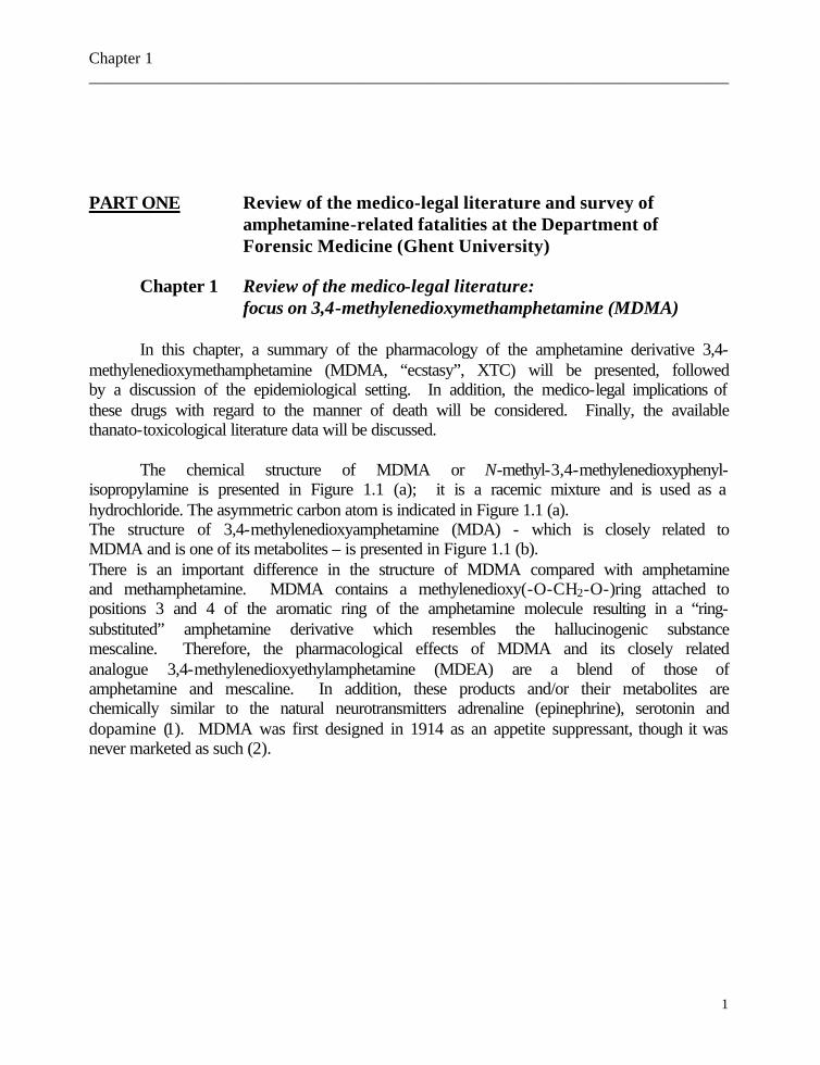
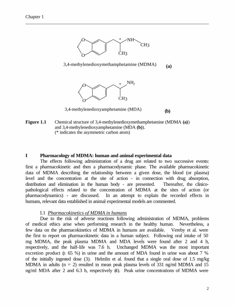
The chemical structure of MDMA or N-methyl-3,4-methylenedioxyphenyl-isopropylamine is presented in Figure 1.1 (a); it is a racemic mixture and is used as a hydrochloride. The asymmetric carbon atom is indicated in Figure 1.1 (a). The structure of 3,4-methylenedioxyamphetamine (MDA) - which is closely related to MDMA and is one of its metabolites – is presented in Figure 1.1 (b). There is an important difference in the structure of MDMA compared with amphetamine and methamphetamine. MDMA contains a methylenedioxy(-O-CH2-O-)ring attached to positions 3 and 4 of the aromatic ring of the amphetamine molecule resulting in a “ring-substituted” amphetamine derivative which resembles the hallucinogenic substance mescaline. Therefore, the pharmacological effects of MDMA and its closely related analogue 3,4-methylenedioxyethylamphetamine (MDEA) are a blend of those of amphetamine and mescaline. In addition, these products and/or their metabolites are chemically similar to the natural neurotransmitters adrenaline (epinephrine), serotonin and dopamine (1). MDMA was first designed in 1914 as an appetite suppressant, though it was never marketed as such (2).
Chapter 1 _____________________________________________________________________________
2
O
O NHCH3
CH3
3,4-methylenedioxymethamphetamine (MDMA) (a)
O
O NH2
CH3
3,4-methylenedioxyamphetamine (MDA) (b)
Figure 1.1 Chemical structure of 3,4-methylenedioxymethamphetamine (MDMA (a)) and 3,4-methylenedioxyamphetamine (MDA (b)). (* indicates the asymmetric carbon atom)
I Pharmacology of MDMA: human and animal experimental data The effects following administration of a drug are related to two successive events: first a pharmacokinetic and then a pharmacodynamic phase. The available pharmacokinetic data of MDMA describing the relationship between a given dose, the blood (or plasma) level and the concentration at the site of action - in connection with drug absorption, distribution and elimination in the human body - are presented. Thereafter, the clinico-pathological effects related to the concentration of MDMA at the sites of action (or pharmacodynamics) - are discussed. In an attempt to explain the recorded effects in humans, relevant data established in animal experimental models are commented.
I.1 Pharmacokinetics of MDMA in humans Due to the risk of adverse reactions following administration of MDMA, problems
of medical ethics arise when performing research in the healthy human. Nevertheless, a few data on the pharmacokinetics of MDMA in humans are available. Vereby et al. were the first to report on pharmacokinetic data in a human subject. Following oral intake of 50 mg MDMA, the peak plasma MDMA and MDA levels were found after 2 and 4 h, respectively, and the half-life was 7.6 h. Unchanged MDMA was the most important excretion product (± 65 %) in urine and the amount of MDA found in urine was about 7 % of the initially ingested dose (3). Helmlin et al. found that a single oral dose of 1.5 mg/kg MDMA in adults (n = 2) resulted in mean peak plasma levels of 331 ng/ml MDMA and 15 ng/ml MDA after 2 and 6.3 h, respectively (4). Peak urine concentrations of MDMA were
*
*
Chapter 1 _____________________________________________________________________________
3
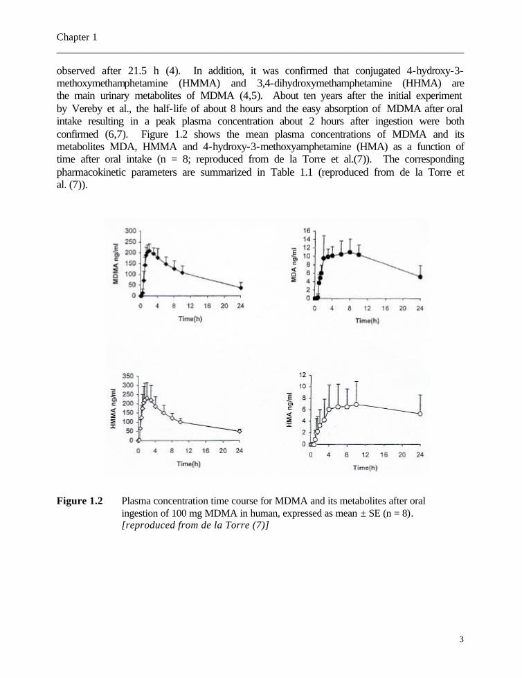
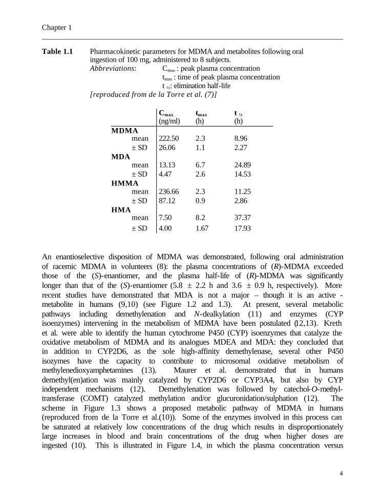
observed after 21.5 h (4). In addition, it was confirmed that conjugated 4-hydroxy-3-methoxymethamphetamine (HMMA) and 3,4-dihydroxymethamphetamine (HHMA) are the main urinary metabolites of MDMA (4,5). About ten years after the initial experiment by Vereby et al., the half-life of about 8 hours and the easy absorption of MDMA after oral intake resulting in a peak plasma concentration about 2 hours after ingestion were both confirmed (6,7). Figure 1.2 shows the mean plasma concentrations of MDMA and its metabolites MDA, HMMA and 4-hydroxy-3-methoxyamphetamine (HMA) as a function of time after oral intake (n = 8; reproduced from de la Torre et al.(7)). The corresponding pharmacokinetic parameters are summarized in Table 1.1 (reproduced from de la Torre et al. (7)). Figure 1.2 Plasma concentration time course for MDMA and its metabolites after oral
ingestion of 100 mg MDMA in human, expressed as mean ± SE (n = 8). [reproduced from de la Torre (7)]
Chapter 1 _____________________________________________________________________________
4
Table 1.1 Pharmacokinetic parameters for MDMA and metabolites following oral ingestion of 100 mg, administered to 8 subjects.
Abbreviations: Cmax : peak plasma concentration tmax : time of peak plasma concentration t ½: elimination half-life [reproduced from de la Torre et al. (7)]
Cmax tmax t ½ (ng/ml) (h) (h) MDMA mean 222.50 2.3 8.96 ± SD 26.06 1.1 2.27 MDA mean 13.13 6.7 24.89 ± SD 4.47 2.6 14.53 HMMA mean 236.66 2.3 11.25 ± SD 87.12 0.9 2.86 HMA mean 7.50 8.2 37.37 ± SD 4.00 1.67 17.93
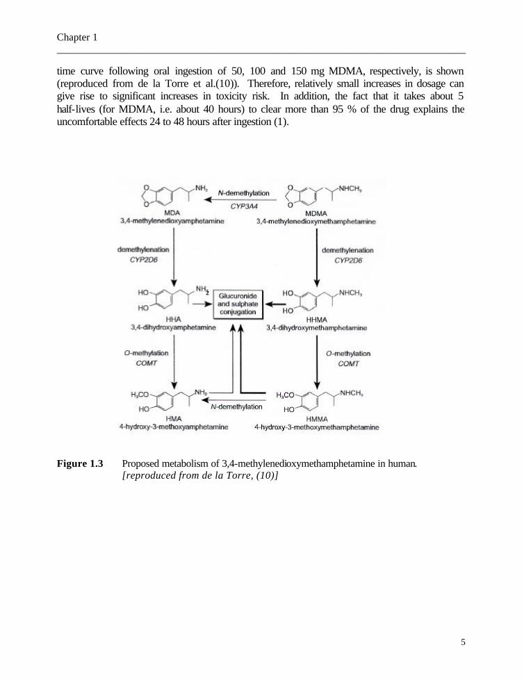
An enantioselective disposition of MDMA was demonstrated, following oral administration of racemic MDMA in volunteers (8): the plasma concentrations of (R)-MDMA exceeded those of the (S)-enantiomer, and the plasma half-life of (R)-MDMA was significantly longer than that of the (S)-enantiomer (5.8 ± 2.2 h and 3.6 ± 0.9 h, respectively). More recent studies have demonstrated that MDA is not a major – though it is an active - metabolite in humans (9,10) (see Figure 1.2 and 1.3). At present, several metabolic pathways including demethylenation and N-dealkylation (11) and enzymes (CYP isoenzymes) intervening in the metabolism of MDMA have been postulated (12,13). Kreth et al. were able to identify the human cytochrome P450 (CYP) isoenzymes that catalyze the oxidative metabolism of MDMA and its analogues MDEA and MDA: they concluded that in addition to CYP2D6, as the sole high-affinity demethylenase, several other P450 isozymes have the capacity to contribute to microsomal oxidative metabolism of methylenedioxyamphetamines (13). Maurer et al. demonstrated that in humans demethyl(en)ation was mainly catalyzed by CYP2D6 or CYP3A4, but also by CYP independent mechanisms (12). Demethylenation was followed by catechol-O-methyl-transferase (COMT) catalyzed methylation and/or glucuronidation/sulphation (12). The scheme in Figure 1.3 shows a proposed metabolic pathway of MDMA in humans (reproduced from de la Torre et al.(10)). Some of the enzymes involved in this process can be saturated at relatively low concentrations of the drug which results in disproportionately large increases in blood and brain concentrations of the drug when higher doses are ingested (10). This is illustrated in Figure 1.4, in which the plasma concentration versus
Chapter 1 _____________________________________________________________________________
5
time curve following oral ingestion of 50, 100 and 150 mg MDMA, respectively, is shown (reproduced from de la Torre et al.(10)). Therefore, relatively small increases in dosage can give rise to significant increases in toxicity risk. In addition, the fact that it takes about 5 half-lives (for MDMA, i.e. about 40 hours) to clear more than 95 % of the drug explains the uncomfortable effects 24 to 48 hours after ingestion (1). Figure 1.3 Proposed metabolism of 3,4-methylenedioxymethamphetamine in human. [reproduced from de la Torre, (10)]
2

Chapter 1 _____________________________________________________________________________
6
Figure 1.4 MDMA (+) and HMMA (? ) plasma concentrations (µg/l) versus time curve
in three subjects administered 50 mg, 100 mg, and 150 mg (one subject per dose). [reproduced from de la Torre(10)] Furthermore Kalant specifies that some of the metabolites of MDMA, especially its first metabolite MDA, are still pharmacologically active so that the duration of action of MDMA may be somewhat longer and therefore can in part explain the “delayed effects of MDMA” (1). Hernández-López et al. demonstrated that MDMA consumption in association with alcohol induced a 13 % increase in plasma concentrations of MDMA (14). The mechanism of this interaction is not known; a change in ethanol absorption or initial distribution was postulated, but the authors note that the changes in pharmacokinetics – though statistically significant - were mild in magnitude and therefore could be considered in the range of interindividual variability (14).

Chapter 1 _____________________________________________________________________________
7
The pharmacokinetic data of MDMA can be compared with those of amphetamine and methamphetamine. The half-life of amphetamine and (+)-methamphetamine is 7 – 34 h and 6 – 15 h, respectively. The renal excretion of both is dependent on the urinary pH. In the 24 h following intake, 30 % of unchanged amphetamine can be retrieved, but alkalinization of urine substantially decreases the fraction found, even to about 1%. To our knowledge, the influence of the urinary pH on the excretion of MDMA is not studied, though can be assumed. On the other hand, acidification of urine can result in retrieval of about 74 % of the initial dose (15). For S(+)-methamphetamine, the average elimination half-life in human volunteers (following oral administration) was 10.1 h (range 6.4 – 15.1 h) (16). At present, the above-mentioned pharmacokinetic parameters have not yet been fully elucidated for MDA, but the half-life for MDA is assumed to be longer than that of MDMA (17). Similarities have been noted in the metabolism of amphetamine-derived designer drugs: these substances undergo predominantly two overlapping metabolic pathways, namely O-demethylenation to dihydroxy derivatives (catechols), followed by methylation of one of the hydroxy groups, and successive degradation of the side chain to N-dealkyl and deaminooxo metabolites (18).
I.2 Pharmacodynamics of MDMA I.2.1 Animal experimental data As mentioned above, human experimental studies with MDMA give rise to
problems of medical ethics. Therefore, standardized animal models are required to solve some questions. However, it often remains difficult to extrapolate conclusions obtained from animal experiments to humans due to a variety of factors which influence the kinetics and metabolism of substances, such as species, strain, gender, route of administration, dose, frequency and time of administration, temperature, coadministration of drugs and surgical manipulation (19). The central nervous system is the predominant target site of MDMA and will therefore be discussed more in detail. In this section, only literature data with a clear link to the human clinico-pathological findings will be referred to.
I.2.1.1 Cardiovascular effects The sympathomimetic properties of MDMA resulting in increased heart rate and
blood pressure is hypothesized to be mediated by MDMA-induced monoamine release in both the central and the peripheral nervous systems, and perhaps also by the direct effect of MDMA on α2-adrenergic receptors (20,21). For methamphetamine, rat cardiomyocytes that are continuously exposed to a low concentration in vitro, become hypertrophic (22).
Chapter 1 _____________________________________________________________________________
8
I.2.1.2 Hepatotoxicity The mechanism of MDMA-induced liver injury has still not been clarified. In
experiments using rats, Beitia et al. described histological findings ranging from foci of individual cell necrosis to centrilobular necrosis and from mild to moderate lobular hepatitis (23). In addition, features of massive hepatic parenchymal collapse with areas of nodular regeneration can be observed (23). Following acute MDMA administration in rats, hepatocyte necrosis particularly in portal areas with inflammatory infiltrate consisting of lymphocytes and macrophages was found (23). Repeated intraperitoneal injection of MDMA in the rat produced hepatocyte necrosis and inflammatory infiltrate around the hepatic vein (23). In these animal experiments, there was no clear evidence that glutathione (GSH) depletion with free radical-induced toxicity is responsible for overt liver cell death (23). Hyperthermia-induced oxidative stress which comes to expression as GSH depletion was found in vitro following d-amphetamine exposure (24). In addition, catecholamines and hyperthermia were postulated to contribute to the mechanism of hepatotoxicity (24). Hyperthermia as a triggering factor for hepatotoxicity induced by MDMA was recently assumed by Carvalho et al. (25).
I.2.1.3 Cerebral effects and neurotoxicity On the basis of the clinical effects described in humans (see below) which
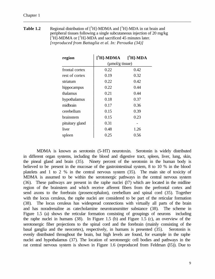
demonstrate that the brain is an important target organ, one would think that MDMA passes easily through the blood-brain barrier. However, the pharmacokinetics of MDMA with respect to the distribution into the brain are not yet elucidated. Due to the high pKa of this weak base (10.38) (26), MDMA is found totally in ionized form at physiological pH and, as a result, MDMA is in fact not expected to diffuse easily to the brain. Therefore, it may be that transport to the brain takes place via an active mechanism. For several substances such as anticonvulsants, efflux mechanisms protecting the homeostasis of the brain can significantly interfere with the functioning of these drugs (27). On the contrary, for MDMA, Mann et al. suggested that P-glycoprotein plays a facilitating role in the entry of this substance via the blood-brain barrier (28). The rapid partitioning of (+)-methamphetamine which is closely related to MDMA in the rat brain, can also partially be explained in terms of physicochemical properties (such as small molecular weight) of that substance (29), and therefore in view of the chemical structure of MDMA (see Figure 1.1), this may also be applicable to MDMA. In addition, data from rats indicate that metabolites of MDMA (such as glutathione conjugates) enter the brain via a transporter and are subsequently metabolized to thioether conjugates, which contribute to the serotonergic neurotoxicity (30,31); thus, the metabolite HHMA may play a role in the neurotoxicity (31-33). In rats, the regional distribution of [3H]-MDMA and [3H]-MDA in brain and a few peripheral tissues was studied following a single subcutaneous injection of 20 mg/kg [3H]-MDMA or [3H]-MDA. Table 1.2 shows that the distribution of MDMA and MDA was fairly comparable in all brain regions. The highest levels were found in the liver and the pituitary gland (34).
Chapter 1 _____________________________________________________________________________
9
Table 1.2 Regional distribution of [3H]-MDMA and [3H]-MDA in rat brain and peripheral tissues following a single subcutaneous injection of 20 mg/kg [3H]-MDMA or [3H]-MDA and sacrificed 45 minutes later. [reproduced from Battaglia et al. In: Peroutka (34)]
region [3H]-MDMA [3H]-MDA (µmol/g tissue) frontal cortex 0.22 0.42 rest of cortex 0.19 0.32 striatum 0.22 0.42 hippocampus 0.22 0.44 thalamus 0.21 0.44 hypothalamus 0.18 0.37 midbrain 0.17 0.36 cerebellum 0.15 0.39 brainstem 0.15 0.23 pituitary gland 0.31 - liver 0.48 1.26 spleen 0.25 0.56
MDMA is known as serotonin (5-HT) neurotoxin. Serotonin is widely distributed
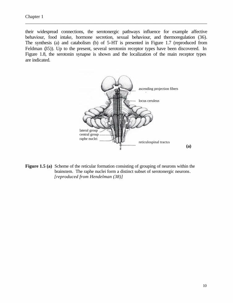
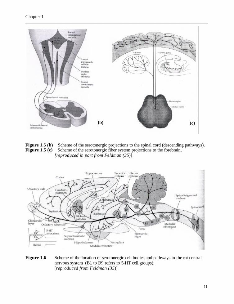
in different organ systems, including the blood and digestive tract, spleen, liver, lung, skin, the pineal gland and brain (35). Ninety percent of the serotonin in the human body is believed to be present in the mucosae of the gastrointestinal system, 8 to 10 % in the blood platelets and 1 to 2 % in the central nervous system (35). The main site of toxicity of MDMA is assumed to be within the serotonergic pathways in the central nervous system (36). These pathways are present in the raphe nuclei (37) which are located in the midline region of the brainstem and which receive afferent fibers from the prefrontal cortex and send axons to the forebrain (prosencephalon), cerebellum and spinal cord (35). Together with the locus ceruleus, the raphe nuclei are considered to be part of the reticular formation (38). The locus ceruleus has widespread connections with virtually all parts of the brain and has noradrenaline as catecholamine neurotransmitter substance (38). The scheme in Figure 1.5 (a) shows the reticular formation consisting of groupings of neurons including the raphe nuclei in humans (38). In Figure 1.5 (b) and Figure 1.5 (c), an overview of the serotonergic fiber projections to the spinal cord and the forebrain (mainly consisting of the basal ganglia and the neocortex), respectively, in humans is presented (35). Serotonin is evenly distributed throughout the brain, but high levels are found, for example in the raphe nuclei and hypothalamus (37). The location of serotonergic cell bodies and pathways in the rat central nervous system is shown in Figure 1.6 (reproduced from Feldman (35)). Due to
Chapter 1 _____________________________________________________________________________
10
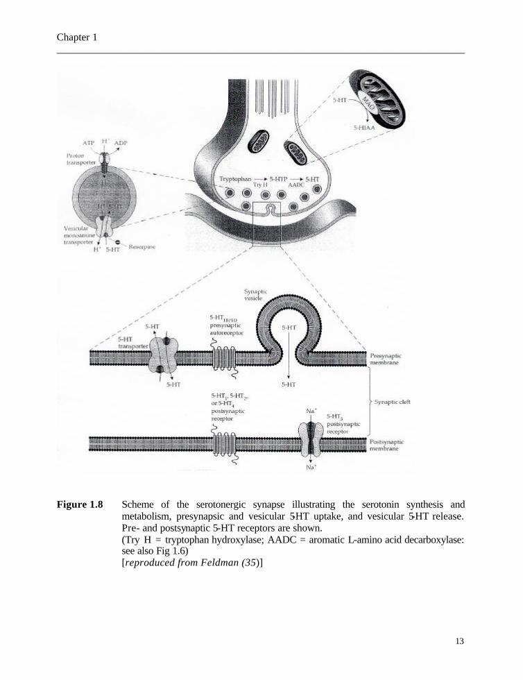
their widespread connections, the serotonergic pathways influence for example affective behaviour, food intake, hormone secretion, sexual behaviour, and thermoregulation (36). The synthesis (a) and catabolism (b) of 5-HT is presented in Figure 1.7 (reproduced from Feldman (35)). Up to the present, several serotonin receptor types have been discovered. In Figure 1.8, the serotonin synapse is shown and the localization of the main receptor types are indicated. Figure 1.5 (a) Scheme of the reticular formation consisting of grouping of neurons within the
brainstem. The raphe nuclei form a distinct subset of serotonergic neurons. [reproduced from Hendelman (38)]
(a)
ascending projection fibers
locus ceruleus
reticulospinal tractus
lateral group central group raphe nuclei
Chapter 1 _____________________________________________________________________________
11
Figure 1.5 (b) Scheme of the serotonergic projections to the spinal cord (descending pathways). Figure 1.5 (c) Scheme of the serotonergic fiber system projections to the forebrain.
[reproduced in part from Feldman (35)]
Figure 1.6 Scheme of the location of serotonergic cell bodies and pathways in the rat central
nervous system (B1 to B9 refers to 5-HT cell groups). [reproduced from Feldman (35)]
(b) (c)

Chapter 1 _____________________________________________________________________________
12
(a) (b) Figure 1.7 Synthesis (a) and catabolism (b) of serotonin (5-HT). The enzymes catalyzing the
reactions and the interfering co-factors are indicated. [reproduced from Feldman (35)]
Chapter 1 _____________________________________________________________________________
13
Figure 1.8 Scheme of the serotonergic synapse illustrating the serotonin synthesis and
metabolism, presynapsic and vesicular 5-HT uptake, and vesicular 5-HT release. Pre- and postsynaptic 5-HT receptors are shown. (Try H = tryptophan hydroxylase; AADC = aromatic L-amino acid decarboxylase: see also Fig 1.6)
[reproduced from Feldman (35)]

Chapter 1 _____________________________________________________________________________
14
Animal experiments showed that MDMA induced serotonin neurotoxicity can be manifested by following mechanisms: reduced cerebral 5-HT content, decreased numbers of identifiable 5-HT-uptake sites and transporter molecules, reduced activity of tryptophan hydroxylase (TPH ; the rate-limiting enzyme in the 5-HT synthetic pathway), and degenerating cerebral serotonergic axons and axon terminals (1,39,40).
Frederick et al. observed altered behavioural effects (including memory and attention) in rhesus monkeys (41). These effects were associated with significant decreases of about 50% in serotonin levels in frontal cortex and hippocampus approximately six months after a short-course high-dose MDMA treatment (41). In rats, changes in 5-HT levels in other regions were demonstrated, such as in the nucleus accumbens and striatum (42), and in the raphe nuclei (43). The nucleus accumbens contains neurons that are part of the basal ganglia. Its function has not yet been elucidated but it is assumed to be involved in integrating certain cognitive aspects of a situation with the emotional component, and in addiction behaviour in animals - and likely in humans as well (44). The human basal ganglia and the nucleus accumbens are presented in Figure 1.9 (reproduced from Hendelman (44)).
Figure 1.9 Schematic view of the various nuclei located in the basal forebrain area including the basal ganglia .
[reproduced from Hendelman (44)]
Chapter 1 _____________________________________________________________________________
15
Regional differences in serotonin neurotoxicity have been reported: e.g. the number of cortical 5-HT uptake sites in rats (measured by specific binding to the transporter) completely recovered at 52 weeks post-treatment, while at the same time the hippocampal 5-HT uptake sites were still significantly decreased (45). The effects of MDMA on 5-HT neurons in specific neuroanatomic loci were studied in the rat using autoradiography. Marked decreases in 5-HT uptake sites in several regions known to receive projections of 5-HT neurons, such as the cerebral cortex, caudate nucleus, hippocampus, and most thalamic nuclei, were observed (46,47).
The underlying mechanism that would explain neuronal cell death is still not yet fully understood, but a few hypotheses have been presented, including hydroxy radical formation (48), and tryptophan hydroxylase inactivation, for instance by increasing the intracellular calcium ion concentration (49). Huether et al. postulated a profound wastage of energy on a 5-HT cellular basis (50).
Histological and immunohistochemical evidence of the degeneration of serotonergic axons has been reported (51-54). In addition, dopamine (DA) is believed to play an unmistakable role in MDMA-induced damage to 5-HT axons (55-57).
When considering the serotonergic neurotoxicity of MDMA, the more pronounced sensitivity of monkeys compared to rats was demonstrated by neurochemical and neurohistological experiments (58). This could indicate species-dependent differences to MDMA-induced toxic effects.
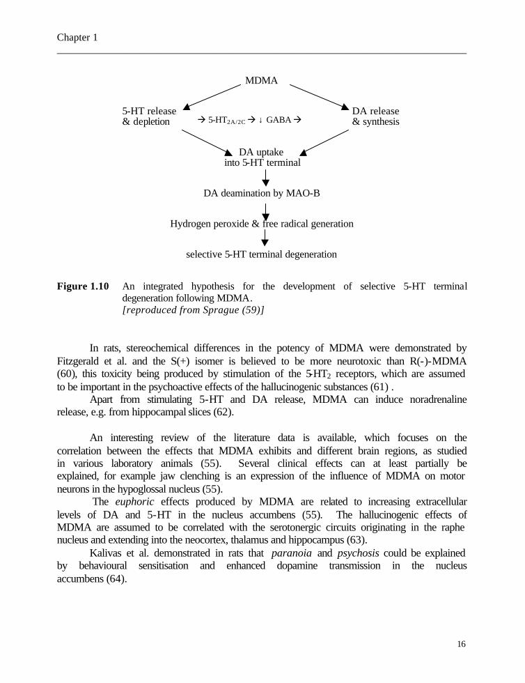
Figure 1.10 shows the integrated hypothesis as explanation for the serotonergic neurotoxicity proposed by Sprague et al. (59). The authors describe the following sequence: MDMA induces an acute release of 5-HT and DA, which is followed by depletion of intraneuronal 5-HT stores. Thereafter, the initially released 5-HT activates post-synaptic 5-HT2A/2C receptors located on γ-aminobutyric acid (GABA) interneurons, resulting in a decrease in GABA-ergic transmission and increased DA release and synthesis. The excessive DA released may then be transported into the depleted 5-HT terminal. The DA is then deaminated by monoamine oxidase B (MAO-B) located within the 5-HT terminal. This results in free-radical formation and the selective degeneration of the serotonergic axons and axon terminals (59).
Chapter 1 _____________________________________________________________________________
16
MDMA
5-HT release DA release & depletion & synthesis
DA uptake into 5-HT terminal
DA deamination by MAO-B
Hydrogen peroxide & free radical generation
selective 5-HT terminal degeneration
Figure 1.10 An integrated hypothesis for the development of selective 5-HT terminal degeneration following MDMA. [reproduced from Sprague (59)]
In rats, stereochemical differences in the potency of MDMA were demonstrated by
Fitzgerald et al. and the S(+) isomer is believed to be more neurotoxic than R(-)-MDMA (60), this toxicity being produced by stimulation of the 5-HT2 receptors, which are assumed to be important in the psychoactive effects of the hallucinogenic substances (61) .
Apart from stimulating 5-HT and DA release, MDMA can induce noradrenaline release, e.g. from hippocampal slices (62).
An interesting review of the literature data is available, which focuses on the
correlation between the effects that MDMA exhibits and different brain regions, as studied in various laboratory animals (55). Several clinical effects can at least partially be explained, for example jaw clenching is an expression of the influence of MDMA on motor neurons in the hypoglossal nucleus (55).
The euphoric effects produced by MDMA are related to increasing extracellular levels of DA and 5-HT in the nucleus accumbens (55). The hallucinogenic effects of MDMA are assumed to be correlated with the serotonergic circuits originating in the raphe nucleus and extending into the neocortex, thalamus and hippocampus (63).
Kalivas et al. demonstrated in rats that paranoia and psychosis could be explained by behavioural sensitisation and enhanced dopamine transmission in the nucleus accumbens (64).
à 5-HT2A/2C à ↓ GABA à
Chapter 1 _____________________________________________________________________________
17
Callaway et al. demonstrated that locomotor hyperactivity induced by MDMA administration in rats was dependent upon serotonin (5-HT) rather than dopamine release and therefore a central role for 5-HT release in the stimulant-like behavioural effects of MDMA was assumed (65). In addition, McCreary et al. supported a role for the 5-HT1B/1D receptor in mediating acute hyperactivity induced by (+)-MDMA (66).
Moreover, MDMA-induced hyperthermia and locomotor hyperactivity in laboratory animals can be inhibited by drugs that prevent MDMA-induced 5-HT release and can be attenuated by administering 5-HT receptor antagonists (55). Pederson et al. proved in a rabbit animal model, that sympathetically mediated cutaneous vasoconstriction is one mechanism contributing to MDMA-induced hyperthermia (67). Therefore, drugs acting as 5-HT2A receptor antagonists (such as clozapine) can be therapeutically important in treating severe life-threatening hyperthermia (67). Darvesh et al. proposed - as possible mechanism important for the development of hyperthermia - the influence of MDMA on brain energy regulation: they were able to demonstrate MDMA-induced glycogenolysis in rat brain and that this process involves 5-HT2 receptor activation. Therefore, they concluded that MDMA promotes energy dysregulation and that hyperthermia may be an expression of MDMA-induced alterations in cellular energetics (68). In rats, Mechan et al. demonstrated that MDMA-induced hyperthermia could rather be explained by the increased release of dopamine acting at D1 receptors than by 5-HT release as such (69). As dopamine and serotonin are important mediators of body temperature - lowering and raising, respectively - drugs with antidopaminergic or serotonin releasing properties can be responsible for hyperthermia syndromes (70). By this means, MDMA-induced hyperthermia can be partially understood. Malpass et al. proposed that, being cytochrome P-450 2D6 deficient, human poor metabolizers may be genetically predisposed towards a fatal outcome, but comparison of deficient and normal rats revealed that this cannot be explained by a simple increased hyperthermic response to the drug (71). Malberg demonstrated in rats that high ambient temperatures are required to induce neurotoxicity, and therefore ambient temperature has a significant influence on MDMA-induced neurotoxicity, body temperature and thus thermoregulation (72). In mice, Carvalho et al. were able to support the hypothesis that oxidative stress is important in the first stage of MDMA-induced liver damage and that liver antioxidant status is deteriorated by high ambient temperature. Therefore they concluded that increased ambient temperature may potentiate MDMA-induced hepatotoxicity by increasing body hyperthermia (73). This confirms that in humans, promoting environmental conditions are important (such as high ambient temperature in dancings) to induce toxicity (1,74).
I.2.2 Desired effects in humans First we summarize the desired clinical effects in recreational use. Minor and
severe adverse effects are discussed thereafter (see I.2.3). MDMA is sometimes classified as a hallucinogen, though it can also be classed with
the central stimulants. In view of the psycho-pharmacological effects, MDMA can be rated among the “entactogens”, a term which refers to the feeling of enhanced closeness and communication with others (75-77).

Chapter 1 _____________________________________________________________________________
18
The typical dosage range for recreational use of MDMA is 50 to 150 mg (1). However, the content of an “ecstasy” tablet may vary enormously: different amphetamines can be found and the amount of MDMA can vary significantly. For example, an examination of tablets sold as “ecstasy” revealed that only about the half of them actually contained MDMA and the mean content was 91.3 mg with a wide range (2 – 149 mg) (78). The clinical effects of MDMA after oral ingestion – which is the most common route of administration in recreational use – start at about 20 to 60 minutes. Initially, the user experiences a brief “rush” of energy, which is often described as mild, but euphoric. This “rush” is followed by a more comfortable episode lasting 2 to 3 hours which is then followed by a gradual “coming down” sensation or feeling of fatigue (79). Questionnaires for the purpose of obtaining more information about the desired effects of MDMA use revealed that physically, MDMA produces a feeling of increased alertness, energy, and sexual arousal (80). Psychologically an increased feeling of “closeness” and “peace” with other people, well-being, and euphoria, were commonly mentioned (76,80). These feelings of increased empathy gave rise to the name “entactogens” or “empathogens” (77). In other words, the primary reported effects of MDMA are a “positive mood state” and feelings of intimacy and closeness to others (81). The secondary effects described refer to the stimulant properties - namely feelings of energy and activation - and to the psychedelic effects of insight and perceptual and sensual enhancement (81).
Hallucinogenic effects have only been described following ingestion of high doses (82). Gender differences have been reported: women were found to be more susceptible (for example to hallucinogenic-like perceptions) than men (83). In addition, MDMA has a much shorter action than MDA, which is known to cause hallucinogenic effects similar to those of mescaline or LSD. Therefore, it can be generally assumed that the additional N-methyl group in the chemical structure of MDMA limits the duration of action and attenuates or even abolishes the hallucinogenic properties described after MDA use (75,77).
Combining MDMA with alcohol may result into a longer lasting euphoria and sense of well being, and may partially reverse the subjective alcohol-induced sedation, but not reduce drunkenness feelings (14).
I.2.3 Human toxicity Important interindividual differences exist between MDMA plasma concentrations
and the clinical symptoms, and as a result, adverse effects are correlated not only with the ingested amount. Side effects are due mainly to the sympathomimetic and/or the neuro-toxicological effects of MDMA. As tolerance to the effects of MDMA develops rapidly, more frequent use requires larger doses to achieve the desired effects, but then the unpleasant side-effects increase as well (81,84). MDMA is generally considered as non-addictive, although some cases of dependence are described (85).
In this section, we first describe relatively minor acute and chronic adverse effects (most of which were noticed clinically) of MDMA use, classified by organ system. In addition, more pronounced adverse reactions in some cases involving life-threatening effects are commented upon. Finally, the correlation between the MDMA plasma or blood concentration and the observed effects is discussed.

Chapter 1 _____________________________________________________________________________
19
I.2.3.1 Symptoms I.2.3.1.1 Cardiovascular effects Frequent acute, relatively minor unpleasant effects of MDMA – which indicate the
sympathomimetic involvement – include tachycardia, palpitations, hypertension, mydriasis, and dry mouth (6,7). In addition, gender differences have been observed following MDMA exposure: e.g. men showed higher increases in blood pressure than women (83).
Hypertensive crises and cardiac dysrhythmia (like ventricular tachy-arythmias) are commonly reported acute severe cardiovascular symptoms (86). Hypertensive crises may cause cerebrovascular accidents and other complications from end-organ vasospasms. A few cases of cerebral haemorrhage or infarction following MDMA intake were reported (87-92). Intracerebral haemorrhage was also described in amphetamine and cocaine users (93,92). Hypertensive surges and cerebral angiitis have been postulated as mechanisms causing intracerebral haemorrhage following amphetamine and methamphetamine use (89).
Both, severe hypertension with increased risk for haemorrhages on the one hand, and tachycardia or cardiac dysrythmia on the other hand, can develop into heart failure (1). I.2.3.1.2 Hepatotoxicity
There is a broad spectrum of hepatotoxic effects induced by MDMA: jaundice, hepatomegaly, hepatitis and extensive fibrosis (87-97). In young people presenting with unexplained jaundice or hepatomegaly, questions regarding (mis)use of MDMA should be posed (87). The interval between drug consumption and jaundice is variable and therefore the link between the two can be obscured (98). Hepatitis is the most frequently reported manifestation of MDMA induced liver damage (98-101). In most of the reported hepatitis cases, biochemical tests for viral hepatitis are negative. Rarely, hepatitis due to MDMA exposure can result in fulminant hepatic failure (102) which can require liver transplantation (103).
Proposed hypotheses to explain the hepatotoxic effects include an allergic drug reaction (such as idiosyncratic toxic hepatitis), a toxic contaminant, autosomal recessive inheritance of gene mutations (lack of cytochrome P450 oxidase CYP2D6) resulting in impaired metabolism of the drug, or a secondary effect of hyperpyrexia (87,96,104). Whether an idiosyncratic toxic hepatitis is due to MDMA itself or to a metabolite, a contaminant in MDMA manufacture, or to an additive in the tablets is not yet clear (87). In the case reported by Khakoo, an idiosyncratic reaction was assumed to be the underlying mechanism of ecstasy-induced accelerated hepatic fibrosis (including a predominantly eosinophilic inflammatory infiltrate; 95). Schwab et al. believing that inherited CYP2D6 deficiency is unrelated to MDMA-induced hepatotoxicity, suggested an idiosyncratic reaction because there is no correlation between the severity of liver damage and either the amount of MDMA ingested or the frequency of MDMA use (105). A fatality presenting with hyperthermia and fulminant liver failure – which originated following a single ingestion of one MDMA tablet – was reported (106).
Chapter 1 _____________________________________________________________________________
20
I.2.3.1.3 Central nervous system effects Relatively minor adverse sequelae experienced during the 24 hours following
MDMA ingestion, include lack of energy and appetite, insomnia, jaw clenching, occasional concentration problems, brooding (108). Other reported side-effects are tremor, diaphoresis, trismus (tight jaw) and bruxism (jaw clenching), impaired gait, and restless legs (107,108). Rebound depression and lethargy has been reported in about 80 % of the subjects, in the days following MDMA use, due probably to monoaminergic depletion (109). Following short-term administration of MDMA, a slight impairment in the performance of psychomotor tasks was noticed (110).
Hyperthermia is one of the most feared acute toxic life-threatening complications of MDMA exposure. Biochemical analyses indicating metabolic acidosis, increased creatine kinase activity and hyperkalaemia are compatible with hyperthermia. It has been postulated that dehydration could precipitate MDMA-induced hyperthermia (86). Dysregulation of the thermoregulatory center is promoted when profuse sweating and intense physical activity in a hot environment occurs (1). Hyperthermia as part of a MDMA-induced serotonin syndrome has been postulated (111). Neurotoxicological effects can also manifest themselves indirectly: e.g. signs of multiple organ failure such as acute hepatic or renal failure due to hyperthermia.
It is not excluded that excessive drinking of water following MDMA ingestion can result in dangerous hyponatraemia (112,113) and cerebral oedema (114,115) which can develop into coma (116) and death (117). The mechanism by which excessive fluid consumption occurs is not yet understood. An additional mechanism that can aggravate hemodilution and hyponatraemia is the inappropriate secretion of antidiuretic hormone (118,119).
The long-lasting effects of MDMA, even after abstinence, are not yet completely understood. A few reported cases suggest that these effects should not be underestimated. Verbal and visual memory impairment in abstinent MDMA users (120-124), and long-term memory problems related to storage and retrieval difficulties (125) were reported. In addition, in chronic MDMA users - followed over the course of one year – progressive decline in terms of immediate and delayed recall were noticed (126). The extent of the impairment correlated with the degree of MDMA exposure and the decrements in 5-hydroxyindoleacetic acid [5-HIAA; metabolite of 5-HT (or serotonin)] concentrations determined in cerebrospinal fluid (120).
A case of pure amnesic syndrome after ingestion of half an MDMA tablet was reported and brain magnetic resonance imaging disclosed symmetric lesions in the globus pallidus (which were clinically silent) (127). Spatt et al. assumed alterations in the hippocampi as cause of persistent memory problems in their case (127).
There have been several reports of lasting adverse neuropsychiatric sequelae in humans who have chronically ingested (usually high) doses of MDMA. Moreover, it was suggested that individuals with prior psychiatric medical history can be more susceptible to MDMA’s adverse effects, such as acute (128) and chronic (129,130) paranoid psychosis, panic attacks (131), panic disorder with secondary depression (132) and depression with suicidal behaviour (133). In other words, chronic MDMA use may be associated with a
Chapter 1 _____________________________________________________________________________
21
broad spectrum of psychiatric morbidity (134-136). Neuropsychiatric signs have also been reported following single or brief MDMA use such as panic disorder (137) and prolonged psychosis (138,139). As there is evidence that serotonin (5-HT) has a role in mediating antipsychotic drug effects (140), the involvement of 5-HT into the psychotomimetic and psychotogenic properties of MDMA can be assumed. In addition - similar to the observation in memory impairment (120) - a decreased concentration of the 5-HT metabolite 5-HIAA in cerebrospinal fluid - as an index of brain monoaminergic function – was found in MDMA users (141,142).
Brain-imaging studies are provided in the last few years to investigate the neurotoxic effects of MDMA. For example, a reduced density of 5-HT uptake sites in several brain regions of MDMA users was found, as well as deficits in brain 5-HT transporter molecules (143,144) and altered blood flow in certain parts of the brain (145). Single-photon emission CT studies suggest that MDMA users may be at risk for cerebrovascular accidents due to alterations in the 5-HT-neurotransmission system (down-regulation of 5-HT2-receptors implicating vasoconstriction) (146). A case of toxic leukencephalopathy following a single MDMA use – confirmed by computed tomography and magnetic resonance – has been reported; the dose ingested neither a blood or plasma MDMA level were available (147). Damage of serotonergic afferents could possibly mediate long-lasting alterations of cerebral glucose metabolism as a secondary effect (148). Moreover, a reduction in brain glucose metabolic uptake has been noted, for example, in the hippocampus of regular users (149). Thus memory deficits in MDMA users could possibly be explained on the basis of alteration of the hippocampal function by MDMA. The gender difference, namely that women might be more susceptible to the neurotoxic effects of MDMA, was also noticed by means of single-photon-emission computed tomography (SPECT) (150). Brain anomalies including cerebellar atrophy and thalamic dysfunction have been proven using imaging techniques such as magnetic resonance, but it is difficult to distinguish the relationship of these lesions to the effect of MDMA, on the one hand, and hypoxia and ischaemia, on the other (168).
I.2.3.1.4 Uro-genital effects As MDMA is a potent α-adrenergic agonist, acute urinary retention can occur, and
therefore MDMA (ab)use should be considered in young people presenting with unexplained acute urinary retention (151).
Acute renal failure is often described in MDMA-related multiple organ failure originating from hyperthermia with rhabdomyolysis (86,152). Sometimes haemodialysis is required (87). However, it has been hypothesized that rhabdomyolysis - which is a common finding secondary to hyperthermia - may originate from direct drug toxicity (153).
Chapter 1 _____________________________________________________________________________
22
I.2.3.1.5 Various symptoms Spontaneous pneumomediastinum - which is usually not life-threatening, but can
require medical attention - is a rare complication after MDMA abuse (154-156). Levine et al. postulated an increased intrathoracic pressure due to vomiting as a possible mechanism (154). Pittman et al. proposed that the pneumomediastinum was caused by repeated Valsalva type maneuvers that resulted in episodes of increased intra-alveolar pressure, because their subject was repeatedly blowing a whistle during an eight-hour dancing session (155). In the case reported by Quin, none of these mechanisms were present and therefore the authors concluded that the nature of physical exertion accompanying ecstasy intoxication led to the causative barotrauma (156).
A few reports of aplastic anaemia following exposure to MDMA, which can be fatal, were published (157).
In addition, a few cases of keratopathy after MDMA ingestion were reported (158); the mechanism of this corneal epitheliopathy remains unexplained.
I.2.3.1.6 Multiple organ failure The above-mentioned commonly observed severe acute toxic effects such as
hyperthermia, metabolic disturbances, seizures, hypertensive crises, cardiac dysrhythmia, and cerebrovascular accidents can escalate in severity and result in multiple organ failure. Feared complications include rhabdomyolysis, disseminated intravascular coagulation (DIC), adult respiratory distress syndrome (ARDS) and acute renal failure. Hepatic failure is often part of multiple organ failure originating from hyperthermia. Multiple organ failure is often the mechanism of death, even when intensive monitoring and therapy is performed (see below: IV.1.2. and IV.2.). Figure 1.11 presents an integrated scheme of the mechanisms which can be involved in major MDMA-induced complications such as multiple organ failure (159). For example, the complex cascade occurring in the clinical pattern in which MDMA ingestion leads to prolonged hyperactivity and hypovolaemia (resulting from insufficient volume repletion), which are important factors in the development of hyperthermia is shown. In addition, hyperthermia triggers DIC (see Figure 1.11).

Chapter 1 _____________________________________________________________________________
23
MDMA
hyperactivity convulsions hyperthermia volume depletion rhabdomyolysis DIC bleeding stroke ARDS cardiovascular acute renal failure collapse Figure 1.11 Mechanisms by which MDMA may produce major complications. Hyperthermia is
more commonly an indirect effect, mediated through hyperactivity, rather than a direct pharmacological effect. Abbreviations: DIC: disseminated intravascular coagulation
ARDS: adult respiratory distress syndrome [reproduced from Henry JA In: Hopkins & Ellis (159)]
I.2.3.2 Correlation between blood levels and clinical effects Important inter-individual differences in effects and adverse reactions following
MDMA ingestion have been noted. Therefore, the toxicity of MDMA in humans is an object of current debate (160). It has been postulated that the striking inter-individual differences in intensity, time course and toxicity may be related to individual differences in the metabolic handling of the MDMA isomers (1). Moreover, as cytochrome P450 enzymes are important in the metabolism of MDMA (12), persons who have a genetic defect of these enzymes - and therefore are poor metabolizers – may be particularly sensitive to MDMA and hence be at more risk of toxicity (157,161). In addition, referring to the illicit source of MDMA, it cannot be excluded that unpleasant side-effects or even toxicity are – at least partially – mediated by contaminants (162).
In fatalities solely following MDMA ingestion, a wide range of blood levels has been reported with values ranging from 0.04 to 18.50 µg/ml (94,163, see also below). This inter-individual difference is illustrated by Randall: a patient with an MDMA plasma level of 7.72 µg/ml after ingestion of 42 tablets only complained of “hangover” with tachycardia and hypertension (164). To our knowledge, there are no data available considering the
Chapter 1 _____________________________________________________________________________
24
possible effect of tolerance in chronic MDMA abuse, which could be an explanation for this very high plasma level with minor clinical problems.
A few rare cases of exceptional survival after high doses of MDMA have been described indicating the unclear relationship between a specific blood or plasma level and the clinical effects. Brown et al. reported a nearly fatal case following MDMA intake: 1 to 2 hours after admission to hospital, MDMA plasma levels of 6.5 and 7.0 µg/ml, respectively, were found (165). Roberts et al. reported a subject who presented at the emergency department with a core temperature of 38.6 ° C, sweating profusely, vomiting and irritable with subsequent convulsions and respiratory problems resulting in the need for intubation. A plasma MDMA level of 4.05 µg/ml - after intentional ingestion of 18 tablets – has been reported. The patient recovered within one week, however, although he admitted being forgetful, irritable and having flashbacks to events immediately prior to losing consciousness (166). Ramcharan et al. described a case in which unconsciousness, apnea and convulsions developed after intake of 50 tablets, but recovery occurred within 2 days (74). Unfortunately in this case, an MDMA blood or plasma level determination was not available. The subject published by Mallick et al. ingested three tablets of ecstasy and survived the subsequent hyperpyrexia (42.9 °C) which included convulsions, rhabdomyolysis, metabolic acidosis and respiratory failure, but unfortunately, no MDMA blood or plasma level is available for this case either (167).
It should be emphasized that polydrug abuse makes this toxicological discussion even more hazardous. A woman who suffered from a DIC and a brief cardiac arrest following the combined intake of MDMA, amyl nitrite, lysergic acid diethylamide (LSD), cannabis and alcohol, developed an amnesic syndrome and severe ataxia (168). This question becomes even more complicated to solve when different amphetamines have been taken together. For example, Agaba et al. reported a case presenting with massive intracerebral hematoma and extradural hematoma after “amphetamine” ingestion (169): the composition of the tablets taken was not known, but a combination of pure amphetamine and MDMA was postulated and therefore the impact of the two on the symptoms is not clear (169). This remark can also be applied to the case presenting with chronic renal failure (due to necrotizing vasculopathy) after ingestion of methamphetamine and MDMA (170). II Epidemiological data on the use of MDMA
There are no clear epidemiological data on (ab)use of amphetamine and derivatives. This is probably due to the fact that there are no specific international directives mandating systematic screening for these substances. This also holds for Europe, though efforts have been made to map the use of drugs among the population, including the use of amphetamines and “ecstasy”. In addition, guidelines have been presented for ensuring quality and for comparing the studies performed in the various European member states (171).
The information available is derived from random sampling studies or clinical trials.

Chapter 1 _____________________________________________________________________________
25
II.1 Europe II.1.1 Belgium Epidemiological data on MDMA use in Belgium are scarce, although such data are
being collected by the European Monitoring Centre for Drugs and Drug Addiction (EMCDDA). The use of amphetamines and “ecstasy” in Belgium shows the same evolution as in the other European countries: an obvious increase was observed between 1994 and 1998 in the numbers of 15 to 16 years-old who have ever used MDMA (from 4.1 to 6.2 %) and from then on a plateau phase was recorded (172). In addition, the lifetime prevalence of MDMA and amphetamines for young people aged 15-16 and 17-18 is higher in the French Community than in the Flemish Community (173). Recently, the drug-related medical problems due to the use of recreational drugs during two famous events (‘I love techno’ and ‘De Nacht’) were studied: in the patients evaluated in an emergency department (Ghent), the dominant drug abused was ecstasy (174).
II.1.2 Other European countries In England, an informal survey of undergraduates more than 10 years ago revealed
that about 40 % admitted having used MDMA at least once (76,175). The frequency of use by the subjects varied significantly, and ranged from 1 to 38 times. The median amount of MDMA usage reported by these undergraduates was four doses, while the mean number of doses taken was 5.4. The amount of drug taken in a single dosage ranged from 60 to 250 mg (approximately 1 to 4 mg/per kilogram bodyweight) (175).
In the European studies, amphetamine and MDMA use is usually classified as being part of a subgroup such as the “synthetic drugs” or “hallucinogens” (171). It has been noted that the spread of synthetic drugs in the European Union has stabilized, though the use of ecstasy is still increasing in certain locations (cities, holiday resorts, youth cultures) (171-177). It was reported that in 1998 0.5 – 3 % of the adult European population (mainly young people, of course) had at one time or another used ecstasy (178). In the Health Behaviour of School-aged Children (HBSC) survey, repeated community surveys are performed in schools among young people aged 15 to 18 years: cannabis has been found to be the most popular drug, though until 1999 MDMA was the second most used product; since then a trend toward stabilisation - or perhaps even a slight decrease - has become apparent (173).
More recently, in the UK, the ecstasy-induced death rate in 1996 per 10 000 15-24-year-old users was defined as between 0.2 and 5.3; the death-rate from road traffic accidents in the same age-group was 1.0 (179).
Non-published data in Switzerland indicate that the age of the subjects at first consumption was obviously lower in 1999 than 1996. Forty-five percent of first-time consumers were 17 years of age or younger in 1999, whereas in 1996, about 28 % were younger than 18 (180).
A school-based survey in Oslo of adolescents between 14 and 17 years of age disclosed that ecstasy is used in a polydrug-use pattern (181).
An overview of the available epidemiological data in Italy and other European countries is presented by Schifano (182).
Chapter 1 _____________________________________________________________________________
26
II.2 USA and the rest of the world Surveys in the USA examining the prevalence of MDMA use among high school
sophomores and seniors revealed increasing use in the 1990s (183). For example, for seniors in 1999, lifetime prevalence of MDMA use had risen from 5.0 % to 8.0 % in the previous three years (183 ). A study performed in 119 US colleges (involving a study population of over 14,000 students) revealed an increase of prevalence from 2.8 to 4.7 % between 1997 and 1999 (184). Furthermore, the results obtained in a smaller sample of 10 colleges showed that the increase continued in 2000 (184).
Just as in the European countries, an increase in MDMA use was noted among rave club attendees (185).
To our knowledge, there are no epidemiological data available concerning MDMA use in Australia or the Asian countries although a few fatal cases have been described (163, 186).
The MDMA fatalities reported in the international literature are summarized below (see pag 33- 41).
III Medico-legal implications of MDMA The above-mentioned behavioural and cognitive effects of MDMA can result into
non-lethal accidents, suicidal behaviour or even crimes. As for all drug fatalities, the manner of death can be related to accident, suicide or criminal activity. At present, the risks of MDMA (ab)use for humans – to the user him/herself and to his/her acquaintances – are difficult to define (160).
III.1 Accidents It is difficult, if not impossible, to determine the boundary between the desired
effects, such as euphoria, and feelings of closeness, and the adverse effects, such as impairment of cognition and co-ordination and - in an advanced phase – hallucinations, agitation, abnormal behaviour, and even psychosis. The majority of the reported intoxications are “accidental”, being due to the higher sensitivity of a subject to a “normal” recreational dose, though severe adverse effects can sometimes occur due to the content of certain tablets (which can be of purer quality than one is accustomed to ingest). Furthermore, it is generally accepted that the use of amphetamines and their derivatives can increase the risk of being involved in accidents, though the possible increased risk has not yet been established (187).
Over a three-month period, 16 ecstasy abusers were treated in a single emergency department for injuries due to traffic accidents; all of which had been caused by reckless driving (188). From 1995 through mid-1996, a prospective multicentre study was conducted in hospital emergency departments in Belgium in order to investigate the drug-related traffic accidents (Belgian Toxicology and Trauma Study or BTT Study): urine tested positive for amphetamines in 3 % of all accidents (189). In this study, amphetamines and XTC were detected in urine in 2 % of injured drivers (190). This is in accordance with findings in France (191), the United Kingdom (192), and Vienna (193). Schifano describes

Chapter 1 _____________________________________________________________________________
27
5 cases in which bizarre and dangerous behaviour was exhibited when driving after MDMA ingestion; unfortunately, the MDMA blood or plasma levels were not reported (194).
Recently, drivers under the influence were studied in Belgium: cannabis and amphetamines were obviously the major drugs detected in the impaired driver population (195). Amphetamine as such and combinations (with cannabis and cocaine) accounted for about 32 % and about 20 %, respectively, of this population (195).
Seven non-fatal accidents involving people determined by police officers to be “driving while under the influence” were MDMA or MDEA-related (196). Serum levels of MDMA and related compounds in impaired drivers were described: 18 of the 30 cases were positive for MDMA, with a concentration range of 1 to 514 ng/ml and a median level of 76 ng/ml (197). Omtzigt et al. reported 39 drivers under the influence of amphetamine and derivatives: 9 subjects were MDMA positive and whole blood concentrations ranged from 0.04 to 0.38 µg/ml (198). In the 18 cases of apparent MDMA-impaired driving reported by Logan et al., most subjects showed muscle twitching and body tremors, dilated pupils and slow pupillary reaction to light, increased pulse and blood pressure, problems with balance and co-ordination, and profuse perspiration (199). No clear correlation between the MDMA blood level and the specific demeanour of the subject could be demonstrated (199) and, as a result, the individual variability was confirmed.
Easy monitoring techniques, including the use of alternative matrices such as saliva and sweat, for detecting drugs of abuse (including amphetamine and derivatives) in drivers have been published (200,201).
A few cases have been reported in which the relationship between MDMA intake and traffic accident fatalities has been established (87,188,202,203). Henry et al. described five road traffic accident victims in whom MDMA had been identified; two of these had died (87). In 7 out of the 30 amphetamine-related fatalities reported by Lora-Tamayo et al., the subjects had been involved in a traffic accident; in 3 out of the 7, MDMA had been found (204).
For comparison, in the methamphetamine-related fatalities reviewed in Taiwan, the majority of the cases were accidental deaths (59 %) (205).
Workplace drug testing (WDT) has not yet become well regulated and is therefore not systematically performed in Europe (206). Consequently, industrial accidents related to drug (ab)use may possibly be underestimated.
In one of the cases reported by Dowling et al. (207), the subject died due to electrocution and multiple injuries.
Finally, an “accidental ingestion” of MDMA by a 13-month-old boy is reported. Fortunately, the convulsions, hypertension (180/70 mmHg) and tachy-arrhythmia (170 beats/min) were successfully treated with chlormethiazole. About 90 minutes after ingestion of a capsule of MDMA, a serum level of 0.7 µg/ml was found. The child recovered completely within four days and had no short-term neurological deficit, though was subsequently lost to medical follow-up (208).
Chapter 1 _____________________________________________________________________________
28
III.2 (Attempted) suicide A few suicides and suicide attempts by means of MDMA are described (e.g. 74,
138, 166, 209-211). It is not excluded - though it is difficult to prove – that psychiatric disturbances resulting from intentional recreational MDMA use can develop into suicidal tendencies. However, it is hardly possible to conclude that MDMA is solely responsible for inducing depression and/or suicidal behaviour, though as an association between MDMA use and serotonergic alterations was postulated, some connection can be assumed (210). Moreover, gender differences were noticed: women are more susceptible than men to mid-week depression (212).
Cox reported a suicide case in which a time interval of twelve days between ingestion and death took place: a previously healthy 21-year-old man experienced an acute paranoid psychotic reaction and was therefore admitted to a psychiatric department. This psychosis apparently resolved spontaneously after 48 h. On the eighth day after discharge he committed suicide by drowning (213).
Furthermore, it is not excluded that some suicides are - unintentionally – classified as accidents: for example one of the cases reported by Dowling et al. (207) died due to electrocution and multiple injuries as he climbed up a utility tower. In that case it was not clear whether he fell to the ground following a hallucination (such as thinking he was able to fly) or resulting from a severe depression with suicidal behaviour.
For the sake of completeness, and because “ecstasy” tablets do not necessarily contain MDMA alone: suicide has also been reported after intake of 3,4-methylenedioxyethyl-amphetamine (MDEA or “Eve”) and N-methyl-1-(3,4-methylenedioxyphenyl)-2-butanamine (MBDB), two compounds closely related to MDMA (214-216).
III.3 Criminal offences Aggression and violence associated with substance abuse is well known, having
been described for stimulants such as cocaine (217). Literature data on criminal offence resulting from (ab)use of MDMA or other amphetamine-related substances are not available to our knowledge. For example, incidents of extremely violent behaviour leading to fatalities can be assumed to have happened, but have not been confirmed in the literature, except for the following cases. Omtzigt JGC et al. described a man accused of attempted manslaughter in whom an MDMA blood level of 0.14 µg/ml was found (198). Bernhard presented 25 fatalities (between 1977 and 2001) in which “3 XTC related crimes with violence” are noted (180). Moreover, aggressive behaviour following MDMA use was studied in a few clinical trials (212,218-220). Morgan found that ecstasy users exhibited increased impulsivity and that persons having the most elevated trait impulsiveness scores had taken the highest amounts (218). Data indicate that chronic and heavy recreational use of MDMA can result not only in a significant risk of persistent cognitive impairment, but also disturbances of affect and personality (219). MDMA subjects rated lower levels of aggression on the night they used the drug, but both men and women showed significantly higher levels of aggression three to four days later (212). Differences in personality characteristics following MDMA intake support the view that 5-HT systems are involved in
Chapter 1 _____________________________________________________________________________
29
modulating impulsive and aggressive personality traits (141,221). In addition, aggressiveness in MDMA users was evaluated by quantitation of hormone levels : increased catecholamines reactivity, basal hypothalamus-pituitary-adrenal axis hyperactivity and blunted ACTH responses could be due to MDMA action on monoaminergic pathways and adrenal function (220). These authors concluded that aggressive responses were significantly higher in ecstasy users in comparison with control persons (220,222). However, McCann et al. reported that some MDMA users may have decreased impulsivity as well (141).
MDMA was associated with high-risk sexual behaviours among some gay and bisexual men (223) and therefore, theoretically, an increased chance of sexual assaults could not be excluded. In addition, it is advisable also to screen for MDMA in sexual assaults or “date-rape” cases (224).
The question remains open as to whether someone who commits a criminal offence under the influence of MDMA can be held responsible for his actions or not. In other words, it is hazardous to declare someone to be of unsound mind or not.
For comparison, Ellinwood described 13 persons who committed homicide while intoxicated with amphetamines (225). In the majority of these cases, the criminal events were related to psychiatric disorders such as amphetamine-induced paranoid thinking and panic attacks (225). In the review of methamphetamine-related fatalities by Zhu et al., 4 of the 15 cases were homicides, and the immediate cause of death was related to head injuries and stab wounds (226). In the study group of Shaw, 14 % of the methamphetamine-related fatalities were homicides (205). In these reports, it is not specified whether the subjects were victims or aggressors. IV Thanatological findings
IV.1 Anatomo-pathological data in MDMA-related fatalities IV.1.1 General findings The general considerations addressed in dealing with all drug involved victims are
also applicable to amphetamine-related fatalities (227), namely the person may have died due to a very high fatal blood level, but death may also have been the result of medical derangements originating from chronic consumption. The third possibility is that death may not have been directly due to the drug concentration itself, but rather to drug-induced altered behaviour that can cause a person to take too many risks, thus leading to fatal accidents, such as death due to cranio-cerebral trauma or polytrauma following a traffic accident.
In his literature review, Kalant found 87 fatalities involving ecstasy or related drugs (1); the mechanisms or manners of death were as follows:
- cardiovascular and cerebrovascular: n = 8 - hepatic: n = 4 - cerebral including hyponatraemia: n = 9 - hyperpyrexia: n = 30 - misadventure (suicide, accident): n = 14 - unknown – due to insufficient information: n = 22
Chapter 1 _____________________________________________________________________________
30
External examination of a “pure” acute drug overdose victim generally reveals only non-specific signs which can indicate drug abuse. As polydrug abuse (including amphetamines) is very frequent, non-recent injection marks, caries, pin-point pupils (found in opiate intoxications) or mydriasis (typical for central stimulants such as cocaine, amphetamines), drug paraphernalia etc. should raise suspicions, thus prompting the performance of a toxicological investigation. A frothy foam on the nose and/or mouth resulting from acute pulmonary oedema is often observed in narcotic overdose deaths (227), though it may be found in any intoxication. Cyanosis and congestion of the face, and pronounced dark-violet livores - possibly with vibices - indicating acute to subacute cardiopulmonary failure are also frequently observed.
During internal inspection of an acute drug overdose victim, non-specific signs of an asphyxial mechanism of death - due to acute to subacute cardiopulmonary failure - will be noticed: acute pulmonary oedema and generalized visceral congestion. Tardieu spots on the pleurae or epicardium can be found as well (227).
Pathological consequences resulting from chronic – mainly intravenous - drug abuse which can be found at autopsy include aspiration pneumonitis, viral hepatitis, aspecific lymphadenitis and bacterial endocarditis (227).
The microscopical changes found in drug abuse may be attributed to the direct toxic effect of the drug on a tissue or to indirect adverse physiologic reactions such as hypoxia, which are precipitated by the drug action on the central nervous, respiratory, or cardiovascular systems (228). The combination of perivascular fibrosis, microvascular disease, and contraction band necrosis in the cardiac muscle is assumed to be nearly diagnostic for chronic exposure to high concentrations of catecholamines, which can be explained for example by chronic stimulant abuse (17). As chronic polydrug abuse (and thus variable routes of administration including intravenous injection) is frequent, pulmonary microscopical complications include pneumonitis, thrombotic or embolic phenomena, bronchitis, and diffuse multifocal foreign-body granulomata (228). In these granulomata, birefringent and crystalline material is often present (228). These foreign-body granulomata can also be found in other organs such as the spleen, lymph nodes, and liver (228). Another even more common finding in the liver pointing to chronic – mainly intravenous - polydrug abuse is a chronic inflammatory infiltrate of the portal triads composed predominantly of lymphocytes, but also of occasional mononuclear cells, plasma cells, neutrophils and eosinophils (228). The eosinophils indicate an allergic reaction to foreign material. This portal triaditis can be isolated or associated, for example, with hepatitis, liver steatosis or even cirrhosis (228). In addition, aspecific mucosal inflammation of the gastrointestinal tract and haemorrhagic gastritis can be retrieved (228). In the brain as well, non-specific histological findings predominate: e.g. vascular congestion, perivascular haemorrhage, focal cerebral cortical or cerebellar haemorrhage, oedema, degenerative neuronal changes, focal necrosis, inflammation, glial reaction and encephalomalacia (228).
Chapter 1 _____________________________________________________________________________
31
IV.1.2 Findings pointing to (ab)use of amphetamine and derivatives The pathological findings in deaths associated with the use of amphetamine and
derived compounds such as methamphetamine (METH), 3,4-methylenedioxyamphetamine (MDA), 3,4-methylenedioxymethamphetamine (MDMA, “ecstasy”), and 3,4-methylenedioxyethylamphetamine (MDEA) are not yet completely understood. Indeed, when someone dies after intake, not only can the interpretation of the specific concentration be hazardous, but also the macroscopical and microscopical autopsy findings can include a broad spectrum of non-specific assessments. Postulated mechanisms of death include cardiovascular failure, toxic liver effects, multiple organ failure induced by hyperthermia and disseminated intravascular coagulation (DIC) (94).
As mentioned above, cardiovascular complications resulting from MDMA abuse can be due to severe hypertension with haemorrhages on the one hand, and cardiac arrhythmia (e.g. ventricular tachycardia) on the other. Both mechanisms can develop into heart failure, thus resulting in signs of acute to subacute cardiopulmonary failure at autopsy (1). This is in accordance with the findings in methamphetamine fatalities (229). For amphetamines, the cardiopulmonary failure can be induced by the sympathomimetic properties, though it can also result from central depression of these vital functions, such as when hyperthermia takes place. Occasionally, a severe hypertensive reaction can occur even at recreational doses, particularly in subjects with latent cardiovascular problems (108), which can sometimes be an incidental discovery during autopsy. Noradrenaline release can account for these direct cardiovascular effects, which can lead to sudden cardiovascular collapse (e.g. due to aortic dissection (230,87,94,231). Histologically, changes induced by MDMA can vary from contraction band necrosis to individual myocyte necrosis with a surrounding neutrophil and macrophage inflammatory response (94). These lesions can be correlated to catecholamine induced acute myocardial injury. It is not excluded that the focal subendocardial haemorrhage, which is described in methamphetamine-related fatalities (usually surrounding areas of myocyte disruption) (232), can also be found in MDMA induced fatalities as a sign of increased catecholamine release.
For stimulants such as cocaine and amphetamines, recent and older macro- and micro-infarctions can be found in several organs, though presumably cardiac and cerebral (232).
Haemorrhagic and ischemic stroke have been described following methamphetamine use via smoking, oral ingestion or intravenous means (232) - and it can be assumed that they have also occurred after the use of other amphetamine derivatives. In amphetamine and methamphetamine abuse it was possible (by means of angiographic evidence) to demonstrate that intracranial and subarachnoid haemorrhage could sometimes be correlated with arterial spasm (232).
Small foci of myocardial fibrosis resulting from micro-infarction can indicate non-recent “ecstasy” use (94 ). Cardiomyopathy (associated with an enhanced heart weight, but normal coronary arteries) seems to be more common in chronic amphetamine than in cocaine abuse (232).
Chapter 1 _____________________________________________________________________________
32
Histologically, in chronic amphetamine or methamphetamine abusers – just as in cocaine addicts – interstitial myocardial fibrosis can be a prominent finding (232). In addition, granularity of myocyte fibers, occasional myocyte hypertrophy with disarray, and medial hypertrophy of the arterioles have been described (232). Necrotizing vasculitis resulting from amphetamine abuse is histologically almost identical to that seen in polyarteritis nodosa, including fibrinoid necrosis of the intima and media with mixed cellular infiltrates (232). Both smaller and larger vessels can be involved. When found following longer survival periods, intimal proliferation associated with marked luminal narrowing - especially at the bifurcation of vessels - was noticed. Moreover, giant cells were characteristically absent and the veins were spared (232).
Pulmonary changes that can be found at autopsy – apart from congestion and oedema – include pulmonary infarction, intra-alveolar haemorrhage and inhalation of gastric contents (94). As – to our knowledge - MDMA is principally used orally, the thromboembolic arteriopathy (with thrombosis of small vessels, foreign body granulomas) usually seen in chronic intravenous users of whatever substance is rare, and therefore pulmonary hypertension, which is sometimes described in amphetamine and methamphetamine addicts (232), is scarce. Primary pulmonary hypertension as a consequence of serotonin alterations has been postulated for all stimulants regardless of the means of administration (232). As recreational use of MDMA alone or in combination with alcohol can alter the immunological status, i.e. result in immune dysfunction (233,234), a greater susceptibility to infections can be assumed. In methamphetamine-related fatalities, pneumonia was the most obvious finding capable of evoking natural death (235).
Possible hepatotoxic effects have already been described above (including hepatitis as the most frequently found hepatic lesion; see I.2.3.1.2), but can logically also be found at autopsy. During histological inspection, hepatocellular necrosis (either focal or massive with confluent lytic necrosis of zones 2 and 3) expanded portal tracts due to oedema and inflammatory infiltrates (rich in eosinophils), sinusoidal dilatation, canalicular bile plugs (occasionally) and/or microvesicular fatty degeneration of the hepatocytes can be observed (94,96,100). As mentioned above, there is no clear relationship between the amount ingested or frequency of MDMA use and the degree of liver damage found, and therefore an idiosyncratic type of reaction – whether fatal or not - is hypothesized (97). Moreover, methamphetamine may enhance the toxicity of other hepatotoxic agents, such as carbon tetrachloride (232).
When hyperthermia occurs, apart from acute congestion, brain oedema is obvious. Hyperthermia is frequently followed by multiple organ failure with evidence of rhabdomyolysis often resulting in acute renal failure (152), adult respiratory distress syndrome (ARDS), and DIC (209,236,237). In addition, immunohistochemical demonstration of myoglobin in the kidneys (238) and depletion of myoglobin in the cardiac muscle can confirm the hyperthermia (239). Similar findings have been described in MDEA fatalities (240,241).
The neuropathological findings have been studied using animal experiments, but are hardly known in MDMA users. However, Kish et al. found that striatal (caudate, putamen, nucleus accumbens) levels of serotonin and its major metabolite 5-
Chapter 1 _____________________________________________________________________________
33
hydroxyindolacetic acid were decreased by 50 to 80 % in the autopsied brain of a chronic MDMA user (242). Dopamine concentrations were only moderately depressed (by 47 %) in the nucleus accumbens of this subject and unaffected in the caudate nucleus and putamen, suggesting that the nucleus accumbens – which is a limbic striatal subdivision - might be more sensitive to the dopamine-releasing action of the drug (242). Macroscopically and microscopically, acute congestion and oedema of the brain is often found. Apart from signs of DIC, possible histological findings include, foci of haemorrhage, perivascular haemorrhages, and degeneration of neurons, which can be particularly apparent in the locus ceruleus in the upper pons (94).
Squier et al. described a subject who took a combination of “ecstasy”, amphetamine, heroin and alcohol (243). He was found unconscious and admitted to hospital, where he remained comatose and pyrexial (38.5°C) till his death five weeks later. At autopsy, bronchopneumonia and pulmonary embolism were determined to be the immediate causes of death. Brain dissection revealed bilateral necrosis of the globus pallidus and small foci of necrosis in the white matter. This was confirmed by histological examination and, in addition, mild astrocytic gliosis was found in the amygdala and hypothalamus. Moreover, the cerebral white matter showed diffuse gliosis and spongy change without myelin debris or inflammation, with only the subcortical zones being spared. The cerebral cortex, hippocampus, brainstem and cerebellum were normal (243). Bearing in mind the combined intake of several drugs, it is unfortunately uncertain that the bilateral necrosis of the globus pallidus – though this area is rich in serotonergic and dopaminergic nerve terminals - can be fully attributed to the “ecstasy” and/or amphetamine intake. Referring to the findings in animal experiments (42) in which changes in the nucleus accumbens (which is adjacent to the globus pallidus) have been demonstrated, the hypothesis of Squier et al. can be assumed. However, bilateral globus pallidus necrosis is classically described in carbon monoxide intoxications with prolonged survival, but also in opiate overdoses. Damage to the globus pallidus can be observed in hypoxic-ischaemic cerebral injury, but then almost always in association with damage of the hippocampus or other areas of the cerebral cortex, a condition which was not observed in the case just described (243).
To our knowledge, there are no other data available of neuropathological findings in humans that can be related to the serotonergic toxicity of MDMA. A summary of possible histological findings in MDMA-related fatalities is presented in Table 1.3 (reproduced in part from Fornes: 244). Although the mechanism of MDMA-induced hyperthermia is different, the clinical manifestations and anatomo-pathological findings are to a great extent comparable with those described in exertional heat stroke (see Table 1.4; reproduced from Dickinson JG (245).
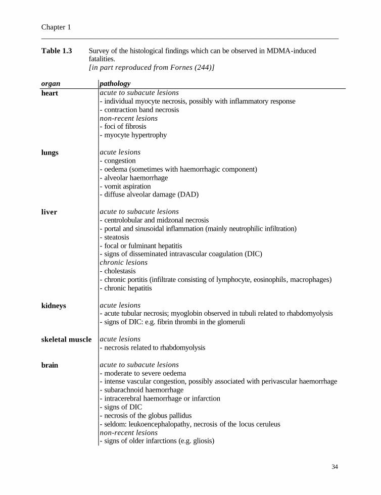
Chapter 1 _____________________________________________________________________________
34
Table 1.3 Survey of the histological findings which can be observed in MDMA-induced fatalities. [in part reproduced from Fornes (244)]
organ pathology heart acute to subacute lesions
- individual myocyte necrosis, possibly with inflammatory response - contraction band necrosis non-recent lesions - foci of fibrosis - myocyte hypertrophy
lungs acute lesions - congestion - oedema (sometimes with haemorrhagic component) - alveolar haemorrhage - vomit aspiration - diffuse alveolar damage (DAD)
liver acute to subacute lesions - centrolobular and midzonal necrosis - portal and sinusoidal inflammation (mainly neutrophilic infiltration) - steatosis - focal or fulminant hepatitis - signs of disseminated intravascular coagulation (DIC) chronic lesions - cholestasis - chronic portitis (infiltrate consisting of lymphocyte, eosinophils, macrophages) - chronic hepatitis
kidneys acute lesions - acute tubular necrosis; myoglobin observed in tubuli related to rhabdomyolysis - signs of DIC: e.g. fibrin thrombi in the glomeruli
skeletal muscle acute lesions - necrosis related to rhabdomyolysis
brain acute to subacute lesions - moderate to severe oedema - intense vascular congestion, possibly associated with perivascular haemorrhage - subarachnoid haemorrhage - intracerebral haemorrhage or infarction - signs of DIC - necrosis of the globus pallidus - seldom: leukoencephalopathy, necrosis of the locus ceruleus non-recent lesions - signs of older infarctions (e.g. gliosis)

Chapter 1 _____________________________________________________________________________
35
Table 1.4 Survey of the complications of heat stroke (clinico-pathological findings). [reproduced from Dickinson (245)]
organ pathology effects brain - oedema - convulsions
- petechial haemorrhages - coma - congestion
muscle - rhabdomyolysis - acute renal failure - hyperkalaemia - hyperuricaemia - hypocalcaemia - hyperphosphataemia - enzyme release, esp. creatine kinase - possibly disseminated intravascular coagulation (DIC)
blood - lactic acidosis - fragmentation of red blood cells - DIC - acute renal failure - thrombocytopenia - haemorrhage - thrombosis - haemolysis
liver - centrilobular necrosis - liver cell failure: jaundice haemorrhage hypoglycaemia enzyme release
kidneys - acute tubular necrosis - acute renal failure: oliguria acidosis hyperkalaemia
lungs - respiratory alkalosis - tetany - aspiration pneumonia - hypoxia - haemorrhagic pneumonia
heart - haemorrhagic myocarditis - shock
gastro-intestinal - non-specific general bleeding - nausea, vomiting, diarrhoea tract diathesis - haemorrhage
Chapter 1 _____________________________________________________________________________
36
IV.2 Thanato-toxicological data in MDMA-related fatalities The pharmacokinetics of drugs taken in overdose may differ from those observed
following therapeutic doses and therefore may result into a poor correlation between the clinical findings in overdose patients and the blood concentrations of ingested drugs (246). Clinically, blood levels in intoxicated persons may thus be difficult to interpret, though after death, this interpretation becomes even more tenuous. In addition, post-mortem blood levels can be influenced by post-mortem processes such as instability, redistribution and even neoformation.
IV.2.1 Animal experimental data To our knowledge, animal models investigating post-mortem distribution and re-distribution of MDMA are not available and therefore this will be investigated (see part two). For methamphetamine, Nagata et al. studied post-mortem rabbit tissue concentrations in vitro for up to a period of 2 years after death, and demonstrated that skeletal muscle and bone marrow are the most appropriate specimens for assessing toxicity of methamphetamine and its metabolite amphetamine (247). For amphetamine and methamphetamine, animal experiments dealing with this subject in situ are scarce (248, 249). Hilberg et al. administered amphetamine to rats by a gastric tube and the animals were killed 90 minutes later. Cardiac blood was sampled immediately before sacrifice and 2 h post mortem (248). Post-mortem blood samples from the inferior vena cava, the vitreous humour and various tissues were tested. The authors concluded that the amphetamine level in the vena cava blood was more closely related to the ante-mortem blood level, confirming that a peripheral blood sample is recommended. However, in the data presented, the difference between amphetamine levels in cardiac blood and vena cava inferior is rather minor (see Table 1.5). The ratios of post-mortem tissue concentrations of amphetamine to the ante-mortem blood level are presented in Table 1.5 (248). The similar mean ratio calculated for the vitreous humour level was 1.1 ± 0.2 (see Table 1.5). Obviously higher amphetamine levels in tissue compared to ante-mortem blood were found in the kidneys, and less pronounced though also elevated tissue levels were seen in liver, lungs, and brain (248) indicating tissue accumulation of the substance. Vitreous humour and muscle amphetamine levels were more closely related to the ante-mortem blood concentration. Moriya et al. studied the redistribution of methamphetamine in the early post-mortem period in the rabbit following intravenous administration and compared the blood and tissue levels with (Group I) or without (Group II) ligation of the large vessels around the heart (249). The lung levels were the highest of all, followed by the blood and myocardium concentrations, and the liver levels. The mean ratios of the cardiac blood concentrations 6 h post mortem to the level at the time of death were for group I in the left and right chambers 1.13 ± 0.42 and 1.07 ± 0.04, respectively. The analogous ratios for group II were 1.62 ± 0.47 and 1.13 ± 0.57. Therefore, Moriya et al. concluded that after death,
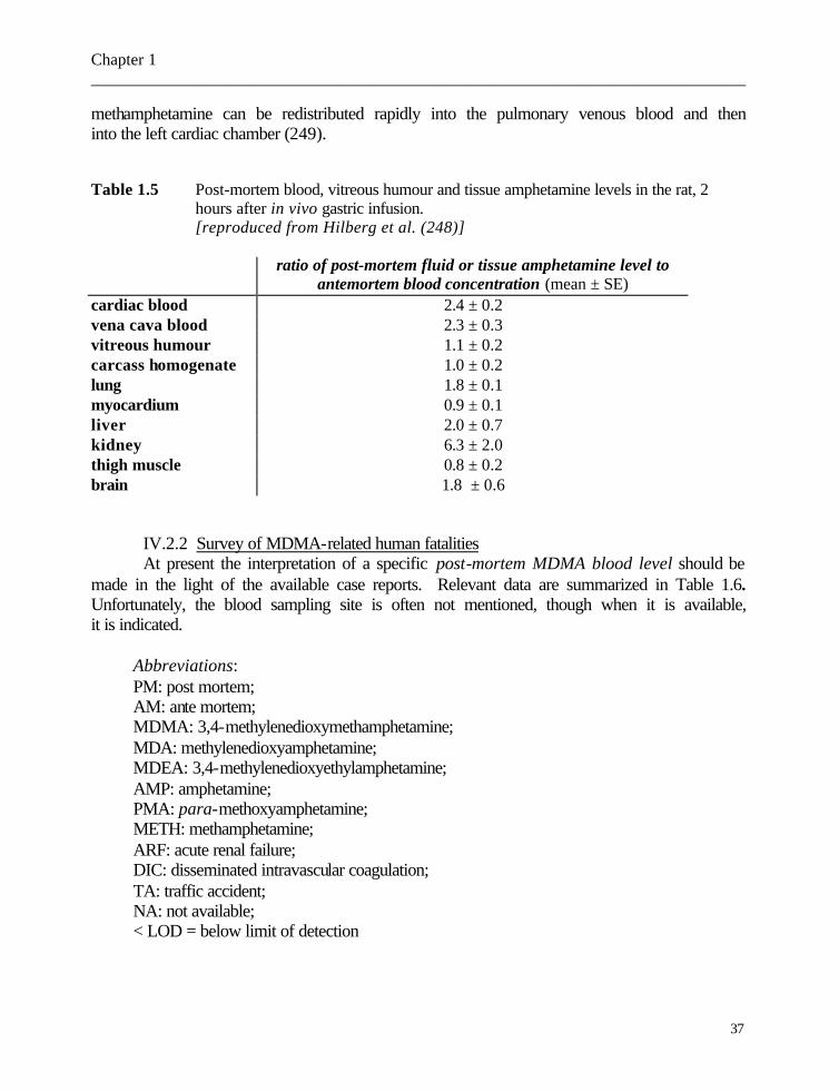
Chapter 1 _____________________________________________________________________________
37
methamphetamine can be redistributed rapidly into the pulmonary venous blood and then into the left cardiac chamber (249). Table 1.5 Post-mortem blood, vitreous humour and tissue amphetamine levels in the rat, 2
hours after in vivo gastric infusion. [reproduced from Hilberg et al. (248)]
ratio of post-mortem fluid or tissue amphetamine level to antemortem blood concentration (mean ± SE) cardiac blood 2.4 ± 0.2 vena cava blood 2.3 ± 0.3 vitreous humour 1.1 ± 0.2 carcass homogenate 1.0 ± 0.2 lung 1.8 ± 0.1 myocardium 0.9 ± 0.1 liver 2.0 ± 0.7 kidney 6.3 ± 2.0 thigh muscle 0.8 ± 0.2 brain 1.8 ± 0.6
IV.2.2 Survey of MDMA-related human fatalities At present the interpretation of a specific post-mortem MDMA blood level should be made in the light of the available case reports. Relevant data are summarized in Table 1.6. Unfortunately, the blood sampling site is often not mentioned, though when it is available, it is indicated.
Abbreviations: PM: post mortem; AM: ante mortem; MDMA: 3,4-methylenedioxymethamphetamine; MDA: methylenedioxyamphetamine; MDEA: 3,4-methylenedioxyethylamphetamine; AMP: amphetamine; PMA: para-methoxyamphetamine; METH: methamphetamine; ARF: acute renal failure; DIC: disseminated intravascular coagulation; TA: traffic accident; NA: not available; < LOD = below limit of detection

Chapter 1 _____________________________________________________________________________
38
Table 1.6 Survey of the reported MDMA-related fatalities reference age
(y) sex sample
type sampling time
level 1 cause of death
manner of death 2
mechanism of death
Bernhard (180;17)
27 32 NA NA
M M NA NA
blood blood blood blood
PM PM PM PM
2.75 3 0.28 0.08 1.56 4 0.04 1.00 3.37 5 5.21 6
MDMA MDA AMP MDMA MDA alcohol MDMA MDMA
NA NA NA NA
NA NA NA NA
Byard (186)
22 26
F F
blood blood
PM PM
0.30 1.32 0.82 2.20 0.09
MDMA PMA MDMA PMA METH
accidental accidental
hyperthermia hyperthermia
Campkin (250)
18 M serum AM 1.26 7 MDMA accidental hyperthermia, DIC, rhabdomyolysis
Chadwick (236)
16 F blood AM 0.42 8 MDMA accidental hyperthermia, DIC
Coore (106)
18 F serum AM NA 9 0.25
MDMA MDA
accidental hyperthermia, DIC, rhabdomyolysisfulminant hepatic failure
1 Blood level expressed in µg/ml for all drugs, unless stated and except for ethanol concentrations
(g/l). 2 When “accidental” is not specified, an unintentional death after recreational use (e.g. long-term
dancing at a “rave party”) is assumed. 3 Femoral blood levels. Cardiac blood levels of MDMA, MDA and AMP are 9.10, 0.83 and 0.11
µg/ml, respectively. Brain MDMA concentrations were 10 µg/g in the medulla (not specified) and 14 µg/g in the cerebellum. In his hair (0 to 6 cm), 6.70 µg/g was found.
4 Femoral blood levels. Cardiac blood levels of MDMA and MDA are 0.42 and 0.04 µg/ml, respectively.
5 Femoral blood level. Cardiac blood concentration: 3.51 µg/ml. 6 Femoral blood level. Cardiac blood concentration: 5.38 µg/ml. 7 Serum MDMA level upon admission to hospital; the man died 5 hours after arrival. 8 Woman admitted to hospital a few hours after ingestion of one “ecstasy” tablet; she died 36 h
later. Blood and stomach MDMA levels on admission were 0.42 and 28 µg/ml, respectively. 9 Woman ingested one tablet; died 9 days after admission to hospital. A toxicological screening
of urine was positive for MDMA and MDA. Serum was only positive for MDA, with peak level

Chapter 1 _____________________________________________________________________________
39
reference age (y)
sex sample type
sampling time
level cause of death
manner of death
mechanism of death
Cox (213) 21 M blood PM <LOD10
drowning suicide asphyxia
Cox (251)
22 M blood PM 0.43 11 0.30
MDMA MDA
accidental hyperthermia, DIC
Crifasi (202)
29 M blood PM 2.32 12 < 0.25
MDMA,MDA TA
accidental (TA)
polytrauma (head and chest)
Dar (237) 17 M blood PM 0.23 13 NA
MDMA ethanol
accidental hyperthermia, DIC, cardiopulmonary failure
Dowling (207)
22 32 18
M M F
blood blood blood
PM PM PM
NA 14 1.10 1.00 0.40
MDMA electro-cution MDMA acute asthma15 MDMA ethanol
accidental combined: natural, accidental
high voltage electrical shock, polytrauma acute pulmonary failure (cf asthma) ventricular fibrillation, acute cardiopulmonary failure
Duflou (230)
29 M blood PM 0.10 16 MDMA accidental aortic dissection, cardiac tamponade
of 0.25 µg/ml; neither at what time this concentration was found, nor the origin of the blood sample are specified.
10 The man committed suicide on the eighth day after an acute psychosis (see also III.2). 11 Femoral blood concentrations. MDMA and MDEA concentration in blood sampled upon
admission to hospital: 0.55 and 0.49 µg/ml, respectively. 12 MDMA concentration in clotted and anticoagulated cardiac blood: 2.32 and 2.14 µg/ml; vitreous
humour and urine MDMA levels: 1.11 and 118.8 µg/ml, respectively. MDA level in blood and vitreous humour was < 0.25 µg/ml and 3.86 µg/ml in urine.
13 Man died 6 h after initial presentation in hospital. He ingested 10 tablets of ecstasy. Liver MDMA concentration:1.2 µg/g.
14 The man climbed a high voltage utility tower to a height of 13 m, was electrocuted and fell to the ground. MDMA was positive in blood, but unfortunately not quantified.
15 The man was found dead beside his car, with an inhaler in his hand. Two hours before, he “had been drinking alcohol with his friends”. Post-mortem examination revealed features of acute and chronic bronchial asthma. No theophylline was detected.
16 Toxicological analysis of both ante- and post-mortem blood revealed the same MDMA concentration; the man died about 48 hours after ingesting one ecstasy tablet.

Chapter 1 _____________________________________________________________________________
40
reference age
(y) sex sample
type sampling time
level cause of death
manner of death
mechanism of death
Ellis (97) 21 18 36 21
F F F F
serum NA NA NA
AM NA NA NA
0.11 17 NA 18 NA 19 NA 20
MDMA LSD MDMA MDMA MDMA
accidental accidental accidental accidental
hyperthermia, DIC, liver failure, sepsis liver failure, sepsis liver failure, sepsis liver failure, sepsis
Felgate (252) 21
22 26 NA
F F NA
blood blood blood
PM PM PM
0.30 22 1.30 0.82 23 2.20 0.09 0.03 0.51 24 0.53 0.23
MDMA PMA MDMA PMA METH AMP MDMA PMA METH
accidental accidental NA
hyperthermia, DIC hyperthermia, rhabdomyolysis NA
Fineschi (239)
19 20
M M
blood blood
PM PM
7.15 0.25 0.18 25 1.59 0.12
MDMA MDA MDMA MDEA MDA
accidental?26 accidental
hyperthermia, DIC hyperthermia, DIC
17 Sampling at about 6 hours following ingestion of ecstasy. She died in spite of a liver
transplantation. 18 Woman took MDMA on a regular basis and even continued to use it when obvious jaundice
occurred. There is no information available about the last ingestion. Arrived at hospital in encephalopatic (grade II) condition due to MDMA-induced acute liver failure.
19 Woman presented to hospital with a six-day illness (jaundice, nausea etc). Ten days before the onset, she had taken a single ecstasy tablet. A few days later, she developed encephalopathy as part of an acute hepatic failure and died in spite of a liver transplantation.
20 A woman, who had been using MDMA for the previous 6 months, consulted a physian following a three-week history of worsening jaundice associated with nausea and vomiting. She developed an acute liver failure and died in spite of a liver transplant.
21 The authors presented ten PMA-related fatalities. Only the cases in which MDMA could also be quantified in blood are reported.
22 MDMA, PMA and AMP liver concentrations: 3.2, 7.4, 0.45 µg/g, respectively. AMP was not detected in blood.
23 MDMA, PMA, METH, and AMP liver levels: 3.2, 5.6, 0.26, and 0.35 µg/g, respectively. 24 MDMA, PMA, and METH liver levels: 3.0, 2.7 and 0.76 µg/g, respectively. 25 Person died four hours following arrival at hospital. MDMA, MDA and MDEA concentrations:
263.13, 5.25, and 183.73 µg/ml in urine and 27.34 < LOQ, and 21.93 µg/ml in bile, respectively. MDMA levels in liver, kidney, lung, brain, and spleen:13.23, 9.81, 10.70, 12.79, and 9.17 µg/g,
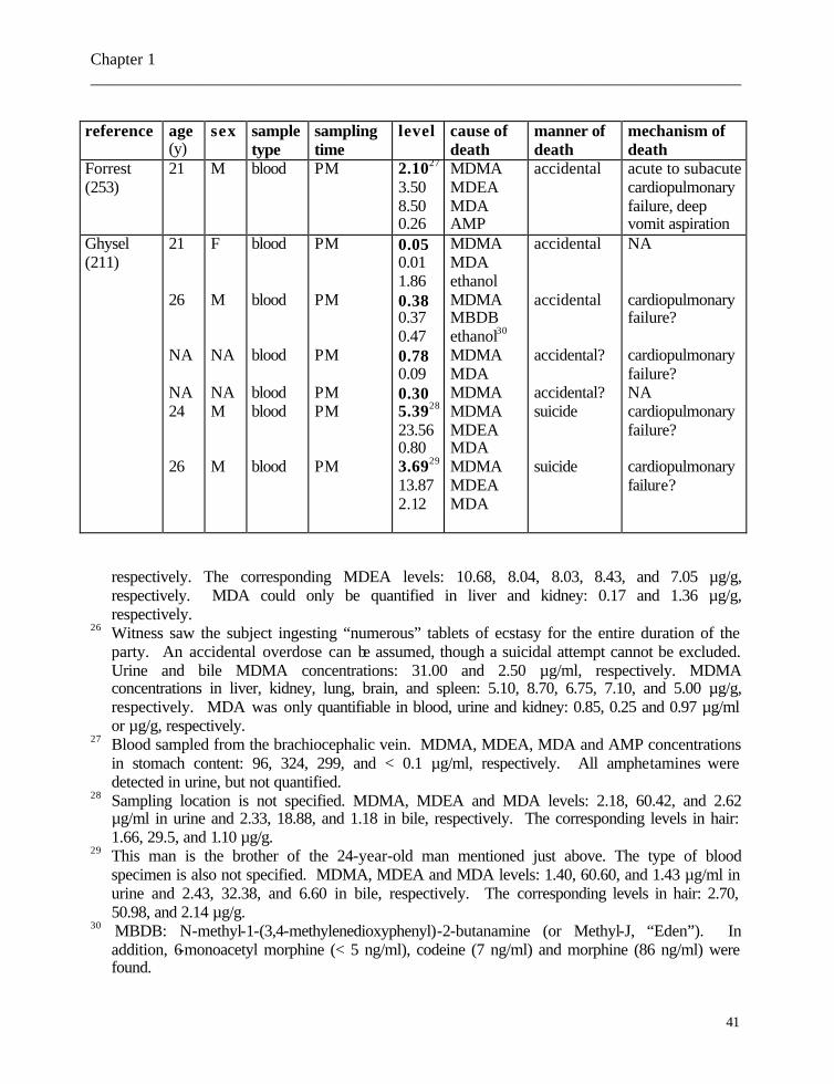
Chapter 1 _____________________________________________________________________________
41
reference age
(y) sex sample
type sampling time
level cause of death
manner of death
mechanism of death
Forrest (253)
21 M blood PM 2.1027 3.50 8.50 0.26
MDMA MDEA MDA AMP
accidental acute to subacute cardiopulmonary failure, deep vomit aspiration
Ghysel (211)
21 26 NA NA 24 26
F M NA NA M M
blood blood blood blood blood blood
PM PM PM PM PM PM
0.05 0.01 1.86 0.38 0.37 0.47 0.78 0.09 0.30 5.3928 23.56 0.80 3.6929 13.87 2.12
MDMA MDA ethanol MDMA MBDB ethanol30 MDMA MDA MDMA MDMA MDEA MDA MDMA MDEA MDA
accidental accidental accidental? accidental? suicide suicide
NA cardiopulmonary failure? cardiopulmonary failure? NA cardiopulmonary failure? cardiopulmonary failure?
respectively. The corresponding MDEA levels: 10.68, 8.04, 8.03, 8.43, and 7.05 µg/g, respectively. MDA could only be quantified in liver and kidney: 0.17 and 1.36 µg/g, respectively.
26 Witness saw the subject ingesting “numerous” tablets of ecstasy for the entire duration of the party. An accidental overdose can be assumed, though a suicidal attempt cannot be excluded. Urine and bile MDMA concentrations: 31.00 and 2.50 µg/ml, respectively. MDMA concentrations in liver, kidney, lung, brain, and spleen: 5.10, 8.70, 6.75, 7.10, and 5.00 µg/g, respectively. MDA was only quantifiable in blood, urine and kidney: 0.85, 0.25 and 0.97 µg/ml or µg/g, respectively.
27 Blood sampled from the brachiocephalic vein. MDMA, MDEA, MDA and AMP concentrations in stomach content: 96, 324, 299, and < 0.1 µg/ml, respectively. All amphetamines were detected in urine, but not quantified.
28 Sampling location is not specified. MDMA, MDEA and MDA levels: 2.18, 60.42, and 2.62 µg/ml in urine and 2.33, 18.88, and 1.18 in bile, respectively. The corresponding levels in hair: 1.66, 29.5, and 1.10 µg/g.
29 This man is the brother of the 24-year-old man mentioned just above. The type of blood specimen is also not specified. MDMA, MDEA and MDA levels: 1.40, 60.60, and 1.43 µg/ml in urine and 2.43, 32.38, and 6.60 in bile, respectively. The corresponding levels in hair: 2.70, 50.98, and 2.14 µg/g.
30 MBDB: N-methyl-1-(3,4-methylenedioxyphenyl)-2-butanamine (or Methyl-J, “Eden”). In addition, 6-monoacetyl morphine (< 5 ng/ml), codeine (7 ng/ml) and morphine (86 ng/ml) were found.

Chapter 1 _____________________________________________________________________________
42
reference age (y)
sex sample type
sampling time
level cause of death
manner of death
mechanism of death
Harries (89)
20 M NA NA NA “ecstasy” AV-mal-formation
combination natural disease and accidental 31
intracranial bleeding, brain death
Henry (87) 18 17 18 16 21 20 18 21 23
M M M F F M M M M
plasma plasma plasma plasma plasma plasma plasma plasma plasma
AM AM AM AM AM AM AM AM AM
0.36 NA NA 0.42
0.11 1.16 0.06 0.10 1.26 0.1032 0.1333
MDMA MDMA MDMA MDMA MDMA MDMA MDA AMP MDMA MDMA, TA MDMA, TA
accidental accidental accidental accidental accidental accidental accidental accidental, TA accidental, TA
hyperthermia, arrhythmia, asystole hyperthermia, DIC hyperthermia, DIC, rhabdomyolysis gastrointestinal haemorrhage hyperthermia, DIC, metabolic acidosis hyperthermia, DIC, rhabdomyolysisARF hyperthermia, DIC, rhabdomyolysisARF hyperthermia, DIC, rhabdomyolysisARF polytrauma (multiple fractures) multiple skull fractures
31 Upon arrival at the hospital – a few hours after ecstasy ingestion - a CT scan revealed a large
frontal haematoma and an angiogram showed a left frontal arteriovenous malformation. In spite of a craniotomy, he was brain dead the following day.
32 Driver of car in head-on collision. 33 Passenger in the same car as 32.
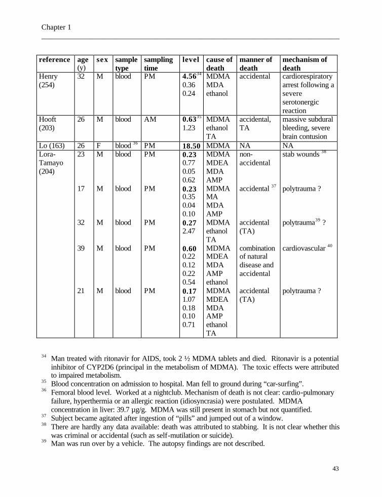
Chapter 1 _____________________________________________________________________________
43
reference age
(y) sex sample
type sampling time
level cause of death
manner of death
mechanism of death
Henry (254)
32 M blood PM 4.5634 0.36 0.24
MDMA MDA ethanol
accidental cardiorespiratory arrest following a severe serotonergic reaction
Hooft (203)
26 M blood AM 0.6335 1.23
MDMA ethanol TA
accidental, TA
massive subdural bleeding, severe brain contusion
Lo (163) 26 F blood 36 PM 18.50 MDMA NA NA Lora-Tamayo (204)
23 17 32 39 21
M M M M M
blood blood blood blood blood
PM PM PM PM PM
0.23 0.77 0.05 0.62 0.23 0.35 0.04 0.10 0.27 2.47 0.60 0.22 0.12 0.22 0.54 0.17 1.07 0.18 0.10 0.71
MDMA MDEA MDA AMP MDMA MA MDA AMP MDMA ethanol TA MDMA MDEA MDA AMP ethanol MDMA MDEA MDA AMP ethanol TA
non-accidental
accidental 37 accidental (TA) combination of natural disease and accidental accidental (TA)
stab wounds 38 polytrauma ? polytrauma39 ? cardiovascular 40 polytrauma ?
34 Man treated with ritonavir for AIDS, took 2 ½ MDMA tablets and died. Ritonavir is a potential
inhibitor of CYP2D6 (principal in the metabolism of MDMA). The toxic effects were attributed to impaired metabolism.
35 Blood concentration on admission to hospital. Man fell to ground during “car-surfing”. 36 Femoral blood level. Worked at a nightclub. Mechanism of death is not clear: cardio-pulmonary
failure, hyperthermia or an allergic reaction (idiosyncrasia) were postulated. MDMA concentration in liver: 39.7 µg/g. MDMA was still present in stomach but not quantified.
37 Subject became agitated after ingestion of “pills” and jumped out of a window. 38 There are hardly any data available: death was attributed to stabbing. It is not clear whether this
was criminal or accidental (such as self-mutilation or suicide). 39 Man was run over by a vehicle. The autopsy findings are not described.
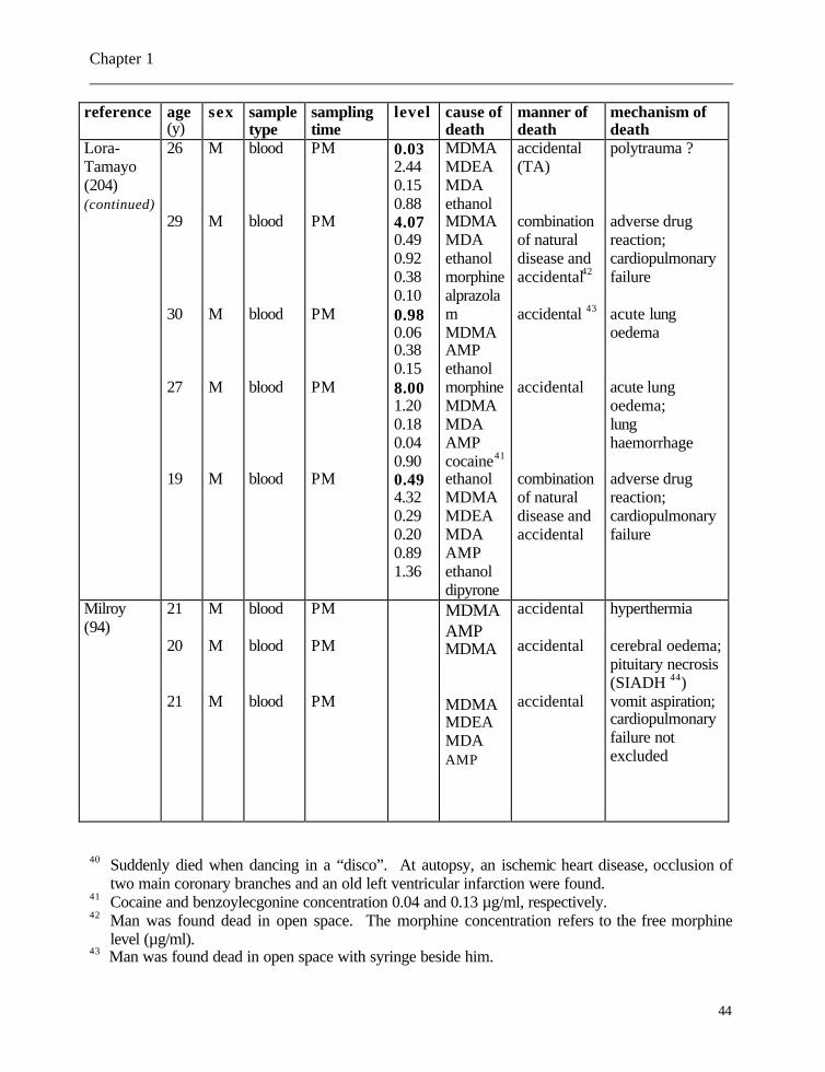
Chapter 1 _____________________________________________________________________________
44
reference age (y)
sex sample type
sampling time
level cause of death
manner of death
mechanism of death
Lora-Tamayo (204) (continued)
26 29 30 27 19
M M M M M
blood blood blood blood blood
PM PM PM PM PM
0.03 2.44 0.15 0.88 4.07 0.49 0.92 0.38 0.10 0.98 0.06 0.38 0.15 8.00 1.20 0.18 0.04 0.90 0.49 4.32 0.29 0.20 0.89 1.36
MDMA MDEA MDA ethanol MDMA MDA ethanol morphine alprazolam MDMA AMP ethanol morphine MDMA MDA AMP cocaine41 ethanol MDMA MDEA MDA AMP ethanol dipyrone
accidental (TA) combination of natural disease and accidental42 accidental 43 accidental combination of natural disease and accidental
polytrauma ? adverse drug reaction; cardiopulmonary failure acute lung oedema acute lung oedema; lung haemorrhage adverse drug reaction; cardiopulmonary failure
Milroy (94) reference
21 20 21 age (y)
M M M sex
blood blood blood sample type
PM PM PM sampling time
MDMA AMP MDMA MDMA MDEA MDA AMP cause of death
accidental accidental accidental manner of death
hyperthermia cerebral oedema; pituitary necrosis (SIADH 44) vomit aspiration; cardiopulmonary failure not excluded mechanism of death
40 Suddenly died when dancing in a “disco”. At autopsy, an ischemic heart disease, occlusion of
two main coronary branches and an old left ventricular infarction were found. 41 Cocaine and benzoylecgonine concentration 0.04 and 0.13 µg/ml, respectively. 42 Man was found dead in open space. The morphine concentration refers to the free morphine
level (µg/ml). 43 Man was found dead in open space with syringe beside him.
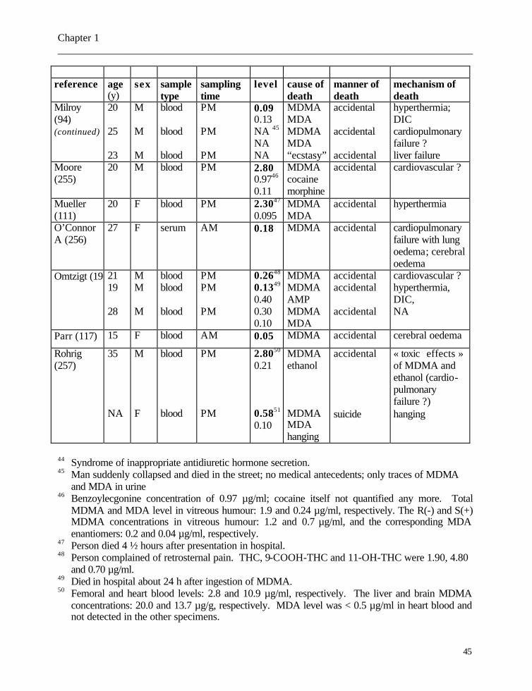
Chapter 1 _____________________________________________________________________________
45
reference age
(y) sex sample
type sampling time
level cause of death
manner of death
mechanism of death
Milroy (94) (continued)
20 25 23
M M M
blood blood blood
PM PM PM
0.09 0.13 NA 45 NA NA
MDMA MDA MDMA MDA “ecstasy”
accidental accidental accidental
hyperthermia; DIC cardiopulmonary failure ? liver failure
Moore (255)
20 M blood PM 2.80 0.9746 0.11
MDMA cocaine morphine
accidental cardiovascular ?
Mueller (111)
20 F blood PM 2.3047 0.095
MDMA MDA
accidental hyperthermia
O’Connor A (256)
27 F serum AM 0.18 MDMA accidental cardiopulmonary failure with lung oedema; cerebral oedema
Omtzigt (19821 19 28
M M M
blood blood blood
PM PM PM
0.2648 0.1349 0.40 0.30 0.10
MDMA MDMA AMP MDMA MDA
accidental accidental accidental
cardiovascular ? hyperthermia, DIC, NA
Parr (117) 15 F blood AM 0.05 MDMA accidental cerebral oedema
Rohrig (257)
35 NA
M F
blood blood
PM PM
2.8050 0.21 0.5851 0.10
MDMA ethanol MDMA MDA hanging
accidental
suicide
« toxic effects » of MDMA and ethanol (cardio-pulmonary failure ?) hanging
44 Syndrome of inappropriate antidiuretic hormone secretion. 45 Man suddenly collapsed and died in the street; no medical antecedents; only traces of MDMA
and MDA in urine 46 Benzoylecgonine concentration of 0.97 µg/ml; cocaine itself not quantified any more. Total
MDMA and MDA level in vitreous humour: 1.9 and 0.24 µg/ml, respectively. The R(-) and S(+) MDMA concentrations in vitreous humour: 1.2 and 0.7 µg/ml, and the corresponding MDA enantiomers: 0.2 and 0.04 µg/ml, respectively.
47 Person died 4 ½ hours after presentation in hospital. 48 Person complained of retrosternal pain. THC, 9-COOH-THC and 11-OH-THC were 1.90, 4.80
and 0.70 µg/ml. 49 Died in hospital about 24 h after ingestion of MDMA. 50 Femoral and heart blood levels: 2.8 and 10.9 µg/ml, respectively. The liver and brain MDMA
concentrations: 20.0 and 13.7 µg/g, respectively. MDA level was < 0.5 µg/ml in heart blood and not detected in the other specimens.
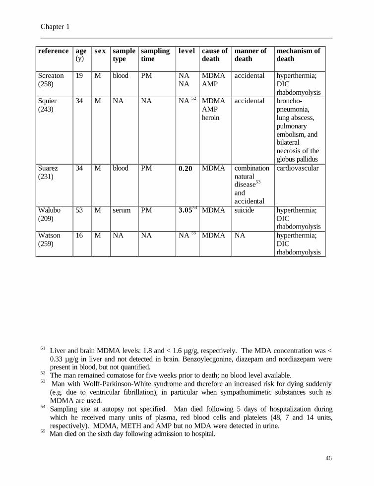
Chapter 1 _____________________________________________________________________________
46
reference age (y)
sex sample type
sampling time
level cause of death
manner of death
mechanism of death
Screaton (258)
19 M blood PM NA NA
MDMA AMP
accidental hyperthermia; DIC rhabdomyolysis
Squier (243)
34 M NA NA NA 52 MDMA AMP heroin
accidental broncho-pneumonia, lung abscess, pulmonary embolism, and bilateral necrosis of the globus pallidus
Suarez (231)
34 M blood PM 0.20 MDMA combination natural disease53 and accidental
cardiovascular
Walubo (209)
53 M serum PM 3.0554 MDMA suicide hyperthermia; DIC rhabdomyolysis
Watson (259)
16 M NA NA NA 55 MDMA NA hyperthermia; DIC rhabdomyolysis
51 Liver and brain MDMA levels: 1.8 and < 1.6 µg/g, respectively. The MDA concentration was <
0.33 µg/g in liver and not detected in brain. Benzoylecgonine, diazepam and nordiazepam were present in blood, but not quantified.
52 The man remained comatose for five weeks prior to death; no blood level available. 53 Man with Wolff-Parkinson-White syndrome and therefore an increased risk for dying suddenly
(e.g. due to ventricular fibrillation), in particular when sympathomimetic substances such as MDMA are used.
54 Sampling site at autopsy not specified. Man died following 5 days of hospitalization during which he received many units of plasma, red blood cells and platelets (48, 7 and 14 units, respectively). MDMA, METH and AMP but no MDA were detected in urine.
55 Man died on the sixth day following admission to hospital.
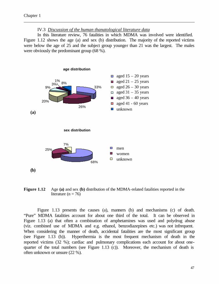
Chapter 1 _____________________________________________________________________________
47
IV.3 Discussion of the human thanatological literature data In this literature review, 76 fatalities in which MDMA was involved were identified.
Figure 1.12 shows the age (a) and sex (b) distribution. The majority of the reported victims were below the age of 25 and the subject group younger than 21 was the largest. The males were obviously the predominant group (68 %).
Figure 1.12 Age (a) and sex (b) distribution of the MDMA-related fatalities reported in the literature (n = 76)
Figure 1.13 presents the causes (a), manners (b) and mechanisms (c) of death.
“Pure” MDMA fatalities account for about one third of the total. It can be observed in Figure 1.13 (a) that often a combination of amphetamines was used and polydrug abuse (viz. combined use of MDMA and e.g. ethanol, benzodiazepines etc.) was not infrequent. When considering the manner of death, accidental fatalities are the most significant group (see Figure 1.13 (b)). Hyperthermia is the most frequent mechanism of death in the reported victims (32 %); cardiac and pulmonary complications each account for about one-quarter of the total numbers (see Figure 1.13 (c)). Moreover, the mechanism of death is often unknown or unsure (22 %).
age distribution
33%
26%
20%
9%3% 8%
1%1
2
34
5
6
7
aged 15 – 20 years aged 21 – 25 years aged 26 – 30 years aged 31 – 35 years aged 36 – 40 years aged 41 - 60 years unknown
(a)
sex distribution
68%
25%7%
12
3
men women unknown
(b)
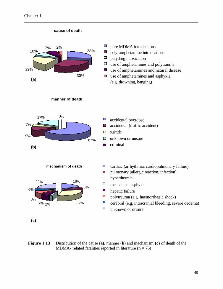
Chapter 1 _____________________________________________________________________________
48
(c)
cause of death
28%
30%
23%
10%7% 2% 1
23456
manner of death
67%9%
7%
17% 0%1
2
3
4
5
accidental overdose accidental (traffic accident) suicide unknown or unsure criminal
mechanism of death
5%
32%2%7%8%
6%
22% 18%
12345678
(c)
(b)
(a)
pure MDMA intoxications poly-amphetamine intoxications polydrug intoxication use of amphetamines and polytrauma use of amphetamines and natural disease use of amphetamines and asphyxia (e.g. drowning, hanging)
cardiac (arrhythmia, cardiopulmonary failure) pulmonary (allergic reaction, infection) hyperthermia
mechanical asphyxia hepatic failure polytrauma (e.g. haemorrhagic shock) cerebral (e.g. intracranial bleeding, severe oedema)unknown or unsure
Figure 1.13 Distribution of the cause (a), manner (b) and mechanism (c) of death of the MDMA- related fatalities reported in literature (n = 76)
Chapter 1 _____________________________________________________________________________
49
The anatomo-pathological findings in MDMA-related fatalities - either macroscopically or microscopically – are non-specific. A whole range of possible pathological observations can be found, such as hepatotoxicity, cardiomyopathy, acute to subacute cardiopulmonary failure, brain oedema, signs of multiple organ failure, and DIC. In fact, the anatomo-pathological disorders in MDMA-related fatalities cannot easily be distinguished from those described in the abuse of amphetamine (or other derivatives) and other stimulant drugs such as cocaine. Bearing in mind the above-mentioned assessments, some anatomo-pathological anomalies can raise suspicions which needs to be confirmed by toxicological investigation.
Referring to the available thanato-toxicological literature data – indicating a wide range of concentrations -, the interpretation of the MDMA blood level after death remains a debatable question. Indeed a broad range of concentrations is found, viz. between 0.04 µg/ml and 18.5 µg/ml (94,163). In the latter case, reported by Lo et al. (163), a femoral blood level of 18.5 µg/ml is extraordinary as the majority of the blood MDMA levels being between 0.5 and 4 to 5 µg/ml. For the most part, blood MDMA levels are given; when persons are admitted to the emergency service, serum or plasma is more frequently used for MDMA quantitation. For all amphetamines, the serum/blood conversion factor is assumed to be nearby or somewhat higher than 1 and therefore the sample type is usually not taken into account (260). However, Garrett found a red blood cell-plasma partition coefficient of 1.48 and 1.45 for MDMA and MDA, respectively, indicating a certain accumulation of MDMA and MDA in red blood cells (26). For amphetamine and methamphetamine, the blood sampling site in human fatalities was proven to be important for the interpretation of a quantified level (261,262). For example, for methamphetamine, concentrations measured in the left heart of human fatalities are about two times higher than those quantified in the right heart (262). Blood collected from the pulmonary vessels sometimes showed concentrations that were many times higher than blood sampled from the heart and, as a result, diffusion out of the lungs into the pulmonary circulation was demonstrated (262).
Although elaborate studies dealing with the post-mortem phenomena in MDMA-related fatalities are at present not available, it is obvious that a peripheral sampling site (such as the femoral vein; see Table 1.6) is recommended. Toxicologists should be aware of the blood sampling location and whether a peripheral blood sample is available. They should be cautious in their conclusions as to whether or not the quantified level is either toxic or lethal.
In conclusion, though the literature data on amphetamine and methamphetamine indicate that post-mortem redistribution can take place, a few questions such as whether MDMA is liable to post-mortem redistribution remain to be elucidated. It is mandatory to consider the anatomo-pathological findings and the toxicological data as a whole in order to come to medico-legal conclusions.

Chapter 1 _____________________________________________________________________________
50
References 1. Kalant H. The pharmacology and toxicology of “ecstasy” (MDMA) and related drugs.
CMAJ 2001;165:917-928. 2. Shulgin AT. The background and chemistry of MDMA. J Psychoactive Drugs 1986;18:291-
304. 3. Vereby K, Alrazi J, Jaffe JH. The complications of “Ecstasy” (MDMA). JAMA 1988;
259:1649-1650. 4. Helmlin HJ, Bracher K, Bourquin D, Vonlanthen D, Brenneisen R. Analysis of 3,4-
methylenedioxymethamphetamine (MDMA) and its metabolites in plasma and urine by HPLC-DAD and GC-MS. J Anal Toxicol 1996;20:432-440.
5. Segura M, Ortuño J, Farré M, McLure JA, Pujadas M, Pizarro N, Llebaria A, Joglar J, Roset
PN, Segura J, de la Torre R. 3,4-Dihydroxymethamphetamine (HHMA). A major in vivo 3,4-methylenedioxymethamphetamine (MDMA) metabolite in humans. Chem Res Toxicol 2001;14 :1203-1208.
6. Mas M, Farré M, de la Torre R, Roset PN, Ortuño J, Segura J, Camí J. Cardiovascular and
neuroendocrine effects and pharmacokinetics of 3,4-methylenedioxymethamphetamine in humans. J Pharmacol Exp Ther 1999; 290:136-145.
7. de la Torre R, Farré M, Roset PN, Hernández-López C, Mas M, Ortuño J, Menoyo E, Pizarro
N, Segura J, Camí J. Pharmacology of MDMA in humans. Ann N Y Acad Sci 2000;914:225-237.
8. Fallon JK, Kicman AT, Henry JA, Milligan PJ, Cowan DA, Hutt AJ. Stereospecific analysis
and enantiomeric disposition of 3,4-methylenedioxymethamphetamine (Ecstasy) in humans. Clin Chem 1999;45:1058-1069.
9. Kunsman GW, Levine B, Kuhlman JJ, Jones RL, Hughes RO, Fujiyama CI, Smith ML.
MDA – MDMA concentrations in urine specimens. J Anal Toxicol 1996;20:517-521. 10. de la Torre R, Farré M, Ortuño J, Mas M, Brenneissen R, Roset PN, Segura J, Camí J. Non-
linear pharmacokinetics of MDMA (‘ecstasy’) in humans. Br J Clin Pharmacol 2000;49:104-109.
11. Maurer HH. On the metabolism and the toxicological analysis of methylenedioxy-
phenylalkylamine designer drugs by gas chromatography-mass spectrometry. Ther Drug Monit 1996;18:465-470.
12. Maurer HH, Bickboeller-Friedrich J, Kraemer T, Peters FT. Toxicokinetics and analytical
toxicology of amphetamine-derived designer drugs (‘Ecstasy’). Toxicol Lett 2000;112-113: 133-142.
Chapter 1 _____________________________________________________________________________
51
13. Kreth KP, Kovar KA, Schwab M, Zanger UM. Identification of the human cytochromes
P450 involved in the oxidative metabolism of “Ecstasy”-related designer drugs. Biochem Pharmacol 2000;59:1563-1571.
14. Hernández-López C, Farré M, Roset PN, Menoyo E, Pizarro N, Ortuño J, Torrens M, Camí J,
de la Torre R. 3,4-Methylenedioxymethamphetamine (Ecstasy) and alcohol interactions in humans: psychomotor performance, subjective effects and pharmacokinetics. J Pharmacol Exp Ther 2002;300:236-244.
15. Baselt RC. (ed) Disposition of toxic drugs and chemicals in man, 5th edn, Chemical
Toxicology Institute, Foster City, California, pp 49-51. 16. Cook CE, Jeffcoat AR, Sadler BM, Hill JM, Voyksner RD, Pugh DE, White WR, Perez-
Reyes M. Pharmacokinetics of oral methamphetamine and effects of repeated daily dosing in humans. Drug Metab Dispos 1992;20:856-862.
17. Karch SB. (ed) (2002) Karch’s pathology of drug abuse. 3rd edn. CRC Press, Boca Raton,
London, New York, Washington DC, pp 288 - 295. 18. Kraemer T, Maurer HH. Toxicokinetics of amphetamines : metabolism and toxicokinetic
data of designer drugs, amphetamine, methamphetamine, and their N-alkyl derivatives. Ther Drug Monit 2002;24:277-289.
19. Campbell DB. The use of toxicokinetics for the safety assessment of drugs acting in the
brain. Mol Neurobiol 1995;11:193-216. 20. Green AR, Cross AJ, Goodwin GM. Review of the pharmacology and clinical pharmacology
of 3,4-methylenedioxymethamphetamine (MDMA or “Ecstasy”). Psychopharmacology 1995;119:247-260.
21. Battaglia G, Brooks BP, Kulsakdinun C, De Souza EB. Pharmacological profile of MDMA
(3,4-methylenedioxymethamphetamine) at various brain recognition sites. Eur J Pharmacol 1988;149:159-163.
22. Maeno Y, Iwasa M, Inoue H, Koyama H, Matoba R. Methamphetamine induces an increase
in cell size and reorganization of myofibrils in cultured adult rat cardiomyocytes. Int J Legal Med 2000;113:201-207.
23. Beitia G, Cobreros A, Sainz L, Cenarruzabeitia E. Ecstasy-induced toxicity in rat liver.
Liver 2000;20:8-15. 24. Carvalho F, Remião, Soares ME, Catarino R, Queiroz G, Bastos ML. d-Amphetamine-
induced hepatotoxicity: possible contribution of catecholamines and hyperthermia to the effect studied in isolated rat hepatocytes. Arch Toxicol 1997;71:429-436.
Chapter 1 _____________________________________________________________________________
52
25. Carvalho M, Carvalho F, Bastos ML. Is hyperthermia the triggering factor for hepatotoxicity
induced by 3,4-methylenedioxymethamphetamine (ecstasy)? An in vitro study using freshly isolated mouse hepatocytes. Arch Toxicol 2001;74:789-793.
26. Garrett ER, Seyda K, Marroum P. High performance liquid chromatographic assays of the
illicit designer drug “Ecstasy”, a modified amphetamine, with applications to stability, partitioning and plasma protein binding. Acta Pharm Nord 1991;3:9-14.
27. Cox DS, Scott KR, Gao H, Raje S, Eddington ND. Influence of multidrug resistance (MDR)
proteins at the blood-brain barrier on the transport and brain distribution of enaminone anticonvulsants. J Pharm Sci 2001;90:1540-1552.
28. Mann H, Ladenheim B, Hirata H, Moran TH, Cadet JL. Differential toxic effects of
methamphetamine (METH) and methylenedioxymethamphetamine (MDMA) in multidrug-resistant (mdr1a) knockout mice. Brain Res 1997;769:340-346.
29. Rivière GJ, Gentry WB, Owens SM. Disposition of methamphetamine and its metabolite
amphetamine in brain and other tissues in rats after intravenous administration. J Pharmacol Exp Ther 2000;292:1042-1047.
30. Monks TJ, Lau SS. Biological reactivity of polyphenolic-glutathione conjugates. Chem Res
Toxicol 1997;10:1296-1313. 31. Bai F, Jones DC, Lau SS, Monks TJ. Serotonergic neurotoxicity of 3,4-(±)-methylene-
dioxyamphetamine and 3,4-(±)-methylenedioxymethamphetamine (Ecstasy) is potentiated by inhibition of γ-glutamyl transpeptidase. Chem Res Toxicol 2001;14:863-870.
32. Hiramatsu M, Kumagai Y, Unger SE, Cho AK. Metabolism of
methylenedioxymethamphetamine: formation of dihydroxymethamphetamine and a quinone identified as its glutathione adduct. J Pharmacol Exp Ther 1990;254:521-527.
33. Bai F, Lau SS, Monks TJ. Glutathione and N-acetylcysteine conjugates of α-methyl-
dopamine produce serotonergic neurotoxicity: possible role in methylenedioxyamphetamine-mediated neurotoxicity. Chem Res Toxicol 1999;12:1150-1157.
34. Battaglia G, Zaczek R, De Souza EB. MDMA effects in brain: pharmacologic profile and
evidence of neurotoxicity from neurochemical and autoradiographic studies. In: Peroutka SJ. (ed) (1990) Ecstasy: the clinical, pharmacological and neurotoxicological effects of the drug MDMA. Kluwer Academic Publishers, Boston, Dordrecht, London, pp 171-199.
35. Feldman RS, Meyer JS, Quenzer LF. (eds) (1997) Principles of neuropsychopharmacology.
Sinauer Associated, Inc, Publishers, Sunderland, Massachusetts, pp 345-389. 36. Rutty GN, Milroy CM. The pathology of the ring-substituted amphetamine analogue 3,4-
methylenedioxymethylamphetamine (MDMA, ‘Ecstasy’). J Pathol 1997;181:255-256.
Chapter 1 _____________________________________________________________________________
53
37. Brownstein MJ, Palkovits M. Catecholamines, serotonin, acetylcholine, and γ-aminobutyric
acid in the rat brain: biochemical studies. In: Björklund A, Hökfelt T. (eds) (1984) Handbook of Chemical Neuroanatomy Vol 2 (Classical transmitters in the CNS, Part 1), Elsevier Science Publishers, Amsterdam, New York, Oxford, pp 23-27.
38. Hendelman WJ. (ed) (2000) Atlas of functional neuroanatomy . CRC Press, Boca Raton,
London, New York, Washington DC, pp 96-99; 162-163. 39. McKenna DJ, Peroutka SJ. Neurochemistry and neurotoxicity of 3,4-methylene-
dioxymethamphetamine (MDMA, “Ecstasy”). J Neurochem 1990;54:14-22. 40. Ricaurte GA, Yuan J, McCann UD. (±)-3,4-Methylenedioxymethamphetamine (‘ecstasy’)-
induced serotonin neurotoxicity: studies in animals. Neuropsychobiology 2000;42:5-10. 41. Frederick DL, Ali SF, Gillam MP, Gossett J, Slikker W Jr, Paule MG. Acute effects of
dexfenfluramine (d-FEN) and methylenedioxymethamphetamine (MDMA) before and after short-course, high-dose treatment. Ann N Y Acad Sci 1998;844:183-190.
42. Mayerhofer A, Kovar K-A, Schmidt WJ. Changes in serotonin, dopamine and noradrenaline
levels in striatum and nucleus accumbens after repeated administration of the abused drug MDMA in rats. Neurosci Lett 2001;308:99-102.
43. Gartside SE, McQuade R, Sharp T. Acute effects of 3,4- methylenedioxy-methamphetamine
(MDMA) on 5-HT cell firing and release: comparison between dorsal and median raphe 5-HT systems. Neuropharmacology 1997;36:1697-1703.
44. Hendelman WJ. (ed) (2000) Atlas of functional neuroanatomy . CRC Press, Boca Raton,
London, New York, Washington DC, pp 218-219. 45. Scanzello CR, Hatzidimitriou G, Martello AL, Katz JL, Ricaurte GA. Serotonergic recovery
after (±)-3,4-(methylenedioxy)methamphetamine injury: Observations in rats. J Pharmacol Exp Ther 1993;264:1484-1491.
46. Battaglia G, Sharkey J, Kuhar MJ, de Souza EB. Neuroanatomic specificity and time course
of alterations in rat brain serotonergic pathways induced by MDMA (3,4- methylenedioxymethamphetamine): assessment using quantitative autoradiography. Synapse 1991;8:249-260.
47. Sharkey J, McBean DE, Kelly PAT. Alterations in hippocampal function following repeated
exposure to the amphetamine derivative methylenedioxymethamphetamine (“Ecstasy”). Psychopharmacology 1991;105:113-118.
48. Seiden LS, Sabol KE. Methamphetamine and methylenedioxymethamphetamine
neurotoxicity: possible mechanisms of cell destruction. NIDA Res Monogr 1996;163:251-276.

Chapter 1 _____________________________________________________________________________
54
49. Rattray M. Ecstasy: towards an understanding of the biochemical basis of the actions of
MDMA. Essays Biochem 1991;26:77-87. 50. Huether G, Zhou D, Rüther E. Causes and consequences of the loss of serotonergic
presynapses elicited by the consumption of 3,4-methylenedioxymethamphetamine (MDMA, “ecstasy”) and it congeners. J Neural Transm 1997;104:771-794.
51. Fischer C, Hatzidimitriou G, Wlos J, Katz J, Ricaurte G. Reorganization of ascending 5-HT
axon projections in animals previously exposed to the recreational drug (±)3,4-methylene-dioxymethamphetamine (MDMA, “Ecstasy”). J Neurosci 1995;15:5476-5485.
52. O’Hearn E, Battaglia G, DeSouza EB, Kuhar MJ, Molliver ME. Methylenedioxy-
amphetamine (MDA) and methylenedioxymethamphetamine (MDMA) cause selective ablation of serotonergic axon terminals in forebrain: immunocytochemical evidence for neurotoxicity. J Neurosci 1988;8:2788-2803.
53. Molliver ME, Berger UV, Mamounas LA, Molliver DE, O’Hearn E, Wilson MA.
Neurotoxicity of MDMA and related compounds: anatomic studies. Ann N Y Acad Sci 1990;600:620-661.
54. Scallet AC, Lipe GW, Ali SF, Holson RR, Frith CH, Slikker W, Jr. Neuropathological
evaluation by combined immunohistochemistry and degeneration-specific methods: application to methylenedioxymethamphetamine. Neurotoxicology 1988;9:529-538.
55. White SR, Obradovic T, Imel KM, Wheaton MJ. The effects of
methylenedioxymethamphetamine (MDMA, “Ecstasy”) on monoaminergic neurotransmission in the central nervous system. Prog Neurobiol 1996;49:455-479.
56. Aguirre N, Barrionuevo M, Lasheras B, Del Río J. The role of dopaminergic systems in the
perinatal sensitivity to 3,4-methylenedioxymethamphetamine-induced neurotoxicity in rats. J Pharmacol Exp Ther 1998;286:1159-1165.
57. Schechter MD. Serotonergic -dopaminergic mediation of 3,4- methylenedioxy-
methamphetamine (MDMA, “Ecstasy”). Pharmacol Biochem Behav 1988;31:817-824. 58. Slikker W, Jr, Ali SF, Scallet AC, Frith CH, Newport GD, Bailey JR. Neurochemical and
neurohistological alterations in the rat and monkey produced by orally administered methylenedioxymethamphetamine (MDMA). Toxicol Appl Pharmacol 1988;94:448-457.
59. Sprague JE, Everman SL, Nichols DE. An integrated hypothesis for the serotonergic axonal
loss induced by 3,4-methylenedioxymethamphetamine. Neurotoxicology 1998;19:427-442. 60. Fitzgerald RL, Blanke RV, Rosecrans JA, Glennon RA. Stereochemistry of the metabolism
of MDMA to MDA. Life Sci 1989;45:295-301.
Chapter 1 _____________________________________________________________________________
55
61. Teitler M, Leonhardt S, Appel NM, de Souza EB, Glennon RA. Receptor pharmacology of
MDMA and related hallucinogens. Ann N Y Acad Sci 1990;600:627-639. 62. Fitzgerald JL, Reid JJ. Effects of methylenedioxymethamphetamine on the release of
monoamines from rat brain slices. Eur J Pharmacol 1990;191:217-220. 63. Kosten TR, Neurobiology of abused drugs. Opioids and stimulants. J Nerv Ment Dis 1990;
178:217-227. 64. Kalivas PW, Duffy P, White SR. MDMA elicits behavioural and neurochemical sensitization
in rats. Neuropsychopharmacology 1998;18:469-479. 65. Callaway CW, Johnson MP, Gold LH, Nichols DE, Geyer MA. Amphetamine derivatives
induce locomotor hyperactivity by acting as indirect serotonin agonists. Psychopharmacology 1991;104:293-301.
66. McCreary AC, Bankson MG, Cunningham KA. Pharmacological studies of the acute and
chronic effects of (+)-3,4-methylenedioxymethamphetamine on locomotor activity: role of 5-hydoxytryptamine1A and 5-hydoxytryptamine1B/1D receptors. J Pharmacol Exp Ther 1999;290:965-973.
67. Pedersen NP, Blessing WW. Cutaneous vasoconstriction contributes to hyperthermia
induced by 3,4-methylenedioxymethamphetamine (Ecstasy) in conscious rabbits. J Neurosci 2001;21:8648-8654.
68. Darvesh AS, Shankaran M, Gudelsky GA. 3,4-Methylenedioxymethamphetamine produces
glycogenolysis and increases the extracellular concentration of glucose in the rat brain. J Pharmacol Exp Ther 2002;300:138-144.
69. Mechan AO, Esteban B, O’Shea E, Elliott JM, Colado MI, Green AR. The pharmacology of
the acute hyperthermic response that follows administration of 3,4-methylenedioxymethamphetamine (MDMA, ‘ecstasy’) to rats. Br J Pharmacol 2002;135:170-180.
70. Nimmo SM, Kennedy BW, Tullett WM, Blyth AS, Dougall JR. Drug-induced hyperthermia.
Anaesthesia 1993;48:892-895. 71. Malpass A, White JM, Irvine RJ, Somogyi AA, Bochner R. Acute toxicity of 3,4-
methylenedioxymethamphetamine (MDMA) in Sprague-Dawley and Dark Agouti rats. Pharmacol Biochem Behav 1999;64:29-34.
72. Malberg JE, Seiden LS. Small changes in ambient temperature cause large changes in 3,4-
methylenedioxymethamphetamine (MDMA)-induced serotonin neurotoxicity and core body temperature in the rat. J Neurosci 1998;18:5086-5094.
Chapter 1 _____________________________________________________________________________
56
73. Carvalho M, Carvalho F, Remião F, de Lourdes Pereira M, Pires-das-Neves R, de Lourdes
Bastos M. Effect of 3,4-methylenedioxymethamphetamine (“ecstasy”) on body temperature and liver anti-oxidant status in mice: influence of ambient temperature. Arch Toxicol 2002;76:166-172.
74. Ramcharan S, Meenhorst PL, Otten JMMB, Koks CHW, de Boer D, Maes RAA, Beijnen JH.
Survival after massive ecstasy overdose. J Toxicol Clin Toxicol 1998;36:727-731. 75. Nichols DE, Oberlender R. Structure-activity relationships of MDMA and related
compounds: a new class of psychoactive agents? In: Peroutka SJ. (ed) (1990) Ecstasy: the clinical, pharmacological and neurotoxicological effects of the drug MDMA. Kluwer Academic Publishers, Boston, Dordrecht, London, pp 105-131.
76. Peroutka SJ, Newman H, Harris H. Subjective effects of 3,4-methylenedioxy-
methamphetamine in recreational users. Neuropsychopharmacology 1988;1:273-277. 77. Nichols DE, Differences between the mechanism of action of MDMA, MBDB, and the
classic hallucinogens. Identification of a new therapeutic class: entactogens. J Psychoactive Drugs 1986;18:305-313.
78. Milroy CM. Ten years of ‘ecstasy’. J R Soc Med 1999;92:68-71. 79. Beck J. (ed) (1990) The public health implications of MDMA use. In: Peroutka SJ. (ed)
(1990) Ecstasy: the clinical, pharmacological and neurotoxicological effects of the drug MDMA. Kluwer Academic Publishers, Boston, Dordrecht, London, pp 88.
80. Cohen RS. Subjective reports on the effects of the MDMA (‘ecstasy’) experience in humans.
Prog Neuro-Psychopharmacol Biol Psychiatry 1995;19:1137-1145. 81. Solowij N, Hall W, Lee N. Recreational MDMA use in Sydney: a profile of ‘Ecstasy’ users
and their experiences with the drug. Br J Addict 1992;87:1161-1172. 82. RK Siegel, MDMA: Nonmedical use and intoxication. J Psychoactive Drugs 1986;18:349-
354. 83. Liechti ME, Gamma A, Vollenweider FX. Gender differences in the subjective effects of
MDMA. Psychopharmacology 2001;154:161-168. 84. Greer G, Tolbert R. Subjective reports of the effects of MDMA in a clinical setting. J
Psychoactive Drugs 1986;18:319-327. 85. Jansen KL. Ecstasy (MDMA) dependence. Drug Alcohol Depend 1999;53:121-124. 86. Bodenham AR, Mallick A. New dimensions in toxicology: hyperthermic syndrome
following amphetamine derivatives. Intensive Care Med 1996;22:622-624.
Chapter 1 _____________________________________________________________________________
57
87. Henry JA, Jeffreys KJ, Dawling S. Toxicity and deaths from 3,4-
methylenedioxymethamphetamine ("ecstasy"). Lancet 1992;340:384-387. 88. Hanyu S, Ikeguchi K, Imai H, Imai N, Yoshida M. Cerebral infarction associated with 3,4-
methylenedioxymethamphetamine (‘Ecstasy’) abuse. Eur Neurol 1995;35:173. 89. Harries DP, De Silva R. ‘Ecstasy’ and intracerebral haemorrhage. Scott Med J 1992;37:150-
152. 90. Gledhill JA, Moore DF, Bell D, Henry JA. Subarachnoid haemorrhage associated with
MDMA abuse. J Neurol Neurosurg Psychiatry 1993;56:1036-1037. 91. Rothwell PM, Grant R. Cerebral venous sinus thrombosis induced by ‘ecstasy’. J Neurol
Neurosurg Psychiatry 1993;56:1035. 92. McEvoy AW, Kitchen ND, Thomas DGT. Intracerebral haemorrhage and drug abuse in
young adults. Br J Neurosurg 2000;14:449-454. 93. Hughes JC, McCabe M, Evans RJ. Intracranial haemorrhage associated with ingestion of
‘Ecstasy’. Arch Emerg Med 1993;10:372-374. 94. Milroy CM, Clark JC, Forrest ARW. Pathology of deaths associated with “ecstasy” and
“eve” misuse. J Clin Pathol 1996;49:149-153. 95. Khakoo SI, Coles CJ, Armstrong JS, Barry RE. Hepatotoxicity and accelerated fibrosis
following 3,4-methylenedioxymethamphetamine (“Ecstasy”) usage. J Clin Gastroenterol 1995;20:244-247.
96. Andreu V, Mas A, Bruguera M, Salmerón JM, Moreno V, Nogué S, Rodés J. Ecstasy: a
common cause of severe acute hepatotoxicity. J Hepatol 1998;29:394-397. 97. Ellis AJ, Wendon JA, Portmann B, Williams R. Acute liver damage and ecstasy ingestion.
Gut 1996;38:454-458. 98. Fidler H, Dhillon A, Gertner D, Burroughs A. Chronic ecstasy (3,4-
methylenedioxymetamphetamine) abuse: a recurrent and unpredictable cause of severe acute hepatitis. J Hepatol 1996;25:563-566.
99. Dykhuizen RS, Brunt PW, Atkinson P, Simpson JG, Smith CC. Ecstasy induced hepatitis
mimicking viral hepatitis. Gut 1995;36:939-941. 100. Lawler LP, Abraham S, Fishman EK. 3,4-Methylenedioxymethamphetamine (Ecstasy)-
induced hepatotoxicity: multidetector CT and pathological findings. J Comput Assist Tomogr 2001;25:649-652.
101. Shearman JD, Chapman RWG, Satsangi J, Ryley NG. Misuse of ecstasy. BMJ 1992;305:309.
Chapter 1 _____________________________________________________________________________
58
102. de Man RA, Wilson JHP, Tjen HSLM. Acuut leverfalen door methyleendioxymet-
amphetamine (‘ecstasy’). Ned Tijdschr Geneeskd 1993;137:727-729. 103. Brauer RB, Heidecke CD, Nathrath W, Beckurts KT, Vorwald P, Zilker TR, Schweigart U,
Hölscher AH, Siewert JR. Liver transplantation for the treatment of fulminant hepatic failure induced by the ingestion of ecstasy. Transpl Int 1997;10:229-233.
104. Jones AL, Simpson KJ. Review article: mechanisms and management of hepatotoxicity in
ecstasy (MDMA) and amphetamine intoxications. Aliment Pharmacol Ther 1999; 13:129-133.
105. Schwab M, Seyringer E, Brauer RB, Hellinger A, Griese E-U, Mikus G. Fatal MDMA
intoxication. Lancet 1999;353(9152):593-594. 106. Coore JR. A fatal trip with ecstasy: a case of 3,4-methylenedioxymethamphetamine/3,4-
methylenedioxyamphetamine toxicity. J R Soc Med 1996;89:51P-52P. 107. Peroutka SJ. Recreational use of MDMA. In: Peroutka SJ. (ed) (1990), Ecstasy: the clinical,
pharmacological and neurotoxicological effects of the drug MDMA. Kluwer Academic Publishers, Boston, Dordrecht, London, pp 53-62.
108. Vollenweider FX, Gamma A, Liechti M, Huber T. Psychological and cardiovascular effects
and short-term sequelae of MDMA (“Ecstasy”) in MDMA-naïve healthy volunteers. Neuropsychopharmacology 1998;19:241–251.
109. Parrott AC. Human psychopharmacology of Ecstasy (MDMA): a review of 15 years of
empirical research. Hum Psychopharmacol Clin Exp 2001;16:557-577. 110. Camí J, Farré M, Mas M, Roset PN, Poudevida S, Mas A, San L,de la Torre R. Human
pharmacology of 3,4-methylenedioxymethamphetamine (“Ecstasy”): psychomotor performance and subjective effects. J Clin Psychopharmacol 2000;20:455-466.
111. Mueller PD, Korey WS. Death by “ecstasy”: the serotonin syndrome ? Ann Emerg Med
1998;32:377-380. 112. Kessel B. Hyponatraemia after ingestion of ecstasy. BMJ 1994;308:414. 113. Maxwell DL, Polkey MI, Henry JA. Hyponatraemia and catatonic stupor after taking
“ecstasy”. BMJ 1993;307:1399. 114. Matthai SM, Sills JA, Davidson DC, Alexandrou D. Cerebral oedema after ingestion of
MDMA (“ecstasy”) and unrestricted intake of water. BMJ 1996;312:1359. 115. Wilkins B. Cerebral oedema after MDMA (“ecstasy”) and unrestricted water intake.
Hyponatraemia must be treated with low water input. BMJ 1996;313:689-690.
Chapter 1 _____________________________________________________________________________
59
116. Nuvials X, Masclans JR, Peracaula R, de la Torre FJ. Hyponatraemic coma after ecstay
ingestion. Intensive Care Med 1997;23:480. 117. Parr MJA, Low HM, Botterill P. Hyponatraemia and death after “ecstasy” ingestion. Med J
Aust 1997;166:136-137. 118. Holden R, Jackson MA. Near-fatal hyponatraemic coma due to vasopressin over-secretion
after “ecstasy” (3,4-MDMA). Lancet 1996;347:1052. 119. Hartung TK, Schofield E, Short AI, Parr MJA, Henry JA. Hyponatraemic states following
3,4-methylenedioxymethamphetamine (MDMA, ‘ecstasy’) ingestion. Q J Med 2002;95:431-437.
120. Bolla KI, McCann UD, Ricaurte GA. Memory impairment in abstinent MDMA (“Ecstasy”)
users. Neurology 1998;51:1532-1537. 121. Parrott AC, Lees A, Garnham NJ. Jones M, Wesnes K. Cognitive performance in
recreational users of MDMA or ‘ecstasy’: evidence for memory deficits. J Psychopharmacol 1998;12:79-83.
122. Fox HC, Toplis AS, Turner JJD, Parrott AC. Auditory verbal learning in drug-free Ecstasy
polydrug users. Hum Psychopharmacol Clin Exp 2001;16:613-618. 123. Parrott AC. Human research on MDMA (3,4-methylenedioxymethamphetamine)
neurotoxicity: cognitive and behavioural indices of change. Neuropsychobiology 2000;42:17-24.
124. Wareing M, Fisk JE, Murphy PN. Working memory deficits in current and previous users of
MDMA (‘ecstasy’). Br J Psychology 2000;91:181-188. 125. Rodgers J, Buchanan T, Scholey AB, Heffernan TM, Ling J, Parrott A. Differential effects of
Ecstasy and cannabis on self-reports of memory ability: a web-based study. Hum Psychopharmacol Clin Exp 2001;16:619-625.
126. Zakzanis KK, Young DA. Memory impairment in abstinent MDMA (“Ecstasy”) users: a
longitudinal investigation. Neurology 2001;56:966-969. 127. Spatt J, Glawar B, Mamoli B. A pure amnestic syndrome after MDMA (“ecstasy”) ingestion.
J Neurol Neurosurg Psychiatry 1997;62:418-428. 128. Creighton FJ, Black DJ, Hyde CE. Ecstasy psychosis and flashbacks. Br J Psychiatry
1991;159:713-715. 129. McGuire P, Fahy T. Chronic paranoid psychosis after misuse of MDMA (“escstasy”). BMJ
1991;302:697.
Chapter 1 _____________________________________________________________________________
60
130. Schifano F. Chronic atypical psychosis associated with MDMA (“ecstasy”) abuse. Lancet
1991;338:1335. 131. Whitaker-Azmitia PM, Aronson TA. “Ecstasy” (MDMA)-induced panic. Am J Psychiatry
1989;146:119. 132. McCann UD, Ricaurte GA. Lasting neuropsychiatric sequelae of (±) methylenedioxy-
methamphetamine (“Ecstasy”) in recreational users. J Clin Psychopharmacol 1991;11:302-305.
133. Benazzi F, Mazolli M. Psychiatric illness associated with “ecstasy”. Lancet 1991;338:1520. 134. McGuire PK, Cope H, Fahy T. Diversity of psychopathology associated with use of 3,4-
methylenedioxymethamphetamine (‘Ecstasy’). Br J Psychiatry 1994;165:391-395. 135. Parrott AC, Milani RM, Parmar R, Turner JJD. Recreational ecstasy/MDMA and other drug
users from the UK and Italy: psychiatric symptoms and psychobiological problems. Psychopharmacology 2001;159:77-82.
136. McGuire P. Long term psychiatric and cognitive effects of MDMA use. Toxicol Lett
2000;112-113:153-156. 137. McCann UD, Ricaurte GA. MDMA (“Ecstasy”) and panic disorder: induction by a single
dose. Biol Psychiatry 1992;32:950-953. 138. Wodarz N, Böning J. “Ecstasy”-induziertes psychotisches Depersonalisationssydrom.
Nervenarzt 1993;64:478-480. 139. Williams H, Meagher D, Galligan P. M.D.M.A. (“Ecstasy”); a case of possible drug-induced
psychosis. Ir J Med Sci 1993;162:43-44. 140. Lieberman JA, Mailman RB, Duncan G, Sikich L, Chakos M, Nichols DE, Kraus JE.
Serotonergic basis of antipsychotic drug effects in schizophrenia. Biol Psychiatry 1998;44:1099-1117.
141. McCann UD, Ridenour A, Shaham Y, Ricaurte GA. Serotonin neurotoxicity after (±)-3,4-
methylenedioxymethamphetamine (MDMA;“Ecstasy”): a controlled study in humans. Neuropsychopharmacology 1994;10:129-138.
142. McCann UD, Mertl M, Eligulashvili V, Ricaurte GA. Cognitive performance in (±)-3,4-
methylenedioxymethamphetamine (MDMA, “Ecstasy”) users: a controlled study. Psychopharmacology 1999;143:417-425.
143. McCann UD, Szabo Z, Scheffel U, Dannals RF, Ricaurte GA. Positron emission
tomographic evidence of toxic effect of MDMA (“ecstasy”) on brain serotonin neurons in human beings. Lancet 1998;352:1433-1437.
Chapter 1 _____________________________________________________________________________
61
144. Reneman L, Lavalaye J, Schmand B, de Wolff FA, van den Brink W, den Heeten GJ, Booij J.
Cortical serotonin transporter density and verbal memory in individuals who stopped using 3,4-methylenedioxymethamphetamine (MDMA or “Ecstasy”). Arch Gen Psychiatry 2001;58:901-906.
145. Chang L, Grob CS, Ernst T, Itti L, Mishkin FS, Jose-Melchor R, Poland RE. Effect of
ecstasy [3,4-methylenedioxymethamphetamine (MDMA)] on cerebral blood flow: a co-registered SPECT and MRI study. Psychiatry Res 2000;98:15-28.
146. Reneman L, Habraken JBA, Majoie CBL, Booij J, den Heeten GJ. MDMA (“Ecstasy”) and
its association with cerebrovascula r accidents: preliminary findings. Am J Neuroradiol 2000;21:1001-1007.
147. Bertram M, Egelhoff T, Schwarz S, Schwab S. Toxic leukencephalopathy following
“ecstasy” ingestion. J Neurol 1999;246:617-618. 148. Buchert R. Obrocki J, Thomasius R, Väterlein O, Petersen K, Jenicke L, Bohuslavizki KH,
Clausen M. Long-term effects of ‘ecstasy’ abuse on the human brain studied by FDG PET. Nucl Med Commun 2001;22:889-897.
149. Obrocki J, Buchert R, Väterlein O, Thomasius R, Beyer W, Schiemann T. Ecstasy – long-
term effects on the human central nervous system revealed by positron emission tomography. Br J Psychiatry 1999;175:186-188.
150. Reneman L, Booij J, de Bruin K, Reitsma JB, de Wolff FA, Gunning WB, den Heeten GJ,
van den Brink W. Effects of dose, sex, and long-term abstention from use on toxic effects of MDMA (ecstasy) on brain serotonin neurons. Lancet 2001;358:1864-1869.
151. Bryden AA, Rothwell PJN, O’Reilly PH. Urinary retention with misuse of “ecstasy”. BMJ
1995;310:504. 152. Fahal IH, Sallomi DF, Yaqoob M, Bell GM. Acute renal failure after ecstasy. BMJ
1992;305:29. 153. Lehmann ED, Thom CH, Croft DN. Delayed severe rhabdomyolysis after taking ‘ecstasy’.
Postgrad Med J 1995;71:186-187. 154. Levine AJ, Drew S, Rees GM. “Ecstasy” induced pneumomediastinum. J R Soc Med
1993;86:232-233. 155. Pittman JAL, Pounsford JC. Spontaneous pneumomediastinum and Ecstasy abuse. J Accid
Emerg Med 1997;14:335-336. 156. Quin GI, McCarthy GM, Harries DK. Spontaneous pneumomediastinum and ecstasy abuse.
J Accid Emerg Med 1999;16:382.
Chapter 1 _____________________________________________________________________________
62
157. Marsh JCW, Abboudi ZH, Gibson FM, Scopes J, Daly S, O’Shaunnessy DF, Baughan ASJ,
Gordon-Smith EC. Aplastic anaemia following exposure to 3,4-methylenedioxy-methamphetamine (“Ecstasy”). Br J Haematol 1994;88:281-285.
158. O’Neill D, Dart JK. Methylenedioxyamphetamine (‘Ecstasy’) associated keratopathy. Eye
1993;7:805-806. 159. Henry JA. Drug overdose, drugs of abuse and hypermetabolism. In: Hopkins PM and Ellis
FR. (eds) (1996) Hyperthermic and hypermetabolic disorders. Excertional heat-stroke, malignant hyperthermia and related syndromes. Cambridge University Press, Cambridge, pp 239-258.
160. Hegadoren KM, Baker GB, Bourin M. 3,4-methylenedioxy analogues of amphetamine:
defining the risks to humans. Neurosci Biobehav Rev 1999;23:539-553. 161. Tucker GT, Lennard MS, Ellis SW, Woods HF, Cho AK, Lin LY, Hiratsuka A, Schmitz DA,
Chu TY. The demethylenation of methylenedioxymethamphetamine (“ecstasy”) by debrisoquine hydroxylase (CYP2D6). Biochem Pharmacol 1994;47:1151-1156.
162. Steele TD, McCann UD, Ricaurte GA. 3,4-methylenedioxymethamphetamine (MDMA,
“Ecstasy”): pharmacology and toxicology in animals and humans. Addiction 1994;89:539-551.
163. Lo DST, Goh EWS, Yao YJ, Wee KP. The first fatal overdose with MDMA in Singapore.
TIAFT Bulletin 2001;31(3):13-14. 164. Randall T. Ecstasy-fueled “rave” parties become dances of death for English youths. JAMA
1992;268:1505-1506. 165. Brown C, Osterloch J. Multiple severe complications from recreational ingestion of MDMA
(‘Ecstasy’). JAMA 1987; 258:780-781. 166. Roberts L, Wright H. Survival following intentional massive overdose of ‘Ecstasy’. J Accid
Emerg Med 1993;11:53-54. 167. Mallick A, Bodenham AR. MDMA induced hyperthermia: a survivor with an initial body
temperature of 42.9 degrees C. J Accid Emerg Med 1997;14:336-338. 168. Kopelman MD, Reed LJ, Marsden P, Mayes AR, Jaldow E, Laing H, Isaac C. Amnesic
syndrome and severe ataxia following the recreational use of 3,4-methylenedioxy-methamphetamine (MDMA, ‘Ecstasy’) and other substances. Neurocase 2001;7:423-432.
169. Agaba EA, Lynch RM, Baskaran A, Jackson T. Massive intracerebral hematoma and
extradural hematoma in amphetamine abuse. Am J Emerg Med 2002;20:55-57.
Chapter 1 _____________________________________________________________________________
63
170. Bingham C, Beaman M, Nicholls AJ, Anthony PP. Necrotizing renal vasculopathy resulting
in chronic renal failure after ingestion of methamphetamine and 3,4-methylene-dioxymethamphetamine (‘Ecstasy’). Nephrol Dial Transplant 1998;13:2654-2655.
171. Europees Waarnemingscentrum voor Drugs en Drugverslaving: Jaarverslag over de stand van
de drugsproblematiek in de Europese Unie 2001. 172. Verstraete A. Abuse of Ecstasy and related compounds in the Benelux. Presented at the
American Academy of Forensic Science Annual Meeting, Seattle, WA, 2001. 173. Belgian Information Reitox Network: Belgian National Report on Drugs 2001: pp 51-59. 174. Rousseau F, Calle PA, Van Sassenbroeck DK, Ringoir M, Haentjens R, Verstraete AG.
Medical problems related to recreational drug use at nocturnal dance parties in Ghent, Belgium. Presented at the Second Joint Meeting of the BLT and LTG, March 8-9 2002, Antwerp.
175. Peroutka SJ. Incidence of recreational use of 3,4-methylenedioxymethamphetamine
(MDMA, ‘ecstacy’) on an undergraduate campus. N Eng J Med 1987;317:1542-1543. 176. Spruit IP, Monitoring synthetic drug markets, trends and public health. Subst Use Misuse
2001;36:23-47. 177. Tossmann P, Boldt S, Tensil MD. The use of drugs within the techno party scene in European
Metropolitan Cities. Eur Addict Res 2001;7:2-23. 178. Christophersen AS. Amphetamine designer drugs – an overview and epidemiology. Toxicol
Lett 2000;112-113:127-131. 179. Gore SM. Fatal uncertainty: death-rate from use of ecstasy or heroin. Lancet 1999;354:1265-
1266. 180. Bernhard W. Ecstasy in Switserland. Presented at the American Academy of Forensic
Science Annual Meeting, Seattle, WA, 2001. 181. Pedersen W, Skrondal A. Ecstasy and new patterns of drug abuse: a normal population
study. Addiction 1999;94:1695-1706. 182. Schifano F. Potential human neurotoxicity of MDMA (‘Ecstasy’): subjective self-reports,
evidence from an Italian drug addiction centre and clinical case studies. Neuropsychobiology 2000;42:25-33.
183. Wood R, Synovitz LB. Addressing the threats of MDMA (Ecstasy): implications for school
health professionals, parents, and community members. J Sch Health 2001;71:38-41.
Chapter 1 _____________________________________________________________________________
64
184. Strote J, Lee JE, Wechsler H. Increasing MDMA use among college students: results of a
national survey. J Adolesc Health 2002;30:64-72. 185. Arria AM, Yacoubian GS, Fost E, Wish ED. Ecstasy use among club rave attendees. Arch
Pediatr Adolesc Med 2002;156:296-297. 186. Byard RW, Gilbert J, James R, Lokan RJ. Amphetamine derivative fatalities in South
Australia – is « Ecstasy » the culprit ? Am J Forensic Med Pathol 1998;19:261-265. 187. Mørland J. Toxicity of drug abuse – amphetamine designer drugs (ecstasy): mental effects
and consequences of single dose use. Toxicol Lett 2000;112-113:147-152. 188. Davies JP, Evans RON, Newington DP. Ecstasy related trauma. J Accid Emerg Med
1998;15:436. 189. Belgian Information Reitox Network: Belgian National Report on Drugs 2001: pp 99. 190. Verstraete A. Results of the Belgian Toxicology and Trauma Study (BTTS): prevalence of
drugs in blood and urine of injured drivers. Blutalkohol 2000;37:44-52. 191. Marquet P, Delpla P-A, Kerguelen S, Bremond J, Facy F, Garnier M, Guery B, Lhermitte M,
Mathé D, Pelissier A-L, Renaudeau C, Vest P, Seguela J-P. Prevalence of drugs of abuse in urine of drivers involved in road accidents in France: a collaborative study. J Forensic Sci 1998;43:806-811.
192. Hansen AC, Bayer Kristensen I, Dragsholt C, Brangstrup Hansen JP. Alcohol and drugs
(medical and illicit) in fatal road accidents in a city of 300,000 inhabitants. Forensic Sci Int 1996;79:49-52.
193. Risser D, Stichenwirth M, Klupp N, Schneider B, Stimpfl T, Vycudilik W, Bauer G. Drugs
and driving in Vienna, Austria. J Forensic Sci 1998;43:817-820. 194. Schifano F. Dangerous driving and MDMA (“Ecstasy”) abuse. J Serotonin Res 1995;1:53-
57. 195. Willekens M, Samyn N, De Boeck G, Maes V, Verstraete A. First experiences with the new
law on DUID in Belgium: field results and plasma levels of illicit drugs. Submitted. 196. Bost RO. 3,4-Methylenedioxymethamphetamine (MDMA) and other amphetamine
derivatives. J Forensic Sci 1988;33:576-587. 197. Moeller MR, Hartung M. Ecstasy and related substances – serum levels in impaired drivers.
J Anal Toxicol 1997;21:591. 198. Omtzigt JGC, Vermaase CJ, Zweipfenning PGM. Deaths associated with amphetamine, 3,4-
methylenedioxymethamphetamine (MDMA), 3,4-methylenedioxyethamphetamine (MDEA),
Chapter 1 _____________________________________________________________________________
65
or 3,4-methylenedioxyamphetamine (MDA) abuse. Proceedings of the 1994 Joint TIAFT/SOFT International Meeting, October 31-November 4, 1994, Tampa, Florida. pp 125-133.
199. Logan BK, Couper FJ. 3,4-Methylendioxymethamphetamine (MDMA, Ecstasy) and driving
impairment. J Forensic Sci 2001;46:1426-1433. 200. Moeller MR, Kraemer T. Drugs of abuse monitoring in blood for control of driving under the
influence of drugs. Ther Drug Monitor 2002;24:210-221. 201. Samyn N, van Haeren C. On-site testing of saliva and sweat with Drugwipe and
determination of concentrations of drugs of abuse in saliva, plasma and urine of suspected users. Int J Leg Med 2000;113:150-154.
202. Crifasi J, Long C. Traffic fatality related to the use of methylenedioxy-methamphetamine. J
Forensic Sci 1996;41:1082-1084. 203. Hooft PJ, van de Voorde HP. Reckless behaviour related to the use of 3,4-methylene-
dioxymethamphetamine (ecstasy): apropos of a fatal accident during car-surfing. Int J Legal Med 1994;106:328-329.
204. Lora-Tamayo C, Tena T, Rodríguez A. Amphetamine derivative related deaths. Forensic Sci
Int 1997;85:149-157. 205. Shaw KP. Human methamphetamine-related fatalities in Taiwan during 1991-1996. J
Forensic Sci 1999;44:27-31. 206. Verstraete AG, Pierce A. Workplace drug testing in Europe. Forensic Sci Int 2001;121:2-6. 207. Dowling GP, McDonough ET III, Bost RO. ‘Eve’ and ‘Ecstasy’. A report of five deaths
associated with the use of MDEA and MDMA. JAMA 1987; 257:1615-1617. 208. Bedford Russell AR, Schwartz RH, Dawling S. Accidental ingestion of ‘Ecstasy’ (3,4-
methylenedioxymethylamphetamine). Arch Dis Child 1992;67:1114-1115. 209. Walubo A, Seger D. Fatal multi-organ failure after suicidal overdose with MDMA,
‘Ecstasy’: case report and review of the literature. Hum Exp Toxicol 1999;18:119-125. 210. Cohen RS. Adverse symptomatology and suicide associated with the use of
methylenedioxymethamphetamine (MDMA; “Ecstasy”). Biol Psychiatry 1996;39:819-820. 211. Ghysel MH, Dupont V, Kintz P, Tracqui A, Pépin G, Tourneau J. De la “rave” … au
cauchemar. 2e partie: Les décès après consommation d’amphétamines et dérivés. J Méd Lég Droit Méd 2000;43:103-106.
Chapter 1 _____________________________________________________________________________
66
212. Verheyden SL, Hadfield J, Calin T, Curran HV. Sub-acute effects of MDMA (+/-3,4-
methylenedioxymethamphetamine, “ecstasy”) on mood: evidence of gender differences. Psychopharmacology (Berl) 2002; 161:23-31.
213. Cox DE. ‘Rave’ to the grave. Forensic Sci Int 1993;60:5-6. 214. Iwersen S, Schmoldt A. Two very different fatal cases associated with the use of
methylenedioxyethylamphetamine (MDEA): Eve as deadly as Adam. J Toxicol Clin Toxicol 1996;34:241-244.
215. Arimany J, Medallo J, Pujol A, Vingut A, Borondo JC, Valverde JL. Intentional overdose
and death with 3,4- methylenedioxyethylamphetamine (MDEA; “eve”): case report. Am J Forensic Med Pathol 1998;19:148-151.
216. Carter N, Rutty GN, Milroy CM, Forrest AR. Deaths associated with MBDB misuse. Int J
Legal Med 2000;113:168-170. 217. Miller MM, Potter-Efron RT. Aggression and violence associated with substance abuse. J
Chem Depend Treat 1989;3:1-36. 218. Morgan MJ. Recreational use of “ecstasy” (MDMA) is associated with elevated impulsivity.
Neuropsychopharmacology 1998;19:252-264. 219. Morgan MJ. Ecstasy (MDMA): a review of its possible persistent psychological effects.
Psychopharmacology (Berl) 2000;152:230-248. 220. Gerra G, Zaimovic A, Ampollini R, Giusti F, Delsignore R, Raggi MA, Laviola G, Macchia
T, Brambilla F. Experimentally induced aggressive behavior in subjects with 3,4-methylenedioxymethamphetamine (“Ecstasy”) use history: psychobiological correlates. J Subst Abuse 2001;13:471-491.
221. McCann UD, Eligulashvili V, Ricaurte GA. (±)-3,4-methylenedioxymethamphetamine
(‘Ecstasy’)-induced serotonin neurotoxicity: clinical studies. Neuropsychobiology 2000;42:11-16.
222. Gerra G, Zaimovic A, Giucastro G, Maestri D, Monica C, Sartori R, Caccavari R, Delsignore
R. Serotonergic function after (±)-3,4-methylene-dioxymethamphetamine (‘Ecstasy’) in humans. Int Clin Psychopharm 1998;13:1-9.
223. Klitzman RL, Pope HG, Hudson JI. MDMA (“Ecstasy”) abuse and high-risk sexual
behaviors among 169 gay and bisexual man. Am J Psychiatry 2000;157:1162-1164. 224. LeBeau M, Andollo W, Hearn WL, Baselt R, Cone E, Finkle B, Fraser D, Jenkins A, Mayer
J, Negrusz A, Poklis A, Walls HC, Raymon L, Robertson M, Saady J. Recommendations for toxicological investigations of drug-facilitated sexual assaults. J Forensic Sci 1999;44:227-230.
Chapter 1 _____________________________________________________________________________
67
225. Ellinwood EH, Jr. Assault and homicide associated with amphetamine abuse. Am J
Psychiatry 1971;127:1170-1175. 226. Zhu BL, Oritani S, Shimotouge K, Ishida K, Quan L, Fujita MQ, Ogawa M, Maeda H.
Methamphetamine-related fatalities in forensic autopsy during 5 years in the southern half of Osaka city and surrounding areas. Forensic Sci Int 2000;113:443-447.
227. Wetli CV. Investigation of drug-related deaths. An overview. Am J Forensic Med Pathol
1984;5:111-120. 228. Froede RC. Microscopic changes in drug abuse. In: Perper JA and Wecht CH. (eds) (1980)
Microscopic diagnosis in forensic pathology. Charles C Thomas Publisher, Springfield, Illinois, USA, pp 149-205.
229. Karch SB, Stephens BG, Ho C-H. Methamphetamine-related deaths in San Francisco:
demographic, pathologic and toxicologic profiles. J Forensic Sci 1999;44:359-368. 230. Duflou J, Mark A. Aortic dissection after ingestion of « Ecstasy » (MDMA). Am J Forensic
Med Pathol 2000;21:261-263. 231. Suarez RV. Riemersma R. “Ecstasy” and sudden cardiac death. Am J Forensic Med Pathol
1988;9:339-341. 232. Karch SB. (ed) (2002) Karch’s pathology of drug abuse. 3rd edn, CRC Press, Boca Raton,
London, New York, Washington DC,pp 233-280. 233. Pacifici R, Zuccaro P, Farré M, Pichini S, Di Carlo S, Roset PN, Ortuño J, Segura J, de la
Torre R. Immunomodulating properties of MDMA alone and in combination with alcohol: a pilot study. Life Sci 1999;65:PL309-316.
234. Pacifici R, Zuccaro P, Farré M, Pichini S, Di Carlo S, Roset PN, Hernández-López C, Ortuño
J, Segura J, Camí J, de la Torre R. Immunomodulating activity of MDMA. Ann N Y Acad Sci 2000;914:215-224.
235. Shaw K-P. Human methamphetamine-related fatalities in Taiwan during 1991-1996. J
Forensic Sci 1999;44:27-31. 236. Chadwick IS, Curry PD, Linsley A, Freemont AJ, Doran B. Ecstasy, 3,4-methylene-
dioxymethamphetamine (MDMA), a fatality associated with coagulopathy and hyperthermia. J Roc Soc Med 1991;84:371.
237. Dar KJ, McBrien ME. MDMA induced hyperthermia: report of a fatality and review of the
current therapy. Intensive Care Med 1996;22:995-996. 238. Fineschi V, Masti A. Fatal poisoning by MDMA (ecstasy) and MDEA: a case report. Int J
Legal Med 1996;108:272-275.
Chapter 1 _____________________________________________________________________________
68
239. Fineschi V, Centini F, Mazzeo E, Turillazzi E. Adam (MDMA) and Eve (MDEA) misuse: an
immunohistochemical study on three fatal cases. Forensic Sci Int 1999;104:65-74. 240. Tsatsakis AM, Michalodimitrakis MN, Patsalis AN. MDEA related death in Crete: a case
report and literature review. Vet Human Toxicol 1997;39:241-244. 241. Weinmann W, Bohnert M. Lethal monointoxication by overdosage of MDEA. Forensic Sci
Int 1998;91:91-101. 242. Kish SJ, Furukawa Y, Ang L, Vorce SP, Kalasinksky KS. Striatal serotonin is depleted in
brain of a human MDMA (Ecstasy) user. Neurology 2000;55:294-296. 243. Squier MV, Hilton-Jones D, Series H. Death after ecstasy ingestion: neuropathological
findings. J Neurol Neurosurg Psychiatry 1995;58:756. 244. Fornes P. Autopsy and histological findings in MDMA-related deaths. Presented at the
American Academy of Forensic Science Annual Meeting, Seattle, WA, 2001. 245. Dickinson JG. Predisposing factors, clinical features, treatment and prevention. In: Hopkins
PM and Ellis FR. (1996) Hyperthermic and hypermetabolic disorders. Excertional heat stroke, malignant hyperthermia and related syndromes. Cambridge University Press, Cambridge, pp 20-41.
246. Rosenberg J, Benowitz NL, Pond S. Pharmacokinetics of drug overdose. Clin
Pharmacokinet 1981;6:161-192. 247. Nagata T, Kimura K, Hara K, Kudo K. Methamphetamine and amphetamine concentrations
in postmortem rabbit tissues. Forensic Sci Int 1990;48:39-47. 248. Hilberg T, Ripel Å, Slørdal L, Bjørneboe A, Mørland J. The extent of postmortem drug
redistribution in a rat model. J Forensic Sci 1999;44:956-962. 249. Moriya F, Hashimoto Y. Redistribution of basic drugs into cardiac blood from surrounding
tissues during early-stages postmortem. J Forensic Sci 1999;44:10-16. 250. Campkin NTA, Davies UM. Another death from Ecstasy. J R Soc Med 1992;85:61. 251. Cox DE, Williams KR. ‘Adam’ or ‘Eve’? – a toxicological conundrum. Forensic Sci Int
1996;77:101-108. 252. Felgate HE, Felgate PD, Ross AJ, Sims DN, Vozzo DC. Recent paramethoxyamphetamine
deaths. J Anal Toxicol 1998;22:169-172. 253. Forrest ARW, Galloway JH, Marsh ID, Strachan GA, Clark JC. A fatal overdose with 3,4-
methylenedioxyamphetamine derivatives Forensic Sci Int 1994;64:57-59.

Chapter 1 _____________________________________________________________________________
69
254. Henry JA, Hill IR. Fatal interaction between ritonavir and MDMA. Lancet 1998;352:1751-
1752. 255. Moore KA, Mozayani A, Fierro MF, Poklis A. Distribution of 3,4-methylenedioxy-
methamphetamine (MDMA) and 3,4-methylenedioxyamphetamine (MDA) stereoisomers in a fatal poisoning. Forensic Sci Int 1996;83:111-119.
256. O’Connor A, Cluroe A, Couch R, Galler L, Lawrence J. Death from hyponatraemia -induced
cerebral oedema associated with MDMA (“ecstasy”) use. N Z Med J 1999;112:255-256. 257. Rohrig TP, Prouty RW. Tissue distribution of methylenedioxymethamphetamine. J Anal
Toxicol 1992;16:52-53. 258. Screaton GR, Singer M, Cairns HS, Thrasher A, Sarner M, Cohen SL. Hyperpyrexia and
rhabdomyolysis after MDMA (“ecstasy”) abuse. Lancet 1992;339:677-678. 259. Watson JD, Ferguson C, Hinds CJ, Skinner R, Coakley JH. Exertional heat stroke induced
by amphetamine analogues: Does dantrolene have a place? Anaesthesia 1993;48:1057-1060. 260. Verstraete A. (project co-ordinator & scientific editor, Ghent University, Belgium), Brusini G
(Project editor, San Patrignano Community, Italy) (2001) Roadside Testing Assessment (ROSITA), pp 172.
261. Prouty RW, Anderson WH. The forensic science implications of site and temporal influences
on postmortem blood-drug concentrations. J Forensic Sci 1990;35:243-270. 262. Miyazaki T, Kojima T, Yashiki M, Wakamoto H, Iwasaki Y, Taniguchi T. Site dependence
of methamphetamine concentrations in blood samples collected from cadavers of people who had been methamphetamine abusers. Am J Forensic Med Pathol 1993;14:121-124.
Chapter 2
Survey of amphetamine-related fatalities at the Department of Forensic Medicine, Ghent University,
between 1976 and 2002
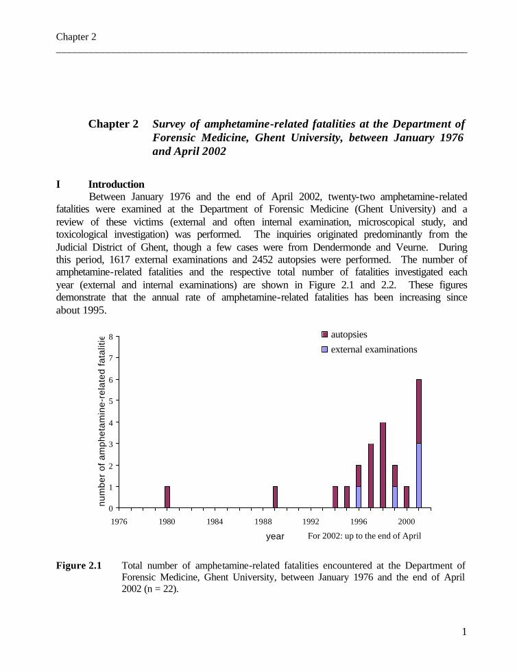
Chapter 2 _________________________________________________________________________________
1
Chapter 2 Survey of amphetamine-related fatalities at the Department of
Forensic Medicine, Ghent University, between January 1976 and April 2002
I Introduction Between January 1976 and the end of April 2002, twenty-two amphetamine-related
fatalities were examined at the Department of Forensic Medicine (Ghent University) and a review of these victims (external and often internal examination, microscopical study, and toxicological investigation) was performed. The inquiries originated predominantly from the Judicial District of Ghent, though a few cases were from Dendermonde and Veurne. During this period, 1617 external examinations and 2452 autopsies were performed. The number of amphetamine-related fatalities and the respective total number of fatalities investigated each year (external and internal examinations) are shown in Figure 2.1 and 2.2. These figures demonstrate that the annual rate of amphetamine-related fatalities has been increasing since about 1995.
Figure 2.1 Total number of amphetamine-related fatalities encountered at the Department of
Forensic Medicine, Ghent University, between January 1976 and the end of April 2002 (n = 22).
0
1
2
3
4
5
6
7
8
1976 1980 1984 1988 1992 1996 2000
year
num
ber
of a
mph
etam
ine-
rela
ted
fata
litie
s autopsiesexternal examinations
For 2002: up to the end of April
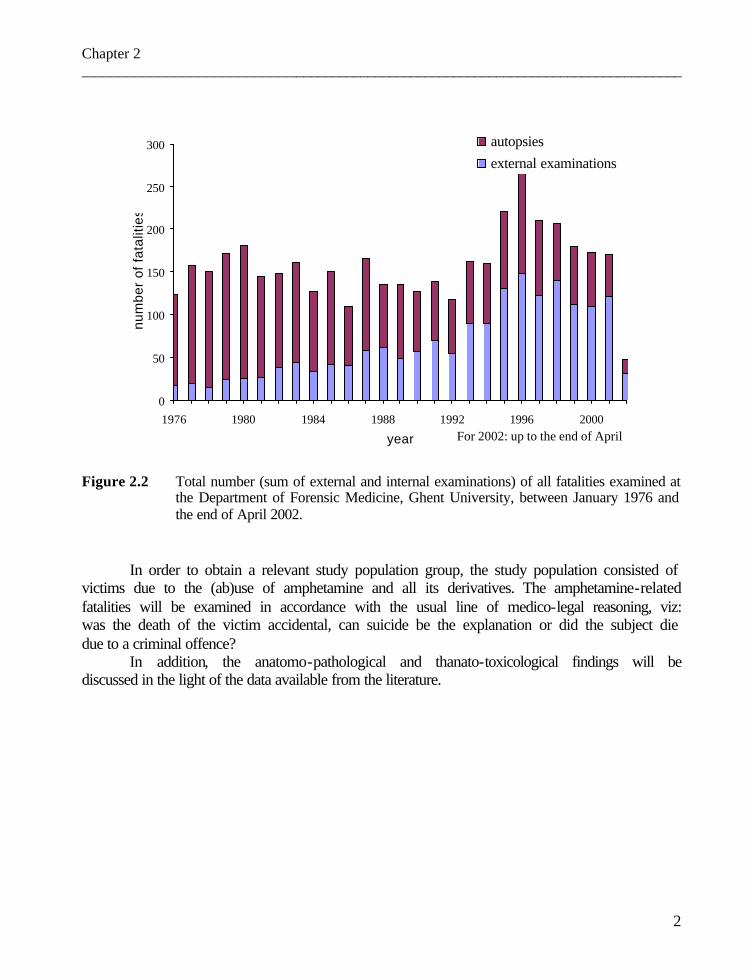
Chapter 2 _________________________________________________________________________________
2
Figure 2.2 Total number (sum of external and internal examinations) of all fatalities examined at
the Department of Forensic Medicine, Ghent University, between January 1976 and the end of April 2002.
In order to obtain a relevant study population group, the study population consisted of
victims due to the (ab)use of amphetamine and all its derivatives. The amphetamine-related fatalities will be examined in accordance with the usual line of medico-legal reasoning, viz: was the death of the victim accidental, can suicide be the explanation or did the subject die due to a criminal offence?
In addition, the anatomo-pathological and thanato-toxicological findings will be discussed in the light of the data available from the literature.
0
50
100
150
200
250
300
1976 1980 1984 1988 1992 1996 2000
year
num
ber
of fa
talit
ies
autopsiesexternal examinations
For 2002: up to the end of April

Chapter 2 _________________________________________________________________________________
3
II Case studies Twenty-two cases were discovered between January 1976 and April 2002. In the case
history, relevant data such as information obtained from the police inquiry (if available) and medical precedents are specified. Thereafter, the findings obtained during the external examination and autopsy followed by the histological data will be summarized. The biometrical data, the organ weights and the reference weight of each organ at various ages are presented in Table 2.1 (a) and Table 2.1 (b), respectively (1). An increased organ weight can be correlated with the mechanism of death (e.g. pulmonary congestion and oedema, brain oedema) or with pre-existing pathology (e.g. cardiomyopathy, liver steatosis). The toxicological data are summarized below (Table 2.2). II.1 Case 80/181
This 15-year-old boy suddenly collapsed about 15 minutes after the start of a football game. There were no surrounding players involved at the very moment of his collapse. Intensive reanimation was performed for a period of about 1.15 h. During external examination, signs of intensive reanimation were found. There were no other traumatic lesions. During autopsy, red-brownish mucus was seen in the trachea. Bloody fluid was present in the thoracic cavities (300 ml). Tardieu spots were noted on the pleurae and pericardium. Inspection of the lungs revealed overwhelming pulmonary congestion and oedema. A generalized visceral congestion was present. Finally, a somewhat narrowed aortic ring (perimeter of 5 cm) was observed. Histological examination disclosed the following findings: heart :congestion, slight focal fatty infiltration, no inflammatory infiltrates lungs: congestion and oedema liver: vacuolisation of the hepatocytes but no fatty infiltration with Scharlach red staining; slight non-specific portitis (lymphocyte infiltration) kidney: congestion brain and brainstem: congestion, a few perivascular bleedings in the brainstem. II.2 Case 89/88
A 28-year-old man ate mussels on the evening of the 20th of May. He had diarrhoea the following night and died on the 21st between 5.45 and 8.10 a.m. During external examination, obvious signs of cardiopulmonary failure (e.g. pronounced cyanotic face), a few small skin lesions consistent with a slight fall or impact and signs of reanimation were observed. At autopsy, vomit was present in the trachea. Overwhelming pulmonary congestion and oedema, and brain oedema were observed. Histological examination disclosed the following findings: heart: signs of ischemia, slight myocardosis, arteriolar media hypertrophy lungs: severe congestion and oedema, neutrophilic sludging, focal subpleural bleeding liver: congestion, slight neutrophilic sludging kidney: congestion

Chapter 2 _________________________________________________________________________________
4
brain and brainstem: congestion and oedema, a few perivascular bleedings in the brainstem II.3 Case 94/15 A 19-year-old man took 10 “XTC” pills at about 10:00 a.m. He became comatose
and was admitted to hospital at 2:00 p.m.: his temperature was 41.7°C and epileptic fits were noticed. He died at about 10:30 p.m. following DIC and multiple organ failure. Ante-mortem samples taken on two different times were available. Toxicological analysis of the serum and urine samples taken upon arrival at the hospital disclosed MDEA concentrations of 2.22 and 24.15 µg/ml, respectively. A few hours later, blood and urine samples were again taken in the hospital and immunoassay determined the following concentrations: amphetamines 0.120 µg/ml, cocaine 0.500 µg/ml, cannabinoids: 0.022 µg/ml, benzodiazepines: 0.224 µg/ml. The corresponding urine levels were 2.400 µg/ml, 6.000 µg/ml, 0.210 µg/ml, and 0.181 µg/ml, respectively. Ethanol was only present in urine (0.54 g/l). The MDEA blood and urine levels were 1.28 and 24.84 µg/ml, respectively. Analysis of a pill disclosed that it contained 119 mg of MDEA. The external inspection showed signs of intensive reanimation, a few slight skin lesions and bleeding out of the nose. During autopsy, 400 ml of bloody fluid were found in the thoracic cavities, as well as Tardieu spots on pleurae and pericardium. A tongue bite was present. On incision, overwhelming pulmonary congestion and oedema were seen. In addition, obvious brain congestion and oedema were observed. Histological examination disclosed the following findings: heart: slight myocardosis lungs: obvious congestion and haemorrhagic oedema liver: slight steatosis (degree I) kidney: acute tubular necrosis brain and brainstem: moderate oedema. II.4 Case 95/271
A 19-year-old female was found unconscious in bed; intensive reanimation failed. External examination revealed signs of intensive reanimation, scars of automutilation, and obvious signs of cardiopulmonary failure (e.g. cyanotic face). At autopsy, yellowish mucus and foamy fluid in trachea, pus plugs in the lungs, zones of dysaeration, and pulmonary congestion and oedema were observed. Tardieu spots on pleurae and pericardium and generalized visceral congestion were noticed. Brain congestion and oedema were remarked. Histological examination disclosed the following findings: heart: congestion lungs: pulmonary hypertension, acute bronchiolitis, onset bronchopneumonia, foreign body granulomata (cf. chronic intravenous drug (ab)use) liver: sign of shock: centrolobular atrophia, vacuolisation of the hepatocytes; moderate non-specific portitis (lymphocyte infiltration) kidney: signs of shock, including acute tubular necrosis

Chapter 2 _________________________________________________________________________________
5
brain and brainstem: congestion and oedema, a few perivascular haemorrhages in the brainstem. II.5 Case 96/26
A 22-year-old man who had joined the Navy, was found dead in his room. He had no medical antecedents. During the external examination, a few skin lesions consistent with a slight fall or impact and signs indicating reanimation were noted. At autopsy, Tardieu spots on pleurae and pericardium, and obvious pulmonary congestion and oedema were found. A somewhat enlarged heart and brain oedema were seen. Histological examination disclosed the following findings: heart: pronounced vascular congestion, slight endocardial fibrosis lungs: pronounced congestion and oedema, slight bone marrow embolisation (in the absence of fractures, assumed to be resulted from severe hyperthermic convulsions), obstruction of smaller bronchi by mucoid material, chronic bronchitis (lympho-plasmocytic and eosinophilic infiltrates) liver: steatosis (degree I to II), acute congestion kidney: acute tubular necrosis, acute congestion brain and brainstem: congestion and oedema. II.6 Case 96/96/1
This 21-year-old woman was found dead in bed, in the arms of her friend. They were recently discharged from hospital for detoxification therapy. They committed suicide together. In this case, the investigation was restricted to an external examination. At external inspection moderate putrefaction was found. Dried oedema around the mouth was noted. A puncture wound at the left wrist was assumed. II.7 Case 97/35
This 19-year-old man was murdered by a thoracic gunshot following an altercation. MDMA use was an incidental discovery. During external examination, signs of reanimation and a few skin lesions consistent with a fall or impact were noted. A thoracic shotgun lesion ("à bout touchant") and subcutaneous emphysema were observed. At autopsy, a haemothorax (1600 ml), a cardiac laceration and blood aspiration were found. The abdominal organs were obviously anemic. In addition, a galea ecchymose (cf. fall) and some brain congestion and oedema were seen. Histological examination disclosed the following findings: heart: normal myocardial tissue apart from anemia lungs: intra-alveolar bleeding (blood aspiration) liver: normal but anemic kidney: normal, anemic brain and brainstem: moderate oedema.

Chapter 2 _________________________________________________________________________________
6
II.8 Case 97/134 This 19-year-old man was found dead lying in a sofa. He was known to have had an
allergic constitution (atopic eczema) and at the scene a bronchodilator was found. An erosion of the nasal septum was noted, which indicated sniffing. A prolonged survival following polydrug abuse was assumed. MDEA was found positive in urine and below the limit of detection in blood (< LOD). During the external inspection, reanimation signs, an obvious cyanotic face and eczema on the right elbow and poplitea (cf. allergic constitution ?) were found. During autopsy, Tardieu spots on pleurae and pericardium were observed. Mucus and vomit plugs in the bronchi were found. Upon incision, obvious pulmonary congestion and oedema was seen. Apart from generalized visceral congestion, brain congestion and oedema were noticed. Histological examination disclosed the following findings: heart: pronounced vascular congestion, slight myocardosis lungs: dysaeration, subacute vascular congestion and haemorrhagic oedema, acute bronchitis and onset bronchopneumonia, onset diffuse alveolar damage (DAD; shock lungs) liver: congestion kidney: pronounced acute vascular congestion pituitary gland: congestion brain and brainstem: acute congestion and oedema, a few perivascular bleedings, signs of onset hypoxia in the hippocampus.
II.9 Case 97/156 A 32-year-old man was detained by the police for “drunkenness”. In jail, he was very
aggressive and disoriented for a few hours and finally he hung himself (using his shirt fixed on the bars) while still in custody. The neuroleptic sulpiride (Dogmatil®) and acetaminophen with codeine (Dafalgan Codeine®) were in his home. At external examination, a ligature mark at the neck and various fresh ecchymoses and excoriations on the limbs (consistent with repeated falls or impacts) were observed. At autopsy, Tardieu spots on pleurae and pericardium, obvious pulmonary congestion and generalized visceral congestion were noted. Slight brain congestion and oedema were found. Histological examination disclosed the following findings: heart: acute vascular congestion, slight myocardosis lungs: pronounced acute vascular congestion and haemorrhagic oedema; chronic bronchitis, chronic pleuritis liver: pronounced acute vascular congestion kidney: acute congestion skin right arm and left elbow: subcutaneous ecchymoses (> 5 hours old) pituitary gland: acute congestion brain and brainstem: acute congestion and oedema; a few perivascular bleedings; sludging and localized perivasculitis in caudate nucleus, sludging in brainstem, obvious perivascular bleedings around the third ventricle with slight neutrophilic infiltration.
Chapter 2 _________________________________________________________________________________
7
II.10 Case 98/14 This 40-year-old man died in a traffic accident following an argument.
Upon external examination, scattered abrasions and ecchymoses on head, limbs and thorax were found. At autopsy, pleural Tardieu spots, pulmonary congestion and an enlarged heart were noticed. Multiple fractures of the skull and the skull base associated with a subdural and arachnoideal bleeding were observed. In addition, an epi- and subdural bleeding of cervical spine was found. Histological examination disclosed the following findings: heart: acute vascular congestion, moderate myocardosis, signs of protracted hypoxia lungs: emphysema with pulmonary hypertension, slight chronic bronchitis, obvious blood aspiration, pronounced congestion and oedema, sludging with onset shock lungs liver: acute vascular congestion, steatosis (3rd degree) kidney: acute vascular congestion, signs of shock pituitary gland: traumatic haemorrhagic zone in neurohypophysis with slight inflammatory infiltration (some hours old) skin occiput: abrasion (ca. 10 hours old) brain and brainstem: acute congestion and oedema; periventricular bleeding in brainstem (possibly of traumatic origin), recent cerebellar and cerebral contusions, signs of hypoxia in lentiform nucleus, thalamus, cortical regions and hippocampus, recent contusion of the cervical medulla.
II.11 Case 98/41
This 22-year-old driver suddenly left the main road without immediate cause. He was admitted to hospital where he received 10 units of packed cells and several units of plasma expanders as part of the treatment for haemorrhagic shock. He died a few hours later. At autopsy, an extremely tiny man was found (BMI = 17) and Graves’ disease was incidentally discovered. Upon external examination, apart from signs of intensive reanimation, scattered abrasions, lacerations and ecchymoses were found. At autopsy, a haemothorax (290 ml), lung contusions and pulmonary oedema were observed. About 750 ml blood in the abdomen due to a laceration of the liver and small bowel perforations were seen. Stress ulcerations in the stomach mucosa were present. Subdural bleeding, brain contusions and oedema, associated with an epidural bleeding of the cervical spine were found. Histological examination disclosed the following findings: heart: some congestion and some sludging, a few small focal lymphocyte infiltrates, slight myocardosis lungs: obvious chronic bronchitis, sludging as part of onset shock lungs, a few scattered foreign body granulomata, obvious fat embolisation liver: findings of protracted shock kidney: normal, anemic spleen: immunoreactive spleen

Chapter 2 _________________________________________________________________________________
8
thyroid gland: chronic lymphocytic thyroiditis compatible with Graves' disease pituitary gland: acute congestion brain and brainstem: oedema, subarachnoideal bleeding, a few small recent brain contusions, commotio of the brainstem, signs of hypoxia in pons and hippocampus.
II.12 Case 98/239 This 24-year-old man had antecedents of drug abuse and was treated with a few
benzodiazepines; the narcotic analgesic piritramide (Dipidolor®) was found. He was found dead lying in an armchair on his back. No reanimation took place. Glucose was strongly positive in urine (++++). External inspection demonstrated signs of cardiopulmonary failure. Scars of automutilation and a small ecchymose on the left forearm possibly compatible with a intravenous puncture were observed. At autopsy, mucus and cloudy oedema in bronchi, hepatisation regions of the left lung, and overwhelming pulmonary congestion and oedema were found. About 175 ml of bloody fluid was present in the thoracic cavities. Some brain oedema was also observed. Histological examination disclosed the following findings: heart: pronounced acute vascular congestion; small scattered subepicardial bleedings lungs: onset bronchopneumonia, obvious vascular congestion and a few microthrombi, polynuclear macrophages with crystalline inclusions (cf. chronic intravenous drug abuse). liver: acute vascular congestion kidney: acute vascular congestion carinal lymph node: acute reactive lymphadenitis skin of left forearm: recent subcutaneous bleeding pituitary gland: acute congestion brain and brainstem: acute congestion and oedema, scattered small perivascular bleedings, signs of protracted hypoxia in cerebellum, pons, cortical regions, hippocampus. Small hippocampal bleedings (cf. increased intracranial pressure).
II.13 Case 98/251 This 31-year-old man suddenly collapsed in a discotheque. His friends declared that
he had used XTC and marihuana on a regular basis. Analysis of a tablet found on this person demonstrated an MDMA content of 9 % or 25.23 mg MDMA. It is not known how many pills he may have ingested. External examination showed signs of intensive reanimation and obvious signs of cardiopulmonary failure. During autopsy, an obviously enlarged heart and a recurrent myocardial infarction on an old lesion associated with a recent coronary thrombosis was found. For his age, a moderate atherosclerosis of all coronary arteries was observed. Overwhelming pulmonary congestion and oedema as well as generalized visceral congestion were present. In addition, brain oedema and a tongue bite were noted.

Chapter 2 _________________________________________________________________________________
9
Histological examination disclosed the following findings: heart: extensive transmural infarction, which is at least ca. 5 weeks old; smaller recent infarction zone, ca. 5 days old; also more recent cellular hypoxia, and recent thrombosis of ramus circumflexus. lungs: pronounced acute vascular congestion and haemorrhagic oedema; chronic bronchitis, deep vomit aspiration, bone marrow embolisation (cf. rib fractures due to reanimation) liver: acute vascular congestion kidney: intima fibrosis; signs of onset shock pituitary gland: acute congestion brain and brainstem: congestion and oedema, signs of protracted hypoxia in cortical regions, cerebellum and hippocampus.
II.14 Case 99/231 This 18-year-old man was found dead in bed. He was known to be addicted to drugs
and had therefore been receiving treatment with methadone and benzodiazepines. Oedema and vomit was present on his bed linen. In his pocket, a small box containing numerous tablets, mainly medication, was retrieved. The investigation was restricted to an external examination. Obvious signs of cardiopulmonary failure were found (e.g. obvious pulmonary oedema, cyanosis).
II.15 Case 99/235
This 27-year-old drug dealer died following ingestion of about 6 pills. In his pockets, a note describing the effects of 4-MTA was found. Upon external inspection, besides signs of reanimation, slight ecchymoses on limbs consistent with a minor fall or impact were observed. Autopsy showed Tardieu spots on pleurae and pericardium associated with obvious pulmonary congestion and oedema. Stress ulcerations in the stomach mucosa were present. Histological examination disclosed the following findings: heart: acute vascular congestion, slight myocardosis lungs: overwhelming vascular congestion and haemorrhagic oedema, some chronic bronchitis liver: acute vascular congestion kidney: acute vascular congestion spleen: acute vascular congestion pituitary gland: congestion brain and brainstem: congestion and oedema; a few perivascular bleedings in pons and brain. Somewhat decreased number of Purkinje cells, perivascular lymphocyte and macrophage infiltration in brainstem and lentiform nucleus. This case is discussed in detail in Chapter 6 of the present work (2,3).

Chapter 2 _________________________________________________________________________________
10
II.16 Case 00/112 This 23-year-old man was found dead in a bar, sitting on a chair.
External examination showed obvious signs of cardiopulmonary failure (e.g. pronounced cyanotic face) and reanimation signs. At autopsy, a great deal of bloody oedema was present in trachea and bronchi. Numerous Tardieu spots on pleurae and epicardium, and an overwhelming lung congestion and oedema were found. A persistence of a left superior caval vein was an incidental discovery. Apart from generalized visceral congestion, congestion of the arachnoidea was found. Histological examination disclosed the following findings: heart: acute vascular congestion, moderate myocardosis, signs of diffuse hypoxia (subendocardial); some fatty infiltration and slight bleedings in the right ventricle lungs: overwhelming vascular congestion and haemorrhagic oedema liver: acute vascular congestion, slight steatosis (1st degree) kidney: acute vascular congestion spleen: acute vascular congestion pituitary gland: congestion brain and brainstem: congestion and oedema; a few perivascular bleedings in pons and brain, perivasculitis (lymphocyte infiltration); no obvious signs of protracted hypoxia (normal hippocampus). This case is also studied in detail in Chapter 6 of this thesis (4).
II.17 Case 01/29 According to the statements of his family and friends, this 17-year old man was not
obviously depressive, though he was somewhat down at times. His motorbike and a bottle of Dutch gin were retrieved nearby the train rails. Marihuana was found in his pockets. It was assumed that he threw himself in front of a passing train. The investigation was restricted to an external examination. A severe cranio-cervical destruction and multiple fractures (thorax, limbs) were found. Inguinal lacerations indicate that he was hit in standing position. Due to the severe cranio-cerebral destruction (including orbital fractures), there was no vitreous humour available. The urine was very bloody (cf. pubic fractures).
II.18 Case 01/34 A 23-year-old man was found dead at home, lying in an armchair. Police inquiry
revealed that he had many visitors and therefore he was suspected to be a drug dealer, which was – to our knowledge – not confirmed. At external examination, onset of putrefaction (onset epidermolysis, dehydration of the eyes, mummification of the fingers and toes), a few small excoriations on the forearms were observed. At autopsy, emphysema of the lungs due to putrefaction, oedema in the trachea and bronchi and obvious pulmonary congestion were noticed. About 250 ml bloody fluid was present in the thoracic cavities. In addition to generalized congestion of all organs, obvious brain oedema was found.
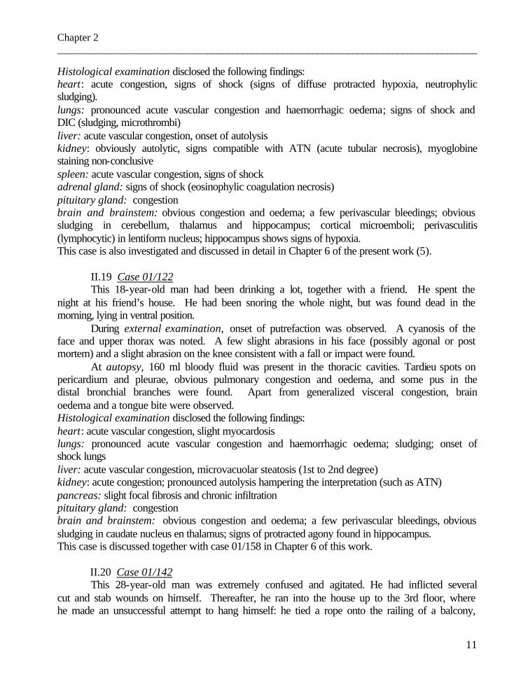
Chapter 2 _________________________________________________________________________________
11
Histological examination disclosed the following findings: heart: acute congestion, signs of shock (signs of diffuse protracted hypoxia, neutrophylic sludging). lungs: pronounced acute vascular congestion and haemorrhagic oedema; signs of shock and DIC (sludging, microthrombi) liver: acute vascular congestion, onset of autolysis kidney: obviously autolytic, signs compatible with ATN (acute tubular necrosis), myoglobine staining non-conclusive spleen: acute vascular congestion, signs of shock adrenal gland: signs of shock (eosinophylic coagulation necrosis) pituitary gland: congestion brain and brainstem: obvious congestion and oedema; a few perivascular bleedings; obvious sludging in cerebellum, thalamus and hippocampus; cortical microemboli; perivasculitis (lymphocytic) in lentiform nucleus; hippocampus shows signs of hypoxia. This case is also investigated and discussed in detail in Chapter 6 of the present work (5).
II.19 Case 01/122 This 18-year-old man had been drinking a lot, together with a friend. He spent the
night at his friend’s house. He had been snoring the whole night, but was found dead in the morning, lying in ventral position.
During external examination, onset of putrefaction was observed. A cyanosis of the face and upper thorax was noted. A few slight abrasions in his face (possibly agonal or post mortem) and a slight abrasion on the knee consistent with a fall or impact were found.
At autopsy, 160 ml bloody fluid was present in the thoracic cavities. Tardieu spots on pericardium and pleurae, obvious pulmonary congestion and oedema, and some pus in the distal bronchial branches were found. Apart from generalized visceral congestion, brain oedema and a tongue bite were observed. Histological examination disclosed the following findings: heart: acute vascular congestion, slight myocardosis lungs: pronounced acute vascular congestion and haemorrhagic oedema; sludging; onset of shock lungs liver: acute vascular congestion, microvacuolar steatosis (1st to 2nd degree) kidney: acute congestion; pronounced autolysis hampering the interpretation (such as ATN) pancreas: slight focal fibrosis and chronic infiltration pituitary gland: congestion brain and brainstem: obvious congestion and oedema; a few perivascular bleedings, obvious sludging in caudate nucleus en thalamus; signs of protracted agony found in hippocampus. This case is discussed together with case 01/158 in Chapter 6 of this work. II.20 Case 01/142
This 28-year-old man was extremely confused and agitated. He had inflicted several cut and stab wounds on himself. Thereafter, he ran into the house up to the 3rd floor, where he made an unsuccessful attempt to hang himself: he tied a rope onto the railing of a balcony,
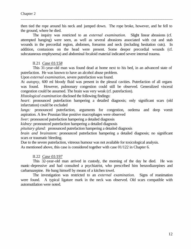
Chapter 2 _________________________________________________________________________________
12
then tied the rope around his neck and jumped down. The rope broke, however, and he fell to the ground, where he died.
The inquiry was restricted to an external examination. Slight linear abrasions (cf. attempted hanging) were seen, as well as several abrasions associated with cut and stab wounds in the precordial region, abdomen, forearms and neck (including hesitation cuts). In addition, contusions on the head were present. Some deeper precordial wounds (cf. subcutaneous emphysema) and abdominal fecaloid material indicated severe internal trauma. II.21 Case 01/158
This 31-year-old man was found dead at home next to his bed, in an advanced state of putrefaction. He was known to have an alcohol abuse problem. Upon external examination, severe putrefaction was found. At autopsy, 600 ml bloody fluid was present in the pleural cavities. Putrefaction of all organs was found. However, pulmonary congestion could still be observed. Generalized visceral congestion could be assumed. The brain was very weak (cf. putrefaction). Histological examination disclosed the following findings: heart: pronounced putrefaction hampering a detailed diagnosis; only significant scars (old infarctation) could be excluded lungs: pronounced putrefaction, arguments for congestion, oedema and deep vomit aspiration. A few Prussian blue positive macrophages were observed liver: pronounced putrefaction hampering a detailed diagnosis kidney: pronounced putrefaction hampering a detailed diagnosis pituitary gland: pronounced putrefaction hampering a detailed diagnosis brain and brainstem: pronounced putrefaction hampering a detailed diagnosis; no significant scars or traumatic bleeding. Due to the severe putrefaction, vitreous humour was not available for toxicological analysis. As mentioned above, this case is considered together with case 01/122 in Chapter 6.
II.22 Case 01/197 This 32-year-old man arrived in custody, the morning of the day he died. He was
manic-depressive and had consulted a psychiatrist, who prescribed him benzodiazepines and carbamazepine. He hung himself by means of a kitchen towel.
The investigation was restricted to an external examination. Signs of reanimation were found. A typical ligature mark in the neck was observed. Old scars compatible with automutilation were noted.

Chapter 2 _________________________________________________________________________________
13
Table 2.1 (a) Biometrical data and organ weights of the amphetamine-related fatalities at the Department of Forensic Medicine, Ghent University (n = 17). (BMI: body mass index)
case age length weight BMI brain heart lungs liver kidneys spleen
number (years) (cm) (kg) (g) (g) (g) (g) (g) (g) 80/181 15 182 65 19.6 1355 290 1375 1720 338 235 89/88 28 169 62 21.7 1470 295 1085 1510 220 100 94/15 19 183 72 21.4 1440 300 1960 1430 300 100 95/271 19 164 53 19.7 1200 315 955 1370 320 215 96/26 22 172 87 29.4 1350 437 1358 1550 310 150 97/35 19 174 55 18.1 1410 243 440 1100 220 90 97/134 19 169 55 19.2 1430 280 1290 1745 250 170 97/156 32 176 65 20.9 1500 335 1350 1510 300 100 98/14 40 177 81 25.6 1465 480 1605 1900 480 90 98/41 22 178 57 18.0 1385 254 1550 1255 245 245 98/239 24 170 61 21.1 1560 345 1495 1545 320 85 98/251 31 171 75 25.6 1580 460 1300 2235 460 180 99/235 27 171 55 18.8 1320 255 970 1300 280 200 00/112 23 186 95 27.5 1545 405 1620 1950 265 275 01/34 23 175 56 18.3 1405 315 1410 1160 190 235 01/122 18 177 61 19.5 1445 270 1280 1470 250 200 01/158 31 178 85 26.8 1300 350 1230 1250 325 120 mean 175 67 22 1421 331 1310 1529 298 164 (SD) (5.7) (13.1) (3.7) (98.9) (73.5) (332.8) (302.3) (77.3) (64.3) Table 2.1 (b) Reference weights of the organs at various ages (in grams).
[reproduced in part from Boyd E. (1)]
age brain heart lungs liver kidneys spleen (years) men women men women men women men women men women men women
15 - 16 1407 1271 258 238 692 709 1315 1330 229 230 135 127 16 - 17 1419 1300 282 243 747 626 1380 1360 244 236 145 134 17 - 18 1409 1254 300 247 777 650 1450 1380 260 240 152 140 18 - 19 1426 1312 310 250 875 655 1510 1395 270 244 157 146 19 - 20 1430 1294 318 251 1036 785 1580 1405 282 247 160 151 20 - 21 - - 322 252 953 793 1630 1415 290 248 162 155
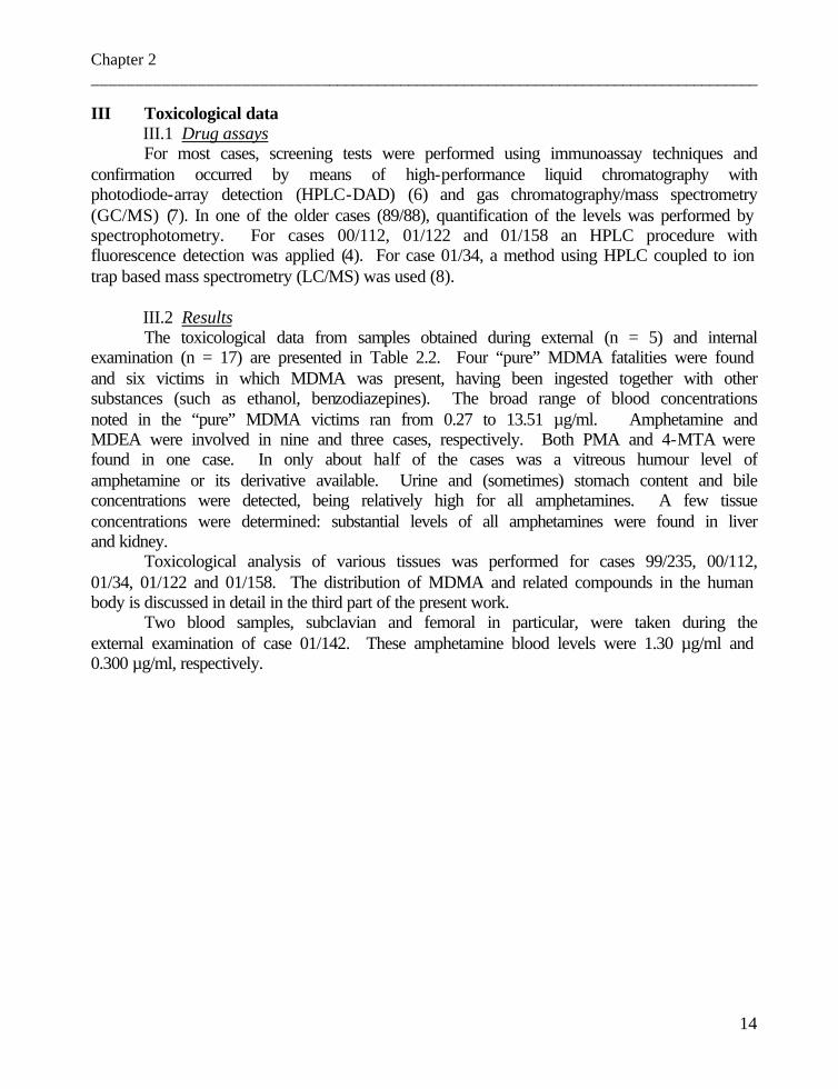
Chapter 2 _________________________________________________________________________________
14
III Toxicological data III.1 Drug assays For most cases, screening tests were performed using immunoassay techniques and
confirmation occurred by means of high-performance liquid chromatography with photodiode-array detection (HPLC-DAD) (6) and gas chromatography/mass spectrometry (GC/MS) (7). In one of the older cases (89/88), quantification of the levels was performed by spectrophotometry. For cases 00/112, 01/122 and 01/158 an HPLC procedure with fluorescence detection was applied (4). For case 01/34, a method using HPLC coupled to ion trap based mass spectrometry (LC/MS) was used (8).
III.2 Results The toxicological data from samples obtained during external (n = 5) and internal
examination (n = 17) are presented in Table 2.2. Four “pure” MDMA fatalities were found and six victims in which MDMA was present, having been ingested together with other substances (such as ethanol, benzodiazepines). The broad range of blood concentrations noted in the “pure” MDMA victims ran from 0.27 to 13.51 µg/ml. Amphetamine and MDEA were involved in nine and three cases, respectively. Both PMA and 4-MTA were found in one case. In only about half of the cases was a vitreous humour level of amphetamine or its derivative available. Urine and (sometimes) stomach content and bile concentrations were detected, being relatively high for all amphetamines. A few tissue concentrations were determined: substantial levels of all amphetamines were found in liver and kidney.
Toxicological analysis of various tissues was performed for cases 99/235, 00/112, 01/34, 01/122 and 01/158. The distribution of MDMA and related compounds in the human body is discussed in detail in the third part of the present work.
Two blood samples, subclavian and femoral in particular, were taken during the external examination of case 01/142. These amphetamine blood levels were 1.30 µg/ml and 0.300 µg/ml, respectively.
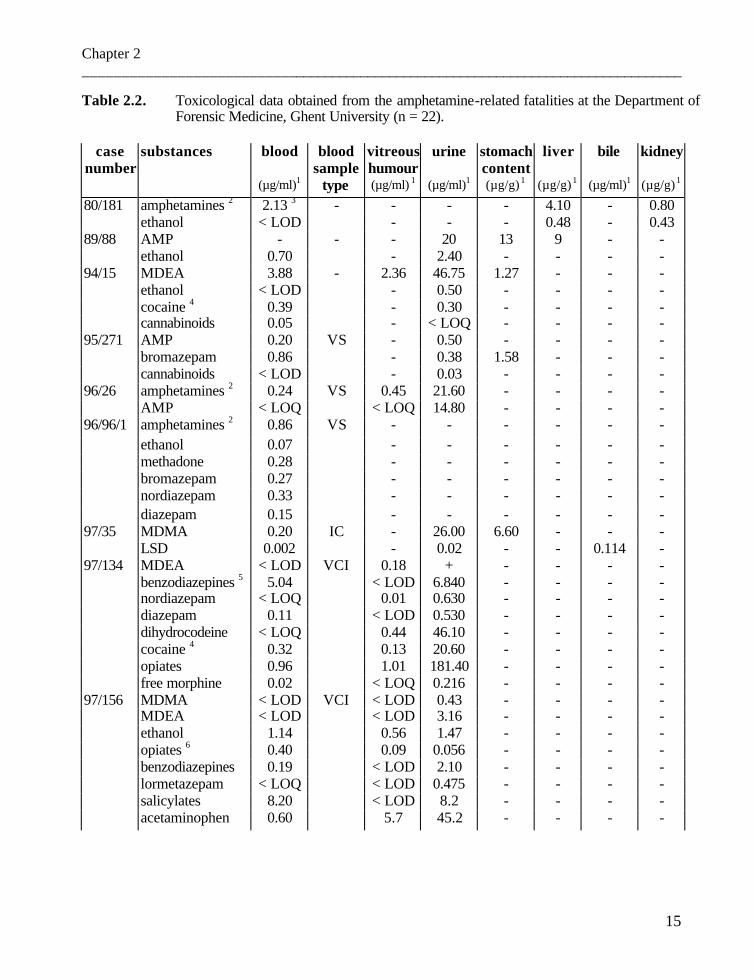
Chapter 2 _________________________________________________________________________________
15
Table 2.2. Toxicological data obtained from the amphetamine-related fatalities at the Department of Forensic Medicine, Ghent University (n = 22).
case substances blood blood vitreous urine stomach liver bile kidney
number sample humour content (µg/ml)1 type (µg/ml) 1 (µg/ml)1 (µg/g) 1 (µg/g) 1 (µg/ml)1 (µg/g) 1
80/181 amphetamines 2 2.13 3 - - - - 4.10 - 0.80 ethanol < LOD - - - 0.48 - 0.43 89/88 AMP - - - 20 13 9 - - ethanol 0.70 - 2.40 - - - - 94/15 MDEA 3.88 - 2.36 46.75 1.27 - - - ethanol < LOD - 0.50 - - - - cocaine 4 0.39 - 0.30 - - - - cannabinoids 0.05 - < LOQ - - - - 95/271 AMP 0.20 VS - 0.50 - - - - bromazepam 0.86 - 0.38 1.58 - - - cannabinoids < LOD - 0.03 - - - - 96/26 amphetamines 2 0.24 VS 0.45 21.60 - - - - AMP < LOQ < LOQ 14.80 - - - - 96/96/1 amphetamines 2 0.86 VS - - - - - - ethanol 0.07 - - - - - - methadone 0.28 - - - - - - bromazepam 0.27 - - - - - - nordiazepam 0.33 - - - - - - diazepam 0.15 - - - - - - 97/35 MDMA 0.20 IC - 26.00 6.60 - - - LSD 0.002 - 0.02 - - 0.114 - 97/134 MDEA < LOD VCI 0.18 + - - - - benzodiazepines 5 5.04 < LOD 6.840 - - - - nordiazepam < LOQ 0.01 0.630 - - - - diazepam 0.11 < LOD 0.530 - - - - dihydrocodeine < LOQ 0.44 46.10 - - - - cocaine 4 0.32 0.13 20.60 - - - - opiates 0.96 1.01 181.40 - - - - free morphine 0.02 < LOQ 0.216 - - - - 97/156 MDMA < LOD VCI < LOD 0.43 - - - - MDEA < LOD < LOD 3.16 - - - - ethanol 1.14 0.56 1.47 - - - - opiates 6 0.40 0.09 0.056 - - - - benzodiazepines 0.19 < LOD 2.10 - - - - lormetazepam < LOQ < LOD 0.475 - - - - salicylates 8.20 < LOD 8.2 - - - - acetaminophen 0.60 5.7 45.2 - - - -
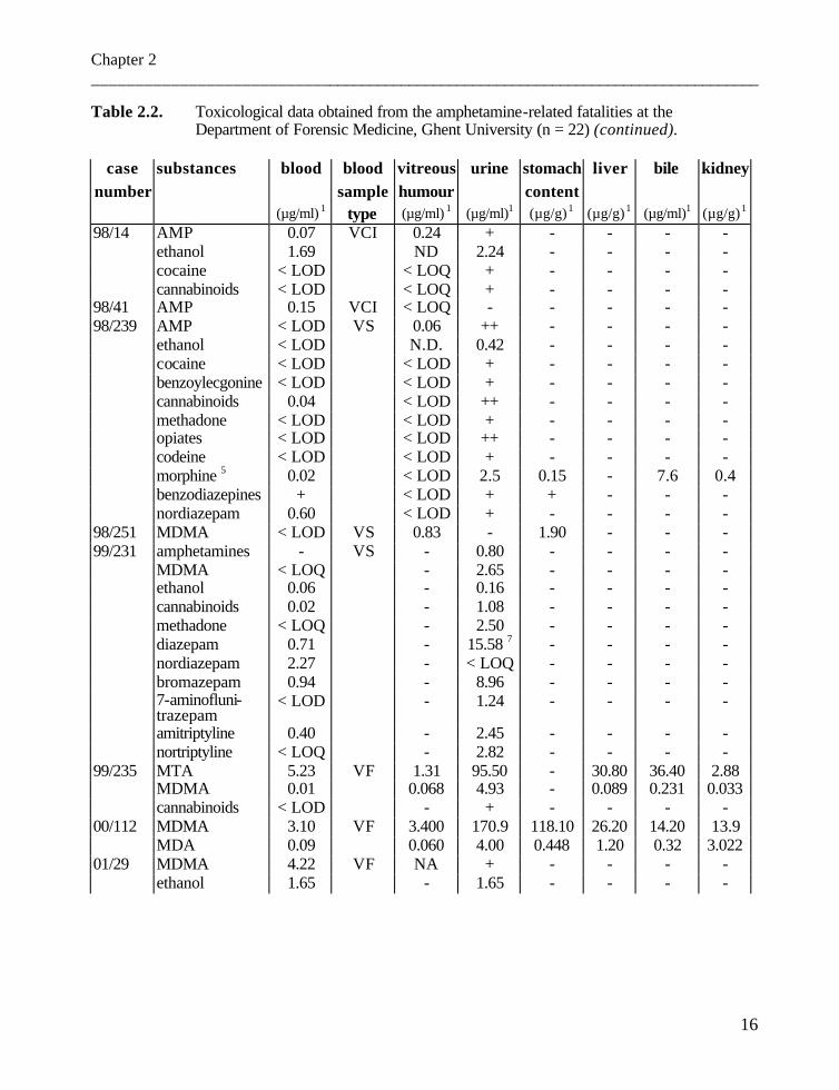
Chapter 2 _________________________________________________________________________________
16
Table 2.2. Toxicological data obtained from the amphetamine-related fatalities at the Department of Forensic Medicine, Ghent University (n = 22) (continued).
case substances blood blood vitreous urine stomach liver bile kidney
number sample humour content (µg/ml) 1 type (µg/ml) 1 (µg/ml)1 (µg/g) 1 (µg/g) 1 (µg/ml)1 (µg/g) 1
98/14 AMP 0.07 VCI 0.24 + - - - - ethanol 1.69 ND 2.24 - - - - cocaine < LOD < LOQ + - - - - cannabinoids < LOD < LOQ + - - - - 98/41 AMP 0.15 VCI < LOQ - - - - - 98/239 AMP < LOD VS 0.06 ++ - - - - ethanol < LOD N.D. 0.42 - - - - cocaine < LOD < LOD + - - - - benzoylecgonine < LOD < LOD + - - - - cannabinoids 0.04 < LOD ++ - - - - methadone < LOD < LOD + - - - - opiates < LOD < LOD ++ - - - - codeine < LOD < LOD + - - - - morphine 5 0.02 < LOD 2.5 0.15 - 7.6 0.4 benzodiazepines + < LOD + + - - - nordiazepam 0.60 < LOD + - - - - 98/251 MDMA < LOD VS 0.83 - 1.90 - - - 99/231 amphetamines - VS - 0.80 - - - - MDMA < LOQ - 2.65 - - - - ethanol 0.06 - 0.16 - - - - cannabinoids 0.02 - 1.08 - - - - methadone < LOQ - 2.50 - - - - diazepam 0.71 - 15.58 7 - - - - nordiazepam 2.27 - < LOQ - - - - bromazepam 0.94 - 8.96 - - - - 7-aminofluni-
trazepam < LOD - 1.24 - - - -
amitriptyline 0.40 - 2.45 - - - - nortriptyline < LOQ - 2.82 - - - - 99/235 MTA 5.23 VF 1.31 95.50 - 30.80 36.40 2.88 MDMA 0.01 0.068 4.93 - 0.089 0.231 0.033 cannabinoids < LOD - + - - - - 00/112 MDMA 3.10 VF 3.400 170.9 118.10 26.20 14.20 13.9 MDA 0.09 0.060 4.00 0.448 1.20 0.32 3.022 01/29 MDMA 4.22 VF NA + - - - - ethanol 1.65 - 1.65 - - - -
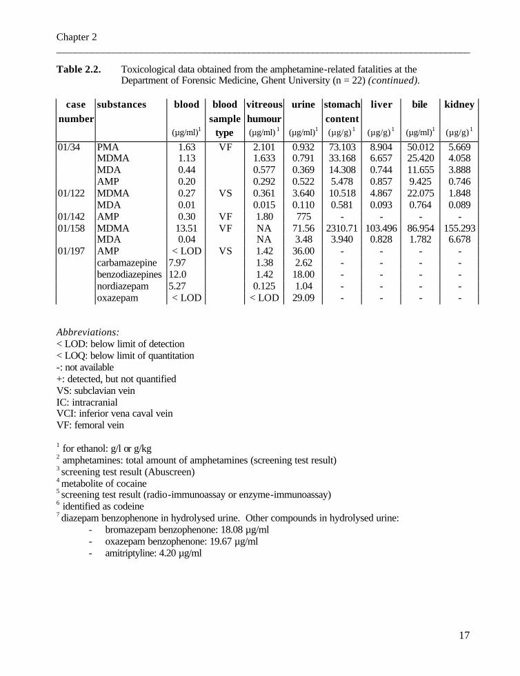
Chapter 2 _________________________________________________________________________________
17
Table 2.2. Toxicological data obtained from the amphetamine-related fatalities at the Department of Forensic Medicine, Ghent University (n = 22) (continued).
case substances blood blood vitreous urine stomach liver bile kidney
number sample humour content (µg/ml)1 type (µg/ml) 1 (µg/ml)1 (µg/g) 1 (µg/g) 1 (µg/ml)1 (µg/g) 1
01/34 PMA 1.63 VF 2.101 0.932 73.103 8.904 50.012 5.669 MDMA 1.13 1.633 0.791 33.168 6.657 25.420 4.058
MDA 0.44 0.577 0.369 14.308 0.744 11.655 3.888 AMP 0.20 0.292 0.522 5.478 0.857 9.425 0.746
01/122 MDMA 0.27 VS 0.361 3.640 10.518 4.867 22.075 1.848 MDA 0.01 0.015 0.110 0.581 0.093 0.764 0.089 01/142 AMP 0.30 VF 1.80 775 - - - - 01/158 MDMA 13.51 VF NA 71.56 2310.71 103.496 86.954 155.293 MDA 0.04 NA 3.48 3.940 0.828 1.782 6.678 01/197 AMP < LOD VS 1.42 36.00 - - - - carbamazepine 7.97 1.38 2.62 - - - - benzodiazepines 12.0 1.42 18.00 - - - - nordiazepam 5.27 0.125 1.04 - - - - oxazepam < LOD < LOD 29.09 - - - -
Abbreviations: < LOD: below limit of detection < LOQ: below limit of quantitation -: not available +: detected, but not quantified VS: subclavian vein IC: intracranial VCI: inferior vena caval vein VF: femoral vein 1 for ethanol: g/l or g/kg 2 amphetamines: total amount of amphetamines (screening test result) 3 screening test result (Abuscreen) 4 metabolite of cocaine 5 screening test result (radio-immunoassay or enzyme-immunoassay) 6 identified as codeine 7 diazepam benzophenone in hydrolysed urine. Other compounds in hydrolysed urine:
- bromazepam benzophenone: 18.08 µg/ml - oxazepam benzophenone: 19.67 µg/ml - amitriptyline: 4.20 µg/ml
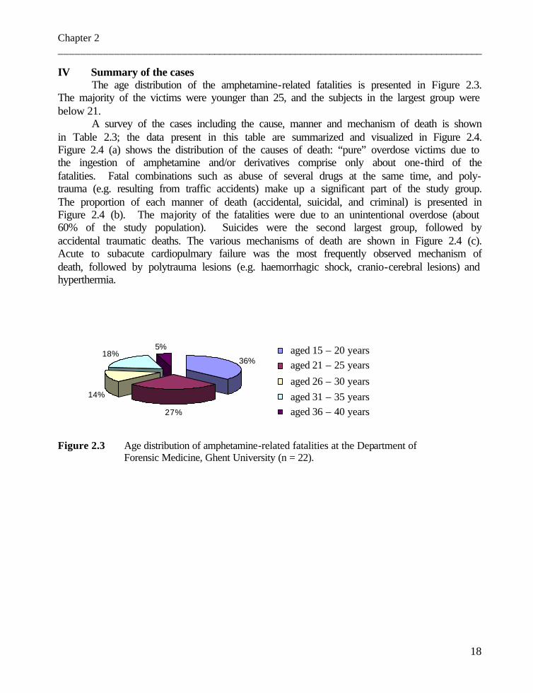
Chapter 2 _________________________________________________________________________________
18
IV Summary of the cases The age distribution of the amphetamine-related fatalities is presented in Figure 2.3.
The majority of the victims were younger than 25, and the subjects in the largest group were below 21.
A survey of the cases including the cause, manner and mechanism of death is shown in Table 2.3; the data present in this table are summarized and visualized in Figure 2.4. Figure 2.4 (a) shows the distribution of the causes of death: “pure” overdose victims due to the ingestion of amphetamine and/or derivatives comprise only about one-third of the fatalities. Fatal combinations such as abuse of several drugs at the same time, and poly-trauma (e.g. resulting from traffic accidents) make up a significant part of the study group. The proportion of each manner of death (accidental, suicidal, and criminal) is presented in Figure 2.4 (b). The majority of the fatalities were due to an unintentional overdose (about 60% of the study population). Suicides were the second largest group, followed by accidental traumatic deaths. The various mechanisms of death are shown in Figure 2.4 (c). Acute to subacute cardiopulmary failure was the most frequently observed mechanism of death, followed by polytrauma lesions (e.g. haemorrhagic shock, cranio-cerebral lesions) and hyperthermia.
Figure 2.3 Age distribution of amphetamine-related fatalities at the Department of Forensic Medicine, Ghent University (n = 22).
36%
27%
14%
18%5% 1
2
3
4
5
aged 15 – 20 years aged 21 – 25 years
aged 26 – 30 years aged 31 – 35 years aged 36 – 40 years
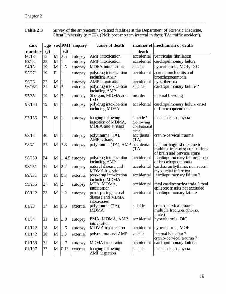
Chapter 2 _________________________________________________________________________________
19
Table 2.3 Survey of the amphetamine-related fatalities at the Department of Forensic Medicine, Ghent University (n = 22). (PMI: post-mortem interval in days; TA: traffic accident).
case age sex PMI inquiry cause of death manner of mechanism of death number (y) (d) death 80/181 15 M 2.5 autopsy AMP intoxication accidental ventricular fibrillation 89/88 28 M 1 autopsy AMP intoxication accidental cardiopulmonary failure 94/15 19 M 1.5 autopsy MDEA intoxication suicide hyperthermia, MOF, DIC 95/271 19 F 1 autopsy polydrug intoxica-tion
including AMP accidental acute bronchiolitis and
bronchopneumonia 96/26 22 M 1 autopsy AMP intoxication accidental hyperthermia 96/96/1 21 M 3 external polydrug intoxica-tion
including AMP suicide cardiopulmonary failure ?
97/35 19 M 3 autopsy Shotgun, MDMA and LSD
murder internal bleeding
97/134 19 M 1 autopsy polydrug intoxica-tion including MDEA
accidental cardiopulmonary failure onset of bronchopneumonia
97/156 32 M 1 autopsy hanging following ingestion of MDMA, MDEA and ethanol
suicide? (following confusional state)
mechanical asphyxia
98/14 40 M 1 autopsy polytrauma (TA), AMP, ethanol
accidental (TA)
cranio-cervical trauma
98/41 22 M 3.8 autopsy polytrauma (TA), AMP accidental (TA)
haemorrhagic shock due to multiple fractures; con- tusions of brain and cervical spine
98/239 24 M ± 4.5 autopsy polydrug intoxica-tion including AMP
accidental cardiopulmonary failure; onset of bronchopneumonia
98/251 31 M 2.2 autopsy natural disease and MDMA ingestion
accidental cardiac arrhythmia, non-recent myocardial infarction
99/231 18 M 0.3 external poly-drug intoxication including MDMA
accidental cardiopulmonary failure ?
99/235 27 M 2 autopsy MTA, MDMA, intoxication
accidental fatal cardiac arrhythmia ? fatal epileptic insults not excluded
00/112 23 M 1.2 autopsy predisposing natural disease and MDMA intoxication
accidental cardiopulmonary failure
01/29 17 M 0.3 external polytrauma (TA), MDMA
suicide cranio-cervical trauma, multiple fractures (thorax, limbs)
01/34 23 M ± 3 autopsy PMA, MDMA, AMP intoxication
accidental hyperthermia, DIC
01/122 18 M ± 5 autopsy MDMA intoxication accidental hyperthermia, MOF 01/142 28 M 1.3 external polytrauma and AMP suicide internal bleeding ?
cranio-cervical trauma ? 01/158 31 M ± 7 autopsy MDMA intoxication accidental cardiopulmonary failure 01/197 32 M 0.13 external hanging following
AMP ingestion suicide mechanical asphyxia
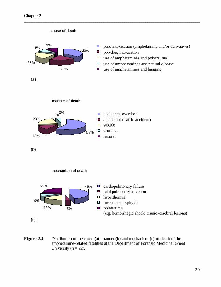
Chapter 2 _________________________________________________________________________________
20
Figure 2.4 Distribution of the cause (a), manner (b) and mechanism (c) of death of the amphetamine-related fatalities at the Department of Forensic Medicine, Ghent University (n = 22).
cause of death
36%
23%
23%
9%9% 1
2
3
4
5
pure intoxication (amphetamine and/or derivatives) polydrug intoxication use of amphetamines and polytrauma use of amphetamines and natural disease use of amphetamines and hanging
(a)
manner of death
58%14%
23%5%
0% 1
2
3
4
5
accidental overdose accidental (traffic accident) suicide criminal natural
(b)
mechanism of death
5%18%
9%
23% 45% 12
3
4
5
cardiopulmonary failure fatal pulmonary infection hyperthermia mechanical asphyxia polytrauma (e.g. hemorrhagic shock, cranio-cerebral lesions)
(c)
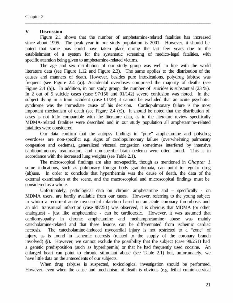
Chapter 2 _________________________________________________________________________________
21
V Discussion Figure 2.1 shows that the number of amphetamine-related fatalities has increased
since about 1995. The peak year in our study population is 2001. However, it should be noted that some bias could have taken place during the last few years due to the establishment of a system for the systematic screening of medico-legal fatalities, with specific attention being given to amphetamine-related victims.
The age and sex distribution of our study group was well in line with the world literature data (see Figure 1.12 and Figure 2.3). The same applies to the distribution of the causes and manners of death. However, besides pure intoxications, polydrug (ab)use was frequent (see Figure 2.4 (a)). Accidental overdoses comprised the majority of deaths (see Figure 2.4 (b)). In addition, in our study group, the number of suicides is substantial (23 %). In 2 out of 5 suicide cases (case 97/156 and 01/142) severe confusion was noted. In the subject dying in a train accident (case 01/29) it cannot be excluded that an acute psychotic syndrome was the immediate cause of his decision. Cardiopulmonary failure is the most important mechanism of death (see Figure 2.4 (c)). It should be noted that the distribution of cases is not fully comparable with the literature data, as in the literature review specifically MDMA-related fatalities were described and in our study population all amphetamine-related fatalities were considered.
Our data confirm that the autopsy findings in “pure” amphetamine and polydrug overdoses are non-specific: e.g. signs of cardiopulmonary failure (overwhelming pulmonary congestion and oedema), generalized visceral congestion sometimes interfered by intensive cardiopulmonary reanimation, and non-specific brain oedema were often found. This is in accordance with the increased lung weights (see Table 2.1).
The microscopical findings are also non-specific, though as mentioned in Chapter 1, some indications, such as pulmonary foreign body granulomata, can point to regular drug (ab)use. In order to conclude that hyperthermia was the cause of death, the data of the external examination at the scene, and the macroscopical and microscopical findings must be considered as a whole.
Unfortunately, pathological data on chronic amphetamine and – specifically - on MDMA users, are hardly available from our cases. However, referring to the young subject in whom a recurrent acute myocardial infarction based on an acute coronary thrombosis and an old transmural infarction (case 98/251) was observed, it is obvious that MDMA (or other analogues) - just like amphetamine - can be cardiotoxic. However, it was assumed that cardiomyopathy in chronic amphetamine and methamphetamine abuse was mainly catecholamine-related and that these lesions can be differentiated from ischemic cardiac necrosis. The catecholamine-induced myocardial injury is not restricted to a “zone” of injury, as is found in ischemic necrosis (related to the supply of the coronary branch involved) (9). However, we cannot exclude the possibility that the subject (case 98/251) had a genetic predisposition (such as hyperlipemia) or that he had frequently used cocaine. An enlarged heart can point to chronic stimulant abuse (see Table 2.1) but, unfortunately, we have little data on the antecedents of our subjects.
When drug (ab)use is suspected, toxicological investigation should be performed. However, even when the cause and mechanism of death is obvious (e.g. lethal cranio-cervical

Chapter 2 _________________________________________________________________________________
22
trauma due to a traffic accident), toxicological screening can be indicative, especially in young subjects. Of our fatalities, 14 % involved traffic accidents. As mentioned above, the dangers of driving under the influence of amphetamines should not be underestimated (see Chapter 1). Moreover, drug use can be an explanation for bizarre behaviour, even when traffic accidents are not involved (see e.g. cases 97/156 and 01/142).
The toxicological data in our cases confirm the broad range of post-mortem concentrations: for the “pure” MDMA overdoses, blood values of between 0.27 and 13.51 µg/ml were found. In a few cases, the amphetamines were below limit of detection in blood and were only barely detected in urine. Prouty and Anderson were able to demonstrate that the interference of post-mortem phenomena such as post-mortem redistribution can be avoided by sampling blood as soon as possible after death from an isolated peripheral vein such as the femoral vein (10). It should be noted that in the older cases (cases 80/181, 89/88, and possibly also in case 94/15) the blood sampling site is not known and it cannot be excluded that cardiac blood was used. For this reason, and also because the analytical techniques in that period might have been less sensitive and/or specific, these levels should be interpreted with caution. Blood sampling from the inferior vena cava was performed in the lower abdomen and thus not nearby the liver, which explains why these levels are not unexpectedly high. The discrepancy between the subclavian and the femoral blood AMP concentration in case 01/142 might be explained by contamination of the subclavian sample. In that case, several stab wounds were found associated with important thoracic and abdominal lesions. An autopsy was not performed, however.
Unfortunately, the available vitreous humour concentrations are restricted and the number of samples is too small to interpret the possible correlation between blood and vitreous humour levels. The data indicate that the vitreous humour levels were still positive when the persons were already in an advanced elimination phase (see cases 97/134, 98/14, 98/239), which can be interpreted in the light of the pharmacokinetics in which the vitreous humour levels lag somewhat behind the blood concentrations. As the vitreous fluid is to a minor extent influenced by the autolytic processes, it is worthwhile to study the value of this specimen (see Chapter 3 and part 3 of this work).
The amphetamine concentrations in urine, bile and liver indicated a possible elimination via hepatic biotransformation and excretion via bile and kidneys. The available stomach content levels pointed to a possible reservoir function, which could give rise to post-mortem redistribution.
In view of our data, the impact of the blood sampling location in the interpretation of the toxicological results is not clear. It will be necessary to study the distribution – and possible redistribution - in various fluids and tissues, to reach a conclusion on this issue. In addition, the value of vitreous humour concentrations also needs to be explored.
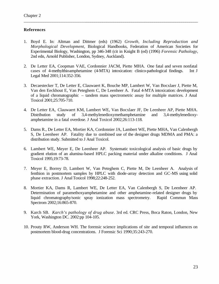
Chapter 2 _________________________________________________________________________________
23
References
1. Boyd E. In: Altman and Dittmer (eds) (1962) Growth, Including Reproduction and
Morphological Development, Biological Handbooks, Federation of American Societies for Experimental Biology, Washington, pp 346-348 (cit in Knight B (ed) (1996) Forensic Pathology, 2nd edn, Arnold Publisher, London, Sydney, Auckland).
2. De Letter EA, Coopman VAE, Cordonnier JACM, Piette MHA. One fatal and seven nonfatal
cases of 4-methylthioamphetamine (4-MTA) intoxication: clinico-pathological findings. Int J Legal Med 2001;114:352-356.
3. Decaestecker T, De Letter E, Clauwaert K, Bouche MP, Lambert W, Van Bocxlaer J, Piette M,
Van den Eeckhout E, Van Peteghem C, De Leenheer A. Fatal 4-MTA intoxication: development of a liquid chromatographic – tandem mass spectrometric assay for multiple matrices. J Anal Toxicol 2001;25:705-710.
4. De Letter EA, Clauwaert KM, Lambert WE, Van Bocxlaer JF, De Leenheer AP, Piette MHA.
Distribution study of 3,4-methylenedioxymethamphetamine and 3,4-methylenedioxy-amphetamine in a fatal overdose. J Anal Toxicol 2002;26:113-118.
5. Dams R., De Letter EA, Mortier KA, Cordonnier JA, Lambert WE, Piette MHA, Van Calenbergh
S, De Leenheer AP. Fatality due to combined use of the designer drugs MDMA and PMA: a distribution study. Submitted to J Anal Toxicol.
6. Lambert WE, Meyer E, De Leenheer AP. Systematic toxicological analysis of basic drugs by
gradient elution of an alumina-based HPLC packing material under alkaline conditions. J Anal Toxicol 1995;19:73-78.
7. Meyer E, Borrey D, Lambert W, Van Peteghem C, Piette M, De Leenheer A. Analysis of
fenthion in postmortem samples by HPLC with diode-array detection and GC-MS using solid phase extraction. J Anal Toxicol 1998;22:248-252.
8. Mortier KA, Dams R, Lambert WE, De Letter EA, Van Calenbergh S, De Leenheer AP.
Determination of paramethoxyamphetamine and other amphetamine-related designer drugs by liquid chromatography/sonic spray ionization mass spectrometry. Rapid Commun Mass Spectrom 2002;16:865-870.
9. Karch SB. Karch’s pathology of drug abuse. 3rd ed. CRC Press, Boca Raton, London, New
York, Washington DC. 2002:pp 104-105. 10. Prouty RW, Anderson WH. The forensic science implications of site and temporal influences on
postmortem blood-drug concentrations. J Forensic Sci 1990;35:243-270.

PART TWO
Experiments in rabbits
Chapter 3
Is vitreous humour useful for the interpretation of 3,4-methylenedioxymethamphetamine (MDMA) blood levels ?
Experimental approach with rabbits
Chapter 3 ________________________________________________________________________________
1
Chapter 3 Is vitreous humour useful for the interpretation of
3,4-methylenedioxymethamphetamine (MDMA) blood levels? Experimental approach with rabbits.
E.A. De Letter 1, P. De Paepe 2, K.M. Clauwaert 3, F.M. Belpaire 2 , W.E. Lambert 4, J.F. Van Bocxlaer 3, M.H.A. Piette 1
Ghent University 1 E.A. De Letter, M.H.A. Piette Department of Forensic Medicine 2 P. De Paepe, F.M. Belpaire Heymans Institute of Pharmacology 3
Karine M. Clauwaert, Jan F. Van Bocxlaer Department of Medical Biochemistry and Clinical Analysis 4 Willy E. Lambert Department of Toxicology
Based on: Int J Legal Med 2000;114:29-35
Chapter 3 ________________________________________________________________________________
2
I Abstract As drug instability and redistribution are factors known to affect the interpretation
of post-mortem blood levels, we questioned whether post-mortem vitreous humour concentrations could be useful as predictors for the MDMA load at the time of death. In a first series of in vivo experiments using rabbits, 3,4-methylenedioxymethamphetamine (MDMA) concentrations in plasma, blood and vitreous humour were studied as a function of time after intravenous (iv) administration of MDMA. Equilibration between the vascular compartment and vitreous humour was attained about 1 h after iv MDMA administration. In a second series of experiments, the post-mortem stability of MDMA in vitreous humour in relation to ambient temperature was investigated. Post-mortem MDMA concentrations in vitreous humour were closer to the ante-mortem blood levels when compared to cardiac blood samples.
These preliminary investigations in the rabbit model indicate that measurements of vitreous humour concentrations could also be of interest for predicting the blood concentration at the time of death in humans.
Key words : 3,4-Methylenedioxymethamphetamine (MDMA) - Vitreous humour – Pharmacokinetics - Post-mortem stability - Rabbits II Introduction
As post-mortem drug levels in blood do not necessarily reflect the concentration at the time of death, the question whether a detected level played an important role in the mechanism of death, remains a complex problem in the forensic practice. In particular, post-mortem instability and redistribution of drugs are important thanato-chemical factors (1). This “toxicological nightmare” is an established fact for various drugs e.g. cocaine (2), and many therapeutic drugs such as barbiturates (1) and digitalis (3,4). To a certain extent, the influence of these post-mortem phenomena can be avoided by sampling blood as soon as possible after death from an isolated peripheral vein such as the femoral vein (5). However, bearing in mind this general recommendation, a single blood sample is often insufficient to draw appropriate conclusions. Another sample (tissue or fluid) should not only be used as an analytical control for the blood level determined, but could also provide information on the pharmacokinetic phase and as a result the time of drug intake. Vitreous humour is one of these supplementary samples and is an interesting medium because the vitreous fluid is less influenced by autolytic processes and is convenient (e.g. simple to sample and not affected by hemolysis). Formerly, vitreous humour determinations have been performed in order to detect various drugs, in particular ethanol (6), morphine (7), cocaine (8), and amitriptyline (9), for example.
To our knowledge, the post-mortem drug distribution in humans has barely been explored for amphetamines and analogues, or for MDMA in particular, with the exception of a few case reports (10-12).
To investigate whether vitreous humour concentrations could be more helpful than blood for determining post-mortem MDMA levels in humans, preliminary experiments in
Chapter 3 ________________________________________________________________________________
3
rabbits were carried out. Rabbits were chosen as the animal model because they have a vitreous volume of about 1.4 ml (13) which is much larger than in rats, and the chemical characteristics of vitreous humour in rabbits are comparable to those in humans (14). As pharmacokinetic data for MDMA in the rabbit are lacking, we first investigated this after intravenous (iv) administration. We then studied whether determination of MDMA in vitreous humour is possible and whether there was a correlation in vivo between the vitreous humour levels and the plasma or blood concentrations. Finally, to explore whether the post-mortem vitreous concentrations could be useful to estimate the blood MDMA levels at the time of death, the influence of the post-mortem interval and the ambient temperature was examined. III Materials and methods
The study protocol was approved by the Ethics Committee for animals of the Medical School, Ghent University (request number ECP 98/1 and ECP 99/9). 3,4-Methylenedioxymethamphetamine hydrochloride was provided by Sigma-Aldrich (Belgium).
III.1 Animals and procedures Female white New Zealand rabbits (weight 2050 – 4500 g) were purchased from
Iffa Credo Belgium. The animals were fasted overnight before the experiment but were allowed free access to water.
III.1.1 In vivo experiments The broad study design is shown in Figure 3.1. A polyethylene catheter (P.E. 50) filled with heparin solution (100 IU/ml) was
implanted (xylocaine 1 %) into the main central artery of the right ear in 18 rabbits under local anaesthesia.
In one group of six rabbits (t = 120 minutes), arterial blood samples (2 ml) were taken at 0, 5, 10, 15, 20, 30, 45, 60, 90, 120 minutes after drug administration for the assay of MDMA and MDA (3,4-methylenedioxyamphetamine) concentrations in plasma. Additional samples (1ml at 10, 20, 60, 120 minutes) were taken for whole blood determinations. Pentobarbital anaesthesia was induced according to Prince (30 – 35 mg/kg body weight (15)).
In all cases, sampled blood was replaced by the same amount of saline. Blood samples were centrifuged at 3500 rpm for 10 minutes, and plasma and whole blood samples were frozen at – 30°C until analysis. Both eyes were immediately frozen after enucleation by immersion in liquid nitrogen for 1 – 2 minutes. The eyes were preserved at –30°C until the vitreous bodies were dissected as described by Abel and Boyle (16).
Finally, in three rabbits the urinary excretion of MDMA and MDA was followed for 96 hours after MDMA iv administration (1 mg/kg). Urine was collected in a metabolism cage and the volume measured. These samples were also frozen until analysis.

Chapter 3 ________________________________________________________________________________
4
III.1.2 Post-mortem experiment The broad study design is shown in Figure 3.1. Sample preservation and dissection
of the eyes were carried out as previously described.
in vivo experiments post-mortem experiments
n = 18 n = 12
at t = 120: - blood sample right ear vein (3 ml) + - immediate killing n = 6 n = 6 n = 6 (neck concussion + air embolism) t = 30 t = 120 t = 240 + - immediate enucleation of one eye (left or right eye, at random) at t = 30, 120 or 240:
- last arterial blood sampling (3 ml) + - pentobarbital anaesthesia n = 6 n = 6 + - immediate enucleation of both eyes T 2 °C 17 °C n = 3 n = 3 n = 3 n = 3 PMI 25 h 73 h 25 h 73 h
At PMI = 25 or 73 h: cardiac blood sampling and enucleation of the second eye
Abbreviations: t: time expressed in minutes T: Temperature in degrees Celsius PMI: post-mortem interval (hours) Figure 3.1 Study design in rabbits (receiving 1 mg/kg MDMA, slowly infused via the left ear
vein) III.2 Protein binding Protein binding of MDMA was determined by ultrafiltration using Amicon
centrifuge micropartition system and YMT membrane discs (Grace, Amicon Division, Beverly, Mass). Blank rabbit plasma samples were spiked with 400 ng/ml MDMA; 1 ml plasma samples were transferred to the micro-partition system and centrifuged at 1500 rpm for 30 minutes resulting in a volume between 100 and 200 µl. MDMA concentration was measured, in a 50 µl aliquot of the ultrafiltrate with HPLC. Adsorption of MDMA to the filter was measured using an analogous solution in saline and found negligible.
Chapter 3 ________________________________________________________________________________
5
III.3 Drug assay Concentrations of MDMA and MDA were determined using a fully validated
HPLC (high pressure liquid chromatography) procedure with fluorescence detection (λex 288, λem 324 nm) . The samples (250 µl) were liquid/liquid extracted with hexane/ethylacetate (70:30, v/v) at pH 9.5 using MDEA (3,4-methylenedioxyethylamphetamine) as the internal standard. Chromatographic separation was achieved using Hypersil BDS C18 columns (3 µm, 100 x 2.1 mm, Alltech, Deerfield, Il.) isocratically eluted at 0.2 ml/min with a mixture of water/methanol/acetonitrile containing 0.1 M ammonium acetate. The method proved linear from 2 to 1000 ng/ml (2 ng/ml being the quantitation limit both for MDMA and MDA, between-day reproducibility < 25 %). With each batch of samples, a calibration curve prepared in the corresponding blank matrix (except for the vitreous humour which was substituted with water for reasons of practical unavailability) and quality control samples (7 and 500 ng/ml) were analysed. Accuracy was between 97 – 102 % (n = 7) and total precision (CV, coefficient of variation) was lower than 13 % (n = 7).
III.4 Analysis of data The results are expressed as means (± SD). The plasma concentration-time profiles of MDMA after iv administration were
individually analysed using a pharmacokinetic computer programme (WinNonlin version 1.5 – Scientific Consulting, Inc.) and were best characterized on the basis of the Akaike Information Criterion (17), by a 2-compartment model using the equation C = A e -αt + B e -β t, where C is the plasma concentration at time t, α and β are hybrid rate constants and A and B are the coefficients of the exponential terms. Calculation of the pharmacokinetic parameters (half-life, volume of distribution at steady state, volume of distribution of the central compartment, clearance, area under the curve (AUC) and mean residence time) was done according to Gibaldi and Perrier (18).
Statistical processing of the data was performed using non-parametric tests. The Wilcoxon Rank test was used for analysis of interindividual differences in concentrations between the left and right eyes. The Friedman test for repeated measurements was used to compare the ratios of blood to plasma MDMA concentrations (10, 20, 60 and 120 minutes after infusion). The ratios between the vitreous humour MDMA concentration and the corresponding plasma or blood level sampled 30, 120 and 240 minutes after administration, were compared using the non-paired Kruskal-Wallis test; and, when appropriate, this was followed by the Mann-Whitney U Test. The correlation between vitreous humour and plasma or blood levels was investigated with the Spearman correlation test. For all tests, P values less than 0.05 were considered to be statistically significant.

Chapter 3 ________________________________________________________________________________
6
IV Results IV.1 In vivo experiments Figure 3.2 shows the time course of the mean plasma concentrations of MDMA
following a 1 mg/kg iv dose (n = 6). The data from each animal were well fitted according to a two compartment model. The corresponding pharmacokinetic parameters are summarized in Table 3.1. The mean blood/plasma ratios calculated at 10, 20, 60 and 120 minutes after infusion, were 1.2 ± 0.1, 1.2 ± 0.2, 1.3 ± 0.5 and 1.3 ± 0.2, respectively. Statistical analysis did not reveal significant differences.
Figure 3.2 Plasma concentrations of MDMA as a function of time in rabbits after an iv dose of 1 mg/kg MDMA (n = 6).
(Results are expressed as means ± SD)
10
100
1000
0 30 60 90 120
time (minutes)
MD
MA
con
cent
ratio
n (n
g/m
l)
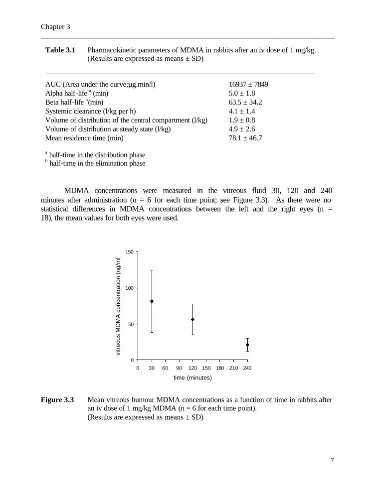
Chapter 3 ________________________________________________________________________________
7
Table 3.1 Pharmacokinetic parameters of MDMA in rabbits after an iv dose of 1 mg/kg. (Results are expressed as means ± SD)
AUC (Area under the curve;µg.min/l) 16937 ± 7849 Alpha half-life a (min) 5.0 ± 1.8 Beta half-life b(min) 63.5 ± 34.2 Systemic clearance (l/kg per h) 4.1 ± 1.4 Volume of distribution of the central compartment (l/kg) 1.9 ± 0.8 Volume of distribution at steady state (l/kg) 4.9 ± 2.6 Mean residence time (min) 78.1 ± 46.7
a half-time in the distribution phase b half-time in the elimination phase
MDMA concentrations were measured in the vitreous fluid 30, 120 and 240
minutes after administration (n = 6 for each time point; see Figure 3.3). As there were no statistical differences in MDMA concentrations between the left and the right eyes (n = 18), the mean values for both eyes were used.
Figure 3.3 Mean vitreous humour MDMA concentrations as a function of time in rabbits after
an iv dose of 1 mg/kg MDMA (n = 6 for each time point). (Results are expressed as means ± SD)
0
50
100
150
0 30 60 90 120 150 180 210 240
time (minutes)
vitr
eous
MD
MA
con
cent
ratio
n (n
g/m
l)

Chapter 3 ________________________________________________________________________________
8
The ratios of the vitreous MDMA concentrations (MDMAvitreous) to the plasma (MDMAplasma ) (see Figure 3.4 (a)) or whole blood (MDMAblood) concentrations 30 minutes after administration (see Figure 3.4 (b)) were less than one and significantly different from the values obtained at 120 and 240 minutes when both ratios were higher than one, but no significant difference was observed between both values (ratios: at t = 120 min 1.4 ± 0.3 and 1.1 ± 0.3; at t = 240 min 1.6 ± 0.2 and 1.1 ± 0.4, respectively).
(a) (b)
Figure 3.4 Individual and mean ratios of the vitreous humour MDMA concentrations to
plasma (a) or blood (b) concentrations as a function of time in rabbits after an iv dose of 1 mg/kg MDMA (n = 18).
Figure 3.5 shows the scatterplot of the MDMAvitreous versus MDMAplasma (Figure
3.5 (a)) levels or MDMAblood (Figure 3.5. (b)) in the elimination phase. The Spearman (rs) correlation coefficients for the MDMAvitreous and MDMAplasma or MDMAblood in the elimination period (at t = 120 and 240 min) were 0.981 and 0.950, respectively. These correlations were significant at the 0.01 level. The correlation performed without the outlier did not change significantly these correlation coefficients being 0.984 and 0.945, respectively.
MDA plasma and blood concentrations were low, ranging between 2 ng/ml and 6 ng/ml. The MDA levels in the vitreous humour were below the limit of quantitation (2 ng/ml).
The percentages of the administered MDMA dose recovered in urine of three rabbits were 12.9, 3.4 and 4.1 %, respectively. MDA levels were below the limit of quantitation. The mean unbound fraction of MDMA in plasma was 63 % ± 3 (n = 6) at a concentration of 400 ng/ml.
time (minutes)
2402101801501209060300
vitre
ous
hum
our/p
lasm
a M
DM
A c
once
ntra
tion 2.0
1.6
1.2
.8
.4
0.0
time (minutes)
2402101801501209060300
vitre
ous
hum
our/b
lood
MD
MA
con
cent
ratio
n 2.0
1.6
1.2
.8
.4
0.0

Chapter 3 ________________________________________________________________________________
9
(a) (b)
Figure 3.5 Scatter plot of vitreous humour versus plasma (a) or blood (b) MDMA
concentrations (120 and 240 minutes after an iv dose of 1 mg/kg MDMA). (n = 12)
IV.2 Post-mortem experiment (see Figure 3.6) The mean plasma, blood levels and the vitreous humour concentrations of one eye
120 minutes after MDMA infusion (just prior to killing of the animals) were 32 ± 17, 42 ± 16, and 27 ± 7 ng/ml, respectively (n = 12). Blood MDMA concentrations were clearly increased post mortem compared to the values obtained ante mortem, whereas the vitreous MDMA concentrations did not change substantially post mortem. An overall slight increase in vitreous MDMA levels was observed, somewhat more pronounced at 17°C 73 h post mortem and vitreous MDMA levels tended to be more stable at 2°C.
plasma MDMA concentration (ng/ml)
120100806040200
vitr
eous
hum
our
MD
MA
con
cent
ratio
n (n
g/m
l)100
80
60
40
20
0
blood MDMA concentration (ng/ml)
120100806040200
vitre
ous
hum
our
MD
MA
con
cent
ratio
n (n
g/m
l)
100
80
60
40
20
0
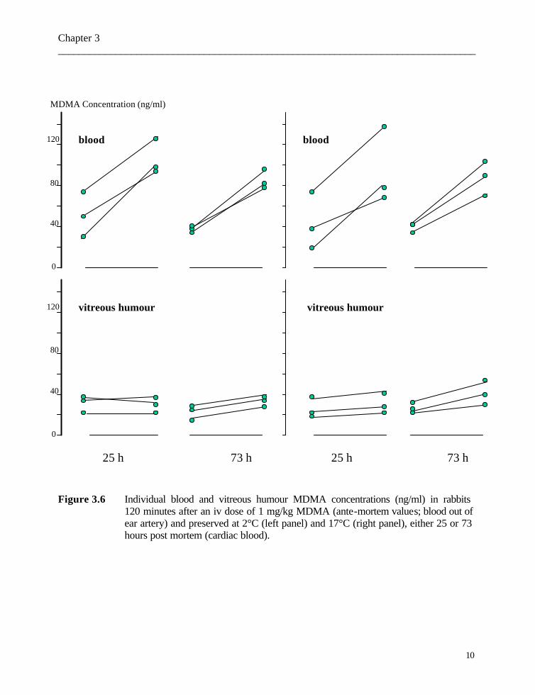
Chapter 3 ________________________________________________________________________________
10
Figure 3.6 Individual blood and vitreous humour MDMA concentrations (ng/ml) in rabbits 120 minutes after an iv dose of 1 mg/kg MDMA (ante-mortem values; blood out of ear artery) and preserved at 2°C (left panel) and 17°C (right panel), either 25 or 73 hours post mortem (cardiac blood).
blood
vitreous humour
40
80
120
0
40
80
0
25 h 73 h 73 h25 h
MDMA Concentration (ng/ml)
blood
vitreous humour 120

Chapter 3 ________________________________________________________________________________
11
V Discussion In this study, we examined whether determination of vitreous MDMA levels can be
useful to predict the MDMA blood concentrations at the time of death with the help of a rabbit experiment.
In order to interpret the post-mortem concentrations of MDMA in the rabbit, we first investigated the pharmacokinetics of MDMA after an intravenous dose of 1 mg/kg. The plasma concentration versus time curves of MDMA were fitted according to a 2- compartment model. MDMA has a high volume of distribution (5 l/kg), a high systemic clearance (4.1 l/kg per h) and a relatively short half-life (1 h). Our results were fairly consistent with previously reported pharmacokinetic parameters of MDMA after intravenous (iv) and subcutaneous (sc) administration in rats (19).
The mean blood/plasma ratios in the distribution as well as the elimination phase indicate a certain accumulation of MDMA in red blood cells.
In our experiments, only 6 % of the MDMA dose was found unchanged in urine and MDMA is probably eliminated by biotransformation or by excretion via the bile. In addition, MDA concentrations in rabbit plasma were very low (below 6 ng/ml) and were below the limit of quantitation in urine. In rats and mice, MDA was identified in plasma as an important metabolite (20) whereas in rat urine, either R(-) MDA and S(+) MDA were rarely found (19). In humans, MDA was originally assumed to be the major metabolite (21), but it was demonstrated that conjugated HMMA (4-hydroxy-3-methoxy-methamphetamine) and HHMA (3,4-dihydroxymeth-amphetamine) were the main urinary metabolites of MDMA (22-25).
The mean binding of MDMA in rabbit plasma (37 %) is similar to that in dogs [26]. In our experiments, after a single intravenous dose, equilibration between plasma
and vitreous humour was obtained between 30 and 120 minutes, as the MDMAvitreous / MDMAplasma ratios did not differ significantly between 120 and 240 minutes post administration. Physiologically, the blood-retina barrier can be compared with the blood-brain barrier and Chu et al. (27) demonstrated that an equilibration between plasma and brain concentrations of MDMA was obtained within 30 minutes after sc administration of MDMA in rats. The MDMAvitreous / MDMAplasma ratios at 120 (1.4 ± 0.3) and 240 minutes (1.6 ± 0.2) and the corresponding ratios of MDMAvitreous to MDMAblood (1.1 ± 0.3 and 1.1 ± 0.4, respectively) indicate a slight accumulation of MDMA in the vitreous compartment. The accumulation is in fact even more important as the plasma protein binding of MDMA is ± 30 %. A possible explanation could be binding to the vitreous humour, but in vitreous fluid protein concentration is only 1 - 3 % of the total serum protein concentration (14). For some drugs with an important level of plasma protein binding, such as fleroxacin (28), vitreous to plasma concentration ratios lower than 1 were seen. However, higher vitreous humour levels compared to serum concentrations were noticed in rabbits for fluconazole (29). For ethanol too, a higher vitreous humour level compared to blood concentration at steady state was observed (6) which was explained by the smaller dry matter content of vitreous humour (30).

Chapter 3 ________________________________________________________________________________
12
To our knowledge, there are only two case reports of MDMA determination in human vitreous humour; both revealing a vitreous humour/blood concentration ratio below 1. Crifasi and Long (31) published a traffic accident fatality attributed to the use of MDMA. The vitreous/blood ratio was 0.5 and very little MDA was detected. These authors concluded that their results support an acute event (i.e. that death occurred before distribution was complete). In the second case report, (an acute poisoning, probably accidental, due to combined intake of MDMA, cocaine and heroin) (32), the ratio of total vitreous/blood MDMA levels was calculated as 0.66; an analogous explanation for their results could be assumed.
Since ambient temperature and post-mortem interval are important thanato-chemical factors, four different conditions, 2°C or 17°C and 25 or 73 hours post mortem, respectively, were investigated in the second part of our study. We demonstrated that MDMA concentrations in cardiac blood increased post mortem, whereas vitreous MDMA levels were much more stable and thus more representative of the ante-mortem blood levels. Relatively small differences were noticed between the peri- and post-mortem vitreous values obtained at ambient temperatures of either 2°C or 17°C. The elevation of the vitreous MDMA concentration 73 hours after death in rabbits preserved at 17°C could partially be explained by the dehydration which occurred and, theoretically, a low level of post-mortem redistribution. On the other hand, the MDMA concentration increase in our rabbit heart blood samples, taken 73 hours post mortem in particular, points to a post-mortem redistribution. Moriya and Hashimoto (33) demonstrated post-mortem diffusion of methamphetamine from lung tissue in rabbits. In humans, post-mortem cardiac blood levels of amphetamine (5) and methamphetamine (34,35) were found to be higher than femoral blood concentrations and in one of the cases reported by Rohrig and Prouty, the MDMA concentration in heart blood was reported to be 5 times higher than in femoral blood (11). However, recently, in two human fatal cases associated with amphetamine intake (36), a post-mortem increase in amphetamine concentrations of 50-60 % in femoral blood was noticed. Further thanato-chemical experiments are needed to explore the mechanism of these increases. Instability of MDMA itself is not very likely as in vitro stability studies of MDMA and MDA in aqueous solutions and dog plasma demonstrated that these products are fairly stable (26). Human blood samples containing amphetamine and methamphetamine, stored in preservation products, are sufficiently stable up to 5 years (37). Nagata and colleagues (38) investigated the stability of amphetamine and methamphetamine in post-mortem rabbit tissues and concluded that these products are sufficiently stable.
In summary, after intravenous administration, MDMA can easily be identified in
the vitreous humour of rabbits and an equilibration between the vitreous humour and the vascular compartment was established after about 1 h. In addition, our results confirm that heart blood samples cannot be used for post-mortem toxicological analysis. In fact rabbit vitreous MDMA was more stable than post-mortem cardiac blood levels.
Vitreous sampling for MDMA determination seems to be a good autopsy practice if blood is lacking (e.g. as a result of severe blood loss or putrefaction). Moreover, after

Chapter 3 ________________________________________________________________________________
13
equilibration, vitreous humour could be a suitable control sample in case of erratic blood values due to either sampling site bias or analytical errors. In fact vitreous humour MDMA levels could be more representative than blood MDMA concentrations when there is a prolonged post-mortem interval. Further thanato-chemical investigations of routine autopsy cases should be performed to confirm these preliminary observations in rabbits.
Acknowledgements The authors gratefully thank Prof. Dr. M. Bogaert for advice in the development of this study and critically reading of the manuscript. Our gratitude also goes to Prof. Dr. A.P. De Leenheer for overall support (equipment, chemicals and accommodation). We would like to thank as well Mr. G. Van Maele for statistical recommendations, Mrs Thérèse De Vuyst for assistance in the English grammar and Mr R. Declercq for skilled technical assistance.

Chapter 3 ________________________________________________________________________________
14
References 1. Pounder DJ, Jones GR. Post-mortem drug redistribution – a toxicological nightmare.
Forensic Sci Int 1990;45:253-263. 2. Hearn WL, Keran EE, Wei H, Hime G. Site-dependent postmortem changes in blood cocaine
concentrations. J Forensic Sci 1991;36:673-684. 3. Donnelly B, Balkon J, Bidanset JH, Belmonte A, Barletta M, Manning T. Comparative kinetics
of serum and vitreous humor digoxin concentrations in a guinea pig model. Part I: intravenous administration of digoxin. J Anal Toxicol 1991;15:60-62.
4. Balkon J, Donnelly B. Comparative kinetics of digoxin in serum and vitreous humor in a
guinea pig model. Part II: Single oral dose administration. J Anal Toxicol 1992;16:155-157. 5. Prouty RW, Anderson WH. The forensic implications of site and temporal influences on
postmortem blood-drug concentrations. J Forensic Sci 1990;35:243-270. 6. Chao TC, Lo DST. Relationship between postmortem blood and vitreous humor ethanol levels.
Am J Forensic Med Pathol 1993;14:303-308. 7. Bermejo AM, Ramos I, Fernández P, López-Rivadulla M, Cruz A, Chiarotti M, Fucci N,
Marsilli R. Morphine determination by gas chromatography/mass spectroscopy in human vitreous humor and comparison with radioimmunoassay. J Anal Toxicol 1992;16:372-374.
8. McKinney PE, Phillips S, Gomez HF, Brent J, MacIntyre M, Watson WA. Vitreous humor
cocaine and metabolite concentrations: do postmortem specimens reflect blood levels at the time of death? J Forensic Sci 1995;40:102-107.
9. Hilberg T, Ripel Å, Smith AJ, Slørdal L, Mørland J, Bjørneboe A. Postmortem
amitriptyline pharmacokinetics in pigs after oral and intravenous routes of administration. J Forensic Sci 1998;43:380-387.
10. Dowling GP, McDonough ET, Bost RO. 'Eve' and 'Ecstasy'. A report of five
deaths associated with the use of MDEA and MDMA. JAMA 1987;257:1615-1617. 11. Rohrig TP, Prouty RW. Tissue distribution of methylenedioxy-methamphetamine. J Anal
Toxicol 1992;16:52-53. 12. Fineschi V, Masti A. Fatal poisoning by MDMA (ecstasy) and MDEA: a case report. Int J
Legal Med 1996;108:272-275. 13. Gardner SK. Ocular drug penetration and pharmacokinetic principles. In : Lamberts DW,
Potter DE. (eds) Clinical ophthalmic pharmacology. (1987) Little, Brown and Compagny, Boston,Toronto, pp 1-52.
Chapter 3 ________________________________________________________________________________
15
14. Berman ER.(ed) Vitreous. In: Biochemistry of the Eye. (1991) Plenum Press, New York,
London, pp 291-307. 15. Prince JH. (ed) The rabbit in eye research. (1964) Charles C Thomas Publisher,
Springfield, Illinois USA, p 618. 16. Abel R. Jr, Boyle GL. Dissecting ocular tissue for intraocular drug studies. Invest
Ophthalmol 1976;15:216-219. 17. Akaike H. A new look at the statistical model identification. IEEE Trans Automat Control
1974;19:716-23. 18. Gibaldi M, Perrier D. (eds) Pharmacokinetics. (1982) Marcel Dekker, New York. 19. Fitzgerald RL, Blanke RV, Poklis A. Stereoselective pharmacokinetics of 3,4-
methylenedioxymethamphetamine in the rat. Chirality 1990;2:241-248. 20. Fitzgerald RL, Blanke RV, Rosecrans JA, Glennon RA. Stereochemistry of the metabolism
of MDMA to MDA. Life Sci 1989;45:295-301. 21. Verebey K, Alrazi J, Jaffe JH. The complications of “Ecstasy” (MDMA). JAMA 1988;
259:1649-1650. 22. de Boer D, Tan LP, Gorter P, van de Wal RMA, Kettenes-van den Bosch JJ, de Bruijn EA,
Maes RAA. Gas chromatographic/mass spectrometric assay for profiling the enantiomers of 3,4-methylenedioxymethamphetamine and its chiral metabolites using positive chemical ionization ion trap mass spectrometry. J Mass Spectrom 1997;32:1236-1246.
23. Lanz M, Brenneisen R, Thormann W. Enantioselective determination of 3,4-
methylenedioxymethamphetamine and two of its metabolites in human urine by cyclodextrin-modified capillary zone electrophoresis. Electrophoresis 1997;18:1035-1043.
24. Lim HK, Foltz RL. Identification of metabolites of 3,4-(methylenedioxy)methamphetamine
in human urine. Chem Res Toxicol 1989;2:142-143. 25. Helmlin HJ, Bracher K, Bourquin D, Vonlanthen D, Brenneisen R, Styk J. Analysis of 3,4-
methylenedioxymethamphetamine (MDMA) and its metabolites in plasma and urine by HPLC-DAD and GC-MS. J Anal Toxicol 1996;20:432-440.
26. Garrett ER, Seyda K, Marroum P. High performance liquid chromatographic assays of the
illicit designer drug “Ecstasy”, a modified amphetamine, with applications to stability, partitioning and plasma protein binding. Acta Pharm Nord 1991;3: 9-14.
27. Chu T, Kumagai Y, DiStefano EW, Cho AK. Disposition of
methylenedioxymethamphetamine and three metabolites in the brains of different rat strains and their possible roles in acute serotonin depletion. Biochem Pharmacol 1996;51:789-796.
Chapter 3 ________________________________________________________________________________
16
28. Miller MH, Madu A, Samathanam G, Rush D, Madu CN, Mathisson K, Mayers M.
Fleroxacin pharmacokinetics in aqueous and vitreous humors determined by using complete concentration-time data from individual rabbits. Antimicrob Agents Chemother 1992;36: 32-8 .
29. O'Day DM, Foulds G, Williams TE, Robinson RD, Allen RH, Head WS. Ocular uptake of
fluconazole following oral administration. Arch Ophthalmol 1990;108:1006-1008. 30. Felby S, Olsen J. Comparative studies of postmortem ethylalcohol in vitreous humor, blood
and muscle. J Forensic Sci 1969;14:93-101. 31. Crifasi J, Long C. Traffic fatality related to the use of methylenedioxymethamphetamine. J
Forensic Sci 1996;41:1082-1084. 32. Moore KA, Mozayani A, Fierro MF, Poklis A. Distribution of 3,4-
methylenedioxymethamphetamine (MDMA) and 3,4-methylenedioxyamphetamine (MDA) stereoisomers in a fatal poisoning. Forensic Sci Int 1996;83:111-119.
33. Moriya F, Hashimoto Y. Redistribution of basic drugs into cardiac blood from surrounding
tissues during early-stages post mortem. J Forensic Sci 1999;44:10-16. 34. Logan BK, Weiss EL, Harruff RC. Case report: distribution of methamphetamine in a
massive fatal ingestion. J Forensic Sci 1996;41:322-323. 35. Miyazaki T, Kojima T, Yashiki M, Wakamoto H, Iwasaki Y, Taniguchi T. Site dependence
of methamphetamine concentrations in blood samples collected from cadavers of people who had been methamphetamine abusers. Am J Forensic Med Pathol 1993;14:121-124.
36. Hilberg T, Rodge S, Mørland J. Postmortem drug redistribution – human cases related to
results in experimental animals. J Forensic Sci 1999;44:3-9. 37. Giorgi SN, Meeker JE. A 5-year stability study of common illicit drugs in blood. J Anal
Toxicol 1995;19:392-398. 38. Nagata T, Kimura K, Hara K, Kudo K. Methamphetamine and amphetamine concentrations
in postmortem rabbit tissues. Forensic Sci Int 1990;48:39-47.
Chapter 4
Post-mortem redistribution of 3,4-methylenedioxy-methamphetamine (MDMA, “ecstasy”) in the rabbit
Part one: Experimental approach after intravenous infusion.
Chapter 4 ________________________________________________________________________________
1
Chapter 4 Post-mortem redistribution of 3,4-methylenedioxymethamphetamine
(MDMA, “ecstasy”) in the rabbit Part one: experimental approach after in vivo intravenous infusion.
Els A. De Letter1, Karine M. Clauwaert2, Frans M. Belpaire3, Willy E. Lambert4 ,
Jan F. Van Bocxlaer2 , Michel H.A. Piette1 Ghent University
1 Els A. De Letter, Michel H.A. Piette Department of Forensic Medicine 2 Karine M. Clauwaert, Jan F. Van Bocxlaer Department of Medical Biochemistry and Clinical Analysis 3 Frans M. Belpaire Heymans Institute of Pharmacology
4 Willy E. Lambert Department of Toxicology
Int J Legal Med 2002;116:216-224

Chapter 4 ________________________________________________________________________________
2
I Abstract Post-mortem redistribution is known to influence blood and tissue levels of various
drugs. An animal model was used in an attempt to elucidate this problem for the amphetamine analogue, 3,4-methylenedioxymethamphetamine (MDMA). Rabbits received 1 mg/kg MDMA intravenously (iv) and were killed two hours later in order to simulate the state of complete distribution in the body. MDMA and 3,4-methylenedioxyamphetamine (MDA) concentrations were determined in blood, urine, bile, vitreous humour, and various tissues (eye globe walls, brain, cardiac muscle, lungs, liver, kidneys, iliopsoas muscle and adipose tissue) using a high pressure liquid chromatographic (HPLC) procedure with fluorescence detection.
In the first group (control group, sampling immediately post mortem) considerable MDMA concentrations were found in the brain and both lungs. In addition, our data indicate the elimination of MDMA by hepatic biotransformation and excretion via the bile. When the animals were preserved either 24 or 72 h post mortem (second group), an increase of MDMA and MDA levels in the liver and the eye globe walls was noticed. In the lungs, on the other hand, they tended to decline as a function of increasing post-mortem interval. MDMA levels in cardiac and iliopsoas muscle were fairly comparable and remained stable up to 72 h after death. In the third group, ligation of the large vessels around the heart took place immediately post mortem, but significant differences in blood and tissue MDMA concentrations between rabbits of group 2 and 3 could not be demonstrated. We therefore conclude that post-mortem redistribution of MDMA at the cellular level (viz. by pure diffusion gradient from higher to lower concentrations) is more important than its redistribution via the vascular pathway. Finally, MDA levels were relatively low in all samples, thus indicating that this is not a major metabolite in the rabbit, at least within the first two hours after administration.
Key words: 3,4-Methylenedioxymethamphetamine (MDMA) - 3,4-Methylenedioxy-
amphetamine (MDA) - IV infusion in rabbits - Tissue distribution - Post-mortem redistribution
II Introduction
For many drugs there is a correlation between plasma concentration and pharmacological effect. However, the interpretation of post-mortem concentrations of many substances differs substantially from in vivo quantified levels. In particular, post-mortem instability and redistribution can be important interfering factors, as has been demonstrated, for example, for ethanol (1) but also barbiturates (2), cocaine (3), and dothiepin (4). Post-mortem distribution has also been investigated for more scarcely encountered substances in forensic practice such as laudanosine (5 ), dichloromethane (6 ).
These thanato-chemical problems have barely been explored for the amphetamine analogue, 3,4-methylenedioxymethamphetamine (MDMA, or “ecstasy”), except in a few case reports (7-10). For amphetamine and methamphetamine, more literature data are available (11-19). Animal experiments dealing with this issue for amphetamine or its analogues are scarce (20-22). To our knowledge, post-mortem redistribution of MDMA has
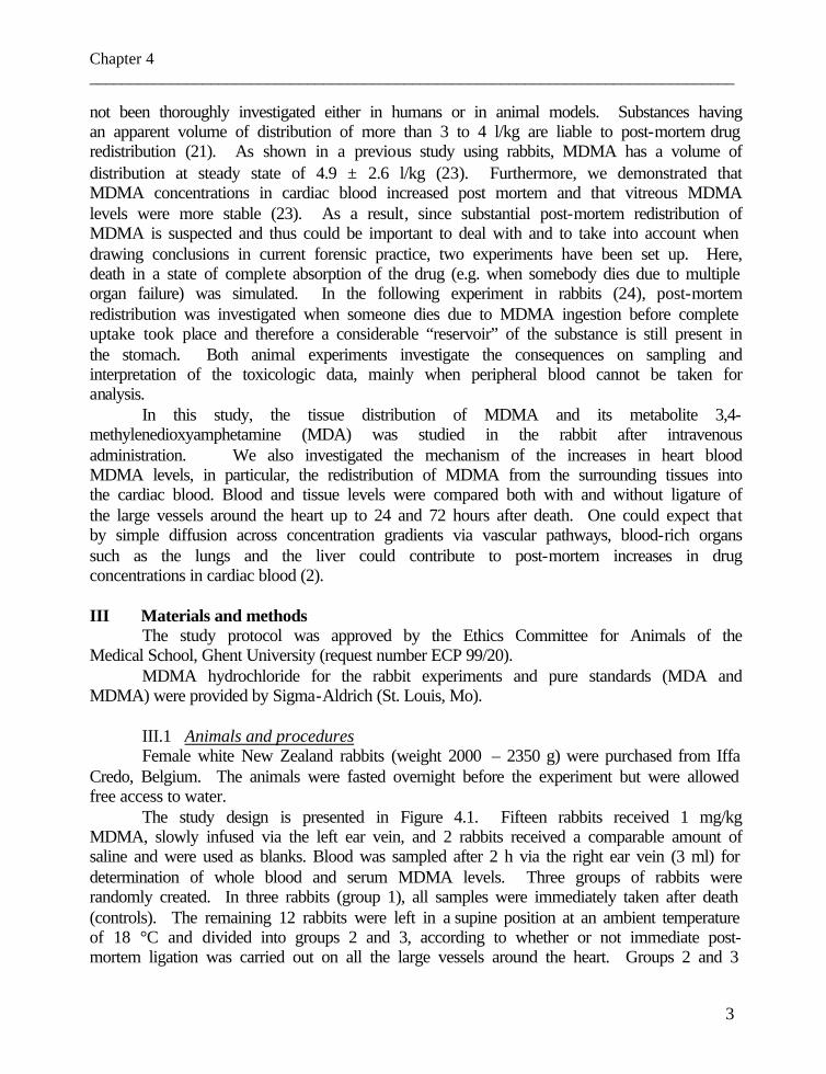
Chapter 4 ________________________________________________________________________________
3
not been thoroughly investigated either in humans or in animal models. Substances having an apparent volume of distribution of more than 3 to 4 l/kg are liable to post-mortem drug redistribution (21). As shown in a previous study using rabbits, MDMA has a volume of distribution at steady state of 4.9 ± 2.6 l/kg (23). Furthermore, we demonstrated that MDMA concentrations in cardiac blood increased post mortem and that vitreous MDMA levels were more stable (23). As a result, since substantial post-mortem redistribution of MDMA is suspected and thus could be important to deal with and to take into account when drawing conclusions in current forensic practice, two experiments have been set up. Here, death in a state of complete absorption of the drug (e.g. when somebody dies due to multiple organ failure) was simulated. In the following experiment in rabbits (24), post-mortem redistribution was investigated when someone dies due to MDMA ingestion before complete uptake took place and therefore a considerable “reservoir” of the substance is still present in the stomach. Both animal experiments investigate the consequences on sampling and interpretation of the toxicologic data, mainly when peripheral blood cannot be taken for analysis.
In this study, the tissue distribution of MDMA and its metabolite 3,4-methylenedioxyamphetamine (MDA) was studied in the rabbit after intravenous administration. We also investigated the mechanism of the increases in heart blood MDMA levels, in particular, the redistribution of MDMA from the surrounding tissues into the cardiac blood. Blood and tissue levels were compared both with and without ligature of the large vessels around the heart up to 24 and 72 hours after death. One could expect that by simple diffusion across concentration gradients via vascular pathways, blood-rich organs such as the lungs and the liver could contribute to post-mortem increases in drug concentrations in cardiac blood (2).
III Materials and methods
The study protocol was approved by the Ethics Committee for Animals of the Medical School, Ghent University (request number ECP 99/20).
MDMA hydrochloride for the rabbit experiments and pure standards (MDA and MDMA) were provided by Sigma-Aldrich (St. Louis, Mo).
III.1 Animals and procedures Female white New Zealand rabbits (weight 2000 – 2350 g) were purchased from Iffa
Credo, Belgium. The animals were fasted overnight before the experiment but were allowed free access to water.
The study design is presented in Figure 4.1. Fifteen rabbits received 1 mg/kg MDMA, slowly infused via the left ear vein, and 2 rabbits received a comparable amount of saline and were used as blanks. Blood was sampled after 2 h via the right ear vein (3 ml) for determination of whole blood and serum MDMA levels. Three groups of rabbits were randomly created. In three rabbits (group 1), all samples were immediately taken after death (controls). The remaining 12 rabbits were left in a supine position at an ambient temperature of 18 °C and divided into groups 2 and 3, according to whether or not immediate post-mortem ligation was carried out on all the large vessels around the heart. Groups 2 and 3

Chapter 4 ________________________________________________________________________________
4
were each divided into two subgroups (n = 3), which were preserved either 24 hours (group a) or 72 hours (group b) post mortem. From each rabbit, cardiac blood and the following tissues were sampled: cardiac muscle, right and left lung, liver, kidney (mixture of right and left), cerebrum, cerebellum, brainstem, stomach wall and stomach content, abdominal adipose tissue, and iliopsoas muscle. In addition, enucleation of the second eye and sampling of urine and bile in toto was carried out. The eyes were handled as previously described (23). The individual eye globe walls, consisting of the retina, choroidea and sclera, were also preserved for toxicological analysis. In order to avoid contamination of these eye globe walls, all inserting muscle fragments were carefully removed. Aqueous humour, cornea and lens were not included in our protocol. As creatinine is a stable parameter post mortem (25), these levels were determined in the vitreous humour samples and the ratio of MDMA to creatinine concentration was calculated. All samples were stored at –30 °C until analysis.
at t = 0 min: 1 mg/ kg MDMA, iv (n = 15) at t = 120 min: - blood sampling
+ - immediate killing (by neck concussion + air embolism) + - enucleation of one eye (left or right, at random)
Group 1 Group 2 Group 3 n = 3 n = 6 n = 6
no ligation ligation
group 2-a group 2-b group 3-a group 3-b n = 3 n = 3 n = 3 n = 3 PMI: 0 h 24 h 72 h 24 h 72 h
At PMI = 0 h: At PMI = 24 or 72 h:
- enucleation of the second eye - cardiac blood sampling - tissue sampling, and urine and bile in toto.
Figure 4.1 Scheme of the study design in rabbits receiving 1 mg/kg MDMA iv.
[PMI = post-mortem interval expressed in hours].
Chapter 4 ________________________________________________________________________________
5
III.2 Analytical methods III.2.1 Drug assay The samples were analyzed using a fully validated procedure developed in our
laboratory for the analysis of MDA, MDMA and 3,4-methylenedioxyethylamphetamine (MDEA) (26,10) in biological matrices. The essentials of the method are described below. Tissue samples were homogenized after a 1:4 dilution in water using an Ultra-Turrax homogenizer from IKA (Staufen, Germany). The resulting homogenates or biological liquids (serum, whole blood, vitreous humour, urine, bile and stomach content) were liquid/liquid extracted with hexane/ethyl acetate (7/3, v/v) at an alkaline pH of 9.5 (using K2CO3). For the tissues, the organic layer was transferred to a test tube containing 1 ml of 1 M hydrochloric acid. After mixing, the organic layer was discarded. The aqueous layer was adjusted to pH 9.5 (using K2CO3) and again extracted with hexane/ethyl acetate (7/3, v/v). The organic layer was evaporated after the addition of 50 µL methanolic HCl.
For the chromatographic separation, a narrow-bore (2.1 x 150 mm, particle size 3 µm) Hypersil BDS C18 column was used with a gradient elution using 0.1 M ammonium acetate in water and acetonitrile/methanol. Fluorescence detection was used with an excitation and emission wavelength of 288 and 324 nm respectively.
Calibration curves were prepared in the corresponding blank matrix and extracted using the general isolation procedure. When the concentration of an unknown sample exceeded the calibration interval, it was reassayed in an appropriate dilution.
The limit of quantitation (LOQ) was 2 ng/ml for whole blood, serum and vitreous humour, 10 ng/g for tissue samples and 0.1 µg/ml for urine.
III.2.2 Quantitation of creatinine Creatinine measurements were performed on a Cobas Mira (Basel, Switzerland)
automated analyzer and were based on the Jaffé reaction (reaction of creatinine and picrate in alkaline medium) (27).
III.3 Analysis of data Statistical processing of the data was performed using non-parametric tests (using
the computer programme SPSS, version 10.0 for Windows). The Wilcoxon Rank test was used both for the analysis of intra-individual differences in concentrations between cerebrum, cerebellum and brainstem and for comparing the values of the right and the left lung. The Wilcoxon Rank test was also used to compare the individual vitreous humour and blood MDMA levels. The Mann-Whitney U-test was used to compare the values of groups 2 and 3. The Kruskal-Wallis test was applied to compare the MDMA and creatinine values as a function of post-mortem interval and, when appropriate, this was followed by the Mann-Whitney U-Test. The correlation between blood and vitreous humour or tissue MDMA levels was investigated with the Spearman correlation test. For all tests, P values less than 0.05 were considered to be statistically significant.
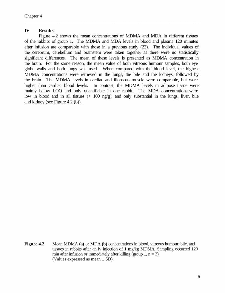
Chapter 4 ________________________________________________________________________________
6
IV Results Figure 4.2 shows the mean concentrations of MDMA and MDA in different tissues
of the rabbits of group 1. The MDMA and MDA levels in blood and plasma 120 minutes after infusion are comparable with those in a previous study (23). The individual values of the cerebrum, cerebellum and brainstem were taken together as there were no statistically significant differences. The mean of these levels is presented as MDMA concentration in the brain. For the same reason, the mean value of both vitreous humour samples, both eye globe walls and both lungs was used. When compared with the blood level, the highest MDMA concentrations were retrieved in the lungs, the bile and the kidneys, followed by the brain. The MDMA levels in cardiac and iliopsoas muscle were comparable, but were higher than cardiac blood levels. In contrast, the MDMA levels in adipose tissue were mainly below LOQ and only quantifiable in one rabbit. The MDA concentrations were low in blood and in all tissues (< 100 ng/g), and only substantial in the lungs, liver, bile and kidney (see Figure 4.2 (b)). Figure 4.2 Mean MDMA (a) or MDA (b) concentrations in blood, vitreous humour, bile, and
tissues in rabbits after an iv injection of 1 mg/kg MDMA. Sampling occurred 120 min after infusion or immediately after killing (group 1, n = 3). (Values expressed as mean ± SD).

Chapter 4 ________________________________________________________________________________
7
In Figure 4.3, the individual MDMA concentrations in blood, vitreous humour and eye globe walls of groups 1, 2 and 3 (Figure 4.3 (a)) are presented. For group 1, the vitreous humour concentration of each individual eye is presented (and not the mean value of both eyes as in Figure 4.2). The Wilcoxon test showed that the post-mortem MDMA blood levels differ significantly from the MDMA vitreous humour levels (p = 0.006; the blood MDMA concentrations were higher than de corresponding vitreous humour levels). The outlier in the blood MDMA levels (R-14) is not relevant, as contamination with thoracic cavity fluid during sampling occurred. The Kruskal-Wallis test showed significantly different MDMA concentrations for the vitreous humour and the eye globe walls of the second sampled eye (p = 0.006 and 0.023, respectively), immediately post mortem and 24 h or 72 h after death. In addition, the individual creatinine concentrations and the ratio of the MDMA to creatinine concentration in vitreous humour are shown (see Figure 4.3 (b)). The Kruskal-Wallis test showed also statistically significant differences for the creatinine values in the second sampled eye (p = 0.002), immediately post mortem and 24 h or 72 h after death. The results of all Kruskal-Wallis tests were confirmed by the Mann-Whitney U-test. However, no statistically significant differences were found when the ratios of MDMA to creatinine concentration were considered. The Spearman correlation coefficient (rs) for the MDMA concentration in the vitreous humour and the eye globe walls of the second eye was 0.64 (p = 0.05).
In Figure 4.4, the mean MDMA (see Figure 4.4 (a)) and MDA (see Figure 4.4 (b)) concentrations in blood and various tissues of groups 1, 2 and 3 are presented. No statistically significant differences between the ligated and non-ligated rabbits were found, either for the MDMA or for the MDA concentrations. The Kruskal-Wallis test, used to compare the values as a function of post-mortem interval, was only significant for the MDMA concentrations in the liver (p = 0.015). The Mann-Whitney U-test confirmed a significant rise in MDMA levels 24 h and 72 h after death. The rs for the MDMA level in post-mortem blood related to cardiac muscle, lungs or liver was 0.64, 0.63 and 0.59, respectively. These correlations were significant at the 0.05 level.
The MDMA concentration in urine, available in 12 rabbits, varied between 500 and 8100 ng/ml. In all rabbits, MDA concentrations in blood and plasma sampled 2 hours after infusion, as well as in the vitreous humour and eye globe walls of the first and second sampled eye, were very low (< 15 ng/g) or below LOQ. MDA could not be quantified in cardiac or iliopsoas muscle, or in adipose tissue. In the brain, MDA was mainly below LOQ, except for one rabbit (13 ng/g). However, relatively high MDA levels were found in the lungs, the liver, the bile and the kidneys (see Figure 4.4 (b)).
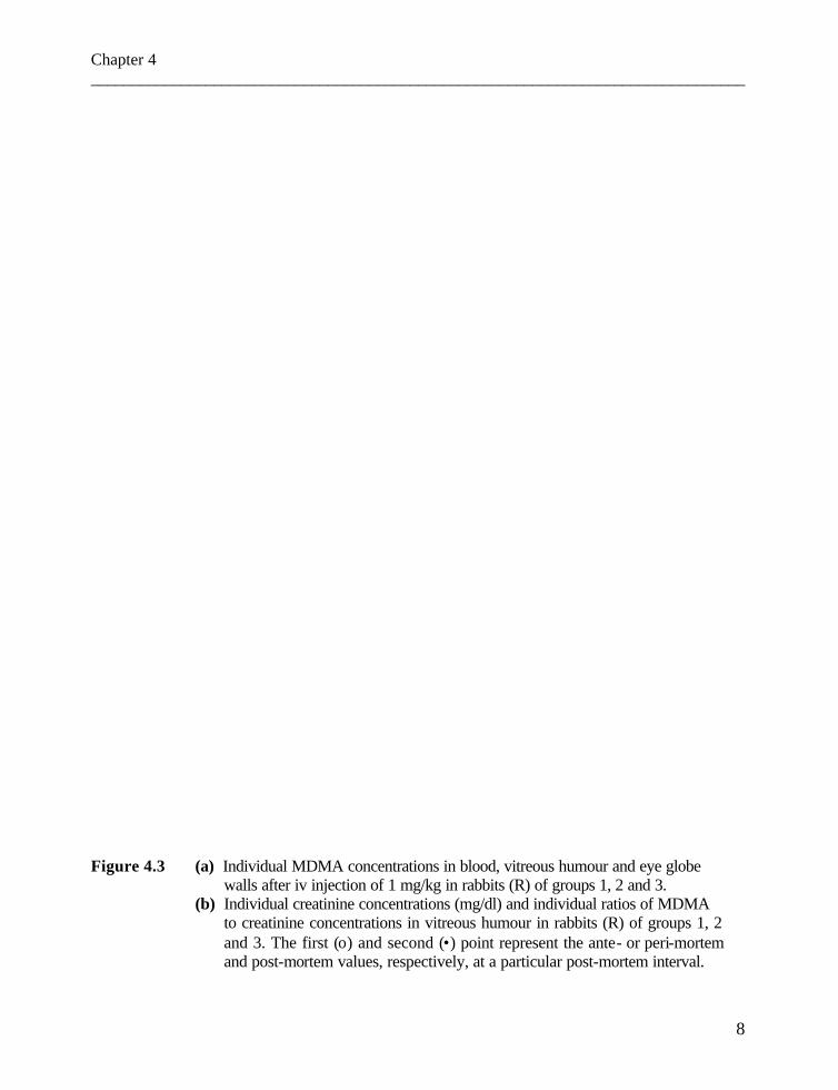
Chapter 4 ________________________________________________________________________________
8
Figure 4.3 (a) Individual MDMA concentrations in blood, vitreous humour and eye globe
walls after iv injection of 1 mg/kg in rabbits (R) of groups 1, 2 and 3. (b) Individual creatinine concentrations (mg/dl) and individual ratios of MDMA
to creatinine concentrations in vitreous humour in rabbits (R) of groups 1, 2 and 3. The first (ο) and second (•) point represent the ante- or peri-mortem and post-mortem values, respectively, at a particular post-mortem interval.

Chapter 4 ________________________________________________________________________________
9
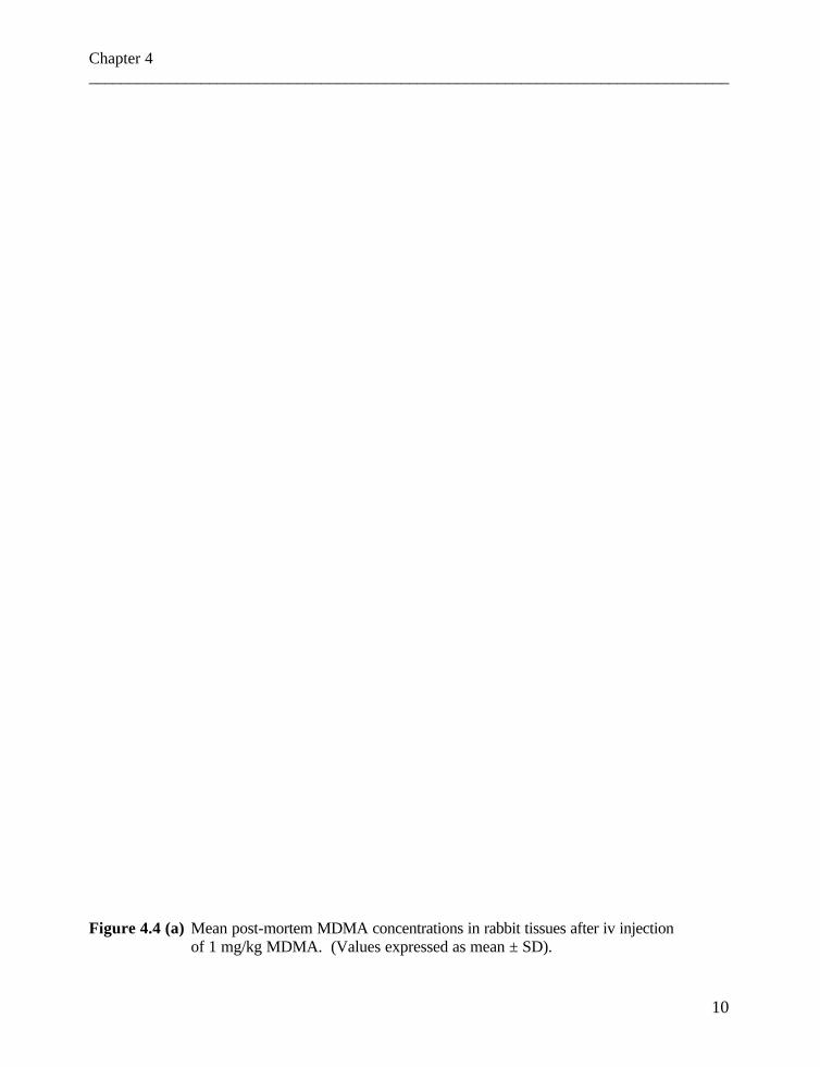
Chapter 4 ________________________________________________________________________________
10
Figure 4.4 (a) Mean post-mortem MDMA concentrations in rabbit tissues after iv injection of 1 mg/kg MDMA. (Values expressed as mean ± SD).

Chapter 4 ________________________________________________________________________________
11
Figure 4.4 (b) Mean post-mortem MDA concentrations in rabbit tissues after iv injection of 1 mg/kg MDMA. (Values expressed as mean ± SD).
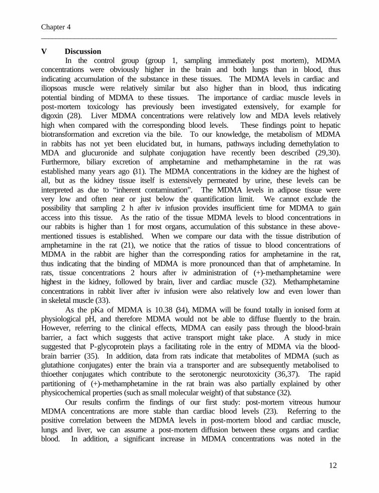
Chapter 4 ________________________________________________________________________________
12
V Discussion In the control group (group 1, sampling immediately post mortem), MDMA
concentrations were obviously higher in the brain and both lungs than in blood, thus indicating accumulation of the substance in these tissues. The MDMA levels in cardiac and iliopsoas muscle were relatively similar but also higher than in blood, thus indicating potential binding of MDMA to these tissues. The importance of cardiac muscle levels in post-mortem toxicology has previously been investigated extensively, for example for digoxin (28). Liver MDMA concentrations were relatively low and MDA levels relatively high when compared with the corresponding blood levels. These findings point to hepatic biotransformation and excretion via the bile. To our knowledge, the metabolism of MDMA in rabbits has not yet been elucidated but, in humans, pathways including demethylation to MDA and glucuronide and sulphate conjugation have recently been described (29,30). Furthermore, biliary excretion of amphetamine and methamphetamine in the rat was established many years ago (31). The MDMA concentrations in the kidney are the highest of all, but as the kidney tissue itself is extensively permeated by urine, these levels can be interpreted as due to “inherent contamination”. The MDMA levels in adipose tissue were very low and often near or just below the quantification limit. We cannot exclude the possibility that sampling 2 h after iv infusion provides insufficient time for MDMA to gain access into this tissue. As the ratio of the tissue MDMA levels to blood concentrations in our rabbits is higher than 1 for most organs, accumulation of this substance in these above-mentioned tissues is established. When we compare our data with the tissue distribution of amphetamine in the rat (21), we notice that the ratios of tissue to blood concentrations of MDMA in the rabbit are higher than the corresponding ratios for amphetamine in the rat, thus indicating that the binding of MDMA is more pronounced than that of amphetamine. In rats, tissue concentrations 2 hours after iv administration of (+)-methamphetamine were highest in the kidney, followed by brain, liver and cardiac muscle (32). Methamphetamine concentrations in rabbit liver after iv infusion were also relatively low and even lower than in skeletal muscle (33).
As the pKa of MDMA is 10.38 (34), MDMA will be found totally in ionised form at physiological pH, and therefore MDMA would not be able to diffuse fluently to the brain. However, referring to the clinical effects, MDMA can easily pass through the blood-brain barrier, a fact which suggests that active transport might take place. A study in mice suggested that P-glycoprotein plays a facilitating role in the entry of MDMA via the blood-brain barrier (35). In addition, data from rats indicate that metabolites of MDMA (such as glutathione conjugates) enter the brain via a transporter and are subsequently metabolised to thioether conjugates which contribute to the serotonergic neurotoxicity (36,37). The rapid partitioning of (+)-methamphetamine in the rat brain was also partially explained by other physicochemical properties (such as small molecular weight) of that substance (32).
Our results confirm the findings of our first study: post-mortem vitreous humour MDMA concentrations are more stable than cardiac blood levels (23). Referring to the positive correlation between the MDMA levels in post-mortem blood and cardiac muscle, lungs and liver, we can assume a post-mortem diffusion between these organs and cardiac blood. In addition, a significant increase in MDMA concentrations was noted in the

Chapter 4 ________________________________________________________________________________
13
homogenates of the eye globe walls sampled in relation to the post-mortem interval. The reliability of creatinine concentrations in post-mortem vitreous humour was substantiated many years ago (25). The rise in creatinine concentration in vitreous humour at increasing post-mortem interval due to dehydration has been confirmed. As the ratios of MDMA to creatinine concentration still show an increasing (though not statistically significant) tendency at longer post-mortem intervals, the (relatively minor) increases in MDMA vitreous humour levels cannot be due exclusively to dehydration. Indeed, bearing in mind the very high MDMA levels in the globe wall of the second sampled eye and the significant correlation between the vitreous humour and globe wall concentration of that particular eye, it can be assumed that diffusion out of these “reservoirs” into the vitreous humour can occur, and that it will occur mainly at longer post-mortem intervals.
Our data are unable to support the findings of Moriya et al (20), who demonstrated redistribution of methamphetamine into cardiac blood via pulmonary blood vessels in the early post-mortem period. Indeed, no statistically significant differences between rabbits with (group 3) or without (group 2) ligation of the large vessels around the heart could be substantiated. However, we cannot exclude the possibility that the lack of significant differences between group 2 and group 3 is influenced by the small number of animals.
The bile MDMA levels tended to decrease at longer post-mortem intervals (see Figure 4.4 (a)), although these changes were not statistically significant. We believe that the significant increases in post-mortem MDMA liver concentrations can partially be explained as a result of diffusion from the bile. On the other hand, MDMA concentrations in cardiac and iliopsoas muscle are fairly stable post mortem and can be of interest when the usual toxicological samples (blood, urine or vitreous humour) are lacking or when advanced putrefaction occurred. This has previously been suggested for methamphetamine and amphetamine (38).
The MDA levels were relatively low in all organs, being below 100 ng/ml. The highest levels were found in the lungs, liver and bile. The high MDA levels in the lungs indicate either non-specific binding of MDA (in addition to accumulation of MDMA) or local metabolism of MDMA to MDA as the lungs contain enzymes such as cytochromes P450. As MDA could be quantified in the eye globe walls of the second sampled eye in groups 2 and 3 and not in the eyes immediately taken after killing (data not shown), this could indicate that MDA can also be formed post mortem. VI Conclusion
In these experiments in rabbits, a state of complete absorption of MDMA was simulated by iv administration. The organ distribution of MDMA and its metabolite MDA was presented and the redistribution up to 72 h post mortem was investigated.
The enhancement of MDMA concentrations in cardiac blood can be due to post-mortem redistribution from the lungs, in the first place, and – to an obviously lesser extent – from the cardiac muscle and liver. As significant differences between rabbits with and without ligation of the large vessels around the heart could not be proven, we believe that post-mortem redistribution on the basis of non-vascular diffusion gradients, namely at

Chapter 4 ________________________________________________________________________________
14
cellular levels (from higher to lower concentrations) could be predominant. However, these findings cannot be totally extrapolated to humans due to the different topographic anatomy of the rabbit, where the organs are inherently closer to one another.
This animal study confirms the findings in a recent fatality (10) that drug concentrations in samples taken for toxicological assay can be influenced by post-mortem redistribution, mainly when sampling takes place centrally in the body, and therefore this phenomenon should be taken into account when drawing medico-legal conclusions, such as whether the MDMA blood concentrations are toxic or potentially lethal. In addition, when an appropriate blood sample is lacking, quantification in the iliopsoas muscle can be helpful to solve the question whether the individual died due to a MDMA overdose.
Acknowledgments The authors wish to thank Mrs Thérèse De Vuyst for her assistance in preparing the manuscript, Mrs Marijke Craeymeersch, Mrs Vera De Vleesschauwer, Mrs Karen Pien, MD, Mr Roland Declercq and Mr Freddy Bekaert for technical assistance, and Prof. A.P. De Leenheer for overall support (equipment, chemicals and accommodation). We would also like to thank as well Mr. G. Van Maele for statistical recommendations and Mr. Richard Sundahl for his assistance with the English grammar.

Chapter 4 ________________________________________________________________________________
15
References 1. Takahashi K, Ikeda N, Kudo K, Funayama M. Forensic significance of concentrations of
ethanol in brain tissues following induced acute subdural hemorrhage. Int J Legal Med 2001;115:1-5.
2. Pounder DJ, Jones GR. Post-mortem drug redistribution – a toxicological nightmare. Forensic
Sci Int 1990;45:253-263. 3. Hearn WL, Keran EE, Wei H, Hime G. Site-dependent postmortem changes in blood cocaine
concentrations. J Forensic Sci 1991;36:673-684. 4. Pounder DJ, Hartley AK, Watmough PJ. Postmortem redistribution and degradation of
dothiepin. Human case studies and an animal model. Am J Forensic Med Pathol 1994;15:231-235.
5. Kintz P, Tracqui A, Ludes B. The distribution of laudanosine in tissues after death from
atracurium injection. Int J Legal Med 2000;114:93-95. 6. Takeshita H, Mogi K, Yasuda T, Mori S, Nakashima Y, Nakajima T, Akuzawa H, Nakajo S,
Hirota Y, Kishi K. Postmortem absorption of dichloromethane: a case study and animal experiments. Int J Legal Med 2000;114:96-100.
7. Dowling GP, McDonough ET III, Bost RO. ‘Eve’ and ‘Ecstasy’. A report of five deaths
associated with the use of MDEA and MDMA. JAMA 1987;257:1615-1617. 8. Rohrig TP, Prouty RW. Tissue distribution of methylenedioxymethamphetamine. J Anal
Toxicol 1992;16:52-53. 9. Fineschi V, Masti A. Fatal poisoning by MDMA (ecstasy) and MDEA: a case report. Int J
Legal Med 1996;108:272-275. 10. De Letter EA, Clauwaert KM, Lambert WE, Van Bocxlaer JF, De Leenheer AP, Piette MHA.
Distribution study of 3,4-methylenedioxymethamphetamine and 3,4-methylenedioxyamphetamine in a fatal overdose. J Anal Toxicol 2002;26:113-118.
11. Meyer E, Van Bocxlaer JF, Dirinck IM, Lambert WE, Thienpont L, De Leenheer AP. Tissue
distribution of amphetamine isomers in a fatal overdose. J Anal Toxicol 1997;21:236-239. 12. Prouty RW, Anderson WH. The forensic science implications of site and temporal influences
on postmortem blood-drug concentrations. J Forensic Sci 1990;35:243-270. 13. Barnhart FE, Reed DW. Methamphetamine – a study of postmortem redistribution. J Anal
Toxicol 1999;23:69-70.

Chapter 4 ________________________________________________________________________________
16
14. Katsumata S, Sato K, Kashiwade H, Yamanami S, Zhou H, Yonemura I, Nakajima H, Hasekura
H. Sudden death due presumably to internal use of methamphetamine. Forensic Sci Int 1993;62:209-215.
15. Miyazaki T, Kojima T, Yashiki M, Wakamoto H, Iwasaki Y, Taniguchi T. Site dependence of
methamphetamine concentrations in blood samples collected from cadavers of people who had been methamphetamine abusers. Am J Forensic Med Pathol 1993;14:121-124.
16. Moriya F, Hashimoto Y. Redistribution of methamphetamine in the early postmortem period. J
Anal Toxicol 2000;24:153-154. 17. Logan BK, Weiss EL, Harruff R. Case report: Distribution of methamphetamine in a massive
fatal ingestion. J Forensic Sci 1996;41:322-323. 18. Kalasinsky KS, Bosy TZ, Schmunk GA, Reiber G, Anthony RM, Furukawa Y, Guttman M,
Kish SJ. Regional distribution of methamphetamine in autopsied brain of chronic human methamphetamine users. Forensic Sci Int 2001;116:163-169.
19. Kojima T, Une I, Yashiki M. CI-mass fragmentographic analysis of methamphetamine and
amphetamine in human autopsy tissues after acute methamphetamine poisoning. Forensic Sci Int 1983;21:253-258.
20. Moriya F, Hashimoto Y. Redistribution of basic drugs into cardiac blood from surrounding
tissues during early-stages postmortem. J Forensic Sci 1999;44:10-16. 21. Hilberg T, Ripel Å, Slørdal L, Bjørneboe A, Mørland J. The extent of postmortem drug
redistribution in a rat model. J Forensic Sci 1999;44:956-962. 22. Hilberg T, Rogde S, Mørland J. Postmortem drug redistribution – human cases related to
results in experimental animals. J Forensic Sci 1999;44:3-9. 23. De Letter EA, De Paepe P, Clauwaert KM, Belpaire FM, Lambert WE, Van Bocxlaer JF, Piette
MHA. Is vitreous humour useful for the interpretation of 3,4-methylenedioxymethamphetamine (MDMA) blood levels? Experimental approach with rabbits. Int J Legal Med 2000;114:29-35.
24. De Letter EA, Belpaire FM, Clauwaert KM, Lambert WE, Van Bocxlaer JF, Piette MHA. Post-
mortem redistribution of 3,4-methylenedioxymethamphetamine (MDMA, “ecstasy”) in the rabbit. Part two: post-mortem infusion in trachea or stomach. Int J Legal Med 2002;116:225-232.
25. Leahy MS, Farber ER. Postmortem chemistry of human vitreous humor. J Forensic Sci
1967;12:214-222. 26. Clauwaert KM, Van Bocxlaer JF, De Letter EA, Van Calenbergh S, Lambert WE, De Leenheer
AP. Determination of the designer drugs 3,4-methylenedioxy-methamphetamine, 3,4-

Chapter 4 ________________________________________________________________________________
17
methylenedioxyethylamphetamine, and 3,4-methylenedioxy-amphetamine with HPLC and fluorescence detection in whole blood, serum, vitreous humor, and urine. Clin. Chem. 2000;46:1968-1977.
27. Butler AR. The Jaffe reaction. Identification of the coloured species. Clin. Chim. Acta 1976;59:
227-232. 28. Ottosson A, Edvinsson L, Sjögren A, Löwenhielm P. Digoxin, magnesium, and potassium
levels in a forensic autopsy material of sudden death from ischemic heart disease. Z Rechtsmed 1988;101:27-36.
29. Torre R de la, Farré M, Ortuño J, Mas M, Brenneisen R, Roset PN, Segura J, Camí J. Non-
linear pharmacokinetics of MDMA (‘ecstasy’) in humans. Br J Clin Pharmacol 2000;49:104-109.
30. Maurer HH, Bickeboeller-Friedrich J, Kraemer T, Peters FT. Toxicokinetics and analytical
toxicology of amphetamine-derived designer drugs (“Ecstasy”). Toxicol Lett 2000;112-113:133-142.
31. Caldwell J, Dring LG, Williams RT. Biliary excretion of amphetamine and methamphetamine
in the rat. Biochem J 1972;129:25-29. 32. Rivière GJ, Gentry WB, Owens SM. Disposition of methamphetamine and its metabolite
amphetamine in brain and other tissues in rats after intravenous administration. J Pharmacol Exp Ther 2000; 292:1042-1047.
33. Nagata T, Kimura K, Hara K, Kudo K. Methamphetamine and amphetamine concentrations in
postmortem rabbit tissues. Forensic Sci Int 1990;48:39-47. 34. Garrett ER, Seyda K, Marroum P. High performance liquid chromatographic assays of the
illicit designer drug “Ecstasy”, a modified amphetamine, with applications to stability, partitioning and plasma protein binding. Acta Pharm Nord 1991;3:9-14.
35. Mann H, Ladenheim B, Hirata H, Moran TH, Cadet JL. Differential toxic effects of
methamphetamine (METH) and methylenedioxymethamphetamine (MDMA) in multidrug-resistant (mdr1a) knockout mice. Brain Res 1997;769:340-346.
36. Monks TJ, Lau SS. Biological reactivity of polyphenolic-gluthatione conjugates. Chem Res
Toxicol 1997;10:1296-1313. 37. Bai F, Jones DC, Lau SS, Monks TJ. Serotonergic neurotoxicity of 3,4-(±)-
methylenedioxyamphetamine and 3,4-(±)-methylenedioxymethamphetamine (Ecstasy) is potentiated by inhibition of γ-glutamyl transpeptidase. Chem Res Toxicol 2001;14:863-870.

Chapter 4 ________________________________________________________________________________
18
38. Hara K, Nagata T, Kimura K. Forensic toxicologic analysis of methamphetamine and
amphetamine in body materials by gas chromatography/mass spectrometry. Z Rechtsmed 1986;96:93-104.

Chapter 5
Post-mortem redistribution of 3,4-methylenedioxy-methamphetamine (MDMA, “ecstasy”) in the rabbit
Part two: Post-mortem infusion in trachea or stomach

Chapter 5 ________________________________________________________________________________
1
Chapter 5 Post-mortem redistribution of 3,4-methylenedioxymeth-
amphetamine (MDMA, “ecstasy”) in the rabbit Part two: post-mortem infusion in trachea or stomach.
Els A. De Letter1, Frans M. Belpaire2, Karine M. Clauwaert3, Willy E. Lambert4 ,
Jan F. Van Bocxlaer3 , Michel H.A. Piette1 Ghent University 1 Els A. De Letter, Michel H.A. Piette Department of Forensic Medicine 2 Frans M. Belpaire Heymans Institute of Pharmacology 3 Karine M. Clauwaert, Jan F. Van Bocxlaer Department of Medical Biochemistry and Clinical Analysis
4 Willy E. Lambert Department of Toxicology
Int J Legal Med 2002;116:225-232
Chapter 5 ________________________________________________________________________________
2
I Abstract Drug concentrations in autopsy samples can also be influenced by post-mortem
gastric diffusion when the stomach contains a substantial amount of the drug or by diffusion from the trachea when agonal aspiration or post-mortem regurgitation of vomit occurs. This was studied in a rabbit animal model in which MDMA solutions were infused post mortem either in the trachea or in the stomach. At 24, 48 or 72 hours post mortem, samples including cardiac blood, vitreous humour, urine, bile, gastric content and several tissues were taken for toxicologic analysis.
After post-mortem tracheal infusion, MDMA can easily diffuse not only into the lungs but also in great quantities into the cardiac blood and – to a lesser extent – into the cardiac muscle. MDMA was also found in the closely adjacent diaphragm and in the upper abdominal organs, including the liver and the stomach.
Following post-mortem infusion into the stomach, considerable MDMA levels were found in cardiac blood and muscle, both lungs, diaphragm and liver tissue when the solution was concentrated nearby the lower oesophageal sphincter. However, when the MDMA solution was present deeper in the stomach, MDMA levels were high in the spleen and the liver and relatively low in cardiac blood and muscle.
In both experiments, MDA levels were in most tissues low or below the limit of quantitation, but were substantial in cardiac blood and muscle, lung and diaphragm, indicating that MDMA can be metabolised to MDA after death.
These results in the rabbit model indicate that the diffusion of MDMA out of the stomach content, or due to aspirated vomit and gastro-oesophageal reflux can lead to considerable post-mortem redistribution and thus should be taken into account in current forensic practice in order to draw the right conclusions when a peripheral blood sample is not available. Key words: 3,4-Methylenedioxymethamphetamine (MDMA) - 3,4-Methylenedioxy-amphetamine (MDA) - Post-mortem tracheal and gastric infusion - Post-mortem redistribution - Experiment on rabbits II Introduction
Post-mortem drug levels can be difficult to interpret due to interfering thanato-chemical processes such as drug instability and post-mortem redistribution (1). These processes can result from diffusion of the substance out of adjacent organs. They can also be due to diffusion from high concentrations present in the gastric content and/or from vomit aspiration in the airways or even from post-mortem regurgitation (2,3).
Post-mortem absorption of ethanol, paracetamol and propoxyphene from simulated vomit aspiration was found to result in an increase in post-mortem cardiac blood concentrations in five human bodies (4).
Diffusion of ethanol from the stomach cavity after death has been investigated extensively for more than 50 years (5). As this post-mortem diffusion results in an elevation of the blood alcohol level in cardiac blood (6) and even in aortic blood (7), peripheral blood sampling such as from the femoral (or external iliac) vein is recommended
Chapter 5 ________________________________________________________________________________
3
because it is obviously less liable to post-mortem changes. Post-mortem diffusion from gastric residue into blood and surrounding tissues has also been studied for a few other drugs including zopiclone (8), benzodiazepines (9), paracetamol (9) and amitriptyline (10,11).
The amphetamine derivative, 3,4-methylenedioxymethamphetamine (MDMA, “ecstasy”) is stable in blood and plasma in vitro (12). In part one, post-mortem redistribution due to diffusion of MDMA out of several organ tissues was evaluated after simulation of a complete distribution of the substance prior to death (13). In this study, post-mortem diffusion of MDMA from a “reservoir” in the stomach or from agonal vomit aspiration was explored using a rabbit animal model. This can be compared with the condition when somebody dies shortly after MDMA ingestion (e.g. due to cardiac arrhythmia) and therefore an incomplete absorption occurred, or when substantial regurgitation or vomit aspiration takes place in the peri-mortem period. When blood and tissue concentrations (studied up to 72 hours after death) in both experimental settings change substantially, this should be taken into account in the interpretation of the toxicologic results in humans. III Materials and methods
Provision of MDMA and rabbits as well as handling of the animals prior to the onset of the experiments and preservation of the samples took place as previously described (13).
III.1 Animals and procedures The rabbits (weight 1900 – 2420 g) were killed using a CO2-O2 gas chamber (70/30
%). Thereafter, two groups of rabbits were created: group 1 (n = 6) was used for post-mortem infusion into the trachea (PIT) and group 2 (n = 6) for post-mortem infusion into the stomach (PIS). Randomisation of the animals occurred and infusion of the MDMA solution (diluted in saline; 1 mg/kg) took place within the first hour post mortem.
The study design is presented in Figure 5.1. After preparation of either the trachea or the oesophagus, ligation towards the laryngeal/pharyngeal region took place. For group 1 (PIT), a highly concentrated MDMA solution was used in order to reduce the volume of fluid to be infused (< 0.5 ml). The solution was injected into the trachea using a 1-ml syringe and 26 G needle. In group 2 (PIS), a polyethylene catheter (inner diameter of 2.5 mm) was inserted into the oesophagus up to the lower oesophageal sphincter. After infusion of MDMA, flushing of the catheter with saline took place. All rabbits were left in a supine position at ambient temperature (15 °C). Dissection was carried out in a strict manner so as to avoid contamination of the samples and sampling took place in the same sequence for all rabbits. The samples taken in group 1 (PIT) were: cardiac blood and muscle, left and right lungs, left and right diaphragm, liver, stomach wall and stomach content, spleen, left and right iliopsoas muscle, abdominal adipose tissue, left and right kidneys, urine, cerebrum, cerebellum, brainstem, and both eyes. In the second group (PIS), furthermore, lower vena cava blood, duodenal wall and content, distal small bowel and content, and left and right abdominal muscle wall were sampled. Although the rabbits
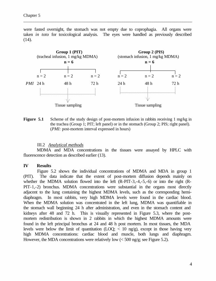
Chapter 5 ________________________________________________________________________________
4
were fasted overnight, the stomach was not empty due to coprophagia. All organs were taken in toto for toxicological analysis. The eyes were handled as previously described (14).
Group 1 (PIT) Group 2 (PIS) (tracheal infusion, 1 mg/kg MDMA) (stomach infusion, 1 mg/kg MDMA) n = 6 n = 6
n = 2 n = 2 n = 2 n = 2 n = 2 n = 2
PMI 24 h 48 h 72 h 24 h 48 h 72 h
Tissue sampling Tissue sampling
Figure 5.1 Scheme of the study design of post-mortem infusion in rabbits receiving 1 mg/kg in the trachea (Group 1; PIT; left panel) or in the stomach (Group 2; PIS; right panel).
(PMI: post-mortem interval expressed in hours) III.2 Analytical methods MDMA and MDA concentrations in the tissues were assayed by HPLC with
fluorescence detection as described earlier (13). IV Results
Figure 5.2 shows the individual concentrations of MDMA and MDA in group 1 (PIT). The data indicate that the extent of post-mortem diffusion depends mainly on whether the MDMA solution flowed into the left (R-PIT-3,-4,-5,-6) or into the right (R-PIT-1,-2) bronchus. MDMA concentrations were substantial in the organs most directly adjacent to the lung containing the highest MDMA levels, such as the corresponding hemi-diaphragm. In most rabbits, very high MDMA levels were found in the cardiac blood. When the MDMA solution was concentrated in the left lung, MDMA was quantifiable in the stomach wall beginning 24 h after administration, and even in the stomach content and kidneys after 48 and 72 h. This is visually represented in Figure 5.3, where the post-mortem redistribution is shown in 2 rabbits in which the highest MDMA amounts were found in the left principal bronchus at 24 and 48 h post mortem. In most tissues, the MDA levels were below the limit of quantitation (LOQ; < 10 ng/g), except in those having very high MDMA concentrations: cardiac blood and muscle, both lungs and diaphragm. However, the MDA concentrations were relatively low (< 500 ng/g; see Figure 5.2).
Chapter 5 ________________________________________________________________________________
5
Figure 5.2 Individual MDMA and MDA concentrations in rabbits (n = 6) after post-mortem infusion of 1 mg/kg MDMA in the trachea (PIT), 24, 48 and 72 h after administration.

Chapter 5 ________________________________________________________________________________
6
Figure 5.3 Thoracic and abdominal post-mortem diffusion after tracheal instillation of 1 mg/kg
MDMA in rabbits (n = 2), in which spreading of the solution occurred predominantly in the left bronchus, 24 and 48 h after administration.
Labels: RA: right atrium AO: aorta RV: right ventricle VI: inferior vena cava LA: left atrium VS: superior vena cava LV: left ventricle AR: arteria renalis TP: truncus pulmonalis VR: vena renalis VP: venae pulmonales D: diaphragm
MDMA levels (ng/ml or ng/g): > 10,000 5,000 – 10,000 1,000 – 5,000 500 – 1,000 100 - 500 10 - 100 < 10
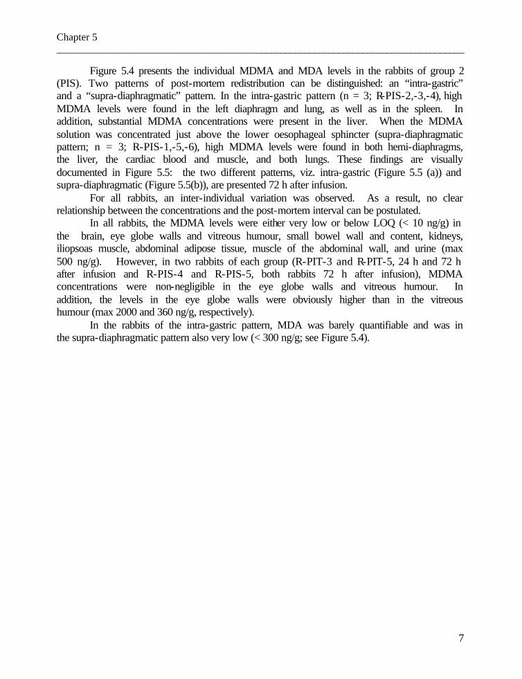
Chapter 5 ________________________________________________________________________________
7
Figure 5.4 presents the individual MDMA and MDA levels in the rabbits of group 2 (PIS). Two patterns of post-mortem redistribution can be distinguished: an “intra-gastric” and a “supra-diaphragmatic” pattern. In the intra-gastric pattern (n = 3; R-PIS-2,-3,-4), high MDMA levels were found in the left diaphragm and lung, as well as in the spleen. In addition, substantial MDMA concentrations were present in the liver. When the MDMA solution was concentrated just above the lower oesophageal sphincter (supra-diaphragmatic pattern; n = 3; R-PIS-1,-5,-6), high MDMA levels were found in both hemi-diaphragms, the liver, the cardiac blood and muscle, and both lungs. These findings are visually documented in Figure 5.5: the two different patterns, viz. intra-gastric (Figure 5.5 (a)) and supra-diaphragmatic (Figure 5.5(b)), are presented 72 h after infusion.
For all rabbits, an inter-individual variation was observed. As a result, no clear relationship between the concentrations and the post-mortem interval can be postulated.
In all rabbits, the MDMA levels were either very low or below LOQ (< 10 ng/g) in the brain, eye globe walls and vitreous humour, small bowel wall and content, kidneys, iliopsoas muscle, abdominal adipose tissue, muscle of the abdominal wall, and urine (max 500 ng/g). However, in two rabbits of each group (R-PIT-3 and R-PIT-5, 24 h and 72 h after infusion and R-PIS-4 and R-PIS-5, both rabbits 72 h after infusion), MDMA concentrations were non-negligible in the eye globe walls and vitreous humour. In addition, the levels in the eye globe walls were obviously higher than in the vitreous humour (max 2000 and 360 ng/g, respectively).
In the rabbits of the intra-gastric pattern, MDA was barely quantifiable and was in the supra-diaphragmatic pattern also very low (< 300 ng/g; see Figure 5.4).

Chapter 5 ________________________________________________________________________________
8
Figure 5.4 Individual MDMA and MDA concentrations in rabbits (n = 6) after post-mortem
infusion of 1 mg/kg MDMA in the stomach (PIS), 24, 48 and 72 h after administration.

Chapter 5 ________________________________________________________________________________
9
Figure 5.5 Thoracic and abdominal post-mortem diffusion after gastric instillation of 1 mg/kg
MDMA in rabbits (n = 2), showing the difference between the intra-gastric (a), and supra-diaphragmatic pattern (b) 72 h after instillation. Labels: RA: right atrium AO: aorta
RV: right ventricle VI: inferior vena cava LA: left atrium VS: superior vena cava LV: left ventricle AR: arteria renalis TP: truncus pulmonalis VR: vena renalis VP: venae pulmonales D: diaphragm
MDMA levels (ng/ml or ng/g): > 10,000 5,000 – 10,000 1,000 – 5,000 500 – 1,000 100 - 500 10 - 100 < 10
Chapter 5 ________________________________________________________________________________
10
V Discussion MDMA tissue levels after post-mortem tracheal instillation depend on the
dispersion of the solution into either the left or the right bronchus. In both cases, however, the MDMA concentrations were high in the cardiac blood and – to a lesser extent – also in the cardiac muscle. In addition, our data show that MDMA can easily diffuse out of the trachea into the thoracic and upper abdominal organs, and the amounts diffused slightly increase with the post-mortem interval. In the lower abdominal tissues, such as the kidneys, the iliopsoas muscle and adipose tissue, the MDMA levels were either very low or below the quantitation limit.
After post-mortem instillation in the stomach, two different diffusion patterns were observed depending on whether the MDMA solution was concentrated intra-gastrically or supra-diaphragmatically. The supra-diaphragmatic situation is comparable to gastro-oesophageal reflux, which involves substantial diffusion into cardiac blood and muscle, both lungs and liver. When the MDMA solution was concentrated more deeply in the stomach, post-mortem redistribution did affect the thoracic organs to a minor extent, and the intra-gastric solution diffused mainly into the closely adjacent spleen. Our results indicate that peri- or post-mortem gastro-oesophageal reflux is obviously more responsible for the redistribution of MDMA than a high MDMA concentration in the stomach itself.
In four rabbits, non-negligible MDMA levels were found in vitreous humour and eye globe walls. These levels were clearly higher than in the corresponding brain, which indicates that another mechanism than pure diffusion from the brain should be assumed. One possible explanation is that there was direct or indirect reflux into the naso-pharynx with diffusion of MDMA into the sinuses, the skull base and the orbitae. Such diffusion has formerly been established for ethanol: in a human model, diffusion from an ethanol solution in the mouth and pharynx into the skull and also into the vitreous humour was observed, although at relatively longer post-mortem intervals (more than 60 or 72 h) (15).
In all the experiments in rabbits we performed at present, we observe that the MDMA concentrations in the iliopsoas muscle are not subject to post-mortem diffusion, and thus remain stable after death. Therefore, iliopsoas muscle can be an interesting specimen when the usual samples for drug assay are lacking. However, muscle sampling is not recommended for some other substances (such as temazepam, prothiaden, paracetamol and amitriptyline) (16,17). These studies did not include concentrations in iliopsoas muscle, however.
In both post-mortem instillation experiments, the MDA levels were only quantifiable when very high local MDMA concentrations were found, which proves that MDMA can be metabolised post mortem into MDA. The MDA concentrations were lower when the MDMA solution was concentrated intra-gastrically instead of supra-diaphragmatically. This is in accordance with a previous study in which we hypothesized that the lungs play a role in the metabolism of MDMA to MDA (13).

Chapter 5 ________________________________________________________________________________
11
VI Conclusion In this experiment, we used tracheal instillation of MDMA to demonstrate that agonal vomit aspiration can lead to substantial post-mortem redistribution, mainly into cardiac blood and muscle, and into both lungs. To a less pronounced extent, MDMA also diffused to the liver tissue and the lower abdominal organs. Using infusion into the stomach, we proved that peri- or post-mortem gastro-oesophageal reflux gives rise to significant post-mortem diffusion of MDMA. When the MDMA reservoir is concentrated in the stomach itself, the thoracic organs are not substantially affected by redistribution up to 72 h post mortem. These rabbit experimental results could be extrapolated to humans as agonal aspiration in the lungs or post-mortem regurgitation frequently occurs in medico-legal practice. Our results demonstrate once more that peripheral sampling should be recommended in current practice. However, when this is not possible, the MDMA and MDA levels should be interpreted with great caution, especially regarding toxic or lethal levels. Finally, as in all experiments performed at present, the iliopsoas muscle concentrations remain stable post mortem, this specimen can be useful in current forensic practice when an appropriate blood sample is lacking. Acknowledgments The authors wish to thank Mrs Thérèse De Vuyst for her assistance in preparing the manuscript, Dr. Beatrice De Smet, Mrs Marijke Craeymeersch, Mrs Vera De Vleesschauwer, and Mr Freddy Bekaert for skilled technical assistance, and Prof. A.P. De Leenheer for overall support (equipment, chemicals and accommodation). We would also like to thank Mr. Richard Sundahl for his assistance with the English grammar.
Chapter 5 ________________________________________________________________________________
12
References 1. Pounder DJ, Jones GR. Post-mortem drug redistribution – a toxicological nightmare. Forensic
Sci Int 1990;45:253-263. 2. Knight BH. (ed) (1996), Forensic Pathology, 2nd edn, Arnold, London, Sydney, Auckland, pp
356-358. 3. Knight BH. The significance of the postmortem discovery of gastric contents in the air
passages. Forensic Sci 1975;6:229-234. 4. Pounder DJ, Yonemitsu K. Postmortem absorption of drugs and ethanol from aspirated vomitus
– an experimental model. Forensic Sci Int 1991;51:189-195. 5. Hecke W van, Handovsky H, Thomas F. Analyse statistique de 597 dosages d’alcool éthylique
practiqués dans le sang, les humeurs et les organes d’un total de 93 cadavres. Ann Med Leg 1951;31 :291-338.
6. Iwasaki Y, Yashiki M, Namera A, Miyazaki T, Kojima T. On the influence of postmortem
alcohol diffusion from the stomach contents to the heart blood. Forensic Sci Int 1998; 94:111-118.
7. Pounder DJ, Smith DRW. Postmortem diffusion of alcohol from the stomach. Am J Forensic
Med Pathol 1995;16:89-96. 8. Pounder DJ, Davies JI. Zopiclone poisoning: tissue distribution and potential for postmortem
diffusion. Forensic Sci Int 1994;65:177-183. 9. Pounder DJ, Adams E, Fuke C, Langford AM. Site to site variability of postmortem drug
concentrations in liver and lung. J Forensic Sci 1996;41:927-932. 10. Pounder DJ, Fuke C, Cox DE, Smith D, Kuroda N. Postmortem diffusion of drugs from gastric
residue. An experimental study. Am J Forensic Med Pathol 1996;17:1-7. 11. Hilberg T, Bugge A, Beylich K-M, Mørland J, Bjørneboe A. Diffusion as a mechanism of
postmortem drug redistribution: an experimental study in rats. Int J Legal Med 1992;105: 87-91.
12. Garrett ER, Seyda K, Marroum P. High performance liquid chromatographic assays of the
illicit designer drug “Ecstasy”, a modified amphetamine, with applications to stability, partitioning and plasma protein binding. Acta Pharm Nord 1991;3:9-14.
13. De Letter EA, Clauwaert KM, Belpaire FM, Lambert WE, Van Bocxlaer JF, Piette MHA Post-
mortem redistribution of 3,4-methylenedioxymethamphetamine (MDMA, “ecstasy”) in the rabbit. Part one: Experimental approach after in vivo intravenous infusion. Int J Legal Med 2002;116:216-224.

Chapter 5 ________________________________________________________________________________
13
14. De Letter EA, De Paepe P, Clauwaert KM, Belpaire FM, Lambert WE, Van Bocxlaer JF, Piette
MHA. Is vitreous humour useful for the interpretation of 3,4-methylenedioxy-methamphetamine (MDMA) blood levels? Experimental approach with rabbits. Int J Legal Med 2000;114:29-35.
15. Saternus K-S, Langenberg K, Iffland R, Staak M. Untersuchungen zur postmortalen Diffusion
von Äthanol in den intracraniellen Raum. Blutalkohol 1982;19:171-180. 16. Langford AM, Taylor KK, Pounder DJ. Drug concentration in selected skeletal muscles. J
Forensic Sci 1998;43:22-27. 17. Williams KR, Pounder DJ. Site-to-site variability of drug concentrations in skeletal muscle.
Am J Forensic Med Pathol 1997;18:246-250.
Chapter 6 ________________________________________________________________________________
1
Chapter 6 Thanato-toxicological approach
I MDMA AND ITS METABOLITE MDA
I.1 Distribution study of 3,4-methylenedioxymethamphetamine and 3,4-methylenedioxyamphetamine in a fatal overdose
Els A. De Letter1, Karine M. Clauwaert2, Willy E. Lambert3 , Jan F. Van Bocxlaer2 ,
André P. De Leenheer2,3, Michel H.A. Piette1
Ghent University
1 Els A. De Letter, Michel H.A. Piette Department of Forensic Medicine
2 Karine M. Clauwaert, Jan F. Van Bocxlaer, André P. De Leenheer Department of Medical Biochemistry and Clinical Analysis
3 Willy E. Lambert, André P. De Leenheer
Department of Toxicology
J Anal Toxicol 2002;26:113-118

Chapter 6 ________________________________________________________________________________
2
I Abstract In this study, regional tissue distributions of the amphetamine analogue, 3,4-
methylenedioxymethamphetamine (MDMA, “ecstasy”) and its metabolite 3,4-methylenedioxyamphetamine (MDA) in a fatal overdose are presented. Quantitation of MDMA and MDA levels occurred in blood samples taken centrally (right and left heart and main adjacent great vessels) and peripherally (subclavian and femoral blood). In addition, MDMA and MDA concentrations were determined in cardiac and iliopsoas muscle, both lungs, liver, both kidneys, spleen, the four brain lobes, cerebellum and brainstem, and adipose tissue. Finally, MDMA and MDA levels were determined in serum, vitreous humour, urine and bile. For all samples, a fully validated high-pressure liquid chromatography procedure with fluorescence detection was used. The found substances were also identified with liquid chromatography-tandem mass spectrometry.
Our data confirm that blood sampling from an isolated peripheral vein is recommended for MDMA and MDA. In addition, the vitreous humour MDMA level indicates that this fluid can be an interesting alternative when a suitable blood sample is missing. Considering the substantial differences in concentrations in blood samples taken from various sites in the body and the high levels in some tissues (e.g. in liver), we concluded that the influence of post-mortem redistribution should be taken into account in the interpretation of toxicological data when an appropriate peripheral sample cannot be obtained or when blood samples are not available because of putrefaction. II Introduction
Post-mortem instability and redistribution are known interfering processes in thanato-toxicological investigations (1). However, amphetamine and its derivative 3,4-methylenedioxymethamphetamine (MDMA, “ecstasy”) in particular are stable in blood and plasma in vitro (2). The stability of methamphetamine and amphetamine in post-mortem rabbit tissues – stored in test tubes under four different conditions - was studied over a two-year period, and the authors concluded that skeletal muscle and bone marrow proved to be the most appropriate samples for accessing toxicity (3). To our knowledge, post-mortem drug distribution and redistribution of MDMA and 3,4-methylenedioxy-amphetamine (MDA) have barely been explored in humans, except for a few case reports (4-6). More literature data are available for amphetamine and methamphetamine (7-16). We report an extended regional tissue distribution study in a fatal overdose case.
III Case history
One morning, a 23-year-old man was found unconscious in a bar. He was sitting on a chair, resting with his head upon his forearms on the table in front of him. The emergency team attempted intensive reanimation, which failed. Upon examination 28 h post mortem, the body weighed approximately 100 kg and was 186 cm tall. In the pocket of the decedent, a small amount of white powder was found. Some vomit was noticed on his T-shirt and his boxer shorts were soiled with urine. During further external examination, many vibices were observed in the post-mortem lividity located on the upper thorax and back, and his face was strongly cyanotic. Obvious congestion of the
Chapter 6 ________________________________________________________________________________
3
conjunctivae and intermediary pupils was present. A fresh puncture wound was seen on the right arm but inquiry revealed that it occurred during the reanimation attempt. During internal inspection, signs of intensive reanimation, including a sternal fracture, were found. Numerous Tardieu spots were observed on the pericardium and both pleurae. Both lungs weighed 1620 g, and, upon sectioning, some emphysema, severe congestion and moderate oedema were found. The heart weighed 405 g and an aberrant course of the superior vena cava, a persistence of the left superior caval vein, was noticed. The stomach contained a brownish liquid without food fragments, and the mucosa showed a few pin-point ulcerations. The brain weighed 1545 g, and, apart from slight oedema and congestion of the white matter, nothing unusual was observed. The remaining organs showed no obvious anomalies macroscopically, except for congestion.
On histological examination, pronounced pulmonary congestion, haemorrhagic oedema, a slight intra-alveolar infiltration with a few polymorphonuclear cells and some leucocyte sludging in the pulmonary veins were found. Groups of alveolar macrophages were seen, although staining with Prussian blue was negative and there were no signs of pulmonary hypertension. The liver showed slight fatty infiltration, and a few peripancreatic lymphocyte infiltrates were found. In the caudate nucleus and the nucleus lentiformis, a few venulae were surrounded by a lymphocyte infiltration. The hippocampus showed no marked hypoxemic lesions. No obvious pre-existing disease was identified histologically.
Because drug abuse was suspected and amphetamines could be involved, appropriate samples for a distribution study were taken. Sampling included all possible central and peripheral blood samples, stomach content, urine, bile and vitreous humour. Several small tissue fragments were taken at random throughout the organs. The samples of cardiac and iliopsoas muscle, both lungs, both kidneys, liver, spleen, abdominal adipose tissue, all four lobes of the cerebrum, cerebellum and brainstem were preserved at – 30°C until analysis.
IV Materials and methods
IV.1 Reagents and materials All reagents and chemicals were of analytical grade and were obtained from Aldrich
(Gillingham, U.K.) unless stated otherwise. Solvents were of HPLC grade from Merck (Darmstadt, Germany). Pure MDA, MDMA and 3,4-methylenedioxyethylamphetamine (MDEA) standards were obtained from Sigma (St. Louis, MO). 3,4-Methylenedioxy-methylpropylamphetamine (MDMPA) was synthesized by in-house following a procedure described earlier (17). Stock solutions of these active substances were prepared by dissolving 10 mg of the pure compound in 10 ml of methanol. Appropriate dilution with methanol yielded the working solutions containing all three compounds. All concentrations of the standards are expressed as the free base. The stock solutions were stored in the dark at - 20 °C and were stable for at least 1 year. Working solutions were stored under the same conditions as the stock standards but discarded after 6 months.
Chapter 6 ________________________________________________________________________________
4
IV.2 Drug screening A comprehensive screening was performed on blood, urine and stomach content.
Screening methods used were the enzyme-multiplied immunoassay technique (EMIT®), radioimmunoassay (RIA), and various chromatographic techniques, including high-performance liquid chromatography-diode array detection (HPLC-DAD) (following extraction under alkaline conditions), thin-layer chromatography (TLC) on Sunshine extracts, and gas chromatography-mass spectrometry (GC-MS), as described previously (18).
IV.3 Apparatus The HPLC unit was composed of a ternary low pressure gradient pump, and an
autosampler with a 25-µl loop (Kontron Instruments, Milano, Italy) equipped with a solvent degassing module (Shodex, Tokyo, Japan). A spectrofluorometric detector (RF-10Axl, Shimadzu, Kyoto, Japan) linked to a Kromasystem 2000 data system (Kontron Instruments) was used for data acquisition and storage.
The MS analyses were carried out on a Micromass Q-TOF hybrid MS (Micromass, Wythenshawe, U.K.) equipped with an orthogonal electrospray source (Z-spray) and a Waters Alliance 2790 separation module (Waters, Milford, MA) integrated with the Q-TOF instrument.
IV.4 Isolation of the compounds Serum, whole blood, vitreous humour, and urine samples (250 µl) were extracted
with 8 ml of hexane/ethylacetate (7:3, v/v), after the addition of 50 µl of the internal standard solution (containing 400 ng/ml MDMPA for water, serum, whole blood and vitreous humour and 5 µg/ml MDMPA for urine), dilution with 1 ml of H2O and adjustment of the pH with 0.5 ml of 1M aqueous K2CO3 (brought to pH 9.5 with 37% HCl). Samples were mixed on a rotary mixing device (10 min) and centrifuged for 15 min (1200 x g). The organic layer was transferred to a test tube containing 50 µl methanolic HCl (5M acetylchloride in methanol) and evaporated using a Turbovap® evaporator
(Zymark, Hopkinton, MA) at 35°C under nitrogen. Tissue samples were homogenized after a 1:4 dilution in water (1 ml of the
homogenate corresponds to 250 mg tissue) using an Ultra-Turrax homogenizer from IKA (Staufen, Germany). The resulting homogenate of the tissue samples as well as bile and stomach content were extracted using a liquid-liquid extraction with back extraction that was especially developed for the analysis of amphetamines from degraded post-mortem samples. After addition of the internal standard (containing 400 ng/ml MDMPA) to 1 ml tissue homogenate or 250 µl bile or stomach contents, 1 ml of water was added, and the pH was adjusted with 0.5 ml of the 1M aqueous K2CO3 solution. Subsequently, the samples were extracted with 8 ml of hexane/ethylacetate (7:3, v/v). To that end, samples were mixed on a rotary mixing device (10 min) and centrifuged for 15 min (1200 x g). The organic layer was transferred to a test tube containing 1 ml of 1M hydrochloric acid. After mixing on a rotary mixing device (10 min) and centrifuging for 15 min (1200 x g), the organic layer was discarded. The aqueous layer was brought to pH 9.5 with 2 ml of 2M
Chapter 6 ________________________________________________________________________________
5
aqueous K2CO3 (also brought to pH 9.5 with 37% HCl) and again extracted with 8 ml of hexane/ethylacetate (7:3, v/v). After mixing on a rotary mixing device (10 min) and centrifuging for 15 min (1200 x g), the organic layer was transferred to a test tube containing 50 µl methanolic HCl (5M acetylchloride in methanol) and evaporated at 35°C under nitrogen.
The dry residues from both extraction procedures were redissolved in 125 µl of HPLC eluent A (for all matrices except for urine where the residue was redissolved in 250 µl of HPLC eluent A) (see Chromatography section), and a 25-µl aliquot was injected for liquid chromatography coupled to fluorescence detection (LC-Fl) or LC-MS-MS.
IV.5 Chromatography Chromatographic separation was achieved on a Hypersil BDS C18 column (100 x
2.1 mm, 3 µm, Alltech Associates, Deerfield, IL). The mobile phase was a 0.1 M solution of ammonium acetate in HPLC-grade water (90%), methanol (5%) and acetonitrile (5%) (Eluent A) or in methanol (45%), acetonitrile (45%), and HPLC-grade water (10%). After an isocratic part (100 % A) of 6 min, a linear gradient from 0 to 70 % B within 14 min was used. After completion of the chromatographic run, the pump was programmed to regain its initial conditions within 0.5 min, and 8 min was allowed for reconditioning.
IV.6 Fluorescence Detection The excitation and emission wavelengths of the fluorescence detector were 288 and
324 nm, respectively (bandwidth was 15 nm for both slits). The results obtained with fluorescence detection were used for quantification.
IV.7 Mass Spectrometry ESI positive mass spectra (single MS and product ion scans) were acquired on a
Q-TOF MS. The conditions, which were optimized using flow injection of standard solutions, were as follows: ESI capillary voltage 3100 V, cone voltage 14 V, and source temperature 120 °C. The ESI gas was nitrogen. For LC-MS-MS product ion analysis, the quadrupole was set to pass precursor ions of the selected mass (180.1 for MDA, 194.1 for MDMA, 208.1 for MDEA and 236.1 for MDMPA) to the hexapole collision cell (using argon as the collision gas for collision-induced dissociation (CID)) and product ion spectra were acquired with the TOF analyser. The collision energy was optimized for each compound (14 eV for MDA and 16 eV for MDMA, MDEA and MDMPA). All TOF measurements were performed at high resolution settings (5000 fwhm at mass 1500), and the TOF analyser was “scanned” over m/z 100 to 250 with a 3-s integration time.
IV.8 Specimens Toxicological analyses were performed on blood collected from the subclavian
vein, femoral vein, vena iliaca, inferior vena cava, right and left atrium, left ventricle and aorta ascendens. Left ventricular blood was not available. Other specimens that have been analyzed include urine, vitreous humour, serum (obtained from the subclavian vein and aorta ascendens), bile, stomach content, cardiac muscle, left and right lung, liver, left and
Chapter 6 ________________________________________________________________________________
6
right kidney, spleen, iliopsoas muscle, abdominal adipose tissue, and different parts of the brain, such as the temporal lobe, the parietal lobe, the frontal lobe, the occipital lobe, the cerebellum and the brainstem.
IV.9 Calibration samples Calibration curves were prepared in the corresponding blank matrix except for
vitreous humour, which was substituted by water because of its practical unavailability and its high water content (± 98 %). The calibrators were prepared in serum, whole blood, tissue samples (for each tissue sample, kidney, liver, etc. a calibration curve in the corresponding blank matrix was used), bile, stomach contents and water by spiking 50 µl of the appropriate working solution, containing MDA, MDMA, and MDEA, in a 250-µl aliquot of the sample (for tissue samples, 1 ml of the homogenate was used, which corresponds to 250 mg tissue), resulting in 2, 10, 20, 40, 100, 400 and 1000 ng/ml concentrations (ng/g for tissue samples). They were all extracted according to the general isolation procedure. For urine the calibration samples contained MDA, MDMA, and MDEA at levels of 0.1, 0.2, 0.5, 1, 2, and 5 µg/ml. Samples exceeding the calibration range were appropriately diluted and reanalyzed.
V Results
V.1 Drug screening The routine screening of blood and urine by immunoassay techniques disclosed the
presence of a high level of amphetamines in urine only (68.4 µg/ml), toxicologically irrelevant levels of cotinine (6.9 and 1.0 µg/ml urine and blood, respectively), caffeine (22.9 and 3.9 µg/ml urine and blood, respectively) and trace amounts of benzoylecgonine (only present in urine, 0.7 µg/ml). Head-space GC analysis demonstrated the absence of ethanol in blood and urine. The analysis of blood, urine, and stomach contents using general purpose HPLC-DAD, GC-MS, and TLC methods as well as a method developed for the determination of cocaine and metabolites (19,20) in urine and blood, confirmed the results found by the preliminary screening. Simultaneously, it revealed the presence of MDMA in blood, urine and stomach contents. For additional confirmation, we developed a fully quantitative LC assay for the determination of the methylenedioxyamphetamines in all specimens available.
V.2 Analytical performance Calibration curves were constructed for MDA (metabolite of MDMA, present in the
majority of the matrices) and MDMA. The linearity ranged from 10 to 1000 ng/g for tissues; from 2 to 1000 ng/ml for blood, serum, and vitreous humour; and from 0.1 to 5 µg/ml for urine. The correlation coefficients in the different matrices ranged from 98.2 (kidney homogenate) to 99.8 % (spleen homogenate) for MDA and from 97.6 (liver homogenate) to 99.9 % (serum) for MDMA. The limits of detection (LOD), which were determined by analyzing decreasing concentrations of the compounds added to blank matrices, were 0.8 ng/ml for MDA and MDMA in whole blood, serum and vitreous humour; 2 ng/g for MDA and MDMA in tissue samples; and 2 ng/ml for MDA and

Chapter 6 ________________________________________________________________________________
7
MDMA in urine. The limit of quantitation (LOQ), which was defined as the lowest concentration that could be quantitated with an imprecision of < 20 %, was 2 ng/ml for whole blood, serum and vitreous humour, 10 ng/g for tissue samples, and 0.1 µg/ml for urine. Reproducibility (within-day and between-day, n = 6) was tested at low, medium and high concentration levels in whole blood, serum, water (substitute for vitreous humour) and tissue (brain tissue) and was found to be < 20% in all cases. All samples were assayed in parallel using LC-MS-MS and the obtained MS data confirmed the proper identities of the target compounds.
V.3 Toxicological findings The toxicological findings are summarized in Table 6.1. The MDMA and MDA
tissue-to-blood ratios were calculated using the femoral blood level as reference. The ratio of blood to serum MDMA levels in the subclavian vein and in the aorta are 0.83 and 0.54, respectively. The corresponding ratios for MDA are 0.90 and 0.85, respectively.
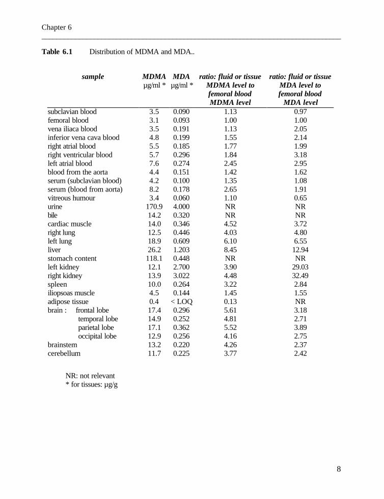
Chapter 6 ________________________________________________________________________________
8
Table 6.1 Distribution of MDMA and MDA..
sample MDMA MDA ratio: fluid or tissue ratio: fluid or tissue µg/ml * µg/ml * MDMA level to MDA level to
femoral blood MDMA level
femoral blood MDA level
subclavian blood 3.5 0.090 1.13 0.97 femoral blood 3.1 0.093 1.00 1.00 vena iliaca blood 3.5 0.191 1.13 2.05 inferior vena cava blood 4.8 0.199 1.55 2.14 right atrial blood 5.5 0.185 1.77 1.99 right ventricular blood 5.7 0.296 1.84 3.18 left atrial blood 7.6 0.274 2.45 2.95 blood from the aorta 4.4 0.151 1.42 1.62 serum (subclavian blood) 4.2 0.100 1.35 1.08 serum (blood from aorta) 8.2 0.178 2.65 1.91 vitreous humour 3.4 0.060 1.10 0.65 urine 170.9 4.000 NR NR bile 14.2 0.320 NR NR cardiac muscle 14.0 0.346 4.52 3.72 right lung 12.5 0.446 4.03 4.80 left lung 18.9 0.609 6.10 6.55 liver 26.2 1.203 8.45 12.94 stomach content 118.1 0.448 NR NR left kidney 12.1 2.700 3.90 29.03 right kidney 13.9 3.022 4.48 32.49 spleen 10.0 0.264 3.22 2.84 iliopsoas muscle 4.5 0.144 1.45 1.55 adipose tissue 0.4 < LOQ 0.13 NR brain : frontal lobe 17.4 0.296 5.61 3.18 temporal lobe 14.9 0.252 4.81 2.71 parietal lobe 17.1 0.362 5.52 3.89 occipital lobe 12.9 0.256 4.16 2.75 brainstem 13.2 0.220 4.26 2.37 cerebellum 11.7 0.225 3.77 2.42
NR: not relevant * for tissues: µg/g

Chapter 6 ________________________________________________________________________________
9
Figure 6.1 Possible mechanism of redistribution (indicated by direction of arrows).
Labels: RA: right atrium D: diaphragm RV: right ventricle AO: aorta LA: left atrium VI: inferior vena cava LV: left ventricle VS: superior vena cava TP: truncus pulmonalis AR: arteria renalis VP: venae pulmonales VR: vena renalis
Chapter 6 ________________________________________________________________________________
10
I Discussion The autopsy findings, including macroscopical features (such as increased lung weight) and the microscopical examination (pronounced pulmonary congestion and oedema), are consistent with an acute to subacute cardiopulmonary failure. From the purely physiological point of view, the aberrant course of the superior vena cava (persistence of the left superior caval vein) was not important, as the outlet of the vena cava superior was also present in the right atrial cavity. Referring to the toxicological data, we can conclude that the cardio-respiratory insufficiency was caused by the sympaticomimetic mechanism of MDMA. Indeed, electrical instability of the heart has been described in this anomaly (21).
We present a detailed distribution of MDMA and MDA concentrations in this fatal overdose. Possible mechanisms of redistribution are presented in Figure 6.1.
Our data (Table 6.1) confirm that a peripheral blood sample is strongly recommended and femoral blood remains the most representative. When femoral blood is not available, blood from the vena subclavia or vena iliaca may be appropriate; however, cardiac blood samples and left atrial blood in particular, should be avoided. The site-dependent differences in heart blood concentrations have previously been observed for methamphetamine (12). In our case, the left atrial MDMA level was the highest of all and can probably – or at least partially – be explained by diffusion from both lungs via the venae pulmonales. It is not excluded that the high MDMA level in cardiac muscle is also correlated with the high MDMA concentration in the adjacent lungs, but diffusion from the stomach content volume could also be speculated. The obvious difference in MDMA and MDA levels between both lungs could be explained by post-mortem diffusion out of the high reservoir of these substances present in the stomach content volume. Indeed, the stomach is only separated from the left lung by the diaphragm. As the gastric mucosa is easily influenced by the autolytic process, post-mortem diffusion to the closely adjacent organs can be assumed. The relatively high MDMA level in vena cava inferior blood can be correlated with diffusion out of the kidneys and the liver. Indeed, the MDMA concentration in the liver was the highest of all organ levels. As the liver seems to be an important “reservoir” of MDMA, this organ can be assumed to be capable of inducing considerable post-mortem redistribution at increasing post-mortem intervals. As a result, blood sampling near the liver (e.g. inferior vena cava blood or blood from the right heart) should be avoided.
The MDMA and MDA levels in liver and bile can point to elimination by biotransformation or by excretion via the bile. However, MDA levels in the kidneys were higher than the corresponding MDA liver concentrations, which may indicate that the impact of tubular reabsorption should not be overlooked because the MDA level in urine was rather low. In our case, because the MDA blood and tissue levels (also the ratios as given in Table 6.1) are mainly consistent with the MDMA distribution, though with a considerable difference in size, we can assume that MDA acts as a metabolite of MDMA. Referring to the MDA amounts, our data confirm the results of previous studies, namely MDA is not a major metabolite in humans (22-24).
Chapter 6 ________________________________________________________________________________
11
The ratio of vitreous humour to femoral blood MDMA level of 1.1 was consistent with previous research in rabbits (25), indicating that equilibration was attained. This assumes that vitreous humour can be a suitable alternative when an appropriate blood sample is lacking, but this should be confirmed with larger series. In addition, when a significant degree of putrefaction has already taken place, thus making blood and vitreous humour sampling impossible, quantitation of MDMA in iliopsoas muscle can give relevant information (see values in Table 6.1). The MDMA levels in the various brain regions demonstrate regional differences, with the highest levels being in the frontal and parietal lobes. In our data, these regional differences are similar for MDA. Because the sampling method was different, our results cannot be compared with recently published findings (16). Finally, the high cerebral levels are concordant with the strong neuropharmacologic effects of “ecstasy”. Acknowledgments The authors wish to gratefully thank Mrs Thérèse De Vuyst for her assistance in preparing the manuscript, and Mrs Marijke Craeymeersch and Mrs Vera De Vleesschauwer for technical assistance. Our thanks also to Mr. Richard Sundahl for his assistance with the English grammar.
Chapter 6 ________________________________________________________________________________
12
References 1. Pounder DJ, Jones GR. Post-mortem drug redistribution – a toxicological nightmare. Forensic
Sci Int 1990;45:253-263. 2. Garrett ER, Seyda K, Marroum P. High performance liquid chromatographic assays of the
illicit designer drug “Ecstasy”, a modified amphetamine, with applications to stability, partitioning and plasma protein binding. Acta Pharm Nord 1991;3:9-14.
3. Nagata T, Kimura K, Hara K, Kudo K. Methamphetamine and amphetamine concentrations in
postmortem rabbit tissues. Forensic Sci Int 1990;48:39-47. 4. Dowling GP, McDonough ET III, Bost RO. ‘Eve’ and ‘Ecstasy’. A report of five deaths
associated with the use of MDEA and MDMA. J Am Med Assoc 1987;257:1615-1617. 5. Rohrig TP, Prouty RW. Tissue distribution of methylenedioxymethamphetamine. J Anal
Toxicol 1992;16:52-53. 6. Fineschi V, Masti A. Fatal poisoning by MDMA (ecstasy) and MDEA: a case report. Int J
Legal Med 1996;108:272-275. 7. Meyer E, Van Bocxlaer JF, Dirinck IM, Lambert WE, Thienpont L, De Leenheer AP. Tissue
distribution of amphetamine isomers in a fatal overdose. J Anal Toxicol 1997;21:236-239. 8. Prouty RW, Anderson WH. The forensic science implications of site and temporal influences
on postmortem blood-drug concentrations. J Forensic Sci 1990;35:243-270. 9. Hilberg T, Rogde S, Mørland J. Postmortem drug redistribution – human cases related to
results in experimental animals. J Forensic Sci 1999;44:3-9. 10. Barnhart FE, Fogacci JR, Reed DW. Methamphetamine – a study of postmortem redistribution.
J Anal Toxicol 1999;23:69-70. 11. Katsumata S, Sato K, Kashiwade H, Yamanami S, Zhou H, Yonemura I, Nakajima H, Hasekura
H. Sudden death due presumably to internal use of methamphetamine. Forensic Sci Int 1993;62:209-215.
12. Miyazaki T, Kojima T, Yashiki M, Wakamoto H, Iwasaki Y, Taniguchi T. Site dependence of
methamphetamine concentrations in blood samples collected from cadavers of people who had been methamphetamine abusers. Am J Forensic Med Pathol 1993;14:121-124.
13. Moriya F, Hashimoto Y. Redistribution of basic drugs into cardiac blood from surrounding
tissues during early-stages postmortem. J. Forensic Sci 1999;44:10-16. 14. Moriya F, Hashimoto Y. Redistribution of methamphetamine in the early postmortem period.
J Anal Toxicol 2000;24:153-154.
Chapter 6 ________________________________________________________________________________
13
15. Logan BK, Weiss EL, Harruff R. Case report: Distribution of methamphetamine in a massive
fatal ingestion. J Forensic Sci 1996;41:322-323. 16. Kalasinsky KS, Bosy TZ, Schmunk GA, Reiber G, Anthony RM, Furukawa Y, Guttman M,
Kish SJ. Regional distribution of methamphetamine in autopsied brain of chronic human methamphetamine users. Forensic Sci Int 2001;116:163-169.
17. Clauwaert KM, Van Bocxlaer JF, De Letter EA, Van Calenbergh S, Lambert WE, De Leenheer
AP. Determination of the designer drugs 3,4-methylenedioxy-methamphetamine, 3,4-methylenedioxyethylamphetamine, and 3,4-methylenedioxy-amphetamine with HPLC and fluorescence detection in whole blood, serum, vitreous humor, and urine. Clin Chem 2000;46:1968-1977.
18. Van Bocxlaer J, Meyer E, Clauwaert K, Lambert W, Piette M, De Leenheer A. Analysis of
zopiclone (Imovane) in postmortem specimens by GC-MS and HPLC with diode-array detection. J Anal Toxicol 1996;20:52-54.
19. Clauwaert KM, Van Bocxlaer JF, Lambert WE, De Leenheer AP. Analysis of cocaine,
benzoylecgonine, and cocaethylene in urine by HPLC with diode array detection. Anal Chem 1996;68:3021-3028.
20. Clauwaert KM, Van Bocxlaer JF, Lambert WE, De Leenheer AP. Liquid chromatographic
determination of cocaine, benzoylecgonine, and cocaethylene in whole blood and serum samples with diode-array detection. J Chromatogr Sci 1997;35:321-328.
21. Lenox CC, Hashida Y, Anderson RH, Hubbard JD. Conduction tissue anomalies in absence of
the right superior caval vein. Int J Cardiol 1985;8:251-260. 22. Kunsman GW, Levine B, Kuhlman JJ, Jones RL, Hughes RO, Fujiyama CI, Smith ML. MDA-
MDMA concentrations in urine specimens. J Anal Toxicol 1996;20:517-521. 23. Mas M, Farré M, de la Torre R, Roset PN, Ortuño J, Segura J, Cami J. Cardiovascular and
neuroendocrine effects and pharmacokinetics of 3,4-methylenedioxy-methamphetamine in humans. J Pharmacol Exp Ther 1999;290:136-145.
24. de la Torre R, Farré M, Ortuño J, Mas M, Brenneissen R, Roset PN, Segura J, Cami J. Non-
linear pharmacokinetics of MDMA (‘ecstasy’) in humans. Br J Clin Pharmacol 2000;49:104-109.
25. De Letter EA, De Paepe P, Clauwaert KM, Belpaire FM, Lambert WE, Van Bocxlaer JF, Piette
MHA. Is vitreous humour useful for the interpretation of 3,4-methylenedioxy-methamphetamine (MDMA) blood levels? Experimental approach with rabbits. Int J Legal Med 2000;114:29-35.
Chapter 6 ________________________________________________________________________________
1
I.2 Distribution study of the amphetamine derivative MDMA and its metabolite 3,4-methylenedioxyamphetamine (MDA) in two overdose cases
I Introduction
In this section, the distribution of MDMA and its metabolite MDA in various body fluids and tissues is further investigated on the basis of two additional fatalities in order to determine which body fluid and/or tissue sample post mortem most closely approximates the blood concentration at the time of death. The question is raised as to whether the toxicological data are consistent with those in the above-mentioned case (1). Moreover, a few additional samples (such as pleural and pericardial fluids, both hemi-diaphragms and endocrine glands including the pituitary gland) are studied. II Case histories II.1 Case 01/122
At about 11 a.m., an 18-year-old man was found dead. The night before, he had gone out with a friend and had been drinking a lot. He had been snoring the whole night, but was found lifeless, in ventral position, in the morning. An initial external examination was performed in the mortuary the same day at 3:30 p.m. The body had been kept in the cold store since about 1:30 p.m. His wallet was found with the body in the plastic body bag though without any cash. His face and upper thorax were obviously cyanotic. The lips and tip of the tongue were parchment-like desiccated. Rigor mortis was already obviously present on both upper and lower limbs; the hypostasis was distinct. The rectal temperature measured at 3:42 p.m. was 36°C. Subclavian blood and urine were sampled for toxicological screening. As the friend of the deceased denied the use of drugs, and the post-mortem blood alcohol concentration was relatively low (0.56 g/l), and – moreover - the wallet of the young man was empty, the prosecutor ordered an extensive inquiry including an autopsy. As the prosecutor delayed his decision whether or not to order an autopsy until further toxicological results were available, the autopsy was performed only 5 days post mortem. External inspection disclosed a rather small young man : the body weighed 61 kg and was 177 cm tall. Onset of putrefaction was noted. Only a few slight desiccated abrasions in his face (possibly agonal or post mortem) and a slight excoriation on his knee consistent with a fall or impact, were found. Some small scarce petechiae on the left outer eyelid and conjunctivae were observed. Dental caries and a tongue bite were seen. During internal examination, 160 ml bloody fluid was present in both pleural cavities. Scattered Tardieu spots on both pleurae, thymus and epicardium were noted. Pus material was present in the distal bronchial branches, and an overwhelming pulmonary congestion and oedema were found. Generalized visceral congestion was seen, which was confirmed by the increased organ weights (see Table 2.1 (a)). The histological study confirmed the pronounced pulmonary congestion and haemorrhagic oedema and the generalized visceral congestion. Furthermore, microscopical examination demonstrated shock lungs and 1st to
Chapter 6 ________________________________________________________________________________
2
2nd degree microvacuolar liver steatosis. Autolysis hampered the diagnosis of an acute tubular necrosis (ATN). Examination of several brain regions disclosed - apart from pronounced congestion and oedema - a few petechial haemorrhages, obvious sludging in the basal ganglia and signs of protracted hypoxia in the hippocampus. II.2 Case 01/158
A 31-year-old man known to be addicted to alcohol, was found dead at home next to his bed, in an advanced state of putrefaction.
During external examination, the body exuded a miasmatic odor. Typical decomposition signs such as generalized bloating of the body associated with a discoloured face, venous marbling and peeling off of the skin were found. Purge fluid was draining out from the mouth and nose. The body weighed 85 kg and was 178 cm tall. The external findings and police inquiry indicated a post-mortem interval of at least 7 days.
At autopsy, putrefaction of all organs was confirmed. About 600 ml bloody fluid was present in both pleural cavities. Putrefaction vesicles on the lung surfaces were present and obvious pulmonary congestion could still be observed. The organ weights are presented in Table 2.1 (Chapter 2). The heart was somewhat enlarged but the coronary arteries showed no substantial atherosclerosis. The putrefaction interfered with the microscopical examination. However, significant myocardial scars and pronounced liver steatosis (by means of a fat staining) could be excluded. Arguments for pulmonary congestion and oedema associated with deep vomit aspiration could be substantiated. In addition, a few Prussian blue positive macrophages were observed. There were no important bleeding lesions in the brain. III Toxicological data
The previously described HPLC-method was used for the quantitation of MDMA and MDA (1,2). MDMA and MDA concentrations in various body fluids and tissues are presented in Table 6.2. For both cases, the cardiac blood concentrations were higher than the peripheral blood levels, and the pericardial fluid levels were in line with the cardiac blood levels. Substantial levels were found in the majority of the organs, except for the abdominal adipose tissue. The highest concentrations were observed in all lung lobes, the stomach content, the liver, the bile, both kidneys and the urine. In addition, the concentrations in the pituitary gland were obviously higher than the brain levels. The vitreous humour MDMA level in case 01/122 was in the same range as the peripheral blood levels.
For case 01/122, two subclavian blood and urine samples were available: sampling of these occurred during the initial external examination and at the autopsy, thus with an interval of 5 days. Toxicological screening of the initial samples disclosed the presence of a blood and urinary alcohol concentration of 0.56 g/l and 1.23 g/l, respectively. MDMA was detected in blood. In addition, a potentially toxic methadone level of 1.1µg/ml in blood was found.
For case 01/158 – due to the pronounced putrefaction – it was unfortunately not possible to obtain all the samples of the protocol. In particular, the number of the available
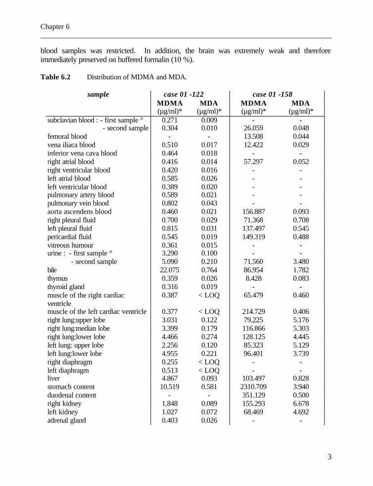
Chapter 6 ________________________________________________________________________________
3
blood samples was restricted. In addition, the brain was extremely weak and therefore immediately preserved on buffered formalin (10 %). Table 6.2 Distribution of MDMA and MDA.
sample case 01 -122 case 01 -158 MDMA MDA MDMA MDA (µg/ml)* (µg/ml)* (µg/ml)* (µg/ml)*
subclavian blood : - first sample ° 0.271 0.009 - - - second sample 0.304 0.010 26.059 0.048 femoral blood - - 13.508 0.044 vena iliaca blood 0.510 0.017 12.422 0.029 inferior vena cava blood 0.464 0.018 - - right atrial blood 0.416 0.014 57.297 0.052 right ventricular blood 0.420 0.016 - - left atrial blood 0.585 0.026 - - left ventricular blood 0.389 0.020 - - pulmonary artery blood 0.589 0.021 - - pulmonary vein blood 0.802 0.043 - - aorta ascendens blood 0.460 0.021 156.887 0.093 right pleural fluid 0.700 0.029 71.368 0.708 left pleural fluid 0.815 0.031 137.497 0.545 pericardial fluid 0.545 0.019 149.319 0.488 vitreous humour 0.361 0.015 - - urine : - first sample ° 3.290 0.100 - - - second sample 5.090 0.210 71.560 3.480 bile 22.075 0.764 86.954 1.782 thymus 0.359 0.026 8.428 0.083 thyroid gland 0.316 0.019 - - muscle of the right cardiac ventricle
0.387 < LOQ 65.479 0.460
muscle of the left cardiac ventricle 0.377 < LOQ 214.729 0.406 right lung:upper lobe 3.031 0.122 79.225 5.176 right lung:median lobe 3.399 0.179 116.866 5.303 right lung:lower lobe 4.466 0.274 128.125 4.445 left lung: upper lobe 2.256 0.120 85.323 5.129 left lung:lower lobe 4.955 0.221 96.401 3.739 right diaphragm 0.255 < LOQ - - left diaphragm 0.513 < LOQ - - liver 4.867 0.093 103.497 0.828 stomach content 10.519 0.581 2310.709 3.940 duodenal content - - 351.129 0.500 right kidney 1.848 0.089 155.293 6.678 left kidney 1.027 0.072 68.469 4.692 adrenal gland 0.403 0.026 - -

Chapter 6 ________________________________________________________________________________
4
Table 6.2 Distribution of MDMA and MDA (continued)
sample case 01 -122 case 01 -158 MDMA MDA MDMA MDA (µg/ml)* (µg/ml)* (µg/ml)* (µg/ml)*
spleen 0.835 0.040 - - pancreas 0.369 0.018 36.909 0.179 iliopsoas muscle 0.302 < LOQ 61.501 0.200 abdominal adipose tissue < LOQ < LOQ - - pituitary gland 3.611 0.190 - - brain: frontal lobe 0.685 0.026 - - temporal lobe 0.407 0.015 - - parietal lobe 0.811 0.026 - - occipital lobe 0.858 0.034 - - medulla oblongata 0.492 0.017 - - pons 0.753 0.030 - - cerebellum 0.668 0.025 - -
* for tissues: µg/g -: sample not available < LOQ: below limit of quantitation ° first sampling occured during the initial external examination; second sample taken during the autopsy 112 hours later
IV Discussion
The medico-legal findings in the first case (01/122) were compatible with fatal hyperthermia. The rectal temperature of 36°C about 4.5 h following the discovery of the man – even after the body had been cooled for a few hours before the examination took place - suggested a high body temperature at the time of death. The shock lungs, the obvious congestion and oedema associated with a few perivascular bleedings in the brain and signs of protracted hypoxia (e.g. in the hippocampus) were in accordance with fatal hyperthermia. The pronounced putrefaction process interfered substantially with the investigation of the second case. However, the obvious pleural exudations, pulmonary congestion and generalized visceral congestion were compatible with acute to subacute cardiopulmonary failure.
These two cases were interesting for the thanato-toxicological study because of the relatively long post-mortem intervals of 5 and at least 7 days, respectively, which were more substantial than in the above mentioned distribution study in case 00/112 (1). The difficulty of interpreting a MDMA blood level was confirmed in cases 01/122 and 01/158. Indeed, a distinct difference in concentration range of MDMA was found, viz. 0.270 µg/ml and 13.510 µg/ml, respectively. The relatively low MDMA blood level in the first case was in accordance with a protracted agony and substantiated the diagnosis of hyperthermia. The MDMA femoral blood level in the second case was extraordinary high and – to our
Chapter 6 ________________________________________________________________________________
5
knowledge – only one higher MDMA level is reported in the literature, namely 18.500 µg/ml (3). These two extreme MDMA levels confirmed that the toxicological data should be interpreted in the light of the autopsy findings in order to come to a conclusion.
The toxicological results in these two cases substantiated the recommendation that peripheral blood samples (preferably femoral) are the “golden standard” and that blood sampled centrally in the body (such as cardiac blood) should be avoided (4). Indeed, referring to the high MDMA and MDA levels in the lungs and liver, cardiac blood samples were subject to post-mortem redistribution and can therefore be misleading in the interpretation as to whether the post-mortem level is potentially toxic or lethal. Moreover, the MDMA concentration in the subclavian blood samples in the first case which were taken with a post-mortem interval of about 5 days – demonstrated that subclavian blood can be an appropriate sample for post-mortem toxicological analysis. However, when pronounced putrefaction occurs - which was present in case 01/158 – a subclavian blood sample should be interpreted with caution.
The higher MDMA and MDA concentrations in several tissues compared with their concentrations in blood corroborated the results in the animal experiments and the findings in other case studies, viz. that MDMA accumulates in or binds to various tissues (5,1,9).
The substantial MDMA and MDA levels in the lower lung lobes were not only in accordance with the hypostasis - which is more pronounced in these areas - but could also be correlated with the closely adjacent liver and stomach. The pericardial and pleural fluid levels were on the same order as the cardiac blood and lung tissue concentrations, respectively.
The MDMA and MDA concentrations in liver and bile confirmed the hepatic biotransformation and excretion of MDMA via the bile. The substantial kidney levels were directly related to the urinary levels because these tissues are inherently “contaminated” with urine. The relatively low MDA levels in several body fluids and tissues confirmed that though MDA is an active metabolite, it is not a major metabolite of MDMA in humans.
For case 01/122, both the urinary MDMA and MDA concentrations were obviously higher in the second than in the first sample; this could be due to post-mortem dehydration with diffusion of fluid into the tissues nearby the bladder, resulting in a concentration effect in the urine.
The MDMA concentration difference between the left and the right hemi-diaphragm could be correlated with the high levels in the stomach content, which is closely adjacent to the left hemi-diaphragm, thus substantiating some post-mortem diffusion out of the stomach content. This is in accordance with the findings in the rabbit model (6).
The high brain MDMA and MDA concentrations were in line with expectations and in accordance with the brain as target organ. In addition, the concentrations in the pituitary gland were even more substantial, being consistent with the neuroendocrine effects such as the increases in plasma cortisol and prolactin levels which have been documented in humans (7).
The vitreous humour MDMA and MDA concentrations in case 01/122 demonstrated that this specimen can be valuable, mainly when protracted agony takes place (which occurs in hyperthermia), resulting in equilibration between the vascular

Chapter 6 ________________________________________________________________________________
6
compartment and the vitreous body (1,8). Unfortunately, due to the pronounced putrefaction in the second case, the vitreous humour was lacking.
Finally, there is an incoherence between the MDMA concentrations in the iliopsoas muscle in the two cases. In view of the first case, we could assume the iliopsoas muscle to be an interesting specimen when blood is lacking. This was in line with other cases (1,9). However, the iliopsoas level in the second case indicated that when advanced putrefaction has taken place, these levels, as well, should be interpreted with caution. The more substantial MDMA concentrations in the iliopsoas muscle could relate to urinary leakage in an advanced putrefactive status.
In conclusion, these two cases confirm that MDMA and MDA can accumulate in various tissues and that post-mortem redistribution can occur out of organs such as the lungs and the liver into mainly cardiac blood. To a certain extent, the influence of this post-mortem phenomenon can be avoided by peripheral sampling (such as using subclavian or femoral blood). Vitreous humour and iliopsoas muscle can be valuable specimens for toxicological analysis, provided the putrefactive stage is not too pronounced. The same remark is applicable to the subclavian blood. Finally, these cases substantiate that a broad range of MDMA concentrations can be found in fatalities and therefore the toxicological and anatomo-pathological findings should be considered as a whole when drawing a conclusion.
Chapter 6 ________________________________________________________________________________
7
References 1. De Letter EA, Clauwaert KM, Lambert WE, Van Bocxlaer JF, De Leenheer AP, Piette MHA.
Distribution study of 3,4-methylenedioxymethamphetamine and 3,4-methylenedioxy-amphetamine in a fatal overdose. J Anal Toxicol 2002;26:113-118.
2. Clauwaert KM, Van Bocxlaer JF, De Letter EA, Van Calenbergh S, Lambert WE, De Leenheer
AP. Determination of the designer drugs 3,4-methylenedioxymethamphetamine, 3,4-methylenedioxyethylamphetamine and 3,4-methylenedioxyamphetamine with HPLC and fluorescence detection in whole blood, serum, vitreous humor, and urine. Clin Chem 2000;46:1968-1977.
3. Lo DST, Goh EWS, Yao YJ, Wee KP. The first fatal overdose with MDMA in Singapore.
TIAFT Bulletin 2001;31(3):13-14. 4. Prouty RW, Anderson WH. The forensic science implications of site and temporal influences
on postmortem blood-drug concentrations. J Forensic Sci 1990;35:243-270. 5. De Letter EA, Clauwaert KM, Belpaire FM, Lambert WE, Van Bocxlaer JF , Piette MHA. Post-
mortem redistribution of 3,4-methylenedioxymethamphetamine (MDMA, “ecstasy”) in the rabbit. Part one: Experimental approach after intravenous infusion. Int J Legal Med 2002;116: 216-224.
6. De Letter EA, Belpaire FM, Clauwaert KM, Lambert WE, Van Bocxlaer JF , Piette MHA. Post-
mortem redistribution of 3,4-methylenedioxymethamphetamine (MDMA, “ecstasy”) in the rabbit. Part two: Post-mortem infusion in trachea or stomach. Int J Legal Med 2002;116: 225-232.
7. Mas M, Farré M, de la Torre R, Roset PN, Ortuño J, Segura J, Camí J. Cardiovascular and
neuroendocrine effects and pharmacokinetics of 3,4-methylenedioxy-methamphetamine in humans. J Pharmacol Exp Ther 1999;290:136-145.
8. De Letter EA, De Paepe P, Clauwaert KM, Belpaire FM, Lambert WE, Van Bocxlaer JF, Piette
MHA. Is vitreous humour useful for the interpretation of 3,4-methylenedioxymethamphetamine (MDMA) blood levels? Experimental approach with rabbits. Int J Legal Med 2000;114:29-35.
9. De Caestecker T, De Letter E, Clauwaert K, Bouche MP, Lambert W, Van Bocxlaer J, Van den
Eeckhout E, Van Peteghem C, De Leenheer A. Fatal 4-MTA intoxication : development of a liquid chromatographic – tandem mass spectrometric assay for multiple matrices. J Anal Toxicol 2001;25:705-710.
Chapter 6 ________________________________________________________________________________
1
II. OTHER AMPHETAMINE DERIVATIVES RECENTLY OBSERVED IN A
FEW CASES IN BELGIUM
II.1 One fatal and seven nonfatal cases of 4-methylthioamphetamine (4-MTA) intoxication: clinico-pathological findings
Els A. De Letter 1, Vera A.E. Coopman 2, Jan A.C.M. Cordonnier 2, Michel H.A. Piette 1
1 Els A. De Letter, Michel H.A. Piette Ghent University - Department of Forensic Medicine 2 Vera A.E. Coopman, Jan A.C.M. Cordonnier Chemiphar NV - Toxicological laboratory,
Based on: Int J Legal Med 2001;114:352-356

Chapter 6 ________________________________________________________________________________
2
I Abstract We present a case history involving one fatal case and seven survived cases of intoxication
with 4-methylthioamphetamine (4-MTA), also called para-methylthioamphetamine (p-MTA) or simply methylthioamphetamine (MTA), a relatively new amphetamine analogue. Two of the seven survivors required a 24-h-period of observation in hospital. This report proves once again that the new amphetamine "designer drugs" are not without danger, as is thought by many young people. In addition, individually different subjective reactions are described. Finally, the medico-legal implications of new, as yet unregistered drugs are discussed. Key words: Drug abuse - 4-MTA - 4-Methylthioamphetamine – Amphetamines - Intoxication. II Introduction
The substance 4-methylthioamphetamine (4-MTA), also called para-methylthioamphetamine (p-MTA), methylthioamphetamine (MTA) or “Flatliner” (1), is a relatively new phenylethylamine-based compound. It was first synthesized and investigated by Huang et al. (2) in the rat animal model in which MTA was proven to be non-neurotoxic at low doses, but was found to induce typical serotonergic behaviour at high doses. MTA was in fact developed as a potent and “purer” serotonin-releasing agent for use in experimental research (2). This serotonin-releasing property was compared with that of other amphetamine derivatives, and a study in rats indicated, for example, that MTA had a delayed reaction compared to MBDB (3) (also known as Methyl-J and Eden (4)). The main site of toxicity with MDMA is believed to be within the serotonergic pathways in the central nervous system and explains the influence of MDMA on affective behaviour and thermoregulation, for example (5). Studies in the rat indicated that another amphetamine derivative, methamphetamine, was able to induce apoptosis of the thymic and splenic lymphocytes (6). Recently, the cardio-toxic effect of methamphetamine has been studied in isolated adult rat cardiomyocytes (7). Others have investigated the liver toxicity caused by single or repeated intraperitoneal doses of MDMA in the rat (8). However, many mechanisms of toxicity caused by amphetamine and its derivatives still remain to be elucidated. MTA, just as MBDB (4), can be sold as an “ecstasy” pill, and the use of MTA as an illegal “designer” drug was first reported in the Netherlands (9). MTA is structurally closely related to para-methoxyamphetamine (PMA), another ring-substituted amphetamine (see Figure 6.2). Some cases of fatal poisoning involving PMA have recently been reported (10,11). To our knowledge, only one fatal case of MTA intoxication (1) and a very few cases of poisonings have been published (12,13)
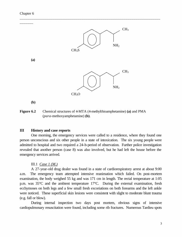
Chapter 6 ________________________________________________________________________________
3
(a)
(b) Figure 6.2 Chemical structures of 4-MTA (4-methylthioamphetamine) (a) and PMA (para-methoxyamphetamine) (b). III History and case reports
One morning, the emergency services were called to a residence, where they found one person unconscious and six other people in a state of intoxication. The six young people were admitted to hospital and two required a 24-h-period of observation. Further police investigation revealed that another person (case 8) was also involved, but he had left the house before the emergency services arrived. III.1 Case 1 (M.)
A 27-year-old drug dealer was found in a state of cardiorespiratory arrest at about 9:00 a.m. The emergency team attempted intensive reanimation which failed. On post-mortem examination, the body weighed 55 kg and was 171 cm in length. The rectal temperature at 1:05 p.m. was 35°C and the ambient temperature 17°C. During the external examination, fresh ecchymoses on both legs and a few small fresh excoriations on both forearms and the left ankle were noticed. These superficial skin lesions were consistent with slight to moderate blunt trauma (e.g. fall or blow).
During internal inspection two days post mortem, obvious signs of intensive cardiopulmonary resuscitation were found, including some rib fractures. Numerous Tardieu spots
CH3S NH2
CH3
CH3O
NH2
CH3

Chapter 6 ________________________________________________________________________________
4
were observed on the pericardium, thymus and both pleurae. Both lungs weighed 970 g and obvious emphysema, severe congestion and moderate oedema were found. The heart weighed 255 g and showed no conspicuous anomalies. The stomach contained only a small amount of bloody mucus and the mucosa showed a few pin-point ulcerations. The brain weighed 1320 g and, apart from slight oedema and congestion of the white matter, nothing unusual was found. The remaining organs showed no obvious macroscopical anomalies, except for congestion.
On histological examination pronounced pulmonary congestion, hemorrhagic oedema, a slight intra-alveolar infiltration with a few polymorphonuclear cells and some leucocytic sludging in the septal veins were found. Groups of alveolar macrophages were seen, although staining with Prussian blue was negative. Obvious eosinophilic infiltration was found in some lymph nodules although it was not seen in the lungs. The Purkinje cells in the cerebellar vermis were pyknotic and somewhat reduced in number. In the nucleus lentiformis a few venulae were surrounded by a lymphocyte infiltration and macrophages containing hemosiderine. The hippocampus showed no marked hypoxemic lesions. No obvious pre-existing disease was identified.
Toxicological screening revealed MDMA in the urine, MTA in the blood, urine and liver, and tetrahydrocannabinoic acid in the urine. These results were confirmed by GC/MS (liver/urine) and HPLC/DAD (blood) analysis. Blood, urine and liver analysis showed MTA concentrations of 8.38 µg/ml, 100 µg/ml and 30 µg/g, respectively. The MDMA level in the urine was 1.2 µg/ml. The amount of MDMA in the blood was below the limit of quantitation (< 0.1 µg/ml). The urine pH was 5.6. The size of the stomach content sample was insufficient for toxicological analysis. No other psycho-active drugs were found. In addition, analysis of two small plastic bags (containing green-brownish herbs) found in the victim's pockets, demonstrated cannabinol (marijuana).
Questioning of the surviving persons revealed that M. had been walking around the whole night, but around 8:00 a.m. he started sweating and thrashing around on the floor. M. started shaking intensely and his behaviour became increasingly more strange. This went on for quite some time, at least 1 h. Somewhat later, one of them noted that M.'s heartbeat was fading and oedema was appearing on his mouth. They then tried to resuscitate him, but even before the emergency services arrived, M. felt cold to the touch. One of the survivors declared that M. had taken at least six pills: first, two pills at the same time, and then each of the other four pills at intervals of about 2 h. In addition, M. had smoked five or six joints. The girlfriend of the deceased (case 7) admitted that M. was dealing in “ecstasy” pills, marijuana and speed, and he drove to the Netherlands for his supplies. M. was heavily addicted to “smart pills” and easily took six of them in the course of a single evening followed by a shaking period that usually subsided after a few minutes. On the morning in question, M. started shaking but this time it did not stop after a few minutes. He had told her about ingesting fifteen pills at one time in the previous year. In the dealer's car, a note printed in Dutch describing some characteristics of MTA (called "MTA-1" in the note) was retrieved.

Chapter 6 ________________________________________________________________________________
5
Chapter 6 ________________________________________________________________________________
6
III.2 Case 2 (D.W.) This 18-year-old man found lying in a chair, apathetic and staring vacantly into space, was
conveyed to hospital for observation. Upon being discharged 24 h later, he stated to the police officer that he had been using drugs since the age of 14, mainly cannabis, and sometimes "a pill". On the night in question, all those present in the house had smoked joints and swallowed pills. He could not remember exactly when, but he himself had taken two pills at the same time. He described how he had taken leave of his senses thereafter, had had a "threefold" vision and hallucinations, and was unable to control all his acts (e.g. he could not stop his chin from shivering). He felt that his heart rate had clearly increased and one moment he was sweating, while a few moments later he would turn icy cold. III.3 Case 3 (B.)
The emergency services found this 22-year-old female unsteady on her feet, although a certain amount of conversation was possible. She had arrived at home at about midnight, when she smoked some joints together with her friends and then took one pill. From that point on, she could remember very little, although she knew she had slept a lot. She regularly smoked joints at the weekends, but it was the first time that she had taken such a pill. III.4 Case 4 (W.) This 15-year-old female looked extremely tired but was able to confirm that everyone had smoked joints and, shortly thereafter, M. had handed out "smart" pills. She was admitted to hospital, although observation was not required, and she was interrogated about 12 h later. She herself took one pill at about 00:15 a.m., and 20 min later, she “experienced a very pleasant feeling”. In order to enhance the sensation, she smoked a joint. She was awake during the whole night and, like the others, she drank no alcohol.
This subject was also able to give some information about the number of pills the different individuals had swallowed (see Table 6.3). W. claimed that she herself takes a pill and smokes a joint "now and then", but only at weekends. III.5 Case 5 (W. W.)
The story of this 19-year-old man, was consistent with the others. He stated that he had taken only one pill, followed by three joints, and had played music the whole night through. III.6 Case 6 (D.J.)
Although this 22-year-old man was heavily intoxicated, he agreed to make a statement. Since his answer frequently went beyond the question and he behaved very strangely (e.g. hitting his head against the table, almost falling off his chair), the police officers decided to send him to hospital for observation. He was discharged 24 h later, when he was capable of being interrogated. His mind was a blank except for a few fragments, but he believed that he had taken

Chapter 6 ________________________________________________________________________________
7
two pills at an interval of half an hour. He explained that when he took one pill, nothing happened, but following the second, he felt good and was unable to sit still. Somewhat later he took a third pill, and from then on everything was fuzzy. He recalled having difficulty in urinating but he did not remember being in hospital. According to his girlfriend (case 3) it was the first time he had taken such a pill. III.7 Case 7 (V.)
This 19-year-old woman, the girlfriend of the deceased (case 1), was also interrogated the evening following the occurrence. She had been using drugs since the age of 10 and was addicted to marijuana and stimulating drugs (including speed and "MTA-1"). The two pills she swallowed made her feel sick and she vomited several times. She fell asleep for a few short periods. III.8 Case 8 (G.)
This 20-year-old man arrived at the house at about 1:00 – 1:30 a.m. and left at about 7:30 a.m. He confirmed that everybody was “in a whirl” when he arrived. For the first time in his life, he took one pill at about 1:45 a.m. and somewhat later fell asleep. By the time he went home, the effects of the pill had worn off. When he was interrogated more than 36 h after taking the pill, he was behaving totally normally, so no blood or urine sampling was ordered by the coroner.
Blood and urine samples were obtained from all the other survivors (cases 2 - 7) and all
were positive for tetrahydrocannabinoic acid. The MTA levels found in the blood and urine samples and the estimated number of pills ingested are presented in Table 6.3. The MDMA levels were < 0.1 µg/ml.
Table 6.3 Summary of the data found in the surviving persons.
Case number a blood urine urinary MTA level (µg/ml) sampling sampling pH in blood in urine
time time ________________________________________________________________________ (2) D.W. ≥ 2 12:44 h 12:20 h 6.6 0.63 10
(3) B. 1 16:52 h 16:57 h 8.1 1.08 4
(4) W. 1 12:41 h 15:23 h 7.3 2.08 8
(5) W.W. 1 or1½ 12:33 h 12:23 h 6.4 1.93 4
(6) D.J. 3, 5 or 6? 12:43 h 15:22 h 5.8 1.26 40
(7) V. ≥ 2 12:36 h 12:31 h 5.5 0.43 32

Chapter 6 ________________________________________________________________________________
8
a possible number of ingested pills, according to the persons’ statements
The police searched the house where the event took place and the properties of all the
persons involved. In the building, a few pills were found. The yellow pills, weighing 345 mg, contained 28 % or 97 mg MTA. In addition, a small amount of an unknown product, possibly 1-(4-methylthiophenyl)propene or 1-(4-methylthiophenyl)-2-propene, was found. This compound was present in the pills in less than 0.5 % of the MTA peak area in the chromatogram and could not be quantified due to the absence of a reference standard. IV Analytical procedures
4-MTA reference material was supplied by the Wetenschappelijk Instituut Louis Pasteur in Brussels, Belgium. The reagents and chemicals were of analytical grade. The drug standards and the internal standards were obtained from commercial suppliers (Sigma, Radian).
IV.1 Biological samples Routine systematic toxicological analysis was performed on the samples to investigate for
illegal drugs, medical drugs, alcohol, volatile substances and other poisons. Immunoassay screenings (ADx) were performed to test for amphetamines, cannabinoids, opiate groups, methadone and cocaine metabolite in urine and barbiturates, salicylates, tricyclic antidepressants and benzodiazepines in blood. Radio-immunoassay (RIA) was used to screen for LSD in urine and benzodiazepines in blood. Color spot tests on urine and gastric content were used to detect salicylates, acetaminophen, phenothiazines and imipramines. Post-mortem blood was analyzed for the presence of carboxyhemoglobine and cyanide. Urine was screened for the presence of acidic, neutral and basic drugs by thin-layer chromatography. Gas chromatography/mass spectrometry (GC/MS ion trap) was used to screen urine samples for the presence of basic drugs. Blood was screened by high-performance liquid chromatography with photodiode-array detection (HPLC/DAD). Analysis for the presence of alcohol and other volatile substances in blood and urine was performed by headspace gas chromatography with a flame ionization detector (GC/FID). The U.V. spectrum of 4-MTA obtained by HPLC/DAD was characterized by a strong absorption at 253 nm. 4-MTA was confirmed by GC/MS monitoring ions at m/z 44, 138, 165 and 182.
For the quantitation of 4-MTA in blood 1 ml of water was added to 1 ml of a sample or a spiked blood standard and then 100 µl of 3% sodium hydroxide w/v was added, along with 20 µl of diphenylamine as an internal standard and 6 ml of diethylether. After mixing and centrifugation (10 mins, 1121 x G), the organic phase was separated and transferred to a new 10 ml glass screw-top tube. The ether was then mixed with 0.025 M HCl, the ether was discarded and the ether remaining in the aqueous phase was evaporated using nitrogen at room temperature. The
Chapter 6 ________________________________________________________________________________
9
extract was submitted for HPLC/DAD analysis. The correlation coefficient of the calibration curve was 0.9996, recovery for 4-MTA 82% (Cordonnier and Coopman, personal communication).
IV.2 Illicit preparation The yellow powder was finely pulverized. The quantitation of MTA was performed with
HPLC/DAD (Varian) monitored at 254 nm: 10 mg amounts of the powder were dissolved in 1 ml of methanol, then diluted with the mobile phase acetonitrile-buffer containing an external standard (diphenylamine 20 µg/ml) and then separated by chromatography on a Lichrocart cartridge column (125 x 4 mm i.d., 4 µm) filled with Superspher 10 RP-18 packing (14). Standards (10-100 µg/ml) containing a constant amount of the same external standard were prepared in the mobile phase. The concentration was calculated by comparing the peak area of the drug to the external standard versus the standard calibration curve (r = 0.9998).
Identification of the contaminant was achieved by a GC/MS Saturn III Ion Trap (Varian). All GC/MS analyses were performed using a 0.25 mm ID x 30 m fused silica capillary column coated with 0.25 µm of 5% phenyl - 95% methyl-silicone. The injector was set at 250°C. The GC oven program consisted of 70°C, 2 min; 70-290°C at 12°C /min; 5 min. Helium was used as the carrier gas, with an inlet pressure of 275 kPa. Mass spectra were obtained at 70 eV. Ethyl acetate was used as the solvent, instead of methanol in order to exclude the possibility of the formation of methylated analytical artefacts inside the injection port of the gas chromatograph.
The contaminant was identified as possibly 1-(4-methylthiophenyl)propene or 1-(4-methylthiophenyl)-2-propene. V Discussion
We report seven more or less serious and one fatal intoxication involving a relatively new amphetamine analogue, 4-methylthioamphetamine, also called para-methylthioamphetamine. To our knowledge, there are only rare reports in the literature of such intoxications (1,12,13). The analytical profile of 4-MTA was recently described (9). As the event took place near the border with the Netherlands, the source of the product could probably be traced to that country.
As MTA is a new "designer drug", there is no consensus concerning the toxic or lethal blood concentrations. In our fatal case, the 4-MTA blood level established (8.38 µg/ml) was higher than in the previously reported case which had peri- and post-mortem MTA levels of 4.2 µg/ml and 4.6 µg/ml, respectively (1). Compared to other amphetamine-related compounds, the author assumed that MTA blood levels exceeding 4 µg/ml can potentially result in death, or at least constitute a serious health risk (1). However, guidelines indicate that blood MTA levels of 0.2 – 0.6 µg/ml result in moderate toxicity, levels higher than 0.6 µg/ml cause dangerous toxicity and levels exceeding 1.5 µg/ml can lead to death (12). We believe that in our fatal case, there will be no argument that the detected MTA concentration in subclavian blood (8.38 µg/ml) was capable of causing death. However – assuming we can believe the statements of his girlfriend – a few
Chapter 6 ________________________________________________________________________________
10
months prior to his death, the man claimed to have taken 15 MTA pills at once and survived (without medical intervention).
Compared to MTA, blood PMA levels greater than 0.5 µg/ml are likely to be associated with toxic effects (11). As for MDMA, there is also no consensus about the lethal blood level but in general, a blood MDMA concentration higher than 1.0 µg/ml can be potentially lethal, whereas levels lower than 0.6 µg/ml are capable of inducing intoxication (15). However, there is a considerable range in reported fatal blood MDMA levels (16-18). Furthermore, some toxic effects could also be related to contaminants (19).
A number of studies have been carried out on the content of clandestine tablets containing amphetamines, analogues and various possible impurities (e.g. 20-23). Various analytical techniques have been proposed as a means of identifying the drugs incorporated in tablets or powder, and capillary electrophoresis has been established as a rapid method for qualitative and quantitative determination (24). In the pills taken by our cases only a small amount of an unknown product - possibly 1-(4-methylthiophenyl)propene or 1-(4-methylthiophenyl)-2-propene - was found. This product was recently also identified by Kirkbride KP et al. (25). This may indicate that the illicit 4-methylthioamphetamine might be derived from this compound, since a shipment of 1-(4-methylthiophenyl)-2-propene was recently seized in Europe (9) .
All the persons involved in this report took pills having the same content. In addition, in some of our surviving cases, an obvious inconsistency between the MTA levels detected and the described clinical symptoms can be noted. For example, case 4 had an MTA blood level of 2.08 µg/ml and showed relatively less obvious symptoms than cases 2 and 6, with MTA blood levels of 0.63 and 1.26 µg/ml, respectively. In contrast to cases 2 and 6, case 4 did not require a 24-h-period of observation in hospital. Furthermore, case 7 showed the lowest MTA blood level. She took at least 2 pills, but had vomited a lot. In addition, although she admitted being addicted to marijuana and stimulating drugs, including speed and "MTA-1", it seems somewhat bizarre that she got so sick after taking the pills. Thus, the obvious differences in individual responses to MTA must be taken into consideration. This fact is substantiated by the report of de Boer et al., who reported a patient who suffered from amnesia and other problems during a 2-week-period following the ingestion of a single pill (13). However, the possibility cannot be excluded that adverse reactions may occur due to contaminants. In addition, all of them had smoked marijuana followed by the intake of MTA. We can not exclude the possibility that the MTA effect was enhanced by the simultaneous intake of cannabinoids.
Pharmacological and toxicological information for MTA in the literature are scarce, but it is clearly not a safe product. Moreover, although it is too speculative to draw a correlation between the ingested amount declared by the persons involved and their corresponding blood and urine concentrations, we believe that individually different rates of metabolism and/or excretion of the product cannot be excluded. For amphetamine and d-methamphetamine, the urinary excretion is pH-dependent, and it has previously been established that acidification increases the level
Chapter 6 ________________________________________________________________________________
11
retrieved (26). In our cases, the persons with the lowest urinary pH, cases 6 and 7 for example, showed the highest urinary MTA levels.
Since MTA is a relatively new amphetamine derivative and the number of reported poisonings is limited, the pathological findings and the mechanism of death due to this product have not yet been fully evaluated. The side-effects of MTA are probably comparable with those reported after MDMA intake, being mainly related to the sympathomimetic and neurotoxic effect of “ecstasy”, but multiple organ failure including acute hepatic decompensation and/or renal failure (due to rhabdomyolysis) should also be considered (27). Some of the frequently reported sympathomimetic effects include tachycardia, tremors, palpitations, diaphoresis, paraesthesias, trismus and bruxism (28). The symptoms described by M.’s friends were similar. The different experiences of the survivors, such as agitation (see cases 1 and 6), insomnia or even a feeling of fatigue or being sleepy have previously also been reported after MDMA intake (29).
The mechanism of death in this case remains uncertain. The pathology of seven deaths associated with MDMA and MDEA abuse has been described, and it involves centrilobular liver cell necrosis, catecholamine-induced myocardial damage and injuries caused by hyperthermia (19). Fineschi et al. described a fatal case following MDMA and MDEA intake that presented a morphological picture consistent with hyperthermia and disseminated intravascular coagulation (DIC) (30). In our case, the rectal temperature of 35°C measured at the scene 4 h after death, makes hyperthermia unlikely. In addition, the fact that the brain weighed 1,320 g is not consistent with hyperpyrexia. Moreover, none of the above-mentioned microscopical findings were established, and neither were there any particular arguments for multiple organ failure. “Ecstasy” ingestion and sudden cardiac death has previously been reported, although the deceased in this particular case had a history of Wolff-Parkinson-White (WPW) syndrome (17). In view of our autopsy findings a fatal cardiac arrhythmia should be considered as a possible mechanism of death. However, fatal epileptic insults cannot be excluded. The mechanism could perhaps be compared with epilepsy-related cardiac shock due to activation of the autonomic nervous system (31). Finally, only a few microscopical signs consistent with chronic drug abuse were noted: eosinophilic infiltration in the lymph nodes and a somewhat decreased number of Purkinje cells in the brain, as well as atypical perivascular lymphocytic infiltration and siderophages in the nucleus lentiformis.
As 4-methylthioamphetamine is a fairly recent “designer drug”, the medico-legal implications of these cases of intoxication are of considerable importance. Indeed, at the time this incident occurred, MTA was not included on the list of forbidden drugs. As MTA at present is still not a registered illegal drug, it is very difficult to prosecute dealers. In our case, incidentally, the dealer himself died.
Chapter 6 ________________________________________________________________________________
12
Acknowledgements The authors gratefully thank Mrs Thérèse De Vuyst for her assistance in preparing the manuscript and Mr. Richard Sundahl for his assistance with the English grammar. We also wish to thank Dr. M. De Leeuw for additional information concerning the first case. References 1. Elliott SP. Fatal poisoning with a new phenylethylamine: 4-methylthioamphetamine (4-MTA) J
Anal Toxicol 2000;24:85-89. 2. Huang X, Marona-Lewicka D, Nichols DE. p-Methylthioamphetamine is a potent new non-
neurotoxic serotonin-releasing agent. Eur J Pharmacol 1992;229:31-38. 3. Li Q, Murakami I, Stall S, Levy AD, Brownfield MS, Nichols DE, Van de Kar LD.
Neuroendocrine pharmacology of three serotonin releasers: 1-(1,3-benzodioxol-5-yl)-2-(methylamino)butane (MBDB), 5-methoxy-6-methyl-2-aminoindan (MMAI) and p-methylthioamphetamine (MTA). J Pharmacol Exp Ther 1996;279:1261-1267.
4. Carter N, Rutty GN, Milroy CM, Forrest ARW. Deaths associated with MBDB misuse. Int J
Legal Med 2000;113:168-170. 5. Rutty GN, Milroy CM. The pathology of the ring-substituted amphetamine analogue 3,4-
methylenedioxymethamphetamine (MDMA, “Ecstasy”). J Pathol 1997;181:255-256. 6. Iwasa M, Maeno Y, Inoue H, Koyama H, Matoba R. Induction of apoptotic cell death in rat
thymus and spleen after a bolus injection of methamphetamine. Int J Legal Med 1996;109:23-8. 7. Maeno Y, Iwasa M, Inoue H, Koyama H, Matoba R. Methamphetamine induces an increase
in cell size and reorganization of myofibrils in cultured adult rat cardiomyocytes. Int J Legal Med 2000;113:201-207.
8. Beitia G, Cobreros A, Sainz L, Cenarruzabeitia E. Ecstasy-induced toxicity in rat liver. Liver
2000;20:8-15. 9. Poortman AJ, Lock E. Analytical profile of 4-methylthioamphetamine (4-MTA), a new street
drug. Forensic Sci Int 1999;100:221-233. 10. Byard RW, Gilbert J, James R, Lokan RJ. Amphetamine derivative fatalities in South Australia
- Is "Ecstasy" the Culprit ? Am J Forensic Med Pathol 1998;19:261-265. 11. Felgate HE, Felgate PD, James RA, Sims DN, Vozzo DC. Recent paramethoxyamphetamine
deaths. J Anal Toxicol 1998;22:169-172.
Chapter 6 ________________________________________________________________________________
13
12. Elliott SP. An initial review of analytical findings in cases involving 4-methylthioamphetamine
(4-MTA). TIAFT Bull 1999;29(2): 5-10. 13. Boer D de, Egberts T, Maes RAA. Para-methylthioamphetamine, a new amphetamine
designer drug of abuse. Pharm World Sci 1999;21:47-48. 14. Bogusz M, Wu M. Standardized HPLC/DAD system, based on retention indices and spectral
library, applicable for systematic toxicological screening. J Anal Toxicol 1991;15:188-197. 15. Dowling D. Human deaths and toxic reactions attributed to MDMA and MDEA In: Peroutka
SJ. (ed) (1990) Ecstasy: the clinical, pharmacological and neurotoxicological effects of the drug MDMA. Kluwer Academic Publishers, Boston, Dordrecht, London, pp 73.
16. Forrest ARW, Galloway JH, Marsh ID, Strachan GA, Clark JC. A fatal overdose with 3,4-
methylenedioxyamphetamine derivatives. Forensic Sci Int 1994;64:57-59. 17. Suarez RV, Riemersma R. « Ecstasy » and sudden cardiac death. Am J Forensic Med Pathol
1988;9:339-341. 18. Rohrig TP, Prouty RW. Tissue distribution of methylenedioxymethamphetamine. J Anal Toxicol
1992;16: 52-53. 19. Milroy CM, Clark JC, Forrest ARW. Pathology of deaths associated with “ecstasy” and “eve”
misuse. J Clin Pathol 1996;49:149-153. 20. Lomonte JN, Lowry WT, Stone IC. Contaminants in illicit amphetamine preparations. J
Forensic Sci 1976;21:575-582. 21. van der Ark AM, Verweij AMA, Sinnema A. Weakly basic impurities in illicit amphetamine. J
Forensic Sci 1978;23:693-700. 22. Furnari C, Ottaviano V, Rosati F, Tondi V. Identification of 3,4-methylenedioxyamphetamine
analogs encountered in clandestine tablets. Forensic Sci Int 1998;92:49-58. 23. Coumbaros JC, Kirkbride KP, Klass G. Application of solid-phase microextraction to the
profiling of an illicit drug: manufacturing impurities in illicit 4-methoxyamphetamine. J Forensic Sci 1999;44:1237-1242.
24. Frost M, Köhler H, Blaschke G. Analysis of "Ecstasy" by capillary electrophoresis. Int J Legal
Med 1996;109:53-57. 25. Kirkbride KP, Ward AD, Jenkins NF, Klass G, Coumbaros JC. Synthesis of 4-methyl-5-
arylpyrimidines and 4-arylpyrimidines: route specific markers for the Leukardt preparation of
Chapter 6 ________________________________________________________________________________
14
amphetamine, 4-methoxyamphetamine, and 4-methylthioamphetamine. Forensic Sci Int 2001;115:53-67.
26. Baselt RC. (ed) (2000) Disposition of toxic drugs and chemicals in man, 5th edn, Chemical
Toxicology Institute, Foster City, California: pp 49-51 & 527-530. 27. Wills S. (1997) Drugs of abuse. Pharmaceutical Press, Cambridge University, Cambridge, pp
67-77. 28. Peroutka SJ. Recreational use of MDMA. In: Peroutka SJ. (ed) (1990) Ecstasy: the clinical,
pharmacological and neurotoxicological effects of the drug MDMA. Kluwer Academic Publishers, Boston, Dordrecht, London, p 53-62.
29. Greer G, Tolbert A. Subjective reports of the effects of 3,4-methylenedioxy-methamphetamine
in a clinical setting. J Psychoactive Drugs 1986;18:319-327. 30. Fineschi F, Masti A. Fatal poisoning by MDMA (ecstasy) and MDEA : a case report. Int J
Legal Med 1996;108:272-275. 31. Hirsch C, Martin DL. Unexpected death in young epileptics. Neurology 1971;21:682-690.
Chapter 6 ________________________________________________________________________________
1
II.2 Fatal 4-MTA Intoxication : Development of a Liquid
Chromatographic – Tandem Mass Spectrometric Assay for Multiple Matrices
Tineke Decaestecker 1, Els De Letter 2, Karine Clauwaert 1, Marie-Paule Bouche 1, Willy Lambert 3, Jan Van Bocxlaer 1, Michel Piette 2, Elfrida Van den Eeckhout 4,
Carlos Van Peteghem 1,3, André De Leenheer 1,3
Ghent University
1 Tineke Decaestecker, Karine Clauwaert, Marie-Paule Bouche, Jan Van Bocxlaer, Carlos Van Peteghem, André De Leenheer Department of Medical Biochemistry and Clinical Analysis
2 Els De Letter, Michel Piette Department of Forensic Medicine 3 Willy E. Lambert, Carlos Van Peteghem, André P. De Leenheer
Department of Toxicology 4 Elfrida Van den Eeckhout
Department of Pharmaceutical Biotechnology
Based on: J Anal Toxicol 2001;25:705-710
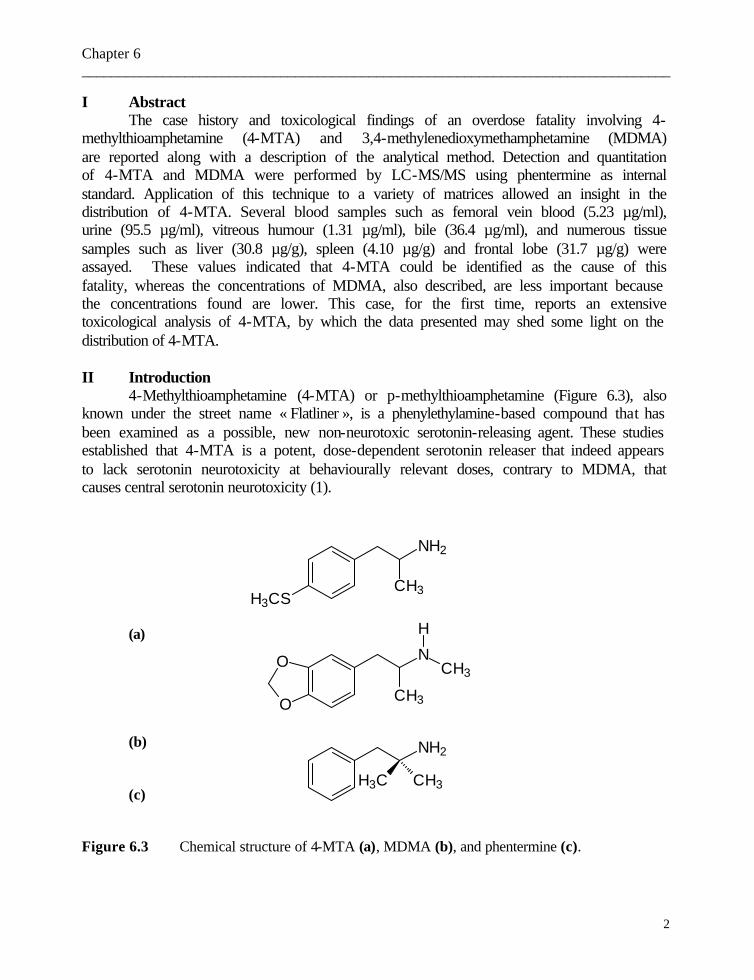
Chapter 6 ________________________________________________________________________________
2
I Abstract The case history and toxicological findings of an overdose fatality involving 4-
methylthioamphetamine (4-MTA) and 3,4-methylenedioxymethamphetamine (MDMA) are reported along with a description of the analytical method. Detection and quantitation of 4-MTA and MDMA were performed by LC-MS/MS using phentermine as internal standard. Application of this technique to a variety of matrices allowed an insight in the distribution of 4-MTA. Several blood samples such as femoral vein blood (5.23 µg/ml), urine (95.5 µg/ml), vitreous humour (1.31 µg/ml), bile (36.4 µg/ml), and numerous tissue samples such as liver (30.8 µg/g), spleen (4.10 µg/g) and frontal lobe (31.7 µg/g) were assayed. These values indicated that 4-MTA could be identified as the cause of this fatality, whereas the concentrations of MDMA, also described, are less important because the concentrations found are lower. This case, for the first time, reports an extensive toxicological analysis of 4-MTA, by which the data presented may shed some light on the distribution of 4-MTA.
II Introduction
4-Methylthioamphetamine (4-MTA) or p-methylthioamphetamine (Figure 6.3), also known under the street name « Flatliner », is a phenylethylamine-based compound that has been examined as a possible, new non-neurotoxic serotonin-releasing agent. These studies established that 4-MTA is a potent, dose-dependent serotonin releaser that indeed appears to lack serotonin neurotoxicity at behaviourally relevant doses, contrary to MDMA, that causes central serotonin neurotoxicity (1).
(a)
(b)
(c)
Figure 6.3 Chemical structure of 4-MTA (a), MDMA (b), and phentermine (c).
NH2
CH3H3CS
O
O
N
H
CH3
CH3
NH2
H3C CH3

Chapter 6 ________________________________________________________________________________
3
Except for seizures in Germany and Switzerland, where the tablets featured a five pointed star design, all other tablets encountered in Europe and Australia have been flat, half-scored and white, hence the nickname « Flatliner » was adopted (2). However, the name flatliner may refer to 4-MTA, ketamine, or even phencyclidine tablets in the UK. The term may derive from a cult 1990s film called Flatliners, from a German motorcycle club or from US engineering clubs (3). However, the link with a flatliner-EKG (i.e. no cardiac output) is also described (3). Currently the exact route by which 4-MTA is produced in illicit laboratories is not known. As 4-MTA tablets have been investigated for the presence of 4-(4-methylthiobenzyl)pyrimidine and 4-methyl-5-(4-methylthiophenyl)pyrimidine and as these by-products were not detected, it appears that illicit laboratories are not using the Leuckardt method for the preparation of 4-MTA (4). Identification and quantitation of 4-MTA in tablets, coming from a drug seizure in the U.K., were performed by GC-MS and NMR (5).
4-MTA has up until now been encountered in three fatal overdose cases in The Netherlands and the UK, of which in two cases other drugs were involved. Based on these data, Elliott has suggested a toxicity range for 4-MTA. Blood concentrations between 0.2 and 0.6 µg/ml are considered as moderate toxic, higher concentrations as severe toxic, and concentrations above 1.5 µg/ml as fatal. This range is only meant as a guide to the interpretation of results, particularly in post-mortem cases (6). Two analytical techniques have been described to determine 4-MTA in blood and urine, namely HPLC-DAD and GC-MS. To our knowledge, a detailed distribution study considering 4-MTA levels in various blood samples and tissues has not been published. In addition, there are only rare reports in the literature describing post-mortem redistribution of amphetamine and analogues in humans. For 3,4-methylenedioxymethamphetamine (MDMA, “ecstasy”) in particular, only a few case reports are published (7,8). This paper reports the first LC-MS/MS method for the determination of the tissue distribution of both 4-MTA and MDMA in a forensic case.
III Case history
One morning, a 27-year-old drug dealer suddenly died after a collapse and an intensive reanimation attempt. Police inquiry revealed that seven other youngsters were involved – all being, to some extent, in an intoxicated state.
The autopsy of the normally built young man was performed 2 days after death. A few superficial skin lesions on the limbs indicating slight blunt trauma, and reanimation signs were observed. On internal examination, emphysema as well as overwhelming congestion and oedema of both lungs were observed. No cardiac anomalies were found. The brain showed only a slight congestion and oedema of the white matter. Generalized congestion of all organs was noticed. These macroscopical findings were confirmed by histological examination. Moreover, a few microscopical signs consistent with chronic drug abuse such as moderate eosinophilic infiltration in the lymphenodes and a slightly decreased number of Purkinje cells were observed. In addition, aspecific perivascular lymphocyte infiltration and siderophages in the nucleus lentiformis were noticed.

Chapter 6 ________________________________________________________________________________
4
There were no arguments for a prolonged multiple organ failure. Neither on external examination at the scene, nor at autopsy, there were signs pointing to hyperthermia. No pre-existing pathologies were identified. Police inquiry revealed all youngsters had taken « smart pills». Therefore samples for an extensive toxicological investigation were taken. IV Experimental
IV.1 Reagents and materials All reagents and chemicals were of analytical grade. 4-MTA hydrochloride was a
kind gift of the Scientific Institute of Public Health (Brussels, Belgium). MDMA and phentermine hydrochloride were available from the standards collection at the Laboratory for Toxicology (Ghent University, Belgium). Solvents were of HPLC grade from Romil Chemicals (Cambridge, UK) with the exception of water, which was from Prosan (Merelbeke, Belgium).
Stock solutions containing either 4-MTA or MDMA or phentermine (internal standard) at a concentration of 1.0 mg/ml were prepared in methanol and stored in plastic bottles. Dilution with methanol of the 4-MTA and MDMA stock solutions yielded the working solutions at appropriate concentrations to prepare spiked calibration samples in the various matrices. The internal standard, phentermine, was diluted to a concentration of 10 µg/ml in methanol. All these solutions were stored in the freezer (-20°C) and proved stable for at least 6 months.
IV.2 Systematic Drug screening A comprehensive screening was performed on post-mortem blood (vena cava
inferior blood) and urine using enzyme-multiplied immunoassay techniques (EMIT), radioimmunoassay (RIA), and chromatographic techniques (HPLC-DAD following alkaline extraction, and gas chromatography with nitrogen-phosphorus detection [GC-NPD] and with mass spectrometry [GC-MS]) as described earlier (9).
IV.3 Quantitative LC-MS/MS method IV.3.1 Calibration Calibration curves were prepared in blank matrices for blood, urine and kidney
homogenate (used as a reference for tissue homogenates). The following calibrators were prepared in these three matrices: 0.1, 0.5, 1, 2.5, 5, 10, 25, 50 µg/ml for both 4-MTA and MDMA, with phentermine present as internal standard at 10 µg/ml. In each case a 1-ml aliquot of the sample or appropriate prepared tissue homogenate was spiked with both 50µl of the appropriate standard solution in methanol and 25µl of the internal standard solution. Subsequently, extraction was performed according to the general isolation procedure (see below). The resulting calibration curves were created by using weighted least-squares regression analysis (weighing factor 1/x). Concentrations of 4-MTA and MDMA were calculated by comparing the peak-area ratio of the specific compound and phentermine (internal standard) against the calibration curve. For the quantitation of vitreous humour the calibration curve of urine was used because both have a high water content. Forensic

Chapter 6 ________________________________________________________________________________
5
samples were at least analyzed in duplicate: after a first analysis to determine roughly the concentration range of the sample, a second analysis was performed after appropriate dilution of the sample (in view of the linear dynamic range constraints of the TOF analyzer).
IV.3.2 Isolation of the compounds Whole blood, vitreous humour, and urine of the victim were used as such. Tissue
samples were homogenized in an ice bath after appropriate dilution with water (depending on concentration) using an Ultra-Turrax homogenizer from IKA (Staufen, Germany). All post-mortem samples were extracted according to a liquid-liquid extraction procedure developed in our laboratory (10), especially suited for the extraction of amphetamine analogues. In short, after addition of the internal standard (25µl of a 10 µg/ml-standard solution in methanol), and after adjustment of the pH with 0.5ml of a K2CO3 buffer (1.0M, pH 9.5), 1 ml of the sample was extracted with 8 ml of a mixture of hexane/ethyl acetate (70:30, v/v). Samples were mixed on a rotary mixing device for 15 min and centrifuged at 1100g for 10 min. The organic layer was transferred to a conical tube containing 50 µl of methanolic HCl (5 mol/L acetyl chloride in methanol) and evaporated using a Turbovap evaporator at 35°C under nitrogen. Finally, the dry residue was dissolved in 250µl of HPLC solvent A (see “HPLC conditions”) and 50-µl aliquots were injected for LC-MS/MS analysis.
IV.3.3 HPLC conditions Chromatographic separation was achieved on an Hypersil BDS phenyl column (100
mm x 2.1 mm i.d., 3-µm particle size) protected by a Hypersil BDS phenyl guard column (7.5 mm x 2.1 mm i.d., 3-µm particle size) using a Waters Alliance 2790 separation module integrated with the Q-TOF instrument. Both the analytical and guard column were from Alltech (Deerfield, IL, USA). The flow rate was set to 0.2 ml/min. After a 5 min isocratic elution at 100% of a mixture of water/methanol/acetonitrile (94 :3 :3, by vol.) containing 0.1 M ammonium acetate (solvent A), gradient elution was performed, starting at 100% of solvent A, then programmed linearly, within 13 min, to 35 % of a mixture of water/methanol/acetonitrile (6 :47 :47, by vol.) again containing 0.1 M ammonium acetate (solvent B). After completion of the chromatographic run, the pump was programmed to regain its initial conditions within 0.5 min, and a 5-min reconditioning time was allowed before the next injection.
IV.3.4 Mass spectrometry The mass spectrometric analyses were conducted using a Micromass Q-TOF hybrid
mass spectrometer (Micromass, Wythenshawe, UK) equipped with an orthogonal electrospray source (Z-spray) operated in the electrospray positive ion mode (ESI+). Nitrogen acted both as nebulizer and desolvation gas, and argon as collision gas in the MS/MS mode. Optimal tuning of capillary (3000V) and cone (20V) voltage, source (150°C) and desolvation (395°C) temperature, and collision energy was achieved by introducing solutions of the different compounds into the electrospray ion source of the
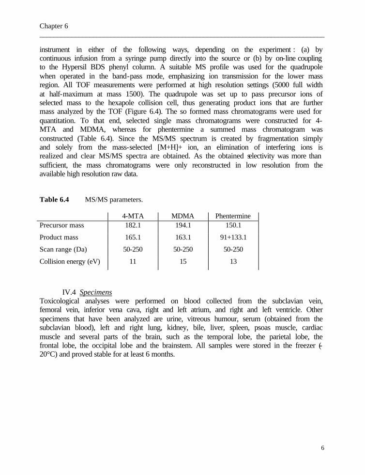
Chapter 6 ________________________________________________________________________________
6
instrument in either of the following ways, depending on the experiment : (a) by continuous infusion from a syringe pump directly into the source or (b) by on-line coupling to the Hypersil BDS phenyl column. A suitable MS profile was used for the quadrupole when operated in the band-pass mode, emphasizing ion transmission for the lower mass region. All TOF measurements were performed at high resolution settings (5000 full width at half-maximum at mass 1500). The quadrupole was set up to pass precursor ions of selected mass to the hexapole collision cell, thus generating product ions that are further mass analyzed by the TOF (Figure 6.4). The so formed mass chromatograms were used for quantitation. To that end, selected single mass chromatograms were constructed for 4-MTA and MDMA, whereas for phentermine a summed mass chromatogram was constructed (Table 6.4). Since the MS/MS spectrum is created by fragmentation simply and solely from the mass-selected [M+H]+ ion, an elimination of interfering ions is realized and clear MS/MS spectra are obtained. As the obtained selectivity was more than sufficient, the mass chromatograms were only reconstructed in low resolution from the available high resolution raw data. Table 6.4 MS/MS parameters.
4-MTA MDMA Phentermine Precursor mass 182.1 194.1 150.1
Product mass 165.1 163.1 91+133.1
Scan range (Da) 50-250 50-250 50-250
Collision energy (eV) 11 15 13
IV.4 Specimens Toxicological analyses were performed on blood collected from the subclavian vein, femoral vein, inferior vena cava, right and left atrium, and right and left ventricle. Other specimens that have been analyzed are urine, vitreous humour, serum (obtained from the subclavian blood), left and right lung, kidney, bile, liver, spleen, psoas muscle, cardiac muscle and several parts of the brain, such as the temporal lobe, the parietal lobe, the frontal lobe, the occipital lobe and the brainstem. All samples were stored in the freezer (-20°C) and proved stable for at least 6 months.
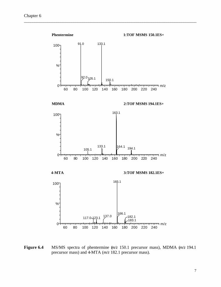
Chapter 6 ________________________________________________________________________________
7
1:TOF MSMS 150.1ES+Phentermine
60 80 100 120 140 160 180 200 220 240m/z0
100
%
133.191.0
92.0105.1 150.1
2:TOF MSMS 194.1ES+MDMA
60 80 100 120 140 160 180 200 220 240m/z0
100
%
163.1
133.1105.1 194.1164.1
3:TOF MSMS 182.1ES+4-MTA
60 80 100 120 140 160 180 200 220 240m/z0
100
%
165.1
123.1117.0 137.0166.1
182.1183.1
Figure 6.4 MS/MS spectra of phentermine (m/z 150.1 precursor mass), MDMA (m/z 194.1 precursor mass) and 4-MTA (m/z 182.1 precursor mass).

Chapter 6 ________________________________________________________________________________
8
V Results and discussion V.1 Drug screening The routine screening of vena cava inferior blood and urine by immunoassay
techniques disclosed the presence of amphetamines (only in urine, 4.5 µg/ml) and cannabinoids (32 ng/ml blood, 1.4 µg/ml urine) and, of toxicologically irrelevant levels of caffeine (1.62 µg/ml blood, 2.45 µg/ml urine) and cotinine (0.14 µg/ml blood, 4.16 µg/ml urine). The creatinine level in urine was 1.54 g/l. The analysis of blood and urine using general purpose HPLC-DAD, GC-NPD, and GC-MS methods confirmed the presence of 4-MTA, MDMA and caffeine. Based on these findings, the dedicated, fully quantitative LC-MS/MS assay was used for all of the different specimens available.
V.2 Analytical performance Calibration curves were constructed for 4-MTA and MDMA. The linearity range
for 4-MTA varied from one matrix to another: for blood and urine it was established between 5-2500 ng/ml, and for kidney homogenate between 125-2500 ng/ml. For MDMA linearity was obtained between 5-250 ng/ml. As can be seen in Figure 6.5, for MDMA better correlation coefficients were obtained when excluding the three highest calibrators. This deviation from linearity can be attributed to the limited linear dynamic range due to possible depletion of the MS detector plate charge, effectively blinding the detector and leading to saturation of the ion counting electronics (11). The correlation coefficients in the different matrices ranged from 0.9987 (kidney homogenate) to 0.9992 (blood and urine) for 4-MTA and from 0.9967 (urine) to 0.9997 (kidney homogenate) for MDMA. The limit of detection (LOD), the lower limit of quantitation (LLQ), and the upper limit of quantitation (ULQ) for 4-MTA and MDMA were determined by analyzing respectively decreasing and increasing concentrations of the compounds added to the blank matrices. The LOD was established at the lowest concentration that produced a response of three times the background noise. The LLQ and ULQ were defined respectively as the lowest and upper concentration that could be quantified with an imprecision of <20%. These values together with the reproducibility data are listed in Table 6.5.

Chapter 6 ________________________________________________________________________________
9
Figure 6.5 Linear least-squares of standards for 4-MTA and MDMA in whole blood, urine, and
tissue homogenate (σ represents the excluded calibrators of MDMA).
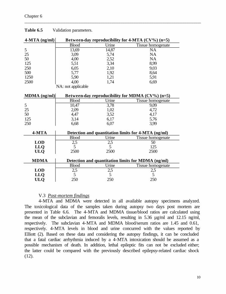
Chapter 6 ________________________________________________________________________________
10
Table 6.5 Validation parameters. 4-MTA (ng/ml) Between-day reproducibility for 4-MTA (CV%) (n=5)
Blood Urine Tissue homogenate 5 13,69 14,87 NA 25 3,09 5,74 NA 50 4,00 2,52 NA 125 5,51 3,34 8,99 250 6,05 2,10 9,03 500 5,77 1,92 8,64 1250 5,90 1,21 5,91 2500 4,00 1,74 6,69
NA: not applicable
MDMA (ng/ml) Between-day reproducibility for MDMA (CV%) (n=5) Blood Urine Tissue homogenate
5 10,47 3,78 9,09 25 2,09 1,02 4,72 50 4,47 3,52 4,17 125 3,14 6,17 5,76 250 6,68 6,07 3,99
4-MTA Detection and quantitation limits for 4-MTA (ng/ml)
Blood Urine Tissue homogenate LOD 2,5 2,5 50 LLQ 5 5 125 ULQ 2500 2500 2500
MDMA Detection and quantitation limits for MDMA (ng/ml)
Blood Urine Tissue homogenate LOD 2,5 2,5 2,5 LLQ 5 5 5 ULQ 250 250 250
V.3 Post-mortem findings 4-MTA and MDMA were detected in all available autopsy specimens analyzed.
The toxicological data of the samples taken during autopsy two days post mortem are presented in Table 6.6. The 4-MTA and MDMA tissue/blood ratios are calculated using the mean of the subclavian and femoralis levels, resulting in 5.36 µg/ml and 12.15 ng/ml, respectively. The subclavian 4-MTA and MDMA blood/serum ratios are 1.45 and 0.61, respectively. 4-MTA levels in blood and urine concurred with the values reported by Elliott (2). Based on these data and considering the autopsy findings, it can be concluded that a fatal cardiac arrhythmia induced by a 4-MTA intoxication should be assumed as a possible mechanism of death. In addition, lethal epileptic fits can not be excluded either; the latter could be compared with the previously described epilepsy-related cardiac shock (12).

Chapter 6 ________________________________________________________________________________
11
Table 6.6 Distribution of 4-MTA and MDMA.
4-MTA (µg/ml or µg/g)
MDMA (ng/ml or ng/g)
4-MTA tissue /blood ratio1
MDMA tissue /blood ratio2
Blood subclavian vein 5.49 13.8 NA NA Serum subclavian vein 3.79 22.5 NA NA Blood femoralis vein 5.23 10.5 NA NA Blood inferior vena cava 6.39 11.8 1.19 0.97 Right atrial blood 5.57 15.7 1.04 1.29 Right ventricular blood 7.60 16.5 1.42 1.36 Left atrial blood 5.16 12.9 0.96 1.06 Left ventricular blood 8.20 18.1 1.53 1.49 Urine 95.5 4932 NR NR Vitreous humour 1.31 67.6 0.24 5.56 Left lung 16.4 59.1 3.06 4.86 Right lung 16.7 68.6 3.12 5.65 Kidney 2.88 32.7 0.54 2.69 Bile 36.4 231.1 NR NR Liver 30.8 89.3 5.75 7.35 Spleen 4.10 62.7 0.77 5.16 Psoas muscle 5.79 31.6 1.08 2.60 Cardiac muscle 6.32 48.2 1.18 3.97 Brain: temporal lobe 34.5 82.1 6.44 6.76 Brain: parietal lobe 35.0 96.3 6.53 7.93 Brain: frontal lobe 31.7 76.6 5.91 6.31 Brain: occipital lobe 36.5 89.3 6.81 7.35 Brainstem 34.4 72.6 6.42 5.98
1 Mean of subclavian blood and femoralis blood for 4-MTA: 5.36 µg/ml 2 Mean of subclavian blood and femoralis blood for MDMA: 12.15 ng/ml NA : not applicable NR : not relevant
Referring to the 4-MTA levels following remarks can be made. The femoralis 4-
MTA blood level is the lowest peripheral, although there is only a slight difference with the subclavian blood concentration (a difference of 0.260 µg/ml). The importance of peripheral blood specimens has previously been emphasized (13). However, since the 4-MTA blood levels in the cardiac ventricles in this case approach the subclavian and femoral concentration, these sampling sites could be considered for 4-MTA determination when peripheral samples are completely lacking. On the other hand, atrial blood samples should definitely be avoided. The high 4-MTA blood concentration in the right atrium can be explained by diffusion of 4-MTA from the liver via the vascular pathway. The 4-MTA
Chapter 6 ________________________________________________________________________________
12
levels in liver and bile are indeed 6 to almost 7 times higher than the peripheral blood concentration. The liver 4-MTA concentrations are in accordance with those reported in fatal PMA (para-methoxyamphetamine)-intoxications (14). 4-MTA is indeed structurally closely related to PMA. In our case, the left atrial 4-MTA blood concentration is even more increased than the right one and can be correlated to diffusion out of the lungs. The lungs indeed do show levels which are almost 3 times higher than those in the peripheral blood. On the other hand, 4-MTA levels in cardiac and skeletal muscle are only slightly higher than the peripheral blood concentrations, which might indicate that 4-MTA does not tend to bind at these tissues. The subclavian blood/serum ratio may point to a certain accumulation of 4-MTA in red blood cells. The urinary 4-MTA concentration should probably be interpreted in light of the pH (= 5.6). Indeed, as for amphetamine and d-methamphetamine, the urinary excretion is pH-dependent, and it has previously been established that acidification increases the level found (15). In addition, in view of the important 4-MTA concentration in the liver and the bile, this might indicate that 4-MTA is eliminated by excretion via the bile.
4-MTA levels in the various lobes of the brain and the brainstem are similar. Since the brain is the target organ, these high 4-MTA levels are in line with the expectations. Although it is not clear whether the man died during the distribution or the elimination phase, the very low vitreous humour concentration can indicate that the 4-MTA distribution has not yet been fully completed. However, it is not known whether 4-MTA can easily pass through the blood-retinal barrier.
The distribution of MDMA in this case is not fully comparable with the findings for 4-MTA. The difference between the subclavian and femoral vein concentration of MDMA is somewhat more pronounced. The atrial and ventricular MDMA blood levels can be explained similarly to the corresponding 4-MTA concentrations. However, the majority of the MDMA organ/blood ratios are higher than the corresponding 4-MTA ratios. Muscle MDMA concentrations are increased compared with the blood levels determined. The MDMA blood/serum ratio neither is comparable. In addition, a blood/serum ratio lower than one is rather surprising. However, hemolysis could partially account for this ratio in the low concentration range. Similar to 4-MTA, the MDMA levels in the brain are high but in contrast with the corresponding 4-MTA levels, these show more regional variations. The high MDMA vitreous humour level is in contradiction with rabbit experiments where a good correlation between the vascular compartment and the vitreous humour was established: the MDMA vitreous humour to blood ratio was 1.1 in the distribution and elimination phase (16).
Considering the 4-MTA and MDMA concentrations on the whole, it might be suggested that the MDMA has been ingested earlier than 4-MTA.
Based on the presented data, unequivocal statements on similarity in the distribution of both 4-MTA and MDMA cannot be made.
The extensiveness by which this case has been examined, could in one respect be of interest in forensic science, particularly when blood samples are not available, and in another respect, attribute to studies of distribution and eventual redistribution.
Chapter 6 ________________________________________________________________________________
13
Acknowledgements This work was supported by grant GOA99-120501.99 (Bijzonder OnderZoeksFonds). M.P. Bouche acknowledges her position with the Fund for Scientific Research Flanders (FWO -Vlaanderen). References 1. Huang X, Marona-Lewicka D, Nichols DE. p-Methylthioamphetamine is a potent new non-
neurotoxic serotonin-releasing agent. Eur J Pharmacol 1992;229:31-38. 2. Elliott SP. Fatal poisoning with a new phenylethylamine: 4-methylthioamphetamine (4-MTA).
J Anal Toxicol 2000;24:85-89. 3. Report on the risk assessment of 4-MTA in the framework of the joint action on new synthetic
drugs, EMCDDA 1999. 4. Kirkbride KP, Ward D, Jenkins NF, Klass G, Coumbaros JC. Synthesis of 4-methyl-5-
arylpyrimidines and 4-arylpyrimidines: route specific markers for the Leuckardt preparation of amphetamine, 4-methoxyamphetamine, and 4-methylthioamphetamine. Forensic Sci Int 2001;115:53-67.
5. Groombridge CJ. The identification of 4-methylthioamphetamine in a drug seizure. Microgram
1998;31:150-159. 6. Elliott SP. An initial review of analytical findings in cases involving 4-methylthioamphetamine
(4-MTA). TIAFT Bull 1999;29:7-9. 7. Rohrig TP, Prouty RW. Tissue distribution of methylenedioxymethamphetamine. J Anal
Toxicol 1992;16:52-53. 8. Fineschi V, Masti A. A fatal poisoning by MDMA (ecstasy) and MDEA: a case report. Int J
Legal Med 1996;108:272-275. 9. Van Bocxlaer J, Meyer E, Clauwaert K, Lambert W, Piette M, De Leenheer A. Analysis of
zopiclone (Imovane) in postmortem specimens by GC-MS and HPLC with diode-array detection. J Anal Toxicol 1996;20:52-54.
10. Clauwaert KM, Van Bocxlaer JF, De Letter EA, Van Calenbergh S, Lambert WE, De Leenheer
AP. Determination of the designer drugs 3,4-methylenedioxymethamphetamine, 3,4-methylenedioxyethylamphetamine, and 3,4-methylenedioxyamphetamine with HPLC and fluorescence detection in whole blood, serum, vitreous humor, and urine. Clin Chem 2000;46:1968-1977.
11. Burlingame AL, Boyd R, Gaskell S. Mass Spectrometry Anal Chem 1998;70:647R. 12. Hirsch C, Martin DL. Unexpected death in young epileptics. Neurology 1971;21:682-690.

Chapter 6 ________________________________________________________________________________
14
13. Prouty RW, Anderson WH. The forensic science implications of site and temporal influences on postmortem blood-drug concentrations. J Forensic Sci 1990;35:243-270.
14. Felgate HE, Felgate PD, James RA, Sims DN, Vozzo DC. Recent para-methoxyamphetamine
deaths. J Anal Toxicol 1998;22:169-172. 15. Baselt RC (ed) (2000). Disposition of Toxic Drugs and Chemicals in Man. 5th edn. Chemical
Toxicology Institute, Foster City, CA, pp. 49-51 & 527-530. 16. De Letter EA, De Paepe P, Clauwaert KM, Belpaire FM, Lambert WE, Van Bocxlaer JF, Piette
MHA. Is vitreous humour useful for the interpretation of post-mortem 3,4-methylenedioxymethamphetamine (MDMA) blood levels? Experimental approach with rabbits. Int J Legal Med 2000;114:29-35.
Chapter 6 ________________________________________________________________________________
1
II.3. Fatality due to combined use of the designer drugs MDMA and
PMA: a distribution study
Riet Dams 1, Els A. De Letter 2, Kjell A. Mortier 1, Jan A.C.M. Cordonnier 3, Willy E. Lambert 1, Michel H.A. Piette 2, Serge Van Calenbergh 4,
and André P. De Leenheer 1
1 Riet Dams, Kjell A. Mortier, Willy E. Lambert, André P. De Leenheer Ghent University - Department of Toxicology
2 Els De Letter, Michel Piette Ghent University - Department of Forensic Medicine 3 Jan A.C.M. Cordonnier, Chemiphar NV
4 Serge Van Calenbergh
Ghent University - Department of Medicinal Chemistry
Accepted for publication in: J Anal Toxicol
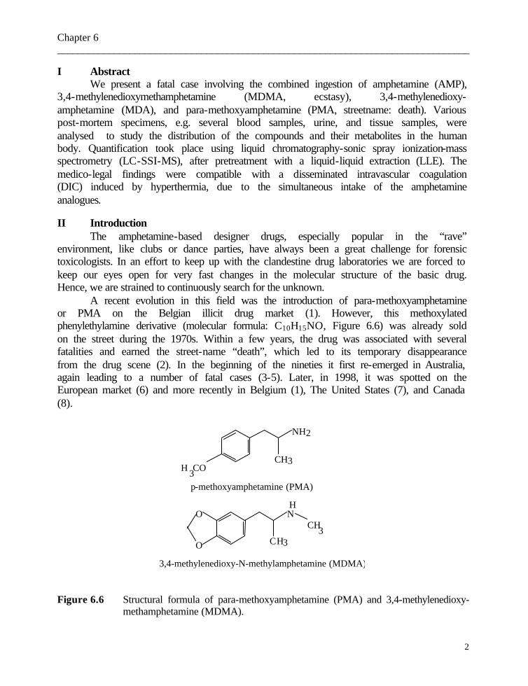
Chapter 6 ________________________________________________________________________________
2
I Abstract We present a fatal case involving the combined ingestion of amphetamine (AMP),
3,4-methylenedioxymethamphetamine (MDMA, ecstasy), 3,4-methylenedioxy-amphetamine (MDA), and para-methoxyamphetamine (PMA, streetname: death). Various post-mortem specimens, e.g. several blood samples, urine, and tissue samples, were analysed to study the distribution of the compounds and their metabolites in the human body. Quantification took place using liquid chromatography-sonic spray ionization-mass spectrometry (LC-SSI-MS), after pretreatment with a liquid-liquid extraction (LLE). The medico-legal findings were compatible with a disseminated intravascular coagulation (DIC) induced by hyperthermia, due to the simultaneous intake of the amphetamine analogues. II Introduction
The amphetamine-based designer drugs, especially popular in the “rave” environment, like clubs or dance parties, have always been a great challenge for forensic toxicologists. In an effort to keep up with the clandestine drug laboratories we are forced to keep our eyes open for very fast changes in the molecular structure of the basic drug. Hence, we are strained to continuously search for the unknown.
A recent evolution in this field was the introduction of para-methoxyamphetamine or PMA on the Belgian illicit drug market (1). However, this methoxylated phenylethylamine derivative (molecular formula: C10H15NO, Figure 6.6) was already sold on the street during the 1970s. Within a few years, the drug was associated with several fatalities and earned the street-name “death”, which led to its temporary disappearance from the drug scene (2). In the beginning of the nineties it first re-emerged in Australia, again leading to a number of fatal cases (3-5). Later, in 1998, it was spotted on the European market (6) and more recently in Belgium (1), The United States (7), and Canada (8).
O
O NCH3
CH3
CH3H 3CO
NH2
p-methoxyamphetamine (PMA)
3,4-methylenedioxy-N-methylamphetamine (MDMA)
H
Figure 6.6 Structural formula of para-methoxyamphetamine (PMA) and 3,4-methylenedioxy- methamphetamine (MDMA).
Chapter 6 ________________________________________________________________________________
3
As its structurally related compounds MDA (3,4-methylenedioxyamphetamine), MDMA (3,4-methylenedioxymethamphetamine, Figure 6.6), and MDEA (3,4-methylenedioxyethylamphetamine), PMA exhibits hallucinogenic properties. However, it has been postulated that it is more toxic than MDMA because of its potent effect on the serotonergic transmission (7). Furthermore, PMA is closely related to another new ring-substituted amphetamine, 4-MTA (4-methylthioamphetamine) which was also identified in a few fatalities (9-11). Doses of less than 50 milligrams of PMA, without co-ingestion of other drugs or alcohol, induce symptoms similar to MDMA, like increased heart rate, blood pressure, and respiratory rate, elevated body temperature, erratic eye movements, muscle spasms, nausea, and visual hallucinations. Higher doses are considered lethal, certainly when taken with other amphetamine derivatives, cannabis, cocaine, prescription medication or alcohol. Symptoms in severe intoxications can be cardiopulmonary related (e.g. cardiac arrhythmia, pulmonary oedema), but vomiting, renal failure, hyperthermia, convulsions, and coma prior to death can also occur (7). The drug is sold in tablets, capsules, or powder form, and its appearance and cost are comparable to MDMA. Analysis of tablets revealed that “ecstasy” tablets can contain not only PMA but also PMMA (para-methoxymethamphetamine) (12). Because of its great similarity with the popular and well-known MDMA it has been mistakenly ingested as “ecstasy”, with sometimes lethal consequences. In the last few years, an increasing number of fatal intoxications involving PMA have been reported (1, 3-8). However, to our knowledge, data on PMA tissue concentrations are scarce (13).
We present a fatal case of the combined use of amphetamine, MDMA, MDA, and PMA. The complete toxicological findings of the specimens, analysed by LC-SSI-MS after appropriate liquid-liquid extraction, are presented and the distribution of the drugs is discussed. To the best of our knowledge, post-mortem drug distribution of PMA in combination with MDMA has barely been explored in humans.
III Case history
A 23-year-old man was found dead at home, lying in a divan. It was warm in the room and the ambient temperature was about 25°C. The man was naked and his clothes were close nearby as if he had just undressed. The rectal temperature had reached the ambient temperature. The lividities were fixed, the rigor mortis had almost disappeared and greenish coloration of the whole abdominal wall was noticed. Slight mummification of lips, nose tip, fingers and toes was found. The eyeballs were depressed and dehydrated. These findings, correlated with the police information, revealed that the post-mortem interval was about three days. The man was very tiny: the body weighed 56 kg and was 175 cm tall (body mass index: 18.3). The face showed no petechiae. Apart from a few slight recent excoriations on the arms, consistent with slight blunt trauma (e.g. fall or blow), nothing unusual was observed.
During internal inspection, moderate putrefaction and congestion of all organs was found. Both lungs weighed 1410 g and showed obvious congestion and distinct oedema. The left and right pleural cavity contained about 150 and 100 ml bloody fluid, respectively. Somewhat vinous colored pericardial fluid was present (about 10 ml). The heart weighed
Chapter 6 ________________________________________________________________________________
4
315 g and showed - apart from a few Tardieu spots - macroscopically no anomalies. The liver weighed 1160 g and slight steatosis, confirmed by microscopical inspection, was found. The stomach and the bladder contained about 50 ml brown-greenish fluid and 200 ml clear yellow urine, respectively. The fresh brain weighed 1405 g and on dissection after fixation congestion and oedema were found. Microscopical study of the organs confirmed the generalized congestion and obvious signs of shock were found (such as leucocyte sludging, micro-emboli) - mainly in the sections of the heart, lungs, liver, kidney and brain. Eosinophilic cylinders were seen in the renal tubuli, but myoglobin immunostaining was negative. The medico-legal findings were consistent with disseminated intravascular coagulation (DIC) induced by hyperthermia. IV Experimental
IV.1 Materials Para-methoxyamphetamine (PMA) was synthesized in-house according to a
described procedure (14). 3,4-Methylenedioxymethamphetamine (MDMA or ecstasy) and 3,4-methylenedioxyamphetamine (MDA) were purchased from Sigma-Aldrich (Bornem, Belgium). Amphetamine and ephedrine (internal standard) were available from the collection of our laboratory and also came from Sigma-Aldrich (Bornem, Belgium). All reagents and chemicals were of analytical grade (Merck-Eurolab Leuven, Belgium). Solvents were all of HPLC gradient grade, also purchased from Merck-Eurolab (Leuven, Belgium).
An individual standard solution of 1 mg/ml of each compound was prepared in methanol and stored in the dark at -20°C until use. Under these conditions all solutions proved stable for more than six months. A 2-µg/ml solution of internal standard, ephedrine, in methanol was also prepared. We decided to use this compound because of its structural similarity and its sufficient separation from the amphetamine mixture.
IV.2 Analysis of biochemical parameters in vitreous humour Glucose, lactate and potassium were determined in vitreous humour using
enzymatic tests and specific electrodes on a routine automatic analyzer instrument. IV.3 Drug screening
Routine systematic toxicological analysis was performed on the samples to investigate for illegal drugs, medical drugs, alcohol, volatile substances and other poisons. Immunoassay screens (ADx) were performed to test for amphetamines, cannabinoids, opiates, methadone and benzoyl ecgonine on urine, and barbiturates and tricyclic antidepressants in blood. Radio-immunoassay (RIA) was used to screen for LSD in urine, and for morphine and benzodiazepines in blood. Color spot tests on urine and gastric content were used to detect salicylates, acetaminophen, phenothiazines and imipramines. Post-mortem blood was analysed for the presence of carboxyhaemoglobin and cyanide. Gastric content and urine were screened for the presence of basic drugs, and hydrolysed benzodiazepines; blood for the presence of acidic and neutral drugs by thin-layer chromatography. Gas chromatography/mass spectrometry (GC/MS ion trap) without
Chapter 6 ________________________________________________________________________________
5
derivatization, and GS-MS quadrupole with TFA/BSTFA derivatization (trifluoroacetic acid/N,O-bis(trimethylsilyl)trifluoroacetamide) was used to screen the urine and the gastric content for the presence of basic drugs. Blood was screened by high performance liquid chromatography with photodiode-array detection (HPLC/DAD). Analysis for the presence of alcohol and other volatile substances in blood and urine was performed by headspace gas chromatography with a flame ionization detector (GC/FID) (10).
IV.4 LC-MS instrument Chromatography was carried out using a LaChrom separation module (Merck,
Darmstadt, Germany) including a L-7100 Low-Pressure Gradient Pump, L-7200 Autosampler (injection loop 100 µL), L-7360 Column Oven, and D-7000 Interface. The system uses the LC/3DQ-MS System Manager Software running under Windows NT TM version 4.0 on a Compaq Deskpro EN.
All MS experiments were carried out on the M-8000 ion-trap based mass spectrometer from Merck (Darmstadt, Germany) equipped with an on-axis sonic spray interface (SSI) operated in positive ion mode.
IV.5 Method IV.5.1 Sample pretreatment Blood and urine were not pretreated. Tissue samples were homogenized after
appropriate dilution with water using an Ultra-Turrax homogenizer from IKA (Staufen, Germany). Most of the post-mortem samples were extracted according to a liquid-liquid extraction procedure previously developed in our laboratory (15). However, for the more complicated (greasy or degraded) matrices such as adipose tissue, stomach content, and different brain parts a liquid-liquid extraction with back-extraction was applied (16).
IV.5.2 Liquid chromatography-mass spectrometry Chromatographic separation was achieved on a Hypersil BDS phenyl column (100
x 2.1 mm i.d., 3-µm particle size) protected by a Hypersil BDS phenyl guard column (7.5 x 2.1 mm i.d., 3-µm particle size), purchased from Alltech (Lokeren, Belgium). A gradient program with water and acetonitrile, both containing 0.001 vol% formic acid was used. The complete and detailed LC-SSI-MS method is described in a previous paper (17).
IV.6 Specimen collection Toxicological analyses were performed on blood collected from the subclavian
vein, femoral and iliac vein, inferior vena cava, right atrium, aorta ascendens, pulmonary artery, and right and left pulmonary vein. Left atrial and ventricular blood (right and left) were not available. Other specimens analysed included urine, vitreous humour, pericardial fluid, bile, stomach content, liver, spleen, iliopsoas muscle, abdominal adipose tissue. For pleural fluid, cardiac muscle, lungs and kidneys separate sampling of the left and the right occurred. In addition, brainstem, cerebellum, and brain lobes were sampled. All samples were stored in the freezer (-20°C) until analysis.

Chapter 6 ________________________________________________________________________________
6
IV.7 Calibration Calibration curves were prepared in blank matrices of blood, urine, brain
homogenate and liver. Each calibrator sample (1 ml) was spiked with 50 µl of the 2-µg/ml internal standard solution and with the compounds of interest, resulting in a final concentration of 10-1000 ng/ml (blood and urine) and 20-2000 ng/ml (tissues). The spiked compound levels for each calibration graph were 0.1, 0.4, 1.0, 2.0, 4.0, and 10.0 µg/ml, respectively, for each matrix. After extraction of a 1-ml aliquot of each sample according to the previously mentioned liquid-liquid extraction (see above), samples were analysed and calibration curves were created using quadratic regression analysis (in view of the limited linear dynamic range of the mass analyser). Quantification of the autopsy samples was performed by comparing the peak-area ratio of each specific compound and the internal standard against the calibration curve. All samples were analysed twice. First, 1 ml of the available post-mortem matrix (blood and urine) was spiked with the internal standard solution and analysed. Following these preliminary results, a second extraction was performed on appropriately diluted sample specimens. V Results and Discussion
V.1 Routine biochemical parameters The urinary pH and creatinine concentration in this case were 7.5 and 0.3 g/l,
respectively. The potassium level in the vitreous humour was 38.9 mmol/l. The vitreous glucose and lactate concentrations were 3 and 309 mg/dl (sum value of 312 mg/dl), excluding hypoglycaemia.
V.2 Drug screening The initial drug screening (including GC-MS) revealed the presence of MDMA in
blood and urine and amphetamine in urine. In addition, nicotine and caffeine were detected in blood and urine. The results of the screening tests for the presence of other relevant drugs or medications were all negative.
GC-MS analysis showed a small broad peak with a mass spectrum corresponding to PMA. However, this was initially considered as non-toxicologically relevant because the peak area/height was relatively low and it was thought to be a methylated artefact of a p-hydroxylated metabolite of amphetamine.
V.3 Liquid chromatography-mass spectrometry The LC-SSI-MS method was completely validated and proved to fulfil analytical
standard criteria (17). The toxicological findings for amphetamine, MDMA, MDA, and PMA in the
different autopsy samples are summarized in Table 6.7. The ratios of vitreous humour to femoral blood level for PMA, MDMA, MDA and AMP were 1.29, 1.45, 1.33 and 1.47, respectively.
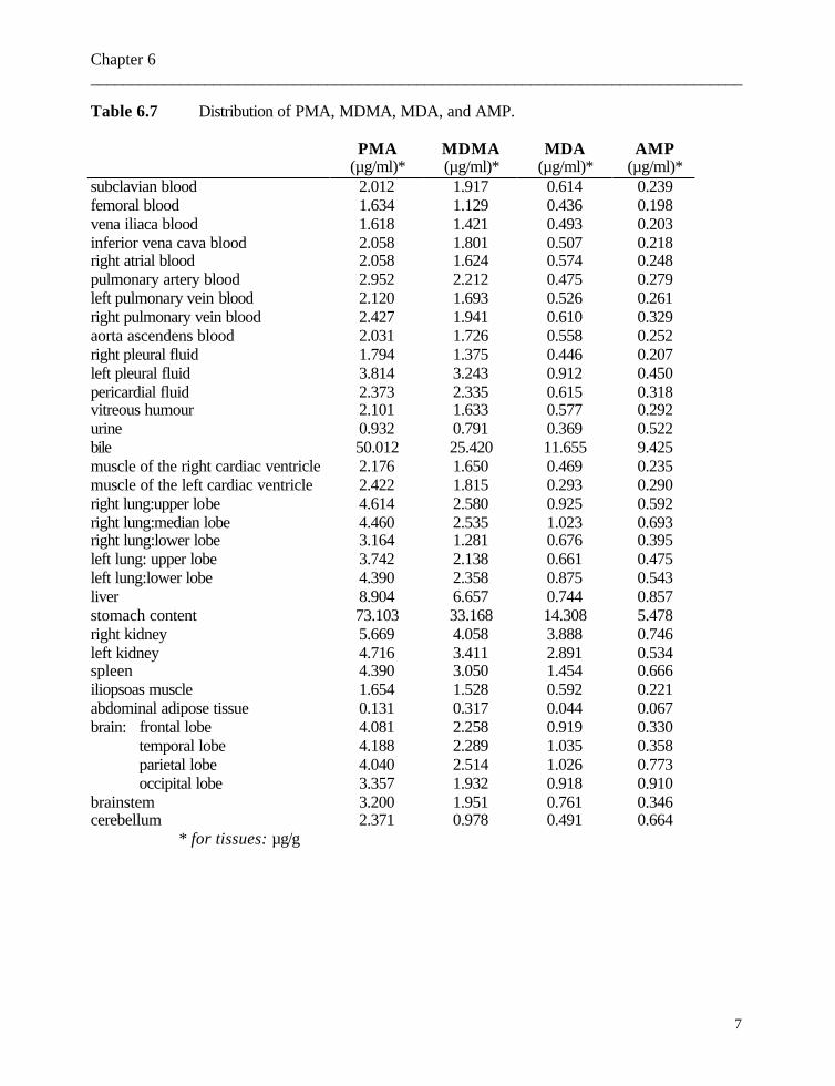
Chapter 6 ________________________________________________________________________________
7
Table 6.7 Distribution of PMA, MDMA, MDA, and AMP.
PMA MDMA MDA AMP (µg/ml)* (µg/ml)* (µg/ml)* (µg/ml)*
subclavian blood 2.012 1.917 0.614 0.239 femoral blood 1.634 1.129 0.436 0.198 vena iliaca blood 1.618 1.421 0.493 0.203 inferior vena cava blood 2.058 1.801 0.507 0.218 right atrial blood 2.058 1.624 0.574 0.248 pulmonary artery blood 2.952 2.212 0.475 0.279 left pulmonary vein blood 2.120 1.693 0.526 0.261 right pulmonary vein blood 2.427 1.941 0.610 0.329 aorta ascendens blood 2.031 1.726 0.558 0.252 right pleural fluid 1.794 1.375 0.446 0.207 left pleural fluid 3.814 3.243 0.912 0.450 pericardial fluid 2.373 2.335 0.615 0.318 vitreous humour 2.101 1.633 0.577 0.292 urine 0.932 0.791 0.369 0.522 bile 50.012 25.420 11.655 9.425 muscle of the right cardiac ventricle 2.176 1.650 0.469 0.235 muscle of the left cardiac ventricle 2.422 1.815 0.293 0.290 right lung:upper lobe 4.614 2.580 0.925 0.592 right lung:median lobe 4.460 2.535 1.023 0.693 right lung:lower lobe 3.164 1.281 0.676 0.395 left lung: upper lobe 3.742 2.138 0.661 0.475 left lung:lower lobe 4.390 2.358 0.875 0.543 liver 8.904 6.657 0.744 0.857 stomach content 73.103 33.168 14.308 5.478 right kidney 5.669 4.058 3.888 0.746 left kidney 4.716 3.411 2.891 0.534 spleen 4.390 3.050 1.454 0.666 iliopsoas muscle 1.654 1.528 0.592 0.221 abdominal adipose tissue 0.131 0.317 0.044 0.067 brain: frontal lobe 4.081 2.258 0.919 0.330 temporal lobe 4.188 2.289 1.035 0.358 parietal lobe 4.040 2.514 1.026 0.773 occipital lobe 3.357 1.932 0.918 0.910 brainstem 3.200 1.951 0.761 0.346 cerebellum 2.371 0.978 0.491 0.664 * for tissues: µg/g
Chapter 6 ________________________________________________________________________________
8
V.4 Combined discussion of post-mortem findings and toxicological / biochemical data
Referring to the situation on the scene and the autopsy findings, the mechanism of death is consistent with a DIC due to hyperthermia. Even in the seventies, the hyperthermic effect of PMA has been investigated, and in mice, it was found that this effect was not as pronounced as for 3,4-methylenedioxyamphetamine (MDA)(18). Hyperthermia was a frequently seen symptom in patients who presented to the Emergency Department after ingestion of PMA (19) and was also described in a few fatalities (4). In addition, hypoglycaemia and hyperkalaemia have been described as typical in PMA intoxications (19). Analysis of the vitreous humour in our case revealed a high potassium level. This value could reflect an ante-mortem hyperkalaemia but - referring to the post-mortem interval of about 3 days (20) and the dehydration of the eyes - this potassium concentration should be interpreted with caution. The glucose and lactate sum value of 312 mg/dl as such is not consistent with hypoglycaemia. According to the study of Sippel and Möttönen, sum values lower than 160 mg/dl are compatible with a hypoglycaemia (21). However, in cases experiencing a prolonged and/or intense agony, high sum values were noticed (22) and as a result, the sum value in this case can be correlated to the DIC due to hyperthermia as the mechanism of death.
As for MDMA, there is no consensus about the lethal blood level but in general, a blood MDMA concentration higher than 1.0 µg/ml can be potentially lethal (23). For PMA, blood levels greater than 0.5 µg/ml are likely to induce toxic effects (3). We believe that in our case - referring to the considerable MDMA and PMA levels in particular – both concentrations are definitely capable of inducing death. In the cases published by Byard et al. PMA blood levels between 0.24 and 4.9 µg/ml were found (5). A PMA blood level up to 5.7 µg/ml has even been reported (6).
Only after incorporation of the LC-MS technique, it became clear that the peak and also the concentration of PMA was toxicologically relevant. The poor gas chromatographic properties of PMA resulted in a broad peak. This was eliminated by the LC-MS procedure where it became clear that substantial amounts of PMA were present in various matrices.
In this case, the body distribution of the four amphetamine related drugs was fairly comparable (Table 6.7). Our data also confirm that blood sampled from the femoral vein should be considered as the preferred anatomical site for blood sampling. However, when this sample is not available, blood from the nearby iliac vein is also appropriate. A gradient from the inferior vena cava and the iliac vein to the femoral vein was noticed. This “diffusion gradient” can be explained by the relatively high levels in the liver. The huge concentrations in the bile are consistent with the excretion via the bile and are also indicative of a relatively long survival period after intake. However, it is not excluded that some post-mortem diffusion from the very high concentration in the stomach content could account for the latter as well. The relatively high levels in the spleen can be correlated with the “reservoir” of amphetamines found in the closely adjacent stomach content. The high MDA levels found in the stomach content points to MDA intake and as a result, the MDA
Chapter 6 ________________________________________________________________________________
9
blood and tissue concentrations can only partially be explained by the metabolism of MDMA to MDA.
Due to the degree of putrefaction, there were only a few cardiac blood samples available. However, considerable levels of all four amphetamines were found in the pericardial fluid. Just as for the pleural fluid, this can in part be explained by post-mortem diffusion out of the lungs. The higher concentrations in the left pleural fluid (as compared with the right) can be explained by diffusion out of the stomach, which was previously also postulated for co-proxamol (24). The levels in the pulmonary veins can be correlated to the concentrations of the adjacent lung lobes. The concentrations in the right atrial blood were in accordance with the levels in the cardiac muscle. The concentrations in the iliopsoas muscle were fairly comparable with the peripheral blood samples and therefore this muscle specimen can be interesting when a blood sample is lacking (e.g. due to fulminant blood loss or due to putrefaction).
The ratio of vitreous humour to femoral blood concentration of the four substances were fairly comparable. In an experiment in rabbits, the ratio of MDMA vitreous humour to blood levels at equilibration was 1.1 (25). The fact that the ratios in this case were somewhat higher than one can probably be explained by a dehydration factor. Indeed, the vitreous humour sample which was taken from both eye balls was in this case hardly 1 ml. Normally, each eye of an adult contains about 3.9 ml of vitreous humour (26). The concentrations in the brain lobe homogenates are fairly comparable. The high brain levels are consistent with the described clinical symptoms (such as hyperthermia) related to the central effects. Considerable brain levels of PMA and MDMA were also previously substantiated (13, 16).
The urinary levels were relatively low, but this may be explained by the alkaline pH (7.5). Indeed, for amphetamine it has been established that acidification of the urine enhances excretion up to about 75 % but alkalinization can reduce recovery in urine to about 1 % (27). This was postulated for PMA as well (28). However, it is not known when and how many times prior to death, the person was urinating. In addition, for PMA it was shown that in humans an average of 15 % is eliminated as unchanged drug (39).
These data also confirm that the concentrations of amphetamine derivatives in adipose tissue are very low (16). VI Conclusion
In summary, a fatal poisoning in which considerable blood levels of MDMA and PMA were found, is presented. We conclude that the man died of disseminated intravascular coagulation (DIC) - induced by hyperthermia – due to the combined ingestion of amphetamines. In addition, the post-mortem distribution of these amphetamine derivatives in the human body was discussed and further supported the use of peripheral blood for toxicological interpretation.
The results also indicate that due to the poor gas chromatographic properties of PMA under specific conditions, this compound could be erroneously overlooked. However, systematic toxicological analysis based on the combination of both gas
Chapter 6 ________________________________________________________________________________
10
chromatography and high performance liquid chromatography can reveal the presence of PMA in biological matrices without any difficulty. Acknowledgments The authors wish to thank Merck KGaA Darmstadt and Merck-Eurolab Belgium for their cooperation and support in this work. Also they wish to thank Mrs. M. Craeymeersch and Mrs. G. Van Nuffel for technical assistance.
Chapter 6 ________________________________________________________________________________
11
References 1. Voorspoels S, Coucke V, Schepens P, Jacobs W. Paramethoxyamphetamine: first fatalities in
Belgium. TIAFT Bull 2001;16:12-13. 2. Cimbura G. PMA deaths in Ontario. Can Med Assoc J 1974;110:1263-1267. 3. Felgate HE, Felgate PD, James RA, Sims DN, Vozzo DC. Recent paramethoxyamphetamine
deaths. J Anal Toxicol 1998;22:169-172. 4. James RA, Dinan A. Hyperpyrexia associated with fatal paramethoxyamphetamine (PMA)
abuse. Med Sci Law 1998;38:83-85. 5. Byard RW, Gilbert J, James R, Lokan RJ. Amphetamine derivative fatalities in South
Australia – Is “Ecstasy” the culprit ? Am J Forensic Med Pathol 1998;19:261-265. 6. Lora-Tamayo C, Tena T, Rodríguez A. Amphetamine derivative related deaths. Forensic Sci
Int 1997;85:149-157. 7. Kraner JC, McCoy DJ, Evans MA, Evans LE, Sweeney BJ. Fatalities caused by the MDMA-
related drug paramethoxyamphetamine (PMA). J Anal Toxicol 2001;25:645-648. 8. Martin TL. Three cases of fatal paramethoxyamphetamine overdose. J Anal Toxicol
2001;25:649-651. 9. Elliott SP. Fatal poisoning with a new phenylethylamine : 4-methylthioamphetamine (4-
MTA). J Anal Toxicol 2000;24:85-89. 10. De Letter EA, Coopman VAE, Cordonnier JACM, Piette MHA. One fatal and seven non-fatal
cases of 4-methylthioamphetamine (4-MTA) intoxication : clinico-pathological findings. Int J Legal Med 2001;114: 352-356.
11. De Caestecker T, De Letter E, Clauwaert K, Bouche MP, Lambert W, Van Bocxlaer J, Piette
M, Van den Eeckhout E, Van Peteghem C, De Leenheer A. Fatal 4-MTA intoxication: development of a liquid chromatographic-tandem mass spectrometric assay for multiple matrices. J Anal Toxicol 2001;25:705-710.
12. Dal Cason TA. A re-examination of the mono-methoxy positional ring isomers of
amphetamine, metamphetamine, and phenyl-2-propane. Forensic Sci Int 2001;119: 168-194. 13. Baselt RC. (ed) (2000) Disposition of toxic drugs and chemicals in man, 5th edn. Chemical
Toxicology Institute, Foster City, California, pp 547-548. 14. Noggle FT, Clark CR, Mc Millian CL, Deruiter J. Liquid-chromatographic and mass-spectral
analysis of N-substituted analogues of 4-methoxyamphetamine. J Chromatogr Sci 1989;27:607-611.

Chapter 6 ________________________________________________________________________________
12
15. Lambert WE, Meyer E, De Leenheer AP. Systematic toxicological analysis of basic drugs by
gradient elution of an alumina-based HPLC packing material under alkaline conditions. J Anal Toxicol 1995;19:73-78.
16. De Letter EA, Clauwaert KM, Lambert WE, Van Bocxlaer JF, De Leenheer AP, Piette MHA.
Distribution study of 3,4-methylenedioxymethamphetamine (MDMA, “ecstasy”) and 3,4-methylenedioxyamphetamine (MDA) in a fatal overdose. J Anal Toxicol 2002;26:113-118.
17. Mortier KA, Dams R, Lambert WE, De Letter EA, Van Calenbergh S, De Leenheer AP.
Determination of paramethoxyamphetamine and other amphetamine-related designer drug by liquid chromatography/sonic spray ionization mass spectrometry. Rapid Commun Mass Spectrom 2002;16:865-870.
18. Nichols DE, Ilhan M, Long JP. Comparison of cardiovascular, hyperthermic and toxic effects
of para-methoxyamphetamine (PMA) and 3,4-methylenedioxyamphetamine (MDA). Arch Int Pharmacodyn Ther 1975;214:133-140.
19. Ling LH, Marchant C, Buckley NA, Prior M, Irvine RJ. Poisoning with the recreational drug
paramethoxyamphetamine (“death”). Med J Aust 2001;174:453-455. 20. Madea B, Henssge C. Eye changes after death. In: B. Knight (ed) (1995) The estimation of
the time since death in the early postmortem period. Edward Arnold Publisher, London, Boston, Melbourne, Auckland, pp 111-118 .
21. Sippel H, Möttönen M. Combined glucose and lactate values in vitreous humour for
postmortem diagnosis of diabetes mellitus. Forensic Sci Int 1982;19:217-222. 22. De Letter EA, Piette MHA. Can routinely combined analysis of glucose and lactate in vitreous
humour be useful in current forensic practice? Am J Forensic Med Pathol 1998;19:335-342. 23. Dowling GP. Human deaths and toxic reactions attributed to MDMA and MDEA. In: S.J.
Peroutka (ed) (1990) Ecstasy: the clinical, pharmacological and neurotoxicological effects of the drug MDMA. Kluwer Academic, Boston, Dordrecht, London, p 73.
24. Yonemitsu K, Pounder DJ. Postmortem toxico-kinetics of co-proxamol. Int J Legal Med
1992;104:347-353. 25. De Letter EA, De Paepe P, Clauwaert KM, Belpaire FM, Lambert WE, Van Bocxlaer JF, Piette
MHA. Is vitreous humour useful for the interpretation of 3,4-methylenedioxymethamphetamine (MDMA) blood levels? Experimental approach with rabbits. Int J Legal Med 2000;114:29-35.
26. Gloor BP. The vitreous. In: Moses RA, Hart WM (eds) (1987) Adler’s physiology of the eye.
Clinical application, The C.V. Mosby Compagny, St. Louis, Washington D.C., Toronto, p 246.
Chapter 6 ________________________________________________________________________________
13
27. Baselt RC. (ed) (2000) Disposition of toxic drugs and chemicals in man, 5th edn, Chemical
Toxicology Institute, Foster City, California, p 49-51. 28. Drummer OH. (ed) (2001) The forensic pharmacology of drugs of abuse,, Arnold Publisher,
London, New York, New Delhi, pp 377-378. 29. Kitchen I, Tremblay J, Andre J, Dring LG, Idle JR, Smith RL, Williams RT. Interindividual
and interspecies variation in the metabolism of the hallucinogen 4-methoxyamphetamine. Xenobiotica 1979;9:397-404.
Chapter 7 ________________________________________________________________________________
1
Chapter 7 Immunohistochemical demonstration of the amphetamine
derivatives 3,4-methylenedioxymethamphetamine (MDMA) and 3,4-methylenedioxyamphetamine (MDA) in human post-mortem brain tissues and the pituitary gland
Els A. De Letter1, Marc F.A. Espeel2, Marijke E.C. Craeymeersch1,
Willy E. Lambert3, Karine M. Clauwaert4, Riet Dams3, Kjell A. Mortier3 Michel H.A. Piette1
Ghent University 1 Els A. De Letter, Marijke E.C. Craeymeersch, Michel H.A. Piette Department of Forensic Medicine 2 Marc F.A. Espeel Department of Anatomy, Embryology, Histology and Medical Physics 3 Willy E. Lambert, Riet Dams, Kjell A. Mortier Department of Toxicology 4 Karine M. Clauwaert Department of Medical Biochemistry and Clinical Analysis
Accepted for publication in: Int J Legal Med

Chapter 7 ________________________________________________________________________________
2
I Abstract Abuse of amphetamine derivatives such as 3,4-methylenedioxymethamphetamine
(MDMA) and 3,4-methylenedioxyamphetamine (MDA) is an important issue in current forensic practice and fatalities are not infrequent. Therefore, we investigated an immunohistochemical method to detect the amphetamine analogues MDMA and MDA in human tissues. For the staining procedure, the Catalysed Signal Amplification (CSA) method using Peroxidase (HRP) - provided by Dako® - and specific monoclonal antibodies were used. Appropriate controls for validation of the technique were included.
The distribution of these designer drugs was studied in various brain regions including the four lobes, the basal ganglia, hypothalamus, hippocampus, corpus callosum, medulla oblongata, pons, and cerebellar vermis. In addition, the pituitary gland was investigated. A distinct positive reaction was observed in all cortical brain regions and the neurons of the basal ganglia, the hypothalamus, the hippocampus and the cerebellar vermis. In the brainstem, relatively weak staining of neurons was seen. The reaction presented as a mainly diffuse cytoplasmic staining of the perikaryon of the neurons, and often axons and dendrites were also visualised. In addition, the immunoreactivity was present in the white matter. In the pituitary gland, distinct immunopositive cells were observed, with a prominent heterogeneity, however. The immunohistochemical findings were supported by the toxicological data.
This immunostaining technique can be used as evidence of intake or even poisoning with MDMA and/or MDA and can be an interesting tool in forensic practice when the usual samples for toxicological analysis are not available. Furthermore, this method can be used to investigate the distribution of these substances in the human body. Key words: 3,4-Methylenedioxymethamphetamine (MDMA) - 3,4-Methylenedioxy-amphetamine (MDA) – Immunohistochemistry - Human brain - Pituitary gland II Introduction
Abuse of amphetamine and derivatives is an important problem in current forensic practice. Moreover, fatal and nearly-fatal intoxications are not infrequent (1). Post-mortem distribution has barely been explored for amphetamine analogues, except for some case reports (2-6). Animal experiments considering this item for amphetamine or analogues are scarce (7-9).
Immunological methods are routinely used for detection of illegal drugs in clinical and forensic toxicology, mainly in urinary screening tests. However, the principle of antigen-antibody recognition can also be applied in histological specimens (immunohistochemistry), allowing detection of drugs in tissue sections. Immunofluorescence procedures have previously been applied successfully in animal experiments e.g. detection of morphine in rat tissues (10), demonstration of tetrahydrocannabinol (11) and phenobarbital (12) in mice tissues. Immunohistochemical demonstration of morphine and methadone in brain sections of overdose victims has recently been published (13,14). Insulin was demonstrated at injection sites by means of immunohistochemistry (15-17).
Chapter 7 ________________________________________________________________________________
3
Part of the behavioural, psychotomimetic and neurochemical effects of MDMA (e.g. increase of body temperature, mood alterations, anxiolytic-like effects) may be explained by affecting the serotonergic system (18,19). Interaction of MDMA with post- as well as pre-synaptic 5-hydroxytryptamine (5-HT, serotonin) recognition sites is postulated (18). In animal studies, high affinity of MDMA for 5-HT2 and 5-HT1A serotonin receptors (18) as well as 5-HT uptake sites was proven (20). The neurotoxicity of MDMA on 5-HT neurons was also investigated in humans and subtle, but significant cognitive deficits were noticed (21).
At present, immunohistochemical methods were used to investigate the biological effects of MDMA, for example, to demonstrate reductions in 5-HT axon density in rats and monkeys (22) or rhabdomyolysis (4,23). Ishiyama et al. were able to demonstrate methamphetamine in mice tissues (24). To our knowledge, the amphetamine derivatives 3,4-methylenedioxyamphetamine (MDA) and 3,4-methylenedioxymethamphetamine (MDMA, “ecstasy”) have not yet been demonstrated in human tissues.
In this study, we applied the CSA immunostaining method - known as the most sensitive method at present - for the microscopical immunodetection of MDMA and MDA. In particular, the distribution of MDMA and MDA in slides of the human brain and the pituitary gland of two fatal intoxications was investigated and correlated with the toxicological findings. III Materials and methods
III.1 Brief Case histories III.1.1 Case 1 A 23-year-old man was found dead, sitting on a chair in a bar. Upon examination,
28 hours after death, the body was 186 cm tall and weighed about 100 kg. No conspicuous lesions neither injection sites were noticed. At autopsy, both lungs weighed 1620 g. Severe pulmonary congestion and oedema, numerous Tardieu spots on the pleurae and the pericardium were noticed. The heart weighed 405 g and a persistence of the left vena cava superior was found. The brain weighed 1545 g and congestion and oedema were confirmed by histological examination. The macro- and microscopical findings were consistent with an acute to subacute cardiopulmonary failure as the mechanism of death.
MDMA and MDA concentrations were determined in femoral blood and brain using a HPLC (high pressure liquid chromatography) procedure with fluorescence detection (25,5). For additional confirmation, a fully quantitative LC-MS (liquid chromatography-mass spectrometry) assay for the determination of all specimens available, was used. Cocaine, benzoylecgonine, cocaethylene, ecgonine methylester, amphetamine, MDMA, MDEA (3,4-methylenedioxyethylamphetamine) and MDA were detected using chromatographic analysis (HPLC-DAD (HPLC with diode array detection), LC-MS, GC-NPD (gas chromatography with nitrogen-phosphorus detection) and GC-MS of the urine sample (26,27). The blood alcohol level was quantified by head-space GC analysis.
Chapter 7 ________________________________________________________________________________
4
III.2.2 Case 2 This 23-year-old man was found dead at home. He was naked and his clothes were
nearby the body (as if he just undressed). The body was in a state of beginning putrefaction and the cadaveric signs combined with the police inquiry revealed that the post-mortem interval was about 3 days. The body weighed 56 kg and was 175 cm tall. At autopsy, both lungs weighed 1410 g and showed obvious congestion and oedema. Apart from congestion and moderate putrefaction, the other organs showed no conspicuous anomalies. The heart and brain weighed 315 and 1405 g, respectively. Microscopical examination confirmed the general congestion and revealed signs of shock (such as leucocyte sludging, micro-emboli), mainly in the sections of the heart, lungs, liver, kidneys and brain. Eosinophilic cylinders were found in the renal tubuli, but myoglobin staining was negative. In several brain regions, apart from congestion and oedema, small – mainly perivascular – bleedings were found. The medico-legal findings were consistent with a disseminated intravascular coagulation (DIC) induced by hyperthermia.
Amphetamines and related compounds were screened by routine methods such as GC-MS and HPLC-DAD, while quantitative results were obtained by GC-MS and LC-MS.
For both fatalities, since drug abuse was suspected, the standard protocol used at our
department was applied and appropriate sampling for toxicological and histological examination was performed. Small parts of the four brain lobes were sampled for drug assay and for each lobe, cortex and medulla were isolated. Thereafter, the brain was fixed in 4 % buffered formaldehyde during three to four weeks. On dissection, samples of various brain regions were taken, followed by embedding in paraffin. Tissue sections were prepared from the frontal, temporal, parietal and occipital region, the medulla oblongata, pons, cerebellar vermis, corpus callosum, hippocampus, the basal ganglia (mammillary bodies, lentiform nucleus, caudate nucleus and thalamus) and adjacent hypothalamus. The pituitary gland was treated in the same way.
All similar samples in the control case 00/116 were taken as blanks for drug assay and negative control tissue for immunohistochemistry. At random samples of other control cases for immunohistochemistry were also available.
III.2 Antibodies and immunostaining procedure III.2.1 Staining procedure Monoclonal antibodies – which specifically recognize MDMA and MDA - were
kindly supplied by Microgenics Corp, Fremont, CA, USA. They were purified from mouse ascites and available in two clones (clone 1A9 and 5C2).
For the staining procedure, the DAKO® Catalysed Signal Amplification (CSA) System, Peroxidase (HRP) was used (supplied as a kit; Code K 1500). CSA is a very sensitive immunohistochemical staining procedure incorporating a signal amplification method based on the peroxidase catalyzed deposition of a biotinylated phenolic compound, followed by a secondary reaction with streptavidin-peroxidase (28). Sections were deparaffinized and rehydrated according to standard protocols. After blocking endogenous biotin and suppressing non-specific background staining – as prescribed in the procedure -
Chapter 7 ________________________________________________________________________________
5
the primary monoclonal mouse antibodies were applied to the tissues and an incubation of 15 minutes followed. For the primary antibodies dilution series ranging from 1:20 to 1:5000 were tested. The best results were obtained with dilutions between 1:500 and 1:1000. The slides were then gently rinsed with TBST buffer solution (Tris Buffered Saline with Tween 20) and three times placed in a fresh TBST buffer bath for 3 – 5 minutes each. The procedure was then continued by sequential 15-minute incubations with biotinylated link antibody, streptavidin-biotin complex, amplification reagent and streptavidin-peroxidase, respectively. The staining was completed by a 3 to 5-minute incubation with 3,3’-diaminobenzidine tetrahydrochloride (DAB) which results in a brown-coloured precipitate. Hematoxylin counterstain was sometimes performed (e.g. in the pituitary gland in order to visualize the basophilic cells).
For the pituitary gland, a Periodic Acid Schiff (PAS)-orange G-staining on an immediately adjacent slide was performed in order to discern the cell subtypes of the pituitary gland (acidophils, basophils and chromophobes).
III.2.2 Setup of the immunohistochemical procedure In all staining experiments of the two amphetamine fatalities, a simultaneous
incubation was performed with analogue sections from control cases. Similar samples were taken during the autopsy of a 28–year-old woman (case
number 00/116; post-mortem interval (PMI) of 48 h) for negative control tissue. The woman was murdered by a shotgun lesion through heart and lungs. In a few staining procedures, at random samples of five other control cases were used. These were a 32-year-old female, murdered by thoracic gunshot wounds (case number 00/8; PMI of 36 h), a 27-year-old female, murdered by multiple stab wounds (case number 00/14; PMI of 56 h), a 26-year-old man, murdered by a gunshot through the heart (case number 00/19; PMI of 42 h), a 17-year-old man, who died due to polytrauma after a traffic accident (case number 01/68, PMI of 91 h), and a 30-year old female, murdered by multiple stab wounds in thorax and neck (case number 01/181; PMI of 58 h). In all control cases, extensive toxicological investigations were negative.
A positive control staining (recommended in the CSA-kit) with monoclonal mouse antibodies to human B-cell (CD231) on formalin-fixed and paraffin-embedded lymph node (from a patient with malignant B-cell lymphoma) and palatine tonsil tissue was performed. At the same time, as an additional negative control, the staining procedure with IgG1 fraction from normal mouse (provided in the kit) and with the antibodies recognizing MDMA and MDA on adjacent sections of the lymph node and palatine tonsil, was applied.
In order to test the specificity of the antibodies, we checked whether it is possible to saturate the antibody binding sites with its specific antigens and thus induce a negative immunodetection. Therefore, either MDMA or MDA were added in various concentrations (dilution solution series from 10-12 g/ml to 1.5 x 10⁻³ g/ml) to the antibody solution. This solution was placed on a rotary mixing device during 24 hours (at 4°C) prior to incubation of the slides. The same procedure was performed using PMA (para-methoxyamphetamine) or AMP (amphetamine). For negative control incubations, the antibody was replaced by IgG1 fraction from normal mouse serum (supplied with the CSA-kit) for each brain region.
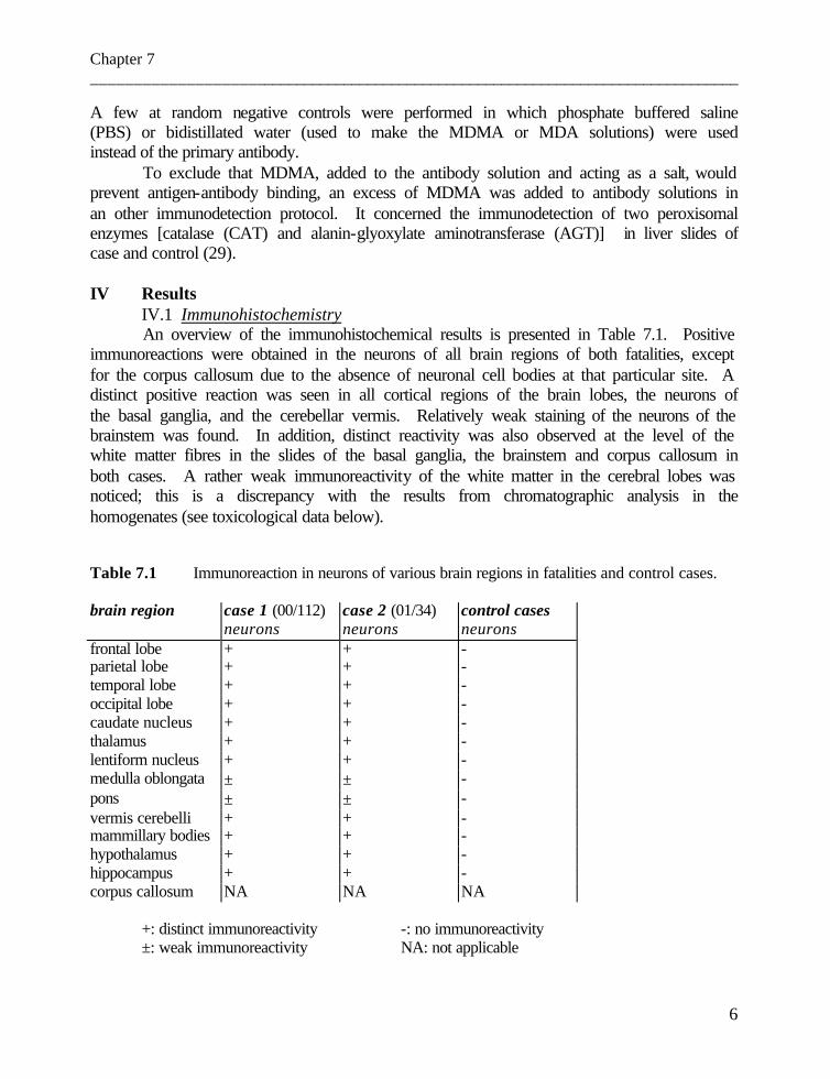
Chapter 7 ________________________________________________________________________________
6
A few at random negative controls were performed in which phosphate buffered saline (PBS) or bidistillated water (used to make the MDMA or MDA solutions) were used instead of the primary antibody.
To exclude that MDMA, added to the antibody solution and acting as a salt, would prevent antigen-antibody binding, an excess of MDMA was added to antibody solutions in an other immunodetection protocol. It concerned the immunodetection of two peroxisomal enzymes [catalase (CAT) and alanin-glyoxylate aminotransferase (AGT)] in liver slides of case and control (29). IV Results
IV.1 Immunohistochemistry An overview of the immunohistochemical results is presented in Table 7.1. Positive
immunoreactions were obtained in the neurons of all brain regions of both fatalities, except for the corpus callosum due to the absence of neuronal cell bodies at that particular site. A distinct positive reaction was seen in all cortical regions of the brain lobes, the neurons of the basal ganglia, and the cerebellar vermis. Relatively weak staining of the neurons of the brainstem was found. In addition, distinct reactivity was also observed at the level of the white matter fibres in the slides of the basal ganglia, the brainstem and corpus callosum in both cases. A rather weak immunoreactivity of the white matter in the cerebral lobes was noticed; this is a discrepancy with the results from chromatographic analysis in the homogenates (see toxicological data below).
Table 7.1 Immunoreaction in neurons of various brain regions in fatalities and control cases. brain region case 1 (00/112) case 2 (01/34) control cases
neurons neurons neurons frontal lobe + + - parietal lobe + + - temporal lobe + + - occipital lobe + + - caudate nucleus + + - thalamus + + - lentiform nucleus + + - medulla oblongata ± ± - pons ± ± - vermis cerebelli + + - mammillary bodies + + - hypothalamus + + - hippocampus + + - corpus callosum NA NA NA
+: distinct immunoreactivity -: no immunoreactivity ±: weak immunoreactivity NA: not applicable
Chapter 7 ________________________________________________________________________________
7
Figure 7.1 (a), 7.1 (d), 7.3 (b) and 7.4 (b) show immunoreactive neurons in the parietal and frontal region, the cerebellar vermis and the hippocampus, respectively. The reaction presented as a mainly diffuse brownish cytoplasmic staining of the perikaryon of the neurons. When the orientation of the section plane was appropriate, staining of the axons and dendrites was noticed. In particular, this was obvious in the Purkinje cells presented in Figure 7.3 (b) where the course of the dendrites to the molecular layer could be followed. In addition, the granular layer cells of the cerebellar vermis were positive, but at higher magnification, a heterogeneity in the staining pattern of these cells was observed [see Figure 7.3 (b)]. The cells of the dentate gyrus in the hippocampus were also visualized [see Figure 7.4 (c)]. However, the heterogeneity was less pronounced there. When comparing all brain regions, differences in staining intensity and number of positive neurons were seen, but these were not quantified.
In Figure 7.2, a macroscopic overview of the immunoreactivity in the sections of the cerebellar vermis in case 1 and in a control case is presented. The focal staining pattern in the control differs from that in the case, as the white matter is unreactive in the control. Moreover, upon microscopical examination, no immunoreactive neurons were seen in the control.
Positive staining reactions were found for both antibody clones; however, the staining aspect of both clones was somewhat different. The two neuronal staining patterns are presented in the parietal and frontal lobe (Figure 7.1 (a) and (d), respectively). When antibodies of the 5C2 clone were used, the staining in the perikaryon was usually somewhat flocky. Incubation with 1A9 rendered a more diffuse coloring in the neuronal cytoplasm and the DAB precipitate was more heterogenous. Variations in staining intensity were also noticed when both clones were compared. Although both clones distinctly revealed visualization of the cortical neurons, we further proceeded with 1A9 because this antibody clone produced a crisp microscopical image.
The validation procedure for positive control (as recommended in the kit) - using anti-CD231 on formalin-fixed and paraffin-embedded lymph node and palatine tonsil tissue - was applied and found to be positive. The same procedure with IgG1 fraction and with the antibodies recognizing MDMA and MDA on lymph node and tonsil sections was negative.
In the control cases, no neurons were revealed, except for a very faint diffuse cytoplasmic staining in the perikarya [see Figure 7.1 (c) and Figure 7.3 (c), parietal cortex and cerebellar vermis, respectively].
Addition of pure MDMA or MDA to the antibody solution – in the high concentration of the series –, totally abolished the staining reaction or reduced staining to the background level as seen in the control cases [see Figure 7.1 (b) and Figure 7.2, parietal lobe and cerebellar vermis, respectively]. In the lower MDMA concentration range, a gradient in the staining intensity was observed. These results point to the specificity of the antibodies. Addition of PMA or amphetamine to the antibody solution was not able to induce negative immunoreactivity. In addition, incubations with mouse IgG1 fraction were negative in the case and control tissues (data not shown).
Chapter 7 ________________________________________________________________________________
8
Figure 7.1 (a) Immunohistochemical staining of neurons (nerve cell bodies, axons and dendrites) in the parietal lobe of case 1.
(b) Staining of the parietal lobe of case 1 after saturation of the antibody solution with MDMA. As a result, negative immunodetection was induced.
(c) Negative immunohistochemical staining of the parietal cortex in the control case.
For pictures (a), (b), (c): antibody clone 1A9; magnification 190, 140 and 140x respectively. (d) Immunohistochemical staining of neurons (nerve cell bodies, axons and
dendrites) in the frontal lobe of case 1 (antibody clone 5C2; magnification 190x).
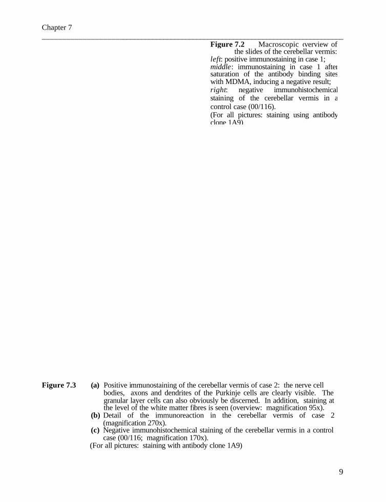
Chapter 7 ________________________________________________________________________________
9
Figure 7.3 (a) Positive immunostaining of the cerebellar vermis of case 2: the nerve cell bodies, axons and dendrites of the Purkinje cells are clearly visible. The granular layer cells can also obviously be discerned. In addition, staining at the level of the white matter fibres is seen (overview: magnification 95x).
(b) Detail of the immunoreaction in the cerebellar vermis of case 2 (magnification 270x).
(c) Negative immunohistochemical staining of the cerebellar vermis in a control case (00/116; magnification 170x).
(For all pictures: staining with antibody clone 1A9)
Figure 7.2 Macroscopic overview of the slides of the cerebellar vermis: left: positive immunostaining in case 1; middle: immunostaining in case 1 after saturation of the antibody binding sites with MDMA, inducing a negative result; right: negative immunohistochemical staining of the cerebellar vermis in a control case (00/116). (For all pictures: staining using antibody clone 1A9)

Chapter 7 ________________________________________________________________________________
10
Figure 7.4 Staining of neurons in the hippocampus of case 2 (antibody clone 1A9).
(a): overview (magnification 25x); (b): detail of the cortical neurons (magnification 100x); (c): detail of the cells of the dentate gyrus (magnification 100x).
The immunodetection of catalase and alanin/glyoxylate aminotransferase in the case
and control liver was not affected by addition of a similar amount of MDMA used to saturate the MDMA-antibody.
Figure 7.5 shows the results in the pituitary gland. Distinct immunoreactivity was observed in many cells, together with a prominent heterogeneity. As a result, different types of staining intensity can be discerned: highly intensely staining cells and abundant positive cells on the one hand. On the other hand, some weakly stained and a few negative cells can be noticed. Comparison of these results with the PAS-orange G-staining in adjacent sections indicates that the strongly stained cells correspond to the acidophils. The weakly stained and negative cells are assumed to be the basophils and chromophobes, respectively. The cells of the latter subtype are indeed a minority.
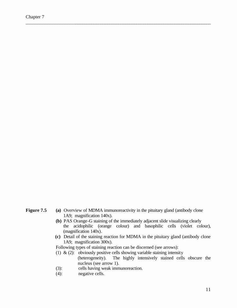
Chapter 7 ________________________________________________________________________________
11
Figure 7.5 (a) Overview of MDMA immunoreactivity in the pituitary gland (antibody clone 1A9; magnification 140x).
(b) PAS Orange-G staining of the immediately adjacent slide visualizing clearly the acidophilic (orange colour) and basophilic cells (violet colour), (magnification 140x).
(c) Detail of the staining reaction for MDMA in the pituitary gland (antibody clone 1A9; magnification 300x).
Following types of staining reaction can be discerned (see arrows): (1) & (2): obviously positive cells showing variable staining intensity
(heterogeneity). The highly intensively stained cells obscure the nucleus (see arrow 1).
(3): cells having weak immunoreaction. (4): negative cells.

Chapter 7 ________________________________________________________________________________
12
IV.2 Toxicological data In Table 7.2, the MDMA, MDA, PMA and amphetamine concentrations in femoral
blood and homogenates of the cortex and white matter of the four brain lobes are presented. The analysis revealed also an important level of the drugs in the white matter.
In the first fatality, the routine analysis of blood and urine disclosed the presence of a high level of amphetamines (68.4 µg/ml in urine, immunoassay result) and trace amounts of benzoylecgonine (0.7 mg/ml in urine, chromatographic result). Head-space GC analysis demonstrated the absence of ethanol in blood and urine. In addition, the presence of MDMA in blood was demonstrated by HPLC-DAD and GC-MS.
For the second case - apart from the data presented in Table 7.2 - no alcohol, neither other drugs were found. Table 7.2 Toxicological data in femoral blood as well as cortex and medulla of the four brain
lobes in the two fatalities.
case 1 (00/112) case 2 (01/34) MDMA MDA MDMA MDA PMA AMP
femoral blood (µg/ml) 3.07 0.09 1.22 0.39 1.43 0.22 brain region (µg/g) frontal lobe - cortex 13.65 0.15 1.66 0.57 3.89 0.33 - white matter 13.05 0.13 1.89 0.68 3.52 0.49 temporal lobe - cortex 15.42 0.16 1.62 0.81 3.25 0.43 - white matter 13.53 0.21 3.00 0.81 4.40 0.44 parietal lobe - cortex 15.19 0.23 2.61 0.57 3.92 0.33 - white matter 12.46 0.15 3.05 0.62 2.94 0.43 occipital lobe - cortex 15.98 0.18 1.87 0.52 3.23 0.33 - white matter 10.87 0.19 3.30 0.57 4.74 0.32 brainstem 13.18 0.22 1.95 0.76 3.20 0.35 cerebellum 11.69 0.23 0.98 0.49 2.37 0.66
MDMA: 3,4-methylenedioxymethamphetamine MDA: 3,4-methylenedioxyamphetamine PMA: para-methoxyamphetamine AMP: amphetamine
Chapter 7 ________________________________________________________________________________
13
V Discussion In this study, a method for immunohistochemical detection of MDMA and MDA in
human tissues is presented. In addition, we examined the distribution of MDMA and MDA in various brain regions and the pituitary gland.
Positive immunostaining for MDMA and MDA was established in the neuron cell bodies, axons and dendrites in various regions of the brain in two fatal intoxications. However, the immunoreactivity was also present at the level of the white matter fibres. The toxicological data confirmed that substantial concentrations of MDMA are present in the white matter. This is in accordance with toxicological data obtained in methamphetamine users (30). The discrepancy between the substantial concentrations in the white and the grey matter, as detected by GC-MS and LC-MS, and the relatively weak immunoreactivity of the white matter in the brain lobes, may be explained by the fact that in the latter approach MDMA can only be detected if it is bound to the tissue and that this complex is not affected by the procedure of fixation, paraffin embedding and sectioning. We presume that unbound MDMA is rinsed off during the immunohistochemical preparation procedure. In the tissue homogenates, used for the GC-MS and LC-MS analysis, both the bound MDMA and the soluble form remain available for detection. For methamphetamine as well, positive immunoreactivity in cerebral cortex and in the white matter was demonstrated (24). In our study, topographic differences in staining intensity were observed. In particular, distinct immunoreactivity was found in the neurons of all cortical brain regions, the basal ganglia and the cerebellum. However, the neuron cell bodies in the brainstem were relatively weakly stained and the white matter fibres were more pronounced at that site. In addition, immunoreactivity was also found in the cells of the dentate gyrus in the hippocampus and the granular layer cells of the cerebellar vermis. This was not observed in the immunohistochemistry of morphine and methadone (13, 14).
The observed results in our two fatalities were well consistent with the topographic data obtained in rats after injection of [3H]-MDMA and [3H]-MDA (18). We were able to provide further information about the regional distribution of MDMA and MDA in human brain tissue. In addition, the method may be used to further investigate the biological effects, for example the interaction of MDMA and MDA on serotonin systems - which was previously demonstrated (18, 31) – , to study the influence on memory (32) etc. Immunostaining of the pituitary gland revealed a variable reaction intensity in the different cells (acidophils, basophils and chromophobes). The most intensely stained cells are probably the acidophils as these are metabolically the most active cells. This is in accordance with the MDMA induced neuroendocrine effects such as increases in plasma cortisol and prolactin levels which are documented in humans (33). We will undertake studies to correlate the heterogeneity of the MDMA reactivity with the hormone producing cell types.
Although the mechanism of death in our two fatalities was different – cardio-pulmonary failure related to the sympaticomimetic effects of MDMA, and DIC due to hyperthermia, respectively – and though there were obvious differences in toxicological data, the immunoreactivity pattern in the brain regions of both cases was fairly comparable. Despite the hyperthermia and the post-mortem interval of about three days in the second
Chapter 7 ________________________________________________________________________________
14
case – leading to a more pronounced tissue autolysis – the drugs could be demonstrated immunohistochemically. However, it should be well kept in mind that interfering factors such as putrefaction may give rise to false negative results. On the contrary, it cannot be excluded that – theoretically – the putrefaction process might lead to artefactual epitopes which are recognized immunologically and thus could induce a false positive reaction. However, in the control cases (with post-mortem interval up to 2 days) no reaction was observed, except for a background level (see below).
In the cases and controls no immunoreactivity was found after incubation with IgG1 fraction from normal serum. In the controls a very faint diffuse staining was found in a few neurons after incubation with the anti-MDMA antibodies. Given the fact that saturation of the antibody with pure MDMA abolished staining in the cases and that in the controls no amphetamines could be detected by chromatographic analysis, the very weak diffuse reaction in some neurons of the controls must be considered as the background level. A similar phenomenon of diffuse staining in control neurons was experienced by Wehner et al. (13, 14) in their study on immunodetection of methadone and morphine in human brain. As emphasized by these authors, it must be kept in mind that the CSA detection method yields an extreme amplification of the primary signal (antigen-antibody complex) and that also the slightest background becomes enhanced as a result of the sensitivity of the method.
Since saturation of the antibody with either MDMA or MDA reduced staining to background level, the specificity of the antibodies and the method was confirmed. According to the information obtained by the supplier, the antibodies are highly specific for ecstasy (MDMA) and related compounds (MDA and MDEA) while cross-reaction with amphetamine, methamphetamine and some medications (phentermine, phenylpropanolamine, pseudoephedrine) is always below 1 % (34).
In summary, a reliable method for the specific immunohistochemical detection of MDMA and MDA in human brain tissues is presented. This method can be used as evidence of intake or even poisoning with MDA, MDMA and/or MDEA and can be a reliable alternative when the usual samples (mainly blood and urine) are not available for toxicological analysis (for example due to fulminant blood loss in severely destructed bodies, such as polytrauma in train accident fatalities).
Further studies will be undertaken in order to investigate whether this technique might be influenced by post-mortem processes. In addition, MDMA immunodetection might be used as a basis for further study of the distribution of these amphetamine analogues in the human body, and thus be useful in the understanding of their biological effects.
Acknowledgments The authors wish to thank Microgenics Corp, Fremont, CA, USA and Mr R Ramage, PhD, Sr. Scientist in particular, for their advice and support and for gently providing the appropriate antibodies. We also thank Mrs Thérèse De Vuyst for her assistance in preparing the manuscript and Mr G. Van Limbergen for technical assistance.

Chapter 7 ________________________________________________________________________________
15
References 1. De Letter EA, Coopman VAE, Cordonnier JACM, Piette MHA. One fatal and seven non-fatal
cases of 4-methylthioamphetamine (4-MTA) intoxication : clinico-pathological findings. Int J Legal Med 2001;114:352-356.
2. Dowling GP, McDonough III ET, Bost RO. ‘Eve’ and ‘Ecstasy’. A report of five deaths
associated with the use of MDEA and MDMA. JAMA 1987;257:1615-1617. 3. Rohrig TP, Prouty RW. Tissue distribution of methylenedioxymethamphetamine. J Anal
Toxicol 1992;16:52-53. 4. Fineschi V, Masti A. Fatal poisoning by MDMA (ecstasy) and MDEA: a case report. Int J
Legal Med 1996;108:272-275. 5. De Letter EA, Clauwaert KM, Lambert WE, Van Bocxlaer JF, De Leenheer AP, Piette MHA.
Distribution study of 3,4-methylenedioxymethamphetamine (MDMA, “ecstasy”) and 3,4-methylenedioxyamphetamine (MDA) in a fatal overdose. J Anal Toxicol 2002;26:113-118.
6. De Caestecker T, De Letter E, Clauwaert K, Bouche MP, Lambert W, Van Bocxlaer J, Van den
Eeckhout E, Van Peteghem C, De Leenheer A. Fatal 4-MTA intoxication : development of a liquid chromatographic – tandem mass spectrometric assay for multiple matrices. J Anal Toxicol 2001;25:705-710.
7. Moriya F, Hashimoto Y. Redistribution of basic drugs into cardiac blood from surrounding
tissues during early-stages postmortem. J Forensic Sci 1999;44:10-16. 8. Hilberg T, Ripel Å, Slørdal L, Bjørneboe A, Mørland J. The extent of postmortem drug
redistribution in a rat model. J Forensic Sci 1999;44:956-962. 9. De Letter EA, De Paepe P, Clauwaert KM, Belpaire FM, Lambert WE, Van Bocxlaer JF, Piette
MHA. Is vitreous humour useful for the interpretation of 3,4-methylenedioxymethamphetamine (MDMA) blood levels? Experimental approach with rabbits. Int J Legal Med 2000;114:29-35.
10. Balkon J, Bidanset JH, Lynch VD. Immunofluorescence detection of drugs in postmortem
tissues: a new technique with potential for assessment of drug influence in cause of death. J Forensic Sci 1980;25:88-94.
11. Morley M, Gee DJ. An assessment of the immunofluorescence technique as a method for
demonstrating the histological localization of tetrahydrocannabinol in mammalian tissues. J Forensic Sci 1982;27:837-843.
12. Ishiyama I, Mukaida M, Tanabe R, Kaiho M, Ueyama M. Histochemical demonstration of
Phenobarbital by immunocytochemistry. J Forensic Sci 1987;32:1221-1234.

Chapter 7 ________________________________________________________________________________
16
13. Wehner F, Wehner H-D, Subke J, Meyermann R, Fritz P. Demonstration of morphine in
ganglion cells of the hippocampus from vic tims of heroin overdose by means of anti-morphine antiserum. Int J Legal Med 2000;113:117-120.
14. Wehner F, Wehner H-D, Schieffer MC, Subke J. Immunohistochemical detection of methadone
in the human brain. Forensic Sci Int 2000;112:11-16. 15. Hood I, Mirchandani H, Monforte J, Stacer W. Immunohistochemical demonstration of
homicidal insulin injection site. Arch Pathol Lab Med 1986;110:973-974. 16. Lutz R, Pedal I, Wetzel C, Mattern R. Insulin injection sites: morphology and
immunohistochemistry. Forensic Sci Int 1997;90:93-101. 17. Wehner F, Mittmeyer H-J, Wehner H-D, Schieffer MC. Insulin- or morphine-injection ?
Immunohistochemical contribution to the elucidation of a case. Forensic Sci Int 1998;95:241-246.
18. Battaglia G, Zaczek R, de Souza EB. MDMA effects in brain: pharmacologic profile and
evidence of neurotoxicity from neurochemical and autoradiographic studies. In: Peroutka SJ (ed) (1990) Ecstasy: the clinical, pharmacological and neurotoxicological effects of the drug MDMA. Kluwer Academic Publishers, Boston, Dordrecht, London, pp 171–199.
19. Feldman RS, Meyer JS, Quenzer LF. (eds) (1997) Principles of neuropsychopharmacology.
Chapter 9: Serotonin . Sinauer Associates, Inc, Sunderland, Massachusetts, pp 358-359. 20. Battaglia G, Sharkey J, Kuhar MJ, de Souza EB (1991) Neuroanatomic specificity and time
course of alterations in rat brain serotonergic pathways induced by MDMA (3,4-methylenedioxymethamphetamine): assessment using quantitative autoradiography. Synapse 8:249-260.
21. McCann UD, Mertl M, Eligulashvili V, Ricaurte GA. Cognitive performance in (±)3,4-
methylenedioxymethamphetamine (MDMA, “Ecstasy”) users: a controlled study. Psychopharmacology 1999;143:417-425.
22. Fischer C, Hatzidimitriou G, Wlos J, Katz J, Ricaurte G. Reorganization of ascending 5-HT
axon projections in animals previously exposed to the recreational drug (±)3,4-methylenedioxymethamphetamine (MDMA, “Ecstasy”). J Neurosci 1995;15:5476-5485.
23. Fineschi V, Centini F, Mazzeo E, Turillazzi E. Adam (MDMA) and Eve (MDEA) misuse: an
immunohistochemical study on three fatal cases. Forensic Sci Int 1999;104:65-74. 24. Ishiyama I, Mukaida M, Yoshii T, Suyama H. Histochemical demonstration of
methamphetamine by immunocytochemistry. J Forensic Sci 1987;32:658-672.

Chapter 7 ________________________________________________________________________________
17
25. Clauwaert KM, Van Bocxlaer JF, De Letter EA, Van Calenbergh S, Lambert WE, De Leenheer
AP. Determination of the Designer Drugs 3,4-Methylenedioxymethamphetamine, 3,4-Methylenedioxyethylamphetamine, and 3,4-Methylenedioxyamphetamine with HPLC and Fluorescence Detection in Whole Blood, Serum, Vitreous Humor, and Urine. Clin Chem 2000;46:1968-1977.
26. Clauwaert KM, Van Bocxlaer JF, Lambert WE, De Leenheer AP. Analysis of cocaine,
benzoylecgonine, and cocaethylene in urine by HPLC with diode array detection. Anal Chem 1996;68:3021-3028.
27. Clauwaert KM, Van Bocxlaer JF, Lambert WE, De Leenheer AP. Liquid chromatographic
determination of cocaine, benzoylecgonine, and cocaethylene in whole blood and serum samples with diode-array detection. J Chromatogr Sci 1997;35:321-328.
28. Dako® Catalysed Signal Amplification (CSA) System, Peroxidase for mouse primary
antibodies - User instructions. 29. Espeel M, Van Limbergen G. Immunocytochemical localization of peroxisomal proteins in
human liver and kidney. J Inher Metab Dis 1995;18 (S1):135-154. 30. Kalasinsky KS, Bosy TZ, Schmunk GA, Reiber G, Anthony RM, Furukawa Y, Guttman M,
Kish SJ. Regional distribution of methamphetamine in autopsied brain of chronic human methamphetamine users. Forensic Sci Int 2001;116:163-169.
31. Hatzidimitriou G, McCann UD, Ricaurte GA. Altered serotonin innervation patterns in the
forebrain of monkeys treated with (±)3,4-methylenedioxymethamphetamine seven years previously: factors influencing abnormal recovery. J Neurosci 1999;19:5096-5107.
32. Bolla KI, McCann UD, Ricaurte GA. Memory impairment in abstinent MDMA (“Ecstasy”)
users. Neurology 1998;51:1532-1537. 33. Mas M, Farré M, de la Torre R, Roset PN, Ortuño J, Segura J, Camí J. Cardiovascular and
neuroendocrine effects and pharmacokinetics of 3,4-methylenedioxy-methamphetamine in humans. J Pharmacol Exp Ther 1999;290:136-145.
34. Personal communication Mr R Ramage, PhD, Sr. Scientist from Microgenics Corp, Fremont,
CA, USA.

Summary and conclusions
Summary and conclusions ________________________________________________________________________________
1
Summary and conclusions
The use and abuse of amphetamine derivatives such as 3,4-methylenedioxymeth-amphetamine (MDMA, “ecstasy”, “XTC”) has become an important public issue in view of the growing numbers of fatalities encountered in current forensic practice. Considering the broad range of blood and plasma levels in MDMA-related fatalities reported in the literature, the level of MDMA that can be toxic or even potentially lethal remains unclear. The question as to whether or not the (ab)use of the substance has contributed to the cause of death remains particularly unclear because of post-mortem phenomena such as instability, redistribution and even neo-formation, all of which can interfere with post-mortem blood levels. Post-mortem degradation, on the one hand, can lead to falsely decreased levels, whereas post-mortem redistribution and/or neo-formation can result in erratically high concentrations. Possible mechanisms of post-mortem redistribution include the diffusion of a substance into the cardiac blood out of the lungs and liver, or the stomach content.
In this thesis research, we have studied the post-mortem redistribution of certain
amphetamine derivatives in order to evaluate which fluid and/or tissue sampled after death most closely approximates the ante-mortem blood concentration. Due to the well-isolated position of the eye and the reliability of vitreous humour for post-mortem toxicology (e.g. alcohol (1)), the value of this medium for post-mortem determinations of amphetamine derivatives was investigated. Our work was particularly focused on MDMA and its metabolite 3,4-methylenedioxyamphetamine (MDA), though we also examined cases involving the amphetamine analogues 4-methylthioamphetamine (4-MTA) and para-methoxyamphetamine (PMA), which have recently been prominently featured in the early warning system on synthetic drugs.
Part one consists of a literature survey including a review of the reported
MDMA-related fatalities and a summary of the amphetamine-related casework examined at the Department of Forensic Medicine of Ghent University. In Chapter 1, the relevant literature data are reviewed with emphasis on MDMA and its medico-legal implications. Significant inter-individual in vivo differences in susceptibility to the effects of MDMA were noted. Unfortunately, at present, it is impossible to estimate the individual risk of using MDMA or other amphetamines.
The majority of the fatalities reported in the literature were men younger than the age of 25. The number of “pure” MDMA intoxication were roughly equal to the numbers of poly-amphetamine intoxication or polydrug abuse (including MDMA ingestion). With respect to the manner of death, it was obvious that MDMA-related fatalities were for the major part due to an unintentional overdose, though the use of MDMA in association with other events such as traffic accidents and suicidal acts should not be underestimated. When MDMA is involved, various mechanisms of death have been described with hyperthermia and fatal cardiac or pulmonary complications being the most frequent. In many cases, however, the manner and mechanism of death
Summary and conclusions ________________________________________________________________________________
2
remains undetermined or uncertain. In view of the broad range of MDMA levels reported in the literature (viz. blood levels between 0.04 and 18.50 µg/ml in “pure” overdoses), it is not possible to conclude solely on the basis of the toxicological data whether or not a subject died of MDMA (ab)use. Moreover, in most of the reported cases the blood sampling location was not specified and, as a result, it is not clear whether the observed post-mortem MDMA blood concentration actually represented the concentration at the time of death. However, guidelines indicate that an MDMA blood concentration higher than 1.0 µg/ml can be potentially lethal, whereas levels below approximately 0.6 µg/ml are capable of inducing intoxication (2).
The amphetamine-related fatalities examined at the Department of Forensic Medicine of Ghent University were reviewed and discussed in the light of the literature data (Chapter 2). Because of the low number of “pure” MDMA-related deaths, we have included all amphetamines in this survey. The amphetamine-related fatalities represent only a small fraction of all the medico-legal investigations, though the number obviously started increasing from 1995. We are convinced, however, that there is an underestimation of amphetamine-related fatalities: when somebody dies at home (e.g. found dead in bed, whether or not after a night out dancing), the death is often classified as a “natural death” and consequently no police inquiry or medico-legal investigation is performed. In our cases, 11 subjects were found dead either in bed or in a chair.
The age and sex distributions of the amphetamine-related fatalities examined at the Department were fully in accordance with the data from the literature. A wide range of MDMA concentrations was found in cases of “pure” intoxication (blood levels between 0.27 µg/ml and 13.51 µg/ml), which was in line with those reported in the literature. “Pure” amphetamine intoxications, polydrug overdoses and the combination of amphetamine (ab)use and polytrauma were the most prominent causes of death. As for the manner of death in these fatalities, unintentional overdoses were most frequent, though traffic accidents and suicides associated with amphetamine use also accounted for significant percentages. As for the mechanism of death, acute to subacute cardiopulmonary failure was most frequent, followed by hyperthermia.
On the basis of both surveys in Part One (Chapters 1 and 2), post-mortem
distribution and redistribution were studied in order to evaluate which post-mortem fluid or tissue sample best approximates the actual concentration at the time of death. At first, the results obtained in experiments using rabbits were presented (Part Two). Thereafter, the experimental data were compared with some cases found in current forensic practice (Department of Forensic Medicine, Ghent University) (Part Three).
In the first study presented in Part Two (Chapter 3), the value of post-mortem
vitreous humour levels was investigated for the purpose of avoiding possible thanato-chemical difficulties such as post-mortem redistribution. First, the pharmacokinetics of MDMA in the rabbit after intravenous (iv) administration were considered. A high volume of distribution (5 l/kg), a high systemic clearance (4.1 l/kg per h) and a relatively short half-life (1 h) was found following iv administration of MDMA. A distinct
Summary and conclusions ________________________________________________________________________________
3
relationship between the MDMA concentrations in the vascular compartment and the vitreous humour was substantiated. Equilibration between the vascular compartment and the vitreous humour was attained about one hour after intravenous administration. The ratio of the MDMA concentration in vitreous humour to the MDMA blood level was about 1.1 at 120 and 240 minutes after infusion, which indicates a slight accumulation of MDMA in the vitreous compartment. Moreover, a preliminary thanato-toxicological investigation - in which a post-mortem interval up to 72 h was considered - demonstrated that MDMA concentrations in cardiac blood increased post mortem whereas vitreous humour MDMA levels were more stable and thus presumably more representative of the ante-mortem blood concentration.
In Chapters 4 and 5, the post-mortem stability and redistribution of MDMA in rabbits were further analysed. In Chapter 4, the distribution and redistribution of MDMA and its metabolite MDA were studied in various fluids and tissues after intravenous administration. In a first group (control group, sampling done immediately after killing) considerable MDMA concentrations were found in the brain and both lungs. Our data also pointed to substantial elimination of MDMA by hepatic biotransformation and excretion via the bile in addition to renal excretion. In a second group (post-mortem interval either 24 or 72 h prior to sampling), an increase of MDMA and MDA levels in the liver and the eye globe walls was noted. In the lungs, on the other hand, levels tended to decline as a function of increasing post-mortem interval. MDMA concentrations in cardiac and iliopsoas muscle were fairly comparable, remaining stable up to 72 h after death. As post-mortem increases in cardiac blood levels can be due to vascular diffusion out of blood-rich organs such as the liver and lungs (3), the large vessels around the heart were ligated (immediately after killing) in another group (group 3), and the animals were further handled as in group 2. However, significant differences in blood and tissue MDMA concentrations between the animals of groups 2 and 3 could not be demonstrated. Therefore, in the rabbit, post-mortem redistribution of MDMA at the cellular level (viz. by pure diffusion gradient from higher to lower concentrations) is probably more important than its redistribution via vascular pathways. In addition, MDA levels were relatively low in all samples, indicating that this molecule is not a major metabolite in the rabbit, at least within the first two hours after administration. Furthermore, the value of vitreous humour as a reliable post-mortem specimen was confirmed. The distribution and redistribution of MDMA and MDA in rabbit tissues were in line with the data obtained after administration of amphetamine in the rat (4,5).
Since drug levels can be affected by gastric diffusion when the stomach contains a substantial amount of the drug or by diffusion from the trachea when agonal aspiration or post-mortem regurgitation of vomit occurs, these phenomena were simulated in another rabbit model (Chapter 5). After post-mortem tracheal infusion, MDMA can easily diffuse not only into the lungs but also in large quantities into the cardiac blood and – to a lesser extent – into the cardiac muscle. MDMA was also found in the closely adjacent diaphragm and in the upper abdominal organs, including the liver and the stomach. Following post-mortem infusion into the stomach, considerable MDMA levels
Summary and conclusions ________________________________________________________________________________
4
were found in cardiac blood and muscle, both lungs, diaphragm and liver tissue when the solution was concentrated nearby the lower oesophageal sphincter. However, when the MDMA solution was present deeper in the stomach, MDMA levels were high in the spleen and liver and relatively low in cardiac blood and muscle. These results indicate that the diffusion of MDMA out of the stomach content or due to aspirated vomit and particularly gastro-oesophageal reflux, can lead to considerable post-mortem redistribution. In both experiments, MDA levels were low or below the limit of quantitation in most tissues, but were substantial in cardiac blood and muscle, lung and diaphragm, thus indicating that MDMA can be converted into MDA even after death.
In Part Three, the post-mortem distribution of MDMA (and its metabolite MDA)
and some other amphetamine derivatives was studied in the human body in order to evaluate which fluid and/or tissue sampled after death best approximates the ante-mortem concentration at the time of death. These findings were correlated with the experimental data on post-mortem redistribution in rabbits. In the human fatalities, the post-mortem phenomena were investigated using two different - but complementary – approaches, namely the thanato-toxicological and the immunohistochemical.
In the study discussed in Chapter 6, concentrations in various fluids (blood sampled at different locations, vitreous humour, urine and bile) and tissues such as cardiac muscle, lungs, liver, kidneys, spleen, ilio-psoas muscle, and brain were determined in overdose victims. Apart from MDMA and MDA, some other amphetamine derivatives, namely 4-methylthioamphetamine (4-MTA) and para-methoxyamphetamine (PMA), were examined as well. For the relatively new derivative, 4-MTA, the data of persons who survived after ingestion are also presented, and clinico-pathological findings are discussed. In the surviving subjects, an obvious inconsistency between the 4-MTA levels detected and the described clinical symptoms was noted, substantiating that differences in individual responses to 4-MTA must be taken into consideration. In the amphetamine-related fatalities, very high concentrations were found in cardiac blood and tissues located centrally in the body (lungs and liver in particular). This confirms that post-mortem redistribution due to diffusion from higher to lower concentration can easily take place, mainly at longer post-mortem intervals and when putrefaction occurs. These findings corroborated the animal experimental data in which post-mortem redistribution of MDMA into cardiac blood was substantiated (6,7). Our data confirmed that for post-mortem quantitation of amphetamine and derivatives, peripheral blood sampling remains compulsory. When such samples are not available (due to severe loss of blood, such as in polytrauma), iliopsoas-muscle and vitreous humour could be valuable alternatives. However, when advanced putrefaction has taken place, vitreous humour is often no longer available due to dehydration, and iliopsoas muscle levels should be interpreted with caution (see case 01/158: the iliopsoas muscle value was obviously higher than the femoral blood level). In contrast to the rabbits experiments, there are arguments for a direct transvascular redistribution, for example, from the lungs to the cardiac chambers. Moreover, due to the post-mortem processes,

Summary and conclusions ________________________________________________________________________________
5
the toxicological and autopsy findings should be considered as a whole in drawing the medico-legal conclusions.
In Chapter 7, an immunohistochemical method for the detection of MDMA and MDA in human post-mortem brain tissues and the pituitary gland is presented and correlated with the toxicological findings. The detection method comprises an elaborate amplification of the original signal (antigen-antibody recognition; Catalyzed Signal Amplification; CSA (8)). The method has already been applied for the detection of morphine and methadone in human fatalities (9,10). A distinct positive reaction was observed in all cortical brain regions and the neurons of the basal ganglia, the hypothalamus, the hippocampus, the cerebellar vermis, and the pituitary gland. In the brainstem, relatively weak staining of neurons was seen. These findings were in line with the toxicological data. This immunohistochemical method can be used as evidence of intake of or even poisoning with MDA, MDMA and/or MDEA, and it can serve as an alternative method when the usual samples (mainly blood and urine) are not available for toxicological analysis (for example in polytrauma). However, with the currently available antibodies, it is not possible to distinguish the closely related amphetamine derivatives (MDA, MDMA and MDEA) from one another. In addition, immunodetection could possibly be used as a basis for further study of the distribution of these amphetamine analogues in the human body, and may contribute to the understanding of their neuro-biological effects. However, the limitation of the immunohistochemical approach is that in the brain sections, only the fraction bound to the tissue can be demonstrated. This constitutes a fundamental difference between this approach and the toxicological quantitation in tissue homogenates, in which both the bound and the unbound fractions can be assessed.
In conclusion, the experimental data in rabbits, which were corroborated by the results in some human cases, have enabled us to demonstrate that post-mortem redistribution of the amphetamine derivatives – MDMA and MDA, in particular, but also 4-MTA and PMA – should be taken into account in the toxicological assessment of the cause of death, especially with longer post-mortem intervals. Peripheral blood sampling remains the golden standard. However, when this is not possible due, for example, to extreme loss of blood in polytrauma or putrefaction, iliopsoas muscle and vitreous humour can be useful in arriving at a valid conclusion. Immunohistochemical detection in brain tissues can serve as an additional tool in the forensic inquiry as well. Finally, as there is still considerable debate as to what MDMA level can be toxic or even potentially lethal, it is strongly advisable to interpret the anatomo-pathological findings and the toxicological results together in arriving at a conclusion. This guideline is important in view of the different possible mechanisms of death which implicate quite different survival times following amphetamine intake (e.g. cardiopulmonary complications, hyperthermia).

Summary and conclusions ________________________________________________________________________________
6
References 1. Chao TC, Lo DST. Relationship between postmortem blood and vitreous humor ethanol
levels. Am J Forensic Med Pathol 1993;14:303-308. 2. Dowling GP. Human deaths and toxic reactions attributed to MDMA and MDEA. In:
Peroutka SJ (ed) (1990) Ecstasy: the clinical, pharmacological and neurotoxicological effects of the drug MDMA. Kluwer Academic Publishers, Boston, Dordrecht, London, pp 73.
3. Moriya F, Hashimoto Y. Redistribution of basic drugs into cardiac blood from surrounding
tissues during early-stages postmortem. J Forensic Sci 1999;44:10-16. 4. Hilberg T, Ripel Å, Slørdal L, Bjørneboe A, Mørland J. The extent of postmortem drug
redistribution in a rat model. J Forensic Sci 1999;44:956-962. 5. Battaglia G, Zaczek R, De Souza EB. MDMA effects in brain: pharmacological profile and
evidence of neurotoxicity from neurochemical and autoradiographic studies. In: Peroutka SJ. (ed) (1990) Ecstasy: the clinical, pharmacological and neurotoxicological effects of the drug MDMA. Kluwer Academic Publishers, Boston, Dordrecht, London, pp 171-199.
6. De Letter EA, Clauwaert KM, Belpaire FM, Lambert WE, Van Bocxlaer JF, Piette MHA. Post-
mortem redistribution of 3,4-methylenedioxymethamphetamine (MDMA, “ecstasy”) in the rabbit. Part one: Experimental approach after intravenous infusion. Int J Legal Med 2002;116:216-224.
7. De Letter EA, Belpaire FM, Clauwaert KM, Lambert WE, Van Bocxlaer JF, Piette MHA. Post-
mortem redistribution of 3,4-methylenedioxymethamphetamine (MDMA, “ecstasy”) in the rabbit. Part two: Post-mortem infusion in trachea or stomach. Int J Legal Med 2002;116:225-232.
8. Bobrow MN, Harriss TD, Shaughnessy KJ, Litt GJ. Catalyzed reporter depositions, a novel
method of signal amplification. Application to immunoassays. J Immunol Methods 1989;125:279-285.
9. Wehner F, Wehner H-D, Subke J, Meyermann R, Fritz P. Demonstration of morphine in
ganglion cells of the hippocampus from victims of heroin overdose by means of anti-morphine antiserum. Int J Legal Med 2000;113:117-120.
10. Wehner F, Wehner H-D, Schieffer MC, Subke J. Immunohistochemical detection of
methadone in the human brain. Forensic Sci Int 2000;112:11-16.

Résumé et conclusions

Résumé et conclusions ________________________________________________________________________________
1
Résumé et conclusions La consommation et l’abus d’amphétamine et analogues comme la 3,4-méthylène-dioxyméthamphétamine (MDMA, « Ecstasy », « XTC ») constituent un important problème social et des cas fatals sont assez fréquents en pratique médico-légale courante. En se référant aux importantes différences entre les concentrations sanguines ou plasmatiques détectées dans les cas fatals décrits dans la littérature, il est difficile de se faire une idée précise de la concentration toxique ou même potentiellement fatale de la MDMA. Reste à savoir si l’usage ou l’abus d’un produit a contribué à la mort vu les phénomènes post mortem comme l’instabilité d’un produit, la redistribution et même la néoformation qui peuvent tous interférer avec la concentration sanguine post mortem. En cas de dégradation post mortem, des taux faussement diminués sont retrouvés, tandis que la redistribution et/ou néoformation post mortem peuvent donner lieu à des taux exceptionnellement élevés. La diffusion d’une substance dans le sang cardiaque à partir des poumons ou du foie, ou du contenu gastrique, est un mécanisme possible de redistribution post mortem. Dans cette thèse de doctorat, la redistribution post mortem de quelques dérivés d’amphétamines a été étudiée afin de savoir quel liquide et/ou tissu corporel cadavérique est le plus approprié pour évaluer la concentration sanguine ante mortem. En se référant à la position bien isolée de l’œil et à la valeur de l’humeur vitrée pour la toxicologie post mortem (p.ex. pour la détermination d’alcool (1)), la fiabilité de ce médium pour la détection d’amphétamines post mortem a été examinée. Dans cet ouvrage, la MDMA et son métabolite 3,4-méthylènedioxyamphétamine (MDA) ont été particulièrement étudiés, mais également les dérivés d’amphétamine, la 4-méthylthioamphétamine (4-MTA) et la para-méthoxyamphétamine (PMA) – qui ont reçu récemment pas mal d’attention. La première partie comprend un aperçu de la littérature y compris un résumé des cas mortels publiés liés à la MDMA et un bilan des fatalités relatives à l’usage d’amphétamines examinées au Département de Médecine Légale de l’Université de Gand. Chapitre 1 décrit les données de la littérature pertinentes concernant la MDMA et ses implications médico-légales. In vivo, on aperçoit une importante vulnérabilité inter-individuelle aux effets de la MDMA. En ce moment, il est impossible d’estimer le risque individuel et, par conséquent, de prévoir les effets de la consommation de la MDMA ou d’autres amphétamines. La majorité des cas mortels qui ont été décrit dans la littérature concerne des hommes de moins de 25 ans. Le nombre d’intoxications « pures et simples » relatives à la MDMA égale à peu près le nombre d’ingestion de la MDMA faisant partie d’une intoxication complexe liée à d’autres amphétamines, drogues ou médicaments. En ce qui concerne les circonstances médico-légales du décès, il est bien clair que la plupart des décès étaient dus à une dose excessive involontaire, mais l’usage de la MDMA associé à d’autres évènements comme p.ex. les accidents de la route et le comportement suicidaire ne
Résumé et conclusions ________________________________________________________________________________
2
peut pas être sous-estimé. Lorsque la MDMA est impliquée dans la mort, différents mécanismes de décès sont décrits: l’hyperthermie et les complications cardiales ou pulmonaires sont les plus fréquentes. Toutefois, dans beaucoup de cas, les circonstances ou le mécanisme de la mort sont indéterminés ou indécis. Se référant à l’intervalle large des concentrations létales de la MDMA retrouvé dans la littérature (plus particulièrement, des concentrations de 0.04 à 18.5 µg/ml considérant des intoxications « pures »), il n’est pas évident de répondre correctement à la question si – uniquement à base d’un résultat toxicologique – une personne est morte à cause de la consommation ou d’abus de la MDMA. En outre, dans la majorité des cas, l’endroit où le sang a été prélevé n’est pas spécifié et – par conséquent – il n’est pas clair si le taux de la MDMA déterminé dans le sang post mortem représente effectivement la concentration réelle au moment du décès. Néanmoins, une règle empirique indique qu’une concentration de la MDMA excédant 1 µg/ml serait potentiellement mortelle, tandis que des taux équivalents ou inférieurs à 0.6 µg/ml pourraient être toxiques (2).
Dans le deuxième Chapitre – à la lumière de ces données de la littérature – tous les cas mortels liés aux amphétamines rencontrés au Département de Médecine Légale de l’Université de Gand sont révisés. Dû au nombre relativement petit des intoxications mortelles « pures » à la MDMA, tous les décès liés à l’usage des amphétamines sont rapportés. Le nombre de fatalités dans lesquelles les amphétamines sont concernées n’est qu’une fraction minime de toutes les expertises médico-légales, mais ce nombre augmente sensiblement depuis 1995. Néanmoins, nous sommes convaincus qu’il doit y avoir une sous-estimation du nombre de décès liés à la consommation des amphétamines: lorsque quelqu’un est retrouvé mort à la maison (p.ex. dans son lit, à la suite d’ une sortie), le décès est souvent classé comme « naturel » et par conséquent, il n’y a pas d’investigation policière ni d’examen médico-légal. Des cas étudiés, 11 victimes ont été retrouvées décédées au lit ou dans un fauteuil.
La distribution de l’âge et du sexe retrouvée chez la majorité des victimes liées aux amphétamines sont plutôt en concordance avec les données de la littérature. Un grand éventail de concentrations de la MDMA chez les intoxications « pures » a été constaté (concentrations entre 0.27 et 13.51 µg/ml) et ceci est également conforme aux cas décrits dans la littérature. Des intoxications « pures » aux amphétamines, des intoxications combinées et la consommation d’amphétamines combinée à des polytraumatismes étaient les causes de la mort les plus fréquentes. En ce qui concerne les circonstances du décès constatées dans notre échantillon d’étude, il apparaît que les intoxications non-intentionnelles étaient les plus fréquentes. L’usage des amphétamines en rapport avec les accidents de trafic et le suicide constituent aussi un groupe important. Quant au mécanisme du décès, on note en premier lieu une insuffisance cardio-pulmonaire aiguë ou subaiguë suivie par l’hyperthermie.

Résumé et conclusions ________________________________________________________________________________
3
A base des aperçus rapportés dans la première partie (Chapitre 1 et 2), la distribution et redistribution post mortem ont été étudiées afin d’examiner quel liquide ou tissu prélevé après la mort représente au mieux la concentration réelle au moment du décès. D’abord, les résultats obtenus à l’aide d’un modèle expérimental animal (utilisant des lapins) sont présentés (deuxième partie). Ensuite, on a examiné si les données expérimentales pourraient être confirmées par l’étude des cas en pratique médico-légale courante (Département de Médecine Légale de l’Université de Gand) (troisième partie).
Dans la première étude de la deuxième partie, la valeur de l’humeur vitrée prélevée
après la mort a été étudiée afin d’éviter les problèmes thanato-chimiques comme la redistribution post mortem. D’abord, la pharmacocinétique de la MDMA chez le lapin après injection intraveineuse (iv) a été examinée. Un volume de distribution assez grand (5 l/kg), une clairance totale importante (4.1 l/kg par heure) et une demi-vie relativement courte (1 heure) étaient remarqués chez le lapin après injection iv de la MDMA. On a pu démontrer une corrélation nette entre la concentration de la MDMA dans le compartiment vasculaire et dans l’humeur vitrée. Un équilibre entre le compartiment vasculaire et l’humeur vitrée est atteint environ une heure après injection iv. Le rapport de la concentration de la MDMA dans le vitré à la concentration sanguine de la MDMA, 120 minutes ou 240 minutes après administration iv, s’approchait de 1.1; ce qui porte à croire qu’il y a une accumulation légère de la MDMA dans l’humeur vitrée. En plus, notre étude thanato-toxicologique préliminaire – en considérant un délai post mortem jusqu’à 72 heures – a prouvé que les concentrations de la MDMA dans le sang cardiaque augmentaient après la mort, tandis que les concentrations de la MDMA dans l’humeur vitrée étaient plus stables et donc mieux représentatives de la concentration sanguine ante mortem.
Dans le quatrième et cinquième Chapitre, la stabilité et la redistribution de la MDMA après la mort ont été analysées plus profondément chez le lapin. Dans le quatrième Chapitre, la distribution et la redistribution de la MDMA et son métabolite MDA ont été étudiées dans divers liquides et tissus du corps après administration iv. Dans un premier groupe (groupe de contrôle, prélèvement immédiatement après la mort), de considérables concentrations de la MDMA étaient démontrées dans le cerveau et les poumons. Les données toxicologiques indiquent donc une élimination importante de la MDMA par biotransformation hépatique et excrétion biliaire associée à élimination rénale. Dans un deuxième groupe d’animaux (conservé pendant soit 24, soit 72 heures avant de prendre des prélèvements), une élévation des concentrations de la MDMA et de la MDA dans le foie et dans la paroi oculaire a été remarquée. D’autre part, les concentrations pulmonaires avaient tendance à diminuer en fonction d’un délai post mortem avancé. Les concentrations de la MDMA dans le muscle cardiaque et l’iliopsoas étaient bien comparables et restaient stables jusqu’à 72 heures après le décès. Puisque les élévations des concentrations post mortem dans le sang cardiaque pourraient être le résultat d’une diffusion vasculaire à partir des organes fort vascularisés comme le foie et les poumons (3), les grands vaisseaux affluant au cœur ont été ligaturés (immédiatement après la mort); ces animaux ont été traités comme ceux du deuxième groupe. On n’a pas pu démontrer des différences significatives entre les groupes 2 et 3. C’est pourquoi, chez le lapin, la
Résumé et conclusions ________________________________________________________________________________
4
redistribution post mortem de la MDMA au niveau cellulaire est probablement plus importante que la redistribution de cette substance par voie vasculaire. Par ailleurs, les concentrations de la MDA étaient relativement basses dans tous les échantillons, ce qui signifie que cette molécule n’est pas un métabolite important pour le lapin, tout au moins dans les 2 heures après administration. En plus, la valeur de l’humeur vitrée comme spécimen stable après la mort, a été confirmée. La distribution et la redistribution de la MDMA et de la MDA dans les tissus des lapins étaient en concordance avec les données établies après l’administration de l’amphétamine chez le rat (4,5).
Puisque les concentrations de divers produits tels que les drogues peuvent être influencées par la diffusion à partir de l’estomac – quand il s’y trouve encore une quantité importante au moment du décès – ou par la diffusion à partir de la trachée lors d’ une aspiration agonale ou d’une régurgitation post mortem du contenu gastrique, ces phénomènes ont été simulés à l’aide d’un autre modèle expérimental animal de laboratoire (Chapitre 5). L’infusion post mortem de la MDMA dans la trachée du lapin a donné lieu à une diffusion aisée de la MDMA non seulement dans les poumons, mais aussi dans le sang cardiaque d’une façon considérable et à un degré moindre dans le muscle cardiaque. La MDMA a été également retrouvée dans le diaphragme adjacent et les organes abdominaux supérieurs comme le foie et l’estomac. Après infusion post mortem de la MDMA dans l’estomac, des concentrations de la MDMA importantes étaient découvertes dans le sang cardiaque, le muscle cardiaque, les poumons, le diaphragme et le foie – ceci quand la solution de la MDMA était concentrée au niveau du sphincter cardial. Si la solution de la MDMA était concentrée dans le corps de l’estomac des concentrations considérables étaient retrouvées dans la rate et le foie, et des concentrations relativement basses dans le sang cardiaque et le muscle cardiaque. Ces résultats indiquent que la diffusion de la MDMA à partir du contenu de l’estomac ou de l’aspiration de vomissure, et plus particulièrement du reflux gastro-oesophagale, peut contribuer à une redistribution post mortem importante. Dans ces deux expériences, les concentrations dans la majorité des tissus de la MDA étaient relativement modestes ou inférieures à la limite de quantification, sauf dans le sang cardiaque, le muscle cardiaque, les poumons et le diaphragme ce qui montre que après la mort, la MDMA peut encore être transformée en MDA.
Dans la troisième partie, la distribution post mortem de la MDMA (et son
métabolite la MDA) ainsi de quelques autres dérivés de l’amphétamine a été étudiée dans le corps humain afin d’examiner quel liquide et/ou tissu prélevé après la mort est le plus approprié pour évaluer la concentration ante mortem au moment de la mort. Ces constatations ont été corrélées aux données expérimentales de la redistribution post mortem chez le lapin. En cas de fatalités humaines, les phénomènes post mortem ont été examinés au moyen de deux approches différentes – mais toutefois complémentaires, c’est-à-dire, du point de vue thanato-toxicologique et immunohistochimique.
Dans le sixième Chapitre, les concentrations de la MDMA et MDA dans des divers liquides corporels (le sang prélevé à divers endroits, l’humeur vitrée, l’urine et la bile) et dans les tissus comme le muscle cardiaque, les poumons, le foie, les reins, la rate, le muscle iliopsoas, le cerveau étaient déterminées dans les victimes d’une dose excessive. Quelques
Résumé et conclusions ________________________________________________________________________________
5
autres dérivés d’amphétamine ont aussi été étudiés, à savoir la 4-méthylthioamphétamine (4-MTA) et la para-méthoxyamphétamine (PMA). En ce qui concerne le dérivé relativement récent, la 4-MTA, les données toxicologiques des personnes ayant survécu une ingestion ont été présentées et les constatations clinico-pathologiques également discutées. Chez ces survivants, on a observé une inconsistance évidente entre les concentrations de la 4-MTA et les symptômes cliniques observés, de sorte qu’après une consommation de la 4-MTA, des réactions individuelles très différentes doivent être considérées. Dans toutes les fatalités liées aux amphétamines, des concentrations très élevées ont été constatées dans le sang cardiaque et les tissus se trouvant au centre du corps humain (les poumons et le foie en particulier). Ceci confirme que la redistribution post mortem se fait à la suite d’une diffusion d’organes à concentration élevée de MDMA, plus particulièrement en cas de délais post mortem considérables et de putréfaction. Ces constatations sont en concordance avec les expérimentations chez les animaux de laboratoire où une redistribution post mortem de la MDMA dans le sang cardiaque a été démontrée (6,7). Nos données confirment qu’un échantillon sanguin périphérique pour les analyses toxicologiques post mortem de l’amphétamine et ses dérivés est recommandé. Lorsque ceci est impossible (dû à une perte de sang considérable, p.ex. dans les polytraumatismes), le muscle iliopsoas et l’humeur vitrée peuvent être des substances de substitution valables. Toutefois, lorsque la putréfaction est avancée, il n’y a plus d’humeur vitrée suite à la déshydratation, et les concentrations du muscle iliopsoas doivent être interprétées avec précaution (voir cas 01/158: la concentration du muscle iliopsoas était nettement plus élevée que celle du sang provenant de la veine fémorale). Par opposition aux résultats expérimentaux chez le lapin, il existe des arguments pour supposer une redistribution trans-vasculaire directe p.ex. des veines pulmonaires vers les cavités cardiaques. En plus – se référant aux procès post mortem – les données toxicologiques et les résultats de l’autopsie doivent être conçues comme un tout afin d’aboutir à une conclusion médico-légale fiable.
Dans le septième Chapitre, une approche immunohistochimique post mortem pour la détection de la MDMA et la MDA dans le tissu cérébral humain et dans l’hypophyse est présentée et corrélée aux résultats toxicologiques. La méthode de détection est basée sur une amplification détaillée de la réaction antigène-anticorps initiale (Catalyzed Signal Amplification; CSA (8)). Cette méthode a déjà été appliquée dans la détection de la morphine et la méthadone chez des victimes intoxiquées (9,10). Une réaction nettement positive a été observée dans toutes les régions cérébrales ainsi dans les neurones des noyaux gris centraux, l’hypothalamus, l’hippocampe, le vermis cérébelleux et l’hypophyse. Dans le tronc cérebral, une coloration relativement faible des neurones a été observée. Ces constatations étaient en concordance avec les données toxicologiques. La méthode immunohistochimique présentée peut être utilisée comme preuve d’ingestion ou même d’intoxication à la MDA, MDMA et/ou MDEA et peut être considérée comme une méthode alternative lorsque les échantillons classiques pour l’analyse toxicologique (principalement le sang et l’urine) ne sont pas disponibles (p.ex. comme conséquence de polytraumatisme). Néanmoins, il n’est pas possible de distinguer les dérivés d’amphétamines similaires (MDA, MDMA et MDEA) au moyen des anticorps disponibles
Résumé et conclusions ________________________________________________________________________________
6
actuellement. En outre, l’immunodétection peut être employée comme base d’étude ultérieure de la distribution de ces analogues d’amphétamine dans le corps humain et être une méthode valable pour comprendre leurs effets neurobiologiques. Néanmoins, l’approche immunohistochimique est limitée par le fait que uniquement la fraction liée aux tissus peut être démontrée dans les coupes histologiques. Ceci est une différe fondamentalement des analyses toxicologiques des tissus homogénéisés par lesquelles aussi bien la fraction liée que la fraction non-liée peut être détectée.
En conclusion, on peut affirmer que suite aux données expérimentales animales
confirmées par des cas humains, la distribution post mortem des dérivés d’amphétamine – plus particulièrement la MDMA et la MDA, mais également de la 4-MTA et la PMA – a été démontrée et qu’il faut en tenir compte lors de la constatation du décès, et principalement lors des délais post mortem plus longs. Un prélèvement sanguin périphérique doit être la norme. Lorsque le prélèvement sanguin est impossible suite à une hémorrhagie sérieuse en cas de polytraumatisme ou de putréfaction, le muscle iliopsoas et l’humeur vitrée doivent être considérés comme spécimens valables pour aboutir à une conclusion fiable. La détection immunohistochimique usant du tissu cérébral peut également être une aide complémentaire dans l’examen médico-légal. En définitive, comme il n’existe à présent pas de consensus véritable pour évaluer une concentration toxique ou létale de MDMA, il est recommandé d’interpréter les constatations anatomo-pathologiques et toxicologiques simultanément afin d’arriver à une conclusion fiable. Cette directive est primordiale, se référant aux différents mécanismes possibles du décès (p.ex. des complications cardio-pulmonaires, l’hyperthermie) qui peuvent résulter en des temps de survie très divers après l’ingestion d’amphétamines.

Résumé et conclusions ________________________________________________________________________________
7
Références 1. Chao TC, Lo DST. Relationship between postmortem blood and vitreous humor ethanol levels.
Am J Forensic Med Pathol 1993;14:303-308. 2. Dowling GP. Human deaths and toxic reactions attributed to MDMA and MDEA. In: Peroutka
SJ (ed) (1990) Ecstasy: the clinical, pharmacological and neurotoxicological effects of the drug MDMA. Kluwer Academic Publishers, Boston, Dordrecht, London, pp 73.
3. Moriya F, Hashimoto Y. Redistribution of basic drugs into cardiac blood from surrounding
tissues during early-stages postmortem. J Forensic Sci 1999;44:10-16. 4. Hilberg T, Ripel Å, Slørdal L, Bjørneboe A, Mørland J. The extent of postmortem drug
redistribution in a rat model. J Forensic Sci 1999;44:956-962. 5. Battaglia G, Zaczek R, De Souza EB. MDMA effects in brain: pharmacological profile and
evidence of neurotoxicity from neurochemical and autoradiographic studies. In: Peroutka SJ. (ed) (1990) Ecstasy: the clinical, pharmacological and neurotoxicological effects of the drug MDMA. Kluwer Academic Publishers, Boston, Dordrecht, London, pp 171-199.
6. De Letter EA, Clauwaert KM, Belpaire FM, Lambert WE, Van Bocxlaer JF , Piette MHA. Post-
mortem redistribution of 3,4-methylenedioxy-methamphetamine (MDMA, “ecstasy”) in the rabbit model. Part one: Experimental approach after intravenous infusion. Int J Legal Med 2002;116:216-224.
7. De Letter EA, Belpaire FM, Clauwaert KM, Lambert WE, Van Bocxlaer JF , Piette MHA. Post-
mortem redistribution of 3,4-methylenedioxy-methamphetamine (MDMA, “ecstasy”) in the rabbit. Part two: Post-mortem infusion in trachea or stomach. Int J Legal Med 2002;116:225-232.
8. Bobrow MN, Harriss TD, Shaughnessy KJ, Litt GJ. Catalyzed reporter depositions, a novel
method of signal amplification. Application to immunoassays. J Immunol Methods 1989;125:279-285.
9. Wehner F, Wehner H-D, Subke J, Meyermann R, Fritz P. Demonstration of morphine in
ganglion cells of the hippocampus from victims of heroin overdose by means of anti-morphine antiserum. Int J Legal Med 2000;113:117-120.
10. Wehner F, Wehner H-D, Schieffer MC, Subke J. Immunohistochemical detection of methadone
in the human brain. Forensic Sci Int 2000;112:11-16.

Samenvatting en conclusies
Samenvatting en conclusies ________________________________________________________________________________
1
Samenvatting en conclusies Gebruik en misbruik van amfetamine – en analogen zoals 3,4-methyleendioxy-methamfetamine (MDMA, “Ecstasy”, “XTC”) is een belangrijk maatschappelijk probleem en fatale gevallen zijn vrij frequent in de courante medico-legale praktijk. Verwijzend naar de grote verschillen in bloed- en plasmaconcentraties die werden gedetecteerd bij de in de literatuur beschreven fatale MDMA-gerelateerde doden, is het nog niet duidelijk welke MDMA concentratie toxisch of zelfs potentieel dodelijk kan zijn. De vraag of het gebruik/misbruik van een product heeft bijgedragen tot de dood blijkt moeilijk te beantwoorden omwille van postmortale fenomenen, zoals instabiliteit van een stof, redistributie en zelfs neoformatie die alle een post-mortem bloedconcentratie kunnen beïnvloeden. Als postmortale afbraak plaatsvindt, worden vals verlaagde concentraties gevonden, terwijl postmortale redistributie en/of neoformatie aanleiding kunnen geven tot buitengewoon hoge concentraties. Diffusie naar het cardiaal bloed van een substantie vanuit de longen of de lever enerzijds en vanuit de maaginhoud anderzijds zijn mogelijke mechanismen van postmortale redistributie. In dit proefschrift werd de postmortale redistributie van enkele amfetamine-derivaten bestudeerd teneinde te evalueren welke lichaamsvloeistof en/of welk weefsel na de dood het best de ante-mortem bloedconcentratie benaderen. Verwijzend naar de goed geïsoleerde positie van het oog en de reeds bewezen waarde van het vitreumvocht (of glasvocht) voor postmortale toxicologie (bv. voor alcohol-bepalingen (1)), werd de betrouwbaarheid van dit medium voor post-mortem toxicologie onderzocht. In dit werk werden, in het bijzonder, MDMA en zijn metaboliet 3,4-methyleendioxyamfetamine (MDA) bestudeerd, maar ook de amfetaminederivaten 4-methylthioamfetamine (4-MTA) en para-methoxyamfetamine (PMA) – die recent heel wat aandacht kregen – werden onderzocht. Deel 1 bevat een beschouwing van de literatuur inclusief een overzicht van de gepubliceerde MDMA-gerelateerde overlijdens en een samenvatting van de amfetamine-gerelateerde doden die werden onderzocht binnen de Vakgroep Gerechtelijke Geneeskunde van de Universiteit Gent. In Hoofdstuk 1 werden de relevante literatuurgegevens, geconcentreerd op MDMA en zijn medico-legale implicaties, samengevat. In vivo wordt een belangrijke inter-individuele gevoeligheid voor de effecten van MDMA opgemerkt. Voor het ogenblik is het onmogelijk om een individueel risico en aldus de gevolgen in te schatten bij gebruik van MDMA of andere amfetamines.
De meerderheid van de dodelijke slachtoffers die in de literatuur beschreven werden zijn mannen jonger dan 25 jaar. Het aantal “pure” MDMA intoxicaties en MDMA inname als onderdeel van poly-amfetamine intoxicaties of poly-drug misbruik was ongeveer evenredig verdeeld. Wat betreft de wijze van sterven was het duidelijk dat de MDMA-gerelateerde slachtoffers voor het overgrote deel een gevolg waren van onvrijwillige overdosis, maar het gebruik van MDMA geassocieerd aan andere gebeurtenissen, zoals
Samenvatting en conclusies ________________________________________________________________________________
2
verkeersongevallen en zelfmoordgedrag, mag niet onderschat worden. Wanneer MDMA betrokken is bij een overlijden worden verschillende mechanismen van sterven beschreven: hyperthermie en fatale cardiale of pulmonale complicaties waren het meest frequent. In vele gevallen echter is de wijze en het mechanisme van overlijden onbepaald of onzeker. Verwijzend naar het brede interval van MDMA concentraties beschreven in de literatuur (meer in het bijzonder bloedconcentraties tussen 0.04 en 18.5 µg/ml in “pure” intoxicaties) is het dus niet evident om zich - zuiver op basis van een toxicologisch resultaat - uit te spreken over de vraag of een persoon al dan niet overleden is ten gevolge van MDMA gebruik of misbruik. Bovendien werd in het merendeel van de gepubliceerde casussen de bloedafnameplaats niet gespecifiëerd en aldus was het niet duidelijk of de vastgestelde postmortale MDMA bloedconcentratie effectief de concentratie op het moment van het overlijden weerspiegelde. Nochtans, een vuistregel geeft aan dat een MDMA bloedconcentratie hoger dan 1 µg/ml potentieel dodelijk kan zijn, terwijl waarden gelijk aan of lager dan 0.6 µg/ml mogelijk een intoxicatie kunnen induceren (2).
In Hoofstuk 2 werden - in het licht van deze literatuurgegevens - alle amfetamine-gerelateerde doden, onderzocht binnen de Vakgroep Gerechtelijke Geneeskunde van de Universiteit Gent, opnieuw kritisch bekeken. Omwille van het relatief kleine aantal “pure” MDMA-gerelateerde overlijdens werden alle amfetamine-gecorreleerde slachtoffers in dit overzicht opgenomen. Het aantal overlijdens waarin amfetamines betrokken zijn, is slechts een fractie van het totaal aantal medico-legale opdrachten, maar het aantal is duidelijk aan het toenemen sedert 1995. Nochtans zijn we er sterk van overtuigd dat er een onderschatting van het aantal amfetamine-gerelateerde doden is: wanneer iemand thuis levenloos wordt aangetroffen (bv. dood in bed gevonden, al dan niet na een party), wordt dit dikwijls als een “natuurlijk overlijden” geklasseerd en bijgevolg wordt er geen politioneel onderzoek noch een medico-legale schouwing verricht. In onze studiegroep werden 11 slachtoffers dood aangetroffen in bed of zetel.
De bevindingen qua verdeling van leeftijd en geslacht bij de amfetamine- gecorreleerde slachtoffers die werden onderzocht binnen de Vakgroep waren merendeels in overeenstemming met de literatuurgegevens. Een brede waaier van MDMA concentraties bij “pure” intoxicaties werd vastgesteld (bloedconcentraties tussen 0.27 en 13.51 µg/ml): ze lagen in dezelfde lijn als deze beschreven in de literatuur. “Pure” amfetamine intoxicaties, poly-drug overdosissen en amfetamine-gebruik gecombineerd met polytrauma werden als meest voorkomende doodsoorzaken opgemerkt. Wat de wijze van sterven in onze studiegroep betrof, bleken niet-intentionele intoxicaties het meest frequent, maar amfetaminegebruik in relatie tot verkeersongevallen en zelfmoord vormde eveneens een belangrijke groep. Wanneer het mechanisme van overlijden beschouwd werd, bleek acuut tot subacuut cardiopulmonaal falen het meest voorkomend, gevolgd door hyperthermie.
Op basis van de overzichten beschreven in Deel 1 (Hoofdstuk 1 en 2), werden de
postmortale distributie en redistributie bestudeerd teneinde na te gaan welke lichaamsvloeistof of welk weefsel na de dood gepreleveerd, het meest de werkelijke bloedconcentratie op het moment van het overlijden benaderen. Vooreerst worden de resultaten bekomen in een proefdiermodel (gebruik makend van konijnen) voorgesteld
Samenvatting en conclusies ________________________________________________________________________________
3
(Deel 2). Vervolgens werd nagegaan of deze experimentele gegevens bevestigd worden bij enkele casussen van de courante forensische praktijk (Vakgroep Gerechtelijke Geneeskunde, Universiteit Gent) (Deel 3).
In een eerste studie van Deel 2 (Hoofdstuk 3) werd het belang van een post-mortem
vitreum- of glasvochtconcentratie bestudeerd om de mogelijke thanato-chemische problemen zoals post-mortem redistributie te omzeilen. Vooreerst werd de farmacokinetiek van MDMA in het konijn na intraveneuze (iv) toediening onderzocht. Een vrij hoog distributievolume (5 l/kg), een belangrijke systemische klaring (4.1 l/kg per uur) en een relatief kort half-leven (1 uur) werden opgemerkt na iv toediening van MDMA bij het konijn. Een duidelijk verband tussen de MDMA concentraties in het vasculaire compartiment en het vitreumvocht werd aangetoond. Een evenwicht tussen het vasculaire compartiment en het glasvocht werd bereikt ongeveer een uur na iv toediening. De verhouding van de MDMA concentratie in het glasvocht op de MDMA bloedconcentratie 120 en 240 minuten na intraveneuze toediening benaderde 1.1, wat wijst op een lichte opstapeling van MDMA in het vitreumvocht. Bovendien toonde een preliminair thanato-toxicologisch onderzoek – waarbij een post-mortem interval tot 72 uur werd bekeken - aan dat MDMA concentraties na de dood in het cardiaal bloed toenamen, terwijl MDMA concentraties in het vitreumvocht meer stabiel waren en dus meer representatief waren voor de ante-mortem bloed concentratie.
In Hoofdstuk 4 en 5 werden de postmortale stabiliteit en redistributie van MDMA in het konijn verder geanalyseerd. In Hoofdstuk 4 werden de distributie en redistributie van MDMA en zijn metaboliet MDA onderzocht in verscheidene lichaamsvochten en weefsels na iv toediening. In een eerste groep (controle groep, staalname onmiddellijk na het doden) werden aanzienlijke MDMA concentraties gevonden in de hersenen en beide longen. De toxicologische gegevens wezen eveneens op een belangrijke eliminatie van MDMA via hepatische biotransformatie en biliaire excretie geassocieerd aan renale excretie. In een tweede groep dieren (bewaard gedurende hetzij 24, hetzij 72 uur vooraleer over te gaan tot staalname), werd een toename van MDMA en MDA concentraties in de lever en de oogbindvliezen opgemerkt. Anderzijds toonden de longconcentraties een neiging om te dalen in functie van het toenemend postmortaal interval. MDMA concentraties in de hart- en iliopsoasspier waren vrij goed vergelijkbaar en bleven stabiel tot 72 uur na het overlijden. Vermits postmortale stijgingen in cardiaal bloedconcentraties een gevolg kunnen zijn van vasculaire diffusie vanuit bloedrijke organen zoals de lever en de longen (3), werden in een andere groep (groep 3) de grote vaten rond het hart afgebonden (onmiddellijk na het doden); deze dieren werden overigens op dezelfde manier behandeld als groep 2. Significante verschillen tussen groep 2 en 3 konden niet aangetoond worden. Vandaar dat, in het konijn, postmortale redistributie van MDMA op cellulair niveau (met name door een zuivere diffusiegradiënt van hoge naar lage concentratie) waarschijnlijk belangrijker is dan redistributie van deze stof via vasculaire weg. Daarenboven waren de MDA concentraties relatief laag in alle stalen, wat erop wijst dat deze molecule geen belangrijke metaboliet is in het konijn, tenminste binnen de 2 uur na toediening. Bovendien werd de waarde van het vitreumvocht als stabiel post-mortem specimen bevestigd. De
Samenvatting en conclusies ________________________________________________________________________________
4
distributie en redistributie van MDMA en MDA in de weefsels van konijnen lagen in de lijn van de data die bekomen werden na toediening van amphetamine bij de rat (4,5). Vermits concentraties van diverse producten zoals drugs kunnen beïnvloed worden door diffusie vanuit de maag - wanneer aldaar nog een belangrijke hoeveelheid van het product aanwezig is - of door diffusie vanuit de trachea bij agonale aspiratie of postmortale regurgitatie van braaksel, werden deze fenomenen in een ander proefdiermodel gesimuleerd (Hoofdstuk 5). Bij postmortale infusie van MDMA in de trachea van het konijn, bleek dat MDMA niet alleen gemakkelijk diffundeerde in de longen, maar ook in belangrijke mate in het cardiaal bloed en in mindere mate in de hartspier. MDMA werd eveneens teruggevonden in het dichtbij gelegen diafragma en de bovenste abdominale organen zoals de lever en de maag. Na post-mortem infusie van MDMA in de maag, werden belangrijke MDMA- concentraties teruggevonden in het cardiaal bloed, de hartspier, beide longen, het diafragma, en de lever voor zover dat de MDMA-oplossing vlakbij de onderste slokdarmsphincter geconcentreerd was. Anderzijds - wanneer de MDMA-oplossing dieper in de maag aanwezig was - werden hoge MDMA concentraties aangetroffen in de milt en de lever en relatief lage concentraties in het cardiaal bloed en de hartspier. Deze resultaten wijzen erop dat de diffusie van MDMA vanuit de maaginhoud of door braakselaspiratie, en meer in het bijzonder gastro-oesophageale reflux, aanleiding kan geven tot belangrijke postmortale redistributie. In beide experimenten waren de MDA concentraties relatief laag of beneden de kwantificeerbare limiet in de meeste weefsels, maar toch belangrijk in het cardiaal bloed, de hartspier, de longen en het diafragma wat erop wijst dat MDMA ook na het overlijden kan omgezet worden naar MDA.
In Deel 3 werd de postmortale distributie van MDMA (en zijn metaboliet MDA) evenals van enkele andere amfetaminederivaten bestudeerd in het menselijk lichaam teneinde na te gaan welke vloeistof en/of welk weefsel gepreleveerd na de dood het best de ante-mortem concentratie op het moment van het overlijden benaderen. Deze bevindingen werden gecorreleerd aan de experimentele post-mortem redistributie-data bij het konijn. Bij de humane slachtoffers werden de postmortale fenomenen onderzocht gebruik makend van twee verschillende – doch complementaire – benaderingen, namelijk respectievelijk vanuit thanato-toxicologisch en vanuit immunohistochemisch oogpunt.
In Hoofdstuk 6 werden concentraties in verschillende lichaamsvochten (bloed gepreleveerd op verschillende plaatsen, vitreumvocht, urine en gal) en in weefsels zoals hartspier, longen, lever, nieren, milt, iliopsoasspier, hersenen bepaald in slachtoffers van een overdosis. Naast MDMA en MDA werden enkele andere amfetaminederivaten, namelijk 4-methylthioamphetamine (4-MTA) en para-methoxyamphetamine (PMA) eveneens bestudeerd. Voor het relatief nieuwe derivaat, 4-MTA, werden eveneens gegevens van personen die overleefden na inname ervan, voorgesteld en aldus werden de klinisch-pathologische bevindingen besproken. Bij de overlevende personen werd een duidelijke inconsistentie tussen de 4-MTA concentraties en de geobserveerde klinische symptomen vastgesteld, waardoor individuele verschillen in reactie na 4-MTA inname beschouwd moeten worden. In alle fatale amfetamine-gerelateerde casussen werden zeer hoge concentraties gevonden in cardiaal bloed en weefsels die zich centraal in het menselijk

Samenvatting en conclusies ________________________________________________________________________________
5
lichaam bevinden (longen en lever in het bijzonder). Dit bevestigt dat postmortale redistributie als gevolg van diffusie van een hoge naar een lage concentratie gemakkelijk plaatsvindt, voornamelijk bij grotere postmortale intervallen en wanneer putrefactie voorkomt. Deze vaststellingen zijn in overeenstemming met de dierexperimentele gegevens bij dewelke post-mortem redistributie van MDMA in cardiaal bloed werd aangetoond (6,7). Onze data bevestigden dat een perifeer genomen bloedstaal voor post-mortem toxicologische bepaling van amfetamine en analogen aangewezen is. Wanneer dit echter onmogelijk is (als gevolg van ernstig bloedverlies bv. in polytrauma) kunnen iliopsoasspier en vitreumvocht waardevolle alternatieven zijn. Echter, wanneer een gevorderde putrefactie heeft plaatsgevonden, is er meestal geen vitreumvocht meer aanwezig ten gevolge van dehydratatie, en concentraties in iliopsoasspier moeten voorzichtig geïnterpreteerd worden (zie casus 01/158: de iliopsoasspierconcentratie was duidelijk hoger dan de concentratie in het vena femoralis bloed). In tegenstelling tot de resultaten in de experimenten bij het konijn, zijn er bij de mens argumenten om aan te nemen dat een directe transvasculaire redistributie optreedt bv. vanuit de longen naar het hartbloed. Daarenboven - verwijzend naar de post-mortem processen – moeten de toxicologische en de autoptische gegevens als een geheel beschouwd worden om tot een betrouwbare medico-legale conclusie te kunnen komen.
In Hoofdstuk 7 werd een immunohistochemische methode voor de detectie van MDMA en MDA in post-mortem humaan hersenweefsel en de hypofyse voorgesteld en gecorreleerd met de toxicologische bevindingen. De detectiemethode is gebaseerd op een uitgebreide amplificatie van het oorspronkelijke signaal (antigen-antilichaam herkenning; Catalyzed Signal Amplification; CSA (8)). Deze methode werd reeds toegepast voor de detectie van morfine en methadone in slachtoffers van dergelijke intoxicatie (9,10). Een duidelijk positieve reactie werd geobserveerd in alle hersengebieden en de neuronen van de basale ganglia, de hypothalamus, de hippocampus, de vermis cerebelli en de hypofyse. In de hersenstam werd een relatief zwakke aankleuring van de neuronen opgemerkt. Deze bevindingen waren in overeenstemming met de toxicologische data. De voorgestelde immunohistochemische methode kan gebruikt worden als een bewijs van inname of zelfs intoxicatie met MDA, MDMA en/of MDEA en kan een alternatief zijn wanneer de klassieke stalen voor toxicologisch onderzoek (voornamelijk bloed en urine) niet voorhanden zijn (bv. als gevolg van polytrauma). Nochtans is het met de huidig beschikbare antilichamen niet mogelijk om de sterk op elkaar gelijkende amfetamine-derivaten (MDA, MDMA en MDEA) van elkaar te onderscheiden. Daarenboven kan immunodetectie gebruikt worden als basis voor verdere studie van de distributie van deze amfetamine-analogen in het menselijk lichaam en kan de methode waardevol zijn in het begrijpen van hun neuro-biologische effecten. Echter, de immunohistochemische benadering is gelimiteerd door het feit dat enkel de fractie gebonden aan weefsels kan aangetoond worden in de histologische coupes. Dit is een fundamenteel verschil met de toxicologische bepalingen in weefsel-homogenaten waarbij zowel de gebonden als de niet-gebonden fractie gedetecteerd wordt.
Samenvatting en conclusies ________________________________________________________________________________
6
Samenvattend kunnen we stellen dat, aan de hand van de dierexperimentele data ondersteund door enkele humane gevallen, postmortale redistributie van amfetaminederivaten – MDMA en MDA in het bijzonder, maar ook van 4-MTA en PMA – aangetoond werd en dat hiermee moet rekening gehouden worden bij het vaststellen van het overlijden, voornamelijk bij langere postmortale intervallen. Perifere afname van een bloedstaal blijft de “gouden standaard”. Echter, wanneer dit onmogelijk is als gevolg van ernstig bloedverlies in polytrauma of door putrefactie, kunnen iliopsoasspier en vitreumvocht waardevolle specimens zijn om tot een betrouwbare conclusie te komen. Immunohistochemische detectie in hersenweefsel kan eveneens een aanvullend hulpmiddel zijn in het forensisch onderzoek. Tot slot, vermits er nog steeds geen consensus bestaat over welke MDMA concentratie toxisch of potentieel letaal kan zijn, is het aangewezen om de anatomo-pathologische en toxicologische bevindingen als één geheel te interpreteren om tot een conclusie te komen. Deze richtlijn is belangrijk verwijzend naar de verschillende mogelijke mechanismen van overlijden (bv. cardiopulmonale verwikkelingen, hyperthermie) die tot sterk verschillende overlevingstijden na inname van amfetamines aanleiding geven.

Samenvatting en conclusies ________________________________________________________________________________
7
Referenties 1. Chao TC, Lo DST. Relationship between postmortem blood and vitreous humor ethanol levels.
Am J Forensic Med Pathol 1993;14:303-308. 2. Dowling GP. Human deaths and toxic reactions attributed to MDMA and MDEA. In: Peroutka
SJ (ed) (1990) Ecstasy: the clinical, pharmacological and neurotoxicological effects of the drug MDMA. Kluwer Academic Publishers, Boston, Dordrecht, London, pp 73.
3. Moriya F, Hashimoto Y. Redistribution of basic drugs into cardiac blood from surrounding
tissues during early-stages postmortem. J Forensic Sci 1999;44:10-16. 4. Hilberg T, Ripel Å, Slørdal L, Bjørneboe A, Mørland J. The extent of postmortem drug
redistribution in a rat model. J Forensic Sci 1999;44:956-962. 5. Battaglia G, Zaczek R, De Souza EB. MDMA effects in brain: pharmacological profile and
evidence of neurotoxicity from neurochemical and autoradiographic studies. In: Peroutka SJ. (ed) (1990) Ecstasy: the clinical, pharmacological and neurotoxicological effects of the drug MDMA. Kluwer Academic Publishers, Boston, Dordrecht, London, pp 171-199.
6. De Letter EA, Clauwaert KM, Belpaire FM, Lambert WE, Van Bocxlaer JF, Piette MHA. Post-
mortem redistribution of 3,4-methylenedioxymethamphetamine (MDMA, “ecstasy”) in the rabbit model. Part one: Experimental approach after intravenous infusion. Int J Legal Med 2002;116:216-224.
7. De Letter EA, Belpaire FM, Clauwaert KM, Lambert WE, Van Bocxlaer JF, Piette MHA. Post-
mortem redistribution of 3,4-methylenedioxymethamphetamine (MDMA, “ecstasy”) in the rabbit. Part two: Post-mortem infusion in trachea or stomach. Int J Legal Med 2002;116:225-232.
8. Bobrow MN, Harriss TD, Shaughnessy KJ, Litt GJ. Catalyzed reporter depositions, a novel
method of signal amplification. Application to immunoassays. J Immunol Methods 1989;125:279-285.
9. Wehner F, Wehner H-D, Subke J, Meyermann R, Fritz P. Demonstration of morphine in
ganglion cells of the hippocampus from victims of heroin overdose by means of anti-morphine antiserum. Int J Legal Med 2000;113:117-120.
10. Wehner F, Wehner H-D, Schieffer MC, Subke J. Immunohistochemical detection of methadone
in the human brain. Forensic Sci Int 2000;112:11-16.

plasma (t = 120 min)blood (t = 120 min)post-mortem cardiac bloodvitreous humour eye globe wallsbraincardiac musclelungsliverbile kidneyiliopsoas muscleadipose tissue
(a)
post-mortem cardiac bloodbrainlungsliverbilekidney
(b)
Figure 4.2 Mean MDMA (a) or MDA (b) concentrations in blood, vitreous humour, bile, and tissues in rabbits after an iv injection of 1 mg/kg MDMA. Sampling occurred 120 min after infusion or immediately after killing (group 1, n = 3). (Values expressed as mean ± SD).
0 200 400 600 800 1000 1200
MDMA (ng/ml or ng/g)
0 20 40 60 80 100MDA (ng/ml or ng/g)
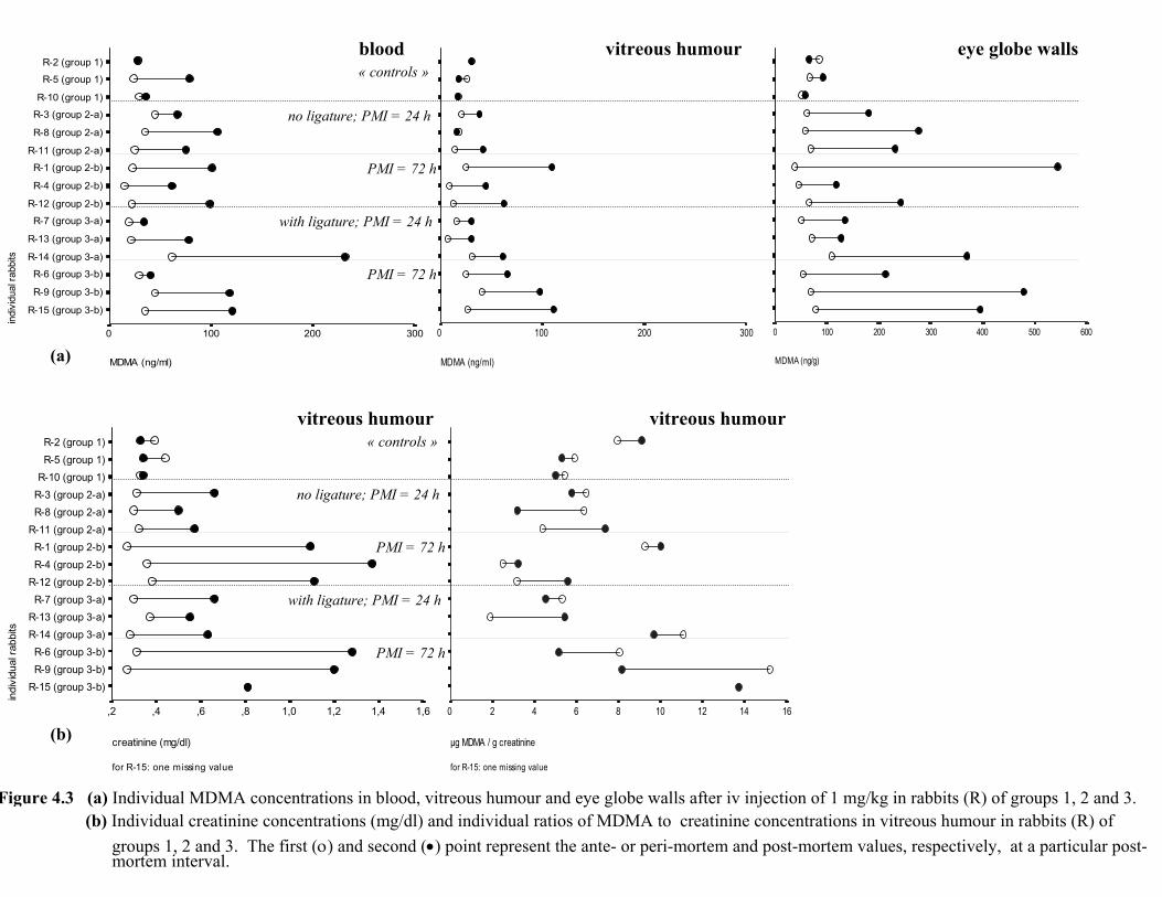
(a) MDMA (ng/g)
6005004003002001000
eye globe walls
for R-15: one missing value
indi
vidu
al ra
bbits
R-2 (group 1)R-5 (group 1)
R-10 (group 1)R-3 (group 2-a)R-8 (group 2-a)
R-11 (group 2-a)R-1 (group 2-b)R-4 (group 2-b)
R-12 (group 2-b)R-7 (group 3-a)
R-13 (group 3-a)R-14 (group 3-a)R-6 (group 3-b)R-9 (group 3-b)
R-15 (group 3-b)
creatinine (mg/dl)
1,61,41,21,0,8,6,4,2
vitreous humour
for R-15: one missing value
µg MDMA / g creatinine
1614121086420
vitreous humour
indi
vidu
al ra
bbits
R-2 (group 1)
R-5 (group 1)
R-10 (group 1)
R-3 (group 2-a)
R-8 (group 2-a)
R-11 (group 2-a)
R-1 (group 2-b)
R-4 (group 2-b)
R-12 (group 2-b)
R-7 (group 3-a)
R-13 (group 3-a)
R-14 (group 3-a)
R-6 (group 3-b)
R-9 (group 3-b)
R-15 (group 3-b)
MDMA (ng/ml)
3002001000
MDMA (ng/ml)
3002001000
blood vitreous humour
« controls »
no ligature; PMI = 24 h
PMI = 72 h
with ligature; PMI = 24 h
PMI = 72 h
« controls »
no ligature; PMI = 24 h
PMI = 72 h
PMI = 72 h
with ligature; PMI = 24 h
Figure 4.3 (a) Individual MDMA concentrations in blood, vitreous humour and eye globe walls after iv injection of 1 mg/kg in rabbits (R) of groups 1, 2 and 3.(b) Individual creatinine concentrations (mg/dl) and individual ratios of MDMA to creatinine concentrations in vitreous humour in rabbits (R) of
groups 1, 2 and 3. The first (ο) and second (•) point represent the ante- or peri-mortem and post-mortem values, respectively, at a particular post-mortem interval.
(a)
(b)
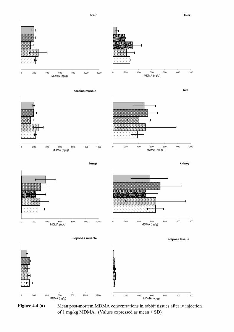
Figure 4.4 (a) Mean post-mortem MDMA concentrations in rabbit tissues after iv injection of 1 mg/kg MDMA. (Values expressed as mean ± SD)
cardiac muscle
0 200 400 600 800 1000 1200MDMA (ng/g)
liver
0 200 400 600 800 1000 1200MDMA (ng/g)
bile
0 200 400 600 800 1000 1200MDMA (ng/ml)
iliopsoas muscle
0 200 400 600 800 1000 1200MDMA (ng/g)
adipose tissue
0 200 400 600 800 1000 1200MDMA (ng/g)
brain
0 200 400 600 800 1000 1200MDMA (ng/g)
lungs
0 200 400 600 800 1000 1200MDMA (ng/g)
kidney
0 200 400 600 800 1000 1200MDMA (ng/g)

group 1: "control" cases
group 2(a):cases without ligature; PMI = 24 h
group 2(b):cases without ligature; PMI = 72 h
group 3(a):cases with ligature; PMI = 24 h
group 3(b):cases with ligature; PMI = 72 h
Figure 4.4 (b) Mean post-mortem MDA concentrations in rabbit tissues after iv injectionof 1 mg/kg MDMA. (Values expressed as mean ± SD)
post-mortem blood
0 20 40 60 80 100MDA (ng/ml)
lungs
0 20 40 60 80 100MDA (ng/g)
liver
0 20 40 60 80 100MDA (ng/g)
kidney
0 20 40 60 80 100MDA (ng/g)
bile
0 20 40 60 80 100
MDA (ng/ml)
0

n
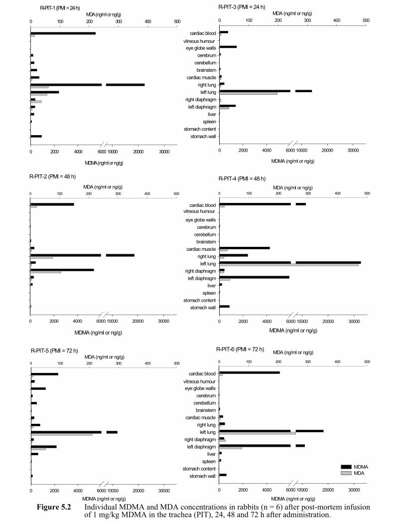
Figure 5.2 Individual MDMA and MDA concentrations in rabbits (n = 6) after post-mortem infusion of 1 mg/kg MDMA in the trachea (PIT), 24, 48 and 72 h after administration.
R-PIT-1 (PMI = 24 h)
MDMA (ng/ml or ng/g)
0 2000 4000 6000 10000 20000 30000
MDA (ng/ml or ng/g)0 100 200 300 400 500
R-PIT-3 (PMI = 24 h)MDA (ng/ml or ng/g)
0 100 200 300 400 500
stomach wallstomach content
spleenliver
left diaphragmright diaphragm
left lungright lung
cardiac musclebrainstem
cerebellumcerebrum
eye globe walls
cardiac blood
MDMA (ng/ml or ng/g)
0 2000 4000 6000 10000 20000 30000
vitreous humour
MDMA (ng/ml or ng/g)
0 2000 4000 6000 10000 20000 30000
MDA (ng/ml or ng/g)0 100 200 300 400 500
R-PIT-2 (PMI = 48 h) R-PIT-4 (PMI = 48 h)
MDMA (ng/ml or ng/g)
0 2000 4000 6000 10000 20000 30000
stomach wallstomach content
spleenliver
left diaphragmright diaphragm
left lungright lung
cardiac musclebrainstem
cerebellumcerebrum
eye globe walls
cardiac blood
MDA (ng/ml or ng/g)0 100 200 300 400 500
vitreous humour
R-PIT-5 (PMI = 72 h)
MDMA (ng/ml or ng/g)0 2000 4000 6000 10000 20000 30000
MDA (ng/ml or ng/g)0 100 200 300 400 500
vitreous humour
R-PIT-6 (PMI = 72 h)
MDMA (ng/ml or ng/g)
0 2000 4000 6000 10000 20000 30000
stomach wallstomach content
spleenliver
left diaphragmright diaphragm
left lungright lung
cardiac musclebrainstem
cerebellumcerebrum
eye globe walls
cardiac blood
MDA (ng/ml or ng/g)
0 100 200 300 400 500
MDMA MDA

R-PIT-3(PMI = 24 h)
R-PIT-4 (PMI = 48 h)
VS
RVLV
RA LA
TP
AO
AOVI
VR
AR
VP
AR
VR
Figure 5.3 Thoracic and abdominal post-mortem diffusion after tracheal instillation of 1 mg/kg MDMA in rabbits (n = 2), in which spreading of the solution occurred predominantly in the left bronchus, 24 and 48 h after administration.
Labels: RA: right atrium AO: aortaRV: right ventricle VI: inferior vena cavaLA: left atrium VS: superior vena cavaLV: left ventricle AR: arteria renalisTP: truncus pulmonalis VR: vena renalisVP: venae pulmonales D: diaphragm
VS
RVLV
RA LA
TP
AO
AOVI
VR
AR
VP
AR
VR
D D
VIVI
MDMA levels (ng/ml or ng/g):
> 10,0005,000 – 10,0001,000 – 5,000500 – 1,000100 – 50010 – 100< 10
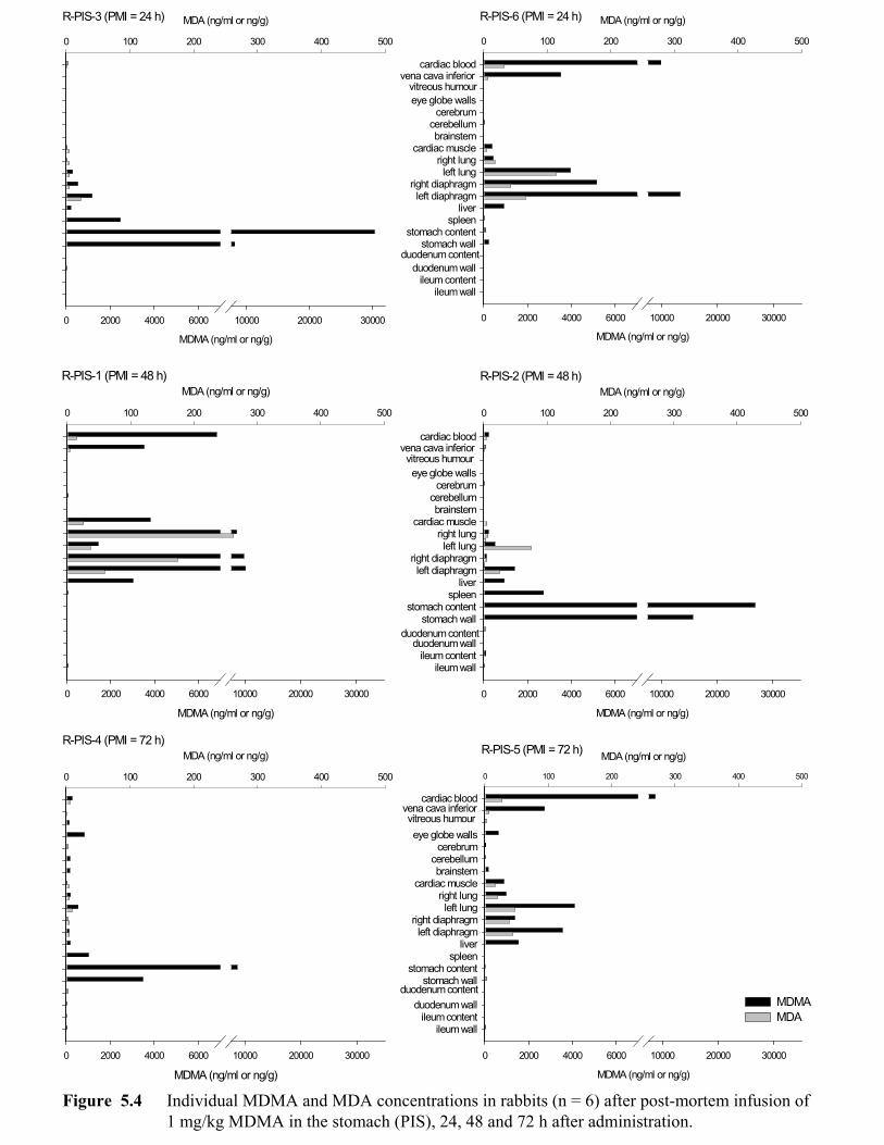
Figure 5.4 Individual MDMA and MDA concentrations in rabbits (n = 6) after post-mortem infusion of 1 mg/kg MDMA in the stomach (PIS), 24, 48 and 72 h after administration.
R-PIS-3 (PMI = 24 h)
MDMA (ng/ml or ng/g)
0 2000 4000 6000 10000 20000 30000
MDA (ng/ml or ng/g)
0 100 200 300 400 500
vitreous humourvena cava inferior
duodenum content
R-PIS-6 (PMI = 24 h)
MDMA (ng/ml or ng/g)
0 2000 4000 6000 10000 20000 30000
ileum wallileum content
duodenum wall
stomach wallstomach content
spleenliver
left diaphragmright diaphragm
left lungright lung
cardiac musclebrainstem
cerebellumcerebrum
eye globe walls
cardiac blood
MDA (ng/ml or ng/g)
0 100 200 300 400 500
R-PIS-1 (PMI = 48 h)
MDMA (ng/ml or ng/g)
0 2000 4000 6000 10000 20000 30000
MDA (ng/ml or ng/g)
0 100 200 300 400 500
vitreous humourvena cava inferior
duodenum content
R-PIS-2 (PMI = 48 h)
MDMA (ng/ml or ng/g)
0 2000 4000 6000 10000 20000 30000
ileum wallileum content
duodenum wall
stomach wallstomach content
spleenliver
left diaphragmright diaphragm
left lungright lung
cardiac musclebrainstem
cerebellumcerebrum
eye globe walls
cardiac blood
MDA (ng/ml or ng/g)
0 100 200 300 400 500
R-PIS-4 (PMI = 72 h)
MDMA (ng/ml or ng/g)0 2000 4000 6000 10000 20000 30000
MDA (ng/ml or ng/g)
0 100 200 300 400 500
R-PIS-5 (PMI = 72 h)
MDMA (ng/ml or ng/g)
0 2000 4000 6000 10000 20000 30000
ileum wallileum content
duodenum wall
stomach wallstomach content
spleenliver
left diaphragmright diaphragm
left lungright lung
cardiac musclebrainstem
cerebellumcerebrum
eye globe walls
cardiac blood
MDA (ng/ml or ng/g)
0 100 200 300 400 500
vena cava inferiorvitreous humour
duodenum contentMDMA MDA

RVLV
RA LA
TP
AO
AOVI
VRAR
VP
AR
VR
R-PIS-4(PMI = 72 h)
RVLV
RA LA
TP
AO
AOVI
VRAR
VP
AR
VR
R-PIS-5(PMI = 72 h)
Figure 5.5 Thoracic and abdominal post-mortem diffusion after gastric instillation of 1 mg/kg MDMA in rabbits (n = 2), showing the difference between the intra-gastric (a), and supra-diaphragmatic pattern (b) 72 h after instillation.
Labels: RA: right atrium AO: aortaRV: right ventricle VI: inferior vena cavaLA: left atrium VS: superior vena cavaLV: left ventricle AR: arteria renalisTP: truncus pulmonalis VR: vena renalisVP: venae pulmonales D: diaphragm
(a)
D D
VS
VI
VS
VI
(b)
MDMA levels (ng/ml or ng/g):
> 10,0005,000 – 10,0001,000 – 5,000500 – 1,000100 – 50010 – 100< 10

RVLV
RA LA
TP
AO
AOVI
VRAR
VI
VSVP
AR
VR
Figure 6.1 Possible mechanism of redistribution (indicated by direction of arrows)
Labels: RA: right atrium D: diaphragmRV: right ventricle AO: aortaLA: left atrium VI: inferior vena cavaLV: left ventricle VS: superior vena cavaTP: truncus pulmonalis AR: arteria renalisVP: venae pulmonales VR: vena renalis
D
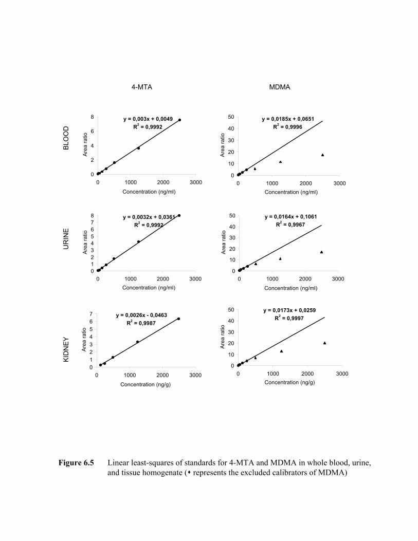
BLOOD
URINE
KIDNEY
4-MTA MDMA
y = 0,0185x + 0,0651R2 = 0,9996
0
10
20
30
40
50
0 1000 2000 3000Concentration (ng/ml)
Arearatio
y = 0,003x + 0,0049R2 = 0,9992
0
2
4
6
8
0 1000 2000 3000
Concentration (ng/ml)
Arearatio
y = 0,0032x + 0,0361R2 = 0,9992
012345678
0 1000 2000 3000Concentration (ng/ml)
Arearatio
y = 0,0164x + 0,1061R2 = 0,9967
0
10
20
30
40
50
0 1000 2000 3000Concentration (ng/ml)
Arearatio
y = 0,0173x + 0,0259R2 = 0,9997
0
10
20
30
40
50
0 1000 2000 3000Concentration (ng/g)
Arearatio
y = 0,0026x - 0,0463R2 = 0,9987
01234567
0 1000 2000 3000
Concentration (ng/g)
Arearatio
Figure 6.5 Linear least-squares of standards for 4-MTA and MDMA in whole blood, urine,and tissue homogenate ( represents the excluded calibrators of MDMA)
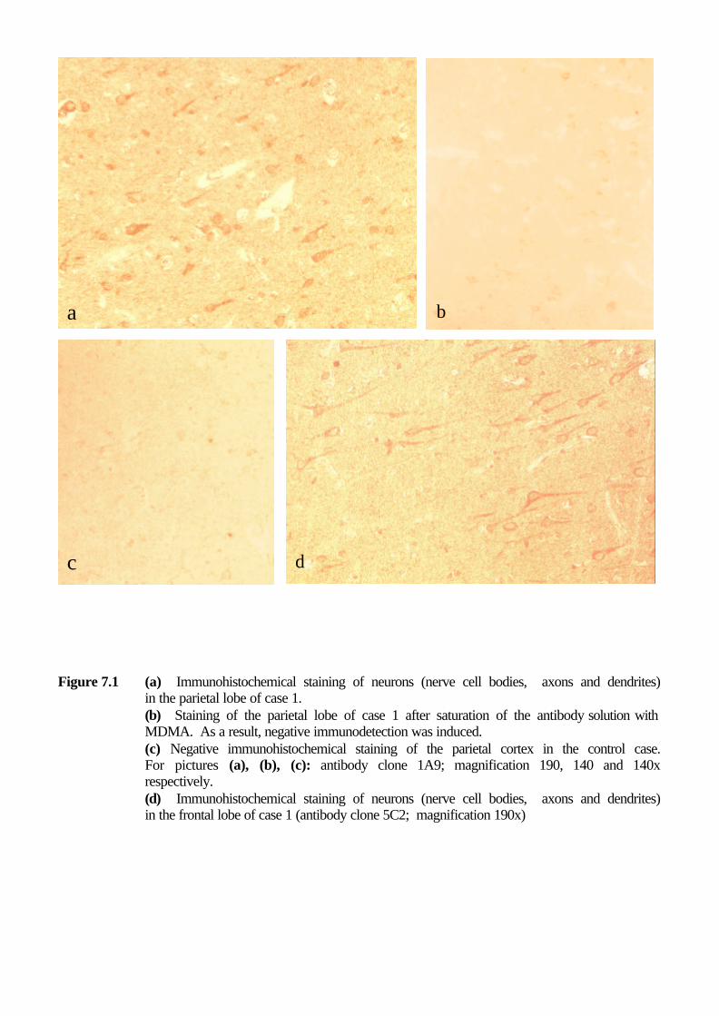
Figure 7.1 (a) Immunohistochemical staining of neurons (nerve cell bodies, axons and dendrites)
in the parietal lobe of case 1. (b) Staining of the parietal lobe of case 1 after saturation of the antibody solution with MDMA. As a result, negative immunodetection was induced. (c) Negative immunohistochemical staining of the parietal cortex in the control case. For pictures (a), (b), (c): antibody clone 1A9; magnification 190, 140 and 140x respectively. (d) Immunohistochemical staining of neurons (nerve cell bodies, axons and dendrites) in the frontal lobe of case 1 (antibody clone 5C2; magnification 190x)
a
c d
b
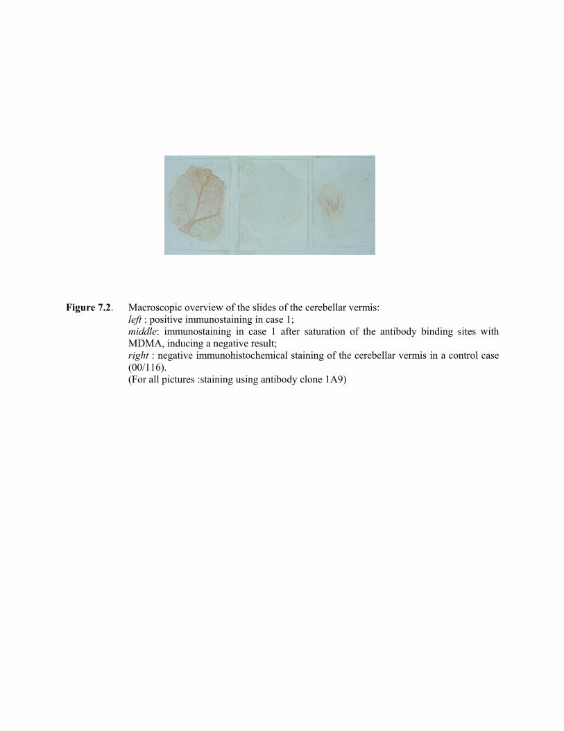
Figure 7.2. Macroscopic overview of the slides of the cerebellar vermis:
left : positive immunostaining in case 1; middle: immunostaining in case 1 after saturation of the antibody binding sites with MDMA, inducing a negative result; right : negative immunohistochemical staining of the cerebellar vermis in a control case (00/116). (For all pictures :staining using antibody clone 1A9)

Figure 7.3 (a) Positive immunostaining of the cerebellar vermis of case 2: the nerve cell bodies,
axons and dendrites of the Purkinje cells are clearly visible. The granular layer cells can also obviously be discerned. In addition, staining at the level of the white matter fibres is seen (overview: magnification 95x). (b) Detail of the immunoreaction in the cerebellar vermis of case 2 (magnification
270x). (c) Negative immunohistochemical staining of the cerebellar vermis in a control case
(00/116; magnification 170x). (For all pictures: staining with antibody clone 1A9).
a
b
c
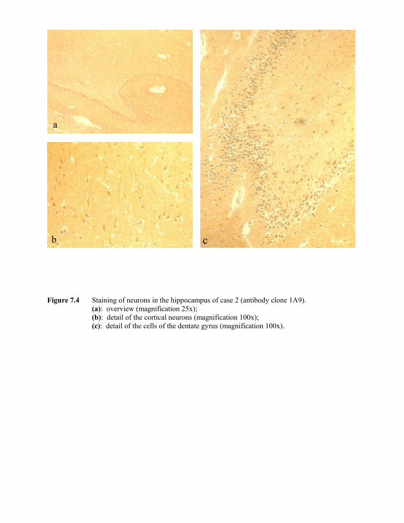
Figure 7.4 Staining of neurons in the hippocampus of case 2 (antibody clone 1A9).
(a): overview (magnification 25x); (b): detail of the cortical neurons (magnification 100x); (c): detail of the cells of the dentate gyrus (magnification 100x).
a
b c

Figure 7.5 (a) Overview of MDMA immunoreactivity in the pituitary gland (antibody clone 1A9;
magnification 140x). (b) PAS Orange-G staining of the immediately adjacent slide visualizing clearly the acidophilic (orange colour) and basophilic cells (violet colour), (magnification 140x). (c) Detail of the staining reaction for MDMA in the pituitary gland (antibody clone 1A9; magnification 300x). Following types of staining reaction can be discerned (see arrows): (1) & (2): obviously positive cells showing variable staining intensity (heterogeneity). The highly intensively stained cells obscure the nucleus (see arrow 1). (3) cells having weak immunoreaction. (4) negative cells.
a
b c
1
2
3
4
4
Copyright © 2022 FDOKUMEN