toxicological profile for ethylene glycol and propylene glycol
Upload
independentCategory
view
0download
0
Nanotoxicology, 2011; Early Online, 1–11©2011 Informa UK, Ltd.ISSN: 1743-5390 print / 1743-5404 onlineDOI: 10.3109/17435390.2011.604439
A novel platform for pulmonary and cardiovascular toxicologicalcharacterization of inhaled engineered nanomaterials
Georgios A. Sotiriou1, Edgar Diaz2, Mark S. Long2, John Godleski2, Joseph Brain2, Sotiris E. Pratsinis1, &Philip Demokritou2
1Department of Mechanical and Process Engineering, Particle Technology Laboratory, Institute of Process Engineering, SwissFederal Institute of Technology Zurich (ETH Zurich), Sonneggstrasse 3, CH-8092, Zurich, Switzerland and 2Department ofEnvironmental Health, Harvard School of Public Health, Harvard University, 401 Park Drive, Boston, MA 02215, USA
AbstractA novel method is presented which is suitable for assessingin vivo the link between the physicochemical properties ofengineered nanomaterials (ENM) and their biological outcomes.The ability of the technique to generate a variety of industry-relevant, property-controlled ENM exposure atmospheres forinhalation studies was systematically investigated. The primaryparticle size for Fe2O3, SiO2, Ag and Ag/SiO2 was controlled from4 to 25 nm, while the corresponding agglomerate mobilitydiameter of the aerosol was also controlled and varied from40 to 120 nm. The suitability of the technique to characterize thepulmonary and cardiovascular effects of inhaled ENMs in intactanimal models is also demonstrated using in vivochemiluminescence (IVCL). The IVCL technique is a highlysensitive method for identifying cardiopulmonary responses toinhaled ENMs under relatively small doses and acute exposures.It is shown that moderate and acute exposures to inhalednanostructured Fe2O3 can cause both pulmonary andcardiovascular effects.
Keywords: engineered nanomaterials, in vivo inhalation,nanotoxicology, cardiovascular and pulmonary effects,nanotoxicology, iron oxide
Introduction
The use of engineered nanomaterials (ENMs) in householdproducts, textiles, industrial processes, medical devices andtherapeutics is widespread and increasing exponentially.Environmental and occupational exposures are consideredby experts to be ‘inevitable’ (National Research Council2008; National Science and Technology Council Comiteeon Technology 2008; Powell et al. 2010). Preliminary evi-dence demonstrates the potential for ENM to cause adversebiological effects (Colvin 2003; Nel et al. 2009; Service 2000).Indeed, the potential of nanoparticles and nanofibres to
translocate through the air–blood barriers, and thus to reachthe pulmonary connective tissues, lymphatic system, or evento reach the circulating blood and thus have access to othercritical organs, is of concern. Nano-sized particles may entercells, and be more biologically active than their micro-sized counterparts due to their small size and large surfaceto volume ratio (Hamilton et al. 2009; Larsen et al. 2010;Li et al. 2010; Muller et al. 2005; Poland et al. 2008; Rossi et al.2010; Service 2004; Witzmann & Monteiro-Riviere 2006) andincreased release of ions (Sotiriou & Pratsinis 2010).
Many in vitro assays and novel aerosol generation meth-ods suitable for inhalation studies have been developed andcontributed significantly to improving our understanding ofbiological mechanisms related to atmospheric particlehealth effects (Demokritou et al. 2002; Demokritou et al.2003; Gupta et al. 2004). However, these currently availableparticle exposure methods cannot be applied in the nano-toxicology field, primarily because of the unique propertiesof ENM. For in vivo ENM inhalation studies, an importantlimiting factor is the difficulty in aerosolizing and dispersingcommercially available nanopowder ENM down to thenano-size level, as they would exist in many relevant inha-lation exposures (Schmoll et al. 2009). Some common aero-sol generators used in the past to disperse nanopowders forinhalation studies, including nebulizers, fluidized beds andother venturi aspirator-type systems, have limited applica-bility for nanotoxicology (Schmoll et al. 2009). For example,such nanopowder aerosol-generation systems have difficultyin producing consistent nano-sized aerosol distributionsand, more importantly, fail to control physicochemicalproperties in order to elucidate the link between propertiesand toxic biological responses (Fischer & Chan 2007;Schmoll et al. 2009).
As a result of the challenges and limitations, the majorityof nanotoxicity research has focused on in vitro cellular orin vivo instillation studies using commercially available ENMnanopowders and nanopowder liquid suspensions; however,
Correspondence: Philip Demokritou, Department of Environmental Health, Harvard School of Public Health, Harvard University, 401 Park Drive, Boston,MA 02215, USA. E-mail: [email protected]
(Received 21 February 2011; accepted 6 July 2011)
Nan
otox
icol
ogy
Dow
nloa
ded
from
info
rmah
ealth
care
.com
by
134.
174.
180.
146
on 0
8/08
/11
For
pers
onal
use
onl
y.

there is a consensus among scientists that data will requireverification from animal inhalation experiments (NationalScience and Technology Council Comitee on Technology2008; Schmoll et al. 2009). There are also serious short-comings related to the physiological relevance of this particleto cell delivery approach using liquid ENM suspensions. Theshortcomings of these in vitro and instillation approaches aretwofold: First, commercial ENM are limited in diversity ofphysicochemical and morphological properties – usually to afew sizes for a given composition – making it impossible toperform comparative parametric toxicological studies ofENM properties (size, surface, composition, shape, charge,etc.). Second, nanoparticles suspended in culture mediaflocculate or dissolve, and interact with serum components(Fadeel & Garcia-Bennett 2010; Jones & Grainger 2009;Verma and Stellacci 2010), which can alter their biologicalproperties and effects. Furthermore, comparison of particledoses delivered in suspension to those administered byinhalation is difficult, which can result in large differencesin effective dosage between in vivo and in vitro studies.These limitations may explain some of the disparitiesreported in the literature between in vivo and in vitroENM studies (Fadeel & Garcia-Bennett 2010; Fischer &Chan 2007; Sayes et al. 2007). It is apparent that newmethods and systems suitable for both in vitro andin vivo inhalation toxicological characterization need to bedeveloped.
Here, a novel technique is presented that is suitable forboth ENM in vivo inhalation and in vitro toxicologicalcharacterization studies. The ability of this technique togenerate a variety of industry-relevant, property-controlled
exposure atmospheres for inhalation studies was systemat-ically investigated. The suitability of the technique to char-acterize the pulmonary and cardiovascular effects of inhaledENM in intact animal models was also demonstrated in anin vivo study involving Sprague–Dawley rats, using freshlygenerated nano-iron oxide (Fe2O3) as a test aerosol. Wedemonstrated both pulmonary and systemic toxicity usingin vivo chemiluminescence (IVCL) of heart and lung(Bartoli et al. 2009; Clarke et al. 1999; Clarke et al. 2000;Godleski et al. 2002; Gurgueira et al. 2002; Rhoden et al.2004; Saldiva et al. 2002). This novel platform will makeit possible for toxicologists to link physicochemical proper-ties of inhaled ENM to biological outcomes and help theindustry to develop safer ENM.
Methods
Versatile Engineered Nanomaterial Generation SystemFigure 1 illustrates the Versatile Engineered NanomaterialGeneration System (VENGES) (Demokritou et al. 2010). Thesystem consists of four main components: ENM synthesis,particle sampling and collection system, animal exposuresystem and exposure monitoring system.
ENM synthesisThe synthesis of ENM is based on the industry-relevant flame spray pyrolysis (FSP) method, as most oftoday’s ENMs are made by gas-phase techniques (Pratsinis2010). An inflammable liquid precursor, containing dis-solved organometallic compounds, is pumped through astainless-steel capillary tube (inner diameter = 0.41 mm;
1 Laerosol
container
ENM sampling/collection
Filter
HE
PA
HE
PA
Qs
QR
T1
QD
QA QP
Flame synthesis Animal exposuresystem
Animal exposurechamber
HEPA
FMPS P-TRAK
Exposure monitoring equipment
O2dispersion Liquid
precursor
CH4/O2supportflame
50 c
m CO2, CO,RH, T2
NO2
Figure 1. Overview of VENGES. All different parts of VENGES are presented. Engineered nanomaterials (ENMs) are generated by flame synthesis.The nanoparticles are collected on a filter for further ex situ characterization. Directly above the flame, in situ sampling of the test aerosol isperformed (QS). Immediately after, the ENM aerosol stream can be further diluted with HEPA-filtered room air (QD) and directed into the exposureanimal chambers (QA) for in vivo toxicological testing. Simultaneously, the test ENM aerosol is in situ real-time characterized (QP). The total particlenumber concentration is measured, as well as the mobility particle-size distribution. Finally, the CO2, CO, NO2, relative humidity (RH) andtemperature (T2) are continuously monitored.
G. A. Sotiriou et al.
Nan
otox
icol
ogy
Dow
nloa
ded
from
info
rmah
ealth
care
.com
by
134.
174.
180.
146
on 0
8/08
/11
For
pers
onal
use
onl
y.

outer diameter = 0.71 mm). Immediately surrounding thecapillary tube is a narrow annular gap of adjustable cross-sectional area that supplies oxygen, which is used to dis-perse the liquid precursor into fine droplets. A small pilotflamelet (premixed CH4 and O2 at 1 and 2 l/min, respec-tively) ignites the droplets so that the organic parts of theprecursor combust, while the metals are oxidized, and formparticles that grow by coagulation and sintering in the flame.The precursor liquid feed is metered by a mass rate-controlled syringe pump, and all gas flows are controlledby mass flow controllers (Madler et al. 2002; Teleki et al.2008).
Sampling and off-line characterization of generated ENMThe flame-generated particles are collected using a water-cooled, stainless-steel filter housing supporting a glass fibrefilter (Whatman GF6, 25.7 cm in diameter, Switzerland), withexhaust gases drawn by a vacuum pump (Busch, Seco SV1040C, Netherlands). Collected particles can be extractedfrom the filter as nanopowder, and used for off-line physicochemical and morphological characterization,as well as for in vitro or in vivo instillation studies. Off-line ENM characterization included N2 adsorption (five-point isotherm; Micromeritic Tristar, Switzerland). ENMsamples were degassed under N2 at 150�C for 1.5 h. Theaverage primary particle diameter dBET was calculated fromtheir specific surface area, SSA: dBET = 6000/(r �SSA), wherer is the material’s density. X-ray diffraction (XRD, BrukerD8 Advance, Cu Ka, Switzerland) was also performed overthe scan range 2q = 20�–70�. Morphology of the generatedENM was also obtained using transmission electron micros-copy and scanning transmission electron microscopy(STEM, instrument for both: Tecnai F30, Switzerland). Thesamples were dispersed in ethanol and then placed on acarbon-coated copper grid.
Animal exposure systemThe continuously produced (in the gas phase) nanoparticlesare sampled in situ over the flame (QS, Figure 1), andtransferred to inhalation chambers for animal studiesthrough a 1-l aerosol container to stabilize the flow rate.The sampled aerosol can be further diluted with high-efficiency particulate air (HEPA)-filtered dry air (QD). TheENMs remain airborne, with a very low level of agglomer-ation, and are drawn directly through the animal exposurechambers (QR, Figure 1). For this study, each animal washoused in its own whole-body exposure chamber (10 cm indiameter, 18 cm in length). Chambers were made of clearpolycarbonate to allow visualization of the animals during
the exposure, and used a modified rubber stopper as a cap.Flow through each chamber was maintained constant at1.5 l/min.
Exposure monitoring systemThe animal exposure atmosphere was monitored continu-ously, using a number of real-time instruments. Exposureparameters monitored included total particle counts(P-Track; TSI, MA, USA), aerosol-size distribution (FMPS;TSI, MA, USA), NO2, CO, CO2, temperature and relativehumidity (Figure 1).
Performance characterization experimentsThe versatility and ability of VENGES to generate an array ofindustrially relevant ENM while controlling important nano-particle properties (i.e. primary particle size, agglomerationstate, surface properties, shape) as well as its suitability forin vivo inhalation studies were demonstrated in a number ofperformance characterization experiments, as follows.
ENM-generated nanopanelFour different ENM, silicon dioxide (silica, SiO2), iron oxide(Fe2O3), silver (Ag, nanosilver) and composite nanosilversupported on silica particles (Ag/SiO2), were generated here.Table I summarizes the ENMs used in these experiments andtheir respective liquid precursor characteristics. It is worthpointing out that the FSP synthesis method used in VENGESallows for the generation of a large universe of industriallyrelevant ENM (Strobel & Pratsinis 2007).
Controlling both the aerosol size (airborne phase)and primary particle sizeIn order to investigate whether VENGES can successfullyvary both the size of the generated aerosol as well as theprimary particle size, a study was performed by varying twoimportant VENGES process parameters: (1) liquid precursorfeed rate (from now on: x) over the O2 dispersion flow rate(from now on: y) ratio (x/y); and (2) liquid precursor molar-ity, M (mol/l). Table I summarizes for each ENM the x/yratios and the variable molarity experiments, used in theexperiments. For all experiments, the properties of the ENMVENGES-generated aerosol were characterized in situ in realtime. In addition, the physicochemical and morphologicalcharacterization of the ex situ collected nanoparticles wasalso performed off-line as previously described.
Reproducibility of the generated test aerosols over timeFor inhalation toxicological studies, it is important to: (1) gen-erate in a reproducible manner test aerosols with constantconcentration and size distribution for the duration of the
Table I. Synthesized ENM, their corresponding precursors and solvent, as well as their process parameters (precursor molarity and x/y ratio).
ENM Metal precursor SolventPrecursor
molarity (M)
Liquid feed rate(x)/O2 dispersion
flow rate (y)
SiO2 Hexamethyl disiloxane Ethanol 0.1–1 2/7 to 6/4
Fe2O3 Iron acetyloacetonate Acetonitrile/2-ethylhexanoic acid (1:1) 0.01–0.5 2/7 to 6/4
Ag Silver nitrate Ethanol/diethylene glycol monobutyl ether (1:1) 0.06–0.31 2/7
Ag/SiO2 Silver nitrate/Hexamethyl disiloxane Ethanol/diethylene glycol monobutyl ether (1:1) 0.1 2/7
In vivo toxicological characterization of inhaled ENM
Nan
otox
icol
ogy
Dow
nloa
ded
from
info
rmah
ealth
care
.com
by
134.
174.
180.
146
on 0
8/08
/11
For
pers
onal
use
onl
y.
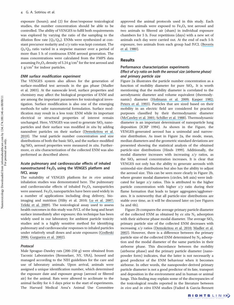
exposure (hours); and (2) for dose/response toxicologicalstudies, the number concentration should be able to becontrolled. The ability of VENGES to fulfil both requirementswas explored by varying the ratio of the sampling to thedilution flow rate (QS/QD). ENMs were synthesized at con-stant precursor molarity and x/y ratio was kept constant. TheQS/QD ratio varied in a stepwise manner over a period ofmore than 5 h of continuous ENM aerosol generation. Themass concentrations were calculated from the FMPS dataassuming Fe2O3 density of 5.24 g/cm
3 for the test aerosol and1 g/cm3 for indoor particles.
ENM surface modification experimentThe VENGES system also allows for the generation ofsurface-modified test aerosols in the gas phase (Madleret al. 2003). At the nanoscale level, surface properties andchemistry may affect the biological properties of ENM andare among the important parameters for toxicological inves-tigation. Surface modification is also one of the emergingmethods for safer nanomaterial formulation. Surface mod-ification may result in a less toxic ENM while its importantelectrical or structural properties of interest remainunchanged. Here, VENGES was used to generate SiO2 nano-particles and their surface was modified in situ by addingnanosilver particles on their surface (Demokritou et al.2010). The total particle number concentration and sizedistributions of both the basic SiO2 and the surface-modifiedAg/SiO2 aerosol properties were measured in situ. Further-more, ex situ characterization of the collected ENM was alsoperformed as described above.
Acute pulmonary and cardiovascular effects of inhalednanostructured Fe2O3 using the VENGES platform andIVCL assayThe suitability of VENGES platform for in vivo animalinhalation studies was demonstrated here. The pulmonaryand cardiovascular effects of inhaled Fe2O3 nanoparticleswere assessed. Fe2O3 nanoparticles have been used widely ina number of applications including drug delivery, bio-imaging and nutrition (Hilty et al. 2010; Lu et al. 2007;Teleki et al. 2009). The toxicological assay used to assesshealth outcomes in this study was IVCL of the lung and heartsurface immediately after exposure; this technique has beenwidely used in our laboratory for ambient particle toxicitystudies and is a highly sensitive method for identifyingpulmonary and cardiovascular responses to inhaled particlesunder relatively small doses and acute exposures (Godleski2006; Gurgueira et al. 2002).
ProtocolMale Sprague–Dawley rats (200–250 g) were obtained fromTaconic Laboratories (Rensselaer, NY, USA), housed andmanaged according to the NIH guidelines for the care anduse of laboratory animals. Upon arrival, animals wereassigned a unique identification number, which determinedthe exposure date and exposure group (aerosol or filteredair) for the animal. Rats were allowed to acclimate to theanimal facility for 4–5 days prior to the start of experiments.The Harvard Medical Area’s Animal Use Committee
approved the animal protocols used in this study. Eachday two animals were exposed to Fe2O3 test aerosol andtwo animals to filtered air (sham) in individual exposurechambers for 5 h. Four repetitions (days) with a new set ofanimals each day were carried out. At the end of each 5-hexposure, two animals from each group had IVCL (Boveriset al. 1980).
Results
Performance characterization experimentsEffect of x/y ratio on both the aerosol size (airborne phase)and primary particle sizeFigure 2a illustrates the particle number concentration as afunction of mobility diameter for pure SiO2. It is worthmentioning that the mobility diameter is correlated to theaerodynamic diameter and volume equivalent (thermody-namic) diameter (Hofmann et al. 2009; Kasper 1982;Peters et al. 1993). Particles that are sized based on theirmobility in an electric field are considered for practicalpurposes classified by their thermodynamic diameter(McCawley et al. 2001; Schiller et al. 1988). Thermodynamicdiameter is an important determinant of nanoparticle lungdeposition (ICRP 1994). As shown in the Figure, theVENGES-generated aerosol has a unimodal and narrow-size distribution. As inset in Figure 2a, the mode, mean,median diameters and the geometric standard deviations arepresented showing the statistical analysis of the obtainedparticle-size distributions (Hinds 1999). Additionally, themodal diameter increases with increasing x/y ratios, asthe SiO2 aerosol concentration increases. It is clear thatVENGES not only has the ability to generate aerosols withunimodal-size distributions but also has the ability to adjustthe aerosol size. This can be seen more clearly in Figure 2b,where greater modal diameters (circles, left axis) were indi-cated for larger x/y ratios. This is attributed to the higherparticle concentration with higher x/y ratio during theirflame formation that leads to larger aggregates/agglomer-ates. It is noteworthy that all particle-size distributions arestable over time, as it will be discussed later on (see Figures5a and 6b).
Figure 2b compares the average primary particle diameterof the collected ENM as obtained by ex situ N2 adsorptionwith their airborne phase modal diameter. The average SiO2
primary particle size of the collected ENM decreases withincreasing x/y ratios (Demokritou et al. 2010; Madler et al.2002). However, there is a difference between the primaryparticle size of the collected ENM determined by N2 adsorp-tion and the modal diameter of the same particles in theirairborne phase. This discordance between the mobility(airborne phase) and the primary particle diameter (nano-powder form) indicates, that the latter is not necessarily agood predictor of the ENM behaviour when it becomesairborne. In other words, the nanopowder-derived primaryparticle diameter is not a good predictor of its fate, transportand deposition in the environment and in human or animallungs. This finding may explain some of the discrepancies ofthe toxicological results reported in the literature betweenin vivo and in vitro ENM studies (Fadeel & Garcia-Bennett
G. A. Sotiriou et al.
Nan
otox
icol
ogy
Dow
nloa
ded
from
info
rmah
ealth
care
.com
by
134.
174.
180.
146
on 0
8/08
/11
For
pers
onal
use
onl
y.

2010; Fischer & Chan 2007; Sayes et al. 2007). The in vivovalidation of in vitro (cellular) studies is considered animportant element of the proposed Environmental Healthand Safety strategy recently released by the NationalNanotechnology Initiative (NNI).
Similarly, Figure 2d show the effect of x/y ratio on boththe primary particle and the mobility diameter of Fe2O3.Figure 2c also shows the XRD patterns of the ex situ collectedand characterized nanoparticles as a function of the x/y ratio.This indicates that larger Fe2O3 particles are formed withhigher x/y ratio, in agreement with literature (Demokritouet al. 2010; Teleki et al. 2009). This is due to their largerresidence time in higher temperature zones that resultin larger crystals. It is worth pointing out that theXRD-estimated average crystal particle sizes (filled triangles,
Figure 2d) are consistent with their average primary particlesizes (open triangles, Figure 2d), with the latter havingslightly larger values indicating polycrystalline or aggregatednanoparticles. The modal mobility diameter for the Fe2O3
aerosol (Figure 2d, circles, left axis) is not significantlyaffected by the x/y ratio, as for most ratios it is fairly constant.Furthermore, as it is shown above in the case of SiO2, there isa difference between mobility diameter (airborne phase)and the primary particle diameter (nanopowder form).
Effect of precursor molarity on both the aerosol size andprimary nanopowder particle sizeFigure 3 indicates the effect of another important processparameter of VENGES, the metal precursor molarity, onboth the aerosol size (mobility diameter) and primary
6000SiO2, 0.5 M SiO2, 0.5 M
Fe2O3, 0.5 M
2/7 80.693.193.1107.1124.0
71.881.991.6101.1112.2
76.3 1.551.551.521.451.40
85.494.4106.2116.0
Mode(nm)
Mode mobility diameter
Primary particle ” , dBET
Mode mobility diameterPrimary particle ” , dBET
Crystal ” , dXRD
Median(nm)
Mean(nm)
σg
3/74/65/56/4
CT = 2*105 #/cm3
140
120
100
80
60
40
20
02/7 3/7 4/6 5/5 6/4
0
5
10
15
20
25
30
5000
4000
3000
2000
1000
01 10
Fe2O3, 0.5 M
x/y
6/4
120
100
80
60
40
20
02/7 3/7 4/6 5/5 6/4
0
2
4
6
8
10
12
14
5/5
4/6
2/7
20 30 40 50 60 70
Inte
nsi
ty, a
.u.
Mo
de
mo
bili
ty d
iam
eter
, nm
Pri
mar
y p
arti
cle
(dB
ET)
-
crys
tal d
iam
eter
(d
XR
D),
nm
Liquid feed rate/Dispersion O2 flow rateDiffraction angle, 2q
100 1000
Mobility diameter dm, nm Liquid feed rate/Dispersion O2 flow rate
Par
ticl
e co
nce
ntr
atio
n, #
/cm
3
SiO
2F
e 2O
3
Mo
de
mo
bili
ty d
iam
eter
, nm
Pri
mar
y p
arti
cle
dia
met
er (
dB
ET),
nm
b
c d
a
Figure 2. Controlling particle-size distribution by precursor liquid feed rate (x) over O2 dispersion flow rate (y) ratio (x/y). (a) Mobility-size distributions of SiO2 for a varying x/y ratio and the mode, median, mean diameters and the geometric standard deviation are shown. (b)The mode mobility (circles, left axis) and primary particle diameter (triangles, right axis) as a function of the x/y ratio. (c) XRD patterns of Fe2O3
nanoparticles for different x/y ratios. (d) Mode mobility (circles, left axis), primary particle (open triangles, right axis) and crystal (filled triangles,right axis) diameters as a function of the x/y ratio. The precursor molarity was 0.5 M and the total particle number concentration (CT) 2� 105 #/cm3
for both SiO2 and Fe2O3 samples.
In vivo toxicological characterization of inhaled ENM
Nan
otox
icol
ogy
Dow
nloa
ded
from
info
rmah
ealth
care
.com
by
134.
174.
180.
146
on 0
8/08
/11
For
pers
onal
use
onl
y.

4000
3000
2000
1000
0
4000
5000
3000
2000
1000
0
1 10 100 1000
Mobility diameter dm, nm
1 10 100 1000
Mobility diameter dm, nm
1 10 100 1000
Mobility diameter dm, nm
Metal precursor molarity, M
Metal precursor molarity, M
Par
ticl
e co
nce
ntr
atio
n, #
/cm
3P
arti
cle
con
cen
trat
ion
, #/c
m3
4000
3000
2000
1000
0
Par
ticl
e co
nce
ntr
atio
n, #
/cm
3
60.469.880.693.1
58.064.471.890.3
53.167.476.394.5
1.731.551.551.50
0.10 M
SiO2, 2/7
CT = 2*105 #/cm
3
CT = 2*105 #/cm
3
CT = 2*105 #/cm
3
0.35 M0.50 M1.00 M
Mode(nm)
Median(nm)
Mean(nm)
σg
Mode(nm)
Mode(nm)
39.269.869.880.693.1
38.061.264.278.286.5
44.4
29.434.052.3
26.1 24.829.646.3
1.611.601.57
0.06 M0.22 ”0.31 ”
Ag, 2/7
31.151.8
1.921.601.521.511.50
0.01 M0.02 ”0.10 ”0.35 ”0.50 ”
Fe2O3, 2/7
66.370.383.182.5
Median(nm)
Median(nm)
Mean(nm)
Mean(nm)
σg
σg
100
80
60
40
20
00.0 0.2 0.4 0.6 0.8 1.0
0.10.0
20 nm
Ag
Fe 2
O3
SiO
2
Fe2O3, 2/7, 0.5 M
0.2 0.3 0.4
Fe2O3, 2/7
0.5
0
8
6
4
2
0
10
15
20
25
30
5Mo
de
mo
bili
ty d
iam
eter
, nm
Mo
bili
ty m
od
e d
iam
eter
, nm
Mode mobility diameter
Primary particle ” , dBET
Pri
mar
y p
arti
cle
dia
met
er (
dB
ET),
nm
Pri
mar
y p
arti
cle
(dB
ET)
-
crys
tal d
iam
eter
(d
XR
D),
nm
Mode mobility diameter
Primary particle ” , dBET
Crystal particle ” , dXRD
100
80
60
40
20
0
a b
f
dc
e
Figure 3. Controlling particle size by precursor molarity. Mobility (a,c,f) of SiO2 (a), Fe2O3 (c) and Ag (nanosilver, f), for different precursormolarities. The mode, median, mean diameters and the geometric standard deviation are also shown. (b,d) Mode mobility (circles, left axis),primary particle (open triangles, right axis) and crystal (filled triangles, right axis) diameters for SiO2 (b) and Fe2O3 (d). (e) TEM image of Fe2O3
nanoparticles for precursor molarity 0.5 M and 2/7 ratio.
G. A. Sotiriou et al.
Nan
otox
icol
ogy
Dow
nloa
ded
from
info
rmah
ealth
care
.com
by
134.
174.
180.
146
on 0
8/08
/11
For
pers
onal
use
onl
y.

nanopowder particle size for SiO2, Fe2O3 and Ag ENM.Figure 3a shows for the SiO2 aerosol, the particle numberconcentration as a function of mobility diameter (for aconstant 2/7 ratio) for different metal precursor molaritieswith their corresponding diameters and geometric standarddeviation as inset. It can be seen that the mobility-size distribution shifts to larger sizes with an increasedprecursor molarity. It is worth mentioning that there is anoverlapping among the different particle-size distributionspresented in the figure. However, the aerosol-size distribu-tions were randomly selected to prove the point that aerosolproperties can be adjusted. Whether these size distributiondifferences are clinically relevant remains to be seen infuture toxicological studies.
Similarly, this pattern can be seen also in Figure 3b, wherethe mode mobility diameter (circles, left axis) is also increas-ing for higher precursor molarity. This could be attributed tothe higher concentration of the particles at an early stageduring their synthesis that would lead to the formation oflarger agglomerates. In contrast to the behaviour of the SiO2
particles in airborne phase, the average primary particlediameter (dBET, triangles, right axis) does not significantlyvary with precursor molarity (Jossen et al. 2005) as sinteringrather than coagulation determines the primary particle sizein contrast to mobility diameter that is determined bycoagulation and particle concentration levels.
Similarly for Fe2O3 nanoparticles, the mobility-size dis-tributions shift to larger sizes for higher precursor molarities(Figures 3c and 3d). The average primary particle (Figure 3d,dBET, open triangles, right axis) and the crystal (Figure 3d,dXRD, filled triangles, right axis) diameter seem to slightlyincrease for higher precursor molarity, forming polycrystal-line nanoparticles, as with SiO2. The TEM image of Fe2O3
nanoparticles shown in Figure 3e shows that there are manyagglomerates/aggregates composed of smaller primary par-ticles, in agreement with the results obtained by XRD, N2
adsorption and mobility-size distribution analysis. Finally,Figure 3f indicates that the mobility-size distribution of
nanosilver (Ag) can also be shifted to larger values for higherprecursor molarity.
Control of concentration/reproducibility of the generatedtest aerosols over timeWe have demonstrated earlier that VENGES has the ability tocontrol both the aerosol or agglomerate size (airbornephase) and the primary particle or crystal size (nanopowderform), in a highly reproducible way. However, for inhalationtoxicological studies, aerosol-generation methods need to beable to (1) adjust the concentration level (dose/responsestudies); and (2) deliver test aerosols with constant proper-ties for hours. Figure 4a shows the total particle numberconcentration CT (solid line) and the modal (broken line)and median (dotted line) mobility diameters of the nano-silver aerosol (2/7, 0.02 M precursor concentration) as afunction of exposure time. The dilution of the sampledaerosol (QS/QD, Figure 1) was periodically adjusted in astepwise manner during the experiment to control the con-centration level. As expected, for higher QS/QD ratio, higherCT is reached. Most importantly, when a constant QS/QD
ratio is used, both the aerosol total particle concentration(CT) and the size distribution indicated by the modal andmedian (dotted line) mobility diameters remain constant.This is clearly an indication that VENGES can control theaerosol concentration levels and deliver test aerosols withconstant size distributions over time.
It is noteworthy that when the QS/QD ratio is adjustedback to its initial value, the aerosol-size distribution returnsto its initial one, a clear indication of the reproducibility ofthe method. Similarly, for Fe2O3, Figure 4b shows thisconstant aerosol particle concentration and size distributionover time for different QS/QD ratios.
ENM surface modificationThe surface of ENMs can be modified in order to add desiredattributes (Teleki et al. 2008) such as dispersibility (e.g. SiO2-coated Ag; Sotiriou et al. 2010) or antibacterial activity (e.g.
108
107
106
105
104
103
102
101
100
108
107
106
105
104
103
102
101
0 2 4 6 80 5 10
Ag
4/5
Total particle concentrationMode mobility diameterMedian mobility diameter
Total particle concentration
Mode mobility diameterMedian ”
4/5 4/5 5/4Qs/QD: Qs/QD:
Fe2O3
4.25/4.75
4.75/4.25
4.5/4.5
Flame off
Flame on
Flame on
15 20
Time, min Time, min25 30
0
20
40
60
80
100
0
20
40
60
80
100
120
Tota
l par
ticl
e co
nce
ntr
atio
n C
T, #
/cm
3
Tota
l par
ticl
e co
nce
ntr
atio
n C
T, #
/cm
3
Mo
de
or
med
ian
mo
bili
ty d
iam
eter
, nm
Mo
de
or
med
ian
mo
bili
ty d
iam
eter
, nm
a b
Figure 4. Controlling total particle number concentration and mobility-size distribution by dilution. Total particle number concentration (CT, solidlines, left axis), mode (broken lines, right axis) andmedian (dotted lines, right axis) mobility diameters of nanosilver (a) and Fe2O3 (b) nanoparticlesfor different QS/QD ratios.
In vivo toxicological characterization of inhaled ENM
Nan
otox
icol
ogy
Dow
nloa
ded
from
info
rmah
ealth
care
.com
by
134.
174.
180.
146
on 0
8/08
/11
For
pers
onal
use
onl
y.

SiO2 supported nanosilver; Sotiriou & Pratsinis 2010). It mayalso be a useful concept for the formulation of safer ENM(Demokritou et al. 2010; Sotiriou et al. 2010). Recently, it wasshown that doping ZnO nanoparticles with Fe can reduce therelease of Zn ions (George et al. 2010). Figure 5a illustratesthe particle number concentration as a function of mobilitydiameter for both pure SiO2 and the surface-modified nano-composite Ag/SiO2 (Ag content: 10 wt%). Both size distribu-tions are almost identical (Figure 5a) as verified also by themode, median, mean diameters and the geometric standarddeviations (inset). This indicates that VENGES has the abilityto generate surface-modified nanoparticles while maintain-ing the intended size distribution. This will be useful whenstudying the comparative toxicity of surface modified nano-particles in vivo. By controlling the size, we can also controlthe particle deposition in the lungs. In addition, ENMs canalso be used as a ‘vehicle’ for delivery of drugs in vivo.Figure 5b shows a STEM image of the composite Ag/SiO2
nanoparticles. The nanosilver particles (bright spots) arehomogeneously dispersed on the amorphous SiO2 (diffusegrey) surface (Demokritou et al. 2010;Sotiriou & Pratsinis2010).
Pulmonary and cardiovascular effects of inhalednanostructured Fe2O3 using the VENGES platformFigure 6a indicates the total particle number concentrationas a function of time in the 5-h exposure period for the Fe2O3
aerosol (2/7 ratio, 0.02 M precursor molarity). As shown inthis figure, the concentration levels remained fairly constantfor the whole animal exposure (the 5-min stops indicated inthe figure were necessary in order to replace the syringe withliquid precursor). For this exposure scenario, VENGESwas tuned to generate a test aerosol of a total numberconcentration of 2–3 ��105 particles/cm3 (this would
correspond to approximately 100–200 mg/m3), approxi-mately 10–20 times higher concentration of indoor condi-tions (10 mg/m3). The temperature, relative humidity, CO2,CO and NO2 concentrations of the test aerosol wereidentical to the room conditions, ensuring no interferencewith the in vivo toxicological results. Additionally,Figure 6b shows the averaged mobility-size distributionof the test aerosol. The error bars correspond to thestandard deviation of the particle concentration for eachparticle size, over the exposure period. This clearly showsthat VENGES can generate a highly reproducible exposureaerosol with constant properties (size, size distributionand concentration) for toxicological inhalation studies.
Figure 7 shows the IVCL difference [cps (counts persecond)/cm2] of lungs and hearts, which corresponds tothe relative reactive oxygen species (ROS) concentration inthose organs of both exposed (red) and unexposed (blue)groups of animals. The error bars correspond to the standarddeviation of the data points, and the significance level of themeasured data is p < 0.001. The IVCL measurements in thelungs of the exposed animals were about 60 times higherthan for the unexposed animals, indicating that the Fe2O3
test aerosol increased ROS in the lungs. In addition, oxidativestress was present in the heart of the animals (11-foldincrease in the chemiluminescence of the heart).
Discussion
A promising technological platform suitable for both in vitroand in vivo toxicological characterization of ENMs, withemphasis on the cardiovascular and pulmonary effects ofinhaled ENM, is presented in this study. ENMs are producedcontinuously in the gas phase allowing their continuoustransfer to inhalation chambers, with minimal alterations
Ag/SiO2, 0.1 M, 2/7
20 nm
4000Mode(nm)
2/7, 0.10 M
Ag/SiO2
SiO2
60.4
60.4
66.2
58.0
62.3
53.1
1.63
1.73
Median(nm)
Mean(nm)
3000
2000
1000
0
Par
ticl
e co
nce
ntr
atio
n, #
/cm
3
1 10 100 1000
OR
Mobility diameter dm, nm
σgCT = 2*105 #/cm3
a b
Figure 5. Surface modification of SiO2 with nanosilver. (a) Mobility-size distributions of SiO2 (triangles) and composite Ag/SiO2 nanoparticles(circles, Ag content: 10 wt%) for 0.10 M precursor molarity and 2/7 x/y ratio for a total particle number concentration CT = 2 � 105 #/cm3. Thesize distribution remains the same, as seen also in the inset, where the mode, median, mean diameters and the geometric standard deviation arealso shown. The error bars correspond to the standard deviation of the particle concentration corresponding to each particle size over time.(b) STEM image of the Ag/SiO2 nanoparticles. The nanosilver particles (bright spots) are homogeneously dispersed on the amorphous SiO2
matrix (diffuse grey).
G. A. Sotiriou et al.
Nan
otox
icol
ogy
Dow
nloa
ded
from
info
rmah
ealth
care
.com
by
134.
174.
180.
146
on 0
8/08
/11
For
pers
onal
use
onl
y.

in their state of agglomeration. Defining properties of thegenerated aerosols (i.e. primary and aerosol particle size,concentration, shape, state of agglomeration, surface chemis-try) can be easily modified by adjusting simple process para-meters allowing for both in vitro and in vivo investigations oftoxicity. The ability of the developed technique to generate avariety of industry relevant, property-controlled exposureatmospheres for inhalation studies was systematicallyinvestigated and documented in the previous section.
The suitability of the technique to characterize thepulmonary and cardiovascular effects of inhaled ENM in
intact animal models was also demonstrated here using thehighly sensitive IVCL assay. IVCL measures the ROS gener-ation. ROS and free radical generation is considered one ofthe primary mechanisms of nanoparticle toxicity; it wasshown in many ambient particle health effect studies thatROS generation may result in oxidative stress, inflammation,and damage to proteins, membranes and DNA (Bartoli et al.2009; Clarke et al. 1999; Clarke et al. 2000; Godleski et al.2002; Gurgueira et al. 2002; Rhoden et al. 2004; Saldiva et al.2002). ROS generation has also been found in many ENMsincluding carbon-based ENMs (fullerenes, carbon nano-tubes) and metal oxides (Nel et al. 2006). Our results indicatethat moderate and acute exposures to inhaled nanostruc-tured Fe2O3 can generate ROS and oxidative stress and causeboth pulmonary and cardiovascular effects. The IVCL mea-surements in the lungs of the exposed animals were about60 times higher than for the unexposed animals, indicatingthat the Fe2O3 test aerosol increased ROS in the lungs(Figure 7). This oxidative stress was also present in the heartof the animals showing that the inhalation of ENM influencesnot only the respiratory but also the cardiovascular systemwith a 11-fold increase in the chemiluminescence of theheart. This substantial effect was found with a moderatemass exposure concentration of 200 mg/m3, a concentrationapproximately 20 times the fine-particle concentration ofroom air. The substantial IVCL response observed in ourstudy indicates that these particles reached deep in the lungand evoked a toxicological effect. The increased IVCLresponse of the heart may not only indicate a direct effecton the heart (Godleski 2006) but also may be a manifestationof indirect effects via the autonomic nervous system(Ghelfi et al. 2008; Rhoden et al. 2005). It should be noted,however, that the proposed platform is not only limited tothe evaluation of pulmonary and cardiovascular effects, but itcan also be used to assess other biological outcomes related
Par
ticl
e co
nce
ntr
atio
n, #
/cm
3
To
tal p
arti
cle
con
cen
trat
ion
, #/c
m3 30000
106
105
104
103
20000
10000
010 1 2
Systemfill up
3 4 5
Time, hours
Fe2O3, 2/7, 0.02 M
10 100 1000
Mobility diameter dm, nm
Mode(nm)
Median(nm)
Mean(nm)
60.4 55.5 50.8 1.62
σg
a b
Figure 6. (a) Total particle number concentration (CT) of the test ENM aerosol containing Fe2O3 nanoparticles monitored continuously over thewhole period of exposure for in vivo toxicological characterization. The CT remains constant at approximately 2–3�105 #/cm3. Every 25min there wasa 5-min break during the system was filled up (grey area). (b) Mobility-size distribution averaged over the periods that the CT was constant. Themode, median, mean diameters and the geometric standard deviation are also shown. The error bars correspond to the standard deviation of theparticle concentration corresponding to each particle size over time.
10
8
6
4
2
0Lung Heart
P < 0.001
Exposed
Filtered room air
Ch
emilu
min
esce
nce
, cp
s/cm
2
Figure 7. In vivo chemiluminescence of the lungs and hearts of theexposed to the test aerosol for 5-h rats (red) and of the ones to filteredroom air (blue). The error bars correspond to the standard deviation ofthe data and the significance level p is also shown.
In vivo toxicological characterization of inhaled ENM
Nan
otox
icol
ogy
Dow
nloa
ded
from
info
rmah
ealth
care
.com
by
134.
174.
180.
146
on 0
8/08
/11
For
pers
onal
use
onl
y.

to inhaled ENM . Therefore, this platform can be a powerfultool to understand the link between certain ENM propertiesand their bioavailability and toxicity.
Conclusions
In conclusion, this novel approach enables us to generateindustry-relevant, property-controlled ENM exposure atmo-spheres suitable for inhalation toxicological studies andassess the link between ENM physicochemical propertiesand specific biological outcomes. In addition, ability of thetechnique to alter in situ ENM surface properties can be oneof the ways to further explore the formulation of safer ENM.Furthermore, this technological platform can be a powerfultool for the validation of in vitro screening assays withadverse biological effects in intact animals, an importantelement of the strategy recently proposed by the NNI onEnvironmental Health and Safety of ENM. Its future use willhelp to assess the cardiovascular, pulmonary and othertoxicological effects of inhaled ENM and improve our under-standing on our central hypothesis that physical and chem-ical characteristics of ENM determine their bioavailability,redistribution, and toxicity in the lungs and elsewhere.
Acknowledgements
This research project was supported by NIEHS Grant(ES-0000002), the Swiss National Science Foundation (Grant200020-126694) and the European Research Council. Theauthors also thank Dr Frank Krumeich (Electron MicroscopyCenter, ETH Zurich) for the electron microscopy analysis.
Declaration of interest
The authors report no conflicts of interest. The authors aloneare responsible for the content and writing of the paper.
ReferencesBartoli CR, Wellenius GA, Diaz EA, Lawrence J, Coull BA, et al. 2009.
Mechanisms of inhaled fine particulate air pollution-induced arte-rial blood pressure changes. Environ Health Perspect 117:361.
Boveris A, Cadenas E, Reiter R, Filipkowski M, Nakase Y,Chance B. 1980. Organ chemi-luminescence - Non-invasive assayfor oxidative radical reactions. Proc Natl Acad Sci USA 77:347.
Clarke RW, Catalano PJ, Koutrakis P, Murthy GGK, Sioutas C,Paulauskis J, et al. 1999. Urban air particulate inhalation alterspulmonary function and induces pulmonary inflammation in arodent model of chronic bronchitis. Inhal Toxicol 11:637.
Clarke RW, Coull B, Reinisch U, Catalano P, Killingsworth CR,Koutrakis P, et al. 2000. Inhaled concentrated ambient particlesare associated with hematologic and bronchoalveolar lavagechanges in canines. Environ Health Perspect 108:1179.
Colvin VL. 2003. The potential environmental impact of engineerednanomaterials. Nat Biotechnol 21:1166.
Demokritou P, Büchel R, Molina RM, Deloid GM, Brain JD,Pratsinis SE. 2010. Development and characterization of a VersatileEngineered Nanomaterial Generation System (VENGES) suitable fortoxicological studies. Inhal Toxicol 22:107.
Demokritou P, Gupta T, Ferguson S, Koutrakis P. 2003. Development ofa high-volume concentrated ambient particles system (CAPS) forhuman and animal inhalation toxicological studies. Inhal Toxicol15:111.
Demokritou P, Kavouras IG, Ferguson ST, Koutrakis P. 2002. Devel-opment of a high volume cascade impactor for toxicological andchemical characterization studies. Aerosol Sci Technol 36:925.
Fadeel B, Garcia-Bennett AE. 2010. Better safe than sorry: Under-standing the toxicological properties of inorganic nanoparticles
manufactured for biomedical applications. Adv Drug Deliv Rev62:362.
Fischer HC, Chan WCW. 2007. Nanotoxicity: the growing need forin vivo study. Curr Opin Biotechnol 18:565.
George S, Pokhrel S, Xia T, Gilbert B, Ji ZX, Schowalter M, et al. 2010.Use of a rapid cytotoxicity screening approach to engineer a saferzinc oxide nanoparticle through iron doping. ACS Nano 4:15.
Ghelfi E, Rhoden CR, Wellenius GA, Lawrence J, Gonzalez-Flecha B. 2008. Cardiac Oxidative Stress and ElectrophysiologicalChanges in Rats Exposed to Concentrated Ambient Particles areMediated by TRP-Dependent Pulmonary Reflexes. Toxicol Sci102:328.
Godleski JJ. 2006. Responses of the heart to ambient particle inhala-tion. Clin Occup Environ Med 5:849.
Godleski JJ, Clarke RW, Coull BA, Saldiva PHN, Jiang N-F, Lawrence J,Koutrakis P. 2002. Composition of inhaled urban air particlesdetermines acute pulmonary responses. Ann Occup Hyg 46:419.
Gupta T, Demokritou P, Koutrakis P. 2004. Development and perfor-mance evaluation of a high-volume ultrafine particle concentratorfor inhalation toxicological studies. Inhal Toxicol 16:851.
Gurgueira SA, Lawrence J, Coull B, Murthy GGK, Gonzalez-Flecha B. 2002. Rapid increases in the steady-state concentration ofreactive oxygen species in the lungs and heart after particulate airpollution inhalation. Environ Health Perspect 110:749.
Hamilton RF, Wu NQ, Porter D, Buford M, Wolfarth M, Holian A. 2009.Particle length-dependent titanium dioxide nanomaterials toxicityand bioactivity. Part Fibre Toxicol 6:1.
Hilty FM, Arnold M, Hilbe M, Teleki A, Knijnenburg JTN,Ehrensperger F, et al. 2010. Iron from nanocompounds containingiron and zinc is highly bioavailable in rats without tissue accumu-lation. Nat Nanotechnol 5:374.
Hinds WC. 1999. Aerosol technology. New York: Wiley-Interscience.Hofmann W, Morawska L, Winkler-Heil R, Moustafa M. 2009. Depo-
sition of combustion aerosols in the human respiratory tract: Com-parison of theoretical predictions with experimental dataconsidering nonspherical shape. Inhal Toxicol 21:1154.
ICRP. 1994. International commission on radiological protection,publication 66: Human respiratory tract model for radiologicalprotection. Oxford.
Jones CF, Grainger DW. 2009. In vitro assessments of nanomaterialtoxicity. Adv Drug Deliv Rev 61:438.
Jossen R, Pratsinis SE, Stark WJ, Madler L. 2005. Criteria for flame-spray synthesis of hollow, shell-like, or inhomogeneous oxides. J AmCeram Soc 88:1388.
Kasper G. 1982. Dynamics and measurement of smokes 1: Sizecharacterization of non-spherical particles. Aerosol Sci Technol1:187.
Larsen ST, Roursgaard M, Jensen KA, Nielsen GD. 2010. Nano Tita-nium Dioxide Particles Promote Allergic Sensitization and LungInflammation in Mice. Basic Clin Pharmacol Toxicol 106:114.
Li JJ, Muralikrishnan S, Ng CT, Yung LYL, Bay BH. 2010. Nanoparti-cle-induced pulmonary toxicity. Exp Biol Med 235:1025.
Lu AH, Salabas EL, Schuth F. 2007. Magnetic nanoparticles: synthesis,protection, functionalization, and application. Angew Chem Int Ed46:1222.
Madler L, Kammler HK, Mueller R, Pratsinis SE. 2002. Controlledsynthesis of nanostructured particles by flame spray pyrolysis.J Aerosol Sci 33:369.
Madler L, Stark WJ, Pratsinis SE. 2003. Simultaneous deposition of Aunanoparticles during flame synthesis of TiO2 and SiO2. J Mater Res18:115.
McCawleyMA, KentMS, BerakisMT. 2001. Ultrafine beryllium numberconcentration as a possiblemetric for chronic beryllium disease risk.Appl Occup Environ Hyg 16:631.
Muller J, Huaux F, Moreau N, Misson P, Heilier JF, Delos M, et al. 2005.Respiratory toxicity of multi-wall carbon nanotubes. Toxicol ApplPharmacol 207:221.
National Research Council. 2008. Review of Federal Strategy for Nano-technology-Related Environmental, Health, and Safety Research.
National Science and Technology Council Comitee on Technology SoN-SEngineering and Technology. 2008. Strategy for Nanotechnology-Related Environmental, Health and Safety Research. NationalNanotechnology Initiative.
Nel AE, Madler L, Velegol D, Xia T, Hoek EMV, Somasundaran P, et al.2009. Understanding biophysicochemical interactions at the nano-bio interface. Nat Mater 8:543.
Nel AE, Xia T, Madler L, Li N. 2006. Toxic potential of materials at thenanolevel. Science 311:622.
G. A. Sotiriou et al.
Nan
otox
icol
ogy
Dow
nloa
ded
from
info
rmah
ealth
care
.com
by
134.
174.
180.
146
on 0
8/08
/11
For
pers
onal
use
onl
y.
Peters TM, Chein HM, Lundgren DA, Keady PB. 1993. Comparison andcombination of aerosol-size distributions measured with a low-pressure impactor, differential mobility particle sizer, electricalaerosol analyzer, and aerodynamic particle sizer. Aerosol SciTechnol 19:396.
Poland CA, Duffin R, Kinloch I, Maynard A, Wallace WAH,Seaton A, et al. 2008. Carbon nanotubes introduced into theabdominal cavity of mice show asbestos-like pathogenicity in apilot study. Nat Nanotechnol 3:423.
Powell JJ, Faria N, Thomas-McKay E, Pele LC. 2010. Origin and fate ofdietary nanoparticles and microparticles in the gastrointestinaltract. J Autoimmun 34:J226.
Pratsinis SE. 2010. Aerosol-based technologies in nanoscalemanufacturing: from functional materials to devices through corechemical engineering. AIChE J 56:3028.
Rhoden CR, Lawrence J, Godleski JJ, Gonzalez-Flecha B. 2004.N-acetylcysteine prevents lung inflammation after short-terminhalation exposure to concentrated ambient particles. ToxicolSci 79:296.
Rhoden CR, Wellenius GA, Ghelfi E, Lawrence J, González-Flecha B. 2005. PM-induced cardiac oxidative stress and dysfunctionare mediated by autonomic stimulation. Biochem Biophys Acta1725:305.
Rossi EM, Pylkkanen L, Koivisto AJ, Vippola M, Jensen KA,Miettinen M, et al. 2010. Airway Exposure to Silica-Coated TiO2
Nanoparticles Induces Pulmonary Neutrophilia in Mice. Toxicol Sci113:422.
Saldiva PHN, Clarke RW, Coull BA, Stearns RC, Lawrence J,Murthy GGK, et al. 2002. Lung inflammation induced by concen-trated ambient air particles is related to particle composition. Am JRespir Crit Care Med 165:1610.
Sayes CM, Reed KL, Warheit DB. 2007. Assessing toxicity of fine andnanoparticles: Comparing in vitro measurements to in vivo pulmo-nary toxicity profiles. Toxicol Sci 97:163.
Schiller C, Gebhart J, Heyder J, Rudolf G, Stahlhofen W. 1988.Deposition of monodisperse insoluble aerosol particles in the0.005–0.2 um size range within the human respiratory tract. AnnOccup Hyg 32:41.
Schmoll LH, Elzey S, Grassian VH, O’Shaughnessy PT. 2009. Nano-particle aerosol generation methods from bulk powders for inhala-tion exposure studies. Nanotoxicology 3:265.
Service RF. 2000. Is nanotechnology dangerous? Science 290:1526.Service RF. 2004. Nanotoxicology: Nanotechnology grows up. Science
304:1732.Sotiriou GA, Pratsinis SE. 2010. Antibacterial activity of nanosilver ions
and particles. Environ Sci Technol 44:5649.Sotiriou GA, Sannomiya T, Teleki A, Krumeich F, Vörös J,
Pratsinis SE. 2010. Non-toxic dry-coated nanosilver for plasmonicbiosensors. Adv Funct Mater 20:4250.
Strobel R, Pratsinis SE. 2007. Flame aerosol synthesis of smart nanos-tructured materials. J Mater Chem 17:4743.
Teleki A, Heine MC, Krumeich F, Akhtar MK, Pratsinis SE. 2008. In situcoating of flame-made TiO2 particles with nanothin SiO2 films.Langmuir 24:12553.
Teleki A, Suter M, Kidambi PR, Ergeneman O, Krumeich F, Nelson BJ,Pratsinis SE. 2009. Hermetically coated superparamagneticFe2O3 particles with SiO2 nanofilms. Chem Mater 21:2094.
Verma A, Stellacci F. 2010. Effect of Surface Properties onNanoparticle-Cell Interactions. Small 6:12.
Witzmann FA, Monteiro-Riviere NA. 2006. Multi-walled carbon nano-tube exposure alters protein expression in human keratinocytes.Nanomed-Nanotechnol Biol Med 2:158.
In vivo toxicological characterization of inhaled ENM
Nan
otox
icol
ogy
Dow
nloa
ded
from
info
rmah
ealth
care
.com
by
134.
174.
180.
146
on 0
8/08
/11
For
pers
onal
use
onl
y.
Copyright © 2022 FDOKUMEN