A Brief Overview of SIV Pathogenesis with an Emphasis on ...
67
Citation: Kleinman, A.J.; Pandrea, I.; Apetrei, C. So Pathogenic or So What?—A Brief Overview of SIV Pathogenesis with an Emphasis on Cure Research. Viruses 2022, 14, 135. https://doi.org/10.3390/v14010135 Academic Editor: Caijun Sun Received: 6 November 2021 Accepted: 25 December 2021 Published: 12 January 2022 Publisher’s Note: MDPI stays neutral with regard to jurisdictional claims in published maps and institutional affil- iations. Copyright: © 2022 by the authors. Licensee MDPI, Basel, Switzerland. This article is an open access article distributed under the terms and conditions of the Creative Commons Attribution (CC BY) license (https:// creativecommons.org/licenses/by/ 4.0/). viruses Review So Pathogenic or So What?—A Brief Overview of SIV Pathogenesis with an Emphasis on Cure Research Adam J. Kleinman 1 , Ivona Pandrea 2,3 and Cristian Apetrei 1,2, * 1 Division of Infectious Diseases, DOM, School of Medicine, University of Pittsburgh, Pittsburgh, PA 15261, USA; [email protected] 2 Department of Infectious Diseases and Immunology, School of Public Health, University of Pittsburgh, Pittsburgh, PA 15261, USA; [email protected] 3 Department of Pathology, School of Medicine, University of Pittsburgh, Pittsburgh, PA 15261, USA * Correspondence: [email protected]; Tel.: +1-(412)-383-1272 Abstract: HIV infection requires lifelong antiretroviral therapy (ART) to control disease progression. Although ART has greatly extended the life expectancy of persons living with HIV (PWH), PWH nonetheless suffer from an increase in AIDS-related and non-AIDS related comorbidities resulting from HIV pathogenesis. Thus, an HIV cure is imperative to improve the quality of life of PWH. In this review, we discuss the origins of various SIV strains utilized in cure and comorbidity research as well as their respective animal species used. We briefly detail the life cycle of HIV and describe the pathogenesis of HIV/SIV and the integral role of chronic immune activation and inflammation on disease progression and comorbidities, with comparisons between pathogenic infections and nonpathogenic infections that occur in natural hosts of SIVs. We further discuss the various HIV cure strategies being explored with an emphasis on immunological therapies and “shock and kill”. Keywords: human immunodeficiency virus (HIV); simian immunodeficiency virus (SIV); latency reversing agents (LRAs); HIV latency; reactivation; pathogenesis; cure; strategies; nonhuman primate models In 1985, shortly after the first discovery of HIV-1 [1], a group at the New England Regional Primate Research Center (NEPRC) reported the identification of a nonhuman primate (NHP) lentivirus counterpart of HIV-1 (that would later be known as SIVmac), which was responsible for AIDS cases in the rhesus macaques (Macaca mulatta, RMs) colony of the NEPRC [2]. SIVmac, specifically clone SIVmac239 and viral swarm SIVmac251, would become the gold standard for nonhuman primate cure modeling. As discussed later, SIVmac greatly recapitulates many aspects of HIV pathogenesis. Regardless, curing HIV has become a substantial challenge. Antiretroviral therapy (ART) is one of the greatest medical miracles of the last few decades, being able to drastically suppress HIV replication and incredibly extend the life expectancy of persons living with HIV (PWH) [3]. However, ART is virostatic and does not directly eliminate infected cells or proviruses from PWH and the aging PWH population is suffering from increased comorbidities, leading to a decreased quality of life and an increase in healthcare costs [4–6]. Additionally, through decades of cure research, only two PWH have been demonstrated to achieve complete HIV remission: the “Berlin patient” and the “London patient”. These two individuals were treated with stem cell transplantations from donors homozygous for the CCR5 Δ32 allele for their cancers [7,8]. However, this strategy is neither scalable nor does it have acceptable toxicity for the vast majority of PWH. In this review, we discuss the origins of SIV strains used in research and the roles of various SIV-nonhuman primate models, as well as the pathogenesis of SIV/HIV and current strategies utilized in HIV cure. Viruses 2022, 14, 135. https://doi.org/10.3390/v14010135 https://www.mdpi.com/journal/viruses
-
Upload
khangminh22 -
Category
Documents
-
view
2 -
download
0
Transcript of A Brief Overview of SIV Pathogenesis with an Emphasis on ...

�����������������
Citation: Kleinman, A.J.; Pandrea, I.;
Apetrei, C. So Pathogenic or So
What?—A Brief Overview of SIV
Pathogenesis with an Emphasis on
Cure Research. Viruses 2022, 14, 135.
https://doi.org/10.3390/v14010135
Academic Editor: Caijun Sun
Received: 6 November 2021
Accepted: 25 December 2021
Published: 12 January 2022
Publisher’s Note: MDPI stays neutral
with regard to jurisdictional claims in
published maps and institutional affil-
iations.
Copyright: © 2022 by the authors.
Licensee MDPI, Basel, Switzerland.
This article is an open access article
distributed under the terms and
conditions of the Creative Commons
Attribution (CC BY) license (https://
creativecommons.org/licenses/by/
4.0/).
viruses
Review
So Pathogenic or So What?—A Brief Overview of SIVPathogenesis with an Emphasis on Cure ResearchAdam J. Kleinman 1, Ivona Pandrea 2,3 and Cristian Apetrei 1,2,*
1 Division of Infectious Diseases, DOM, School of Medicine, University of Pittsburgh,Pittsburgh, PA 15261, USA; [email protected]
2 Department of Infectious Diseases and Immunology, School of Public Health, University of Pittsburgh,Pittsburgh, PA 15261, USA; [email protected]
3 Department of Pathology, School of Medicine, University of Pittsburgh, Pittsburgh, PA 15261, USA* Correspondence: [email protected]; Tel.: +1-(412)-383-1272
Abstract: HIV infection requires lifelong antiretroviral therapy (ART) to control disease progression.Although ART has greatly extended the life expectancy of persons living with HIV (PWH), PWHnonetheless suffer from an increase in AIDS-related and non-AIDS related comorbidities resultingfrom HIV pathogenesis. Thus, an HIV cure is imperative to improve the quality of life of PWH. Inthis review, we discuss the origins of various SIV strains utilized in cure and comorbidity researchas well as their respective animal species used. We briefly detail the life cycle of HIV and describethe pathogenesis of HIV/SIV and the integral role of chronic immune activation and inflammationon disease progression and comorbidities, with comparisons between pathogenic infections andnonpathogenic infections that occur in natural hosts of SIVs. We further discuss the various HIV curestrategies being explored with an emphasis on immunological therapies and “shock and kill”.
Keywords: human immunodeficiency virus (HIV); simian immunodeficiency virus (SIV); latencyreversing agents (LRAs); HIV latency; reactivation; pathogenesis; cure; strategies; nonhumanprimate models
In 1985, shortly after the first discovery of HIV-1 [1], a group at the New EnglandRegional Primate Research Center (NEPRC) reported the identification of a nonhumanprimate (NHP) lentivirus counterpart of HIV-1 (that would later be known as SIVmac),which was responsible for AIDS cases in the rhesus macaques (Macaca mulatta, RMs) colonyof the NEPRC [2]. SIVmac, specifically clone SIVmac239 and viral swarm SIVmac251,would become the gold standard for nonhuman primate cure modeling. As discussed later,SIVmac greatly recapitulates many aspects of HIV pathogenesis. Regardless, curing HIVhas become a substantial challenge. Antiretroviral therapy (ART) is one of the greatestmedical miracles of the last few decades, being able to drastically suppress HIV replicationand incredibly extend the life expectancy of persons living with HIV (PWH) [3]. However,ART is virostatic and does not directly eliminate infected cells or proviruses from PWHand the aging PWH population is suffering from increased comorbidities, leading to adecreased quality of life and an increase in healthcare costs [4–6]. Additionally, throughdecades of cure research, only two PWH have been demonstrated to achieve complete HIVremission: the “Berlin patient” and the “London patient”. These two individuals weretreated with stem cell transplantations from donors homozygous for the CCR5 ∆32 allelefor their cancers [7,8]. However, this strategy is neither scalable nor does it have acceptabletoxicity for the vast majority of PWH. In this review, we discuss the origins of SIV strainsused in research and the roles of various SIV-nonhuman primate models, as well as thepathogenesis of SIV/HIV and current strategies utilized in HIV cure.
Viruses 2022, 14, 135. https://doi.org/10.3390/v14010135 https://www.mdpi.com/journal/viruses

Viruses 2022, 14, 135 2 of 67
1. You Can Call Me SIV: Introduction and the Origin of SIVmac
In the early 1970s, an outbreak of lymphomas, resembling Burkitt’s lymphoma, was re-ported in RMs housed at the California National Primate Research Center (CNPRC) [9–12]and would later be demonstrated to play a role in the SIVmac infections at NEPRC. How-ever, the origin of these pathogenic lentiviruses in the RMs remained unknown then, asstudies in the wild macaques from Asia did not identify any circulation of SIV-like virusesin these NHP species [13–15]. Meanwhile, a plethora of SIVs was shown to naturally infectmultiple species of monkeys and apes in Africa [13]. These viruses are highly divergentfrom each other and show a diversity profile evocative of host-dependent evolution [16,17],suggesting a very old origin of SIVs, predating the monkey speciation in Africa [18]. Yet, thefact that no New World monkeys carry SIVs, nor are the Old World monkeys in Asia, pointsto an origin of the AIDS viruses sometime after the speciation of the Asian monkeys [18].Interestingly, the virus isolated from the macaques in NEPRC and CNPRC was closelyrelated to the SIV naturally infecting sooty mangabeys (Cercocebus atys, SM) [19].
As such, the origin of the SIV infection in captive macaques at the NEPRC was per-plexing, particularly when applying the criteria of the cross-species transmission thatallowed the identification of the sources of HIVs in chimps and gorillas from Cameroon forHIV-1 [20] and in SMs from West Africa for HIV-2, respectively [21]: (a) genetic, antigenicand phylogenetic similarities between the human and NHP viruses; (b) coincidence be-tween the species habitat and the HIV-1/HIV-2 epicenters; (c) favoring factors of trans-mission. These requirements were largely not fulfilled for the origin of the SIVmac inthe macaque colony at the NEPRC, as the NEPRC did not have any SM. Meanwhile, atthe CNPRC, both RMs and SMs were housed at the same time in the 1960s, yet, reportssuggested that the two species did not enter in close direct or indirect contact [11]. In a twistof events, however, virus archeology studies performed at the NEPRC clearly demonstratedthat the origin of SIVmac was in fact at the CNPRC, from survivors of the original lym-phoma outbreak that were shipped to the NEPRC in the 1970s (Figure 1) [22,23]. The virusthen went undetected for >10 years in the NEPRC colony. The proofs of the virus transferare: (i) detection of SIV antibodies in the CRPRC RMs with lymphomas; (ii) pathologiesobserved were similar to what is now known as pathogenic SIV infection; (iii) detectionof SIV antibodies in the SMs in the CRPRC colony prior to the outbreak RM exposure tosooty mangabey tissues; (iv) detection of SIVmac DNA in the spleen and lymph nodes inone of the RMs sent to NERPRC [23]. More recently, extensive phylogenetic analyses ofthe SIVs naturally infecting SMs from different Primate Centers in the US traced the originof the SIVmac to SMs in the CNPRC [22]. Moreover, the circumstances of the accidentaltransmission from SMs to RMs were established to rely on the kuru experiments carriedout extensively at the CNPRC and New Iberia Research Center (NIRC) in the 1960s [19].These experiments by D. Carleton Gadjdusek initially passaged human brain extracts intoSMs to try to discover the cause of kuru after he discovered the disease in New Guinea. SMbrain extracts were then serially passaged into RMs, allowing for direct transmission of SIV.
Furthermore, studies have shown [22] that the same experiments carried out at theCNPRC were likely responsible for the infection of SIVs of different other species ofmacaques, such as the pigtailed macaques (Macaca nemestrina) [24] and the stumptailedmacaque (Macaca arctoides) [25,26] and crab eating macaque (Macaca fascicularis) [27].

Viruses 2022, 14, 135 3 of 67Viruses 2022, 13, x FOR PEER REVIEW 3 of 70
Figure 1. Origin of SIVmac251, SIVmac239, and derivative clones. SIVmac239 and SIVmac251 orig-inate from rhesus macaques housed at the California National Primate Research Center (CNPRC). The progenitor viruses were from sooty mangabeys at the CNPRC which were used for kuru exper-iments, allowing for serial passaging and eventual establishment of the SIVmac239 and SIVmac251 isolates. White boxes are animals and passages; bluish grey boxes are the primary strains recovered from passaging; orange boxes are the clones most used in research; yellow boxes are lesser used clones.
Furthermore, studies have shown [22] that the same experiments carried out at the CNPRC were likely responsible for the infection of SIVs of different other species of ma-caques, such as the pigtailed macaques (Macaca nemestrina) [24] and the stumptailed ma-caque (Macaca arctoides) [25,26] and crab eating macaque (Macaca fascicularis) [27].
2. Why Don’t You Infect Me? The Animal Model for AIDS Research There are multiple advantages of the use of the NHP model for AIDS research. The
most important of these is that animal studies allows us to perform interventions that would otherwise be impossible to perform in PWH: staged infections, invasive sampling, exploratory interruptions of antiretroviral therapies, testing of new therapeutic ap-proaches and vaccines. The model has been extensively characterized over the last three decades, and a wealth of data is available for comparisons. Moreover, multiple virological, immunological and clinical biomarkers have been extensively tested and developed, con-ferring the model predictability and consistency. It is therefore not surprising that the NHP models for AIDS research, which recapitulate the key features of HIV infection, pro-vided seminal results for HIV prevention, pathogenesis, and treatment.
The early events of HIV transmission and dissemination in the host, with the poten-tial impact on prevention and treatment were obtained in NHPs and showed a very rapid seeding of the reservoirs [28,29]. Further, the use of NHP models has provided seminal information regarding the persistence of this reservoir and acts as an excellent tool for screening new strategies aimed at inducing cure/functional cure [28,30–36]. For example, studies showed that the major site of virus replication and CD4+ T-cell depletion is at the mucosal sites, pointing to the mucosa as the major target of vaccine interventions for the prevention of HIV transmission [37–40] and these studies predated those in PWH study participants by a decade. Further, because of the ease of manipulation in NHPs, it has
Figure 1. Origin of SIVmac251, SIVmac239, and derivative clones. SIVmac239 and SIVmac251 origi-nate from rhesus macaques housed at the California National Primate Research Center (CNPRC). Theprogenitor viruses were from sooty mangabeys at the CNPRC which were used for kuru experiments,allowing for serial passaging and eventual establishment of the SIVmac239 and SIVmac251 isolates.White boxes are animals and passages; bluish grey boxes are the primary strains recovered frompassaging; orange boxes are the clones most used in research; yellow boxes are lesser used clones.
2. Why Don’t You Infect Me? The Animal Model for AIDS Research
There are multiple advantages of the use of the NHP model for AIDS research. Themost important of these is that animal studies allows us to perform interventions thatwould otherwise be impossible to perform in PWH: staged infections, invasive sampling,exploratory interruptions of antiretroviral therapies, testing of new therapeutic approachesand vaccines. The model has been extensively characterized over the last three decades,and a wealth of data is available for comparisons. Moreover, multiple virological, immuno-logical and clinical biomarkers have been extensively tested and developed, conferring themodel predictability and consistency. It is therefore not surprising that the NHP modelsfor AIDS research, which recapitulate the key features of HIV infection, provided seminalresults for HIV prevention, pathogenesis, and treatment.
The early events of HIV transmission and dissemination in the host, with the potentialimpact on prevention and treatment were obtained in NHPs and showed a very rapidseeding of the reservoirs [28,29]. Further, the use of NHP models has provided seminalinformation regarding the persistence of this reservoir and acts as an excellent tool forscreening new strategies aimed at inducing cure/functional cure [28,30–36]. For example,studies showed that the major site of virus replication and CD4+ T-cell depletion is atthe mucosal sites, pointing to the mucosa as the major target of vaccine interventions forthe prevention of HIV transmission [37–40] and these studies predated those in PWHstudy participants by a decade. Further, because of the ease of manipulation in NHPs, ithas been shown that diet can have a large effect on disease progression such that highfat diets accelerate progression [41]. Similarly, experimental infections allow for a betterunderstanding of transmission and the differences between routes of infection [42–44].
The comparison between natural hosts of various SIV strains, which do not progressto AIDS, have played a big role in understanding pathogenesis. Studies on the cartog-raphy of viral dissemination [45] pointed to major differences between pathogenic and

Viruses 2022, 14, 135 4 of 67
nonpathogenic infections at these early stages of infection, that have the potential to drivethese different outcomes [46–48]. Additionally, studies in natural hosts have also estab-lished the key role of the immune activation and inflammation for the progression to AIDSand the development of comorbidities [49–53].
Fifty Ways to Infect a Monkey—SIV/SHIV Strains for Use in Nonhuman Primates
While virtually every SIV strain can be used for studies in NHPs, there are severalreference SIV strains that have been extensively used for studies in NHPs. In addition tothe SIVmac lineage strains and the other strains accidentally generated through the kuruexperiments carried out at the CNPRC (SIVmne, SIVstm and SIVmfa), many other SIVstrains have been generated and employed over the years for experiments in macaques.Virtually concomitantly with the discovery of the SIVmac at the CNPRC, SIVsmB670 wasisolated from macaques at the Tulane National Primate Research Center (TNPRC) [54].There, in 1979, a female SM from the Gulf South Research Institute (currently New IberiaPrimate Center) suspected of having leprosy was used in an extensive experiment involvingserial passages of blood and tissues, with the goal of developing an NHP model for leprosy.Due to the very long incubation of leprosy, these experiments were only partially successful.Nevertheless, the passage of M. leprae to other SMs and RMs resulted in cases of full-blownAIDS in several macaques (particularly in the macaque B670) (Figure 2).
Viruses 2022, 13, x FOR PEER REVIEW 4 of 70
been shown that diet can have a large effect on disease progression such that high fat diets accelerate progression [41]. Similarly, experimental infections allow for a better under-standing of transmission and the differences between routes of infection [42–44].
The comparison between natural hosts of various SIV strains, which do not progress to AIDS, have played a big role in understanding pathogenesis. Studies on the cartog-raphy of viral dissemination [45] pointed to major differences between pathogenic and nonpathogenic infections at these early stages of infection, that have the potential to drive these different outcomes [46–48]. Additionally, studies in natural hosts have also estab-lished the key role of the immune activation and inflammation for the progression to AIDS and the development of comorbidities [49–53].
Fifty Ways to Infect a Monkey—SIV/SHIV Strains for Use in Nonhuman Primates While virtually every SIV strain can be used for studies in NHPs, there are several
reference SIV strains that have been extensively used for studies in NHPs. In addition to the SIVmac lineage strains and the other strains accidentally generated through the kuru experiments carried out at the CNPRC (SIVmne, SIVstm and SIVmfa), many other SIV strains have been generated and employed over the years for experiments in macaques. Virtually concomitantly with the discovery of the SIVmac at the CNPRC, SIVsmB670 was isolated from macaques at the Tulane National Primate Research Center (TNPRC) [54]. There, in 1979, a female SM from the Gulf South Research Institute (currently New Iberia Primate Center) suspected of having leprosy was used in an extensive experiment involv-ing serial passages of blood and tissues, with the goal of developing an NHP model for leprosy. Due to the very long incubation of leprosy, these experiments were only partially successful. Nevertheless, the passage of M. leprae to other SMs and RMs resulted in cases of full-blown AIDS in several macaques (particularly in the macaque B670) (Figure 2).
Figure 2. Isolation of various SIV strains used for NHP models. Different SIV strains vary in their pathogenic features, allowing for different uses by strain and animal species. The origin of different Figure 2. Isolation of various SIV strains used for NHP models. Different SIV strains vary in theirpathogenic features, allowing for different uses by strain and animal species. The origin of differentpathogenic isolates from sooty mangabeys at the Tulane National Primate Research Center (TNPRC)and those resulting from leprosy experiments, as designated by asterisk, from a sooty mangabeyoriginally housed at the Gulf South Research Institute (now New Iberia Primate Center). Yellow andtan boxes are progenitor animals; white boxes are animals that were received serial passaging; orangeboxes are initial strains isolated from SMs; blue boxes are strains isolated from serial passaging; redboxes are clones; darker tint indicates more prevalence in research.
Viruses 2022, 14, 135 5 of 67
With each serial passage, the number of AIDS cases increased in the macaque groupsand an SIVmac-related, albeit different, SIV could be isolated from both the RMs and SMs(Figure 2) [11].
After the discovery of SIVsmm in the SMs-naturally infected at the TNPRC, a newexperiment aimed at rederiving a “clean” viral inoculum for the infection of the RMswas performed. Blood from the SM A022 was passaged into an uninfected SMM (E038),which was used as a source of virus for the infection of a RM (F236) [55]. The isolateSIVsmmF236 was lambda cloned into two relatively low pathogenicity clones (SIVsmH3and SIVsmH4) [56]. Meanwhile SIVsmmF236 was passaged into a pigtailed macaque(PTM62) and a RM (E543). The isolate SIVsmmE543 was cloned into a highly pathogenicclone (SIVsmmE543-3) [57] and passaged into another naïve macaque (RM E660) [58].SIVsmmE660 is currently a reference strain. It has a relatively high pathogenicity [59], andit is a tier 2 strain with regards to the neutralization sensitivity [60,61].
One of the issues with the SIVsmm family of reference strains is that they are suscepti-ble to the TRIM5α restriction, unlike SIVmac-derived viruses, resulting in a wide rangeof viral loads (VLs) based on the TRIM5α genotypes [62]. Conversely, the SIVmac groupaccumulated mutations that conferred resistance to TRIM5a restriction [62].
More recently, both SIVmac and SIVsmm founder-transmitted infectious molecularclones have been derived for use in vaccine studies [63]. Meanwhile, for the purpose ofcure studies, in which reservoir diversity and virus reactivation have to be investigated andwhich require viral diversity, both tagged [64,65] and barcoded [66,67] SIVmac clones havebeen produced, that combine the advantages of both infectious molecular clones (IMCs)(uniformity of the pathogenicity of the vial inoculum) and of the viral swarms, thus allowinga proper tracking of the number of viral variants that are reactivated during therapeuticinterventions aimed at curbing the reservoir or analytical treatment interruptions (ATIs).
Finally, during a survey of SIVsmm diversity in the Primate Centers in the US, weidentified multiple SIVsmm lineages that roughly mirror HIV-1 diversity and selected newpotential references strains representative of every lineage [22]. For the vast majority ofthese new strains transmitted founder IMCs were derived and available.
For the studies of SIV pathogenicity in the African NHP natural hosts, several isolateshave been used over the last few decades. Due to the endangered nature of most of theAfrican NHP species, the vast majority of experimental infections in natural hosts arecarried in African green monkeys (AGMs). Of these, the sabaeus monkeys are the model ofchoice due to the availability of a large wild population in the Caribbean. The referencestrain for the studies in the sabaeus monkeys is SIVsab92018, which was derived froma chronically infected sabaeus monkey from Senegal [68]. Plasma from this animal wasdirectly inoculated into naïve monkeys and collected during the acute infection for furtheruse, without in vitro passage. A transmitted-founder clone has been derived from theacute plasma [69]. Additionally, SIVsab92018 was also directly passaged into pigtailedmacaques and established a model of increased comorbidity prevalence and faster rate ofprogression while still recapitulating the pathogenic features of HIV infection [70]. Unlikein pigtailed macaques, direct passaging of SIVsab92018 (SIVsab) into RMs results in a stateof functional cure, whereby the RMs naturally control the virus replication to below thelimits of detection, immune populations are restored during chronic infection, and immuneactivation and inflammation (IA/INFL) are controlled back to baseline levels [71,72]. Weutilize this model for experimental agent testing due to the ability to reactivate virus andbolster viral production by de novo infection off of antiretrovirals, thereby increasingresolution of viral reactivation.
Simian-human immunodeficiency viruses (SHIVs) are chimeric SIV-HIV viruses,which are developed as a method to try and mimic HIV-1 infection as best as possible inNHP models, with a large emphasis on vaccine development with the inclusion of the HIVglycoprotein. This is necessary due to the host restriction factors in NHPs that preventproductive infection of HIV-1 and HIV-2 [73]. The first SHIV developed was in 1992 withan SIVmac239 backbone that had its rev, tat, and env genes replaced with HIV-1 rev, tat, vpu,
Viruses 2022, 14, 135 6 of 67
and env [74]. Replication was lacking in the first SHIV in vivo and thus researchers replacedthe env with a dual-tropic CCR5/CXCR4 HIV-1 env, generating SHIV-89.6. SHIV-89.6 repli-cated to high levels in vivo, but still lacked some pathogenic features, such as sustainedCD4+ T-cell depletion [75]. Serial passaging and additional modifications created SHIV-KB9,which recapitulated many features [76]. However, a major problem was that this virus wasprimarily CXCR4-tropic and resulted in modified (quickened) disease progression and didnot properly represent HIV infection [77,78]. Importantly, the CXCR4-tropic SHIVs wereoverly sensitive to neutralizing antibodies, thus diminishing their usefulness in vaccinestudies [78,79]. Thus, CCR5-tropic SHIVs became the focus of developing SHIVs [80–84].In fact, SHIVSF162P3 has had great success in vaccine and broadly neutralizing antibodystudies [82,85,86]. Serial passage of a SHIV using HIV-1Ada env resulted in SHIVAD8 and itsderivatives, which have also been used in vaccine, antibody, and therapeutics studies withrelative success [87–89]. Nonetheless, even the CCR5-tropic SHIVs are not necessarily idealdue to the use of env sequences from chronically infected PWH and their passaging in NHPsresults in modified env sequences [82,84,90]. They are therefore not as clinically relevantfor vaccine studies as transmitted founder (TF) viruses which have special characteristicsthat increase fitness, and importantly, will have the relevant Env for targeting in vaccine orantibody studies [91]. Thus, with the new knowledge it became imperative to design trans-mitted founder SHIVs that do not require passaging [79], such as in vivo competition [92]with rational design and specific residue modifications (Env residue 375) [93–96] to improvebinding and subsequent replication. However, issues still occur with spontaneous controland incomplete CD4+ T-cell depletion [95,96], likely due to insufficient viral replicationthat then prevents efficient immune escape. This also leads to issues with vaccine andbNAb studies, as the viruses do not properly recapitulate the pathogenicity of HIV, therebypotentially providing exaggerated protection.
3. Everything Put Together Falls Apart. A Brief Introduction to HIV/SIV Pathogenesis3.1. HIV/SIV Reservoir Is Rapidly Seeded after Transmission
Studies of SIV transmission to RMs allowed us to characterize HIV transmission ingreat detail, with the goal of identifying windows of opportunity to prevent infection. Themucosal barriers highly hinder infection at the entry site, thereby limiting the infectionto a very small, transmitted founder population, which then establishes a productive,disseminating infection in lymphatic tissues [97,98]. In the vast majority of PWH, a singlevirus initiates systemic infection [99] and the same is true for intravaginal exposure ofRMs to low doses of SIVs [44]. While both CXCR4 and CCR5-tropic viruses can be foundin sperm and vaginal secretions [100–102], the viruses that initiate infection (which arebaptized TF viruses) are exclusively CCR5-tropic [99]. TF viruses have a specific fitnessdue to a lower glycosylation [103,104] and less sensitivity to type I interferons, especiallyIFN-α [105,106].
During sexual transmission, the virus migrates across the epithelial barrier at the siteof entry via M cell transepithelial transport [107,108], dendritic cells (DCs) [109,110], andmicrotears in the epithelial layer [111]. HIV is then exposed to the local immune cell popu-lations, including lymphocytes and macrophages, and rapidly infects and spreads throughthe primary target cells: CCR5+ memory CD4+ T cells [112,113]. The virus undergoes rapiddissemination from the site of entry. Thus, two days after intravaginal inoculation, the viruscould be detected in the draining and even in the distant lymph nodes, before becomingdetectable in circulation (5 days post-inoculation) [109]. Intrarectal transmission resultsin an even more rapid viral spread throughout the body [114,115], with no window ofopportunity for potential interventions being observed upon intrarectal challenge [116].As such, the study of the early events of HIV/SIV transmission showed that the immuneresponse to infection is a double-edged sword: it helps establish the transmission bottleneckand eliminate virus, but the cellular activation also contributes to infection by increasingthe amount of target cells at the site of entry [117].

Viruses 2022, 14, 135 7 of 67
3.2. Immune Response during Acute Infection Drives Viral Set-Point
During the acute infection, a massive immune response is triggered in response to theviral infection, with two separate waves of cytokines and chemokines. First, IFN-α, IFN-γ,inducible protein 10 (IP-10), interleukin (IL)-12, IL-15, and monocyte chemoattractantprotein 1 (MCP-1) all rapidly increase prior to the peak of viremia. This is followed byincreases in TNF-α, IL-6, IL-8, IL-18, and IL-12p40 [118–121]. Production of these moleculesis mostly by dendritic cells (DCs), with additional production by monocytes, natural killer(NK) cells, and even T cells. Activation of plasmacytoid DCs is mediated by toll-like receptor7 (TLR7) after endocytosis of HIV [122]. Myeloid-derived DCs are also responsible for crosspresentation of antigens [123] while plasmacytoid DCs produce IFN-α primes T cells [124].NK cells are direct effectors that are activated during HIV infection and clear infected cellsthrough cytolysis and degranulation. NK cell cytotoxicity and antibody-dependent cell-mediated cytotoxicity (ADCC) result from binding of killer immunoglobulin-like receptors(KIRs), CD16, and the NKG2 protein family [125]. Through degranulation perforin andgranzymes are released and induce target cell apoptosis [126]. NK cells produce variouscytokines and chemokines, such as IFN-γ and TNFα [127] to limit viral infection and spreadand β-chemokines which inhibit HIV entry to CD4+ T cells [128]. The adaptive CD8+ T-cellimmune response begins prior to the peak of viremia. CD8+ T cells recognize foreignantigens that are presented on the cell surface by major histocompatibility complex (MHC)class I, stimulating the release of perforin, granzymes, and Fas ligand, leading to target cellapoptosis. CD8+ T cells also release IFN-γ and TNF-α into the microenvironment [129].CD8+ T cells proliferation peaks around 2 weeks after the viral peak, their activation statusbeing inversely correlated with viral set point. This proves that the post-acute viral controloccurs via CD8+ T cells [130]. The emergence of the cellular immune responses exertspressures on the virus at the transition from acute-to-chronic infection, and mutationsare selected in the viral genome for CD8+ T-cell escape, leading to a continuous chessgame between the CD8+ T cells that can respond to the new epitopes and subsequent viralescape [131,132]. B cells are also activated during acute infection, generate plasmocytes andinitiate antibody production. Initially, the antibody response to HIV is non-neutralizingand does not impact the plasma viremia [133]. However, the antibodies are enriched forIgG3, suggesting that they have not gone through affinity maturation and may be due tothe rapid dysregulation of lymphoid tissues where the B cells would interact with T cellsfor maturation [134]. Indeed, CD4+ T cells are rapidly depleted during acute infectionwhich has deleterious consequences for proper adaptive immune responses [135] due totheir role in providing stimulatory cytokines. Beyond that, the elimination of CD4+ T cellshelps drive the mucosal dysfunction discussed later and eventually will reduce to levelsdefining AIDS without treatment.
The viral set point, which occurs around 5–6 weeks post-infection, marks the passageto the chronic infection phase, when the immune system and HIV have reached a pseudo-equilibrium of steady-state viral replication, immune-mediated clearance, viral escape, andT cell adaptation. It is thus unsurprising that the levels of plasma VLs are predictive for therate of disease progression to AIDS [136,137]. Unlike VLs, the immune activation continuesto rise into chronic phase, at which point it eventually hits the immune activation set point,in which CD8+ T-cell activation parallels the rate of CD4+ T-cell loss [138]. The immuneactivation set-point was also negatively correlated with the viral set point. In conjunctionwith data demonstrating that immune activation rapidly decreases with ART, it is likelythat VLs is one of the drivers of the immune activation set-point [139–141].
3.3. Control at Last—Antiretroviral Therapy for HIV
The first antiretroviral (ARV), zidovudine, was approved by the FDA in 1987. Trither-apy, associating nucleozide reverse-transcriptase (RT) inhibitors with either non-nucleosideRT inhibitors or with protease inhibitors was introduced in 1996 and spectacularly impactedthe outcome of infection: it completely suppressed viral replication and boosted the CD4+
T-cell counts [142]. Current ARVs target most of the HIV life cycle: entry inhibitors, prevent

Viruses 2022, 14, 135 8 of 67
virus penetration in the target cells, by blocking CCR5 or CXCR4; fusion inhibitors pre-vent entry; RT inhibitors (nucleoside and non-nucleoside, NRTI and NNRTI, respectively)abort reverse transcription; integrase inhibitors, or integrase strand transfer inhibitors(INSTI) prevent viral integration; protease inhibitors (PI) prevent virion maturation [143].The current first line of therapy is two NRTIs and either an NNRTI or INSTI, althoughnew data support a two-drug regimen of dolutegravir (INSTI) and lamivudine (NRTI)for initial treatment [144]. ART thus effectively inhibits viral replication and decreasesplasma VLs in PWH. In fact, most individuals will achieve viral suppression below thelimits of detection, 50 vRNA copies/mL of plasma, as long as they maintain their regimen,and as long as their VLs are undetectable, the paradigm (and media slogan) has become“Undetectable = Untransmittable; U = U” [145].
While ART decreases VLs, it reciprocally restores CD4+ T cell counts, although thisis highly variable and dependent upon the stage of disease progression and degree ofimmunodeficiency at treatment initiation. Studies have shown that the earlier ART ini-tiation the better prognosis, with much better although incomplete restoration of CD4+
T cells [146–148], including in the GALT [149]. Studies also show that unsatisfactory CD4+
T cell restoration is correlated with higher mortality [150]. ART administration also con-tributes to a partial control of the levels of inflammation and immune activation, but cannotrestore them to pre-infection levels [151,152] and similar to CD4+ T cell restoration, lateART initiation results in a more limited control of immune activation [153]. With advancesin ART and accessibility, ART has drastically increased the life expectancy of PWH. Whilein the 2000s’ the life expectancy of a 21-year old PWH was 38 years, by 2016 it had increasedto 57 years, a nearly 20 year increase [154]. Thus, ART has changed HIV infection froma life-threatening condition to a manageable chronic disease. Yet, life expectancy is stillbelow uninfected persons (64-year life expectancy for 21-year old) [154] and, as discussedlater, ART is not curative, nor does it completely prevent AIDS-related comorbidities.
4. At the Zoo. Nonhuman Primate Models for HIV Pathogenesis4.1. Similarities and Recapitulation of Specific Pathogenicities
Nonhuman primates (NHPs) are excellent models for the study of HIV-1 due to thevariety of pathogenic outcomes that can be induced through various combinations of NHPspecies and SIV strains. Further, due to their size it is possible to take far more consistentsample volumes (blood and tissues) than with other models, e.g., humanized mice, andtheir use allows for extensive tissue sampling that would otherwise not be possible inhumans. Meanwhile, NHPs are outbred and more genetically close to humans than anyother model, which allow a more rigorous modeling in NHPs compared to other inbredspecies. Of the several NHP species that can be utilized, cure research primarily usesRMs infected with SIVmac (either the reference swarm SIVmac251 or the infectious molec-ular clone SIVmac239) as the reference model. Notably, the SIVsmm family is the onlyone to induce pathogenic infection to RMs upon direct cross-species transmission of thevirus from the natural host (SMs), and SIVsmm infection yields a pathogenic diversitywith a wide range of outcomes of the infection (due to a partial TRIM5α restriction) [62],unlike the SIVmac infection [155–160]. Therefore, the combination of RM and SIVmacstrains is the gold standard for HIV modeling because of it reproduces all the majorfeatures of HIV-1 infection in a condensed time frame [31]: (i) integration into host cellgenome with similar integration site preference [161–163]; (ii) conversion to latency ininfected cells; (iii) infected cell distribution to mucosal sites, lymph nodes, and peripheralblood [164–166]; (iv) Depletion of memory CD4+ T cells from the mucosal and lymphoidsites [37–40]; (v) chronic immune activation and inflammation, associating gut dysfunctionand microbial translocation [39,113,117,167,168]. Although other species/strain combina-tions are available, they provide targeted usefulness, such as pigtailed macaques infectedwith SIVsab, which produces a highly pathogenic infection that is perfectly suited for thestudy of HIV/SIV-associated comorbidities [51,169,170], but long-term chronic illness isnot easily achieved due to the very high pathogenicity of this infection, in which about
Viruses 2022, 14, 135 9 of 67
40% of SIVsab-infected PTMs progress to AIDS within the 6 months following the SIVchallenge [70]. Further, for the study of HIV/SIV effects on the central nervous system(CNS), pigtailed macaques coinfected with SIVDeltaB670 and SIV17E-Fr are used becausethey quickly progress to immunodeficiency that associates CNS pathologies. This modelshowed that upon SIV infection, the CNS reservoir is seeded as early as 4 days, and thatthe macrophages are the major target cells of the virus in the brain [171]. Recently, anew model of RMs infected with a new molecular clone, SIVsmE-CL757, was reported toreproduce the CNS events without the rapid disease induction seen in PTMs [172,173]. Atthe opposite spectrum of pathogenic diversity from the SIVmac infection, RM exposure toSIVsab leads to a very robust acute SIV infection followed by a spontaneous complete viralsuppression below the limits of detection, allowing for investigation of virus reactivationfrom the latency without the use of ART [72]. This is particularly helpful for understandingthe reactivation potential of “shock and kill” latency reversing agents (LRAs), as the lackof ART enables de novo infection and therefore, larger viral bursts, allowing for easierdetection of viral reactivation after the administration of latency reversal agents. The caveatis the inability to properly compare the reservoir before and after therapy due to the widespread of the reactivated virus in the absence of ART.
4.2. Everything about It Is Inflammation and Immune Activation
Chronic T-cell immune activation and systemic inflammation are key pathogenic fea-tures of HIV/SIV infection [174,175]. T-cell immune activation and inflammation increasein response to virus early during infection, but they are not resolved after establishmentof the viral setpoint, nor after viral suppression with ART [151,152]. In fact, the immuneactivation set point is one of the strongest predictors of disease progression [138,174,176],better than plasma VLs or CD4+ T-cell counts. This is due to the close association of theimmune activation and inflammation with non-AIDS comorbidities and mortality in PWHand SIV-infected NHPs [49–53]. The determinants of chronic immune activation and in-flammation in HIV/SIV infection are complex and multiple: (i) activation of the immuneresponse through viral production and replication [139–141]; (ii) loss of gastrointestinaltract mucosal barrier integrity through the depletion of Th17 cells, which maintain mucosalbarrier integrity [177,178]; (iii) microbial translocation from the lumen into systemic cir-culation and organs results from the damage to the mucosal barrier and epithelial tightjunctions [179–182]; (iv) coinfections (e.g., hepatitis C virus [183], hepatitis B virus [184],herpes simplex virus type 2 [185], cytomegalovirus [186,187], and Epstein–Barr virus [188])contribute to antigen-specific immune activation or pattern recognition receptor (PRR)activation and are increasingly active with progressive immunodeficiency [189]; (v) Toxicityof ART and other risk factors [52,190].
The chronic immune activation and inflammation impact disease progression throughmultiple pathways: (i) activated T cells become HIV/SIV target cells through expressinghigher levels of coreceptors CCR5 and CXCR4 [191,192]; (ii) activation of NF-κB resultsin virus production [193]; (iii) constant activation results in increased T cell turnoverand homeostatic proliferation, thereby decreasing the progenitor pool and inducing im-mune senescence [194,195]; (iv) increased expression of immune checkpoint expression(e.g., PD-1 [196–199] and CTLA-4 [200]) which results in decreased functionality (T-cellexhaustion) [201]; (v) collagen deposition and fibrosis (via transforming growth factorbeta [TGF-β]) damages the fibroblastic reticular cell network in lymph nodes, resultingin aberrant immune reconstitution [202–205]; (vi) prolonged inflammation facilitates anincreased risk of cancers [206,207]; and (vii) chronic inflammation damages vasculatureand induces hypercoagulability, resulting in increased risk for cardiovascular diseases(CVD) [49,51,208–210]. In the end, these consequences result in both a higher frequency andearlier onset [4,5] of AIDS and non-AIDS comorbidities [211]. Comorbidities also includepremature aging [211], sarcopenia [212], nonalcoholic fatty liver disease (NAFLD) [213],and HIV-associated neurocognitive disorder (HAND) [214].

Viruses 2022, 14, 135 10 of 67
Although there are several mechanisms that contribute to the chronic immune activa-tion and inflammation, gut dysfunction and microbial translocation are arguably the largestcontributors. Importantly, the onset of microbial translocation results in a vicious cycleof inflammation, mucosal barrier damage, and more microbial translocation; rinse andrepeat [179,215]. Translocated microbial products activate monocytes and macrophagesthat then produce inflammatory cytokines (IFN-α, TNF-α, IL-1, IL-6, and IL-18), furtheractivating the immune system [170,216,217]. This not only results in chronic immuneactivation and inflammation, but also drives HIV enteropathy, which was described in theearliest stages of the pandemic, when diarrhea, weight loss, malnutrition, malabsorptionand villous atrophy were frequently diagnosed in AIDS patients [218].
4.3. Gut Dysfunction and Microbial Translocation Potentiate Immune Activationand Inflammation
HIV-associated GI pathology is triggered by the early and massive HIV-1 replication,and is characterized by immunological and structural abnormalities, including alterationsof both the adaptive and innate mucosal immunity and substantial disruptions of theepithelial barrier [218–220]. These changes lead to increased local inflammation, microbialtranslocation and dysbiosis, and consequently to generalized immune activation andinflammation, and comorbidities [52]. This current pathogenic paradigm of AIDS, forwhich the impact of HIV infection on gut mucosa is the quintessential determinant ofHIV infection pathogenesis, was made possible only through extensive use of NHPs.The animal models allowed invasive serial studies of the gut [219,221], and, as such, thereports on massive rapid depletion of the mucosal CD4+ T cells in NHPs preceded similarobservations in humans by a decade [37–40]. Detailed comparative studies facilitated byinvasive sampling at key time points of infection in multiple NHP models with differentoutcomes of SIV infection furthered this major paradigm shift in AIDS pathogenesis [222].
Intestinal mucosal lesions occur early in HIV infection and are rapidly establishedas part of a vicious circle in which gut damage, microbial translocation and IA/INFLpotentiate each other [219]. Virus suppression with ART improves infection outcome, butfrequently does not reverse GI dysfunction [52]. As a result, even in study participants inwhich the virus is suppressed for prolonged periods of time (some PWH received ARTfor >20 years), residual levels of IA/INFL nonetheless persist, leading to an only partialimmune restoration at the mucosal sites, and an increased frequency accelerated aging andHIV-related comorbidities than in the general population.
Two major mechanisms are responsible for the gut dysfunction observed in HIV infection:(i) First, mucosal CD4+ T cell loss [218,220], the hallmark of HIV/SIV infection [37,223–226].The virus infects and kills activated memory and effector CCR5-expressing CD4+ T cells,the major CD4+ T cell subset at the mucosal sites, particularly in the lamina propria ofthe gut. CD4+ T cell killing occurs in a caspase-1-dependent manner, resulting in a highlyinflammatory form of death known as pyroptosis, which drives gut barrier dysfunctionthrough production of inflammatory cytokines [227,228]. Exposure to microbial productsmay also divert the mechanism of mucosal cell death toward apoptosis [227]. Increasedinflammation induced by microbial products is probably also responsible, at least in part,for enhanced bystander lymphoid and epithelial cell death and gut damage [227]. Similarto HIV-1 infection, CD4+ T cell depletion occurs early in SIV-infected macaques, is sub-stantial, and is one of the correlates of the clinical outcome [50,220,229]. Depletion of T-cellsubsets that control mucosal defense and homeostasis by limiting bacterial penetration andepithelial barrier integrity and function (i.e., Th-17 and Th-22) has been correlated with thedevelopment of intestinal pathogenesis [177,178]. Loss of T helper cells may also facilitateproliferation of opportunistic bacteria and damage to the gut [230]. In support of the directrole played by the CD4+ T cell loss in the gut damage is the observation that in patientswith idiopathic CD4 lymphopenia, plasma lipopolysaccharide (LPS) levels are elevated,indicating increased gut permeabilization [231]. (ii) The second mechanism responsible forthe gut dysfunction in HIV/SIV infections is through the loss of gut epithelial integrity. In

Viruses 2022, 14, 135 11 of 67
progressive HIV/SIV infection, the excessive gut inflammation induced by virus replicationdamages the gut epithelium, allowing microbial products to first penetrate the gut mucosaand then translocate into the general circulation [180,219]. Immune cells exposed to thesemicrobial products are subsequently activated through different PRRs, such LPS bindingto toll-like receptor 4 (TLR4) [179], and thus lead to further gut damage by either directlyfueling virus replication or indirectly through the release of proinflammatory cytokinesand excessive cell death [230,232]. Conversely, the natural hosts of SIVs, which do not haveprogressive infection, have low levels of LPS in the periphery, indicating a lack of microbialtranslocation throughout infection [179,233]. AGMs were found to rapidly activate andmaintain regenerative mechanisms in the gut mucosal tissue, thereby counteracting thevicious cycle [46]. Indeed, intravenous administration of LPS to SIV-infected AGMs re-sulted in systemic inflammation uncharacteristic of the infection [51,234]. These data werefurther supported by direct mucosal damage of SIV-infected AGMs through administrationof dextran sulphate with similar results: systemic inflammation, T-cell activation, andincreased plasma viremia [168]. Conversely, PTMs were treated with sevelamer, whichbinds LPS, and transiently reduced immune activation, inflammation, and even plasmaviremia in the animals [182]. Thus, mucosal barrier damage, microbial translocation, andinflammation/immune activation are irrefutably intertwined.
4.4. Under African Skies—Study of Natural Hosts Demonstrates Important Differences betweenPathogenic and Nonpathogenic Infections
The natural reservoir of SIVs is represented by African NHPs. Over 40 species ofmonkeys in Africa are infected with species-specific SIVs [18]. In their natural hosts, such asAGMs, SMs and mandrills (MNDs), SIV infection appears to be nonpathogenic [18,235,236].In these species, disease progression is highly uncommon, only occurring in a handful ofanimals which had greatly outlived their normal life expectancy [237,238].
Extensive studies performed over the last three decades, allowed us to thoroughly char-acterize the pathogenesis of SIV infections in their natural hosts. Through these comparativepathogenesis studies, we identified similarities and differences between the pathogenic andthe nonpathogenic infections, thus establishing features that were specifically associatedwith the progression to AIDS in the pathogenic infections [239]. The most important sharedfeature of the pathogenic and nonpathogenic HIV/SIV infections is the robust acute viralreplication, followed by high steady-state replication that is higher than in the majority ofuntreated chronically PWH [68,160,240–245]. Meanwhile, African natural hosts similarlyundergo a severe CD4+ T-cell depletion at the mucosal sites with the same order of mag-nitude as that observed in PWH and pathogenic SIV infections, in line with the primarytarget cell of SIV in African NHPs being the CD4+ T cell [37,39,113,117,233,246–248]. Fur-thermore, the humoral and cellular immune responses are similar between the pathogenicand nonpathogenic SIV infections [13,17,239,249–251].
These common features between pathogenic and nonpathogenic infections suggestthat the lack of disease progression in natural hosts is not the result of a viral attenu-ation. Indeed, the rare cases of AIDS documented in African NHPs [237,238] and theobservation that direct SIV cross-species transmission from their natural hosts to macaquesresults in pathogenic infections that progresses to AIDS [70,160,252] confirm that controlof disease progression is independent of the virus and instead relies on host adaptations.This likely occurred because of the SIV-African NHP host coevolution occurring overhundreds of millennia [16,22,253–255]. This virus-host coevolution allows natural hoststo counteract the deleterious consequences of the SIV infection and resulted in pheno-typic features of natural hosts that contribute to the prevention of disease progression toAIDS [16,253,254,256–261]. In particular, these would be: few target cells (CCR5+ CD4+
T cells) at mucosal sites [22,247,262,263] and downregulation of CD4 on helper T cellswhen they transition to memory phenotype [262,264]. The usage of CXCR6 as a coreceptormay also server to further preserve CD4+ cells in AGMs and sooty mangabeys [261,265].Furthermore, NK cells are found at much higher levels in the lymph node follicles of AGMs

Viruses 2022, 14, 135 12 of 67
than in pathogenic models [266], while also displaying a greater number of terminallydifferentiated NK cells and increased SIV-specific activity [267]. Thus, this mechanism canhelp explain the reduced damage occurring in nonpathogenic infection.
The main factor behind the lack of disease progression in the natural hosts of SIVsis their ability to actively control chronic immune activation and inflammation [47,239],the main drivers of disease progression and mortality in PWH [174,175]. Chronic systemicT-cell immune activation and inflammation are kept at bay through an exquisite ability ofthe natural hosts of SIVs to maintain the integrity of the mucosal barrier throughout thecourse of SIV infection [48,168], due to specific healing mechanisms recently described [46].This lack of mucosal dysfunction allows the natural hosts to avert microbial transloca-tion [179,233], in stark contrast to the pathogenic HIV/SIV infections, in which microbialtranslocation occurs as a result of acute viral replication and proinflammatory responsescausing extensive damage to the intestinal mucosa [268].
4.5. How the Heart Approaches What It Yearns—SIV as Models for the Study ofHIV-Related Comorbidities
Although ART is able to curb viremia, there is still a disproportionate risk of non-AIDScomorbidities in PWH, with higher rates of CVD, kidney disease, hepatic disease, andother events [269], replacing opportunistic infections as the leading causes of mortalityand morbidity. In fact, from 2000 to 2010, AIDS-related deaths in a French study groupdecreased from 47% to 25% [270], while a multicohort study showed a decrease from 34%to 22% in 1999–2000 to 2009–2011, respectively [271]. The transition from AIDS-relatedmortality and morbidity to non-AIDS is associated with an increased lifespan for PWH, yetthere is still a life expectancy deficit, averaging 8 years less [151,272]. Further, as the PWHpopulation ages, there is an increasing risk of multiple comorbidities arising per individualthan in the uninfected population [273].
Due to the differences in natural hosts and pathogenic infections, a method to increas-ing our understanding of HIV pathogenesis is to compare the two and find differences inhost biology. This strategy has allowed for incredible progress in our understanding ofHIV transmission, pathogenesis, prevention, and treatment [31,53,274,275]. SIVsab, the SIVthat naturally infects AGMs, also infects PTMs. Both infections present with high VLs, butcompletely opposite disease outcomes. SIVsab-infected AGMs do not progress to AIDS,while SIVsab-infected PTMs present with nearly all pathogenic features of HIV infectionand readily progress to simian AIDS [53,169]. As mentioned earlier, comparisons betweenthe two models were integral to understanding immune activation, inflammation, gutdysfunction, and microbial translocation in HIV infection. Indeed, other comorbidities arealso investigated with NHP models. PWH are at an undeniably higher risk for CVD [276],which is recapitulated in both SIVsab-infected PTMs and SIVmac-infected RMs. Thesemodels present with hypercoagulation, demonstrated by significant increases in D-dimerand thrombin-antithrombin complex. This is especially prevalent in the SIVsab-infectedPTMs, where these biomarkers were increased early after infection and associated withcardiovascular lesions and were greatly indicative of progression to AIDS and mortal-ity [51]. Additionally, thrombotic microangiopathy was present in multiple organs, whilemyocardial hypertrophy, fibrosis, myocarditis and infarction were also observed [51]. Thismodel has also shown that therapeutic interventions for reducing microbial translocation,immune activation, and inflammation resulted in decreased hypercoagulation, furthersupporting the role of immune activation and inflammation in hypercoagulation [169,182].
Liver dysfunction is frequent in PWH and has multiple sources: (i) infection of theKupffer and stellate cells in the liver [277–279]; (ii) microbial translocation and the chronicinflammation [278,280]; (iii) coagulopathy [52,281]; (iv) cofactors, e.g., hepatitis C viruscoinfection [282] and excessive alcohol consumption [283]; and (v) ART toxicity [280].SIVsab-infected PTMs demonstrated inflammatory infiltrates and hepatic fibrosis, whichtogether resemble chronic active hepatitis [53], and RMs demonstrated that the liver ishighly involved in clearing virus from circulation [284]. Further, CD4+ T cells are greatly

Viruses 2022, 14, 135 13 of 67
reduced and CD8+ T cells are highly increased in the liver after SIV infection, indicatingthe liver as a site of antigenic stimulation and CD4+ T cell depletion [285,286].
Respiratory comorbidities are on the rise with PWH living longer, such as chronicobstructive pulmonary disease (COPD) [287], however the mechanisms are not well elu-cidated. The SIVsab PTM model was also used to investigate pulmonary lesions thatmay play a role in the rise of COPD in PWH. In the PTMs, early infection presented withimmune infiltrates in the lung parenchyma and near large bronchi. During chronic infec-tion, emphysema and thickened alveolar walls are observed with disruption of the lungarchitecture and fibrosis, in direct contrast the SIVsab-infected AGMs which presented withno immune infiltration or subsequent lung disruption [53]. The elimination of interstitialmacrophages present in the lungs is also a cause for pulmonary disease [288].
Acute renal failure and chronic kidney disease are associated with advanced immunod-eficiency and age, therefore greatly increasing the risk in older PWH [289]. HIV-associatednephropathy (HIVAN) can quickly progress to end-stage renal disease and mortality if leftuntreated [290]. However, like respiratory comorbidities, the mechanisms are not fullyknown. Although several ART drugs have been associated with kidney damage, theydo not explain the full extent of renal disease [291]. It is believed that chronic immuneactivation and inflammation are likely the main mechanism because early initiation of ART,which allows for better maintenance of immune function, minimizes the risk of kidneydisease in PWH [291]. In RMs infected with SHIVKU-1, researchers found the equivalentof HIVAN with glomerulosclerosis and collapsing glomerulopathy [292], and anotherSHIV-infected RM presented with nephrotic syndrome: peripheral edema, hypoalbumine-mia, and proteinuria [293]. In our model of SIVsab-infected PTMs, we have shown similarkidney pathologies to HIVAN, including hyperplasia of the Bowman capsule epitheliallining, glomerulosclerosis and collapsing glomerulopathy, and interstitial nephritis [53].
The rate of HAND in PWH has drastically decreased after the advent of ART, but lesssevere neurocognitive issues remain and risk increases with age [294]. HAND is a spectrumthat includes asymptomatic neurocognitive impairment (ANI), mild neurocognitive disor-der (MND), and HIV-associated dementia (HAD), with HAD being the most severe form.The spectrum is defined by neuropsychological testing and functional status assessments.The biomarkers accessible by blood for HAND are not very specific: CD4+ T cell count atnadir of depletion, sCD14, sCD163, and viral DNA, all of which can be associated withgeneral progression [294]. Cerebrospinal fluid, however, shows associations with neuronalinjury markers, as well as inflammation, demonstrating more specific markers [294]. Fur-ther, neuroimaging markers are helpful and functional MRI has demonstrated acceleratedaging in the brains of PWH [295]. Animal models allow for invasive approaches and eu-thanasia further permits investigation into brain pathologies at necropsy with SIV-infectedPTMs being the model of choice [296,297].
5. For the Cure, Whenever We May Find Her5.1. The Need for an HIV Cure
An essential step of the HIV replication cycle is integration into the host genome,whereby it can use host cell machinery to produce its viral mRNA products and RNAgenome. Once the viral latency is established, the cells cease to produce viral products, andthey can no longer be recognized by the immune system, allowing the provirus to persistin these cells indefinitely [298]. The totality of the integrated proviruses forms the latentreservoir; the HIV-infected CD4+ cells that contain integrated HIV and revert to a restingstate with altered gene expression, for example reduced NF-κB, which is normally triggeredby T cell activation, results in a pool of hidden, activatable provirus [299]. While ARTeffectively suppresses the circulating virus [300], the reservoir cells are not impacted byART, and treatment cessation is always followed by a viral rebound with VL levels similar tothose observed pretherapy [301–304]. The source of this virus rebound is the latent reservoir,which can be reactivated by multiple stimuli inducing T-cell activation and latency reversal.This is the scientific basis of the need for a life-long adherence to ART. ART was one of

Viruses 2022, 14, 135 14 of 67
the greatest achievements of modern medicine, yet long-term toxicity, viral resistance,stigma, and costs, all call for an effective HIV cure aimed at complete HIV eradication fromPWH. ART does not completely restore the immune system, nor eradicate HIV. Multiplestrategies towards an HIV cure are pursued [305–323], but none effectively curbed thereservoir nor induced robust and durable virus control, except the hematopoietic stem-celltransplantation, which is not scalable and has unreasonable limitations [7,8,324,325]. Themajor barriers to a successful HIV eradication are: (i) HIV persistence in latently infectedcells invisible to immune responses; (ii) inability of a damaged/exhausted immune systemto eliminate HIV-infected cells; and (iii) chronic INFL that persists on ART [326–330].
5.2. The Latent Reservoir Currently Prevents HIV Cure
The existence of the HIV latent reservoir is the primary obstacle for cure HIV eradicationfrom the host. The latent reservoir is established immediately following infection, as early as3 dpi, and prior to detectable viremia [28–30,331], in resting CD4+ T cells: [303,304,332–335] withdifferent immunophenotypes: central memory [312,335,336], transitional memory [312,336],stem cell memory T cells [337], Tregs [338], and follicular T helper CD4+ cells [339]. Inaddition to the CD4+ T cells, macrophages and monocytes can be latently infected byHIV/SIV [340]. Dendritic cells are suspected to contribute to the reservoir by carryingSIV/HIV virions on their surface [341]. Latently infected cells lack a specific surface markerwhich would allow specific targeting of the latently infected cells [342] which is one of themajor barriers against an HIV cure.
The prospect for an HIV cure became a reality after the success of the “Berlin patient”,a PWH who underwent allogeneic bone marrow stem-cell transplantation to treat acutemyeloid leukemia. The donor was chosen specifically for homozygosity for the CCR5∆32 allele, and thus without a functional CCR5 coreceptor and resistance to HIV infection;as a result, after two stem cell transplantations, graft-versus-host disease, irradiation,immunosuppressive therapies and whole body irradiation, the Berlin patient presentedwith a drug-free HIV remission [343] which lasted for 12 years prior to his death. A secondpatient that underwent a similar procedure with a CCR5 ∆32 allele donor (the “Londonpatient”) is also reported to be in remission [8]. Yet, while cure research got a tremendousboost leading to major improvements in our understanding of the nature of viral reservoirsand of the mechanisms of HIV latency in the decade following this remarkable success story,this procedure is not scalable, and, as such, there were not many subsequent cases of successin this field. The “Boston patients,” which also went through a similar transplantation(yet with stem cells from donors with intact CCR5), rebounded by months 3 and 8 post-ART interruption [344]. As such, these cases demonstrated that standard bone marrowtransplantation is not sufficient to cure HIV. Furthermore, while ART can suppress plasmaviral RNA to below limits of quantification, cessation of ART results in viral rebound invirtually every situation, including the “Mississippi baby,” who was on ART from 30 h to18 months of age, and was thought to be functionally cured [345]. In this patient, the viruseventually rebounded 2 years after interruption of ART [346], due to the persistence in thelatent reservoir. Additional cases of people believed to have been cured or functionallycured post-cessation of treatment based on conventional measurements of the viral reservoirinclude the VISCONTI cohort [347] and a South African child [348].
On the other hand, NHP models have demonstrated that early initiation of ART doesnot prevent the viral rebound post-therapy interruption [28], indicating that the reservoiris established very early in infection, suggesting that interventions aimed at curing HIVinfection will need to curb the reservoir rather than prevent its formation. Nevertheless,in the same NHP studies, a delay in virus rebound at the cessation of art was observed inmacaques in which therapy was initiated very early, at 3 dpi, prior to detectable viremia.In a case of an PWH treated with allogeneic stem cell transplantation for treatment ofacute lymphoblastic leukemia, researchers found that the virus rebounding nearly one yearafter treatment interruption was phylogenetically distinct from the HIV strain detected inPBMCs prior to transplantation [349]. These rebounds illustrate that not only we do not

Viruses 2022, 14, 135 15 of 67
have an effective cure strategy, but we also have not fully mastered the diagnostic toolsnecessary for monitoring the effectiveness of various cure strategies, indicating a need formore effective methods and strategies.
5.3. Multi-Trick Pony—Mechanisms of HIV Latency Establishment
HIV latency was first described with in vitro experiments demonstrating that cells that sur-vived infection did not produce virus, but could be induced with 5-iodo-2′-deoxyuridine [350].Shortly after, studies showed the stimulation of HIV transcription was regulated by thesame pathways that induce T-cell activation [351–353], which suggested that activatedCD4+ T cells were not likely to support latency. However, resting CD4+ T cells poorlysupport productive infection [354,355]. Thus, the paradigm of reservoir formation becamethe transition of infected, active CD4+ T cells to a resting state, and it was proven in 1995that resting CD4+ T cells from PWH can harbor replication competent provirus [356]. Infact, multiple in vitro studies have since supported that infected, activated CD4+ T cellsgradually transition back to the resting state and support latent infection [357–363].
The preferential integration of HIV into transcriptionally active sites [364–366] sug-gests that HIV expression is at least partially independent of the host gene expression. Afterintegration, two nucleosome structures, Nuc-0 and Nuc-1, are formed at the 5′ long termi-nal repeats (LTR), blocking transcription initiation by RNA polymerase II (RNAP II) [367].These nucleosomes are associated with epigenetic modifications that contribute to HIVlatency: histone deacetylation [368–370] and methylation [371–373], leading to contrac-tion of the chromatin structure and repression of transcription. Further, not only are themodifications observed, but the histone methyltransferases and deacetylases are also asso-ciated with the LTR [368,371,373,374] and recruitment is facilitated by various transactingfactors [361,375–377]. These data also help explain the strong reactivation potentials ofvarious HDAC inhibitors.
Transcriptional interference is another mechanism driving HIV latency, depending onthe relative orientation of the provirus in the host gene. With same sense polarity, the ten-dency to integrate into active sites can readily cause elongation of the host gene to displacetranscription factors at the HIV LTR, thereby preventing transcription initiation [378,379].When the provirus is integrated in the opposite polarity to the host gene, transcriptionalinterference manifests with collisions between the elongation complexes of the host geneand HIV transcription [380].
Recruitment of the host factor positive transcription elongation factor b (P-TEFb) fromthe 7SK small nuclear ribonucleoprotein (snRNP) complex is facilitated by competitivebinding of HIV Tat (transactivator of transcription) to HEXIM1, causing the release ofP-TEFb [381,382]. P-TEFb then mediates the phosphorylation of RNAP II [383,384] andSpt5 [385], preventing early termination of transcription, which leads to efficient transcrip-tion elongation. The bromodomain proteins BRD2 and BRD4 act competitively with HIVTat for P-TEFb binding, resulting in diminished transcription elongation [386,387]. Thus,it is not surprising that BRD2/4 binding by bromodomain inhibitor JQ1 results in viralreactivation [388,389].
5.4. Reservoir Decay Is Not Curative
Early reservoir decay modeling suggested that maintaining ART for 7.7 years may beable to completely eradicate the latent reservoir [390], yet this has been clearly debunked,with PWH reaching decades without complete clearance on ART. Newer modeling fromPWH on ART indicates that the half-life of total HIV DNA is 42 years, whereas the intactprovirus half-life is 7 years [391], thereby negating the theory of eradicating HIV-infectedcells solely through sustained ART. The data demonstrate that early ART initiation isbeneficial for the rate of reservoir decay [391], but still not enough to eliminate the reservoir.

Viruses 2022, 14, 135 16 of 67
5.5. Somewhere Researchers Cannot Find Me: Technical Obstacles towards Reservoir Quantification
A technical obstacle towards the eradication of the latent reservoir is the lack of aproper quantification of the inducible virus. Initial measurements used cell-associatedHIV DNA (caDNA) to quantify the latent reservoir [301,356,392], but it soon became clearthat only a fraction of these cells were capable of producing infectious virus [332], thusdemonstrating an inherent issue with measuring caDNA: not all cells may be relevantfor the recrudescence of infection after ART cessation. Full genome sequencing revealedthat the proviruses forming the latent reservoir are both intact and defective [393], furtherdiminishing the significance of caDNA as a measurement of the inducible virus. Reservoirquantification took a step further when a the quantitative viral outgrowth assay (qVOA)was established to be the gold standard for measuring the inducible virus [332]. TheqVOA dilutes purified, resting CD4+ T cells from HIV donors and activates them with astimulant (e.g., phytohemagglutinin [PHA] [332], phorbol 12-myristate 13-acetate [PMA]and ionomycin [394], or anti-CD3/CD28 [303,395]) in the presence of feeder cells (irradiatedPBMCs). The original method of activation (PHA) was shown to induce activation innearly all resting T cells [396], and minimal differences were seen with the other activationmethodologies [397]. However, qVOA is time consuming because it requires that stimulatedcells are cultured for 14–21 days so that enough p24 can be generated for quantification viaELISA [335]. An alternative method of using PCR as the end quantification [398] decreasestime consumption, but has its own issue of viral RNA being produced from a fractionof defective proviruses, thus artificially increasing the size of the replication competentreservoir [399].
An additional problem with the qVOA is that the in vitro stimulation lacked efficacyin reactivating all of the replication-competent viruses from the purified CD4+ T cells,as demonstrated by the observation that multiple rounds of cell activation yielded addi-tional virus [400,401] and sequencing with subsequent infection of cells in vitro confirmedreplication capabilities of wells negative for viral outgrowth [400]. Beyond the immediateramifications towards quantification, this also pointed towards another barrier of HIVcure: HIV-infected T cells can activate and clonally expand without reactivating virus,thereby avoiding the immune response while bolstering the reservoir size [402]. A way tomitigate the problem of incomplete activation, is to perform sequencing for determiningthe percentage of provirus that has intact provirus.
For full-genome sequencing, researchers extract genomic DNA and use nested PCRwith limiting dilutions. The PCR products are then run on agarose gels and extracted forsequencing [403], thus this technique minimizes errors, but it also is highly time consumingand intensive. Unfortunately, reducing the time constraints and labor by using subge-nomic sequencing introduces detection and accuracy problems due to either defects inregions outside of the amplified region or deletions overlapping the amplified region [404].Utilizing next-generation sequencing is one method to increase efficiency and cost effec-tiveness [405–407] and has higher sensitivity than Sanger sequencing, allowing for a betterdetection of mutations [408]. The recently developed intact proviral DNA assay (IPDA) isbased on digital droplet PCR (ddPCR) multiplex technology [409]. It uses primers againstconserved regions of env, the packaging signal (PS) and Rev-response element (RRE), toelucidate defective versus intact provirus. The benefit of this assay is that it requires fewcells (5 million CD4+ T cells) and does not have the inefficiency of long-distance PCR. Thecaveat is that by only detecting a small region of the genome (~2%), the IPDA can easilymiss other defects that would render the virus replication incompetent [409] and also hasissues with polymorphisms affecting detection [410]. To mitigate this issue, a combinationof quadruplex qPCR and NGS, termed Q4PCR was developed. Like IPDA, Q4PCR alsouses the PS and RRE regions, but also includes primers for pol and gag. Using Q4PCR withNGS showed that IPDA had high variability in detection of true intact provirus due tomissing polymorphisms outside of the amplified sequence [411]. Additional head-to-headcomparisons between these two methods are warranted.

Viruses 2022, 14, 135 17 of 67
Nonetheless, the proviral sequencing demonstrated that only a small fraction (~5–7%)of the proviruses are intact, regardless of the timing of ART initiation [393] and accountsfor around 60 per million CD4+ T cells [400], a 60-fold increase in the number of replicationcompetent virus estimated by qVOA [412]. These data were initially promising for theeradication of HIV, as it suggested the possibility of eliminating far less infected cells thanpreviously thought. However, studies demonstrated that the ability of defective provirusesto produce viral proteins may be stimulating the immune system, thus contributing to theviral pathogenesis [399,413,414].
5.6. SIVmac-Infected RMs as a Model for Cure Research
In addition to the general roadblocks to cure, there are specific limitations to cureresearch in humans [31,415]: (a) ART cannot be stopped without the risk of emergenceof drug-resistant strains; (b) residual viral replication prevents proper characterizationof the reservoir; and (c) invasive sampling of multiple potential reservoir sites is lim-ited. These limitations make use of animal models imperative for the study of the viralreservoir and for testing cure strategies. Although humanized mice have potential forcure research [416–420], size limitations of individual animals prevent detailed reservoirassessment. Therefore, the model of choice is the SIVmac-infected RM on ART.
HIV and SIV share key features of virus persistence: (a) HIV/SIV DNA are similarlyintegrated in the target cell genome [421–423]; (b) response to interferons results in tran-scriptional control of long terminal repeat sequences (LTRs) through histone acetylationfavoring HIV/SIV DNA persistence [424]; (c) costimulatory signals induce latent HIV/SIVwithout co-engagement of T cell receptors [425]; and (d) distribution of cells containingHIV/SIV DNA and RNA sequences in blood, LNs, and mucosal sites are similar in humansand RMs [164–166]. SIVmac infection of RMs reproduces all the stages of HIV infection in ashorter time frame. These characteristics demonstrate similar reservoir dynamics betweenHIV and SIV infection. Historically, SIVmac was difficult to control with ART, requiringcomplex and expensive drug combinations [426]. Emergence of new integrase inhibitorsand use of coformulated drugs now allow SIVmac suppression with ART regimens thatare similar to, or the same as, those used in HIV infection [427], thus further establishingSIVmac-infected RMs as the gold standard model.
6. Still Searching after All These Years: Strategies towards an HIV/SIV Cure
Numerous strategies have been proposed for the elimination of the viral reser-voir: (i) ART intensification [428–431]; (ii) “block and lock” permanent transcriptionalsilencing [307,432]; (iii) gene editing of CCR5 [433]; (iv) chimeric antigen receptor (CAR)T cells [322,434,435]; (v) apoptosis promotion and viral cytopathic effect enhancement [436,437];(vi) bone marrow transplantation [344]; (vii) broadly neutralizing antibodies [438,439];(viii) vaccines and therapeutic vaccines [440]; (ix) regulatory T cell (Treg) manipulation anddepletion [338,441,442]; (x) use of checkpoint inhibitors to enhance HIV-specific immuneresponses [443–446]; and (xi) “shock and kill” (Figure 3). However, these have been metwith limited success.
In spite of some promising results being reported by previous studies, none of theLRAs tested so far showed enough potency to justify their large-scale use as HIV cureagents [394,447,448]. In addition to this lack of efficacy resulting in insufficient reservoirreactivation [448], some LRAs were reported to induce a massive, indiscriminate T-cellactivation that can be detrimental to the host health or even lethal [449,450]. Finally, someLRAs, particularly HDACi, were reported to have a negative impact on the cell-mediatedimmune response [451–454].
Other cure strategies also have issues with efficiency, be it the lack of reducing residualviremia or reservoir size with ART intensification [455], insufficient virus reactivation withTreg manipulation [441] and additional toxicity when combined with ART [442], lack ofviral clearance in response to checkpoint inhibitors [443,444], inherent resistance and escapemutations against broadly neutralizing antibodies [456], lack of great enough efficiency

Viruses 2022, 14, 135 18 of 67
and long-term stability of gene therapies and CAR T cells [322,457], or safety concerns ofbone marrow transplants coupled with the lack of success [344,458]. As such, after morethan a decade of intensive research, the end is still not in sight.
Viruses 2022, 13, x FOR PEER REVIEW 18 of 70
[307,432]; (iii) gene editing of CCR5 [433]; (iv) chimeric antigen receptor (CAR) T cells [322,434,435]; (v) apoptosis promotion and viral cytopathic effect enhancement [436,437]; (vi) bone marrow transplantation [344]; (vii) broadly neutralizing antibodies [438,439]; (viii) vaccines and therapeutic vaccines [440]; (ix) regulatory T cell (Treg) manipulation and depletion [338,441,442]; (x) use of checkpoint inhibitors to enhance HIV-specific im-mune responses [443–446]; and (xi) “shock and kill” (Figure 3). However, these have been met with limited success.
Figure 3. Cure strategies focusing upon clearance of the latent reservoir. There are many different therapeutics that are being investigated for HIV cure. CAR T-cells are engineered with chimeric receptors to better respond to virally infected cells and eliminate them. Vaccines can induce B cell production of antibodies against virus, but also T-cell responses to help clear the infected cells. PD-1/L1 and CTLA-4 are suppressors of T-cell activation and by blocking these interactions, there can be increased T-cell activation. Il-15 superagonist and TLR agonists can be administered to individ-uals to bolster the cell-mediated immune response. Broadly neutralizing antibodies (bNAbs) can be directly administered to neutralize virus. Of note, eradication of the latent HIV reservoir and estab-lishment of potent immune responses may result in eventual functional or sterilizing cure and will likely require combination approaches as shown in this schematic with various potential drugs, small molecules, or interleukins as treatment.
In spite of some promising results being reported by previous studies, none of the LRAs tested so far showed enough potency to justify their large-scale use as HIV cure agents [394,447,448]. In addition to this lack of efficacy resulting in insufficient reservoir
Figure 3. Cure strategies focusing upon clearance of the latent reservoir. There are many differenttherapeutics that are being investigated for HIV cure. CAR T-cells are engineered with chimericreceptors to better respond to virally infected cells and eliminate them. Vaccines can induce B cellproduction of antibodies against virus, but also T-cell responses to help clear the infected cells.PD-1/L1 and CTLA-4 are suppressors of T-cell activation and by blocking these interactions, therecan be increased T-cell activation. Il-15 superagonist and TLR agonists can be administered toindividuals to bolster the cell-mediated immune response. Broadly neutralizing antibodies (bNAbs)can be directly administered to neutralize virus. Of note, eradication of the latent HIV reservoir andestablishment of potent immune responses may result in eventual functional or sterilizing cure andwill likely require combination approaches as shown in this schematic with various potential drugs,small molecules, or interleukins as treatment.
6.1. ART Intensification as a Cure Strategy
ART intensification was thought to potentially alleviate some of the residual virusreplication in patients on ART [459,460]. However, several studies have demonstratedthat ART intensification does not solve the issue of low-level residual replication. In fact,efavirenz, lopinavir/ritonavir, and atazanavir/ritonavir [428], raltegravir [461], dolute-

Viruses 2022, 14, 135 19 of 67
gravir [462], and maraviroc [463] were all used to intensify ART, to no avail. Due to themajor role of the gut in HIV pathogenesis, another study focused upon the gut whenattempting ART intensification with raltegravir and maraviroc. With this combination,there was no benefit to the immune populations of the gut, decreases in inflammatorymarkers beyond what was seen in the control group, further demonstrating the futility ofART intensification [464].
6.2. “Block and Lock”—Transcriptional Silencing for HIV Functional Cure
HIV transcription involves both viral and cellular machinery. During viral production,HIV initially transcribes short, completely spliced transcripts that create the Tat and theregulator of virion expression (Rev). Tat acts as an autoregulator for HIV and binds tothe HIV transactivation response element (TAR) of the HIV promoter. This allows for therecruitment of the RNAP II elongation factor, P-TEFb, that results in transcription initiationand elongation [465,466]. One strategy for an HIV cure is to force the viruses into latency,by blocking viral transcription and locking the viral promoter into a late state, therebypreventing disease progression. This is called the “block-and-lock” strategy, and it utilizesantagonists to viral proteins or host transcription machinery [467]. There are several differ-ent targets of “block-and-lock”, but the most investigated is the use of Tat inhibitors, due toTat’s role in HIV transcription [465,466]. NullBasic was the first Tat-inhibitor developedin 2009 and is comprised of a transdominant Tat mutant which is meant to outcompetewild-type Tat [468]. In vitro, NullBasic-expressing cells produced significantly fewer virusand reduced the efficacy of viral reactivation after PMA stimulation. However, this proteindid not completely inhibit production of full-length mRNA and also had to be stablyexpressed in the cells to silence transcription [469]. Didehydro-cortistatin A (dCA) [470]is currently the most advanced small molecule inhibitor developed. In cell cultures, dCAwas shown to inhibit viral activation and even inhibit reactivation after stimulation withprostratin [471]. When administered to HIV-infected, ART-treated, humanized bone mar-row/liver/thymus (BLT) mice, treatment of dCA significantly decreased the aggregatenumber of viral RNA copies vs. controls and increased the time to viral rebound after ana-lytic treatment interruption. There, all mice rebounded by 10 days post-interruption in thecontrol group and 19 days post-interruption in the dCA group [471]. Unfortunately, otherblock-and-lock small molecule inhibitors, such as HSP90 inhibitors, Jak-STAT inhibitors,and kinase inhibitors, are more prone to side effects, due to their roles in host transcrip-tion [472]. In fact, characterization of CDK and the mammalian target of rampamycin(mTOR) inhibitors indicated that due to cellular toxicity, the vast majority these inhibitorshad to be discarded [473]. This illustrates the difficulty of developing small molecularinhibitors against host proteins for HIV silencing.
Finally, transcriptional gene silencing was another method of silencing that differentgroups tested. It is based on the use of short hairpin RNAs and short interfering RNAs thatare able to reduce viral burden, but run into issues with delivery methods [467].
6.3. Gene Therapy and Engineered CAR T Cells for HIV Cure
In vitro and ex vivo gene therapy with CRISPR/Cas9 has been able to disruptproviruses [474–476], but the lack of a systemic delivery mechanism and of target effects inhumans hinders gene therapy as an HIV intervention [477]. Nonetheless, advancementsin gene therapy have allowed for engineered immune cells to be used to combat HIV. Forinstance, engineered T cells with CAR against HIV have shown promise in eliminatingHIV, but are still hindered in vivo by lacking sustained activity of the cells for the timeperiod necessary for eradication, off target effects, the threat of CAR immunogenicity, andthe possibility of inducing a cytokine storm in the patients [322]. One of the large issueswith CAR T cells is protection of the CAR T cells against HIV infection. Thus, researchersdeveloped conjugated coreceptors, C34 conjugated to CCR5 or CXCR4, and tested themin vitro. These were found to be able to protect against HIV infection, and a conjugate ofCXCR4 and 34 peptides from the heptad repeat domain 2 (HR2) of gp41 (C34) demonstrated

Viruses 2022, 14, 135 20 of 67
better protection than the C34-CCR5 conjugate [478]. Following these results, new dualCD4-CAR T cells, expressing both 4-1BB/CD3-ζ and CD28/CD3-ζ ectodomains with aco-expressed C34-CXCR4 fusion inhibitor, to protect against HIV infection, were testedin humanized BLT mice. The results demonstrated elimination of infected cells in vivo,including memory CD4+ T cells, while also reducing the loss of CD4+ T cells during acutephase, and decreasing plasma viremia and cell-associated HIV DNA (from memory CD4+
T cells). However, the protection from HIV was eventually lost over time, demonstratingthe need to develop chimeric cells with complete resistance to HIV [479].
6.4. Enhancing Apoptosis and Cytopathic Effects as a Cure Strategy
Due to the production of the HIV protease, infected cells are pushed to a pro-survivalstate. The HIV protease cleaves procaspase 8 to create Casp8p41, which is capable ofbinding to BAK and inducing apoptosis [480–483]. However, in cells producing highlevels of the antiapoptotic protein BCL-2, such as central memory CD4+ T cells, whichform the bulk of the HIV/SIV reservoir, Casp8p41 binds to BCL-2 instead of BAK, therebyneutralizing the proapoptotic ability of Casp8p41 and inducing the pro-survival phenotype.Transcriptional profiling of CD4+ T cells that survived coculture with HIV-specific CTLsdemonstrated that BCL-2 is a major overexpressed marker, further substantiating its rolein persistence. Venetoclax is a Bcl2 antagonist, which was shown to induce the deathof HIV-infected cells in vitro, with a strong selectivity towards infected cells [436,437].Another proapoptotic drug is ixazomib, a proteasome inhibitor. Because BCL-2-Casp8p41complex together and become polyubiquinated and then degraded at the proteasome, theuse of a proteasome inhibitor prevented degradation of Casp8p41, allowing for increasedactivity and binding to BAK. When administered to cells in vitro, ixazomib was ableto increase death of HIV-infected cells with the added benefit of reactivating HIV viaNF-κB [484]. It thus appeared that the combination of the two drugs is ideal for firstincreasing viral reactivation, then induce a proapoptotic state, while preventing the decay ofthe Casp8p41. When this combination was attempted in vitro, the reduction of HIV-infectedcells surpassed that of either drug alone. However, when used ex vivo, the nonspecifictoxicity was overwhelming [485]. Meanwhile, when venetoclax was administered ex vivoafter bryostatin-1 stimulation, there was insignificant clearance of infected cells, but whenvenetoclax was combined with HIV-specific CTLs, there was a modest decrease in theamount of HIV-infected cells and when anti-CD3/CD28 was used instead of bryostatin-1,there were significant decreases of the infected cell counts [486]. These data thus supportthe use of BCL-2 antagonists, but also show that more potent, safe LRAs are neededfor this strategy, because anti-CD3/CD28 stimulation cannot be used in vivo for safetyreasons [487,488].
6.5. Bone Marrow Transplant for HIV Cure
The only two cases of cured PWH have been the “Berlin patient” [343] and “Londonpatient” [8], both of which were treated with allogeneic bone marrow stem-cell transplan-tation with donors homozygous for the CCR5 ∆32 allele. This resulted in reconstructedimmune systems that lacked functional CCR5 coreceptors and thereby conferred resistanceto HIV. As much as these two cases bring hope to the world of HIV, they are unfortunatelyalone. The “Boston patients” tried to recapitulate the results without the CCR5 ∆32 alleleand resulted in viral rebound [344]. Additional follow-up attempts have also not beensuccessful [489], especially with the emergence of non-CCR5 tropic virus negating theuse of the CCR5 ∆32 [490] and rebound regardless of reservoir quantification [491]. Theoverall safety of hematopoietic stem cell transplantation is also a heavy burden that restrictsthis strategy [489].
6.6. Broadly Neutralizing Antibodies for the HIV Cure
The use of broadly neutralizing (bn) antibodies (bnAbs) stem from the study ofmonoclonal antibodies (mAbs), the first of which being b12, which was isolated from a

Viruses 2022, 14, 135 21 of 67
PWH in the 1990s [492]. bnAbs allow for virus elimination through multiple mechanisms:(i) binding blocks the virus from interacting with host cell receptors and can prevent endocy-tosis, fusion, or penetration [493]; (ii) aggregation of virions due to antibody binding [493];(iii) recognition by intracellular TRIM21 and targets to proteosome [494]. Antibodies canalso target infected cells for antibody-dependent cellular cytotoxicity, although this isconsidered a function of non-neutralizing antibodies [495]. About a decade ago, the firstbnAbs were isolated from a PWH that displayed potent neutralization specific for the CD4binding site (CD4bs) of gp120. The primary bnAb studied from this individual, VRC01,neutralized 91% of 190 viral strains representing all major circulating HIV-1 clades; theother two bnAbs from the same individual were not as potent. In comparison, b12 wasonly able to neutralize 41% of isolates [496]. Other bnAbs have since been identified,with other affinities, e.g., the V3 loop and its glycans, the MPER, and the V1-V2 loopof gp120, and are reviewed in [497]. NHP studies demonstrated efficient protection byvarious bnAbs against SHIV challenges [498–503]. Phase I clinical trials have reportedthe safety of bnAbs in humans [504–506] and follow-up trials have now demonstratedthat bnAbs are able to suppress viremia. Additionally, administration of bnAbs 3NBC117and 10-1074 to PWH during ATI resulted in increased Gag-specific CD8+ and CD4+ T cellresponses, as well as lengthened viral suppression [507]. However, there are caveats tothe results: (i) suppression is short lived [508–510]; (ii) patients developed anti-bnAb re-sponses [508,509], although they were left susceptible to bnAbs targeting different epitopes;(iii) lackluster prevention of virus via cell-to-cell transmission [511–513]. Thus, groups areworking to develop bnAb cocktails that will cover multiple epitopes for neutralizationto potentially enhance efficacy [514,515], including the development of bispecific [516]and trispecific [439] antibodies. bnAbs have been tested in RMs to either cure or inducefunctional cure. SHIV-infected RMs were treated with bnAbs 3BNC117 and 10-1074 3 dayspostinfection. In both groups, viremia was controlled while bnAbs were detectable andrebounded after clearance. Postrebound viral control occurred in half of the RMs [517].However, treatment at 3 dpi lacks clinical relevancy. Thus, repeated experiments wereconducted at 14 dpi and ART-treated RMs were included. Postrebound control was partial,and only one animal controlled below the limit of detection. ART did not result in morepostrebound controllers [518]. In both studies, CD8+ T-cell depletion resulted in abrogatedviral control [517,518], supporting the role of CD8+ T cells in viral control. BnAbs have alsobeen combined with other cure agents. bnAb PGT121 was combined with TLR7 agonistGS-9620 and administered to SHIVSF162P3-infected RMs on ART (initiated 7 dpi). Thisresulted in delayed viral rebound, with 45% not rebounding and neither adoptive transfernor CD8+ T-cell depletion demonstrated replication competent virus in those RMs [519]. Afollow-up study started ART at 14 dpi to increase clinical relevancy [520], and received adifferent TLR7 agonist, GS-986, with both bnAbs PGT121 and N6-LS. However, treatmentsdid not result in the same viral control, while still being associated with a delay in viralrebound [520]. Another recent study utilized different combinations of IL-15 superagonist,N-803, with bnAbs 10-1074 and 3BNC117. The combination of N-803 and 10-1074 hadan efficacy of postrebound control of 60%, while N-803 + 10-1074 and 3BNC117 had apostrebound efficacy of 75% [521].
6.7. HIV Vaccines for HIV Prevention or Therapeutics
An HIV vaccine is still not in our grasp. Of the main roadblocks against the devel-opment of an effective HIV vaccine one is that no individual has been known to haveestablished an HIV infection and spontaneously completely cleared the infection. A smallfraction of PWH spontaneously control the plasma viremia to below the limits of detectionand are known as elite controllers, but the mechanisms of their viral control have yet to beelucidated and replicated in viremic individuals [522]. Furthermore, the elite controllersare not eradicating the virus, thus being only useful as models of a functional cure. Todate, HIV vaccine clinical trials were not successful: the STEP trial had to be discontinuedafter the first interim review found non-efficacy and, in fact, increased the risk for HIV

Viruses 2022, 14, 135 22 of 67
acquisition in those that were already Ad5 seropositive [523]. Another vaccine strategybased on the use of the canarypox vector (ALVAC) surpassed the threshold necessaryfor 50% vaccine efficacy in phase 1–2 trials [524] and continual analysis after a 12-monthbooster showed increased IgG binding antibody response rates and CD4+ T-cell responserates [525]. Unfortunately, after the advancement to a phase 2b/3 trial, HVTN 702, the in-terim analysis determined that there was no significant difference between infection rates ofthose vaccinated and thus determined inefficacious [526]. A third vaccine strategy is the useof mosaic vaccines which are focusing on eliciting a CD8+ T-cell-mediated control [527,528].These vaccines use mosaic proteins, polyvalent antigens formed from peptides of naturalsequences determined by computer modeling for the optimal coverage of epitopes [529].A phase I/IIa clinical trial found that the mosaic Ad26 prime with Ad26 + gp140 boostvaccine regimen was highly immunogenic, eliciting strong binding antibody responses,antibody-dependent cellular phagocytosis responses, and T-cell responses. Further, inRMs they found it produced 66% protection from six SHIV-SF162P3 challenges [528]. Thisvaccine regimen is now being tested in two clinical trials, HVTN 705 (HPX2008; imbokodo)and HVTN 706 (HPX3002; Mosaico). However, the imbokodo study only provided a 25.2%efficacy estimate against HIV infection and was discontinued after phase 2b [530]. An-other vaccine using a recombinant modified vaccinia Ankara-based (MVA-B) vaccine withgp120 and a fused Gag-Pol-Nef polyprotein was found to be well tolerated and increasedT cell responses against Gag, but did not change the latent reservoir or time to reboundafter ATI [531].
Another vaccine strategy that is based on the use of the RhCMV vector was reportedto induce a functional cure in half of the vaccinated monkeys when administered prophy-lactically; cell-associated DNA was detectable without disease progression [34,532,533].However, given as a therapeutic, the RhCMV/SIV vaccine did not result in control ofinfection [30]. To try to improve upon their RhCMV vaccine, Hansen et al. [33] deletedthe Rh110 gene to suppress lytic capabilities. This ∆Rh110 RhCMV/SIV vaccine enabledviral control and progressive clearance in 59% of vaccinated RMs. Further, 75% of the RMswhich cleared viremia were able to control a second challenge 3 years later. An adaptedhuman CMV (HCMV) vaccine was tested in uninfected persons to try to mimic the sameunconventional MHC II CD8+ T cell responses seen with the RhCMV vector but were un-successful [534]. Peptides-based strategies are also used for HIV vaccines [535,536]. Earlypeptide vaccines were moderately immunogenic in humans [537,538] and lacked protectionin most recipients [539]. Currently, the VAC-3S vaccine utilizes a gp41 motif (3S) with theCRM197 carrier protein to induce responses against HIV and increased CD4 restorationand reduced PD-1 expression, suggesting an improvement of T-cell exhaustion [540]. TheVacc-4x is another peptide-based vaccine that uses p24gag domains. Vacc-4x did not resultin protection, but viral set point was reduced after ATI [541]. Vaccinations several yearslater, in the same participants, resulted in decreased viral DNA and maintained a reducedviral set point at ATI, but protection remained elusive [542]. However, not all peptides areused for vaccines and are reviewed in [543]. Enfuvirtide is a synthetic peptide that bindsgp41 and blocks fusion with the host membrane and is a current HIV therapeutic [544] andMaraviroc blocks CCR5 binding [545]. More recently, a short HIV fusion inhibitory peptidewith a longer half-life was developed, IBP-CP24, to overcome the half-life and resistanceissues of Enfuvirtide. IBP-CP24 shows promising results with a half-life 14-fold greaterthan enfuvirtide while reducing plasma viremia in HIV-infected humanized mice [546]. Atthe extreme end of vaccination are two vaccination strategies: (i) an engineered herpesvirusthat expresses all nine SIV gene products [547]; (ii) vaccine regimen with DNA, modifiedvaccinia Ankara, VSV, Ad5, RM rhadinovirus, and DNA a second time, to achieve vaccina-tion containing the entirety of the SIV Env [548]. From the herpesvirus vaccine, 4/6 animalswere protected through six intravenous challenges within four months, supporting futureinvestigation [547]. Unfortunately, the sequential proteome-based vaccine method wasunable to elicit protective immunity against SIVmac239, demonstrating the difficulty ofSIV/HIV vaccines [548].

Viruses 2022, 14, 135 23 of 67
6.8. Targeting Tregs as a Strategy for Cure Research
The suppressive function of Tregs during HIV infection and the correlations betweenTreg frequency and HIV/SIV-specific immune responses have led to the strategy of Tregdepletion and manipulation. Tregs can be latently infected with HIV and represent apotentially important HIV reservoir: (a) they expand in blood and tissues during chronicHIV and SIV infections [549]; (b) The Treg fraction containing HIV/SIV DNA is higher thanin non-Tregs in PWH on ART [550] and RMs [551]; (c) Tregs are less susceptible to cell deaththan conventional T cells [549]. Meanwhile, during acute infection, Tregs may decisivelycontribute to the rapid seeding of the HIV reservoir by reversing CD4+ T cell immuneactivation. Finally, during chronic HIV/SIV infection, multiple lines of evidence supporta Treg involvement in suppressing protective effector immune responses against HIV:(a) Treg expansion correlates with loss of CTL function [552–554]; (b) ex vivo Treg depletionfrom blood and LNs enhances T-cell responses to HIV/SIV antigens [549]; (c) HIV non-progressors have a high perforin/FoxP3 ratio; (d) HLAB27+ and B57+ HIV-specific CD8+
T cells from controllers evade Tregs [555,556]. With the suppression of HIV/SIV-specificimmune responses by Tregs, and the potential impact it may have on “shock and kill”efficacy, Treg depletion/manipulation may represent a strong cure strategy. Here, througha single intervention, we could directly reduce the reservoir size (via Treg killing), reactivatethe virus, and boost cell-mediated immune responses (Figure 4).
Viruses 2022, 13, x FOR PEER REVIEW 24 of 70
Figure 4. Treg depletion decreases suppressive signaling and allows for T cell activation and latency reactivation. Through the production of various cytokines and immune checkpoints, Tregs suppress other immune cells towards the resting state. By depleting Tregs with various agents, e.g., IL-2-diphtheria toxin or αCCR4, this should allow for activation of T cells to occur and thereby reactivate latent virus as well as bolster reservoir clearance by no longer impeding the cell-mediated immune responses.
However, Treg depletion has its own issues because Tregs are beneficial through the suppression of general immune activation. Additionally, because the best marker for Tregs, FoxP3, is intracellular, targeting in vivo requires the use of less specific markers. Nonetheless, there are several other targets that have been tested for Treg depletion, such as: targeting CD25, CCR4, and GITR, while low dose cyclophosphamide is used through a different mechanism.
6.8.1. Targeting CD25 Denileukin difitox (ONTAK), IL-2-diphtheria toxin conjugate, has been used to de-
plete Tregs [557,558]. IL-2 binds to its receptor, CD25 which is enriched on Tregs, allowing the conjugated diphtheria toxin to induce cell death after entry [559]. In cancer patients Ontak showed some efficacy [560–564]. In SIVsab-infected RMs, Ontak depleted ~80% of circulating Tregs and resulted in massive increases in T-cell activation, bolstered SIV-spe-cific immune responses, and viral reactivation [441]. A follow-up study in SIVsab-infected RMs with a new anti-human, bivalent IL-2-DT [565,566] demonstrated similar positive results of depletion of peripheral Tregs, including partial depletion in the lymph nodes (>50%) and intestines (25%), immune activation, viral rebound up-to 103 vRNA copies/mL, and bolstered SIV-specific CD8+ T cell responses [442]. However, when bivalent IL-2-DT was given to functionally cured RMs on ART, there were severe adverse effects necessi-tating the suspension of treatment [442]. Further, the specificity of Treg depletion was substantially hindered, with CD4+ and CD8+ T cells being greatly depleted, and there was no viral reactivation despite the immune activation [442].
Figure 4. Treg depletion decreases suppressive signaling and allows for T cell activation and la-tency reactivation. Through the production of various cytokines and immune checkpoints, Tregssuppress other immune cells towards the resting state. By depleting Tregs with various agents,e.g., IL-2-diphtheria toxin or αCCR4, this should allow for activation of T cells to occur and therebyreactivate latent virus as well as bolster reservoir clearance by no longer impeding the cell-mediatedimmune responses.
However, Treg depletion has its own issues because Tregs are beneficial through thesuppression of general immune activation. Additionally, because the best marker forTregs, FoxP3, is intracellular, targeting in vivo requires the use of less specific markers.Nonetheless, there are several other targets that have been tested for Treg depletion, suchas: targeting CD25, CCR4, and GITR, while low dose cyclophosphamide is used through adifferent mechanism.

Viruses 2022, 14, 135 24 of 67
6.8.1. Targeting CD25
Denileukin difitox (ONTAK), IL-2-diphtheria toxin conjugate, has been used to depleteTregs [557,558]. IL-2 binds to its receptor, CD25 which is enriched on Tregs, allowingthe conjugated diphtheria toxin to induce cell death after entry [559]. In cancer patientsOntak showed some efficacy [560–564]. In SIVsab-infected RMs, Ontak depleted ~80% ofcirculating Tregs and resulted in massive increases in T-cell activation, bolstered SIV-specificimmune responses, and viral reactivation [441]. A follow-up study in SIVsab-infected RMswith a new anti-human, bivalent IL-2-DT [565,566] demonstrated similar positive resultsof depletion of peripheral Tregs, including partial depletion in the lymph nodes (>50%)and intestines (25%), immune activation, viral rebound up-to 103 vRNA copies/mL, andbolstered SIV-specific CD8+ T cell responses [442]. However, when bivalent IL-2-DT wasgiven to functionally cured RMs on ART, there were severe adverse effects necessitating thesuspension of treatment [442]. Further, the specificity of Treg depletion was substantiallyhindered, with CD4+ and CD8+ T cells being greatly depleted, and there was no viralreactivation despite the immune activation [442].
6.8.2. Targeting CCR4
Tregs express high levels of CCR4, especially compared to conventional T cells [567–569],and can be used as a coreceptor for HIV [570]. Similar to IL-2-DT, an anti-human CCR4-DTconjugate immunotoxin was tested in RMs. Treatment resulted in limited depletion of Tregsrelative to IL-2-DT, with greatly limited depletion in the lymph nodes [571]. The anti-CCR4monoclonal antibody, mogamulizumab, also showed promise against CCR4+ malignantcells and CCR4+ Tregs [572,573]. Unfortunately, the use of anti-CCR4 immunotoxin infunctionally-cured RMs did not result in viral reactivation, regardless of prominent immuneactivation and proficient Treg depletion [442].
6.8.3. Targeting GITR
Glucocorticoid-induced tumor necrosis factor receptor (GITR) is a member of thetumor necrosis factor receptor family and is expressed on T lymphocytes [574]. The mainfunction of GITR is to protect T lymphocytes from activation-induced cell death [575].Tregs express more GITR on their surface than non-Tregs and signaling through GITRfor Tregs results in suppression of activity [576], different from non-Tregs which resultsin activation [577,578]. Therefore, anti-GITR antibodies have been explored for the pos-sibility to deplete Tregs and inducing both indirect and direct T-cell activation [579]. InHIV-infected cells, it has been demonstrated that GITR signaling protects infected cells fromapoptosis [580]. Thus, although GITR targeting has primarily been established for cancerimmunotherapy with some promising results [579,581,582], this can readily be appliedto PWH.
6.8.4. Cyclophosphamide (Cy) for Treg Depletion
Cy is a long-standing chemotherapeutic agent, which acts as a nonselective cytore-ductive agent in normal chemotherapeutic doses [583–586]. In low, metronomic dosages,Cy maintains its antitumor properties, but results in decreased adverse effects while im-proving responses to treatment [587]. Additionally, the low dose treatments result inselective depletion, as well as reduction of suppressive function, of Tregs [588–590]. Tregselectivity is attributed to decreased DNA repair and decreased production of glutathione,which eliminates acrolein toxicity [591]. In persons treated with low dose metronomic Cyfor four total weeks, Treg depletion occurred and resulted in bolstered cell-mediated cyto-toxicity [592]. However, in SIVsab-infected RMs, low dose Cy did not result in acceptableTreg specificity, nor did it greatly induce T-cell activation nor viral reactivation [593]. Withadditional toxicity occurring when Cy is combined with ART [593], Treg depletion via Cyloses feasibility.
Viruses 2022, 14, 135 25 of 67
6.9. T-Cell Exhaustion and Targeted Therapies
Tackling the immune system dysregulation in PWH is an important goal of any cureapproach. During HIV infection, T cells, especially CD8+ T cells, gradually lose theireffector functions, in a state known as T cell exhaustion, which was first described inchronic lymphocytic choriomeningitis virus-infected mice [594]. In fact, in HIV progressors,the basal phosphorylation levels of proteins downstream from T cell receptor signalingwere increased and correlated with impaired signaling [201]. These cells are marked byexpression of immune checkpoint molecules, such as cytotoxic T lymphocyte-associatedmolecule-4 (CTLA-4) [200], programmed cell death-1 (PD-1) [196–199], T-cell immunore-ceptor with immunoglobulin and ITIM domains (TIGIT) [595], and lymphocyte activationgene-3 (LAG-3) [596], as well as glycoprotein T-cell immunoglobulin and mucin domain-containing molecule 3 (Tim-3) [315,597]. PD-1 and LAG-3 expression levels are evencorrelated with time to virus rebound after ART cessation [598].
6.9.1. CTLA-4 Blockade in HIV Cure
CTLA-4+ CD4+ T cells are enriched for replication-competent virus [200]. DuringHIV infection, CTLA-4 prevents proper stimulation of HIV-specific immune cells, therebyprotecting infected cells [599,600]. In PWH treated with Ipilimumab (α-CTLA-4 mAb),there was minimal effects on viremia, although single copy assay demonstrated a trendof decreased viral production [443]. In chronically infected animals treated with CTLA-4blockade, similar decreases in viremia were noted, but there was also increased SIV-specificimmune response [444], indicating therapeutic effects of CTLA-4 blockade. CTLA-4 block-ade in ART-treated RMs increased T-cell activation and viremia, but did not augmentresponses to vaccination, nor increase SIV-specific responses [601]. This is contrary to amouse study which demonstrated increased HIV-specific B-cell and T follicular helperresponses with CTLA-4 blockade combined with HIV virus-like particle vaccination [602].Thus, the evidence points towards a potential usage for CTLA-4 blockade as an immuneboosting agent when combined with an LRA, as standalone CTLA-4 blockade does notpotently reactivate virus.
6.9.2. PD-1 Blockade in HIV Cure
PD-1 expressing CD4+ T cells during HIV infection are also enriched for inducible virusand blockade with nivolumab (anti-PD-1) administered to a patient resulted in increasedcell-associated unspliced RNA, yet not plasma viremia, consistent with slight latency re-versal [199]. A PWH with advanced nonsmall cell lung cancer was given nivolumab andmonitored. The patient had insignificant changes in the plasma viral loads, with an increasein cell-associated DNA which normalized one month later. Overall immune activationmarkers remained stable, although there were increases in IFN-γ+ CD8+ cells and cellcounts [603]. More promising data emerged from another nivolumab-treated lung cancerPWH. In this individual, plasma viremia demonstrated latency reactivation beginningat D14, while HIV DNA was decreased, and concomitant immune activation increasedalong with HIV reverse transcriptase and Nef-specific CD8+ T cells as well, thus pointingtowards a shock and kill mechanism [604]. Unfortunately, the effects of PD-1 blockadeare simply inconsistent between patients, as another study demonstrated divergent datafrom the others, with no consistency between changes in cell associated DNA, RNA, orplasma viremia, as well as the HIV-specific immune responses [605]. However, ex vivotreatment of pembrolizumab (monoclonal anti-PD-1 antibody) with the latency reversingagent bryostatin was able to drastically, and significantly increase the amount of virusinduction [606]. The ectonucleotidase CD39, which converts ATP to AMP (subsequentlyconverted to the immunosuppressive adenosine by CD73) [607], is used to identify termi-nally exhausted CD8+ T cells, which are often coexpressing PD-1 [608]. In CD39+ CD8+
T cells, the adenosine receptor, A2aR is expressed at higher levels in PWH, especiallyin the treatment naïve. Further, in vitro combination inhibition of PD-1 and A2aR wassynergistically more effective than either inhibition standalone in rescuing CD8+ T cell

Viruses 2022, 14, 135 26 of 67
function [609]. Thus, CTLA-4 and PD-1 blockades are likely to be most helpful when usedin combination with other therapies and/or more potent LRAs.
6.9.3. IL-15 for HIV Cure
IL-15 is associated with the generation and survival of CD8+ T cells [610,611], includingHIV-specific CD8+ T cells and is investigated as an ART alternative or enhancement [612].IL-15 enhances NK cell activation and function [613]. In RMs, IL-15 induced proliferationof SIV-specific CD8+ T cells but did not increase functionality [614,615]. The combinationtreatment of IL-15 after latency reversal with vorinostat resulted in increased clearance ofinfected cells [613]. However, free IL-15 administration can be very toxic [616], leadingto the development of safer and more effective IL-15 superagonists. The heterodimericIL-15/IL-15Rα increased CD8+ T cells and NK cells activity and decreased viral RNA inthe plasma and LN of SHIV-infected RMs [617]. Different IL-15 modifications have beenmade, but only N-803 [618] (previously ALT-803), has been tested beyond in vitro due tohaving the greatest efficacy thus far. N-803 has shown reactivation potential in vitro andprimed CD4+ T cells for recognition by immune effectors [619]. N-803 has shown improvedNK cell activation and functionality in vitro and in HIV-infected humanized mice and pro-tected against HIV challenge when given up-to 3 days after challenge [319]. In ART-naive,SIV-infected RMs, N-803 transiently decreased VLs by 1–2 logs [620] while in two studies,latency reversal occurred in ART-treated RMs, but only with CD8+ T-cell depletion, whichreactivates virus on its own [621,622]. Indeed, in ART-treated SHIV-infected RMs, N-803did not reactivate latent virus on its own and there was no change in viral DNA, even withimmune activation [623]. Thus, N-803 would benefit from combinatorial treatments with alatency reversing agent for reservoir clearance.
6.9.4. IL-21 in HIV Cure
Similar to IL-15, IL-21 expression in SIV/HIV is associated with maintaining theNK, B, and T cell responses [624] and enhances effector functions when given in vitroand ex vivo without large increases in general immune activation [625,626]. Adminis-tered to SIV-infected RMs, IL-21 increased CD8+ T cell and NK cell activity and increasedSIV-specific antibodies in the serum, without inducing CD4+ T cell activation nor viralreactivation [627]. In SIV-infected RMs on ART, IL-21 improved intestinal CD4+ T cellrestoration, with reduced immune activation in both the gut and circulation. After ARTcessation, immune activation and plasma VLs remained lower than control animals demon-strating a greatly positive effect of IL-21 treatment [628]. IL-21 in conjunction with IFNα inART-treated, SIV-infected RMs drastically improved NK cell functionality and Env-specificactivity. Further, the treatments resulted in reduced replication competent virus in LNs andincreased time to rebound after analytical treatment interruption [629] supporting furtherinvestigation of IL-21 treatments.
6.10. “Shock and Kill”—Latency Reactivation for HIV Cure
The “shock and kill” approach has become the most widely explored HIV cure strategywith several different classes of agents being tested as potential LRAs: histone deacetylaseinhibitors (HDACis) [630–632], protein kinase C (PKC) agonists [633,634], ingenol deriva-tives [635–638], bromodomain inhibitors [639], second mitochondrial activator of caspases(SMAC) mimetics [640], stimulator of interferon genes (STING) agonists [641], and Toll-likereceptor (TLR) agonists [642–646]. This strategy utilizes LRAs to induce viral transcriptionand replication, which potentiates antigen recognition by immune surveillance and allowsimmune effectors to eliminate the productively infected cells, theoretically resulting in thereduction of the viral reservoir.
6.10.1. HDAC Inhibitors for “Shock and Kill”
Theoretically, HDACi are strong candidates for latency reversal. Nucleosomes area basic structural unit of DNA that contain chromosomal DNA wrapped around two of

Viruses 2022, 14, 135 27 of 67
each core histone, H2A, H2B, H3, and H4, forming an octameric core. As the DNA wrapsaround the cores, it can be modified with acetylation, methylation, and phosphorylation,which changes the binding tightness through charge, thereby affecting the function [647]. Inthe case of acetylation, histone acetyl transferases (HATS) acetylate the positively chargedlysine residues of the histone N termini. This epigenetic change decreases the electrostaticaffinity between the histone proteins and DNA and as a result, the DNA becomes moreaccessible to transcription factors [648–650].
Among genome modifications, deacetylation of the integrated HIV proviral structurearound the LTR inhibits transcription of the provirus by tightening the DNA around thehistone, thereby preventing proper binding of transcription factors and RNA polymerase II,and therefore preventing viral transcription [361,369,651–653] (Figure 5). Disruption of deacety-lation has been shown to reactivate latent HIV-1 in vitro [630] and in vivo [632,654–658], how-ever, long lasting changes in the reservoir have yet to be achieved.
Viruses 2022, 13, x FOR PEER REVIEW 28 of 70
deacetylation has been shown to reactivate latent HIV-1 in vitro [630] and in vivo [632,654–658], however, long lasting changes in the reservoir have yet to be achieved.
Figure 5. HDAC inhibitors drive chromatin towards a euchromatin state. The HIV LTR is associated with a closed, heterochromatin state suppressing viral transcription by being physically difficult to access. By inhibiting the deacetylation of the histones, this changes the charges associated with the histones and provides a looser conformation such that transcription factors and RNA Polymerase II are better able to access the DNA.
Romidepsin (RMD), a bicyclic class I HDACi (targets HDACs 1, 2, 3, and 8) [659–661], produces the most potent HIV reactivation ex vivo when compared to other HDACi [306] even at plasma concentrations that are lower than what is used for chemotherapy. These data were reproducible in cells from ART-treated PWH ex vivo [306]. In RMs, in vivo administration of RMD resulted in a massive increases in T-cell activation and viral re-bound in post-treatment controllers [453]. RMD administration to PWH and RMs on ART also demonstrated T-cell activation and viral reactivation [658,662]. Yet, neither of these studies were able to demonstrate a statistically significant decrease in the SIV/HIV reser-voir. Ex vivo, RMD (as well as Panobinostat and vorinostat [SAHA]) was shown to have a negative effect on the HIV-specific immune response, i.e., suppression of cytokine pro-duction and decreased cellular viability [451], as well as reduced proliferation and viabil-ity and also restriction of de novo infections after stimulation with IL-2 and PHA [663]. Unlike Jones et al. [451], Jønsson et al. showed that RMD treatment differentially changed expression patterns of interferon-stimulated genes, such as increases in IFIT1, ISG15, and STAT1, but decreases in APOBEC3G, MX2, and TRIM22 [663]. In RMs, the SIV-specific immune response was not significantly altered [453], nor were the HIV-specific immune responses altered in PWH in vivo [658]. In the BCN02 clinical trial (NCT02616874), PWH received MVA. HIVconsv vaccination and weekly infusions of RMD, and, while RMD administration reduced the total number of vaccine-elicited T cells secreting multiple cy-tokines, the CD8+ T cells retained their HIV suppressive functionality [664].
Figure 5. HDAC inhibitors drive chromatin towards a euchromatin state. The HIV LTR is associatedwith a closed, heterochromatin state suppressing viral transcription by being physically difficult toaccess. By inhibiting the deacetylation of the histones, this changes the charges associated with thehistones and provides a looser conformation such that transcription factors and RNA Polymerase IIare better able to access the DNA.
Romidepsin (RMD), a bicyclic class I HDACi (targets HDACs 1, 2, 3, and 8) [659–661],produces the most potent HIV reactivation ex vivo when compared to other HDACi [306]even at plasma concentrations that are lower than what is used for chemotherapy. Thesedata were reproducible in cells from ART-treated PWH ex vivo [306]. In RMs, in vivo ad-ministration of RMD resulted in a massive increases in T-cell activation and viral reboundin post-treatment controllers [453]. RMD administration to PWH and RMs on ART alsodemonstrated T-cell activation and viral reactivation [658,662]. Yet, neither of these stud-ies were able to demonstrate a statistically significant decrease in the SIV/HIV reservoir.Ex vivo, RMD (as well as Panobinostat and vorinostat [SAHA]) was shown to have a nega-

Viruses 2022, 14, 135 28 of 67
tive effect on the HIV-specific immune response, i.e., suppression of cytokine productionand decreased cellular viability [451], as well as reduced proliferation and viability andalso restriction of de novo infections after stimulation with IL-2 and PHA [663]. UnlikeJones et al. [451], Jønsson et al. showed that RMD treatment differentially changed expres-sion patterns of interferon-stimulated genes, such as increases in IFIT1, ISG15, and STAT1,but decreases in APOBEC3G, MX2, and TRIM22 [663]. In RMs, the SIV-specific immuneresponse was not significantly altered [453], nor were the HIV-specific immune responsesaltered in PWH in vivo [658]. In the BCN02 clinical trial (NCT02616874), PWH receivedMVA. HIVconsv vaccination and weekly infusions of RMD, and, while RMD administra-tion reduced the total number of vaccine-elicited T cells secreting multiple cytokines, theCD8+ T cells retained their HIV suppressive functionality [664].
6.10.2. Protein Kinase C (PKC) Agonists for “Shock and Kill”
PKC agonists work through the canonical NF-κB pathway to enable HIV reactiva-tion [665,666]. Many of the agents: phorbol esters (prostratin) [633]; bryostatin-1 [667]and its analogs [668,669]; have demonstrated HIV reactivation potential in vitro and exvivo [670], but the common problem when moving to in vivo models was generalizedimmune activation and its resulting toxicity/tolerability due to higher doses required forin vivo reactivation [449,671]. To counter this issue, combinatorial LRA treatments, such asbryostatin-1 or prostratin with an HDAC inhibitor (e.g., Romidepsin, SAHA, and largazole)or bromodomain inhibitor JQ1, allow for lower LRA doses with similar or increased potencyand decreased toxicity ex vivo [672,673]. Similarly, prodrugs for prostratin, ingenol, andbryostatin-1 were developed to improve tolerability and reduce bolus toxicity, and main-tained immune activation and HIV reactivation in vitro and ex vivo [674]. However, thesecombinations and prodrugs have yet to be tested in vivo and further testing is warranted.
6.10.3. Ingenol Derivatives for “Shock and Kill”
Ingenol 3-angelate is an inflammatory substance extracted from the sap of the Euphorbiapeplus plant [675]. Ingenol 3-angelate and its derivatives are structurally analogous tophorbol esters and mechanistically act through the PKC and NF-κB pathway [676,677]. Byactivating the NF-κB pathway, ingenol derivatives reactivate latent HIV in vitro and exvivo [636,637,677,678]. Initially, ingenol derivatives were investigated for HIV inhibitionand CD4 downregulation through PKC activation and data from the same study showedsome derivatives reactivated HIV [679]. With data demonstrating that PKC agonists haveanti-latency properties, renewed focus was placed upon new ingenol derivatives thatcan promote HIV reactivation, while reducing the toxicity associated with early ingenolderivatives, PMA, and prostratin [678,680]. The derivative ingenol-B reactivated HIVin vitro [681], but in ART-treated, SIV-infected RMs, ingenol-B did not reactivate virus inthe circulation, but did have a viral blip in the cerebrospinal fluid (CSF). In combinationwith the HDACi vorinostat, reactivation was achieved in circulation and CSF [173]. InPWH treated with ingenol mebutate gel on the skin, HIV was reactivated locally in skinbiopsies without plasma viremia, nor systemic immune activation [682]. The intravenousversion of ingenol mebutate, PEP005, was found to have a synergistic reactivation whencombined with JQ1, while also downregulating surface receptors CD4, CCR5, and CXCR4ex vivo [636]. Thus, further research is warranted.
6.10.4. Bromodomain Inhibitors for “Shock and Kill”
JQ1, a small molecule bromodomain inhibitor developed in 2010, was shown to bind toBRD4 [683]. BRD4 competes with Tat for P-TEFb binding, restricting HIV replication [684].JQ1 treatment of HIV-infected CD4+ T cells modestly reactivated virus, while suppress-ing T cell proliferation and downregulating CD3, CD28, and CXCR4 in vitro and ex vivowith minimal toxicity [639]. The combination of JQ1 with prostratin produced synergis-tic increases in reactivation in vitro [388]. Due to the modest reactivation potential ofJQ1, new bromodomain inhibitors have been tested, with OTX015 [685], UMB-136 [686],
Viruses 2022, 14, 135 29 of 67
apabetalone [687], and CPI-203 [688] showing greater reactivation potential, while alsomaintaining minimal toxicity and the synergistic activity with PKC agonists (e.g., prostratinand bryostatin-1) in vitro. Additionally, 8-methoxy-6-methylquinolin-4-ol (MMQO) [689],a quinolone based bromodomain inhibitor, reactivates HIV ex vivo and maintains im-munosuppression similar to JQ1, without acting through the Tat transactivator [690]. Thus,these different bromodomain inhibitors are promising LRAs, but require in vivo studies tofurther elucidate their potential.
6.10.5. Second Mitochondrial Activator of Caspases (SMAC) Mimetics for “Shock and Kill”
SMAC mimetics are of high interest as LRAs because: (i) BIRC2 acts as a repressorof the NF-κB pathway and is antagonized by SMAC mimetics [691,692]; (ii) less gener-alized immune activation through the noncanonical NF-κB pathway versus canonicalpathway [693]; (iii) reactivates HIV in vitro and synergizes with HDACis [694]. The SMACmimetics AZD5582 [640] and birinapant [695] have both shown reactivation potentialin vitro and ex vivo. AZD5582 was also shown to induce viral reactivation in BLT hu-manized mice and RMs with minimal systemic immune activation, no reduction in CD8+
T-cell immune responses, and low toxicity [696]. AZD5582 also has greater reactivationpotency when combined with CD8+ cell depleting antibody M-T807R1 [697]. The SMACmimetic Ciapavir demonstrated similar functionality in humanized mice and synergizedwith bromodomain inhibitors JQ1 and I-BET151, but when used with bryostatin-1 oringenol-3-angelate induced greater toxicity [698]. SMAC mimetics are also showing efficacyagainst infected macrophages, but this is discussed later. Overall, SMAC mimetics warrantadditional exploration in vivo.
6.10.6. Stimulator of Interferon Genes (STING) Agonists for “Shock and Kill”
STING agonists were of initial interest as boosters for the innate immune responseand antigen-specific immune responses [699]. Indeed, one STING agonist, 3′3′-cGAMPprimed HIV-1-specific CD8+ T cells [700] and cGAMP delivered with nanoparticle PC7Ainduced protection against HIV through type I IFN and inhibited HIV-1 replication [701].However, cyclic GMP-AMP (cGAMP) and c-di-AMP are also capable of latency reactivationex vivo while also increasing the frequency of SIV-specific CD8+ T cells [641]. In an ex vivocombinatorial study, the STING agonist cGAMP and HDACi, resminostat, had additive,but not synergistic reactivation potential [702]. In a pilot study of ART-treated, infectedRMs, the STING agonist reactivated SIV in a third of RMs [703]. Thus, STING agonists arepromising LRAs, but will be much more effective in combinatorial regimens.
6.10.7. Toll-like Receptor (TLR) Agonists for “Shock and Kill”
TLR agonists are similar to STING agonists in that they were initially looked at forHIV inhibition [704], yet are now investigated as both LRAs and immunomodulatoryagents. Because TLRs are pattern recognition receptors they react to signals indicatingthe necessity for an immune response [705]. TLR2 and TLR9 agonists reactivated HIVfrom transgenic mouse spleen cells ex vivo [706]. Although that was the first major studyto demonstrate and explain the reactivation, a previous clinical trial for the antisenseoligodeoxynucleotide phosphorothioate GEM91 [707] had noted increased HIV-1 [708],contradictory to the ex vivo data with GEM91 [704], and it is hypothesized that this wasthrough TLR9 activation [708]. Further investigation into a TLR9 agonist, CPG7909, as apneumococcal vaccine adjuvant in PWH showed increased immunogenicity, a boost inHIV-specific CD8+ T cells and a reduction in caDNA, but also increased adverse effects [709].MGN1703 was thus developed to decrease the toxicity of existing TLR9 agonists [710]and was tested ex vivo [711] and then in PWH with 4 weeks [712], followed up with asecond study of 24 weeks of treatment [713]. Although MG1703 resulted in increased innateimmune responses and HIV reactivation, overall viral burden was not significantly reduced,nor was a there difference in time to rebound [713]. TLR7 agonist GS-9620 reactivated HIVex vivo and enhanced HIV-specific CD8+ T cells [310]. These results were seconded in

Viruses 2022, 14, 135 30 of 67
SIV-infected RMs, with reduced caDNA and inducible virus after TLR7 agonist GS-986treatment [35]. However, GS-9620 was unable to reproduce any of the virological effects ofGS-986 in RMs [714] nor PWH [715]. Combinatorial TLR2 (Pam2CSK4) and TLR7 (GS-9620)agonists were tested ex vivo and enhanced reactivation potency by acting through separatemechanisms, thus suggesting this strategy should be tested further in vivo to improveoutcomes versus single TLR agonist administration [646].
6.11. When Calories Get Serious—Role of Immunometabolism in HIV Pathogenesis and Cure6.11.1. Immunometabolism and HIV Pathogenesis
Immunometabolism refers to the interface between the previously distinct fields ofmetabolism and immunology. With time, researchers have clearly shown that these are infact linked, with specific metabolites being required for a proper function of macrophages,neutrophils and T cells, such as glucose, glutamine, fatty acids, and amino acids [716]. Asfor the pathways involved, there are six utilized in immune cells: glycolysis (the mainmetabolic pathway for T cell effector functions [717]), tricarboxylic acid (TCA) cycle, pen-tose phosphate pathway (PPP), fatty acid oxidation (FAO), fatty acid synthesis (FAS), andamino acid metabolism. Additionally, mTOR is an important regulator in the adaptiveimmunity, especially for the CD8+ T cell response [718]. However, immunometabolismdiffers between acute, chronic, and latent viral infections. During acute infection, when theCD8+ T cells are developing they upregulate mTORC1 and aerobic glycolysis to supporttheir energy demands. However, during this time, HIV-specific T cells are beginningtheir metabolic dysregulation with extensive proliferation and activation, promoting analtered mitochondria that is burnt out to sustain the hyperproliferative state [719]. Withchronic infection, T cell exhaustion and T cell metabolism become highly correlated, such asPD-1 ligation which results in diminished glucose and amino acid metabolism and mTORactivity [720–722]. Differently from PD-1, CTLA-4 modulates glycolysis and amino acidmetabolism, but does not enhance FAO as seen with PD-1 ligation [720], a mechanismwhich is thought to promote survival during PD-1 ligation when the other metabolitesare not being utilized. During chronic viral infections CD8+ T cells were profiled withglycolysis dependency, dysfunctional mitochondria, and abrogated oxidative phospho-rylation (OXPHOS), which is involved in the TCA cycle [723]. Interestingly, the samestudy demonstrated that CMV-specific T cells were more functional and able to utilizeOXPHOS, but the glycolysis pathway was inhibited. This points towards differences inthe metabolism of not only chronic versus latent infections, but also of functional CD8+
T-cell responses [723]. In HIV controllers, CD8+ T cells were found to be metabolicallyseparate from progressor CD8+ T cells. HIV controller CD8+ T cells were characterizedby the upregulation of survival genes pathways and metabolic plasticity, (with functionalmitochondria and OXPHOS) and is supported by the mTORC2 pathway [724].
6.11.2. Modulation of Immunometabolic Programming for HIV Cure/Therapeutics
With the complex relationship between HIV/SIV pathogenesis, immunometabolism,and T-cell exhaustion, there is growing interest in targeted metabolic therapies. Anothereffect of metabolism is the susceptibility to HIV. The accepted paradigm is that CD4+ T-cellsusceptibility to HIV increases with differentiation, but what was recently described isthe propensity for HIV to selectively infect CD4+ T cells that are utilizing high levels ofOXPHOS and glycolysis. Partial in vitro inhibition of glycolysis with 2-deoxy glucose(2-DG) demonstrated that the glycolytic environment is required to complete reverse tran-scription and had a greater effect in further differentiated cells. Further, limiting glycolysiswith 2-DG also showed a selective toxicity towards infected cells and 2-DG was also able togreatly reduce HIV replication after phytohemagglutinin (PHA) stimulation [725], therebyshowcasing an additional metabolic regulation of HIV that can potentially be exploited.Regarding dysfunctional mitochondria, IL-12 administration was able to reverse the depen-dence on glycolysis and restore mitochondrial changes and metabolic pathways [723]. IL-15is also known to promote FAO and mitochondrial biogenesis [726]. Ex vivo, CD8+ T cells

Viruses 2022, 14, 135 31 of 67
from noncontrollers were shown to be enhanced after IL-15 treatment, with increased fattyacid uptake and enhanced mitochondrial respiratory capacity. Further, IL-15 pretreatmentenhanced the SIV-specific CD8+ T-cell response of SIV-infected macaques and restoredmetabolic plasticity [724]. These two studies thus point towards potential therapeutics forHIV via metabolic restoration.
6.12. True or False—Macrophages and the HIV/SIV Reservoir6.12.1. Macrophages Contribution to the HIV/SIV reservoir
In addition to the CD4+ T cells, macrophages also harbor provirus and are capa-ble of producing replication competent virus. In macaques infected with SHIVDH12R(highly pathogenic SHIV containing envelope glycoproteins from HIV-1 strain DH12 [727]),following the characteristic extensive depletion of the CD4+ T cells, the remaining virus-producing cells were 95% macrophages with less than 2% expressing the CD4 receptor [728].Although studies have not been able to concretely agree on the presence of replicationcompetent virus in peripheral blood monocytes, the presence of HIV in tissue macrophagesis undeniable [312,729–732]. HIV studies utilizing humanized myeloid-only-mice (MoM)demonstrated the ability of macrophages to maintain infection without CD4 cells [418].Further, ART administration to infected MoM after infection resulted in two-thirds of theMoM from developing persistent infection with one third of the treated mice developingpersistent infection that allowed for viral rebound after ART cessation, thus demonstratingthe ability of HIV to persist in macrophages and reconstitute infection after ART [733].Unfortunately, HIV infection of macrophages does not lead to viral cytolysis or apoptosis,with macrophages resistant to Vpr-mediated apoptosis [734].
HIV-infected macrophages are particularly present in the brain and central nervoussystem (CNS) [730]. During acute infection, infiltration of CD4 cells and monocytes fromthe blood to the brain allows for infection of the microglia and perivascular macrophages,causing neurological disorders, such as asymptomatic neurocognitive impairment, mildneurocognitive disorder, and HIV-associated dementia [735]. Fortunately, ART reducesneurological disorders. In brains of ART-treated and untreated PWH with HIV-associatedneurocognitive disorders, genome-wide microarray analysis found that ART reduced thedysregulation of the brain transcriptome relative to untreated individuals, but regardlessof ART, there was still a portion of adaptive and innate immune response genes that wereupregulated in both treated and untreated.
6.12.2. Crosstalk between Macrophages and Exhausted T Cells
The exhaustion of T lymphocytes during HIV infection may provide further detrimentto the host than lack of reservoir clearance. During infection, killing of HIV-infected CD4+ Tlymphocytes by CTLs is a major mechanism of viral suppression. However, the eliminationof macrophages is a harder task to accomplish. In SIV-infected macaques, CD8+ T cells exvivo were unable to eliminate infected macrophages to the same extent as they could CD4+
T cells [736]. Although they are not the primary reservoir for HIV/SIV, the interactionbetween CTLs and infected macrophages yields a new dilemma: the extended formation ofthe synapse induces further secretion of IFN-γ and other pro-inflammatory cytokines [737],thereby potentially increasing the chronic inflammation during HIV infection. Similari-ties to this are seen in dendritic cell: T cell interactions [738], and the priming of naïveCD8+ T cells is demonstrated to alleviate the lack of killing [739,740].
6.12.3. Targeting Macrophages with “Shock and Kill”
Although most of the “shock and kill” therapeutics are aimed at CD4+ T cells, recentresearch shows that SMAC mimetics LCL-161, AT-406 (also known as Debio-1143), andbirinapant, can also play a role in the direct elimination of HIV-infected macrophages.With an upregulation of BIRC2 and XIAP in HIVBA-L-infected macrophages, similar toHIV-infected CD4+ T cells, the infected macrophages are 10-100x more susceptible to celldeath via SMAC mimetic than uninfected macrophages [741]. Of the three mimetics

Viruses 2022, 14, 135 32 of 67
tested, only AT-406 resulted in viral reactivation in the macrophages [741], supportingprevious findings that AT-406 induces viral reactivation in resting CD4+ T cells from PWHand humanized mouse models with ART [742]. Thus, SMAC mimetics are looking to bepromising new LRAs.
7. Further to Fly: Future Perspectives for HIV Cure and Avenues to Explore
During progressive HIV infection, a healthy immune response is not present, andagents aimed at improving the immune system of PWH have not recapitulated the viralcontrol observed in our model of RM functional cure [71,72] when used alone. Additionally,LRAs were shown to only reactivate a small portion of reservoir [400,448,743], regardlessof cellular activation status [744] and have yet to demonstrate substantial reductions in thelatent reservoir. As such, these results further diminish the usefulness of single therapyregimens, as the vast majority of the reservoir will remain untouched because the lackof viral production will prevent recognition by the immune system, let alone the lack ofclearance by viral cytopathic effects. Nonetheless, the silver lining in current HIV cureresearch is that recent combinatorial studies have resulted in much better efficacy than thesingle treatment studies [521,613,636,702]. However, these studies have been completedeither during acute infection or with ART initiation during acute infection, and are thusnot as applicable to the majority of PWH. Regardless, those studies point towards theeventual development of a functional cure being possible through bolstering the immuneresponse while limiting extensive viremia. Moving forward, the field will need to continueto rely heavily upon NHP models for HIV cure due to the intricacy of combinatorial studiesand, more importantly, the potential for unexpected adverse effects. For example, theuse of IL2-DT for Treg depletion had a positive impact on the spontaneously functionallycured SIV infection in RMs [441], but when combined with ART, resulted in unacceptabletoxicity [442]. Similarly, Cy used as a cytoreductive agent close to chemotherapeuticdosage with ART resulted in unacceptable toxicity and morbidity [593]. Further, thevenetoclax + ixazomib combination had synergistic in vitro efficacy, but did not make itto in vivo testing because ex vivo toxicity with PBMCs was too great [485]. Nevertheless,new combinations must still be tested and given the parallels between cancer and HIV,particularly in relation to the role of immune dysfunction in both disease paradigms, it isimperative that we work closely together to achieve greater results [745]. In addition, thefield should take note from its own research on ART and try to find combinations that targetdifferent mechanisms of HIV latency persistence or immune dysfunction, similar to howthere is an antiretroviral for each step of the HIV life cycle. This is because, as demonstratedin every study, what works for one animal or patient may not work for another and thisneeds to be taken into consideration. By trying different combinations, we may end upwith separate treatments for separate phenotypes or stages of infection, e.g., PWH thatinitiated ART late during chronic infection may respond better with the inclusion of aPD-1/PD-L1 or CTLA-4 blockade due to the further extent of T-cell exhaustion. Meanwhile,during acute infection, a combination of IL-15/21 or TLR agonist with bnAbs may be moreefficient in boosting the innate and adaptive immune responses while decreasing excessiveviremia and immune depletion, allowing for the cell-mediated immune response to havea better response to virus. Because researchers control when the animals are infectedand begin antiretroviral therapy, the NHP models are a wonderful resource for testingthese hypotheses.
One of several strategies worth investigating is combining TLR2 and TLR7 agonists.Dual TLR2/TLR7 agonists resulted in increased efficacy through separate immune activa-tion mechanisms [646], and may work better than single TLR7 agonists for viral reactivationand immune stimulation. Further, combination of TLR2/7 agonists with bnAbs [519,520]may be a valid strategy for clearance after reactivation and maintaining immune control,similar to the GS-9620 + bnAb PGT121 [519] or IL-15 superagonist (N-803) + bnAbs 10-1074and 3BNC117 [521] studies. Interestingly, a cancer study showed that HDACis increaseproduction of human endogenous retroviral elements in cancer cells and that concomitant

Viruses 2022, 14, 135 33 of 67
treatment of TLR7/8 agonists allows for the cells to induce intrinsic apoptosis when theyotherwise would not have enough stimuli to do so, while also at doses that are individuallysubcytotoxic [746]. This therapy can readily be applied to HIV research, but will first re-quire ex vivo testing to ensure cell viability of uninfected cells is maintained and bystanderdeath is not prolific. Should toxicity prove acceptable, additional inclusion of bnAbs couldassist by providing another mechanism of reservoir clearance and would not modulateimmune activation, thereby avoiding an increase in the chance of cytokine storm. However,as HDACi do not reactivate a substantial portion of the reservoir [400,448,743], repeatedtreatments would likely be necessary with advancement to in vivo testing.
Other combinations for improving the immune function should also be investigated.IL-15 and IL-21 have both shown efficacy for improving viral control [319,627,628] but haveyet to be tested in combination for HIV cure. In mice, IL-15 and IL-21 both act on B, T, andNK cells, but IL-15 is essential for T cells, whereas IL-21 is more important for NK cells. In-deed, there is still overlap between the two and they act synergistically to boost CD8+ T cellsand function [747,748] and, importantly, improve antigen-specific T-cell responses [749].Thus, a study of the combination of IL-15/21 as an HIV therapeutic is warranted ex vivoand in vivo, although this combination would likely require substantially decreased dosesrelative to single treatments to prevent cytokine storms. IL-15/21 would likely also notbe feasible as a combination with a LRA, due to too much immune stimulation, but couldwork very well with bnAbs during acute or early chronic infection. One downside to IL-15therapy is the induction of PD-1 and PD-L1 expression [750] and unsurprisingly, IL-15superagonist N-803 is currently being tested with PD-1 monoclonal antibodies to reducethe increased PD-1/PD-L1 expression while also inherently decreasing T-cell exhaustionfor cancer treatments [751–753]. Another method of combination IL-15 or IL-21 and αPD-1is the use of fusion proteins. By placing IL-15 [754] or IL-21 [755] on a PD-L1 or PD-1antibody, IL-15/21 are targeted to PD-1 expressing cells, which will be enriched for tumorreactive CD8+ T cells. This can also apply to HIV-specific CD8+ T cells which are likewiseenriched for PD-1/PD-L1 [196–199] and may reduce the risk of inducing systemic immuneactivation with combination therapy. Both of these concepts deserve further investigation.Further, as neither IL-15, IL-21, nor PD-1 therapies have strong latency reversal potency,in vitro experiments should be carried out with the addition of a stronger LRA. In fact, thecombination of a SMAC mimetic (i.e., ciapavir) with a bromodomain inhibitor (i.e., JQ1or a newer molecule) could be a strong contender, as these two drugs have been shownto be synergistic for latency reversal [698]. This particular combination is preferable fora combination with IL-15 or 21 because both ciapavir and JQ1 induce viral reactivationwith minimal immune activation, and thus this should limit the potential adverse effectsfrom systemic immune activation. Nonetheless, NHP modeling should be utilized prior toadministration to human study participants to elucidate the extent of adverse effects.
Overall, there are many developing combinatorial strategies that are increasing ourhope for an HIV cure or remission. In the meantime, we will continue to rely on ART, oneof the most successful therapeutic approach of the 20th century. Similar to Paul and Art,HIV and ART are an exquisite combination and had a tremendous and sustained success.In time, cure research will do exactly what Paul Simon succeeded to do so well—live andsucceed without ART.
Author Contributions: Writing—original draft preparation, A.J.K. and C.A.; writing—review andediting, A.J.K., I.P. and C.A. All authors have read and agreed to the published version of the manuscript.
Funding: A.J.K. was supported in part by NIH training grants T32 Immunology of Infectious Diseases(IID) AI060525 and Pittsburgh AIDS Research Training (PART) grant AI065380. IP and CA weresupported by grants R01AI119346 (CA), R01DK113919 (IP/CA), R01DK119936 (CA), R01DK131476(CA), R01DK13481 (IP), RO1 HL117715 (IP), R01 HL123096 (IP) from the National Institutes of Health(NIH)/National Institute of Allergy and Infectious Diseases (NIAID), National Institute of Diabetesand Digestive and Kidney Diseases (NIDDK) and National Heart, Lung and Blood Institute (NHLBI).Funders had no role in study design, data collection and analysis, decision to publish, or preparationof the manuscript.

Viruses 2022, 14, 135 34 of 67
Institutional Review Board Statement: Not applicable.
Informed Consent Statement: Not applicable.
Acknowledgments: We thank Charles Rinaldo, Bernard Macatangay, Robbie Mailliard, and TheresaWhiteside for helpful discussion and review. Figures 3–5 were created with BioRender.com, accessedon 5 November 2021. We also thank Paul Simon and Art Garfunkel for their wonderful music. Thefollowing songs titles were used or modified for the title and the section headings: “So Beautiful or soWhat?”, “You Can Call Me Al”, “Why Don’t You Write Me”, “50 Ways to Leave Your Lover”, Born atthe Right Time”, “Everything Put Together Falls Apart”, “Gone at Last”, “At the Zoo”, “EverythingAbout It Is a Love Song”, “Under African Skies”, “How the Heart Approaches What It Yearns”, “ForEmily, Whenever I May Find Her”, “One-trick Pony”, “Somewhere They Can’t Find Me”, “Still CrazyAfter All These Years”, “When Numbers Get Serious”, “True or False”, “Further To Fly”, “EverythingAbout it is a Love Song”, “Graceland”, and “I Know What I Know”.
Conflicts of Interest: The authors declare no conflict of interest.
References1. Barré-Sinoussi, F.; Chermann, J.C.; Rey, F.; Nugeyre, M.T.; Chamaret, S.; Gruest, J.; Dauguet, C.; Axler-Blin, C.; Vézinet-Brun, F.;
Rouzioux, C.; et al. Isolation of a T-lymphotropic retrovirus from a patient at risk for acquired immune deficiency syndrome(AIDS). Science 1983, 220, 868–871. [CrossRef] [PubMed]
2. Daniel, M.; Letvin, N.; King, N.; Kannagi, M.; Sehgal, P.; Hunt, R.; Kanki, P.; Essex, M.; Desrosiers, R. Isolation of T-cell tropicHTLV-III-like retrovirus from macaques. Science 1985, 228, 1201–1204. [CrossRef]
3. Johnson, L.F.; Mossong, J.; Dorrington, R.E.; Schomaker, M.; Hoffmann, C.J.; Keiser, O.; Fox, M.P.; Wood, R.; Prozesky, H.;Giddy, J.; et al. Life Expectancies of South African Adults Starting Antiretroviral Treatment: Collaborative Analysis of CohortStudies. PLoS Med. 2013, 10, e1001418. [CrossRef]
4. Guaraldi, G.; Orlando, G.; Zona, S.; Menozzi, M.; Carli, F.; Garlassi, E.; Berti, A.; Rossi, E.; Roverato, A.; Palella, F. Prematureage-related comorbidities among HIV-infected persons compared with the general population. Clin. Infect. Dis. Off. Publ. Infect.Dis. Soc. Am. 2011, 53, 1120–1126. [CrossRef] [PubMed]
5. Guaraldi, G.; Zona, S.; Brothers, T.D.; Carli, F.; Stentarelli, C.; Dolci, G.; Santoro, A.; Beghetto, B.; Menozzi, M.; Mussini, C.; et al.Aging with HIV vs. HIV seroconversion at older age: A diverse population with distinct comorbidity profiles. PLoS ONE 2015,10, e0118531. [CrossRef] [PubMed]
6. Gallant, J.; Hsue, P.; Budd, D.; Meyer, N. Healthcare utilization and direct costs of non-infectious comorbidities in HIV-infectedpatients in the USA. Curr. Med. Res. Opin. 2018, 34, 13–23. [CrossRef]
7. Allers, K.; Hütter, G.; Hofmann, J.; Loddenkemper, C.; Rieger, K.; Thiel, E.; Schneider, T. Evidence for the cure of HIV infection byCCR5∆32/∆32 stem cell transplantation. Blood 2011, 117, 2791–2799. [CrossRef]
8. Gupta, R.K.; Abdul-Jawad, S.; McCoy, L.E.; Mok, H.P.; Peppa, D.; Salgado, M.; Martinez-Picado, J.; Nijhuis, M.; Wensing, A.M.J.;Lee, H.; et al. HIV-1 remission following CCR5∆32/∆32 haematopoietic stem-cell transplantation. Nature 2019, 568, 244–248.[CrossRef] [PubMed]
9. Stowell, R.E.; Smith, E.K.; España, C.; Nelson, V.G. Outbreak of malignant lymphoma in rhesus monkeys. Lab. Investig. A J. Tech.Methods Pathol. 1971, 25, 476–479.
10. Terrell, T.G.; Gribble, D.H.; Osburn, B.I. Malignant lymphoma in macaques: A clinicopathologic study of 45 cases. J. Natl. CancerInst. 1980, 64, 561–568.
11. Gardner, M.B. The history of simian AIDS. J. Med. Primatol. 1996, 25, 148–157. [CrossRef]12. Letvin, N.L.; Daniel, M.D.; Sehgal, P.K.; Desrosiers, R.C.; Hunt, R.D.; Waldron, L.M.; MacKey, J.J.; Schmidt, D.K.; Chalifoux, L.V.;
King, N.W. Induction of AIDS-like disease in macaque monkeys with T-cell tropic retrovirus STLV-III. Science 1985, 230, 71–73.[CrossRef] [PubMed]
13. Ansari, A.A.; Silvestri, G. Natural Hosts of SIV: Implication in AIDS; Newnes: Oxford, UK, 2014.14. Lowenstine, L.J.; Pedersen, N.C.; Higgins, J.; Pallis, K.C.; Uyeda, A.; Marx, P.; Lerche, N.W.; Munn, R.J.; Gardner, M.B.
Seroepidemiologic survey of captive Old-World primates for antibodies to human and simian retroviruses, and isolation of alentivirus from sooty mangabeys (Cercocebus atys). Int. J. Cancer 1986, 38, 563–574. [CrossRef]
15. Ohta, Y.; Masuda, T.; Tsujimoto, H.; Ishikawa, K.; Kodama, T.; Morikawa, S.; Nakai, M.; Honjo, S.; Hayami, M. Isolation of simianimmunodeficiency virus from African green monkeys and seroepidemiologic survey of the virus in various non-human primates.Int. J. Cancer 1988, 41, 115–122. [CrossRef]
16. Ma, D.; Jasinska, A.; Kristoff, J.; Grobler, J.P.; Turner, T.; Jung, Y.; Schmitt, C.; Raehtz, K.; Feyertag, F.; Martinez Sosa, N.; et al.SIVagm infection in wild African green monkeys from South Africa: Epidemiology, natural history, and evolutionary considera-tions. PLoS Pathog. 2013, 9, e1003011. [CrossRef]
17. Raehtz, K.; Pandrea, I.; Apetrei, C. The well-tempered SIV infection: Pathogenesis of SIV infection in natural hosts in the wild,with emphasis on virus transmission and early events post-infection that may contribute to protection from disease progression.Infect. Genet. Evol. 2016, 46, 308–323. [CrossRef] [PubMed]

Viruses 2022, 14, 135 35 of 67
18. VandeWoude, S.; Apetrei, C. Going wild: Lessons from naturally occurring T-lymphotropic lentiviruses. Clin. Microbiol. Rev.2006, 19, 728–762. [CrossRef] [PubMed]
19. Apetrei, C.; Lerche, N.W.; Pandrea, I.; Gormus, B.; Silvestri, G.; Kaur, A.; Robertson, D.L.; Hardcastle, J.; Lackner, A.A.; Marx, P.A.Kuru experiments triggered the emergence of pathogenic SIVmac. AIDS (Lond. Engl.) 2006, 20, 317–321. [CrossRef]
20. Corbet, S.; Müller-Trutwin, M.C.; Versmisse, P.; Delarue, S.; Ayouba, A.; Lewis, J.; Brunak, S.; Martin, P.; Brun-Vezinet, F.;Simon, F.; et al. env sequences of simian immunodeficiency viruses from chimpanzees in Cameroon are strongly related to thoseof human immunodeficiency virus group N from the same geographic area. J. Virol. 2000, 74, 529–534. [CrossRef]
21. Chen, Z.; Telfier, P.; Gettie, A.; Reed, P.; Zhang, L.; Ho, D.D.; Marx, P.A. Genetic characterization of new West African simianimmunodeficiency virus SIVsm: Geographic clustering of household-derived SIV strains with human immunodeficiency virustype 2 subtypes and genetically diverse viruses from a single feral sooty mangabey troop. J. Virol. 1996, 70, 3617–3627. [CrossRef]
22. Apetrei, C.; Kaur, A.; Lerche, N.W.; Metzger, M.; Pandrea, I.; Hardcastle, J.; Falkenstein, S.; Bohm, R.; Koehler, J.;Traina-Dorge, V.; et al. Molecular epidemiology of simian immunodeficiency virus SIVsm in U.S. primate centers unrav-els the origin of SIVmac and SIVstm. J. Virol. 2005, 79, 8991–9005. [CrossRef]
23. Mansfield, K.G.; Lerch, N.W.; Gardner, M.B.; Lackner, A.A. Origins of simian immunodeficiency virus infection in macaques atthe New England Regional Primate Research Center. J. Med. Primatol. 1995, 24, 116–122. [CrossRef]
24. Benveniste, R.E.; Morton, W.R.; Clark, E.A.; Tsai, C.C.; Ochs, H.D.; Ward, J.M.; Kuller, L.; Knott, W.B.; Hill, R.W.; Gale, M.J.Inoculation of baboons and macaques with simian immunodeficiency virus/Mne, a primate lentivirus closely related to humanimmunodeficiency virus type 2. J. Virol. 1988, 62, 2091–2101. [CrossRef]
25. Khan, A.S.; Galvin, T.A.; Jennings, M.B.; Gardner, M.B.; Lowenstine, L.J. SIV of stump-tailed macaque (SIVstm) is a divergentAsian isolate. J. Med. Primatol. 1991, 20, 167–171. [CrossRef] [PubMed]
26. Novembre, F.J.; Hirsch, V.M.; McClure, H.M.; Fultz, P.N.; Johnson, P.R. SIV from stump-tailed macaques: Molecular characteriza-tion of a highly transmissible primate lentivirus. Virology 1992, 186, 783–787. [CrossRef]
27. McCarthy, K.R.; Johnson, W.E.; Kirmaier, A. Phylogeny and History of the Lost SIV from Crab-Eating Macaques: SIVmfa.PLoS ONE 2016, 11, e0159281. [CrossRef]
28. Whitney, J.B.; Hill, A.L.; Sanisetty, S.; Penaloza-MacMaster, P.; Liu, J.; Shetty, M.; Parenteau, L.; Cabral, C.; Shields, J.;Blackmore, S.; et al. Rapid seeding of the viral reservoir prior to SIV viraemia in rhesus monkeys. Nature 2014, 512, 74–77.[CrossRef]
29. Henrich, T.J.; Hatano, H.; Bacon, O.; Hogan, L.E.; Rutishauser, R.; Hill, A.; Kearney, M.F.; Anderson, E.M.; Buchbinder, S.P.;Cohen, S.E.; et al. HIV-1 persistence following extremely early initiation of antiretroviral therapy (ART) during acute HIV-1infection: An observational study. PLOS Med. 2017, 14, e1002417. [CrossRef] [PubMed]
30. Okoye, A.A.; Hansen, S.G.; Vaidya, M.; Fukazawa, Y.; Park, H.; Duell, D.M.; Lum, R.; Hughes, C.M.; Ventura, A.B.; Ainslie, E.; et al.Early antiretroviral therapy limits SIV reservoir establishment to delay or prevent post-treatment viral rebound. Nat. Med. 2018,24, 1430–1440. [CrossRef] [PubMed]
31. Policicchio, B.B.; Pandrea, I.; Apetrei, C. Animal Models for HIV Cure Research. Front. Immunol. 2016, 7, 12. [CrossRef]32. Deleage, C.; Wietgrefe, S.W.; Del Prete, G.; Morcock, D.R.; Hao, X.P.; Piatak, M., Jr.; Bess, J.; Anderson, J.L.; Perkey, K.E.;
Reilly, C.; et al. Defining HIV and SIV Reservoirs in Lymphoid Tissues. Pathog. Immun. 2016, 1, 68–106. [CrossRef] [PubMed]33. Hansen, S.G.; Marshall, E.E.; Malouli, D.; Ventura, A.B.; Hughes, C.M.; Ainslie, E.; Ford, J.C.; Morrow, D.; Gilbride, R.M.;
Bae, J.Y.; et al. A live-attenuated RhCMV/SIV vaccine shows long-term efficacy against heterologous SIV challenge. Sci. Transl.Med. 2019, 11, eaaw2607. [CrossRef]
34. Hansen, S.G.; Ford, J.C.; Lewis, M.S.; Ventura, A.B.; Hughes, C.M.; Coyne-Johnson, L.; Whizin, N.; Oswald, K.; Shoemaker, R.;Swanson, T.; et al. Profound early control of highly pathogenic SIV by an effector memory T-cell vaccine. Nature 2011,473, 523–527. [CrossRef] [PubMed]
35. Lim, S.-Y.; Osuna, C.E.; Hraber, P.T.; Hesselgesser, J.; Gerold, J.M.; Barnes, T.L.; Sanisetty, S.; Seaman, M.S.; Lewis, M.G.;Geleziunas, R.; et al. TLR7 agonists induce transient viremia and reduce the viral reservoir in SIV-infected rhesus macaques onantiretroviral therapy. Sci. Transl. Med. 2018, 10, eaao4521. [CrossRef] [PubMed]
36. Siddiqui, S.; Perez, S.; Gao, Y.; Doyle-Meyers, L.; Foley, B.T.; Li, Q.; Ling, B. Persistent Viral Reservoirs in Lymphoid Tissues inSIV-Infected Rhesus Macaques of Chinese-Origin on Suppressive Antiretroviral Therapy. Viruses 2019, 11, 105. [CrossRef]
37. Veazey, R.S.; DeMaria, M.; Chalifoux, L.V.; Shvetz, D.E.; Pauley, D.R.; Knight, H.L.; Rosenzweig, M.; Johnson, R.P.; Desrosiers, R.C.;Lackner, A.A. Gastrointestinal tract as a major site of CD4+ T cell depletion and viral replication in SIV infection. Science 1998,280, 427–431. [CrossRef]
38. Smit-McBride, Z.; Mattapallil, J.J.; McChesney, M.; Ferrick, D.; Dandekar, S. Gastrointestinal T lymphocytes retain high potentialfor cytokine responses but have severe CD4(+) T-cell depletion at all stages of simian immunodeficiency virus infection comparedto peripheral lymphocytes. J. Virol. 1998, 72, 6646–6656. [CrossRef]
39. Brenchley, J.M.; Schacker, T.W.; Ruff, L.E.; Price, D.A.; Taylor, J.H.; Beilman, G.J.; Nguyen, P.L.; Khoruts, A.; Larson, M.;Haase, A.T.; et al. CD4+ T cell depletion during all stages of HIV disease occurs predominantly in the gastrointestinal tract.J. Exp. Med. 2004, 200, 749–759. [CrossRef] [PubMed]
40. Mehandru, S.; Poles, M.A.; Tenner-Racz, K.; Horowitz, A.; Hurley, A.; Hogan, C.; Boden, D.; Racz, P.; Markowitz, M. PrimaryHIV-1 infection is associated with preferential depletion of CD4+ T lymphocytes from effector sites in the gastrointestinal tract.J. Exp. Med. 2004, 200, 761–770. [CrossRef]

Viruses 2022, 14, 135 36 of 67
41. He, T.; Xu, C.; Krampe, N.; Dillon, S.M.; Sette, P.; Falwell, E.; Haret-Richter, G.S.; Butterfield, T.; Dunsmore, T.L.;McFadden, W.M., Jr.; et al. High-fat diet exacerbates SIV pathogenesis and accelerates disease progression. J. Clin. Inves-tig. 2019, 129, 5474–5488. [CrossRef]
42. Miller, C.J.; Hu, J. T Cell—Tropic Simian Immunodeficiency Virus (SIV) and Simian-Human Immunodeficiency Viruses AreReadily Transmitted by Vaginal Inoculation of Rhesus Macaques, and Langerhans’ Cells of the Female Genital Tract Are Infectedwith SIV. J. Infect. Dis. 1999, 179, S413–S417. [CrossRef] [PubMed]
43. Heeney, J.L.; Rutjens, E.; Verschoor, E.J.; Niphuis, H.; ten Haaft, P.; Rouse, S.; McClure, H.; Balla-Jhagjhoorsingh, S.; Bogers, W.;Salas, M.; et al. Transmission of simian immunodeficiency virus SIVcpz and the evolution of infection in the presence and absenceof concurrent human immunodeficiency virus type 1 infection in chimpanzees. J. Virol. 2006, 80, 7208–7218. [CrossRef]
44. Stone, M.; Keele, B.F.; Ma, Z.M.; Bailes, E.; Dutra, J.; Hahn, B.H.; Shaw, G.M.; Miller, C.J. A limited number of simian immun-odeficiency virus (SIV) env variants are transmitted to rhesus macaques vaginally inoculated with SIVmac251. J. Virol. 2010,84, 7083–7095. [CrossRef]
45. Deleage, C.; Immonen, T.T.; Fennessey, C.M.; Reynaldi, A.; Reid, C.; Newman, L.; Lipkey, L.; Schlub, T.E.; Camus, C.;O’Brien, S.; et al. Defining early SIV replication and dissemination dynamics following vaginal transmission. Sci. Adv. 2019,5, eaav7116. [CrossRef]
46. Barrenas, F.; Raehtz, K.; Xu, C.; Law, L.; Green, R.R.; Silvestri, G.; Bosinger, S.E.; Nishida, A.; Li, Q.; Lu, W.; et al. Macrophage-associated wound healing contributes to African green monkey SIV pathogenesis control. Nat. Commun. 2019, 10, 5101. [CrossRef][PubMed]
47. Kornfeld, C.; Ploquin, M.J.; Pandrea, I.; Faye, A.; Onanga, R.; Apetrei, C.; Poaty-Mavoungou, V.; Rouquet, P.; Estaquier, J.;Mortara, L.; et al. Antiinflammatory profiles during primary SIV infection in African green monkeys are associated with protectionagainst AIDS. J. Clin. Investig. 2005, 115, 1082–1091. [CrossRef]
48. Raehtz, K.D.; Barrenäs, F.; Xu, C.; Busman-Sahay, K.; Valentine, A.; Law, L.; Ma, D.; Policicchio, B.B.; Wijewardana, V.; Brocca-Cofano, E.; et al. African green monkeys avoid SIV disease progression by preventing intestinal dysfunction and maintainingmucosal barrier integrity. PLoS Pathog. 2020, 16, e1008333. [CrossRef] [PubMed]
49. Kuller, L.H.; Tracy, R.; Belloso, W.; De Wit, S.; Drummond, F.; Lane, H.C.; Ledergerber, B.; Lundgren, J.; Neuhaus, J.; Nixon, D.; et al.Inflammatory and coagulation biomarkers and mortality in patients with HIV infection. PLoS Med. 2008, 5, e203. [CrossRef][PubMed]
50. Pandrea, I.; Apetrei, C. Where the wild things are: Pathogenesis of SIV infection in African nonhuman primate hosts. Curr.HIV/AIDS Rep. 2010, 7, 28–36. [CrossRef]
51. Pandrea, I.; Cornell, E.; Wilson, C.; Ribeiro, R.M.; Ma, D.; Kristoff, J.; Xu, C.; Haret-Richter, G.S.; Trichel, A.; Apetrei, C.; et al.Coagulation biomarkers predict disease progression in SIV-infected nonhuman primates. Blood 2012, 120, 1357–1366. [CrossRef]
52. Deeks, S.G.; Tracy, R.; Douek, D.C. Systemic effects of inflammation on health during chronic HIV infection. Immunity 2013,39, 633–645. [CrossRef] [PubMed]
53. Pandrea, I.; Landay, A.; Wilson, C.; Stock, J.; Tracy, R.; Apetrei, C. Using the pathogenic and nonpathogenic nonhuman primatemodel for studying non-AIDS comorbidities. Curr. HIV/AIDS Rep. 2015, 12, 54–67. [CrossRef] [PubMed]
54. Murphey-Corb, M.; Martin, L.N.; Rangan, S.R.; Baskin, G.B.; Gormus, B.J.; Wolf, R.H.; Andes, W.A.; West, M.; Montelaro, R.C.Isolation of an HTLV-III-related retrovirus from macaques with simian AIDS and its possible origin in asymptomatic mangabeys.Nature 1986, 321, 435–437. [CrossRef]
55. Hirsch, V.M.; Dapolito, G.; McGann, C.; Olmsted, R.A.; Purcell, R.H.; Johnson, P.R. Molecular Cloning of SIV From SootyMangabey Monkeys. J. Med. Primatol. 1989, 18, 279–285. [CrossRef]
56. Hirsch, V.M.; Olmsted, R.A.; Murphey-Corb, M.; Purcell, R.H.; Johnson, P.R. An African primate lentivirus (SIVsm) closely relatedto HIV-2. Nature 1989, 339, 389–392. [CrossRef] [PubMed]
57. Hirsch, V.; Adger-Johnson, D.; Campbell, B.; Goldstein, S.; Brown, C.; Elkins, W.R.; Montefiori, D.C. A molecularly cloned,pathogenic, neutralization-resistant simian immunodeficiency virus, SIVsmE543-3. J. Virol. 1997, 71, 1608–1620. [CrossRef][PubMed]
58. Hirsch, V.M.; Zack, P.M.; Johnson, P.R. Molecular characterization of SIV in tissues from experimentally infected macaques.J. Med. Primatol. 1990, 19, 287–294. [CrossRef]
59. Hirsch, V.M.; Johnson, P.R. Pathogenic diversity of simian immunodeficiency viruses. Virus Res. 1994, 32, 183–203. [CrossRef]60. Letvin, N.L.; Rao, S.S.; Montefiori, D.C.; Seaman, M.S.; Sun, Y.; Lim, S.Y.; Yeh, W.W.; Asmal, M.; Gelman, R.S.; Shen, L.; et al.
Immune and Genetic Correlates of Vaccine Protection Against Mucosal Infection by SIV in Monkeys. Sci. Transl. Med. 2011,3, 81ra36. [CrossRef]
61. Wu, F.; Ourmanov, I.; Kuwata, T.; Goeken, R.; Brown Charles, R.; Buckler-White, A.; Iyengar, R.; Plishka, R.; Aoki Scott, T.; HirschVanessa, M. Sequential Evolution and Escape from Neutralization of Simian Immunodeficiency Virus SIVsmE660 Clones inRhesus Macaques. J. Virol. 2012, 86, 8835–8847. [CrossRef] [PubMed]
62. Kirmaier, A.; Wu, F.; Newman, R.M.; Hall, L.R.; Morgan, J.S.; O’Connor, S.; Marx, P.A.; Meythaler, M.; Goldstein, S.;Buckler-White, A.; et al. TRIM5 Suppresses Cross-Species Transmission of a Primate Immunodeficiency Virus and Selects forEmergence of Resistant Variants in the New Species. PLoS Biol. 2010, 8, e1000462. [CrossRef]

Viruses 2022, 14, 135 37 of 67
63. Lopker, M.J.; Del Prete, G.Q.; Estes, J.D.; Li, H.; Reid, C.; Newman, L.; Lipkey, L.; Camus, C.; Easlick, J.L.; Wang, S.; et al.Derivation and Characterization of Pathogenic Transmitted/Founder Molecular Clones from Simian Immunodeficiency VirusSIVsmE660 and SIVmac251 following Mucosal Infection. J. Virol. 2016, 90, 8435–8453. [CrossRef] [PubMed]
64. Brocca-Cofano, E.; Xu, C.; Wetzel, K.S.; Cottrell, M.L.; Policicchio, B.B.; Raehtz, K.D.; Ma, D.; Dunsmore, T.; Haret-Richter, G.S.;Musaitif, K.; et al. Marginal Effects of Systemic CCR5 Blockade with Maraviroc on Oral Simian Immunodeficiency VirusTransmission to Infant Macaques. J. Virol. 2018, 92, e00576-00518. [CrossRef] [PubMed]
65. Del Prete, G.Q.; Park, H.; Fennessey, C.M.; Reid, C.; Lipkey, L.; Newman, L.; Oswald, K.; Kahl, C.; Piatak, M., Jr.;Quinones, O.A.; et al. Molecularly tagged simian immunodeficiency virus SIVmac239 synthetic swarm for tracking independentinfection events. J. Virol. 2014, 88, 8077–8090. [CrossRef]
66. Fennessey, C.M.; Pinkevych, M.; Immonen, T.T.; Reynaldi, A.; Venturi, V.; Nadella, P.; Reid, C.; Newman, L.; Lipkey, L.;Oswald, K.; et al. Genetically-barcoded SIV facilitates enumeration of rebound variants and estimation of reactivation rates innonhuman primates following interruption of suppressive antiretroviral therapy. PLoS Pathog. 2017, 13, e1006359. [CrossRef]
67. Khanal, S.; Fennessey, C.M.; O’Brien, S.P.; Thorpe, A.; Reid, C.; Immonen, T.T.; Smith, R.; Bess, J.W., Jr.; Swanstrom, A.E.;Del Prete, G.Q.; et al. In Vivo Validation of the Viral Barcoding of Simian Immunodeficiency Virus SIVmac239 and the Develop-ment of New Barcoded SIV and Subtype B and C Simian-Human Immunodeficiency Viruses. J. Virol. 2019, 94, e01420-01419.[CrossRef]
68. Diop, O.M.; Gueye, A.; Dias-Tavares, M.; Kornfeld, C.; Faye, A.; Ave, P.; Huerre, M.; Corbet, S.; Barre-Sinoussi, F.;Müller-Trutwin, M.C. High levels of viral replication during primary simian immunodeficiency virus SIVagm infection arerapidly and strongly controlled in African green monkeys. J. Virol. 2000, 74, 7538–7547. [CrossRef] [PubMed]
69. Gnanadurai, C.W.; Pandrea, I.; Parrish, N.F.; Kraus, M.H.; Learn, G.H.; Salazar, M.G.; Sauermann, U.; Töpfer, K.; Gautam, R.;Münch, J.; et al. Genetic identity and biological phenotype of a transmitted/founder virus representative of nonpathogenicsimian immunodeficiency virus infection in African green monkeys. J. Virol. 2010, 84, 12245–12254. [CrossRef]
70. Mandell, D.T.; Kristoff, J.; Gaufin, T.; Gautam, R.; Ma, D.; Sandler, N.; Haret-Richter, G.; Xu, C.; Aamer, H.; Dufour, J.; et al.Pathogenic features associated with increased virulence upon Simian immunodeficiency virus cross-species transmission fromnatural hosts. J. Virol. 2014, 88, 6778–6792. [CrossRef]
71. Pandrea, I.; Gaufin, T.; Gautam, R.; Kristoff, J.; Mandell, D.; Montefiori, D.; Keele, B.F.; Ribeiro, R.M.; Veazey, R.S.; Apetrei, C.Functional Cure of SIVagm Infection in Rhesus Macaques Results in Complete Recovery of CD4+ T Cells and Is Reverted byCD8+ Cell Depletion. PLoS Pathog. 2011, 7, e1002170. [CrossRef]
72. Malikov, V.; da Silva, E.S.; Jovasevic, V.; Bennett, G.; de Souza Aranha Vieira, D.A.; Schulte, B.; Diaz-Griffero, F.; Walsh, D.;Naghavi, M.H. HIV-1 capsids bind and exploit the kinesin-1 adaptor FEZ1 for inward movement to the nucleus. Nat. Commun.2015, 6, 6660. [CrossRef] [PubMed]
73. Hatziioannou, T.; Bieniasz, P.D. Antiretroviral restriction factors. Curr. Opin. Virol. 2011, 1, 526–532. [CrossRef]74. Li, J.; Lord, C.I.; Haseltine, W.; Letvin, N.L.; Sodroski, J. Infection of cynomolgus monkeys with a chimeric HIV-1/SIVmac virus
that expresses the HIV-1 envelope glycoproteins. J. Acquir. Immune Defic. Syndr. 1992, 5, 639–646. [PubMed]75. Reimann, K.A.; Li, J.T.; Voss, G.; Lekutis, C.; Tenner-Racz, K.; Racz, P.; Lin, W.; Montefiori, D.C.; Lee-Parritz, D.E.; Lu, Y.; et al. An
env gene derived from a primary human immunodeficiency virus type 1 isolate confers high in vivo replicative capacity to achimeric simian/human immunodeficiency virus in rhesus monkeys. J. Virol. 1996, 70, 3198–3206. [CrossRef] [PubMed]
76. Karlsson, G.B.; Halloran, M.; Li, J.; Park, I.W.; Gomila, R.; Reimann, K.A.; Axthelm, M.K.; Iliff, S.A.; Letvin, N.L.; Sodroski, J.Characterization of molecularly cloned simian-human immunodeficiency viruses causing rapid CD4+ lymphocyte depletion inrhesus monkeys. J. Virol. 1997, 71, 4218–4225. [CrossRef]
77. Nishimura, Y.; Igarashi, T.; Donau, O.K.; Buckler-White, A.; Buckler, C.; Lafont, B.A.; Goeken, R.M.; Goldstein, S.; Hirsch, V.M.;Martin, M.A. Highly pathogenic SHIVs and SIVs target different CD4+ T cell subsets in rhesus monkeys, explaining theirdivergent clinical courses. Proc. Natl. Acad. Sci. USA 2004, 101, 12324–12329. [CrossRef]
78. Del Prete, G.Q.; Lifson, J.D.; Keele, B.F. Nonhuman primate models for the evaluation of HIV-1 preventive vaccine strategies:Model parameter considerations and consequences. Curr. Opin. HIV AIDS 2016, 11. [CrossRef]
79. Sharma, A.; Boyd, D.F.; Overbaugh, J. Development of SHIVs with circulating, transmitted HIV-1 variants. J. Med. Primatol. 2015,44, 296–300. [CrossRef]
80. Ndung’u, T.; Lu, Y.; Renjifo, B.; Touzjian, N.; Kushner, N.; Pena-Cruz, V.; Novitsky, V.A.; Lee, T.H.; Essex, M. Infectioussimian/human immunodeficiency virus with human immunodeficiency virus type 1 subtype C from an African isolate: Rhesusmacaque model. J. Virol. 2001, 75, 11417–11425. [CrossRef]
81. Pal, R.; Taylor, B.; Foulke, J.S.; Woodward, R.; Merges, M.; Praschunus, R.; Gibson, A.; Reitz, M. Characterization of a simianhuman immunodeficiency virus encoding the envelope gene from the CCR5-tropic HIV-1 Ba-L. J. Acquir. Immune Defic. Syndr.(1999) 2003, 33, 300–307. [CrossRef]
82. Harouse, J.M.; Gettie, A.; Eshetu, T.; Tan, R.C.; Bohm, R.; Blanchard, J.; Baskin, G.; Cheng-Mayer, C. Mucosal transmission andinduction of simian AIDS by CCR5-specific simian/human immunodeficiency virus SHIV(SF162P3). J. Virol. 2001, 75, 1990–1995.[CrossRef]
83. Chen, Z.; Huang, Y.; Zhao, X.; Skulsky, E.; Lin, D.; Ip, J.; Gettie, A.; Ho, D.D. Enhanced infectivity of an R5-tropic simian/humanimmunodeficiency virus carrying human immunodeficiency virus type 1 subtype C envelope after serial passages in pig-tailedmacaques (Macaca nemestrina). J. Virol. 2000, 74, 6501–6510. [CrossRef]

Viruses 2022, 14, 135 38 of 67
84. Nishimura, Y.; Shingai, M.; Willey, R.; Sadjadpour, R.; Lee, W.R.; Brown, C.R.; Brenchley, J.M.; Buckler-White, A.; Petros, R.;Eckhaus, M.; et al. Generation of the pathogenic R5-tropic simian/human immunodeficiency virus SHIVAD8 by serial passagingin rhesus macaques. J. Virol. 2010, 84, 4769–4781. [CrossRef] [PubMed]
85. Tartaglia, L.J.; Gupte, S.; Pastores, K.C.; Trott, S.; Abbink, P.; Mercado, N.B.; Li, Z.; Liu, P.T.; Borducchi, E.N.; Chandrashekar, A.; et al.Differential Outcomes following Optimization of Simian-Human Immunodeficiency Viruses from Clades AE, B, and C. J. Virol.2020, 94. [CrossRef] [PubMed]
86. Bauer, A.M.; Bar, K.J. Advances in simian–human immunodeficiency viruses for nonhuman primate studies of HIV preventionand cure. Curr. Opin. HIV AIDS 2020, 15, 275–281. [CrossRef]
87. Gautam, R.; Nishimura, Y.; Lee, W.R.; Donau, O.; Buckler-White, A.; Shingai, M.; Sadjadpour, R.; Schmidt, S.D.; LaBranche, C.C.;Keele, B.F.; et al. Pathogenicity and mucosal transmissibility of the R5-tropic simian/human immunodeficiency virus SHIV(AD8)in rhesus macaques: Implications for use in vaccine studies. J. Virol. 2012, 86, 8516–8526. [CrossRef] [PubMed]
88. Shingai, M.; Donau, O.K.; Schmidt, S.D.; Gautam, R.; Plishka, R.J.; Buckler-White, A.; Sadjadpour, R.; Lee, W.R.; LaBranche, C.C.;Montefiori, D.C.; et al. Most rhesus macaques infected with the CCR5-tropic SHIV(AD8) generate cross-reactive antibodies thatneutralize multiple HIV-1 strains. Proc. Natl. Acad. Sci. USA 2012, 109, 19769–19774. [CrossRef]
89. Nishimura, Y.; Francis, J.N.; Donau, O.K.; Jesteadt, E.; Sadjadpour, R.; Smith, A.R.; Seaman, M.S.; Welch, B.D.; Martin, M.A.;Kay, M.S. Prevention and treatment of SHIVAD8 infection in rhesus macaques by a potent d-peptide HIV entry inhibitor. Proc.Natl. Acad. Sci. USA 2020, 117, 22436–22442. [CrossRef]
90. Song, R.J.; Chenine, A.L.; Rasmussen, R.A.; Ruprecht, C.R.; Mirshahidi, S.; Grisson, R.D.; Xu, W.; Whitney, J.B.; Goins, L.M.;Ong, H.; et al. Molecularly cloned SHIV-1157ipd3N4: A highly replication- competent, mucosally transmissible R5 simian-humanimmunodeficiency virus encoding HIV clade C Env. J. Virol. 2006, 80, 8729–8738. [CrossRef]
91. Keele, B.F.; Derdeyn, C.A. Genetic and antigenic features of the transmitted virus. Curr. Opin. HIV AIDS 2009, 4, 352–357.[CrossRef]
92. Del Prete, G.Q.; Ailers, B.; Moldt, B.; Keele, B.F.; Estes, J.D.; Rodriguez, A.; Sampias, M.; Oswald, K.; Fast, R.; Trubey, C.M.; et al.Selection of unadapted, pathogenic SHIVs encoding newly transmitted HIV-1 envelope proteins. Cell Host Microbe 2014,16, 412–418. [CrossRef]
93. Li, H.; Wang, S.; Kong, R.; Ding, W.; Lee, F.H.; Parker, Z.; Kim, E.; Learn, G.H.; Hahn, P.; Policicchio, B.; et al. Envelope residue375 substitutions in simian-human immunodeficiency viruses enhance CD4 binding and replication in rhesus macaques. Proc.Natl. Acad. Sci. USA 2016, 113, E3413–E3422. [CrossRef]
94. O’Brien, S.P.; Swanstrom, A.E.; Pegu, A.; Ko, S.-Y.; Immonen, T.T.; Del Prete, G.Q.; Fennessey, C.M.; Gorman, J.; Foulds, K.E.;Schmidt, S.D.; et al. Rational design and in vivo selection of SHIVs encoding transmitted/founder subtype C HIV-1 envelopes.PLoS Pathog. 2019, 15, e1007632. [CrossRef]
95. Bauer, A.M.; Ziani, W.; Lindemuth, E.; Kuri-Cervantes, L.; Li, H.; Lee, F.H.; Watkins, M.; Ding, W.; Xu, H.; Veazey, R.; et al.Novel Transmitted/Founder Simian-Human Immunodeficiency Viruses for Human Immunodeficiency Virus Latency and CureResearch. J. Virol. 2020, 94, e01659-19. [CrossRef]
96. Bar, K.J.; Coronado, E.; Hensley-McBain, T.; O’Connor, M.A.; Osborn, J.M.; Miller, C.; Gott, T.M.; Wangari, S.; Iwayama, N.;Ahrens, C.Y.; et al. Simian-Human Immunodeficiency Virus SHIV.CH505 Infection of Rhesus Macaques Results in Persistent ViralReplication and Induces Intestinal Immunopathology. J. Virol. 2019, 93, e00372-19. [CrossRef] [PubMed]
97. Miller, C.J.; Li, Q.; Abel, K.; Kim, E.Y.; Ma, Z.M.; Wietgrefe, S.; La Franco-Scheuch, L.; Compton, L.; Duan, L.; Shore, M.D.; et al.Propagation and dissemination of infection after vaginal transmission of simian immunodeficiency virus. J. Virol. 2005,79, 9217–9227. [CrossRef] [PubMed]
98. Sagar, M. HIV-1 transmission biology: Selection and characteristics of infecting viruses. J. Infect. Dis. 2010, 202 (Suppl. 2), S289–S296.[CrossRef]
99. Keele, B.F.; Giorgi, E.E.; Salazar-Gonzalez, J.F.; Decker, J.M.; Pham, K.T.; Salazar, M.G.; Sun, C.; Grayson, T.; Wang, S.; Li, H.; et al.Identification and characterization of transmitted and early founder virus envelopes in primary HIV-1 infection. Proc. Natl. Acad.Sci. USA 2008, 105, 7552–7557. [CrossRef]
100. Haaland, R.E.; Sullivan, S.T.; Evans-Strickfaden, T.; Lennox, J.L.; Hart, C.E. Female genital tract shedding of CXCR4-tropic HIVType 1 is associated with a majority population of CXCR4-tropic HIV Type 1 in blood and declining CD4(+) cell counts. AIDS Res.Hum. Retrovir. 2012, 28, 1524–1532. [CrossRef] [PubMed]
101. Gianella, S.; Mehta, S.R.; Young, J.A.; Vargas, M.V.; Little, S.J.; Richman, D.D.; Kosakovsky Pond, S.L.; Smith, D.M. Sexualtransmission of predicted CXCR4-tropic HIV-1 likely originating from the source partner’s seminal cells. Virology 2012, 434, 2–4.[CrossRef]
102. Muciaccia, B.; Padula, F.; Gandini, L.; Lenzi, A.; Stefanini, M. HIV-1 chemokine co-receptor CCR5 is expressed on the surface ofhuman spermatozoa. AIDS (Lond. Engl.) 2005, 19, 1424–1426. [CrossRef]
103. Chohan, B.; Lang, D.; Sagar, M.; Korber, B.; Lavreys, L.; Richardson, B.; Overbaugh, J. Selection for human immunodeficiencyvirus type 1 envelope glycosylation variants with shorter V1-V2 loop sequences occurs during transmission of certain geneticsubtypes and may impact viral RNA levels. J. Virol. 2005, 79, 6528–6531. [CrossRef] [PubMed]
104. Gnanakaran, S.; Bhattacharya, T.; Daniels, M.; Keele, B.F.; Hraber, P.T.; Lapedes, A.S.; Shen, T.; Gaschen, B.; Krishnamoorthy,M.; Li, H.; et al. Recurrent signature patterns in HIV-1 B clade envelope glycoproteins associated with either early or chronicinfections. PLoS Pathog. 2011, 7, e1002209. [CrossRef]

Viruses 2022, 14, 135 39 of 67
105. Parrish, N.F.; Gao, F.; Li, H.; Giorgi, E.E.; Barbian, H.J.; Parrish, E.H.; Zajic, L.; Iyer, S.S.; Decker, J.M.; Kumar, A.; et al. Phenotypicproperties of transmitted founder HIV-1. Proc. Natl. Acad. Sci. USA 2013, 110, 6626–6633. [CrossRef] [PubMed]
106. Fenton-May, A.E.; Dibben, O.; Emmerich, T.; Ding, H.; Pfafferott, K.; Aasa-Chapman, M.M.; Pellegrino, P.; Williams, I.;Cohen, M.S.; Gao, F.; et al. Relative resistance of HIV-1 founder viruses to control by interferon-alpha. Retrovirology 2013,10, 146. [CrossRef] [PubMed]
107. Amerongen, H.M.; Weltzin, R.; Farnet, C.M.; Michetti, P.; Haseltine, W.A.; Neutra, M.R. Transepithelial transport of HIV-1 byintestinal M cells: A mechanism for transmission of AIDS. J. Acquir. Immune Defic. Syndr. 1991, 4, 760–765. [PubMed]
108. Fotopoulos, G.; Harari, A.; Michetti, P.; Trono, D.; Pantaleo, G.; Kraehenbuhl, J.-P. Transepithelial transport of HIV-1 by M cells isreceptor-mediated. Proc. Natl. Acad. Sci. USA 2002, 99, 9410–9414. [CrossRef]
109. Spira, A.I.; Marx, P.A.; Patterson, B.K.; Mahoney, J.; Koup, R.A.; Wolinsky, S.M.; Ho, D.D. Cellular targets of infection and route ofviral dissemination after an intravaginal inoculation of simian immunodeficiency virus into rhesus macaques. J. Exp. Med. 1996,183, 215–225. [CrossRef]
110. Hu, J.; Gardner, M.B.; Miller, C.J. Simian immunodeficiency virus rapidly penetrates the cervicovaginal mucosa after intravaginalinoculation and infects intraepithelial dendritic cells. J. Virol. 2000, 74, 6087–6095. [CrossRef]
111. Kreiss, J.K.; Hopkins, S.G. The association between circumcision status and human immunodeficiency virus infection amonghomosexual men. J. Infect. Dis. 1993, 168, 1404–1408. [CrossRef] [PubMed]
112. Veazey, R.S.; Tham, I.C.; Mansfield, K.G.; DeMaria, M.; Forand, A.E.; Shvetz, D.E.; Chalifoux, L.V.; Sehgal, P.K.; Lackner, A.A.Identifying the target cell in primary simian immunodeficiency virus (SIV) infection: Highly activated memory CD4(+) T cells arerapidly eliminated in early SIV infection in vivo. J. Virol. 2000, 74, 57–64. [CrossRef]
113. Mattapallil, J.J.; Douek, D.C.; Hill, B.; Nishimura, Y.; Martin, M.; Roederer, M. Massive infection and loss of memory CD4+ T cellsin multiple tissues during acute SIV infection. Nature 2005, 434, 1093–1097. [CrossRef] [PubMed]
114. Ribeiro Dos Santos, P.; Rancez, M.; Prétet, J.-L.; Michel-Salzat, A.; Messent, V.; Bogdanova, A.; Couëdel-Courteille, A.; Souil, E.;Cheynier, R.; Butor, C. Rapid dissemination of SIV follows multisite entry after rectal inoculation. PLoS ONE 2011, 6, e19493.[CrossRef]
115. Miyake, A.; Ibuki, K.; Enose, Y.; Suzuki, H.; Horiuchi, R.; Motohara, M.; Saito, N.; Nakasone, T.; Honda, M.; Watanabe, T.; et al.Rapid dissemination of a pathogenic simian/human immunodeficiency virus to systemic organs and active replication inlymphoid tissues following intrarectal infection. J. Gen. Virol. 2006, 87, 1311–1320. [CrossRef]
116. Haase, A.T. Early events in sexual transmission of HIV and SIV and opportunities for interventions. Annu. Rev. Med. 2011,62, 127–139. [CrossRef]
117. Li, Q.; Duan, L.; Estes, J.D.; Ma, Z.M.; Rourke, T.; Wang, Y.; Reilly, C.; Carlis, J.; Miller, C.J.; Haase, A.T. Peak SIV replication inresting memory CD4+ T cells depletes gut lamina propria CD4+ T cells. Nature 2005, 434, 1148–1152. [CrossRef]
118. Huang, X.; Liu, X.; Meyers, K.; Liu, L.; Su, B.; Wang, P.; Li, Z.; Li, L.; Zhang, T.; Li, N.; et al. Cytokine cascade and networks amongMSM HIV seroconverters: Implications for early immunotherapy. Sci. Rep. 2016, 6, 36234. [CrossRef] [PubMed]
119. Muema, D.M.; Akilimali, N.A.; Ndumnego, O.C.; Rasehlo, S.S.; Durgiah, R.; Ojwach, D.B.A.; Ismail, N.; Dong, M.; Moodley, A.;Dong, K.L.; et al. Association between the cytokine storm, immune cell dynamics, and viral replicative capacity in hyperacuteHIV infection. BMC Med. 2020, 18, 81. [CrossRef] [PubMed]
120. Roberts, L.; Passmore, J.-A.S.; Williamson, C.; Little, F.; Bebell, L.M.; Mlisana, K.; Burgers, W.A.; van Loggerenberg, F.; Walzl, G.;Djoba Siawaya, J.F.; et al. Plasma cytokine levels during acute HIV-1 infection predict HIV disease progression. AIDS (Lond. Engl.)2010, 24, 819–831. [CrossRef]
121. Stacey, A.R.; Norris, P.J.; Qin, L.; Haygreen, E.A.; Taylor, E.; Heitman, J.; Lebedeva, M.; DeCamp, A.; Li, D.; Grove, D.; et al.Induction of a striking systemic cytokine cascade prior to peak viremia in acute human immunodeficiency virus type 1 infection,in contrast to more modest and delayed responses in acute hepatitis B and C virus infections. J. Virol. 2009, 83, 3719–3733.[CrossRef]
122. Beignon, A.S.; McKenna, K.; Skoberne, M.; Manches, O.; DaSilva, I.; Kavanagh, D.G.; Larsson, M.; Gorelick, R.J.; Lifson, J.D.;Bhardwaj, N. Endocytosis of HIV-1 activates plasmacytoid dendritic cells via Toll-like receptor-viral RNA interactions. J. Clin.Investig. 2005, 115, 3265–3275. [CrossRef]
123. Sabado, R.L.; Babcock, E.; Kavanagh, D.G.; Tjomsland, V.; Walker, B.D.; Lifson, J.D.; Bhardwaj, N.; Larsson, M. Pathways utilizedby dendritic cells for binding, uptake, processing and presentation of antigens derived from HIV-1. Eur. J. Immunol. 2007,37, 1752–1763. [CrossRef] [PubMed]
124. Romagnani, C.; Della Chiesa, M.; Kohler, S.; Moewes, B.; Radbruch, A.; Moretta, L.; Moretta, A.; Thiel, A. Activation of humanNK cells by plasmacytoid dendritic cells and its modulation by CD4+ T helper cells and CD4+ CD25hi T regulatory cells. Eur. J.Immunol. 2005, 35, 2452–2458. [CrossRef]
125. Florez-Alvarez, L.; Hernandez, J.C.; Zapata, W. NK Cells in HIV-1 Infection: From Basic Science to Vaccine Strategies. Front.Immunol 2018, 9, 2290. [CrossRef] [PubMed]
126. Pardo, J.; Balkow, S.; Anel, A.; Simon, M.M. Granzymes are essential for natural killer cell-mediated and perf-facilitated tumorcontrol. Eur. J. Immunol. 2002, 32, 2881–2886. [CrossRef]
127. Scott-Algara, D.; Truong, L.X.; Versmisse, P.; David, A.; Luong, T.T.; Nguyen, N.V.; Theodorou, I.; Barré-Sinoussi, F.; Pancino, G.Cutting Edge: Increased NK Cell Activity in HIV-1-Exposed but Uninfected Vietnamese Intravascular Drug Users. J. Immunol.2003, 171, 5663–5667. [CrossRef] [PubMed]

Viruses 2022, 14, 135 40 of 67
128. Oliva, A.; Kinter, A.L.; Vaccarezza, M.; Rubbert, A.; Catanzaro, A.; Moir, S.; Monaco, J.; Ehler, L.; Mizell, S.; Jackson, R.; et al.Natural killer cells from human immunodeficiency virus (HIV)-infected individuals are an important source of CC-chemokinesand suppress HIV-1 entry and replication in vitro. J. Clin. Investig. 1998, 102, 223–231. [CrossRef]
129. Demers, K.R.; Reuter, M.A.; Betts, M.R. CD8(+) T-cell effector function and transcriptional regulation during HIV pathogenesis.Immunol Rev. 2013, 254, 190–206. [CrossRef]
130. Ndhlovu, Z.M.; Kamya, P.; Mewalal, N.; Kløverpris, H.N.; Nkosi, T.; Pretorius, K.; Laher, F.; Ogunshola, F.; Chopera, D.;Shekhar, K.; et al. Magnitude and Kinetics of CD8+ T Cell Activation during Hyperacute HIV Infection Impact Viral Set Point.Immunity 2015, 43, 591–604. [CrossRef]
131. Bernardin, F.; Kong, D.; Peddada, L.; Baxter-Lowe, L.A.; Delwart, E. Human immunodeficiency virus mutations during the firstmonth of infection are preferentially found in known cytotoxic T-lymphocyte epitopes. J. Virol. 2005, 79, 11523–11528. [CrossRef]
132. Goonetilleke, N.; Liu, M.K.P.; Salazar-Gonzalez, J.F.; Ferrari, G.; Giorgi, E.; Ganusov, V.V.; Keele, B.F.; Learn, G.H.; Turnbull, E.L.;Salazar, M.G.; et al. The first T cell response to transmitted/founder virus contributes to the control of acute viremia in HIV-1infection. J. Exp. Med. 2009, 206, 1253–1272. [CrossRef] [PubMed]
133. Tomaras, G.D.; Yates, N.L.; Liu, P.; Qin, L.; Fouda, G.G.; Chavez, L.L.; Decamp, A.C.; Parks, R.J.; Ashley, V.C.; Lucas, J.T.; et al.Initial B-cell responses to transmitted human immunodeficiency virus type 1: Virion-binding immunoglobulin M (IgM) and IgGantibodies followed by plasma anti-gp41 antibodies with ineffective control of initial viremia. J. Virol. 2008, 82, 12449–12463.[CrossRef]
134. Levesque, M.C.; Moody, M.A.; Hwang, K.K.; Marshall, D.J.; Whitesides, J.F.; Amos, J.D.; Gurley, T.C.; Allgood, S.; Haynes, B.B.;Vandergrift, N.A.; et al. Polyclonal B cell differentiation and loss of gastrointestinal tract germinal centers in the earliest stages ofHIV-1 infection. PLoS Med. 2009, 6, e1000107. [CrossRef]
135. Le Hingrat, Q.; Sereti, I.; Landay, A.L.; Pandrea, I.; Apetrei, C. The Hitchhiker Guide to CD4(+) T-Cell Depletion in LentiviralInfection. A Critical Review of the Dynamics of the CD4(+) T Cells in SIV and HIV Infection. Front. Immunol 2021, 12, 695674.[CrossRef]
136. Lavreys, L.; Baeten, J.M.; Chohan, V.; McClelland, R.S.; Hassan, W.M.; Richardson, B.A.; Mandaliya, K.; Achola, J.O.N.;Overbaugh, J. Higher Set Point Plasma Viral Load and More-Severe Acute HIV Type 1 (HIV-1) Illness Predict Mortality amongHigh-Risk HIV-1–Infected African Women. Clin. Infect. Dis. 2006, 42, 1333–1339. [CrossRef]
137. Mellors, J.W.; Rinaldo, C.R.; Gupta, P.; White, R.M.; Todd, J.A.; Kingsley, L.A. Prognosis in HIV-1 Infection Predicted by theQuantity of Virus in Plasma. Science 1996, 272, 1167–1170. [CrossRef]
138. Deeks, S.G.; Kitchen, C.M.R.; Liu, L.; Guo, H.; Gascon, R.; Narváez, A.B.; Hunt, P.; Martin, J.N.; Kahn, J.O.; Levy, J.; et al. Immuneactivation set point during early HIV infection predicts subsequent CD4+ T-cell changes independent of viral load. Blood 2004,104, 942–947. [CrossRef]
139. Zaunders, J.J.; Cunningham, P.H.; Kelleher, A.D.; Kaufmann, G.R.; Jaramillo, A.B.; Wright, R.; Smith, D.; Grey, P.; Vizzard, J.;Carr, A.; et al. Potent antiretroviral therapy of primary human immunodeficiency virus type 1 (HIV-1) infection: Partial normal-ization of T lymphocyte subsets and limited reduction of HIV-1 DNA despite clearance of plasma viremia. J. Infect. Dis. 1999,180, 320–329. [CrossRef]
140. Tilling, R.; Kinloch, S.; Goh, L.E.; Cooper, D.; Perrin, L.; Lampe, F.; Zaunders, J.; Hoen, B.; Tsoukas, C.; Andersson, J.; et al. Paralleldecline of CD8+/CD38++ T cells and viraemia in response to quadruple highly active antiretroviral therapy in primary HIVinfection. AIDS (Lond. Engl.) 2002, 16, 589–596. [CrossRef] [PubMed]
141. Mohri, H.; Perelson, A.S.; Tung, K.; Ribeiro, R.M.; Ramratnam, B.; Markowitz, M.; Kost, R.; Hurley, A.; Weinberger, L.;Cesar, D.; et al. Increased turnover of T lymphocytes in HIV-1 infection and its reduction by antiretroviral therapy. J. Exp. Med.2001, 194, 1277–1287. [CrossRef] [PubMed]
142. Saravolatz, L.D.; Winslow, D.L.; Collins, G.; Hodges, J.S.; Pettinelli, C.; Stein, D.S.; Markowitz, N.; Reves, R.; Loveless, M.O.;Crane, L.; et al. Zidovudine alone or in combination with didanosine or zalcitabine in HIV-infected patients with the acquiredimmunodeficiency syndrome or fewer than 200 CD4 cells per cubic millimeter. Investigators for the Terry Beirn CommunityPrograms for Clinical Research on AIDS. N. Engl. J. Med. 1996, 335, 1099–1106. [CrossRef]
143. Deeks, S.G.; Overbaugh, J.; Phillips, A.; Buchbinder, S. HIV infection. Nat. Rev. Dis. Primers 2015, 1, 15035. [CrossRef] [PubMed]144. DHHS. Panel on Antiretroviral Guidelines for Adults and Adolescents. Guidelines for the Use of Antiretroviral Agents in
Adults and Adolescents with HIV. Available online: https://clinicalinfo.hiv.gov/sites/default/files/guidelines/documents/AdultandAdolescentGL.pdf (accessed on 9 September 2021).
145. Eisinger, R.W.; Dieffenbach, C.W.; Fauci, A.S. HIV Viral Load and Transmissibility of HIV Infection: Undetectable EqualsUntransmittable. JAMA 2019, 321, 451–452. [CrossRef]
146. Guadalupe, M.; Reay, E.; Sankaran, S.; Prindiville, T.; Flamm, J.; McNeil, A.; Dandekar, S. Severe CD4+ T-Cell Depletion inGut Lymphoid Tissue during Primary Human Immunodeficiency Virus Type 1 Infection and Substantial Delay in Restorationfollowing Highly Active Antiretroviral Therapy. J. Virol. 2003, 77, 11708–11717. [CrossRef] [PubMed]
147. Verhoeven, D.; Sankaran, S.; Silvey, M.; Dandekar, S. Antiviral Therapy during Primary Simian Immunodeficiency Virus InfectionFails To Prevent Acute Loss of CD4+ T Cells in Gut Mucosa but Enhances Their Rapid Restoration through Central MemoryT Cells. J. Virol. 2008, 82, 4016–4027. [CrossRef] [PubMed]

Viruses 2022, 14, 135 41 of 67
148. Macal, M.; Sankaran, S.; Chun, T.W.; Reay, E.; Flamm, J.; Prindiville, T.J.; Dandekar, S. Effective CD4+ T-cell restoration ingut-associated lymphoid tissue of HIV-infected patients is associated with enhanced Th17 cells and polyfunctional HIV-specificT-cell responses. Mucosal Immunol. 2008, 1, 475–488. [CrossRef]
149. Deleage, C.; Schuetz, A.; Alvord, W.G.; Johnston, L.; Hao, X.-P.; Morcock, D.R.; Rerknimitr, R.; Fletcher, J.L.K.; Puttamaswin, S.;Phanuphak, N.; et al. Impact of early cART in the gut during acute HIV infection. JCI Insight 2016, 1. [CrossRef]
150. Kelly, C.; Gaskell, K.M.; Richardson, M.; Klein, N.; Garner, P.; MacPherson, P. Discordant Immune Response with AntiretroviralTherapy in HIV-1: A Systematic Review of Clinical Outcomes. PLoS ONE 2016, 11, e0156099. [CrossRef]
151. Schuetz, A.; Deleage, C.; Sereti, I.; Rerknimitr, R.; Phanuphak, N.; Phuang-Ngern, Y.; Estes, J.D.; Sandler, N.G.; Sukhumvittaya, S.;Marovich, M.; et al. Initiation of ART during Early Acute HIV Infection Preserves Mucosal Th17 Function and ReversesHIV-Related Immune Activation. PLoS Pathog. 2014, 10, e1004543. [CrossRef]
152. de Paula, H.H.S.; Ferreira, A.C.G.; Caetano, D.G.; Delatorre, E.; Teixeira, S.L.M.; Coelho, L.E.; João, E.G.; de Andrade, M.M.;Cardoso, S.W.; Grinsztejn, B.; et al. Reduction of inflammation and T cell activation after 6 months of cART initiation duringacute, but not in early chronic HIV-1 infection. Retrovirology 2018, 15, 76. [CrossRef]
153. Ghislain, M.; Bastard, J.-P.; Meyer, L.; Capeau, J.; Fellahi, S.; Gérard, L.; May, T.; Simon, A.; Vigouroux, C.; Goujard, C.; et al.Late Antiretroviral Therapy (ART) Initiation Is Associated with Long-Term Persistence of Systemic Inflammation and MetabolicAbnormalities. PLoS ONE 2015, 10, e0144317. [CrossRef] [PubMed]
154. Marcus, J.L.; Leyden, W.A.; Alexeeff, S.E.; Anderson, A.N.; Hechter, R.C.; Hu, H.; Lam, J.O.; Towner, W.J.; Yuan, Q.;Horberg, M.A.; et al. Comparison of Overall and Comorbidity-Free Life Expectancy Between Insured Adults With and WithoutHIV Infection, 2000–2016. JAMA Netw. Open 2020, 3, e207954. [CrossRef] [PubMed]
155. Hirsch, V.M.; Dapolito, G.; Johnson, P.R.; Elkins, W.R.; London, W.T.; Montali, R.J.; Goldstein, S.; Brown, C. Induction of AIDS bysimian immunodeficiency virus from an African green monkey: Species-specific variation in pathogenicity correlates with theextent of in vivo replication. J. Virol. 1995, 69, 955–967. [CrossRef] [PubMed]
156. Chen, Z.; Telfer, P.; Reed, P.; Zhang, L.; Getti, A.; Ho, D.D.; Marx, P.A. Isolation and characterization of the first simianimmunodeficiency virus from a feral sooty mangabey (Cercocebus atys) in West Africa. J. Med. Primatol. 1995, 24, 108–115.[CrossRef]
157. Smith, S.M.; Makuwa, M.; Lee, F.; Gettie, A.; Russo, C.; Marx, P.A. SIVrcm infection of macaques. J. Med. Primatol. 1998, 27, 94–98.[CrossRef] [PubMed]
158. Osterhaus, A.D.; Pedersen, N.; van Amerongen, G.; Frankenhuis, M.T.; Marthas, M.; Reay, E.; Rose, T.M.; Pamungkas, J.;Bosch, M.L. Isolation and partial characterization of a lentivirus from talapoin monkeys (Myopithecus talapoin). Virology 1999,260, 116–124. [CrossRef]
159. Takehisa, J.; Harada, Y.; Ndembi, N.; Mboudjeka, I.; Taniguchi, Y.; Ngansop, C.; Kuate, S.; Zekeng, L.; Ibuki, K.; Shimada, T.; et al.Natural infection of wild-born mandrills (Mandrillus sphinx) with two different types of simian immunodeficiency virus. AIDSRes. Hum. Retrovir. 2001, 17, 1143–1154. [CrossRef] [PubMed]
160. Silvestri, G.; Fedanov, A.; Germon, S.; Kozyr, N.; Kaiser, W.J.; Garber, D.A.; McClure, H.; Feinberg, M.B.; Staprans, S.I. Divergenthost responses during primary simian immunodeficiency virus SIVsm infection of natural sooty mangabey and nonnaturalrhesus macaque hosts. J. Virol. 2005, 79, 4043–4054. [CrossRef] [PubMed]
161. Wagner, T.A.; McLaughlin, S.; Garg, K.; Cheung, C.Y.; Larsen, B.B.; Styrchak, S.; Huang, H.C.; Edlefsen, P.T.; Mullins, J.I.;Frenkel, L.M. HIV latency. Proliferation of cells with HIV integrated into cancer genes contributes to persistent infection. Science2014, 345, 570–573. [CrossRef]
162. Cohn, L.B.; Silva, I.T.; Oliveira, T.Y.; Rosales, R.A.; Parrish, E.H.; Learn, G.H.; Hahn, B.H.; Czartoski, J.L.; McElrath, M.J.;Lehmann, C.; et al. HIV-1 integration landscape during latent and active infection. Cell 2015, 160, 420–432. [CrossRef]
163. Ferris, A.L.; Wells, D.W.; Guo, S.; Del Prete, G.Q.; Swanstrom, A.E.; Coffin, J.M.; Wu, X.; Lifson, J.D.; Hughes, S.H. Clonal expansionof SIV-infected cells in macaques on antiretroviral therapy is similar to that of HIV-infected cells in humans. PLoS Pathog. 2019,15, e1007869. [CrossRef]
164. Sellier, P.; Mannioui, A.; Bourry, O.; Dereuddre-Bosquet, N.; Delache, B.; Brochard, P.; Calvo, J.; Prévot, S.; Roques, P. AntiretroviralTreatment Start-Time during Primary SIVmac Infection in Macaques Exerts a Different Impact on Early Viral Replication andDissemination. PLoS ONE 2010, 5, e10570. [CrossRef] [PubMed]
165. Mannioui, A.; Bourry, O.; Sellier, P.; Delache, B.; Brochard, P.; Andrieu, T.; Vaslin, B.; Karlsson, I.; Roques, P.; Le Grand, R.Dynamics of viral replication in blood and lymphoid tissues during SIVmac251 infection of macaques. Retrovirology 2009, 6, 106.[CrossRef]
166. Bourry, O.; Mannioui, A.; Sellier, P.; Roucairol, C.; Durand-Gasselin, L.; Dereuddre-Bosquet, N.; Benech, H.; Roques, P.; LeGrand, R. Effect of a short-term HAART on SIV load in macaque tissues is dependent on time of initiation and antiviral diffusion.Retrovirology 2010, 7, 78. [CrossRef]
167. Canary, L.A.; Vinton, C.L.; Morcock, D.R.; Pierce, J.B.; Estes, J.D.; Brenchley, J.M.; Klatt, N.R. Rate of AIDS Progression IsAssociated with Gastrointestinal Dysfunction in Simian Immunodeficiency Virus–Infected Pigtail Macaques. J. Immunol. 2013,190, 2959–2965. [CrossRef] [PubMed]
168. Hao, X.P.; Lucero, C.M.; Turkbey, B.; Bernardo, M.L.; Morcock, D.R.; Deleage, C.; Trubey, C.M.; Smedley, J.; Klatt, N.R.;Giavedoni, L.D.; et al. Experimental colitis in SIV-uninfected rhesus macaques recapitulates important features of pathogenic SIVinfection. Nat. Commun. 2015, 6, 8020. [CrossRef] [PubMed]

Viruses 2022, 14, 135 42 of 67
169. Pandrea, I.; Xu, C.; Stock, J.L.; Frank, D.N.; Ma, D.; Policicchio, B.B.; He, T.; Kristoff, J.; Cornell, E.; Haret-Richter, G.S.; et al.Antibiotic and Antiinflammatory Therapy Transiently Reduces Inflammation and Hypercoagulation in Acutely SIV-InfectedPigtailed Macaques. PLoS Pathog. 2016, 12, e1005384. [CrossRef]
170. Schechter, M.E.; Andrade, B.B.; He, T.; Richter, G.H.; Tosh, K.W.; Policicchio, B.B.; Singh, A.; Raehtz, K.D.; Sheikh, V.; Ma, D.; et al.Inflammatory monocytes expressing tissue factor drive SIV and HIV coagulopathy. Sci. Transl. Med. 2017, 9, eaam5441. [CrossRef]
171. Zink, M.C.; Suryanarayana, K.; Mankowski, J.L.; Shen, A.; Piatak, M., Jr.; Spelman, J.P.; Carter, D.L.; Adams, R.J.; Lifson, J.D.;Clements, J.E. High viral load in the cerebrospinal fluid and brain correlates with severity of simian immunodeficiency virusencephalitis. J. Virol. 1999, 73, 10480–10488. [CrossRef]
172. Matsuda, K.; Riddick, N.E.; Lee, C.A.; Puryear, S.B.; Wu, F.; Lafont, B.A.P.; Whitted, S.; Hirsch, V.M. A SIV molecular clone thattargets the CNS and induces neuroAIDS in rhesus macaques. PLoS Pathog. 2017, 13, e1006538. [CrossRef]
173. Gama, L.; Abreu, C.M.; Shirk, E.N.; Price, S.L.; Li, M.; Laird, G.M.; Pate, K.A.M.; Wietgrefe, S.W.; O’Connor, S.L.;Pianowski, L.; et al. Reactivation of simian immunodeficiency virus reservoirs in the brain of virally suppressed macaques. AIDS(Lond. Engl.) 2017, 31, 5–14. [CrossRef]
174. Giorgi, J.V.; Hultin, L.E.; McKeating, J.A.; Johnson, T.D.; Owens, B.; Jacobson, L.P.; Shih, R.; Lewis, J.; Wiley, D.J.; Phair, J.P.; et al.Shorter survival in advanced human immunodeficiency virus type 1 infection is more closely associated with T lymphocyteactivation than with plasma virus burden or virus chemokine coreceptor usage. J. Infect. Dis. 1999, 179, 859–870. [CrossRef][PubMed]
175. Hazenberg, M.D.; Otto, S.A.; van Benthem, B.H.; Roos, M.T.; Coutinho, R.A.; Lange, J.M.; Hamann, D.; Prins, M.; Miedema, F.Persistent immune activation in HIV-1 infection is associated with progression to AIDS. AIDS (Lond. Engl.) 2003, 17, 1881–1888.[CrossRef] [PubMed]
176. Chevalier, M.F.; Petitjean, G.; Dunyach-Rémy, C.; Didier, C.; Girard, P.M.; Manea, M.E.; Campa, P.; Meyer, L.; Rouzioux, C.;Lavigne, J.P.; et al. The Th17/Treg ratio, IL-1RA and sCD14 levels in primary HIV infection predict the T-cell activation set pointin the absence of systemic microbial translocation. PLoS Pathog. 2013, 9, e1003453. [CrossRef] [PubMed]
177. Brenchley, J.M.; Paiardini, M.; Knox, K.S.; Asher, A.I.; Cervasi, B.; Asher, T.E.; Scheinberg, P.; Price, D.A.; Hage, C.A.;Kholi, L.M.; et al. Differential Th17 CD4 T-cell depletion in pathogenic and nonpathogenic lentiviral infections. Blood 2008,112, 2826–2835. [CrossRef]
178. Favre, D.; Lederer, S.; Kanwar, B.; Ma, Z.M.; Proll, S.; Kasakow, Z.; Mold, J.; Swainson, L.; Barbour, J.D.; Baskin, C.R.; et al. Criticalloss of the balance between Th17 and T regulatory cell populations in pathogenic SIV infection. PLoS Pathog. 2009, 5, e1000295.[CrossRef] [PubMed]
179. Brenchley, J.M.; Price, D.A.; Schacker, T.W.; Asher, T.E.; Silvestri, G.; Rao, S.; Kazzaz, Z.; Bornstein, E.; Lambotte, O.;Altmann, D.; et al. Microbial translocation is a cause of systemic immune activation in chronic HIV infection. Nat. Med. 2006,12, 1365–1371. [CrossRef]
180. Estes, J.D.; Harris, L.D.; Klatt, N.R.; Tabb, B.; Pittaluga, S.; Paiardini, M.; Barclay, G.R.; Smedley, J.; Pung, R.; Oliveira, K.M.; et al.Damaged intestinal epithelial integrity linked to microbial translocation in pathogenic simian immunodeficiency virus infections.PLoS Pathog. 2010, 6, e1001052. [CrossRef]
181. Brenchley, J.M.; Douek, D.C. Microbial translocation across the GI tract. Annu. Rev. Immunol. 2012, 30, 149–173. [CrossRef]182. Kristoff, J.; Haret-Richter, G.; Ma, D.; Ribeiro, R.M.; Xu, C.; Cornell, E.; Stock, J.L.; He, T.; Mobley, A.D.; Ross, S.; et al. Early
microbial translocation blockade reduces SIV-mediated inflammation and viral replication. J. Clin. Investig. 2014, 124, 2802–2806.[CrossRef]
183. Kovacs, A.; Al-Harthi, L.; Christensen, S.; Mack, W.; Cohen, M.; Landay, A. CD8+ T Cell Activation in Women Coinfected withHuman Immunodeficiency Virus Type 1 and Hepatitis C Virus. J. Infect. Dis. 2008, 197, 1402. [CrossRef]
184. Bautista-Amorocho, H.; Castellanos-Domínguez, Y.Z.; Rodríguez-Villamizar, L.A.; Velandia-Cruz, S.A.; Becerra-Peña, J.A.;Farfán-García, A.E. Epidemiology, Risk Factors and Genotypes of HBV in HIV-Infected Patients in the Northeast Region ofColombia: High Prevalence of Occult Hepatitis B and F3 Subgenotype Dominance. PLoS ONE 2014, 9, e114272. [CrossRef]
185. Sheth, P.M.; Sunderji, S.; Shin, L.Y.Y.; Rebbapragada, A.; Huibner, S.; Kimani, J.; MacDonald, K.S.; Ngugi, E.; Bwayo, J.J.;Moses, S.; et al. Coinfection with Herpes Simplex Virus Type 2 Is Associated with Reduced HIV-Specific T Cell Responses andSystemic Immune Activation. J. Infect. Dis. 2008, 197, 1394–1401. [CrossRef]
186. Gianella, S.; Strain, M.C.; Rought, S.E.; Vargas, M.V.; Little, S.J.; Richman, D.D.; Spina, C.A.; Smith, D.M. Associations betweenvirologic and immunologic dynamics in blood and in the male genital tract. J. Virol. 2012, 86, 1307–1315. [CrossRef]
187. Freeman, M.L.; Mudd, J.C.; Shive, C.L.; Younes, S.A.; Panigrahi, S.; Sieg, S.F.; Lee, S.A.; Hunt, P.W.; Calabrese, L.H.;Gianella, S.; et al. CD8 T-cell expansion and inflammation linked to CMV coinfection in ART-treated HIV infection. Clin. Infect.Dis. Off. Publ. Infect. Dis. Soc. Am. 2016, 62, 392–396. [CrossRef] [PubMed]
188. Petrara, M.R.; Cattelan, A.M.; Zanchetta, M.; Sasset, L.; Freguja, R.; Gianesin, K.; Cecchetto, M.G.; Carmona, F.; De Rossi, A.Epstein-Barr Virus load and immune activation in Human Immunodeficiency Virus type 1-infected patients. J. Clin. Virol. 2012,53, 195–200. [CrossRef]
189. Boulougoura, A.; Sereti, I. HIV infection and immune activation: The role of coinfections. Curr Opin HIV AIDS 2016, 11, 191–200.[CrossRef] [PubMed]
190. Friis-Møller, N.; Reiss, P.; Sabin, C.A.; Weber, R.; Monforte, A.; El-Sadr, W.; Thiébaut, R.; De Wit, S.; Kirk, O.; Fontas, E.; et al. Classof antiretroviral drugs and the risk of myocardial infarction. N. Engl. J. Med. 2007, 356, 1723–1735. [CrossRef]

Viruses 2022, 14, 135 43 of 67
191. Contento, R.L.; Molon, B.; Boularan, C.; Pozzan, T.; Manes, S.; Marullo, S.; Viola, A. CXCR4–CCR5: A couple modulating T cellfunctions. Proc. Natl. Acad. Sci.USA 2008, 105, 10101–10106. [CrossRef] [PubMed]
192. Gornalusse, G.G.; Mummidi, S.; Gaitan, A.A.; Jimenez, F.; Ramsuran, V.; Picton, A.; Rogers, K.; Manoharan, M.S.; Avadhanam, N.;Murthy, K.K.; et al. Epigenetic mechanisms, T-cell activation, and CCR5 genetics interact to regulate T-cell expression of CCR5,the major HIV-1 coreceptor. Proc. Natl. Acad. Sci. USA 2015, 112, E4762–E4771. [CrossRef]
193. Kawakami, K.; Scheidereit, C.; Roeder, R.G. Identification and purification of a human immunoglobulin-enhancer-binding protein(NF-kappa B) that activates transcription from a human immunodeficiency virus type 1 promoter in vitro. Proc. Natl. Acad. Sci.USA 1988, 85, 4700–4704. [CrossRef] [PubMed]
194. Papagno, L.; Spina, C.A.; Marchant, A.; Salio, M.; Rufer, N.; Little, S.; Dong, T.; Chesney, G.; Waters, A.; Easterbrook, P.; et al.Immune Activation and CD8+ T-Cell Differentiation towards Senescence in HIV-1 Infection. PLOS Biol. 2004, 2, e20. [CrossRef][PubMed]
195. Cobos Jiménez, V.; Wit, F.W.; Joerink, M.; Maurer, I.; Harskamp, A.M.; Schouten, J.; Prins, M.; van Leeuwen, E.M.; Booiman, T.;Deeks, S.G.; et al. T-Cell Activation Independently Associates With Immune Senescence in HIV-Infected Recipients of Long-termAntiretroviral Treatment. J. Infect. Dis. 2016, 214, 216–225. [CrossRef]
196. Day, C.L.; Kaufmann, D.E.; Kiepiela, P.; Brown, J.A.; Moodley, E.S.; Reddy, S.; Mackey, E.W.; Miller, J.D.; Leslie, A.J.;DePierres, C.; et al. PD-1 expression on HIV-specific T cells is associated with T-cell exhaustion and disease progression. Nature2006, 443, 350–354. [CrossRef]
197. Trautmann, L.; Janbazian, L.; Chomont, N.; Said, E.A.; Gimmig, S.; Bessette, B.; Boulassel, M.-R.; Delwart, E.; Sepulveda, H.;Balderas, R.S.; et al. Upregulation of PD-1 expression on HIV-specific CD8+ T cells leads to reversible immune dysfunction. Nat.Med. 2006, 12, 1198–1202. [CrossRef]
198. Zhang, J.-Y.; Zhang, Z.; Wang, X.; Fu, J.-L.; Yao, J.; Jiao, Y.; Chen, L.; Zhang, H.; Wei, J.; Jin, L.; et al. PD-1 up-regulation iscorrelated with HIV-specific memory CD8+ T-cell exhaustion in typical progressors but not in long-term nonprogressors. Blood2007, 109, 4671–4678. [CrossRef] [PubMed]
199. Evans, V.A.; van der Sluis, R.M.; Solomon, A.; Dantanarayana, A.; McNeil, C.; Garsia, R.; Palmer, S.; Fromentin, R.; Chomont, N.;Sékaly, R.-P.; et al. Programmed cell death-1 contributes to the establishment and maintenance of HIV-1 latency. AIDS (Lond.Engl.) 2018, 32, 1491–1497. [CrossRef] [PubMed]
200. McGary, C.S.; Deleage, C.; Harper, J.; Micci, L.; Ribeiro, S.P.; Paganini, S.; Kuri-Cervantes, L.; Benne, C.; Ryan, E.S.;Balderas, R.; et al. CTLA-4+PD-1- Memory CD4+ T Cells Critically Contribute to Viral Persistence in Antiretroviral Therapy-Suppressed, SIV-Infected Rhesus Macaques. Immunity 2017, 47, 776–788.e775. [CrossRef] [PubMed]
201. Schweneker, M.; Favre, D.; Martin, J.N.; Deeks, S.G.; McCune, J.M. HIV-Induced Changes in T Cell Signaling Pathways. J. Immunol.2008, 180, 6490–6500. [CrossRef]
202. Estes, J.D.; Wietgrefe, S.; Schacker, T.; Southern, P.; Beilman, G.; Reilly, C.; Milush, J.M.; Lifson, J.D.; Sodora, D.L.; Carlis, J.V.; et al.Simian immunodeficiency virus-induced lymphatic tissue fibrosis is mediated by transforming growth factor beta 1-positiveregulatory T cells and begins in early infection. J. Infect. Dis. 2007, 195, 551–561. [CrossRef]
203. Estes, J.D.; Haase, A.T.; Schacker, T.W. The role of collagen deposition in depleting CD4+ T cells and limiting reconstitution inHIV-1 and SIV infections through damage to the secondary lymphoid organ niche. Semin. Immunol. 2008, 20, 181–186. [CrossRef][PubMed]
204. Zeng, M.; Smith, A.J.; Wietgrefe, S.W.; Southern, P.J.; Schacker, T.W.; Reilly, C.S.; Estes, J.D.; Burton, G.F.; Silvestri, G.;Lifson, J.D.; et al. Cumulative mechanisms of lymphoid tissue fibrosis and T cell depletion in HIV-1 and SIV infections.J. Clin. Investig. 2011, 121, 998–1008. [CrossRef]
205. Estes, J.D. Pathobiology of HIV/SIV-associated changes in secondary lymphoid tissues. Immunol Rev. 2013, 254, 65–77. [CrossRef][PubMed]
206. Schottenfeld, D.; Beebe-Dimmer, J. Chronic inflammation: A common and important factor in the pathogenesis of neoplasia. CAA Cancer J. Clin. 2006, 56, 69–83. [CrossRef]
207. Dubrow, R.; Silverberg, M.J.; Park, L.S.; Crothers, K.; Justice, A.C. HIV infection, aging, and immune function: Implications forcancer risk and prevention. Curr. Opin. Oncol. 2012, 24, 506–516. [CrossRef]
208. Triant, V.A.; Lee, H.; Hadigan, C.; Grinspoon, S.K. Increased acute myocardial infarction rates and cardiovascular risk factorsamong patients with human immunodeficiency virus disease. J. Clin. Endocrinol. Metab. 2007, 92, 2506–2512. [CrossRef]
209. Hsue, P.Y.; Deeks, S.G.; Hunt, P.W. Immunologic basis of cardiovascular disease in HIV-infected adults. J. Infect. Dis. 2012, 205(Suppl. 3), S375–S382. [CrossRef]
210. Levi, M.; van der Poll, T.; Büller, H.R. Bidirectional relation between inflammation and coagulation. Circulation 2004,109, 2698–2704. [CrossRef]
211. He, T.; Falwell, E.; Brocca-Cofano, E.; Pandrea, I. Modeling aging in HIV infection in nonhuman primates to address an emergingchallenge of the post-ART era. Curr. Opin. Virol 2017, 25, 66–75. [CrossRef] [PubMed]
212. Dudgeon, W.D.; Phillips, K.D.; Carson, J.A.; Brewer, R.B.; Durstine, J.L.; Hand, G.A. Counteracting muscle wasting in HIV-infectedindividuals. HIV Med. 2006, 7, 299–310. [CrossRef] [PubMed]
213. Lemoine, M.; Serfaty, L.; Capeau, J. From nonalcoholic fatty liver to nonalcoholic steatohepatitis and cirrhosis in HIV-infectedpatients: Diagnosis and management. Curr. Opin. Infect. Dis. 2012, 25, 10–16. [CrossRef]
Viruses 2022, 14, 135 44 of 67
214. Sanmarti, M.; Ibáñez, L.; Huertas, S.; Badenes, D.; Dalmau, D.; Slevin, M.; Krupinski, J.; Popa-Wagner, A.; Jaen, A. HIV-associatedneurocognitive disorders. J. Mol. Psychiatry 2014, 2, 2. [CrossRef] [PubMed]
215. Klatt, N.R.; Funderburg, N.T.; Brenchley, J.M. Microbial translocation, immune activation, and HIV disease. Trends Microbiol.2013, 21, 6–13. [CrossRef]
216. Kim, W.K.; Sun, Y.; Do, H.; Autissier, P.; Halpern, E.F.; Piatak, M., Jr.; Lifson, J.D.; Burdo, T.H.; McGrath, M.S.; Williams, K.Monocyte heterogeneity underlying phenotypic changes in monocytes according to SIV disease stage. J. Leukoc Biol 2010,87, 557–567. [CrossRef]
217. Ziegler-Heitbrock, L. The CD14+ CD16+ blood monocytes: Their role in infection and inflammation. J. Leukoc Biol 2007,81, 584–592. [CrossRef]
218. Dandekar, S. Pathogenesis of HIV in the gastrointestinal tract. Curr. HIV/AIDS Rep. 2007, 4, 10–15. [CrossRef]219. Brenchley, J.M.; Price, D.A.; Douek, D.C. HIV disease: Fallout from a mucosal catastrophe? Nat. Immunol. 2006, 7, 235–239.
[CrossRef] [PubMed]220. Brenchley, J.M.; Douek, D.C. HIV infection and the gastrointestinal immune system. Mucosal Immunol 2008, 1, 23–30. [CrossRef]221. Grossman, Z.; Meier-Schellersheim, M.; Paul, W.E.; Picker, L.J. Pathogenesis of HIV infection: What the virus spares is as
important as what it destroys. Nat. Med. 2006, 12, 289–295. [CrossRef] [PubMed]222. Douek, D.C.; Roederer, M.; Koup, R.A. Emerging Concepts in the Immunopathogenesis of AIDS. Annu. Rev. Med. 2008,
60, 471–484. [CrossRef]223. Veazey, R.S.; Mansfield, K.G.; Tham, I.C.; Carville, A.C.; Shvetz, D.E.; Forand, A.E.; Lackner, A.A. Dynamics of CCR5 expression
by CD4(+) T cells in lymphoid tissues during simian immunodeficiency virus infection. J. Virol. 2000, 74, 11001–11007. [CrossRef]224. Veazey, R.S.; Marx, P.A.; Lackner, A.A. The mucosal immune system: Primary target for HIV infection and AIDS. Trends Immunol
2001, 22, 626–633. [CrossRef]225. Veazey, R.S.; Lackner, A.A. Getting to the guts of HIV pathogenesis. J. Exp. Med. 2004, 200, 697–700. [CrossRef]226. Picker, L.J.; Watkins, D.I. HIV pathogenesis: The first cut is the deepest. Nat. Immunol. 2005, 6, 430–432. [CrossRef]227. Steele, A.K.; Lee, E.J.; Manuzak, J.A.; Dillon, S.M.; Beckham, J.D.; McCarter, M.D.; Santiago, M.L.; Wilson, C.C. Microbial exposure
alters HIV-1-induced mucosal CD4+ T cell death pathways Ex vivo. Retrovirology 2014, 11, 14. [CrossRef]228. Doitsh, G.; Galloway, N.L.; Geng, X.; Yang, Z.; Monroe, K.M.; Zepeda, O.; Hunt, P.W.; Hatano, H.; Sowinski, S.;
Muñoz-Arias, I.; et al. Cell death by pyroptosis drives CD4 T-cell depletion in HIV-1 infection. Nature 2014, 505, 509–514.[CrossRef] [PubMed]
229. Paiardini, M.; Frank, I.; Pandrea, I.; Apetrei, C.; Silvestri, G. Mucosal immune dysfunction in AIDS pathogenesis. AIDS Rev. 2008,10, 36–46. [PubMed]
230. Sandler, N.G.; Douek, D.C. Microbial translocation in HIV infection: Causes, consequences and treatment opportunities. Nat.Reviews. Microbiol. 2012, 10, 655–666. [CrossRef]
231. Kovacs, S.B.; Sheikh, V.; Thompson, W.L.; Morcock, D.R.; Perez-Diez, A.; Yao, M.D.; Rupert, A.W.; Utay, N.S.; Roby, G.;Freeman, A.F.; et al. T-cell depletion in the colonic mucosa of patients with idiopathic CD4 lymphopenia. J. Infect. Dis. 2015.[CrossRef]
232. Sodora, D.L.; Silvestri, G. Immune activation and AIDS pathogenesis. AIDS (Lond. Engl.) 2008, 22, 439–446. [CrossRef] [PubMed]233. Pandrea, I.V.; Gautam, R.; Ribeiro, R.M.; Brenchley, J.M.; Butler, I.F.; Pattison, M.; Rasmussen, T.; Marx, P.A.; Silvestri, G.;
Lackner, A.A.; et al. Acute loss of intestinal CD4+ T cells is not predictive of simian immunodeficiency virus virulence. J. Immunol.2007, 179, 3035–3046. [CrossRef] [PubMed]
234. Pandrea, I.; Gaufin, T.; Brenchley, J.M.; Gautam, R.; Monjure, C.; Gautam, A.; Coleman, C.; Lackner, A.A.; Ribeiro, R.M.;Douek, D.C.; et al. Cutting edge: Experimentally induced immune activation in natural hosts of simian immunodeficiency virusinduces significant increases in viral replication and CD4+ T cell depletion. J. Immunol. 2008, 181, 6687–6691. [CrossRef]
235. Silvestri, G.; Paiardini, M.; Pandrea, I.; Lederman, M.M.; Sodora, D.L. Understanding the benign nature of SIV infection in naturalhosts. J. Clin. Investig. 2007, 117, 3148–3154. [CrossRef]
236. Apetrei, C.; Gormus, B.; Pandrea, I.; Metzger, M.; ten Haaft, P.; Martin, L.N.; Bohm, R.; Alvarez, X.; Koopman, G.;Murphey-Corb, M.; et al. Direct inoculation of simian immunodeficiency virus from sooty mangabeys in black mangabeys(Lophocebus aterrimus): First evidence of AIDS in a heterologous African species and different pathologic outcomes ofexperimental infection. J. Virol. 2004, 78, 11506–11518. [CrossRef]
237. Ling, B.; Apetrei, C.; Pandrea, I.; Veazey, R.S.; Lackner, A.A.; Gormus, B.; Marx, P.A. Classic AIDS in a sooty mangabey after an18-year natural infection. J. Virol. 2004, 78, 8902–8908. [CrossRef] [PubMed]
238. Pandrea, I.; Onanga, R.; Rouquet, P.; Bourry, O.; Ngari, P.; Wickings, E.J.; Roques, P.; Apetrei, C. Chronic SIV infection ultimatelycauses immunodeficiency in African non-human primates. AIDS (Lond. Engl.) 2001, 15, 2461–2462. [CrossRef]
239. Pandrea, I.; Sodora, D.L.; Silvestri, G.; Apetrei, C. Into the wild: Simian immunodeficiency virus (SIV) infection in natural hosts.Trends Immunol. 2008, 29, 419–428. [CrossRef]
240. Apetrei, C.; Gautam, R.; Sumpter, B.; Carter, A.C.; Gaufin, T.; Staprans, S.I.; Else, J.; Barnes, M.; Cao, R., Jr.; Garg, S.; et al.Virus subtype-specific features of natural simian immunodeficiency virus SIVsmm infection in sooty mangabeys. J. Virol. 2007,81, 7913–7923. [CrossRef] [PubMed]
Viruses 2022, 14, 135 45 of 67
241. Broussard, S.R.; Staprans, S.I.; White, R.; Whitehead, E.M.; Feinberg, M.B.; Allan, J.S. Simian immunodeficiency virus replicatesto high levels in naturally infected African green monkeys without inducing immunologic or neurologic disease. J. Virol. 2001,75, 2262–2275. [CrossRef]
242. Gueye, A.; Diop, O.M.; Ploquin, M.J.; Kornfeld, C.; Faye, A.; Cumont, M.C.; Hurtrel, B.; Barré-Sinoussi, F.; Müller-Trutwin, M.C.Viral load in tissues during the early and chronic phase of non-pathogenic SIVagm infection. J. Med. Primatol. 2004, 33, 83–97.[CrossRef]
243. Onanga, R.; Kornfeld, C.; Pandrea, I.; Estaquier, J.; Souquière, S.; Rouquet, P.; Mavoungou, V.P.; Bourry, O.; M’Boup, S.;Barré-Sinoussi, F.; et al. High levels of viral replication contrast with only transient changes in CD4(+) and CD8(+) cell numbersduring the early phase of experimental infection with simian immunodeficiency virus SIVmnd-1 in Mandrillus sphinx. J. Virol.2002, 76, 10256–10263. [CrossRef]
244. Pandrea, I.; Silvestri, G.; Onanga, R.; Veazey, R.S.; Marx, P.A.; Hirsch, V.; Apetrei, C. Simian immunodeficiency viruses replicationdynamics in African non-human primate hosts: Common patterns and species-specific differences. J. Med. Primatol. 2006,35, 194–201. [CrossRef] [PubMed]
245. Souquière, S.; Onanga, R.; Makuwa, M.; Pandrea, I.; Ngari, P.; Rouquet, P.; Bourry, O.; Kazanji, M.; Apetrei, C.; Simon, F.; et al.Simian immunodeficiency virus types 1 and 2 (SIV mnd 1 and 2) have different pathogenic potentials in rhesus macaques uponexperimental cross-species transmission. J. Gen. Virol. 2009, 90, 488–499. [CrossRef] [PubMed]
246. Pandrea, I.; Apetrei, C.; Dufour, J.; Dillon, N.; Barbercheck, J.; Metzger, M.; Jacquelin, B.; Bohm, R.; Marx, P.A.;Barre-Sinoussi, F.; et al. Simian immunodeficiency virus SIVagm.sab infection of Caribbean African green monkeys: Anew model for the study of SIV pathogenesis in natural hosts. J. Virol. 2006, 80, 4858–4867. [CrossRef]
247. Pandrea, I.; Apetrei, C.; Gordon, S.; Barbercheck, J.; Dufour, J.; Bohm, R.; Sumpter, B.; Roques, P.; Marx, P.A.; Hirsch, V.M.; et al.Paucity of CD4+CCR5+ T cells is a typical feature of natural SIV hosts. Blood 2006, 109, 1069–1076. [CrossRef]
248. Pandrea, I.; Parrish, N.F.; Raehtz, K.; Gaufin, T.; Barbian, H.J.; Ma, D.; Kristoff, J.; Gautam, R.; Zhong, F.; Haret-Richter, G.S.; et al.Mucosal simian immunodeficiency virus transmission in African green monkeys: Susceptibility to infection is proportional totarget cell availability at mucosal sites. J. Virol. 2012, 86, 4158–4168. [CrossRef]
249. Dunham, R.; Pagliardini, P.; Gordon, S.; Sumpter, B.; Engram, J.; Moanna, A.; Paiardini, M.; Mandl, J.N.; Lawson, B.; Garg, S.; et al.The AIDS resistance of naturally SIV-infected sooty mangabeys is independent of cellular immunity to the virus. Blood 2006,108, 209–217. [CrossRef]
250. Schmitz, J.E.; Zahn, R.C.; Brown, C.R.; Rett, M.D.; Li, M.; Tang, H.; Pryputniewicz, S.; Byrum, R.A.; Kaur, A.; Montefiori, D.C.; et al.Inhibition of Adaptive Immune Responses Leads to a Fatal Clinical Outcome in SIV-Infected Pigtailed Macaques but Not VervetAfrican Green Monkeys. PLoS Pathog. 2009, 5, e1000691. [CrossRef]
251. Zahn, R.C.; Rett, M.D.; Li, M.; Tang, H.; Korioth-Schmitz, B.; Balachandran, H.; White, R.; Pryputniewicz, S.; Letvin, N.L.;Kaur, A.; et al. Suppression of adaptive immune responses during primary SIV infection of sabaeus African green monkeysdelays partial containment of viremia but does not induce disease. Blood 2010, 115, 3070–3078. [CrossRef] [PubMed]
252. Klatt, N.R.; Canary, L.A.; Vanderford, T.H.; Vinton, C.L.; Engram, J.C.; Dunham, R.M.; Cronise, H.E.; Swerczek, J.M.; Lafont, B.A.;Picker, L.J.; et al. Dynamics of simian immunodeficiency virus SIVmac239 infection in pigtail macaques. J. Virol. 2012,86, 1203–1213. [CrossRef]
253. Compton, A.A.; Emerman, M. Convergence and Divergence in the Evolution of the APOBEC3G-Vif Interaction Reveal AncientOrigins of Simian Immunodeficiency Viruses. PLoS Pathog. 2013, 9, e1003135. [CrossRef] [PubMed]
254. Gifford, R.J.; Katzourakis, A.; Tristem, M.; Pybus, O.G.; Winters, M.; Shafer, R.W. A transitional endogenous lentivirus from thegenome of a basal primate and implications for lentivirus evolution. Proc. Natl. Acad. Sci. USA 2008, 105, 20362–20367. [CrossRef][PubMed]
255. Worobey, M.; Telfer, P.; Souquière, S.; Hunter, M.; Coleman Clint, A.; Metzger Michael, J.; Reed, P.; Makuwa, M.; Hearn, G.;Honarvar, S.; et al. Island Biogeography Reveals the Deep History of SIV. Science 2010, 329, 1487. [CrossRef] [PubMed]
256. Apetrei, C.; Gaufin, T.; Gautam, R.; Vinton, C.; Hirsch, V.; Lewis, M.; Brenchley, J.; Pandrea, I. Pattern of SIVagm Infection in PatasMonkeys Suggests that Host Adaptation to Simian Immunodeficiency Virus Infection May Result in Resistance to Infection andVirus Extinction. J. Infect. Dis. 2010, 202, S371–S376. [CrossRef]
257. Harris, L.D.; Tabb, B.; Sodora, D.L.; Paiardini, M.; Klatt, N.R.; Douek, D.C.; Silvestri, G.; Muller-Trutwin, M.; Vasile-Pandrea, I.;Apetrei, C.; et al. Downregulation of robust acute type I interferon responses distinguishes nonpathogenic simian immunode-ficiency virus (SIV) infection of natural hosts from pathogenic SIV infection of rhesus macaques. J. Virol. 2010, 84, 7886–7891.[CrossRef]
258. Palesch, D.; Bosinger, S.E.; Tharp, G.K.; Vanderford, T.H.; Paiardini, M.; Chahroudi, A.; Johnson, Z.P.; Kirchhoff, F.; Hahn, B.H.;Norgren, R.B.; et al. Sooty mangabey genome sequence provides insight into AIDS resistance in a natural SIV host. Nature 2018,553, 77–81. [CrossRef]
259. Riddick, N.E.; Hermann, E.A.; Loftin, L.M.; Elliott, S.T.; Wey, W.C.; Cervasi, B.; Taaffe, J.; Engram, J.C.; Li, B.; Else, J.G.; et al.A Novel CCR5 Mutation Common in Sooty Mangabeys Reveals SIVsmm Infection of CCR5-Null Natural Hosts and EfficientAlternative Coreceptor Use In Vivo. PLoS Pathog. 2010, 6, e1001064. [CrossRef] [PubMed]
260. Vinton, C.; Klatt, N.R.; Harris, L.D.; Briant, J.A.; Sanders-Beer, B.E.; Herbert, R.; Woodward, R.; Silvestri, G.; Pandrea, I.;Apetrei, C.; et al. CD4-like immunological function by CD4- T cells in multiple natural hosts of simian immunodeficiency virus. J.Virol. 2011, 85, 8702–8708. [CrossRef]

Viruses 2022, 14, 135 46 of 67
261. Wetzel, K.S.; Yi, Y.; Elliott, S.T.C.; Romero, D.; Jacquelin, B.; Hahn, B.H.; Muller-Trutwin, M.; Apetrei, C.; Pandrea, I.; Collman, R.G.CXCR6-Mediated Simian Immunodeficiency Virus SIVagmSab Entry into Sabaeus African Green Monkey Lymphocytes ImplicatesWidespread Use of Non-CCR5 Pathways in Natural Host Infections. J. Virol. 2017, 91. [CrossRef]
262. Paiardini, M.; Cervasi, B.; Reyes-Aviles, E.; Micci, L.; Ortiz, A.M.; Chahroudi, A.; Vinton, C.; Gordon, S.N.; Bosinger, S.E.;Francella, N.; et al. Low levels of SIV infection in sooty mangabey central memory CD4+ T cells are associated with limited CCR5expression. Nat. Med. 2011, 17, 830–836. [CrossRef]
263. Pandrea, I.; Onanga, R.; Souquiere, S.; Mouinga-Ondéme, A.; Bourry, O.; Makuwa, M.; Rouquet, P.; Silvestri, G.; Simon, F.;Roques, P.; et al. Paucity of CD4+ CCR5+ T Cells May Prevent Transmission of Simian Immunodeficiency Virus in NaturalNonhuman Primate Hosts by Breast-Feeding. J. Virol. 2008, 82, 5501–5509. [CrossRef]
264. Beaumier, C.M.; Harris, L.D.; Goldstein, S.; Klatt, N.R.; Whitted, S.; McGinty, J.; Apetrei, C.; Pandrea, I.; Hirsch, V.M.;Brenchley, J.M. CD4 downregulation by memory CD4+ T cells in vivo renders African green monkeys resistant to progres-sive SIVagm infection. Nat. Med. 2009, 15, 879–885. [CrossRef] [PubMed]
265. Elliott, S.T.; Wetzel, K.S.; Francella, N.; Bryan, S.; Romero, D.C.; Riddick, N.E.; Shaheen, F.; Vanderford, T.; Derdeyn, C.A.;Silvestri, G.; et al. Dualtropic CXCR6/CCR5 Simian Immunodeficiency Virus (SIV) Infection of Sooty Mangabey Primary Lym-phocytes: Distinct Coreceptor Use in Natural versus Pathogenic Hosts of SIV. J. Virol. 2015, 89, 9252–9261. [CrossRef] [PubMed]
266. Huot, N.; Jacquelin, B.; Garcia-Tellez, T.; Rascle, P.; Ploquin, M.J.; Madec, Y.; Reeves, R.K.; Derreudre-Bosquet, N.;Müller-Trutwin, M. Natural killer cells migrate into and control simian immunodeficiency virus replication in lymphnode follicles in African green monkeys. Nat. Med. 2017, 23, 1277–1286. [CrossRef]
267. Huot, N.; Rascle, P.; Petitdemange, C.; Contreras, V.; Stürzel, C.M.; Baquero, E.; Harper, J.L.; Passaes, C.; Legendre, R.;Varet, H.; et al. SIV-induced terminally differentiated adaptive NK cells in lymph nodes associated with enhanced MHC-Erestricted activity. Nat. Commun. 2021, 12, 1282. [CrossRef] [PubMed]
268. Marchetti, G.; Tincati, C.; Silvestri, G. Microbial Translocation in the Pathogenesis of HIV Infection and AIDS. Clin. Microbiol. Rev.2013, 26, 2–18. [CrossRef]
269. Lerner, A.M.; Eisinger, R.W.; Fauci, A.S. Comorbidities in Persons With HIV: The Lingering Challenge. JAMA 2020, 323, 19–20.[CrossRef]
270. Morlat, P.; Roussillon, C.; Henard, S.; Salmon, D.; Bonnet, F.; Cacoub, P.; Georget, A.; Aouba, A.; Rosenthal, E.; May, T.; et al.Causes of death among HIV-infected patients in France in 2010 (national survey): Trends since 2000. AIDS (Lond. Engl.) 2014,28, 1181–1191. [CrossRef]
271. Smith, C.J.; Ryom, L.; Weber, R.; Morlat, P.; Pradier, C.; Reiss, P.; Kowalska, J.D.; de Wit, S.; Law, M.; el Sadr, W.; et al. Trends inunderlying causes of death in people with HIV from 1999 to 2011 (D:A:D): A multicohort collaboration. Lancet (Lond. Engl. ) 2014,384, 241–248. [CrossRef]
272. Marcus, J.L.; Chao, C.R.; Leyden, W.A.; Xu, L.; Quesenberry, C.P., Jr.; Klein, D.B.; Towner, W.J.; Horberg, M.A.; Silverberg, M.J.Narrowing the Gap in Life Expectancy Between HIV-Infected and HIV-Uninfected Individuals With Access to Care. J. Acquir.Immune Defic. syndromes 2016, 73, 39–46. [CrossRef]
273. Schouten, J.; Wit, F.W.; Stolte, I.G.; Kootstra, N.A.; van der Valk, M.; Geerlings, S.E.; Prins, M.; Reiss, P.; for the, A.C.S.G.Cross-sectional Comparison of the Prevalence of Age-Associated Comorbidities and Their Risk Factors Between HIV-Infectedand Uninfected Individuals: The AGEhIV Cohort Study. Clin. Infect. Dis. 2014, 59, 1787–1797. [CrossRef] [PubMed]
274. Evans, D.T.; Silvestri, G. Nonhuman primate models in AIDS research. Curr Opin HIV AIDS 2013, 8, 255–261. [CrossRef]275. Lifson, J.D.; Haigwood, N.L. Lessons in nonhuman primate models for AIDS vaccine research: From minefields to milestones.
Cold Spring Harb. Perspect. Med. 2012, 2, a007310. [CrossRef] [PubMed]276. Palella, F.J., Jr.; Phair, J.P. Cardiovascular disease in HIV infection. Curr Opin HIV AIDS 2011, 6, 266–271. [CrossRef]277. Balagopal, A.; Ray, S.C.; De Oca, R.M.; Sutcliffe, C.G.; Vivekanandan, P.; Higgins, Y.; Mehta, S.H.; Moore, R.D.; Sulkowski, M.S.;
Thomas, D.L.; et al. Kupffer cells are depleted with HIV immunodeficiency and partially recovered with antiretroviral immunereconstitution. AIDS (Lond. Engl.) 2009, 23, 2397–2404. [CrossRef]
278. Tuyama, A.C.; Hong, F.; Saiman, Y.; Wang, C.; Ozkok, D.; Mosoian, A.; Chen, P.; Chen, B.K.; Klotman, M.E.; Bansal, M.B. Humanimmunodeficiency virus (HIV)-1 infects human hepatic stellate cells and promotes collagen I and monocyte chemoattractantprotein-1 expression: Implications for the pathogenesis of HIV/hepatitis C virus-induced liver fibrosis. Hepatol. (Baltim. Md.)2010, 52, 612–622. [CrossRef]
279. Ahsan, M.H.; Gill, A.F.; Alvarez, X.; Lackner, A.A.; Veazey, R.S. Kinetics of liver macrophages (Kupffer cells) in SIV-infectedmacaques. Virology 2013, 446, 77–85. [CrossRef]
280. Justice, A.C.; Freiberg, M.S.; Tracy, R.; Kuller, L.; Tate, J.P.; Goetz, M.B.; Fiellin, D.A.; Vanasse, G.J.; Butt, A.A.;Rodriguez-Barradas, M.C.; et al. Does an index composed of clinical data reflect effects of inflammation, coagulation, andmonocyte activation on mortality among those aging with HIV? Clin. Infect. Dis. Off. Publ. Infect. Dis. Soc. Am. 2012, 54, 984–994.[CrossRef]
281. Violi, F.; Ferro, D.; Basili, S.; Saliola, M.; Quintarelli, C.; Alessandri, C.; Cordova, C. Association between low-grade disseminatedintravascular coagulation and endotoxemia in patients with liver cirrhosis. Gastroenterology 1995, 109, 531–539. [CrossRef]
282. Shmagel, K.V.; Saidakova, E.V.; Shmagel, N.G.; Korolevskaya, L.B.; Chereshnev, V.A.; Robinson, J.; Grivel, J.C.; Douek, D.C.;Margolis, L.; Anthony, D.D.; et al. Systemic inflammation and liver damage in HIV/hepatitis C virus coinfection. HIV Med. 2016,17, 581–589. [CrossRef] [PubMed]
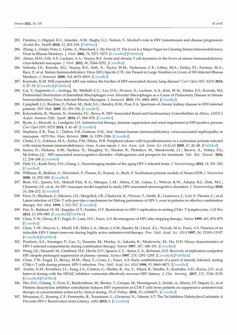
Viruses 2022, 14, 135 47 of 67
283. Pandrea, I.; Happel, K.I.; Amedee, A.M.; Bagby, G.J.; Nelson, S. Alcohol’s role in HIV transmission and disease progression.Alcohol Res. Health 2010, 33, 203–218. [PubMed]
284. Zhang, L.; Dailey Peter, J.; Gettie, A.; Blanchard, J.; Ho David, D. The Liver Is a Major Organ for Clearing Simian ImmunodeficiencyVirus in Rhesus Monkeys. J. Virol. 2002, 76, 5271–5273. [CrossRef] [PubMed]
285. Ahsan, M.H.; Gill, A.F.; Lackner, A.A.; Veazey, R.S. Acute and chronic T cell dynamics in the livers of simian immunodeficiencyvirus-infected macaques. J. Virol. 2012, 86, 5244–5252. [CrossRef]
286. Schmitz, J.E.; Kuroda, M.J.; Veazey, R.S.; Seth, A.; Taylor, W.M.; Nickerson, C.E.; Lifton, M.A.; Dailey, P.J.; Forman, M.A.;Racz, P.; et al. Simian Immunodeficiency Virus (SIV)-Specific CTL Are Present in Large Numbers in Livers of SIV-Infected RhesusMonkeys. J. Immunol. 2000, 164, 6015–6019. [CrossRef]
287. Kunisaki, K.M. Will expanded ART use reduce the burden of HIV-associated chronic lung disease? Curr Opin HIV AIDS 2014,9, 27–33. [CrossRef] [PubMed]
288. Cai, Y.; Sugimoto, C.; Arainga, M.; Midkiff, C.C.; Liu, D.X.; Alvarez, X.; Lackner, A.A.; Kim, W.-K.; Didier, E.S.; Kuroda, M.J.Preferential Destruction of Interstitial Macrophages over Alveolar Macrophages as a Cause of Pulmonary Disease in SimianImmunodeficiency Virus–Infected Rhesus Macaques. J. Immunol. 2015, 195, 4884–4891. [CrossRef]
289. Campbell, L.J.; Ibrahim, F.; Fisher, M.; Holt, S.G.; Hendry, B.M.; Post, F.A. Spectrum of chronic kidney disease in HIV-infectedpatients. HIV Med. 2009, 10, 329–336. [CrossRef]
290. Kalyesubula, R.; Wearne, N.; Semitala, F.C.; Bowa, K. HIV-Associated Renal and Genitourinary Comorbidities in Africa. JAIDS J.Acquir. Immune Defic. Syndr. 2014, 67, S68–S78. [CrossRef]
291. Ryom, L.; Mocroft, A.; Lundgren, J.D. Antiretroviral therapy, immune suppression and renal impairment in HIV-positive persons.Curr Opin HIV AIDS 2014, 9, 41–47. [CrossRef]
292. Stephens, E.B.; Tian, C.; Dalton, S.B.; Gattone, V.H., 2nd. Simian-human immunodeficiency virus-associated nephropathy inmacaques. AIDS Res. Hum. Retrovir. 2000, 16, 1295–1306. [CrossRef]
293. Clarke, C.L.; Eckhaus, M.A.; Zerfas, P.M.; Elkins, W.R. Peripheral edema with hypoalbuminemia in a nonhuman primate infectedwith simian-human immunodeficiency virus: A case report. J. Am. Assoc. Lab. Anim. Sci. JAALAS 2008, 47, 42–48. [PubMed]
294. Saylor, D.; Dickens, A.M.; Sacktor, N.; Haughey, N.; Slusher, B.; Pletnikov, M.; Mankowski, J.L.; Brown, A.; Volsky, D.J.;McArthur, J.C. HIV-associated neurocognitive disorder—Pathogenesis and prospects for treatment. Nat. Rev. Neurol. 2016,12, 234–248. [CrossRef]
295. Holt, J.L.; Kraft-Terry, S.D.; Chang, L. Neuroimaging studies of the aging HIV-1-infected brain. J. Neurovirology 2012, 18, 291–302.[CrossRef]
296. Williams, R.; Bokhari, S.; Silverstein, P.; Pinson, D.; Kumar, A.; Buch, S. Nonhuman primate models of NeuroAIDS. J. Neurovirol2008, 14, 292–300. [CrossRef]
297. Beck, S.E.; Queen, S.E.; Metcalf Pate, K.A.; Mangus, L.M.; Abreu, C.M.; Gama, L.; Witwer, K.W.; Adams, R.J.; Zink, M.C.;Clements, J.E.; et al. An SIV/macaque model targeted to study HIV-associated neurocognitive disorders. J. Neurovirology 2018,24, 204–212. [CrossRef]
298. Finzi, D.; Blankson, J.; Siliciano, J.D.; Margolick, J.B.; Chadwick, K.; Pierson, T.; Smith, K.; Lisziewicz, J.; Lori, F.; Flexner, C.; et al.Latent infection of CD4+ T cells provides a mechanism for lifelong persistence of HIV-1, even in patients on effective combinationtherapy. Nat. Med. 1999, 5, 512–517. [CrossRef]
299. Pan, X.; Baldauf, H.-M.; Keppler, O.T.; Fackler, O.T. Restrictions to HIV-1 replication in resting CD4+ T lymphocytes. Cell Res.2013, 23, 876–885. [CrossRef] [PubMed]
300. Chun, T.-W.; Davey, R.T.; Engel, D.; Lane, H.C.; Fauci, A.S. Re-emergence of HIV after stopping therapy. Nature 1999, 401, 874–875.[CrossRef]
301. Chun, T.-W.; Stuyver, L.; Mizell, S.B.; Ehler, L.A.; Mican, J.A.M.; Baseler, M.; Lloyd, A.L.; Nowak, M.A.; Fauci, A.S. Presence of aninducible HIV-1 latent reservoir during highly active antiretroviral therapy. Proc. Natl. Acad. Sci. USA 1997, 94, 13193–13197.[CrossRef] [PubMed]
302. Perelson, A.S.; Essunger, P.; Cao, Y.; Vesanen, M.; Hurley, A.; Saksela, K.; Markowitz, M.; Ho, D.D. Decay characteristics ofHIV-1-infected compartments during combination therapy. Nature 1997, 387, 188–191. [CrossRef]
303. Wong, J.K.; Hezareh, M.; Günthard, H.F.; Havlir, D.V.; Ignacio, C.C.; Spina, C.A.; Richman, D.D. Recovery of replication-competentHIV despite prolonged suppression of plasma viremia. Science 1997, 278, 1291–1295. [CrossRef] [PubMed]
304. Chun, T.W.; Engel, D.; Berrey, M.M.; Shea, T.; Corey, L.; Fauci, A.S. Early establishment of a pool of latently infected, restingCD4(+) T cells during primary HIV-1 infection. Proc. Natl. Acad. Sci. USA 1998, 95, 8869–8873. [CrossRef]
305. Archin, N.M.; Kirchherr, J.L.; Sung, J.A.; Clutton, G.; Sholtis, K.; Xu, Y.; Allard, B.; Stuelke, E.; Kashuba, A.D.; Kuruc, J.D.; et al.Interval dosing with the HDAC inhibitor vorinostat effectively reverses HIV latency. J. Clin. Investig. 2017, 127, 3126–3135.[CrossRef] [PubMed]
306. Wei, D.G.; Chiang, V.; Fyne, E.; Balakrishnan, M.; Barnes, T.; Graupe, M.; Hesselgesser, J.; Irrinki, A.; Murry, J.P.; Stepan, G.; et al.Histone deacetylase inhibitor romidepsin induces HIV expression in CD4 T cells from patients on suppressive antiretroviraltherapy at concentrations achieved by clinical dosing. PLoS Pathog. 2014, 10, e1004071. [CrossRef]
307. Mousseau, G.; Kessing, C.F.; Fromentin, R.; Trautmann, L.; Chomont, N.; Valente, S.T. The Tat Inhibitor Didehydro-Cortistatin APrevents HIV-1 Reactivation from Latency. mBio 2015, 6. [CrossRef]

Viruses 2022, 14, 135 48 of 67
308. Donahue, D.A.; Kuhl, B.D.; Sloan, R.D.; Wainberg, M.A. The viral protein Tat can inhibit the establishment of HIV-1 latency. J.Virol. 2012, 86, 3253–3263. [CrossRef]
309. Borducchi, E.N.; Cabral, C.; Stephenson, K.E.; Liu, J.; Abbink, P.; Ng’ang’a, D.; Nkolola, J.P.; Brinkman, A.L.; Peter, L.;Lee, B.C.; et al. Ad26/MVA therapeutic vaccination with TLR7 stimulation in SIV-infected rhesus monkeys. Nature 2016,540, 284. [CrossRef]
310. Tsai, A.; Irrinki, A.; Kaur, J.; Cihlar, T.; Kukolj, G.; Sloan, D.D.; Murry, J.P. Toll-Like Receptor 7 Agonist GS-9620 Induces HIVExpression and HIV-Specific Immunity in Cells from HIV-Infected Individuals on Suppressive Antiretroviral Therapy. J. Virol.2017, 91, e02166-16. [CrossRef]
311. Murray, S.M.; Down, C.M.; Boulware, D.R.; Stauffer, W.M.; Cavert, W.P.; Schacker, T.W.; Brenchley, J.M.; Douek, D.C. Reductionof Immune Activation with Chloroquine Therapy during Chronic HIV Infection. J. Virol. 2010, 84, 12082–12086. [CrossRef][PubMed]
312. Josefsson, L.; von Stockenstrom, S.; Faria, N.R.; Sinclair, E.; Bacchetti, P.; Killian, M.; Epling, L.; Tan, A.; Ho, T.; Lemey, P.; et al.The HIV-1 reservoir in eight patients on long-term suppressive antiretroviral therapy is stable with few genetic changes over time.Proc. Natl. Acad. Sci. 2013, 110, E4987–E4996. [CrossRef]
313. Porichis, F.; Hart, M.G.; Zupkosky, J.; Barblu, L.; Kwon, D.S.; McMullen, A.; Brennan, T.; Ahmed, R.; Freeman, G.J.;Kavanagh, D.G.; et al. Differential impact of PD-1 and/or interleukin-10 blockade on HIV-1-specific CD4 T cell and antigen-presenting cell functions. J. Virol. 2014, 88, 2508–2518. [CrossRef]
314. Palmer, B.E.; Neff, C.P.; LeCureux, J.; Ehler, A.; Dsouza, M.; Remling-Mulder, L.; Korman, A.J.; Fontenot, A.P.; Akkina, R. In vivoblockade of the PD-1 receptor suppresses HIV-1 viral loads and improves CD4+ T cell levels in humanized mice. J. Immunol. 2013,190, 211–219. [CrossRef]
315. Jones, R.B.; Ndhlovu, L.C.; Barbour, J.D.; Sheth, P.M.; Jha, A.R.; Long, B.R.; Wong, J.C.; Satkunarajah, M.; Schweneker, M.;Chapman, J.M.; et al. Tim-3 expression defines a novel population of dysfunctional T cells with highly elevated frequencies inprogressive HIV-1 infection. J. Exp. Med. 2008, 205, 2763–2779. [CrossRef]
316. Zhang, Z.-N.; Zhu, M.-L.; Chen, Y.-H.; Fu, Y.-J.; Zhang, T.-W.; Jiang, Y.-J.; Chu, Z.-X.; Shang, H. Elevation of Tim-3 and PD-1Expression on T Cells Appears Early in HIV Infection, and Differential Tim-3 and PD-1 Expression Patterns Can Be Induced byCommon γ-Chain Cytokines. BioMed Res. Int. 2015, 2015, 916936. [CrossRef] [PubMed]
317. Peretz, Y.; He, Z.; Shi, Y.; Yassine-Diab, B.; Goulet, J.-P.; Bordi, R.; Filali-Mouhim, A.; Loubert, J.-B.; El-Far, M.; Dupuy, F.P.; et al.CD160 and PD-1 Co-Expression on HIV-Specific CD8 T Cells Defines a Subset with Advanced Dysfunction. PLoS Pathog. 2012,8, e1002840. [CrossRef] [PubMed]
318. Vandergeeten, C.; Fromentin, R.; DaFonseca, S.; Lawani, M.B.; Sereti, I.; Lederman, M.M.; Ramgopal, M.; Routy, J.P.; Sekaly, R.P.;Chomont, N. Interleukin-7 promotes HIV persistence during antiretroviral therapy. Blood 2013, 121, 4321–4329. [CrossRef]
319. Seay, K.; Church, C.; Zheng, J.H.; Deneroff, K.; Ochsenbauer, C.; Kappes, J.C.; Liu, B.; Jeng, E.K.; Wong, H.C.; Goldstein, H. InVivo Activation of Human NK Cells by Treatment with an Interleukin-15 Superagonist Potently Inhibits Acute In Vivo HIV-1Infection in Humanized Mice. J. Virol. 2015, 89, 6264–6274. [CrossRef]
320. Younes, S.A.; Freeman, M.L.; Mudd, J.C.; Shive, C.L.; Reynaldi, A.; Panigrahi, S.; Estes, J.D.; Deleage, C.; Lucero, C.;Anderson, J.; et al. IL-15 promotes activation and expansion of CD8+ T cells in HIV-1 infection. J. Clin. Investig. 2016,126, 2745–2756. [CrossRef]
321. Mendez-Lagares, G.; Lu, D.; Merriam, D.; Baker, C.A.; Villinger, F.; Van Rompay, K.K.A.; McCune, J.M.; Hartigan-O’Connor, D.J.IL-21 Therapy Controls Immune Activation and Maintains Antiviral CD8(+) T Cell Responses in Acute Simian ImmunodeficiencyVirus Infection. AIDS Res. Hum. Retrovir. 2017, 33, S81–S92. [CrossRef] [PubMed]
322. Hale, M.; Mesojednik, T.; Romano Ibarra, G.S.; Sahni, J.; Bernard, A.; Sommer, K.; Scharenberg, A.M.; Rawlings, D.J.; Wagner, T.A.Engineering HIV-Resistant, Anti-HIV Chimeric Antigen Receptor T Cells. Mol. Ther. : J. Am. Soc. Gene Ther. 2017, 25, 570–579.[CrossRef] [PubMed]
323. Hütter, G.; Bodor, J.; Ledger, S.; Boyd, M.; Millington, M.; Tsie, M.; Symonds, G. CCR5 Targeted Cell Therapy for HIV andPrevention of Viral Escape. Viruses 2015, 7, 4186–4203. [CrossRef] [PubMed]
324. Eberhard, J.M.; Angin, M.; Passaes, C.; Salgado, M.; Monceaux, V.; Knops, E.; Kobbe, G.; Jensen, B.; Christopeit, M.;Kroger, N.; et al. Vulnerability to reservoir reseeding due to high immune activation after allogeneic hematopoietic stem celltransplantation in individuals with HIV-1. Sci. Transl Med. 2020, 12. [CrossRef]
325. Saez-Cirion, A.; Muller-Trutwin, M. The Yellow Brick Road towards HIV Eradication. Trends Immunol 2019, 40, 465–467. [CrossRef]326. Dufour, C.; Gantner, P.; Fromentin, R.; Chomont, N. The multifaceted nature of HIV latency. J. Clin. Investig. 2020, 130, 3381–3390.
[CrossRef]327. Margolis, D.M.; Archin, N.M.; Cohen, M.S.; Eron, J.J.; Ferrari, G.; Garcia, J.V.; Gay, C.L.; Goonetilleke, N.; Joseph, S.B.;
Swanstrom, R.; et al. Curing HIV: Seeking to Target and Clear Persistent Infection. Cell 2020, 181, 189–206. [CrossRef] [PubMed]328. Massanella, M.; Fromentin, R.; Chomont, N. Residual inflammation and viral reservoirs: Alliance against an HIV cure. Curr Opin
HIV AIDS 2016, 11, 234–241. [CrossRef]329. Passaes, C.P.; Saez-Cirion, A. HIV cure research: Advances and prospects. Virology 2014, 454–455, 340–352. [CrossRef] [PubMed]330. Fromentin, R.; Chomont, N. HIV persistence in subsets of CD4+ T cells: 50 shades of reservoirs. Semin. Immunol. 2020, 101438.
[CrossRef] [PubMed]

Viruses 2022, 14, 135 49 of 67
331. Rabezanahary, H.; Moukambi, F.; Palesch, D.; Clain, J.; Racine, G.; Andreani, G.; Benmadid-Laktout, G.; Zghidi-Abouzid, O.;Soundaramourty, C.; Tremblay, C.; et al. Despite early antiretroviral therapy effector memory and follicular helper CD4 T cells aremajor reservoirs in visceral lymphoid tissues of SIV-infected macaques. Mucosal Immunol. 2020, 13, 149–160. [CrossRef] [PubMed]
332. Finzi, D.; Hermankova, M.; Pierson, T.; Carruth, L.M.; Buck, C.; Chaisson, R.E.; Quinn, T.C.; Chadwick, K.; Margolick, J.;Brookmeyer, R.; et al. Identification of a Reservoir for HIV-1 in Patients on Highly Active Antiretroviral Therapy. Science 1997,278, 1295–1300. [CrossRef]
333. Jordan, A.; Bisgrove, D.; Verdin, E. HIV reproducibly establishes a latent infection after acute infection of T cells in vitro. EMBO J.2003, 22. [CrossRef]
334. Siliciano, J.D.; Kajdas, J.; Finzi, D.; Quinn, T.C.; Chadwick, K.; Margolick, J.B.; Kovacs, C.; Gange, S.J.; Siliciano, R.F. Long-termfollow-up studies confirm the stability of the latent reservoir for HIV-1 in resting CD4+ T cells. Nat. Med. 2003, 9, 727–728.[CrossRef]
335. Delobel, P.; Sandres-Sauné, K.; Cazabat, M.; L’Faqihi, F.-E.; Aquilina, C.; Obadia, M.; Pasquier, C.; Marchou, B.; Massip, P.;Izopet, J. Persistence of distinct HIV-1 populations in blood monocytes and naive and memory CD4 T cells during prolongedsuppressive HAART. AIDS (Lond. Engl.) 2005, 19, 1739–1750. [CrossRef] [PubMed]
336. Chomont, N.; El-Far, M.; Ancuta, P.; Trautmann, L.; Procopio, F.A.; Yassine-Diab, B.; Boucher, G.; Boulassel, M.-R.; Ghattas, G.;Brenchley, J.M.; et al. HIV reservoir size and persistence are driven by T cell survival and homeostatic proliferation. Nat. Med.2009, 15, 893–900. [CrossRef]
337. Buzon, M.J.; Sun, H.; Li, C.; Shaw, A.; Seiss, K.; Ouyang, Z.; Martin-Gayo, E.; Leng, J.; Henrich, T.J.; Li, J.Z.; et al. HIV-1 persistencein CD4+ T cells with stem cell-like properties. Nat. Med. 2014, 20, 139–142. [CrossRef] [PubMed]
338. Kleinman, A.J.; Sivanandham, R.; Pandrea, I.; Chougnet, C.A.; Apetrei, C. Regulatory T Cells As Potential Targets for HIV CureResearch. Front. Immunol 2018, 9, 734. [CrossRef]
339. Pallikkuth, S.; Sharkey, M.; Babic, D.Z.; Gupta, S.; Stone, G.W.; Fischl, M.A.; Stevenson, M.; Pahwa, S. Peripheral T FollicularHelper Cells Are the Major HIV Reservoir within Central Memory CD4 T Cells in Peripheral Blood from Chronically HIV-InfectedIndividuals on Combination Antiretroviral Therapy. J. Virol. 2016, 90, 2718–2728. [CrossRef] [PubMed]
340. Wong, M.E.; Jaworowski, A.; Hearps, A.C. The HIV Reservoir in Monocytes and Macrophages. Front. Immunol. 2019, 10, 1435.[CrossRef] [PubMed]
341. Spiegel, H.; Herbst, H.; Niedobitek, G.; Foss, H.D.; Stein, H. Follicular dendritic cells are a major reservoir for human immunode-ficiency virus type 1 in lymphoid tissues facilitating infection of CD4+ T-helper cells. Am. J. Pathol 1992, 140, 15–22.
342. Badia, R.; Ballana, E.; Castellví, M.; García-Vidal, E.; Pujantell, M.; Clotet, B.; Prado, J.G.; Puig, J.; Martínez, M.A.; Riveira-Muñoz, E.; et al. CD32 expression is associated to T-cell activation and is not a marker of the HIV-1 reservoir. Nat. Commun. 2018,9, 2739. [CrossRef]
343. Hütter, G.; Nowak, D.; Mossner, M.; Ganepola, S.; Müßig, A.; Allers, K.; Schneider, T.; Hofmann, J.; Kücherer, C.; Blau, O.; et al.Long-Term Control of HIV by CCR5 Delta32/Delta32 Stem-Cell Transplantation. N. Engl. J. Med. 2009, 360, 692–698. [CrossRef]
344. Henrich, T.J.; Hanhauser, E.; Marty, F.M.; Sirignano, M.N.; Keating, S.; Lee, T.H.; Robles, Y.P.; Davis, B.T.; Li, J.Z.; Heisey, A.; et al.Antiretroviral-free hiv-1 remission and viral rebound after allogeneic stem cell transplantation: Report of 2 cases. Ann. Intern.Med. 2014, 161, 319–327. [CrossRef]
345. Persaud, D.; Gay, H.; Ziemniak, C.; Chen, Y.H.; Piatak, M.J.; Chun, T.-W.; Strain, M.; Richman, D.; Luzuriaga, K. Absence ofDetectable HIV-1 Viremia after Treatment Cessation in an Infant. N. Engl. J. Med. 2013, 369, 1828–1835. [CrossRef] [PubMed]
346. Luzuriaga, K.; Gay, H.; Ziemniak, C.; Sanborn, K.B.; Somasundaran, M.; Rainwater-Lovett, K.; Mellors, J.W.; Rosenbloom, D.;Persaud, D. Viremic Relapse after HIV-1 Remission in a Perinatally Infected Child. N. Engl. J. Med. 2015, 372, 786–788. [CrossRef]
347. Sáez-Cirión, A.; Bacchus, C.; Hocqueloux, L.; Avettand-Fenoel, V.; Girault, I.; Lecuroux, C.; Potard, V.; Versmisse, P.; Melard, A.;Prazuck, T.; et al. Post-Treatment HIV-1 Controllers with a Long-Term Virological Remission after the Interruption of EarlyInitiated Antiretroviral Therapy ANRS VISCONTI Study. PLoS Pathog. 2013, 9, e1003211. [CrossRef]
348. Violari, A.; Cotton, M.; Kuhn, L.; Schramm, D.; Paximadis, M.; Shalekoff, S.; Dias, B.d.C.; Otwombe, K.; Liberty, A.;McIntyre, J.; et al. Viral and host characterists of a child with perinatal HIV-1 following a prolonged period after ART cessation inthe CHER trial. In Proceedings of the International AIDS Society 2017, Paris, France, 23–26 July 2017.
349. Cummins, N.W.; Rizza, S.; Litzow, M.R.; Hua, S.; Lee, G.Q.; Einkauf, K.; Chun, T.-W.; Rhame, F.; Baker, J.V.; Busch, M.P.; et al.Extensive virologic and immunologic characterization in an HIV-infected individual following allogeneic stem cell transplantand analytic cessation of antiretroviral therapy: A case study. PLoS Med. 2017, 14, e1002461. [CrossRef]
350. Folks, T.; Powell, D.M.; Lightfoote, M.M.; Benn, S.; Martin, M.A.; Fauci, A.S. Induction of HTLV-III/LAV from a nonvirus-producing T-cell line: Implications for latency. Science 1986, 231, 600–602. [CrossRef] [PubMed]
351. Nabel, G.; Baltimore, D. An inducible transcription factor activates expression of human immunodeficiency virus in T cells.Nature 1987, 326, 711–713. [CrossRef]
352. Siekevitz, M.; Josephs, S.F.; Dukovich, M.; Peffer, N.; Wong-Staal, F.; Greene, W.C. Activation of the HIV-1 LTR by T cell mitogensand the trans-activator protein of HTLV-I. Science 1987, 238, 1575–1578. [CrossRef] [PubMed]
353. Duh, E.J.; Maury, W.J.; Folks, T.M.; Fauci, A.S.; Rabson, A.B. Tumor necrosis factor alpha activates human immunodeficiencyvirus type 1 through induction of nuclear factor binding to the NF-kappa B sites in the long terminal repeat. Proc. Natl. Acad. Sci.USA 1989, 86, 5974–5978. [CrossRef]
Viruses 2022, 14, 135 50 of 67
354. Zack, J.A.; Arrigo, S.J.; Weitsman, S.R.; Go, A.S.; Haislip, A.; Chen, I.S.Y. HIV-1 entry into quiescent primary lymphocytes:Molecular analysis reveals a labile, latent viral structure. Cell 1990, 61, 213–222. [CrossRef]
355. Bukrinsky, M.I.; Stanwick, T.L.; Dempsey, M.P.; Stevenson, M. Quiescent T Lymphocytes as an Inducible Virus Reservoir in HIV-1Infection. Science 1991, 254, 423–427. [CrossRef] [PubMed]
356. Chun, T.-W.; Finzi, D.; Margolick, J.; Chadwick, K.; Schwartz, D.; Siliciano, R.F. In vivo fate of HIV-1-infected T cells: Quantitativeanalysis of the transition to stable latency. Nat. Med. 1995, 1, 1284–1290. [CrossRef] [PubMed]
357. Sahu, G.K.; Lee, K.; Ji, J.; Braciale, V.; Baron, S.; Cloyd, M.W. A novel in vitro system to generate and study latently HIV-infectedlong-lived normal CD4+ T-lymphocytes. Virology 2006, 355, 127–137. [CrossRef] [PubMed]
358. Bosque, A.; Planelles, V. Induction of HIV-1 latency and reactivation in primary memory CD4+ T cells. Blood 2009, 113, 58–65.[CrossRef]
359. Burke, B.; Brown, H.J.; Marsden, M.D.; Bristol, G.; Vatakis, D.N.; Zack, J.A. Primary cell model for activation-inducible humanimmunodeficiency virus. J. Virol. 2007, 81, 7424–7434. [CrossRef]
360. Marini, A.; Harper, J.M.; Romerio, F. An in vitro system to model the establishment and reactivation of HIV-1 latency. J. Immunol.2008, 181, 7713–7720. [CrossRef]
361. Tyagi, M.; Pearson, R.J.; Karn, J. Establishment of HIV Latency in Primary CD4+ Cells Is due to Epigenetic TranscriptionalSilencing and P-TEFb Restriction. J. Virol. 2010, 84, 6425–6437. [CrossRef]
362. Yang, H.C.; Xing, S.; Shan, L.; O’Connell, K.; Dinoso, J.; Shen, A.; Zhou, Y.; Shrum, C.K.; Han, Y.; Liu, J.O.; et al. Small-moleculescreening using a human primary cell model of HIV latency identifies compounds that reverse latency without cellular activation.J. Clin. Investig. 2009, 119, 3473–3486. [CrossRef]
363. Yang, H.-C. Primary cell models of HIV latency. Curr. Opin. HIV AIDS 2011, 6, 62–67. [CrossRef]364. Schröder, A.R.; Shinn, P.; Chen, H.; Berry, C.; Ecker, J.R.; Bushman, F. HIV-1 integration in the human genome favors active genes
and local hotspots. Cell 2002, 110, 521–529. [CrossRef]365. Lewinski, M.K.; Yamashita, M.; Emerman, M.; Ciuffi, A.; Marshall, H.; Crawford, G.; Collins, F.; Shinn, P.; Leipzig, J.;
Hannenhalli, S.; et al. Retroviral DNA integration: Viral and cellular determinants of target-site selection. PLoS Pathog. 2006,2, e60. [CrossRef]
366. Han, Y.; Lassen, K.; Monie, D.; Sedaghat, A.R.; Shimoji, S.; Liu, X.; Pierson, T.C.; Margolick, J.B.; Siliciano, R.F.; Siliciano, J.D.Resting CD4+ T cells from human immunodeficiency virus type 1 (HIV-1)-infected individuals carry integrated HIV-1 genomeswithin actively transcribed host genes. J. Virol. 2004, 78, 6122–6133. [CrossRef]
367. Verdin, E.; Paras Jr, P.; Van Lint, C. Chromatin disruption in the promoter of human immunodeficiency virus type 1 duringtranscriptional activation. EMBO J. 1993, 12, 3249–3259. [CrossRef] [PubMed]
368. Keedy, K.S.; Archin, N.M.; Gates, A.T.; Espeseth, A.; Hazuda, D.J.; Margolis, D.M. A limited group of class I histone deacetylasesacts to repress human immunodeficiency virus type 1 expression. J. Virol. 2009, 83, 4749–4756. [CrossRef]
369. Van Lint, C.; Emiliani, S.; Ott, M.; Verdin, E. Transcriptional activation and chromatin remodeling of the HIV-1 promoter inresponse to histone acetylation. EMBO J. 1996, 15, 1112–1120. [CrossRef] [PubMed]
370. Lusic, M.; Marcello, A.; Cereseto, A.; Giacca, M. Regulation of HIV-1 gene expression by histone acetylation and factor recruitmentat the LTR promoter. EMBO J. 2003, 22, 6550–6561. [CrossRef]
371. Friedman, J.; Cho, W.K.; Chu, C.K.; Keedy, K.S.; Archin, N.M.; Margolis, D.M.; Karn, J. Epigenetic silencing of HIV-1 by thehistone H3 lysine 27 methyltransferase enhancer of Zeste 2. J. Virol. 2011, 85, 9078–9089. [CrossRef] [PubMed]
372. Boehm, D.; Jeng, M.; Camus, G.; Gramatica, A.; Schwarzer, R.; Johnson, J.R.; Hull, P.A.; Montano, M.; Sakane, N.; Pagans, S.; et al.SMYD2-Mediated Histone Methylation Contributes to HIV-1 Latency. Cell Host Microbe 2017, 21, 569–579. [CrossRef]
373. Imai, K.; Togami, H.; Okamoto, T. Involvement of Histone H3 Lysine 9 (H3K9) Methyltransferase G9a in the Maintenance ofHIV-1 Latency and Its Reactivation by BIX01294 *. J. Biol. Chem. 2010, 285, 16538–16545. [CrossRef] [PubMed]
374. Chéné, I.d.; Basyuk, E.; Lin, Y.-L.; Triboulet, R.; Knezevich, A.; Chable-Bessia, C.; Mettling, C.; Baillat, V.; Reynes, J.;Corbeau, P.; et al. Suv39H1 and HP1γ are responsible for chromatin-mediated HIV-1 transcriptional silencing and post-integration latency. EMBO J. 2007, 26, 424–435. [CrossRef]
375. He, G.; Margolis, D.M. Counterregulation of chromatin deacetylation and histone deacetylase occupancy at the integratedpromoter of human immunodeficiency virus type 1 (HIV-1) by the HIV-1 repressor YY1 and HIV-1 activator Tat. Mol. Cell Biol.2002, 22, 2965–2973. [CrossRef]
376. Williams, S.A.; Chen, L.-F.; Kwon, H.; Ruiz-Jarabo, C.M.; Verdin, E.; Greene, W.C. NF-κB p50 promotes HIV latency throughHDAC recruitment and repression of transcriptional initiation. EMBO J. 2006, 25, 139–149. [CrossRef]
377. Tyagi, M.; Karn, J. CBF-1 promotes transcriptional silencing during the establishment of HIV-1 latency. EMBO J. 2007,26, 4985–4995. [CrossRef] [PubMed]
378. Greger, I.H.; Demarchi, F.; Giacca, M.; Proudfoot, N.J. Transcriptional interference perturbs the binding of Sp1 to the HIV-1promoter. Nucleic Acids Res. 1998, 26, 1294–1301. [CrossRef]
379. Lenasi, T.; Contreras, X.; Peterlin, B.M. Transcriptional Interference Antagonizes Proviral Gene Expression to Promote HIVLatency. Cell Host Microbe 2008, 4, 123–133. [CrossRef] [PubMed]
380. Han, Y.; Lin, Y.B.; An, W.; Xu, J.; Yang, H.-C.; O’Connell, K.; Dordai, D.; Boeke, J.D.; Siliciano, J.D.; Siliciano, R.F. Orientation-Dependent Regulation of Integrated HIV-1 Expression by Host Gene Transcriptional Readthrough. Cell Host Microbe 2008,4, 134–146. [CrossRef] [PubMed]

Viruses 2022, 14, 135 51 of 67
381. Sedore, S.C.; Byers, S.A.; Biglione, S.; Price, J.P.; Maury, W.J.; Price, D.H. Manipulation of P-TEFb control machinery by HIV:Recruitment of P-TEFb from the large form by Tat and binding of HEXIM1 to TAR. Nucleic Acids Res. 2007, 35, 4347–4358.[CrossRef]
382. Muniz, L.; Egloff, S.; Ughy, B.; Jády, B.E.; Kiss, T. Controlling Cellular P-TEFb Activity by the HIV-1 Transcriptional TransactivatorTat. PLoS Pathog. 2010, 6, e1001152. [CrossRef]
383. Parada, C.A.; Roeder, R.G. Enhanced processivity of RNA polymerase II triggered by Tat-induced phosphorylation of itscarboxy-terminal domain. Nature 1996, 384, 375–378. [CrossRef]
384. Kim, Y.K.; Bourgeois, C.F.; Isel, C.; Churcher, M.J.; Karn, J. Phosphorylation of the RNA polymerase II carboxyl-terminal domainby CDK9 is directly responsible for human immunodeficiency virus type 1 Tat-activated transcriptional elongation. Mol. Cell Biol.2002, 22, 4622–4637. [CrossRef]
385. Bourgeois, C.F.; Kim, Y.K.; Churcher, M.J.; West, M.J.; Karn, J. Spt5 cooperates with human immunodeficiency virus type 1 Tat bypreventing premature RNA release at terminator sequences. Mol. Cell Biol. 2002, 22, 1079–1093. [CrossRef] [PubMed]
386. Jang, M.K.; Mochizuki, K.; Zhou, M.; Jeong, H.-S.; Brady, J.N.; Ozato, K. The Bromodomain Protein Brd4 Is a Positive RegulatoryComponent of P-TEFb and Stimulates RNA Polymerase II-Dependent Transcription. Mol. Cell 2005, 19, 523–534. [CrossRef]
387. Yang, Z.; Yik, J.H.N.; Chen, R.; He, N.; Jang, M.K.; Ozato, K.; Zhou, Q. Recruitment of P-TEFb for Stimulation of TranscriptionalElongation by the Bromodomain Protein Brd4. Mol. Cell 2005, 19, 535–545. [CrossRef]
388. Li, Z.; Guo, J.; Wu, Y.; Zhou, Q. The BET bromodomain inhibitor JQ1 activates HIV latency through antagonizing Brd4 inhibitionof Tat-transactivation. Nucleic Acids Res. 2013, 41, 277–287. [CrossRef]
389. Boehm, D.; Calvanese, V.; Dar, R.D.; Xing, S.; Schroeder, S.; Martins, L.; Aull, K.; Li, P.-C.; Planelles, V.; Bradner, J.E.; et al. BETbromodomain-targeting compounds reactivate HIV from latency via a Tat-independent mechanism. Cell Cycle 2013, 12, 452–462.[CrossRef] [PubMed]
390. Chun, T.-W.; Justement, J.S.; Moir, S.; Hallahan, C.W.; Maenza, J.; Mullins, J.I.; Collier, A.C.; Corey, L.; Fauci, A.S. Decay of theHIV Reservoir in Patients Receiving Antiretroviral Therapy for Extended Periods: Implications for Eradication of Virus. J. Infect.Dis. 2007, 195, 1762–1764. [CrossRef] [PubMed]
391. Gandhi, R.T.; Cyktor, J.C.; Bosch, R.J.; Mar, H.; Laird, G.M.; Martin, A.; Collier, A.C.; Riddler, S.A.; Macatangay, B.J.;Rinaldo, C.R.; et al. Selective Decay of Intact HIV-1 Proviral DNA on Antiretroviral Therapy. J. Infect. Dis. 2021, 223, 225–233.[CrossRef]
392. Chun, T.W.; Carruth, L.; Finzi, D.; Shen, X.; DiGiuseppe, J.A.; Taylor, H.; Hermankova, M.; Chadwick, K.; Margolick, J.;Quinn, T.C.; et al. Quantification of latent tissue reservoirs and total body viral load in HIV-1 infection. Nature 1997, 387, 183–188.[CrossRef] [PubMed]
393. Bruner, K.M.; Murray, A.J.; Pollack, R.A.; Soliman, M.G.; Laskey, S.B.; Capoferri, A.A.; Lai, J.; Strain, M.C.; Lada, S.M.;Hoh, R.; et al. Defective proviruses rapidly accumulate during acute HIV-1 infection. Nat. Med. 2016, 22, 1043–1049. [CrossRef]
394. Bullen, C.K.; Laird, G.M.; Durand, C.M.; Siliciano, J.D.; Siliciano, R.F. New ex vivo approaches distinguish effective and ineffectivesingle agents for reversing HIV-1 latency in vivo. Nat. Med. 2014, 20, 425–429. [CrossRef]
395. Kuzmichev, Y.V.; Veenhuis, R.T.; Pohlmeyer, C.W.; Garliss, C.C.; Walker-Sperling, V.E.; Blankson, J.N. A CD3/CD28 microbead-based HIV-1 viral outgrowth assay. J. Virus Erad. 2017, 3, 85–89. [CrossRef]
396. Patel, S.S.; Duby, A.D.; Thiele, D.L.; Lipsky, P.E. Phenotypic and functional characterization of human T cell clones. J. Immunol.1988, 141, 3726–3736.
397. Beliakova-Bethell, N.; Hezareh, M.; Wong, J.K.; Strain, M.C.; Lewinski, M.K.; Richman, D.D.; Spina, C.A. Relative efficacy of T cellstimuli as inducers of productive HIV-1 replication in latently infected CD4 lymphocytes from patients on suppressive cART.Virology 2017, 508, 127–133. [CrossRef]
398. Laird, G.M.; Eisele, E.E.; Rabi, S.A.; Lai, J.; Chioma, S.; Blankson, J.N.; Siliciano, J.D.; Siliciano, R.F. Rapid quantification of thelatent reservoir for HIV-1 using a viral outgrowth assay. PLoS Pathog. 2013, 9, e1003398. [CrossRef] [PubMed]
399. Pollack, R.A.; Jones, R.B.; Pertea, M.; Bruner, K.M.; Martin, A.R.; Thomas, A.S.; Capoferri, A.A.; Beg, S.A.; Huang, S.-H.;Karandish, S.; et al. Defective HIV-1 Proviruses Are Expressed and Can Be Recognized by Cytotoxic T Lymphocytes, whichShape the Proviral Landscape. Cell Host Microbe 2017, 21, 494–506. [CrossRef]
400. Ho, Y.-C.; Shan, L.; Hosmane, N.N.; Wang, J.; Laskey, S.B.; Rosenbloom, D.I.S.; Lai, J.; Blankson, J.N.; Siliciano, J.D.; Siliciano, R.F.Replication-Competent Noninduced Proviruses in the Latent Reservoir Increase Barrier to HIV-1 Cure. Cell 2013, 155, 540–551.[CrossRef]
401. Hosmane, N.N.; Kwon, K.J.; Bruner, K.M.; Capoferri, A.A.; Beg, S.; Rosenbloom, D.I.; Keele, B.F.; Ho, Y.C.; Siliciano, J.D.;Siliciano, R.F. Proliferation of latently infected CD4(+) T cells carrying replication-competent HIV-1: Potential role in latentreservoir dynamics. J. Exp. Med. 2017, 214, 959–972. [CrossRef] [PubMed]
402. Bui, J.K.; Sobolewski, M.D.; Keele, B.F.; Spindler, J.; Musick, A.; Wiegand, A.; Luke, B.T.; Shao, W.; Hughes, S.H.; Coffin, J.M.; et al.Proviruses with identical sequences comprise a large fraction of the replication-competent HIV reservoir. PLoS Pathog. 2017,13, e1006283. [CrossRef] [PubMed]
403. Li, B.; Gladden, A.D.; Altfeld, M.; Kaldor, J.M.; Cooper, D.A.; Kelleher, A.D.; Allen, T.M. Rapid reversion of sequence polymor-phisms dominates early human immunodeficiency virus type 1 evolution. J. Virol. 2007, 81, 193–201. [CrossRef]
404. Wang, Z.; Simonetti, F.R.; Siliciano, R.F.; Laird, G.M. Measuring replication competent HIV-1: Advances and challenges indefining the latent reservoir. Retrovirology 2018, 15, 21. [CrossRef]

Viruses 2022, 14, 135 52 of 67
405. Hiener, B.; Horsburgh, B.A.; Eden, J.S.; Barton, K.; Schlub, T.E.; Lee, E.; von Stockenstrom, S.; Odevall, L.; Milush, J.M.;Liegler, T.; et al. Identification of Genetically Intact HIV-1 Proviruses in Specific CD4(+) T Cells from Effectively Treated Partici-pants. Cell Rep. 2017, 21, 813–822. [CrossRef] [PubMed]
406. Lee, G.Q.; Orlova-Fink, N.; Einkauf, K.; Chowdhury, F.Z.; Sun, X.; Harrington, S.; Kuo, H.H.; Hua, S.; Chen, H.R.; Ouyang, Z.; et al.Clonal expansion of genome-intact HIV-1 in functionally polarized Th1 CD4+ T cells. J. Clin. Investig. 2017, 127, 2689–2696.[CrossRef]
407. Kijak, G.H.; Sanders-Buell, E.; Pham, P.; Harbolick, E.A.; Oropeza, C.; O’Sullivan, A.M.; Bose, M.; Beckett, C.G.; Milazzo, M.;Robb, M.L.; et al. Next-generation sequencing of HIV-1 single genome amplicons. Biomol Detect. Quantif. 2019, 17, 100080.[CrossRef] [PubMed]
408. Arias, A.; López, P.; Sánchez, R.; Yamamura, Y.; Rivera-Amill, V. Sanger and Next Generation Sequencing Approaches to EvaluateHIV-1 Virus in Blood Compartments. Int. J. Env. Res. Public Health 2018, 15, 1697. [CrossRef] [PubMed]
409. Bruner, K.M.; Wang, Z.; Simonetti, F.R.; Bender, A.M.; Kwon, K.J.; Sengupta, S.; Fray, E.J.; Beg, S.A.; Antar, A.A.R.;Jenike, K.M.; et al. A quantitative approach for measuring the reservoir of latent HIV-1 proviruses. Nature 2019, 566, 120–125.[CrossRef]
410. Kinloch, N.N.; Ren, Y.; Conce Alberto, W.D.; Dong, W.; Khadka, P.; Huang, S.H.; Mota, T.M.; Wilson, A.; Shahid, A.;Kirkby, D.; et al. HIV-1 diversity considerations in the application of the Intact Proviral DNA Assay (IPDA). Nat. Commun. 2021,12, 165. [CrossRef]
411. Gaebler, C.; Lorenzi, J.C.C.; Oliveira, T.Y.; Nogueira, L.; Ramos, V.; Lu, C.-L.; Pai, J.A.; Mendoza, P.; Jankovic, M.; Caskey, M.; et al.Combination of quadruplex qPCR and next-generation sequencing for qualitative and quantitative analysis of the HIV-1 latentreservoir. J. Exp. Med. 2019, 216, 2253–2264. [CrossRef]
412. Eriksson, S.; Graf, E.H.; Dahl, V.; Strain, M.C.; Yukl, S.A.; Lysenko, E.S.; Bosch, R.J.; Lai, J.; Chioma, S.; Emad, F.; et al. ComparativeAnalysis of Measures of Viral Reservoirs in HIV-1 Eradication Studies. PLoS Pathog. 2013, 9, e1003174. [CrossRef] [PubMed]
413. Imamichi, H.; Dewar, R.L.; Adelsberger, J.W.; Rehm, C.A.; O’Doherty, U.; Paxinos, E.E.; Fauci, A.S.; Lane, H.C. Defective HIV-1proviruses produce novel protein-coding RNA species in HIV-infected patients on combination antiretroviral therapy. Proc. Natl.Acad. Sci. USA 2016, 113, 8783–8788. [CrossRef] [PubMed]
414. Imamichi, H.; Smith, M.; Adelsberger, J.W.; Izumi, T.; Scrimieri, F.; Sherman, B.T.; Rehm, C.A.; Imamichi, T.; Pau, A.;Catalfamo, M.; et al. Defective HIV-1 proviruses produce viral proteins. Proc. Natl. Acad. Sci. USA 2020, 117, 3704–3710.[CrossRef]
415. Apetrei, C.; Pandrea, I.; Mellors, J.W. Nonhuman primate models for HIV cure research. PLoS Pathog. 2012, 8, e1002892. [CrossRef][PubMed]
416. Choudhary, S.K.; Archin, N.M.; Cheema, M.; Dahl, N.P.; Garcia, J.V.; Margolis, D.M. Latent HIV-1 infection of resting CD4(+)T cells in the humanized Rag2(-)/(-) gammac(-)/(-) mouse. J. Virol. 2012, 86, 114–120. [CrossRef]
417. Denton, P.W.; Olesen, R.; Choudhary, S.K.; Archin, N.M.; Wahl, A.; Swanson, M.D.; Chateau, M.; Nochi, T.; Krisko, J.F.;Spagnuolo, R.A.; et al. Generation of HIV latency in humanized BLT mice. J. Virol. 2012, 86, 630–634. [CrossRef]
418. Honeycutt, J.B.; Wahl, A.; Baker, C.; Spagnuolo, R.A.; Foster, J.; Zakharova, O.; Wietgrefe, S.; Caro-Vegas, C.; Madden, V.;Sharpe, G.; et al. Macrophages sustain HIV replication in vivo independently of T cells. J. Clin. Investig. 2016, 126, 1353–1366.[CrossRef]
419. Olesen, R.; Swanson, M.D.; Kovarova, M.; Nochi, T.; Chateau, M.; Honeycutt, J.B.; Long, J.M.; Denton, P.W.; Hudgens, M.G.;Richardson, A.; et al. ART influences HIV persistence in the female reproductive tract and cervicovaginal secretions. J. Clin.Investig. 2016, 126, 892–904. [CrossRef]
420. Garcia, J.V. In vivo platforms for analysis of HIV persistence and eradication. J. Clin. Investig. 2016, 126, 424–431. [CrossRef]421. Nishimura, Y.; Sadjadpour, R.; Mattapallil, J.J.; Igarashi, T.; Lee, W.; Buckler-White, A.; Roederer, M.; Chun, T.W.; Martin, M.A.
High frequencies of resting CD4+ T cells containing integrated viral DNA are found in rhesus macaques during acute lentivirusinfections. Proc. Natl. Acad. Sci. USA 2009, 106, 8015–8020. [CrossRef] [PubMed]
422. Shen, A.; Zink, M.C.; Mankowski, J.L.; Chadwick, K.; Margolick, J.B.; Carruth, L.M.; Li, M.; Clements, J.E.; Siliciano, R.F.Resting CD4+ T lymphocytes but not thymocytes provide a latent viral reservoir in a simian immunodeficiency virus-Macacanemestrina model of human immunodeficiency virus type 1-infected patients on highly active antiretroviral therapy. J. Virol.2003, 77, 4938–4949. [CrossRef]
423. Crise, B.; Li, Y.; Yuan, C.; Morcock, D.R.; Whitby, D.; Munroe, D.J.; Arthur, L.O.; Wu, X. Simian immunodeficiency virus integrationpreference is similar to that of human immunodeficiency virus type 1. J. Virol. 2005, 79, 12199–12204. [CrossRef] [PubMed]
424. Barber, S.A.; Gama, L.; Dudaronek, J.M.; Voelker, T.; Tarwater, P.M.; Clements, J.E. Mechanism for the establishment oftranscriptional HIV latency in the brain in a simian immunodeficiency virus-macaque model. J. Infect. Dis. 2006, 193, 963–970.[CrossRef] [PubMed]
425. Shen, A.; Yang, H.C.; Zhou, Y.; Chase, A.J.; Boyer, J.D.; Zhang, H.; Margolick, J.B.; Zink, M.C.; Clements, J.E.; Siliciano, R.F. Novelpathway for induction of latent virus from resting CD4(+) T cells in the simian immunodeficiency virus/macaque model ofhuman immunodeficiency virus type 1 latency. J. Virol. 2007, 81, 1660–1670. [CrossRef]
426. Shytaj, I.L.; Norelli, S.; Chirullo, B.; Della Corte, A.; Collins, M.; Yalley-Ogunro, J.; Greenhouse, J.; Iraci, N.; Acosta, E.P.;Barreca, M.L.; et al. A highly intensified ART regimen induces long-term viral suppression and restriction of the viral reservoir ina simian AIDS model. PLoS Pathog. 2012, 8, e1002774. [CrossRef]

Viruses 2022, 14, 135 53 of 67
427. Del Prete, G.Q.; Smedley, J.; Macallister, R.; Jones, G.S.; Li, B.; Hattersley, J.; Zheng, J.; Piatak, M., Jr.; Keele, B.F.;Hesselgesser, J.; et al. Short Communication: Comparative Evaluation of Coformulated Injectable Combination AntiretroviralTherapy Regimens in Simian Immunodeficiency Virus-Infected Rhesus Macaques. AIDS Res. Hum. Retrovir. 2016, 32, 163–168.[CrossRef]
428. Dinoso, J.B.; Kim, S.Y.; Wiegand, A.M.; Palmer, S.E.; Gange, S.J.; Cranmer, L.; O’Shea, A.; Callender, M.; Spivak, A.;Brennan, T.; et al. Treatment intensification does not reduce residual HIV-1 viremia in patients on highly active antiretroviraltherapy. Proc. Natl. Acad. Sci. USA 2009, 106, 9403–9408. [CrossRef]
429. Gandhi, R.T.; Bosch, R.J.; Aga, E.; Albrecht, M.; Demeter, L.M.; Dykes, C.; Bastow, B.; Para, M.; Lai, J.; Siliciano, R.F.; et al. NoEvidence for Decay of the Latent Reservoir in HIV-1–Infected Patients Receiving Intensive Enfuvirtide-Containing AntiretroviralTherapy. J. Infect. Dis. 2010, 201, 293–296. [CrossRef] [PubMed]
430. Gandhi, R.T.; Zheng, L.; Bosch, R.J.; Chan, E.S.; Margolis, D.M.; Read, S.; Kallungal, B.; Palmer, S.; Medvik, K.;Lederman, M.M.; et al. The Effect of Raltegravir Intensification on Low-level Residual Viremia in HIV-Infected Patientson Antiretroviral Therapy: A Randomized Controlled Trial. PLOS Med. 2010, 7, e1000321. [CrossRef]
431. Hammer, S.M.; Ribaudo, H.; Bassett, R.; Mellors, J.W.; Demeter, L.M.; Coombs, R.W.; Currier, J.; Morse, G.D.; Gerber, J.G.;Martinez, A.I.; et al. A randomized, placebo-controlled trial of abacavir intensification in HIV-1-infected adults with virologicsuppression on a protease inhibitor-containing regimen. HIV Clin. Trials 2010, 11, 312–324. [CrossRef]
432. Mousseau, G.; Mediouni, S.; Valente, S.T. Targeting HIV Transcription: The Quest for a Functional Cure. Curr. Top. Microbiol.Immunol. 2015, 389, 121–145. [CrossRef]
433. Tebas, P.; Stein, D.; Tang, W.W.; Frank, I.; Wang, S.Q.; Lee, G.; Spratt, S.K.; Surosky, R.T.; Giedlin, M.A.; Nichol, G.; et al. Geneediting of CCR5 in autologous CD4 T cells of persons infected with HIV. N. Engl. J. Med. 2014, 370, 901–910. [CrossRef] [PubMed]
434. Deeks, S.G.; Wagner, B.; Anton, P.A.; Mitsuyasu, R.T.; Scadden, D.T.; Huang, C.; Macken, C.; Richman, D.D.; Christopherson, C.;June, C.H.; et al. A Phase II Randomized Study of HIV-Specific T-Cell Gene Therapy in Subjects with Undetectable PlasmaViremia on Combination Antiretroviral Therapy. Mol. Ther. 2002, 5, 788–797. [CrossRef] [PubMed]
435. Zhen, A.; Peterson, C.W.; Carrillo, M.A.; Reddy, S.S.; Youn, C.S.; Lam, B.B.; Chang, N.Y.; Martin, H.A.; Rick, J.W.; Kim, J.; et al.Long-term persistence and function of hematopoietic stem cell-derived chimeric antigen receptor T cells in a nonhuman primatemodel of HIV/AIDS. PLoS Pathog. 2017, 13, e1006753. [CrossRef]
436. Cummins, N.W.; Sainski, A.M.; Dai, H.; Natesampillai, S.; Pang, Y.P.; Bren, G.D.; de Araujo Correia, M.C.M.; Sampath, R.;Rizza, S.A.; O’Brien, D.; et al. Prime, Shock, and Kill: Priming CD4 T Cells from HIV Patients with a BCL-2 Antagonist beforeHIV Reactivation Reduces HIV Reservoir Size. J. Virol. 2016, 90, 4032–4048. [CrossRef] [PubMed]
437. Cummins, N.W.; Sainski-Nguyen, A.M.; Natesampillai, S.; Aboulnasr, F.; Kaufmann, S.; Badley, A.D. Maintenance of the HIVReservoir Is Antagonized by Selective BCL2 Inhibition. J. Virol. 2017, 91. [CrossRef] [PubMed]
438. Liu, J.; Ghneim, K.; Sok, D.; Bosche, W.J.; Li, Y.; Chipriano, E.; Berkemeier, B.; Oswald, K.; Borducchi, E.; Cabral, C.; et al.Antibody-mediated protection against SHIV challenge includes systemic clearance of distal virus. Science 2016, 353, 1045–1049.[CrossRef] [PubMed]
439. Xu, L.; Pegu, A.; Rao, E.; Doria-Rose, N.; Beninga, J.; McKee, K.; Lord, D.M.; Wei, R.R.; Deng, G.; Louder, M.; et al. Trispecificbroadly neutralizing HIV antibodies mediate potent SHIV protection in macaques. Science 2017, 358, 85–90. [CrossRef]
440. Ng’uni, T.; Chasara, C.; Ndhlovu, Z.M. Major Scientific Hurdles in HIV Vaccine Development: Historical Perspective and FutureDirections. Front. Immunol. 2020, 11. [CrossRef]
441. He, T.; Brocca-Cofano, E.; Policicchio, B.B.; Sivanandham, R.; Gautam, R.; Raehtz, K.D.; Xu, C.; Pandrea, I.; Apetrei, C. CuttingEdge: T Regulatory Cell Depletion Reactivates Latent Simian Immunodeficiency Virus (SIV) in Controller Macaques WhileBoosting SIV-Specific T Lymphocytes. J. Immunol. 2016. [CrossRef]
442. Sivanandham, R.; Kleinman, A.J.; Sette, P.; Brocca-Cofano, E.; Kilapandal Venkatraman, S.M.; Policicchio, B.B.; He, T.; Xu, C.;Swarthout, J.; Wang, Z.; et al. Nonhuman Primate Testing of the Impact of Different Regulatory T Cell Depletion Strategies onReactivation and Clearance of Latent Simian Immunodeficiency Virus. J. Virol. 2020, 94, e00533-00520. [CrossRef] [PubMed]
443. Wightman, F.; Solomon, A.; Kumar, S.S.; Urriola, N.; Gallagher, K.; Hiener, B.; Palmer, S.; McNeil, C.; Garsia, R.; Lewin, S.R.Effect of ipilimumab on the HIV reservoir in an HIV-infected individual with metastatic melanoma. AIDS (Lond. Engl.) 2015,29, 504–506. [CrossRef]
444. Hryniewicz, A.; Boasso, A.; Edghill-Smith, Y.; Vaccari, M.; Fuchs, D.; Venzon, D.; Nacsa, J.; Betts, M.R.; Tsai, W.-P.;Heraud, J.-M.; et al. CTLA-4 blockade decreases TGF-β, IDO, and viral RNA expression in tissues of SIVmac251-infectedmacaques. Blood 2006, 108, 3834–3842. [CrossRef] [PubMed]
445. Petrovas, C.; Casazza, J.P.; Brenchley, J.M.; Price, D.A.; Gostick, E.; Adams, W.C.; Precopio, M.L.; Schacker, T.; Roederer, M.;Douek, D.C.; et al. PD-1 is a regulator of virus-specific CD8+ T cell survival in HIV infection. J. Exp. Med. 2006, 203, 2281–2292.[CrossRef] [PubMed]
446. Velu, V.; Kannanganat, S.; Ibegbu, C.; Chennareddi, L.; Villinger, F.; Freeman, G.J.; Ahmed, R.; Amara, R.R. Elevated expressionlevels of inhibitory receptor programmed death 1 on simian immunodeficiency virus-specific CD8 T cells during chronic infectionbut not after vaccination. J. Virol. 2007, 81, 5819–5828. [CrossRef]
447. Blazkova, J.; Chun, T.-W.; Belay, B.W.; Murray, D.; Justement, J.S.; Funk, E.K.; Nelson, A.; Hallahan, C.W.; Moir, S.;Wender, P.A.; et al. Effect of Histone Deacetylase Inhibitors on HIV Production in Latently Infected, Resting CD4+ T Cells FromInfected Individuals Receiving Effective Antiretroviral Therapy. J. Infect. Dis. 2012, 206, 765–769. [CrossRef] [PubMed]

Viruses 2022, 14, 135 54 of 67
448. Cillo, A.R.; Sobolewski, M.D.; Bosch, R.J.; Fyne, E.; Piatak, M.; Coffin, J.M.; Mellors, J.W. Quantification of HIV-1 latency reversalin resting CD4+ T cells from patients on suppressive antiretroviral therapy. Proc. Natl. Acad. Sci. USA 2014, 111, 7078–7083.[CrossRef]
449. Gutierrez, C.; Serrano-Villar, S.; Madrid-Elena, N.; Perez-Elias, M.J.; Martin, M.E.; Barbas, C.; Ruiperez, J.; Munoz, E.; Munoz-Fernandez, M.A.; Castor, T.; et al. Bryostatin-1 for latent virus reactivation in HIV-infected patients on antiretroviral therapy.AIDS (Lond. Engl.) 2016, 30, 1385–1392. [CrossRef]
450. Prins, J.M.; Jurriaans, S.; van Praag, R.M.; Blaak, H.; van Rij, R.; Schellekens, P.T.; ten Berge, I.J.; Yong, S.L.; Fox, C.H.;Roos, M.T.; et al. Immuno-activation with anti-CD3 and recombinant human IL-2 in HIV-1-infected patients on potent an-tiretroviral therapy. AIDS (Lond. Engl.) 1999, 13, 2405–2410. [CrossRef] [PubMed]
451. Jones, R.B.; O’Connor, R.; Mueller, S.; Foley, M.; Szeto, G.L.; Karel, D.; Lichterfeld, M.; Kovacs, C.; Ostrowski, M.A.; Trocha, A.; et al.Histone Deacetylase Inhibitors Impair the Elimination of HIV-Infected Cells by Cytotoxic T-Lymphocytes. PLoS Pathog. 2014,10, e1004287. [CrossRef]
452. Shan, L.; Deng, K.; Shroff, N.S.; Durand, C.M.; Rabi, S.A.; Yang, H.-C.; Zhang, H.; Margolick, J.B.; Blankson, J.N.; Siliciano, R.F.Stimulation of HIV-1-Specific Cytolytic T Lymphocytes Facilitates Elimination of Latent Viral Reservoir after Virus Reactivation.Immunity 2012, 36, 491–501. [CrossRef]
453. Policicchio, B.B.; Xu, C.; Brocca-Cofano, E.; Raehtz, K.D.; He, T.; Ma, D.; Li, H.; Sivanandham, R.; Haret-Richter, G.S.;Dunsmore, T.; et al. Multi-dose Romidepsin Reactivates Replication Competent SIV in Post-antiretroviral Rhesus MacaqueControllers. PLoS Pathog. 2016, 12, e1005879. [CrossRef]
454. Migueles, S.A.; Weeks, K.A.; Nou, E.; Berkley, A.M.; Rood, J.E.; Osborne, C.M.; Hallahan, C.W.; Cogliano-Shutta, N.A.;Metcalf, J.A.; McLaughlin, M.; et al. Defective human immunodeficiency virus-specific CD8+ T-cell polyfunctionality, pro-liferation, and cytotoxicity are not restored by antiretroviral therapy. J. Virol. 2009, 83, 11876–11889. [CrossRef]
455. Ananworanich, J.; Chomont, N.; Fletcher, J.L.; Pinyakorn, S.; Schuetz, A.; Sereti, I.; Rerknimitr, R.; Dewar, R.; Kroon, E.;Vandergeeten, C.; et al. Markers of HIV reservoir size and immune activation after treatment in acute HIV infection with andwithout raltegravir and maraviroc intensification. J. Virus Erad. 2015, 1, 116–122. [CrossRef]
456. Cohen, Y.Z.; Caskey, M. Broadly neutralizing antibodies for treatment and prevention of HIV-1 infection. Curr Opin HIV AIDS2018, 13, 366–373. [CrossRef] [PubMed]
457. Wang, C.X.; Cannon, P.M. The clinical applications of genome editing in HIV. Blood 2016, 127, 2546–2552. [CrossRef]458. Duarte, R.; Labopin, M.; Badoglio, M.; Cahn, J.-Y.; Leblond, V.; Milpied, N.; Peniket, A.; Passweg, J.; Richard, C.; Russell, N.; et al.
Allogeneic Transplantation in Patients with HIV-Infection: A Pair Matched Cohort Study by the European Society for Blood andMarrow Transplantation. Bone Marrow Transplant. 2020, 55, 22–23.
459. Dornadula, G.; Zhang, H.; VanUitert, B.; Stern, J.; Livornese, J.L.; Ingerman, M.J.; Witek, J.; Kedanis, R.J.; Natkin, J.;DeSimone, J.; et al. Residual HIV-1 RNA in Blood Plasma of Patients Taking Suppressive Highly Active Antiretroviral Therapy.JAMA 1999, 282, 1627–1632. [CrossRef] [PubMed]
460. Palmer, S.; Maldarelli, F.; Wiegand, A.; Bernstein, B.; Hanna, G.J.; Brun, S.C.; Kempf, D.J.; Mellors, J.W.; Coffin, J.M.; King, M.S.Low-level viremia persists for at least 7 years in patients on suppressive antiretroviral therapy. Proc. Natl. Acad. Sci. USA 2008,105, 3879–3884. [CrossRef]
461. Gandhi, R.T.; Coombs, R.W.; Chan, E.S.; Bosch, R.J.; Zheng, L.; Margolis, D.M.; Read, S.; Kallungal, B.; Chang, M.;Goecker, E.A.; et al. No effect of raltegravir intensification on viral replication markers in the blood of HIV-1-infected patientsreceiving antiretroviral therapy. J. Acquir. Immune Defic. Syndr. (1999) 2012, 59, 229–235. [CrossRef]
462. Rasmussen, T.A.; McMahon, J.H.; Chang, J.J.; Audsley, J.; Rhodes, A.; Tennakoon, S.; Dantanarayana, A.; Spelman, T.; Schmidt, T.;Kent, S.J.; et al. The effect of antiretroviral intensification with dolutegravir on residual virus replication in HIV-infected individu-als: A randomised, placebo-controlled, double-blind trial. Lancet. HIV 2018, 5, e221–e230. [CrossRef]
463. Chaillon, A.; Gianella, S.; Lada, S.M.; Perez-Santiago, J.; Jordan, P.; Ignacio, C.; Karris, M.; Richman, D.D.; Mehta, S.R.;Little, S.J.; et al. Size, Composition, and Evolution of HIV DNA Populations during Early Antiretroviral Therapy and Intensifica-tion with Maraviroc. J. Virol. 2018, 92, e01589-01517. [CrossRef]
464. Kim, C.J.; Rousseau, R.; Huibner, S.; Kovacs, C.; Benko, E.; Shahabi, K.; Kandel, G.; Ostrowski, M.; Kaul, R. Impact of intensifiedantiretroviral therapy during early HIV infection on gut immunology and inflammatory blood biomarkers. AIDS (Lond. Engl.)2017, 31. [CrossRef]
465. Kao, S.Y.; Calman, A.F.; Luciw, P.A.; Peterlin, B.M. Anti-termination of transcription within the long terminal repeat of HIV-1 bytat gene product. Nature 1987, 330, 489–493. [CrossRef] [PubMed]
466. Feng, S.; Holland, E.C. HIV-1 tat trans-activation requires the loop sequence within tar. Nature 1988, 334, 165–167. [CrossRef][PubMed]
467. Ahlenstiel, C.L.; Symonds, G.; Kent, S.J.; Kelleher, A.D. Block and Lock HIV Cure Strategies to Control the Latent Reservoir. Front.Cell Infect. Microbiol. 2020, 10, 424. [CrossRef]
468. Meredith, L.W.; Sivakumaran, H.; Major, L.; Suhrbier, A.; Harrich, D. Potent inhibition of HIV-1 replication by a Tat mutant. PLoSONE 2009, 4, e7769. [CrossRef]
469. Jin, H.; Li, D.; Sivakumaran, H.; Lor, M.; Rustanti, L.; Cloonan, N.; Wani, S.; Harrich, D. Shutdown of HIV-1 Transcription inT Cells by Nullbasic, a Mutant Tat Protein. mBio 2016, 7, e00518-00516. [CrossRef]

Viruses 2022, 14, 135 55 of 67
470. Mousseau, G.; Clementz, M.A.; Bakeman, W.N.; Nagarsheth, N.; Cameron, M.; Shi, J.; Baran, P.; Fromentin, R.; Chomont, N.;Valente, S.T. An analog of the natural steroidal alkaloid cortistatin A potently suppresses Tat-dependent HIV transcription. CellHost Microbe 2012, 12, 97–108. [CrossRef] [PubMed]
471. Kessing, C.F.; Nixon, C.C.; Li, C.; Tsai, P.; Takata, H.; Mousseau, G.; Ho, P.T.; Honeycutt, J.B.; Fallahi, M.; Trautmann, L.; et al. InVivo Suppression of HIV Rebound by Didehydro-Cortistatin A, a “Block-and-Lock” Strategy for HIV-1 Treatment. Cell Rep. 2017,21, 600–611. [CrossRef]
472. Vansant, G.; Bruggemans, A.; Janssens, J.; Debyser, Z. Block-And-Lock Strategies to Cure HIV Infection. Viruses 2020, 12, 84.[CrossRef]
473. Vargas, B.; Giacobbi, N.S.; Sanyal, A.; Venkatachari, N.J.; Han, F.; Gupta, P.; Sluis-Cremer, N. Inhibitors of Signaling PathwaysThat Block Reversal of HIV-1 Latency. Antimicrob Agents Chemother 2019, 63, e01744-18. [CrossRef]
474. Ebina, H.; Misawa, N.; Kanemura, Y.; Koyanagi, Y. Harnessing the CRISPR/Cas9 system to disrupt latent HIV-1 provirus. Sci.Rep. 2013, 3, 2510. [CrossRef] [PubMed]
475. Zhu, W.; Lei, R.; Le Duff, Y.; Li, J.; Guo, F.; Wainberg, M.A.; Liang, C. The CRISPR/Cas9 system inactivates latent HIV-1 proviralDNA. Retrovirology 2015, 12, 22. [CrossRef]
476. Yin, C.; Zhang, T.; Qu, X.; Zhang, Y.; Putatunda, R.; Xiao, X.; Li, F.; Xiao, W.; Zhao, H.; Dai, S.; et al. In Vivo Excision of HIV-1Provirus by saCas9 and Multiplex Single-Guide RNAs in Animal Models. Mol. Ther. 2017, 25, 1168–1186. [CrossRef] [PubMed]
477. Mout, R.; Ray, M.; Lee, Y.-W.; Scaletti, F.; Rotello, V.M. In Vivo Delivery of CRISPR/Cas9 for Therapeutic Gene Editing: Progressand Challenges. Bioconjugate Chem. 2017, 28, 880–884. [CrossRef]
478. Leslie, G.J.; Wang, J.; Richardson, M.W.; Haggarty, B.S.; Hua, K.L.; Duong, J.; Secreto, A.J.; Jordon, A.P.; Romano, J.;Kumar, K.E.; et al. Potent and Broad Inhibition of HIV-1 by a Peptide from the gp41 Heptad Repeat-2 Domain Conjugated to theCXCR4 Amino Terminus. PLoS Pathog. 2016, 12, e1005983. [CrossRef]
479. Maldini, C.R.; Claiborne, D.T.; Okawa, K.; Chen, T.; Dopkin, D.L.; Shan, X.; Power, K.A.; Trifonova, R.T.; Krupp, K.;Phelps, M.; et al. Dual CD4-based CAR T cells with distinct costimulatory domains mitigate HIV pathogenesis in vivo. Nat. Med.2020, 26, 1776–1787. [CrossRef]
480. Ventoso, I.; Navarro, J.; Munoz, M.A.; Carrasco, L. Involvement of HIV-1 protease in virus-induced cell killing. Antivir. Res. 2005,66, 47–55. [CrossRef] [PubMed]
481. Algeciras-Schimnich, A.; Belzacq-Casagrande, A.-S.; Bren, G.D.; Nie, Z.; Taylor, J.A.; Rizza, S.A.; Brenner, C.; Badley, A.D. Analysisof HIV Protease Killing Through Caspase 8 Reveals a Novel Interaction Between Caspase 8 and Mitochondria. Open Virol. J. 2007,1, 39–46. [CrossRef]
482. Sainski, A.M.; Natesampillai, S.; Cummins, N.W.; Bren, G.D.; Taylor, J.; Saenz, D.T.; Poeschla, E.M.; Badley, A.D. The HIV-1-SpecificProtein Casp8p41 Induces Death of Infected Cells through Bax/Bak. J. Virol. 2011, 85, 7965–7975. [CrossRef]
483. Sainski, A.M.; Dai, H.; Natesampillai, S.; Pang, Y.-P.; Bren, G.D.; Cummins, N.W.; Correia, C.; Meng, X.W.; Tarara, J.E.;Ramirez-Alvarado, M.; et al. Casp8p41 generated by HIV protease kills CD4 T cells through direct Bak activation. J. Cell Biol.2014, 206, 867–876. [CrossRef] [PubMed]
484. Natesampillai, S.; Cummins, N.W.; Nie, Z.; Sampath, R.; Baker, J.V.; Henry, K.; Pinzone, M.; O’Doherty, U.; Polley, E.C.;Bren, G.D.; et al. HIV Protease-Generated Casp8p41, When Bound and Inactivated by Bcl2, Is Degraded by the Proteasome.J. Virol. 2018, 92, e00037-18. [CrossRef] [PubMed]
485. Alto, A.; Natesampillai, S.; Chandrasekar, A.P.; Krogman, A.; Misra, A.; Shweta, F.; VanLith, C.; Yao, J.D.; Cummins, N.W.; Badley,A.D. The Combination of Venetoclax and Ixazomib Selectively and Efficiently Kills HIV-Infected Cell Lines but Has UnacceptableToxicity in Primary Cell Models. J. Virol. 2021, 95, e00138-00121. [CrossRef]
486. Ren, Y.; Huang, S.H.; Patel, S.; Alberto, W.D.C.; Magat, D.; Ahimovic, D.; Macedo, A.B.; Durga, R.; Chan, D.; Zale, E.; et al. BCL-2antagonism sensitizes cytotoxic T cell–resistant HIV reservoirs to elimination ex vivo. J. Clin. Investig. 2020, 130, 2542–2559.[CrossRef] [PubMed]
487. Suntharalingam, G.; Perry, M.R.; Ward, S.; Brett, S.J.; Castello-Cortes, A.; Brunner, M.D.; Panoskaltsis, N. Cytokine Storm in aPhase 1 Trial of the Anti-CD28 Monoclonal Antibody TGN1412. N. Engl. J. Med. 2006, 355, 1018–1028. [CrossRef] [PubMed]
488. Alegre, M.L.; Vandenabeele, P.; Depierreux, M.; Florquin, S.; Deschodt-Lanckman, M.; Flamand, V.; Moser, M.; Leo, O.; Urbain, J.;Fiers, W.; et al. Cytokine release syndrome induced by the 145-2C11 anti-CD3 monoclonal antibody in mice: Prevention by highdoses of methylprednisolone. J. Immunol. 1991, 146, 1184–1191. [PubMed]
489. Hütter, G. Stem cell transplantation in strategies for curing HIV/AIDS. AIDS Res. Ther. 2016, 13, 31. [CrossRef]490. Kordelas, L.; Verheyen, J.; Beelen, D.W.; Horn, P.A.; Heinold, A.; Kaiser, R.; Trenschel, R.; Schadendorf, D.; Dittmer, U.; Esser, S.
Shift of HIV tropism in stem-cell transplantation with CCR5 Delta32 mutation. N. Engl. J. Med. 2014, 371, 880–882. [CrossRef][PubMed]
491. Koelsch, K.K.; Rasmussen, T.A.; Hey-Nguyen, W.J.; Pearson, C.; Xu, Y.; Bailey, M.; Marks, K.H.; Sasson, S.C.; Taylor, M.S.;Tantau, R.; et al. Impact of Allogeneic Hematopoietic Stem Cell Transplantation on the HIV Reservoir and Immune Response in 3HIV-Infected Individuals. J. Acquir. Immune Defic. Syndr. 2017, 75, 328–337. [CrossRef]
492. Burton, D.R.; Barbas, C.F.; Persson, M.A.; Koenig, S.; Chanock, R.M.; Lerner, R.A. A large array of human monoclonal antibodiesto type 1 human immunodeficiency virus from combinatorial libraries of asymptomatic seropositive individuals. Proc. Natl. Acad.Sci. USA 1991, 88, 10134–10137. [CrossRef]

Viruses 2022, 14, 135 56 of 67
493. Klasse, P.J. Neutralization of Virus Infectivity by Antibodies: Old Problems in New Perspectives. Adv. Biol. 2014, 2014, 157895.[CrossRef]
494. Mallery, D.L.; McEwan, W.A.; Bidgood, S.R.; Towers, G.J.; Johnson, C.M.; James, L.C. Antibodies mediate intracellular immunitythrough tripartite motif-containing 21 (TRIM21). Proc. Natl. Acad. Sci. USA 2010, 107, 19985–19990. [CrossRef] [PubMed]
495. Forthal, D.; Hope, T.J.; Alter, G. New paradigms for functional HIV-specific nonneutralizing antibodies. Curr. Opin. HIV AIDS2013, 8, 393–401. [CrossRef]
496. Wu, X.; Yang, Z.-Y.; Li, Y.; Hogerkorp, C.-M.; Schief, W.R.; Seaman, M.S.; Zhou, T.; Schmidt, S.D.; Wu, L.; Xu, L.; et al. RationalDesign of Envelope Identifies Broadly Neutralizing Human Monoclonal Antibodies to HIV-1. Science 2010, 329, 856–861.[CrossRef]
497. McCoy, L.E. The expanding array of HIV broadly neutralizing antibodies. Retrovirology 2018, 15, 70. [CrossRef]498. Moldt, B.; Rakasz, E.G.; Schultz, N.; Chan-Hui, P.-Y.; Swiderek, K.; Weisgrau, K.L.; Piaskowski, S.M.; Bergman, Z.; Watkins, D.I.;
Poignard, P.; et al. Highly potent HIV-specific antibody neutralization in vitro translates into effective protection against mucosalSHIV challenge in vivo. Proc. Natl. Acad. Sci. USA 2012, 109, 18921–18925. [CrossRef]
499. Shingai, M.; Donau, O.K.; Plishka, R.J.; Buckler-White, A.; Mascola, J.R.; Nabel, G.J.; Nason, M.C.; Montefiori, D.; Moldt, B.;Poignard, P.; et al. Passive transfer of modest titers of potent and broadly neutralizing anti-HIV monoclonal antibodies blockSHIV infection in macaques. J. Exp. Med. 2014, 211, 2061–2074. [CrossRef] [PubMed]
500. Saunders, K.O.; Wang, L.; Joyce, M.G.; Yang, Z.-Y.; Balazs, A.B.; Cheng, C.; Ko, S.-Y.; Kong, W.-P.; Rudicell, R.S.; Georgiev, I.S.; et al.Broadly Neutralizing Human Immunodeficiency Virus Type 1 Antibody Gene Transfer Protects Nonhuman Primates fromMucosal Simian-Human Immunodeficiency Virus Infection. J. Virol. 2015, 89, 8334–8345. [CrossRef] [PubMed]
501. Gautam, R.; Nishimura, Y.; Pegu, A.; Nason, M.C.; Klein, F.; Gazumyan, A.; Golijanin, J.; Buckler-White, A.; Sadjadpour, R.;Wang, K.; et al. A single injection of anti-HIV-1 antibodies protects against repeated SHIV challenges. Nature 2016, 533, 105–109.[CrossRef]
502. Julg, B.; Sok, D.; Schmidt, S.D.; Abbink, P.; Newman, R.M.; Broge, T.; Linde, C.; Nkolola, J.; Le, K.; Su, D.; et al. Protective Efficacyof Broadly Neutralizing Antibodies with Incomplete Neutralization Activity against Simian-Human Immunodeficiency Virus inRhesus Monkeys. J. Virol. 2017, 91, e01187-01117. [CrossRef]
503. Julg, B.; Tartaglia, L.J.; Keele, B.F.; Wagh, K.; Pegu, A.; Sok, D.; Abbink, P.; Schmidt, S.D.; Wang, K.; Chen, X.; et al. Broadlyneutralizing antibodies targeting the HIV-1 envelope V2 apex confer protection against a clade C SHIV challenge. Sci. Transl. Med.2017, 9, eaal1321. [CrossRef]
504. Ledgerwood, J.E.; Coates, E.E.; Yamshchikov, G.; Saunders, J.G.; Holman, L.; Enama, M.E.; DeZure, A.; Lynch, R.M.; Gordon, I.;Plummer, S.; et al. Safety, pharmacokinetics and neutralization of the broadly neutralizing HIV-1 human monoclonal antibodyVRC01 in healthy adults. Clin. Exp. Immunol. 2015, 182, 289–301. [CrossRef] [PubMed]
505. Gaudinski, M.R.; Coates, E.E.; Houser, K.V.; Chen, G.L.; Yamshchikov, G.; Saunders, J.G.; Holman, L.A.; Gordon, I.; Plummer, S.;Hendel, C.S.; et al. Safety and pharmacokinetics of the Fc-modified HIV-1 human monoclonal antibody VRC01LS: A Phase 1open-label clinical trial in healthy adults. PLOS Med. 2018, 15, e1002493. [CrossRef]
506. Huang, Y.; Zhang, L.; Ledgerwood, J.; Grunenberg, N.; Bailer, R.; Isaacs, A.; Seaton, K.; Mayer, K.H.; Capparelli, E.; Corey, L.; et al.Population pharmacokinetics analysis of VRC01, an HIV-1 broadly neutralizing monoclonal antibody, in healthy adults. mAbs2017, 9, 792–800. [CrossRef] [PubMed]
507. Niessl, J.; Baxter, A.E.; Mendoza, P.; Jankovic, M.; Cohen, Y.Z.; Butler, A.L.; Lu, C.-L.; Dubé, M.; Shimeliovich, I.; Gruell, H.; et al.Combination anti-HIV-1 antibody therapy is associated with increased virus-specific T cell immunity. Nat. Med. 2020, 26, 222–227.[CrossRef]
508. Caskey, M.; Klein, F.; Lorenzi, J.C.C.; Seaman, M.S.; West, A.P.; Buckley, N.; Kremer, G.; Nogueira, L.; Braunschweig, M.;Scheid, J.F.; et al. Viraemia suppressed in HIV-1-infected humans by broadly neutralizing antibody 3BNC117. Nature 2015,522, 487–491. [CrossRef] [PubMed]
509. Caskey, M.; Schoofs, T.; Gruell, H.; Settler, A.; Karagounis, T.; Kreider, E.F.; Murrell, B.; Pfeifer, N.; Nogueira, L.; Oliveira, T.Y.; et al.Antibody 10-1074 suppresses viremia in HIV-1-infected individuals. Nat. Med. 2017, 23, 185–191. [CrossRef]
510. Crowell, T.A.; Colby, D.J.; Pinyakorn, S.; Sacdalan, C.; Pagliuzza, A.; Intasan, J.; Benjapornpong, K.; Tangnaree, K.; Chomchey, N.;Kroon, E.; et al. Safety and efficacy of VRC01 broadly neutralising antibodies in adults with acutely treated HIV (RV397): Aphase 2, randomised, double-blind, placebo-controlled trial. Lancet. HIV 2019, 6, e297–e306. [CrossRef]
511. Reh, L.; Magnus, C.; Schanz, M.; Weber, J.; Uhr, T.; Rusert, P.; Trkola, A. Capacity of Broadly Neutralizing Antibodies to InhibitHIV-1 Cell-Cell Transmission Is Strain- and Epitope-Dependent. PLoS Pathog. 2015, 11, e1004966. [CrossRef]
512. Li, H.; Zony, C.; Chen, P.; Chen, B.K. Reduced Potency and Incomplete Neutralization of Broadly Neutralizing Antibodies againstCell-to-Cell Transmission of HIV-1 with Transmitted Founder Envs. J. Virol. 2017, 91. [CrossRef]
513. Parsons, M.S.; Lloyd, S.B.; Lee, W.S.; Kristensen, A.B.; Amarasena, T.; Center, R.J.; Keele, B.F.; Lifson, J.D.; LaBranche, C.C.;Montefiori, D.; et al. Partial efficacy of a broadly neutralizing antibody against cell-associated SHIV infection. Sci. Transl. Med.2017, 9. [CrossRef]
514. Walker, L.M.; Huber, M.; Doores, K.J.; Falkowska, E.; Pejchal, R.; Julien, J.-P.; Wang, S.-K.; Ramos, A.; Chan-Hui, P.-Y.;Moyle, M.; et al. Broad neutralization coverage of HIV by multiple highly potent antibodies. Nature 2011, 477, 466–470. [CrossRef]

Viruses 2022, 14, 135 57 of 67
515. Kong, R.; Louder, M.K.; Wagh, K.; Bailer, R.T.; deCamp, A.; Greene, K.; Gao, H.; Taft, J.D.; Gazumyan, A.; Liu, C.; et al. ImprovingNeutralization Potency and Breadth by Combining Broadly Reactive HIV-1 Antibodies Targeting Major Neutralization Epitopes.J. Virol. 2015, 89, 2659–2671. [CrossRef] [PubMed]
516. Wagh, K.; Seaman, M.S.; Zingg, M.; Fitzsimons, T.; Barouch, D.H.; Burton, D.R.; Connors, M.; Ho, D.D.; Mascola, J.R.; Nussen-zweig, M.C.; et al. Potential of conventional & bispecific broadly neutralizing antibodies for prevention of HIV-1 subtype A,C & D infections. PLoS Pathog. 2018, 14, e1006860. [CrossRef]
517. Nishimura, Y.; Gautam, R.; Chun, T.-W.; Sadjadpour, R.; Foulds, K.E.; Shingai, M.; Klein, F.; Gazumyan, A.; Golijanin, J.;Donaldson, M.; et al. Early antibody therapy can induce long-lasting immunity to SHIV. Nature 2017, 543, 559–563. [CrossRef][PubMed]
518. Nishimura, Y.; Donau, O.K.; Dias, J.; Ferrando-Martinez, S.; Jesteadt, E.; Sadjadpour, R.; Gautam, R.; Buckler-White, A.;Geleziunas, R.; Koup, R.A.; et al. Immunotherapy during the acute SHIV infection of macaques confers long-term suppression ofviremia. J. Exp. Med. 2020, 218. [CrossRef] [PubMed]
519. Borducchi, E.N.; Liu, J.; Nkolola, J.P.; Cadena, A.M.; Yu, W.-H.; Fischinger, S.; Broge, T.; Abbink, P.; Mercado, N.B.;Chandrashekar, A.; et al. Antibody and TLR7 agonist delay viral rebound in SHIV-infected monkeys. Nature 2018, 563, 360–364.[CrossRef] [PubMed]
520. Hsu, D.C.; Schuetz, A.; Imerbsin, R.; Silsorn, D.; Pegu, A.; Inthawong, D.; Sopanaporn, J.; Visudhiphan, P.; Chuenarom, W.;Keawboon, B.; et al. TLR7 agonist, N6-LS and PGT121 delayed viral rebound in SHIV-infected macaques after antiretroviraltherapy interruption. PLoS Pathog. 2021, 17, e1009339. [CrossRef]
521. Lim, S.-Y.; Osuna, C.E.; Lee, J.; Silva-Ayala, D.; Vikhe, P.; Chen, E.; Lundgren, S.; Eliot, M.; Schalk, D.; Schultz-Darken, N.; et al.Combination IL-15 Therapy in a SHIV NHP Model. In Proceedings of the Conference on Retroviruses and OpportunisticInfections, Boston, MA, USA, 10 December 2021.
522. Gebara, N.Y.; El Kamari, V.; Rizk, N. HIV-1 elite controllers: An immunovirological review and clinical perspectives. J. Virus Erad.2019, 5, 163–166. [CrossRef]
523. Gray, G.; Buchbinder, S.; Duerr, A. Overview of STEP and Phambili trial results: Two phase IIb test-of-concept studies investigatingthe efficacy of MRK adenovirus type 5 gag/pol/nef subtype B HIV vaccine. Curr. Opin. HIV AIDS 2010, 5, 357–361. [CrossRef]
524. Bekker, L.-G.; Moodie, Z.; Grunenberg, N.; Laher, F.; Tomaras, G.D.; Cohen, K.W.; Allen, M.; Malahleha, M.; Mngadi, K.;Daniels, B.; et al. Subtype C ALVAC-HIV and bivalent subtype C gp120/MF59 HIV-1 vaccine in low-risk, HIV-uninfected, SouthAfrican adults: A phase 1/2 trial. Lancet. HIV 2018, 5, e366–e378. [CrossRef]
525. Laher, F.; Moodie, Z.; Cohen, K.W.; Grunenberg, N.; Bekker, L.-G.; Allen, M.; Frahm, N.; Yates, N.L.; Morris, L.;Malahleha, M.; et al. Safety and immune responses after a 12-month booster in healthy HIV-uninfected adults in HVTN100 in South Africa: A randomized double-blind placebo-controlled trial of ALVAC-HIV (vCP2438) and bivalent subtype Cgp120/MF59 vaccines. PLoS Med. 2020, 17, e1003038. [CrossRef]
526. Gray, G.E.; Bekker, L.-G.; Laher, F.; Malahleha, M.; Allen, M.; Moodie, Z.; Grunenberg, N.; Huang, Y.; Grove, D.; Prigmore, B.; et al.Vaccine Efficacy of ALVAC-HIV and Bivalent Subtype C gp120–MF59 in Adults. N. Engl. J. Med. 2021, 384, 1089–1100. [CrossRef][PubMed]
527. Abdul-Jawad, S.; Ondondo, B.; van Hateren, A.; Gardner, A.; Elliott, T.; Korber, B.; Hanke, T. Increased Valency of Conserved-mosaic Vaccines Enhances the Breadth and Depth of Epitope Recognition. Mol. Ther. J. Am. Soc. Gene Ther. 2016, 24, 375–384.[CrossRef]
528. Barouch, D.H.; Tomaka, F.L.; Wegmann, F.; Stieh, D.J.; Alter, G.; Robb, M.L.; Michael, N.L.; Peter, L.; Nkolola, J.P.;Borducchi, E.N.; et al. Evaluation of a mosaic HIV-1 vaccine in a multicentre, randomised, double-blind, placebo-controlled,phase 1/2a clinical trial (APPROACH) and in rhesus monkeys (NHP 13-19). Lancet (Lond. Engl. ) 2018, 392, 232–243. [CrossRef]
529. Fischer, W.; Perkins, S.; Theiler, J.; Bhattacharya, T.; Yusim, K.; Funkhouser, R.; Kuiken, C.; Haynes, B.; Letvin, N.L.;Walker, B.D.; et al. Polyvalent vaccines for optimal coverage of potential T-cell epitopes in global HIV-1 variants. Nat. Med. 2007,13, 100–106. [CrossRef] [PubMed]
530. Johnson & Johnson and Global Partners Announce Results from Phase 2b Imbokodo HIV Vaccine Clinical Trial in Young Womenin Sub-Saharan Africa. Available online: https://www.jnj.com/johnson-johnson-and-global-partners-announce-results-from-phase-2b-imbokodo-hiv-vaccine-clinical-trial-in-young-women-in-sub-saharan-africa (accessed on 13 September 2021).
531. Mothe, B.; Climent, N.; Plana, M.; Rosàs, M.; Jiménez, J.L.; Muñoz-Fernández, M.; Puertas, M.C.; Carrillo, J.; Gonzalez, N.;León, A.; et al. Safety and immunogenicity of a modified vaccinia Ankara-based HIV-1 vaccine (MVA-B) in HIV-1-infectedpatients alone or in combination with a drug to reactivate latent HIV-1. J. Antimicrob. Chemother. 2015, 70, 1833–1842. [CrossRef]
532. Hansen, S.G.; Vieville, C.; Whizin, N.; Coyne-Johnson, L.; Siess, D.C.; Drummond, D.D.; Legasse, A.W.; Axthelm, M.K.;Oswald, K.; Trubey, C.M.; et al. Effector memory T cell responses are associated with protection of rhesus monkeys from mucosalsimian immunodeficiency virus challenge. Nat. Med. 2009, 15, 293–299. [CrossRef] [PubMed]
533. Hansen, S.G.; Piatak, M., Jr.; Ventura, A.B.; Hughes, C.M.; Gilbride, R.M.; Ford, J.C.; Oswald, K.; Shoemaker, R.; Li, Y.;Lewis, M.S.; et al. Immune clearance of highly pathogenic SIV infection. Nature 2013, 502, 100–104. [CrossRef]
534. Murray, S.E.; Nesterenko, P.A.; Vanarsdall, A.L.; Munks, M.W.; Smart, S.M.; Veziroglu, E.M.; Sagario, L.C.; Lee, R.; Claas, F.H.J.;Doxiadis, I.I.N.; et al. Fibroblast-adapted human CMV vaccines elicit predominantly conventional CD8 T cell responses inhumans. J. Exp. Med. 2017, 214, 1889–1899. [CrossRef]

Viruses 2022, 14, 135 58 of 67
535. Combadière, B.; Beaujean, M.; Chaudesaigues, C.; Vieillard, V. Peptide-Based Vaccination for Antibody Responses Against HIV.Vaccines 2019, 7, 105. [CrossRef]
536. Belyakov, I.M.; Derby, M.A.; Ahlers, J.D.; Kelsall, B.L.; Earl, P.; Moss, B.; Strober, W.; Berzofsky, J.A. Mucosal immunizationwith HIV-1 peptide vaccine induces mucosal and systemic cytotoxic T lymphocytes and protective immunity in mice againstintrarectal recombinant HIV-vaccinia challenge. Proc. Natl. Acad. Sci. USA 1998, 95, 1709–1714. [CrossRef]
537. Kelleher, A.D.; Emery, S.; Cunningham, P.; Duncombe, C.; Carr, A.; Golding, H.; Forde, S.; Hudson, J.; Roggensack, M.;Forrest, B.D.; et al. Safety and immunogenicity of UBI HIV-1MN octameric V3 peptide vaccine administered by subcutaneousinjection. AIDS Res. Hum. Retrovir. 1997, 13, 29–32. [CrossRef] [PubMed]
538. Gorse, G.J.; Keefer, M.C.; Belshe, R.B.; Matthews, T.J.; Forrest, B.D.; Hsieh, R.H.; Koff, W.C.; Hanson, C.V.; Dolin, R.;Weinhold, K.J.; et al. A dose-ranging study of a prototype synthetic HIV-1MN V3 branched peptide vaccine. The NationalInstitute of Allergy and Infectious Diseases AIDS Vaccine Evaluation Group. J. Infect. Dis. 1996, 173, 330–339. [CrossRef][PubMed]
539. Flynn, N.M.; Forthal, D.N.; Harro, C.D.; Judson, F.N.; Mayer, K.H.; Para, M.F. Placebo-controlled phase 3 trial of a recombinantglycoprotein 120 vaccine to prevent HIV-1 infection. J. Infect. Dis. 2005, 191, 654–665. [CrossRef] [PubMed]
540. Vieillard, V.; Combadière, B.; Tubiana, R.; Launay, O.; Pialoux, G.; Cotte, L.; Girard, P.-M.; Simon, A.; Dudoit, Y.; Reynes, J.; et al.HIV therapeutic vaccine enhances non-exhausted CD4+ T cells in a randomised phase 2 trial. NPJ Vaccines 2019, 4, 25. [CrossRef][PubMed]
541. Pollard, R.B.; Rockstroh, J.K.; Pantaleo, G.; Asmuth, D.M.; Peters, B.; Lazzarin, A.; Garcia, F.; Ellefsen, K.; Podzamczer, D.;van Lunzen, J.; et al. Safety and efficacy of the peptide-based therapeutic vaccine for HIV-1, Vacc-4x: A phase 2 randomised,double-blind, placebo-controlled trial. Lancet. Infect. Dis. 2014, 14, 291–300. [CrossRef]
542. Rockstroh, J.K.; Asmuth, D.; Pantaleo, G.; Clotet, B.; Podzamczer, D.; van Lunzen, J.; Arastéh, K.; Mitsuyasu, R.; Peters, B.;Silvia, N.; et al. Re-boost immunizations with the peptide-based therapeutic HIV vaccine, Vacc-4x, restores geometric mean viralload set-point during treatment interruption. PLoS ONE 2019, 14, e0210965. [CrossRef]
543. Chupradit, K.; Moonmuang, S.; Nangola, S.; Kitidee, K.; Yasamut, U.; Mougel, M.; Tayapiwatana, C. Current Peptide and ProteinCandidates Challenging HIV Therapy beyond the Vaccine Era. Viruses 2017, 9, 281. [CrossRef]
544. Dando, T.M.; Perry, C.M. Enfuvirtide. Drugs 2003, 63, 2755–2766. [CrossRef]545. Dorr, P.; Westby, M.; Dobbs, S.; Griffin, P.; Irvine, B.; Macartney, M.; Mori, J.; Rickett, G.; Smith-Burchnell, C.; Napier, C.; et al.
Maraviroc (UK-427,857), a potent, orally bioavailable, and selective small-molecule inhibitor of chemokine receptor CCR5 withbroad-spectrum anti-human immunodeficiency virus type 1 activity. Antimicrob. Agents Chemother. 2005, 49, 4721–4732. [CrossRef]
546. Bi, W.; Xu, W.; Cheng, L.; Xue, J.; Wang, Q.; Yu, F.; Xia, S.; Wang, Q.; Li, G.; Qin, C.; et al. IgG Fc-binding motif-conjugated HIV-1fusion inhibitor exhibits improved potency and in vivo half-life: Potential application in combination with broad neutralizingantibodies. PLoS Pathog. 2019, 15, e1008082. [CrossRef] [PubMed]
547. Martins, M.A.; Bischof, G.F.; Shin, Y.C.; Lauer, W.A.; Gonzalez-Nieto, L.; Watkins, D.I.; Rakasz, E.G.; Lifson, J.D.; Desrosiers, R.C.Vaccine protection against SIVmac239 acquisition. Proc. Natl. Acad. Sci. USA 2019, 116, 1739–1744. [CrossRef]
548. Martins, M.A.; Gonzalez-Nieto, L.; Ricciardi, M.J.; Bailey, V.K.; Dang, C.M.; Bischof, G.F.; Pedreño-Lopez, N.; Pauthner, M.G.;Burton, D.R.; Parks, C.L.; et al. Rectal Acquisition of Simian Immunodeficiency Virus (SIV) SIVmac239 Infection despiteVaccine-Induced Immune Responses against the Entire SIV Proteome. J. Virol. 2020, 94, e00979-00920. [CrossRef] [PubMed]
549. Moreno-Fernandez, M.E.; Presicce, P.; Chougnet, C.A. Homeostasis and function of regulatory T cells in HIV/SIV infection.J. Virol. 2012, 86, 10262–10269. [CrossRef]
550. Tran, T.A.; de Goër de Herve, M.G.; Hendel-Chavez, H.; Dembele, B.; Le Névot, E.; Abbed, K.; Pallier, C.; Goujard, C.; Gasnault, J.;Delfraissy, J.F.; et al. Resting regulatory CD4 T cells: A site of HIV persistence in patients on long-term effective antiretroviraltherapy. PLoS ONE 2008, 3, e3305. [CrossRef] [PubMed]
551. Allers, K.; Loddenkemper, C.; Hofmann, J.; Unbehaun, A.; Kunkel, D.; Moos, V.; Kaup, F.J.; Stahl-Hennig, C.; Sauermann, U.;Epple, H.J.; et al. Gut mucosal FOXP3+ regulatory CD4+ T cells and Nonregulatory CD4+ T cells are differentially affected bysimian immunodeficiency virus infection in rhesus macaques. J. Virol. 2010, 84, 3259–3269. [CrossRef]
552. Bi, X.; Suzuki, Y.; Gatanaga, H.; Oka, S. High frequency and proliferation of CD4+ FOXP3+ Treg in HIV-1-infected patients withlow CD4 counts. Eur. J. Immunol. 2009, 39, 301–309. [CrossRef]
553. Weiss, L.; Donkova-Petrini, V.; Caccavelli, L.; Balbo, M.; Carbonneil, C.; Levy, Y. Human immunodeficiency virus-drivenexpansion of CD4+CD25+ regulatory T cells, which suppress HIV-specific CD4 T-cell responses in HIV-infected patients. Blood2004, 104, 3249–3256. [CrossRef]
554. Nikolova, M.; Lelievre, J.D.; Carriere, M.; Bensussan, A.; Levy, Y. Regulatory T cells differentially modulate the maturation andapoptosis of human CD8+ T-cell subsets. Blood 2009, 113, 4556–4565. [CrossRef]
555. Nilsson, J.; Boasso, A.; Velilla, P.A.; Zhang, R.; Vaccari, M.; Franchini, G.; Shearer, G.M.; Andersson, J.; Chougnet, C. HIV-1-driven regulatory T-cell accumulation in lymphoid tissues is associated with disease progression in HIV/AIDS. Blood 2006,108, 3808–3817. [CrossRef]
556. Elahi, S.; Dinges, W.L.; Lejarcegui, N.; Laing, K.J.; Collier, A.C.; Koelle, D.M.; McElrath, M.J.; Horton, H. Protective HIV-specificCD8+ T cells evade Treg cell suppression. Nat. Med. 2011, 17, 989–995. [CrossRef]
Viruses 2022, 14, 135 59 of 67
557. Queen, C.; Schneider, W.P.; Selick, H.E.; Payne, P.W.; Landolfi, N.F.; Duncan, J.F.; Avdalovic, N.M.; Levitt, M.; Junghans, R.P.;Waldmann, T.A. A humanized antibody that binds to the interleukin 2 receptor. Proc. Natl. Acad. Sci. USA 1989, 86, 10029–10033.[CrossRef]
558. Williams, D.P.; Parker, K.; Bacha, P.; Bishai, W.; Borowski, M.; Genbauffe, F.; Strom, T.B.; Murphy, J.R. Diphtheria toxin receptorbinding domain substitution with interleukin-2: Genetic construction and properties of a diphtheria toxin-related interleukin-2fusion protein. Protein Eng. 1987, 1, 493–498. [CrossRef]
559. Collier, R.J. Diphtheria toxin: Mode of action and structure. Bacteriol. Rev. 1975, 39, 54–85. [CrossRef]560. Atchison, E.; Eklund, J.; Martone, B.; Wang, L.; Gidron, A.; Macvicar, G.; Rademaker, A.; Goolsby, C.; Marszalek, L.;
Kozlowski, J.; et al. A pilot study of denileukin diftitox (DD) in combination with high-dose interleukin-2 (IL-2) for patients withmetastatic renal cell carcinoma (RCC). J. Immuno. (Hagerstown Md. 1997) 2010, 33, 716–722. [CrossRef] [PubMed]
561. Kaminetzky, D.; Hymes, K.B. Denileukin diftitox for the treatment of cutaneous T-cell lymphoma. Biol. Targets Ther. 2008,2, 717–724.
562. Baur, A.S.; Lutz, M.B.; Schierer, S.; Beltrame, L.; Theiner, G.; Zinser, E.; Ostalecki, C.; Heidkamp, G.; Haendle, I.; Erdmann, M.; et al.Denileukin diftitox (ONTAK) induces a tolerogenic phenotype in dendritic cells and stimulates survival of resting Treg. Blood2013, 122, 2185–2194. [CrossRef]
563. Foss, F.M.; Sjak-Shie, N.; Goy, A.; Jacobsen, E.; Advani, R.; Smith, M.R.; Komrokji, R.; Pendergrass, K.; Bolejack, V. A multicenterphase II trial to determine the safety and efficacy of combination therapy with denileukin diftitox and cyclophosphamide,doxorubicin, vincristine and prednisone in untreated peripheral T-cell lymphoma: The CONCEPT study. Leuk. Lymphoma 2013,54, 1373–1379. [CrossRef] [PubMed]
564. Telang, S.; Rasku, M.A.; Clem, A.L.; Carter, K.; Klarer, A.C.; Badger, W.R.; Milam, R.A.; Rai, S.N.; Pan, J.; Gragg, H.; et al. Phase IItrial of the regulatory T cell-depleting agent, denileukin diftitox, in patients with unresectable stage IV melanoma. BMC Cancer2011, 11, 515. [CrossRef]
565. Wang, Z.; Zheng, Q.; Zhang, H.; Bronson, R.T.; Madsen, J.C.; Sachs, D.H.; Huang, C.A.; Wang, Z. Ontak-like human IL-2 fusiontoxin. J. Immunol. Methods 2017, 448, 51–58. [CrossRef] [PubMed]
566. Peraino, J.S.; Zhang, H.; Rajasekera, P.V.; Wei, M.; Madsen, J.C.; Sachs, D.H.; Huang, C.A.; Wang, Z. Diphtheria toxin-basedbivalent human IL-2 fusion toxin with improved efficacy for targeting human CD25(+) cells. J. Immunol. Methods 2014, 405, 57–66.[CrossRef]
567. Ishida, T.; Ueda, R. CCR4 as a novel molecular target for immunotherapy of cancer. Cancer Sci. 2006, 97, 1139–1146. [CrossRef][PubMed]
568. Nishikawa, H.; Sakaguchi, S. Regulatory T cells in tumor immunity. Int. J. Cancer 2010, 127, 759–767. [CrossRef]569. Iellem, A.; Mariani, M.; Lang, R.; Recalde, H.; Panina-Bordignon, P.; Sinigaglia, F.; D’Ambrosio, D. Unique chemotactic response
profile and specific expression of chemokine receptors CCR4 and CCR8 by CD4(+)CD25(+) regulatory T cells. J. Exp. Med. 2001,194, 847–853. [CrossRef] [PubMed]
570. Agrawal, L.; Vanhorn-Ali, Z.; Alkhatib, G. Multiple determinants are involved in HIV coreceptor use as demonstrated byCCR4/CCL22 interaction in peripheral blood mononuclear cells (PBMCs). J. Leukoc. Biol 2002, 72, 1063–1074. [PubMed]
571. Wang, Z.; Pratts, S.G.; Zhang, H.; Spencer, P.J.; Yu, R.; Tonsho, M.; Shah, J.A.; Tanabe, T.; Powell, H.R.; Huang, C.A.; et al. Tregdepletion in non-human primates using a novel diphtheria toxin-based anti-human CCR4 immunotoxin. Mol. Oncol. 2016,10, 553–565. [CrossRef]
572. Ni, X.; Jorgensen, J.L.; Goswami, M.; Challagundla, P.; Decker, W.K.; Kim, Y.H.; Duvic, M.A. Reduction of regulatory T cellsby Mogamulizumab, a defucosylated anti-CC chemokine receptor 4 antibody, in patients with aggressive/refractory mycosisfungoides and Sezary syndrome. Clin. Cancer Res. Off. J. Am. Assoc. Cancer Res. 2015, 21, 274–285. [CrossRef] [PubMed]
573. Ogura, M.; Ishida, T.; Hatake, K.; Taniwaki, M.; Ando, K.; Tobinai, K.; Fujimoto, K.; Yamamoto, K.; Miyamoto, T.; Uike, N.; et al.Multicenter phase II study of mogamulizumab (KW-0761), a defucosylated anti-cc chemokine receptor 4 antibody, in patientswith relapsed peripheral T-cell lymphoma and cutaneous T-cell lymphoma. J. Clin. Oncol. Off. J. Am. Soc. Clin. Oncol. 2014,32, 1157–1163. [CrossRef]
574. Coe, D.; Begom, S.; Addey, C.; White, M.; Dyson, J.; Chai, J.-G. Depletion of regulatory T cells by anti-GITR mAb as a novelmechanism for cancer immunotherapy. Cancer Immunol. Immunother. 2010, 59, 1367–1377. [CrossRef]
575. Nocentini, G.; Giunchi, L.; Ronchetti, S.; Krausz, L.T.; Bartoli, A.; Moraca, R.; Migliorati, G.; Riccardi, C. A new member of thetumor necrosis factor/nerve growth factor receptor family inhibits T cell receptor-induced apoptosis. Proc. Natl. Acad. Sci. USA1997, 94, 6216–6221. [CrossRef]
576. Shimizu, J.; Yamazaki, S.; Takahashi, T.; Ishida, Y.; Sakaguchi, S. Stimulation of CD25+CD4+ regulatory T cells through GITRbreaks immunological self-tolerance. Nat. Immunol. 2002, 3, 135–142. [CrossRef] [PubMed]
577. Kanamaru, F.; Youngnak, P.; Hashiguchi, M.; Nishioka, T.; Takahashi, T.; Sakaguchi, S.; Ishikawa, I.; Azuma, M. Costimulation viaglucocorticoid-induced TNF receptor in both conventional and CD25+ regulatory CD4+ T cells. J. Immunol. (Baltimore Md. 1950)2004, 172, 7306–7314. [CrossRef]
578. Stephens, G.L.; McHugh, R.S.; Whitters, M.J.; Young, D.A.; Luxenberg, D.; Carreno, B.M.; Collins, M.; Shevach, E.M. Engagementof glucocorticoid-induced TNFR family-related receptor on effector T cells by its ligand mediates resistance to suppression byCD4+CD25+ T cells. J. Immunol. (Baltimore Md. 1950) 2004, 173, 5008–5020. [CrossRef] [PubMed]

Viruses 2022, 14, 135 60 of 67
579. Mahne, A.E.; Mauze, S.; Joyce-Shaikh, B.; Xia, J.; Bowman, E.P.; Beebe, A.M.; Cua, D.J.; Jain, R. Dual Roles for Regulatory T-cellDepletion and Costimulatory Signaling in Agonistic GITR Targeting for Tumor Immunotherapy. Cancer Res. 2017, 77, 1108–1118.[CrossRef]
580. Lahey, T.P.; Loisel, S.D.; Wieland-Alter, W. Glucocorticoid-induced tumor necrosis factor receptor family-related protein triggeringenhances HIV-specific CD4+ T cell cytokine secretion and protects HIV-specific CD4+ T cells from apoptosis. J. Infect. Dis. 2007,196, 43–49. [CrossRef]
581. Schoenhals, J.E.; Cushman, T.R.; Barsoumian, H.B.; Li, A.; Cadena, A.P.; Niknam, S.; Younes, A.I.; Caetano, M.D.S.; Cortez, M.A.;Welsh, J.W. Anti-glucocorticoid-induced Tumor Necrosis Factor-Related Protein (GITR) Therapy Overcomes Radiation-InducedTreg Immunosuppression and Drives Abscopal Effects. Front. Immunol 2018, 9, 2170. [CrossRef]
582. Shrimali, R.; Ahmad, S.; Berrong, Z.; Okoev, G.; Matevosyan, A.; Razavi, G.S.E.; Petit, R.; Gupta, S.; Mkrtichyan, M.; Khleif, S.N.Agonist anti-GITR antibody significantly enhances the therapeutic efficacy of Listeria monocytogenes-based immunotherapy.J. Immunol. Therapy Cancer 2017, 5, 64. [CrossRef]
583. Appelbaum, F.R.; Sullivan, K.M.; Buckner, C.D.; Clift, R.A.; Deeg, H.J.; Fefer, A.; Hill, R.; Mortimer, J.; Neiman, P.E.; Sanders, J.E.Treatment of malignant lymphoma in 100 patients with chemotherapy, total body irradiation, and marrow transplantation. J. Clin.Oncol. Off. J. Am. Soc. Clin. Oncol. 1987, 5, 1340–1347. [CrossRef] [PubMed]
584. McCune, W.J.; Golbus, J.; Zeldes, W.; Bohlke, P.; Dunne, R.; Fox, D.A. Clinical and Immunologic Effects of Monthly Administrationof Intravenous Cyclophosphamide in Severe Systemic Lupus Erythematosus. N. Engl. J. Med. 1988, 318, 1423–1431. [CrossRef]
585. Gladstone, D.E.; Prestrud, A.A.; Pradhan, A.; Styler, M.J.; Topolsky, D.L.; Crilley, P.A.; Hoch, S.; Huppert, A.; Brodsky, I. High-dosecyclophosphamide for severe systemic lupus erythematosus. Lupus 2002, 11, 405–410. [CrossRef]
586. Petri, M.; Brodsky, R.A.; Jones, R.J.; Gladstone, D.; Fillius, M.; Magder, L.S. High Dose Cyclophosphamide versus MonthlyIntravenous Cyclophosphamide for Systemic Lupus Erythematosus. Arthritis Rheum. 2010, 62, 1487–1493. [CrossRef] [PubMed]
587. Glode, L.M.; Barqawi, A.; Crighton, F.; Crawford, E.D.; Kerbel, R. Metronomic therapy with cyclophosphamide and dexametha-sone for prostate carcinoma. Cancer 2003, 98, 1643–1648. [CrossRef] [PubMed]
588. Lutsiak, M.E.C.; Semnani, R.T.; De Pascalis, R.; Kashmiri, S.V.S.; Schlom, J.; Sabzevari, H. Inhibition of CD4+25+ T regulatorycell function implicated in enhanced immune response by low-dose cyclophosphamide. Blood 2005, 105, 2862–2868. [CrossRef][PubMed]
589. Ikezawa, Y.; Nakazawa, M.; Tamura, C.; Takahashi, K.; Minami, M.; Ikezawa, Z. Cyclophosphamide decreases the number,percentage and the function of CD25+ CD4+ regulatory T cells, which suppress induction of contact hypersensitivity. J. Dermatol.Sci. 2005, 39, 105–112. [CrossRef] [PubMed]
590. Heylmann, D.; Bauer, M.; Becker, H.; van Gool, S.; Bacher, N.; Steinbrink, K.; Kaina, B. Human CD4+CD25+ Regulatory T CellsAre Sensitive to Low Dose Cyclophosphamide: Implications for the Immune Response. PLoS ONE 2013, 8, e83384. [CrossRef][PubMed]
591. Zhao, J.; Cao, Y.; Lei, Z.; Yang, Z.; Zhang, B.; Huang, B. Selective Depletion of CD4+CD25+Foxp3+ Regulatory T Cells by Low-DoseCyclophosphamide Is Explained by Reduced Intracellular ATP Levels. Cancer Res. 2010, 70, 4850–4858. [CrossRef]
592. Ghiringhelli, F.; Menard, C.; Puig, P.E.; Ladoire, S.; Roux, S.; Martin, F.; Solary, E.; Le Cesne, A.; Zitvogel, L.; Chauffert, B.Metronomic cyclophosphamide regimen selectively depletes CD4+CD25+ regulatory T cells and restores T and NK effectorfunctions in end stage cancer patients. Cancer Immunol. Immunother. 2007, 56, 641–648. [CrossRef]
593. Kleinman, A.J.; Sivanandham, R.; Sette, P.; Brocca-Cofano, E.; McAndrews, C.; Keele, B.F.; Pandrea, I.; Apetrei, C. Lack of SpecificRegulatory T Cell Depletion and Cytoreduction Associated with Extensive Toxicity After Administration of Low and High Dosesof Cyclophosphamide. AIDS Res. Hum. Retrovir. 2021. [CrossRef]
594. Zajac, A.J.; Blattman, J.N.; Murali-Krishna, K.; Sourdive, D.J.D.; Suresh, M.; Altman, J.D.; Ahmed, R. Viral Immune Evasion Dueto Persistence of Activated T Cells Without Effector Function. J. Exp. Med. 1998, 188, 2205–2213. [CrossRef] [PubMed]
595. Chew, G.M.; Fujita, T.; Webb, G.M.; Burwitz, B.J.; Wu, H.L.; Reed, J.S.; Hammond, K.B.; Clayton, K.L.; Ishii, N.;Abdel-Mohsen, M.; et al. TIGIT Marks Exhausted T Cells, Correlates with Disease Progression, and Serves as a Targetfor Immune Restoration in HIV and SIV Infection. PLoS Pathog. 2016, 12, e1005349. [CrossRef]
596. Fromentin, R.; Bakeman, W.; Lawani, M.B.; Khoury, G.; Hartogensis, W.; DaFonseca, S.; Killian, M.; Epling, L.; Hoh, R.;Sinclair, E.; et al. CD4+ T Cells Expressing PD-1, TIGIT and LAG-3 Contribute to HIV Persistence during ART. PLoS Pathog. 2016,12, e1005761. [CrossRef] [PubMed]
597. Vali, B.; Jones, R.B.; Sakhdari, A.; Sheth, P.M.; Clayton, K.; Yue, F.-Y.; Gyenes, G.; Wong, D.; Klein, M.B.; Saeed, S.; et al. HCV-specific T cells in HCV/HIV co-infection show elevated frequencies of dual Tim-3/PD-1 expression that correlate with liverdisease progression. Eur. J. Immunol. 2010, 40, 2493–2505. [CrossRef] [PubMed]
598. Hurst, J.; Hoffmann, M.; Pace, M.; Williams, J.P.; Thornhill, J.; Hamlyn, E.; Meyerowitz, J.; Willberg, C.; Koelsch, K.K.;Robinson, N.; et al. Immunological biomarkers predict HIV-1 viral rebound after treatment interruption. Nat. Commun. 2015,6, 8495. [CrossRef]
599. Kaufmann, D.E.; Kavanagh, D.G.; Pereyra, F.; Zaunders, J.J.; Mackey, E.W.; Miura, T.; Palmer, S.; Brockman, M.; Rathod, A.;Piechocka-Trocha, A.; et al. Upregulation of CTLA-4 by HIV-specific CD4+ T cells correlates with disease progression and definesa reversible immune dysfunction. Nat. Immunol. 2007, 8, 1246–1254. [CrossRef]

Viruses 2022, 14, 135 61 of 67
600. Elrefaei, M.; Burke, C.M.; Baker, C.A.R.; Jones, N.G.; Bousheri, S.; Bangsberg, D.R.; Cao, H. HIV-Specific TGF-β-Positive CD4(+) TCells Do Not Express Regulatory Surface Markers and Are Regulated by CTLA-4. AIDS Res. Hum. Retrovir. 2010, 26, 329–337.[CrossRef]
601. Cecchinato, V.; Tryniszewska, E.; Ma, Z.M.; Vaccari, M.; Boasso, A.; Tsai, W.-P.; Petrovas, C.; Fuchs, D.; Heraud, J.-M.;Venzon, D.; et al. Immune Activation Driven by CTLA-4 Blockade Augments Viral Replication at Mucosal Sites in SimianImmunodeficiency Virus Infection. J. Immunol. 2008, 180, 5439–5447. [CrossRef] [PubMed]
602. Lewis, P.E.; Poteet, E.C.; Liu, D.; Chen, C.; LaBranche, C.C.; Stanfield-Oakley, S.A.; Montefiori, D.C.; Ferrari, G.; Yao, Q. CTLA-4Blockade, during HIV Virus-Like Particles Immunization, Alters HIV-Specific B-Cell Responses. Vaccines 2020, 8, 284. [CrossRef]
603. Le Garff, G.; Samri, A.; Lambert-Niclot, S.; Even, S.; Lavolé, A.; Cadranel, J.; Spano, J.-P.; Autran, B.; Marcelin, A.-G.; Guihot, A.Transient HIV-specific T cells increase and inflammation in an HIV-infected patient treated with nivolumab. AIDS (Lond. Engl.)2017, 31. [CrossRef] [PubMed]
604. Guihot, A.; Marcelin, A.G.; Massiani, M.A.; Samri, A.; Soulié, C.; Autran, B.; Spano, J.P. Drastic decrease of the HIV reservoir in apatient treated with nivolumab for lung cancer. Ann. Oncol. 2018, 29, 517–518. [CrossRef]
605. Scully, E.P.; Rutishauser, R.L.; Simoneau, C.R.; Delagrèverie, H.; Euler, Z.; Thanh, C.; Li, J.Z.; Hartig, H.; Bakkour, S.;Busch, M.; et al. Inconsistent HIV reservoir dynamics and immune responses following anti-PD-1 therapy in cancer patients withHIV infection. Ann. Oncol. 2018, 29, 2141–2142. [CrossRef]
606. Fromentin, R.; DaFonseca, S.; Costiniuk, C.T.; El-Far, M.; Procopio, F.A.; Hecht, F.M.; Hoh, R.; Deeks, S.G.; Hazuda, D.J.;Lewin, S.R.; et al. PD-1 blockade potentiates HIV latency reversal ex vivo in CD4+ T cells from ART-suppressed individuals. Nat.Commun. 2019, 10, 814. [CrossRef]
607. Antonioli, L.; Pacher, P.; Vizi, E.S.; Haskó, G. CD39 and CD73 in immunity and inflammation. Trends Mol. Med. 2013, 19, 355–367.[CrossRef]
608. Gupta, P.K.; Godec, J.; Wolski, D.; Adland, E.; Yates, K.; Pauken, K.E.; Cosgrove, C.; Ledderose, C.; Junger, W.G.; Robson, S.C.; et al.CD39 Expression Identifies Terminally Exhausted CD8+ T Cells. PLoS Pathog. 2015, 11, e1005177. [CrossRef] [PubMed]
609. Li, J.; Huang, H.H.; Tu, B.; Zhou, M.J.; Hu, W.; Fu, Y.L.; Li, X.Y.; Yang, T.; Song, J.W.; Fan, X.; et al. Reversal of the CD8(+) T-CellExhaustion Induced by Chronic HIV-1 Infection Through Combined Blockade of the Adenosine and PD-1 Pathways. Front.Immunol 2021, 12, 687296. [CrossRef] [PubMed]
610. Zhang, X.; Sun, S.; Hwang, I.; Tough, D.F.; Sprent, J. Potent and Selective Stimulation of Memory-Phenotype CD8+ T Cells In Vivoby IL-15. Immunity 1998, 8, 591–599. [CrossRef]
611. Lodolce, J.P.; Boone, D.L.; Chai, S.; Swain, R.E.; Dassopoulos, T.; Trettin, S.; Ma, A. IL-15 Receptor Maintains LymphoidHomeostasis by Supporting Lymphocyte Homing and Proliferation. Immunity 1998, 9, 669–676. [CrossRef]
612. Mueller, Y.M.; Bojczuk, P.M.; Halstead, E.S.; Kim, A.H.J.; Witek, J.; Altman, J.D.; Katsikis, P.D. IL-15 enhances survival andfunction of HIV-specific CD8+ T cells. Blood 2003, 101, 1024–1029. [CrossRef]
613. Garrido, C.; Abad-Fernandez, M.; Tuyishime, M.; Pollara, J.J.; Ferrari, G.; Soriano-Sarabia, N.; Margolis, D.M. Interleukin-15-Stimulated Natural Killer Cells Clear HIV-1-Infected Cells following Latency Reversal Ex Vivo. J. Virol. 2018, 92, e00235-00218.[CrossRef] [PubMed]
614. Lugli, E.; Mueller, Y.M.; Lewis, M.G.; Villinger, F.; Katsikis, P.D.; Roederer, M. IL-15 delays suppression and fails to promoteimmune reconstitution in virally suppressed chronically SIV-infected macaques. Blood 2011, 118, 2520–2529. [CrossRef]
615. Mueller, Y.M.; Petrovas, C.; Bojczuk, P.M.; Dimitriou, I.D.; Beer, B.; Silvera, P.; Villinger, F.; Cairns, J.S.; Gracely, E.J.;Lewis, M.G.; et al. Interleukin-15 increases effector memory CD8+ t cells and NK Cells in simian immunodeficiency virus-infected macaques. J. Virol. 2005, 79, 4877–4885. [CrossRef]
616. Harwood, O.; O’Connor, S. Therapeutic Potential of IL-15 and N-803 in HIV/SIV Infection. Viruses 2021, 13, 1750. [CrossRef]617. Watson, D.C.; Moysi, E.; Valentin, A.; Bergamaschi, C.; Devasundaram, S.; Fortis, S.P.; Bear, J.; Chertova, E.; Bess, J., Jr.;
Sowder, R.; et al. Treatment with native heterodimeric IL-15 increases cytotoxic lymphocytes and reduces SHIV RNA in lymphnodes. PLoS Pathog. 2018, 14, e1006902. [CrossRef]
618. Zhu, X.; Marcus, W.D.; Xu, W.; Lee, H.-i.; Han, K.; Egan, J.O.; Yovandich, J.L.; Rhode, P.R.; Wong, H.C. Novel Human Interleukin-15Agonists. J. Immunol. 2009, 183, 3598–3607. [CrossRef]
619. Jones, R.B.; Mueller, S.; O’Connor, R.; Rimpel, K.; Sloan, D.D.; Karel, D.; Wong, H.C.; Jeng, E.K.; Thomas, A.S.; Whitney, J.B.; et al.A Subset of Latency-Reversing Agents Expose HIV-Infected Resting CD4+ T-Cells to Recognition by Cytotoxic T-Lymphocytes.PLoS Pathog. 2016, 12, e1005545. [CrossRef]
620. Ellis-Connell, A.L.; Balgeman, A.J.; Zarbock, K.R.; Barry, G.; Weiler, A.; Egan, J.O.; Jeng, E.K.; Friedrich, T.; Miller, J.S.;Haase, A.T.; et al. ALT-803 Transiently Reduces Simian Immunodeficiency Virus Replication in the Absence of Antiretrovi-ral Treatment. J. Virol. 2018, 92, e01748-01717. [CrossRef]
621. McBrien, J.B.; Mavigner, M.; Franchitti, L.; Smith, S.A.; White, E.; Tharp, G.K.; Walum, H.; Busman-Sahay, K.; Aguilera-Sandoval, C.R.;Thayer, W.O.; et al. Robust and persistent reactivation of SIV and HIV by N-803 and depletion of CD8+ cells. Nature 2020,578, 154–159. [CrossRef] [PubMed]
622. McBrien, J.B.; Wong, A.K.H.; White, E.; Carnathan, D.G.; Lee, J.H.; Safrit, J.T.; Vanderford, T.H.; Paiardini, M.; Chahroudi, A.;Silvestri, G. Combination of CD8beta Depletion and Interleukin-15 Superagonist N-803 Induces Virus Reactivation in Simian-Human Immunodeficiency Virus-Infected, Long-Term ART-Treated Rhesus Macaques. J. Virol. 2020, 94, e00755-00720. [CrossRef][PubMed]

Viruses 2022, 14, 135 62 of 67
623. Webb, G.M.; Molden, J.; Busman-Sahay, K.; Abdulhaqq, S.; Wu, H.L.; Weber, W.C.; Bateman, K.B.; Reed, J.S.; Northrup, M.;Maier, N.; et al. The human IL-15 superagonist N-803 promotes migration of virus-specific CD8+ T and NK cells to B cell folliclesbut does not reverse latency in ART-suppressed, SHIV-infected macaques. PLoS Pathog. 2020, 16, e1008339. [CrossRef] [PubMed]
624. Pallikkuth, S.; Parmigiani, A.; Pahwa, S. The role of interleukin-21 in HIV infection. Cytokine Growth Factor Rev. 2012, 23, 173–180.[CrossRef]
625. White, L.; Krishnan, S.; Strbo, N.; Liu, H.; Kolber, M.A.; Lichtenheld, M.G.; Pahwa, R.N.; Pahwa, S. Differential effects of IL-21 andIL-15 on perforin expression, lysosomal degranulation, and proliferation in CD8 T cells of patients with human immunodeficiencyvirus-1 (HIV). Blood 2007, 109, 3873–3880. [CrossRef] [PubMed]
626. Strbo, N.; de Armas, L.; Liu, H.; Kolber, M.A.; Lichtenheld, M.; Pahwa, S. IL-21 augments natural killer effector functions inchronically HIV-infected individuals. AIDS (Lond. Engl.) 2008, 22, 1551–1560. [CrossRef] [PubMed]
627. Pallikkuth, S.; Rogers, K.; Villinger, F.; Dosterii, M.; Vaccari, M.; Franchini, G.; Pahwa, R.; Pahwa, S. Interleukin-21 administrationto rhesus macaques chronically infected with simian immunodeficiency virus increases cytotoxic effector molecules in T cells andNK cells and enhances B cell function without increasing immune activation or viral replication. Vaccine 2011, 29, 9229–9238.[CrossRef]
628. Micci, L.; Ryan, E.S.; Fromentin, R.; Bosinger, S.E.; Harper, J.L.; He, T.; Paganini, S.; Easley, K.A.; Chahroudi, A.; Benne, C.; et al.Interleukin-21 combined with ART reduces inflammation and viral reservoir in SIV-infected macaques. J. Clin. Investig. 2015,125, 4497–4513. [CrossRef] [PubMed]
629. Harper, J.; Huot, N.; Micci, L.; Tharp, G.; King, C.; Rascle, P.; Shenvi, N.; Wang, H.; Galardi, C.; Upadhyay, A.A.; et al. IL-21 andIFNα therapy rescues terminally differentiated NK cells and limits SIV reservoir in ART-treated macaques. Nat. Commun. 2021,12, 2866. [CrossRef] [PubMed]
630. Ylisastigui, L.; Archin, N.M.; Lehrman, G.; Bosch, R.J.; Margolis, D.M. Coaxing HIV-1 from resting CD4 T cells: Histonedeacetylase inhibition allows latent viral expression. AIDS (Lond. Engl.) 2004, 18, 1101–1108. [CrossRef]
631. Archin, N.M.; Espeseth, A.; Parker, D.; Cheema, M.; Hazuda, D.; Margolis, D.M. Expression of Latent HIV Induced by the PotentHDAC Inhibitor Suberoylanilide Hydroxamic Acid. AIDS Res. Hum. Retrovir. 2009, 25, 207–212. [CrossRef] [PubMed]
632. Elliott, J.H.; Wightman, F.; Solomon, A.; Ghneim, K.; Ahlers, J.; Cameron, M.J.; Smith, M.Z.; Spelman, T.; McMahon, J.;Velayudham, P.; et al. Activation of HIV transcription with short-course vorinostat in HIV-infected patients on suppressiveantiretroviral therapy. PLoS Pathog. 2014, 10, e1004473. [CrossRef]
633. Kulkosky, J.; Culnan, D.M.; Roman, J.; Dornadula, G.; Schnell, M.; Boyd, M.R.; Pomerantz, R.J. Prostratin: Activation of latentHIV-1 expression suggests a potential inductive adjuvant therapy for HAART. Blood 2001, 98, 3006–3015. [CrossRef]
634. Korin, Y.D.; Brooks, D.G.; Brown, S.; Korotzer, A.; Zack, J.A. Effects of Prostratin on T-Cell Activation and Human Immunodefi-ciency Virus Latency. J. Virol. 2002, 76, 8118–8123. [CrossRef] [PubMed]
635. Spivak, A.M.; Bosque, A.; Balch, A.H.; Smyth, D.; Martins, L.; Planelles, V. Ex Vivo Bioactivity and HIV-1 Latency Reversal byIngenol Dibenzoate and Panobinostat in Resting CD4(+) T Cells from Aviremic Patients. Antimicrob. Agents Chemother. 2015,59, 5984–5991. [CrossRef]
636. Jiang, G.; Mendes, E.A.; Kaiser, P.; Wong, D.P.; Tang, Y.; Cai, I.; Fenton, A.; Melcher, G.P.; Hildreth, J.E.K.; Thompson, G.R.; et al.Synergistic Reactivation of Latent HIV Expression by Ingenol-3-Angelate, PEP005, Targeted NF-kB Signaling in Combinationwith JQ1 Induced p-TEFb Activation. PLoS Pathog. 2015, 11, e1005066. [CrossRef] [PubMed]
637. Wang, P.; Lu, P.; Qu, X.; Shen, Y.; Zeng, H.; Zhu, X.; Zhu, Y.; Li, X.; Wu, H.; Xu, J.; et al. Reactivation of HIV-1 from Latency by anIngenol Derivative from Euphorbia Kansui. Sci. Rep. 2017, 7, 9451. [CrossRef] [PubMed]
638. Yang, H.; Li, X.; Yang, X.; Lu, P.; Wang, Y.; Jiang, Z.; Pan, H.; Zhao, L.; Zhu, Y.; Khan, I.U.; et al. Dual effects of the novel ingenolderivatives on the acute and latent HIV-1 infections. Antivir. Res. 2019, 169, 104555. [CrossRef] [PubMed]
639. Banerjee, C.; Archin, N.; Michaels, D.; Belkina, A.C.; Denis, G.V.; Bradner, J.; Sebastiani, P.; Margolis, D.M.; Montano, M. BETbromodomain inhibition as a novel strategy for reactivation of HIV-1. J. Leukoc. Biol. 2012, 92, 1147–1154. [CrossRef]
640. Sampey, G.C.; Irlbeck, D.M.; Browne, E.P.; Kanke, M.; McAllister, A.B.; Ferris, R.G.; Brehm, J.H.; Favre, D.; Routy, J.-P.;Jones, C.D.; et al. The SMAC Mimetic AZD5582 is a Potent HIV Latency Reversing Agent. bioRxiv 2018, 312447. [CrossRef]
641. Yamamoto, T.; Kanuma, T.; Takahama, S.; Okamura, T.; Moriishi, E.; Ishii, K.J.; Terahara, K.; Yasutomi, Y. STING agonists activatelatently infected cells and enhance SIV-specific responses ex vivo in naturally SIV controlled cynomolgus macaques. Sci. Rep.2019, 9, 5917. [CrossRef]
642. Thibault, S.; Imbeault, M.; Tardif, M.R.; Tremblay, M.J. TLR5 stimulation is sufficient to trigger reactivation of latent HIV-1provirus in T lymphoid cells and activate virus gene expression in central memory CD4+ T cells. Virology 2009, 389, 20–25.[CrossRef]
643. Novis, C.L.; Archin, N.M.; Buzon, M.J.; Verdin, E.; Round, J.L.; Lichterfeld, M.; Margolis, D.M.; Planelles, V.; Bosque, A.Reactivation of latent HIV-1 in central memory CD4+T cells through TLR-1/2 stimulation. Retrovirology 2013, 10, 119. [CrossRef]
644. Li, P.; Kaiser, P.; Lampiris, H.W.; Kim, P.; Yukl, S.A.; Havlir, D.V.; Greene, W.C.; Wong, J.K. Stimulating the RIG-I pathway to killcells in the latent HIV reservoir following viral reactivation. Nat. Med. 2016, 22, 807–811. [CrossRef]
645. Bam, R.A.; Hansen, D.; Irrinki, A.; Mulato, A.; Jones, G.S.; Hesselgesser, J.; Frey, C.R.; Cihlar, T.; Yant, S.R. TLR7 Agonist GS-9620Is a Potent Inhibitor of Acute HIV-1 Infection in Human Peripheral Blood Mononuclear Cells. Antimicrob. Agents Chemother. 2017,61, e01369-01316. [CrossRef]

Viruses 2022, 14, 135 63 of 67
646. Macedo, A.B.; Novis, C.L.; De Assis, C.M.; Sorensen, E.S.; Moszczynski, P.; Huang, S.-H.; Ren, Y.; Spivak, A.M.; Jones, R.B.;Planelles, V.; et al. Dual TLR2 and TLR7 agonists as HIV latency-reversing agents. JCI Insight 2018, 3, e122673. [CrossRef]
647. Grunstein, M. Histone acetylation in chromatin structure and transcription. Nature 1997, 389, 349–352. [CrossRef] [PubMed]648. McGhee, J.D.; Felsenfeld, G. Nucleosome structure. Annu. Rev. Biochem. 1980, 49, 1115–1156. [CrossRef]649. Norton, V.G.; Marvin, K.W.; Yau, P.; Bradbury, E.M. Nucleosome linking number change controlled by acetylation of histones H3
and H4. J. Biol. Chem. 1990, 265, 19848–19852. [CrossRef]650. Lee, D.Y.; Hayes, J.J.; Pruss, D.; Wolffe, A.P. A positive role for histone acetylation in transcription factor access to nucleosomal
DNA. Cell 1993, 72, 73–84. [CrossRef]651. Coull, J.J.; Romerio, F.; Sun, J.M.; Volker, J.L.; Galvin, K.M.; Davie, J.R.; Shi, Y.; Hansen, U.; Margolis, D.M. The human factors YY1
and LSF repress the human immunodeficiency virus type 1 long terminal repeat via recruitment of histone deacetylase 1. J. Virol.2000, 74, 6790–6799. [CrossRef]
652. Lu, H.K.; Gray, L.R.; Wightman, F.; Ellenberg, P.; Khoury, G.; Cheng, W.J.; Mota, T.M.; Wesselingh, S.; Gorry, P.R.;Cameron, P.U.; et al. Ex vivo response to histone deacetylase (HDAC) inhibitors of the HIV long terminal repeat (LTR)derived from HIV-infected patients on antiretroviral therapy. PLoS ONE 2014, 9, e113341. [CrossRef]
653. Kim, H.-J.; Bae, S.-C. Histone deacetylase inhibitors: Molecular mechanisms of action and clinical trials as anti-cancer drugs. Am.J. Transl. Res. 2011, 3, 166–179.
654. Archin, N.M.; Liberty, A.L.; Kashuba, A.D.; Choudhary, S.K.; Kuruc, J.D.; Crooks, A.M.; Parker, D.C.; Anderson, E.M.;Kearney, M.F.; Strain, M.C.; et al. Administration of vorinostat disrupts HIV-1 latency in patients on antiretroviral therapy.Nature 2012, 487, 482–485. [CrossRef]
655. Kroon, E.D.M.B.; Ananworanich, J.; Pagliuzza, A.; Rhodes, A.; Phanuphak, N.; Trautmann, L.; Mitchell, J.L.; Chintanaphol, M.;Intasan, J.; Pinyakorn, S.; et al. A randomized trial of vorinostat with treatment interruption after initiating antiretroviral therapyduring acute HIV-1 infection. J. Virus Erad. 2020, 6, 100004. [CrossRef]
656. Tsai, P.; Wu, G.; Baker, C.E.; Thayer, W.O.; Spagnuolo, R.A.; Sanchez, R.; Barrett, S.; Howell, B.; Margolis, D.; Hazuda, D.J.; et al.In vivo analysis of the effect of panobinostat on cell-associated HIV RNA and DNA levels and latent HIV infection. Retrovirology2016, 13, 36. [CrossRef]
657. Rasmussen, T.A.; Tolstrup, M.; Brinkmann, C.R.; Olesen, R.; Erikstrup, C.; Solomon, A.; Winckelmann, A.; Palmer, S.; Dinarello, C.;Buzon, M.; et al. Panobinostat, a histone deacetylase inhibitor, for latent-virus reactivation in HIV-infected patients on suppressiveantiretroviral therapy: A phase 1/2, single group, clinical trial. Lancet HIV 2014, 1, e13–e21. [CrossRef]
658. Søgaard, O.S.; Graversen, M.E.; Leth, S.; Olesen, R.; Brinkmann, C.R.; Nissen, S.K.; Kjaer, A.S.; Schleimann, M.H.; Denton, P.W.;Hey-Cunningham, W.J.; et al. The Depsipeptide Romidepsin Reverses HIV-1 Latency In Vivo. PLoS Pathog. 2015, 11, e1005142.[CrossRef]
659. Nakajima, H.; Kim, Y.B.; Terano, H.; Yoshida, M.; Horinouchi, S. FR901228, a Potent Antitumor Antibiotic, Is a Novel HistoneDeacetylase Inhibitor. Exp. Cell Res. 1998, 241, 126–133. [CrossRef]
660. Furumai, R.; Matsuyama, A.; Kobashi, N.; Lee, K.-H.; Nishiyama, M.; Nakajima, H.; Tanaka, A.; Komatsu, Y.; Nishino, N.;Yoshida, M.; et al. FK228 (Depsipeptide) as a Natural Prodrug That Inhibits Class I Histone Deacetylases. Cancer Res. 2002,62, 4916–4921.
661. Grant, C.; Rahman, F.; Piekarz, R.; Peer, C.; Frye, R.; Robey, R.W.; Gardner, E.R.; Figg, W.D.; Bates, S.E. Romidepsin: A new therapyfor cutaneous T-cell lymphoma and a potential therapy for solid tumors. Exp. Rev. Anticancer. 2010, 10, 997–1008. [CrossRef][PubMed]
662. Del Prete, G.Q.; Oswald, K.; Lara, A.; Shoemaker, R.; Smedley, J.; Macallister, R.; Coalter, V.; Wiles, A.; Wiles, R.; Li, Y.; et al.Elevated plasma viral loads in romidepsin treated SIV-infected rhesus macaques on suppressive combination antiretroviraltherapy. Antimicrob. Agents Chemother. 2015. [CrossRef]
663. Jonsson, K.L.; Tolstrup, M.; Vad-Nielsen, J.; Kjaer, K.; Laustsen, A.; Andersen, M.N.; Rasmussen, T.A.; Sogaard, O.S.;Ostergaard, L.; Denton, P.W.; et al. Histone deacetylase inhibitor romidepsin inhibits de novo HIV-1 infections. Antimicrob. AgentsChemother. 2015, 59, 3984–3994. [CrossRef] [PubMed]
664. Rosas-Umbert, M.; Ruiz-Riol, M.; Fernandez, M.A.; Marszalek, M.; Coll, P.; Manzardo, C.; Cedeno, S.; Miro, J.M.; Clotet, B.;Hanke, T.; et al. In vivo Effects of Romidepsin on T-Cell Activation, Apoptosis and Function in the BCN02 HIV-1 Kick&KillClinical Trial. Front Immunol. 2020, 11, 418. [CrossRef] [PubMed]
665. Williams, S.A.; Chen, L.F.; Kwon, H.; Fenard, D.; Bisgrove, D.; Verdin, E.; Greene, W.C. Prostratin antagonizes HIV latency byactivating NF-kappaB. J. Biol. Chem. 2004, 279, 42008–42017. [CrossRef]
666. Chowdhury, I.H.; Koyanagi, Y.; Kobayashi, S.; Hamamoto, Y.; Yoshiyama, H.; Yoshida, T.; Yamamoto, N. The phorbol ester TPAstrongly inhibits HIV-1-induced syncytia formation but enhances virus production: Possible involvement of protein kinase Cpathway. Virology 1990, 176, 126–132. [CrossRef]
667. Bögi, K.; Lorenzo, P.S.; Szállási, Z.; Acs, P.; Wagner, G.S.; Blumberg, P.M. Differential selectivity of ligands for the C1a and C1bphorbol ester binding domains of protein kinase Cdelta: Possible correlation with tumor-promoting activity. Cancer Res. 1998,58, 1423–1428. [PubMed]
668. DeChristopher, B.A.; Loy, B.A.; Marsden, M.D.; Schrier, A.J.; Zack, J.A.; Wender, P.A. Designed, synthetically accessible bryostatinanalogues potently induce activation of latent HIV reservoirs in vitro. Nat. Chem. 2012, 4, 705–710. [CrossRef] [PubMed]

Viruses 2022, 14, 135 64 of 67
669. Marsden, M.D.; Wu, X.; Navab, S.M.; Loy, B.A.; Schrier, A.J.; DeChristopher, B.A.; Shimizu, A.J.; Hardman, C.T.; Ho, S.;Ramirez, C.M.; et al. Characterization of designed, synthetically accessible bryostatin analog HIV latency reversing agents.Virology 2018, 520, 83–93. [CrossRef]
670. Brogdon, J.; Ziani, W.; Wang, X.; Veazey, R.S.; Xu, H. In vitro effects of the small-molecule protein kinase C agonists on HIVlatency reactivation. Sci. Rep. 2016, 6, 39032. [CrossRef] [PubMed]
671. McKernan, L.N.; Momjian, D.; Kulkosky, J. Protein Kinase C: One Pathway towards the Eradication of Latent HIV-1 Reservoirs.Adv. Virol. 2012, 2012, 805347. [CrossRef] [PubMed]
672. Albert, B.J.; Niu, A.; Ramani, R.; Marshall, G.R.; Wender, P.A.; Williams, R.M.; Ratner, L.; Barnes, A.B.; Kyei, G.B. Combinationsof isoform-targeted histone deacetylase inhibitors and bryostatin analogues display remarkable potency to activate latent HIVwithout global T-cell activation. Sci. Rep. 2017, 7, 7456. [CrossRef]
673. Laird, G.M.; Bullen, C.K.; Rosenbloom, D.I.S.; Martin, A.R.; Hill, A.L.; Durand, C.M.; Siliciano, J.D.; Siliciano, R.F. Ex vivo analysisidentifies effective HIV-1 latency–reversing drug combinations. J. Clin. Investig. 2015, 125, 1901–1912. [CrossRef]
674. Sloane, J.L.; Benner, N.L.; Keenan, K.N.; Zang, X.; Soliman, M.S.A.; Wu, X.; Dimapasoc, M.; Chun, T.-W.; Marsden, M.D.;Zack, J.A.; et al. Prodrugs of PKC modulators show enhanced HIV latency reversal and an expanded therapeutic window. Proc.Natl. Acad. Sci. USA 2020, 117, 10688–10698. [CrossRef]
675. Adolf, W.; Chanai, S.; Hecker, E. 3-0-angeloylingenol, the toxic and skin irritant factor from latex of euphorbiaanitiquorum l.(euphorbiaceae) and from a derived thai purgative and anthelimintic (vermifuge) drug. Sci. Asia 1983,9, 81–88. [CrossRef]
676. Kedei, N.; Lundberg, D.J.; Toth, A.; Welburn, P.; Garfield, S.H.; Blumberg, P.M. Characterization of the Interaction of Ingenol3-Angelate with Protein Kinase C. Cancer Res. 2004, 64, 3243–3255. [CrossRef] [PubMed]
677. Jiang, G.; Mendes, E.A.; Kaiser, P.; Sankaran-Walters, S.; Tang, Y.; Weber, M.G.; Melcher, G.P.; Thompson, G.R., III.; Tanuri, A.;Pianowski, L.F.; et al. Reactivation of HIV latency by a newly modified Ingenol derivative via protein kinase Cδ–NF-κB signaling.AIDS (Lond. Engl.) 2014, 28. [CrossRef] [PubMed]
678. Abreu, C.M.; Price, S.L.; Shirk, E.N.; Cunha, R.D.; Pianowski, L.F.; Clements, J.E.; Tanuri, A.; Gama, L. Dual role of novel ingenolderivatives from Euphorbia tirucalli in HIV replication: Inhibition of de novo infection and activation of viral LTR. PLoS ONE2014, 9, e97257. [CrossRef]
679. Fujiwara, M.; Ijichi, K.; Tokuhisa, K.; Katsuura, K.; Wang, G.Y.S.; Uemura, D.; Shigeta, S.; Konno, K.; Yokota, T.; Baba, M. IngenolDerivatives are Highly Potent and Selective Inhibitors of HIV Replication in Vitro. Antivir. Chem. Chemother. 1996, 7, 230–236.[CrossRef]
680. Ersvaer, E.; Kittang, A.O.; Hampson, P.; Sand, K.; Gjertsen, B.T.; Lord, J.M.; Bruserud, Ø. The Protein Kinase C Agonist PEP005(Ingenol 3-Angelate) in the Treatment of Human Cancer: A Balance between Efficacy and Toxicity. Toxins 2010, 2, 174–194.[CrossRef]
681. Pandeló José, D.; Bartholomeeusen, K.; da Cunha, R.D.; Abreu, C.M.; Glinski, J.; da Costa, T.B.; Bacchi Rabay, A.F.; PianowskiFilho, L.F.; Dudycz, L.W.; Ranga, U.; et al. Reactivation of latent HIV-1 by new semi-synthetic ingenol esters. Virology 2014,462–463, 328–339. [CrossRef]
682. Jiang, G.; Maverakis, E.; Cheng, M.Y.; Elsheikh, M.M.; Deleage, C.; Méndez-Lagares, G.; Shimoda, M.; Yukl, S.A.; Hartigan-O’Connor, D.J.; Thompson, G.R., III; et al. Disruption of latent HIV in vivo during the clearance of actinic keratosis by ingenolmebutate. JCI Insight 2019, 4. [CrossRef]
683. Filippakopoulos, P.; Qi, J.; Picaud, S.; Shen, Y.; Smith, W.B.; Fedorov, O.; Morse, E.M.; Keates, T.; Hickman, T.T.; Felletar, I.; et al.Selective inhibition of BET bromodomains. Nature 2010, 468, 1067–1073. [CrossRef] [PubMed]
684. Bisgrove, D.A.; Mahmoudi, T.; Henklein, P.; Verdin, E. Conserved P-TEFb-interacting domain of BRD4 inhibits HIV transcription.Proc. Natl. Acad. Sci. USA 2007, 104, 13690–13695. [CrossRef]
685. Lu, P.; Qu, X.; Shen, Y.; Jiang, Z.; Wang, P.; Zeng, H.; Ji, H.; Deng, J.; Yang, X.; Li, X.; et al. The BET inhibitor OTX015 reactivateslatent HIV-1 through P-TEFb. Sci. Rep. 2016, 6, 24100. [CrossRef]
686. Huang, H.; Liu, S.; Jean, M.; Simpson, S.; Huang, H.; Merkley, M.; Hayashi, T.; Kong, W.; Rodriguez-Sanchez, I.; Zhang, X.; et al.A Novel Bromodomain Inhibitor Reverses HIV-1 Latency through Specific Binding with BRD4 to Promote Tat and P-TEFbAssociation. Front. Microbiol 2017, 8, 1035. [CrossRef]
687. Zhang, X.-x.; Lin, J.; Liang, T.-z.; Duan, H.; Tan, X.-h.; Xi, B.-m.; Li, L.; Liu, S.-w. The BET bromodomain inhibitor apabetaloneinduces apoptosis of latent HIV-1 reservoir cells following viral reactivation. Acta Pharmacol. Sin. 2019, 40, 98–110. [CrossRef]
688. Liang, T.; Zhang, X.; Lai, F.; Lin, J.; Zhou, C.; Xu, X.; Tan, X.; Liu, S.; Li, L. A novel bromodomain inhibitor, CPI-203, serves as anHIV-1 latency-reversing agent by activating positive transcription elongation factor b. Biochem. Pharmacol. 2019, 164, 237–251.[CrossRef]
689. Gallastegui, E.; Marshall, B.; Vidal, D.; Sanchez-Duffhues, G.; Collado, J.A.; Alvarez-Fernandez, C.; Luque, N.; Terme, J.M.;Gatell, J.M.; Sanchez-Palomino, S.; et al. Combination of biological screening in a cellular model of viral latency and virtualscreening identifies novel compounds that reactivate HIV-1. J. Virol. 2012, 86, 3795–3808. [CrossRef]
690. Abner, E.; Stoszko, M.; Zeng, L.; Chen, H.C.; Izquierdo-Bouldstridge, A.; Konuma, T.; Zorita, E.; Fanunza, E.; Zhang, Q.;Mahmoudi, T.; et al. A New Quinoline BRD4 Inhibitor Targets a Distinct Latent HIV-1 Reservoir for Reactivation from Other“Shock” Drugs. J. Virol. 2018, 92, e02056-17. [CrossRef] [PubMed]
Viruses 2022, 14, 135 65 of 67
691. Vamos, M.; Welsh, K.; Finlay, D.; Lee, P.S.; Mace, P.D.; Snipas, S.J.; Gonzalez, M.L.; Ganji, S.R.; Ardecky, R.J.; Riedl, S.J.; et al.Expedient Synthesis of Highly Potent Antagonists of Inhibitor of Apoptosis Proteins (IAPs) with Unique Selectivity for ML-IAP.ACS Chem. Biol. 2013, 8, 725–732. [CrossRef] [PubMed]
692. Finlay, D.; Vamos, M.; González-López, M.; Ardecky, R.J.; Ganji, S.R.; Yuan, H.; Su, Y.; Cooley, T.R.; Hauser, C.T.; Welsh, K.; et al.Small-Molecule IAP Antagonists Sensitize Cancer Cells to TRAIL-Induced Apoptosis: Roles of XIAP and cIAPs. Mol. Cancer Ther.2014, 13, 5–15. [CrossRef] [PubMed]
693. Sun, S.-C. The noncanonical NF-κB pathway. Immunol Rev. 2012, 246, 125–140. [CrossRef]694. Pache, L.; Dutra, S.; Spivak, M.; John, M.; Murry, P.; Hwang, Y.; Maestre, M.; Manganaro, L.; Vamos, M.; Teriete, P.; et al.
BIRC2/cIAP1 Is a Negative Regulator of HIV-1 Transcription and Can Be Targeted by Smac Mimetics to Promote Reversal ofViral Latency. Cell Host Microbe 2015, 18, 345–353. [CrossRef] [PubMed]
695. Hattori, S.I.; Matsuda, K.; Tsuchiya, K.; Gatanaga, H.; Oka, S.; Yoshimura, K.; Mitsuya, H.; Maeda, K. Combination of aLatency-Reversing Agent With a Smac Mimetic Minimizes Secondary HIV-1 Infection in vitro. Front. Microbiol. 2018, 9, 2022.[CrossRef]
696. Nixon, C.C.; Mavigner, M.; Sampey, G.C.; Brooks, A.D.; Spagnuolo, R.A.; Irlbeck, D.M.; Mattingly, C.; Ho, P.T.; Schoof, N.;Cammon, C.G.; et al. Systemic HIV and SIV latency reversal via non-canonical NF-κB signalling in vivo. Nature 2020, 578, 160–165.[CrossRef]
697. Mavigner, M.; Liao, L.E.; Brooks, A.D.; Ke, R.; Mattingly, C.; Schoof, N.; McBrien, J.; Carnathan, D.; Liang, S.; Vanderford, T.H.; et al.CD8 lymphocyte depletion enhances the latency reversal activity of the SMAC mimetic AZD5582 in ART-suppressed SIV-infectedrhesus macaques. J. Virol. 2021, 95, e01429-01420. [CrossRef] [PubMed]
698. Pache, L.; Marsden, M.D.; Teriete, P.; Portillo, A.J.; Heimann, D.; Kim, J.T.; Soliman, M.S.A.; Dimapasoc, M.; Carmona, C.;Celeridad, M.; et al. Pharmacological Activation of Non-canonical NF-κB Signaling Activates Latent HIV-1 Reservoirs In Vivo.Cell Rep. Med. 2020, 1, 100037. [CrossRef] [PubMed]
699. Takahama, S.; Yamamoto, T. Pattern Recognition Receptor Ligands as an Emerging Therapeutic Agent for Latent HIV-1 Infection.Front. Cell. Infect. Microbiol. 2020, 10. [CrossRef] [PubMed]
700. Kuse, N.; Sun, X.; Akahoshi, T.; Lissina, A.; Yamamoto, T.; Appay, V.; Takiguchi, M. Priming of HIV-1-specific CD8(+) T cells withstrong functional properties from naive T cells. EBioMedicine 2019, 42, 109–119. [CrossRef] [PubMed]
701. Aroh, C.; Wang, Z.; Dobbs, N.; Luo, M.; Chen, Z.; Gao, J.; Yan, N. Innate Immune Activation by cGMP-AMP Nanoparticles Leadsto Potent and Long-Acting Antiretroviral Response against HIV-1. J. Immunol. 2017, 199, 3840–3848. [CrossRef] [PubMed]
702. Palermo, E.; Acchioni, C.; Di Carlo, D.; Zevini, A.; Muscolini, M.; Ferrari, M.; Castiello, L.; Virtuoso, S.; Borsetti, A.;Antonelli, G.; et al. Activation of Latent HIV-1 T Cell Reservoirs with a Combination of Innate Immune and Epigenetic Regulators.J. Virol. 2019, 93, e01194-01119. [CrossRef] [PubMed]
703. Mavigner, M.; Brooks, A.D.; Koblansky, A.; Galardi, C.; Madsen, H.; Margolis, D.M.; Silvestri, G.; Chahroudi, A. 26 - Sting agonistas a kick and kill agent to target the HIV reservoir. J. Virus Erad. 2019, 5, 14–15. [CrossRef]
704. Lisziewicz, J.; Sun, D.; Weichold, F.F.; Thierry, A.R.; Lusso, P.; Tang, J.; Gallo, R.C.; Agrawal, S. Antisense oligodeoxynucleotidephosphorothioate complementary to Gag mRNA blocks replication of human immunodeficiency virus type 1 in human peripheralblood cells. Proc. Natl. Acad. Sci. USA 1994, 91, 7942–7946. [CrossRef] [PubMed]
705. Martinsen, J.T.; Gunst, J.D.; Højen, J.F.; Tolstrup, M.; Søgaard, O.S. The Use of Toll-Like Receptor Agonists in HIV-1 Cure Strategies.Front. Immunol. 2020, 11, 1112. [CrossRef]
706. Equils, O.; Schito, M.L.; Karahashi, H.; Madak, Z.; Yarali, A.; Michelsen, K.S.; Sher, A.; Arditi, M. Toll-like receptor 2 (TLR2)and TLR9 signaling results in HIV-long terminal repeat trans-activation and HIV replication in HIV-1 transgenic mouse spleencells: Implications of simultaneous activation of TLRs on HIV replication. J. Immunol. (Baltimore, Md. 1950) 2003, 170, 5159–5164.[CrossRef]
707. Séréni, D.; Tubiana, R.; Lascoux, C.; Katlama, C.; Taulera, O.; Bourque, A.; Cohen, A.; Dvorchik, B.; Martin, R.R.; Tournerie, C.;et al. Pharmacokinetics and Tolerability of Intravenous Trecovirsen (GEM®91), an Antisense Phosphorothioate Oligonucleotide,in HIV-Positive Subjects. J. Clin. Pharmacol. 1999, 39, 47–54. [CrossRef]
708. Agrawal, S.; Martin, R.R. Was Induction of HIV-1 Through TLR9? J. Immunol. 2003, 171, 1621–1622. [CrossRef]709. Winckelmann, A.A.; Munk-Petersen, L.V.; Rasmussen, T.A.; Melchjorsen, J.; Hjelholt, T.J.; Montefiori, D.; Østergaard, L.; Søgaard,
O.S.; Tolstrup, M. Administration of a Toll-Like Receptor 9 Agonist Decreases the Proviral Reservoir in Virologically SuppressedHIV-Infected Patients. PLoS ONE 2013, 8, e62074. [CrossRef]
710. Schmidt, M.; Hagner, N.; Marco, A.; König-Merediz, S.A.; Schroff, M.; Wittig, B. Design and Structural Requirements of the Potentand Safe TLR-9 Agonistic Immunomodulator MGN1703. Nucleic Acid Ther. 2015, 25, 130–140. [CrossRef]
711. Offersen, R.; Nissen, S.K.; Rasmussen, T.A.; Ostergaard, L.; Denton, P.W.; Sogaard, O.S.; Tolstrup, M. A Novel Toll-LikeReceptor 9 Agonist, MGN1703, Enhances HIV-1 Transcription and NK Cell-Mediated Inhibition of HIV-1-Infected AutologousCD4+ T Cells. J. Virol. 2016, 90, 4441–4453. [CrossRef]
712. Vibholm, L.; Schleimann, M.H.; Højen, J.F.; Benfield, T.; Offersen, R.; Rasmussen, K.; Olesen, R.; Dige, A.; Agnholt, J.; Grau, J.; et al.Short-Course Toll-Like Receptor 9 Agonist Treatment Impacts Innate Immunity and Plasma Viremia in Individuals With HumanImmunodeficiency Virus Infection. Clin. Infect. Dis. 2017, 64, 1686–1695. [CrossRef] [PubMed]
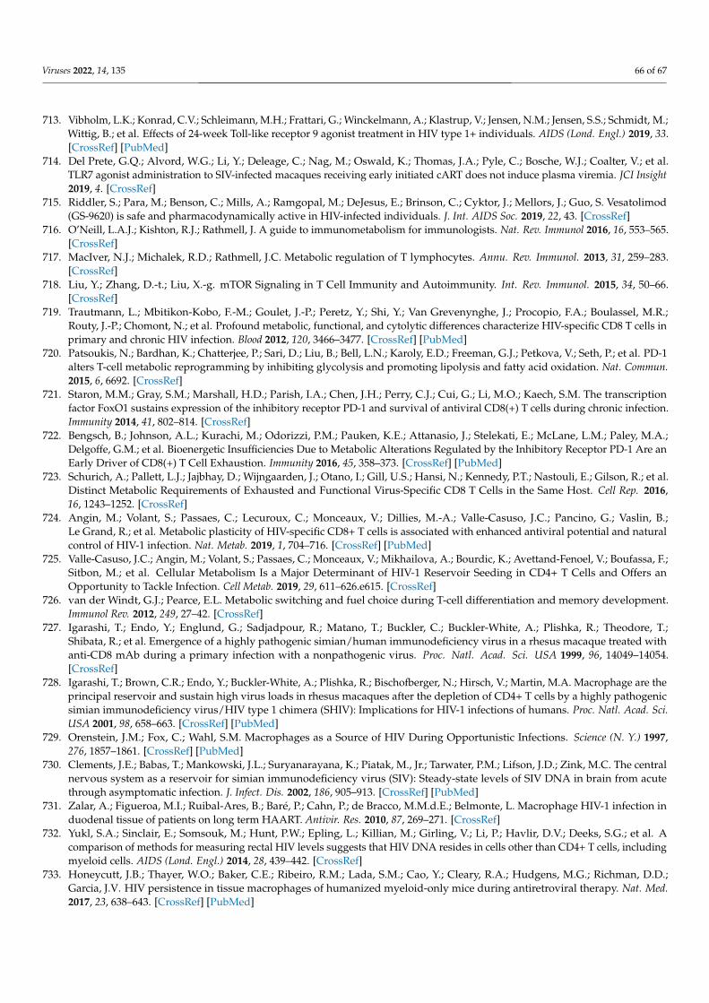
Viruses 2022, 14, 135 66 of 67
713. Vibholm, L.K.; Konrad, C.V.; Schleimann, M.H.; Frattari, G.; Winckelmann, A.; Klastrup, V.; Jensen, N.M.; Jensen, S.S.; Schmidt, M.;Wittig, B.; et al. Effects of 24-week Toll-like receptor 9 agonist treatment in HIV type 1+ individuals. AIDS (Lond. Engl.) 2019, 33.[CrossRef] [PubMed]
714. Del Prete, G.Q.; Alvord, W.G.; Li, Y.; Deleage, C.; Nag, M.; Oswald, K.; Thomas, J.A.; Pyle, C.; Bosche, W.J.; Coalter, V.; et al.TLR7 agonist administration to SIV-infected macaques receiving early initiated cART does not induce plasma viremia. JCI Insight2019, 4. [CrossRef]
715. Riddler, S.; Para, M.; Benson, C.; Mills, A.; Ramgopal, M.; DeJesus, E.; Brinson, C.; Cyktor, J.; Mellors, J.; Guo, S. Vesatolimod(GS-9620) is safe and pharmacodynamically active in HIV-infected individuals. J. Int. AIDS Soc. 2019, 22, 43. [CrossRef]
716. O’Neill, L.A.J.; Kishton, R.J.; Rathmell, J. A guide to immunometabolism for immunologists. Nat. Rev. Immunol 2016, 16, 553–565.[CrossRef]
717. MacIver, N.J.; Michalek, R.D.; Rathmell, J.C. Metabolic regulation of T lymphocytes. Annu. Rev. Immunol. 2013, 31, 259–283.[CrossRef]
718. Liu, Y.; Zhang, D.-t.; Liu, X.-g. mTOR Signaling in T Cell Immunity and Autoimmunity. Int. Rev. Immunol. 2015, 34, 50–66.[CrossRef]
719. Trautmann, L.; Mbitikon-Kobo, F.-M.; Goulet, J.-P.; Peretz, Y.; Shi, Y.; Van Grevenynghe, J.; Procopio, F.A.; Boulassel, M.R.;Routy, J.-P.; Chomont, N.; et al. Profound metabolic, functional, and cytolytic differences characterize HIV-specific CD8 T cells inprimary and chronic HIV infection. Blood 2012, 120, 3466–3477. [CrossRef] [PubMed]
720. Patsoukis, N.; Bardhan, K.; Chatterjee, P.; Sari, D.; Liu, B.; Bell, L.N.; Karoly, E.D.; Freeman, G.J.; Petkova, V.; Seth, P.; et al. PD-1alters T-cell metabolic reprogramming by inhibiting glycolysis and promoting lipolysis and fatty acid oxidation. Nat. Commun.2015, 6, 6692. [CrossRef]
721. Staron, M.M.; Gray, S.M.; Marshall, H.D.; Parish, I.A.; Chen, J.H.; Perry, C.J.; Cui, G.; Li, M.O.; Kaech, S.M. The transcriptionfactor FoxO1 sustains expression of the inhibitory receptor PD-1 and survival of antiviral CD8(+) T cells during chronic infection.Immunity 2014, 41, 802–814. [CrossRef]
722. Bengsch, B.; Johnson, A.L.; Kurachi, M.; Odorizzi, P.M.; Pauken, K.E.; Attanasio, J.; Stelekati, E.; McLane, L.M.; Paley, M.A.;Delgoffe, G.M.; et al. Bioenergetic Insufficiencies Due to Metabolic Alterations Regulated by the Inhibitory Receptor PD-1 Are anEarly Driver of CD8(+) T Cell Exhaustion. Immunity 2016, 45, 358–373. [CrossRef] [PubMed]
723. Schurich, A.; Pallett, L.J.; Jajbhay, D.; Wijngaarden, J.; Otano, I.; Gill, U.S.; Hansi, N.; Kennedy, P.T.; Nastouli, E.; Gilson, R.; et al.Distinct Metabolic Requirements of Exhausted and Functional Virus-Specific CD8 T Cells in the Same Host. Cell Rep. 2016,16, 1243–1252. [CrossRef]
724. Angin, M.; Volant, S.; Passaes, C.; Lecuroux, C.; Monceaux, V.; Dillies, M.-A.; Valle-Casuso, J.C.; Pancino, G.; Vaslin, B.;Le Grand, R.; et al. Metabolic plasticity of HIV-specific CD8+ T cells is associated with enhanced antiviral potential and naturalcontrol of HIV-1 infection. Nat. Metab. 2019, 1, 704–716. [CrossRef] [PubMed]
725. Valle-Casuso, J.C.; Angin, M.; Volant, S.; Passaes, C.; Monceaux, V.; Mikhailova, A.; Bourdic, K.; Avettand-Fenoel, V.; Boufassa, F.;Sitbon, M.; et al. Cellular Metabolism Is a Major Determinant of HIV-1 Reservoir Seeding in CD4+ T Cells and Offers anOpportunity to Tackle Infection. Cell Metab. 2019, 29, 611–626.e615. [CrossRef]
726. van der Windt, G.J.; Pearce, E.L. Metabolic switching and fuel choice during T-cell differentiation and memory development.Immunol Rev. 2012, 249, 27–42. [CrossRef]
727. Igarashi, T.; Endo, Y.; Englund, G.; Sadjadpour, R.; Matano, T.; Buckler, C.; Buckler-White, A.; Plishka, R.; Theodore, T.;Shibata, R.; et al. Emergence of a highly pathogenic simian/human immunodeficiency virus in a rhesus macaque treated withanti-CD8 mAb during a primary infection with a nonpathogenic virus. Proc. Natl. Acad. Sci. USA 1999, 96, 14049–14054.[CrossRef]
728. Igarashi, T.; Brown, C.R.; Endo, Y.; Buckler-White, A.; Plishka, R.; Bischofberger, N.; Hirsch, V.; Martin, M.A. Macrophage are theprincipal reservoir and sustain high virus loads in rhesus macaques after the depletion of CD4+ T cells by a highly pathogenicsimian immunodeficiency virus/HIV type 1 chimera (SHIV): Implications for HIV-1 infections of humans. Proc. Natl. Acad. Sci.USA 2001, 98, 658–663. [CrossRef] [PubMed]
729. Orenstein, J.M.; Fox, C.; Wahl, S.M. Macrophages as a Source of HIV During Opportunistic Infections. Science (N. Y.) 1997,276, 1857–1861. [CrossRef] [PubMed]
730. Clements, J.E.; Babas, T.; Mankowski, J.L.; Suryanarayana, K.; Piatak, M., Jr.; Tarwater, P.M.; Lifson, J.D.; Zink, M.C. The centralnervous system as a reservoir for simian immunodeficiency virus (SIV): Steady-state levels of SIV DNA in brain from acutethrough asymptomatic infection. J. Infect. Dis. 2002, 186, 905–913. [CrossRef] [PubMed]
731. Zalar, A.; Figueroa, M.I.; Ruibal-Ares, B.; Baré, P.; Cahn, P.; de Bracco, M.M.d.E.; Belmonte, L. Macrophage HIV-1 infection induodenal tissue of patients on long term HAART. Antivir. Res. 2010, 87, 269–271. [CrossRef]
732. Yukl, S.A.; Sinclair, E.; Somsouk, M.; Hunt, P.W.; Epling, L.; Killian, M.; Girling, V.; Li, P.; Havlir, D.V.; Deeks, S.G.; et al. Acomparison of methods for measuring rectal HIV levels suggests that HIV DNA resides in cells other than CD4+ T cells, includingmyeloid cells. AIDS (Lond. Engl.) 2014, 28, 439–442. [CrossRef]
733. Honeycutt, J.B.; Thayer, W.O.; Baker, C.E.; Ribeiro, R.M.; Lada, S.M.; Cao, Y.; Cleary, R.A.; Hudgens, M.G.; Richman, D.D.;Garcia, J.V. HIV persistence in tissue macrophages of humanized myeloid-only mice during antiretroviral therapy. Nat. Med.2017, 23, 638–643. [CrossRef] [PubMed]

Viruses 2022, 14, 135 67 of 67
734. Busca, A.; Saxena, M.; Kumar, A. Critical role for antiapoptotic Bcl-xL and Mcl-1 in human macrophage survival and cellularIAP1/2 (cIAP1/2) in resistance to HIV-Vpr-induced apoptosis. J. Biol. Chem. 2012, 287, 15118–15133. [CrossRef]
735. Rappaport, J.; Volsky, D.J. Role of the macrophage in HIV-associated neurocognitive disorders and other comorbidities in patientson effective antiretroviral treatment. J. Neurovirol. 2015, 21, 235–241. [CrossRef]
736. Vojnov, L.; Martins, M.A.; Bean, A.T.; Veloso de Santana, M.G.; Sacha, J.B.; Wilson, N.A.; Bonaldo, M.C.; Galler, R.; Stevenson, M.;Watkins, D.I. The majority of freshly sorted simian immunodeficiency virus (SIV)-specific CD8(+) T cells cannot suppress viralreplication in SIV-infected macrophages. J. Virol. 2012, 86, 4682–4687. [CrossRef]
737. Clayton, K.L.; Collins, D.R.; Lengieza, J.; Ghebremichael, M.; Dotiwala, F.; Lieberman, J.; Walker, B.D. Resistance of HIV-infectedmacrophages to CD8(+) T lymphocyte-mediated killing drives activation of the immune system. Nat. Immunol. 2018, 19, 475–486.[CrossRef]
738. Mailliard, R.B.; Smith, K.N.; Fecek, R.J.; Rappocciolo, G.; Nascimento, E.J.M.; Marques, E.T.; Watkins, S.C.; Mullins, J.I.;Rinaldo, C.R. Selective Induction of CTL Helper Rather Than Killer Activity by Natural Epitope Variants Promotes DendriticCell–Mediated HIV-1 Dissemination. J. Immunol. 2013, 191, 2570–2580. [CrossRef] [PubMed]
739. Smith, K.N.; Mailliard, R.B.; Piazza, P.A.; Fischer, W.; Korber, B.T.; Fecek, R.J.; Ratner, D.; Gupta, P.; Mullins, J.I.; Rinaldo, C.R.Effective Cytotoxic T Lymphocyte Targeting of Persistent HIV-1 during Antiretroviral Therapy Requires Priming of Naive CD8+T Cells. mBio 2016, 7, e00473-16. [CrossRef]
740. Smith, K.N.; Mailliard, R.B.; Rinaldo, C.R. Programming T cell Killers for an HIV Cure: Teach the New Dogs New Tricks and Letthe Sleeping Dogs Lie. Forum Immunopathol. Dis. Ther. 2015, 6, 67–77. [CrossRef] [PubMed]
741. Campbell, G.R.; To, R.K.; Zhang, G.; Spector, S.A. SMAC mimetics induce autophagy-dependent apoptosis of HIV-1-infectedmacrophages. Cell Death Dis. 2020, 11, 590. [CrossRef]
742. Bobardt, M.; Kuo, J.; Chatterji, U.; Chanda, S.; Little, S.J.; Wiedemann, N.; Vuagniaux, G.; Gallay, P.A. The inhibitor apoptosisprotein antagonist Debio 1143 Is an attractive HIV-1 latency reversal candidate. PLoS ONE 2019, 14, e0211746. [CrossRef]
743. Sanyal, A.; Mailliard, R.B.; Rinaldo, C.R.; Ratner, D.; Ding, M.; Chen, Y.; Zerbato, J.M.; Giacobbi, N.S.; Venkatachari, N.J.;Patterson, B.K.; et al. Novel assay reveals a large, inducible, replication-competent HIV-1 reservoir in resting CD4(+) T cells. Nat.Med. 2017, 23, 885–889. [CrossRef]
744. Battivelli, E.; Dahabieh, M.S.; Abdel-Mohsen, M.; Svensson, J.P.; Tojal Da Silva, I.; Cohn, L.B.; Gramatica, A.; Deeks, S.;Greene, W.C.; Pillai, S.K.; et al. Distinct chromatin functional states correlate with HIV latency reactivation in infected primaryCD4(+) T cells. Elife 2018, 7, e34655. [CrossRef]
745. Paiardini, M.; Dhodapkar, K.; Harper, J.; Deeks, S.G.; Ahmed, R. Editorial: HIV and Cancer Immunotherapy: Similar Challengesand Converging Approaches. Front. Immunol 2020, 11, 519. [CrossRef] [PubMed]
746. Díaz-Carballo, D.; Saka, S.; Acikelli, A.H.; Homp, E.; Erwes, J.; Demmig, R.; Klein, J.; Schröer, K.; Malak, S.; D’Souza, F.; et al.Enhanced antitumoral activity of TLR7 agonists via activation of human endogenous retroviruses by HDAC inhibitors. Commun.Biol. 2021, 4, 276. [CrossRef] [PubMed]
747. Zeng, R.; Spolski, R.; Finkelstein, S.E.; Oh, S.; Kovanen, P.E.; Hinrichs, C.S.; Pise-Masison, C.A.; Radonovich, M.F.; Brady, J.N.;Restifo, N.P.; et al. Synergy of IL-21 and IL-15 in regulating CD8+ T cell expansion and function. J. Exp. Med. 2005, 201, 139–148.[CrossRef]
748. Strengell, M.; Matikainen, S.; Sirén, J.; Lehtonen, A.; Foster, D.; Julkunen, I.; Sareneva, T. IL-21 in Synergy with IL-15 or IL-18Enhances IFN-γ Production in Human NK and T Cells. J. Immunol. 2003, 170, 5464–5469. [CrossRef] [PubMed]
749. Pouw, N.; Treffers-Westerlaken, E.; Kraan, J.; Wittink, F.; ten Hagen, T.; Verweij, J.; Debets, R. Combination of IL-21 and IL-15enhances tumour-specific cytotoxicity and cytokine production of TCR-transduced primary T cells. Cancer Immunol. Immunother.CII 2010, 59, 921–931. [CrossRef]
750. Kinter, A.L.; Godbout, E.J.; McNally, J.P.; Sereti, I.; Roby, G.A.; O’Shea, M.A.; Fauci, A.S. The Common γ-Chain Cytokines IL-2,IL-7, IL-15, and IL-21 Induce the Expression of Programmed Death-1 and Its Ligands. J. Immunol. 2008, 181, 6738–6746. [CrossRef]
751. Wrangle, J.M.; Velcheti, V.; Patel, M.R.; Garrett-Mayer, E.; Hill, E.G.; Ravenel, J.G.; Miller, J.S.; Farhad, M.; Anderton, K.;Lindsey, K.; et al. ALT-803, an IL-15 superagonist, in combination with nivolumab in patients with metastatic non-small cell lungcancer: A non-randomised, open-label, phase 1b trial. Lancet. Oncol. 2018, 19, 694–704. [CrossRef]
752. Drusbosky, L.; Nangia, C.; Nguyen, A.; Szeto, C.; Newton, Y.; Spilman, P.; Reddy, S.B. Complete response to avelumab and IL-15superagonist N-803 with Abraxane in Merkel cell carcinoma: A case study. J. ImmunoTherapy Cancer 2020, 8, e001098. [CrossRef]
753. Knudson, K.M.; Hicks, K.C.; Alter, S.; Schlom, J.; Gameiro, S.R. Mechanisms involved in IL-15 superagonist enhancement ofanti-PD-L1 therapy. J. Immunol. Therapy Cancer 2019, 7, 82. [CrossRef]
754. Jochems, C.; Tritsch, S.R.; Knudson, K.M.; Gameiro, S.R.; Rumfield, C.S.; Pellom, S.T.; Morillon, Y.M.; Newman, R.; Marcus, W.;Szeto, C.; et al. The multi-functionality of N-809, a novel fusion protein encompassing anti-PD-L1 and the IL-15 superagonistfusion complex. OncoImmunology 2019, 8, e1532764. [CrossRef] [PubMed]
755. Li, Y.; Cong, Y.; Jia, M.; He, Q.; Zhong, H.; Zhao, Y.; Li, H.; Yan, M.; You, J.; Liu, J.; et al. Targeting IL-21 to tumor-reactive T cellsenhances memory T cell responses and anti-PD-1 antibody therapy. Nat. Commun. 2021, 12, 951. [CrossRef]