Nongenetic method for purifying stem cell–derived cardiomyocytes
Upload
independentCategory
view
0download
0
..............................................................
Fusion of bone-marrow-derivedcells with Purkinje neurons,cardiomyocytes and hepatocytesManuel Alvarez-Dolado1, Ricardo Pardal2, Jose M. Garcia-Verdugo3,John R. Fike1, Hyun O. Lee2, Klaus Pfeffer4, Carlos Lois5,Sean J. Morrison2 & Arturo Alvarez-Buylla1
1Department of Neurological Surgery, University of California at San Francisco,San Francisco, California 94143-0520, USA2Howard Hughes Medical Institute, Department of Internal Medicine, Universityof Michigan, Ann Arbor, Michigan 48109-0934, USA3Instituto Cavanilles, University of Valencia, Valencia 46100, Spain4Institute of Medical Microbiology, University of Dusseldorf, D-40225 Dusseldorf,Germany5Picower Center for Learning and Memory, Massachusetts Institute of Technology,Cambridge, Massachusetts 02139-4307, USA.............................................................................................................................................................................
Recent studies have suggested that bone marrow cells possess abroad differentiation potential, being able to form new liver cells,cardiomyocytes and neurons1,2. Several groups have attributedthis apparent plasticity to ‘transdifferentiation’3–5. Others, how-ever, have suggested that cell fusion could explain these results6–9.Using a simple method based on Cre/lox recombination to detectcell fusion events, we demonstrate that bone-marrow-derivedcells (BMDCs) fuse spontaneously with neural progenitors invitro. Furthermore, bone marrow transplantation demonstratesthat BMDCs fuse in vivo with hepatocytes in liver, Purkinjeneurons in the brain and cardiac muscle in the heart, resulting inthe formation of multinucleated cells. No evidence of trans-differentiation without fusion was observed in these tissues.These observations provide the first in vivo evidence for cellfusion of BMDCs with neurons and cardiomyocytes, raising thepossibility that cell fusion may contribute to the development ormaintenance of these key cell types.
In order to detect cell fusion we used a method based on Cre/loxrecombination, a technique extensively used to conditionally turnon or off gene expression in specific cell types or tissues, or atparticular stages in development10. For this study we first used miceexpressing Cre recombinase ubiquitously under the control of ahybrid cytomegalovirus (CMV) enhancer b-actin promoter11
(Fig. 1a), and the conditional Cre reporter mouse line R26R12
(Fig. 1b). In this line, the LacZ reporter gene is exclusively expressedafter the excision of a loxP-flanked (floxed) stop cassette by Cre-mediated recombination (Fig. 1b). When Cre-expressing (Creþ)cells fuse with R26R cells, Cre recombinase excises the floxed stopcassette of the reporter gene in the R26R nuclei, resulting inexpression of LacZ in the fused cells. Consequently, fused cells canbe detected easily by 5-bromo-4-chloro-3-indolyl-b-D-galactoside(X-gal) staining (Fig. 1c). This method previously failed to detectevidence of cell fusion in the pancreas13. For this reason, we firstverified this cell fusion detection method in vitro.
We co-cultured bone marrow stromal cells (BMSCs) from R26Rreporter mice with Creþ multipotent progenitor cells isolated frompostnatal brain and grown as neurospheres14. Previous studies haveshown that these two cell types can fuse with embryonic stem cellsin vitro6,7. After 4 days in vitro (DIV), a small proportion of b-galþ
cells were found in these co-cultures (1 to 2 cells per 80,000 cells)(Fig. 1d). Importantly, most b-galþ cells at 4 DIV had two or morenuclei, an observation that was confirmed by electron microscopy(Fig. 1e, i; see also Supplementary Fig. 1). This is consistent with thegeneration of b-galþ cells by fusion. Notably, after 10 or 15 DIVb-galþ cells formed small colonies and some of these cells weremitotic (Fig. 1g and inset). Cells in these colonies were invariably
mononucleated, suggesting that with time and cell division thenuclei of these cells fuse or supernumerary nuclei are eliminated. Inaddition to neurosphere cells, R26R BMSCs were also co-culturedwith primary cultures of Creþ fibroblasts. Three independentco-cultures did not yield positive cells, suggesting that not all celltypes are equally capable of fusion in culture. To further confirmthat b-gal expression was due to cell fusion, Creþ neurosphere cellswere labelled with 5-bromodeoxyuridine (BrdU) and then co-cultured for 5 days with R26R BMSCs. Most bi-nucleated b-galþ
cells in these cultures had only one of the two nuclei labelled withBrdU (Fig. 1h–k), further confirming the reliability of this methodto detect fusion events.
These results confirm previous work demonstrating that cellfusion occurs spontaneously in vitro6,7. To study cell fusion in vivo,R26R reporter mice were lethally irradiated, and 2 days later weregrafted with bone marrow from mice constitutively expressing Crerecombinase and green fluorescent protein (GFP) under thecontrol of the b-actin promoter (b-actin-Cre–GFP mice, see‘allogeneic bone marrow transplantation’ section of Methods).We analysed the grafted mice at 2 (n ¼ 3) and 4 (n ¼ 3) monthsafter transplantation. These mice showed significant levels ofhaematopoietic reconstitution, which was measured by flow cyto-metry based on the frequency of GFPþ cells in peripheral blood(from 54.6% to 79.8% of nucleated blood cells). Brain, liver, heart,gut, kidney, lung and skeletal muscle from these mice were seriallysectioned and stained for the presence of X-galþ cells. In allanimals, cells labelled with b-gal were only found in brain, heartand liver, and not in the other organs studied (Table 1). As anegative control, we grafted R26R mice with bone marrow fromwild-type mice. We did not find b-galþ cells in these animals,demonstrating that the reporter was not inappropriately activated,even after irradiation.
BMDCs fuse with hepatocytes in fumarylacetoacetate-hydrolase-deficient mice8,9. Consistent with this finding, we also observedfused hepatocytes in our grafted mice (Fig. 2). b-galþ hepatocytesexpressed albumin (a characteristic hepatocyte marker) (Fig. 2, h)but were negative for CD45 (a haematopoietic marker) (data notshown). Electron microscopy confirmed that these fused cells weretypical hepatocytes with no features of haematopoietic cells(Fig. 2c). They contained glycogen granules, complete desmosomesand bile canaliculi (Fig. 2d). Two months after transplant mostb-galþ hepatocytes co-expressed the donor marker GFP (Fig. 2e, f);however, a small fraction of fused hepatocytes were GFP negative.This fraction had increased 4 months after transplantation (Table 1).This result suggests that after fusion donor genes may be inacti-vated/eliminated over time.
At 2 and 4 months after transplantation, b-galþ cells weredetected in the cerebellum, where labelled cells displayed the typicallocation and morphology of Purkinje cells (Fig. 3a). Two b-galþ
cells were embedded in plastic and serially sectioned for light andelectron microscopy. Serial reconstruction of the soma of these cellsdemonstrated the presence of two nuclei (Fig. 3b; see also Sup-plementary Fig. 2). Notably, the two nuclei presented very differentmorphologies: one had a wrinkled surface with multiple invagina-tions and a single nucleolus, typical of Purkinje cells15, whereas thesecond nucleus showed a uniform spherical shape with multiplenucleoli, suggesting a different origin (Fig. 3b, c). Electronmicroscopy analysis confirmed that these cells were Purkinje neur-ons (Fig. 3c) with typical Purkinje cell somata and organelledistribution, including structures with the characteristics of synap-tic contacts (Fig. 3d). No signs of degeneration or abnormalstructures were observed in the cytoplasm of these cells (Fig. 3c,d). b-galþ Purkinje cells stained positively for the Purkinjecell marker calbindin (data not shown). This is the first directevidence, to our knowledge, showing that a neuron can fuse witha BMDC. These results suggest that previous observations ofsmall numbers of Purkinje cells bearing markers of transplanted
letters to nature
NATURE | VOL 425 | 30 OCTOBER 2003 | www.nature.com/nature968 © 2003 Nature Publishing Group

bone marrow cells5,16,17 may have arisen by fusion rather thantransdifferentiation.
The third organ where b-galþ cells were found was the heart(Fig. 4). The b-galþ cells were integrated into the myocardial walland had a morphology and alignment that was indistinguishablefrom the surrounding cardiac muscle fibres (Fig. 4a–d). At theelectron microscopy level, the fused cells had the morphology ofmature cardiomyocytes, including developed filament bands andmature intercalated discs with desmosomes and GAP junctionsconnecting to neighbouring fibres (Fig. 4e). In addition, fusedcardiomyocytes expressed cardiac troponin I (Fig. 4g). As seen inthe liver, GFP was expressed in most of the fused cardiomyocytes at2 months after transplantation (Table 1). In contrast, fused cardi-omyocytes lost GFP expression at 4 months (Fig. 4h). It has beensuggested that haematopoietic stem cells can partially restore theinfarcted heart by transdifferentiation, giving rise to new myocar-dium4. Our results suggest that BMDCs can fuse with cells withinthe heart to form mature cardiomyocytes, but it remains unknown
whether any new cardiomyocytes are generated as a result of thisfusion.
Bone marrow cells include haematopoietic cells as well asmesenchymal cells and possibly other cell types. To test whetherhaematopoietic cells were participating in fusion events in vivo weused donor mice in which Cre recombinase was knocked into theCD45 locus (CD45-Cre). As CD45 is specifically expressed byhaematopoietic cells18,19, recombination should only occur infusions involving cells of the haematopoietic lineage. To confirmthe CD45-Cre expression pattern these mice were mated with R26Rreporter mice. We observed widespread b-gal expression in haema-topoietic stem cells, bone marrow cells, and blood cells but not inother tissues such as brain, liver, or skeletal muscle (SupplementaryFig. 3). Nonetheless, we cannot rule out the possibility of CD45-Creexpression by very rare cells in other tissues or that non-haemato-poietic cells might transiently activate CD45 expression duringwhatever nuclear reprogramming might occur after cell fusion.
CD45-Cre bone marrow cells were injected into four lethally
Figure 1 Method to detect cell fusion events. a, b, Schematic representation of the
transgenes expressed by the mouse lines used. c, When a cell expressing Cre
recombinase (a) fuses with a cell bearing the LacZ reporter transgene (b), the floxed stop
cassette is excised and the LacZ reporter is expressed in the fused cell. LacZ
expression can be detected by the generation of a blue precipitate after X-gal staining.
RTV, Integration retroviral sequence (LTR). d–g, Co-cultures of R26R BMSCs with Creþ
neurospheres stained for b-gal and the nuclear dye DAPI (4,6-diamidino-2-phenylindole).
d, e, Multinucleated b-galþ fused cell (4 DIV). f, g, Colony of b-galþ fused cells (15 DIV).
These cells were mononucleated and mitotically active, as demonstrated by a cell in
metaphase (inset in g). h–k, Co-culture of R26R BMSCs with BrdU-labelled Creþ
neurospheres. Bi-nucleated b-galþ fused cells (h) with one nucleus immunopositive for
BrdU ( j, k, arrowhead) and the second nucleus negative for BrdU ( i, k, arrow). Scale bars:
d, f, h, 20 mm; g (inset), 5 mm.
letters to nature
NATURE | VOL 425 | 30 OCTOBER 2003 | www.nature.com/nature 969© 2003 Nature Publishing Group
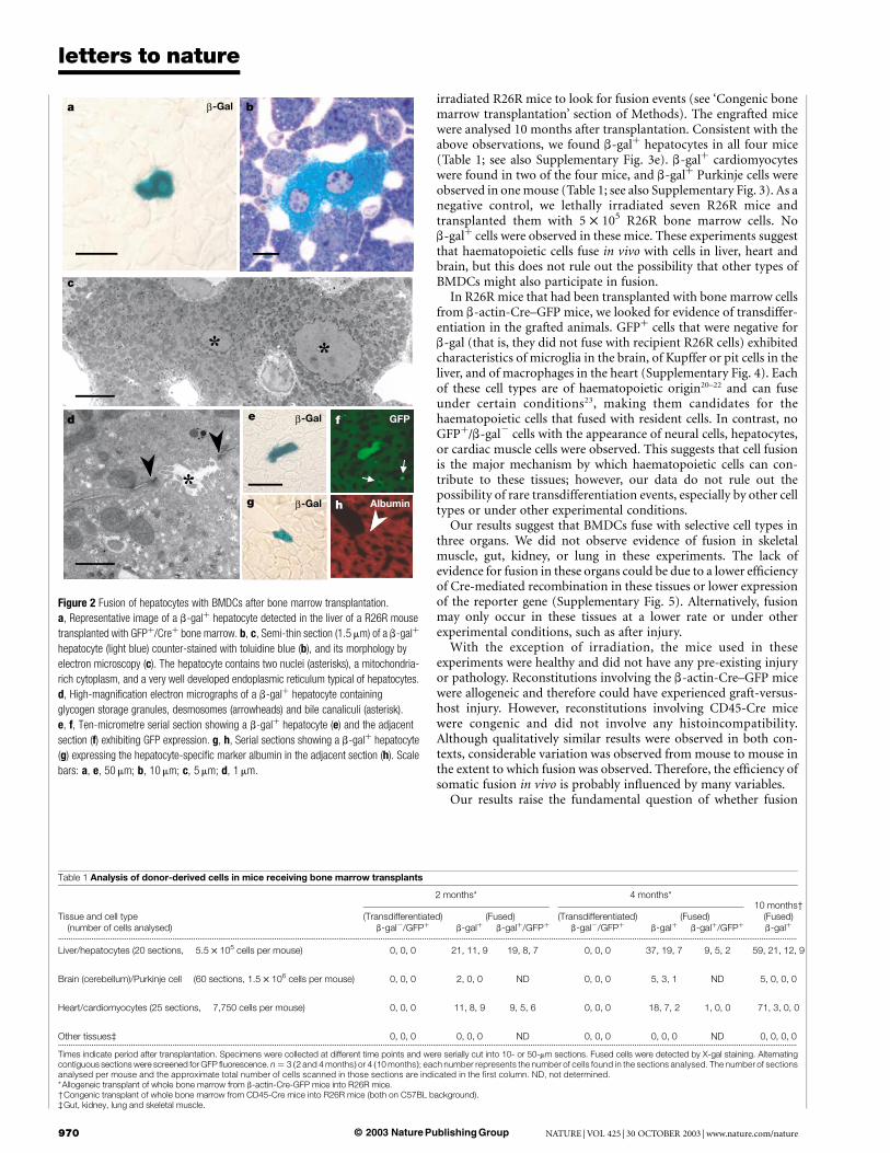
irradiated R26R mice to look for fusion events (see ‘Congenic bonemarrow transplantation’ section of Methods). The engrafted micewere analysed 10 months after transplantation. Consistent with theabove observations, we found b-galþ hepatocytes in all four mice(Table 1; see also Supplementary Fig. 3e). b-galþ cardiomyocyteswere found in two of the four mice, and b-galþ Purkinje cells wereobserved in one mouse (Table 1; see also Supplementary Fig. 3). As anegative control, we lethally irradiated seven R26R mice andtransplanted them with 5 £ 105 R26R bone marrow cells. Nob-galþ cells were observed in these mice. These experiments suggestthat haematopoietic cells fuse in vivo with cells in liver, heart andbrain, but this does not rule out the possibility that other types ofBMDCs might also participate in fusion.
In R26R mice that had been transplanted with bone marrow cellsfrom b-actin-Cre–GFP mice, we looked for evidence of transdiffer-entiation in the grafted animals. GFPþ cells that were negative forb-gal (that is, they did not fuse with recipient R26R cells) exhibitedcharacteristics of microglia in the brain, of Kupffer or pit cells in theliver, and of macrophages in the heart (Supplementary Fig. 4). Eachof these cell types are of haematopoietic origin20–22 and can fuseunder certain conditions23, making them candidates for thehaematopoietic cells that fused with resident cells. In contrast, noGFPþ/b-gal2 cells with the appearance of neural cells, hepatocytes,or cardiac muscle cells were observed. This suggests that cell fusionis the major mechanism by which haematopoietic cells can con-tribute to these tissues; however, our data do not rule out thepossibility of rare transdifferentiation events, especially by other celltypes or under other experimental conditions.
Our results suggest that BMDCs fuse with selective cell types inthree organs. We did not observe evidence of fusion in skeletalmuscle, gut, kidney, or lung in these experiments. The lack ofevidence for fusion in these organs could be due to a lower efficiencyof Cre-mediated recombination in these tissues or lower expressionof the reporter gene (Supplementary Fig. 5). Alternatively, fusionmay only occur in these tissues at a lower rate or under otherexperimental conditions, such as after injury.
With the exception of irradiation, the mice used in theseexperiments were healthy and did not have any pre-existing injuryor pathology. Reconstitutions involving the b-actin-Cre–GFP micewere allogeneic and therefore could have experienced graft-versus-host injury. However, reconstitutions involving CD45-Cre micewere congenic and did not involve any histoincompatibility.Although qualitatively similar results were observed in both con-texts, considerable variation was observed from mouse to mouse inthe extent to which fusion was observed. Therefore, the efficiency ofsomatic fusion in vivo is probably influenced by many variables.
Our results raise the fundamental question of whether fusion
Table 1 Analysis of donor-derived cells in mice receiving bone marrow transplants
2 months* 4 months*10 months†
Tissue and cell type (Transdifferentiated) (Fused) (Transdifferentiated) (Fused) (Fused)(number of cells analysed) b-gal2/GFPþ b-galþ b-galþ/GFPþ b-gal2/GFPþ b-galþ b-galþ/GFPþ b-galþ
...................................................................................................................................................................................................................................................................................................................................................................
Liver/hepatocytes (20 sections, 5.5 £ 105 cells per mouse) 0, 0, 0 21, 11, 9 19, 8, 7 0, 0, 0 37, 19, 7 9, 5, 2 59, 21, 12, 9
Brain (cerebellum)/Purkinje cell (60 sections, 1.5 £ 106 cells per mouse) 0, 0, 0 2, 0, 0 ND 0, 0, 0 5, 3, 1 ND 5, 0, 0, 0
Heart/cardiomyocytes (25 sections, 7,750 cells per mouse) 0, 0, 0 11, 8, 9 9, 5, 6 0, 0, 0 18, 7, 2 1, 0, 0 71, 3, 0, 0
Other tissues‡ 0, 0, 0 0, 0, 0 ND 0, 0, 0 0, 0, 0 ND 0, 0, 0, 0...................................................................................................................................................................................................................................................................................................................................................................
Times indicate period after transplantation. Specimens were collected at different time points and were serially cut into 10- or 50-mm sections. Fused cells were detected by X-gal staining. Alternatingcontiguous sections were screened for GFP fluorescence. n ¼ 3 (2 and 4 months) or 4 (10 months); each number represents the number of cells found in the sections analysed. The number of sectionsanalysed per mouse and the approximate total number of cells scanned in those sections are indicated in the first column. ND, not determined.*Allogeneic transplant of whole bone marrow from b-actin-Cre-GFP mice into R26R mice.†Congenic transplant of whole bone marrow from CD45-Cre mice into R26R mice (both on C57BL background).‡Gut, kidney, lung and skeletal muscle.
Figure 2 Fusion of hepatocytes with BMDCs after bone marrow transplantation.
a, Representative image of a b-galþ hepatocyte detected in the liver of a R26R mouse
transplanted with GFPþ/Creþ bone marrow. b, c, Semi-thin section (1.5 mm) of a b-galþ
hepatocyte (light blue) counter-stained with toluidine blue (b), and its morphology by
electron microscopy (c). The hepatocyte contains two nuclei (asterisks), a mitochondria-
rich cytoplasm, and a very well developed endoplasmic reticulum typical of hepatocytes.
d, High-magnification electron micrographs of a b-galþ hepatocyte containing
glycogen storage granules, desmosomes (arrowheads) and bile canaliculi (asterisk).
e, f, Ten-micrometre serial section showing a b-galþ hepatocyte (e) and the adjacent
section (f) exhibiting GFP expression. g, h, Serial sections showing a b-galþ hepatocyte
(g) expressing the hepatocyte-specific marker albumin in the adjacent section (h). Scale
bars: a, e, 50 mm; b, 10 mm; c, 5 mm; d, 1 mm.
letters to nature
NATURE | VOL 425 | 30 OCTOBER 2003 | www.nature.com/nature970 © 2003 Nature Publishing Group
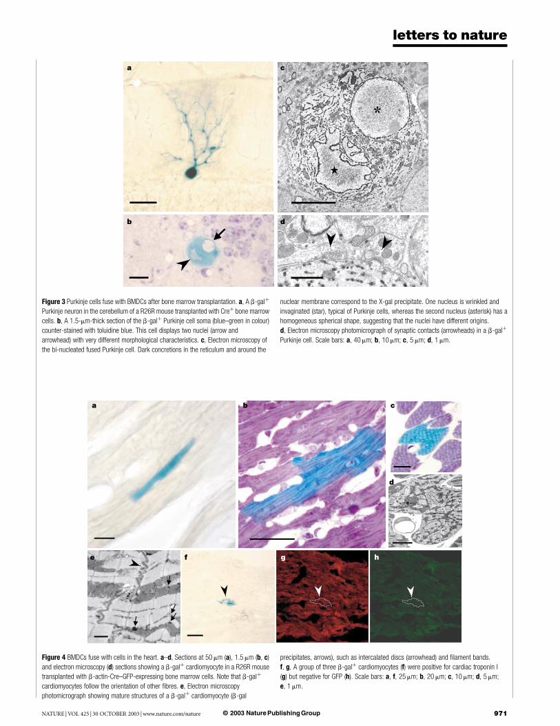
Figure 3 Purkinje cells fuse with BMDCs after bone marrow transplantation. a, A b-galþ
Purkinje neuron in the cerebellum of a R26R mouse transplanted with Creþ bone marrow
cells. b, A 1.5-mm-thick section of the b-galþ Purkinje cell soma (blue–green in colour)
counter-stained with toluidine blue. This cell displays two nuclei (arrow and
arrowhead) with very different morphological characteristics. c, Electron microscopy of
the bi-nucleated fused Purkinje cell. Dark concretions in the reticulum and around the
nuclear membrane correspond to the X-gal precipitate. One nucleus is wrinkled and
invaginated (star), typical of Purkinje cells, whereas the second nucleus (asterisk) has a
homogeneous spherical shape, suggesting that the nuclei have different origins.
d, Electron microscopy photomicrograph of synaptic contacts (arrowheads) in a b-galþ
Purkinje cell. Scale bars: a, 40 mm; b, 10 mm; c, 5 mm; d, 1 mm.
Figure 4 BMDCs fuse with cells in the heart. a–d, Sections at 50 mm (a), 1.5 mm (b, c)
and electron microscopy (d) sections showing a b-galþ cardiomyocyte in a R26R mouse
transplanted with b-actin-Cre–GFP-expressing bone marrow cells. Note that b-galþ
cardiomyocytes follow the orientation of other fibres. e, Electron microscopy
photomicrograph showing mature structures of a b-galþ cardiomyocyte (b-gal
precipitates, arrows), such as intercalated discs (arrowhead) and filament bands.
f, g, A group of three b-galþ cardiomyocytes (f) were positive for cardiac troponin I
(g) but negative for GFP (h). Scale bars: a, f, 25 mm; b, 20 mm; c, 10 mm; d, 5 mm;
e, 1 mm.
letters to nature
NATURE | VOL 425 | 30 OCTOBER 2003 | www.nature.com/nature 971© 2003 Nature Publishing Group

between haematopoietic cells and cells of the brain, liver and hearthas a physiological role in the development or maintenance of theseorgans. Interestingly, many hepatocytes and cardiomyocytes undernormal conditions have two or more nuclei22,24. To our knowledgethis is the first study to demonstrate Purkinje cells with two nuclei,but other studies have suggested that these neurons can be poly-ploid25,26. Our results suggest that cell fusion may be the mechanismby which these cells become multinucleated or polyploid. Geneticmaterial derived from blood cells may contribute through cellfusion to the survival and function of cells in different organs.Previous studies have shown that fused cells are positively selectedduring hepatic degeneration, helping to rescue a mutant mousedeficient for fumarylacetoacetate hydrolase8,9. Our observation thatfusion is a major mechanism by which BMDCs contribute to theheart, liver and brain draws into question the rationale for clinicalprocedures based on the idea that transdifferentiation of BMDCscan lead to the de novo generation of heart or brain cells. Additionalstudies in animal models will be required to determine whetherfusion by BMDC cells can be used in reparative cell therapy. A
MethodsCell culturesBone marrow cells from b-actin-Cre or R26R transgenic mice were collected by flushingtibias and femurs with RPMI medium 1640 (Gibco BRL) supplemented with 3% fetal calfserum. Red blood cells were depleted using ice-cold ammonium chloride (140 mM in Tris17 mM), and bone marrow cells were plated at a density of 2–4 £ 107 cells per 9.5 cm2 inIscove’s modified Dulbecco’s medium (IMDM; Gibco BRL) supplemented with 10% fetalcalf serum, 100 U ml21 penicillin, 100 mg ml21 streptomycin and 10 mg ml21 glutamine(complete IMDM medium). The non-adherent cell population was removed after 48 hand the adherent BMDC layer washed once with fresh medium; cells were thencontinuously cultured for 1–4 weeks.
For neurospheres, brain subventricular zone from 5–10-day-old b-actin-Cre or R26Rmice was collected. After papain dissociation, neurospheres were cultured and expandedin the presence of both epidermal growth factor (20 ng ml21; Peprotech) and fibroblastgrowth factor-2 (10 ng ml21; Peprotech), as described27. For BrdU labelling, neurosphereswere cultured for 15 min in the presence of 2 mM BrdU, washed, and expanded for twoadditional passages before being cultured with BMSCs. This procedure labelled 70–80% ofthe neurosphere cells. Fibroblasts were cultured as described previously28.
Co-culturesBMDCs and dissociated neurospheres were mixed in a 1:1 ratio and plated on Matrigel-coated dishes (BD Bioscience) at a density of 2 £ 105 cells ml21 in complete IMDMmedium. BMDCs and primary fibroblasts were cultured in a 1:1 ratio on plastic dishes incomplete IMDM medium. After 4–15 days, co-cultures were washed and fixed in 2%paraformaldehyde for 10 min and analysed by X-gal staining or immunohistochemistry.As a negative control, R26R BMDC monocultures were grown for 15 days in the presenceof conditioned medium or cell extracts from Cre-expressing cells (data not shown). Cellextracts from Cre-expressing cells were freshly prepared by two freeze/thaw series andadded to the culture medium.
Animal care and bone marrow transplantAnimal care and all procedures were approved by the Institutional Animal CareCommittees at UCSF and the University of Michigan.
Allogeneic bone marrow transplantationHomozygous mice expressing Cre recombinase under the control of the hybrid regulatoryelement CMVenhancer b-actin promoter11, and homozygous mice expressing GFP underthe same promoter29 were bred to generate b-actin-Cre–GFP mice for use as bone marrowdonors. Bone marrow cells from 8–10-week-old Cre–GFPþ mice were extracted asdescribed, and 10–20 £ 106 cells were intraperitoneally injected into R26R mice irradiatedwith a single whole-body dose of 7.5 Gy. To avoid allograft rejection, mice that received thebone marrow transplantation procedure were treated one week before transplantation and3 weeks after transplantation with Neoral cyclosporine (100 mg l21; Novartis) in thedrinking water. Drinking water was acidified and contained neomycin sulphate (1 mg l21;Sigma) to suppress pathogens.
Congenic bone marrow transplantationIn an independent experiment 8–10-week-old CD45-Cre ‘knock-in’ mice on a C57BL/Ka-Thy1.1 background were used as donors of bone marrow cells. The generation of CD45-Cre knock-in mice will be described elsewhere (E. Schaller and K.P., manuscript inpreparation). Eight-week-old R26R mice on a C57BL/Ka-Thy1.2 background were used asrecipients. Approximately 5 £ 105 bone marrow cells were injected into the retro-orbitalvenous plexus of R26R mice lethally irradiated with two doses of 5.7 Gy each. The drinkingwater of the transplanted mice contained neomycin sulphate (1 g l21) and polymyxin Bsulphate (1 £ 106 U l21) to suppress pathogens. On a monthly basis after transplantation,mice were bled and the peripheral blood was stained with antibodies against Thy1.1 andhaematopoietic markers to confirm reconstitution.
CD45-Cre knock-in mice were bred with R26R to obtain CD45-Cre/R26R mice. Theb-gal expression pattern in these mice was examined by X-gal staining of tissues and byfluorescein di-b-D-galactopyranoside (Molecular Probes) staining of haematopoietic cells.Bone marrow cells were incubated for 5 min at 37 8C with 10 mM fluorescein di-b-D-galactopyranoside in a hypotonic solution (1:1 staining medium (HBSS plus 2% calfserum):distilled water). Haematopoietic stem cells (Sca-1þ c-Kitþ Flk-22 lineage2 cells)30
were analysed for b-gal expression using a FACS Vantage flow-cytometer (Becton-Dickinson) (Supplementary Fig. 3a).
Tissue collectionAfter 2, 4 or 10 months mice were anaesthetized and transcardially perfused with 0.9%saline followed by 50 ml 4% paraformaldehyde or 2% paraformaldehyde plus 0.25%glutaraldehyde. Brain, spinal cord, liver, lung, kidney, heart, skeletal muscle and gut weredissected. Brain and one liver lobe were serially cut in 50-mm vibrotome sections. The restof the liver and other tissues were cryopreserved and frozen in optimum cuttingtemperature compound (Sakura-Finetec) at 280 8C. Serial 10- or 50-mm sections were cutin a cryostat and stained with X-gal or by immunohistochemistry.
X-gal staining and immunohistochemistrySpecimens were placed in phosphate buffer containing 10 mM K3Fe(CN)6 and 10 mMK4Fe(CN)6 along with the b-gal substrate X-gal (1 mg ml21) (Molecular Probes) at 37 8Cfor 8–12 h. Antibodies against albumin (A-6684; 1:100) and calbindin (C-9848; 1:1,000)were from Sigma, cardiac troponin I (sc-1881; 1:1,000) was from Santa CruzBiotechnology, BrdU (M0744, 1:100) was from DAKO, CD45 (558750, 1:100) was fromBD PharMingen, and Iba1 was a gift from Y. Imai. Secondary antibodies anti-mouse-,goat- or rabbit-IgG (H þ L) (Cy-2, 1:400; Cy-3, 1:400; biotinylated, 1:500) conjugatedwere from Jackson Immunoresearch.
Plastic embedding and electron microscopyFifty-micrometre b-gal-stained sections were post-fixed with 1% osmium and 7% glucosefor 2 h, rinsed, dehydrated and embedded in araldite (Durcupan, Fluka). Semi-thinsections (1.5 mm) were cut with a diamond knife and stained lightly with 1% toluidineblue. Semi-thin sections were re-embedded in an araldite block and detached from theglass slide by repeated freezing (liquid nitrogen) and thawing. Ultra-thin (0.05 mm)sections were cut with a diamond knife, stained with lead citrate and examined under aJeol100CX electron microscope.
Received 18 August; accepted 24 September 2003; doi:10.1038/nature02069.
Published online 12 October 2003.
1. Morrison, S. J. Stem cell potential: can anything make anything? Curr. Biol. 11, R7–R9 (2001).
2. Orkin, S. H. & Zon, L. I. Hematopoiesis and stem cells: plasticity versus developmental heterogeneity.
Nature Immunol. 3, 323–328 (2002).
3. Krause, D. S. et al. Multi-organ, multi-lineage engraftment by a single bone marrow-derived stem cell.
Cell 105, 369–377 (2001).
4. Orlic, D. et al. Bone marrow cells regenerate infarcted myocardium. Nature 410, 701–705 (2001).
5. Priller, J. et al. Neogenesis of cerebellar Purkinje neurons from gene-marked bone marrow cells in vivo.
J. Cell Biol. 155, 733–738 (2001).
6. Terada, N. et al. Bone marrow cells adopt the phenotype of other cells by spontaneous cell fusion.
Nature 416, 542–545 (2002).
7. Ying, Q. L., Nichols, J., Evans, E. P. & Smith, A. G. Changing potency by spontaneous fusion. Nature
416, 545–548 (2002).
8. Vassilopoulos, G., Wang, P. R. & Russell, D. W. Transplanted bone marrow regenerates liver by cell
fusion. Nature 422, 901–904 (2003).
9. Wang, X. et al. Cell fusion is the principal source of bone-marrow-derived hepatocytes. Nature 422,
897–901 (2003).
10. Sauer, B. Inducible gene targeting in mice using the Cre/lox system. Methods 14, 381–392 (1998).
11. Lewandoski, M., Meyers, E. N. & Martin, G. R. Analysis of Fgf8 gene function in vertebrate
development. Cold Spring Harb. Symp. Quant. Biol. 62, 159–168 (1997).
12. Mao, X., Fujiwara, Y. & Orkin, S. H. Improved reporter strain for monitoring Cre recombinase-
mediated DNA excisions in mice. Proc. Natl Acad. Sci. USA 96, 5037–5042 (1999).
13. Ianus, A., Holz, G. G., Theise, N. D. & Hussain, M. A. In vivo derivation of glucose-competent
pancreatic endocrine cells from bone marrow without evidence of cell fusion. J. Clin. Invest. 111,
843–850 (2003).
14. Weiss, S. et al. Multipotent CNS stem cells are present in the adult mammalian spinal cord and
ventricular neuroaxis. J. Neurosci. 16, 7599–7609 (1996).
15. Palay, L. P. & Chan-Palay, V. Cerebellar Cortex 15–25 (Springer, Berlin, 1974).
16. Weimann, J. M., Charlton, C. A., Brazelton, T. R., Hackman, R. C. & Blau, H. M. Contribution of
transplanted bone marrow cells to Purkinje neurons in human adult brains. Proc. Natl Acad. Sci. USA
100, 2088–2093 (2003).
17. Wagers, A. J., Sherwood, R. I., Christensen, J. L. & Weissman, I. L. Little evidence for developmental
plasticity of adult hematopoietic stem cells. Science 297, 2256–2259 (2002).
18. Ledbetter, J. A. & Herzenberg, L. A. Xenogeneic monoclonal antibodies to mouse lymphoid
differentiation antigens. Immunol. Rev. 47, 63–90 (1979).
19. van Ewijk, W., van Soest, P. L. & van den Engh, G. J. Fluorescence analysis and anatomic distribution of
mouse T lymphocyte subsets defined by monoclonal antibodies to the antigens Thy-1, Lyt-1, Lyt-2,
and T-200. J. Immunol. 127, 2594–2604 (1981).
20. Ling, E. A. & Wong, W. C. The origin and nature of ramified and amoeboid microglia: a historical
review and current concepts. Glia 7, 9–18 (1993).
21. Gehrmann, J., Matsumoto, Y. & Kreutzberg, G. W. Microglia: intrinsic immuneffector cell of the
brain. Brain Res. Brain Res. Rev. 20, 269–287 (1995).
22. Arias, I. M., et al. The Liver Biology and Pathobiology (Lippincott Williams and Wilkins, Philadelphia,
2001).
23. Anderson, J. M. Multinucleated giant cells. Curr. Opin. Hematol. 7, 40–47 (2000).
24. Piper, H. M. & Isenberg, I. Isolated Adult Cardiomyocytes (CRC, Boca Raton, 1989).
letters to nature
NATURE | VOL 425 | 30 OCTOBER 2003 | www.nature.com/nature972 © 2003 Nature Publishing Group

25. Lapham, L. W. Tetraploid DNA content of Purkinje neurons of human cerebellar cortex. Science 159,
310–312 (1968).
26. Mares, V., Lodin, Z. & Sacha, J. A cytochemical and autoradiographic study of nuclear DNA in mouse
Purkinje cells. Brain Res. 53, 273–289 (1973).
27. Doetsch, F., Caille, I., Lim, D. A., Garcia-Verdugo, J. M. & Alvarez-Buylla, A. Subventricular zone
astrocytes are neural stem cells in the adult mammalian brain. Cell 97, 703–716 (1999).
28. Spector, D. L., Goldman, R. D. & Leinwand, L. A. Cells: a Laboratory Manual 4.1–4.7 (Cold Spring
Harbor Laboratory Press, New York, 1998).
29. Hadjantonakis, A. K., Gertsenstein, M., Ikawa, M., Okabe, M. & Nagy, A. Generating green fluorescent
mice by germline transmission of green fluorescent ES cells. Mech. Dev. 76, 79–90 (1998).
30. Christensen, J. L. & Weissman, I. L. Flk-2 is a marker in hematopoietic stem cell differentiation: a
simple method to isolate long-term stem cells. Proc. Natl Acad. Sci. USA 98, 14541–14546 (2001).
Supplementary Information accompanies the paper on www.nature.com/nature.
Acknowledgements The authors thank G. Martin and P. Soriano for transgenic mouse lines,
J. Maher at the UCSF Liver Centre for advice and assistance, and M. Kiel, O. Yilmaz and The
University of Michigan Flow Cytometry Core for help with flow cytometry. R.P. thanks E. Schaller
for technical help. M.A-D. thanks B. Rico, I. Cobos, T. Aragon and U. Borello for personal and
scientific support. This work was supported by grants from NIH, the Sandler Foundation, the
Spanish Ministry of Science and Technology (Ataxias Cerebelosa), and the Deutsche
Forschungsgemeinschaft (DFG). R.P. was the recipient of a postdoctoral fellowship from the
Spanish Ministry of Science and Technology.
Competing interests statement The authors declare that they have no competing financial
interests.
Correspondence and requests for materials should be addressed to A.A-B.
..............................................................
SNARE-protein-mediated diseaseresistance at the plant cell wallNicholas C. Collins1*, Hans Thordal-Christensen2*, Volker Lipka3,Stephan Bau3, Erich Kombrink3, Jin-Long Qiu2, Ralph Huckelhoven4,Monica Stein5, Andreas Freialdenhoven3, Shauna C. Somerville5
& Paul Schulze-Lefert3
1Sainsbury Laboratory John Innes Centre, Norwich, Norfolk NR4 7UH, UK2Plant Research Department, Risø National Laboratory, DK-4000 Roskilde,Denmark3Department of Plant Microbe Interactions, Max Planck Institute for PlantBreeding Research, Cologne D-50829, Germany4Institute of Phytopathology and Applied Zoology Justus-Liebig-University,Giessen D-35392, Germany5Department of Plant Biology, Carnegie Institute of Washington, Stanford,California 94305, USA
* These authors contributed equally to this work
.............................................................................................................................................................................
Failure of pathogenic fungi to breach the plant cell wall consti-tutes a major component of immunity of non-host plant species—species outside the pathogen host range—and accounts for aproportion of aborted infection attempts on ‘susceptible’ hostplants (basal resistance)1–4. Neither form of penetration resist-ance is understood at the molecular level. We developed a screenfor penetration (pen) mutants of Arabidopsis, which are disabledin non-host penetration resistance against barley powdery mil-dew, Blumeria graminis f. sp. hordei, and we isolated the PEN1gene. We also isolated barley ROR2 (ref. 2), which is required forbasal penetration resistance against B. g. hordei. The genesencode functionally homologous syntaxins, demonstrating amechanistic link between non-host resistance and basal pen-etration resistance in monocotyledons and dicotyledons. Weshow that resistance in barley requires a SNAP-25 (synapto-some-associated protein, molecular mass 25 kDa) homologuecapable of forming a binary SNAP receptor (SNARE) complexwith ROR2. Genetic control of vesicle behaviour at penetrationsites, and plasma membrane location of PEN1/ROR2, is consist-
ent with a proposed involvement of SNARE-complex-mediatedexocytosis and/or homotypic vesicle fusion events in resistance.Functions associated with SNARE-dependent penetration resist-ance are dispensable for immunity mediated by race-specificresistance (R) genes, highlighting fundamental differencesbetween these two resistance forms.
Most types of plant pathogens fail to produce disease on themajority of plant species. Although ‘non-host’ resistance is the mostcommon form of resistance, its basis is poorly understood owing tothe dearth of tractable genetic systems. This contrasts with ‘race-specific’ resistance triggered by corresponding AVIRULENCE(AVR)/R genes in otherwise compatible host–pathogen inter-actions, for which many components have been identified5. Suicideof cells surrounding the infection site (often referred to as thehypersensitive response) typically accompanies R-gene-mediatedresistance, and hypersensitive-response-like cell death can also beassociated with non-host resistance. These drastic measures formsecondary lines of defence that are normally triggered once a fungushas overcome active defences at the plant cell periphery3,6.
We investigated whether the immunity of the model plantArabidopsis to the barley powdery mildew B. g. hordei couldbe used to develop a system for dissecting non-host resistance.Blumeria g. hordei conidiospores germinated on Arabidopsis butmost sporelings failed to enter the plant cells, accompanied by theformation of a cell wall deposition (papilla) by the plant cell directlybeneath penetration attempts. About 10% of sites showed successfulpenetration as indicated by the presence of a fungal feedingstructure (haustorium; Fig. 1a); however, most of the penetratedcells underwent hypersensitive-response-like cell death (Fig. 1b),manifested as whole-cell autofluorescence. Haustoria becameencased in deposits containing callose, as revealed by aniline bluestaining. Rarely, short hyphae were produced on the leaf surface(Fig. 1a), indicative of successful nutrient uptake through haustoria,before further fungal growth was invariably halted. Independentscreens for Arabidopsis mutants allowing increased penetration byB. g. hordei (pen mutants) were performed, using either whole-cellautofluorescence or induced callose deposition as indicators ofpenetration. Mutants were identified for at least three genes(PEN1, -2 and -3; data not shown). Mutant alleles of PEN1 wererecovered from each screen.
Map-based cloning of PEN1, supported by the sequencing of fourmutant alleles (Fig. 1c), revealed that it encodes A. thaliana syntaxin(At)SYP121 (ref. 7). The pen1-1 mutation results in a stop codonearly in the open reading frame and presumably leads to completeloss of PEN1 function. pen1-1 mutant plants allowed a sevenfoldhigher incidence of B. g. hordei penetration compared with wild-type plants, as well as a concomitant increase in the incidence ofhypersensitive-response-like cell death induced by B. g. hordei(Fig. 1b). Further B. g. hordei growth was invariably arrested inpen1-1 plants. Thus, although impairment of penetration resistancewould be necessary for Arabidopsis to be an effective host forB. g. hordei, it is not sufficient. Nicotiana tabacum SYR1, a tobaccohomologue of PEN1/AtSYP121 (AtSYR1), has been suggested tohave roles in mediating abscisic acid signalling, stomatal closing andnormal growth in tobacco8; however, pen1 mutants showed nodiscernible defects in general growth, stomatal closing ability, orroot development (data not shown).
The barley–B. g. hordei combination also provides a useful systemfor the analysis of penetration resistance. Mutants of the barleyMLO suppressor of resistance show highly effective penetrationresistance against all tested B. g. hordei isolates. ROR1 and ROR2were identified in a mutant search as genes required for full mloresistance2, but they also contribute to low-level basal penetrationresistance expressed in ‘susceptible’ wild-type MLO backgrounds(Supplementary Fig. 1a). Combining mutations in ROR1 and ROR2had an additive effect on susceptibility (Supplementary Fig. 1b). Weisolated ROR2 using a barley–rice syntenic-map-based cloning
letters to nature
NATURE | VOL 425 | 30 OCTOBER 2003 | www.nature.com/nature 973© 2003 Nature Publishing Group
Copyright © 2022 FDOKUMEN