Mathematical modelling of mechano-electric feedback in cardiomyocytes
21
Russ. J. Numer. Anal. Math. Modelling, Vol. 19, No. 4, pp. 331–351 (2004) c VSP 2004 Mathematical modelling of mechano-electric feedback in cardiomyocytes O. E. SOLOVYOVA ∗ , N. A. VIKULOVA ∗ , P. V. KONOVALOV ∗ , P. KOHL † , and V. S. MARKHASIN ∗ Abstract — We earlier developed the mathematical model of electrical and mechanical activity in myocardium, which takes into account both direct coupling and feedback between excitation and con- traction. In this paper, in the framework of the model we found conditions under which both the abrupt shortening and stretch of cardiac preparation can cause extra action potentials and hence anomalous deformations can be arrhythmia sources. In the framework of the model, we establish possible mecha- nisms underlying the Anrep phenomenon that reflects a relationship between myocardium contraction and vascular resistance in the intact heart. In the classical scheme of excitation-contraction coupling in cardiomyocyte, the in- tracellular Ca 2+ kinetics is the most important link providing a direct coupling be- tween electric cell activity and a contractile act. This coupling is mainly realized by two potential-dependent currents transferring Ca 2+ ions via the cell membrane. The first current, an inward Ca 2+ current via L-type channels, i CaL , which is activated by membrane depolarization, triggers Ca 2+ release from sarcoplasmic reticulum, which is necessary for the activation of contractile proteins [2]. Besides, a Ca 2+ influx into a cell, together with i CaL , provides the maintenance of a certain amount of Ca 2+ in a cell. The second current, an electrogeneous Na + – Ca 2+ exchange current, i NCX , transfers Ca 2+ ions via the membrane into a cell (reverse mode) or outward (forward mode) due to exchange with Na + ions. The current i NCX participates to a certain degree in increasing the free calcium concen- tration in cytosol, [Ca 2+ ] i , in the contraction process and also provides the Ca 2+ extrusion from a cell in the relaxation process sustaining Ca 2+ homeostasis in a cell [2]. Moreover, the above two currents play an important role in the regulation of the form and duration of the action potential (AP), i.e. specific change in the membrane potential during a contractile cycle. In the classical scheme of excitation-contraction coupling the effect of mechani- cal conditions for cardiomyocyte contraction on the excitation process was not taken into account. However, it was shown, for example, in experiments on solitary car- ∗ Institute of Immunology and Physiology of the Ural Branch of the Russian Academy of Sciences, Ekaterinburg 620219, Russia † Laboratory of Physiology, University of Oxford, Oxford OX1 3PT, UK The work was supported by the Russian Foundation for Basic Research (03-04-48260), the Wel- come Trust (074152), and a Young Scientists Grant from the Ural Branch of the Russian Academy of Sciences.
-
Upload
independent -
Category
Documents
-
view
6 -
download
0
Transcript of Mathematical modelling of mechano-electric feedback in cardiomyocytes

Russ. J. Numer. Anal. Math. Modelling, Vol. 19, No. 4, pp. 331–351 (2004)c© VSP 2004
Mathematical modelling of mechano-electric feedbackin cardiomyocytes
O. E. SOLOVYOVA∗, N. A. VIKULOVA∗, P. V. KONOVALOV∗,P. KOHL†, and V. S. MARKHASIN∗
Abstract — We earlier developed the mathematical model of electrical and mechanical activity inmyocardium, which takes into account both direct coupling and feedback between excitation and con-traction. In this paper, in the framework of the model we found conditions under which both the abruptshortening and stretch of cardiac preparation can cause extra action potentials and hence anomalousdeformations can be arrhythmia sources. In the framework of the model, we establish possible mecha-nisms underlying the Anrep phenomenon that reflects a relationship between myocardium contractionand vascular resistance in the intact heart.
In the classical scheme of excitation-contraction coupling in cardiomyocyte, the in-tracellular Ca2+ kinetics is the most important link providing a direct coupling be-tween electric cell activity and a contractile act. This coupling is mainly realized bytwo potential-dependent currents transferring Ca2+ ions via the cell membrane. Thefirst current, an inward Ca2+ current via L-type channels, iCaL, which is activatedby membrane depolarization, triggers Ca2+ release from sarcoplasmic reticulum,which is necessary for the activation of contractile proteins [2].
Besides, a Ca2+ influx into a cell, together with iCaL, provides the maintenanceof a certain amount of Ca2+ in a cell. The second current, an electrogeneous Na+–Ca2+ exchange current, iNCX, transfers Ca2+ ions via the membrane into a cell(reverse mode) or outward (forward mode) due to exchange with Na+ ions. Thecurrent iNCX participates to a certain degree in increasing the free calcium concen-tration in cytosol, [Ca2+]i, in the contraction process and also provides the Ca2+
extrusion from a cell in the relaxation process sustaining Ca2+ homeostasis in a cell[2]. Moreover, the above two currents play an important role in the regulation of theform and duration of the action potential (AP), i.e. specific change in the membranepotential during a contractile cycle.
In the classical scheme of excitation-contraction coupling the effect of mechani-cal conditions for cardiomyocyte contraction on the excitation process was not takeninto account. However, it was shown, for example, in experiments on solitary car-
∗Institute of Immunology and Physiology of the Ural Branch of the Russian Academy of Sciences,Ekaterinburg 620219, Russia
†Laboratory of Physiology, University of Oxford, Oxford OX1 3PT, UKThe work was supported by the Russian Foundation for Basic Research (03-04-48260), the Wel-
come Trust (074152), and a Young Scientists Grant from the Ural Branch of the Russian Academy ofSciences.

332 O. E. Solovyova, N. A. Vikulova, P.V. Konovalov, P. Kohl, and V. S. Markhasin
diomyocytes and multicellular heart preparations that the duration of the AP (APD)substantially varies with the initial length of the preparation or load during contrac-tion [16, 17, 28, 29]. This data and a great deal of other experimental and clinicaldata testify that there is mechano-electric feedback in the regulation of a cardiomy-ocyte function (see [3, 14, 18] for review). By now there are two key mechanisms ofmechano-electric feedback at the cellular level: (i) the activation of ionic currents viamechanosensitive channels; (ii) the effect of the mechanosensitive Ca2+ kinetics oncalcium-dependent currents. The interrelation between these mechanisms and theirrelative contribution to the contraction-excitaion coupling have remained unclear sofar.
Since numerous intracellular processes participating in electro-mechanical cou-pling and feedback are closely connected and mutually affect one another, searchfor mechanisms of mechano-electric feedback in physiological experiments is ex-tremely difficult. Therefore the mathematical models are unique tools for explainingthe causal links between the mechanical and electrical processes in cardiomyocytes[7, 21, 23, 25]. We developed the mathematical model of electrical and mechanicalactivity of cardiomyocytes (see Section 1) and suggested a method for assessing thecontribution of various intracellular mechanisms to the AP response to mechanicalperturbations (see Section 2). In the framework of this model a number of exper-imentally registered effects of the influence of the muscle length and load duringcontraction on the form and duration of the AP, which are generated by cardiaccells, were reproduced [25]. In the present paper, using the model, we analysed theeffect of transient forced deformations (stretch or shortening) of myocardium onits electric function (Subsections 3.1, 3.2). We further give examples of deforma-tions caused by extra perturbations of cardiac cells (Subsection 3.3), which couldbe a source of occurrence of arrhythmias in an intact tissue. In the framework ofthe model we also consider prolonged transitions proceeding in myocardium in re-sponse to the change in the mechanical conditions (Section 4).
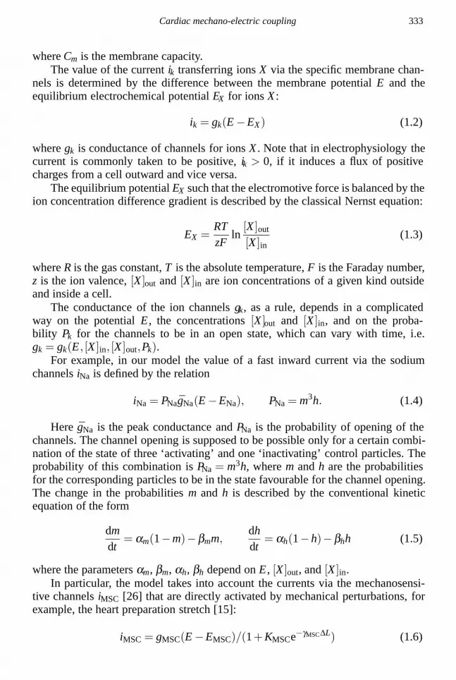
1. MATHEMATICAL MODEL OF ELECTRICAL AND MECHANICALACTIVITY IN HEART MUSCLE
We have developed the mathematical model of electromechanical coupling in heartmuscle [25]. The model describes the generation of the AP created by ionic currentsvia a sarcolemma, as well as Ca2+, Na+, K+ ion kinetics in cardiomyocytes. Alongwith electrochemical processes, the model describes the time variation in length andforce, which are generated by sarcomeres and the muscle as a whole. Let us brieflydescribe this model.
The description of electric activity is borrowed from the well-verified modeldeveloped by Noble et al. [22], which is widely used by electrophysiologists.
The rate of change of the membrane potential E is defined by the sum of ioniccurrents ik:
dEdt
= − 1Cm
∑k
ik (1.1)
Cardiac mechano-electric coupling 333
where Cm is the membrane capacity.The value of the current ik transferring ions X via the specific membrane chan-
nels is determined by the difference between the membrane potential E and theequilibrium electrochemical potential EX for ions X :
ik = gk(E −EX) (1.2)
where gk is conductance of channels for ions X . Note that in electrophysiology thecurrent is commonly taken to be positive, ik > 0, if it induces a flux of positivecharges from a cell outward and vice versa.
The equilibrium potential EX such that the electromotive force is balanced by theion concentration difference gradient is described by the classical Nernst equation:
EX =RTzF
ln[X ]out
[X ]in(1.3)
where R is the gas constant, T is the absolute temperature, F is the Faraday number,z is the ion valence, [X ]out and [X ]in are ion concentrations of a given kind outsideand inside a cell.
The conductance of the ion channels gk, as a rule, depends in a complicatedway on the potential E , the concentrations [X ]out and [X ]in, and on the proba-bility Pk for the channels to be in an open state, which can vary with time, i.e.gk = gk(E, [X ]in, [X ]out,Pk).
For example, in our model the value of a fast inward current via the sodiumchannels iNa is defined by the relation
iNa = PNagNa(E −ENa), PNa = m3h. (1.4)
Here gNa is the peak conductance and PNa is the probability of opening of thechannels. The channel opening is supposed to be possible only for a certain combi-nation of the state of three ‘activating’ and one ‘inactivating’ control particles. Theprobability of this combination is PNa = m3h, where m and h are the probabilitiesfor the corresponding particles to be in the state favourable for the channel opening.The change in the probabilities m and h is described by the conventional kineticequation of the form
dmdt
= αm(1−m)−βmm,dhdt
= αh(1−h)−βhh (1.5)
where the parameters αm, βm, αh, βh depend on E , [X ]out, and [X ]in.In particular, the model takes into account the currents via the mechanosensi-
tive channels iMSC [26] that are directly activated by mechanical perturbations, forexample, the heart preparation stretch [15]:
iMSC = gMSC(E −EMSC)/(1+ KMSCe−γMSC∆L) (1.6)

334 O. E. Solovyova, N. A. Vikulova, P.V. Konovalov, P. Kohl, and V. S. Markhasin
where EMSC is the reversion potential for iMSC, gMSC is the peak conductivity formechanosensitive membrane channels, ∆L is the deviation in the current prepara-tion length from some fixed length, KMSC and γMSC are the parameters defining the‘length dependence’ of the current.
Along with the ion transport over the concentration gradient via the membranechannels, there are also cell currents that are induced by molecular pumps translo-cating ions against concentration gradients. The active ion transport is possible dueto either exchange with other ions (for example, Na+–Ca2+ exchange current) orthe energy consumption of ATP (for example, Na+ and K+ currents via Na+–K+
ATPhase). In order to describe the active ion transport in the model, we use qua-sistationary equations for interaction between ions and proteins, their conformationchanges provide ion translocation via the membrane. For example, the expressionfor Na+–Ca2+ exchange current iNCX capable of changing the polarity and translo-cating 3Na+ against 1Ca2+ has the form
iNCX = kNCXe2γNCX
EF2RT [Na+]3in[Ca2+]out − e−2(1−γNCX) EF
2RT [Na+]3out[Ca2+]in1+[Ca2+]in
/KNCX
(1.7)
where kNCX is a parameter defining the exchange current amplitude and the param-eters KNCX and γNCX define the exchange mechanism sensitivity to the change inNa+ and Ca2+ ion concentrations.
The [Ca2+]out, [Na+]out, and [K+]out concentrations are assumed to be constant.At the same time, the transmembrane ion fluxes result in a substantial change in[Na+]in, [K+]out, and especially [Ca2+]in concentrations. The change in the [Ca2+]inconcentration in a cell also essentially depends on intracellular processes, in partic-ular, on interaction between Ca2+ ions and intracellular ligands Li and on exchangewith intracellular Ca2+ sources. The change in the above concentrations is describedby the equations:
d[Ca2+]indt
= ∑k1
Fk1,Ca2+ −∑i
d[Ca−Li]dt
+ FSR,rel −FSR,pump
d[Na+]indt
= ∑k2
Fk2,Na+ ,d[K+]out
dt= ∑
k3
Fk3,K+ −DK+ .
(1.8)
Here Fk,X = ±|ik|/(zVcytF) is the transmembrane ion flux X with current ik,where the sign of flux is defined by the direction of ion motion, Vcyt is the cytosolvolume; [Ca−Li] is the concentration of Ca2+ complexes with intracellular ligands;FSR,rel and FSR,pump are Ca2+ fluxes between intracellular compartments (see below);DK+ is K+ diffusion in an extracellular medium.
The constant membrane potential at rest, which is called a resting potential, isdefined by a number of differently directed background currents via the channelsand exchangers sustaining cellular homestasis. The resting potential is close to theequilibrium K+ potential EK = −94.5 mV because the membrane permeability atrest for other ions is low.

Cardiac mechano-electric coupling 335
During the cardiac cell excitation (due to a stimulating signal from the conduct-ing system of the heart or the artificial electrical stimulation of heart preparation ina physiological experiment) the membrane potential increases to a threshold leveland thus the ion channels and the exchangers become activated. In the model, theprethreshold perturbation of the membrane potential, which initiates the cell excita-tion, is prescribed by the short-time stimulating depolarizing current istim < 0. Ioniccurrents that occur after threshold depolarization provide the characteristic cyclicchange in the membrane potential, which is called an action potential. In particu-lar, the fast sodium current via Na+ channels, iNa, is responsible for fast upstroke.The slow inward calcium current via L-channels, iCaL, mostly provides the phase ofAP plateau. Outward currents via K+ channels, viz. delayed outward K+ current,iK, and the inward rectifier K+ current, iK1, define the membrane repolarization upto the resting potential level. The above currents, iNCX and iMSC, also substantiallyaffect the AP configuration.
Model description of the mechanical activity of heart muscle is based on theclassical three-element scheme of a contraction unit, which consists of a contractileelement (CE) or sarcomere as well as associated serial (SE) and parallel (PE) elas-tic elements. According to the above scheme the muscle length, L, is taken to beproportional to the parallel element length, whereas the tension T produced by themuscle is proportional to the sum of tensions in elastic elements TSE + TPE.
In the framework of the model, we describe dynamic changes in the musclelength L and the tension T under various contraction conditions. For example, themodel can generate either the change in the tension T , given the change in themuscle length L = ϕ(t) (in particular, in the isometric regime at the fixed lengthL ≡ const), or the change in the length L, given the change in the load T = ψ(t) (forexample, in the isotonic regime at the fixed load T ≡ const).
Suppose l1 is the CE deformation, i.e. the deviation in the sarcomere length fromthe resting length in the process of contraction (relaxation), l2 is the PE deformation.These two variables are basic phase variables in the mechanical model block.
It is obvious that the SE deformation is equal to the difference between the PEand CE deformations, i.e. to l2 − l1. The deformation-tension coupling for elasticelements is prescribed by the experimental data in the form of the parametric func-tions:
TSE = TSE(l2 − l1), TPE = TPE(l2). (1.9)
The force TCE generated by sarcomere is due to interaction between crossbridgesof myosin molecules and the active centers of actin molecules and depends on thesarcomere length and the velocity of its shortening/lengthening in the model, as wellas the calcium activation process of fine filaments.
It is logical to putTCE = λ f N. (1.10)
Here f is the force generated by an averaged force-generating bridge, N is thefraction of force-generating bridges per sarcomere, λ is a proportionality coefficient.
It is assumed that f depends on the shortening/lengthening velocity of sarcom-ere v = dl1/dt, i.e. f = f (v). The function f (v) is given in explicit form (by the

336 O. E. Solovyova, N. A. Vikulova, P.V. Konovalov, P. Kohl, and V. S. Markhasin
experimental data) and allows us to find the velocity f in explicit form, given thebridge load v = v( f ). In view of this and the fact that the tensions of the series-connected sarcomere CE and the elastic element SE are equal, TCE = TSE, we derivefrom (1.9)–(1.10) the equation for l1:
dl1dt
= v(TSE(l2 − l1)
λN
). (1.11)
The kinetics of the force-generating bridges is described by the equation:
dNdt
= k+
(l1,
dl1dt
, [Ca−TnC])(1−N)− k−N. (1.12)
The rate of attachment of the bridges to actin depends on the sarcomere length(i.e. on l1), the velocity of its shortening or stretch, dl1
/dt, and on the average con-
centration of calcium complexes with regulator protein-specific troponin C, [Ca–TnC].
The change in [Ca–TnC] is described by the kinetic equation:
d[Ca−TnC]dt
= kon(TnCtot − [Ca−TnC])[Ca2+]in−koff(N, [Ca−TnC])[Ca−TnC].(1.13)
One of the most important features of our model is that it takes into account inequations (1.12)–(1.13) the cooperative mechanisms in the process of calcium ac-tivation of contractile proteins, which are found experimentally earlier [4 – 6]. Theprobability of binding the bridges cooperatively increases due to conformationaltransformations of regulator actin proteins, which are caused by the formation ofcalcium complexes with troponin C. This mechanism is formalized as a power de-pendence of the rate of binding the bridges k+ on the value of [Ca–TnC] [see equa-tion (1.12)]. On the other hand, the affinity of troponin for calcium increases, first,as the concentration of strongly bound crossbridges, N, increases and, second, withincreasing [Ca–TnC]. These links are formalized as a decreasing dependence ofthe constant of the decay rate koff on the values of N and [Ca–TnC] [see equation(1.13)].
Taking into account the above mechanisms in the model allowed us to repro-duce and explain quite a number of sophisticated mechanical and mechanochemicalphenomena observed in the active myocardium [9, 12, 25, 27].
Finally, the final equation in the mechanical model block is the equation forfinding l2. Given the change in the muscle length, l2 is specified in the explicit form:l2 = ϕ(t). For example, l2 ≡ const in the isometric regime. Given the change in themuscle load, l2 is found from the identity TSE(l2− l1)+TPE(l2) = ψ(t). For example,we can write the equation for l2:
dl2dt
=
•ϕ( •
ψ −(TSE)′l1dl1dt
)/(TSE + TPE)′l2
if length controlif load control. (1.14)
Cardiac mechano-electric coupling 337
The free intracellular calcium kinetics is a connecting link between electricaland mechanical activities of a cardiac cell because it is closely connected to trans-membrane potential-dependent Ca2+ fluxes [see equation (1.8)] and directly in-cluded in the regulation of a contractile response of heart muscle [see equation(1.13)]. As mentioned above, exchange with intracellular structures, in particular,exchange with intracellular calcium sources, i.e. sarcoplasmic reticulum (SR) [theterms FSR,rel and FSR,pump in equation (1.8)] plays an important role in the [Ca2+]inkinetics. An increase in [Ca2+]in by an order of magnitude (from 10−7M in diastoleto 10−6M in systole), which is needed for contractile cell activity during a cardiaccycle, is not mainly due to Ca2+ supply from the outside of a cell, but to calcium-induced release of calcium from SR [2]. The Ca2+ flux via the release channels onthe SR membrane,
FSR,rel = η(iCaL, [Ca2+]JSR) (1.15)
depends in a complicated way on the current iCaL, which is a trigger of Ca2+ releasefrom SR, and nonlinearly depends on the calcium concentration in junctional SR,[Ca2+]JSR.
The process of the heart muscle relaxation is mainly due to the absorption ofCa2+ from cytosol back to SR by calcium pumps on the SR membrane. The Ca2+
flux produced by these pumps
FSR,pump = kpump1
1+[Ca2+]NSR/KNSR
[Ca2+]in[Ca2+]in + Kpump
(1.16)
depends on both [Ca2+]in and the calcium concentration in net SR, [Ca2+]NSR. Theparameter kpump specifies the maximum absorption rate, Kpump is the affinity ofpumps for [Ca2+]in, and KNSR is the degree of the pump inhibition with increas-ing [Ca2+]NSR.
Finally, cytosol Ca2+ forms complexes with calcium-binding legands, in partic-ular, with protein TnC [see equation (1.13)]. The Ca2+ consumption for the forma-tion of these complexes is represented in equation (1.8) by the terms d[Ca−Li]/dt.
Thus, the model of mechanical and electrical phenomena in myocardium is thesystem of 27 ordinary differential equations. The electrophysiological model blockcomprises an equation for the membrane potential E [equation (1.1) with relationsin the right-hand side of the form (1.2) or (1.7)] and an equation for dynamic pa-rameters that specify the probabilities Pk for the ionic channels to be in an openstate [the equations of the form (1.5)]. The mechanical model block supplementsequations (1.11)–(1.14) for the deformations l1, l2 the concentration of the force-generating bridges N, and the control calcium complex with troponin C, [Ca–TnC].The ‘chemical’ block of the system comprises the equations for [Ca2+]in, [Na+]in,and [K+]out [equations (1.8) with relations (1.15), (1.16) in the right-hand side], theequations for [Ca−Li] [analogous to equation (1.13)] and finally the equations forthe [Ca2+]JSR and [Ca2+]NSR kinetics in the release and absorption compartments ofSR.

338 O. E. Solovyova, N. A. Vikulova, P.V. Konovalov, P. Kohl, and V. S. Markhasin
In the framework of the described model we took into account both direct cou-pling and feedback between electrical and mechanical phenomena in cardiac cells.Indeed, due to the effect of the membrane potential on Ca2+ currents and hence on[Ca2+]in the excitation contraction coupling is realized. On the other hand, the effectof the kinetics of the force-generating bridges on the [Ca–TnC] kinetics and henceon [Ca2+]in and the calcium-dependent currents ensures feedback between the con-traction and electric activity of cardiomyocytes, which is called the mechanoelectricfeedback. In addition to this mechanism, currents via the mechanosensitive channels[see (1.6)], which are directly activated by mechanical perturbations, can also con-tribute to the mechanically caused modulation of electric activity.
Most of the parameters in the model equations are taken from the experimentalworks, where these parameters were thoroughly determined. The other model pa-rameters were chosen to achieve qualitative agreement between the model resultsand the values recorded in the physiological experiments.
The model system of ordinary differential equations belongs to the class of stiffsystems. For the numerical integration, we first used the Runge-Kutta fourth-ordermethod with a small step to obtain as the more exact numerical solution as possi-ble. Then we compared the numerical solutions obtained by the Euler method withthe obtained solution. We found the numerical integration step such that the twosolutions are sufficiently close. We also used the combined explicit-implicit Eulermethod for the calculation of part of the variables explicitly and solved the systemof algebraic equations for finding the other variables. This method allows us to in-crease the integration step as compared to that in the explicit method without lossof the solution quality. For the experiments with the model we developed a programthat allows us to simulate various experimental conditions for muscle contraction,specify different initial conditions in the system, and change the free parameters.
The combined electromechanical model describes the mechanical and electricalbehaviour of cardiomyocytes both in solitary isometric and isotonic contraction-relaxation cycles and in the series of contractions for various stimulation frequen-cies. The model successfully reproduces numerous effects that demonstrate the in-fluence of the mechanical contraction conditions on the AP generation in cardiaccells. In particular, in the framework of the model we correctly reproduced boththe effect of the muscle length on the APD in the isometric regime and the effectof the muscle load on the APD in the isotonic regime [25]. The inclusion of themechanosensitive channels in the model allowed us to describe a number of phe-nomena that are irreproducible without considering the channels, for example, therepolarization crossover phenomenon after the cell stretch as compared to the con-trol one [11, 29].
2. INTEGRALS OF DIFFERENCE CURRENTS
We developed the method for defining the relative contribution of various mecha-nisms of mechano-electric feedback to the change in the time dependence of theAP after the change in the mechanical contraction conditions [25]. The method is

Cardiac mechano-electric coupling 339
based on the calculation of the integrals of difference in individual currents beforeand after a perturbation (difference current) and in the current time:
∆ik(t) = ik,test(t)− ik,control(t). (2.1)
The sign of difference current (the sign ‘+’ is conventionally ascribed to a repo-larization current transferring positive charges from a cell) indicates if the currentalterations are repolarizing (∆ik > 0) or depolarizing ones (∆ik < 0) with respectto the control AP no matter where the control current ik was directed to: inside acell (a depolarizing current) or outside it (a repolarizing current). In other words,∆ik > 0 can imply either an increase in the control repolarizing current (ik > 0) ora decrease in the absolute value of the depolarizing current (ik < 0).The alterationof each current due to a test perturbation contributes to the dynamic change of thesum of the currents, which is responsible for the change in the membrane potential.Thus, the contribution of the alteration of each individual current to the total changein the potential can be assessed by the mathematical model.
The change in the membrane potential ∆E(t) in response to a perturbation isdefined by the formula:
∆E(t) = Etest(t)−Econtrol(t) = ∆E(t0)+(− 1
C
)∑k
∆Qk(t) (2.2)
where ∆Qk(t) is an additional charge transferred by the difference current ∆ik(t).The relation holds for ∆Qk(t):
∆Qk(t) =t∫
t0
∆ik(τ)dτ . (2.3)
To assess the contribution of the additional charge transferred by the differencecurrent ∆ik(t) to the change in the membrane potential, we also calculate the incre-ment of the potential ∆Ek(t) corresponding to this difference current:
∆Ek(t) = − 1C
∆Qk(t). (2.4)
A comparison of the time difference of ∆Ek(t) allows us to assess the contribu-tion of each difference current to the change in the AP after a perturbation of theheart muscle.
3. EFFECT OF MECHANICAL PERTURBATIONSON THE ELECTRICAL ACTIVITY IN HEART MUSCLE
3.1. Effect of a transient release on the action potential
In the framework of the model we investigated the effect of ‘forced’ cyclic defor-mations of the ‘quick release-quick stretch’ heart muscle on the AP generation. In
340 O. E. Solovyova, N. A. Vikulova, P.V. Konovalov, P. Kohl, and V. S. Markhasin
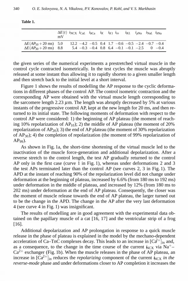
Table 1.
∆E(t) iNCX iCaL ibCa iK iK1 ito iKL ipNa iNaL ibNamV
∆E(AP10 +20 ms) 5.0 12.2 −4.2 −0.5 0.4 1.7 −0.6 −0.5 −2.4 −0.7 −0.4∆E(AP30 +20 ms) 8.8 5.4 −0.3 −0.4 0.8 6.4 −0.1 −0.1 −2.5 0 −0.4
the given series of the numerical experiments a prestretched virtual muscle in thecontrol cycle contracted isometrically. In the test cycles the muscle was abruptlyreleased at some instant thus allowing it to rapidly shorten to a given smaller lengthand then stretch back to the initial level at a short interval.
Figure 1 shows the results of modelling the AP response to the cyclic deforma-tions in different phases of the control AP. The control isometric contraction and thecorresponding AP were obtained with the virtual muscle length corresponding tothe sarcomere length 2.23 µm. The length was abruptly decreased by 5% at variousinstants of the progressive control AP, kept at the new length for 20 ms, and then re-turned to its initial state. The following moments of deformation with respect to thecontrol AP were considered: 1) the beginning of AP plateau (the moment of reach-ing 10% repolarization of AP10); 2) the middle of AP plateau (the moment of 20%repolarization of AP20); 3) the end of AP plateau (the moment of 30% repolarizationof AP30); 4) the completion of repolarization (the moment of 99% repolarization ofAP99).
As shown in Fig. 1a, the short-time shortening of the virtual muscle led to theinactivation of the muscle force-generation and additional depolarization. After areverse stretch to the control length, the test AP gradually returned to the controlAP only in the first case (curve 1 in Fig. 1), whereas under deformations 2 and 3the test APs terminated later than the control AP (see curves 2, 3 in Fig. 1). TheAPD at the instant of reaching 90% of the repolarization level did not change underdeformation at the beginning of plateau, increased by 6.6% (from 180 ms to 192 ms)under deformation in the middle of plateau, and increased by 12% (from 180 ms to202 ms) under deformation at the end of AP plateau. Consequently, the closer wasthe moment of muscle release towards the end of AP plateau, the larger turned outto be the change in the APD. The change in the AP after the very last deformation4 (see curve 4 in Fig. 1) was insignificant.
The results of modelling are in good agreement with the experimental data ob-tained on the papillary muscle of a cat [16, 17] and the ventricular strip of a frog[16].
Additional depolarization and AP prolongation in response to a quick musclerelease in the phase of plateau is explained in the model by the mechano-dependentacceleration of Ca–TnC complexes decay. This leads to an increase in [Ca2+]in and,as a consequence, to the change in the time course of the current iNCX via Na+–Ca2+ exchanger (Fig. 1b). When the muscle releases in the phase of AP plateau, anincrease in [Ca2+]in reduces the repolarizing component of the current iNCX in thereverse-mode phase and under deformations closer to AP completion it increases the

Cardiac mechano-electric coupling 341
(a) (b)Figure 1. Effect of transient releases of a virtual muscle on APD in various phases of AP. (a) thechange in length (L), force (F), and membrane potential (E) during an isometric contraction at thecontrol muscle length (thick lines) and after short-time 5% release at different instants 1– 4 (thinlines). Arrows indicate the instants in 20 ms after release (Trel + 20 ms) at which the increments ofthe membrane potential are calculated (see Subsection 3.1.1). (b) the change in [Ca2+]in, Na+–Ca2+
exchange current, iNCX, slowly activated outward calcium current, iK1, in the control (thick lines) andin response to deformations 1 and 3 (thin lines).
depolarizing component of the current iNCX in the forward-mode phase (Fig. 1b).In order to find out why the similar muscle shortening in different phases of
AP plateau finally leads to a different increase in the APD, we used the methodof integrals of difference currents (see Section 2). Table 1 gives the change in themembrane potential ∆E in 20 ms after release (Trel +20 ms), when the instant of theTrel release coincides with either AP10 or AP30 (in Fig. 1 these instants Trel + 20 msare indicated by arrows). In the case of the earlier shortening, ∆E(AP10 + 20 ms)is 5 mV and ∆E(AP30 + 20 ms) is 8.8 mV. In Table 1, the values of the change inthe membrane potential ∆Ek(Trel + 20 ms), which are due to the change in a chargetransferred by each individual ionic current ik, are also compared.
As was expected, the method of integrals of difference currents showed that themain reason for additional depolarization ∆E > 0 caused by the muscle release isthe change in the potential ∆ENCX > 0, which is connected with the depolarizingdifference current ∆iNCX < 0 between the test and control currents iNCX [recall thatbecause of formula (2.3) ∆Ek and ∆ik have opposite signs]. Note that in both cases∆iNCX < 0 after release due to a decrease in the repolarizing component of the cur-rent iNCX > 0 in the reverse-mode phase (Fig. 1b).
A decrease in the slowly activated outward K+ current, iK1, also contributes tothe depolarizing deformation effect (Fig. 1b). Indeed, the increment in the potentialcaused by the difference current ∆iK1 < 0 is ∆EK1(Trel + 20 ms) (see Table 1). Notethat a depolarizing increment in the potential ∆ENCX(Trel +20 ms) under earlier de-

342 O. E. Solovyova, N. A. Vikulova, P.V. Konovalov, P. Kohl, and V. S. Markhasin
formations is larger than the corresponding increment ∆EK1(Trel + 20 ms), whereasunder later deformations the reverse was true. Since the muscle release is producedat different values of the membrane potential, the current iK1 was activated differ-ently by the moment of deformation and ∆E variously affects the change in thiscurrent. Under the earlier deformation when the value of the membrane potentialis rather large, the absolute value of the current iK1 is smaller and its sensitivity tothe change in the potential, which is assessed by the value diK1/dE , is also smallerthan that for the muscle shortening at the lower level of the potential. Therefore un-der delayed muscle shortenings in the plateau phase (but the potential level is stilllarger than −65 mV) there is a relatively large decrease in iK and, as a consequence,a longer delay in the completion of the repolarization phase than that in the case ofmuscle shortening in the initial phase of plateau (Fig. 1b).
With the instantaneous muscle release after the completion of repolarization thedifference currents ∆iNCX < 0 and ∆iK1 > 0 are differently directed. At the values ofthe membrane potential close to those of the resting potential an additional amountof [Ca2+]in caused by shortening increases the value of the depolarizing componentiNCX < 0, whereas a small increase in the potential (to the level no larger than −65mV) leads to an increase in repolarizing iK1 > 0. Thus, the total change in the APresults from a subtle balance between the changes in these two currents and, as arule, total ∆E is not large.
The changes in the depolarizing Ca2+ current via L-channels, ∆iCaL, also affectthe change in AP. In the case of muscle shortening at the AP10 level, an increasein [Ca2+]in leads to the acceleration of the inactivation of iCaL, which decreases its
(a) (b) (c) (d)Figure 2. Effect of the short-time stretch of a virtual muscle on AP. Protocols of the change in lengthare shown on the top panels. APs during the control isometric contraction (thick lines) and afterstretch-release deformations (QS-QR, thin lines) are shown below. The results are obtained for differ-ent parameters of conductivity of mechanosensitive channels gMSC = 0.013 (middle row of plots) andgMSC = 0.09 (bottom row of plots).

Cardiac mechano-electric coupling 343
(a) (b)Figure 3. Extra APs caused by deformations. The virtual muscle was instantly released (a) orstretched (b) shortly before the completion of the next AP during the control isometric contraction(thick lines). (a) the change in muscle length (L), [Ca2+]in, Na+–Ca2+ exchange current (iNCX), andmembrane potential (E) in response to the instantaneous 11% shortening (thin lines). (b) the changein muscle length, the current via mechanosensitive channels iMSC and membrane potential (E) inresponse to 5% stretch (thin lines).
depolarizing influence on the membrane potential, i.e. ∆iCaL > 0, which inhibits anincrease in the potential. Under delayed deformation, ∆iCaL does not greatly affectthe total change in the AP.
We also investigated the dependence of the APD on the deformation duration,the shortening amplitude, and the shortening velocity. Under the above mechanicalperturbations, the APD increased as compared to the control value as the value ofperturbations increased. The results of the numerical experiments are also in goodagreement with the earlier published experimental data [8].
3.2. Effect of a transient stretch on the action potential
In the framework of the model that takes into account the currents via the mechano-sensitive channels iMSC at the two values of conductance of the mechanosensi-tive channels gMSC: low (gMSC = 0.013 µS, Fig. 2, middle row of plots) and high(gMSC = 0.09 µS, Fig. 2, bottom row of plots), we investigated the effect of a short-time stretch on the heart muscle activity. The virtual muscle was instantly stretchedby 5% of the control length (the initial sarcomere length in the control cycle was 2.1µm), then kept at this level for 30 ms, and afterwards returned to the initial level.
Under cyclic deformations, in the early phase of plateau of the control AP (at the

344 O. E. Solovyova, N. A. Vikulova, P.V. Konovalov, P. Kohl, and V. S. Markhasin
level over 25% of repolarization for gMSC = 0.013 µS or over 20% of repolarizationfor gMSC = 0.09 µS) the membrane potential abruptly decreased in response to amuscle stretch and then returned to the control AP after the restoration of the controlmuscle length and, as a result, even became longer (Fig. 2a).
As the moment of a stretch shifted to lower potentials but higher than the reversepotential for iMSC (EMSC = −20 mV, [29]), the repolarization acceleration after astretch led to the APD reduction as compared to the control one (Fig. 2b). Further,at the stretch level somewhat larger than the reverse potential for iMSC, there oc-curred a crossover of the time course of repolarization as compared to the controlone (Fig. 2c). This crossover was pronounced only at sufficiently large values ofconductance gMSC. Only in this case, the initial acceleration of repolarization rightafter a stretch could be overlapped by the depolarizing current that occurred uponreaching the reverse potential for iMSC.
Finally, at larger gMSC a further shift of the beginning of a stretch below thereverse potential for the mechanosensitive channels led to a lengthening of the AP(for example, in stretching at the AP60 level, Fig. 2d, bottom row of plots), whichwas caused by the activation of the depolarizing current via the mechanosensitivechannels.
The above results of modelling are in good agreement with the experimen-
(a) (b)Figure 4. Slow response of a virtual muscle in stepwise transition from the isometric to isotonicregime of contractions. (a) the superposition of the force generated by the muscle in stationary isomet-ric contractions (F , in relative units) and isotonic muscle shortenings in transition (∆L, in percent ofthe initial length Linit). (b), (c), (d) the superposition of changes in membrane potentials (E), [Ca2+]inand the free Ca2+ concentration in release lumens of SR ([Ca2+]JSR) during stationary isometric con-tractions (black lines) and isotonic muscle shortenings in transition (grey lines). A slow increase fromcycle to cycle in the shortening amplitude (peak ∆L), APD, the amplitude of Ca2+ transients (peak[Ca2+]in), and the total amount of calcium in SR in the free and bound to calsequestrin forms (SRCa2+ load) is shown in the insets of the corresponding rows of plots.

Cardiac mechano-electric coupling 345
Figure 5. Change in Ca2+ input-output via the cell membrane in transition from the isometric toisotonic regime of contractions. The total Ca2+ innflux in a cell during the cycle consists of the Ca2+
influx with calcium current via L-type channels (iCaL, black bars) and with reverse mode Na+–Ca2+
exchange current (iNCX, grey bars). On transition to the isotonic regime the amount of Ca2+ enteringa cell decreases (cumulative black + grey bars) mainly due to a decrease in the influx with iNCX. Theextrusion of Ca2+ from a cell is provided by the forward mode iNCX. The total amount extruded froma cell in one cycle (white bars) also decreases in transition to the isotonic regime and to a greaterextent as compared to the input. The temporal input-output unbalance provides Ca2+ accumulation inSR (SR Ca2+ gain) (see the inset).
tal data [11, 29]. A more sophisticated treatment of individual currents whosechange contributes to the total change in the AP in response to the cyclic ‘stretch-shortening’ deformation showed that it was necessary to take into account in themodel the activation of mechanosensitive channels (MSC) with substantially nonzeroconductance in order to explain the nonmonotonous change in the potential with thetime of a mechanical perturbation.
3.3. Extra action potentials mechanically induced by a release or stretch
In this paper, in the framework of our model, we reproduced and explained the ex-perimental data of the occurrence of delayed afterdepolarization and an extra actionpotential in the heart preparation as a result of either its shortening [8] or stretch[11, 29] shortly before or after the completion of a successive AP (Fig. 3).
3.3.1. Extra action potentials induced by a release. As shown in Subsection3.1, the 5% muscle shortening due to an instant release near the AP completiondid not lead to overcoming the threshold depolarization level necessary for the oc-currence of a new AP. However, when increasing the amplitude of virtual musclerelease (for example, beginning with 11% shortening in the example in Fig. 3a)there occurred an extra AP caused by an increase in the depolarizing component ofcalcium-dependent iNCX in response to a splash of [Ca2+]i due to the deformation.The extra AP caused by shortening is 35% shorter than the regular AP. It essen-

346 O. E. Solovyova, N. A. Vikulova, P.V. Konovalov, P. Kohl, and V. S. Markhasin
tially has no early repolarization phase and the plateau phase is also shorter so thatrepolarization ceases faster than that in the regular AP.
Note that it was possible to initiate an extra AP in response to an abrupt musclerelease and muscle shortening only in a very short time interval near the controlAP completion (for 34 ms for a given virtual muscle). Thus, 11% muscle shorten-ing by 238 ms of the cycle (Fig. 3a) was accompanied by the generation of a newAP. The same muscle shortening only by 1ms previously brought about additionaldepolarization but did not lead to the generation of a new AP. In the framework ofthe model (as shown in the Subsection 3.1) the physiologically justified ‘stability’of AP with respect to mechanical perturbations in the delayed repolarization phaseis due to an oppositely directed influence of the mechanically induced alterationsof the currents iNCX and iK1, which compensate each other in most cases, on themembrane potential.
3.3.2. Extra action potentials induced by a stretch. If we assume that the onlytransducer of a mechanical signal to an electrical one under deformation is intra-cellular Ca2+, the muscle stretch would not have to lead to the occurrence of extrapotentials because the intracellular Ca2+ concentration must only be reduced dueto an increase in the affinity of TnC for Ca2+ when the muscle length increases.Therefore we failed to obtain an extra AP in response to a stretch in the frameworkof the model that does not take into account the kinetics of the mechanosensitivechannels. Introducing in the description of the model the mechanosensitive chan-nels with nonzero conductance, we could obtain extra APs in response to a stretchof the muscle.
In Fig. 3b the virtual muscle that contracts isometrically at control length (sar-comere length is 1.9 µm) was instantly stretched by 5% right after the control APcompletion. In response to an increase in length we imitated the activation of themechanosensitive channels prescribing the nonzero conductance gMSC = 0.13 µSin the model. The occurrence of the depolarizing current iMSC was accompanied bydepolarization up to a threshold value and, as a result, by the generation of an ex-tra AP that had a smaller amplitude and duration than the regular AP. Note that atsmaller values of gMSC, 5% stretch did not lead to the additional AP occurrence.
4. SLOW TRANSITIONS IN THE ELECTROMECHANICALACTIVITY OF HEART MUSCLE
In the previous section, we presented the results of modelling the ‘fast’ electrome-chanical muscle responses right after the mechanical perturbations. Besides, we re-produced and analysed a number of ‘slow’ responses observed in physiological ex-periments, viz. transitions from one to another stationary state in stepwise switchingfrom isometric to isotonic regimes of contraction and vice versa [13].
In the control cycle, the virtual muscle periodically contracted in the isomet-ric regime at stimulation frequency 0.2 Hz. The initial muscle length correspondedto the initial sarcomere length 2 µm. On completion of a successive isometric cy-
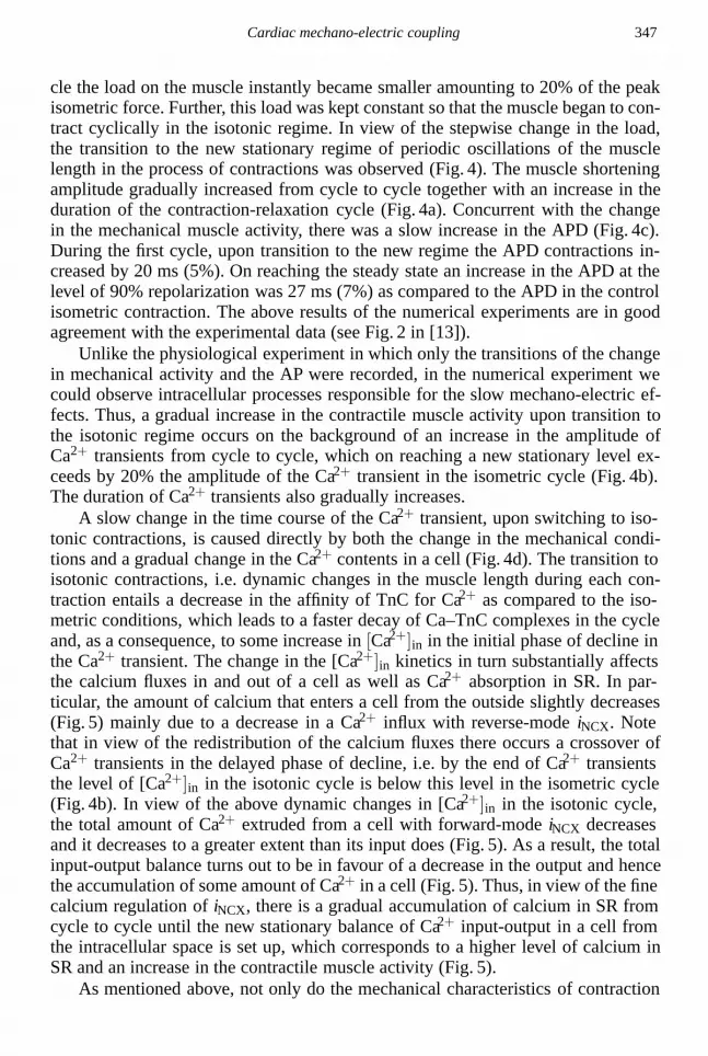
Cardiac mechano-electric coupling 347
cle the load on the muscle instantly became smaller amounting to 20% of the peakisometric force. Further, this load was kept constant so that the muscle began to con-tract cyclically in the isotonic regime. In view of the stepwise change in the load,the transition to the new stationary regime of periodic oscillations of the musclelength in the process of contractions was observed (Fig. 4). The muscle shorteningamplitude gradually increased from cycle to cycle together with an increase in theduration of the contraction-relaxation cycle (Fig. 4a). Concurrent with the changein the mechanical muscle activity, there was a slow increase in the APD (Fig. 4c).During the first cycle, upon transition to the new regime the APD contractions in-creased by 20 ms (5%). On reaching the steady state an increase in the APD at thelevel of 90% repolarization was 27 ms (7%) as compared to the APD in the controlisometric contraction. The above results of the numerical experiments are in goodagreement with the experimental data (see Fig. 2 in [13]).
Unlike the physiological experiment in which only the transitions of the changein mechanical activity and the AP were recorded, in the numerical experiment wecould observe intracellular processes responsible for the slow mechano-electric ef-fects. Thus, a gradual increase in the contractile muscle activity upon transition tothe isotonic regime occurs on the background of an increase in the amplitude ofCa2+ transients from cycle to cycle, which on reaching a new stationary level ex-ceeds by 20% the amplitude of the Ca2+ transient in the isometric cycle (Fig. 4b).The duration of Ca2+ transients also gradually increases.
A slow change in the time course of the Ca2+ transient, upon switching to iso-tonic contractions, is caused directly by both the change in the mechanical condi-tions and a gradual change in the Ca2+ contents in a cell (Fig. 4d). The transition toisotonic contractions, i.e. dynamic changes in the muscle length during each con-traction entails a decrease in the affinity of TnC for Ca2+ as compared to the iso-metric conditions, which leads to a faster decay of Ca–TnC complexes in the cycleand, as a consequence, to some increase in [Ca2+]in in the initial phase of decline inthe Ca2+ transient. The change in the [Ca2+]in kinetics in turn substantially affectsthe calcium fluxes in and out of a cell as well as Ca2+ absorption in SR. In par-ticular, the amount of calcium that enters a cell from the outside slightly decreases(Fig. 5) mainly due to a decrease in a Ca2+ influx with reverse-mode iNCX. Notethat in view of the redistribution of the calcium fluxes there occurs a crossover ofCa2+ transients in the delayed phase of decline, i.e. by the end of Ca2+ transientsthe level of [Ca2+]in in the isotonic cycle is below this level in the isometric cycle(Fig. 4b). In view of the above dynamic changes in [Ca2+]in in the isotonic cycle,the total amount of Ca2+ extruded from a cell with forward-mode iNCX decreasesand it decreases to a greater extent than its input does (Fig. 5). As a result, the totalinput-output balance turns out to be in favour of a decrease in the output and hencethe accumulation of some amount of Ca2+ in a cell (Fig. 5). Thus, in view of the finecalcium regulation of iNCX, there is a gradual accumulation of calcium in SR fromcycle to cycle until the new stationary balance of Ca2+ input-output in a cell fromthe intracellular space is set up, which corresponds to a higher level of calcium inSR and an increase in the contractile muscle activity (Fig. 5).
As mentioned above, not only do the mechanical characteristics of contraction

348 O. E. Solovyova, N. A. Vikulova, P.V. Konovalov, P. Kohl, and V. S. Markhasin
and the calcium kinetics in a cardiomyocyte change during transition, but the AP du-ration also changes. As in most of the above examples, the main trigger for AP mod-ulation is mechanically induced changes in iNCX, especially in the reverse- modephase. Seemingly, an increase in the AP duration was to provide a larger calciuminflux from the outside of a cell with current iCaL. However, in our model, this smallincrease is essentially leveled by the calcium current inactivation in view of an in-crease in release from SR. In fact, it is not improbable that the contribution of thevoltage-dependent modulation of this current to the calcium accumulation in transi-tion is more substantial.
5. DISCUSSION
We developed the mathematical model of electrical and mechanical phenomena inthe heart muscle [25]. Using this model in the present paper and in our previousstudies [25, 26], we essentially reproduced all the experimental results availablein the literature, which demonstrate the effect of the mechanical conditions of thecontraction of cardiomyocytes on their electric activity. In the framework of themodel we could establish the possible chain of events providing a link betweenthe mechanical and electrical activities. The mechano-dependent modulation of theCa–TnC complex kinetics and the calcium-dependent changes in iNCX, which are inmost cases a trigger for the AP modulation, play an important role in this chain. Thevoltage-dependent alterations of other currents, in particular, quickly and slowly ac-tivated K+ currents and the persistent Na+ current, which regeneratively affect theprogressive AP, can also substantially modulate the total deviation in the membranepotential from the control AP in response to a mechanical perturbation. Besides, cur-rents via the mechanosensitive channels directly reacting to the muscle deformationcan also substantially modulate the electrophysiological response to a mechanicalperturbation.
Analysis of the model showed that at muscle shortening the mechano-electicfeedback is mainly realized by the modulation of the Ca2+ kinetics (Figs. 1 and 3).Conversely, when the myocardium is stretched, the contribution of the mechanosen-sitive channels to the change in the potential can prove dominant and can be eitheradded to the influence of the alteration of the calcium-dependent currents or op-positely directed (Figs. 2 and 3). Analysis of the model allows us to explain theapparent contradictions in the experimental data demonstrating the opposite AP re-sponses to the stretch of heart preparations [1, 11, 28]. The model predicts that theresponse of cardiomyocytes to stretch can really be quite diverse: the AP can be ei-ther shortened or lengthened and the crossover of the repolarization with respect tothe control AP can be observed (Fig. 2). The character of the changes in the AP willdepend both on the experimental conditions and the individual electromechanicalproperties of cardiomyocytes.
In the framework of the model, we found the conditions under which both theabrupt shortening and stretch of the heart preparation can cause extra APs (Fig. 3).To this end, the perturbing deformations must be no less than some threshold am-

Cardiac mechano-electric coupling 349
plitude and must also fall into a certain rather short time interval of the progressiveAP. Thus, our results allow us to suggest that there exists in norm an extremelysmall range of vulnerability of the electrical activity of cardiomyocytes when anabrupt mechanical perturbation (shortening or stretch) can lead to an extra excita-tion of myocardium and provoke the rhythm disturbance. For example, the casesof commotio cordis are known in the clinical practice: a sudden death from an im-pact in the chest as a result of fibrillation caused by an abrupt deformation of thecardiac muscle without its mechanical damage [19]. It was shown experimentallythat the impacts responsible for arrhythmia occurred at a high rate and coincidedwith a certain phase of the membrane potential repolarization. Another example ofthe pathological effect of mechanical factors on the electric function of the heartis the occurrence of arrhythmias on the background of a collapse, i.e. a steep de-cline of vascular resistance and, as a consequence, a decrease in load on the cardiacmuscle [10]. Note that unlike the stretch effects widely investigated in experimentand in clinic, the possible arrhythmic consequences of the abrupt unloading of theheart have not essentially been studied. The model predictions allow us to advancethe hypothesis that with pathology the range of vulnerability of electric activity tomyocardium deformation can increase as compared to the norm.
Slow responses (changes in the state during several cycles) analogous to theones observed in isolated myocardium preparations [13] in switching from heavyloaded isometric contractions to low loaded isotonic contractions and vice versawere recorded in the intact heart under the abrupt change in vascular resistance.This transition of the change in the inotropic state depending on the load in theintact heart was called the Anrep phenomenon [24]. The mechanisms and the phys-iological significance of the Anrep phenomenon have not been understood so far.The model allows one at least partially to close this gap.
In the experiment on an isolated cardiac muscle in transition from the isomericto isotonic regime the muscle begins to shorten cyclically, which simulates the phaseof cardiac output in the intact heart. During several cycles the amplitude of the short-ened muscle increases (Fig. 4), which matches an increase in an ejection fraction.An increase in cardiac output in the intact heart is associated with a larger volume ofvenous return and hence an increase in the amount of blood flowing into a ventriclein diastole. In order for the influx of an increasing blood volume to be effective,the ventricle must relax faster as the cardiac output increases. The experiments onthe model show that in transition to isotonic contractions, together with a decreasein finite systolic muscle length, we observe a significant increase in the rate of theisometric relaxation phase (on the ejection completion), which is in agreement withthe physiological predictions. Thus, the maximum rate of the isometric relaxationphase increases by 60% in the first isotonic cycle and by the end of transition it istwice larger than the maximum rate of relaxation in the isometric cycle. Accordingto the model predictions, the changes in the inotropic state of the muscle and therelaxation characteristics, which, in the framework of intact heart, would allow oneto increase the ejection fraction without increasing the finite diastolic volume, areclosely connected with the change in the AP duration and the change in the calcium-and voltage-dependent Ca2+ fluxes in cardiomyocyte, which provide an increase in

350 O. E. Solovyova, N. A. Vikulova, P.V. Konovalov, P. Kohl, and V. S. Markhasin
the Ca2+ level in SR.In conclusion, we emphasize that the immune or autoimmune processes in my-
ocardium, for example, with myocarditis can substantially affect the viscoelasticproperties of myocardium, ionic channels, ionic pumps, and ion exchangers in car-diomyocytes, as we showed earlier on myocardium preparations in patients suffer-ing from a rheumatic heart disease [20]. We believe that our model is adequate forthe analysis of pathophysiological mechanisms of this kind of disturbances of theelectromechanical function of myocardium.
REFERENCES
1. A. Belus and E. White, Streptomycin and intracellular calcium modulate the response of a singleguinea-pig ventricular myocytes to axial stretch. J. Physiol. (2003) 546, 501–509.
2. D. M. Bers, Excitation–Contraction Coupling and Cardiac Contractile Force. Kluwer AcademicPublishers, 2001.
3. M. R. Franz, Mechano-electrical feedback in ventricular myocardium. Cardiovas. Res. (1996)32, 15–24.
4. A. M. Gordon, M. Regnier, and E. Homsher, Skeletal and cardiac muscle contractile activation:tropomyosin ‘rocks and rolls’. News Physiol. Sci. (2001) 16, 49–55.
5. Z. Grabarek and J. Gergely, Appendix. On the applicability of Hill type analysis to fluorescencedata. J. Biol. Chem. (1983) 258, 14103–14105.
6. J. Gulati, S. Scordilis, and A. Babu, Effect of troponin C on the cooperativity in Ca2+ activationof cardiac muscle. FEBS Lett. (1988) 236, 441– 444.
7. C. Han, P. Tavi, and M. Weckstrom, Modulation of action potential by [Ca2+]in in modeledrat atrial and guinea pig ventricular myocytes. Am. J. Physiol. Heart Circ. Physiol. (2002) 282,H1047–1054.
8. R. Hennekes, R. Kaufmann, and M. Lab, The dependence of cardiac membrane excitation andcontractile ability on active muscle shortening (cat papillary muscle). Pflugers Arch. (1981) 392,22–28.
9. V. Izakov, L. B. Katsnelson, F. A. Blyakhman, V. S. Markhasin, and T. F. Shklyar, Cooperativeeffects due to calcium binding by troponin and their consequences for contraction and relaxationof cardiac muscle under various conditions of mechanical loading. Circ. Res. (1991) 69, 1171–1184.
10. M. J. Janse and A. L. Wit, Electrophysiological mechanisms of ventricular arrhythmias resultingfrom myocardial ischemia and infarction. Physiol. Rev. (1989) 69, 1049–1169.
11. A. Kamkin, I. Kiseleva, and G. Isenberg, Stretch-activated currents in ventricular myocytes:amplitude and arrhythmogenic effects increase with hypertrophy. Cardiovasc. Res. (2000) 48,409– 420.
12. L. B. Katsnelson and V. S. Markhasin, Mathematical modeling of relations between the kinet-ics of free intracellular calcium and mechanical function of myocardium. J. Mol. Cell Cardiol.(1996) 28, 475–486.
13. R. L. Kaufmann, M. J. Lab, R. Hennekes, and H. Krause, Feedback interaction of mechanicaland electrical events in the isolated mammalian ventricular myocardium (cat papillary muscle).Pflugers Arch. (1971) 324, 100–123.
14. P. Kohl and U. Ravens, Cardiac mechano-electric feedback; past, present, and prospect. Prog.Biophys. Mol. Biol. (2003) 82, 3–9.

Cardiac mechano-electric coupling 351
15. P. Kohl and F. Sachs, Mechano-electric feedback in cardiac cells. Phil. Trans. R. Soc. Lond A(2001) 359, 1173–1185.
16. M. J. Lab, Transient depolarisation and action potential alterations following mechanicalchanges in isolated myocardium. Cardiovasc. Res. (1980) 14, 624–637.
17. M. J. Lab, D. G. Allen, and C. H. Orchard, The effects of shortening on myoplasmic calciumconcentration and on the action potential in mammalian ventricular muscle. Circ. Res. (1984)55, 825–829.
18. M. J. Lab, Mechanosensitivity as an integrative system in heart: an audit. Prog. Biophys. Mol.Biol. (1999) 71, 7–27.
19. M. S. Link, Mechanically induced sudden death in chest wall impact (commotio cordis). Prog.Biophys. Mol. Biol. (2003) 82, 175–186.
20. V. S. Markhasin, V. Y. Izakov, and V.I. Shumakov, Physiological Reasons of the MyocardiumContractile Junction Disturbance. Nauka, St. Petersburg, 1994 (in Russian).
21. D. P. Nickerson, N. P. Smith, and P. J. Hunter, A model of cardiac cellular electromechanics.Phil. Trans. R. Soc. Lond A (2001) 359, 1159–1172.
22. D. Noble, A. Varghese, P. Kohl, and P. Noble, Improved guinea-pig ventricular cell model incor-porating diadic space, IKr and IKs, and length- and tension-dependent processes. Can. J. Cardiol.(1998) 14, 123–134.
23. J. J. Rice, R. L. Winslow, J. Dekanski, and McVeigh, Model studies of the role of mechano-sensitive currents in the generation of cardiac arrhythmias. J. Theor. Biol. (1998) 190, 295–312.
24. S. J. Sarnoff, J. H. Mitchell, J. P. Gilmore, and J. P. Remensnyder, Homeometric autoregulationin the heart. Circ. Res. (1960) 8, 1077–1091.
25. O. Solovyova, N. Vikulova, L. B. Katsnelson, V. S. Markhasin, P. J. Noble, A. F. Garny, P. Kohl,and D. Noble, Mechanical Interaction of heterogeneous cardiac muscle in silico: effects on Ca2+
handling and action potential. Inter. J. Bifurcation and Chaos (2003) 13, 3757–3782.
26. O. Solovyova, N. Vikulova, V. Markhasin, and P. Kohl, A novel method for quantifying thecontribution of different intracellular mechanisms to mechanically induced changes in actionpotential characteristics. Lectures Notes in Comp. Sci. (2003) 2674, 8–17.
27. O. Solovyova, L. Katsnelson, S. Guriev, N. L. Nikitina, Y. Protsenko, S. Routkevitch, and V.Markhasin, Mechanical inhomogeneity of myocardium studied in parallel and series cardiacmuscle duplexes: experiments and models. Chaos, Solitons & Fractals (2002) 13, 1685–1711.
28. E. White, J. Y. Le Guennec, J. M. Nigretto, F. Gannier, J. A. Argibay, and D. Garnier, Theeffects of increasing cell length on auxotonic contractions; membrane potential and intracellularcalcium transients in single guinea-pig ventricular myocytes. Exp. Physiol. (1993) 78, 65–78.
29. M. Zabel, B. S. Koller, F. Sachs, and M. R. Franz, Stretch-induced voltage changes in the iso-lated beating heart: importance of the timing of stretch and implications for stretch-activated ionchannels. Cardiovascular Research (1996) 32, 120–130.