Oligomeric structure of the α1b-adrenoceptor: Comparisons with rhodopsin

N-Ethylmaleimide-Sensitive Factor Regulates �2 AdrenoceptorTrafficking and Signaling in Cardiomyocytes
Yongyu Wang, Benjamin Lauffer, Mark Von Zastrow, Brian K. Kobilka, and Yang XiangDepartment of Molecular and Integrative Physiology, University of Illinois at Urbana-Champaign, Urbana, Illinois (Y.W., Y.X.);Program in Pharmaceutical Sciences and Pharmacogenomics and Department of Pharmaceutical Chemistry (B.L.), Departmentof Psychiatry and Department of Cellular and Molecular Pharmacology (M.V.Z.), University of California at San Francisco,San Francisco, California; and Department of Molecular and Cellular Physiology, Stanford Medical Center,Palo Alto, California (B.K.K.)
Received May 3, 2007; accepted May 16, 2007
ABSTRACTRecycling of G protein-coupled receptors determines the func-tional resensitization of receptors and is implicated in switching�2 adrenoceptor (�2AR) G protein specificity in cardiomyo-cytes. The human �2AR carboxyl end binds to the N-ethylma-leimide-sensitive factor (NSF), an ATPase integral to membranetrafficking machinery. It is interesting that the human �2AR(h�2AR) carboxyl end pulled down NSF from mouse heart ly-sates, whereas the murine one did not. Despite this difference,both �2ARs exhibited substantial agonist-induced internaliza-tion, recycling, and Gi coupling in cardiomyocytes. The h�2AR,however, displayed faster rates of agonist-induced internaliza-tion and recycling compared with the murine �2AR (m�2AR)and a more profound Gi component in its contraction response.Replacing the m�2AR proline (�1) with a leucine generated a
gain-of-function mutation, m�2AR-P417L, with a rescued abil-ity to bind NSF, faster internalization and recycling than them�2AR, and a significant enhancement in Gi signaling, whichmimics the h�2AR. Selective disruption of the m�2AR-P417Lbinding to NSF inhibited the receptor coupling to Gi. Mean-while, inhibiting NSF with N-ethylmaleimide blocked the m�2ARrecycling after agonist-induced endocytosis. Expressing theNSF-E329Q mutant lacking ATPase activity inhibited them�2AR coupling to Gi in cardiomyocytes. Our results revealeda dual regulation on h�2AR trafficking and signaling by NSFthrough direct binding to cargo receptor and its ATPase activityand uncovered an unprecedented role for the receptor bindingto NSF in regulating G protein specificity that has divergedbetween mouse and human �2ARs.
�-Adrenoceptors play a pivotal role in regulating cardio-myocyte contraction through distinct signaling pathways.The �1AR couples to Gs protein(s), which increases cAMP/protein kinase A activity and the contraction rate, whereasthe activated �2AR sequentially couples to both Gs and Gi inneonatal cardiomyocytes, creating a biphasic change in con-traction. �2AR Gi coupling seems to be dependent on receptortrafficking, which includes both endocytosis and recycling.Inhibiting either process blocks receptor coupling to Gi incardiomyocytes (Xiang et al., 2002; Xiang and Kobilka, 2003).
Many G protein-coupled receptors (GPCRs) undergo endo-
cytosis in response to activation, yet their subsequent sortingin endosomes is variable, creating variable regulation of theiractivity during prolonged or repeated stimulation. Some re-ceptors are targeted to lysosomes to down-regulate cellularresponses mediated by the receptor, whereas many GPCRspossess the ability to efficiently return to the cell surface.This recycling of receptors underlies the resensitization ofcorresponding cellular responses (von Zastrow, 2003). ManyGPCRs depend on sequences residing in their intracellulardomains for recycling. A well-defined class of recycling se-quences are PSD-95/Discs-large/ZO-1 (PDZ) domain bindingmotifs (also called PDZ ligands) that are usually located atthe carboxyl-terminal end of different GPCR tails (Bockaertet al., 2004; Gage et al., 2005). The �2AR has a type I PDZligand at its carboxyl-terminal end that is necessary for re-cycling and sufficient to reroute the �-opioid receptor from adegradative to a recycling pathway (Cao et al., 1999; Gage et
These studies were supported by grants 1R01-HL71078 (to B.K.K.), R01-DA12864 (to M.V.Z.), and 1R01- HL82846 (to Y.X.) from the National Insti-tutes of Health.
Y.W. and B.L. contributed to this study equally.Article, publication date, and citation information can be found at
http://molpharm.aspetjournals.org.doi:10.1124/mol.107.037747.
ABBREVIATIONS: �2AR, �2-adrenergic receptor; �1AR, �1-adrenergic receptor; GPCR, G protein-coupled receptor; KO, knockout; NSF,N-ethylmaleimide-sensitive factor; PDZ, PSD-95/Discs-large/ZO-1; NHERF/EBP50, Na�/H� exchanger regulatory factor/ezrin/radixin/moesin-binding phosphoprotein of 50 kDa; GST, glutathione; PTX, pertussis toxin; NEM, N-ethylmaleimide; ELISA, enzyme-linked immunosorbent assay;HEK, human embryonic kidney; ANOVA, analysis of variance; AMPA, �-amino-3-hydroxy-5-methyl-4-isoxazolepropionic acid; SNARE, solubleN-ethylmaleimide-sensitive factor attachment protein receptor.
0026-895X/07/7202-429–439$20.00MOLECULAR PHARMACOLOGY Vol. 72, No. 2Copyright © 2007 The American Society for Pharmacology and Experimental Therapeutics 37747/3234545Mol Pharmacol 72:429–439, 2007 Printed in U.S.A.
429
at ASPE
T Journals on A
pril 3, 2016m
olpharm.aspetjournals.org
Dow
nloaded from

al., 2001). In cultured neonatal mouse cardiomyocytes, thissequence is also required for the temporal switch from Gs toGi-mediated signal transduction observed in the contraction-rate response to the agonist isoproterenol (Xiang and Ko-bilka, 2003). Several lines of evidence now indicate thatmembrane trafficking of this receptor dictates not only cel-lular resensitization but also signal transduction specificity.Despite progress in understanding the �2AR recycling pro-cess, numerous questions concerning the core mechanismand physiological variations remain.
Although the recycling sequence at the �2AR C terminushas been shown to bind PDZ domains in NHERF familyproteins (NHERF-1/EBP50 and NHERF-2/E3KARP) (Hall etal., 1998; Cao et al., 1999), it also binds at least one proteinwith no identifiable PDZ domain: the N-ethylmaleimide sen-sitive factor (NSF) (Cong et al., 2001). NSF has been identi-fied as an ATPase that binds SNAP receptor (SNARE) com-plexes in an ATP-dependent fashion to separate them duringATP hydrolysis; this and a wealth of other evidence hasdemonstrated its general role in vesicle fusion between var-ious membrane compartments (Morgan and Burgoyne, 2004;Whiteheart and Matveeva, 2004). Moreover, NSF has beenshown to bind to �-arrestin, an adaptor protein involved inGPCR desensitization and endocytosis upon agonist stimu-lation. �-Arrestin preferentially interacts with the ATP-bound form of NSF, and this NSF binding facilitates clathrincoat-mediated GPCR internalization (McDonald et al., 1999).In heterologous HEK293 cells, selective ablation of NSFbinding to the �2AR was inferred to inhibit recycling of re-ceptors, whereas imparting NSF binding on the �-opioid re-ceptor slightly enhanced its ability to recycle (Cong et al.,2001; Gage et al., 2005). Although there is also evidence toshow that PDZ interactions promote receptor recycling (Caoet al., 1997) and are functionally important for Gi coupling incardiomyocytes (Xiang and Kobilka, 2003), it is not clear howNSF may affect �2AR trafficking and signaling in these cells.
Here, we used neonatal mouse cardiomyocytes as a modelsystem to address these questions. It is interesting that theNSF binding sites on the �2AR were not conserved amongmammalian species, providing a naturally occurring diver-gence in NSF binding to exploit. The �1 position of the �2ARcarboxyl terminus is proline in m�2AR and leucine in h�2AR.Because of this single amino acid difference, m�2AR bindingto NSF was not detectable. Nevertheless, despite the lack ofdetectable binding of the m�2AR carboxyl terminus to NSF inbiochemical assays, we found that inhibition of NSF activitywith N-ethylmaleimide (NEM) inhibited murine �2AR(m�2AR) recycling despite this poor affinity. In addition, bothhuman and murine �2ARs sufficiently recycled after endocy-tosis and coupled to Gi pathways in cardiomyocytes. Thedifferent affinities for NSF seemed to have a minimum roleon receptor trafficking and signaling. In contrast, inactiva-tion of NSF ATPase activity with a point mutation was suf-ficient to block both human and murine �2AR recycling andcoupling to Gi in cardiomyocytes, indicating that NSF isrequired for proper trafficking and signaling of �2ARs incardiomyocytes independent of a high-affinity interactionwith the receptor. This study strengthens the relationshipbetween �2AR recycling and signaling specificity and dem-onstrates an unprecedented role for NSF in regulating phys-iologically relevant signal transduction.
Materials and MethodscDNA Constructs and Mutagenesis. Constructs containing the
cloned human and murine �2AR in pcDNA3 (Invitrogen, Carlsbad,CA) with a FLAG epitope attached at the N terminus were used forthese studies and have been described before (Cao et al., 1999;Swaminath et al., 2004). Constructs encoding for GST-�2AR andGST-�2AR-alanine proteins (encompassing amino acids 328 to 413 ofthe human �2AR, and the latter with an additional alanine added tothe C terminus) have also been reported (Cao et al., 1999). A com-parable murine GST-�2AR construct was created by insertion of apolymerase chain reaction product of the region encoding aminoacids 328 to 418 of the FLAG-m�2AR construct using primers con-taining EcoRI and HindIII appendages and performing the appro-priate digestion and ligation into pGEX-KG (Pfizer, New York, NY).The human NSF coding sequence was similarly ligated intopEGFP-N1 (Clontech, Mountain View, CA) after SacI digestion of thevector and a polymerase chain reaction product containing a 3�-SacIappendage and including the 5�-SacI restriction site from the sourcevector, NSF in pBluescriptR (American Type Culture Collection,Manassas, VA). The P417L mutation was introduced into the FLAG-m�2AR and GST-m�2AR constructs via the QuikChange site-di-rected mutagenesis kit (Stratagene, La Jolla, CA), as was the E329QNSF mutation into the pEGFP-N1 construct. Plasmid amplificationwas done in DH5� Escherichia coli, and all sequences were verifiedby dideoxynucleotide sequencing (University of California San Fran-cisco Biomolecular Resource Center, San Francisco, CA).
Cell Culture and Transfection. Spontaneously beating neona-tal cardiomyocytes were prepared from hearts of 1-day-old �1/�2AR-KO mouse pups as before (Devic et al., 2001). The myocyte-enriched cells remaining in suspension after preplating were platedin 35-mm dishes for contraction-rate studies and in 12-well plates forimmunological assays (with coverslips for immunofluorescent mi-croscopy). Recombinant adenovirus encoding FLAG-m�2AR has beendescribed previously (Xiang et al., 2002), and the FLAG-m�2AR/P417L, FLAG-h�2AR, GFP-NSF, and GFP-NSF-E329Q adenoviralvectors were generated with the same pAdEasy system (QbiogeneInc., Irvine, CA). Neonatal myocytes were infected with viruses at amultiplicity of infection of 100 after being cultured for 24 h. Thereceptor expression levels were determined by ligand binding assaysas described previously (Xiang et al., 2002). They were expressed atequivalent levels in cardiac myocytes (FLAG-m�2AR, 147.3 � 22fmol/mg; FLAG-m�2AR/P417L, 171.6 � 9.1 fmol/mg; and FLAG-h�2AR, 160.3 � 21.8 fmol/mg membrane).
GST Pulldown Assays. The various GST-�2AR fusion proteinswere produced in BL21 E. coli and bound to glutathione-Sepharoseagarose beads (GE Healthcare, Chalfont St. Giles, Buckinghamshire,UK). Beads containing 10 �g of the full-length fusion protein (as-sessed by densitometry of Coomassie-stained protein resolved bySDS-polyacrylamide gel electrophoresis) were incubated for 4 h at4°C in 0.5 ml of clarified extracts from frozen mouse hearts with atriaremoved (Pel-Freez Biologicals, Rogers, AR), prepared to �10 mg/ml.Beads were washed four times in 1 ml of extract buffer [0.1% (v/v)Triton X-100, 150 mM NaCl, 25 mM KCl, and 10 mM Tris, pH 7.4,complete Roche protease inhibitor cocktail], and protein was elutedin lithium dodecyl sulfate sample buffer (Invitrogen) with dithiothre-itol added to 20 mM. Samples were divided in two for SDS-polyacryl-amide gel electrophoresis, transfer to nitrocellulose, and Westernblotting using rabbit anti-EBP50 antibodies (courtesy of Dr. AnthonyBretscher, Cornell University, Ithaca, NY) or the mouse 2E5 anti-NSF antibody (courtesy of Dr. Sidney W. Whiteheart, University ofKentucky, Lexington, KY).
Immunofluorescence Microscopy. Myocyte images were ob-tained using a similar setup on a Zeiss Axioplan 2 microscope (CarlZeiss Inc., Thornwood, NY). Fluorescent measurements of the myo-cyte receptor trafficking were made by a ratiometric normalization offluorescent intensities measured using Metamorph software (Molec-ular Devices, Sunnyvale, CA). Epitope-tagged receptors were de-
430 Wang et al.
at ASPE
T Journals on A
pril 3, 2016m
olpharm.aspetjournals.org
Dow
nloaded from

tected using M1 anti-FLAG antibody (Sigma, St. Louis, MO). Selec-tive detection of surface relative to total pools of receptor and its useto estimate receptor recycling have been described previously(Tanowitz and von Zastrow, 2003). The recycling estimates wereconducted without the EDTA strip. The primary antibody used inthese experiments was M1 conjugated to Alexa Fluor 488 (Invitro-gen) using standard procedures as described previously (Tanowitzand von Zastrow, 2003). Secondary staining was performed using acommercial goat antimouse IgG Alexa Fluor 594 conjugate (Invitro-gen). Experiments were performed at least in triplicate, and repre-sentative results are shown.
Immunofluorescence Spectroscopy. Surface receptor levelswere determined as before (Swaminath et al., 2004) in the indicatedcell type expressing the indicated FLAG-�2AR. Media were refreshed1 h before 10 �M isoproterenol (Sigma) stimulation for 10 or 30 min.Periods of agonist washout after 30-min isoproterenol stimulationswere also performed for an additional 30 or 60 min as indicated.
Myocyte Contraction Rate Assay. Measurement of spontane-ous contraction rates from myocytes expressing either the endoge-nous or the indicated FLAG-�2AR were carried out with and withoutthe use of PTX as described previously (Devic et al., 2001). In someassays, NEM was applied 30 min before the addition of isoproterenol.Tat peptide, Tat-�2-DSAL consisting of Tat linked to GRQGFSSD-SAL of �2AR, and Tat-�2-ASLL consisting of Tat linked to GRQGF-SSASLL of �2AR through a cysteine bridge were synthesized in theStanford Core facility and EZ-Biolab (Indianapolis, IN). Neonatalmyocytes were preincubated at 37°C with 10 �M peptide for 25 minbefore isoproterenol (10 �M; Sigma) exposure.
Statistical Analysis. Curve-fitting and statistical analyses wereperformed using Prism (GraphPad Software, Inc., San Diego, CA).
ResultsNSF Had Higher Binding Affinity to Human �2AR
than Murine �2AR. To understand the molecular mecha-nism of the NSF effect on �2AR signaling in cardiomyocytes,the interaction between �2AR and NSF from heart lysate wasexamined. NSF, a hexameric ATPase involved in membranefusion, can bind to the carboxyl terminus of the h�2AR. Theprotein-binding region on this receptor involves a four-resi-due stretch at the distal C terminus of the receptor (Cong etal., 2001). In addition, NHERF-1/EBP50, a cytoskeleton-as-sociated protein, can also bind to the same stretch of residueson the carboxyl terminus of the h�2AR. It is interesting thatseveral rodent �2ARs, including the m�2AR, are identicalwith the h�2AR in the carboxyl-binding region except at oneresidue (leucine �1 of the h�2AR, DSLL) that is required forbinding to NSF but not to NHERF-1/EBP50 (Cong et al.,2001). Rather, the m�2AR has a proline at the �1 position(DSPL). To determine whether both the human and murine
�2ARs have a similar capacity to bind NSF, GST-fusion pro-teins, including various �2AR carboxyl-terminal tail se-quences, were prepared. Protein binding was evaluated witha pull-down assay using the GST-fusion proteins coupled toglutathione-agarose beads.
GST fusion proteins were incubated with tissue lysateprepared from mouse hearts. NSF only bound to the cyto-plasmic tail of the h�2AR but not the m�2AR (Fig. 1A). Incontrast, NHERF-1/EBP50 bound to the cytoplasmic tail ofboth the h�2AR and m�2AR under these conditions (Fig. 1B).As a negative control for nonspecific binding, an addition of asingle alanine residue to the h�2AR carboxyl terminus (GST-h�2AR-Ala) was tested as well. As reported previously, thismutant failed to exhibit the PDZ domain-mediated and NSFprotein binding (Fig. 1, A and B; Cao et al., 1999). In addition,we attempted to “rescue” an NSF interaction with the m�2ARtail by substitution of the m�2AR proline 417 with a leucineresidue (m�2AR-P417L). The m�2AR-P417L pulled downsimilar amounts of NSF from lysates compared with theh�2AR, indicating that the m�2AR-P417L cytoplasmic tailfully rescued binding to NSF (Fig. 1A). Likewise, this mutantm�2AR-P417L pulled down NHERF-1/EBP50 from mouseheart lysates (Fig. 1B).
The Binding of NSF and the NSF ATPase ActivityHad Distinct Effects on �2AR Trafficking in Cardiomy-ocytes. To examine whether NSF plays any role in �2ARtrafficking in cardiomyocytes, we analyzed the localization offlag-tagged �2ARs in cardiac myocytes. The m�2AR, m�2AR-P417L, and h�2AR were transiently expressed in cardiacmyocytes using recombinant adenovirus. Immunofluores-cence studies showed that all three receptors had a cell-surface staining during a nonstimulated state (Fig. 2A).Upon isoproterenol stimulation, all three receptors had re-duced cell-surface staining together with increased punctateintracellular staining, suggesting a significant internaliza-tion of the receptors in cardiac myocytes. These observationswere confirmed quantitatively using an ELISA-basedmethod for assaying surface receptor levels (Swaminath etal., 2004) in a large number of cells and a ratiometric methodfor analysis of fluorescence micrographs (Tanowitz and vonZastrow, 2003) (Fig. 2B; data not shown). It is interestingthat when we measured the short-term decrease in cell-surface receptors after agonist stimulation in cardiac myo-cytes, we found that the m�2AR-P417L had a faster rate(t1/2 � 2.63 � 0.05 min) of cell surface-receptor decreasethan the m�2AR (t1/2 � 10.93 � 0.01 min; Fig. 3). Becausethe receptor level change in the short time points after
Fig. 1. The binding of m�2AR and h�2AR to NSF andNHERF-1/EBP50. The binding of m�2AR and h�2AR toNSF and NHERF-1/EBP50 from mouse heart extracts.GST pull-downs were performed as described under Mate-rials and Methods. Western detection of NHERF-1/EBP50pulled down by the indicated GST-�2AR fusion proteinfrom mouse heart extract, and the corresponding detectionof NSF is shown in A, whereas the detection of NHERF-1/EBP50 from this extract is shown in B. Ponceau-stainedGST fusion proteins are shown under each blot. Images arerepresentative of three or more experiments.
NSF-Regulated Adrenergic Receptor Signaling in Cardiomyocyte 431
at ASPE
T Journals on A
pril 3, 2016m
olpharm.aspetjournals.org
Dow
nloaded from

agonist stimulation is primarily determined by agonist-induced endocytosis, these data suggested a faster rate ofendocytosis for the m�2AR-P417L than for the m�2AR incardiac myocytes. The h�2AR had a similar rate (t1/2 �3.4 � 0.03 min) of cell surface-receptor decrease comparedwith the m�2AR-P417L (Fig. 3). However, after 30 min ofagonist stimulation, we observed a similar amount of sur-face receptor decreases with the m�2AR (30.54 � 2.90%),the m�2AR-P417L (28.43 � 1.69%), and the h�2AR(24.94 � 2.74%). The observed decrease of receptor densityat 30 min of stimulation should have been a composite ofreceptor endocytosis and recycling. The equivalent de-creases of receptors at cell surface are usually due to muchslower recycling process than endocytosis in cells.
When isoproterenol was removed, both the h�2AR andm�2AR-P417L recovered cell-surface staining almost com-pletely after a 60-min incubation (Fig. 2). In contrast, them�2AR did not show a fully recovered cell-surface stainingpattern, and some residual intracellular staining was ob-served in these cells (Fig. 2), even though the majority of theinternalized receptors seemed to return to the surface within60 min. When we examined the cell surface-receptor densityat different time points with the fluorescent ELISA assay,the m�2AR exhibited a lower recovery of cell surface-recep-tors after recycling for 60 min than the m�2AR-P417L mu-tant and the h�2AR (Fig. 2B; �, p � 0.05). A significantdifference in surface recovery was also observed using ratio-metric image measurements 60 min after agonist washout(data not shown). These data indicate that the m�2AR, al-
though capable of undergoing agonist-induced internaliza-tion and recycling in cardiomyocytes, differs in rates of recy-cling compared with the h�2AR and m�2AR-P417L.
It has been well-established that NSF ATPase activityplays an important role in membrane cargo trafficking. Wethen tested whether NSF activity was necessary for the en-docytic recycling of the receptor by using NEM to inhibit NSFactivity in myocytes. In the presence of NEM, endocytosis ofthe receptor was preserved; however, a return of the receptorto the cell surface after removal of agonist for 60 min was not(Fig. 4A). This observation was confirmed with measure-ments of surface receptor levels by a fluorescent ELISA as-say. The cell surface receptor levels dropped after agonistaddition and only recovered with agonist withdrawal in theabsence of NEM (Fig. 4B). These data suggested that NEMtreatment can block the receptor from recycling after endo-cytosis.
Dominant-Negative NSF Lacking ATPase ActivityInhibited Endogenous m�2AR Coupling to Gi Pathwayin Cardiomyocytes. Our finding that NEM inhibits �2ARrecycling in cardiac myocytes suggested that NSF function isrequired for this process. To further probe whether NSFenzymatic activity can affect the receptor signaling indepen-dent from the direct NSF-receptor interaction, we examinedthe signaling mediated by the endogenous m�2AR when over-expressing an inactivated NSF, the E329Q mutant (White-heart et al., 1994). This mutation abolishes ATPase activityand has been shown to block AMPA receptor trafficking(Whiteheart et al., 1994; Whiteheart and Matveeva, 2004).
Fig. 2. NSF binding enhances recycling of FLAG-�2ARs in neonatal cardiac myocytes from �1/�2AR-KO mice. A, human and murine �2ARs inter-nalize and recycle in cardiac myocytes. Cardiacmyocytes expressing a FLAG-tagged h�2AR,m�2AR, or m�2AR-P417L were stained with M1primary antibody conjugated to the Alexa-488 flu-orophore to observe a starting “total” receptor pop-ulation. After no treatment (0), 30-min 10 �M iso-proterenol treatment (30), or 30-min isoproterenoltreatment followed by a surface antibody strip and60 min of agonist removal (30 � 60), cells werestained under nonpermeable conditions with a goatanti-mouse-IgG secondary antibody conjugated tothe Alexa-594 fluorophore to observe the relativecomplement of “surface” receptor. Images are rep-resentative of three experiments. B, NSF binding�2ARs recycle faster. Surface levels of the three�2ARs were quantified by fluorescence spectroscopymeasurements of M1-Alexa 488 associated with thecell surface receptors after the indicated periods ofdrug administration and removal (1, control; 2, 30min of isoproterenol stimulation; 3, 30 min of iso-proterenol followed by 30 min of drug removal; and4, 30 min of isoproterenol followed by 60 min of drugremoval). Surface levels are normalized as a per-centage of untreated cell surface fluorescence, anderror bars reflect standard deviations over threeexperiments. �, p � 0.05, significantly different be-tween m�2AR and h�2AR or m�2AR-P417L by ttest.
432 Wang et al.
at ASPE
T Journals on A
pril 3, 2016m
olpharm.aspetjournals.org
Dow
nloaded from

When the endogenous m�2AR in the �1AR-KO myocyte wasstimulated by isoproterenol, the activated receptor induced abiphasic contraction-rate response with an initial increasemediated by Gs coupling followed by a sustained Gi-depen-
dent decrease to reduce the contraction rate below basal level(Fig. 5A; Xiang et al., 2002). When wild-type NSF was ex-pressed in �1AR-KO cardiac myocytes, we did not observeany significant change in the endogenous m�2AR-mediatedcontraction-rate response (Fig. 5A). In contrast, when theNSF-E329Q mutant was overexpressed in cardiomyocytes,the contraction rate mediated by the m�2AR was signifi-cantly higher than the control and did not display a decreaselower than the basal level (Fig. 5B). This response profile wassimilar to that observed with an inhibition of Gi by PTX (Fig.5C). Indeed, additional treatment of PTX did not generateany further increases in contraction rates (Fig. 5D). There-fore, the NSF-E329Q behaved as a dominant-negative toblock the receptor coupling to Gi in cardiomyocytes. In addi-tion, when myocytes are pretreated with NEM to inhibit theNSF ATPase activity, we also observed effects similar tothose by NSF-E329Q mutant on m�2AR signaling mediatedcontraction-rate response (data not shown).
The Divergent C Termini of the Human and Murine�2AR Had Different Effects on Contraction Rate Re-sponses in Neonatal Cardiomyocytes. Our previous stud-ies have shown that the localization and trafficking of them�2AR is important for the receptor’s G protein signalingspecificity and subsequent regulation of the myocyte contrac-tion rate. In the course of this study, we found that thedivergent PDZ ligand of the human and murine �2AR af-fected the receptor trafficking rates after agonist stimulation
Fig. 3. NSF binding enhances the internalization kinetics of FLAG-�2ARs expressed in neonatal cardiac myocytes from �1/�2AR KO mice.Surface levels of the three �2ARs were quantified by fluorescent mea-surement of M1-Alexa 488 associated with the cell surface receptors afterthe indicated periods of 10 �M isoproterenol administration. Data werenormalized as a percentage decrease of untreated cell surface fluores-cence, and error bars reflect standard deviations over three experiments.The data represent the mean � S.E. of experiments from at least threedifferent myocyte preparations.
Fig. 4. Inhibiting NSF with NEM blocks the FLAG-�2ARrecycling after agonist-induced endocytosis in cardiomyo-cytes. A, murine �2ARs internalize and recycle in cardiacmyocytes. Cardiac myocytes expressing a FLAG-taggedm�2AR were treated as described under Materials andMethods and Fig. 3. Images are representative of threeexperiments. Inhibiting NSF with NEM blocks the FLAG-�2AR recycling after agonist-induced endocytosis in cardi-omyocytes. B, surface levels of the m�2ARs were quantifiedby fluorescent measurement of M1-Alexa 488 associatedwith the cell surface receptors after the indicated periods ofdrug administration and removal (1, control; 2, 30 min ofisoproterenol stimulation; 3, 30 min of isoproterenol fol-lowed by 30 min of drug removal; and 4, 30 min of isopro-terenol followed by 60 min of drug removal). Surface levelswere normalized as a percentage of untreated cell surfacefluorescence, and error bars reflect standard deviationsover three experiments. �, p � 0.05, significantly differentbetween cells with and without NEM treatment by t test.
NSF-Regulated Adrenergic Receptor Signaling in Cardiomyocyte 433
at ASPE
T Journals on A
pril 3, 2016m
olpharm.aspetjournals.org
Dow
nloaded from

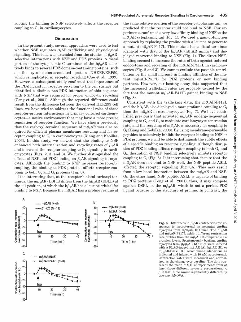
in cardiac myocytes. Thus, we wanted to examine whetherdifferences in NSF binding and/or altered trafficking ratescould modulate the receptor signaling in cardiac myocytes.When the m�2AR was expressed in �1/�2AR-KO myocytesand stimulated by isoproterenol, the activated receptor in-duced a biphasic contraction-rate response with an initialincrease followed by a sustained decrease to reduce the con-traction rate lower than basal level (Fig. 6A; Xiang et al.,2002). This contraction-rate change is equivalent to thatinduced by the endogenous m�2AR in �1AR-KO myocytes(Fig. 6A). The m�2AR-P417L induced a similar contraction-rate response profile and initial increase compared with them�2AR in �1/�2AR-KO myocytes (Figs. 6C and 7D). However,the contraction rate decreased faster, and the contractionrate was lower than that induced by the m�2AR during latestimulation in cardiac myocytes (Figs. 6C and 7E). In addi-tion, when stimulating the h�2AR expressed in �1/�2AR-KOmyocytes with isoproterenol, the activated receptor also in-duced a biphasic, contraction-rate change with an initialincrease followed by a sustained decrease (Fig. 6B). Althoughit is interesting that the initial contraction-rate increase wassmaller than that induced by the activated m�2AR andm�2AR-P417L (Figs. 6B and 7D), it is more surprising thatthe late decrease in contraction rate induced by the exoge-nous h�2AR was greater than that induced by the m�2ARand m�2AR-P417L (Figs. 6B and 7E).
The profound contraction-rate decrease induced by them�2AR-P417L and the h�2AR suggests that these receptorsmay have enhanced coupling to Gi and/or reduced coupling toGs compared with the m�2AR. We therefore examined the Gi
signaling induced by the activated receptors in cardiac myo-cytes. PTX was used to block Gi signaling in cardiac myocytesexpressing the different �2ARs before isoproterenol stimula-tion. Upon inhibiting Gi with PTX, the isoproterenol-stimu-lated m�2AR induced a slightly greater but not significantcontraction-rate increase in myocytes compared with the con-trol and prevented the late Gi-dependent contraction ratedecrease (Fig. 7A; Xiang et al., 2002). PTX treatment alsoinhibited the contraction rate decrease mediated by them�2AR-P417L or the h�2AR during the late phase of stimu-lation (Fig. 7, B and C). These data suggest that compared
with the activated m�2AR, the m�2AR-P417L had an en-hanced Gi coupling upon isoproterenol stimulation, and theactivated h�2AR coupled to Gi more efficiently in neonatalcardiac myocytes (Fig. 7E). This indicates that the divergentreceptor C termini can induce different changes in contrac-tion-rate responses that correlate with subtle changes inreceptor transportation rates.
The Binding of NSF and PDZ Had Distinct Effects on�2AR Activation-Induced Contraction Rates in Cardi-omyocyte. To further probe the effect of the �2AR binding toNSF and PDZ on receptor signaling in cardiomyocytes, wetook advantage of the different binding affinities betweenreceptor and proteins by using peptides to selectively disruptthe interactions. We expressed either m�2AR or m�2AR-P417L (the h�2AR mimic) in �1/�2AR-KO cardiomyocyte forthe contraction rate assay. Membrane-permeable peptidescontaining ASLL sequence and DSAL sequence were used toselectively disrupt NSF and NHERF/EBP50 binding, respec-tively. When m�2AR-expressing myocytes were treated withNSF (ASLL) peptide, the activated receptor induced aslightly bigger but not significant initial increase than thecells without pretreatment (Fig. 8, A and C). The increasewas sustained during stimulation and lacked a late decreasemediated by receptor/Gi coupling in control cells (Fig. 8, Aand D). When m�2AR-expressing myocytes were treated withPDZ (DSAL) peptide, the activated receptor induced a signif-icantly greater initial increase than the control (Fig. 8, B andC), and the increase was sustained and lacked a late Gi-dependent decrease (Fig. 8, B and D).
In contrast, pretreatment with NSF (ASLL) peptide did notaffect the activated m�2AR-P417L-induced initial increase(Fig. 8, E and G). However, the increase was sustained anddid not display a late Gi-induced decrease of contraction rate(Fig. 8, E and H). When myocytes expressing m�2AR-P417Lwere treated with PDZ (DSAL) peptide, the activated recep-tor induced a significantly greater initial increase in contrac-tion rate than the control (Fig. 8, F and G), and the increasewas sustained and did not show a late Gi-induced decrease(Fig. 8, F and H). Together, these data showed that althoughdisrupting the binding to PDZ protein (such as NHERF/EBP50) affects the receptor coupling to both Gs and Gi, dis-
Fig. 5. Dominant-negative NSF-E329Qmutant inhibits the m�2AR coupling toGi protein. Spontaneously beating car-diac myocytes from �1AR KO mice weretransfected with a wild-type NSF (A andC) or NSF-E329Q (B and D) mutant ad-enovirus as indicated. The cells were ad-ministered 10 �M isoproterenol with in-hibition of Gi by PTX. Overexpressingthe NSF E329Q mutant enhanced thecontraction-rate increase induced by iso-proterenol stimulation. Additional PTXtreatment did not further enhance thecontraction-rate increase induced by them�2AR. The data represent the mean �S.E. of experiments from at least threedifferent myocyte preparations. �, p �0.05, time course significantly differentby two-way ANOVA.
434 Wang et al.
at ASPE
T Journals on A
pril 3, 2016m
olpharm.aspetjournals.org
Dow
nloaded from
rupting the binding to NSF selectively affects the receptorcoupling to Gi in cardiomyocytes.
DiscussionIn the present study, several approaches were used to test
whether NSF regulates �2AR trafficking and physiologicalsignaling. This idea was extended from the studies of �2AR-selective interactions with NSF and PDZ proteins. A distalportion of the cytoplasmic C terminus of the h�2AR selec-tively binds to several PDZ domain-containing proteins, suchas the cytoskeleton-associated protein NHERF/EBP50,which is implicated in receptor recycling (Cao et al., 1999).However, a subsequent study confirmed the importance ofthe PDZ ligand for receptor recycling to the cell surface butidentified a distinct non-PDZ interaction of this sequencewith NSF that was required for proper endocytic recycling(Cong et al., 2001). Although the reported difference couldresult from the difference between the derived HEK293 celllines, we have tried to address the functional roles of thesereceptor-protein interactions in primary cultured cardiomy-ocytes—a native environment that may have a more preciseregulation of receptor function. We have shown previouslythat the carboxyl-terminal sequence of m�2AR was also re-quired for efficient plasma membrane recycling and for re-ceptor coupling to Gi in cardiomyocytes (Xiang and Kobilka,2003). In this study, we showed that the binding to NSFenhanced both internalization and recycling rates of �2ARand increased the receptor coupling to Gi signaling in cardi-omyocytes (Figs. 2, 3, and 8). We further distinguished theeffects of NSF and PDZ binding on �2AR signaling in myo-cytes. Although the binding to NSF increases receptor/Gi
coupling, the binding to PDZ proteins affects receptor cou-pling to both Gs and Gi proteins (Fig. 8).
It is interesting that, at the receptor’s distal carboxyl ter-minus, the m�2AR (DSPL) differs from the h�2AR (DSLL) atthe �1 position, at which the h�2AR has a leucine critical forbinding to NSF. Because the m�2AR has a proline residue at
the same relative position of the receptor cytoplasmic tail, wepredicted that the receptor could not bind to NSF. Our ex-periments confirmed a very low affinity binding of NSF to them�2AR cytoplasmic tail (Fig. 1). We used a gain-of-functionapproach by replacing the proline with a leucine to generatea mutant m�2AR-P417L. This mutant has a distal terminusidentical with that of the h�2AR (h�2AR mimic) and dis-played recovered binding to NSF (Fig. 1). The direct NSF-binding seemed to increase the rates of both agonist-inducedendocytosis and recycling of the m�2AR-P417L in cardiomy-ocytes (Fig. 2 and 3). We cannot exclude the possible contri-bution by the small increase in binding affinities of the mu-tant m�2AR-P417L for PDZ proteins or new bindingpartners. However, our beating assay data supported thatthe increased trafficking rates are probably caused by thefact that the mutant m�2AR-P417L gained binding to NSF(Fig. 8).
Consistent with the trafficking data, the m�2AR-P417Land the h�2AR also displayed a more profound coupling to Gi
than the m�2AR in cardiomyocytes (Fig. 7). We have estab-lished previously that activated m�2AR undergo sequentialcoupling to Gs and Gi to modulate cardiomyocyte contractionrate, and the recycling of m�2AR is necessary for coupling toGi (Xiang and Kobilka, 2003). By using membrane-permeablepeptides to selectively inhibit the receptor binding to NSF orPDZ proteins, we will be able to distinguish the subtle effectsof a specific binding on receptor signaling. Although disrup-tion of PDZ binding affects receptor coupling to both Gs andGi, disruption of NSF binding selectively inhibits receptorcoupling to Gi (Fig. 8). It is interesting that despite that them�2AR does not bind to NSF well, the NSF peptide ASLLaffected the receptor signaling (Fig. 8A). This may resultfrom a low basal interaction between the m�2AR and NSF.On the other hand, NSF peptide ASLL is capable of bindingto PDZ proteins (Cong et al., 2001); thus, it may competeagainst DSPL on the m�2AR, which is not a perfect PDZligand because of the structure of proline. In contrast, the
Fig. 6. Differences in �2AR contraction-rate re-sponses to isoproterenol in neonatal cardiacmyocytes from �1/�2AR KO mice. The h�2ARand m�2AR-P417L exhibit different contractionrate profiles than the m�2AR at comparable ex-pression levels. Spontaneously beating, cardiacmyocytes from �1/�2AR KO mice were infectedwith a FLAG-tagged m�2AR (A), h�2AR (B), orm�2AR-P417L (C) recombinant adenovirus asindicated and infused with 10 �M isoproterenol.Contraction rates were measured and normal-ized as the change over baseline. The data rep-resent the mean � S.E. of experiments from atleast three different myocyte preparations. �,p � 0.05, time course significantly different bytwo-way ANOVA.
NSF-Regulated Adrenergic Receptor Signaling in Cardiomyocyte 435
at ASPE
T Journals on A
pril 3, 2016m
olpharm.aspetjournals.org
Dow
nloaded from

binding between DSLL of the m�2AR-P417L and PDZ pro-teins is less likely to be affected by the NSF peptide (Fig. 8E).Thus, the effect on the m�2AR-P417L signaling by the NSFpeptides suggested that the binding to NSF affects the recep-tor/Gi coupling, which is consistent with its role in the mod-ulation of receptor recycling.
NSF was identified as an ATPase, binding to SNARE com-plexes required for membrane fusion, thus playing criticalroles in protein trafficking of many membrane receptors(Whiteheart and Matveeva, 2004). In agreement, we showedthat NSF ATPase activity was essential for m�2AR traffick-ing and signaling in cardiomyocytes (Fig. 4 and 5). It isinteresting that NSF can bind to �-arrestin, an adaptor-likeprotein linking most GPCRs to clathrin-coated vesicles forendocytosis (McDonald et al., 1999). NSF binding to �-arres-tin, like binding to classic SNARE substrates, is an ATP-dependent event (McDonald et al., 1999). Thus, NSF couldplay a role together with �-arrestin in recruiting the cargoreceptors into clathrin-coated vesicles for budding. This pro-
cess can be fine-tuned if NSF directly binds membrane cargoreceptors, including h�2AR (Heydorn et al., 2004). In addi-tion, the binding of NSF to the h�2AR is enhanced in theATP-bound form (Gage et al., 2005), and the NSF ATPaseactivity dissociates the AMPA receptor from PDZ proteinsallowing endocytosis (Osten et al., 1998; Hanley et al., 2002).Therefore, NSF can facilitate receptor recruitment into clath-rin-coated vesicle by both direct binding to the cargo receptorand its ATPase activity. In the case of m�2AR, the activatedreceptor recruits �-arrestin; this brings NSF to the receptor.NSF ATPase activity helps to dissociate the receptor fromPDZ proteins to enter clathrin-coated vesicles, and later NSFregulates the vesicle fusion to endosome. In comparison,h�2AR can directly bind to the NSF. When NSF is recruitedto the receptor/arrestin complexes, it can compete against thereceptor binding to PDZ proteins. This competition can leadto an increase in internalization rates (Fig. 3). During thereceptor recycling, NSF binding can bridge the cargo receptorto SNARE complexes, which facilitate the docking of recy-
Fig. 7. NSF binding enhances the Gisignaling components of �2ARs. Spon-taneously beating cardiac myocytesfrom �1/�2AR KO mice were trans-fected with a FLAG-tagged m�2AR(A), m�2AR-P417L (B), or h�2AR (C)adenovirus as indicated. The cellswere administered 10 �M isoprotere-nol with inhibition of Gi with PTX.PTX treatment did not affect initialresponse usually mediated by recep-tor/Gs coupling (D) but significantlyenhanced the contraction rate duringthe late stimulation induced by the Gicoupling to the activated h�2AR,m�2AR, or m�2AR-P417L (E). Thedata represent the mean � S.E. ofexperiments from at least three differ-ent myocyte preparations. �, p � 0.05,time course significantly different bytwo-way ANOVA. ��, p � 0.05, un-paired t test significantly different oninitial maximum contraction rate in-creases or late contraction-rate de-creases mediated by different �2ARs.���, p � 0.05, unpaired t test signifi-cantly different on late contractionrate decreases after PTX treatment.
436 Wang et al.
at ASPE
T Journals on A
pril 3, 2016m
olpharm.aspetjournals.org
Dow
nloaded from

cling vesicles to plasma membrane, hence enhancing therecycling rate of h�2AR and m�2AR-P417L but not m�2AR.In this study, we only measured the cell surface receptor levelduring endocytosis and recycling. Any additional role of NSFin receptor trafficking among endosomal compartments re-mains to be addressed.
It is noteworthy that the subtle effects of NSF-h�2AR bind-ing on trafficking and signaling is not conserved throughoutmammals; such an effect can be overlooked easily in anexperimental procedure. Although NSF is a common factorinvolved in membrane receptor trafficking, the context of theNSF-receptor complex can further complicate the type anddegree of receptor regulation. These regulations will proba-bly include the binding of the receptor to PDZ-domain con-taining proteins and cytoskeleton-associated proteins andadditional binding of NSF to other trafficking proteins suchas SNARE complexes and arrestin. Further studies usingNSF mutants with selective ablation of binding to the �2AR
or other proteins such as arrestin will help to dissect anyroles of individual protein-protein interactions on the �2ARtrafficking and signaling in cardiomyocytes or physiologicalsettings.
Indeed, an effect of NSF binding to the h�2AR is likely to becomplicated by competitive binding of PDZ proteins on thesame sequence at the carboxyl-terminal end (Cong et al.,2001). A PDZ binding can have multiple effects on membranereceptor distribution and trafficking. One effect of PDZ bind-ing is to stabilize and restrict the receptors at distinct sub-cellular domains. This is supported by the recent evidencethat overexpressing NHERF-1/EBP50 reduced the agonist-induced internalization of two GPCRs, the parathyroid hor-mone receptor type-1 and thromboxane A(2)� receptor inHEK293 cells (Rochdi and Parent, 2003; Sneddon et al.,2003). By binding to cytoskeleton and/or scaffold proteins,the receptors can associate with signaling components andform complexes to either facilitate or restrict signal trans-
Fig. 8. Selective disruptions of NSF and NHERF-1/EBP50 binding have distinct effects on �2ARsignaling. Spontaneously beating cardiac myo-cytes from �1/�2AR KO mice were transfectedwith a FLAG-tagged m�2AR (A–D) or m�2AR-P417L (E–H) adenovirus as indicated. The cellswere administered 10 �M isoproterenol with pre-treatment of membrane-permeable NSF peptideASLL and PDZ peptide DSAL to disrupt the re-ceptor binding to NSF and PDZ protein, respec-tively. NSF peptide ASLL significantly affectedthe receptor-mediated contraction response dur-ing the late stimulation, which are usually medi-ated by receptor/Gi coupling (D and H). In con-trast, PDZ peptide affected both initialcontraction rate increase mediated by receptor/Gscoupling (C and G) and the late contraction-rateresponse mediated by receptor/Gi coupling (D andH). The data represent the mean � S.E. of exper-iments from at least three different myocyte prep-arations. �, p � 0.05, time course significantlydifferent by two-way ANOVA. ��, p � 0.05, un-paired t test significantly different on initial max-imum contraction-rate increases or late contrac-tion-rate decreases after treatment with peptides.
NSF-Regulated Adrenergic Receptor Signaling in Cardiomyocyte 437
at ASPE
T Journals on A
pril 3, 2016m
olpharm.aspetjournals.org
Dow
nloaded from

duction. Consistent with the notion, our previous and currentstudies support that disrupting the PDZ binding to the�2ARs enhances the receptor coupling to Gs in cardiomyo-cytes (Xiang and Kobilka, 2003). In contrast, the PDZ proteinGRIP/ABP binding has been shown to play a role in thestabilization of an intracellular pool of AMPA receptors thathave been internalized with stimulation, thus inhibitingtheir recycling to the synaptic membrane (Braithwaite et al.,2002). Therefore, depending on the receptors and their bind-ing partners, the PDZ domain-containing proteins can stabi-lize the receptor complexes at either the cell surface or intra-cellular compartments to fine-tune the receptor function in agiven cell type. The third effect of PDZ binding seems topromote receptor trafficking to another subcellular location.Both PICK1 and NHERF-1/EBP50 have been shown to becritical for AMPA receptor and �2AR recycling back to thecell surface (Cao et al., 1999; Xiang and Kobilka, 2003; Luand Ziff, 2005). In cardiomyocytes, selective disruption ofPDZ binding with point mutations or with membrane-perme-able peptide blocks receptor recycling and also inhibits re-ceptor coupling to Gi ((Xiang and Kobilka, 2003) and Fig. 8).This PDZ-promoted trafficking may simply be a result of PDZsequestration of receptors away from a competing traffickingfate, which could generalize PDZ interactions as hindrancesto trafficking. The function of PDZ binding on receptor endo-cytosis and recycling could be further complicated by agonist-dependent phosphorylation of the receptor C-terminal end byG-protein receptor kinases and subsequent receptor dephos-phorylation by pH-sensitive phosphatases (Sibley et al.,1986; Pitcher et al., 1995, 1998; Cao et al., 1999). The signif-icance of this interplay in cardiomyocytes remains to be seen.
When the h�2AR was expressed in murine cardiomyocytes,the receptor displayed sequential coupling to Gs and Gi toregulate the myocyte contraction rate (Fig. 6). This resultreinforced the notion from our previous studies that therecycling of the �2AR is part of a mechanism necessary forthe receptor to switch from Gs to Gi (Xiang et al., 2002, 2005;Xiang and Kobilka, 2003). Both the human and murine�2ARs displayed a dual coupling to both Gs and Gi proteins incardiac myocytes. Our studies revealed a species-dependentdifference between human and murine �2ARs. The h�2ARseemed to have a lower efficiency in coupling to the Gs path-way and a significantly higher efficiency in coupling to Gi
than the m�2AR when regulating the myocyte contractionrate (Figs. 6 and 7). Our results suggest that the profound Gi
coupling is in part due to the increased binding to NSF. Themechanism of the low Gs coupling efficiency is not clear,although the higher receptor endocytosis rate could be anindication of enhanced desensitization. Another clue lies inthe differences between receptor species. Despite the factthat the m�2AR-P417L had recovered the ability to bindNSF, much of the signaling properties of this mutant m�2ARstill resembled those of the m�2AR rather than the h�2AR(Fig. 6). The differences of other structural domains on theh�2AR and m�2AR must thus account for the differencesobserved between the m�2AR-P417L and the h�2AR in car-diomyocytes. The notable regions include both the third loopand the proximal region of the carboxyl tail, which can di-rectly influence G protein coupling. Another species-depen-dent difference is that a unique sugar-modification site lo-cated on the second extracellular domain of the h�2AR, butnot rodent �2ARs, promotes receptor degradation upon long-
term agonist stimulation (Mialet-Perez et al., 2004). Whenoverexpressed in mice, the h�2AR seems to enhance thecardiac contraction in animal hearts without developingheart failure (Milano et al., 1995). The �2AR/Gs signaling isproapoptotic (Zhu et al., 2001), whereas the �2AR/Gi signal-ing plays an antiapoptotic role in both mouse hearts andcultured mouse cardiac myocytes (Zhu et al., 2001; Pattersonet al., 2004). Thus, the more preferential coupling of theh�2AR to Gi over Gs observed in our experiments could ex-plain the lack of pathologic changes observed with overex-pression of the h�2AR in the hearts of mice. Further studiescharacterizing the differences between the h�2AR andm�2AR are needed to advance our understanding of adren-ergic physiology in vivo.
In conclusion, the present results indicate that NSF AT-Pase activity is necessary for agonist-dependent �2AR traf-ficking in cardiomyocytes, whereas NSF binding enhancesthe receptor transportation rates. Both the direct binding toNSF and its ATPase activity are important for the receptorcoupling to Gi. Our data also showed different affinities ofNSF binding to �2ARs from different species, and the directbinding to NSF contributes to the differences of receptorsignaling in cardiomyocytes. Our data further revealed dis-tinct effects of NSF and PDZ binding on �2AR signaling. Incontrast to the selective effect on Gi coupling by the receptorbinding to NSF, the receptor binding to PDZ proteins affectsthe receptor coupling to both Gs and Gi proteins. The presentresults add to the growing appreciation of diversified cellularfactors as part of comprehensive mechanisms to fine-tuneGPCR signaling and membrane trafficking in native mam-malian cardiomyocytes.
Acknowledgments
We acknowledge Anthony Bretscher (Cornell) for anti-EBP50 andSidney Whiteheart (University of Kentucky) for valuable discussionand for anti-NSF.
ReferencesBockaert J, Dumuis A, Fagni L, and Marin P (2004) GPCR-GIP networks: a first step
in the discovery of new therapeutic drugs? Curr Opin Drug Discov Dev 7:649–657.Braithwaite SP, Xia H, and Malenka RC (2002) Differential roles for NSF and
GRIP/ABP in AMPA receptor cycling. Proc Natl Acad Sci U S A 99:7096–7101.Cao TT, Deacon HW, Reczek D, Bretscher A, and von Zastrow M (1999) A kinase-
regulated PDZ-domain interaction controls endocytic sorting of the beta2-adrenergic receptor. Nature 401:286–290.
Cong M, Perry SJ, Hu LA, Hanson PI, Claing A, and Lefkowitz RJ (2001) Binding ofthe �2 adrenergic receptor to N-ethylmaleimide-sensitive factor regulates receptorrecycling. J Biol Chem 276:45145–45152.
Devic E, Xiang Y, Gould D, and Kobilka B (2001) �-Adrenergic receptor subtype-specific signaling in cardiac myocytes from �1 and �2 adrenoceptor knockout mice.Mol Pharmacol 60:577–583.
Gage RM, Kim KA, Cao TT, and von Zastrow M (2001) A transplantable sortingsignal that is sufficient to mediate rapid recycling of G protein-coupled receptors.J Biol Chem 276:44712–44720.
Gage RM, Matveeva EA, Whiteheart SW, and von Zastrow M (2005) Type I PDZligands are sufficient to promote rapid recycling of G protein-coupled receptorsindependent of binding to N-ethylmaleimide-sensitive factor. J Biol Chem 280:3305–3313.
Hall RA, Premont RT, Chow CW, Blitzer JT, Pitcher JA, Claing A, Stoffel RH, BarakLS, Shenolikar S, Weinman EJ, et al. (1998) The beta2-adrenergic receptor inter-acts with the Na�/H�-exchanger regulatory factor to control Na�/H� exchange.Nature 392:626–630.
Hanley JG, Khatri L, Hanson PI, and Ziff EB (2002) NSF ATPase and alpha-/beta-SNAPs disassemble the AMPA receptor-PICK1 complex. Neuron 34:53–67.
Heydorn A, Sondergaard BP, Ersboll B, Holst B, Nielsen FC, Haft CR, Whistler J,and Schwartz TW (2004) A library of 7TM receptor C-terminal tails. Interactionswith the proposed post-endocytic sorting proteins ERM-binding phosphoprotein 50(EBP50), N-ethylmaleimide-sensitive factor (NSF), sorting nexin 1 (SNX1), and Gprotein-coupled receptor-associated sorting protein (GASP). J Biol Chem 279:54291–54303.
Lu W and Ziff EB (2005) PICK1 interacts with ABP/GRIP to regulate AMPA receptortrafficking. Neuron 47:407–421.
McDonald PH, Cote NL, Lin FT, Premont RT, Pitcher JA, and Lefkowitz RJ (1999)
438 Wang et al.
at ASPE
T Journals on A
pril 3, 2016m
olpharm.aspetjournals.org
Dow
nloaded from

Identification of NSF as a �-arrestin1-binding protein. Implications for �2-adrenergic receptor regulation. J Biol Chem 274:10677–10680.
Mialet-Perez J, Green SA, Miller WE, and Liggett SB (2004) A primate-dominantthird glycosylation site of the �2-adrenergic receptor routes receptors to degrada-tion during agonist regulation. J Biol Chem 279:38603–38607.
Milano CA, Allen LF, Dolber PC, Johnson TD, Rockman HA, Bond RA, and Lefkow-itz RJ (1995) Marked enhancement in myocardial function resulting from overex-pression of a human beta-adrenergic receptor gene. J Thorac Cardiovasc Surg109:236–241.
Morgan A and Burgoyne RD (2004) Membrane traffic: controlling membrane fusionby modifying NSF. Curr Biol 14:R968–R970.
Osten P, Srivastava S, Inman GJ, Vilim FS, Khatri L, Lee LM, States BA, EinheberS, Milner TA, Hanson PI, et al. (1998) The AMPA receptor GluR2 C terminus canmediate a reversible, ATP-dependent interaction with NSF and alpha- and beta-SNAPs. Neuron 21:99–110.
Patterson A, Zhu W, Chow A, Agrawal R, Kosek J, Xiao R, and Kobilka B (2004)Protecting the myocardium: A role for the beta2 adrenergic receptor in the heart.Crit Care Med 32:1041–1048.
Pitcher JA, Freedman NJ, and Lefkowitz RJ (1998) G protein-coupled receptorkinases. Annu Rev Biochem 67:653–692.
Pitcher JA, Payne ES, Csortos C, DePaoli-Roach AA, and Lefkowitz RJ (1995) TheG-protein-coupled receptor phosphatase: a protein phosphatase type 2A with adistinct subcellular distribution and substrate specificity. Proc Natl Acad Sci U SA 92:8343–8347.
Rochdi MD and Parent JL (2003) G�q-coupled receptor internalization specificallyinduced by G�q signaling. Regulation by EBP50. J Biol Chem 278:17827–17837.
Sibley DR, Strasser RH, Benovic JL, Daniel K, and Lefkowitz RJ (1986) Phosphor-ylation/dephosphorylation of the �-adrenergic receptor regulates its functionalcoupling to adenylate cyclase and subcellular distribution. Proc Natl Acad Sci U SA 83:9408–9412.
Sneddon WB, Syme CA, Bisello A, Magyar CE, Rochdi MD, Parent JL, Weinman EJ,Abou-Samra AB, and Friedman PA (2003) Activation-independent parathyroid
hormone receptor internalization is regulated by NHERF1 (EBP50). J Biol Chem278:43787–43796.
Swaminath G, Xiang Y, Lee TW, Steenhuis J, Parnot C, and Kobilka BK (2004)Sequential binding of agonists to the �2 adrenoceptor. Kinetic evidence for inter-mediate conformational states. J Biol Chem 279:686–691.
Tanowitz M and von Zastrow M (2003) A novel endocytic recycling signal thatdistinguishes the membrane trafficking of naturally occurring opioid receptors.J Biol Chem 278:45978–45986.
von Zastrow M (2003) Mechanisms regulating membrane trafficking of G protein-coupled receptors in the endocytic pathway. Life Sci 74:217–224.
Whiteheart SW and Matveeva EA (2004) Multiple binding proteins suggest diversefunctions for the N-ethylmaleimide sensitive factor. J Struct Biol 146:32–43.
Whiteheart SW, Rossnagel K, Buhrow SA, Brunner M, Jaenicke R, and Rothman JE(1994) N-ethylmaleimide-sensitive fusion protein: a trimeric ATPase whose hydro-lysis of ATP is required for membrane fusion. J Cell Biol 126:945–954.
Xiang Y and Kobilka B (2003) The PDZ-binding motif of the �2-adrenoceptor isessential for physiologic signaling and trafficking in cardiac myocytes. Proc NatlAcad Sci U S A 100:10776–10781.
Xiang Y, Naro F, Zoudilova M, Jin SL, Conti M, and Kobilka B (2005) Phosphodi-esterase 4D is required for �2 adrenoceptor subtype-specific signaling in cardiacmyocytes. Proc Natl Acad Sci U S A 102:909–914.
Xiang Y, Rybin VO, Steinberg SF, and Kobilka B (2002) Caveolar localizationdictates physiologic signaling of �2-adrenoceptors in neonatal cardiac myocytes.J Biol Chem 277:34280–34286.
Zhu WZ, Zheng M, Koch WJ, Lefkowitz RJ, Kobilka BK, and Xiao RP (2001) Dualmodulation of cell survival and cell death by �2-adrenergic signaling in adultmouse cardiac myocytes. Proc Natl Acad Sci U S A 98:1607–1612.
Address correspondence to: Dr. Yang Xiang, Department of Molecular andIntegrative Physiology, University of Illinois at Urbana-Champaign, B523Burrill Hall, MC-114, 407 S. Goodwin Avenue, Urbana, IL 61801. E-mail:[email protected]
NSF-Regulated Adrenergic Receptor Signaling in Cardiomyocyte 439
at ASPE
T Journals on A
pril 3, 2016m
olpharm.aspetjournals.org
Dow
nloaded from
Copyright © 2022 FDOKUMEN