Oligomeric structure of the α1b-adrenoceptor: Comparisons with rhodopsin
8
Vision Research 46 (2006) 4434–4441 www.elsevier.com/locate/visres 0042-6989/$ - see front matter © 2006 Elsevier Ltd. All rights reserved. doi:10.1016/j.visres.2006.08.007 Oligomeric structure of the 1b -adrenoceptor: Comparisons with rhodopsin Graeme Milligan ¤ , John D. Pediani, Meritxell Canals, Juan F. Lopez-Gimenez Molecular Pharmacology Group, Division of Biochemistry and Molecular Biology, Institute of Biomedical and Life Sciences, University of Glasgow, Glasgow G12 8QQ, Scotland, UK Received 31 May 2006; received in revised form 7 August 2006 Abstract The structural basis of the quaternary organization of rhodopsin has recently been explored and modeled. Because information obtained from studying rhodopsin has frequently been directly applicable to other G protein-coupled receptors we wished to ascertain if dimeric and/or oligomeric forms of the 1b -adrenoceptor could be observed and if so whether rhodopsin might provide insights into the quaternary structure of this receptor. Co-immunoprecipitation and both conventional and time-resolved Xuorescence resonance energy transfer studies demonstrated quaternary structure of the 1b -adrenoceptor and, in concert with the reconstitution of fragments of this receptor, provided information on the molecular basis of these interactions. Development of three color Xuorescence resonance energy transfer (FRET) allowed the imaging of 1b -adrenoceptor oligomers in single living cells. Mutation of hydrophobic residues in transmem- brane domains I and IV of the receptor resulted in marked reduction in three color FRET suggesting an alteration in oligomeric organi- zation and potential similarities with rhodopsin. The mutated 1b -adrenoceptor was unable to reach the cell surface, did not become terminally N-glycosylated and was unable to signal. © 2006 Elsevier Ltd. All rights reserved. Keywords: G protein-coupled receptor; Resonance energy transfer; Catecholamine; Receptor fragmentation; Oligomerization 1. Introduction Recent evidence has indicated that most G protein- coupled receptors (GPCRs) 1 do not generally exist as monomers but rather as dimers or, potentially, within higher-order oligomers (Milligan, 2004a). Support for such models has been provided by a range of studies employing diVerent approaches, including co-operativity of ligand binding, co-immunoprecipitation of co-expressed but diVerentially epitope-tagged forms of the same GPCR and a variety of resonance energy transfer techniques (Milligan & Bouvier, 2005; PXeger & Eidne, 2006). However, only for a few GPCRs has information on the molecular basis of quaternary interactions become available and only for rho- dopsin has the organizational structure of a GPCR been imaged in situ. Because many aspects of the function and structure of rhodopsin have proved to have direct parallels in other GPCRs, this information has impacted on our studies on the 1b -adrenoceptor. 2. The structural organization of rhodopsin High resolution, three-dimensional crystal structures of a GPCR are only available for rhodopsin (Li, Edwards, Burghammer, Villa, & Schertler, 2004; Palczew- ski et al., 2000). However, these static structures of deter- gent-extracted receptor, although remarkably informative on the orientation and organization of the seven transmembrane helix bundle, provided no insights into potential quaternary structure. By contrast, both the * Corresponding author. Fax: +44 141 330 4620. E-mail address: [email protected] (G. Milligan). 1 Abbreviations used: CFP, cyan Xuorescent protein; FRET, Xuorescence resonance energy transfer; GFP, green Xuorescent protein; GPCR, G pro- tein-coupled receptor; YFP, yellow Xuorescent protein.
-
Upload
independent -
Category
Documents
-
view
1 -
download
0
Transcript of Oligomeric structure of the α1b-adrenoceptor: Comparisons with rhodopsin

Vision Research 46 (2006) 4434–4441www.elsevier.com/locate/visres
Oligomeric structure of the �1b-adrenoceptor: Comparisons with rhodopsin
Graeme Milligan ¤, John D. Pediani, Meritxell Canals, Juan F. Lopez-Gimenez
Molecular Pharmacology Group, Division of Biochemistry and Molecular Biology, Institute of Biomedical and Life Sciences, University of Glasgow, Glasgow G12 8QQ, Scotland, UK
Received 31 May 2006; received in revised form 7 August 2006
Abstract
The structural basis of the quaternary organization of rhodopsin has recently been explored and modeled. Because informationobtained from studying rhodopsin has frequently been directly applicable to other G protein-coupled receptors we wished to ascertain ifdimeric and/or oligomeric forms of the �1b-adrenoceptor could be observed and if so whether rhodopsin might provide insights into thequaternary structure of this receptor. Co-immunoprecipitation and both conventional and time-resolved Xuorescence resonance energytransfer studies demonstrated quaternary structure of the �1b-adrenoceptor and, in concert with the reconstitution of fragments of thisreceptor, provided information on the molecular basis of these interactions. Development of three color Xuorescence resonance energytransfer (FRET) allowed the imaging of �1b-adrenoceptor oligomers in single living cells. Mutation of hydrophobic residues in transmem-brane domains I and IV of the receptor resulted in marked reduction in three color FRET suggesting an alteration in oligomeric organi-zation and potential similarities with rhodopsin. The mutated �1b-adrenoceptor was unable to reach the cell surface, did not becometerminally N-glycosylated and was unable to signal.© 2006 Elsevier Ltd. All rights reserved.
Keywords: G protein-coupled receptor; Resonance energy transfer; Catecholamine; Receptor fragmentation; Oligomerization
1. Introduction
Recent evidence has indicated that most G protein-coupled receptors (GPCRs)1 do not generally exist asmonomers but rather as dimers or, potentially, withinhigher-order oligomers (Milligan, 2004a). Support for suchmodels has been provided by a range of studies employingdiVerent approaches, including co-operativity of ligandbinding, co-immunoprecipitation of co-expressed butdiVerentially epitope-tagged forms of the same GPCR anda variety of resonance energy transfer techniques (Milligan& Bouvier, 2005; PXeger & Eidne, 2006). However, only for
* Corresponding author. Fax: +44 141 330 4620.E-mail address: [email protected] (G. Milligan).
1 Abbreviations used: CFP, cyan Xuorescent protein; FRET, Xuorescenceresonance energy transfer; GFP, green Xuorescent protein; GPCR, G pro-tein-coupled receptor; YFP, yellow Xuorescent protein.
0042-6989/$ - see front matter © 2006 Elsevier Ltd. All rights reserved.doi:10.1016/j.visres.2006.08.007
a few GPCRs has information on the molecular basis ofquaternary interactions become available and only for rho-dopsin has the organizational structure of a GPCR beenimaged in situ. Because many aspects of the function andstructure of rhodopsin have proved to have direct parallelsin other GPCRs, this information has impacted on ourstudies on the �1b-adrenoceptor.
2. The structural organization of rhodopsin
High resolution, three-dimensional crystal structuresof a GPCR are only available for rhodopsin (Li,Edwards, Burghammer, Villa, & Schertler, 2004; Palczew-ski et al., 2000). However, these static structures of deter-gent-extracted receptor, although remarkablyinformative on the orientation and organization of theseven transmembrane helix bundle, provided no insightsinto potential quaternary structure. By contrast, both the

G. Milligan et al. / Vision Research 46 (2006) 4434–4441 4435
application of atomic force microscopy to discs frommouse rod outer segments (Liang et al., 2003) and the useof cryo-electron microscopy on two-dimensional crystalsof squid rhodopsin (Davies, Gowen, Krebs, Schertler, &Saibil, 2001) have shown higher-order organization of theprotein. In the pictures obtained by atomic force micros-copy, rhodopsin is organized within para-crystallinearrays with densely packed, double rows of the receptor.Using such images as a starting point, and given the highdensity of rhodopsin in rod outer segments, models ofpotential quaternary structure were generated thatoptimized packing arrangements. These models suggestedinteractions between rows of dimers to be provided bycontacts between elements of transmembrane domain I,whilst key interactions between monomers of a dimerappear to be provided by contacts involving transmem-brane domains IV and V (Fotiadis et al., 2004). Interest-ingly, the electron density maps derived from the two-dimensional crystals of squid rhodopsin provide evidenceof a symmetrical transmembrane domain IV–transmem-brane domain IV interaction as a key structural interface(Davies et al., 2001) and although squid rhodopsin hasrelatively low overall sequence identity with mammalianopsins this may well represent a conserved element ininteractions between rhodopsin-like, family A GPCRs.As an alternative approach to explore the basis of pro-tein–protein interactions, opsin was recently expressedheterologously in COS1 cells (Kota, Reeves, Rajbhan-dary, & Khorana, 2006). Following initial Xuorescenceresonance energy transfer (FRET) experiments to con-Wrm interactions between forms of opsin tagged witheither cyan or yellow Xuorescent protein, dimer forma-tion was assessed by the rate of disulphide bond forma-tion in the presence of cupric ortho-phenanthroline, usingopsin mutants in which a range of speciWc amino acidswere mutated to cysteine. These studies showed mostrapid dimer formation with the mutants W175C (trans-membrane domain IV) and Y206C (transmembranedomain V) (Kota et al., 2006) consistent with key roles ofthese regions in quaternary structure. Of course, the rodouter segment is a highly specialized structure in whichrhodopsin comprises close to 50% of the total protein. Bycontrast, many GPCRs in other tissues are expressed inrelatively small amounts. It is therefore unclear if thein situ oligomeric organization of rhodopsin is relevantto other GPCRs or to other environments. Recently,however, molecules of rhodopsin reconstituted into aso-lectin liposomes were shown to self-associate into dimersor multimers (Mansoor, Palczewski, & Farrens, 2006)and this provided evidence that rhodopsin can spontane-ously self-associate in membranes other than the rodouter segment (Mansoor et al., 2006). Furthermore,recent studies on the molecular basis of dimerization ofthe dopamine D2 receptor has provided clear evidencefor a key role of transmembrane domain IV (Guo, Shi, &Javitch, 2003; Guo, Shi, Filizola, Weinstein, & Javitch,2005).
3. The �1-adrenoceptors
The �1-adrenoceptor family in man and other mammalsis derived from three related genes. These encode the�1a-, �1b- and �1d-adrenoceptor subtypes (Piascik & Perez,2001). Although ligands with substantial discriminationbetween the subtypes have been relatively slow to emerge,recent advances in this area, combined with gene knockouttechnologies, have indicated overlap of distribution andkey functions of these GPCRs in regulating vascular toneand hypertrophic growth of smooth muscle and cardiaccells (Koshimizu, Tanoue, Hirasawa, Yamauchi, & Tsujim-oto, 2003).
3.1. �1b-Adrenoceptor dimerization: Initial studies
Initial studies on the structural organization of the �1b-adrenoceptor used co-immunoprecipitation of diVerentlyepitope-tagged forms of the receptor. Only when the twoforms were co-expressed in heterologous cells and notwhen diVerent cell populations, each expressing one formof the receptor, were mixed prior to immunoprecipitationwith antibody to one of the two epitope tags, was co-immunoprecipitation achieved (Carrillo, Pediani, & Milli-gan, 2003; Stanasila, Perez, Vogel, & Cotecchia, 2003;Uberti, Hall, & Minneman, 2003). In each of these cases,following SDS–PAGE of the immunoprecipitated sam-ples and immunoblotting to detect the tag not used forimmunoprecipitation, a series of bands were detected. Thepolypeptide with highest mobility corresponded to theexpected size for an �1b-adrenoceptor monomer. Bandswith lesser mobility were approximately twice the size ofthe monomer, whilst a further, less well deWned group ofimmunoreactive bands entered the gels poorly. Althoughthere may be exceptions (Zeng & Wess, 1999), interactionsbetween rhodopsin-family GPCRs are not generallyanticipated to involve covalent links. It thus remains pos-sible that the polypeptides with apparent high molecularmass are simply protein aggregates stemming fromremoval of the receptors from the membrane lipid envi-ronment. In the studies of Carrillo et al. (2003) and Sta-nasila et al. (2003), further approaches were employed tosupport the co-immunoprecipitation data. Stanasila et al.(2003) C-terminally tagged the �1b-adrenoceptor witheither cyan Xuorescent protein (CFP) or green Xuorescentprotein (GFP) and employed FRET to demonstrate prox-imity and hence potential interactions when the two formswere co-expressed. They also demonstrated that addi-tional expression of �1b-adrenoceptor not tagged with aXuorescent protein reduced the FRET signal and thatsuch a reduction in FRET signal was not produced by co-expressing the CCR5 chemokine receptor, a GPCR that isonly distantly related to the �1b-adrenoceptor. Carrilloet al. (2003) employed a distinct FRET-based technique.Taking advantage of the N-terminal epitope tags intro-duced for the co-immunoprecipitation studies, theyemployed time-resolved FRET between anti-epitope tag
4436 G. Milligan et al. / Vision Research 46 (2006) 4434–4441
antibodies labeled with appropriate energy donor andacceptor species and hence demonstrated interactionsbetween successfully cell surface-delivered forms of the�1b-adrenoceptor in intact cells. Carrillo et al. (2003) alsodeveloped a functional complementation strategy. Theyhad previously shown that a single open-reading framefusion protein between the �1b-adrenoceptor and the �subunit of the Ca2+ mobilizing G protein G11 was func-tional and could be used to measure agonist stimulatedbinding of [35S]GTP�S (Carrillo, Stevens, & Milligan,2002). Now they demonstrated that the ability of thefusion protein to bind [35S]GTP�S in an agonist-depen-dent manner could be eliminated by either a point muta-tion in the second intracellular loop of the receptor or bya point mutation in the G protein. When the two inacti-vated fusion proteins were co-expressed, agonist-mediatedbinding of [35S]GTP�S was restored. Because this alsooccurred in cells that lack endogenous expression of anyCa2+ mobilizing G proteins, it must have involved aninter-molecular interaction between the two inactivefusion proteins. Carrillo, Lopez-Gimenez, and Milligan(2004) subsequently conWrmed the capacity of C-termi-nally CFP and yellow Xuorescent protein (YFP)-taggedforms of the �1b-adrenoceptor to generate FRET signalsfollowing co-expression.
3.2. How does the �1b-adrenoceptor dimerize?
As a Wrst approach to this question Stanasila et al.(2003) generated a series of mutated and modiWed formsof the receptor. Studies on the yeast � factor receptor hadidentiWed a glycophorin A-like motif (GXXXG) in trans-membrane helix I and shown that modiWcation of keyamino acids of this motif resulted in a reduction in FRETconsistent with abrogation of dimerization (Overton, Chi-nault, & Blumer, 2003). Although the �1b-adrenoceptorhas a similar motif in transmembrane helix I, the muta-tion Gly53Leu in this region did not reduce FRET signals(Stanasila et al., 2003). Neither did an equivalent muta-tion of a second glycophorin A-like motif in transmem-brane helix VI. A variety of studies had suggested rolesfor both the intracellular C-terminal tail and the glycosyl-ation state of the extracellular N-terminal region in pro-tein–protein interactions of other rhodopsin-like GPCRs(Milligan, 2004a). However, neither truncation of the C-terminal tail nor mutation to prevent N-glycosylationreduced FRET signals of the �1b-adrenoceptor. Indeed, C-terminal truncation actually resulted in a higher FRETsignal (Stanasila et al., 2003). However, as FRET signalsare dependent upon both distance between, and the rela-tive orientation of, the FRET donor and acceptor (Milli-gan, 2004b), interpretation of this observation is diYcult.Based on demonstration of an interaction between the fulllength �1b-adrenoceptor and a fragment comprising onlythe N-terminal extracellular domain and transmembranedomain I by time-resolved FRET, Carrillo et al. (2003,2004) adopted a systematic receptor fragment-interaction
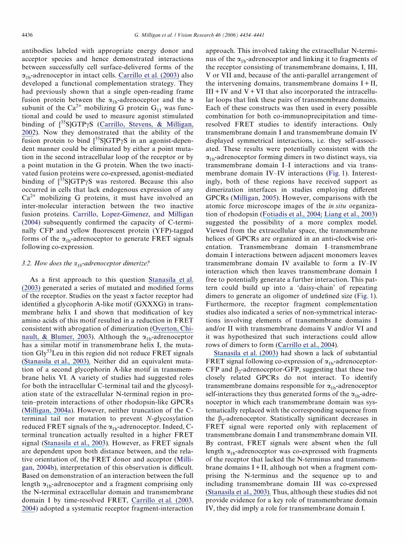
approach. This involved taking the extracellular N-termi-nus of the �1b-adrenoceptor and linking it to fragments ofthe receptor consisting of transmembrane domains, I, III,V or VII and, because of the anti-parallel arrangement ofthe intervening domains, transmembrane domains I + II,III + IV and V + VI that also incorporated the intracellu-lar loops that link these pairs of transmembrane domains.Each of these constructs was then used in every possiblecombination for both co-immunoprecipitation and time-resolved FRET studies to identify interactions. Onlytransmembrane domain I and transmembrane domain IVdisplayed symmetrical interactions, i.e. they self-associ-ated. These results were potentially consistent with the�1b-adrenoceptor forming dimers in two distinct ways, viatransmembrane domain I–I interactions and via trans-membrane domain IV–IV interactions (Fig. 1). Interest-ingly, both of these regions have received support asdimerization interfaces in studies employing diVerentGPCRs (Milligan, 2005). However, comparisons with theatomic force microscope images of the in situ organiza-tion of rhodopsin (Fotiadis et al., 2004; Liang et al., 2003)suggested the possibility of a more complex model.Viewed from the extracellular space, the transmembranehelices of GPCRs are organized in an anti-clockwise ori-entation. Transmembrane domain I–transmembranedomain I interactions between adjacent monomers leavestransmembrane domain IV available to form a IV–IVinteraction which then leaves transmembrane domain Ifree to potentially generate a further interaction. This pat-tern could build up into a ‘daisy-chain’ of repeatingdimers to generate an oligomer of undeWned size (Fig. 1).Furthermore, the receptor fragment complementationstudies also indicated a series of non-symmetrical interac-tions involving elements of transmembrane domains Iand/or II with transmembrane domains V and/or VI andit was hypothesized that such interactions could allowrows of dimers to form (Carrillo et al., 2004).
Stanasila et al. (2003) had shown a lack of substantialFRET signal following co-expression of �1b-adrenoceptor-CFP and �2-adrenoceptor-GFP, suggesting that these twoclosely related GPCRs do not interact. To identifytransmembrane domains responsible for �1b-adrenoceptorself-interactions they thus generated forms of the �1b-adre-noceptor in which each transmembrane domain was sys-tematically replaced with the corresponding sequence fromthe �2-adrenoceptor. Statistically signiWcant decreases inFRET signal were reported only with replacement oftransmembrane domain I and transmembrane domain VII.By contrast, FRET signals were absent when the fulllength �1b-adrenoceptor was co-expressed with fragmentsof the receptor that lacked the N-terminus and transmem-brane domains I + II, although not when a fragment com-prising the N-terminus and the sequence up to andincluding transmembrane domain III was co-expressed(Stanasila et al., 2003). Thus, although these studies did notprovide evidence for a key role of transmembrane domainIV, they did imply a role for transmembrane domain I.

G. Milligan et al. / Vision Research 46 (2006) 4434–4441 4437
Fig. 1. Models of the organization of the �1b-adrenoceptor. Models of the quaternary structure of the �1b-adrenoceptor are based on the anti-clockwiseorientation of transmembrane domains I–VII of individual GPCR polypeptides as viewed from the extracellular space. Data from co-immunoprecipita-tion and time-resolved FRET studies performed using fragments of the �1b-adrenoceptor indicate symmetrical transmembrane domain I-transmembranedomain I interactions and transmembrane domain IV–transmembrane domain IV interactions. These observations are compatible with two distinct waysof forming �1b-adrenoceptor dimers (A and B). However, data also indicate that the �1b-adrenoceptor can exist as a higher-order oligomer. One way thatthis may be achieved, that is consistent with both models A and B, is shown in C (See the text and Carrillo et al., 2004 for further details).
12
34
56
7
12
3 4
56
7
12
34
56
7
12
34
56
7
34
56
7
12
12
3 4
56
7
12
3 4
56
71
23 4
56
7
12
3 4
56
7
A
C
B
Fig. 2. Establishing conditions to monitor sequential three color FRET. CFP can act as an energy donor to YFP in FRET-based studies. YFP can also actas an energy donor to dsRED2. It is conceptually possible to obtain direct energy transfer from CFP to dsRED2. This must be excluded to allow analysisof oligomeric rather than dimeric interactions between proteins tagged with these three Xuorescent polypeptides. A useful means to eliminate sequentialCFP to YFP to dsRED2 FRET is to employ the Y67C mutant of YFP. This mutation ablates the Xuorescence of YFP and this means that Y67C YFP isincapable of acting as either a FRET acceptor (from CFP) or a FRET donor (to dsRED2). Any CFP to dsRED2 FRET observed with excitation at430 nm when Y67C YFP is employed must therefore represent direct CFP to dsRED2 and the diVerence in FRET signal when employing YFP rather thanY67C YFP therefore represents the ‘sequential’ three color signal that can report on oligomeric organization of the �1b-adrenoceptor (see text for furtherdetails). (For interpretation of the references in color in this Wgure legend, the reader is referred to the web version of this article.)
CFP
YPF
DsRed2
Y67CYFP
CFP DsRed2
430 nm
430 nm
470 nm 535 nm
470 nm
630 nm
630 nm
• Direct + sequential FRET
• Direct FRET
470 nm
470 nm

4438 G. Milligan et al. / Vision Research 46 (2006) 4434–4441
3.3. The �1b-adrenoceptor can form an oligomeric complex
Two component FRET cannot usefully discriminatebetween dimers and higher-order structures. Lopez-Gime-nez, Canals, Pediani, and Milligan (2006) therefore devel-oped and employed a sequential three color FRET imagingapproach to gain support for oligomeric structures of the�1b-adrenoceptor in single living cells that might be similarto the observed organization of rhodopsin in rod outer seg-ment discs. Initially, these studies employed a single open-reading frame concatamer of CFP, YFP and DsRed2, asinitially used by others (Galperin, Verkhusha, & Sorkin,2004) to establish appropriate imaging conditions. Follow-ing transfection of this concatamer into HEK293 cells, Xuo-rescence corresponding to each of CFP, YFP and DsRed2could be observed in single cells and with appropriate exci-tation, and the application of a novel FRET algorithm,each of CFP to YFP, YFP to DsRed2 and CFP to DsRed2FRET could be measured in single cells. To prove that theCFP to DsRed2 FRET signal truly represented sequentialCFP to YFP to DsRed2 energy transfer and not direct CFPto DsRed2 FRET, a second concatamer was employed.This was identical to the Wrst except it contained a Tyr67Cysmutation in the YFP element. This mutation ablates
Xuorescence of YFP and therefore Tyr67Cys YFP can act asneither a resonance energy transfer donor nor acceptor.CFP to DsRed2 FRET was abolished, conWrming the CFPto DsRed2 FRET produced from the initial concatamerrepresented sequential CFP to YFP to DsRed2 FRET andnot direct CFP to DsRed2 FRET (Fig. 2).
With this information in place, forms of the �1b-adreno-ceptor C-terminally tagged with each of CFP, YFP andDsRed2 were co-expressed in HEK293 cells and producedCFP to DsRed2 FRET. Such observations are consistentwith at least a proportion of the �1b-adrenoceptor existingin an oligomeric complex (Lopez-Gimenez et al., 2006). Bycontrast, simple co-expression of each of CFP, YFPand DsRed2 produced no such FRET signal. Equallyco-expression of �1b-adrenoceptor-CFP, �1b-adrenoceptor-Tyr67CysYFP and �1b-adrenoceptor-DsRed2 failed to gen-erate CFP to DsRed2 FRET (Lopez-Gimenez et al., 2006).
3.4. Disruption of eVective oligomerization prevents cell surface delivery of the �1b-adrenoceptor
To explore the role of transmembrane domains I and IVin oligomerization, pairs of adjacent, hydrophobic residuesin these regions (Fig. 3) were mutated to alanines and three
Fig. 3. Mutations introduced into the �1b-adrenoceptor to attempt to modulate oligomeric organization. The �1b-adrenoceptor is displayed in a ribbondiagram as a monomeric polypeptide. Based, in part, on the bio-informatic analysis of (de Juan et al., 2005) hydrophobic amino acids at the locationsnoted in transmembrane domains I and IV were mutated to alanines. These amino acids are coded using the nomenclature of Ballesteros and Weinsteinthat relates positions in the transmembrane domains to the location of the most conserved residue in that helix.
L1.52AV1.53A
L4.47AL4.46A
NH2
COOH

G. Milligan et al. / Vision Research 46 (2006) 4434–4441 4439
color FRET studies performed. The mutations resulted inreduced sequential three color FRET consistent with alter-ations in the oligomeric interactions and organization ofthe receptor (Lopez-Gimenez et al., 2006). The functionalconsequences of this were also explored. Immunocyto-chemistry to detect an N-terminal epitope tag demon-strated wild type �1b-adrenoceptor at the cell surface.However, cell surface expression of the mutant �1b-adreno-ceptor could not be detected although, following cell per-meabilization, it was clear that the mutant was expressed aseVectively as the wild type receptor. Good expression of themutated form could also be shown by simply monitoringXuorescence of cells expressing C-terminally YFP-taggedforms of the wild type and mutated �1b-adrenoceptor(Lopez-Gimenez et al., 2006).
Following transfection into a range of cell types(Morris, Price, Smith, Lei, & Schwinn, 2004; Pediani et al.,2005) and in physiological settings (Mackenzie, Daly,Pediani, & McGrath, 2000) wild type �1-adrenoceptorsrecycle between the cell surface and endosomal compart-ments in a constitutive, agonist-independent manner. Thiscan be shown using an anti-epitope tagged antibodylabeled with the pH-sensitive dye CypHer-5 if the receptoris appropriately N-terminally tagged (Adie et al., 2003).CypHer-5 displays red Xuorescence only when in an acidicenvironment, such as an endosome, and addition of thelabeled antibody to cells expressing a suitably tagged�1b-adrenoceptor resulted in the development of red
Xuorescence without addition of an agonist ligand. This isconsistent with the receptor moving from the cell surfaceinto an endosomal pool in a ligand-independent manner.By contrast, for many other GPCRs, including the orexin-1 receptor, this only occurs in the presence of agonist(Milasta et al., 2005). A number of other GPCRs, includ-ing the cannabinoid CB1 receptor (Leterrier, Bonnard,Carrel, Rossier, & Lenkei, 2004; Leterrier et al., 2006),share the characteristic of spontaneous internalizationwith �1-adrenoceptors. Whilst addition of receptor antag-onists restricts the spontaneous recycling of most of theseGPCRs, this is not so for the �1-adrenoceptors. Indeed,�1-adrenoceptors are able to bind cell-impermeable antag-onists whilst at the cell surface and carry these into thecell as the receptor cycles. BODIPY-labeled variants ofantagonists related to the prototypic �1-adrenoceptorblocker prazosin display Xuorescence only when bound tothe receptor (Pediani et al., 2005). They can therefore beused to monitor whether an �1-adrenoceptor had previ-ously been at the cell surface. When a BODIPY-labeledantagonist, that displays red Xuorescence when bound toreceptor, was added to cells expressing a C-terminallyYFP-tagged form of the �1b-adrenoceptor, the cells devel-oped red Xuorescence in punctuate intracellular vesicles(Fig. 4). Pixel by pixel analysis demonstrated strong yel-low/red color overlap (Fig. 4), indicating that the antago-nist remained associated with the receptor inside the cell.This was not observed in cells expressing the mutated
Fig. 4. The wild type �1b-adrenoceptor but not Leu65Ala, Val66Ala, Leu166Ala, Leu167Ala �1b-adrenoceptor is able to reach the cell surface. HEK293 cellswere transfected to express FLAG-tagged forms of wild type �1b-adrenoceptor-YFP (A) or Leu65Ala, Val66Ala, Leu166Ala, Leu167Ala �1b-adrenoceptor-YFP (B). Cells were treated with Hoeschst nuclear stain and Red QAPB for 30 min and imaged; A1, B1 YFP; A2, B2 Hoeschst; A3, B3 Red QAPB; A4, B4
merged images. The ability of the wild type �1b-adrenoceptor to bind Red QAPB is a demonstration that only this form of the receptor was successfullydelivered to the cell surface.
A1 A3 A4A2
B1 B3 B4B2

4440 G. Milligan et al. / Vision Research 46 (2006) 4434–4441
�1b-adrenoceptor, indicating again that the mutatedreceptor had not been delivered to the plasma membrane(Fig. 4).
To explore a possible basis for the lack of cell surfacedelivery of the mutated �1b-adrenoceptor, studies were per-formed to examine receptor N-glycosylation. Although thewild type receptor was present as both apparent 75 and105kDa polypeptides, no 105 kDa form of the mutant wasobserved. De-glycosylation studies demonstrated the105kDa form of the wild type receptor to represent themature form of the protein. Following treatment with N-gly-cosidase F this band was absent and the wild type receptornow migrated with apparent Mr close to 70 kDa (Lopez-Gimenez et al., 2006). The mutant receptor thus appeared tobe unable to become terminally N-glycosylated. An inabilityto be terminally N-glycosylated often is associated with poorplasma membrane delivery and traYcking of mutants of arange of GPCRs which are then sent to the proteasome fordestruction (Petaja-Repo et al., 2001).
3.5. The mutated �1b-adrenoceptor is unable to signal
As the mutated �1b-adrenoceptor appeared unable toreach the cell surface then it was expected to be unable tosignal in response to agonist ligands. This was conWrmed inHEK293 cells expressing either the wild type �1b-adreno-ceptor, C-terminally tagged with YFP or the mutant recep-tor C-terminally tagged with CFP. Such cells were grownon the same cover slip, loaded with the ratiometric Ca2+
indicator dye FURA-2 and exposed to the �1-adrenoceptoragonist phenylephrine. Increases in intracellular Ca2+ wereonly observed in cells expressing the wild type receptor(Lopez-Gimenez et al., 2006). This was not a reXection thatcells expressing the mutated �1b-adrenoceptor were unableto respond to receptor ligands. Subsequent to washout ofthe �1-adrenoceptor agonist, ATP was added to activateP2Y purinoceptors that are expressed endogenously byHEK293 cells. Equivalent increases in intracellular Ca2+
were now observed in all cells.
4. Conclusions
As with many aspects of GPCR structure, function andregulation, understanding of the structural organization ofrhodopsin in rod outer segments has greatly inXuenced ideasabout other members of the rhodopsin-like GPCR family.Although a range of studies have indicated that the �1b-adre-noceptor possesses quaternary structure, it had been unclearif this was restricted solely to the formation of dimers orwhether higher-order complexes might exist. Application ofsequential three color FRET imaging in concert with frag-ment complementation approaches have help to deWne pro-tein–protein interaction interfaces for the �1b-adrenoceptorand indicate similarities in overall organization withrhodopsin. However, these studies have relied on heterolo-gous expression of the �1b-adrenoceptor and it will be impor-tant to develop means to assess if �1b-adrenoceptor
oligomerization can also be observed in native tissues. Itremains to be established whether oligomerization will be ageneral feature of other family A GPCRs and whether themolecular contacts involved will also be highly conserved.Although it appears that rhodopsin may retain functionwhen limited to be a monomer, one role for oligomerizationof the �1b-adrenoceptor appears to be to ensure cell surfacedelivery and hence a means to bind and response to circulat-ing norepinephrine and epinephrine.
References
Adie, E., Francis, M. J., Davies, J., Smith, L., Marenghi, A., Hather, C., etal. (2003). CypHer 5—a generic approach for measuring the activationand traYcking of G-protein-coupled receptors in live cells. Assays andDrug Development Technologies, 1, 251–259.
Carrillo, J. J., Lopez-Gimenez, J. F., & Milligan, G. (2004). Multiple inter-actions between transmembrane helices generate the oligomeric �1b-adrenoceptor. Molecular Pharmacology, 66, 1123–1137.
Carrillo, J. J., Pediani, J., & Milligan, G. (2003). Dimers of class A G pro-tein-coupled receptors function via agonist-mediated trans-activationof associated G proteins. Journal of Biological Chemistry, 278, 42578–42587.
Carrillo, J. J., Stevens, P. A., & Milligan, G. (2002). Measurement of ago-nist-dependent and-independent signal initiation of �1b-adrenoceptormutants by direct analysis of guanine nucleotide exchange on the Gprotein G�11. Journal of Pharmacology and Experimental Therapeutics,302, 1080–1088.
Davies, A., Gowen, B. E., Krebs, A. M., Schertler, G. F., & Saibil, H. R.(2001). Three-dimensional structure of an invertebrate rhodopsin andbasis for ordered alignment in the photoreceptor membrane. Journal ofMolecular Biology, 314, 455–463.
de Juan, D., Mellado, M., Rodriguez-Frade, J. M., Hernanz-Falcon, P.,Serrano, A., Del Sol, A., et al. (2005). A framework for computationaland experimental methods: identifying dimerization residues in CCRchemokine receptors. Bioinformatics, 21(Suppl. 2), ii13–ii18.
Fotiadis, D., Liang, Y., Filipek, S., Saperstein, D. A., Engel, A., & Palczew-ski, K. (2004). The G protein-coupled receptor rhodopsin in the nativemembrane. FEBS Letters, 564, 281–288.
Galperin, E., Verkhusha, V. V., & Sorkin, A. (2004). Three-chromophoreFRET microscopy to analyse multiprotein interactions in living cells.Nature Methods, 1, 209–217.
Guo, W., Shi, L., Filizola, M., Weinstein, H., & Javitch, J. A. (2005). Cross-talk in G protein-coupled receptors: changes at the transmembranehomodimer interface determine activation. Proceedings of the NationalAcademy of Sciences of the United States of America, 102, 17495–17500.
Guo, W., Shi, L., & Javitch, J. A. (2003). The fourth transmembrane seg-ment forms the interface of the dopamine D2 receptor homodimer.Journal of Biological Chemistry, 278, 4385–4388.
Koshimizu, T. A., Tanoue, A., Hirasawa, A., Yamauchi, J., & Tsujimoto,G. (2003). Recent advances in alpha1-adrenoceptor pharmacology.Pharmacology and Therapeutics, 98, 235–244.
Kota, P., Reeves, P. J., Rajbhandary, U. L., & Khorana, H. G. (2006).Opsin is present as dimers in COS1 cells: identiWcation of amino acidsat the dimeric interface. Proceedings of the National Academy of Sci-ences of the United States of America, 103, 3054–3059.
Leterrier, C., Bonnard, D., Carrel, D., Rossier, J., & Lenkei, Z. (2004). Con-stitutive endocytic cycle of the CB1 cannabinoid receptor. Journal ofBiological Chemistry, 279, 36013–36021.
Leterrier, C., Laine, J., Darmon, M., Boudin, H., Rossier, J., & Lenkei, Z.(2006). Constitutive activation drives compartment-selective endocyto-sis and axonal targeting of type 1 cannabinoid receptors. Journal ofNeuroscience, 26, 3141–3153.
Liang, Y., Fotiadis, D., Filipek, S., Saperstein, D. A., Palczewski, K., &Engel, A. (2003). Organization of the G protein-coupled receptors

G. Milligan et al. / Vision Research 46 (2006) 4434–4441 4441
rhodopsin and opsin in native membranes. Journal of Biological Chem-istry, 278, 21655–21662.
Li, J., Edwards, P. C., Burghammer, M., Villa, C., & Schertler, G. F. (2004).Structure of bovine rhodopsin in a trigonal crystal form. Journal ofMolecular Biology, 343, 1409–1438.
Lopez-Gimenez, J. F., Canals, M., Pediani, J. D., & Milligan, G. (2006).Molecular organization of the �1b-adrenoceptor probed using sequen-tial three color Xuorescence resonance energy transfer imaging: eVec-tive oligomerization is required for receptor maturation and function,submitted for publication.
Mackenzie, J. F., Daly, C. J., Pediani, J. D., & McGrath, J. C. (2000). Quan-titative imaging in live human cells reveals intracellular alpha(1)-adre-noceptor ligand-binding sites. Journal of Pharmacology andExperimental Therapeutics, 294, 434–443.
Mansoor, S. E., Palczewski, K., & Farrens, D. L. (2006). Rhodopsin self-associates in asolectin liposomes. Proceedings of the National Academyof Sciences of the United States of America, 103, 3060–3065.
Milasta, S., Evans, N., Ormiston, L., Wilson, S., Lefkowitz, R. J., & Milli-gan, G. (2005). The sustainability of interactions between the orexin-1receptor and �-arrestin-2 is deWned by a single C-terminal cluster ofhydroxy amino acids and modulates the kinetics of ERK MAP kinaseregulation. Biochemical Journal, 387, 573–584.
Milligan, G. (2004a). G protein-coupled receptor dimerization: functionand ligand pharmacology. Molecular Pharmacology, 66, 1–7.
Milligan, G. (2004b). Applications of bioluminescence- and Xuorescenceresonance energy transfer to drug discovery at G protein-coupledreceptors’. European Journal of Pharmaceutical Sciences, 21, 397–405.
Milligan, G. (2005). The molecular basis of dimerisation of family A Gprotein-coupled receptors. In Chui Lundstrom (Ed.), GPCRs in drugdiscovery (pp. 329–340). Marcel Dekker, Inc..
Milligan, G., & Bouvier, M. (2005). Methods to monitor the quaternarystructure of G protein-coupled receptors. FEBS Journal, 272, 2914–2925.
Morris, D. P., Price, R. R., Smith, M. P., Lei, B., & Schwinn, D. A. (2004).Cellular traYcking of human alpha1a-adrenergic receptors is continu-
ous and primarily agonist-independent. Molecular Pharmacology, 66,843–854.
Overton, M. C., Chinault, S. L., & Blumer, K. J. (2003). Oligomerization,biogenesis, and signaling is promoted by a glycophorin A-like dimer-ization motif in transmembrane domain 1 of a yeast G protein-coupledreceptor. Journal of Biological Chemistry, 278, 49369–49377.
Palczewski, K., Kumasaka, T., Hori, T., Behnke, C. A., Motoshima, H.,Fox, B. A., et al. (2000). Crystal structure of rhodopsin: a G protein-coupled receptor. Science, 289, 739–745.
Pediani, J. D., Colston, J. F., Caldwell, D., Milligan, G., Daly, C. J., &McGrath, J. C. (2005). Beta-arrestin-dependent spontaneous alpha1a-adrenoceptor endocytosis causes intracellular transportation of alpha-blockers via recycling compartments. Molecular Pharmacology, 67,992–1004.
Petaja-Repo, U. E., Hogue, M., Laperriere, A., Bhalla, S., Walker, P., &Bouvier, M. (2001). Newly synthesized human delta opioid receptorsretained in the endoplasmic reticulum are retrotranslocated to thecytosol, deglycosylated, ubiquitinated, and degraded by the protea-some. Journal of Biological Chemistry, 276, 4416–4423.
PXeger, K. D., & Eidne, K. A. (2006). Illuminating insights into protein–protein interactions using bioluminescence resonance energy transfer(BRET). Nature Methods, 3, 165–174.
Piascik, M. T., & Perez, D. M. (2001). Alpha1-adrenergic receptors: newinsights and directions. Journal Pharmacology and Experimental Thera-peutics, 298, 403–410.
Stanasila, L., Perez, J. B., Vogel, H., & Cotecchia, S. (2003). Oligomeriza-tion of the alpha 1a- and alpha 1b-adrenergic receptor subtypes. Poten-tial implications in receptor internalization. Journal of BiologicalChemistry, 278, 40239–40251.
Uberti, M. A., Hall, R. A., & Minneman, K. P. (2003). Subtype-speciWc dimer-ization of alpha 1-adrenoceptors: eVects on receptor expression and phar-macological properties. Molecular Pharmacology, 64, 1379–1390.
Zeng, F. Y., & Wess, J. (1999). IdentiWcation and molecular characteriza-tion of m3 muscarinic receptor dimers. Journal of Biological Chemistry,274, 19487–19497.